Notice (8): Undefined variable: solution_of_interest [APP/View/Products/view.ctp, line 755]Code Context<!-- BEGIN: REQUEST_FORM MODAL -->
<div id="request_formModal" class="reveal-modal medium" data-reveal aria-labelledby="modalTitle" aria-hidden="true" role="dialog">
<?= $this->element('Forms/simple_form', array('solution_of_interest' => $solution_of_interest, 'header' => $header, 'message' => $message, 'campaign_id' => $campaign_id)) ?>
$viewFile = '/home/website-server/www/app/View/Products/view.ctp'
$dataForView = array(
'language' => 'en',
'meta_keywords' => '',
'meta_description' => 'H3K27ac (Histone H3 acetylated at lysine 27) Polyclonal Antibody validated in ChIP-seq, ChIP-qPCR, CUT&Tag, ELISA, DB, WB and IF. Batch-specific data available on the website. Sample size available. ',
'meta_title' => 'H3K27ac Antibody - ChIP-seq Grade (C15410196) | Diagenode',
'product' => array(
'Product' => array(
'id' => '2270',
'antibody_id' => '109',
'name' => 'H3K27ac Antibody',
'description' => '<p><span>Polyclonal antibody raised in rabbit against the region of histone <strong>H3 containing the acetylated lysine 27</strong> (<strong>H3K27ac</strong>), using a KLH-conjugated synthetic peptide.</span></p>',
'label1' => 'Validation Data',
'info1' => '<div class="row">
<div class="small-6 columns">A.<img src="https://www.diagenode.com/img/product/antibodies/C15410196-ChIP-Fig1a.png" width="356" /><br /> B.<img src="https://www.diagenode.com/img/product/antibodies/C15410196-ChIP-Fig1b.png" width="356" /></div>
<div class="small-6 columns">
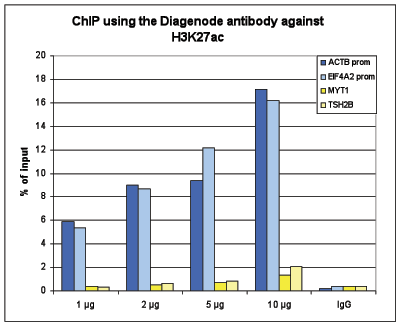
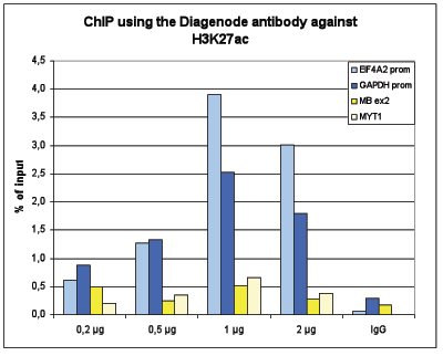
<p><strong>Figure 1. ChIP results obtained with the Diagenode antibody directed against H3K27ac</strong></p>
<p>Figure 1A ChIP assays were performed using human HeLa cells, the Diagenode antibody against H3K27ac (Cat. No. C15410196) and optimized PCR primer pairs for qPCR. ChIP was performed with the “Auto Histone ChIP-seq” kit on the IP-Star automated system, using sheared chromatin from 1,000,000 cells. A titration consisting of 1, 2, 5 and 10 µg of antibody per ChIP experiment was analyzed. IgG (2 µg/IP) was used as a negative IP control. Quantitative PCR was performed with primers for the promoters of the active EIF4A2 and ACTB genes, used as positive controls, and for the inactive TSH2B and MYT1 genes, used as negative controls.</p>
<p>Figure 1B ChIP assays were performed using human K562 cells, the Diagenode antibody against H3K27ac (Cat. No. C15410196)and optimized PCR primer pairs for qPCR. ChIP was performed with the “iDeal ChIP-seq” kit (Cat. No. C01010051), using sheared chromatin from 100,000 cells. A titration consisting of 0.2, 0.5, 1 and 2 µg of antibody per ChIP experiment was analyzed. IgG (1 µg/IP) was used as a negative IP control. Quantitative PCR was performed with primers for the promoters of the active GAPDH and EIF4A2 genes, used as positive controls, and for the coding regions of the inactive MB and MYT1 genes, used as negative controls. Figure 1 shows the recovery, expressed as a % of input (the relative amount of immunoprecipitated DNA compared to input DNA after qPCR analysis)</p>
</div>
</div>
<div class="extra-spaced"></div>
<div class="row">
<div class="small-12 columns"><center>
<p>A.<img src="https://www.diagenode.com/img/product/antibodies/C15410196-ChIP-Fig2a.png" /></p>
</center><center>
<p>B.<img src="https://www.diagenode.com/img/product/antibodies/C15410196-ChIP-Fig2b.png" /></p>
</center><center>
<p>C.<img src="https://www.diagenode.com/img/product/antibodies/C15410196-ChIP-Fig2c.png" /></p>
</center></div>
</div>
<div class="row">
<div class="small-12 columns">
<p><strong>Figure 2. ChIP-seq results obtained with the Diagenode antibody directed against H3K27ac</strong></p>
<p>ChIP was performed on sheared chromatin from 100,000 K562 cells using 1 µg of the Diagenode antibody against H3K27ac (Cat. No. C15410196<span class="label-primary"></span>) as described above. The IP’d DNA was subsequently analysed on an Illumina Genome Analyzer. Library preparation, cluster generation and sequencing were performed according to the manufacturer’s instructions. The 36 bp tags were aligned to the human genome using the ELAND algorithm. Figure 2A shows the peak distribution along the complete human X-chromosome. Figure 2 B and C show the peak distribution in two regions surrounding the EIF4A2 and GAPDH positive control genes, respectively. The position of the PCR amplicon, used for validating the ChIP assay is indicated with an arrow.</p>
</div>
</div>
<div class="row">
<div class="small-12 columns"><center><img src="https://www.diagenode.com/img/product/antibodies/C15410196-fig3.jpg" /></center></div>
</div>
<div class="row">
<div class="small-12 columns">
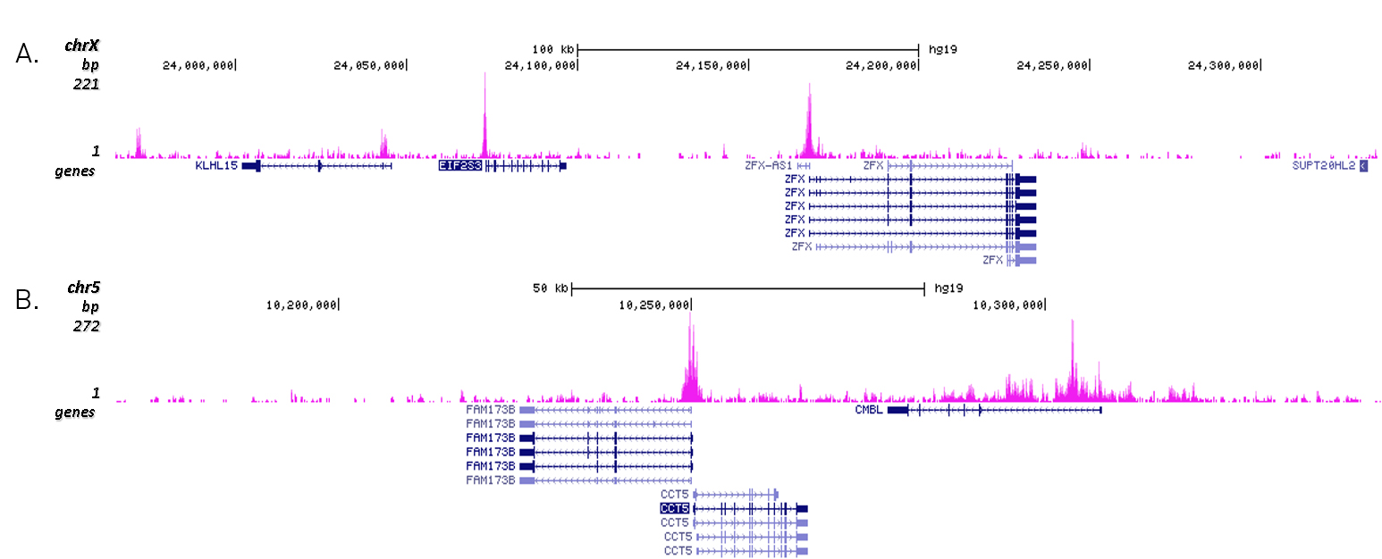
<p><strong>Figure 3. Cut&Tag results obtained with the Diagenode antibody directed against H3K27ac</strong></p>
<p>CUT&TAG (Kaya-Okur, H.S., Nat Commun 10, 1930, 2019) was performed on 50,000 K562 cells using 1 µg of the Diagenode antibody against H3K27ac (cat. No. C15410196) and the Diagenode pA-Tn5 transposase (C01070001). The libraries were subsequently analysed on an Illumina NextSeq 500 sequencer (2x75 paired-end reads) according to the manufacturer's instructions. The tags were aligned to the human genome (hg19) using the BWA algorithm. Figure 3 shows the peak distribution in 2 genomic regions surrounding the EIF2S3 gene on the X-chromosome and the CCT5 gene on chromosome 5 (figure 3A and B, respectively).</p>
</div>
</div>
<div class="row">
<div class="small-6 columns"><img src="https://www.diagenode.com/img/product/antibodies/C15410196-ELISA-Fig3.png" /></div>
<div class="small-6 columns">
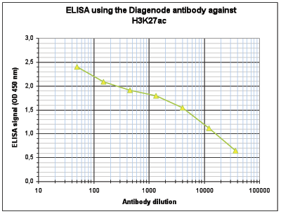
<p><strong>Figure 4. Determination of the antibody titer</strong></p>
<p>To determine the titer of the antibody, an ELISA was performed using a serial dilution of the Diagenode antibody against H3K27ac (Cat. No. C15410196<span class="label-primary"></span>). The antigen used was a peptide containing the histone modification of interest. By plotting the absorbance against the antibody dilution (Figure 4), the titer of the antibody was estimated to be 1:8,300.</p>
</div>
</div>
<div class="row">
<div class="small-4 columns"><center><img src="https://www.diagenode.com/img/product/antibodies/C15410196-DB-Fig4.png" /></center></div>
<div class="small-8 columns">
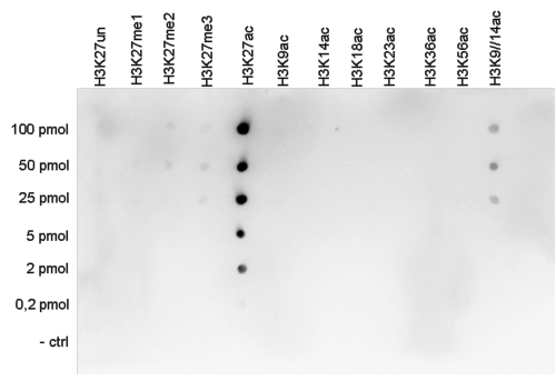
<p><strong>Figure 5. Cross reactivity tests using the Diagenode antibody directed against H3K27ac</strong><br />To test the cross reactivity of the Diagenode antibody against H3K27ac (Cat. No. C15410196<span class="label-primary"></span>), a Dot Blot analysis was performed with peptides containing other histone modifications and the unmodified H3K27. One hundred to 0.2 pmol of the respective peptides were spotted on a membrane. The antibody was used at a dilution of 1:20,000. Figure 5 shows a high specificity of the antibody for the modification of interest.</p>
</div>
</div>
<div class="row">
<div class="small-4 columns"><center><img src="https://www.diagenode.com/img/product/antibodies/C15410196-WB-Fig5.png" /></center></div>
<div class="small-8 columns">
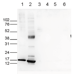
<p><strong>Figure 6. Western blot analysis using the Diagenode antibody directed against H3K27ac</strong><br />Western blot was performed on whole cell (25 µg, lane 1) and histone extracts (15 µg, lane 2) from HeLa cells, and on 1 µg of recombinant histone H2A, H2B, H3 and H4 (lane 3, 4, 5 and 6, respectively) using the Diagenode antibody against H3K27ac (Cat. No. C1541196). The antibody was diluted 1:1,000 in TBS-Tween containing 5% skimmed milk. The marker (in kDa) is shown on the left.</p>
</div>
</div>
<div class="row">
<div class="small-4 columns"><img src="https://www.diagenode.com/img/product/antibodies/C15410196-IF-Fig6.png" /></div>
<div class="small-8 columns">
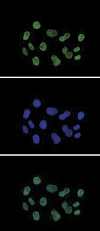
<p><strong>Figure 7. Immunofluorescence using the Diagenode antibody directed against H3K27ac</strong></p>
<p>HeLa cells were stained with the Diagenode antibody against H3K27ac (Cat. No. C15410196<span class="label-primary"></span>) and with DAPI. Cells were fixed with 4% formaldehyde for 10’ and blocked with PBS/ TX-100 containing 5% normal goat serum and 1% BSA. The cells were immunofluorescently labeled with the H3K27ac antibody (top) diluted 1:500 in blocking solution followed by an anti-rabbit antibody conjugated to Alexa488. The middle panel shows staining of the nuclei with DAPI. A merge of the two stainings is shown at the bottom.</p>
</div>
</div>',
'label2' => 'Target Description',
'info2' => '<p style="text-align: justify;">Histones are the main constituents of the protein part of chromosomes of eukaryotic cells. They are rich in the amino acids arginine and lysine and have been greatly conserved during evolution. Histones pack the DNA into tight masses of chromatin. Two core histones of each class H2A, H2B, H3 and H4 assemble and are wrapped by 146 base pairs of DNA to form one octameric nucleosome. Histone tails undergo numerous post-translational modifications, which either directly or indirectly alter chromatin structure to facilitate transcriptional activation or repression or other nuclear processes. In addition to the genetic code, combinations of the different histone modifications reveal the so-called “histone code”. Histone methylation and demethylation is dynamically regulated by respectively histone methyl transferases and histone demethylases. Acetylation of histone H3K27 is associated with active promoters and enhancers.</p>',
'label3' => '',
'info3' => '',
'format' => '50 μg',
'catalog_number' => 'C15410196',
'old_catalog_number' => 'pAb-196-050',
'sf_code' => 'C15410196-D001-000581',
'type' => 'FRE',
'search_order' => '03-Antibody',
'price_EUR' => '480',
'price_USD' => '470',
'price_GBP' => '430',
'price_JPY' => '75190',
'price_CNY' => '',
'price_AUD' => '1175',
'country' => 'ALL',
'except_countries' => 'None',
'quote' => false,
'in_stock' => false,
'featured' => false,
'no_promo' => false,
'online' => true,
'master' => true,
'last_datasheet_update' => 'January 11, 2021',
'slug' => 'h3k27ac-polyclonal-antibody-premium-50-mg-18-ml',
'meta_title' => 'H3K27ac Antibody - ChIP-seq Grade (C15410196) | Diagenode',
'meta_keywords' => '',
'meta_description' => 'H3K27ac (Histone H3 acetylated at lysine 27) Polyclonal Antibody validated in ChIP-seq, ChIP-qPCR, CUT&Tag, ELISA, DB, WB and IF. Batch-specific data available on the website. Sample size available. ',
'modified' => '2021-10-20 10:28:57',
'created' => '2015-06-29 14:08:20',
'locale' => 'eng'
),
'Antibody' => array(
'host' => '*****',
'id' => '109',
'name' => 'H3K27ac polyclonal antibody',
'description' => 'Histones are the main constituents of the protein part of chromosomes of eukaryotic cells. They are rich in the amino acids arginine and lysine and have been greatly conserved during evolution. Histones pack the DNA into tight masses of chromatin. Two core histones of each class H2A, H2B, H3 and H4 assemble and are wrapped by 146 base pairs of DNA to form one octameric nucleosome. Histone tails undergo numerous post-translational modifications, which either directly or indirectly alter chromatin structure to facilitate transcriptional activation or repression or other nuclear processes. In addition to the genetic code, combinations of the different histone modifications reveal the so-called “histone code”. Histone methylation and demethylation is dynamically regulated by respectively histone methyl transferases and histone demethylases. Acetylation of histone H3K27 is associated with active promoters and enhancers.',
'clonality' => '',
'isotype' => '',
'lot' => 'A1723-0041D',
'concentration' => '2.8 µg/µl',
'reactivity' => 'Human, mouse, rat, Arabidopsis, wide range expected',
'type' => 'Polyclonal,<strong>ChIP grade, ChIP-seq grade</strong>',
'purity' => 'Affinity purified',
'classification' => 'Premium',
'application_table' => '<table>
<thead>
<tr>
<th>Applications</th>
<th>Suggested dilution</th>
<th>References</th>
</tr>
</thead>
<tbody>
<tr>
<td>ChIP/ChIP-seq <sup>*</sup></td>
<td>1 μg/IP</td>
<td>Fig 1, 2</td>
</tr>
<tr>
<td>CUT&TAG</td>
<td>1 μg</td>
<td>Fig 3</td>
</tr>
<tr>
<td>ELISA</td>
<td>1:500</td>
<td>Fig 4</td>
</tr>
<tr>
<td>Dot Blotting</td>
<td>1:20,000</td>
<td>Fig 5</td>
</tr>
<tr>
<td>Western Blotting</td>
<td>1:1,000</td>
<td>Fig 6</td>
</tr>
<tr>
<td>Immunofluorescence</td>
<td>1:500</td>
<td>Fig 7</td>
</tr>
</tbody>
</table>
<p></p>
<p><small><sup>*</sup> Please note that the optimal antibody amount per IP should be determined by the end-user. We recommend testing 0.5-5 µg per IP.</small></p>',
'storage_conditions' => 'Store at -20°C; for long storage, store at -80°C. Avoid multiple freeze-thaw cycles.',
'storage_buffer' => 'PBS containing 0.05% azide and 0.05% ProClin 300.',
'precautions' => 'This product is for research use only. Not for use in diagnostic or therapeutic procedures.',
'uniprot_acc' => '',
'slug' => '',
'meta_keywords' => '',
'meta_description' => '',
'modified' => '2021-01-13 17:16:29',
'created' => '0000-00-00 00:00:00',
'select_label' => '109 - H3K27ac polyclonal antibody (A1723-0041D - 2.8 µg/µl - Human, mouse, rat, Arabidopsis, wide range expected - Affinity purified - Rabbit)'
),
'Slave' => array(
(int) 0 => array(
[maximum depth reached]
)
),
'Group' => array(
'Group' => array(
[maximum depth reached]
),
'Master' => array(
[maximum depth reached]
),
'Product' => array(
[maximum depth reached]
)
),
'Related' => array(
(int) 0 => array(
[maximum depth reached]
),
(int) 1 => array(
[maximum depth reached]
),
(int) 2 => array(
[maximum depth reached]
),
(int) 3 => array(
[maximum depth reached]
),
(int) 4 => array(
[maximum depth reached]
)
),
'Application' => array(
(int) 0 => array(
[maximum depth reached]
),
(int) 1 => array(
[maximum depth reached]
),
(int) 2 => array(
[maximum depth reached]
),
(int) 3 => array(
[maximum depth reached]
),
(int) 4 => array(
[maximum depth reached]
),
(int) 5 => array(
[maximum depth reached]
),
(int) 6 => array(
[maximum depth reached]
)
),
'Category' => array(
(int) 0 => array(
[maximum depth reached]
),
(int) 1 => array(
[maximum depth reached]
),
(int) 2 => array(
[maximum depth reached]
),
(int) 3 => array(
[maximum depth reached]
)
),
'Document' => array(
(int) 0 => array(
[maximum depth reached]
),
(int) 1 => array(
[maximum depth reached]
),
(int) 2 => array(
[maximum depth reached]
),
(int) 3 => array(
[maximum depth reached]
),
(int) 4 => array(
[maximum depth reached]
),
(int) 5 => array(
[maximum depth reached]
)
),
'Feature' => array(),
'Image' => array(
(int) 0 => array(
[maximum depth reached]
)
),
'Promotion' => array(),
'Protocol' => array(),
'Publication' => array(
(int) 0 => array(
[maximum depth reached]
),
(int) 1 => array(
[maximum depth reached]
),
(int) 2 => array(
[maximum depth reached]
),
(int) 3 => array(
[maximum depth reached]
),
(int) 4 => array(
[maximum depth reached]
),
(int) 5 => array(
[maximum depth reached]
),
(int) 6 => array(
[maximum depth reached]
),
(int) 7 => array(
[maximum depth reached]
),
(int) 8 => array(
[maximum depth reached]
),
(int) 9 => array(
[maximum depth reached]
),
(int) 10 => array(
[maximum depth reached]
),
(int) 11 => array(
[maximum depth reached]
),
(int) 12 => array(
[maximum depth reached]
),
(int) 13 => array(
[maximum depth reached]
),
(int) 14 => array(
[maximum depth reached]
),
(int) 15 => array(
[maximum depth reached]
),
(int) 16 => array(
[maximum depth reached]
),
(int) 17 => array(
[maximum depth reached]
),
(int) 18 => array(
[maximum depth reached]
),
(int) 19 => array(
[maximum depth reached]
),
(int) 20 => array(
[maximum depth reached]
),
(int) 21 => array(
[maximum depth reached]
),
(int) 22 => array(
[maximum depth reached]
),
(int) 23 => array(
[maximum depth reached]
),
(int) 24 => array(
[maximum depth reached]
),
(int) 25 => array(
[maximum depth reached]
),
(int) 26 => array(
[maximum depth reached]
),
(int) 27 => array(
[maximum depth reached]
),
(int) 28 => array(
[maximum depth reached]
),
(int) 29 => array(
[maximum depth reached]
),
(int) 30 => array(
[maximum depth reached]
),
(int) 31 => array(
[maximum depth reached]
),
(int) 32 => array(
[maximum depth reached]
),
(int) 33 => array(
[maximum depth reached]
),
(int) 34 => array(
[maximum depth reached]
),
(int) 35 => array(
[maximum depth reached]
),
(int) 36 => array(
[maximum depth reached]
),
(int) 37 => array(
[maximum depth reached]
),
(int) 38 => array(
[maximum depth reached]
),
(int) 39 => array(
[maximum depth reached]
),
(int) 40 => array(
[maximum depth reached]
),
(int) 41 => array(
[maximum depth reached]
),
(int) 42 => array(
[maximum depth reached]
),
(int) 43 => array(
[maximum depth reached]
),
(int) 44 => array(
[maximum depth reached]
),
(int) 45 => array(
[maximum depth reached]
),
(int) 46 => array(
[maximum depth reached]
),
(int) 47 => array(
[maximum depth reached]
),
(int) 48 => array(
[maximum depth reached]
),
(int) 49 => array(
[maximum depth reached]
),
(int) 50 => array(
[maximum depth reached]
),
(int) 51 => array(
[maximum depth reached]
),
(int) 52 => array(
[maximum depth reached]
),
(int) 53 => array(
[maximum depth reached]
),
(int) 54 => array(
[maximum depth reached]
),
(int) 55 => array(
[maximum depth reached]
),
(int) 56 => array(
[maximum depth reached]
),
(int) 57 => array(
[maximum depth reached]
),
(int) 58 => array(
[maximum depth reached]
),
(int) 59 => array(
[maximum depth reached]
),
(int) 60 => array(
[maximum depth reached]
),
(int) 61 => array(
[maximum depth reached]
),
(int) 62 => array(
[maximum depth reached]
),
(int) 63 => array(
[maximum depth reached]
),
(int) 64 => array(
[maximum depth reached]
),
(int) 65 => array(
[maximum depth reached]
),
(int) 66 => array(
[maximum depth reached]
),
(int) 67 => array(
[maximum depth reached]
),
(int) 68 => array(
[maximum depth reached]
),
(int) 69 => array(
[maximum depth reached]
),
(int) 70 => array(
[maximum depth reached]
),
(int) 71 => array(
[maximum depth reached]
),
(int) 72 => array(
[maximum depth reached]
),
(int) 73 => array(
[maximum depth reached]
),
(int) 74 => array(
[maximum depth reached]
),
(int) 75 => array(
[maximum depth reached]
),
(int) 76 => array(
[maximum depth reached]
),
(int) 77 => array(
[maximum depth reached]
),
(int) 78 => array(
[maximum depth reached]
),
(int) 79 => array(
[maximum depth reached]
),
(int) 80 => array(
[maximum depth reached]
),
(int) 81 => array(
[maximum depth reached]
),
(int) 82 => array(
[maximum depth reached]
),
(int) 83 => array(
[maximum depth reached]
),
(int) 84 => array(
[maximum depth reached]
),
(int) 85 => array(
[maximum depth reached]
),
(int) 86 => array(
[maximum depth reached]
),
(int) 87 => array(
[maximum depth reached]
),
(int) 88 => array(
[maximum depth reached]
),
(int) 89 => array(
[maximum depth reached]
),
(int) 90 => array(
[maximum depth reached]
),
(int) 91 => array(
[maximum depth reached]
),
(int) 92 => array(
[maximum depth reached]
),
(int) 93 => array(
[maximum depth reached]
),
(int) 94 => array(
[maximum depth reached]
),
(int) 95 => array(
[maximum depth reached]
),
(int) 96 => array(
[maximum depth reached]
),
(int) 97 => array(
[maximum depth reached]
),
(int) 98 => array(
[maximum depth reached]
),
(int) 99 => array(
[maximum depth reached]
),
(int) 100 => array(
[maximum depth reached]
),
(int) 101 => array(
[maximum depth reached]
),
(int) 102 => array(
[maximum depth reached]
),
(int) 103 => array(
[maximum depth reached]
),
(int) 104 => array(
[maximum depth reached]
),
(int) 105 => array(
[maximum depth reached]
),
(int) 106 => array(
[maximum depth reached]
),
(int) 107 => array(
[maximum depth reached]
),
(int) 108 => array(
[maximum depth reached]
),
(int) 109 => array(
[maximum depth reached]
),
(int) 110 => array(
[maximum depth reached]
),
(int) 111 => array(
[maximum depth reached]
),
(int) 112 => array(
[maximum depth reached]
),
(int) 113 => array(
[maximum depth reached]
),
(int) 114 => array(
[maximum depth reached]
),
(int) 115 => array(
[maximum depth reached]
),
(int) 116 => array(
[maximum depth reached]
),
(int) 117 => array(
[maximum depth reached]
),
(int) 118 => array(
[maximum depth reached]
),
(int) 119 => array(
[maximum depth reached]
),
(int) 120 => array(
[maximum depth reached]
),
(int) 121 => array(
[maximum depth reached]
),
(int) 122 => array(
[maximum depth reached]
),
(int) 123 => array(
[maximum depth reached]
),
(int) 124 => array(
[maximum depth reached]
),
(int) 125 => array(
[maximum depth reached]
),
(int) 126 => array(
[maximum depth reached]
),
(int) 127 => array(
[maximum depth reached]
),
(int) 128 => array(
[maximum depth reached]
),
(int) 129 => array(
[maximum depth reached]
),
(int) 130 => array(
[maximum depth reached]
),
(int) 131 => array(
[maximum depth reached]
),
(int) 132 => array(
[maximum depth reached]
),
(int) 133 => array(
[maximum depth reached]
),
(int) 134 => array(
[maximum depth reached]
),
(int) 135 => array(
[maximum depth reached]
),
(int) 136 => array(
[maximum depth reached]
),
(int) 137 => array(
[maximum depth reached]
),
(int) 138 => array(
[maximum depth reached]
),
(int) 139 => array(
[maximum depth reached]
),
(int) 140 => array(
[maximum depth reached]
),
(int) 141 => array(
[maximum depth reached]
),
(int) 142 => array(
[maximum depth reached]
),
(int) 143 => array(
[maximum depth reached]
)
),
'Testimonial' => array(
(int) 0 => array(
[maximum depth reached]
)
),
'Area' => array(),
'SafetySheet' => array(
(int) 0 => array(
[maximum depth reached]
),
(int) 1 => array(
[maximum depth reached]
),
(int) 2 => array(
[maximum depth reached]
),
(int) 3 => array(
[maximum depth reached]
),
(int) 4 => array(
[maximum depth reached]
),
(int) 5 => array(
[maximum depth reached]
),
(int) 6 => array(
[maximum depth reached]
),
(int) 7 => array(
[maximum depth reached]
)
)
),
'meta_canonical' => 'https://www.diagenode.com/en/p/h3k27ac-polyclonal-antibody-premium-50-mg-18-ml'
)
$language = 'en'
$meta_keywords = ''
$meta_description = 'H3K27ac (Histone H3 acetylated at lysine 27) Polyclonal Antibody validated in ChIP-seq, ChIP-qPCR, CUT&Tag, ELISA, DB, WB and IF. Batch-specific data available on the website. Sample size available. '
$meta_title = 'H3K27ac Antibody - ChIP-seq Grade (C15410196) | Diagenode'
$product = array(
'Product' => array(
'id' => '2270',
'antibody_id' => '109',
'name' => 'H3K27ac Antibody',
'description' => '<p><span>Polyclonal antibody raised in rabbit against the region of histone <strong>H3 containing the acetylated lysine 27</strong> (<strong>H3K27ac</strong>), using a KLH-conjugated synthetic peptide.</span></p>',
'label1' => 'Validation Data',
'info1' => '<div class="row">
<div class="small-6 columns">A.<img src="https://www.diagenode.com/img/product/antibodies/C15410196-ChIP-Fig1a.png" width="356" /><br /> B.<img src="https://www.diagenode.com/img/product/antibodies/C15410196-ChIP-Fig1b.png" width="356" /></div>
<div class="small-6 columns">
<p><strong>Figure 1. ChIP results obtained with the Diagenode antibody directed against H3K27ac</strong></p>
<p>Figure 1A ChIP assays were performed using human HeLa cells, the Diagenode antibody against H3K27ac (Cat. No. C15410196) and optimized PCR primer pairs for qPCR. ChIP was performed with the “Auto Histone ChIP-seq” kit on the IP-Star automated system, using sheared chromatin from 1,000,000 cells. A titration consisting of 1, 2, 5 and 10 µg of antibody per ChIP experiment was analyzed. IgG (2 µg/IP) was used as a negative IP control. Quantitative PCR was performed with primers for the promoters of the active EIF4A2 and ACTB genes, used as positive controls, and for the inactive TSH2B and MYT1 genes, used as negative controls.</p>
<p>Figure 1B ChIP assays were performed using human K562 cells, the Diagenode antibody against H3K27ac (Cat. No. C15410196)and optimized PCR primer pairs for qPCR. ChIP was performed with the “iDeal ChIP-seq” kit (Cat. No. C01010051), using sheared chromatin from 100,000 cells. A titration consisting of 0.2, 0.5, 1 and 2 µg of antibody per ChIP experiment was analyzed. IgG (1 µg/IP) was used as a negative IP control. Quantitative PCR was performed with primers for the promoters of the active GAPDH and EIF4A2 genes, used as positive controls, and for the coding regions of the inactive MB and MYT1 genes, used as negative controls. Figure 1 shows the recovery, expressed as a % of input (the relative amount of immunoprecipitated DNA compared to input DNA after qPCR analysis)</p>
</div>
</div>
<div class="extra-spaced"></div>
<div class="row">
<div class="small-12 columns"><center>
<p>A.<img src="https://www.diagenode.com/img/product/antibodies/C15410196-ChIP-Fig2a.png" /></p>
</center><center>
<p>B.<img src="https://www.diagenode.com/img/product/antibodies/C15410196-ChIP-Fig2b.png" /></p>
</center><center>
<p>C.<img src="https://www.diagenode.com/img/product/antibodies/C15410196-ChIP-Fig2c.png" /></p>
</center></div>
</div>
<div class="row">
<div class="small-12 columns">
<p><strong>Figure 2. ChIP-seq results obtained with the Diagenode antibody directed against H3K27ac</strong></p>
<p>ChIP was performed on sheared chromatin from 100,000 K562 cells using 1 µg of the Diagenode antibody against H3K27ac (Cat. No. C15410196<span class="label-primary"></span>) as described above. The IP’d DNA was subsequently analysed on an Illumina Genome Analyzer. Library preparation, cluster generation and sequencing were performed according to the manufacturer’s instructions. The 36 bp tags were aligned to the human genome using the ELAND algorithm. Figure 2A shows the peak distribution along the complete human X-chromosome. Figure 2 B and C show the peak distribution in two regions surrounding the EIF4A2 and GAPDH positive control genes, respectively. The position of the PCR amplicon, used for validating the ChIP assay is indicated with an arrow.</p>
</div>
</div>
<div class="row">
<div class="small-12 columns"><center><img src="https://www.diagenode.com/img/product/antibodies/C15410196-fig3.jpg" /></center></div>
</div>
<div class="row">
<div class="small-12 columns">
<p><strong>Figure 3. Cut&Tag results obtained with the Diagenode antibody directed against H3K27ac</strong></p>
<p>CUT&TAG (Kaya-Okur, H.S., Nat Commun 10, 1930, 2019) was performed on 50,000 K562 cells using 1 µg of the Diagenode antibody against H3K27ac (cat. No. C15410196) and the Diagenode pA-Tn5 transposase (C01070001). The libraries were subsequently analysed on an Illumina NextSeq 500 sequencer (2x75 paired-end reads) according to the manufacturer's instructions. The tags were aligned to the human genome (hg19) using the BWA algorithm. Figure 3 shows the peak distribution in 2 genomic regions surrounding the EIF2S3 gene on the X-chromosome and the CCT5 gene on chromosome 5 (figure 3A and B, respectively).</p>
</div>
</div>
<div class="row">
<div class="small-6 columns"><img src="https://www.diagenode.com/img/product/antibodies/C15410196-ELISA-Fig3.png" /></div>
<div class="small-6 columns">
<p><strong>Figure 4. Determination of the antibody titer</strong></p>
<p>To determine the titer of the antibody, an ELISA was performed using a serial dilution of the Diagenode antibody against H3K27ac (Cat. No. C15410196<span class="label-primary"></span>). The antigen used was a peptide containing the histone modification of interest. By plotting the absorbance against the antibody dilution (Figure 4), the titer of the antibody was estimated to be 1:8,300.</p>
</div>
</div>
<div class="row">
<div class="small-4 columns"><center><img src="https://www.diagenode.com/img/product/antibodies/C15410196-DB-Fig4.png" /></center></div>
<div class="small-8 columns">
<p><strong>Figure 5. Cross reactivity tests using the Diagenode antibody directed against H3K27ac</strong><br />To test the cross reactivity of the Diagenode antibody against H3K27ac (Cat. No. C15410196<span class="label-primary"></span>), a Dot Blot analysis was performed with peptides containing other histone modifications and the unmodified H3K27. One hundred to 0.2 pmol of the respective peptides were spotted on a membrane. The antibody was used at a dilution of 1:20,000. Figure 5 shows a high specificity of the antibody for the modification of interest.</p>
</div>
</div>
<div class="row">
<div class="small-4 columns"><center><img src="https://www.diagenode.com/img/product/antibodies/C15410196-WB-Fig5.png" /></center></div>
<div class="small-8 columns">
<p><strong>Figure 6. Western blot analysis using the Diagenode antibody directed against H3K27ac</strong><br />Western blot was performed on whole cell (25 µg, lane 1) and histone extracts (15 µg, lane 2) from HeLa cells, and on 1 µg of recombinant histone H2A, H2B, H3 and H4 (lane 3, 4, 5 and 6, respectively) using the Diagenode antibody against H3K27ac (Cat. No. C1541196). The antibody was diluted 1:1,000 in TBS-Tween containing 5% skimmed milk. The marker (in kDa) is shown on the left.</p>
</div>
</div>
<div class="row">
<div class="small-4 columns"><img src="https://www.diagenode.com/img/product/antibodies/C15410196-IF-Fig6.png" /></div>
<div class="small-8 columns">
<p><strong>Figure 7. Immunofluorescence using the Diagenode antibody directed against H3K27ac</strong></p>
<p>HeLa cells were stained with the Diagenode antibody against H3K27ac (Cat. No. C15410196<span class="label-primary"></span>) and with DAPI. Cells were fixed with 4% formaldehyde for 10’ and blocked with PBS/ TX-100 containing 5% normal goat serum and 1% BSA. The cells were immunofluorescently labeled with the H3K27ac antibody (top) diluted 1:500 in blocking solution followed by an anti-rabbit antibody conjugated to Alexa488. The middle panel shows staining of the nuclei with DAPI. A merge of the two stainings is shown at the bottom.</p>
</div>
</div>',
'label2' => 'Target Description',
'info2' => '<p style="text-align: justify;">Histones are the main constituents of the protein part of chromosomes of eukaryotic cells. They are rich in the amino acids arginine and lysine and have been greatly conserved during evolution. Histones pack the DNA into tight masses of chromatin. Two core histones of each class H2A, H2B, H3 and H4 assemble and are wrapped by 146 base pairs of DNA to form one octameric nucleosome. Histone tails undergo numerous post-translational modifications, which either directly or indirectly alter chromatin structure to facilitate transcriptional activation or repression or other nuclear processes. In addition to the genetic code, combinations of the different histone modifications reveal the so-called “histone code”. Histone methylation and demethylation is dynamically regulated by respectively histone methyl transferases and histone demethylases. Acetylation of histone H3K27 is associated with active promoters and enhancers.</p>',
'label3' => '',
'info3' => '',
'format' => '50 μg',
'catalog_number' => 'C15410196',
'old_catalog_number' => 'pAb-196-050',
'sf_code' => 'C15410196-D001-000581',
'type' => 'FRE',
'search_order' => '03-Antibody',
'price_EUR' => '480',
'price_USD' => '470',
'price_GBP' => '430',
'price_JPY' => '75190',
'price_CNY' => '',
'price_AUD' => '1175',
'country' => 'ALL',
'except_countries' => 'None',
'quote' => false,
'in_stock' => false,
'featured' => false,
'no_promo' => false,
'online' => true,
'master' => true,
'last_datasheet_update' => 'January 11, 2021',
'slug' => 'h3k27ac-polyclonal-antibody-premium-50-mg-18-ml',
'meta_title' => 'H3K27ac Antibody - ChIP-seq Grade (C15410196) | Diagenode',
'meta_keywords' => '',
'meta_description' => 'H3K27ac (Histone H3 acetylated at lysine 27) Polyclonal Antibody validated in ChIP-seq, ChIP-qPCR, CUT&Tag, ELISA, DB, WB and IF. Batch-specific data available on the website. Sample size available. ',
'modified' => '2021-10-20 10:28:57',
'created' => '2015-06-29 14:08:20',
'locale' => 'eng'
),
'Antibody' => array(
'host' => '*****',
'id' => '109',
'name' => 'H3K27ac polyclonal antibody',
'description' => 'Histones are the main constituents of the protein part of chromosomes of eukaryotic cells. They are rich in the amino acids arginine and lysine and have been greatly conserved during evolution. Histones pack the DNA into tight masses of chromatin. Two core histones of each class H2A, H2B, H3 and H4 assemble and are wrapped by 146 base pairs of DNA to form one octameric nucleosome. Histone tails undergo numerous post-translational modifications, which either directly or indirectly alter chromatin structure to facilitate transcriptional activation or repression or other nuclear processes. In addition to the genetic code, combinations of the different histone modifications reveal the so-called “histone code”. Histone methylation and demethylation is dynamically regulated by respectively histone methyl transferases and histone demethylases. Acetylation of histone H3K27 is associated with active promoters and enhancers.',
'clonality' => '',
'isotype' => '',
'lot' => 'A1723-0041D',
'concentration' => '2.8 µg/µl',
'reactivity' => 'Human, mouse, rat, Arabidopsis, wide range expected',
'type' => 'Polyclonal,<strong>ChIP grade, ChIP-seq grade</strong>',
'purity' => 'Affinity purified',
'classification' => 'Premium',
'application_table' => '<table>
<thead>
<tr>
<th>Applications</th>
<th>Suggested dilution</th>
<th>References</th>
</tr>
</thead>
<tbody>
<tr>
<td>ChIP/ChIP-seq <sup>*</sup></td>
<td>1 μg/IP</td>
<td>Fig 1, 2</td>
</tr>
<tr>
<td>CUT&TAG</td>
<td>1 μg</td>
<td>Fig 3</td>
</tr>
<tr>
<td>ELISA</td>
<td>1:500</td>
<td>Fig 4</td>
</tr>
<tr>
<td>Dot Blotting</td>
<td>1:20,000</td>
<td>Fig 5</td>
</tr>
<tr>
<td>Western Blotting</td>
<td>1:1,000</td>
<td>Fig 6</td>
</tr>
<tr>
<td>Immunofluorescence</td>
<td>1:500</td>
<td>Fig 7</td>
</tr>
</tbody>
</table>
<p></p>
<p><small><sup>*</sup> Please note that the optimal antibody amount per IP should be determined by the end-user. We recommend testing 0.5-5 µg per IP.</small></p>',
'storage_conditions' => 'Store at -20°C; for long storage, store at -80°C. Avoid multiple freeze-thaw cycles.',
'storage_buffer' => 'PBS containing 0.05% azide and 0.05% ProClin 300.',
'precautions' => 'This product is for research use only. Not for use in diagnostic or therapeutic procedures.',
'uniprot_acc' => '',
'slug' => '',
'meta_keywords' => '',
'meta_description' => '',
'modified' => '2021-01-13 17:16:29',
'created' => '0000-00-00 00:00:00',
'select_label' => '109 - H3K27ac polyclonal antibody (A1723-0041D - 2.8 µg/µl - Human, mouse, rat, Arabidopsis, wide range expected - Affinity purified - Rabbit)'
),
'Slave' => array(
(int) 0 => array(
'id' => '23',
'name' => 'C15410196',
'product_id' => '2270',
'modified' => '2016-02-18 18:04:33',
'created' => '2016-02-18 18:04:33'
)
),
'Group' => array(
'Group' => array(
'id' => '23',
'name' => 'C15410196',
'product_id' => '2270',
'modified' => '2016-02-18 18:04:33',
'created' => '2016-02-18 18:04:33'
),
'Master' => array(
'id' => '2270',
'antibody_id' => '109',
'name' => 'H3K27ac Antibody',
'description' => '<p><span>Polyclonal antibody raised in rabbit against the region of histone <strong>H3 containing the acetylated lysine 27</strong> (<strong>H3K27ac</strong>), using a KLH-conjugated synthetic peptide.</span></p>',
'label1' => 'Validation Data',
'info1' => '<div class="row">
<div class="small-6 columns">A.<img src="https://www.diagenode.com/img/product/antibodies/C15410196-ChIP-Fig1a.png" width="356" /><br /> B.<img src="https://www.diagenode.com/img/product/antibodies/C15410196-ChIP-Fig1b.png" width="356" /></div>
<div class="small-6 columns">
<p><strong>Figure 1. ChIP results obtained with the Diagenode antibody directed against H3K27ac</strong></p>
<p>Figure 1A ChIP assays were performed using human HeLa cells, the Diagenode antibody against H3K27ac (Cat. No. C15410196) and optimized PCR primer pairs for qPCR. ChIP was performed with the “Auto Histone ChIP-seq” kit on the IP-Star automated system, using sheared chromatin from 1,000,000 cells. A titration consisting of 1, 2, 5 and 10 µg of antibody per ChIP experiment was analyzed. IgG (2 µg/IP) was used as a negative IP control. Quantitative PCR was performed with primers for the promoters of the active EIF4A2 and ACTB genes, used as positive controls, and for the inactive TSH2B and MYT1 genes, used as negative controls.</p>
<p>Figure 1B ChIP assays were performed using human K562 cells, the Diagenode antibody against H3K27ac (Cat. No. C15410196)and optimized PCR primer pairs for qPCR. ChIP was performed with the “iDeal ChIP-seq” kit (Cat. No. C01010051), using sheared chromatin from 100,000 cells. A titration consisting of 0.2, 0.5, 1 and 2 µg of antibody per ChIP experiment was analyzed. IgG (1 µg/IP) was used as a negative IP control. Quantitative PCR was performed with primers for the promoters of the active GAPDH and EIF4A2 genes, used as positive controls, and for the coding regions of the inactive MB and MYT1 genes, used as negative controls. Figure 1 shows the recovery, expressed as a % of input (the relative amount of immunoprecipitated DNA compared to input DNA after qPCR analysis)</p>
</div>
</div>
<div class="extra-spaced"></div>
<div class="row">
<div class="small-12 columns"><center>
<p>A.<img src="https://www.diagenode.com/img/product/antibodies/C15410196-ChIP-Fig2a.png" /></p>
</center><center>
<p>B.<img src="https://www.diagenode.com/img/product/antibodies/C15410196-ChIP-Fig2b.png" /></p>
</center><center>
<p>C.<img src="https://www.diagenode.com/img/product/antibodies/C15410196-ChIP-Fig2c.png" /></p>
</center></div>
</div>
<div class="row">
<div class="small-12 columns">
<p><strong>Figure 2. ChIP-seq results obtained with the Diagenode antibody directed against H3K27ac</strong></p>
<p>ChIP was performed on sheared chromatin from 100,000 K562 cells using 1 µg of the Diagenode antibody against H3K27ac (Cat. No. C15410196<span class="label-primary"></span>) as described above. The IP’d DNA was subsequently analysed on an Illumina Genome Analyzer. Library preparation, cluster generation and sequencing were performed according to the manufacturer’s instructions. The 36 bp tags were aligned to the human genome using the ELAND algorithm. Figure 2A shows the peak distribution along the complete human X-chromosome. Figure 2 B and C show the peak distribution in two regions surrounding the EIF4A2 and GAPDH positive control genes, respectively. The position of the PCR amplicon, used for validating the ChIP assay is indicated with an arrow.</p>
</div>
</div>
<div class="row">
<div class="small-12 columns"><center><img src="https://www.diagenode.com/img/product/antibodies/C15410196-fig3.jpg" /></center></div>
</div>
<div class="row">
<div class="small-12 columns">
<p><strong>Figure 3. Cut&Tag results obtained with the Diagenode antibody directed against H3K27ac</strong></p>
<p>CUT&TAG (Kaya-Okur, H.S., Nat Commun 10, 1930, 2019) was performed on 50,000 K562 cells using 1 µg of the Diagenode antibody against H3K27ac (cat. No. C15410196) and the Diagenode pA-Tn5 transposase (C01070001). The libraries were subsequently analysed on an Illumina NextSeq 500 sequencer (2x75 paired-end reads) according to the manufacturer's instructions. The tags were aligned to the human genome (hg19) using the BWA algorithm. Figure 3 shows the peak distribution in 2 genomic regions surrounding the EIF2S3 gene on the X-chromosome and the CCT5 gene on chromosome 5 (figure 3A and B, respectively).</p>
</div>
</div>
<div class="row">
<div class="small-6 columns"><img src="https://www.diagenode.com/img/product/antibodies/C15410196-ELISA-Fig3.png" /></div>
<div class="small-6 columns">
<p><strong>Figure 4. Determination of the antibody titer</strong></p>
<p>To determine the titer of the antibody, an ELISA was performed using a serial dilution of the Diagenode antibody against H3K27ac (Cat. No. C15410196<span class="label-primary"></span>). The antigen used was a peptide containing the histone modification of interest. By plotting the absorbance against the antibody dilution (Figure 4), the titer of the antibody was estimated to be 1:8,300.</p>
</div>
</div>
<div class="row">
<div class="small-4 columns"><center><img src="https://www.diagenode.com/img/product/antibodies/C15410196-DB-Fig4.png" /></center></div>
<div class="small-8 columns">
<p><strong>Figure 5. Cross reactivity tests using the Diagenode antibody directed against H3K27ac</strong><br />To test the cross reactivity of the Diagenode antibody against H3K27ac (Cat. No. C15410196<span class="label-primary"></span>), a Dot Blot analysis was performed with peptides containing other histone modifications and the unmodified H3K27. One hundred to 0.2 pmol of the respective peptides were spotted on a membrane. The antibody was used at a dilution of 1:20,000. Figure 5 shows a high specificity of the antibody for the modification of interest.</p>
</div>
</div>
<div class="row">
<div class="small-4 columns"><center><img src="https://www.diagenode.com/img/product/antibodies/C15410196-WB-Fig5.png" /></center></div>
<div class="small-8 columns">
<p><strong>Figure 6. Western blot analysis using the Diagenode antibody directed against H3K27ac</strong><br />Western blot was performed on whole cell (25 µg, lane 1) and histone extracts (15 µg, lane 2) from HeLa cells, and on 1 µg of recombinant histone H2A, H2B, H3 and H4 (lane 3, 4, 5 and 6, respectively) using the Diagenode antibody against H3K27ac (Cat. No. C1541196). The antibody was diluted 1:1,000 in TBS-Tween containing 5% skimmed milk. The marker (in kDa) is shown on the left.</p>
</div>
</div>
<div class="row">
<div class="small-4 columns"><img src="https://www.diagenode.com/img/product/antibodies/C15410196-IF-Fig6.png" /></div>
<div class="small-8 columns">
<p><strong>Figure 7. Immunofluorescence using the Diagenode antibody directed against H3K27ac</strong></p>
<p>HeLa cells were stained with the Diagenode antibody against H3K27ac (Cat. No. C15410196<span class="label-primary"></span>) and with DAPI. Cells were fixed with 4% formaldehyde for 10’ and blocked with PBS/ TX-100 containing 5% normal goat serum and 1% BSA. The cells were immunofluorescently labeled with the H3K27ac antibody (top) diluted 1:500 in blocking solution followed by an anti-rabbit antibody conjugated to Alexa488. The middle panel shows staining of the nuclei with DAPI. A merge of the two stainings is shown at the bottom.</p>
</div>
</div>',
'label2' => 'Target Description',
'info2' => '<p style="text-align: justify;">Histones are the main constituents of the protein part of chromosomes of eukaryotic cells. They are rich in the amino acids arginine and lysine and have been greatly conserved during evolution. Histones pack the DNA into tight masses of chromatin. Two core histones of each class H2A, H2B, H3 and H4 assemble and are wrapped by 146 base pairs of DNA to form one octameric nucleosome. Histone tails undergo numerous post-translational modifications, which either directly or indirectly alter chromatin structure to facilitate transcriptional activation or repression or other nuclear processes. In addition to the genetic code, combinations of the different histone modifications reveal the so-called “histone code”. Histone methylation and demethylation is dynamically regulated by respectively histone methyl transferases and histone demethylases. Acetylation of histone H3K27 is associated with active promoters and enhancers.</p>',
'label3' => '',
'info3' => '',
'format' => '50 μg',
'catalog_number' => 'C15410196',
'old_catalog_number' => 'pAb-196-050',
'sf_code' => 'C15410196-D001-000581',
'type' => 'FRE',
'search_order' => '03-Antibody',
'price_EUR' => '480',
'price_USD' => '470',
'price_GBP' => '430',
'price_JPY' => '75190',
'price_CNY' => '',
'price_AUD' => '1175',
'country' => 'ALL',
'except_countries' => 'None',
'quote' => false,
'in_stock' => false,
'featured' => false,
'no_promo' => false,
'online' => true,
'master' => true,
'last_datasheet_update' => 'January 11, 2021',
'slug' => 'h3k27ac-polyclonal-antibody-premium-50-mg-18-ml',
'meta_title' => 'H3K27ac Antibody - ChIP-seq Grade (C15410196) | Diagenode',
'meta_keywords' => '',
'meta_description' => 'H3K27ac (Histone H3 acetylated at lysine 27) Polyclonal Antibody validated in ChIP-seq, ChIP-qPCR, CUT&Tag, ELISA, DB, WB and IF. Batch-specific data available on the website. Sample size available. ',
'modified' => '2021-10-20 10:28:57',
'created' => '2015-06-29 14:08:20'
),
'Product' => array(
(int) 0 => array(
[maximum depth reached]
)
)
),
'Related' => array(
(int) 0 => array(
'id' => '1836',
'antibody_id' => null,
'name' => 'iDeal ChIP-seq kit for Histones',
'description' => '<p><a href="https://www.diagenode.com/files/products/kits/ideal-chipseq-for-histones-complete-manual.pdf"><img src="https://www.diagenode.com/img/buttons/bt-manual.png" /></a></p>
<p>Don’t risk wasting your precious sequencing samples. Diagenode’s validated <strong>iDeal ChIP-seq kit for Histones</strong> has everything you need for a successful start-to-finish <strong>ChIP of histones prior to Next-Generation Sequencing</strong>. The complete kit contains all buffers and reagents for cell lysis, chromatin shearing, immunoprecipitation and DNA purification. In addition, unlike competing solutions, the kit contains positive and negative control antibodies (H3K4me3 and IgG, respectively) as well as positive and negative control PCR primers pairs (GAPDH TSS and Myoglobin exon 2, respectively) for your convenience and a guarantee of optimal results. The kit has been validated on multiple histone marks.</p>
<p> The iDeal ChIP-seq kit for Histones<strong> </strong>is perfect for <strong>cells</strong> (<strong>100,000 cells</strong> to <strong>1,000,000 cells</strong> per IP) and has been validated for <strong>tissues</strong> (<strong>1.5 mg</strong> to <strong>5 mg</strong> of tissue per IP).</p>
<p> The iDeal ChIP-seq kit is the only kit on the market validated for the major sequencing systems. Our expertise in ChIP-seq tools allows reproducible and efficient results every time.</p>
<p></p>
<p> <strong></strong></p>
<p></p>',
'label1' => 'Characteristics',
'info1' => '<ul style="list-style-type: disc;">
<li>Highly <strong>optimized</strong> protocol for ChIP-seq from cells and tissues</li>
<li><strong>Validated</strong> for ChIP-seq with multiple histones marks</li>
<li>Most <strong>complete</strong> kit available (covers all steps, including the control antibodies and primers)</li>
<li>Optimized chromatin preparation in combination with the Bioruptor ensuring the best <strong>epitope integrity</strong></li>
<li>Magnetic beads make ChIP easy, fast and more <strong>reproducible</strong></li>
<li>Combination with Diagenode ChIP-seq antibodies provides high yields with excellent <strong>specificity</strong> and <strong>sensitivity</strong></li>
<li>Purified DNA suitable for any downstream application</li>
<li>Easy-to-follow protocol</li>
</ul>
<p>Note: to obtain optimal results, this kit should be used in combination with the DiaMag1.5 - magnetic rack.</p>
<h3>ChIP-seq on cells</h3>
<p><img src="https://www.diagenode.com/img/product/kits/iDeal-kit-C01010053-figure-1.jpg" alt="Figure 1A" style="display: block; margin-left: auto; margin-right: auto;" /></p>
<p><strong>Figure 1A. The high consistency of the iDeal ChIP-seq kit on the Ion Torrent™ PGM™ (Life Technologies) and GAIIx (Illumina<sup>®</sup>)</strong><br /> ChIP was performed on sheared chromatin from 1 million HelaS3 cells using the iDeal ChIP-seq kit and 1 µg of H3K4me3 positive control antibody. Two different biological samples have been analyzed using two different sequencers - GAIIx (Illumina<sup>®</sup>) and PGM™ (Ion Torrent™). The expected ChIP-seq profile for H3K4me3 on the GAPDH promoter region has been obtained.<br /> Image A shows a several hundred bp along chr12 with high similarity of read distribution despite the radically different sequencers. Image B is a close capture focusing on the GAPDH that shows that even the peak structure is similar.</p>
<p class="text-center"><strong>Perfect match between ChIP-seq data obtained with the iDeal ChIP-seq workflow and reference dataset</strong></p>
<p><img src="https://www.diagenode.com/img/product/kits/perfect-match-between-chipseq-data.png" alt="Figure 1B" style="display: block; margin-left: auto; margin-right: auto;" /></p>
<p><strong>Figure 1B.</strong> The ChIP-seq dataset from this experiment has been compared with a reference dataset from the Broad Institute. We observed a perfect match between the top 40% of Diagenode peaks and the reference dataset. Based on the NIH Encode project criterion, ChIP-seq results are considered reproducible between an original and reproduced dataset if the top 40% of peaks have at least an 80% overlap ratio with the compared dataset.</p>
<p><img src="https://www.diagenode.com/img/product/kits/iDeal-kit-C01010053-figure-2.jpg" alt="Figure 2" style="display: block; margin-left: auto; margin-right: auto;" /></p>
<p><strong>Figure 2. Efficient and easy chromatin shearing using the Bioruptor<sup>®</sup> and Shearing buffer iS1 from the iDeal ChIP-seq kit</strong><br /> Chromatin from 1 million of Hela cells was sheared using the Bioruptor<sup>®</sup> combined with the Bioruptor<sup>®</sup> Water cooler (Cat No. BioAcc-cool) during 3 rounds of 10 cycles of 30 seconds “ON” / 30 seconds “OFF” at HIGH power setting (position H). Diagenode 1.5 ml TPX tubes (Cat No. M-50001) were used for chromatin shearing. Samples were gently vortexed before and after performing each sonication round (rounds of 10 cycles), followed by a short centrifugation at 4°C to recover the sample volume at the bottom of the tube. The sheared chromatin was then decross-linked as described in the kit manual and analyzed by agarose gel electrophoresis.</p>
<p><img src="https://www.diagenode.com/img/product/kits/iDeal-kit-C01010053-figure-3.jpg" alt="Figure 3" style="display: block; margin-left: auto; margin-right: auto;" width="264" height="320" /></p>
<p><strong>Figure 3. Validation of ChIP by qPCR: reliable results using Diagenode’s ChIP-seq grade H3K4me3 antibody, isotype control and sets of validated primers</strong><br /> Specific enrichment on positive loci (GAPDH, EIF4A2, c-fos promoter regions) comparing to no enrichment on negative loci (TSH2B promoter region and Myoglobin exon 2) was detected by qPCR. Samples were prepared using the Diagenode iDeal ChIP-seq kit. Diagenode ChIP-seq grade antibody against H3K4me3 and the corresponding isotype control IgG were used for immunoprecipitation. qPCR amplification was performed with sets of validated primers.</p>
<h3>ChIP-seq on tissue</h3>
<p><img src="https://www.diagenode.com/img/product/kits/ideal-figure-h3k4me3.jpg" alt="Figure 4A" style="display: block; margin-left: auto; margin-right: auto;" /></p>
<p><strong>Figure 4A.</strong> Chromatin Immunoprecipitation has been performed using chromatin from mouse liver tissue, the iDeal ChIP-seq kit for Histones and the Diagenode ChIP-seq-grade H3K4me3 (Cat. No. C15410003) antibody. The IP'd DNA was subsequently analysed on an Illumina® HiSeq. Library preparation, cluster generation and sequencing were performed according to the manufacturer's instructions. This figure shows the peak distribution in a region surrounding the GAPDH positive control gene.</p>
<p><img src="https://www.diagenode.com/img/product/kits/match-of-the-top40-peaks-2.png" alt="Figure 4B" caption="false" style="display: block; margin-left: auto; margin-right: auto;" width="700" height="280" /></p>
<p><strong>Figure 4B.</strong> The ChIP-seq dataset from this experiment has been compared with a reference dataset from the Broad Institute. We observed a perfect match between the top 40% of Diagenode peaks and the reference dataset. Based on the NIH Encode project criterion, ChIP-seq results are considered reproducible between an original and reproduced dataset if the top 40% of peaks have at least an 80% overlap ratio with the compared dataset.</p>',
'label2' => 'Species, cell lines, tissues tested',
'info2' => '<p>The iDeal ChIP-seq Kit for Histones is compatible with a broad variety of cell lines, tissues and species - some examples are shown below. Other species / cell lines / tissues can be used with this kit.</p>
<p><u>Cell lines:</u></p>
<p>Human: A549, A673, CD8+ T, Blood vascular endothelial cells, Lymphatic endothelial cells, fibroblasts, K562, MDA-MB231</p>
<p>Pig: Alveolar macrophages</p>
<p>Mouse: C2C12, primary HSPC, synovial fibroblasts, HeLa-S3, FACS sorted cells from embryonic kidneys, macrophages, mesodermal cells, myoblasts, NPC, salivary glands, spermatids, spermatocytes, skeletal muscle stem cells, stem cells, Th2</p>
<p>Hamster: CHO</p>
<p>Other cell lines / species: compatible, not tested</p>
<p><u>Tissues</u></p>
<p>Bee – brain</p>
<p>Daphnia – whole animal</p>
<p>Horse – brain, heart, lamina, liver, lung, skeletal muscles, ovary</p>
<p>Human – Erwing sarcoma tumor samples</p>
<p>Other tissues: compatible, not tested</p>
<p>Did you use the iDeal ChIP-seq for Histones Kit on other cell line / tissue / species? <a href="mailto:agnieszka.zelisko@diagenode.com?subject=Species, cell lines, tissues tested with the iDeal ChIP-seq Kit for TF&body=Dear Customer,%0D%0A%0D%0APlease, leave below your feedback about the iDeal ChIP-seq for Transcription Factors (cell / tissue type, species, other information...).%0D%0A%0D%0AThank you for sharing with us your experience !%0D%0A%0D%0ABest regards,%0D%0A%0D%0AAgnieszka Zelisko-Schmidt, PhD">Let us know!</a></p>',
'label3' => ' Additional solutions compatible with iDeal ChIP-seq Kit for Histones',
'info3' => '<p><a href="../p/chromatin-shearing-optimization-kit-low-sds-100-million-cells">Chromatin EasyShear Kit - Ultra Low SDS </a>optimizes chromatin shearing, a critical step for ChIP.</p>
<p> The <a href="https://www.diagenode.com/en/categories/library-preparation-for-ChIP-seq">MicroPlex Library Preparation Kit </a>provides easy and optimal library preparation of ChIPed samples.</p>
<p><a href="../categories/chip-seq-grade-antibodies">ChIP-seq grade anti-histone antibodies</a> provide high yields with excellent specificity and sensitivity.</p>
<p> Plus, for our IP-Star Automation users for automated ChIP, check out our <a href="../p/auto-ideal-chip-seq-kit-for-histones-x24-24-rxns">automated</a> version of this kit.</p>',
'format' => '4 chrom. prep./24 IPs',
'catalog_number' => 'C01010051',
'old_catalog_number' => 'AB-001-0024',
'sf_code' => 'C01010051-',
'type' => 'RFR',
'search_order' => '04-undefined',
'price_EUR' => '915',
'price_USD' => '1130',
'price_GBP' => '840',
'price_JPY' => '143335',
'price_CNY' => '',
'price_AUD' => '2825',
'country' => 'ALL',
'except_countries' => 'None',
'quote' => false,
'in_stock' => false,
'featured' => true,
'no_promo' => false,
'online' => true,
'master' => true,
'last_datasheet_update' => '0000-00-00',
'slug' => 'ideal-chip-seq-kit-x24-24-rxns',
'meta_title' => 'iDeal ChIP-seq kit x24',
'meta_keywords' => '',
'meta_description' => 'iDeal ChIP-seq kit x24',
'modified' => '2023-04-20 16:00:20',
'created' => '2015-06-29 14:08:20',
'ProductsRelated' => array(
[maximum depth reached]
),
'Image' => array(
[maximum depth reached]
)
),
(int) 1 => array(
'id' => '1927',
'antibody_id' => null,
'name' => 'MicroPlex Library Preparation Kit v2 (12 indexes)',
'description' => '<p><a href="https://www.diagenode.com/files/products/kits/MicroPlex-Libary-Prep-Kit-v2-manual.pdf"><img src="https://www.diagenode.com/img/buttons/bt-manual.png" /></a></p>
<p><span><strong>Specifically optimized for ChIP-seq</strong></span><br /><br /><span>The MicroPlex Library Preparation™ kit is the only kit on the market which is validated for ChIP-seq and which allows the preparation of indexed libraries from just picogram inputs. In combination with the </span><a href="./true-microchip-kit-x16-16-rxns">True MicroChIP kit</a><span>, it allows for performing ChIP-seq on as few as 10,000 cells. Less input, fewer steps, fewer supplies, faster time to results! </span></p>
<p>The MicroPlex v2 kit (Cat. No. C05010012) contains all necessary reagents including single indexes for multiplexing up to 12 samples using single barcoding. For higher multiplexing (using dual indexes) check <a href="https://www.diagenode.com/en/p/microplex-lib-prep-kit-v3-48-rxns">MicroPlex Library Preparation Kits v3</a>.</p>',
'label1' => 'Characteristics',
'info1' => '<ul>
<li><strong>1 tube, 2 hours, 3 steps</strong> protocol</li>
<li><strong>Input: </strong>50 pg – 50 ng</li>
<li><strong>Reduce potential bias</strong> - few PCR amplification cycles needed</li>
<li><strong>High sensitivity ChIP-seq</strong> - low PCR duplication rate</li>
<li><strong>Great multiplexing flexibility</strong> with 12 barcodes (8 nt) included</li>
<li><strong>Validated with the <a href="https://www.diagenode.com/p/sx-8g-ip-star-compact-automated-system-1-unit" title="IP-Star Automated System">IP-Star<sup>®</sup> Automated Platform</a></strong></li>
</ul>
<h3>How it works</h3>
<center><img src="https://www.diagenode.com/img/product/kits/microplex-method-overview-v2.png" /></center>
<p style="margin-bottom: 0;"><small><strong>Microplex workflow - protocol with single indexes</strong><br />An input of 50 pg to 50 ng of fragmented dsDNA is converted into sequencing-ready libraries for Illumina® NGS platforms using a fast and simple 3-step protocol</small></p>
<ul class="accordion" data-accordion="" id="readmore" style="margin-left: 0;">
<li class="accordion-navigation"><a href="#first" style="background: #ffffff; padding: 0rem; margin: 0rem; color: #13b2a2;"><small>Read more about MicroPlex workflow</small></a>
<div id="first" class="content">
<p><small><strong>Step 1. Template Preparation</strong> provides efficient repair of the fragmented double-stranded DNA input.</small></p>
<p><small>In this step, the DNA is repaired and yields molecules with blunt ends.</small></p>
<p><small><strong>Step 2. Library Synthesis.</strong> enables ligation of MicroPlex patented stem- loop adapters.</small></p>
<p><small>In the next step, stem-loop adaptors with blocked 5’ ends are ligated with high efficiency to the 5’ end of the genomic DNA, leaving a nick at the 3’ end. The adaptors cannot ligate to each other and do not have single- strand tails, both of which contribute to non-specific background found with many other NGS preparations.</small></p>
<p><small><strong>Step 3. Library Amplification</strong> enables extension of the template, cleavage of the stem-loop adaptors, and amplification of the library. Illumina- compatible indexes are also introduced using a high-fidelity, highly- processive, low-bias DNA polymerase.</small></p>
<p><small>In the final step, the 3’ ends of the genomic DNA are extended to complete library synthesis and Illumina-compatible indexes are added through a high-fidelity amplification. Any remaining free adaptors are destroyed. Hands-on time and the risk of contamination are minimized by using a single tube and eliminating intermediate purifications.</small></p>
<p><small>Obtained libraries are purified, quantified and sized. The libraries pooling can be performed as well before sequencing.</small></p>
</div>
</li>
</ul>
<p></p>
<h3>Reliable detection of enrichments in ChIP-seq</h3>
<p><img src="https://www.diagenode.com/img/product/kits/microplex-library-prep-kit-figure-a.png" alt="Reliable detection of enrichments in ChIP-seq figure 1" style="display: block; margin-left: auto; margin-right: auto;" /></p>
<p><strong>Figure A.</strong> ChIP has been peformed with H3K4me3 antibody, amplification of 17 pg of DNA ChIP'd from 10.000 cells and amplification of 35 pg of DNA ChIP'd from 100.000 cells (control experiment). The IP'd DNA was amplified and transformed into a sequencing-ready preparation for the Illumina plateform with the MicroPlex Library Preparation kit. The library was then analysed on an Illumina<sup>®</sup> Genome Analyzer. Cluster generation and sequencing were performed according to the manufacturer's instructions.</p>
<p><img src="https://www.diagenode.com/img/product/kits/microplex-library-prep-kit-figure-b.png" alt="Reliable detection of enrichments in ChIP-seq figure 2" style="display: block; margin-left: auto; margin-right: auto;" /></p>
<p><strong>Figure B.</strong> We observed a perfect match between the top 40% of True MicroChIP peaks and the reference dataset. Based on the NIH Encode project criterion, ChIP-seq results are considered reproducible between an original and reproduced dataset if the top 40% of peaks have at least an 80% overlap ratio with the compared dataset.</p>',
'label2' => '',
'info2' => '',
'label3' => '',
'info3' => '',
'format' => '12 rxns',
'catalog_number' => 'C05010012',
'old_catalog_number' => 'C05010010',
'sf_code' => 'C05010012-',
'type' => 'FRE',
'search_order' => '04-undefined',
'price_EUR' => '935',
'price_USD' => '1215',
'price_GBP' => '835',
'price_JPY' => '146470',
'price_CNY' => '',
'price_AUD' => '3038',
'country' => 'ALL',
'except_countries' => 'None',
'quote' => false,
'in_stock' => false,
'featured' => false,
'no_promo' => false,
'online' => true,
'master' => true,
'last_datasheet_update' => '0000-00-00',
'slug' => 'microplex-library-preparation-kit-v2-x12-12-indices-12-rxns',
'meta_title' => 'MicroPlex Library Preparation Kit v2 x12 (12 indices)',
'meta_keywords' => '',
'meta_description' => 'MicroPlex Library Preparation Kit v2 x12 (12 indices)',
'modified' => '2023-04-20 15:01:16',
'created' => '2015-06-29 14:08:20',
'ProductsRelated' => array(
[maximum depth reached]
),
'Image' => array(
[maximum depth reached]
)
),
(int) 2 => array(
'id' => '2173',
'antibody_id' => '115',
'name' => 'H3K4me3 Antibody',
'description' => '<p><span>Polyclonal antibody raised in rabbit against the region of histone <strong>H3 containing the trimethylated lysine 4</strong> (<strong>H3K4me3</strong>), using a KLH-conjugated synthetic peptide.</span></p>',
'label1' => 'Validation data',
'info1' => '<div class="row">
<div class="small-6 columns"><center><img src="https://www.diagenode.com/img/product/antibodies/C15410003-fig1-ChIP.jpg" /></center></div>
<div class="small-6 columns">
<p><small><strong>Figure 1. ChIP results obtained with the Diagenode antibody directed against H3K4me3</strong><br />ChIP assays were performed using human K562 cells, the Diagenode antibody against H3K4me3 (cat. No. C15410003) and optimized PCR primer pairs for qPCR. ChIP was performed with the iDeal ChIP-seq kit (cat. No. C01010051), using sheared chromatin from 500,000 cells. A titration consisting of 0.5, 1, 2 and 5 µg of antibody per ChIP experiment was analyzed. IgG (1 µg/IP) was used as a negative IP control. Quantitative PCR was performed with primers specific for the promoter of the active genes GAPDH and EIF4A2, used as positive controls, and for the inactive MYOD1 gene and the Sat2 satellite repeat, used as negative controls. Figure 1 shows the recovery, expressed as a % of input (the relative amount of immunoprecipitated DNA compared to input DNA after qPCR analysis). </small></p>
</div>
</div>
<p></p>
<div class="row">
<div class="small-12 columns"><center>A.<img src="https://www.diagenode.com/img/product/antibodies/C15410003-fig2a-ChIP-seq.jpg" width="800" /></center><center>B.<img src="https://www.diagenode.com/img/product/antibodies/C15410003-fig2b-ChIP-seq.jpg" width="800" /></center><center>C.<img src="https://www.diagenode.com/img/product/antibodies/C15410003-fig2c-ChIP-seq.jpg" width="800" /></center><center>D.<img src="https://www.diagenode.com/img/product/antibodies/C15410003-fig2d-ChIP-seq.jpg" width="800" /></center></div>
</div>
<div class="row">
<div class="small-12 columns">
<p><small><strong>Figure 2. ChIP-seq results obtained with the Diagenode antibody directed against H3K4me3</strong><br />ChIP was performed on sheared chromatin from 1 million HeLaS3 cells using 1 µg of the Diagenode antibody against H3K4me3 (cat. No. C15410003) as described above. The IP'd DNA was subsequently analysed on an Illumina Genome Analyzer. Library preparation, cluster generation and sequencing were performed according to the manufacturer's instructions. The 36 bp tags were aligned to the human genome using the ELAND algorithm. Figure 2 shows the peak distribution along the complete sequence and a 600 kb region of the X-chromosome (figure 2A and B) and in two regions surrounding the GAPDH and EIF4A2 positive control genes, respectively (figure 2C and D). These results clearly show an enrichment of the H3K4 trimethylation at the promoters of active genes.</small></p>
</div>
</div>
<div class="row">
<div class="small-12 columns"><center>A.<img src="https://www.diagenode.com/img/product/antibodies/C15410003-cuttag-a.png" width="800" /></center></div>
<div class="small-12 columns"><center>B.<img src="https://www.diagenode.com/img/product/antibodies/C15410003-cuttag-b.png" width="800" /></center></div>
</div>
<div class="row">
<div class="small-12 columns">
<p><small><strong>Figure 3. Cut&Tag results obtained with the Diagenode antibody directed against H3K4me3</strong><br />CUT&TAG (Kaya-Okur, H.S., Nat Commun 10, 1930, 2019) was performed on 50,000 K562 cells using 0.5 µg of the Diagenode antibody against H3K4me3 (cat. No. C15410003) and the Diagenode pA-Tn5 transposase (C01070001). The libraries were subsequently analysed on an Illumina NextSeq 500 sequencer (2x75 paired-end reads) according to the manufacturer's instructions. The tags were aligned to the human genome (hg19) using the BWA algorithm. Figure 3 shows the peak distribution in 2 genomic regions surrounding the FOS gene on chromosome 14 and the ACTB gene on chromosome 7 (figure 3A and B, respectively).</small></p>
</div>
</div>
<div class="row">
<div class="small-6 columns"><center><img src="https://www.diagenode.com/img/product/antibodies/C15410003-fig3-ELISA.jpg" width="350" /></center><center></center><center></center><center></center><center></center></div>
<div class="small-6 columns">
<p><small><strong>Figure 4. Determination of the antibody titer</strong><br />To determine the titer of the antibody, an ELISA was performed using a serial dilution of the Diagenode antibody against H3K4me3 (cat. No. C15410003). The antigen used was a peptide containing the histone modification of interest. By plotting the absorbance against the antibody dilution (Figure 4), the titer of the antibody was estimated to be 1:11,000.</small></p>
</div>
</div>
<div class="row">
<div class="small-6 columns"><img src="https://www.diagenode.com/img/product/antibodies/C15410003-fig4-DB.jpg" /></div>
<div class="small-6 columns">
<p><small><strong>Figure 5. Cross reactivity tests using the Diagenode antibody directed against H3K4me3</strong><br />To test the cross reactivity of the Diagenode antibody against H3K4me3 (cat. No. C15410003), a Dot Blot analysis was performed with peptides containing other histone modifications and the unmodified H3K4. One hundred to 0.2 pmol of the respective peptides were spotted on a membrane. The antibody was used at a dilution of 1:2,000. Figure 5A shows a high specificity of the antibody for the modification of interest.</small></p>
</div>
</div>
<div class="row">
<div class="small-4 columns"><img src="https://www.diagenode.com/img/product/antibodies/C15410003-fig5-WB.jpg" /></div>
<div class="small-8 columns">
<p><small><strong>Figure 6. Western blot analysis using the Diagenode antibody directed against H3K4me3</strong><br />Western blot was performed on whole cell extracts (40 µg, lane 1) from HeLa cells, and on 1 µg of recombinant histone H3 (lane 2) using the Diagenode antibody against H3K4me3 (cat. No. C15410003). The antibody was diluted 1:1,000 in TBS-Tween containing 5% skimmed milk. The position of the protein of interest is indicated on the right; the marker (in kDa) is shown on the left.</small></p>
</div>
</div>
<div class="row">
<div class="small-12 columns"><center><img src="https://www.diagenode.com/img/product/antibodies/C15410003-fig6-if.jpg" /></center></div>
</div>
<div class="row">
<div class="small-12 columns">
<p><small><strong>Figure 7. Immunofluorescence using the Diagenode antibody directed against H3K4me3</strong><br />HeLa cells were stained with the Diagenode antibody against H3K4me3 (cat. No. C15410003) and with DAPI. Cells were fixed with 4% formaldehyde for 20’ and blocked with PBS/TX-100 containing 5% normal goat serum. The cells were immunofluorescently labelled with the H3K4me3 antibody (left) diluted 1:200 in blocking solution followed by an anti-rabbit antibody conjugated to Alexa568 or with DAPI (middle), which specifically labels DNA. The right picture shows a merge of both stainings.</small></p>
</div>
</div>
<script src="chrome-extension://hhojmcideegachlhfgfdhailpfhgknjm/web_accessible_resources/index.js"></script>',
'label2' => 'Target Description',
'info2' => '<p>Histones are the main constituents of the protein part of chromosomes of eukaryotic cells. They are rich in the amino acids arginine and lysine and have been greatly conserved during evolution. Histones pack the DNA into tight masses of chromatin. Two core histones of each class H2A, H2B, H3 and H4 assemble and are wrapped by 146 base pairs of DNA to form one octameric nucleosome. Histone tails undergo numerous post-translational modifications, which either directly or indirectly alter chromatin structure to facilitate transcriptional activation or repression or other nuclear processes. In addition to the genetic code, combinations of the different histone modifications reveal the so-called "histone code". Histone methylation and demethylation is dynamically regulated by respectively histone methyl transferases and histone demethylases. Methylation of histone H3K4 is associated with activation of gene transcription.</p>
<script src="chrome-extension://hhojmcideegachlhfgfdhailpfhgknjm/web_accessible_resources/index.js"></script>',
'label3' => '',
'info3' => '<p></p>
<script src="chrome-extension://hhojmcideegachlhfgfdhailpfhgknjm/web_accessible_resources/index.js"></script>',
'format' => '50 µg',
'catalog_number' => 'C15410003',
'old_catalog_number' => 'pAb-003-050',
'sf_code' => 'C15410003-D001-000581',
'type' => 'FRE',
'search_order' => '03-Antibody',
'price_EUR' => '480',
'price_USD' => '470',
'price_GBP' => '430',
'price_JPY' => '75190',
'price_CNY' => '',
'price_AUD' => '1175',
'country' => 'ALL',
'except_countries' => 'None',
'quote' => false,
'in_stock' => false,
'featured' => false,
'no_promo' => false,
'online' => true,
'master' => true,
'last_datasheet_update' => 'January 8, 2021',
'slug' => 'h3k4me3-polyclonal-antibody-premium-50-ug-50-ul',
'meta_title' => 'H3K4me3 Antibody - ChIP-seq Grade (C15410003) | Diagenode',
'meta_keywords' => '',
'meta_description' => 'H3K4me3 (Histone H3 trimethylated at lysine 4) Polyclonal Antibody validated in ChIP-seq, ChIP-qPCR, CUT&Tag, ELISA, DB, WB and IF. Specificity confirmed by Peptide array. Batch-specific data available on the website. Sample size available.',
'modified' => '2024-11-19 16:51:19',
'created' => '2015-06-29 14:08:20',
'ProductsRelated' => array(
[maximum depth reached]
),
'Image' => array(
[maximum depth reached]
)
),
(int) 3 => array(
'id' => '2264',
'antibody_id' => '121',
'name' => 'H3K9me3 Antibody',
'description' => '<p><span>Polyclonal antibody raised in rabbit against the region of histone<strong> H3 containing the trimethylated lysine 9</strong> (<strong>H3K9me3</strong>), using a KLH-conjugated synthetic peptide.</span></p>',
'label1' => 'Validation Data',
'info1' => '<div class="row">
<div class="small-6 columns"><center><img src="https://www.diagenode.com/img/product/antibodies/C15410193-ChIP-Fig1.png" /></center></div>
<div class="small-6 columns">
<p><small><strong>Figure 1. ChIP results obtained with the Diagenode antibody directed against H3K9me3</strong><br />ChIP assays were performed using human HeLa cells, the Diagenode antibody against H3K9me3 (cat. No. C15410193) and optimized PCR primer sets for qPCR. ChIP was performed on sheared chromatin from 1 million HeLaS3 cells using the “iDeal ChIP-seq” kit (cat. No. C01010051). A titration of the antibody consisting of 0.5, 1, 2, and 5 µg per ChIP experiment was analysed. IgG (1 µg/IP) was used as negative IP control. QPCR was performed with primers for the heterochromatin marker Sat2 and for the ZNF510 gene, used as positive controls, and for the promoters of the active EIF4A2 and GAPDH genes, used as negative controls. Figure 1 shows the recovery, expressed as a % of input (the relative amount of immunoprecipitated DNA compared to input DNA after qPCR analysis).</small></p>
</div>
</div>
<div class="row">
<div class="small-12 columns"><center>A.<img src="https://www.diagenode.com/img/product/antibodies/C15410193-ChIP-Fig2a.png" width="700" /></center><center>B.<img src="https://www.diagenode.com/img/product/antibodies/C15410193-ChIP-Fig2b.png" width="700" /></center><center>C.<img src="https://www.diagenode.com/img/product/antibodies/C15410193-ChIP-Fig2c.png" width="700" /></center><center>D.<img src="https://www.diagenode.com/img/product/antibodies/C15410193-ChIP-Fig2d.png" width="700" /></center></div>
</div>
<div class="row">
<div class="small-12 columns">
<p><small><strong>Figure 2. ChIP-seq results obtained with the Diagenode antibody directed against H3K9me3</strong><br />ChIP was performed with 0.5 µg of the Diagenode antibody against H3K9me3 (cat. No. C15410193) on sheared chromatin from 1,000,000 HeLa cells using the “iDeal ChIP-seq” kit as described above. The IP'd DNA was subsequently analysed on an Illumina HiSeq 2000. Library preparation, cluster generation and sequencing were performed according to the manufacturer's instructions. The 50 bp tags were aligned to the human genome using the BWA algorithm. Figure 2A shows the signal distribution along the long arm of chromosome 19 and a zoomin to an enriched region containing several ZNF repeat genes. The arrows indicate two satellite repeat regions which exhibit a stronger signal. Figures 2B, 2C and 2D show the enrichment along the ZNF510 positive control target and at the H19 and KCNQ1 imprinted genes.</small></p>
</div>
</div>
<div class="row">
<div class="small-12 columns"><center>A.<img src="https://www.diagenode.com/img/product/antibodies/C15410193-CT-Fig3a.png" width="700" /></center><center>B.<img src="https://www.diagenode.com/img/product/antibodies/C15410193-CT-Fig3b.png" width="700" /></center></div>
</div>
<div class="row">
<div class="small-12 columns">
<p><small><strong>Figure 3. Cut&Tag results obtained with the Diagenode antibody directed against H3K9me3</strong><br />CUT&TAG (Kaya-Okur, H.S., Nat Commun 10, 1930, 2019) was performed on 50,000 K562 cells using 1 µg of the Diagenode antibody against H3K9me3 (cat. No. C15410193) and the Diagenode pA-Tn5 transposase (C01070001). The libraries were subsequently analysed on an Illumina NextSeq 500 sequencer (2x75 paired-end reads) according to the manufacturer's instructions. The tags were aligned to the human genome (hg19) using the BWA algorithm. Figure 3 shows the peak distribution in a genomic regions on chromosome 1 containing several ZNF repeat genes and in a genomic region surrounding the KCNQ1 imprinting control gene on chromosome 11 (figure 3A and B, respectively).</small></p>
</div>
</div>
<div class="row">
<div class="small-6 columns"><center><img src="https://www.diagenode.com/img/product/antibodies/C15410193-Elisa-Fig4.png" /></center></div>
<div class="small-6 columns">
<p><small><strong>Figure 4. Determination of the antibody titer</strong><br />To determine the titer of the antibody, an ELISA was performed using a serial dilution of the antibody directed against human H3K9me3 (cat. No. C15410193) in antigen coated wells. The antigen used was a peptide containing the histone modification of interest. By plotting the absorbance against the antibody dilution (Figure 4), the titer of the antibody was estimated to be 1:87,000.</small></p>
</div>
</div>
<div class="row">
<div class="small-4 columns"><center><img src="https://www.diagenode.com/img/product/antibodies/C15410193-DB-Fig5.png" /></center></div>
<div class="small-8 columns">
<p><small><strong>Figure 5. Cross reactivity tests using the Diagenode antibody directed against H3K9me3</strong><br />A Dot Blot analysis was performed to test the cross reactivity of the Diagenode antibody against H3K9me3 (cat. No. C15410193) with peptides containing other modifications and unmodified sequences of histone H3 and H4. One hundred to 0.2 pmol of the peptide containing the respective histone modification were spotted on a membrane. The antibody was used at a dilution of 1:20,000. Figure 5 shows a high specificity of the antibody for the modification of interest.</small></p>
</div>
</div>
<div class="row">
<div class="small-4 columns"><center><img src="https://www.diagenode.com/img/product/antibodies/C15410193-WB-Fig6.png" /></center></div>
<div class="small-8 columns">
<p><small><strong>Figure 6. Western blot analysis using the Diagenode antibody directed against H3K9me3</strong><br />Western blot was performed on whole cell (25 µg, lane 1) and histone extracts (15 µg, lane 2) from HeLa cells, and on 1 µg of recombinant histone H2A, H2B, H3 and H4 (lane 3, 4, 5 and 6, respectively) using the Diagenode antibody against H3K9me3 (cat. No. C15410193). The antibody was diluted 1:1,000 in TBS-Tween containing 5% skimmed milk. The position of the protein of interest is indicated on the right; the marker (in kDa) is shown on the left.</small></p>
</div>
</div>
<div class="row">
<div class="small-12 columns"><center><img src="https://www.diagenode.com/img/product/antibodies/C15410193-IF-Fig7.png" /></center></div>
</div>
<div class="row">
<div class="small-12 columns">
<p><small><strong>Figure 7. Immunofluorescence using the Diagenode antibody directed against H3K9me3</strong><br />HeLa cells were stained with the Diagenode antibody against H3K9me3 (cat. No. C15410193) and with DAPI. Cells were fixed with 4% formaldehyde for 10’ and blocked with PBS/TX-100 containing 5% normal goat serum and 1% BSA. The cells were immunofluorescently labelled with the H3K9me3 antibody (middle) diluted 1:500 in blocking solution followed by an anti-rabbit antibody conjugated to Alexa488. The left panel shows staining of the nuclei with DAPI. A merge of both stainings is shown on the right.</small></p>
</div>
</div>',
'label2' => 'Target Description',
'info2' => '<p>Histones are the main constituents of the protein part of chromosomes of eukaryotic cells. They are rich in the amino acids arginine and lysine and have been greatly conserved during evolution. Histones pack the DNA into tight masses of chromatin. Two core histones of each class H2A, H2B, H3 and H4 assemble and are wrapped by 146 base pairs of DNA to form one octameric nucleosome. Histone tails undergo numerous post-translational modifications, which either directly or indirectly alter chromatin structure to facilitate transcriptional activation or repression or other nuclear processes. In addition to the genetic code, combinations of the different histone modifications reveal the so-called “histone code”. Histone methylation and demethylation is dynamically regulated by respectively histone methyl transferases and histone demethylases. Trimethylation of histone H3K9 is associated with inactive genomic regions, satellite repeats and ZNF gene repeats.</p>',
'label3' => '',
'info3' => '',
'format' => '50 μg',
'catalog_number' => 'C15410193',
'old_catalog_number' => 'pAb-193-050',
'sf_code' => 'C15410193-D001-000581',
'type' => 'FRE',
'search_order' => '03-Antibody',
'price_EUR' => '480',
'price_USD' => '470',
'price_GBP' => '430',
'price_JPY' => '75190',
'price_CNY' => '0',
'price_AUD' => '1175',
'country' => 'ALL',
'except_countries' => 'None',
'quote' => false,
'in_stock' => false,
'featured' => false,
'no_promo' => false,
'online' => true,
'master' => true,
'last_datasheet_update' => 'December 12, 2017',
'slug' => 'h3k9me3-polyclonal-antibody-premium-50-mg',
'meta_title' => 'H3K9me3 Antibody - ChIP-seq Grade (C15410193) | Diagenode',
'meta_keywords' => '',
'meta_description' => 'H3K9me3 (Histone H3 trimethylated at lysine 9) Polyclonal Antibody validated in ChIP-seq, ChIP-qPCR, CUT&Tag, ELISA, DB, WB and IF. Specificity confirmed by Peptide array assay. Batch-specific data available on the website. Sample size available.',
'modified' => '2021-10-20 09:55:53',
'created' => '2015-06-29 14:08:20',
'ProductsRelated' => array(
[maximum depth reached]
),
'Image' => array(
[maximum depth reached]
)
),
(int) 4 => array(
'id' => '2268',
'antibody_id' => '70',
'name' => 'H3K27me3 Antibody',
'description' => '<p>Polyclonal antibody raised in rabbit against the region of histone <strong>H3 containing the trimethylated lysine 27</strong> (<strong>H3K27me3</strong>), using a KLH-conjugated synthetic peptide.</p>',
'label1' => 'Validation Data',
'info1' => '<div class="row">
<div class="small-6 columns">
<p>A. <img src="https://www.diagenode.com/img/product/antibodies/C15410195-ChIP-Fig1.png" alt="H3K27me3 Antibody ChIP Grade" /></p>
<p>B. <img src="https://www.diagenode.com/img/product/antibodies/C15410195-ChIP-Fig2.png" alt="H3K27me3 Antibody for ChIP" /></p>
</div>
<div class="small-6 columns">
<p><small><strong>Figure 1. ChIP results obtained with the Diagenode antibody directed against H3K27me3</strong><br />ChIP assays were performed using human HeLa cells, the Diagenode antibody against H3K27me3 (Cat. No. C15410195) and optimized PCR primer pairs for qPCR. ChIP was performed with the “iDeal ChIP-seq” kit (Cat. No. C01010051), using sheared chromatin from 1 million cells. The chromatin was spiked with a panel of in vitro assembled nucleosomes, each containing a specific lysine methylation. A titration consisting of 0.5, 1, 2 and 5 µg of antibody per ChIP experiment was analyzed. IgG (1 µg/IP) was used as a negative IP control.</small></p>
<p><small><strong>Figure 1A.</strong> Quantitative PCR was performed with primers specific for the promoter of the active GAPDH and EIF4A2 genes, used as negative controls, and for the inactive TSH2B and MYT1 genes, used as positive controls. The graph shows the recovery, expressed as a % of input (the relative amount of immunoprecipitated DNA compared to input DNA after qPCR analysis).</small></p>
<p><small><strong>Figure 1B.</strong> Recovery of the nucleosomes carrying the H3K27me1, H3K27me2, H3K27me3, H3K4me3, H3K9me3 and H3K36me3 modifications and the unmodified H3K27 as determined by qPCR. The figure clearly shows the antibody is very specific in ChIP for the H3K27me3 modification.</small></p>
</div>
</div>
<div class="row">
<div class="small-12 columns">
<p>A. <img src="https://www.diagenode.com/img/product/antibodies/C15410195-ChIP-Fig2a.png" alt="H3K27me3 Antibody ChIP-seq Grade" /></p>
</div>
</div>
<div class="extra-spaced"></div>
<div class="row">
<div class="small-12 columns">
<p>B. <img src="https://www.diagenode.com/img/product/antibodies/C15410195-ChIP-Fig2b.png" alt="H3K27me3 Antibody for ChIP-seq" /></p>
<p>C. <img src="https://www.diagenode.com/img/product/antibodies/C15410195-ChIP-Fig2c.png" alt="H3K27me3 Antibody for ChIP-seq assay" /></p>
<p>D. <img src="https://www.diagenode.com/img/product/antibodies/C15410195-ChIP-Fig2d.png" alt="H3K27me3 Antibody validated in ChIP-seq" /></p>
</div>
</div>
<div class="row">
<div class="small-12 columns">
<p><small><strong>Figure 2. ChIP-seq results obtained with the Diagenode antibody directed against H3K27me3</strong><br />ChIP was performed on sheared chromatin from 1 million HeLa cells using 1 µg of the Diagenode antibody against H3K27me3 (Cat. No. C15410195) as described above. The IP'd DNA was subsequently analysed on an Illumina HiSeq. Library preparation, cluster generation and sequencing were performed according to the manufacturer's instructions. The 50 bp tags were aligned to the human genome using the BWA algorithm. Figure 2 shows the enrichment in genomic regions of chromosome 6 and 20, surrounding the TSH2B and MYT1 positive control genes (fig 2A and 2B, respectively), and in two genomic regions of chromosome 1 and X (figure 2C and D).</small></p>
</div>
</div>
<div class="extra-spaced"></div>
<div class="row">
<div class="small-12 columns">
<p>A. <img src="https://www.diagenode.com/img/product/antibodies/C15410195-CUTTAG-Fig3A.png" /></p>
<p>B. <img src="https://www.diagenode.com/img/product/antibodies/C15410195-CUTTAG-Fig3B.png" /></p>
</div>
</div>
<div class="row">
<div class="small-12 columns">
<p><small><strong>Figure 3. Cut&Tag results obtained with the Diagenode antibody directed against H3K27me3</strong><br />CUT&TAG (Kaya-Okur, H.S., Nat Commun 10, 1930, 2019) was performed on 50,000 K562 cells using 1 µg of the Diagenode antibody against H3K27me3 (cat. No. C15410195) and the Diagenode pA-Tn5 transposase (C01070001). The libraries were subsequently analysed on an Illumina NextSeq 500 sequencer (2x75 paired-end reads) according to the manufacturer's instructions. The tags were aligned to the human genome (hg19) using the BWA algorithm. Figure 3 shows the peak distribution in 2 genomic regions on chromosome and 13 and 20 (figure 3A and B, respectively).</small></p>
</div>
</div>
<div class="extra-spaced"></div>
<div class="row">
<div class="small-6 columns">
<p><img src="https://www.diagenode.com/img/product/antibodies/C15410195-ELISA-Fig4.png" alt="H3K27me3 Antibody ELISA Validation " /></p>
</div>
<div class="small-6 columns">
<p><small><strong>Figure 4. Determination of the antibody titer</strong><br />To determine the titer of the antibody, an ELISA was performed using a serial dilution of the Diagenode antibody directed against H3K27me3 (Cat. No. C15410195). The antigen used was a peptide containing the histone modification of interest. By plotting the absorbance against the antibody dilution (Figure 4), the titer of the antibody was estimated to be 1:3,000.</small></p>
</div>
</div>
<div class="extra-spaced"></div>
<div class="row">
<div class="small-6 columns">
<p><img src="https://www.diagenode.com/img/product/antibodies/C15410195-DB-Fig5a.png" alt="H3K27me3 Antibody Dot Blot Validation " /></p>
</div>
<div class="small-6 columns">
<p><small><strong>Figure 5. Cross reactivity tests using the Diagenode antibody directed against H3K27me3</strong><br />A Dot Blot analysis was performed to test the cross reactivity of the Diagenode antibody against H3K27me3 (Cat. No. C15410195) with peptides containing other modifications of histone H3 and H4 and the unmodified H3K27 sequence. One hundred to 0.2 pmol of the peptide containing the respective histone modification were spotted on a membrane. The antibody was used at a dilution of 1:5,000. Figure 5 shows a high specificity of the antibody for the modification of interest. Please note that the antibody also recognizes the modification if S28 is phosphorylated.</small></p>
</div>
</div>
<div class="row">
<div class="small-6 columns">
<p><img src="https://www.diagenode.com/img/product/antibodies/C15410195-WB-Fig6.png" alt="H3K27me3 Antibody validated in Western Blot" /></p>
</div>
<div class="small-6 columns">
<p><small><strong>Figure 6. Western blot analysis using the Diagenode antibody directed against H3K27me3</strong><br />Western blot was performed on whole cell (25 µg, lane 1) and histone extracts (15 µg, lane 2) from HeLa cells, and on 1 µg of recombinant histone H2A, H2B, H3 and H4 (lane 3, 4, 5 and 6, respectively) using the Diagenode antibody against H3K27me3 (cat. No. C15410195) diluted 1:500 in TBS-Tween containing 5% skimmed milk. The position of the protein of interest is indicated on the right; the marker (in kDa) is shown on the left.</small></p>
</div>
</div>
<div class="row">
<div class="small-12 columns">
<p><img src="https://www.diagenode.com/img/product/antibodies/C15410195-IF-Fig7.png" alt="H3K27me3 Antibody validated for Immunofluorescence" /></p>
</div>
</div>
<div class="row">
<div class="small-12 columns">
<p><small><strong>Figure 7. Immunofluorescence using the Diagenode antibody directed against H3K27me3</strong><br />Human HeLa cells were stained with the Diagenode antibody against H3K27me3 (Cat. No. C15410195) and with DAPI. Cells were fixed with 4% formaldehyde for 10’ and blocked with PBS/TX-100 containing 5% normal goat serum and 1% BSA. The cells were immunofluorescently labelled with the H3K27me3 antibody (left) diluted 1:200 in blocking solution followed by an anti-rabbit antibody conjugated to Alexa488. The middle panel shows staining of the nuclei with DAPI. A merge of the two stainings is shown on the right.</small></p>
</div>
</div>',
'label2' => 'Target Description',
'info2' => '<p><small>Histones are the main constituents of the protein part of chromosomes of eukaryotic cells. They are rich in the amino acids arginine and lysine and have been greatly conserved during evolution. Histones pack the DNA into tight masses of chromatin. Two core histones of each class H2A, H2B, H3 and H4 assemble and are wrapped by 146 base pairs of DNA to form one octameric nucleosome. Histone tails undergo numerous post-translational modifications, which alter chromatin structure to facilitate transcriptional activation, repression or other nuclear processes. In addition to the genetic code, combinations of the different histone modifications reveal the so-called “histone code”. Histone methylation and demethylation is regulated by histone methyl transferases and histone demethylases. Methylation of histone H3K27 is associated with inactive genomic regions.</small></p>',
'label3' => '',
'info3' => '',
'format' => '50 μg',
'catalog_number' => 'C15410195',
'old_catalog_number' => 'pAb-195-050',
'sf_code' => 'C15410195-D001-000581',
'type' => 'FRE',
'search_order' => '03-Antibody',
'price_EUR' => '480',
'price_USD' => '470',
'price_GBP' => '430',
'price_JPY' => '75190',
'price_CNY' => '',
'price_AUD' => '1175',
'country' => 'ALL',
'except_countries' => 'None',
'quote' => false,
'in_stock' => false,
'featured' => false,
'no_promo' => false,
'online' => true,
'master' => true,
'last_datasheet_update' => 'January 14, 2021',
'slug' => 'h3k27me3-polyclonal-antibody-premium-50-mg-27-ml',
'meta_title' => 'H3K27me3 Antibody - ChIP-seq Grade (C15410195) | Diagenode',
'meta_keywords' => '',
'meta_description' => 'H3K27me3 (Histone H3 trimethylated at lysine 27) Polyclonal Antibody validated in ChIP-seq, ChIP-qPCR, CUT&Tag, ELISA, DB, WB and IF. Specificity confirmed by Peptide array assay. Batch-specific data available on the website. Sample size available.',
'modified' => '2024-01-17 13:55:58',
'created' => '2015-06-29 14:08:20',
'ProductsRelated' => array(
[maximum depth reached]
),
'Image' => array(
[maximum depth reached]
)
)
),
'Application' => array(
(int) 0 => array(
'id' => '20',
'position' => '10',
'parent_id' => '40',
'name' => 'ELISA',
'description' => '<div class="row">
<div class="small-12 medium-12 large-12 columns">Enzyme-linked immunosorbent assay.</div>
</div>',
'in_footer' => false,
'in_menu' => false,
'online' => true,
'tabular' => true,
'slug' => 'elisa-antibodies',
'meta_keywords' => ' ELISA Antibodies,Monoclonal antibody, Polyclonal antibody',
'meta_description' => 'Diagenode offers Monoclonal & Polyclonal antibodies for ELISA applications',
'meta_title' => 'ELISA Antibodies - Monoclonal & Polyclonal antibody | Diagenode',
'modified' => '2016-01-13 12:21:41',
'created' => '2014-07-08 08:13:28',
'ProductsApplication' => array(
[maximum depth reached]
)
),
(int) 1 => array(
'id' => '28',
'position' => '10',
'parent_id' => '40',
'name' => 'DB',
'description' => '<p>Dot blotting</p>',
'in_footer' => false,
'in_menu' => false,
'online' => true,
'tabular' => true,
'slug' => 'dot-blotting',
'meta_keywords' => 'Dot blotting,Monoclonal & Polyclonal antibody,',
'meta_description' => 'Diagenode offers Monoclonal & Polyclonal antibodies for Dot blotting applications',
'meta_title' => 'Dot blotting Antibodies - Monoclonal & Polyclonal antibody | Diagenode',
'modified' => '2016-01-13 14:40:49',
'created' => '2015-07-08 13:45:05',
'ProductsApplication' => array(
[maximum depth reached]
)
),
(int) 2 => array(
'id' => '19',
'position' => '10',
'parent_id' => '40',
'name' => 'WB',
'description' => '<p><strong>Western blot</strong> : The quality of antibodies used in this technique is crucial for correct and specific protein identification. Diagenode offers huge selection of highly sensitive and specific western blot-validated antibodies.</p>
<p>Learn more about: <a href="https://www.diagenode.com/applications/western-blot">Loading control, MW marker visualization</a><em>. <br /></em></p>
<p><em></em>Check our selection of antibodies validated in Western blot.</p>',
'in_footer' => false,
'in_menu' => false,
'online' => true,
'tabular' => true,
'slug' => 'western-blot-antibodies',
'meta_keywords' => ' Western Blot Antibodies ,western blot protocol,Western Blotting Products,Polyclonal antibodies ,monoclonal antibodies ',
'meta_description' => 'Diagenode offers a wide range of antibodies and technical support for western blot applications',
'meta_title' => ' Western Blot - Monoclonal antibody - Polyclonal antibody | Diagenode',
'modified' => '2016-04-26 12:44:51',
'created' => '2015-01-07 09:20:00',
'ProductsApplication' => array(
[maximum depth reached]
)
),
(int) 3 => array(
'id' => '29',
'position' => '10',
'parent_id' => '40',
'name' => 'IF',
'description' => '<p><strong>Immunofluorescence</strong>:</p>
<p>Diagenode offers huge selection of highly sensitive antibodies validated in IF.</p>
<p><img src="https://www.diagenode.com/img/product/antibodies/C15200229-IF.jpg" alt="" height="245" width="256" /></p>
<p><sup><strong>Immunofluorescence using the Diagenode monoclonal antibody directed against CRISPR/Cas9</strong></sup></p>
<p><sup>HeLa cells transfected with a Cas9 expression vector (left) or untransfected cells (right) were fixed in methanol at -20°C, permeabilized with acetone at -20°C and blocked with PBS containing 2% BSA. The cells were stained with the Cas9 C-terminal antibody (Cat. No. C15200229) diluted 1:400, followed by incubation with an anti-mouse secondary antibody coupled to AF488. The bottom images show counter-staining of the nuclei with Hoechst 33342.</sup></p>
<h5><sup>Check our selection of antibodies validated in IF.</sup></h5>',
'in_footer' => false,
'in_menu' => false,
'online' => true,
'tabular' => true,
'slug' => 'immunofluorescence',
'meta_keywords' => 'Immunofluorescence,Monoclonal antibody,Polyclonal antibody',
'meta_description' => 'Diagenode offers a wide range of antibodies and technical support for Immunofluorescence applications',
'meta_title' => 'Immunofluorescence - Monoclonal antibody - Polyclonal antibody | Diagenode',
'modified' => '2016-04-27 16:23:10',
'created' => '2015-07-08 13:46:02',
'ProductsApplication' => array(
[maximum depth reached]
)
),
(int) 4 => array(
'id' => '42',
'position' => '10',
'parent_id' => '40',
'name' => 'ChIP-seq (ab)',
'description' => '',
'in_footer' => false,
'in_menu' => false,
'online' => true,
'tabular' => true,
'slug' => 'chip-seq-antibodies',
'meta_keywords' => 'Chromatin Immunoprecipitation Sequencing,ChIP-Seq,ChIP-seq grade antibodies,DNA purification,qPCR,Shearing of chromatin',
'meta_description' => 'Diagenode offers a wide range of antibodies and technical support for ChIP Sequencing applications',
'meta_title' => 'ChIP Sequencing Antibodies (ChIP-Seq) | Diagenode',
'modified' => '2016-01-20 11:06:19',
'created' => '2015-10-20 11:44:45',
'ProductsApplication' => array(
[maximum depth reached]
)
),
(int) 5 => array(
'id' => '43',
'position' => '10',
'parent_id' => '40',
'name' => 'ChIP-qPCR (ab)',
'description' => '',
'in_footer' => false,
'in_menu' => false,
'online' => true,
'tabular' => true,
'slug' => 'chip-qpcr-antibodies',
'meta_keywords' => 'Chromatin Immunoprecipitation Sequencing,ChIP-Seq,ChIP-seq grade antibodies,DNA purification,qPCR,Shearing of chromatin',
'meta_description' => 'Diagenode offers a wide range of antibodies and technical support for ChIP-qPCR applications',
'meta_title' => 'ChIP Quantitative PCR Antibodies (ChIP-qPCR) | Diagenode',
'modified' => '2016-01-20 11:30:24',
'created' => '2015-10-20 11:45:36',
'ProductsApplication' => array(
[maximum depth reached]
)
),
(int) 6 => array(
'id' => '55',
'position' => '10',
'parent_id' => '40',
'name' => 'CUT&Tag',
'description' => '<p>CUT&Tagアッセイを成功させるための重要な要素の1つは使用される抗体の品質です。 特異性高い抗体は、目的のタンパク質のみをターゲットとした確実な結果を可能にします。 CUT&Tagで検証済みの抗体のセレクションはこちらからご覧ください。</p>
<p>Read more:</p>
<p><a href="https://www.diagenode.com/en/categories/cutandtag">Products for CUT&Tag assay</a></p>
<p><a href="https://www.diagenode.com/en/pages/cut-and-tag">Performance of Diagenode's antibodies in CUT&Tag</a></p>
<script src="chrome-extension://hhojmcideegachlhfgfdhailpfhgknjm/web_accessible_resources/index.js"></script>',
'in_footer' => false,
'in_menu' => false,
'online' => true,
'tabular' => true,
'slug' => 'cut-and-tag',
'meta_keywords' => 'CUT&Tag',
'meta_description' => 'CUT&Tag',
'meta_title' => 'CUT&Tag',
'modified' => '2021-04-27 05:17:46',
'created' => '2020-08-20 10:13:47',
'ProductsApplication' => array(
[maximum depth reached]
)
)
),
'Category' => array(
(int) 0 => array(
'id' => '17',
'position' => '10',
'parent_id' => '4',
'name' => 'ChIP-seq grade antibodies',
'description' => '<p><b>Unparalleled ChIP-Seq results with the most rigorously validated antibodies</b></p>
<p><span style="font-weight: 400;">Diagenode provides leading solutions for epigenetic research. Because ChIP-seq is a widely-used technique, we validate our antibodies in ChIP and ChIP-seq experiments (in addition to conventional methods like Western blot, Dot blot, ELISA, and immunofluorescence) to provide the highest quality antibody. We standardize our validation and production to guarantee high product quality without technical bias. Diagenode guarantees ChIP-seq grade antibody performance under our suggested conditions.</span></p>
<div class="row">
<div class="small-12 medium-9 large-9 columns">
<p><strong>ChIP-seq profile</strong> of active (H3K4me3 and H3K36me3) and inactive (H3K27me3) marks using Diagenode antibodies.</p>
<img src="https://www.diagenode.com/img/categories/antibodies/chip-seq-grade-antibodies.png" /></div>
<div class="small-12 medium-3 large-3 columns">
<p><small> ChIP was performed on sheared chromatin from 100,000 K562 cells using iDeal ChIP-seq kit for Histones (cat. No. C01010051) with 1 µg of the Diagenode antibodies against H3K27me3 (cat. No. C15410195) and H3K4me3 (cat. No. C15410003), and 0.5 µg of the antibody against H3K36me3 (cat. No. C15410192). The IP'd DNA was subsequently analysed on an Illumina Genome Analyzer. Library preparation, cluster generation and sequencing were performed according to the manufacturer's instructions. The 36 bp tags were aligned to the human genome using the ELAND algorithm. The figure shows the signal distribution along the complete sequence of human chromosome 3, a zoomin to a 10 Mb region and a further zoomin to a 1.5 Mb region. </small></p>
</div>
</div>
<p>Diagenode’s highly validated antibodies:</p>
<ul>
<li>Highly sensitive and specific</li>
<li>Cost-effective (requires less antibody per reaction)</li>
<li>Batch-specific data is available on the website</li>
<li>Expert technical support</li>
<li>Sample sizes available</li>
<li>100% satisfaction guarantee</li>
</ul>',
'no_promo' => false,
'in_menu' => true,
'online' => true,
'tabular' => false,
'hide' => true,
'all_format' => false,
'is_antibody' => true,
'slug' => 'chip-seq-grade-antibodies',
'cookies_tag_id' => null,
'meta_keywords' => 'ChIP-seq grade antibodies,polyclonal antibody,WB, ELISA, ChIP-seq (ab), ChIP-qPCR (ab)',
'meta_description' => 'Diagenode Offers Wide Range of Validated ChIP-Seq Grade Antibodies for Unparalleled ChIP-Seq Results',
'meta_title' => 'Chromatin Immunoprecipitation ChIP-Seq Grade Antibodies | Diagenode',
'modified' => '2019-07-03 10:57:22',
'created' => '2015-02-16 02:24:01',
'ProductsCategory' => array(
[maximum depth reached]
),
'CookiesTag' => array([maximum depth reached])
),
(int) 1 => array(
'id' => '111',
'position' => '40',
'parent_id' => '4',
'name' => 'Histone antibodies',
'description' => '<p>Histones are the main protein components of chromatin involved in the compaction of DNA into nucleosomes, the basic units of chromatin. A <strong>nucleosome</strong> consists of one pair of each of the core histones (<strong>H2A</strong>, <strong>H2B</strong>, <strong>H3</strong> and <strong>H4</strong>) forming an octameric structure wrapped by 146 base pairs of DNA. The different nucleosomes are linked by the linker histone<strong> H1, </strong>allowing for further condensation of chromatin.</p>
<p>The core histones have a globular structure with large unstructured N-terminal tails protruding from the nucleosome. They can undergo to multiple post-translational modifications (PTM), mainly at the N-terminal tails. These <strong>post-translational modifications </strong>include methylation, acetylation, phosphorylation, ubiquitinylation, citrullination, sumoylation, deamination and crotonylation. The most well characterized PTMs are <strong>methylation,</strong> <strong>acetylation and phosphorylation</strong>. Histone methylation occurs mainly on lysine (K) residues, which can be mono-, di- or tri-methylated, and on arginines (R), which can be mono-methylated and symmetrically or asymmetrically di-methylated. Histone acetylation occurs on lysines and histone phosphorylation mainly on serines (S), threonines (T) and tyrosines (Y).</p>
<p>The PTMs of the different residues are involved in numerous processes such as DNA repair, DNA replication and chromosome condensation. They influence the chromatin organization and can be positively or negatively associated with gene expression. Trimethylation of H3K4, H3K36 and H3K79, and lysine acetylation generally result in an open chromatin configuration (figure below) and are therefore associated with <strong>euchromatin</strong> and gene activation. Trimethylation of H3K9, K3K27 and H4K20, on the other hand, is enriched in <strong>heterochromatin </strong>and associated with gene silencing. The combination of different histone modifications is called the "<strong>histone code</strong>”, analogous to the genetic code.</p>
<p><img src="https://www.diagenode.com/img/categories/antibodies/histone-marks-illustration.png" /></p>
<p>Diagenode is proud to offer a large range of antibodies against histones and histone modifications. Our antibodies are highly specific and have been validated in many applications, including <strong>ChIP</strong> and <strong>ChIP-seq</strong>.</p>
<p>Diagenode’s collection includes antibodies recognizing:</p>
<ul>
<li><strong>Histone H1 variants</strong></li>
<li><strong>Histone H2A, H2A variants and histone H2A</strong> <strong>modifications</strong> (serine phosphorylation, lysine acetylation, lysine ubiquitinylation)</li>
<li><strong>Histone H2B and H2B</strong> <strong>modifications </strong>(serine phosphorylation, lysine acetylation)</li>
<li><strong>Histone H3 and H3 modifications </strong>(lysine methylation (mono-, di- and tri-methylated), lysine acetylation, serine phosphorylation, threonine phosphorylation, arginine methylation (mono-methylated, symmetrically and asymmetrically di-methylated))</li>
<li><strong>Histone H4 and H4 modifications (</strong>lysine methylation (mono-, di- and tri-methylated), lysine acetylation, arginine methylation (mono-methylated and symmetrically di-methylated), serine phosphorylation )</li>
</ul>
<p><span style="font-weight: 400;"><strong>HDAC's HAT's, HMT's and other</strong> <strong>enzymes</strong> which modify histones can be found in the category <a href="../categories/chromatin-modifying-proteins-histone-transferase">Histone modifying enzymes</a><br /></span></p>
<p><span style="font-weight: 400;"> Diagenode’s highly validated antibodies:</span></p>
<ul>
<li><span style="font-weight: 400;"> Highly sensitive and specific</span></li>
<li><span style="font-weight: 400;"> Cost-effective (requires less antibody per reaction)</span></li>
<li><span style="font-weight: 400;"> Batch-specific data is available on the website</span></li>
<li><span style="font-weight: 400;"> Expert technical support</span></li>
<li><span style="font-weight: 400;"> Sample sizes available</span></li>
<li><span style="font-weight: 400;"> 100% satisfaction guarantee</span></li>
</ul>',
'no_promo' => false,
'in_menu' => false,
'online' => true,
'tabular' => false,
'hide' => true,
'all_format' => false,
'is_antibody' => true,
'slug' => 'histone-antibodies',
'cookies_tag_id' => null,
'meta_keywords' => 'Histone, antibody, histone h1, histone h2, histone h3, histone h4',
'meta_description' => 'Polyclonal and Monoclonal Antibodies against Histones and their modifications validated for many applications, including Chromatin Immunoprecipitation (ChIP) and ChIP-Sequencing (ChIP-seq)',
'meta_title' => 'Histone and Modified Histone Antibodies | Diagenode',
'modified' => '2020-09-17 13:34:56',
'created' => '2016-04-01 16:01:32',
'ProductsCategory' => array(
[maximum depth reached]
),
'CookiesTag' => array([maximum depth reached])
),
(int) 2 => array(
'id' => '103',
'position' => '0',
'parent_id' => '4',
'name' => 'All antibodies',
'description' => '<p><span style="font-weight: 400;">All Diagenode’s antibodies are listed below. Please, use our Quick search field to find the antibody of interest by target name, application, purity.</span></p>
<p><span style="font-weight: 400;">Diagenode’s highly validated antibodies:</span></p>
<ul>
<li>Highly sensitive and specific</li>
<li>Cost-effective (requires less antibody per reaction)</li>
<li>Batch-specific data is available on the website</li>
<li>Expert technical support</li>
<li>Sample sizes available</li>
<li>100% satisfaction guarantee</li>
</ul>',
'no_promo' => false,
'in_menu' => true,
'online' => true,
'tabular' => false,
'hide' => true,
'all_format' => false,
'is_antibody' => true,
'slug' => 'all-antibodies',
'cookies_tag_id' => null,
'meta_keywords' => 'Antibodies,Premium Antibodies,Classic,Pioneer',
'meta_description' => 'Diagenode Offers Strict quality standards with Rigorous QC and validated Antibodies. Classified based on level of validation for flexibility of Application. Comprehensive selection of histone and non-histone Antibodies',
'meta_title' => 'Diagenode's selection of Antibodies is exclusively dedicated for Epigenetic Research | Diagenode',
'modified' => '2019-07-03 10:55:44',
'created' => '2015-11-02 14:49:22',
'ProductsCategory' => array(
[maximum depth reached]
),
'CookiesTag' => array([maximum depth reached])
),
(int) 3 => array(
'id' => '127',
'position' => '10',
'parent_id' => '4',
'name' => 'ChIP-grade antibodies',
'description' => '<div class="row">
<div class="small-12 columns"><center></center>
<p><br />Chromatin immunoprecipitation (<b>ChIP</b>) is a technique to study the associations of proteins with the specific genomic regions in intact cells. One of the most important steps of this protocol is the immunoprecipitation of targeted protein using the antibody specifically recognizing it. The quality of antibodies used in ChIP is essential for the success of the experiment. Diagenode offers extensively validated ChIP-grade antibodies, confirmed for their specificity, and high level of performance in ChIP. Each batch is validated, and batch-specific data are available on the website.</p>
<p></p>
</div>
</div>
<p><strong>ChIP results</strong> obtained with the antibody directed against H3K4me3 (Cat. No. <a href="../p/h3k4me3-polyclonal-antibody-premium-50-ug-50-ul">C15410003</a>). </p>
<div class="row">
<div class="small-12 medium-6 large-6 columns"><img src="https://www.diagenode.com/img/product/antibodies/C15410003-fig1-ChIP.jpg" alt="" width="400" height="315" /> </div>
<div class="small-12 medium-6 large-6 columns">
<p></p>
<p></p>
<p></p>
</div>
</div>
<p></p>
<p>Our aim at Diagenode is to offer the largest collection of highly specific <strong>ChIP-grade antibodies</strong>. We add new antibodies monthly. Find your ChIP-grade antibody in the list below and check more information about tested applications, extensive validation data, and product information.</p>',
'no_promo' => false,
'in_menu' => true,
'online' => true,
'tabular' => false,
'hide' => true,
'all_format' => false,
'is_antibody' => true,
'slug' => 'chip-grade-antibodies',
'cookies_tag_id' => null,
'meta_keywords' => 'ChIP-grade antibodies, polyclonal antibody, monoclonal antibody, Diagenode',
'meta_description' => 'Diagenode Offers Extensively Validated ChIP-Grade Antibodies, Confirmed for their Specificity, and high level of Performance in Chromatin Immunoprecipitation ChIP',
'meta_title' => 'Chromatin immunoprecipitation ChIP-grade antibodies | Diagenode',
'modified' => '2024-11-19 17:27:07',
'created' => '2017-02-14 11:16:04',
'ProductsCategory' => array(
[maximum depth reached]
),
'CookiesTag' => array([maximum depth reached])
)
),
'Document' => array(
(int) 0 => array(
'id' => '744',
'name' => 'Datasheet H3K27ac C15410196',
'description' => '<p><span>Polyclonal antibody raised in rabbit against the region of histone H3 containing the acetylated lysine 27 (H3K27ac), using a KLH-conjugated synthetic peptide.</span></p>',
'image_id' => null,
'type' => 'Datasheet',
'url' => 'files/products/antibodies/Datasheet_H3K27ac_C15410196.pdf',
'slug' => 'datasheet-h3k27ac-C15410196',
'meta_keywords' => '',
'meta_description' => '',
'modified' => '2015-11-23 17:16:58',
'created' => '2015-07-07 11:47:45',
'ProductsDocument' => array(
[maximum depth reached]
)
),
(int) 1 => array(
'id' => '38',
'name' => 'Epigenetic Antibodies Brochure',
'description' => '<p>More than in any other immuoprecipitation assays, quality antibodies are critical tools in many epigenetics experiments. Since 10 years, Diagenode has developed the most stringent quality production available on the market for antibodies exclusively focused on epigenetic uses. All our antibodies have been qualified to work in epigenetic applications.</p>',
'image_id' => null,
'type' => 'Brochure',
'url' => 'files/brochures/Epigenetic_Antibodies_Brochure.pdf',
'slug' => 'epigenetic-antibodies-brochure',
'meta_keywords' => '',
'meta_description' => '',
'modified' => '2016-06-15 11:24:06',
'created' => '2015-07-03 16:05:27',
'ProductsDocument' => array(
[maximum depth reached]
)
),
(int) 2 => array(
'id' => '37',
'name' => 'Chromatin Brochure',
'description' => '<p>Whether you are experienced or new to the field of chromatin immunoprecipitation, Diagenode has everything you need to make ChIP easy and convenient while ensuring consistent data between samples and experiments. As an expert in the field of epigenetics, Diagenode is committed to providing complete solutions from chromatin shearing reagents, shearing instruments such as the Bioruptor® (the gold standard for chromatin shearing), ChIP kits, the largest number of validated and trusted antibodies on the market, and the SX-8G IP-Star® Compact Automated System to achieve unparalleled productivity and reproducibility.</p>',
'image_id' => null,
'type' => 'Brochure',
'url' => 'files/brochures/Chromatin_Immunoprecipitation_Brochure.pdf',
'slug' => 'chromatin-immunoprecipitation-brochure',
'meta_keywords' => '',
'meta_description' => '',
'modified' => '2022-03-24 12:34:11',
'created' => '2015-07-03 16:05:27',
'ProductsDocument' => array(
[maximum depth reached]
)
),
(int) 3 => array(
'id' => '19',
'name' => 'True MicroChIP and MicroPlex kits',
'description' => '<p><span>From minuscule amounts to magnificent results: reliable ChIP-seq data from 10,000 cells with the True MicroChIP</span>™ <span>and the MicroPlex Library Preparation</span>™ <span>kits. </span></p>',
'image_id' => null,
'type' => 'Application note',
'url' => 'files/application_notes/True_MicroChIP_and_MicroPlex_kits_Application_Note.pdf',
'slug' => 'true-microchip-and-microplex-kits-application-note',
'meta_keywords' => '',
'meta_description' => '',
'modified' => '2016-05-18 11:36:11',
'created' => '2015-07-03 16:05:20',
'ProductsDocument' => array(
[maximum depth reached]
)
),
(int) 4 => array(
'id' => '16',
'name' => 'ChIP kit results with True MicroChIP kit',
'description' => '<p style="text-align: justify;"><span>Chromatin immunoprecipitation coupled with high-throughput sequencing (ChIP-seq) has become the gold standard for whole-genome mapping of protein-DNA interactions. However, conventional ChIP protocols require abundant amounts of starting material (at least hundreds of thousands of cells per immunoprecipitation) limiting the application for the ChIP technology to few cell samples. </span></p>',
'image_id' => null,
'type' => 'Poster',
'url' => 'files/posters/ChIP_kit_results_with_True_MicroChIP_kit_Poster.pdf',
'slug' => 'chip-kit-results-with-true-microchip-kit-poster',
'meta_keywords' => '',
'meta_description' => '',
'modified' => '2015-10-01 20:09:25',
'created' => '2015-07-03 16:05:15',
'ProductsDocument' => array(
[maximum depth reached]
)
),
(int) 5 => array(
'id' => '11',
'name' => 'Antibodies you can trust',
'description' => '<p style="text-align: justify;"><span>Epigenetic research tools have evolved over time from endpoint PCR to qPCR to the analyses of large sets of genome-wide sequencing data. ChIP sequencing (ChIP-seq) has now become the gold standard method for chromatin studies, given the accuracy and coverage scale of the approach over other methods. Successful ChIP-seq, however, requires a higher level of experimental accuracy and consistency in all steps of ChIP than ever before. Particularly crucial is the quality of ChIP antibodies. </span></p>',
'image_id' => null,
'type' => 'Poster',
'url' => 'files/posters/Antibodies_you_can_trust_Poster.pdf',
'slug' => 'antibodies-you-can-trust-poster',
'meta_keywords' => '',
'meta_description' => '',
'modified' => '2015-10-01 20:18:31',
'created' => '2015-07-03 16:05:15',
'ProductsDocument' => array(
[maximum depth reached]
)
)
),
'Feature' => array(),
'Image' => array(
(int) 0 => array(
'id' => '1815',
'name' => 'product/antibodies/ab-cuttag-icon.png',
'alt' => 'cut and tag antibody icon',
'modified' => '2021-02-11 12:45:34',
'created' => '2021-02-11 12:45:34',
'ProductsImage' => array(
[maximum depth reached]
)
)
),
'Promotion' => array(),
'Protocol' => array(),
'Publication' => array(
(int) 0 => array(
'id' => '4995',
'name' => 'NUP98 fusion proteins and KMT2A-MENIN antagonize PRC1.1 to drive gene expression in AML',
'authors' => 'Emily B. Heikamp et al.',
'description' => '<p><strong></strong></p>
<section id="author-highlights-abstract" property="abstract" typeof="Text" role="doc-abstract">
<h2 property="name">Highlights</h2>
<div id="abspara0020" role="paragraph">
<div id="ulist0010" role="list">
<div id="u0010" role="listitem">
<div class="content">
<div id="p0010" role="paragraph">Degradation of NUP98-fp halts nascent transcription of key oncogenes within 1 h</div>
</div>
</div>
<div id="u0015" role="listitem">
<div class="content">
<div id="p0015" role="paragraph">NUP98-fp loss results in accumulation of PRC1.1 and repressive histone modifications</div>
</div>
</div>
<div id="u0020" role="listitem">
<div class="content">
<div id="p0020" role="paragraph">PRC1.1 is needed for stable gene repression but not for acute transcriptional changes</div>
</div>
</div>
<div id="u0025" role="listitem">
<div class="content">
<div id="p0025" role="paragraph">PRC1.1 is required for leukemia cell differentiation upon Menin inhibitor treatment</div>
</div>
</div>
</div>
</div>
</section>
<section id="author-abstract" property="abstract" typeof="Text" role="doc-abstract">
<h2 property="name">Summary</h2>
<div id="abspara0010" role="paragraph">Control of stem cell-associated genes by Trithorax group (TrxG) and Polycomb group (PcG) proteins is frequently misregulated in cancer. In leukemia, oncogenic fusion proteins hijack the TrxG homolog KMT2A and disrupt PcG activity to maintain pro-leukemogenic gene expression, though the mechanisms by which oncofusion proteins antagonize PcG proteins remain unclear. Here, we define the relationship between NUP98 oncofusion proteins and the non-canonical polycomb repressive complex 1.1 (PRC1.1) in leukemia using Menin-KMT2A inhibitors and targeted degradation of NUP98 fusion proteins. Eviction of the NUP98 fusion-Menin-KMT2A complex from chromatin is not sufficient to silence pro-leukemogenic genes. In the absence of PRC1.1, key oncogenes remain transcriptionally active. Transition to a repressed chromatin state requires the accumulation of PRC1.1 and repressive histone modifications. We show that PRC1.1 loss leads to resistance to small-molecule Menin-KMT2A inhibitors<span> </span><i>in vivo</i>. Therefore, a critical function of oncofusion proteins that hijack Menin-KMT2A activity is antagonizing repressive chromatin complexes.</div>
</section>',
'date' => '2024-11-26',
'pmid' => 'https://www.cell.com/cell-reports/fulltext/S2211-1247(24)01252-X',
'doi' => '10.1016/j.celrep.2024.114901',
'modified' => '2024-11-04 10:30:46',
'created' => '2024-11-04 10:30:46',
'ProductsPublication' => array(
[maximum depth reached]
)
),
(int) 1 => array(
'id' => '4983',
'name' => 'Integrated multi-omics analysis of PBX1 in mouse adult neural stem- and progenitor cells identifies a transcriptional module that functionally links PBX1 to TCF3/4',
'authors' => 'Vera Laub et al.',
'description' => '<p><span>Developmental transcription factors act in networks, but how these networks achieve cell- and tissue specificity is still poorly understood. Here, we explored pre-B cell leukemia homeobox 1 (PBX1) in adult neurogenesis combining genomic, transcriptomic, and proteomic approaches. ChIP-seq analysis uncovered PBX1 binding to numerous genomic sites. Integration of PBX1 ChIP-seq with ATAC-seq data predicted interaction partners, which were subsequently validated by mass spectrometry. Whole transcriptome spatial RNA analysis revealed shared expression dynamics of </span><em>Pbx1</em><span><span> </span>and interacting factors. Among these were class I bHLH proteins TCF3 and TCF4. RNA-seq following<span> </span></span><em>Pbx1</em><span>,<span> </span></span><em>Tcf3</em><span><span> </span>or<span> </span></span><em>Tcf4</em><span><span> </span>knockdown identified proliferation- and differentiation associated genes as shared targets, while sphere formation assays following knockdown argued for functional cooperativity of PBX1 and TCF3 in progenitor cell proliferation. Notably, while physiological PBX1-TCF interaction has not yet been described, chromosomal translocation resulting in genomic<span> </span></span><em>TCF3::PBX1</em><span><span> </span>fusion characterizes a subtype of acute lymphoblastic leukemia. Introducing<span> </span></span><em>Pbx1</em><span><span> </span>into Nalm6 cells, a pre-B cell line expressing<span> </span></span><em>TCF3</em><span><span> </span>but lacking<span> </span></span><em>PBX1</em><span>, upregulated the leukemogenic genes<span> </span></span><em>BLK</em><span><span> </span>and<span> </span></span><em>NOTCH3</em><span>, arguing that functional PBX1-TCF cooperativity likely extends to hematopoiesis. Our study hence uncovers a transcriptional module orchestrating the balance between progenitor cell proliferation and differentiation in adult neurogenesis with potential implications for leukemia etiology.</span></p>',
'date' => '2024-10-08',
'pmid' => 'https://academic.oup.com/nar/advance-article/doi/10.1093/nar/gkae864/7815639',
'doi' => 'https://doi.org/10.1093/nar/gkae864',
'modified' => '2024-10-11 10:02:42',
'created' => '2024-10-11 10:02:42',
'ProductsPublication' => array(
[maximum depth reached]
)
),
(int) 2 => array(
'id' => '4965',
'name' => 'Trained immunity is regulated by T cell-induced CD40-TRAF6 signaling',
'authors' => 'Jacobs M.M.E. et al.',
'description' => '<p><span>Trained immunity is characterized by histone modifications and metabolic changes in innate immune cells following exposure to inflammatory signals, leading to heightened responsiveness to secondary stimuli. Although our understanding of the molecular regulation of trained immunity has increased, the role of adaptive immune cells herein remains largely unknown. Here, we show that T cells modulate trained immunity via cluster of differentiation 40-tissue necrosis factor receptor-associated factor 6 (CD40-TRAF6) signaling. CD40-TRAF6 inhibition modulates functional, transcriptomic, and metabolic reprogramming and modifies histone 3 lysine 4 trimethylation associated with trained immunity. Besides </span><i>in vitro</i><span><span> </span>studies, we reveal that single-nucleotide polymorphisms in the proximity of<span> </span></span><i>CD40</i><span><span> </span>are linked to trained immunity responses<span> </span></span><i>in vivo</i><span><span> </span>and that combining CD40-TRAF6 inhibition with cytotoxic T lymphocyte antigen 4-immunoglobulin (CTLA4-Ig)-mediated co-stimulatory blockade induces long-term graft acceptance in a murine heart transplantation model. Combined, our results reveal that trained immunity is modulated by CD40-TRAF6 signaling between myeloid and adaptive immune cells and that this can be leveraged for therapeutic purposes.</span></p>',
'date' => '2024-09-24',
'pmid' => 'https://www.cell.com/cell-reports/fulltext/S2211-1247(24)01015-5',
'doi' => '',
'modified' => '2024-09-02 10:23:11',
'created' => '2024-09-02 10:23:11',
'ProductsPublication' => array(
[maximum depth reached]
)
),
(int) 3 => array(
'id' => '4974',
'name' => 'Systematic prioritization of functional variants and effector genes underlying colorectal cancer risk',
'authors' => 'Law P.J. et al.',
'description' => '<p><span>Genome-wide association studies of colorectal cancer (CRC) have identified 170 autosomal risk loci. However, for most of these, the functional variants and their target genes are unknown. Here, we perform statistical fine-mapping incorporating tissue-specific epigenetic annotations and massively parallel reporter assays to systematically prioritize functional variants for each CRC risk locus. We identify plausible causal variants for the 170 risk loci, with a single variant for 40. We link these variants to 208 target genes by analyzing colon-specific quantitative trait loci and implementing the activity-by-contact model, which integrates epigenomic features and Micro-C data, to predict enhancer–gene connections. By deciphering CRC risk loci, we identify direct links between risk variants and target genes, providing further insight into the molecular basis of CRC susceptibility and highlighting potential pharmaceutical targets for prevention and treatment.</span></p>',
'date' => '2024-09-16',
'pmid' => 'https://www.nature.com/articles/s41588-024-01900-w',
'doi' => 'https://doi.org/10.1038/s41588-024-01900-w',
'modified' => '2024-09-23 10:14:18',
'created' => '2024-09-23 10:14:18',
'ProductsPublication' => array(
[maximum depth reached]
)
),
(int) 4 => array(
'id' => '4971',
'name' => 'Bivalent chromatin accommodates survivin and BRG1/SWI complex to activate DNA damage response in CD4+ cells',
'authors' => 'Chandrasekaran V. et al.',
'description' => '<section aria-labelledby="Abs1" data-title="Abstract" lang="en">
<div class="c-article-section" id="Abs1-section">
<div class="c-article-section__content" id="Abs1-content">
<h3 class="c-article__sub-heading" data-test="abstract-sub-heading">Background</h3>
<p>Bivalent regions of chromatin (BvCR) are characterized by trimethylated lysine 4 (H3K4me3) and lysine 27 on histone H3 (H3K27me3) deposition which aid gene expression control during cell differentiation. The role of BvCR in post-transcriptional DNA damage response remains unidentified. Oncoprotein survivin binds chromatin and mediates IFNγ effects in CD4<sup>+</sup><span> </span>cells. In this study, we explored the role of BvCR in DNA damage response of autoimmune CD4<sup>+</sup><span> </span>cells in rheumatoid arthritis (RA).</p>
<h3 class="c-article__sub-heading" data-test="abstract-sub-heading">Methods</h3>
<p>We performed deep sequencing of the chromatin bound to survivin, H3K4me3, H3K27me3, and H3K27ac, in human CD4<sup>+</sup><span> </span>cells and identified BvCR, which possessed all three histone H3 modifications. Protein partners of survivin on chromatin were predicted by integration of motif enrichment analysis, computational machine-learning, and structural modeling, and validated experimentally by mass spectrometry and peptide binding array. Survivin-dependent change in BvCR and transcription of genes controlled by the BvCR was studied in CD4<sup>+</sup><span> </span>cells treated with survivin inhibitor, which revealed survivin-dependent biological processes. Finally, the survivin-dependent processes were mapped to the transcriptome of CD4<sup>+</sup><span> </span>cells in blood and in synovial tissue of RA patients and the effect of modern immunomodulating drugs on these processes was explored.</p>
<h3 class="c-article__sub-heading" data-test="abstract-sub-heading">Results</h3>
<p>We identified that BvCR dominated by H3K4me3 (H3K4me3-BvCR) accommodated survivin within<span> </span><i>cis</i>-regulatory elements of the genes controlling DNA damage. Inhibition of survivin or JAK-STAT signaling enhanced H3K4me3-BvCR dominance, which improved DNA damage recognition and arrested cell cycle progression in cultured CD4<sup>+</sup><span> </span>cells. Specifically, BvCR accommodating survivin aided sequence-specific anchoring of the BRG1/SWI chromatin-remodeling complex coordinating DNA damage response. Mapping survivin interactome to BRG1/SWI complex demonstrated interaction of survivin with the subunits anchoring the complex to chromatin. Co-expression of BRG1, survivin and IFNγ in CD4<sup>+</sup><span> </span>cells rendered complete deregulation of DNA damage response in RA. Such cells possessed strong ability of homing to RA joints. Immunomodulating drugs inhibited the anchoring subunits of BRG1/SWI complex, which affected arthritogenic profile of CD4<sup>+</sup><span> </span>cells.</p>
<h3 class="c-article__sub-heading" data-test="abstract-sub-heading">Conclusions</h3>
<p>BvCR execute DNA damage control to maintain genome fidelity in IFN-activated CD4<sup>+</sup><span> </span>cells. Survivin anchors the BRG1/SWI complex to BvCR to repress DNA damage response. These results offer a platform for therapeutic interventions targeting survivin and BRG1/SWI complex in autoimmunity.</p>
</div>
</div>
</section>
<section data-title="Background">
<div class="c-article-section" id="Sec1-section">
<h2 class="c-article-section__title js-section-title js-c-reading-companion-sections-item" id="Sec1"></h2>
</div>
</section>',
'date' => '2024-09-11',
'pmid' => 'https://biosignaling.biomedcentral.com/articles/10.1186/s12964-024-01814-4',
'doi' => 'https://doi.org/10.1186/s12964-024-01814-4',
'modified' => '2024-09-16 10:02:18',
'created' => '2024-09-16 10:02:18',
'ProductsPublication' => array(
[maximum depth reached]
)
),
(int) 5 => array(
'id' => '4968',
'name' => 'Innate immune training restores pro-reparative myeloid functions to promote remyelination in the aged central nervous system',
'authors' => 'Tiwari V. et al.',
'description' => '<p><span>The reduced ability of the central nervous system to regenerate with increasing age limits functional recovery following demyelinating injury. Previous work has shown that myelin debris can overwhelm the metabolic capacity of microglia, thereby impeding tissue regeneration in aging, but the underlying mechanisms are unknown. In a model of demyelination, we found that a substantial number of genes that were not effectively activated in aged myeloid cells displayed epigenetic modifications associated with restricted chromatin accessibility. Ablation of two class I histone deacetylases in microglia was sufficient to restore the capacity of aged mice to remyelinate lesioned tissue. We used Bacillus Calmette-Guerin (BCG), a live-attenuated vaccine, to train the innate immune system and detected epigenetic reprogramming of brain-resident myeloid cells and functional restoration of myelin debris clearance and lesion recovery. Our results provide insight into aging-associated decline in myeloid function and how this decay can be prevented by innate immune reprogramming.</span></p>',
'date' => '2024-07-24',
'pmid' => 'https://www.cell.com/immunity/fulltext/S1074-7613(24)00348-0',
'doi' => '',
'modified' => '2024-09-02 17:05:54',
'created' => '2024-09-02 17:05:54',
'ProductsPublication' => array(
[maximum depth reached]
)
),
(int) 6 => array(
'id' => '4954',
'name' => 'A multiomic atlas of the aging hippocampus reveals molecular changes in response to environmental enrichment',
'authors' => 'Perez R. F. at al. ',
'description' => '<p><span>Aging involves the deterioration of organismal function, leading to the emergence of multiple pathologies. Environmental stimuli, including lifestyle, can influence the trajectory of this process and may be used as tools in the pursuit of healthy aging. To evaluate the role of epigenetic mechanisms in this context, we have generated bulk tissue and single cell multi-omic maps of the male mouse dorsal hippocampus in young and old animals exposed to environmental stimulation in the form of enriched environments. We present a molecular atlas of the aging process, highlighting two distinct axes, related to inflammation and to the dysregulation of mRNA metabolism, at the functional RNA and protein level. Additionally, we report the alteration of heterochromatin domains, including the loss of bivalent chromatin and the uncovering of a heterochromatin-switch phenomenon whereby constitutive heterochromatin loss is partially mitigated through gains in facultative heterochromatin. Notably, we observed the multi-omic reversal of a great number of aging-associated alterations in the context of environmental enrichment, which was particularly linked to glial and oligodendrocyte pathways. In conclusion, our work describes the epigenomic landscape of environmental stimulation in the context of aging and reveals how lifestyle intervention can lead to the multi-layered reversal of aging-associated decline.</span></p>',
'date' => '2024-07-16',
'pmid' => 'https://www.nature.com/articles/s41467-024-49608-z',
'doi' => 'https://doi.org/10.1038/s41467-024-49608-z',
'modified' => '2024-07-29 11:33:49',
'created' => '2024-07-29 11:33:49',
'ProductsPublication' => array(
[maximum depth reached]
)
),
(int) 7 => array(
'id' => '4942',
'name' => 'Epigenomic signatures of sarcomatoid differentiation to guide the treatment of renal cell carcinoma',
'authors' => 'Talal El Zarif et al.',
'description' => '<p><span>Renal cell carcinoma with sarcomatoid differentiation (sRCC) is associated with poor survival and a heightened response to immune checkpoint inhibitors (ICIs). Two major barriers to improving outcomes for sRCC are the limited understanding of its gene regulatory programs and the low diagnostic yield of tumor biopsies due to spatial heterogeneity. Herein, we characterized the epigenomic landscape of sRCC by profiling 107 epigenomic libraries from tissue and plasma samples from 50 patients with RCC and healthy volunteers. By profiling histone modifications and DNA methylation, we identified highly recurrent epigenomic reprogramming enriched in sRCC. Furthermore, CRISPRa experiments implicated the transcription factor FOSL1 in activating sRCC-associated gene regulatory programs, and </span><em>FOSL1</em><span><span> </span>expression was associated with the response to ICIs in RCC in two randomized clinical trials. Finally, we established a blood-based diagnostic approach using detectable sRCC epigenomic signatures in patient plasma, providing a framework for discovering epigenomic correlates of tumor histology via liquid biopsy.</span></p>',
'date' => '2024-06-25',
'pmid' => 'https://www.cell.com/cell-reports/fulltext/S2211-1247(24)00678-8',
'doi' => 'https://doi.org/10.1016/j.celrep.2024.114350',
'modified' => '2024-06-24 10:33:29',
'created' => '2024-06-24 10:33:29',
'ProductsPublication' => array(
[maximum depth reached]
)
),
(int) 8 => array(
'id' => '4948',
'name' => 'Single-cell epigenomic reconstruction of developmental trajectories from pluripotency in human neural organoid systems',
'authors' => 'Fides Zenk et al.',
'description' => '<p><span>Cell fate progression of pluripotent progenitors is strictly regulated, resulting in high human cell diversity. Epigenetic modifications also orchestrate cell fate restriction. Unveiling the epigenetic mechanisms underlying human cell diversity has been difficult. In this study, we use human brain and retina organoid models and present single-cell profiling of H3K27ac, H3K27me3 and H3K4me3 histone modifications from progenitor to differentiated neural fates to reconstruct the epigenomic trajectories regulating cell identity acquisition. We capture transitions from pluripotency through neuroepithelium to retinal and brain region and cell type specification. Switching of repressive and activating epigenetic modifications can precede and predict cell fate decisions at each stage, providing a temporal census of gene regulatory elements and transcription factors. Removing H3K27me3 at the neuroectoderm stage disrupts fate restriction, resulting in aberrant cell identity acquisition. Our single-cell epigenome-wide map of human neural organoid development serves as a blueprint to explore human cell fate determination.</span></p>',
'date' => '2024-06-24',
'pmid' => 'https://www.nature.com/articles/s41593-024-01652-0',
'doi' => 'https://doi.org/10.1038/s41593-024-01652-0',
'modified' => '2024-07-04 14:54:14',
'created' => '2024-07-04 14:54:14',
'ProductsPublication' => array(
[maximum depth reached]
)
),
(int) 9 => array(
'id' => '4924',
'name' => 'SURVIVIN IN SYNERGY WITH BAF/SWI COMPLEX BINDS BIVALENT CHROMATIN REGIONS AND ACTIVATES DNA DAMAGE RESPONSE IN CD4+ T CELLS',
'authors' => 'Chandrasekaran V. et al.',
'description' => '<p id="p-2">This study explores a regulatory role of oncoprotein survivin on the bivalent regions of chromatin (BvCR) characterized by concomitant deposition of trimethylated lysine of histone H3 at position 4 (H3K4me3) and 27 (H3K27me3).</p>
<p id="p-3">Intersect between BvCR and chromatin sequences bound to survivin demonstrated their co-localization on<span> </span><em>cis</em>-regulatory elements of genes which execute DNA damage control in primary human CD4<sup>+</sup><span> </span>cells. Survivin anchored BRG1-complex to BvCR to repress DNA damage repair genes in IFNγ-stimulated CD4<sup>+</sup><span> </span>cells. In contrast, survivin inhibition shifted the functional balance of BvCR in favor of H3K4me3, which activated DNA damage recognition and repair. Co-expression of BRG1, survivin and IFNγ in CD4<sup>+</sup><span> </span>cells of patients with rheumatoid arthritis identified arthritogenic BRG1<sup>hi</sup><span> </span>cells abundant in autoimmune synovia. Immunomodulating drugs inhibited the subunits anchoring BRG1-complex to BvCR, which changed the arthritogenic profile.</p>
<p id="p-4">Together, this study demonstrates the function of BvCR in DNA damage control of CD4<sup>+</sup><span> </span>cells offering an epigenetic platform for survivin and BRG1-complex targeting interventions to combat autoimmunity.</p>
<div id="sec-1" class="subsection">
<p id="p-5"><strong>Summary</strong><span> </span>This study shows that bivalent chromatin regions accommodate survivin which represses DNA repair enzymes in IFNγ-stimulated CD4<sup>+</sup><span> </span>T cells. Survivin anchors BAF/SWI complex to these regions and supports autoimmune profile of T cells, providing novel targets for therapeutic intervention.</p>
</div>',
'date' => '2024-03-10',
'pmid' => 'https://www.biorxiv.org/content/10.1101/2024.03.05.583464v1',
'doi' => 'https://doi.org/10.1101/2024.03.05.583464',
'modified' => '2024-03-13 17:07:31',
'created' => '2024-03-13 17:07:31',
'ProductsPublication' => array(
[maximum depth reached]
)
),
(int) 10 => array(
'id' => '4911',
'name' => 'Multiomics uncovers the epigenomic and transcriptomic response to viral and bacterial stimulation in turbot',
'authors' => 'Aramburu O. et al.',
'description' => '<p><span>Uncovering the epigenomic regulation of immune responses is essential for a comprehensive understanding of host defence mechanisms but remains poorly described in farmed fish. Here, we report the first annotation of the innate immune regulatory response in the genome of turbot (</span><em>Scophthalmus maximus</em><span>), a farmed flatfish. We integrated RNA-Seq with ATAC-Seq and ChIP-Seq (histone marks H3K4me3, H3K27ac and H3K27me3) using samples from head kidney. Sampling was performed 24 hours post-stimulation with viral (poly I:C) and bacterial (inactivate<span> </span></span><em>Vibrio anguillarum</em><span>) mimics<span> </span></span><em>in vivo</em><span><span> </span>and<span> </span></span><em>in vitro</em><span><span> </span>(primary leukocyte cultures). Among the 8,797 differentially expressed genes (DEGs), we observed enrichment of transcriptional activation pathways in response to<span> </span></span><em>Vibrio</em><span><span> </span>and immune response pathways - including interferon stimulated genes - for poly I:C. Meanwhile, metabolic and cell cycle were downregulated by both mimics. We identified notable differences in chromatin accessibility (20,617<span> </span></span><em>in vitro</em><span>, 59,892<span> </span></span><em>in vivo</em><span>) and H3K4me3 bound regions (11,454<span> </span></span><em>in vitro</em><span>, 10,275<span> </span></span><em>in viv</em><span>o) - i.e. marking active promoters - between stimulations and controls. Overlaps of DEGs with promoters showing differential accessibility or histone mark binding revealed a significant coupling of the transcriptome and chromatin state. DEGs with activation marks in their promoters were enriched for similar functions to the global DEG set, but not in all cases, suggesting key regulatory genes were in poised or bivalent states. Active promoters and putative enhancers were differentially enriched in transcription factor binding motifs, many of them common to viral and bacterial responses. Finally, an in-depth analysis of immune response changes in chromatin state surrounding key DEGs encoding transcription factors was performed. This comprehensive multi-omics investigation provides an improved understanding of the epigenomic basis for the turbot immune responses and provides novel functional genomic information that can be leveraged in selective breeding towards enhanced disease resistance.</span></p>',
'date' => '2024-02-15',
'pmid' => 'https://www.biorxiv.org/content/10.1101/2024.02.15.580452v1',
'doi' => 'https://doi.org/10.1101/2024.02.15.580452',
'modified' => '2024-02-22 11:41:27',
'created' => '2024-02-22 11:41:27',
'ProductsPublication' => array(
[maximum depth reached]
)
),
(int) 11 => array(
'id' => '4841',
'name' => 'Opposing gene regulatory programs governing myofiber development andmaturation revealed at single nucleus resolution.',
'authors' => 'Dos Santos M. et al.',
'description' => '<p>Skeletal muscle fibers express distinct gene programs during development and maturation, but the underlying gene regulatory networks that confer stage-specific myofiber properties remain unknown. To decipher these distinctive gene programs and how they respond to neural activity, we generated a combined multi-omic single-nucleus RNA-seq and ATAC-seq atlas of mouse skeletal muscle development at multiple stages of embryonic, fetal, and postnatal life. We found that Myogenin, Klf5, and Tead4 form a transcriptional complex that synergistically activates the expression of muscle genes in developing myofibers. During myofiber maturation, the transcription factor Maf acts as a transcriptional switch to activate the mature fast muscle gene program. In skeletal muscles of mutant mice lacking voltage-gated L-type Ca channels (Cav1.1), Maf expression and myofiber maturation are impaired. These findings provide a transcriptional atlas of muscle development and reveal genetic links between myofiber formation, maturation, and contraction.</p>',
'date' => '2023-07-01',
'pmid' => 'https://www.ncbi.nlm.nih.gov/pubmed/37468485',
'doi' => '10.1038/s41467-023-40073-8',
'modified' => '2023-08-01 14:03:35',
'created' => '2023-08-01 15:59:38',
'ProductsPublication' => array(
[maximum depth reached]
)
),
(int) 12 => array(
'id' => '4842',
'name' => 'Alterations in the hepatocyte epigenetic landscape in steatosis.',
'authors' => 'Maji Ranjan K. et al.',
'description' => '<p>Fatty liver disease or the accumulation of fat in the liver, has been reported to affect the global population. This comes with an increased risk for the development of fibrosis, cirrhosis, and hepatocellular carcinoma. Yet, little is known about the effects of a diet containing high fat and alcohol towards epigenetic aging, with respect to changes in transcriptional and epigenomic profiles. In this study, we took up a multi-omics approach and integrated gene expression, methylation signals, and chromatin signals to study the epigenomic effects of a high-fat and alcohol-containing diet on mouse hepatocytes. We identified four relevant gene network clusters that were associated with relevant pathways that promote steatosis. Using a machine learning approach, we predict specific transcription factors that might be responsible to modulate the functionally relevant clusters. Finally, we discover four additional CpG loci and validate aging-related differential CpG methylation. Differential CpG methylation linked to aging showed minimal overlap with altered methylation in steatosis.</p>',
'date' => '2023-07-01',
'pmid' => 'https://www.ncbi.nlm.nih.gov/pubmed/37415213',
'doi' => '10.1186/s13072-023-00504-8',
'modified' => '2023-08-01 14:08:16',
'created' => '2023-08-01 15:59:38',
'ProductsPublication' => array(
[maximum depth reached]
)
),
(int) 13 => array(
'id' => '4860',
'name' => 'Identification of a deltaNp63-Dependent Basal-Like ASubtype-Specific Transcribed Enhancer Program (B-STEP) in Aggressive Pancreatic Ductal Adenocarcinoma.',
'authors' => 'Wang X. et al.',
'description' => '<p>A major hurdle to the application of precision oncology in pancreatic cancer is the lack of molecular stratification approaches and targeted therapy for defined molecular subtypes. In this work, we sought to gain further insight and identify molecular and epigenetic signatures of the basal-like A pancreatic ductal adenocarcinoma (PDAC) subgroup that can be applied to clinical samples for patient stratification and/or therapy monitoring. We generated and integrated global gene expression and epigenome mapping data from patient-derived xenograft (PDX) models to identify subtype-specific enhancer regions that were validated in patient-derived samples. In addition, complementary nascent transcription and chromatin topology (HiChIP) analyses revealed a basal-like A subtype-specific transcribed enhancer program (B-STEP) in PDAC characterized by enhancer RNA (eRNA) production that is associated with more frequent chromatin interactions and subtype-specific gene activation. Importantly, we successfully confirmed the validity of eRNA detection as a possible histological approach for PDAC patient stratification by performing RNA in situ hybridization analyses for subtype-specific eRNAs on pathological tissue samples. Thus, this study provides proof-of-concept that subtype-specific epigenetic changes relevant for PDAC progression can be detected at a single cell level in complex, heterogeneous, primary tumor material. Implications: Subtype-specific enhancer activity analysis via detection of eRNAs on a single cell level in patient material can be used as a potential tool for treatment stratification.</p>',
'date' => '2023-06-01',
'pmid' => 'https://pubmed.ncbi.nlm.nih.gov/37279184/',
'doi' => '10.1158/1541-7786.MCR-22-0916',
'modified' => '2023-08-01 14:51:22',
'created' => '2023-08-01 15:59:38',
'ProductsPublication' => array(
[maximum depth reached]
)
),
(int) 14 => array(
'id' => '4820',
'name' => 'The Fgf/Erf/NCoR1/2 repressive axis controls trophoblast cellfate.',
'authors' => 'Lackner A. et al.',
'description' => '<p><span>Placental development relies on coordinated cell fate decisions governed by signalling inputs. However, little is known about how signalling cues are transformed into repressive mechanisms triggering lineage-specific transcriptional signatures. Here, we demonstrate that upon inhibition of the Fgf/Erk pathway in mouse trophoblast stem cells (TSCs), the Ets2 repressor factor (Erf) interacts with the Nuclear Receptor Co-Repressor Complex 1 and 2 (NCoR1/2) and recruits it to key trophoblast genes. Genetic ablation of Erf or Tbl1x (a component of the NCoR1/2 complex) abrogates the Erf/NCoR1/2 interaction. This leads to mis-expression of Erf/NCoR1/2 target genes, resulting in a TSC differentiation defect. Mechanistically, Erf regulates expression of these genes by recruiting the NCoR1/2 complex and decommissioning their H3K27ac-dependent enhancers. Our findings uncover how the Fgf/Erf/NCoR1/2 repressive axis governs cell fate and placental development, providing a paradigm for Fgf-mediated transcriptional control.</span></p>',
'date' => '2023-05-01',
'pmid' => 'https://www.ncbi.nlm.nih.gov/pubmed/37137875',
'doi' => '10.1038/s41467-023-38101-8',
'modified' => '2023-06-19 10:10:38',
'created' => '2023-06-13 21:11:31',
'ProductsPublication' => array(
[maximum depth reached]
)
),
(int) 15 => array(
'id' => '4776',
'name' => 'Landscape of prostate-specific membrane antigen heterogeneity andregulation in AR-positive and AR-negative metastatic prostate cancer.',
'authors' => 'Bakht MK et al.',
'description' => '<p>Tumor expression of prostate-specific membrane antigen (PSMA) is lost in 15-20\% of men with castration-resistant prostate cancer (CRPC), yet the underlying mechanisms remain poorly defined. In androgen receptor (AR)-positive CRPC, we observed lower PSMA expression in liver lesions versus other sites, suggesting a role of the microenvironment in modulating PSMA. PSMA suppression was associated with promoter histone 3 lysine 27 methylation and higher levels of neutral amino acid transporters, correlating with F-fluciclovine uptake on positron emission tomography imaging. While PSMA is regulated by AR, we identified a subset of AR-negative CRPC with high PSMA. HOXB13 and AR co-occupancy at the PSMA enhancer and knockout models point to HOXB13 as an upstream regulator of PSMA in AR-positive and AR-negative prostate cancer. These data demonstrate how PSMA expression is differentially regulated across metastatic lesions and in the context of the AR, which may inform selection for PSMA-targeted therapies and development of complementary biomarkers.</p>',
'date' => '2023-04-01',
'pmid' => 'https://www.ncbi.nlm.nih.gov/pubmed/37038004',
'doi' => '10.1038/s43018-023-00539-6',
'modified' => '2023-06-13 09:08:46',
'created' => '2023-05-05 12:34:24',
'ProductsPublication' => array(
[maximum depth reached]
)
),
(int) 16 => array(
'id' => '4778',
'name' => 'Comprehensive epigenomic profiling reveals the extent of disease-specificchromatin states and informs target discovery in ankylosing spondylitis',
'authors' => 'Brown A.C. et al.',
'description' => '<p>Ankylosing spondylitis (AS) is a common, highly heritable inflammatory arthritis characterized by enthesitis of the spine and sacroiliac joints. Genome-wide association studies (GWASs) have revealed more than 100 genetic associations whose functional effects remain largely unresolved. Here, we present a comprehensive transcriptomic and epigenomic map of disease-relevant blood immune cell subsets from AS patients and healthy controls.We find that, while CD14+ monocytes and CD4+ and CD8+ T cells show disease-specific differences at the RNA level, epigenomic differences are only apparent upon multi-omics integration. The latter reveals enrichment at disease-associated loci in monocytes. We link putative functional SNPs to genes using high-resolution Capture-C at 10 loci, including PTGER4 and ETS1, and show how disease-specific functional genomic data can be integrated with GWASs to enhance therapeutic target discovery. This study combines epigenetic and transcriptional analysis with GWASs to identify disease-relevant cell types and gene regulation of likely pathogenic relevance and prioritize drug targets.</p>',
'date' => '2023-04-01',
'pmid' => 'https://doi.org/10.1016%2Fj.xgen.2023.100306',
'doi' => '10.1016/j.xgen.2023.100306',
'modified' => '2023-06-13 09:14:26',
'created' => '2023-05-05 12:34:24',
'ProductsPublication' => array(
[maximum depth reached]
)
),
(int) 17 => array(
'id' => '4763',
'name' => 'Chromatin profiling identifies transcriptional readthrough as a conservedmechanism for piRNA biogenesis in mosquitoes.',
'authors' => 'Qu J. et al.',
'description' => '<p>The piRNA pathway in mosquitoes differs substantially from other model organisms, with an expanded PIWI gene family and functions in antiviral defense. Here, we define core piRNA clusters as genomic loci that show ubiquitous piRNA expression in both somatic and germline tissues. These core piRNA clusters are enriched for non-retroviral endogenous viral elements (nrEVEs) in antisense orientation and depend on key biogenesis factors, Veneno, Tejas, Yb, and Shutdown. Combined transcriptome and chromatin state analyses identify transcriptional readthrough as a conserved mechanism for cluster-derived piRNA biogenesis in the vector mosquitoes Aedes aegypti, Aedes albopictus, Culex quinquefasciatus, and Anopheles gambiae. Comparative analyses between the two Aedes species suggest that piRNA clusters function as traps for nrEVEs, allowing adaptation to environmental challenges such as virus infection. Our systematic transcriptome and chromatin state analyses lay the foundation for studies of gene regulation, genome evolution, and piRNA function in these important vector species.</p>',
'date' => '2023-03-01',
'pmid' => 'https://www.ncbi.nlm.nih.gov/pubmed/36930642',
'doi' => '10.1016/j.celrep.2023.112257',
'modified' => '2023-04-17 09:12:37',
'created' => '2023-04-14 13:41:22',
'ProductsPublication' => array(
[maximum depth reached]
)
),
(int) 18 => array(
'id' => '4765',
'name' => 'Epigenetic dosage identifies two major and functionally distinct beta cells ubtypes.',
'authors' => 'Dror E.et al.',
'description' => '<p>The mechanisms that specify and stabilize cell subtypes remain poorly understood. Here, we identify two major subtypes of pancreatic β cells based on histone mark heterogeneity (beta HI and beta LO). Beta HI cells exhibit 4-fold higher levels of H3K27me3, distinct chromatin organization and compaction, and a specific transcriptional pattern. B<span>eta HI and beta LO</span> cells also differ in size, morphology, cytosolic and nuclear ultrastructure, epigenomes, cell surface marker expression, and function, and can be FACS separated into CD24 and CD24 fractions. Functionally, β cells have increased mitochondrial mass, activity, and insulin secretion in vivo and ex vivo. Partial loss of function indicates that H3K27me3 dosage regulates <span>beta HI/beta LO </span>ratio in vivo, suggesting that control of <span>beta HI </span>cell subtype identity and ratio is at least partially uncoupled. Both subtypes are conserved in humans, with <span>beta HI</span> cells enriched in humans with type 2 diabetes. Thus, epigenetic dosage is a novel regulator of cell subtype specification and identifies two functionally distinct beta cell subtypes.</p>',
'date' => '2023-03-01',
'pmid' => 'https://www.ncbi.nlm.nih.gov/pubmed/36948185',
'doi' => '10.1016/j.cmet.2023.03.008',
'modified' => '2023-04-17 09:26:02',
'created' => '2023-04-14 13:41:22',
'ProductsPublication' => array(
[maximum depth reached]
)
),
(int) 19 => array(
'id' => '4769',
'name' => 'Single substitution in H3.3G34 alters DNMT3A recruitment to causeprogressive neurodegeneration.',
'authors' => 'Khazaei S. et al.',
'description' => '<p>Germline histone H3.3 amino acid substitutions, including H3.3G34R/V, cause severe neurodevelopmental syndromes. To understand how these mutations impact brain development, we generated H3.3G34R/V/W knock-in mice and identified strikingly distinct developmental defects for each mutation. H3.3G34R-mutants exhibited progressive microcephaly and neurodegeneration, with abnormal accumulation of disease-associated microglia and concurrent neuronal depletion. G34R severely decreased H3K36me2 on the mutant H3.3 tail, impairing recruitment of DNA methyltransferase DNMT3A and its redistribution on chromatin. These changes were concurrent with sustained expression of complement and other innate immune genes possibly through loss of non-CG (CH) methylation and silencing of neuronal gene promoters through aberrant CG methylation. Complement expression in G34R brains may lead to neuroinflammation possibly accounting for progressive neurodegeneration. Our study reveals that H3.3G34-substitutions have differential impact on the epigenome, which underlie the diverse phenotypes observed, and uncovers potential roles for H3K36me2 and DNMT3A-dependent CH-methylation in modulating synaptic pruning and neuroinflammation in post-natal brains.</p>',
'date' => '2023-03-01',
'pmid' => 'https://www.ncbi.nlm.nih.gov/pubmed/36931244',
'doi' => '10.1016/j.cell.2023.02.023',
'modified' => '2023-04-17 09:35:02',
'created' => '2023-04-14 13:41:22',
'ProductsPublication' => array(
[maximum depth reached]
)
),
(int) 20 => array(
'id' => '4669',
'name' => 'Histone remodeling reflects conserved mechanisms of bovine and humanpreimplantation development.',
'authors' => 'Zhou C. et al.',
'description' => '<p>How histone modifications regulate changes in gene expression during preimplantation development in any species remains poorly understood. Using CUT\&Tag to overcome limiting amounts of biological material, we profiled two activating (H3K4me3 and H3K27ac) and two repressive (H3K9me3 and H3K27me3) marks in bovine oocytes, 2-, 4-, and 8-cell embryos, morula, blastocysts, inner cell mass, and trophectoderm. In oocytes, broad bivalent domains mark developmental genes, and prior to embryonic genome activation (EGA), H3K9me3 and H3K27me3 co-occupy gene bodies, suggesting a global mechanism for transcription repression. During EGA, chromatin accessibility is established before canonical H3K4me3 and H3K27ac signatures. Embryonic transcription is required for this remodeling, indicating that maternally provided products alone are insufficient for reprogramming. Last, H3K27me3 plays a major role in restriction of cellular potency, as blastocyst lineages are defined by differential polycomb repression and transcription factor activity. Notably, inferred regulators of EGA and blastocyst formation strongly resemble those described in humans, as opposed to mice. These similarities suggest that cattle are a better model than rodents to investigate the molecular basis of human preimplantation development.</p>',
'date' => '2023-02-01',
'pmid' => 'https://www.ncbi.nlm.nih.gov/pubmed/36779365',
'doi' => '10.15252/embr.202255726',
'modified' => '2023-04-14 09:34:12',
'created' => '2023-02-28 12:19:11',
'ProductsPublication' => array(
[maximum depth reached]
)
),
(int) 21 => array(
'id' => '4712',
'name' => 'Epigenomic charting and functional annotation of risk loci in renal cellcarcinoma.',
'authors' => 'Nassar A. H. et al.',
'description' => '<p>While the mutational and transcriptional landscapes of renal cell carcinoma (RCC) are well-known, the epigenome is poorly understood. We characterize the epigenome of clear cell (ccRCC), papillary (pRCC), and chromophobe RCC (chRCC) by using ChIP-seq, ATAC-Seq, RNA-seq, and SNP arrays. We integrate 153 individual data sets from 42 patients and nominate 50 histology-specific master transcription factors (MTF) to define RCC histologic subtypes, including EPAS1 and ETS-1 in ccRCC, HNF1B in pRCC, and FOXI1 in chRCC. We confirm histology-specific MTFs via immunohistochemistry including a ccRCC-specific TF, BHLHE41. FOXI1 overexpression with knock-down of EPAS1 in the 786-O ccRCC cell line induces transcriptional upregulation of chRCC-specific genes, TFCP2L1, ATP6V0D2, KIT, and INSRR, implicating FOXI1 as a MTF for chRCC. Integrating RCC GWAS risk SNPs with H3K27ac ChIP-seq and ATAC-seq data reveals that risk-variants are significantly enriched in allelically-imbalanced peaks. This epigenomic atlas in primary human samples provides a resource for future investigation.</p>',
'date' => '2023-01-01',
'pmid' => 'https://www.ncbi.nlm.nih.gov/pubmed/36681680',
'doi' => '10.1038/s41467-023-35833-5',
'modified' => '2023-04-05 08:45:30',
'created' => '2023-02-28 12:19:11',
'ProductsPublication' => array(
[maximum depth reached]
)
),
(int) 22 => array(
'id' => '4802',
'name' => 'Analyzing the Genome-Wide Distribution of Histone Marks byCUT\&Tag in Drosophila Embryos.',
'authors' => 'Zenk F. et al.',
'description' => '<p><span>CUT&Tag is a method to map the genome-wide distribution of histone modifications and some chromatin-associated proteins. CUT&Tag relies on antibody-targeted chromatin tagmentation and can easily be scaled up or automatized. This protocol provides clear experimental guidelines and helpful considerations when planning and executing CUT&Tag experiments.</span></p>',
'date' => '2023-01-01',
'pmid' => 'https://www.ncbi.nlm.nih.gov/pubmed/37212984',
'doi' => '10.1007/978-1-0716-3143-0_1',
'modified' => '2023-06-15 08:43:40',
'created' => '2023-06-13 21:11:31',
'ProductsPublication' => array(
[maximum depth reached]
)
),
(int) 23 => array(
'id' => '4731',
'name' => 'K27M in canonical and noncanonical H3 variants occurs in distinctoligodendroglial cell lineages in brain midline gliomas.',
'authors' => 'Jessa Selin et al.',
'description' => '<p>Canonical (H3.1/H3.2) and noncanonical (H3.3) histone 3 K27M-mutant gliomas have unique spatiotemporal distributions, partner alterations and molecular profiles. The contribution of the cell of origin to these differences has been challenging to uncouple from the oncogenic reprogramming induced by the mutation. Here, we perform an integrated analysis of 116 tumors, including single-cell transcriptome and chromatin accessibility, 3D chromatin architecture and epigenomic profiles, and show that K27M-mutant gliomas faithfully maintain chromatin configuration at developmental genes consistent with anatomically distinct oligodendrocyte precursor cells (OPCs). H3.3K27M thalamic gliomas map to prosomere 2-derived lineages. In turn, H3.1K27M ACVR1-mutant pontine gliomas uniformly mirror early ventral NKX6-1/SHH-dependent brainstem OPCs, whereas H3.3K27M gliomas frequently resemble dorsal PAX3/BMP-dependent progenitors. Our data suggest a context-specific vulnerability in H3.1K27M-mutant SHH-dependent ventral OPCs, which rely on acquisition of ACVR1 mutations to drive aberrant BMP signaling required for oncogenesis. The unifying action of K27M mutations is to restrict H3K27me3 at PRC2 landing sites, whereas other epigenetic changes are mainly contingent on the cell of origin chromatin state and cycling rate.</p>',
'date' => '2022-12-01',
'pmid' => 'https://www.ncbi.nlm.nih.gov/pubmed/36471070',
'doi' => '10.1038/s41588-022-01205-w',
'modified' => '2023-03-07 09:23:41',
'created' => '2023-02-28 12:19:11',
'ProductsPublication' => array(
[maximum depth reached]
)
),
(int) 24 => array(
'id' => '4535',
'name' => 'Identification of genomic binding sites and direct target genes for thetranscription factor DDIT3/CHOP.',
'authors' => 'Osman A. et al.',
'description' => '<p>DDIT3 is a tightly regulated basic leucine zipper (bZIP) transcription factor and key regulator in cellular stress responses. It is involved in a variety of pathological conditions and may cause cell cycle block and apoptosis. It is also implicated in differentiation of some specialized cell types and as an oncogene in several types of cancer. DDIT3 is believed to act as a dominant-negative inhibitor by forming heterodimers with other bZIP transcription factors, preventing their DNA binding and transactivating functions. DDIT3 has, however, been reported to bind DNA and regulate target genes. Here, we employed ChIP sequencing combined with microarray-based expression analysis to identify direct binding motifs and target genes of DDIT3. The results reveal DDIT3 binding to motifs similar to other bZIP transcription factors, known to form heterodimers with DDIT3. Binding to a class III satellite DNA repeat sequence was also detected. DDIT3 acted as a DNA-binding transcription factor and bound mainly to the promotor region of regulated genes. ChIP sequencing analysis of histone H3K27 methylation and acetylation showed a strong overlap between H3K27-acetylated marks and DDIT3 binding. These results support a role for DDIT3 as a transcriptional regulator of H3K27ac-marked genes in transcriptionally active chromatin.</p>',
'date' => '2022-11-01',
'pmid' => 'https://www.ncbi.nlm.nih.gov/pubmed/36402425',
'doi' => '10.1016/j.yexcr.2022.113418',
'modified' => '2022-11-25 08:47:49',
'created' => '2022-11-24 08:49:52',
'ProductsPublication' => array(
[maximum depth reached]
)
),
(int) 25 => array(
'id' => '4480',
'name' => 'Viral transduction of primary human lymphoma B cells reveals mechanismsof NOTCH-mediated immune escape.',
'authors' => 'Mangolini M. et al. ',
'description' => '<p>Hotspot mutations in the PEST-domain of NOTCH1 and NOTCH2 are recurrently identified in B cell malignancies. To address how NOTCH-mutations contribute to a dismal prognosis, we have generated isogenic primary human tumor cells from patients with Chronic Lymphocytic Leukemia (CLL) and Mantle Cell Lymphoma (MCL), differing only in their expression of the intracellular domain (ICD) of NOTCH1 or NOTCH2. Our data demonstrate that both NOTCH-paralogs facilitate immune-escape of malignant B cells by up-regulating PD-L1, partly dependent on autocrine interferon-γ signaling. In addition, NOTCH-activation causes silencing of the entire HLA-class II locus via epigenetic regulation of the transcriptional co-activator CIITA. Notably, while NOTCH1 and NOTCH2 govern similar transcriptional programs, disease-specific differences in their expression levels can favor paralog-specific selection. Importantly, NOTCH-ICD also strongly down-regulates the expression of CD19, possibly limiting the effectiveness of immune-therapies. These NOTCH-mediated immune escape mechanisms are associated with the expansion of exhausted CD8 T cells in vivo.</p>',
'date' => '2022-10-01',
'pmid' => 'https://www.ncbi.nlm.nih.gov/pubmed/36266281',
'doi' => '10.1038/s41467-022-33739-2',
'modified' => '2022-11-18 12:26:16',
'created' => '2022-11-15 09:26:20',
'ProductsPublication' => array(
[maximum depth reached]
)
),
(int) 26 => array(
'id' => '4469',
'name' => 'Linked-read whole-genome sequencing resolves common and privatestructural variants in multiple myeloma.',
'authors' => 'Peña-Pérez L. et al.',
'description' => '<p>Multiple myeloma (MM) is an incurable and aggressive plasma cell malignancy characterized by a complex karyotype with multiple structural variants (SVs) and copy-number variations (CNVs). Linked-read whole-genome sequencing (lrWGS) allows for refined detection and reconstruction of SVs by providing long-range genetic information from standard short-read sequencing. This makes lrWGS an attractive solution for capturing the full genomic complexity of MM. Here we show that high-quality lrWGS data can be generated from low numbers of cells subjected to fluorescence-activated cell sorting (FACS) without DNA purification. Using this protocol, we analyzed MM cells after FACS from 37 patients with MM using lrWGS. We found high concordance between lrWGS and fluorescence in situ hybridization (FISH) for the detection of recurrent translocations and CNVs. Outside of the regions investigated by FISH, we identified >150 additional SVs and CNVs across the cohort. Analysis of the lrWGS data allowed for resolution of the structure of diverse SVs affecting the MYC and t(11;14) loci, causing the duplication of genes and gene regulatory elements. In addition, we identified private SVs causing the dysregulation of genes recurrently involved in translocations with the IGH locus and show that these can alter the molecular classification of MM. Overall, we conclude that lrWGS allows for the detection of aberrations critical for MM prognostics and provides a feasible route for providing comprehensive genetics. Implementing lrWGS could provide more accurate clinical prognostics, facilitate genomic medicine initiatives, and greatly improve the stratification of patients included in clinical trials.</p>',
'date' => '2022-09-01',
'pmid' => 'https://doi.org/10.1101%2F2021.12.09.471893',
'doi' => '10.1182/bloodadvances.2021006720',
'modified' => '2022-11-18 12:11:49',
'created' => '2022-11-15 09:26:20',
'ProductsPublication' => array(
[maximum depth reached]
)
),
(int) 27 => array(
'id' => '4414',
'name' => 'Vitamin D receptor and STAT6 interactome governs oesophagealepithelial barrier responses to IL-13 signalling.',
'authors' => 'Brusilovsky M. et al. ',
'description' => '<p>OBJECTIVE: The contribution of vitamin D (VD) deficiency to the pathogenesis of allergic diseases remains elusive. We aimed to define the impact of VD on oesophageal allergic inflammation. DESIGN: We assessed the genomic distribution and function of VD receptor (VDR) and STAT6 using histology, molecular imaging, motif discovery and metagenomic analysis. We examined the role of VD supplementation in oesophageal epithelial cells, in a preclinical model of IL-13-induced oesophageal allergic inflammation and in human subjects with eosinophilic oesophagitis (EoE). RESULTS: VDR response elements were enriched in oesophageal epithelium, suggesting enhanced VDR binding to functional gene enhancer and promoter regions. Metagenomic analysis showed that VD supplementation reversed dysregulation of up to 70\% of the transcriptome and epigenetic modifications (H3K27Ac) induced by IL-13 in VD-deficient cells, including genes encoding the transcription factors and , endopeptidases () and epithelial-mesenchymal transition mediators (). Molecular imaging and chromatin immunoprecipitation showed VDR and STAT6 colocalisation within the regulatory regions of the affected genes, suggesting that VDR and STAT6 interactome governs epithelial tissue responses to IL-13 signalling. Indeed, VD supplementation reversed IL-13-induced epithelial hyperproliferation, reduced dilated intercellular spaces and barrier permeability, and improved differentiation marker expression (filaggrin, involucrin). In a preclinical model of IL-13-mediated oesophageal allergic inflammation and in human EoE, VD levels inversely associated with severity of oesophageal eosinophilia and epithelial histopathology. CONCLUSIONS: Collectively, these findings identify VD as a natural IL-13 antagonist with capacity to regulate the oesophageal epithelial barrier functions, providing a novel therapeutic entry point for type 2 immunity-related diseases.</p>',
'date' => '2022-08-01',
'pmid' => 'https://www.ncbi.nlm.nih.gov/pubmed/35918104',
'doi' => '10.1136/gutjnl-2022-327276',
'modified' => '2022-09-15 08:57:32',
'created' => '2022-09-08 16:32:20',
'ProductsPublication' => array(
[maximum depth reached]
)
),
(int) 28 => array(
'id' => '4386',
'name' => 'Epigenomic analysis reveals a dynamic and context-specific macrophageenhancer landscape associated with innate immune activation and tolerance.',
'authors' => 'Zhang P. et al.',
'description' => '<p>BACKGROUND: Chromatin states and enhancers associate gene expression, cell identity and disease. Here, we systematically delineate the acute innate immune response to endotoxin in terms of human macrophage enhancer activity and contrast with endotoxin tolerance, profiling the coding and non-coding transcriptome, chromatin accessibility and epigenetic modifications. RESULTS: We describe the spectrum of enhancers under acute and tolerance conditions and the regulatory networks between these enhancers and biological processes including gene expression, splicing regulation, transcription factor binding and enhancer RNA signatures. We demonstrate that the vast majority of differentially regulated enhancers on acute stimulation are subject to tolerance and that expression quantitative trait loci, disease-risk variants and eRNAs are enriched in these regulatory regions and related to context-specific gene expression. We find enrichment for context-specific eQTL involving endotoxin response and specific infections and delineate specific differential regions informative for GWAS variants in inflammatory bowel disease and multiple sclerosis, together with a context-specific enhancer involving a bacterial infection eQTL for KLF4. We show enrichment in differential enhancers for tolerance involving transcription factors NFκB-p65, STATs and IRFs and prioritize putative causal genes directly linking genetic variants and disease risk enhancers. We further delineate similarities and differences in epigenetic landscape between stem cell-derived macrophages and primary cells and characterize the context-specific enhancer activities for key innate immune response genes KLF4, SLAMF1 and IL2RA. CONCLUSIONS: Our study demonstrates the importance of context-specific macrophage enhancers in gene regulation and utility for interpreting disease associations, providing a roadmap to link genetic variants with molecular and cellular functions.</p>',
'date' => '2022-06-01',
'pmid' => 'https://www.ncbi.nlm.nih.gov/pubmed/35751107',
'doi' => '10.1186/s13059-022-02702-1',
'modified' => '2022-08-11 14:07:03',
'created' => '2022-08-11 12:14:50',
'ProductsPublication' => array(
[maximum depth reached]
)
),
(int) 29 => array(
'id' => '4388',
'name' => 'Nuclear receptor RORγ inverse agonists/antagonists display tissue- andgene-context selectivity through distinct activities in altering chromatinaccessibility and master regulator SREBP2 occupancy.',
'authors' => 'Zou Hongye et al. ',
'description' => '<p>The nuclear receptor RORγ is a major driver of autoimmune diseases and certain types of cancer due to its aberrant function in T helper 17 (Th17) cell differentiation and tumor cholesterol metabolism, respectively. Compound screening using the classic receptor-coactivator interaction perturbation scheme led to identification of many small-molecule modulators of RORγ(t). We report here that inverse agonists/antagonists of RORγ such as VTP-43742 derivative VTP-23 and TAK828F, which can potently inhibit the inflammatory gene program in Th17 cells, unexpectedly lack high potency in inhibiting the growth of TNBC tumor cells. In contrast, antagonists such as XY018 and GSK805 that strongly suppress tumor cell growth and survival display only modest activities in reducing Th17-related cytokine expression. Unexpectedly, we found that VTP-23 significantly induces the cholesterol biosynthesis program in TNBC cells. Our further mechanistic analyses revealed that the VTP inhibitor enhances the local chromatin accessibility, H3K27ac mark and the cholesterol master regulator SREBP2 recruitment at the RORγ binding sites, whereas XY018 exerts the opposite activities, despite their similar effects on circadian rhythm program. Similar distinctions between TAK828F and SR2211 in their effects on local chromatin structure at Il17 genes were also observed. Together, our study shows for the first-time that structurally distinct RORγ antagonists possess different or even contrasting activities in tissue/cell-specific manner. Our findings also highlight that the activities at natural chromatin are key determinants of RORγ modulators' tissue selectivity.</p>',
'date' => '2022-06-01',
'pmid' => 'https://www.ncbi.nlm.nih.gov/pubmed/35750301',
'doi' => '10.1016/j.phrs.2022.106324',
'modified' => '2022-08-11 14:10:43',
'created' => '2022-08-11 12:14:50',
'ProductsPublication' => array(
[maximum depth reached]
)
),
(int) 30 => array(
'id' => '4554',
'name' => 'Immune disease variants modulate gene expression in regulatory CD4T cells.',
'authors' => 'Bossini-Castillo L. et al.',
'description' => '<p>Identifying cellular functions dysregulated by disease-associated variants could implicate novel pathways for drug targeting or modulation in cell therapies. However, follow-up studies can be challenging if disease-relevant cell types are difficult to sample. Variants associated with immune diseases point toward the role of CD4 regulatory T cells (Treg cells). We mapped genetic regulation (quantitative trait loci [QTL]) of gene expression and chromatin activity in Treg cells, and we identified 133 colocalizing loci with immune disease variants. Colocalizations of immune disease genome-wide association study (GWAS) variants with expression QTLs (eQTLs) controlling the expression of and , involved in Treg cell activation and interleukin-2 (IL-2) signaling, support the contribution of Treg cells to the pathobiology of immune diseases. Finally, we identified seven known drug targets suitable for drug repurposing and suggested 63 targets with drug tractability evidence among the GWAS signals that colocalized with Treg cell QTLs. Our study is the first in-depth characterization of immune disease variant effects on Treg cell gene expression modulation and dysregulation of Treg cell function.</p>',
'date' => '2022-04-01',
'pmid' => 'https://doi.org/10.1016%2Fj.xgen.2022.100117',
'doi' => '10.1016/j.xgen.2022.100117',
'modified' => '2022-11-24 09:28:15',
'created' => '2022-11-24 08:49:52',
'ProductsPublication' => array(
[maximum depth reached]
)
),
(int) 31 => array(
'id' => '4217',
'name' => 'CREBBP/EP300 acetyltransferase inhibition disrupts FOXA1-bound enhancers to inhibit the proliferation of ER+ breast cancer cells.',
'authors' => 'Bommi-Reddy A. et al.',
'description' => '<p><span>Therapeutic targeting of the estrogen receptor (ER) is a clinically validated approach for estrogen receptor positive breast cancer (ER+ BC), but sustained response is limited by acquired resistance. Targeting the transcriptional coactivators required for estrogen receptor activity represents an alternative approach that is not subject to the same limitations as targeting estrogen receptor itself. In this report we demonstrate that the acetyltransferase activity of coactivator paralogs CREBBP/EP300 represents a promising therapeutic target in ER+ BC. Using the potent and selective inhibitor CPI-1612, we show that CREBBP/EP300 acetyltransferase inhibition potently suppresses in vitro and in vivo growth of breast cancer cell line models and acts in a manner orthogonal to directly targeting ER. CREBBP/EP300 acetyltransferase inhibition suppresses ER-dependent transcription by targeting lineage-specific enhancers defined by the pioneer transcription factor FOXA1. These results validate CREBBP/EP300 acetyltransferase activity as a viable target for clinical development in ER+ breast cancer.</span></p>',
'date' => '2022-03-30',
'pmid' => 'https://pubmed.ncbi.nlm.nih.gov/35353838/',
'doi' => '10.1371/journal.pone.0262378',
'modified' => '2022-04-12 10:56:54',
'created' => '2022-04-12 10:56:54',
'ProductsPublication' => array(
[maximum depth reached]
)
),
(int) 32 => array(
'id' => '4214',
'name' => 'Comprehensive characterization of the epigenetic landscape in Multiple Myeloma',
'authors' => 'Elina Alaterre et al.',
'description' => '<p>Background: Human multiple myeloma (MM) cell lines (HMCLs) have been widely used to understand the<br />molecular processes that drive MM biology. Epigenetic modifications are involved in MM development,<br />progression, and drug resistance. A comprehensive characterization of the epigenetic landscape of MM would<br />advance our understanding of MM pathophysiology and may attempt to identify new therapeutic targets.<br />Methods: We performed chromatin immunoprecipitation sequencing to analyze histone mark changes<br />(H3K4me1, H3K4me3, H3K9me3, H3K27ac, H3K27me3 and H3K36me3) on 16 HMCLs.<br />Results: Differential analysis of histone modification profiles highlighted links between histone modifications<br />and cytogenetic abnormalities or recurrent mutations. Using histone modifications associated to enhancer<br />regions, we identified super-enhancers (SE) associated with genes involved in MM biology. We also identified<br />promoters of genes enriched in H3K9me3 and H3K27me3 repressive marks associated to potential tumor<br />suppressor functions. The prognostic value of genes associated with repressive domains and SE was used to<br />build two distinct scores identifying high-risk MM patients in two independent cohorts (CoMMpass cohort; n =<br />674 and Montpellier cohort; n = 69). Finally, we explored H3K4me3 marks comparing drug-resistant and<br />-sensitive HMCLs to identify regions involved in drug resistance. From these data, we developed epigenetic<br />biomarkers based on the H3K4me3 modification predicting MM cell response to lenalidomide and histone<br />deacetylase inhibitors (HDACi).<br />Conclusions: The epigenetic landscape of MM cells represents a unique resource for future biological studies.<br />Furthermore, risk-scores based on SE and repressive regions together with epigenetic biomarkers of drug<br />response could represent new tools for precision medicine in MM.</p>',
'date' => '2022-01-16',
'pmid' => 'https://www.thno.org/v12p1715',
'doi' => '10.7150/thno.54453',
'modified' => '2022-01-27 13:17:28',
'created' => '2022-01-27 13:14:17',
'ProductsPublication' => array(
[maximum depth reached]
)
),
(int) 33 => array(
'id' => '4225',
'name' => 'Comprehensive characterization of the epigenetic landscape in Multiple
Myeloma',
'authors' => 'Alaterre, Elina and Ovejero, Sara and Herviou, Laurie and de
Boussac, Hugues and Papadopoulos, Giorgio and Kulis, Marta and
Boireau, Stéphanie and Robert, Nicolas and Requirand, Guilhem
and Bruyer, Angélique and Cartron, Guillaume and Vincent,
Laure and M',
'description' => 'Background: Human multiple myeloma (MM) cell lines (HMCLs) have
been widely used to understand the molecular processes that drive MM
biology. Epigenetic modifications are involved in MM development,
progression, and drug resistance. A comprehensive characterization of the
epigenetic landscape of MM would advance our understanding of MM
pathophysiology and may attempt to identify new therapeutic
targets.
Methods: We performed chromatin immunoprecipitation
sequencing to analyze histone mark changes (H3K4me1, H3K4me3,
H3K9me3, H3K27ac, H3K27me3 and H3K36me3) on 16
HMCLs.
Results: Differential analysis of histone modification
profiles highlighted links between histone modifications and cytogenetic
abnormalities or recurrent mutations. Using histone modifications
associated to enhancer regions, we identified super-enhancers (SE)
associated with genes involved in MM biology. We also identified
promoters of genes enriched in H3K9me3 and H3K27me3 repressive
marks associated to potential tumor suppressor functions. The prognostic
value of genes associated with repressive domains and SE was used to
build two distinct scores identifying high-risk MM patients in two
independent cohorts (CoMMpass cohort; n = 674 and Montpellier cohort;
n = 69). Finally, we explored H3K4me3 marks comparing drug-resistant
and -sensitive HMCLs to identify regions involved in drug resistance.
From these data, we developed epigenetic biomarkers based on the
H3K4me3 modification predicting MM cell response to lenalidomide and
histone deacetylase inhibitors (HDACi).
Conclusions: The epigenetic
landscape of MM cells represents a unique resource for future biological
studies. Furthermore, risk-scores based on SE and repressive regions
together with epigenetic biomarkers of drug response could represent new
tools for precision medicine in MM.',
'date' => '2022-01-01',
'pmid' => 'https://www.thno.org/v12p1715.htm',
'doi' => '10.7150/thno.54453',
'modified' => '2022-05-19 10:41:50',
'created' => '2022-05-19 10:41:50',
'ProductsPublication' => array(
[maximum depth reached]
)
),
(int) 34 => array(
'id' => '4234',
'name' => 'Pre-configuring chromatin architecture with histone modifications guideshematopoietic stem cell formation in mouse embryos.',
'authors' => 'Li CC et al.',
'description' => '<p>The gene activity underlying cell differentiation is regulated by a diverse set of transcription factors (TFs), histone modifications, chromatin structures and more. Although definitive hematopoietic stem cells (HSCs) are known to emerge via endothelial-to-hematopoietic transition (EHT), how the multi-layered epigenome is sequentially unfolded in a small portion of endothelial cells (ECs) transitioning into the hematopoietic fate remains elusive. With optimized low-input itChIP-seq and Hi-C assays, we performed multi-omics dissection of the HSC ontogeny trajectory across early arterial ECs (eAECs), hemogenic endothelial cells (HECs), pre-HSCs and long-term HSCs (LT-HSCs) in mouse embryos. Interestingly, HSC regulatory regions are already pre-configurated with active histone modifications as early as eAECs, preceding chromatin looping dynamics within topologically associating domains. Chromatin looping structures between enhancers and promoters only become gradually strengthened over time. Notably, RUNX1, a master TF for hematopoiesis, enriched at half of these loops is observed early from eAECs through pre-HSCs but its enrichment further increases in HSCs. RUNX1 and co-TFs together constitute a central, progressively intensified enhancer-promoter interactions. Thus, our study provides a framework to decipher how temporal epigenomic configurations fulfill cell lineage specification during development.</p>',
'date' => '2022-01-01',
'pmid' => 'https://doi.org/10.1038%2Fs41467-022-28018-z',
'doi' => '10.1038/s41467-022-28018-z',
'modified' => '2022-05-19 16:59:59',
'created' => '2022-05-19 10:41:50',
'ProductsPublication' => array(
[maximum depth reached]
)
),
(int) 35 => array(
'id' => '4236',
'name' => 'Androgen receptor and MYC equilibration centralizes on developmentalsuper-enhancer',
'authors' => 'Guo H. et al.',
'description' => '<p>Androgen receptor (AR) in prostate cancer (PCa) can drive transcriptional repression of multiple genes including MYC, and supraphysiological androgen is effective in some patients. Here, we show that this repression is independent of AR chromatin binding and driven by coactivator redistribution, and through chromatin conformation capture methods show disruption of the interaction between the MYC super-enhancer within the PCAT1 gene and the MYC promoter. Conversely, androgen deprivation in vitro and in vivo increases MYC expression. In parallel, global AR activity is suppressed by MYC overexpression, consistent with coactivator redistribution. These suppressive effects of AR and MYC are mitigated at shared AR/MYC binding sites, which also have markedly higher levels of H3K27 acetylation, indicating enrichment for functional enhancers. These findings demonstrate an intricate balance between AR and MYC, and indicate that increased MYC in response to androgen deprivation contributes to castration-resistant PCa, while decreased MYC may contribute to responses to supraphysiological androgen therapy.</p>',
'date' => '2021-12-01',
'pmid' => 'https://www.ncbi.nlm.nih.gov/pubmed/34911936',
'doi' => '10.1038/s41467-021-27077-y',
'modified' => '2022-05-19 17:03:17',
'created' => '2022-05-19 10:41:50',
'ProductsPublication' => array(
[maximum depth reached]
)
),
(int) 36 => array(
'id' => '4251',
'name' => 'Comparing the epigenetic landscape in myonuclei purified with a PCM1antibody from a fast/glycolytic and a slow/oxidative muscle.',
'authors' => 'Bengtsen Mads et al.',
'description' => '<p>Muscle cells have different phenotypes adapted to different usage, and can be grossly divided into fast/glycolytic and slow/oxidative types. While most muscles contain a mixture of such fiber types, we aimed at providing a genome-wide analysis of the epigenetic landscape by ChIP-Seq in two muscle extremes, the fast/glycolytic extensor digitorum longus (EDL) and slow/oxidative soleus muscles. Muscle is a heterogeneous tissue where up to 60\% of the nuclei can be of a different origin. Since cellular homogeneity is critical in epigenome-wide association studies we developed a new method for purifying skeletal muscle nuclei from whole tissue, based on the nuclear envelope protein Pericentriolar material 1 (PCM1) being a specific marker for myonuclei. Using antibody labelling and a magnetic-assisted sorting approach, we were able to sort out myonuclei with 95\% purity in muscles from mice, rats and humans. The sorting eliminated influence from the other cell types in the tissue and improved the myo-specific signal. A genome-wide comparison of the epigenetic landscape in EDL and soleus reflected the differences in the functional properties of the two muscles, and revealed distinct regulatory programs involving distal enhancers, including a glycolytic super-enhancer in the EDL. The two muscles were also regulated by different sets of transcription factors; e.g. in soleus, binding sites for MEF2C, NFATC2 and PPARA were enriched, while in EDL MYOD1 and SIX1 binding sites were found to be overrepresented. In addition, more novel transcription factors for muscle regulation such as members of the MAF family, ZFX and ZBTB14 were identified.</p>',
'date' => '2021-11-01',
'pmid' => 'https://pubmed.ncbi.nlm.nih.gov/34752468/',
'doi' => '10.1371/journal.pgen.1009907',
'modified' => '2022-05-20 09:39:35',
'created' => '2022-05-19 10:41:50',
'ProductsPublication' => array(
[maximum depth reached]
)
),
(int) 37 => array(
'id' => '4268',
'name' => 'p300 suppresses the transition of myelodysplastic syndromes to acutemyeloid leukemia',
'authors' => 'Man Na et al.',
'description' => '<p>Myelodysplastic syndromes (MDS) are hematopoietic stem and progenitor cell (HSPC) malignancies characterized by ineffective hematopoiesis and an increased risk of leukemia transformation. Epigenetic regulators are recurrently mutated in MDS, directly implicating epigenetic dysregulation in MDS pathogenesis. Here, we identified a tumor suppressor role of the acetyltransferase p300 in clinically relevant MDS models driven by mutations in the epigenetic regulators TET2, ASXL1, and SRSF2. The loss of p300 enhanced the proliferation and self-renewal capacity of Tet2-deficient HSPCs, resulting in an increased HSPC pool and leukemogenicity in primary and transplantation mouse models. Mechanistically, the loss of p300 in Tet2-deficient HSPCs altered enhancer accessibility and the expression of genes associated with differentiation, proliferation, and leukemia development. Particularly, p300 loss led to an increased expression of Myb, and the depletion of Myb attenuated the proliferation of HSPCs and improved the survival of leukemia-bearing mice. Additionally, we show that chemical inhibition of p300 acetyltransferase activity phenocopied Ep300 deletion in Tet2-deficient HSPCs, whereas activation of p300 activity with a small molecule impaired the self-renewal and leukemogenicity of Tet2-deficient cells. This suggests a potential therapeutic application of p300 activators in the treatment of MDS with TET2 inactivating mutations.</p>',
'date' => '2021-10-01',
'pmid' => 'https://www.ncbi.nlm.nih.gov/pubmed/34622806',
'doi' => '10.1172/jci.insight.138478',
'modified' => '2022-05-23 09:44:16',
'created' => '2022-05-19 10:41:50',
'ProductsPublication' => array(
[maximum depth reached]
)
),
(int) 38 => array(
'id' => '4303',
'name' => 'Aiolos regulates eosinophil migration into tissues.',
'authors' => 'Felton Jennifer M et al.',
'description' => '<p>Expression of Ikaros family transcription factor IKZF3 (Aiolos) increases during murine eosinophil lineage commitment and maturation. Herein, we investigated Aiolos expression and function in mature human and murine eosinophils. Murine eosinophils deficient in Aiolos demonstrated gene expression changes in pathways associated with granulocyte-mediated immunity, chemotaxis, degranulation, ERK/MAPK signaling, and extracellular matrix organization; these genes had ATAC peaks within 1 kB of the TSS that were enriched for Aiolos-binding motifs. Global Aiolos deficiency reduced eosinophil frequency within peripheral tissues during homeostasis; a chimeric mouse model demonstrated dependence on intrinsic Aiolos expression by eosinophils. Aiolos deficiency reduced eosinophil CCR3 surface expression, intracellular ERK1/2 signaling, and CCL11-induced actin polymerization, emphasizing an impaired functional response. Aiolos-deficient eosinophils had reduced tissue accumulation in chemokine-, antigen-, and IL-13-driven inflammatory experimental models, all of which at least partially depend on CCR3 signaling. Human Aiolos expression was associated with active chromatin marks enriched for IKZF3, PU.1, and GATA-1-binding motifs within eosinophil-specific histone ChIP-seq peaks. Furthermore, treating the EOL-1 human eosinophilic cell line with lenalidomide yielded a dose-dependent decrease in Aiolos. These collective data indicate that eosinophil homing during homeostatic and inflammatory allergic states is Aiolos-dependent, identifying Aiolos as a potential therapeutic target for eosinophilic disease.</p>',
'date' => '2021-08-01',
'pmid' => 'https://doi.org/10.1038%2Fs41385-021-00416-4',
'doi' => '10.1038/s41385-021-00416-4',
'modified' => '2022-06-20 09:04:40',
'created' => '2022-05-19 10:41:50',
'ProductsPublication' => array(
[maximum depth reached]
)
),
(int) 39 => array(
'id' => '4282',
'name' => 'Enhanced targeted DNA methylation of the CMV and endogenous promoterswith dCas9-DNMT3A3L entails distinct subsequent histonemodification changes in CHO cells.',
'authors' => 'Marx Nicolas et al. ',
'description' => '<p>With the emergence of new CRISPR/dCas9 tools that enable site specific modulation of DNA methylation and histone modifications, more detailed investigations of the contribution of epigenetic regulation to the precise phenotype of cells in culture, including recombinant production subclones, is now possible. These also allow a wide range of applications in metabolic engineering once the impact of such epigenetic modifications on the chromatin state is available. In this study, enhanced DNA methylation tools were targeted to a recombinant viral promoter (CMV), an endogenous promoter that is silenced in its native state in CHO cells, but had been reactivated previously (β-galactoside α-2,6-sialyltransferase 1) and an active endogenous promoter (α-1,6-fucosyltransferase), respectively. Comparative ChIP-analysis of histone modifications revealed a general loss of active promoter histone marks and the acquisition of distinct repressive heterochromatin marks after targeted methylation. On the other hand, targeted demethylation resulted in autologous acquisition of active promoter histone marks and loss of repressive heterochromatin marks. These data suggest that DNA methylation directs the removal or deposition of specific histone marks associated with either active, poised or silenced chromatin. Moreover, we show that de novo methylation of the CMV promoter results in reduced transgene expression in CHO cells. Although targeted DNA methylation is not efficient, the transgene is repressed, thus offering an explanation for seemingly conflicting reports about the source of CMV promoter instability in CHO cells. Importantly, modulation of epigenetic marks enables to nudge the cell into a specific gene expression pattern or phenotype, which is stabilized in the cell by autologous addition of further epigenetic marks. Such engineering strategies have the added advantage of being reversible and potentially tunable to not only turn on or off a targeted gene, but also to achieve the setting of a desirable expression level.</p>',
'date' => '2021-07-01',
'pmid' => 'https://doi.org/10.1016%2Fj.ymben.2021.04.014',
'doi' => '10.1016/j.ymben.2021.04.014',
'modified' => '2022-05-23 10:09:24',
'created' => '2022-05-19 10:41:50',
'ProductsPublication' => array(
[maximum depth reached]
)
),
(int) 40 => array(
'id' => '4349',
'name' => 'Lasp1 regulates adherens junction dynamics and fibroblast transformationin destructive arthritis',
'authors' => 'Beckmann D. et al.',
'description' => '<p>The LIM and SH3 domain protein 1 (Lasp1) was originally cloned from metastatic breast cancer and characterised as an adaptor molecule associated with tumourigenesis and cancer cell invasion. However, the regulation of Lasp1 and its function in the aggressive transformation of cells is unclear. Here we use integrative epigenomic profiling of invasive fibroblast-like synoviocytes (FLS) from patients with rheumatoid arthritis (RA) and from mouse models of the disease, to identify Lasp1 as an epigenomically co-modified region in chronic inflammatory arthritis and a functionally important binding partner of the Cadherin-11/β-Catenin complex in zipper-like cell-to-cell contacts. In vitro, loss or blocking of Lasp1 alters pathological tissue formation, migratory behaviour and platelet-derived growth factor response of arthritic FLS. In arthritic human TNF transgenic mice, deletion of Lasp1 reduces arthritic joint destruction. Therefore, we show a function of Lasp1 in cellular junction formation and inflammatory tissue remodelling and identify Lasp1 as a potential target for treating inflammatory joint disorders associated with aggressive cellular transformation.</p>',
'date' => '2021-06-01',
'pmid' => 'https://www.ncbi.nlm.nih.gov/pubmed/34131132',
'doi' => '10.1038/s41467-021-23706-8',
'modified' => '2022-08-03 17:02:30',
'created' => '2022-05-19 10:41:50',
'ProductsPublication' => array(
[maximum depth reached]
)
),
(int) 41 => array(
'id' => '4143',
'name' => 'Placental uptake and metabolism of 25(OH)Vitamin D determines itsactivity within the fetoplacental unit',
'authors' => 'Ashley, B. et al.',
'description' => '<p>Pregnancy 25-hydroxyvitamin D (25(OH)D) concentrations are associated with maternal and fetal health outcomes, but the underlying mechanisms have not been elucidated. Using physiological human placental perfusion approaches and intact villous explants we demonstrate a role for the placenta in regulating the relationships between maternal 25(OH)D concentrations and fetal physiology. Here, we demonstrate active placental uptake of 25(OH)D3 by endocytosis and placental metabolism of 25(OH)D3 into 24,25-dihydroxyvitamin D3 and active 1,25-dihydroxyvitamin D [1,25(OH)2D3], with subsequent release of these metabolites into both the fetal and maternal circulations. Active placental transport of 25(OH)D3 and synthesis of 1,25(OH)2D3 demonstrate that fetal supply is dependent on placental function rather than solely the availability of maternal 25(OH)D3. We demonstrate that 25(OH)D3 exposure induces rapid effects on the placental transcriptome and proteome. These map to multiple pathways central to placental function and thereby fetal development, independent of vitamin D transfer, including transcriptional activation and inflammatory responses. Our data suggest that the underlying epigenetic landscape helps dictate the transcriptional response to vitamin D treatment. This is the first quantitative study demonstrating vitamin D transfer and metabolism by the human placenta; with widespread effects on the placenta itself. These data show complex and synergistic interplay between vitamin D and the placenta, and inform possible interventions to optimise placental function to better support fetal growth and the maternal adaptations to pregnancy.</p>',
'date' => '2021-05-01',
'pmid' => 'https://doi.org/10.1101%2F2021.03.01.431439',
'doi' => '10.1101/2021.03.01.431439',
'modified' => '2021-12-13 09:29:25',
'created' => '2021-12-06 15:53:19',
'ProductsPublication' => array(
[maximum depth reached]
)
),
(int) 42 => array(
'id' => '4159',
'name' => 'Chromatin accessibility governs the differential response of cancer and T cells to arginine starvation.',
'authors' => 'Crump, N.T. et al.',
'description' => '<p>Depleting the microenvironment of important nutrients such as arginine is a key strategy for immune evasion by cancer cells. Many tumors overexpress arginase, but it is unclear how these cancers, but not T cells, tolerate arginine depletion. In this study, we show that tumor cells synthesize arginine from citrulline by upregulating argininosuccinate synthetase 1 (ASS1). Under arginine starvation, ASS1 transcription is induced by ATF4 and CEBPβ binding to an enhancer within ASS1. T cells cannot induce ASS1, despite the presence of active ATF4 and CEBPβ, as the gene is repressed. Arginine starvation drives global chromatin compaction and repressive histone methylation, which disrupts ATF4/CEBPβ binding and target gene transcription. We find that T cell activation is impaired in arginine-depleted conditions, with significant metabolic perturbation linked to incomplete chromatin remodeling and misregulation of key genes. Our results highlight a T cell behavior mediated by nutritional stress, exploited by cancer cells to enable pathological immune evasion.</p>',
'date' => '2021-05-01',
'pmid' => 'https://www.ncbi.nlm.nih.gov/pubmed/33979616',
'doi' => '10.1016/j.celrep.2021.109101',
'modified' => '2021-12-16 10:53:40',
'created' => '2021-12-06 15:53:19',
'ProductsPublication' => array(
[maximum depth reached]
)
),
(int) 43 => array(
'id' => '4160',
'name' => 'Sarcomere function activates a p53-dependent DNA damage response that promotes polyploidization and limits in vivo cell engraftment.',
'authors' => 'Pettinato, Anthony M. et al. ',
'description' => '<p>Human cardiac regeneration is limited by low cardiomyocyte replicative rates and progressive polyploidization by unclear mechanisms. To study this process, we engineer a human cardiomyocyte model to track replication and polyploidization using fluorescently tagged cyclin B1 and cardiac troponin T. Using time-lapse imaging, in vitro cardiomyocyte replication patterns recapitulate the progressive mononuclear polyploidization and replicative arrest observed in vivo. Single-cell transcriptomics and chromatin state analyses reveal that polyploidization is preceded by sarcomere assembly, enhanced oxidative metabolism, a DNA damage response, and p53 activation. CRISPR knockout screening reveals p53 as a driver of cell-cycle arrest and polyploidization. Inhibiting sarcomere function, or scavenging ROS, inhibits cell-cycle arrest and polyploidization. Finally, we show that cardiomyocyte engraftment in infarcted rat hearts is enhanced 4-fold by the increased proliferation of troponin-knockout cardiomyocytes. Thus, the sarcomere inhibits cell division through a DNA damage response that can be targeted to improve cardiomyocyte replacement strategies.</p>',
'date' => '2021-05-01',
'pmid' => 'https://www.ncbi.nlm.nih.gov/pubmed/33951429',
'doi' => '10.1016/j.celrep.2021.109088',
'modified' => '2021-12-16 10:58:59',
'created' => '2021-12-06 15:53:19',
'ProductsPublication' => array(
[maximum depth reached]
)
),
(int) 44 => array(
'id' => '4125',
'name' => 'Androgen and glucocorticoid receptor direct distinct transcriptionalprograms by receptor-specific and shared DNA binding sites.',
'authors' => 'Kulik, Marina et al.',
'description' => '<p>The glucocorticoid (GR) and androgen (AR) receptors execute unique functions in vivo, yet have nearly identical DNA binding specificities. To identify mechanisms that facilitate functional diversification among these transcription factor paralogs, we studied them in an equivalent cellular context. Analysis of chromatin and sequence suggest that divergent binding, and corresponding gene regulation, are driven by different abilities of AR and GR to interact with relatively inaccessible chromatin. Divergent genomic binding patterns can also be the result of subtle differences in DNA binding preference between AR and GR. Furthermore, the sequence composition of large regions (>10 kb) surrounding selectively occupied binding sites differs significantly, indicating a role for the sequence environment in guiding AR and GR to distinct binding sites. The comparison of binding sites that are shared shows that the specificity paradox can also be resolved by differences in the events that occur downstream of receptor binding. Specifically, shared binding sites display receptor-specific enhancer activity, cofactor recruitment and changes in histone modifications. Genomic deletion of shared binding sites demonstrates their contribution to directing receptor-specific gene regulation. Together, these data suggest that differences in genomic occupancy as well as divergence in the events that occur downstream of receptor binding direct functional diversification among transcription factor paralogs.</p>',
'date' => '2021-04-01',
'pmid' => 'https://www.ncbi.nlm.nih.gov/pubmed/33751115',
'doi' => '10.1093/nar/gkab185',
'modified' => '2021-12-07 10:05:59',
'created' => '2021-12-06 15:53:19',
'ProductsPublication' => array(
[maximum depth reached]
)
),
(int) 45 => array(
'id' => '4132',
'name' => 'USP22 Suppresses Expression in Acute Colitis and Inflammation-AssociatedColorectal Cancer.',
'authors' => 'Kosinsky, R. L. et al.',
'description' => '<p>As a member of the 11-gene "death-from-cancer" gene expression signature, ubiquitin-specific protease 22 (USP22) has been considered an oncogene in various human malignancies, including colorectal cancer (CRC). We recently identified an unexpected tumor-suppressive function of USP22 in CRC and detected intestinal inflammation after deletion in mice. We aimed to investigate the function of USP22 in intestinal inflammation as well as inflammation-associated CRC. We evaluated the effects of a conditional, intestine-specific knockout of during dextran sodium sulfate (DSS)-induced colitis and in a model for inflammation-associated CRC. Mice were analyzed phenotypically and histologically. Differentially regulated genes were identified in USP22-deficient human CRC cells and the occupancy of active histone markers was determined using chromatin immunoprecipitation. The knockout of increased inflammation-associated symptoms after DSS treatment locally and systemically. In addition, deletion resulted in increased inflammation-associated colorectal tumor growth. Mechanistically, USP22 depletion in human CRC cells induced a profound upregulation of secreted protein acidic and rich in cysteine () by affecting H3K27ac and H2Bub1 occupancy on the gene. The induction of was confirmed in vivo in our intestinal -deficient mice. Together, our findings uncover that USP22 controls expression and inflammation intensity in colitis and CRC.</p>',
'date' => '2021-04-01',
'pmid' => 'https://www.ncbi.nlm.nih.gov/pubmed/33920268',
'doi' => '10.3390/cancers13081817',
'modified' => '2021-12-10 17:09:43',
'created' => '2021-12-06 15:53:19',
'ProductsPublication' => array(
[maximum depth reached]
)
),
(int) 46 => array(
'id' => '4151',
'name' => 'The epigenetic landscape in purified myonuclei from fast and slow muscles',
'authors' => 'Bengtsen, M. et al.',
'description' => '<p>Muscle cells have different phenotypes adapted to different usage and can be grossly divided into fast/glycolytic and slow/oxidative types. While most muscles contain a mixture of such fiber types, we aimed at providing a genome-wide analysis of chromatin environment by ChIP-Seq in two muscle extremes, the almost completely fast/glycolytic extensor digitorum longus (EDL) and slow/oxidative soleus muscles. Muscle is a heterogeneous tissue where less than 60\% of the nuclei are inside muscle fibers. Since cellular homogeneity is critical in epigenome-wide association studies we devised a new method for purifying skeletal muscle nuclei from whole tissue based on the nuclear envelope protein Pericentriolar material 1 (PCM1) being a specific marker for myonuclei. Using antibody labeling and a magnetic-assisted sorting approach we were able to sort out myonuclei with 95\% purity. The sorting eliminated influence from other cell types in the tissue and improved the myo-specific signal. A genome-wide comparison of the epigenetic landscape in EDL and soleus reflected the functional properties of the two muscles each with a distinct regulatory program involving distal enhancers, including a glycolytic super-enhancer in the EDL. The two muscles are also regulated by different sets of transcription factors; e.g. in soleus binding sites for MEF2C, NFATC2 and PPARA were enriched, while in EDL MYOD1 and SOX1 binding sites were found to be overrepresented. In addition, novel factors for muscle regulation such as MAF, ZFX and ZBTB14 were identified.</p>',
'date' => '2021-02-01',
'pmid' => 'https://doi.org/10.1101%2F2021.02.04.429545',
'doi' => '10.1101/2021.02.04.429545',
'modified' => '2021-12-14 09:40:02',
'created' => '2021-12-06 15:53:19',
'ProductsPublication' => array(
[maximum depth reached]
)
),
(int) 47 => array(
'id' => '4152',
'name' => 'Environmental enrichment induces epigenomic and genome organization changesrelevant for cognitive function',
'authors' => 'Espeso-Gil, S. et al.',
'description' => '<p>In early development, the environment triggers mnemonic epigenomic programs resulting in memory and learning experiences to confer cognitive phenotypes into adulthood. To uncover how environmental stimulation impacts the epigenome and genome organization, we used the paradigm of environmental enrichment (EE) in young mice constantly receiving novel stimulation. We profiled epigenome and chromatin architecture in whole cortex and sorted neurons by deep-sequencing techniques. Specifically, we studied chromatin accessibility, gene and protein regulation, and 3D genome conformation, combined with predicted enhancer and chromatin interactions. We identified increased chromatin accessibility, transcription factor binding including CTCF-mediated insulation, differential occupancy of H3K36me3 and H3K79me2, and changes in transcriptional programs required for neuronal development. EE stimuli led to local genome re-organization by inducing increased contacts between chromosomes 7 and 17 (inter-chromosomal). Our findings support the notion that EE-induced learning and memory processes are directly associated with the epigenome and genome organization.</p>',
'date' => '2021-02-01',
'pmid' => 'https://doi.org/10.1101%2F2021.01.31.428988',
'doi' => '10.1101/2021.01.31.428988',
'modified' => '2021-12-16 09:56:05',
'created' => '2021-12-06 15:53:19',
'ProductsPublication' => array(
[maximum depth reached]
)
),
(int) 48 => array(
'id' => '4164',
'name' => 'Chromatin dysregulation associated with NSD1 mutation in head and necksquamous cell carcinoma.',
'authors' => 'Farhangdoost, Nargess et al. ',
'description' => '<p>Chromatin dysregulation has emerged as an important mechanism of oncogenesis. To develop targeted treatments, it is important to understand the transcriptomic consequences of mutations in chromatin modifier genes. Recently, mutations in the histone methyltransferase gene nuclear receptor binding SET domain protein 1 (NSD1) have been identified in a subset of common and deadly head and neck squamous cell carcinomas (HNSCCs). Here, we use genome-wide approaches and genome editing to dissect the downstream effects of loss of NSD1 in HNSCC. We demonstrate that NSD1 mutations are responsible for loss of intergenic H3K36me2 domains, followed by loss of DNA methylation and gain of H3K27me3 in the affected genomic regions. In addition, those regions are enriched in cis-regulatory elements, and subsequent loss of H3K27ac correlates with reduced expression of their target genes. Our analysis identifies genes and pathways affected by the loss of NSD1 and paves the way to further understanding the interplay among chromatin modifications in cancer.</p>',
'date' => '2021-02-01',
'pmid' => 'https://www.ncbi.nlm.nih.gov/pubmed/33626351',
'doi' => '10.1016/j.celrep.2021.108769',
'modified' => '2021-12-21 15:35:45',
'created' => '2021-12-06 15:53:19',
'ProductsPublication' => array(
[maximum depth reached]
)
),
(int) 49 => array(
'id' => '4165',
'name' => 'Kmt2c mutations enhance HSC self-renewal capacity and convey a selectiveadvantage after chemotherapy.',
'authors' => 'Chen, Ran et al.',
'description' => '<p>The myeloid tumor suppressor KMT2C is recurrently deleted in myelodysplastic syndrome (MDS) and acute myeloid leukemia (AML), particularly therapy-related MDS/AML (t-MDS/t-AML), as part of larger chromosome 7 deletions. Here, we show that KMT2C deletions convey a selective advantage to hematopoietic stem cells (HSCs) after chemotherapy treatment that may precipitate t-MDS/t-AML. Kmt2c deletions markedly enhance murine HSC self-renewal capacity without altering proliferation rates. Haploid Kmt2c deletions convey a selective advantage only when HSCs are driven into cycle by a strong proliferative stimulus, such as chemotherapy. Cycling Kmt2c-deficient HSCs fail to differentiate appropriately, particularly in response to interleukin-1. Kmt2c deletions mitigate histone methylation/acetylation changes that accrue as HSCs cycle after chemotherapy, and they impair enhancer recruitment during HSC differentiation. These findings help explain why Kmt2c deletions are more common in t-MDS/t-AML than in de novo AML or clonal hematopoiesis: they selectively protect cycling HSCs from differentiation without inducing HSC proliferation themselves.</p>',
'date' => '2021-02-01',
'pmid' => 'https://www.ncbi.nlm.nih.gov/pubmed/33596429',
'doi' => '10.1016/j.celrep.2021.108751',
'modified' => '2021-12-21 15:38:44',
'created' => '2021-12-06 15:53:19',
'ProductsPublication' => array(
[maximum depth reached]
)
),
(int) 50 => array(
'id' => '4157',
'name' => 'Stronger induction of trained immunity by mucosal BCG or MTBVAC vaccination compared to standard intradermal vaccination.',
'authors' => 'Vierboom, M.P.M. et al. ',
'description' => '<p>BCG vaccination can strengthen protection against pathogens through the induction of epigenetic and metabolic reprogramming of innate immune cells, a process called trained immunity. We and others recently demonstrated that mucosal or intravenous BCG better protects rhesus macaques from infection and TB disease than standard intradermal vaccination, correlating with local adaptive immune signatures. In line with prior mouse data, here, we show in rhesus macaques that intravenous BCG enhances innate cytokine production associated with changes in H3K27 acetylation typical of trained immunity. Alternative delivery of BCG does not alter the cytokine production of unfractionated bronchial lavage cells. However, mucosal but not intradermal vaccination, either with BCG or the -derived candidate MTBVAC, enhances innate cytokine production by blood- and bone marrow-derived monocytes associated with metabolic rewiring, typical of trained immunity. These results provide support to strategies for improving TB vaccination and, more broadly, modulating innate immunity via mucosal surfaces.</p>',
'date' => '2021-01-01',
'pmid' => 'https://www.ncbi.nlm.nih.gov/pubmed/33521699',
'doi' => '10.1016/j.xcrm.2020.100185',
'modified' => '2021-12-16 10:50:01',
'created' => '2021-12-06 15:53:19',
'ProductsPublication' => array(
[maximum depth reached]
)
),
(int) 51 => array(
'id' => '4193',
'name' => 'Postoperative abdominal sepsis induces selective and persistent changes inCTCF binding within the MHC-II region of human monocytes.',
'authors' => 'Siegler B. et al.',
'description' => '<p>BACKGROUND: Postoperative abdominal infections belong to the most common triggers of sepsis and septic shock in intensive care units worldwide. While monocytes play a central role in mediating the initial host response to infections, sepsis-induced immune dysregulation is characterized by a defective antigen presentation to T-cells via loss of Major Histocompatibility Complex Class II DR (HLA-DR) surface expression. Here, we hypothesized a sepsis-induced differential occupancy of the CCCTC-Binding Factor (CTCF), an architectural protein and superordinate regulator of transcription, inside the Major Histocompatibility Complex Class II (MHC-II) region in patients with postoperative sepsis, contributing to an altered monocytic transcriptional response during critical illness. RESULTS: Compared to a matched surgical control cohort, postoperative sepsis was associated with selective and enduring increase in CTCF binding within the MHC-II. In detail, increased CTCF binding was detected at four sites adjacent to classical HLA class II genes coding for proteins expressed on monocyte surface. Gene expression analysis revealed a sepsis-associated decreased transcription of (i) the classical HLA genes HLA-DRA, HLA-DRB1, HLA-DPA1 and HLA-DPB1 and (ii) the gene of the MHC-II master regulator, CIITA (Class II Major Histocompatibility Complex Transactivator). Increased CTCF binding persisted in all sepsis patients, while transcriptional recovery CIITA was exclusively found in long-term survivors. CONCLUSION: Our experiments demonstrate differential and persisting alterations of CTCF occupancy within the MHC-II, accompanied by selective changes in the expression of spatially related HLA class II genes, indicating an important role of CTCF in modulating the transcriptional response of immunocompromised human monocytes during critical illness.</p>',
'date' => '2021-01-01',
'pmid' => 'https://www.ncbi.nlm.nih.gov/pubmed/33939725',
'doi' => '10.1371/journal.pone.0250818',
'modified' => '2022-01-06 14:22:15',
'created' => '2021-12-06 15:53:19',
'ProductsPublication' => array(
[maximum depth reached]
)
),
(int) 52 => array(
'id' => '4199',
'name' => 'Promoter-interacting expression quantitative trait loci are enriched forfunctional genetic variants.',
'authors' => 'Chandra V. et al. ',
'description' => '<p>Expression quantitative trait loci (eQTLs) studies provide associations of genetic variants with gene expression but fall short of pinpointing functionally important eQTLs. Here, using H3K27ac HiChIP assays, we mapped eQTLs overlapping active cis-regulatory elements that interact with their target gene promoters (promoter-interacting eQTLs, pieQTLs) in five common immune cell types (Database of Immune Cell Expression, Expression quantitative trait loci and Epigenomics (DICE) cis-interactome project). This approach allowed us to identify functionally important eQTLs and show mechanisms that explain their cell-type restriction. We also devised an approach to eQTL discovery that relies on HiChIP-based promoter interaction maps as a structural framework for deciding which SNPs to test for association with gene expression, and observe ultra-long-distance pieQTLs (>1 megabase away), including several disease-risk variants. We validated the functional role of pieQTLs using reporter assays, CRISPRi, dCas9-tiling guides and Cas9-mediated base-pair editing. In this article we present a method for functional eQTL discovery and provide insights into relevance of noncoding variants for cell-specific gene regulation and for disease association beyond conventional eQTL mapping.</p>',
'date' => '2020-12-01',
'pmid' => 'https://www.ncbi.nlm.nih.gov/pubmed/33349701',
'doi' => '10.1038/s41588-020-00745-3',
'modified' => '2022-01-06 14:40:56',
'created' => '2021-12-06 15:53:19',
'ProductsPublication' => array(
[maximum depth reached]
)
),
(int) 53 => array(
'id' => '4203',
'name' => 'Histone H3.3G34-Mutant Interneuron Progenitors Co-opt PDGFRA for Gliomagenesis.',
'authors' => 'Chen C. et al.',
'description' => '<p>Histone H3.3 glycine 34 to arginine/valine (G34R/V) mutations drive deadly gliomas and show exquisite regional and temporal specificity, suggesting a developmental context permissive to their effects. Here we show that 50\% of G34R/V tumors (n = 95) bear activating PDGFRA mutations that display strong selection pressure at recurrence. Although considered gliomas, G34R/V tumors actually arise in GSX2/DLX-expressing interneuron progenitors, where G34R/V mutations impair neuronal differentiation. The lineage of origin may facilitate PDGFRA co-option through a chromatin loop connecting PDGFRA to GSX2 regulatory elements, promoting PDGFRA overexpression and mutation. At the single-cell level, G34R/V tumors harbor dual neuronal/astroglial identity and lack oligodendroglial programs, actively repressed by GSX2/DLX-mediated cell fate specification. G34R/V may become dispensable for tumor maintenance, whereas mutant-PDGFRA is potently oncogenic. Collectively, our results open novel research avenues in deadly tumors. G34R/V gliomas are neuronal malignancies where interneuron progenitors are stalled in differentiation by G34R/V mutations and malignant gliogenesis is promoted by co-option of a potentially targetable pathway, PDGFRA signaling.</p>',
'date' => '2020-12-01',
'pmid' => 'https://www.ncbi.nlm.nih.gov/pubmed/33259802',
'doi' => '10.1016/j.cell.2020.11.012',
'modified' => '2022-01-06 14:57:14',
'created' => '2021-12-06 15:53:19',
'ProductsPublication' => array(
[maximum depth reached]
)
),
(int) 54 => array(
'id' => '4089',
'name' => 'CDK4/6 inhibition reprograms the breast cancer enhancer landscape bystimulating AP-1 transcriptional activity',
'authors' => 'Watt, April C. and Cejas, Paloma and DeCristo, Molly J. and Metzger-Filho,Otto and Lam, Enid Y. N. and Qiu, Xintao and BrinJones, Haley and Kesten,Nikolas and Coulson, Rhiannon and Font-Tello, Alba and Lim, Klothilda andVadhi, Raga and Daniels, Veerle ',
'description' => '<p>Pharmacologic inhibitors of cyclin-dependent kinases 4 and 6 (CDK4/6) were designed to induce cancer cell cycle arrest. Recent studies have suggested that these agents also exert other effects, influencing cancer cell immunogenicity, apoptotic responses and differentiation. Using cell-based and mouse models of breast cancer together with clinical specimens, we show that CDK4/6 inhibitors induce remodeling of cancer cell chromatin characterized by widespread enhancer activation, and that this explains many of these effects. The newly activated enhancers include classical super-enhancers that drive luminal differentiation and apoptotic evasion, as well as a set of enhancers overlying endogenous retroviral elements that are enriched for proximity to interferon-driven genes. Mechanistically, CDK4/6 inhibition increases the level of several activator protein-1 transcription factor proteins, which are in turn implicated in the activity of many of the new enhancers. Our findings offer insights into CDK4/6 pathway biology and should inform the future development of CDK4/6 inhibitors.</p>',
'date' => '2020-11-01',
'pmid' => 'https://doi.org/10.1038%2Fs43018-020-00135-y',
'doi' => '10.1038/s43018-020-00135-y',
'modified' => '2021-03-15 17:19:25',
'created' => '2021-02-18 10:21:53',
'ProductsPublication' => array(
[maximum depth reached]
)
),
(int) 55 => array(
'id' => '4077',
'name' => 'MITF is a driver oncogene and potential therapeutic target in kidneyangiomyolipoma tumors through transcriptional regulation of CYR61.',
'authors' => 'Zarei, Mahsa and Giannikou, Krinio and Du, Heng and Liu, Heng-Jia andDuarte, Melissa and Johnson, Sneha and Nassar, Amin H and Widlund, Hans Rand Henske, Elizabeth P and Long, Henry W and Kwiatkowski, David J',
'description' => '<p>Tuberous sclerosis complex (TSC) is an autosomal dominant tumor suppressor syndrome, characterized by tumor development in multiple organs, including renal angiomyolipoma. Biallelic loss of TSC1 or TSC2 is a known genetic driver of angiomyolipoma development, however, whether an altered transcriptional repertoire contributes to TSC-associated tumorigenesis is unknown. RNA-seq analyses showed that MITF A isoform (MITF-A) was consistently highly expressed in angiomyolipoma, immunohistochemistry showed microphthalmia-associated transcription factor nuclear localization, and Chromatin immuno-Precipitation Sequencing analysis showed that the MITF-A transcriptional start site was highly enriched with H3K27ac marks. Using the angiomyolipoma cell line 621-101, MITF knockout (MITF.KO) and MITF-A overexpressing (MITF.OE) cell lines were generated. MITF.KO cells showed markedly reduced growth and invasion in vitro, and were unable to form xenografted tumors. In contrast, MITF.OE cells grew faster in vitro and as xenografted tumors compared to control cells. RNA-Seq analysis showed that both ID2 and Cysteine-rich angiogenic inducer 61 (CYR61) expression levels were increased in the MITF.OE cells and reduced in the MITF.KO cells, and luciferase assays showed this was due to transcriptional effects. Importantly, CYR61 overexpression rescued MITF.KO cell growth in vitro and tumor growth in vivo. These findings suggest that MITF-A is a transcriptional oncogenic driver of angiomyolipoma tumor development, acting through regulation of CYR61.</p>',
'date' => '2020-10-01',
'pmid' => 'https://www.ncbi.nlm.nih.gov/pubmed/33082558',
'doi' => '10.1038/s41388-020-01504-8',
'modified' => '2021-03-15 16:48:46',
'created' => '2021-02-18 10:21:53',
'ProductsPublication' => array(
[maximum depth reached]
)
),
(int) 56 => array(
'id' => '4010',
'name' => 'Combined treatment with CBP and BET inhibitors reverses inadvertentactivation of detrimental super enhancer programs in DIPG cells.',
'authors' => 'Wiese, M and Hamdan, FH and Kubiak, K and Diederichs, C and Gielen, GHand Nussbaumer, G and Carcaboso, AM and Hulleman, E and Johnsen, SA andKramm, CM',
'description' => '<p>Diffuse intrinsic pontine gliomas (DIPG) are the most aggressive brain tumors in children with 5-year survival rates of only 2%. About 85% of all DIPG are characterized by a lysine-to-methionine substitution in histone 3, which leads to global H3K27 hypomethylation accompanied by H3K27 hyperacetylation. Hyperacetylation in DIPG favors the action of the Bromodomain and Extra-Terminal (BET) protein BRD4, and leads to the reprogramming of the enhancer landscape contributing to the activation of DIPG super enhancer-driven oncogenes. The activity of the acetyltransferase CREB-binding protein (CBP) is enhanced by BRD4 and associated with acetylation of nucleosomes at super enhancers (SE). In addition, CBP contributes to transcriptional activation through its function as a scaffold and protein bridge. Monotherapy with either a CBP (ICG-001) or BET inhibitor (JQ1) led to the reduction of tumor-related characteristics. Interestingly, combined treatment induced strong cytotoxic effects in H3.3K27M-mutated DIPG cell lines. RNA sequencing and chromatin immunoprecipitation revealed that these effects were caused by the inactivation of DIPG SE-controlled tumor-related genes. However, single treatment with ICG-001 or JQ1, respectively, led to activation of a subgroup of detrimental super enhancers. Combinatorial treatment reversed the inadvertent activation of these super enhancers and rescued the effect of ICG-001 and JQ1 single treatment on enhancer-driven oncogenes in H3K27M-mutated DIPG, but not in H3 wild-type pedHGG cells. In conclusion, combinatorial treatment with CBP and BET inhibitors is highly efficient in H3K27M-mutant DIPG due to reversal of inadvertent activation of detrimental SE programs in comparison with monotherapy.</p>',
'date' => '2020-08-21',
'pmid' => 'http://www.pubmed.gov/32826850',
'doi' => '10.1038/s41419-020-02800-7',
'modified' => '2020-12-18 13:25:09',
'created' => '2020-10-12 14:54:59',
'ProductsPublication' => array(
[maximum depth reached]
)
),
(int) 57 => array(
'id' => '3978',
'name' => 'OxLDL-mediated immunologic memory in endothelial cells.',
'authors' => 'Sohrabi Y, Lagache SMM, Voges VC, Semo D, Sonntag G, Hanemann I, Kahles F, Waltenberger J, Findeisen HM',
'description' => '<p>Trained innate immunity describes the metabolic reprogramming and long-term proinflammatory activation of innate immune cells in response to different pathogen or damage associated molecular patterns, such as oxidized low-density lipoprotein (oxLDL). Here, we have investigated whether the regulatory networks of trained innate immunity also control endothelial cell activation following oxLDL treatment. Human aortic endothelial cells (HAECs) were primed with oxLDL for 24 h. After a resting time of 4 days, cells were restimulated with the TLR2-agonist PAM3cys4. OxLDL priming induced a proinflammatory memory with increased production of inflammatory cytokines such as IL-6, IL-8 and MCP-1 in response to PAM3cys4 restimulation. This memory formation was dependent on TLR2 activation. Furthermore, oxLDL priming of HAECs caused characteristic metabolic and epigenetic reprogramming, including activated mTOR-HIF1α-signaling with increases in glucose consumption and lactate production, as well as epigenetic modifications in inflammatory gene promoters. Inhibition of mTOR-HIF1α-signaling or histone methyltransferases blocked the observed phenotype. Furthermore, primed HAECs showed epigenetic activation of ICAM-1 and increased ICAM-1 expression in a HIF1α-dependent manner. Accordingly, live cell imaging revealed increased monocyte adhesion and transmigration following oxLDL priming. In summary, we demonstrate that oxLDL-mediated endothelial cell activation represents an immunologic event, which triggers metabolic and epigenetic reprogramming. Molecular mechanisms regulating trained innate immunity in innate immune cells also regulate this sustained proinflammatory phenotype in HAECs with enhanced atheroprone cell functions. Further research is necessary to elucidate the detailed metabolic regulation and the functional relevance for atherosclerosis formation in vivo.</p>',
'date' => '2020-07-26',
'pmid' => 'http://www.pubmed.gov/32726647',
'doi' => '10.1016/j.yjmcc.2020.07.006',
'modified' => '2020-08-10 13:08:21',
'created' => '2020-08-10 12:12:25',
'ProductsPublication' => array(
[maximum depth reached]
)
),
(int) 58 => array(
'id' => '3997',
'name' => 'The Human Integrator Complex Facilitates Transcriptional Elongation by Endonucleolytic Cleavage of Nascent Transcripts.',
'authors' => 'Beckedorff F, Blumenthal E, daSilva LF, Aoi Y, Cingaram PR, Yue J, Zhang A, Dokaneheifard S, Valencia MG, Gaidosh G, Shilatifard A, Shiekhattar R',
'description' => '<p>Transcription by RNA polymerase II (RNAPII) is pervasive in the human genome. However, the mechanisms controlling transcription at promoters and enhancers remain enigmatic. Here, we demonstrate that Integrator subunit 11 (INTS11), the catalytic subunit of the Integrator complex, regulates transcription at these loci through its endonuclease activity. Promoters of genes require INTS11 to cleave nascent transcripts associated with paused RNAPII and induce their premature termination in the proximity of the +1 nucleosome. The turnover of RNAPII permits the subsequent recruitment of an elongation-competent RNAPII complex, leading to productive elongation. In contrast, enhancers require INTS11 catalysis not to evict paused RNAPII but rather to terminate enhancer RNA transcription beyond the +1 nucleosome. These findings are supported by the differential occupancy of negative elongation factor (NELF), SPT5, and tyrosine-1-phosphorylated RNAPII. This study elucidates the role of Integrator in mediating transcriptional elongation at human promoters through the endonucleolytic cleavage of nascent transcripts and the dynamic turnover of RNAPII.</p>',
'date' => '2020-07-21',
'pmid' => 'http://www.pubmed.gov/32697989',
'doi' => '10.1016/j.celrep.2020.107917',
'modified' => '2020-09-01 14:44:33',
'created' => '2020-08-21 16:41:39',
'ProductsPublication' => array(
[maximum depth reached]
)
),
(int) 59 => array(
'id' => '3996',
'name' => 'Prostate cancer reactivates developmental epigenomic programs during metastatic progression.',
'authors' => 'Pomerantz MM, Qiu X, Zhu Y, Takeda DY, Pan W, Baca SC, Gusev A, Korthauer KD, Severson TM, Ha G, Viswanathan SR, Seo JH, Nguyen HM, Zhang B, Pasaniuc B, Giambartolomei C, Alaiwi SA, Bell CA, O'Connor EP, Chabot MS, Stillman DR, Lis R, Font-Tello A, Li L, ',
'description' => '<p>Epigenetic processes govern prostate cancer (PCa) biology, as evidenced by the dependency of PCa cells on the androgen receptor (AR), a prostate master transcription factor. We generated 268 epigenomic datasets spanning two state transitions-from normal prostate epithelium to localized PCa to metastases-in specimens derived from human tissue. We discovered that reprogrammed AR sites in metastatic PCa are not created de novo; rather, they are prepopulated by the transcription factors FOXA1 and HOXB13 in normal prostate epithelium. Reprogrammed regulatory elements commissioned in metastatic disease hijack latent developmental programs, accessing sites that are implicated in prostate organogenesis. Analysis of reactivated regulatory elements enabled the identification and functional validation of previously unknown metastasis-specific enhancers at HOXB13, FOXA1 and NKX3-1. Finally, we observed that prostate lineage-specific regulatory elements were strongly associated with PCa risk heritability and somatic mutation density. Examining prostate biology through an epigenomic lens is fundamental for understanding the mechanisms underlying tumor progression.</p>',
'date' => '2020-07-20',
'pmid' => 'http://www.pubmed.gov/32690948',
'doi' => '10.1038/s41588-020-0664-8',
'modified' => '2020-09-01 14:45:54',
'created' => '2020-08-21 16:41:39',
'ProductsPublication' => array(
[maximum depth reached]
)
),
(int) 60 => array(
'id' => '3992',
'name' => 'Egr2-guided histone H2B monoubiquitination is required for peripheral nervous system myelination.',
'authors' => 'Wüst HM, Wegener A, Fröb F, Hartwig AC, Wegwitz F, Kari V, Schimmel M, Tamm ER, Johnsen SA, Wegner M, Sock E',
'description' => '<p>Schwann cells are the nerve ensheathing cells of the peripheral nervous system. Absence, loss and malfunction of Schwann cells or their myelin sheaths lead to peripheral neuropathies such as Charcot-Marie-Tooth disease in humans. During Schwann cell development and myelination chromatin is dramatically modified. However, impact and functional relevance of these modifications are poorly understood. Here, we analyzed histone H2B monoubiquitination as one such chromatin modification by conditionally deleting the Rnf40 subunit of the responsible E3 ligase in mice. Rnf40-deficient Schwann cells were arrested immediately before myelination or generated abnormally thin, unstable myelin, resulting in a peripheral neuropathy characterized by hypomyelination and progressive axonal degeneration. By combining sequencing techniques with functional studies we show that H2B monoubiquitination does not influence global gene expression patterns, but instead ensures selective high expression of myelin and lipid biosynthesis genes and proper repression of immaturity genes. This requires the specific recruitment of the Rnf40-containing E3 ligase by Egr2, the central transcriptional regulator of peripheral myelination, to its target genes. Our study identifies histone ubiquitination as essential for Schwann cell myelination and unravels new disease-relevant links between chromatin modifications and transcription factors in the underlying regulatory network.</p>',
'date' => '2020-07-16',
'pmid' => 'http://www.pubmed.gov/32672815',
'doi' => '10.1093/nar/gkaa606',
'modified' => '2020-09-01 15:02:28',
'created' => '2020-08-21 16:41:39',
'ProductsPublication' => array(
[maximum depth reached]
)
),
(int) 61 => array(
'id' => '3988',
'name' => 'FiTAc-seq: fixed-tissue ChIP-seq for H3K27ac profiling and super-enhancer analysis of FFPE tissues.',
'authors' => 'Font-Tello A, Kesten N, Xie Y, Taing L, Varešlija D, Young LS, Hamid AA, Van Allen EM, Sweeney CJ, Gjini E, Lako A, Hodi FS, Bellmunt J, Brown M, Cejas P, Long HW',
'description' => '<p>Fixed-tissue ChIP-seq for H3K27 acetylation (H3K27ac) profiling (FiTAc-seq) is an epigenetic method for profiling active enhancers and promoters in formalin-fixed, paraffin-embedded (FFPE) tissues. We previously developed a modified ChIP-seq protocol (FiT-seq) for chromatin profiling in FFPE. FiT-seq produces high-quality chromatin profiles particularly for methylated histone marks but is not optimized for H3K27ac profiling. FiTAc-seq is a modified protocol that replaces the proteinase K digestion applied in FiT-seq with extended heating at 65 °C in a higher concentration of detergent and a minimized sonication step, to produce robust genome-wide H3K27ac maps from clinical samples. FiTAc-seq generates high-quality enhancer landscapes and super-enhancer (SE) annotation in numerous archived FFPE samples from distinct tumor types. This approach will be of great interest for both basic and clinical researchers. The entire protocol from FFPE blocks to sequence-ready library can be accomplished within 4 d.</p>',
'date' => '2020-06-26',
'pmid' => 'http://www.pubmed.gov/32591768',
'doi' => '10.1038/s41596-020-0340-6',
'modified' => '2020-09-01 15:09:44',
'created' => '2020-08-21 16:41:39',
'ProductsPublication' => array(
[maximum depth reached]
)
),
(int) 62 => array(
'id' => '3982',
'name' => 'Genomic deregulation of PRMT5 supports growth and stress tolerance in chronic lymphocytic leukemia.',
'authors' => 'Schnormeier AK, Pommerenke C, Kaufmann M, Drexler HG, Koeppel M',
'description' => '<p>Patients suffering from chronic lymphocytic leukemia (CLL) display highly diverse clinical courses ranging from indolent cases to aggressive disease, with genetic and epigenetic features resembling this diversity. Here, we developed a comprehensive approach combining a variety of molecular and clinical data to pinpoint translocation events disrupting long-range chromatin interactions and causing cancer-relevant transcriptional deregulation. Thereby, we discovered a B cell specific cis-regulatory element restricting the expression of genes in the associated locus, including PRMT5 and DAD1, two factors with oncogenic potential. Experimental PRMT5 inhibition identified transcriptional programs similar to those in patients with differences in PRMT5 abundance, especially MYC-driven and stress response pathways. In turn, such inhibition impairs factors involved in DNA repair, sensitizing cells for apoptosis. Moreover, we show that artificial deletion of the regulatory element from its endogenous context resulted in upregulation of corresponding genes, including PRMT5. Furthermore, such disruption renders PRMT5 transcription vulnerable to additional stimuli and subsequently alters the expression of downstream PRMT5 targets. These studies provide a mechanism of PRMT5 deregulation in CLL and the molecular dependencies identified might have therapeutic implementations.</p>',
'date' => '2020-06-17',
'pmid' => 'http://www.pubmed.gov/32555249',
'doi' => '10.1038/s41598-020-66224-1',
'modified' => '2020-09-01 15:17:40',
'created' => '2020-08-21 16:41:39',
'ProductsPublication' => array(
[maximum depth reached]
)
),
(int) 63 => array(
'id' => '3958',
'name' => 'Integration of high-throughput reporter assays identify a critical enhancer of the Ikzf1 gene.',
'authors' => 'Alomairi J, Molitor AM, Sadouni N, Hussain S, Torres M, Saadi W, Dao LTM, Charbonnier G, Santiago-Algarra D, Andrau JC, Puthier D, Sexton T, Spicuglia S',
'description' => '<p>The Ikzf1 locus encodes the lymphoid specific transcription factor Ikaros, which plays an essential role in both T and B cell differentiation, while deregulation or mutation of IKZF1/Ikzf1 is involved in leukemia. Tissue-specific and cell identity genes are usually associated with clusters of enhancers, also called super-enhancers, which are believed to ensure proper regulation of gene expression throughout cell development and differentiation. Several potential regulatory regions have been identified in close proximity of Ikzf1, however, the full extent of the regulatory landscape of the Ikzf1 locus is not yet established. In this study, we combined epigenomics and transcription factor binding along with high-throughput enhancer assay and 4C-seq to prioritize an enhancer element located 120 kb upstream of the Ikzf1 gene. We found that deletion of the E120 enhancer resulted in a significant reduction of Ikzf1 mRNA. However, the epigenetic landscape and 3D topology of the locus were only slightly affected, highlighting the complexity of the regulatory landscape regulating the Ikzf1 locus.</p>',
'date' => '2020-05-26',
'pmid' => 'http://www.pubmed.gov/32453736',
'doi' => '10.1371/journal.pone.0233191',
'modified' => '2020-08-17 09:09:48',
'created' => '2020-08-10 12:12:25',
'ProductsPublication' => array(
[maximum depth reached]
)
),
(int) 64 => array(
'id' => '3937',
'name' => 'Non-coding somatic mutations converge on the PAX8 pathway in ovarian cancer.',
'authors' => 'Corona RI, Seo JH, Lin X, Hazelett DJ, Reddy J, Fonseca MAS, Abassi F, Lin YG, Mhawech-Fauceglia PY, Shah SP, Huntsman DG, Gusev A, Karlan BY, Berman BP, Freedman ML, Gayther SA, Lawrenson K',
'description' => '<p>The functional consequences of somatic non-coding mutations in ovarian cancer (OC) are unknown. To identify regulatory elements (RE) and genes perturbed by acquired non-coding variants, here we establish epigenomic and transcriptomic landscapes of primary OCs using H3K27ac ChIP-seq and RNA-seq, and then integrate these with whole genome sequencing data from 232 OCs. We identify 25 frequently mutated regulatory elements, including an enhancer at 6p22.1 which associates with differential expression of ZSCAN16 (P = 6.6 × 10-4) and ZSCAN12 (P = 0.02). CRISPR/Cas9 knockout of this enhancer induces downregulation of both genes. Globally, there is an enrichment of single nucleotide variants in active binding sites for TEAD4 (P = 6 × 10-11) and its binding partner PAX8 (P = 2×10⁻¹⁰), a known lineage-specific transcription factor in OC. In addition, the collection of cis REs associated with PAX8 comprise the most frequently mutated set of enhancers in OC (P = 0.003). These data indicate that non-coding somatic mutations disrupt the PAX8 transcriptional network during OC development.</p>',
'date' => '2020-04-24',
'pmid' => 'http://www.pubmed.gov/32332753',
'doi' => '10.1038/s41467-020-15951-0',
'modified' => '2020-08-17 10:33:22',
'created' => '2020-08-10 12:12:25',
'ProductsPublication' => array(
[maximum depth reached]
)
),
(int) 65 => array(
'id' => '3889',
'name' => 'LXR Activation Induces a Proinflammatory Trained Innate Immunity-Phenotype in Human Monocytes',
'authors' => 'Sohrabi Yahya, Sonntag Glenn V. H., Braun Laura C., Lagache Sina M. M., Liebmann Marie, Klotz Luisa, Godfrey Rinesh, Kahles Florian, Waltenberger Johannes, Findeisen Hannes M.',
'description' => '<p>The concept of trained innate immunity describes a long-term proinflammatory memory in innate immune cells. Trained innate immunity is regulated through reprogramming of cellular metabolic pathways including cholesterol and fatty acid synthesis. Here, we have analyzed the role of Liver X Receptor (LXR), a key regulator of cholesterol and fatty acid homeostasis, in trained innate immunity.</p>',
'date' => '2020-03-10',
'pmid' => 'https://www.frontiersin.org/articles/10.3389/fimmu.2020.00353/full',
'doi' => '10.3389/fimmu.2020.00353',
'modified' => '2020-03-20 17:19:37',
'created' => '2020-03-13 13:45:54',
'ProductsPublication' => array(
[maximum depth reached]
)
),
(int) 66 => array(
'id' => '3867',
'name' => 'Tissue alarmins and adaptive cytokine induce dynamic and distinct transcriptional responses in tissue-resident intraepithelial cytotoxic T lymphocytes.',
'authors' => 'Zorro MM, Aguirre-Gamboa R, Mayassi T, Ciszewski C, Barisani D, Hu S, Weersma RK, Withoff S, Li Y, Wijmenga C, Jabri B, Jonkers IH',
'description' => '<p>The respective effects of tissue alarmins interleukin (IL)-15 and interferon beta (IFNβ), and IL-21 produced by T cells on the reprogramming of cytotoxic T lymphocytes (CTLs) that cause tissue destruction in celiac disease is poorly understood. Transcriptomic and epigenetic profiling of primary intestinal CTLs showed massive and distinct temporal transcriptional changes in response to tissue alarmins, while the impact of IL-21 was limited. Only anti-viral pathways were induced in response to all the three stimuli, albeit with differences in dynamics and strength. Moreover, changes in gene expression were primarily independent of changes in H3K27ac, suggesting that other regulatory mechanisms drive the robust transcriptional response. Finally, we found that IL-15/IFNβ/IL-21 transcriptional signatures could be linked to transcriptional alterations in risk loci for complex immune diseases. Together these results provide new insights into molecular mechanisms that fuel the activation of CTLs under conditions that emulate the inflammatory environment in patients with autoimmune diseases.</p>',
'date' => '2020-02-04',
'pmid' => 'http://www.pubmed.gov/32033836',
'doi' => '10.1016/j.jaut.2020.102422',
'modified' => '2020-03-20 17:47:24',
'created' => '2020-03-13 13:45:54',
'ProductsPublication' => array(
[maximum depth reached]
)
),
(int) 67 => array(
'id' => '3843',
'name' => 'AP-1 activity induced by co-stimulation is required for chromatin opening during T cell activation.',
'authors' => 'Yukawa M, Jagannathan S, Vallabh S, Kartashov AV, Chen X, Weirauch MT, Barski A',
'description' => '<p>Activation of T cells is dependent on the organized and timely opening and closing of chromatin. Herein, we identify AP-1 as the transcription factor that directs most of this remodeling. Chromatin accessibility profiling showed quick opening of closed chromatin in naive T cells within 5 h of activation. These newly opened regions were strongly enriched for the AP-1 motif, and indeed, ChIP-seq demonstrated AP-1 binding at >70% of them. Broad inhibition of AP-1 activity prevented chromatin opening at AP-1 sites and reduced the expression of nearby genes. Similarly, induction of anergy in the absence of co-stimulation during activation was associated with reduced induction of AP-1 and a failure of proper chromatin remodeling. The translational relevance of these findings was highlighted by the substantial overlap of AP-1-dependent elements with risk loci for multiple immune diseases, including multiple sclerosis, inflammatory bowel disease, and allergic disease. Our findings define AP-1 as the key link between T cell activation and chromatin remodeling.</p>',
'date' => '2020-01-06',
'pmid' => 'http://www.pubmed.gov/31653690',
'doi' => '10.1084/jem.20182009',
'modified' => '2020-02-13 11:13:00',
'created' => '2020-02-13 10:02:44',
'ProductsPublication' => array(
[maximum depth reached]
)
),
(int) 68 => array(
'id' => '3802',
'name' => 'Analysis of Histone Modifications in Rodent Pancreatic Islets by Native Chromatin Immunoprecipitation.',
'authors' => 'Sandovici I, Nicholas LM, O'Neill LP',
'description' => '<p>The islets of Langerhans are clusters of cells dispersed throughout the pancreas that produce several hormones essential for controlling a variety of metabolic processes, including glucose homeostasis and lipid metabolism. Studying the transcriptional control of pancreatic islet cells has important implications for understanding the mechanisms that control their normal development, as well as the pathogenesis of metabolic diseases such as diabetes. Histones represent the main protein components of the chromatin and undergo diverse covalent modifications that are very important for gene regulation. Here we describe the isolation of pancreatic islets from rodents and subsequently outline the methods used to immunoprecipitate and analyze the native chromatin obtained from these cells.</p>',
'date' => '2020-01-01',
'pmid' => 'http://www.pubmed.gov/31586329',
'doi' => '10.1007/978-1-4939-9882-1',
'modified' => '2019-12-05 11:28:01',
'created' => '2019-12-02 15:25:44',
'ProductsPublication' => array(
[maximum depth reached]
)
),
(int) 69 => array(
'id' => '4096',
'name' => 'Changes in H3K27ac at Gene Regulatory Regions in Porcine AlveolarMacrophages Following LPS or PolyIC Exposure.',
'authors' => 'Herrera-Uribe, Juber and Liu, Haibo and Byrne, Kristen A and Bond, Zahra Fand Loving, Crystal L and Tuggle, Christopher K',
'description' => '<p>Changes in chromatin structure, especially in histone modifications (HMs), linked with chromatin accessibility for transcription machinery, are considered to play significant roles in transcriptional regulation. Alveolar macrophages (AM) are important immune cells for protection against pulmonary pathogens, and must readily respond to bacteria and viruses that enter the airways. Mechanism(s) controlling AM innate response to different pathogen-associated molecular patterns (PAMPs) are not well defined in pigs. By combining RNA sequencing (RNA-seq) with chromatin immunoprecipitation and sequencing (ChIP-seq) for four histone marks (H3K4me3, H3K4me1, H3K27ac and H3K27me3), we established a chromatin state map for AM stimulated with two different PAMPs, lipopolysaccharide (LPS) and Poly(I:C), and investigated the potential effect of identified histone modifications on transcription factor binding motif (TFBM) prediction and RNA abundance changes in these AM. The integrative analysis suggests that the differential gene expression between non-stimulated and stimulated AM is significantly associated with changes in the H3K27ac level at active regulatory regions. Although global changes in chromatin states were minor after stimulation, we detected chromatin state changes for differentially expressed genes involved in the TLR4, TLR3 and RIG-I signaling pathways. We found that regions marked by H3K27ac genome-wide were enriched for TFBMs of TF that are involved in the inflammatory response. We further documented that TF whose expression was induced by these stimuli had TFBMs enriched within H3K27ac-marked regions whose chromatin state changed by these same stimuli. Given that the dramatic transcriptomic changes and minor chromatin state changes occurred in response to both stimuli, we conclude that regulatory elements (i.e. active promoters) that contain transcription factor binding motifs were already active/poised in AM for immediate inflammatory response to PAMPs. In summary, our data provides the first chromatin state map of porcine AM in response to bacterial and viral PAMPs, contributing to the Functional Annotation of Animal Genomes (FAANG) project, and demonstrates the role of HMs, especially H3K27ac, in regulating transcription in AM in response to LPS and Poly(I:C).</p>',
'date' => '2020-01-01',
'pmid' => 'https://www.frontiersin.org/articles/10.3389/fgene.2020.00817/full',
'doi' => '10.3389/fgene.2020.00817',
'modified' => '2021-03-17 17:22:56',
'created' => '2021-02-18 10:21:53',
'ProductsPublication' => array(
[maximum depth reached]
)
),
(int) 70 => array(
'id' => '3844',
'name' => 'Charting the cis-regulome of activated B cells by coupling structural and functional genomics.',
'authors' => 'Chaudhri VK, Dienger-Stambaugh K, Wu Z, Shrestha M, Singh H',
'description' => '<p>Cis-regulomes underlying immune-cell-specific genomic states have been extensively analyzed by structure-based chromatin profiling. By coupling such approaches with a high-throughput enhancer screen (self-transcribing active regulatory region sequencing (STARR-seq)), we assembled a functional cis-regulome for lipopolysaccharide-activated B cells. Functional enhancers, in contrast with accessible chromatin regions that lack enhancer activity, were enriched for enhancer RNAs (eRNAs) and preferentially interacted in vivo with B cell lineage-determining transcription factors. Interestingly, preferential combinatorial binding by these transcription factors was not associated with differential enrichment of their sites. Instead, active enhancers were resolved by principal component analysis (PCA) from all accessible regions by co-varying transcription factor motif scores involving a distinct set of signaling-induced transcription factors. High-resolution chromosome conformation capture (Hi-C) analysis revealed multiplex, activated enhancer-promoter configurations encompassing numerous multi-enhancer genes and multi-genic enhancers engaged in the control of divergent molecular pathways. Motif analysis of pathway-specific enhancers provides a catalog of diverse transcription factor codes for biological processes encompassing B cell activation, cycling and differentiation.</p>',
'date' => '2019-12-23',
'pmid' => 'http://www.pubmed.gov/31873292',
'doi' => '10.1038/s41590-019-0565-0',
'modified' => '2020-02-20 11:14:31',
'created' => '2020-02-13 10:02:44',
'ProductsPublication' => array(
[maximum depth reached]
)
),
(int) 71 => array(
'id' => '3839',
'name' => 'Functionally Annotating Regulatory Elements in the Equine Genome Using Histone Mark ChIP-Seq.',
'authors' => 'Kingsley NB, Kern C, Creppe C, Hales EN, Zhou H, Kalbfleisch TS, MacLeod JN, Petersen JL, Finno CJ, Bellone RR',
'description' => '<p>One of the primary aims of the Functional Annotation of ANimal Genomes (FAANG) initiative is to characterize tissue-specific regulation within animal genomes. To this end, we used chromatin immunoprecipitation followed by sequencing (ChIP-Seq) to map four histone modifications (H3K4me1, H3K4me3, H3K27ac, and H3K27me3) in eight prioritized tissues collected as part of the FAANG equine biobank from two thoroughbred mares. Data were generated according to optimized experimental parameters developed during quality control testing. To ensure that we obtained sufficient ChIP and successful peak-calling, data and peak-calls were assessed using six quality metrics, replicate comparisons, and site-specific evaluations. Tissue specificity was explored by identifying binding motifs within unique active regions, and motifs were further characterized by gene ontology (GO) and protein-protein interaction analyses. The histone marks identified in this study represent some of the first resources for tissue-specific regulation within the equine genome. As such, these publicly available annotation data can be used to advance equine studies investigating health, performance, reproduction, and other traits of economic interest in the horse.</p>',
'date' => '2019-12-18',
'pmid' => 'http://www.pubmed.gov/31861495',
'doi' => '10.3390/genes11010003',
'modified' => '2020-02-20 11:20:25',
'created' => '2020-02-13 10:02:44',
'ProductsPublication' => array(
[maximum depth reached]
)
),
(int) 72 => array(
'id' => '3828',
'name' => 'A Study of High-Grade Serous Ovarian Cancer Origins Implicates the SOX18 Transcription Factor in Tumor Development.',
'authors' => 'Lawrenson K, Fonseca MAS, Liu AY, Segato Dezem F, Lee JM, Lin X, Corona RI, Abbasi F, Vavra KC, Dinh HQ, Gill NK, Seo JH, Coetzee S, Lin YG, Pejovic T, Mhawech-Fauceglia P, Rowat AC, Drapkin R, Karlan BY, Hazelett DJ, Freedman ML, Gayther SA, Noushmehr H',
'description' => '<p>Fallopian tube secretory epithelial cells (FTSECs) are likely the main precursor cell type of high-grade serous ovarian cancers (HGSOCs), but these tumors may also arise from ovarian surface epithelial cells (OSECs). We profiled global landscapes of gene expression and active chromatin to characterize molecular similarities between OSECs (n = 114), FTSECs (n = 74), and HGSOCs (n = 394). A one-class machine learning algorithm predicts that most HGSOCs derive from FTSECs, with particularly high FTSEC scores in mesenchymal-type HGSOCs (p < 8 × 10). However, a subset of HGSOCs likely derive from OSECs, particularly HGSOCs of the proliferative type (p < 2 × 10), suggesting a dualistic model for HGSOC origins. Super-enhancer (SE) landscapes were also more similar between FTSECs and HGSOCs than between OSECs and HGSOCs (p < 2.2 × 10). The SOX18 transcription factor (TF) coincided with a HGSOC-specific SE, and ectopic overexpression of SOX18 in FTSECs caused epithelial-to-mesenchymal transition, indicating that SOX18 plays a role in establishing the mesenchymal signature of fallopian-derived HGSOCs.</p>',
'date' => '2019-12-10',
'pmid' => 'http://www.pubmed.gov/31825847',
'doi' => '10.1016/j.celrep.2019.10.122',
'modified' => '2020-02-25 13:33:29',
'created' => '2020-02-13 10:02:44',
'ProductsPublication' => array(
[maximum depth reached]
)
),
(int) 73 => array(
'id' => '3811',
'name' => 'CREB5 Promotes Resistance to Androgen-Receptor Antagonists and Androgen Deprivation in Prostate Cancer.',
'authors' => 'Hwang JH, Seo JH, Beshiri ML, Wankowicz S, Liu D, Cheung A, Li J, Qiu X, Hong AL, Botta G, Golumb L, Richter C, So J, Sandoval GJ, Giacomelli AO, Ly SH, Han C, Dai C, Pakula H, Sheahan A, Piccioni F, Gjoerup O, Loda M, Sowalsky AG, Ellis L, Long H, Root D',
'description' => '<p>Androgen-receptor (AR) inhibitors, including enzalutamide, are used for treatment of all metastatic castration-resistant prostate cancers (mCRPCs). However, some patients develop resistance or never respond. We find that the transcription factor CREB5 confers enzalutamide resistance in an open reading frame (ORF) expression screen and in tumor xenografts. CREB5 overexpression is essential for an enzalutamide-resistant patient-derived organoid. In AR-expressing prostate cancer cells, CREB5 interactions enhance AR activity at a subset of promoters and enhancers upon enzalutamide treatment, including MYC and genes involved in the cell cycle. In mCRPC, we found recurrent amplification and overexpression of CREB5. Our observations identify CREB5 as one mechanism that drives resistance to AR antagonists in prostate cancers.</p>',
'date' => '2019-11-19',
'pmid' => 'http://www.pubmed.gov/31747605',
'doi' => '10.1016/j.celrep.2019.10.068',
'modified' => '2019-12-05 11:01:13',
'created' => '2019-12-02 15:25:44',
'ProductsPublication' => array(
[maximum depth reached]
)
),
(int) 74 => array(
'id' => '3806',
'name' => 'The Lineage Determining Factor GRHL2 Collaborates with FOXA1 to Establish a Targetable Pathway in Endocrine Therapy-Resistant Breast Cancer.',
'authors' => 'Cocce KJ, Jasper JS, Desautels TK, Everett L, Wardell S, Westerling T, Baldi R, Wright TM, Tavares K, Yllanes A, Bae Y, Blitzer JT, Logsdon C, Rakiec DP, Ruddy DA, Jiang T, Broadwater G, Hyslop T, Hall A, Laine M, Phung L, Greene GL, Martin LA, Pancholi S',
'description' => '<p>Notwithstanding the positive clinical impact of endocrine therapies in estrogen receptor-alpha (ERα)-positive breast cancer, de novo and acquired resistance limits the therapeutic lifespan of existing drugs. Taking the position that resistance is nearly inevitable, we undertook a study to identify and exploit targetable vulnerabilities that were manifest in endocrine therapy-resistant disease. Using cellular and mouse models of endocrine therapy-sensitive and endocrine therapy-resistant breast cancer, together with contemporary discovery platforms, we identified a targetable pathway that is composed of the transcription factors FOXA1 and GRHL2, a coregulated target gene, the membrane receptor LYPD3, and the LYPD3 ligand, AGR2. Inhibition of the activity of this pathway using blocking antibodies directed against LYPD3 or AGR2 inhibits the growth of endocrine therapy-resistant tumors in mice, providing the rationale for near-term clinical development of humanized antibodies directed against these proteins.</p>',
'date' => '2019-10-22',
'pmid' => 'http://www.pubmed.gov/31644911',
'doi' => '10.1016/j.celrep.2019.09.032',
'modified' => '2019-12-05 11:20:17',
'created' => '2019-12-02 15:25:44',
'ProductsPublication' => array(
[maximum depth reached]
)
),
(int) 75 => array(
'id' => '3801',
'name' => 'TET2 Regulates the Neuroinflammatory Response in Microglia.',
'authors' => 'Carrillo-Jimenez A, Deniz Ö, Niklison-Chirou MV, Ruiz R, Bezerra-Salomão K, Stratoulias V, Amouroux R, Yip PK, Vilalta A, Cheray M, Scott-Egerton AM, Rivas E, Tayara K, García-Domínguez I, Garcia-Revilla J, Fernandez-Martin JC, Espinosa-Oliva AM, Shen X, ',
'description' => '<p>Epigenomic mechanisms regulate distinct aspects of the inflammatory response in immune cells. Despite the central role for microglia in neuroinflammation and neurodegeneration, little is known about their epigenomic regulation of the inflammatory response. Here, we show that Ten-eleven translocation 2 (TET2) methylcytosine dioxygenase expression is increased in microglia upon stimulation with various inflammogens through a NF-κB-dependent pathway. We found that TET2 regulates early gene transcriptional changes, leading to early metabolic alterations, as well as a later inflammatory response independently of its enzymatic activity. We further show that TET2 regulates the proinflammatory response in microglia of mice intraperitoneally injected with LPS. We observed that microglia associated with amyloid β plaques expressed TET2 in brain tissue from individuals with Alzheimer's disease (AD) and in 5xFAD mice. Collectively, our findings show that TET2 plays an important role in the microglial inflammatory response and suggest TET2 as a potential target to combat neurodegenerative brain disorders.</p>',
'date' => '2019-10-15',
'pmid' => 'http://www.pubmed.gov/31618637',
'doi' => '10.1016/j.celrep.2019.09.013',
'modified' => '2019-12-05 11:29:07',
'created' => '2019-12-02 15:25:44',
'ProductsPublication' => array(
[maximum depth reached]
)
),
(int) 76 => array(
'id' => '3779',
'name' => 'Unique and Shared Epigenetic Programs of the CREBBP and EP300 Acetyltransferases in Germinal Center B Cells Reveal Targetable Dependencies in Lymphoma.',
'authors' => 'Meyer SN, Scuoppo C, Vlasevska S, Bal E, Holmes AB, Holloman M, Garcia-Ibanez L, Nataraj S, Duval R, Vantrimpont T, Basso K, Brooks N, Dalla-Favera R, Pasqualucci L',
'description' => '<p>Inactivating mutations of the CREBBP and EP300 acetyltransferases are among the most common genetic alterations in diffuse large B cell lymphoma (DLBCL) and follicular lymphoma (FL). Here, we examined the relationship between these two enzymes in germinal center (GC) B cells, the normal counterpart of FL and DLBCL, and in lymphomagenesis by using conditional GC-directed deletion mouse models targeting Crebbp or Ep300. We found that CREBBP and EP300 modulate common as well as distinct transcriptional programs implicated in separate anatomic and functional GC compartments. Consistently, deletion of Ep300 but not Crebbp impaired the fitness of GC B cells in vivo. Combined loss of Crebbp and Ep300 completely abrogated GC formation, suggesting that these proteins partially compensate for each other through common transcriptional targets. This synthetic lethal interaction was retained in CREBBP-mutant DLBCL cells and could be pharmacologically targeted with selective small molecule inhibitors of CREBBP and EP300 function. These data provide proof-of-principle for the clinical development of EP300-specific inhibitors in FL and DLBCL.</p>',
'date' => '2019-09-17',
'pmid' => 'http://www.pubmed.gov/31519498',
'doi' => '10.1016/j.immuni.2019.08.006',
'modified' => '2019-10-02 16:56:27',
'created' => '2019-10-02 16:16:55',
'ProductsPublication' => array(
[maximum depth reached]
)
),
(int) 77 => array(
'id' => '3774',
'name' => 'Reactivation of super-enhancers by KLF4 in human Head and Neck Squamous Cell Carcinoma.',
'authors' => 'Tsompana M, Gluck C, Sethi I, Joshi I, Bard J, Nowak NJ, Sinha S, Buck MJ',
'description' => '<p>Head and neck squamous cell carcinoma (HNSCC) is a disease of significant morbidity and mortality and rarely diagnosed in early stages. Despite extensive genetic and genomic characterization, targeted therapeutics and diagnostic markers of HNSCC are lacking due to the inherent heterogeneity and complexity of the disease. Herein, we have generated the global histone mark based epigenomic and transcriptomic cartogram of SCC25, a representative cell type of mesenchymal HNSCC and its normal oral keratinocyte counterpart. Examination of genomic regions marked by differential chromatin states and associated with misregulated gene expression led us to identify SCC25 enriched regulatory sequences and transcription factors (TF) motifs. These findings were further strengthened by ATAC-seq based open chromatin and TF footprint analysis which unearthed Krüppel-like Factor 4 (KLF4) as a potential key regulator of the SCC25 cistrome. We reaffirm the results obtained from in silico and chromatin studies in SCC25 by ChIP-seq of KLF4 and identify ΔNp63 as a co-oncogenic driver of the cancer-specific gene expression milieu. Taken together, our results lead us to propose a model where elevated KLF4 levels sustains the oncogenic state of HNSCC by reactivating repressed chromatin domains at key downstream genes, often by targeting super-enhancers.</p>',
'date' => '2019-09-02',
'pmid' => 'http://www.pubmed.gov/31477832',
'doi' => '10.1038/s41388-019-0990-4',
'modified' => '2019-10-02 17:05:36',
'created' => '2019-10-02 16:16:55',
'ProductsPublication' => array(
[maximum depth reached]
)
),
(int) 78 => array(
'id' => '3775',
'name' => 'BET protein targeting suppresses the PD-1/PD-L1 pathway in triple-negative breast cancer and elicits anti-tumor immune response.',
'authors' => 'Andrieu GP, Shafran JS, Smith CL, Belkina AC, Casey AN, Jafari N, Denis GV',
'description' => '<p>Therapeutic strategies aiming to leverage anti-tumor immunity are being intensively investigated as they show promising results in cancer therapy. The PD-1/PD-L1 pathway constitutes an important target to restore functional anti-tumor immune response. Here, we report that BET protein inhibition suppresses PD-1/PD-L1 in triple-negative breast cancer. BET proteins control PD-1 expression in T cells, and PD-L1 in breast cancer cell models. BET protein targeting reduces T cell-derived interferon-γ production and signaling, thereby suppressing PD-L1 induction in breast cancer cells. Moreover, BET protein inhibition improves tumor cell-specific T cell cytotoxic function. Overall, we demonstrate that BET protein targeting represents a promising strategy to overcome tumor-reactive T cell exhaustion and improve anti-tumor immune responses, by reducing the PD-1/PD-L1 axis in triple-negative breast cancer.</p>',
'date' => '2019-08-29',
'pmid' => 'http://www.pubmed.gov/31473251',
'doi' => '10.1016/j.canlet.2019.08.013',
'modified' => '2019-10-02 17:02:04',
'created' => '2019-10-02 16:16:55',
'ProductsPublication' => array(
[maximum depth reached]
)
),
(int) 79 => array(
'id' => '3757',
'name' => 'Activation of neuronal genes via LINE-1 elements upon global DNA demethylation in human neural progenitors.',
'authors' => 'Jönsson ME, Ludvik Brattås P, Gustafsson C, Petri R, Yudovich D, Pircs K, Verschuere S, Madsen S, Hansson J, Larsson J, Månsson R, Meissner A, Jakobsson J',
'description' => '<p>DNA methylation contributes to the maintenance of genomic integrity in somatic cells, in part through the silencing of transposable elements. In this study, we use CRISPR-Cas9 technology to delete DNMT1, the DNA methyltransferase key for DNA methylation maintenance, in human neural progenitor cells (hNPCs). We observe that inactivation of DNMT1 in hNPCs results in viable, proliferating cells despite a global loss of DNA CpG-methylation. DNA demethylation leads to specific transcriptional activation and chromatin remodeling of evolutionarily young, hominoid-specific LINE-1 elements (L1s), while older L1s and other classes of transposable elements remain silent. The activated L1s act as alternative promoters for many protein-coding genes involved in neuronal functions, revealing a hominoid-specific L1-based transcriptional network controlled by DNA methylation that influences neuronal protein-coding genes. Our results provide mechanistic insight into the role of DNA methylation in silencing transposable elements in somatic human cells, as well as further implicating L1s in human brain development and disease.</p>',
'date' => '2019-07-18',
'pmid' => 'http://www.pubmed.gov/31320637',
'doi' => '10.1038/s41467-019-11150-8',
'modified' => '2019-10-03 10:08:04',
'created' => '2019-10-02 16:16:55',
'ProductsPublication' => array(
[maximum depth reached]
)
),
(int) 80 => array(
'id' => '3754',
'name' => 'The alarmin S100A9 hampers osteoclast differentiation from human circulating precursors by reducing the expression of RANK.',
'authors' => 'Di Ceglie I, Blom AB, Davar R, Logie C, Martens JHA, Habibi E, Böttcher LM, Roth J, Vogl T, Goodyear CS, van der Kraan PM, van Lent PL, van den Bosch MH',
'description' => '<p>The alarmin S100A8/A9 is implicated in sterile inflammation-induced bone resorption and has been shown to increase the bone-resorptive capacity of mature osteoclasts. Here, we investigated the effects of S100A9 on osteoclast differentiation from human CD14 circulating precursors. Hereto, human CD14 monocytes were isolated and differentiated toward osteoclasts with M-CSF and receptor activator of NF-κB (RANK) ligand (RANKL) in the presence or absence of S100A9. Tartrate-resistant acid phosphatase staining showed that exposure to S100A9 during monocyte-to-osteoclast differentiation strongly decreased the numbers of multinucleated osteoclasts. This was underlined by a decreased resorption of a hydroxyapatite-like coating. The thus differentiated cells showed a high mRNA and protein production of proinflammatory factors after 16 h of exposure. In contrast, at d 4, the cells showed a decreased production of the osteoclast-promoting protein TNF-α. Interestingly, S100A9 exposure during the first 16 h of culture only was sufficient to reduce osteoclastogenesis. Using fluorescently labeled RANKL, we showed that, within this time frame, S100A9 inhibited the M-CSF-mediated induction of RANK. Chromatin immunoprecipitation showed that this was associated with changes in various histone marks at the epigenetic level. This S100A9-induced reduction in RANK was in part recovered by blocking TNF-α but not IL-1. Together, our data show that S100A9 impedes monocyte-to-osteoclast differentiation, probably a reduction in RANK expression.-Di Ceglie, I., Blom, A. B., Davar, R., Logie, C., Martens, J. H. A., Habibi, E., Böttcher, L.-M., Roth, J., Vogl, T., Goodyear, C. S., van der Kraan, P. M., van Lent, P. L., van den Bosch, M. H. The alarmin S100A9 hampers osteoclast differentiation from human circulating precursors by reducing the expression of RANK.</p>',
'date' => '2019-06-14',
'pmid' => 'http://www.pubmed.gov/31199668',
'doi' => '10.1096/fj.201802691RR',
'modified' => '2019-10-03 12:20:02',
'created' => '2019-10-02 16:16:55',
'ProductsPublication' => array(
[maximum depth reached]
)
),
(int) 81 => array(
'id' => '3734',
'name' => 'Twist2 amplification in rhabdomyosarcoma represses myogenesis and promotes oncogenesis by redirecting MyoD DNA binding.',
'authors' => 'Li S, Chen K, Zhang Y, Barnes SD, Jaichander P, Zheng Y, Hassan M, Malladi VS, Skapek SX, Xu L, Bassel-Duby R, Olson EN, Liu N',
'description' => '<p>Rhabdomyosarcoma (RMS) is an aggressive pediatric cancer composed of myoblast-like cells. Recently, we discovered a unique muscle progenitor marked by the expression of the Twist2 transcription factor. Genomic analyses of 258 RMS patient tumors uncovered prevalent copy number amplification events and increased expression of in fusion-negative RMS. Knockdown of in RMS cells results in up-regulation of and a decrease in proliferation, implicating TWIST2 as an oncogene in RMS. Through an inducible Twist2 expression system, we identified Twist2 as a reversible inhibitor of myogenic differentiation with the remarkable ability to promote myotube dedifferentiation in vitro. Integrated analysis of genome-wide ChIP-seq and RNA-seq data revealed the first dynamic chromatin and transcriptional landscape of Twist2 binding during myogenic differentiation. During differentiation, Twist2 competes with MyoD at shared DNA motifs to direct global gene transcription and repression of the myogenic program. Additionally, Twist2 shapes the epigenetic landscape to drive chromatin opening at oncogenic loci and chromatin closing at myogenic loci. These epigenetic changes redirect MyoD binding from myogenic genes toward oncogenic, metabolic, and growth genes. Our study reveals the dynamic interplay between two opposing transcriptional regulators that control the fate of RMS and provides insight into the molecular etiology of this aggressive form of cancer.</p>',
'date' => '2019-06-01',
'pmid' => 'http://www.pubmed.gov/30975722',
'doi' => '10.1101/gad.324467.119.',
'modified' => '2019-08-06 17:03:15',
'created' => '2019-07-31 13:35:50',
'ProductsPublication' => array(
[maximum depth reached]
)
),
(int) 82 => array(
'id' => '3751',
'name' => 'Ep400 deficiency in Schwann cells causes persistent expression of early developmental regulators and peripheral neuropathy.',
'authors' => 'Fröb F, Sock E, Tamm ER, Saur AL, Hillgärtner S, Williams TJ, Fujii T, Fukunaga R, Wegner M',
'description' => '<p>Schwann cells ensure efficient nerve impulse conduction in the peripheral nervous system. Their development is accompanied by defined chromatin changes, including variant histone deposition and redistribution. To study the importance of variant histones for Schwann cell development, we altered their genomic distribution by conditionally deleting Ep400, the central subunit of the Tip60/Ep400 complex. Ep400 absence causes peripheral neuropathy in mice, characterized by terminal differentiation defects in myelinating and non-myelinating Schwann cells and immune cell activation. Variant histone H2A.Z is differently distributed throughout the genome and remains at promoters of Tfap2a, Pax3 and other transcriptional regulator genes with transient function at earlier developmental stages. Tfap2a deletion in Ep400-deficient Schwann cells causes a partial rescue arguing that continued expression of early regulators mediates the phenotypic defects. Our results show that proper genomic distribution of variant histones is essential for Schwann cell differentiation, and assign importance to Ep400-containing chromatin remodelers in the process.</p>',
'date' => '2019-05-29',
'pmid' => 'http://www.pubmed.gov/31142747',
'doi' => '10.1038/s41467-019-10287-w',
'modified' => '2019-10-03 12:24:20',
'created' => '2019-10-02 16:16:55',
'ProductsPublication' => array(
[maximum depth reached]
)
),
(int) 83 => array(
'id' => '3663',
'name' => 'Acetate Promotes T Cell Effector Function during Glucose Restriction.',
'authors' => 'Qiu J, Villa M, Sanin DE, Buck MD, O'Sullivan D, Ching R, Matsushita M, Grzes KM, Winkler F, Chang CH, Curtis JD, Kyle RL, Van Teijlingen Bakker N, Corrado M, Haessler F, Alfei F, Edwards-Hicks J, Maggi LB, Zehn D, Egawa T, Bengsch B, Klein Geltink RI, Je',
'description' => '<p>Competition for nutrients like glucose can metabolically restrict T cells and contribute to their hyporesponsiveness during cancer. Metabolic adaptation to the surrounding microenvironment is therefore key for maintaining appropriate cell function. For instance, cancer cells use acetate as a substrate alternative to glucose to fuel metabolism and growth. Here, we show that acetate rescues effector function in glucose-restricted CD8 T cells. Mechanistically, acetate promotes histone acetylation and chromatin accessibility and enhances IFN-γ gene transcription and cytokine production in an acetyl-CoA synthetase (ACSS)-dependent manner. Ex vivo acetate treatment increases IFN-γ production by exhausted T cells, whereas reducing ACSS expression in T cells impairs IFN-γ production by tumor-infiltrating lymphocytes and tumor clearance. Thus, hyporesponsive T cells can be epigenetically remodeled and reactivated by acetate, suggesting that pathways regulating the use of substrates alternative to glucose could be therapeutically targeted to promote T cell function during cancer.</p>',
'date' => '2019-05-14',
'pmid' => 'http://www.pubmed.gov/31091446',
'doi' => '10.1016/j.celrep.2019.04.022',
'modified' => '2019-07-01 11:41:44',
'created' => '2019-06-21 14:55:31',
'ProductsPublication' => array(
[maximum depth reached]
)
),
(int) 84 => array(
'id' => '3664',
'name' => 'Pervasive H3K27 Acetylation Leads to ERV Expression and a Therapeutic Vulnerability in H3K27M Gliomas.',
'authors' => 'Krug B, De Jay N, Harutyunyan AS, Deshmukh S, Marchione DM, Guilhamon P, Bertrand KC, Mikael LG, McConechy MK, Chen CCL, Khazaei S, Koncar RF, Agnihotri S, Faury D, Ellezam B, Weil AG, Ursini-Siegel J, De Carvalho DD, Dirks PB, Lewis PW, Salomoni P, Lupie',
'description' => '<p>High-grade gliomas defined by histone 3 K27M driver mutations exhibit global loss of H3K27 trimethylation and reciprocal gain of H3K27 acetylation, respectively shaping repressive and active chromatin landscapes. We generated tumor-derived isogenic models bearing this mutation and show that it leads to pervasive H3K27ac deposition across the genome. In turn, active enhancers and promoters are not created de novo and instead reflect the epigenomic landscape of the cell of origin. H3K27ac is enriched at repeat elements, resulting in their increased expression, which in turn can be further amplified by DNA demethylation and histone deacetylase inhibitors providing an exquisite therapeutic vulnerability. These agents may therefore modulate anti-tumor immune responses as a therapeutic modality for this untreatable disease.</p>',
'date' => '2019-05-13',
'pmid' => 'http://www.pubmed.gov/31085178',
'doi' => '10.1016/j.ccell.2019.04.004',
'modified' => '2019-07-01 11:40:39',
'created' => '2019-06-21 14:55:31',
'ProductsPublication' => array(
[maximum depth reached]
)
),
(int) 85 => array(
'id' => '3736',
'name' => 'Cardiac Reprogramming Factors Synergistically Activate Genome-wide Cardiogenic Stage-Specific Enhancers.',
'authors' => 'Hashimoto H, Wang Z, Garry GA, Malladi VS, Botten GA, Ye W, Zhou H, Osterwalder M, Dickel DE, Visel A, Liu N, Bassel-Duby R, Olson EN',
'description' => '<p>The cardiogenic transcription factors (TFs) Mef2c, Gata4, and Tbx5 can directly reprogram fibroblasts to induced cardiac-like myocytes (iCLMs), presenting a potential source of cells for cardiac repair. While activity of these TFs is enhanced by Hand2 and Akt1, their genomic targets and interactions during reprogramming are not well studied. We performed genome-wide analyses of cardiogenic TF binding and enhancer profiling during cardiac reprogramming. We found that these TFs synergistically activate enhancers highlighted by Mef2c binding sites and that Hand2 and Akt1 coordinately recruit other TFs to enhancer elements. Intriguingly, these enhancer landscapes collectively resemble patterns of enhancer activation during embryonic cardiogenesis. We further constructed a cardiac reprogramming gene regulatory network and found repression of EGFR signaling pathway genes. Consistently, chemical inhibition of EGFR signaling augmented reprogramming. Thus, by defining epigenetic landscapes these findings reveal synergistic transcriptional activation across a broad landscape of cardiac enhancers and key signaling pathways that govern iCLM reprogramming.</p>',
'date' => '2019-05-06',
'pmid' => 'http://www.pubmed.gov/31080136',
'doi' => '10.1016/j.stem.2019.03.022',
'modified' => '2019-08-06 16:59:57',
'created' => '2019-07-31 13:35:50',
'ProductsPublication' => array(
[maximum depth reached]
)
),
(int) 86 => array(
'id' => '3719',
'name' => 'Epigenetic modulation of a hardwired 3D chromatin landscape in two naive states of pluripotency.',
'authors' => 'Atlasi Y, Megchelenbrink W, Peng T, Habibi E, Joshi O, Wang SY, Wang C, Logie C, Poser I, Marks H, Stunnenberg HG',
'description' => '<p>The mechanisms underlying enhancer activation and the extent to which enhancer-promoter rewiring contributes to spatiotemporal gene expression are not well understood. Using integrative and time-resolved analyses we show that the extensive transcriptome and epigenome resetting during the conversion between 'serum' and '2i' states of mouse embryonic stem cells (ESCs) takes place with minimal enhancer-promoter rewiring that becomes more evident in primed-state pluripotency. Instead, differential gene expression is strongly linked to enhancer activation via H3K27ac. Conditional depletion of transcription factors and allele-specific enhancer analysis reveal an essential role for Esrrb in H3K27 acetylation and activation of 2i-specific enhancers. Restoration of a polymorphic ESRRB motif using CRISPR-Cas9 in a hybrid ESC line restores ESRRB binding and enhancer H3K27ac in an allele-specific manner but has no effect on chromatin interactions. Our study shows that enhancer activation in serum- and 2i-ESCs is largely driven by transcription factor binding and epigenetic marking in a hardwired network of chromatin interactions.</p>',
'date' => '2019-05-01',
'pmid' => 'http://www.pubmed.gov/31036938',
'doi' => '10.1038/s41556-019-0310-9',
'modified' => '2019-07-04 18:08:30',
'created' => '2019-07-04 10:42:34',
'ProductsPublication' => array(
[maximum depth reached]
)
),
(int) 87 => array(
'id' => '3708',
'name' => 'Single-Cell RNA-Sequencing-Based CRISPRi Screening Resolves Molecular Drivers of Early Human Endoderm Development.',
'authors' => 'Genga RMJ, Kernfeld EM, Parsi KM, Parsons TJ, Ziller MJ, Maehr R',
'description' => '<p>Studies in vertebrates have outlined conserved molecular control of definitive endoderm (END) development. However, recent work also shows that key molecular aspects of human END regulation differ even from rodents. Differentiation of human embryonic stem cells (ESCs) to END offers a tractable system to study the molecular basis of normal and defective human-specific END development. Here, we interrogated dynamics in chromatin accessibility during differentiation of ESCs to END, predicting DNA-binding proteins that may drive this cell fate transition. We then combined single-cell RNA-seq with parallel CRISPR perturbations to comprehensively define the loss-of-function phenotype of those factors in END development. Following a few candidates, we revealed distinct impairments in the differentiation trajectories for mediators of TGFβ signaling and expose a role for the FOXA2 transcription factor in priming human END competence for human foregut and hepatic END specification. Together, this single-cell functional genomics study provides high-resolution insight on human END development.</p>',
'date' => '2019-04-16',
'pmid' => 'http://www.pubmed.gov/30995470',
'doi' => '10.1016/j.celrep.2019.03.076',
'modified' => '2019-07-05 14:35:17',
'created' => '2019-07-04 10:42:34',
'ProductsPublication' => array(
[maximum depth reached]
)
),
(int) 88 => array(
'id' => '3711',
'name' => 'Long intergenic non-coding RNAs regulate human lung fibroblast function: Implications for idiopathic pulmonary fibrosis.',
'authors' => 'Hadjicharalambous MR, Roux BT, Csomor E, Feghali-Bostwick CA, Murray LA, Clarke DL, Lindsay MA',
'description' => '<p>Phenotypic changes in lung fibroblasts are believed to contribute to the development of Idiopathic Pulmonary Fibrosis (IPF), a progressive and fatal lung disease. Long intergenic non-coding RNAs (lincRNAs) have been identified as novel regulators of gene expression and protein activity. In non-stimulated cells, we observed reduced proliferation and inflammation but no difference in the fibrotic response of IPF fibroblasts. These functional changes in non-stimulated cells were associated with changes in the expression of the histone marks, H3K4me1, H3K4me3 and H3K27ac indicating a possible involvement of epigenetics. Following activation with TGF-β1 and IL-1β, we demonstrated an increased fibrotic but reduced inflammatory response in IPF fibroblasts. There was no significant difference in proliferation following PDGF exposure. The lincRNAs, LINC00960 and LINC01140 were upregulated in IPF fibroblasts. Knockdown studies showed that LINC00960 and LINC01140 were positive regulators of proliferation in both control and IPF fibroblasts but had no effect upon the fibrotic response. Knockdown of LINC01140 but not LINC00960 increased the inflammatory response, which was greater in IPF compared to control fibroblasts. Overall, these studies demonstrate for the first time that lincRNAs are important regulators of proliferation and inflammation in human lung fibroblasts and that these might mediate the reduced inflammatory response observed in IPF-derived fibroblasts.</p>',
'date' => '2019-04-15',
'pmid' => 'http://www.pubmed.gov/30988425',
'doi' => '10.1038/s41598-019-42292-w',
'modified' => '2019-07-05 14:31:28',
'created' => '2019-07-04 10:42:34',
'ProductsPublication' => array(
[maximum depth reached]
)
),
(int) 89 => array(
'id' => '3682',
'name' => 'ARv7 Represses Tumor-Suppressor Genes in Castration-Resistant Prostate Cancer.',
'authors' => 'Cato L, de Tribolet-Hardy J, Lee I, Rottenberg JT, Coleman I, Melchers D, Houtman R, Xiao T, Li W, Uo T, Sun S, Kuznik NC, Göppert B, Ozgun F, van Royen ME, Houtsmuller AB, Vadhi R, Rao PK, Li L, Balk SP, Den RB, Trock BJ, Karnes RJ, Jenkins RB, Klein EA,',
'description' => '<p>Androgen deprivation therapy for prostate cancer (PCa) benefits patients with early disease, but becomes ineffective as PCa progresses to a castration-resistant state (CRPC). Initially CRPC remains dependent on androgen receptor (AR) signaling, often through increased expression of full-length AR (ARfl) or expression of dominantly active splice variants such as ARv7. We show in ARv7-dependent CRPC models that ARv7 binds together with ARfl to repress transcription of a set of growth-suppressive genes. Expression of the ARv7-repressed targets and ARv7 protein expression are negatively correlated and predicts for outcome in PCa patients. Our results provide insights into the role of ARv7 in CRPC and define a set of potential biomarkers for tumors dependent on ARv7.</p>',
'date' => '2019-03-18',
'pmid' => 'http://www.pubmed.gov/30773341',
'doi' => '10.1016/j.ccell.2019.01.008',
'modified' => '2019-07-01 11:16:32',
'created' => '2019-06-21 14:55:31',
'ProductsPublication' => array(
[maximum depth reached]
)
),
(int) 90 => array(
'id' => '3700',
'name' => 'A critical regulator of Bcl2 revealed by systematic transcript discovery of lncRNAs associated with T-cell differentiation.',
'authors' => 'Saadi W, Kermezli Y, Dao LTM, Mathieu E, Santiago-Algarra D, Manosalva I, Torres M, Belhocine M, Pradel L, Loriod B, Aribi M, Puthier D, Spicuglia S',
'description' => '<p>Normal T-cell differentiation requires a complex regulatory network which supports a series of maturation steps, including lineage commitment, T-cell receptor (TCR) gene rearrangement, and thymic positive and negative selection. However, the underlying molecular mechanisms are difficult to assess due to limited T-cell models. Here we explore the use of the pro-T-cell line P5424 to study early T-cell differentiation. Stimulation of P5424 cells by the calcium ionophore ionomycin together with PMA resulted in gene regulation of T-cell differentiation and activation markers, partially mimicking the CD4CD8 double negative (DN) to double positive (DP) transition and some aspects of subsequent T-cell maturation and activation. Global analysis of gene expression, along with kinetic experiments, revealed a significant association between the dynamic expression of coding genes and neighbor lncRNAs including many newly-discovered transcripts, thus suggesting potential co-regulation. CRISPR/Cas9-mediated genetic deletion of Robnr, an inducible lncRNA located downstream of the anti-apoptotic gene Bcl2, demonstrated a critical role of the Robnr locus in the induction of Bcl2. Thus, the pro-T-cell line P5424 is a powerful model system to characterize regulatory networks involved in early T-cell differentiation and maturation.</p>',
'date' => '2019-03-18',
'pmid' => 'http://www.pubmed.gov/30886319',
'doi' => '10.1038/s41598-019-41247-5',
'modified' => '2019-07-05 14:43:51',
'created' => '2019-07-04 10:42:34',
'ProductsPublication' => array(
[maximum depth reached]
)
),
(int) 91 => array(
'id' => '3571',
'name' => 'The role of TCF3 as potential master regulator in blastemal Wilms tumors.',
'authors' => 'Kehl T, Schneider L, Kattler K, Stöckel D, Wegert J, Gerstner N, Ludwig N, Distler U, Tenzer S, Gessler M, Walter J, Keller A, Graf N, Meese E, Lenhof HP',
'description' => '<p>Wilms tumors are the most common type of pediatric kidney tumors. While the overall prognosis for patients is favorable, especially tumors that exhibit a blastemal subtype after preoperative chemotherapy have a poor prognosis. For an improved risk assessment and therapy stratification, it is essential to identify the driving factors that are distinctive for this aggressive subtype. In our study, we compared gene expression profiles of 33 tumor biopsies (17 blastemal and 16 other tumors) after neoadjuvant chemotherapy. The analysis of this dataset using the Regulator Gene Association Enrichment algorithm successfully identified several biomarkers and associated molecular mechanisms that distinguish between blastemal and nonblastemal Wilms tumors. Specifically, regulators involved in embryonic development and epigenetic processes like chromatin remodeling and histone modification play an essential role in blastemal tumors. In this context, we especially identified TCF3 as the central regulatory element. Furthermore, the comparison of ChIP-Seq data of Wilms tumor cell cultures from a blastemal mouse xenograft and a stromal tumor provided further evidence that the chromatin states of blastemal cells share characteristics with embryonic stem cells that are not present in the stromal tumor cell line. These stem-cell like characteristics could potentially add to the increased malignancy and chemoresistance of the blastemal subtype. Along with TCF3, we detected several additional biomarkers that are distinctive for blastemal Wilms tumors after neoadjuvant chemotherapy and that may provide leads for new therapeutic regimens.</p>',
'date' => '2019-03-15',
'pmid' => 'http://www.pubmed.gov/30155889',
'doi' => '10.1002/ijc.31834',
'modified' => '2019-03-21 17:10:17',
'created' => '2019-03-21 14:12:08',
'ProductsPublication' => array(
[maximum depth reached]
)
),
(int) 92 => array(
'id' => '3671',
'name' => 'Chromatin-Based Classification of Genetically Heterogeneous AMLs into Two Distinct Subtypes with Diverse Stemness Phenotypes.',
'authors' => 'Yi G, Wierenga ATJ, Petraglia F, Narang P, Janssen-Megens EM, Mandoli A, Merkel A, Berentsen K, Kim B, Matarese F, Singh AA, Habibi E, Prange KHM, Mulder AB, Jansen JH, Clarke L, Heath S, van der Reijden BA, Flicek P, Yaspo ML, Gut I, Bock C, Schuringa JJ',
'description' => '<p>Global investigation of histone marks in acute myeloid leukemia (AML) remains limited. Analyses of 38 AML samples through integrated transcriptional and chromatin mark analysis exposes 2 major subtypes. One subtype is dominated by patients with NPM1 mutations or MLL-fusion genes, shows activation of the regulatory pathways involving HOX-family genes as targets, and displays high self-renewal capacity and stemness. The second subtype is enriched for RUNX1 or spliceosome mutations, suggesting potential interplay between the 2 aberrations, and mainly depends on IRF family regulators. Cellular consequences in prognosis predict a relatively worse outcome for the first subtype. Our integrated profiling establishes a rich resource to probe AML subtypes on the basis of expression and chromatin data.</p>',
'date' => '2019-01-22',
'pmid' => 'http://www.pubmed.gov/30673601',
'doi' => '10.1016/j.celrep.2018.12.098',
'modified' => '2019-07-01 11:30:31',
'created' => '2019-06-21 14:55:31',
'ProductsPublication' => array(
[maximum depth reached]
)
),
(int) 93 => array(
'id' => '3550',
'name' => 'High-throughput ChIPmentation: freely scalable, single day ChIPseq data generation from very low cell-numbers.',
'authors' => 'Gustafsson C, De Paepe A, Schmidl C, Månsson R',
'description' => '<p>BACKGROUND: Chromatin immunoprecipitation coupled to sequencing (ChIP-seq) is widely used to map histone modifications and transcription factor binding on a genome-wide level. RESULTS: We present high-throughput ChIPmentation (HT-ChIPmentation) that eliminates the need for DNA purification prior to library amplification and reduces reverse-crosslinking time from hours to minutes. CONCLUSIONS: The resulting workflow is easily established, extremely rapid, and compatible with requirements for very low numbers of FACS sorted cells, high-throughput applications and single day data generation.</p>',
'date' => '2019-01-18',
'pmid' => 'http://www.pubmed.gov/30658577',
'doi' => '10.1186/s12864-018-5299-0',
'modified' => '2019-02-27 15:34:27',
'created' => '2019-02-27 12:54:44',
'ProductsPublication' => array(
[maximum depth reached]
)
),
(int) 94 => array(
'id' => '3629',
'name' => 'Comprehensive Analysis of Chromatin States in Atypical Teratoid/Rhabdoid Tumor Identifies Diverging Roles for SWI/SNF and Polycomb in Gene Regulation.',
'authors' => 'Erkek S, Johann PD, Finetti MA, Drosos Y, Chou HC, Zapatka M, Sturm D, Jones DTW, Korshunov A, Rhyzova M, Wolf S, Mallm JP, Beck K, Witt O, Kulozik AE, Frühwald MC, Northcott PA, Korbel JO, Lichter P, Eils R, Gajjar A, Roberts CWM, Williamson D, Hasselbla',
'description' => '<p>Biallelic inactivation of SMARCB1, encoding a member of the SWI/SNF chromatin remodeling complex, is the hallmark genetic aberration of atypical teratoid rhabdoid tumors (ATRT). Here, we report how loss of SMARCB1 affects the epigenome in these tumors. Using chromatin immunoprecipitation sequencing (ChIP-seq) on primary tumors for a series of active and repressive histone marks, we identified the chromatin states differentially represented in ATRTs compared with other brain tumors and non-neoplastic brain. Re-expression of SMARCB1 in ATRT cell lines enabled confirmation of our genome-wide findings for the chromatin states. Additional generation of ChIP-seq data for SWI/SNF and Polycomb group proteins and the transcriptional repressor protein REST determined differential dependencies of SWI/SNF and Polycomb complexes in regulation of diverse gene sets in ATRTs.</p>',
'date' => '2019-01-14',
'pmid' => 'http://www.pubmed.gov/30595504',
'doi' => '10.1016/j.ccell.2018.11.014',
'modified' => '2019-05-08 12:27:57',
'created' => '2019-04-25 11:11:44',
'ProductsPublication' => array(
[maximum depth reached]
)
),
(int) 95 => array(
'id' => '3651',
'name' => 'DeltaNp63-dependent super enhancers define molecular identity in pancreatic cancer by an interconnected transcription factor network.',
'authors' => 'Hamdan FH, Johnsen SA',
'description' => '<p>Molecular subtyping of cancer offers tremendous promise for the optimization of a precision oncology approach to anticancer therapy. Recent advances in pancreatic cancer research uncovered various molecular subtypes with tumors expressing a squamous/basal-like gene expression signature displaying a worse prognosis. Through unbiased epigenome mapping, we identified deltaNp63 as a major driver of a gene signature in pancreatic cancer cell lines, which we report to faithfully represent the highly aggressive pancreatic squamous subtype observed in vivo, and display the specific epigenetic marking of genes associated with decreased survival. Importantly, depletion of deltaNp63 in these systems significantly decreased cell proliferation and gene expression patterns associated with a squamous subtype and transcriptionally mimicked a subtype switch. Using genomic localization data of deltaNp63 in pancreatic cancer cell lines coupled with epigenome mapping data from patient-derived xenografts, we uncovered that deltaNp63 mainly exerts its effects by activating subtype-specific super enhancers. Furthermore, we identified a group of 45 subtype-specific super enhancers that are associated with poorer prognosis and are highly dependent on deltaNp63. Genes associated with these enhancers included a network of transcription factors, including HIF1A, BHLHE40, and RXRA, which form a highly intertwined transcriptional regulatory network with deltaNp63 to further activate downstream genes associated with poor survival.</p>',
'date' => '2018-12-26',
'pmid' => 'http://www.pubmed.gov/30541891',
'doi' => '10.1073/pnas.1812915116',
'modified' => '2019-06-07 09:29:25',
'created' => '2019-06-06 12:11:18',
'ProductsPublication' => array(
[maximum depth reached]
)
),
(int) 96 => array(
'id' => '3504',
'name' => 'TRPS1 Is a Lineage-Specific Transcriptional Dependency in Breast Cancer.',
'authors' => 'Witwicki RM, Ekram MB, Qiu X, Janiszewska M, Shu S, Kwon M, Trinh A, Frias E, Ramadan N, Hoffman G, Yu K, Xie Y, McAllister G, McDonald R, Golji J, Schlabach M, deWeck A, Keen N, Chan HM, Ruddy D, Rejtar T, Sovath S, Silver S, Sellers WR, Jagani Z, Hogart',
'description' => '<p>Perturbed epigenomic programs play key roles in tumorigenesis, and chromatin modulators are candidate therapeutic targets in various human cancer types. To define singular and shared dependencies on DNA and histone modifiers and transcription factors in poorly differentiated adult and pediatric cancers, we conducted a targeted shRNA screen across 59 cell lines of 6 cancer types. Here, we describe the TRPS1 transcription factor as a strong breast cancer-specific hit, owing largely to lineage-restricted expression. Knockdown of TRPS1 resulted in perturbed mitosis, apoptosis, and reduced tumor growth. Integrated analysis of TRPS1 transcriptional targets, chromatin binding, and protein interactions revealed that TRPS1 is associated with the NuRD repressor complex. These findings uncover a transcriptional network that is essential for breast cancer cell survival and propagation.</p>',
'date' => '2018-10-30',
'pmid' => 'http://www.pubmed.gov/30380416',
'doi' => '10.1016/j.celrep.2018.10.023',
'modified' => '2019-02-27 15:42:51',
'created' => '2019-02-27 12:54:44',
'ProductsPublication' => array(
[maximum depth reached]
)
),
(int) 97 => array(
'id' => '3396',
'name' => 'The Itaconate Pathway Is a Central Regulatory Node Linking Innate Immune Tolerance and Trained Immunity',
'authors' => 'Domínguez-Andrés Jorge, Novakovic Boris, Li Yang, Scicluna Brendon P., Gresnigt Mark S., Arts Rob J.W., Oosting Marije, Moorlag Simone J.C.F.M., Groh Laszlo A., Zwaag Jelle, Koch Rebecca M., ter Horst Rob, Joosten Leo A.B., Wijmenga Cisca, Michelucci Ales',
'description' => '<p>Sepsis involves simultaneous hyperactivation of the immune system and immune paralysis, leading to both organ dysfunction and increased susceptibility to secondary infections. Acute activation of myeloid cells induced itaconate synthesis, which subsequently mediated innate immune tolerance in human monocytes. In contrast, induction of trained immunity by b-glucan counteracted tolerance induced in a model of human endotoxemia by inhibiting the expression of immune-responsive gene 1 (IRG1), the enzyme that controls itaconate synthesis. b-Glucan also increased the expression of succinate dehydrogenase (SDH), contributing to the integrity of the TCA cycle and leading to an enhanced innate immune response after secondary stimulation. The role of itaconate was further validated by IRG1 and SDH polymorphisms that modulate induction of tolerance and trained immunity in human monocytes. These data demonstrate the importance of the IRG1-itaconateSDH axis in the development of immune tolerance and training and highlight the potential of b-glucaninduced trained immunity to revert immunoparalysis.</p>',
'date' => '2018-10-01',
'pmid' => 'http://www.pubmed.gov/30293776',
'doi' => '10.1016/j.cmet.2018.09.003',
'modified' => '2018-11-22 15:18:30',
'created' => '2018-11-08 12:59:45',
'ProductsPublication' => array(
[maximum depth reached]
)
),
(int) 98 => array(
'id' => '3394',
'name' => 'Impact of human sepsis on CCCTC-binding factor associated monocyte transcriptional response of Major Histocompatibility Complex II components.',
'authors' => 'Siegler BH, Uhle F, Lichtenstern C, Arens C, Bartkuhn M, Weigand MA, Weiterer S',
'description' => '<p>BACKGROUND: Antigen presentation on monocyte surface to T-cells by Major Histocompatibility Complex, Class II (MHC-II) molecules is fundamental for pathogen recognition and efficient host response. Accordingly, loss of Major Histocompatibility Complex, Class II, DR (HLA-DR) surface expression indicates impaired monocyte functionality in patients suffering from sepsis-induced immunosuppression. Besides the impact of Class II Major Histocompatibility Complex Transactivator (CIITA) on MHC-II gene expression, X box-like (XL) sequences have been proposed as further regulatory elements. These elements are bound by the DNA-binding protein CCCTC-Binding Factor (CTCF), a superordinate modulator of gene transcription. Here, we hypothesized a differential interaction of CTCF with the MHC-II locus contributing to an altered monocyte response in immunocompromised septic patients. METHODS: We collected blood from six patients diagnosed with sepsis and six healthy controls. Flow cytometric analysis was used to identify sepsis-induced immune suppression, while inflammatory cytokine levels in blood were determined via ELISA. Isolation of CD14++ CD16-monocytes was followed by (i) RNA extraction for gene expression analysis and (ii) chromatin immunoprecipitation to assess the distribution of CTCF and chromatin modifications in selected MHC-II regions. RESULTS: Compared to healthy controls, CD14++ CD16-monocytes from septic patients with immune suppression displayed an increased binding of CTCF within the MHC-II locus combined with decreased transcription of CIITA gene. In detail, enhanced CTCF enrichment was detected on the intergenic sequence XL9 separating two subregions coding for MHC-II genes. Depending on the relative localisation to XL9, gene expression of both regions was differentially affected in patients with sepsis. CONCLUSION: Our experiments demonstrate for the first time that differential CTCF binding at XL9 is accompanied by uncoupled MHC-II expression as well as transcriptional and epigenetic alterations of the MHC-II regulator CIITA in septic patients. Overall, our findings indicate a sepsis-induced enhancer blockade mediated by variation of CTCF at the intergenic sequence XL9 in altered monocytes during immunosuppression.</p>',
'date' => '2018-09-14',
'pmid' => 'http://www.pubmed.gov/30212590',
'doi' => '10.1371/journal.pone.0204168',
'modified' => '2018-11-09 12:14:52',
'created' => '2018-11-08 12:59:45',
'ProductsPublication' => array(
[maximum depth reached]
)
),
(int) 99 => array(
'id' => '3566',
'name' => 'Mapping molecular landmarks of human skeletal ontogeny and pluripotent stem cell-derived articular chondrocytes.',
'authors' => 'Ferguson GB, Van Handel B, Bay M, Fiziev P, Org T, Lee S, Shkhyan R, Banks NW, Scheinberg M, Wu L, Saitta B, Elphingstone J, Larson AN, Riester SM, Pyle AD, Bernthal NM, Mikkola HK, Ernst J, van Wijnen AJ, Bonaguidi M, Evseenko D',
'description' => '<p>Tissue-specific gene expression defines cellular identity and function, but knowledge of early human development is limited, hampering application of cell-based therapies. Here we profiled 5 distinct cell types at a single fetal stage, as well as chondrocytes at 4 stages in vivo and 2 stages during in vitro differentiation. Network analysis delineated five tissue-specific gene modules; these modules and chromatin state analysis defined broad similarities in gene expression during cartilage specification and maturation in vitro and in vivo, including early expression and progressive silencing of muscle- and bone-specific genes. Finally, ontogenetic analysis of freshly isolated and pluripotent stem cell-derived articular chondrocytes identified that integrin alpha 4 defines 2 subsets of functionally and molecularly distinct chondrocytes characterized by their gene expression, osteochondral potential in vitro and proliferative signature in vivo. These analyses provide new insight into human musculoskeletal development and provide an essential comparative resource for disease modeling and regenerative medicine.</p>',
'date' => '2018-09-07',
'pmid' => 'http://www.pubmed.gov/30194383',
'doi' => '10.1038/s41467-018-05573-y',
'modified' => '2019-03-25 11:14:45',
'created' => '2019-03-21 14:12:08',
'ProductsPublication' => array(
[maximum depth reached]
)
),
(int) 100 => array(
'id' => '3564',
'name' => 'Atopic asthma after rhinovirus-induced wheezing is associated with DNA methylation change in the SMAD3 gene promoter.',
'authors' => 'Lund RJ, Osmala M, Malonzo M, Lukkarinen M, Leino A, Salmi J, Vuorikoski S, Turunen R, Vuorinen T, Akdis C, Lähdesmäki H, Lahesmaa R, Jartti T',
'description' => '<p>Children with rhinovirus-induced severe early wheezing have an increased risk of developing asthma later in life. The exact molecular mechanisms for this association are still mostly unknown. To identify potential changes in the transcriptional and epigenetic regulation in rhinovirus-associated atopic or nonatopic asthma, we analyzed a cohort of 5-year-old children (n = 45) according to the virus etiology of the first severe wheezing episode at the mean age of 13 months and to 5-year asthma outcome. The development of atopic asthma in children with early rhinovirus-induced wheezing was associated with DNA methylation changes at several genomic sites in chromosomal regions previously linked to asthma. The strongest changes in atopic asthma were detected in the promoter region of SMAD3 gene at chr 15q22.33 and introns of DDO/METTL24 genes at 6q21. These changes were validated to be present also at the average age of 8 years.</p>',
'date' => '2018-08-01',
'pmid' => 'http://www.pubmed.gov/29729188',
'doi' => '10.1111/all.13473',
'modified' => '2019-03-25 11:19:56',
'created' => '2019-03-21 14:12:08',
'ProductsPublication' => array(
[maximum depth reached]
)
),
(int) 101 => array(
'id' => '3618',
'name' => 'A Somatically Acquired Enhancer of the Androgen Receptor Is a Noncoding Driver in Advanced Prostate Cancer.',
'authors' => 'Takeda DY, Spisák S, Seo JH, Bell C, O'Connor E, Korthauer K, Ribli D, Csabai I, Solymosi N, Szállási Z, Stillman DR, Cejas P, Qiu X, Long HW, Tisza V, Nuzzo PV, Rohanizadegan M, Pomerantz MM, Hahn WC, Freedman ML',
'description' => '<p>Increased androgen receptor (AR) activity drives therapeutic resistance in advanced prostate cancer. The most common resistance mechanism is amplification of this locus presumably targeting the AR gene. Here, we identify and characterize a somatically acquired AR enhancer located 650 kb centromeric to the AR. Systematic perturbation of this enhancer using genome editing decreased proliferation by suppressing AR levels. Insertion of an additional copy of this region sufficed to increase proliferation under low androgen conditions and to decrease sensitivity to enzalutamide. Epigenetic data generated in localized prostate tumors and benign specimens support the notion that this region is a developmental enhancer. Collectively, these observations underscore the importance of epigenomic profiling in primary specimens and the value of deploying genome editing to functionally characterize noncoding elements. More broadly, this work identifies a therapeutic vulnerability for targeting the AR and emphasizes the importance of regulatory elements as highly recurrent oncogenic drivers.</p>',
'date' => '2018-07-12',
'pmid' => 'http://www.pubmed.gov/29909987',
'doi' => '10.1016/j.cell.2018.05.037',
'modified' => '2019-04-17 15:28:52',
'created' => '2019-04-16 13:01:51',
'ProductsPublication' => array(
[maximum depth reached]
)
),
(int) 102 => array(
'id' => '3599',
'name' => 'Enhancer-driven transcriptional regulation is a potential key determinant for human visceral and subcutaneous adipocytes.',
'authors' => 'Liefke R, Bokelmann K, Ghadimi BM, Dango S',
'description' => '<p>Obesity is characterized by the excess of body fat leading to impaired health. Abdominal fat is particularly harmful and is associated with cardiovascular and metabolic diseases and cancer. In contrast, subcutaneous fat is generally considered less detrimental. The mechanisms that establish the cellular characteristics of these distinct fat types in humans are not fully understood. Here, we explored whether differences of their gene regulatory mechanisms can be investigated in vitro. For this purpose, we in vitro differentiated human visceral and subcutaneous pre-adipocytes into mature adipocytes and obtained their gene expression profiles and genome-wide H3K4me3, H3K9me3 and H3K27ac patterns. Subsequently, we compared those data with public gene expression data from visceral and subcutaneous fat tissues. We found that the in vitro differentiated adipocytes show significant differences in their transcriptional landscapes, which correlate with biological pathways that are characteristic for visceral and subcutaneous fat tissues, respectively. Unexpectedly, visceral adipocyte enhancers are rich on motifs for transcription factors involved in the Hippo-YAP pathway, cell growth and inflammation, which are not typically associated with adipocyte function. In contrast, enhancers of subcutaneous adipocytes show enrichment of motifs for common adipogenic transcription factors, such as C/EBP, NFI and PPARγ, implicating substantially disparate gene regulatory networks in visceral and subcutaneous adipocytes. Consistent with the role in obesity, predominantly the histone modification pattern of visceral adipocytes is linked to obesity-associated diseases. Thus, this work suggests that the properties of visceral and subcutaneous fat tissues can be studied in vitro and provides preliminary insights into their gene regulatory processes.</p>',
'date' => '2018-06-30',
'pmid' => 'http://www.pubmed.gov/29966764',
'doi' => '10.1016/j.bbagrm.2018.06.007',
'modified' => '2019-04-17 15:05:35',
'created' => '2019-04-16 12:25:30',
'ProductsPublication' => array(
[maximum depth reached]
)
),
(int) 103 => array(
'id' => '3515',
'name' => 'Integrative multi-omics analysis of intestinal organoid differentiation',
'authors' => 'Rik GH Lindeboom, Lisa van Voorthuijsen1, Koen C Oost, Maria J Rodríguez-Colman, Maria V Luna-Velez, Cristina Furlan, Floriane Baraille, Pascal WTC Jansen, Agnès Ribeiro, Boudewijn MT Burgering, Hugo J Snippert, & Michiel Vermeulen',
'description' => '<p>Intestinal organoids accurately recapitulate epithelial homeostasis in vivo, thereby representing a powerful in vitro system to investigate lineage specification and cellular differentiation. Here, we applied a multi-omics framework on stem cell-enriched and stem cell-depleted mouse intestinal organoids to obtain a holistic view of the molecular mechanisms that drive differential gene expression during adult intestinal stem cell differentiation. Our data revealed a global rewiring of the transcriptome and proteome between intestinal stem cells and enterocytes, with the majority of dynamic protein expression being transcription-driven. Integrating absolute mRNA and protein copy numbers revealed post-transcriptional regulation of gene expression. Probing the epigenetic landscape identified a large number of cell-type-specific regulatory elements, which revealed Hnf4g as a major driver of enterocyte differentiation. In summary, by applying an integrative systems biology approach, we uncovered multiple layers of gene expression regulation, which contribute to lineage specification and plasticity of the mouse small intestinal epithelium.</p>',
'date' => '2018-06-26',
'pmid' => 'https://pubmed.ncbi.nlm.nih.gov/29945941/',
'doi' => '10.15252/msb.20188227',
'modified' => '2022-05-18 18:45:53',
'created' => '2019-02-27 12:54:44',
'ProductsPublication' => array(
[maximum depth reached]
)
),
(int) 104 => array(
'id' => '3423',
'name' => 'The Polycomb-Dependent Epigenome Controls β Cell Dysfunction, Dedifferentiation, and Diabetes.',
'authors' => 'Lu TT, Heyne S, Dror E, Casas E, Leonhardt L, Boenke T, Yang CH, Sagar , Arrigoni L, Dalgaard K, Teperino R, Enders L, Selvaraj M, Ruf M, Raja SJ, Xie H, Boenisch U, Orkin SH, Lynn FC, Hoffman BG, Grün D, Vavouri T, Lempradl AM, Pospisilik JA',
'description' => '<p>To date, it remains largely unclear to what extent chromatin machinery contributes to the susceptibility and progression of complex diseases. Here, we combine deep epigenome mapping with single-cell transcriptomics to mine for evidence of chromatin dysregulation in type 2 diabetes. We find two chromatin-state signatures that track β cell dysfunction in mice and humans: ectopic activation of bivalent Polycomb-silenced domains and loss of expression at an epigenomically unique class of lineage-defining genes. β cell-specific Polycomb (Eed/PRC2) loss of function in mice triggers diabetes-mimicking transcriptional signatures and highly penetrant, hyperglycemia-independent dedifferentiation, indicating that PRC2 dysregulation contributes to disease. The work provides novel resources for exploring β cell transcriptional regulation and identifies PRC2 as necessary for long-term maintenance of β cell identity. Importantly, the data suggest a two-hit (chromatin and hyperglycemia) model for loss of β cell identity in diabetes.</p>',
'date' => '2018-06-05',
'pmid' => 'http://www.pubmed.gov/29754954',
'doi' => '10.1016/j.cmet.2018.04.013',
'modified' => '2018-12-31 11:43:24',
'created' => '2018-12-04 09:51:07',
'ProductsPublication' => array(
[maximum depth reached]
)
),
(int) 105 => array(
'id' => '3380',
'name' => 'The reference epigenome and regulatory chromatin landscape of chronic lymphocytic leukemia',
'authors' => 'Beekman R. et al.',
'description' => '<p>Chronic lymphocytic leukemia (CLL) is a frequent hematological neoplasm in which underlying epigenetic alterations are only partially understood. Here, we analyze the reference epigenome of seven primary CLLs and the regulatory chromatin landscape of 107 primary cases in the context of normal B cell differentiation. We identify that the CLL chromatin landscape is largely influenced by distinct dynamics during normal B cell maturation. Beyond this, we define extensive catalogues of regulatory elements de novo reprogrammed in CLL as a whole and in its major clinico-biological subtypes classified by IGHV somatic hypermutation levels. We uncover that IGHV-unmutated CLLs harbor more active and open chromatin than IGHV-mutated cases. Furthermore, we show that de novo active regions in CLL are enriched for NFAT, FOX and TCF/LEF transcription factor family binding sites. Although most genetic alterations are not associated with consistent epigenetic profiles, CLLs with MYD88 mutations and trisomy 12 show distinct chromatin configurations. Furthermore, we observe that non-coding mutations in IGHV-mutated CLLs are enriched in H3K27ac-associated regulatory elements outside accessible chromatin. Overall, this study provides an integrative portrait of the CLL epigenome, identifies extensive networks of altered regulatory elements and sheds light on the relationship between the genetic and epigenetic architecture of the disease.</p>',
'date' => '2018-06-01',
'pmid' => 'https://www.ncbi.nlm.nih.gov/pubmed/29785028',
'doi' => '',
'modified' => '2018-07-27 17:10:43',
'created' => '2018-07-27 17:10:43',
'ProductsPublication' => array(
[maximum depth reached]
)
),
(int) 106 => array(
'id' => '3469',
'name' => 'Increased H3K9 methylation and impaired expression of Protocadherins are associated with the cognitive dysfunctions of the Kleefstra syndrome.',
'authors' => 'Iacono G, Dubos A, Méziane H, Benevento M, Habibi E, Mandoli A, Riet F, Selloum M, Feil R, Zhou H, Kleefstra T, Kasri NN, van Bokhoven H, Herault Y, Stunnenberg HG',
'description' => '<p>Kleefstra syndrome, a disease with intellectual disability, autism spectrum disorders and other developmental defects is caused in humans by haploinsufficiency of EHMT1. Although EHMT1 and its paralog EHMT2 were shown to be histone methyltransferases responsible for deposition of the di-methylated H3K9 (H3K9me2), the exact nature of epigenetic dysfunctions in Kleefstra syndrome remains unknown. Here, we found that the epigenome of Ehmt1+/- adult mouse brain displays a marked increase of H3K9me2/3 which correlates with impaired expression of protocadherins, master regulators of neuronal diversity. Increased H3K9me3 was present already at birth, indicating that aberrant methylation patterns are established during embryogenesis. Interestingly, we found that Ehmt2+/- mice do not present neither the marked increase of H3K9me2/3 nor the cognitive deficits found in Ehmt1+/- mice, indicating an evolutionary diversification of functions. Our finding of increased H3K9me3 in Ehmt1+/- mice is the first one supporting the notion that EHMT1 can quench the deposition of tri-methylation by other Histone methyltransferases, ultimately leading to impaired neurocognitive functioning. Our insights into the epigenetic pathophysiology of Kleefstra syndrome may offer guidance for future developments of therapeutic strategies for this disease.</p>',
'date' => '2018-06-01',
'pmid' => 'http://www.pubmed.gov/29554304',
'doi' => '10.1093/nar/gky196',
'modified' => '2019-02-15 21:04:02',
'created' => '2019-02-14 15:01:22',
'ProductsPublication' => array(
[maximum depth reached]
)
),
(int) 107 => array(
'id' => '3577',
'name' => 'UTX-mediated enhancer and chromatin remodeling suppresses myeloid leukemogenesis through noncatalytic inverse regulation of ETS and GATA programs.',
'authors' => 'Gozdecka M, Meduri E, Mazan M, Tzelepis K, Dudek M, Knights AJ, Pardo M, Yu L, Choudhary JS, Metzakopian E, Iyer V, Yun H, Park N, Varela I, Bautista R, Collord G, Dovey O, Garyfallos DA, De Braekeleer E, Kondo S, Cooper J, Göttgens B, Bullinger L, Northc',
'description' => '<p>The histone H3 Lys27-specific demethylase UTX (or KDM6A) is targeted by loss-of-function mutations in multiple cancers. Here, we demonstrate that UTX suppresses myeloid leukemogenesis through noncatalytic functions, a property shared with its catalytically inactive Y-chromosome paralog, UTY (or KDM6C). In keeping with this, we demonstrate concomitant loss/mutation of KDM6A (UTX) and UTY in multiple human cancers. Mechanistically, global genomic profiling showed only minor changes in H3K27me3 but significant and bidirectional alterations in H3K27ac and chromatin accessibility; a predominant loss of H3K4me1 modifications; alterations in ETS and GATA-factor binding; and altered gene expression after Utx loss. By integrating proteomic and genomic analyses, we link these changes to UTX regulation of ATP-dependent chromatin remodeling, coordination of the COMPASS complex and enhanced pioneering activity of ETS factors during evolution to AML. Collectively, our findings identify a dual role for UTX in suppressing acute myeloid leukemia via repression of oncogenic ETS and upregulation of tumor-suppressive GATA programs.</p>',
'date' => '2018-06-01',
'pmid' => 'http://www.pubmed.gov/29736013',
'doi' => '10.1038/s41588-018-0114-z',
'modified' => '2019-04-17 15:58:10',
'created' => '2019-04-16 12:25:30',
'ProductsPublication' => array(
[maximum depth reached]
)
),
(int) 108 => array(
'id' => '3467',
'name' => 'Bcl11b, a novel GATA3-interacting protein, suppresses Th1 while limiting Th2 cell differentiation.',
'authors' => 'Fang D, Cui K, Hu G, Gurram RK, Zhong C, Oler AJ, Yagi R, Zhao M, Sharma S, Liu P, Sun B, Zhao K, Zhu J',
'description' => '<p>GATA-binding protein 3 (GATA3) acts as the master transcription factor for type 2 T helper (Th2) cell differentiation and function. However, it is still elusive how GATA3 function is precisely regulated in Th2 cells. Here, we show that the transcription factor B cell lymphoma 11b (Bcl11b), a previously unknown component of GATA3 transcriptional complex, is involved in GATA3-mediated gene regulation. Bcl11b binds to GATA3 through protein-protein interaction, and they colocalize at many important cis-regulatory elements in Th2 cells. The expression of type 2 cytokines, including IL-4, IL-5, and IL-13, is up-regulated in -deficient Th2 cells both in vitro and in vivo; such up-regulation is completely GATA3 dependent. Genome-wide analyses of Bcl11b- and GATA3-regulated genes (from RNA sequencing), cobinding patterns (from chromatin immunoprecipitation sequencing), and Bcl11b-modulated epigenetic modification and gene accessibility suggest that GATA3/Bcl11b complex is involved in limiting Th2 gene expression, as well as in inhibiting non-Th2 gene expression. Thus, Bcl11b controls both GATA3-mediated gene activation and repression in Th2 cells.</p>',
'date' => '2018-05-07',
'pmid' => 'http://www.pubmed.gov/29514917',
'doi' => '10.1084/jem.20171127',
'modified' => '2019-02-15 21:10:37',
'created' => '2019-02-14 15:01:22',
'ProductsPublication' => array(
[maximum depth reached]
)
),
(int) 109 => array(
'id' => '3464',
'name' => 'The novel BET bromodomain inhibitor BI 894999 represses super-enhancer-associated transcription and synergizes with CDK9 inhibition in AML.',
'authors' => 'Gerlach D, Tontsch-Grunt U, Baum A, Popow J, Scharn D, Hofmann MH, Engelhardt H, Kaya O, Beck J, Schweifer N, Gerstberger T, Zuber J, Savarese F, Kraut N',
'description' => '<p>Bromodomain and extra-terminal (BET) protein inhibitors have been reported as treatment options for acute myeloid leukemia (AML) in preclinical models and are currently being evaluated in clinical trials. This work presents a novel potent and selective BET inhibitor (BI 894999), which has recently entered clinical trials (NCT02516553). In preclinical studies, this compound is highly active in AML cell lines, primary patient samples, and xenografts. HEXIM1 is described as an excellent pharmacodynamic biomarker for target engagement in tumors as well as in blood. Mechanistic studies show that BI 894999 targets super-enhancer-regulated oncogenes and other lineage-specific factors, which are involved in the maintenance of the disease state. BI 894999 is active as monotherapy in AML xenografts, and in addition leads to strongly enhanced antitumor effects in combination with CDK9 inhibitors. This treatment combination results in a marked decrease of global p-Ser2 RNA polymerase II levels and leads to rapid induction of apoptosis in vitro and in vivo. Together, these data provide a strong rationale for the clinical evaluation of BI 894999 in AML.</p>',
'date' => '2018-05-01',
'pmid' => 'http://www.pubmed.gov/29491412',
'doi' => '10.1038/s41388-018-0150-2',
'modified' => '2019-02-15 21:11:53',
'created' => '2019-02-14 15:01:22',
'ProductsPublication' => array(
[maximum depth reached]
)
),
(int) 110 => array(
'id' => '3531',
'name' => 'Comparative Analysis of Immune Cells Reveals a Conserved Regulatory Lexicon.',
'authors' => 'Donnard E, Vangala P, Afik S, McCauley S, Nowosielska A, Kucukural A, Tabak B, Zhu X, Diehl W, McDonel P, Yosef N, Luban J, Garber M',
'description' => '<p>Most well-characterized enhancers are deeply conserved. In contrast, genome-wide comparative studies of steady-state systems showed that only a small fraction of active enhancers are conserved. To better understand conservation of enhancer activity, we used a comparative genomics approach that integrates temporal expression and epigenetic profiles in an innate immune system. We found that gene expression programs diverge among mildly induced genes, while being highly conserved for strongly induced genes. The fraction of conserved enhancers varies greatly across gene expression programs, with induced genes and early-response genes, in particular, being regulated by a higher fraction of conserved enhancers. Clustering of conserved accessible DNA sequences within enhancers resulted in over 60 sequence motifs including motifs for known factors, as well as many with unknown function. We further show that the number of instances of these motifs is a strong predictor of the responsiveness of a gene to pathogen detection.</p>',
'date' => '2018-03-28',
'pmid' => 'http://www.pubmed.gov/29454939',
'doi' => '10.1016/j.cels.2018.01.002',
'modified' => '2019-02-28 10:44:59',
'created' => '2019-02-27 12:54:44',
'ProductsPublication' => array(
[maximum depth reached]
)
),
(int) 111 => array(
'id' => '3463',
'name' => 'Epigenetic modifiers promote mitochondrial biogenesis and oxidative metabolism leading to enhanced differentiation of neuroprogenitor cells.',
'authors' => 'Martine Uittenbogaard, Christine A. Brantner, Anne Chiaramello1',
'description' => '<p>During neural development, epigenetic modulation of chromatin acetylation is part of a dynamic, sequential and critical process to steer the fate of multipotent neural progenitors toward a specific lineage. Pan-HDAC inhibitors (HDCis) trigger neuronal differentiation by generating an "acetylation" signature and promoting the expression of neurogenic bHLH transcription factors. Our studies and others have revealed a link between neuronal differentiation and increase of mitochondrial mass. However, the neuronal regulation of mitochondrial biogenesis has remained largely unexplored. Here, we show that the HDACi, sodium butyrate (NaBt), promotes mitochondrial biogenesis via the NRF-1/Tfam axis in embryonic hippocampal progenitor cells and neuroprogenitor-like PC12-NeuroD6 cells, thereby enhancing their neuronal differentiation competency. Increased mitochondrial DNA replication by several pan-HDACis indicates a common mechanism by which they regulate mitochondrial biogenesis. NaBt also induces coordinates mitochondrial ultrastructural changes and enhanced OXPHOS metabolism, thereby increasing key mitochondrial bioenergetics parameters in neural progenitor cells. NaBt also endows the neuronal cells with increased mitochondrial spare capacity to confer resistance to oxidative stress associated with neuronal differentiation. We demonstrate that mitochondrial biogenesis is under HDAC-mediated epigenetic regulation, the timing of which is consistent with its integrative role during neuronal differentiation. Thus, our findings add a new facet to our mechanistic understanding of how pan-HDACis induce differentiation of neuronal progenitor cells. Our results reveal the concept that epigenetic modulation of the mitochondrial pool prior to neurotrophic signaling dictates the efficiency of initiation of neuronal differentiation during the transition from progenitor to differentiating neuronal cells. The histone acetyltransferase CREB-binding protein plays a key role in regulating the mitochondrial biomass. By ChIP-seq analysis, we show that NaBt confers an H3K27ac epigenetic signature in several interconnected nodes of nuclear genes vital for neuronal differentiation and mitochondrial reprogramming. Collectively, our study reports a novel developmental epigenetic layer that couples mitochondrial biogenesis to neuronal differentiation.</p>',
'date' => '2018-03-02',
'pmid' => 'http://www.pubmed.gov/29500414',
'doi' => '10.1038/s41419-018-0396-1',
'modified' => '2019-02-15 21:21:45',
'created' => '2019-02-14 15:01:22',
'ProductsPublication' => array(
[maximum depth reached]
)
),
(int) 112 => array(
'id' => '3529',
'name' => 'Ezh2 and Runx1 Mutations Collaborate to Initiate Lympho-Myeloid Leukemia in Early Thymic Progenitors.',
'authors' => 'Booth CAG, Barkas N, Neo WH, Boukarabila H, Soilleux EJ, Giotopoulos G, Farnoud N, Giustacchini A, Ashley N, Carrelha J, Jamieson L, Atkinson D, Bouriez-Jones T, Prinjha RK, Milne TA, Teachey DT, Papaemmanuil E, Huntly BJP, Jacobsen SEW, Mead AJ',
'description' => '<p>Lympho-myeloid restricted early thymic progenitors (ETPs) are postulated to be the cell of origin for ETP leukemias, a therapy-resistant leukemia associated with frequent co-occurrence of EZH2 and RUNX1 inactivating mutations, and constitutively activating signaling pathway mutations. In a mouse model, we demonstrate that Ezh2 and Runx1 inactivation targeted to early lymphoid progenitors causes a marked expansion of pre-leukemic ETPs, showing transcriptional signatures characteristic of ETP leukemia. Addition of a RAS-signaling pathway mutation (Flt3-ITD) results in an aggressive leukemia co-expressing myeloid and lymphoid genes, which can be established and propagated in vivo by the expanded ETPs. Both mouse and human ETP leukemias show sensitivity to BET inhibition in vitro and in vivo, which reverses aberrant gene expression induced by Ezh2 inactivation.</p>',
'date' => '2018-02-12',
'pmid' => 'http://www.pubmed.gov/29438697',
'doi' => '10.1016/j.ccell.2018.01.006',
'modified' => '2019-02-28 10:55:03',
'created' => '2019-02-27 12:54:44',
'ProductsPublication' => array(
[maximum depth reached]
)
),
(int) 113 => array(
'id' => '3530',
'name' => 'Allele-Specific Chromatin Recruitment and Therapeutic Vulnerabilities of ESR1 Activating Mutations.',
'authors' => 'Jeselsohn R, Bergholz JS, Pun M, Cornwell M, Liu W, Nardone A, Xiao T, Li W, Qiu X, Buchwalter G, Feiglin A, Abell-Hart K, Fei T, Rao P, Long H, Kwiatkowski N, Zhang T, Gray N, Melchers D, Houtman R, Liu XS, Cohen O, Wagle N, Winer EP, Zhao J, Brown M',
'description' => '<p>Estrogen receptor α (ER) ligand-binding domain (LBD) mutations are found in a substantial number of endocrine treatment-resistant metastatic ER-positive (ER) breast cancers. We investigated the chromatin recruitment, transcriptional network, and genetic vulnerabilities in breast cancer models harboring the clinically relevant ER mutations. These mutants exhibit both ligand-independent functions that mimic estradiol-bound wild-type ER as well as allele-specific neomorphic properties that promote a pro-metastatic phenotype. Analysis of the genome-wide ER binding sites identified mutant ER unique recruitment mediating the allele-specific transcriptional program. Genetic screens identified genes that are essential for the ligand-independent growth driven by the mutants. These studies provide insights into the mechanism of endocrine therapy resistance engendered by ER mutations and potential therapeutic targets.</p>',
'date' => '2018-02-12',
'pmid' => 'http://www.pubmed.gov/29438694',
'doi' => '10.1016/j.ccell.2018.01.004',
'modified' => '2019-02-28 10:55:54',
'created' => '2019-02-27 12:54:44',
'ProductsPublication' => array(
[maximum depth reached]
)
),
(int) 114 => array(
'id' => '3446',
'name' => 'Metabolic Induction of Trained Immunity through the Mevalonate Pathway.',
'authors' => 'Bekkering S, Arts RJW, Novakovic B, Kourtzelis I, van der Heijden CDCC, Li Y, Popa CD, Ter Horst R, van Tuijl J, Netea-Maier RT, van de Veerdonk FL, Chavakis T, Joosten LAB, van der Meer JWM, Stunnenberg H, Riksen NP, Netea MG',
'description' => '<p>Innate immune cells can develop long-term memory after stimulation by microbial products during infections or vaccinations. Here, we report that metabolic signals can induce trained immunity. Pharmacological and genetic experiments reveal that activation of the cholesterol synthesis pathway, but not the synthesis of cholesterol itself, is essential for training of myeloid cells. Rather, the metabolite mevalonate is the mediator of training via activation of IGF1-R and mTOR and subsequent histone modifications in inflammatory pathways. Statins, which block mevalonate generation, prevent trained immunity induction. Furthermore, monocytes of patients with hyper immunoglobulin D syndrome (HIDS), who are mevalonate kinase deficient and accumulate mevalonate, have a constitutive trained immunity phenotype at both immunological and epigenetic levels, which could explain the attacks of sterile inflammation that these patients experience. Unraveling the role of mevalonate in trained immunity contributes to our understanding of the pathophysiology of HIDS and identifies novel therapeutic targets for clinical conditions with excessive activation of trained immunity.</p>',
'date' => '2018-01-11',
'pmid' => 'http://www.pubmed.gov/29328908',
'doi' => '10.1016/j.cell.2017.11.025',
'modified' => '2019-02-15 21:37:39',
'created' => '2019-02-14 15:01:22',
'ProductsPublication' => array(
[maximum depth reached]
)
),
(int) 115 => array(
'id' => '3408',
'name' => 'BCG Vaccination Protects against Experimental Viral Infection in Humans through the Induction of Cytokines Associated with Trained Immunity.',
'authors' => 'Arts RJW, Moorlag SJCFM, Novakovic B, Li Y, Wang SY, Oosting M, Kumar V, Xavier RJ, Wijmenga C, Joosten LAB, Reusken CBEM, Benn CS, Aaby P, Koopmans MP, Stunnenberg HG, van Crevel R, Netea MG',
'description' => '<p>The tuberculosis vaccine bacillus Calmette-Guérin (BCG) has heterologous beneficial effects against non-related infections. The basis of these effects has been poorly explored in humans. In a randomized placebo-controlled human challenge study, we found that BCG vaccination induced genome-wide epigenetic reprograming of monocytes and protected against experimental infection with an attenuated yellow fever virus vaccine strain. Epigenetic reprogramming was accompanied by functional changes indicative of trained immunity. Reduction of viremia was highly correlated with the upregulation of IL-1β, a heterologous cytokine associated with the induction of trained immunity, but not with the specific IFNγ response. The importance of IL-1β for the induction of trained immunity was validated through genetic, epigenetic, and immunological studies. In conclusion, BCG induces epigenetic reprogramming in human monocytes in vivo, followed by functional reprogramming and protection against non-related viral infections, with a key role for IL-1β as a mediator of trained immunity responses.</p>',
'date' => '2018-01-10',
'pmid' => 'http://www.pubmed.gov/29324233',
'doi' => '10.1016/j.chom.2017.12.010',
'modified' => '2018-11-22 15:15:09',
'created' => '2018-11-08 12:59:45',
'ProductsPublication' => array(
[maximum depth reached]
)
),
(int) 116 => array(
'id' => '3322',
'name' => 'In Situ Fixation Redefines Quiescence and Early Activation of Skeletal Muscle Stem Cells',
'authors' => 'Machado L. et al.',
'description' => '<div class="abstract">
<h2 class="sectionTitle" tabindex="0">Summary</h2>
<div class="content">
<p>State of the art techniques have been developed to isolate and analyze cells from various tissues, aiming to capture their <em>in vivo</em> state. However, the majority of cell isolation protocols involve lengthy mechanical and enzymatic dissociation steps followed by flow cytometry, exposing cells to stress and disrupting their physiological niche. Focusing on adult skeletal muscle stem cells, we have developed a protocol that circumvents the impact of isolation procedures and captures cells in their native quiescent state. We show that current isolation protocols induce major transcriptional changes accompanied by specific histone modifications while having negligible effects on DNA methylation. In addition to proposing a protocol to avoid isolation-induced artifacts, our study reveals previously undetected quiescence and early activation genes of potential biological interest.</p>
</div>
</div>',
'date' => '2017-11-14',
'pmid' => 'http://www.cell.com/cell-reports/abstract/S2211-1247(17)31543-7',
'doi' => '',
'modified' => '2022-05-19 16:11:43',
'created' => '2018-02-02 16:36:37',
'ProductsPublication' => array(
[maximum depth reached]
)
),
(int) 117 => array(
'id' => '3303',
'name' => 'Genetic Predisposition to Multiple Myeloma at 5q15 Is Mediated by an ELL2 Enhancer Polymorphism',
'authors' => 'Li N. et al.',
'description' => '<p>Multiple myeloma (MM) is a malignancy of plasma cells. Genome-wide association studies have shown that variation at 5q15 influences MM risk. Here, we have sought to decipher the causal variant at 5q15 and the mechanism by which it influences tumorigenesis. We show that rs6877329 G > C resides in a predicted enhancer element that physically interacts with the transcription start site of ELL2. The rs6877329-C risk allele is associated with reduced enhancer activity and lowered ELL2 expression. Since ELL2 is critical to the B cell differentiation process, reduced ELL2 expression is consistent with inherited genetic variation contributing to arrest of plasma cell development, facilitating MM clonal expansion. These data provide evidence for a biological mechanism underlying a hereditary risk of MM at 5q15.</p>',
'date' => '2017-09-12',
'pmid' => 'https://www.ncbi.nlm.nih.gov/pubmed/28903037',
'doi' => '',
'modified' => '2018-01-02 17:58:38',
'created' => '2018-01-02 17:58:38',
'ProductsPublication' => array(
[maximum depth reached]
)
),
(int) 118 => array(
'id' => '3298',
'name' => 'Chromosome contacts in activated T cells identify autoimmune disease candidate genes',
'authors' => 'Burren OS et al.',
'description' => '<div class="abstr">
<div class="">
<h4>BACKGROUND:</h4>
<p><abstracttext label="BACKGROUND" nlmcategory="BACKGROUND">Autoimmune disease-associated variants are preferentially found in regulatory regions in immune cells, particularly CD4<sup>+</sup> T cells. Linking such regulatory regions to gene promoters in disease-relevant cell contexts facilitates identification of candidate disease genes.</abstracttext></p>
<h4>RESULTS:</h4>
<p><abstracttext label="RESULTS" nlmcategory="RESULTS">Within 4 h, activation of CD4<sup>+</sup> T cells invokes changes in histone modifications and enhancer RNA transcription that correspond to altered expression of the interacting genes identified by promoter capture Hi-C. By integrating promoter capture Hi-C data with genetic associations for five autoimmune diseases, we prioritised 245 candidate genes with a median distance from peak signal to prioritised gene of 153 kb. Just under half (108/245) prioritised genes related to activation-sensitive interactions. This included IL2RA, where allele-specific expression analyses were consistent with its interaction-mediated regulation, illustrating the utility of the approach.</abstracttext></p>
<h4>CONCLUSIONS:</h4>
<p><abstracttext label="CONCLUSIONS" nlmcategory="CONCLUSIONS">Our systematic experimental framework offers an alternative approach to candidate causal gene identification for variants with cell state-specific functional effects, with achievable sample sizes.</abstracttext></p>
</div>
</div>',
'date' => '2017-09-04',
'pmid' => 'https://www.ncbi.nlm.nih.gov/pubmed/28870212',
'doi' => '',
'modified' => '2017-12-04 11:25:15',
'created' => '2017-12-04 11:25:15',
'ProductsPublication' => array(
[maximum depth reached]
)
),
(int) 119 => array(
'id' => '3339',
'name' => 'Platelet function is modified by common sequence variation in megakaryocyte super enhancers',
'authors' => 'Petersen R. et al.',
'description' => '<p>Linking non-coding genetic variants associated with the risk of diseases or disease-relevant traits to target genes is a crucial step to realize GWAS potential in the introduction of precision medicine. Here we set out to determine the mechanisms underpinning variant association with platelet quantitative traits using cell type-matched epigenomic data and promoter long-range interactions. We identify potential regulatory functions for 423 of 565 (75%) non-coding variants associated with platelet traits and we demonstrate, through <em>ex vivo</em> and proof of principle genome editing validation, that variants in super enhancers play an important role in controlling archetypical platelet functions.</p>',
'date' => '2017-07-13',
'pmid' => 'https://www.ncbi.nlm.nih.gov/pmc/articles/PMC5511350/#S1',
'doi' => '',
'modified' => '2018-02-15 10:25:39',
'created' => '2018-02-15 10:25:39',
'ProductsPublication' => array(
[maximum depth reached]
)
),
(int) 120 => array(
'id' => '3160',
'name' => 'c-Myc Antagonises the Transcriptional Activity of the Androgen Receptor in Prostate Cancer Affecting Key Gene Networks',
'authors' => 'Stefan J. Barfeld, Alfonso Urbanucci, Harri M. Itkonen, Ladan Fazli , Jessica L. Hicks , Bernd Thiede , Paul S. Rennie , Srinivasan Yegnasubramanian, Angelo M. DeMarzo , Ian G. Mills',
'description' => '<p><span>Prostate cancer (PCa) is the most common non-cutaneous cancer in men. The androgen receptor (AR), a ligand-activated transcription factor, constitutes the main drug target for advanced cases of the disease. However, a variety of other transcription factors and signaling networks have been shown to be altered in patients and to influence AR activity. Amongst these, the oncogenic transcription factor c-Myc has been studied extensively in multiple malignancies and elevated protein levels of c-Myc are commonly observed in PCa. Its impact on AR activity, however, remains elusive. In this study, we assessed the impact of c-Myc overexpression on AR activity and transcriptional output in a PCa cell line model and validated the antagonistic effect of c-MYC on AR-targets in patient samples. We found that c-Myc overexpression partially reprogrammed AR chromatin occupancy and was associated with altered histone marks distribution, most notably H3K4me1 and H3K27me3. We found c-Myc and the AR co-occupy a substantial number of binding sites and these exhibited enhancer-like characteristics. Interestingly, c-Myc overexpression antagonised clinically relevant AR target genes. Therefore, as an example, we validated the antagonistic relationship between c-Myc and two AR target genes, KLK3 (alias PSA, prostate specific antigen), and Glycine N-Methyltransferase (GNMT), in patient samples. Our findings provide unbiased evidence that MYC overexpression deregulates the AR transcriptional program, which is thought to be a driving force in PCa.</span></p>',
'date' => '2017-04-05',
'pmid' => 'http://www.ebiomedicine.com/article/S2352-3964(17)30149-4/abstract',
'doi' => 'http://dx.doi.org/10.1016/j.ebiom.2017.04.006',
'modified' => '2017-04-25 08:25:05',
'created' => '2017-04-25 08:24:27',
'ProductsPublication' => array(
[maximum depth reached]
)
),
(int) 121 => array(
'id' => '3187',
'name' => 'Epigenetically-driven anatomical diversity of synovial fibroblasts guides joint-specific fibroblast functions',
'authors' => 'Frank-Bertoncelj M, Trenkmann M, Klein K, Karouzakis E, Rehrauer H, Bratus A, Kolling C, Armaka M, Filer A, Michel BA, Gay RE, Buckley CD, Kollias G, Gay S, Ospelt C',
'description' => '<p>A number of human diseases, such as arthritis and atherosclerosis, include characteristic pathology in specific anatomical locations. Here we show transcriptomic differences in synovial fibroblasts from different joint locations and that HOX gene signatures reflect the joint-specific origins of mouse and human synovial fibroblasts and synovial tissues. Alongside DNA methylation and histone modifications, bromodomain and extra-terminal reader proteins regulate joint-specific HOX gene expression. Anatomical transcriptional diversity translates into joint-specific synovial fibroblast phenotypes with distinct adhesive, proliferative, chemotactic and matrix-degrading characteristics and differential responsiveness to TNF, creating a unique microenvironment in each joint. These findings indicate that local stroma might control positional disease patterns not only in arthritis but in any disease with a prominent stromal component.</p>',
'date' => '2017-03-27',
'pmid' => 'https://www.ncbi.nlm.nih.gov/pubmed/28332497',
'doi' => '',
'modified' => '2017-05-24 17:07:07',
'created' => '2017-05-24 17:07:07',
'ProductsPublication' => array(
[maximum depth reached]
)
),
(int) 122 => array(
'id' => '3161',
'name' => 'Krüppel-like transcription factor KLF10 suppresses TGFβ-induced epithelial-to-mesenchymal transition via a negative feedback mechanism',
'authors' => 'Mishra V.K. et al.',
'description' => '<p>TGFβ-SMAD signaling exerts a contextual effect that suppresses malignant growth early in epithelial tumorigenesis but promotes metastasis at later stages. Longstanding challenges in resolving this functional dichotomy may uncover new strategies to treat advanced carcinomas. The Krüppel-like transcription factor, KLF10, is a pivotal effector of TGFβ/SMAD signaling that mediates antiproliferative effects of TGFβ. In this study, we show how KLF10 opposes the prometastatic effects of TGFβ by limiting its ability to induce epithelial-to-mesenchymal transition (EMT). KLF10 depletion accentuated induction of EMT as assessed by multiple metrics. KLF10 occupied GC-rich sequences in the promoter region of the EMT-promoting transcription factor SLUG/SNAI2, repressing its transcription by recruiting HDAC1 and licensing the removal of activating histone acetylation marks. In clinical specimens of lung adenocarcinoma, low KLF10 expression associated with decreased patient survival, consistent with a pivotal role for KLF10 in distinguishing the antiproliferative versus prometastatic functions of TGFβ. Our results establish that KLF10 functions to suppress TGFβ-induced EMT, establishing a molecular basis for the dichotomy of TGFβ function during tumor progression.</p>',
'date' => '2017-03-01',
'pmid' => 'https://www.ncbi.nlm.nih.gov/pubmed/28249899',
'doi' => '',
'modified' => '2017-04-27 15:47:38',
'created' => '2017-04-27 15:47:38',
'ProductsPublication' => array(
[maximum depth reached]
)
),
(int) 123 => array(
'id' => '3149',
'name' => 'RNF40 regulates gene expression in an epigenetic context-dependent manner',
'authors' => 'Xie W. et al.',
'description' => '<div class="">
<h4>BACKGROUND:</h4>
<p><abstracttext label="BACKGROUND" nlmcategory="BACKGROUND">Monoubiquitination of H2B (H2Bub1) is a largely enigmatic histone modification that has been linked to transcriptional elongation. Because of this association, it has been commonly assumed that H2Bub1 is an exclusively positively acting histone modification and that increased H2Bub1 occupancy correlates with increased gene expression. In contrast, depletion of the H2B ubiquitin ligases RNF20 or RNF40 alters the expression of only a subset of genes.</abstracttext></p>
<h4>RESULTS:</h4>
<p><abstracttext label="RESULTS" nlmcategory="RESULTS">Using conditional Rnf40 knockout mouse embryo fibroblasts, we show that genes occupied by low to moderate amounts of H2Bub1 are selectively regulated in response to Rnf40 deletion, whereas genes marked by high levels of H2Bub1 are mostly unaffected by Rnf40 loss. Furthermore, we find that decreased expression of RNF40-dependent genes is highly associated with widespread narrowing of H3K4me3 peaks. H2Bub1 promotes the broadening of H3K4me3 to increase transcriptional elongation, which together lead to increased tissue-specific gene transcription. Notably, genes upregulated following Rnf40 deletion, including Foxl2, are enriched for H3K27me3, which is decreased following Rnf40 deletion due to decreased expression of the Ezh2 gene. As a consequence, increased expression of some RNF40-"suppressed" genes is associated with enhancer activation via FOXL2.</abstracttext></p>
<h4>CONCLUSION:</h4>
<p><abstracttext label="CONCLUSION" nlmcategory="CONCLUSIONS">Together these findings reveal the complexity and context-dependency whereby one histone modification can have divergent effects on gene transcription. Furthermore, we show that these effects are dependent upon the activity of other epigenetic regulatory proteins and histone modifications.</abstracttext></p>
</div>',
'date' => '2017-02-16',
'pmid' => 'https://www.ncbi.nlm.nih.gov/pubmed/28209164',
'doi' => '',
'modified' => '2017-03-24 17:22:20',
'created' => '2017-03-24 17:22:20',
'ProductsPublication' => array(
[maximum depth reached]
)
),
(int) 124 => array(
'id' => '3145',
'name' => 'The non-coding variant rs1800734 enhances DCLK3 expression through long-range interaction and promotes colorectal cancer progression.',
'authors' => 'Liu N.Q. et al.',
'description' => '<p>Genome-wide association studies have identified a great number of non-coding risk variants for colorectal cancer (CRC). To date, the majority of these variants have not been functionally studied. Identification of allele-specific transcription factor (TF) binding is of great importance to understand regulatory consequences of such variants. A recently developed proteome-wide analysis of disease-associated SNPs (PWAS) enables identification of TF-DNA interactions in an unbiased manner. Here we perform a large-scale PWAS study to comprehensively characterize TF-binding landscape that is associated with CRC, which identifies 731 allele-specific TF binding at 116 CRC risk loci. This screen identifies the A-allele of rs1800734 within the promoter region of MLH1 as perturbing the binding of TFAP4 and consequently increasing DCLK3 expression through a long-range interaction, which promotes cancer malignancy through enhancing expression of the genes related to epithelial-to-mesenchymal transition.</p>',
'date' => '2017-02-14',
'pmid' => 'https://www.ncbi.nlm.nih.gov/pubmed/28195176',
'doi' => '',
'modified' => '2017-03-23 15:18:03',
'created' => '2017-03-23 15:18:03',
'ProductsPublication' => array(
[maximum depth reached]
)
),
(int) 125 => array(
'id' => '3131',
'name' => 'DNA methylation heterogeneity defines a disease spectrum in Ewing sarcoma',
'authors' => 'Sheffield N.C. et al.',
'description' => '<p>Developmental tumors in children and young adults carry few genetic alterations, yet they have diverse clinical presentation. Focusing on Ewing sarcoma, we sought to establish the prevalence and characteristics of epigenetic heterogeneity in genetically homogeneous cancers. We performed genome-scale DNA methylation sequencing for a large cohort of Ewing sarcoma tumors and analyzed epigenetic heterogeneity on three levels: between cancers, between tumors, and within tumors. We observed consistent DNA hypomethylation at enhancers regulated by the disease-defining EWS-FLI1 fusion protein, thus establishing epigenomic enhancer reprogramming as a ubiquitous and characteristic feature of Ewing sarcoma. DNA methylation differences between tumors identified a continuous disease spectrum underlying Ewing sarcoma, which reflected the strength of an EWS-FLI1 regulatory signature and a continuum between mesenchymal and stem cell signatures. There was substantial epigenetic heterogeneity within tumors, particularly in patients with metastatic disease. In summary, our study provides a comprehensive assessment of epigenetic heterogeneity in Ewing sarcoma and thereby highlights the importance of considering nongenetic aspects of tumor heterogeneity in the context of cancer biology and personalized medicine.</p>',
'date' => '2017-01-30',
'pmid' => 'http://www.nature.com/nm/journal/vaop/ncurrent/full/nm.4273.html',
'doi' => '',
'modified' => '2017-03-07 15:33:50',
'created' => '2017-03-07 15:33:50',
'ProductsPublication' => array(
[maximum depth reached]
)
),
(int) 126 => array(
'id' => '3116',
'name' => 'Rapid Recall Ability of Memory T cells is Encoded in their Epigenome',
'authors' => 'Barski A. et al.',
'description' => '<p>Even though T-cell receptor (TCR) stimulation together with co-stimulation is sufficient for the activation of both naïve and memory T cells, the memory cells are capable of producing lineage specific cytokines much more rapidly than the naïve cells. The mechanisms behind this rapid recall response of the memory cells are still not completely understood. Here, we performed epigenetic profiling of human resting naïve, central and effector memory T cells using ChIP-Seq and found that unlike the naïve cells, the regulatory elements of the cytokine genes in the memory T cells are marked by activating histone modifications even in the resting state. Therefore, the ability to induce expression of rapid recall genes upon activation is associated with the deposition of positive histone modifications during memory T cell differentiation. We propose a model of T cell memory, in which immunological memory state is encoded epigenetically, through poising and transcriptional memory.</p>',
'date' => '2017-01-05',
'pmid' => 'http://www.nature.com/articles/srep39785#methods',
'doi' => '',
'modified' => '2017-01-30 09:36:56',
'created' => '2017-01-30 09:36:56',
'ProductsPublication' => array(
[maximum depth reached]
)
),
(int) 127 => array(
'id' => '3088',
'name' => 'FOXA1 Directs H3K4 Monomethylation at Enhancers via Recruitment of the Methyltransferase MLL3',
'authors' => 'Jozwik K.M. et al.',
'description' => '<p>FOXA1 is a pioneer factor that binds to enhancer regions that are enriched in H3K4 mono- and dimethylation (H3K4me1 and H3K4me2). We performed a FOXA1 rapid immunoprecipitation mass spectrometry of endogenous proteins (RIME) screen in ERα-positive MCF-7 breast cancer cells and found histone-lysine N-methyltransferase (MLL3) as the top FOXA1-interacting protein. MLL3 is typically thought to induce H3K4me3 at promoter regions, but recent findings suggest it may contribute to H3K4me1 deposition. We performed MLL3 chromatin immunoprecipitation sequencing (ChIP-seq) in breast cancer cells, and MLL3 was shown to occupy regions marked by FOXA1 occupancy and H3K4me1 and H3K4me2. MLL3 binding was dependent on FOXA1, indicating that FOXA1 recruits MLL3 to chromatin. MLL3 silencing decreased H3K4me1 at enhancer elements but had no appreciable impact on H3K4me3 at enhancer elements. We propose a mechanism whereby the pioneer factor FOXA1 recruits the chromatin modifier MLL3 to facilitate the deposition of H3K4me1 histone marks, subsequently demarcating active enhancer elements.</p>',
'date' => '2016-12-06',
'pmid' => 'https://www.ncbi.nlm.nih.gov/pubmed/27926873',
'doi' => '',
'modified' => '2017-01-02 11:24:48',
'created' => '2017-01-02 11:24:48',
'ProductsPublication' => array(
[maximum depth reached]
)
),
(int) 128 => array(
'id' => '3075',
'name' => 'Genetic Drivers of Epigenetic and Transcriptional Variation in Human Immune Cells',
'authors' => 'Chen L. et al.',
'description' => '<section id="abs0020" class="articleHighlights"></section>
<section class="graphical"></section>
<div class="abstract">
<p>Characterizing the multifaceted contribution of genetic and epigenetic factors to disease phenotypes is a major challenge in human genetics and medicine. We carried out high-resolution genetic, epigenetic, and transcriptomic profiling in three major human immune cell types (CD14<sup>+</sup> monocytes, CD16<sup>+</sup> neutrophils, and naive CD4<sup>+</sup> T cells) from up to 197 individuals. We assess, quantitatively, the relative contribution of <em>cis</em>-genetic and epigenetic factors to transcription and evaluate their impact as potential sources of confounding in epigenome-wide association studies. Further, we characterize highly coordinated genetic effects on gene expression, methylation, and histone variation through quantitative trait locus (QTL) mapping and allele-specific (AS) analyses. Finally, we demonstrate colocalization of molecular trait QTLs at 345 unique immune disease loci. This expansive, high-resolution atlas of multi-omics changes yields insights into cell-type-specific correlation between diverse genomic inputs, more generalizable correlations between these inputs, and defines molecular events that may underpin complex disease risk.</p>
</div>',
'date' => '2016-11-17',
'pmid' => 'http://www.cell.com/cell/abstract/S0092-8674(16)31446-5',
'doi' => '',
'modified' => '2016-11-28 10:38:18',
'created' => '2016-11-28 10:36:27',
'ProductsPublication' => array(
[maximum depth reached]
)
),
(int) 129 => array(
'id' => '3103',
'name' => 'β-Glucan Reverses the Epigenetic State of LPS-Induced Immunological Tolerance',
'authors' => 'Novakovic B. et al.',
'description' => '<p>Innate immune memory is the phenomenon whereby innate immune cells such as monocytes or macrophages undergo functional reprogramming after exposure to microbial components such as lipopolysaccharide (LPS). We apply an integrated epigenomic approach to characterize the molecular events involved in LPS-induced tolerance in a time-dependent manner. Mechanistically, LPS-treated monocytes fail to accumulate active histone marks at promoter and enhancers of genes in the lipid metabolism and phagocytic pathways. Transcriptional inactivity in response to a second LPS exposure in tolerized macrophages is accompanied by failure to deposit active histone marks at promoters of tolerized genes. In contrast, β-glucan partially reverses the LPS-induced tolerance in vitro. Importantly, ex vivo β-glucan treatment of monocytes from volunteers with experimental endotoxemia re-instates their capacity for cytokine production. Tolerance is reversed at the level of distal element histone modification and transcriptional reactivation of otherwise unresponsive genes.</p>',
'date' => '2016-11-17',
'pmid' => 'https://www.ncbi.nlm.nih.gov/pubmed/27863248',
'doi' => '',
'modified' => '2017-01-03 15:31:46',
'created' => '2017-01-03 15:31:46',
'ProductsPublication' => array(
[maximum depth reached]
)
),
(int) 130 => array(
'id' => '3087',
'name' => 'The Hematopoietic Transcription Factors RUNX1 and ERG Prevent AML1-ETO Oncogene Overexpression and Onset of the Apoptosis Program in t(8;21) AMLs',
'authors' => 'Mandoli A. et al.',
'description' => '<p>The t(8;21) acute myeloid leukemia (AML)-associated oncoprotein AML1-ETO disrupts normal hematopoietic differentiation. Here, we have investigated its effects on the transcriptome and epigenome in t(8,21) patient cells. AML1-ETO binding was found at promoter regions of active genes with high levels of histone acetylation but also at distal elements characterized by low acetylation levels and binding of the hematopoietic transcription factors LYL1 and LMO2. In contrast, ERG, FLI1, TAL1, and RUNX1 bind at all AML1-ETO-occupied regulatory regions, including those of the AML1-ETO gene itself, suggesting their involvement in regulating AML1-ETO expression levels. While expression of AML1-ETO in myeloid differentiated induced pluripotent stem cells (iPSCs) induces leukemic characteristics, overexpression increases cell death. We find that expression of wild-type transcription factors RUNX1 and ERG in AML is required to prevent this oncogene overexpression. Together our results show that the interplay of the epigenome and transcription factors prevents apoptosis in t(8;21) AML cells.</p>',
'date' => '2016-11-15',
'pmid' => 'https://www.ncbi.nlm.nih.gov/pubmed/27851970',
'doi' => '',
'modified' => '2017-01-02 11:07:24',
'created' => '2017-01-02 11:07:24',
'ProductsPublication' => array(
[maximum depth reached]
)
),
(int) 131 => array(
'id' => '3032',
'name' => 'Neonatal monocytes exhibit a unique histone modification landscape',
'authors' => 'Bermick JR et al.',
'description' => '<div xmlns="http://www.w3.org/1999/xhtml" class="AbstractSection" id="ASec1">
<h3 xmlns="" class="Heading">Background</h3>
<p id="Par1" class="Para">Neonates have dampened expression of pro-inflammatory cytokines and difficulty clearing pathogens. This makes them uniquely susceptible to infections, but the factors regulating neonatal-specific immune responses are poorly understood. Epigenetics, including histone modifications, can activate or silence gene transcription by modulating chromatin structure and stability without affecting the DNA sequence itself and are potentially modifiable. Histone modifications are known to regulate immune cell differentiation and function in adults but have not been well studied in neonates.</p>
</div>
<div xmlns="http://www.w3.org/1999/xhtml" class="AbstractSection" id="ASec2">
<h3 xmlns="" class="Heading">Results</h3>
<p id="Par2" class="Para">To elucidate the role of histone modifications in neonatal immune function, we performed chromatin immunoprecipitation on mononuclear cells from 45 healthy neonates (gestational ages 23–40 weeks). As gestation approached term, there was increased activating H3K4me3 on the pro-inflammatory <em xmlns="" class="EmphasisTypeItalic">IL1B</em>, <em xmlns="" class="EmphasisTypeItalic">IL6</em>, <em xmlns="" class="EmphasisTypeItalic">IL12B</em>, and <em xmlns="" class="EmphasisTypeItalic">TNF</em> cytokine promoters (<em xmlns="" class="EmphasisTypeItalic">p</em>  < 0.01) with no change in repressive H3K27me3, suggesting that these promoters in preterm neonates are less open and accessible to transcription factors than in term neonates. Chromatin immunoprecipitation with massively parallel DNA sequencing (ChIP-seq) was then performed to establish the H3K4me3, H3K9me3, H3K27me3, H3K4me1, H3K27ac, and H3K36me3 landscapes in neonatal and adult CD14+ monocytes. As development progressed from neonate to adult, monocytes lost the poised enhancer mark H3K4me1 and gained the activating mark H3K4me3, without a change in additional histone modifications. This decreased H3K4me3 abundance at immunologically important neonatal monocyte gene promoters, including <em xmlns="" class="EmphasisTypeItalic">CCR2</em>, <em xmlns="" class="EmphasisTypeItalic">CD300C</em>, <em xmlns="" class="EmphasisTypeItalic">ILF2</em>, <em xmlns="" class="EmphasisTypeItalic">IL1B</em>, and <em xmlns="" class="EmphasisTypeItalic">TNF</em> was associated with reduced gene expression.</p>
</div>
<div xmlns="http://www.w3.org/1999/xhtml" class="AbstractSection" id="ASec3">
<h3 xmlns="" class="Heading">Conclusions</h3>
<p id="Par3" class="Para">These results provide evidence that neonatal immune cells exist in an epigenetic state that is distinctly different from adults and that this state contributes to neonatal-specific immune responses that leaves them particularly vulnerable to infections.</p>
</div>',
'date' => '2016-09-20',
'pmid' => 'http://clinicalepigeneticsjournal.biomedcentral.com/articles/10.1186/s13148-016-0265-7',
'doi' => '',
'modified' => '2016-09-20 15:19:10',
'created' => '2016-09-20 15:19:10',
'ProductsPublication' => array(
[maximum depth reached]
)
),
(int) 132 => array(
'id' => '3042',
'name' => 'BRD4 localization to lineage-specific enhancers is associated with a distinct transcription factor repertoire',
'authors' => 'Najafova Z. et al.',
'description' => '<p>Proper temporal epigenetic regulation of gene expression is essential for cell fate determination and tissue development. The Bromodomain-containing Protein-4 (BRD4) was previously shown to control the transcription of defined subsets of genes in various cell systems. In this study we examined the role of BRD4 in promoting lineage-specific gene expression and show that BRD4 is essential for osteoblast differentiation. Genome-wide analyses demonstrate that BRD4 is recruited to the transcriptional start site of differentiation-induced genes. Unexpectedly, while promoter-proximal BRD4 occupancy correlated with gene expression, genes which displayed moderate expression and promoter-proximal BRD4 occupancy were most highly regulated and sensitive to BRD4 inhibition. Therefore, we examined distal BRD4 occupancy and uncovered a specific co-localization of BRD4 with the transcription factors C/EBPb, TEAD1, FOSL2 and JUND at putative osteoblast-specific enhancers. These findings reveal the intricacies of lineage specification and provide new insight into the context-dependent functions of BRD4.</p>',
'date' => '2016-09-19',
'pmid' => 'http://nar.oxfordjournals.org/content/early/2016/09/19/nar.gkw826.abstract',
'doi' => '',
'modified' => '2016-10-10 09:58:41',
'created' => '2016-10-10 09:49:57',
'ProductsPublication' => array(
[maximum depth reached]
)
),
(int) 133 => array(
'id' => '3006',
'name' => 'reChIP-seq reveals widespread bivalency of H3K4me3 and H3K27me3 in CD4(+) memory T cells',
'authors' => 'Kinkley S et al.',
'description' => '<p>The combinatorial action of co-localizing chromatin modifications and regulators determines chromatin structure and function. However, identifying co-localizing chromatin features in a high-throughput manner remains a technical challenge. Here we describe a novel reChIP-seq approach and tailored bioinformatic analysis tool, normR that allows for the sequential enrichment and detection of co-localizing DNA-associated proteins in an unbiased and genome-wide manner. We illustrate the utility of the reChIP-seq method and normR by identifying H3K4me3 or H3K27me3 bivalently modified nucleosomes in primary human CD4(+) memory T cells. We unravel widespread bivalency at hypomethylated CpG-islands coinciding with inactive promoters of developmental regulators. reChIP-seq additionally uncovered heterogeneous bivalency in the population, which was undetectable by intersecting H3K4me3 and H3K27me3 ChIP-seq tracks. Finally, we provide evidence that bivalency is established and stabilized by an interplay between the genome and epigenome. Our reChIP-seq approach augments conventional ChIP-seq and is broadly applicable to unravel combinatorial modes of action.</p>',
'date' => '2016-08-17',
'pmid' => 'http://www.ncbi.nlm.nih.gov/pubmed/27530917',
'doi' => '',
'modified' => '2016-08-26 11:56:46',
'created' => '2016-08-26 11:38:15',
'ProductsPublication' => array(
[maximum depth reached]
)
),
(int) 134 => array(
'id' => '3003',
'name' => 'Epigenetic dynamics of monocyte-to-macrophage differentiation',
'authors' => 'Wallner S et al.',
'description' => '<div class="">
<h4>BACKGROUND:</h4>
<p><abstracttext label="BACKGROUND" nlmcategory="BACKGROUND">Monocyte-to-macrophage differentiation involves major biochemical and structural changes. In order to elucidate the role of gene regulatory changes during this process, we used high-throughput sequencing to analyze the complete transcriptome and epigenome of human monocytes that were differentiated in vitro by addition of colony-stimulating factor 1 in serum-free medium.</abstracttext></p>
<h4>RESULTS:</h4>
<p><abstracttext label="RESULTS" nlmcategory="RESULTS">Numerous mRNAs and miRNAs were significantly up- or down-regulated. More than 100 discrete DNA regions, most often far away from transcription start sites, were rapidly demethylated by the ten eleven translocation enzymes, became nucleosome-free and gained histone marks indicative of active enhancers. These regions were unique for macrophages and associated with genes involved in the regulation of the actin cytoskeleton, phagocytosis and innate immune response.</abstracttext></p>
<h4>CONCLUSIONS:</h4>
<p><abstracttext label="CONCLUSIONS" nlmcategory="CONCLUSIONS">In summary, we have discovered a phagocytic gene network that is repressed by DNA methylation in monocytes and rapidly de-repressed after the onset of macrophage differentiation.</abstracttext></p>
</div>',
'date' => '2016-07-29',
'pmid' => 'http://www.ncbi.nlm.nih.gov/pubmed/27478504',
'doi' => '10.1186/s13072-016-0079-z',
'modified' => '2016-08-26 11:59:54',
'created' => '2016-08-26 10:20:34',
'ProductsPublication' => array(
[maximum depth reached]
)
),
(int) 135 => array(
'id' => '2974',
'name' => 'Chromatin accessibility maps of chronic lymphocytic leukaemia identify subtype-specific epigenome signatures and transcription regulatory networks',
'authors' => 'Rendeiro AF et al.',
'description' => '<p>Chronic lymphocytic leukaemia (CLL) is characterized by substantial clinical heterogeneity, despite relatively few genetic alterations. To provide a basis for studying epigenome deregulation in CLL, here we present genome-wide chromatin accessibility maps for 88 CLL samples from 55 patients measured by the ATAC-seq assay. We also performed ChIPmentation and RNA-seq profiling for ten representative samples. Based on the resulting data set, we devised and applied a bioinformatic method that links chromatin profiles to clinical annotations. Our analysis identified sample-specific variation on top of a shared core of CLL regulatory regions. IGHV mutation status-which distinguishes the two major subtypes of CLL-was accurately predicted by the chromatin profiles and gene regulatory networks inferred for IGHV-mutated versus IGHV-unmutated samples identified characteristic differences between these two disease subtypes. In summary, we discovered widespread heterogeneity in the chromatin landscape of CLL, established a community resource for studying epigenome deregulation in leukaemia and demonstrated the feasibility of large-scale chromatin accessibility mapping in cancer cohorts and clinical research.</p>',
'date' => '2016-06-27',
'pmid' => 'http://www.ncbi.nlm.nih.gov/pubmed/27346425',
'doi' => '10.1038/ncomms11938',
'modified' => '2016-07-06 09:42:59',
'created' => '2016-07-06 09:42:59',
'ProductsPublication' => array(
[maximum depth reached]
)
),
(int) 136 => array(
'id' => '2894',
'name' => 'Comprehensive genome and epigenome characterization of CHO cells in response to evolutionary pressures and over time',
'authors' => 'Feichtinger J, Hernández I, Fischer C, Hanscho M, Auer N, Hackl M, Jadhav V, Baumann M, Krempl PM, Schmidl C, Farlik M, Schuster M, Merkel A, Sommer A, Heath S, Rico D, Bock C, Thallinger GG, Borth N',
'description' => '<p>The most striking characteristic of CHO cells is their adaptability, which enables efficient production of proteins as well as growth under a variety of culture conditions, but also results in genomic and phenotypic instability. To investigate the relative contribution of genomic and epigenetic modifications towards phenotype evolution, comprehensive genome and epigenome data are presented for 6 related CHO cell lines, both in response to perturbations (different culture conditions and media as well as selection of a specific phenotype with increased transient productivity) and in steady state (prolonged time in culture under constant conditions). Clear transitions were observed in DNA-methylation patterns upon each perturbation, while few changes occurred over time under constant conditions. Only minor DNA-methylation changes were observed between exponential and stationary growth phase, however, throughout a batch culture the histone modification pattern underwent continuous adaptation. Variation in genome sequence between the 6 cell lines on the level of SNPs, InDels and structural variants is high, both upon perturbation and under constant conditions over time. The here presented comprehensive resource may open the door to improved control and manipulation of gene expression during industrial bioprocesses based on epigenetic mechanisms</p>',
'date' => '2016-04-12',
'pmid' => 'http://www.ncbi.nlm.nih.gov/pubmed/27072894',
'doi' => '10.1002/bit.25990',
'modified' => '2016-04-22 12:53:44',
'created' => '2016-04-22 12:37:44',
'ProductsPublication' => array(
[maximum depth reached]
)
),
(int) 137 => array(
'id' => '2840',
'name' => 'ArrayNinja: An Open Source Platform for Unified Planning and Analysis of Microarray Experiments',
'authors' => 'Dickson BM, Cornett EM, Ramjan Z, Rothbart SB',
'description' => '<p>Microarray-based proteomic platforms have emerged as valuable tools for studying various aspects of protein function, particularly in the field of chromatin biochemistry. Microarray technology itself is largely unrestricted in regard to printable material and platform design, and efficient multidimensional optimization of assay parameters requires fluidity in the design and analysis of custom print layouts. This motivates the need for streamlined software infrastructure that facilitates the combined planning and analysis of custom microarray experiments. To this end, we have developed ArrayNinja as a portable, open source, and interactive application that unifies the planning and visualization of microarray experiments and provides maximum flexibility to end users. Array experiments can be planned, stored to a private database, and merged with the imaged results for a level of data interaction and centralization that is not currently attainable with available microarray informatics tools.</p>',
'date' => '2016-03-02',
'pmid' => 'http://www.sciencedirect.com/science/article/pii/S0076687916000707',
'doi' => '10.1016/bs.mie.2016.02.002',
'modified' => '2016-03-09 12:22:28',
'created' => '2016-03-09 12:22:28',
'ProductsPublication' => array(
[maximum depth reached]
)
),
(int) 138 => array(
'id' => '2849',
'name' => 'MLL-Rearranged Acute Lymphoblastic Leukemias Activate BCL-2 through H3K79 Methylation and Are Sensitive to the BCL-2-Specific Antagonist ABT-199',
'authors' => 'Benito JM et al.',
'description' => '<p>Targeted therapies designed to exploit specific molecular pathways in aggressive cancers are an exciting area of current research. <em>Mixed Lineage Leukemia</em> (<em>MLL</em>) mutations such as the t(4;11) translocation cause aggressive leukemias that are refractory to conventional treatment. The t(4;11) translocation produces an MLL/AF4 fusion protein that activates key target genes through both epigenetic and transcriptional elongation mechanisms. In this study, we show that t(4;11) patient cells express high levels of BCL-2 and are highly sensitive to treatment with the BCL-2-specific BH3 mimetic ABT-199. We demonstrate that MLL/AF4 specifically upregulates the <em>BCL-2</em> gene but not other BCL-2 family members via DOT1L-mediated H3K79me2/3. We use this information to show that a t(4;11) cell line is sensitive to a combination of ABT-199 and DOT1L inhibitors. In addition, ABT-199 synergizes with standard induction-type therapy in a xenotransplant model, advocating for the introduction of ABT-199 into therapeutic regimens for MLL-rearranged leukemias.</p>',
'date' => '2015-12-29',
'pmid' => 'http://www.cell.com/cell-reports/abstract/S2211-1247%2815%2901415-1',
'doi' => ' http://dx.doi.org/10.1016/j.celrep.2015.12.003',
'modified' => '2016-03-11 17:31:23',
'created' => '2016-03-11 17:11:09',
'ProductsPublication' => array(
[maximum depth reached]
)
),
(int) 139 => array(
'id' => '2964',
'name' => 'Glucocorticoid receptor and nuclear factor kappa-b affect three-dimensional chromatin organization',
'authors' => 'Kuznetsova T et al.',
'description' => '<div class="">
<h4>BACKGROUND:</h4>
<p><abstracttext label="BACKGROUND" nlmcategory="BACKGROUND">The impact of signal-dependent transcription factors, such as glucocorticoid receptor and nuclear factor kappa-b, on the three-dimensional organization of chromatin remains a topic of discussion. The possible scenarios range from remodeling of higher order chromatin architecture by activated transcription factors to recruitment of activated transcription factors to pre-established long-range interactions.</abstracttext></p>
<h4>RESULTS:</h4>
<p><abstracttext label="RESULTS" nlmcategory="RESULTS">Using circular chromosome conformation capture coupled with next generation sequencing and high-resolution chromatin interaction analysis by paired-end tag sequencing of P300, we observed agonist-induced changes in long-range chromatin interactions, and uncovered interconnected enhancer-enhancer hubs spanning up to one megabase. The vast majority of activated glucocorticoid receptor and nuclear factor kappa-b appeared to join pre-existing P300 enhancer hubs without affecting the chromatin conformation. In contrast, binding of the activated transcription factors to loci with their consensus response elements led to the increased formation of an active epigenetic state of enhancers and a significant increase in long-range interactions within pre-existing enhancer networks. De novo enhancers or ligand-responsive enhancer hubs preferentially interacted with ligand-induced genes.</abstracttext></p>
<h4>CONCLUSIONS:</h4>
<p><abstracttext label="CONCLUSIONS" nlmcategory="CONCLUSIONS">We demonstrate that, at a subset of genomic loci, ligand-mediated induction leads to active enhancer formation and an increase in long-range interactions, facilitating efficient regulation of target genes. Therefore, our data suggest an active role of signal-dependent transcription factors in chromatin and long-range interaction remodeling.</abstracttext></p>
</div>',
'date' => '2015-12-01',
'pmid' => 'http://www.ncbi.nlm.nih.gov/pubmed/26619937',
'doi' => '10.1186/s13059-015-0832-9',
'modified' => '2016-06-24 10:02:16',
'created' => '2016-06-24 10:02:16',
'ProductsPublication' => array(
[maximum depth reached]
)
),
(int) 140 => array(
'id' => '2925',
'name' => 'Cell-Cycle-Dependent Reconfiguration of the DNA Methylome during Terminal Differentiation of Human B Cells into Plasma Cells',
'authors' => 'Caron G et al.',
'description' => '<p>Molecular mechanisms underlying terminal differentiation of B cells into plasma cells are major determinants of adaptive immunity but remain only partially understood. Here we present the transcriptional and epigenomic landscapes of cell subsets arising from activation of human naive B cells and differentiation into plasmablasts. Cell proliferation of activated B cells was linked to a slight decrease in DNA methylation levels, but followed by a committal step in which an S phase-synchronized differentiation switch was associated with an extensive DNA demethylation and local acquisition of 5-hydroxymethylcytosine at enhancers and genes related to plasma cell identity. Downregulation of both TGF-?1/SMAD3 signaling and p53 pathway supported this final step, allowing the emergence of a CD23-negative subpopulation in transition from B cells to plasma cells. Remarkably, hydroxymethylation of PRDM1, a gene essential for plasma cell fate, was coupled to progression in S phase, revealing an intricate connection among cell cycle, DNA (hydroxy)methylation, and cell fate determination.</p>',
'date' => '2015-11-03',
'pmid' => 'http://www.cell.com/action/showExperimentalProcedures?pii=S2211-1247%2815%2901076-1',
'doi' => 'http://dx.doi.org/10.1016/j.celrep.2015.09.051',
'modified' => '2016-05-15 15:16:30',
'created' => '2016-05-15 15:16:30',
'ProductsPublication' => array(
[maximum depth reached]
)
),
(int) 141 => array(
'id' => '2816',
'name' => 'Non-coding recurrent mutations in chronic lymphocytic leukaemia.',
'authors' => 'Xose S. Puente, Silvia Beà, Rafael Valdés-Mas, Neus Villamor, Jesús Gutiérrez-Abril et al.',
'description' => '<p><span>Chronic lymphocytic leukaemia (CLL) is a frequent disease in which the genetic alterations determining the clinicobiological behaviour are not fully understood. Here we describe a comprehensive evaluation of the genomic landscape of 452 CLL cases and 54 patients with monoclonal B-lymphocytosis, a precursor disorder. We extend the number of CLL driver alterations, including changes in ZNF292, ZMYM3, ARID1A and PTPN11. We also identify novel recurrent mutations in non-coding regions, including the 3' region of NOTCH1, which cause aberrant splicing events, increase NOTCH1 activity and result in a more aggressive disease. In addition, mutations in an enhancer located on chromosome 9p13 result in reduced expression of the B-cell-specific transcription factor PAX5. The accumulative number of driver alterations (0 to ≥4) discriminated between patients with differences in clinical behaviour. This study provides an integrated portrait of the CLL genomic landscape, identifies new recurrent driver mutations of the disease, and suggests clinical interventions that may improve the management of this neoplasia.</span></p>',
'date' => '2015-07-22',
'pmid' => 'http://www.ncbi.nlm.nih.gov/pubmed/26200345',
'doi' => '10.1038/nature14666',
'modified' => '2016-02-10 16:17:29',
'created' => '2016-02-10 16:17:29',
'ProductsPublication' => array(
[maximum depth reached]
)
),
(int) 142 => array(
'id' => '2625',
'name' => 'Epigenome mapping reveals distinct modes of gene regulation and widespread enhancer reprogramming by the oncogenic fusion protein EWS-FLI1.',
'authors' => 'Tomazou EM, Sheffield NC, Schmidl C, Schuster M, Schönegger A, Datlinger P, Kubicek S, Bock C, Kovar H',
'description' => '<p>Transcription factor fusion proteins can transform cells by inducing global changes of the transcriptome, often creating a state of oncogene addiction. Here, we investigate the role of epigenetic mechanisms in this process, focusing on Ewing sarcoma cells that are dependent on the EWS-FLI1 fusion protein. We established reference epigenome maps comprising DNA methylation, seven histone marks, open chromatin states, and RNA levels, and we analyzed the epigenome dynamics upon downregulation of the driving oncogene. Reduced EWS-FLI1 expression led to widespread epigenetic changes in promoters, enhancers, and super-enhancers, and we identified histone H3K27 acetylation as the most strongly affected mark. Clustering of epigenetic promoter signatures defined classes of EWS-FLI1-regulated genes that responded differently to low-dose treatment with histone deacetylase inhibitors. Furthermore, we observed strong and opposing enrichment patterns for E2F and AP-1 among EWS-FLI1-correlated and anticorrelated genes. Our data describe extensive genome-wide rewiring of epigenetic cell states driven by an oncogenic fusion protein.</p>',
'date' => '2015-02-24',
'pmid' => 'https://www.ncbi.nlm.nih.gov/pubmed/25704812',
'doi' => '',
'modified' => '2017-02-14 12:53:04',
'created' => '2015-07-24 15:39:05',
'ProductsPublication' => array(
[maximum depth reached]
)
),
(int) 143 => array(
'id' => '2321',
'name' => 'Genome-wide and single-cell analyses reveal a context dependent relationship between CBP recruitment and gene expression.',
'authors' => 'Kasper LH, Qu C, Obenauer JC, McGoldrick DJ, Brindle PK',
'description' => 'Genome-wide distribution of histone H3K18 and H3K27 acetyltransferases, CBP (CREBBP) and p300 (EP300), is used to map enhancers and promoters, but whether these elements functionally require CBP/p300 remains largely uncertain. Here we compared global CBP recruitment with gene expression in wild-type and CBP/p300 double-knockout (dKO) fibroblasts. ChIP-seq using CBP-null cells as a control revealed nearby CBP recruitment for 20% of constitutively-expressed genes, but surprisingly, three-quarters of these genes were unaffected or slightly activated in dKO cells. Computationally defined enhancer-promoter-units (EPUs) having a CBP peak near the enhancer-like element were more predictive, with CBP/p300 deletion attenuating expression of 40% of such constitutively-expressed genes. Examining signal-responsive (Hypoxia Inducible Factor) genes showed that 97% were within 50 kilobases of an inducible CBP peak, and 70% of these required CBP/p300 for full induction. Unexpectedly, most inducible CBP peaks occurred near signal-nonresponsive genes. Finally, single-cell expression analysis revealed additional context dependence where some signal-responsive genes were not uniformly dependent on CBP/p300 in individual cells. While CBP/p300 was needed for full induction of some genes in single-cells, for other genes CBP/p300 increased the probability of maximal expression. Thus, target gene context influences the transcriptional requirement for CBP/p300, possibly by multiple mechanisms.',
'date' => '2015-02-01',
'pmid' => 'https://www.ncbi.nlm.nih.gov/pubmed/25249627',
'doi' => '',
'modified' => '2015-07-24 15:39:03',
'created' => '2015-07-24 15:39:03',
'ProductsPublication' => array(
[maximum depth reached]
)
)
),
'Testimonial' => array(
(int) 0 => array(
'id' => '43',
'name' => 'Microchip Andrea',
'description' => '<p>I am working with the <a href="../p/true-microchip-kit-x16-16-rxns">True MicroChIP</a> & <a href="../p/microplex-library-preparation-kit-v2-x12-12-indices-12-rxns">Microplex Library Preparation</a> Kits and several histone modification antibodies like H3K27ac, H3K4me3, H3K36me3, and H3K27me3. I got always very good and reproducible results for my ChIP-seq experiments.</p>',
'author' => 'Andrea Thiesen, ZMB, Developmental Biology, Prof. Dr. Andrea Vortkamp´s lab, University Duisburg-Essen, Germany',
'featured' => false,
'slug' => 'microchip-andrea',
'meta_keywords' => '',
'meta_description' => '',
'modified' => '2016-03-09 16:00:08',
'created' => '2015-12-07 13:04:58',
'ProductsTestimonial' => array(
[maximum depth reached]
)
)
),
'Area' => array(),
'SafetySheet' => array(
(int) 0 => array(
'id' => '38',
'name' => 'H3K27ac antibody SDS GB en',
'language' => 'en',
'url' => 'files/SDS/H3K27ac/SDS-C15410196-H3K27ac_antibody-GB-en-GHS_2_0.pdf',
'countries' => 'GB',
'modified' => '2020-03-03 16:40:11',
'created' => '2020-03-03 16:40:11',
'ProductsSafetySheet' => array(
[maximum depth reached]
)
),
(int) 1 => array(
'id' => '40',
'name' => 'H3K27ac antibody SDS US en',
'language' => 'en',
'url' => 'files/SDS/H3K27ac/SDS-C15410196-H3K27ac_antibody-US-en-GHS_2_0.pdf',
'countries' => 'US',
'modified' => '2020-03-03 16:41:33',
'created' => '2020-03-03 16:41:33',
'ProductsSafetySheet' => array(
[maximum depth reached]
)
),
(int) 2 => array(
'id' => '35',
'name' => 'H3K27ac antibody SDS DE de',
'language' => 'de',
'url' => 'files/SDS/H3K27ac/SDS-C15410196-H3K27ac_antibody-DE-de-GHS_2_0.pdf',
'countries' => 'DE',
'modified' => '2020-03-03 16:36:47',
'created' => '2020-03-03 16:36:47',
'ProductsSafetySheet' => array(
[maximum depth reached]
)
),
(int) 3 => array(
'id' => '39',
'name' => 'H3K27ac antibody SDS JP ja',
'language' => 'ja',
'url' => 'files/SDS/H3K27ac/SDS-C15410196-H3K27ac_antibody-JP-ja-GHS_3_0.pdf',
'countries' => 'JP',
'modified' => '2020-03-03 16:40:48',
'created' => '2020-03-03 16:40:48',
'ProductsSafetySheet' => array(
[maximum depth reached]
)
),
(int) 4 => array(
'id' => '34',
'name' => 'H3K27ac antibody SDS BE nl',
'language' => 'nl',
'url' => 'files/SDS/H3K27ac/SDS-C15410196-H3K27ac_antibody-BE-nl-GHS_2_0.pdf',
'countries' => 'BE',
'modified' => '2020-03-03 16:35:17',
'created' => '2020-03-03 16:35:17',
'ProductsSafetySheet' => array(
[maximum depth reached]
)
),
(int) 5 => array(
'id' => '33',
'name' => 'H3K27ac antibody SDS BE fr',
'language' => 'fr',
'url' => 'files/SDS/H3K27ac/SDS-C15410196-H3K27ac_antibody-BE-fr-GHS_2_0.pdf',
'countries' => 'BE',
'modified' => '2020-03-03 16:33:47',
'created' => '2020-03-03 16:33:47',
'ProductsSafetySheet' => array(
[maximum depth reached]
)
),
(int) 6 => array(
'id' => '37',
'name' => 'H3K27ac antibody SDS FR fr',
'language' => 'fr',
'url' => 'files/SDS/H3K27ac/SDS-C15410196-H3K27ac_antibody-FR-fr-GHS_2_0.pdf',
'countries' => 'FR',
'modified' => '2020-03-03 16:39:17',
'created' => '2020-03-03 16:39:17',
'ProductsSafetySheet' => array(
[maximum depth reached]
)
),
(int) 7 => array(
'id' => '36',
'name' => 'H3K27ac antibody SDS ES es',
'language' => 'es',
'url' => 'files/SDS/H3K27ac/SDS-C15410196-H3K27ac_antibody-ES-es-GHS_2_0.pdf',
'countries' => 'ES',
'modified' => '2020-03-03 16:38:05',
'created' => '2020-03-03 16:38:05',
'ProductsSafetySheet' => array(
[maximum depth reached]
)
)
)
)
$meta_canonical = 'https://www.diagenode.com/en/p/h3k27ac-polyclonal-antibody-premium-50-mg-18-ml'
$country = 'US'
$countries_allowed = array(
(int) 0 => 'CA',
(int) 1 => 'US',
(int) 2 => 'IE',
(int) 3 => 'GB',
(int) 4 => 'DK',
(int) 5 => 'NO',
(int) 6 => 'SE',
(int) 7 => 'FI',
(int) 8 => 'NL',
(int) 9 => 'BE',
(int) 10 => 'LU',
(int) 11 => 'FR',
(int) 12 => 'DE',
(int) 13 => 'CH',
(int) 14 => 'AT',
(int) 15 => 'ES',
(int) 16 => 'IT',
(int) 17 => 'PT'
)
$outsource = false
$other_formats = array(
(int) 0 => array(
'id' => '2271',
'antibody_id' => '109',
'name' => 'H3K27ac Antibody (sample size)',
'description' => '<p><span>Polyclonal antibody raised in rabbit against the region of histone <strong>H3 containing the acetylated lysine 27</strong> (<strong>H3K27ac</strong>), using a KLH-conjugated synthetic peptide.</span></p>',
'label1' => 'Validation Data',
'info1' => '<div class="row">
<div class="small-6 columns">A.<img src="https://www.diagenode.com/img/product/antibodies/C15410196-ChIP-Fig1a.png" width="356" /><br /> B.<img src="https://www.diagenode.com/img/product/antibodies/C15410196-ChIP-Fig1b.png" width="356" /></div>
<div class="small-6 columns">
<p><strong>Figure 1. ChIP results obtained with the Diagenode antibody directed against H3K27ac</strong></p>
<p>Figure 1A ChIP assays were performed using human HeLa cells, the Diagenode antibody against H3K27ac (Cat. No. C15410196) and optimized PCR primer pairs for qPCR. ChIP was performed with the “Auto Histone ChIP-seq” kit on the IP-Star automated system, using sheared chromatin from 1,000,000 cells. A titration consisting of 1, 2, 5 and 10 µg of antibody per ChIP experiment was analyzed. IgG (2 µg/IP) was used as a negative IP control. Quantitative PCR was performed with primers for the promoters of the active EIF4A2 and ACTB genes, used as positive controls, and for the inactive TSH2B and MYT1 genes, used as negative controls.</p>
<p>Figure 1B ChIP assays were performed using human K562 cells, the Diagenode antibody against H3K27ac (Cat. No. C15410196)and optimized PCR primer pairs for qPCR. ChIP was performed with the “iDeal ChIP-seq” kit (Cat. No. C01010051), using sheared chromatin from 100,000 cells. A titration consisting of 0.2, 0.5, 1 and 2 µg of antibody per ChIP experiment was analyzed. IgG (1 µg/IP) was used as a negative IP control. Quantitative PCR was performed with primers for the promoters of the active GAPDH and EIF4A2 genes, used as positive controls, and for the coding regions of the inactive MB and MYT1 genes, used as negative controls. Figure 1 shows the recovery, expressed as a % of input (the relative amount of immunoprecipitated DNA compared to input DNA after qPCR analysis)</p>
</div>
</div>
<div class="extra-spaced"></div>
<div class="row">
<div class="small-12 columns"><center>
<p>A.<img src="https://www.diagenode.com/img/product/antibodies/C15410196-ChIP-Fig2a.png" /></p>
</center><center>
<p>B.<img src="https://www.diagenode.com/img/product/antibodies/C15410196-ChIP-Fig2b.png" /></p>
</center><center>
<p>C.<img src="https://www.diagenode.com/img/product/antibodies/C15410196-ChIP-Fig2c.png" /></p>
</center></div>
</div>
<div class="row">
<div class="small-12 columns">
<p><strong>Figure 2. ChIP-seq results obtained with the Diagenode antibody directed against H3K27ac</strong></p>
<p>ChIP was performed on sheared chromatin from 100,000 K562 cells using 1 µg of the Diagenode antibody against H3K27ac (Cat. No. C15410196<span class="label-primary"></span>) as described above. The IP’d DNA was subsequently analysed on an Illumina Genome Analyzer. Library preparation, cluster generation and sequencing were performed according to the manufacturer’s instructions. The 36 bp tags were aligned to the human genome using the ELAND algorithm. Figure 2A shows the peak distribution along the complete human X-chromosome. Figure 2 B and C show the peak distribution in two regions surrounding the EIF4A2 and GAPDH positive control genes, respectively. The position of the PCR amplicon, used for validating the ChIP assay is indicated with an arrow.</p>
</div>
</div>
<div class="row">
<div class="small-12 columns"><center><img src="https://www.diagenode.com/img/product/antibodies/C15410196-fig3.jpg" /></center></div>
</div>
<div class="row">
<div class="small-12 columns">
<p><strong>Figure 3. Cut&Tag results obtained with the Diagenode antibody directed against H3K27ac</strong></p>
<p>CUT&TAG (Kaya-Okur, H.S., Nat Commun 10, 1930, 2019) was performed on 50,000 K562 cells using 1 µg of the Diagenode antibody against H3K27ac (cat. No. C15410196) and the Diagenode pA-Tn5 transposase (C01070001). The libraries were subsequently analysed on an Illumina NextSeq 500 sequencer (2x75 paired-end reads) according to the manufacturer's instructions. The tags were aligned to the human genome (hg19) using the BWA algorithm. Figure 3 shows the peak distribution in 2 genomic regions surrounding the EIF2S3 gene on the X-chromosome and the CCT5 gene on chromosome 5 (figure 3A and B, respectively).</p>
</div>
</div>
<div class="row">
<div class="small-6 columns"><img src="https://www.diagenode.com/img/product/antibodies/C15410196-ELISA-Fig3.png" /></div>
<div class="small-6 columns">
<p><strong>Figure 4. Determination of the antibody titer</strong></p>
<p>To determine the titer of the antibody, an ELISA was performed using a serial dilution of the Diagenode antibody against H3K27ac (Cat. No. C15410196<span class="label-primary"></span>). The antigen used was a peptide containing the histone modification of interest. By plotting the absorbance against the antibody dilution (Figure 4), the titer of the antibody was estimated to be 1:8,300.</p>
</div>
</div>
<div class="row">
<div class="small-4 columns"><center><img src="https://www.diagenode.com/img/product/antibodies/C15410196-DB-Fig4.png" /></center></div>
<div class="small-8 columns">
<p><strong>Figure 5. Cross reactivity tests using the Diagenode antibody directed against H3K27ac</strong><br />To test the cross reactivity of the Diagenode antibody against H3K27ac (Cat. No. C15410196<span class="label-primary"></span>), a Dot Blot analysis was performed with peptides containing other histone modifications and the unmodified H3K27. One hundred to 0.2 pmol of the respective peptides were spotted on a membrane. The antibody was used at a dilution of 1:20,000. Figure 5 shows a high specificity of the antibody for the modification of interest.</p>
</div>
</div>
<div class="row">
<div class="small-4 columns"><center><img src="https://www.diagenode.com/img/product/antibodies/C15410196-WB-Fig5.png" /></center></div>
<div class="small-8 columns">
<p><strong>Figure 6. Western blot analysis using the Diagenode antibody directed against H3K27ac</strong><br />Western blot was performed on whole cell (25 µg, lane 1) and histone extracts (15 µg, lane 2) from HeLa cells, and on 1 µg of recombinant histone H2A, H2B, H3 and H4 (lane 3, 4, 5 and 6, respectively) using the Diagenode antibody against H3K27ac (Cat. No. C1541196). The antibody was diluted 1:1,000 in TBS-Tween containing 5% skimmed milk. The marker (in kDa) is shown on the left.</p>
</div>
</div>
<div class="row">
<div class="small-4 columns"><img src="https://www.diagenode.com/img/product/antibodies/C15410196-IF-Fig6.png" /></div>
<div class="small-8 columns">
<p><strong>Figure 7. Immunofluorescence using the Diagenode antibody directed against H3K27ac</strong></p>
<p>HeLa cells were stained with the Diagenode antibody against H3K27ac (Cat. No. C15410196<span class="label-primary"></span>) and with DAPI. Cells were fixed with 4% formaldehyde for 10’ and blocked with PBS/ TX-100 containing 5% normal goat serum and 1% BSA. The cells were immunofluorescently labeled with the H3K27ac antibody (top) diluted 1:500 in blocking solution followed by an anti-rabbit antibody conjugated to Alexa488. The middle panel shows staining of the nuclei with DAPI. A merge of the two stainings is shown at the bottom.</p>
</div>
</div>',
'label2' => '',
'info2' => '',
'label3' => '',
'info3' => '',
'format' => '10 µg',
'catalog_number' => 'C15410196-10',
'old_catalog_number' => 'pAb-196-050',
'sf_code' => 'C15410196-D001-000582',
'type' => 'FRE',
'search_order' => '03-Antibody',
'price_EUR' => '125',
'price_USD' => '115',
'price_GBP' => '115',
'price_JPY' => '19580',
'price_CNY' => '',
'price_AUD' => '288',
'country' => 'ALL',
'except_countries' => 'None',
'quote' => false,
'in_stock' => false,
'featured' => false,
'no_promo' => false,
'online' => true,
'master' => false,
'last_datasheet_update' => '0000-00-00',
'slug' => 'h3k27ac-polyclonal-antibody-premium-sample-size-10-ug',
'meta_title' => 'H3K27ac Antibody - ChIP-seq Grade (C15410196) | Diagenode',
'meta_keywords' => '',
'meta_description' => 'H3K27ac (Histone H3 acetylated at lysine 27) Polyclonal Antibody validated in ChIP-seq, ChIP-qPCR, DB, WB, IF and ELISA. Batch-specific data available on the website. Sample size available.',
'modified' => '2022-01-06 16:02:09',
'created' => '2015-06-29 14:08:20',
'ProductsGroup' => array(
'id' => '30',
'product_id' => '2271',
'group_id' => '23'
)
)
)
$pro = array(
'id' => '2271',
'antibody_id' => '109',
'name' => 'H3K27ac Antibody (sample size)',
'description' => '<p><span>Polyclonal antibody raised in rabbit against the region of histone <strong>H3 containing the acetylated lysine 27</strong> (<strong>H3K27ac</strong>), using a KLH-conjugated synthetic peptide.</span></p>',
'label1' => 'Validation Data',
'info1' => '<div class="row">
<div class="small-6 columns">A.<img src="https://www.diagenode.com/img/product/antibodies/C15410196-ChIP-Fig1a.png" width="356" /><br /> B.<img src="https://www.diagenode.com/img/product/antibodies/C15410196-ChIP-Fig1b.png" width="356" /></div>
<div class="small-6 columns">
<p><strong>Figure 1. ChIP results obtained with the Diagenode antibody directed against H3K27ac</strong></p>
<p>Figure 1A ChIP assays were performed using human HeLa cells, the Diagenode antibody against H3K27ac (Cat. No. C15410196) and optimized PCR primer pairs for qPCR. ChIP was performed with the “Auto Histone ChIP-seq” kit on the IP-Star automated system, using sheared chromatin from 1,000,000 cells. A titration consisting of 1, 2, 5 and 10 µg of antibody per ChIP experiment was analyzed. IgG (2 µg/IP) was used as a negative IP control. Quantitative PCR was performed with primers for the promoters of the active EIF4A2 and ACTB genes, used as positive controls, and for the inactive TSH2B and MYT1 genes, used as negative controls.</p>
<p>Figure 1B ChIP assays were performed using human K562 cells, the Diagenode antibody against H3K27ac (Cat. No. C15410196)and optimized PCR primer pairs for qPCR. ChIP was performed with the “iDeal ChIP-seq” kit (Cat. No. C01010051), using sheared chromatin from 100,000 cells. A titration consisting of 0.2, 0.5, 1 and 2 µg of antibody per ChIP experiment was analyzed. IgG (1 µg/IP) was used as a negative IP control. Quantitative PCR was performed with primers for the promoters of the active GAPDH and EIF4A2 genes, used as positive controls, and for the coding regions of the inactive MB and MYT1 genes, used as negative controls. Figure 1 shows the recovery, expressed as a % of input (the relative amount of immunoprecipitated DNA compared to input DNA after qPCR analysis)</p>
</div>
</div>
<div class="extra-spaced"></div>
<div class="row">
<div class="small-12 columns"><center>
<p>A.<img src="https://www.diagenode.com/img/product/antibodies/C15410196-ChIP-Fig2a.png" /></p>
</center><center>
<p>B.<img src="https://www.diagenode.com/img/product/antibodies/C15410196-ChIP-Fig2b.png" /></p>
</center><center>
<p>C.<img src="https://www.diagenode.com/img/product/antibodies/C15410196-ChIP-Fig2c.png" /></p>
</center></div>
</div>
<div class="row">
<div class="small-12 columns">
<p><strong>Figure 2. ChIP-seq results obtained with the Diagenode antibody directed against H3K27ac</strong></p>
<p>ChIP was performed on sheared chromatin from 100,000 K562 cells using 1 µg of the Diagenode antibody against H3K27ac (Cat. No. C15410196<span class="label-primary"></span>) as described above. The IP’d DNA was subsequently analysed on an Illumina Genome Analyzer. Library preparation, cluster generation and sequencing were performed according to the manufacturer’s instructions. The 36 bp tags were aligned to the human genome using the ELAND algorithm. Figure 2A shows the peak distribution along the complete human X-chromosome. Figure 2 B and C show the peak distribution in two regions surrounding the EIF4A2 and GAPDH positive control genes, respectively. The position of the PCR amplicon, used for validating the ChIP assay is indicated with an arrow.</p>
</div>
</div>
<div class="row">
<div class="small-12 columns"><center><img src="https://www.diagenode.com/img/product/antibodies/C15410196-fig3.jpg" /></center></div>
</div>
<div class="row">
<div class="small-12 columns">
<p><strong>Figure 3. Cut&Tag results obtained with the Diagenode antibody directed against H3K27ac</strong></p>
<p>CUT&TAG (Kaya-Okur, H.S., Nat Commun 10, 1930, 2019) was performed on 50,000 K562 cells using 1 µg of the Diagenode antibody against H3K27ac (cat. No. C15410196) and the Diagenode pA-Tn5 transposase (C01070001). The libraries were subsequently analysed on an Illumina NextSeq 500 sequencer (2x75 paired-end reads) according to the manufacturer's instructions. The tags were aligned to the human genome (hg19) using the BWA algorithm. Figure 3 shows the peak distribution in 2 genomic regions surrounding the EIF2S3 gene on the X-chromosome and the CCT5 gene on chromosome 5 (figure 3A and B, respectively).</p>
</div>
</div>
<div class="row">
<div class="small-6 columns"><img src="https://www.diagenode.com/img/product/antibodies/C15410196-ELISA-Fig3.png" /></div>
<div class="small-6 columns">
<p><strong>Figure 4. Determination of the antibody titer</strong></p>
<p>To determine the titer of the antibody, an ELISA was performed using a serial dilution of the Diagenode antibody against H3K27ac (Cat. No. C15410196<span class="label-primary"></span>). The antigen used was a peptide containing the histone modification of interest. By plotting the absorbance against the antibody dilution (Figure 4), the titer of the antibody was estimated to be 1:8,300.</p>
</div>
</div>
<div class="row">
<div class="small-4 columns"><center><img src="https://www.diagenode.com/img/product/antibodies/C15410196-DB-Fig4.png" /></center></div>
<div class="small-8 columns">
<p><strong>Figure 5. Cross reactivity tests using the Diagenode antibody directed against H3K27ac</strong><br />To test the cross reactivity of the Diagenode antibody against H3K27ac (Cat. No. C15410196<span class="label-primary"></span>), a Dot Blot analysis was performed with peptides containing other histone modifications and the unmodified H3K27. One hundred to 0.2 pmol of the respective peptides were spotted on a membrane. The antibody was used at a dilution of 1:20,000. Figure 5 shows a high specificity of the antibody for the modification of interest.</p>
</div>
</div>
<div class="row">
<div class="small-4 columns"><center><img src="https://www.diagenode.com/img/product/antibodies/C15410196-WB-Fig5.png" /></center></div>
<div class="small-8 columns">
<p><strong>Figure 6. Western blot analysis using the Diagenode antibody directed against H3K27ac</strong><br />Western blot was performed on whole cell (25 µg, lane 1) and histone extracts (15 µg, lane 2) from HeLa cells, and on 1 µg of recombinant histone H2A, H2B, H3 and H4 (lane 3, 4, 5 and 6, respectively) using the Diagenode antibody against H3K27ac (Cat. No. C1541196). The antibody was diluted 1:1,000 in TBS-Tween containing 5% skimmed milk. The marker (in kDa) is shown on the left.</p>
</div>
</div>
<div class="row">
<div class="small-4 columns"><img src="https://www.diagenode.com/img/product/antibodies/C15410196-IF-Fig6.png" /></div>
<div class="small-8 columns">
<p><strong>Figure 7. Immunofluorescence using the Diagenode antibody directed against H3K27ac</strong></p>
<p>HeLa cells were stained with the Diagenode antibody against H3K27ac (Cat. No. C15410196<span class="label-primary"></span>) and with DAPI. Cells were fixed with 4% formaldehyde for 10’ and blocked with PBS/ TX-100 containing 5% normal goat serum and 1% BSA. The cells were immunofluorescently labeled with the H3K27ac antibody (top) diluted 1:500 in blocking solution followed by an anti-rabbit antibody conjugated to Alexa488. The middle panel shows staining of the nuclei with DAPI. A merge of the two stainings is shown at the bottom.</p>
</div>
</div>',
'label2' => '',
'info2' => '',
'label3' => '',
'info3' => '',
'format' => '10 µg',
'catalog_number' => 'C15410196-10',
'old_catalog_number' => 'pAb-196-050',
'sf_code' => 'C15410196-D001-000582',
'type' => 'FRE',
'search_order' => '03-Antibody',
'price_EUR' => '125',
'price_USD' => '115',
'price_GBP' => '115',
'price_JPY' => '19580',
'price_CNY' => '',
'price_AUD' => '288',
'country' => 'ALL',
'except_countries' => 'None',
'quote' => false,
'in_stock' => false,
'featured' => false,
'no_promo' => false,
'online' => true,
'master' => false,
'last_datasheet_update' => '0000-00-00',
'slug' => 'h3k27ac-polyclonal-antibody-premium-sample-size-10-ug',
'meta_title' => 'H3K27ac Antibody - ChIP-seq Grade (C15410196) | Diagenode',
'meta_keywords' => '',
'meta_description' => 'H3K27ac (Histone H3 acetylated at lysine 27) Polyclonal Antibody validated in ChIP-seq, ChIP-qPCR, DB, WB, IF and ELISA. Batch-specific data available on the website. Sample size available.',
'modified' => '2022-01-06 16:02:09',
'created' => '2015-06-29 14:08:20',
'ProductsGroup' => array(
'id' => '30',
'product_id' => '2271',
'group_id' => '23'
)
)
$edit = ''
$testimonials = '<blockquote><p>I am working with the <a href="../p/true-microchip-kit-x16-16-rxns">True MicroChIP</a> & <a href="../p/microplex-library-preparation-kit-v2-x12-12-indices-12-rxns">Microplex Library Preparation</a> Kits and several histone modification antibodies like H3K27ac, H3K4me3, H3K36me3, and H3K27me3. I got always very good and reproducible results for my ChIP-seq experiments.</p><cite>Andrea Thiesen, ZMB, Developmental Biology, Prof. Dr. Andrea Vortkamp´s lab, University Duisburg-Essen, Germany</cite></blockquote>
'
$featured_testimonials = ''
$testimonial = array(
'id' => '43',
'name' => 'Microchip Andrea',
'description' => '<p>I am working with the <a href="../p/true-microchip-kit-x16-16-rxns">True MicroChIP</a> & <a href="../p/microplex-library-preparation-kit-v2-x12-12-indices-12-rxns">Microplex Library Preparation</a> Kits and several histone modification antibodies like H3K27ac, H3K4me3, H3K36me3, and H3K27me3. I got always very good and reproducible results for my ChIP-seq experiments.</p>',
'author' => 'Andrea Thiesen, ZMB, Developmental Biology, Prof. Dr. Andrea Vortkamp´s lab, University Duisburg-Essen, Germany',
'featured' => false,
'slug' => 'microchip-andrea',
'meta_keywords' => '',
'meta_description' => '',
'modified' => '2016-03-09 16:00:08',
'created' => '2015-12-07 13:04:58',
'ProductsTestimonial' => array(
'id' => '57',
'product_id' => '2270',
'testimonial_id' => '43'
)
)
$related_products = '<li>
<div class="row">
<div class="small-12 columns">
<a href="/en/p/ideal-chip-seq-kit-x24-24-rxns"><img src="/img/product/kits/chip-kit-icon.png" alt="ChIP kit icon" class="th"/></a> </div>
<div class="small-12 columns">
<div class="small-6 columns" style="padding-left:0px;padding-right:0px;margin-top:-6px;margin-left:-1px">
<span class="success label" style="">C01010051</span>
</div>
<div class="small-6 columns text-right" style="padding-left:0px;padding-right:0px;margin-top:-6px">
<!--a href="#" style="color:#B21329"><i class="fa fa-info-circle"></i></a-->
<!-- BEGIN: ADD TO CART MODAL --><div id="cartModal-1836" class="reveal-modal small" data-reveal aria-labelledby="modalTitle" aria-hidden="true" role="dialog">
<form action="/en/carts/add/1836" id="CartAdd/1836Form" method="post" accept-charset="utf-8"><div style="display:none;"><input type="hidden" name="_method" value="POST"/></div><input type="hidden" name="data[Cart][product_id]" value="1836" id="CartProductId"/>
<div class="row">
<div class="small-12 medium-12 large-12 columns">
<p>Add <input name="data[Cart][quantity]" placeholder="1" value="1" min="1" style="width:60px;display:inline" type="number" id="CartQuantity" required="required"/> <strong> iDeal ChIP-seq kit for Histones</strong> to my shopping cart.</p>
<div class="row">
<div class="small-6 medium-6 large-6 columns">
<button class="alert small button expand" onclick="$(this).addToCart('iDeal ChIP-seq kit for Histones',
'C01010051',
'1130',
$('#CartQuantity').val());" name="checkout" id="checkout" value="checkout" type="submit">Checkout</button> </div>
<div class="small-6 medium-6 large-6 columns">
<button class="alert small button expand" onclick="$(this).addToCart('iDeal ChIP-seq kit for Histones',
'C01010051',
'1130',
$('#CartQuantity').val());" name="keepshop" id="keepshop" type="submit">Keep shopping</button> </div>
</div>
</div>
</div>
</form><a class="close-reveal-modal" aria-label="Close">×</a></div><!-- END: ADD TO CART MODAL --><a href="#" id="ideal-chip-seq-kit-x24-24-rxns" data-reveal-id="cartModal-1836" class="" style="color:#B21329"><i class="fa fa-cart-plus"></i></a>
</div>
</div>
<div class="small-12 columns" >
<h6 style="height:60px">iDeal ChIP-seq kit for Histones</h6>
</div>
</div>
</li>
<li>
<div class="row">
<div class="small-12 columns">
<a href="/en/p/microplex-library-preparation-kit-v2-x12-12-indices-12-rxns"><img src="/img/product/kits/chip-kit-icon.png" alt="ChIP kit icon" class="th"/></a> </div>
<div class="small-12 columns">
<div class="small-6 columns" style="padding-left:0px;padding-right:0px;margin-top:-6px;margin-left:-1px">
<span class="success label" style="">C05010012</span>
</div>
<div class="small-6 columns text-right" style="padding-left:0px;padding-right:0px;margin-top:-6px">
<!--a href="#" style="color:#B21329"><i class="fa fa-info-circle"></i></a-->
<!-- BEGIN: ADD TO CART MODAL --><div id="cartModal-1927" class="reveal-modal small" data-reveal aria-labelledby="modalTitle" aria-hidden="true" role="dialog">
<form action="/en/carts/add/1927" id="CartAdd/1927Form" method="post" accept-charset="utf-8"><div style="display:none;"><input type="hidden" name="_method" value="POST"/></div><input type="hidden" name="data[Cart][product_id]" value="1927" id="CartProductId"/>
<div class="row">
<div class="small-12 medium-12 large-12 columns">
<p>Add <input name="data[Cart][quantity]" placeholder="1" value="1" min="1" style="width:60px;display:inline" type="number" id="CartQuantity" required="required"/> <strong> MicroPlex Library Preparation Kit v2 (12 indexes)</strong> to my shopping cart.</p>
<div class="row">
<div class="small-6 medium-6 large-6 columns">
<button class="alert small button expand" onclick="$(this).addToCart('MicroPlex Library Preparation Kit v2 (12 indexes)',
'C05010012',
'1215',
$('#CartQuantity').val());" name="checkout" id="checkout" value="checkout" type="submit">Checkout</button> </div>
<div class="small-6 medium-6 large-6 columns">
<button class="alert small button expand" onclick="$(this).addToCart('MicroPlex Library Preparation Kit v2 (12 indexes)',
'C05010012',
'1215',
$('#CartQuantity').val());" name="keepshop" id="keepshop" type="submit">Keep shopping</button> </div>
</div>
</div>
</div>
</form><a class="close-reveal-modal" aria-label="Close">×</a></div><!-- END: ADD TO CART MODAL --><a href="#" id="microplex-library-preparation-kit-v2-x12-12-indices-12-rxns" data-reveal-id="cartModal-1927" class="" style="color:#B21329"><i class="fa fa-cart-plus"></i></a>
</div>
</div>
<div class="small-12 columns" >
<h6 style="height:60px">MicroPlex Library Preparation Kit v2 (12 indexes)</h6>
</div>
</div>
</li>
<li>
<div class="row">
<div class="small-12 columns">
<a href="/en/p/h3k4me3-polyclonal-antibody-premium-50-ug-50-ul"><img src="/img/product/antibodies/ab-cuttag-icon.png" alt="cut and tag antibody icon" class="th"/></a> </div>
<div class="small-12 columns">
<div class="small-6 columns" style="padding-left:0px;padding-right:0px;margin-top:-6px;margin-left:-1px">
<span class="success label" style="">C15410003</span>
</div>
<div class="small-6 columns text-right" style="padding-left:0px;padding-right:0px;margin-top:-6px">
<!--a href="#" style="color:#B21329"><i class="fa fa-info-circle"></i></a-->
<!-- BEGIN: ADD TO CART MODAL --><div id="cartModal-2173" class="reveal-modal small" data-reveal aria-labelledby="modalTitle" aria-hidden="true" role="dialog">
<form action="/en/carts/add/2173" id="CartAdd/2173Form" method="post" accept-charset="utf-8"><div style="display:none;"><input type="hidden" name="_method" value="POST"/></div><input type="hidden" name="data[Cart][product_id]" value="2173" id="CartProductId"/>
<div class="row">
<div class="small-12 medium-12 large-12 columns">
<p>Add <input name="data[Cart][quantity]" placeholder="1" value="1" min="1" style="width:60px;display:inline" type="number" id="CartQuantity" required="required"/> <strong> H3K4me3 Antibody</strong> to my shopping cart.</p>
<div class="row">
<div class="small-6 medium-6 large-6 columns">
<button class="alert small button expand" onclick="$(this).addToCart('H3K4me3 Antibody',
'C15410003',
'470',
$('#CartQuantity').val());" name="checkout" id="checkout" value="checkout" type="submit">Checkout</button> </div>
<div class="small-6 medium-6 large-6 columns">
<button class="alert small button expand" onclick="$(this).addToCart('H3K4me3 Antibody',
'C15410003',
'470',
$('#CartQuantity').val());" name="keepshop" id="keepshop" type="submit">Keep shopping</button> </div>
</div>
</div>
</div>
</form><a class="close-reveal-modal" aria-label="Close">×</a></div><!-- END: ADD TO CART MODAL --><a href="#" id="h3k4me3-polyclonal-antibody-premium-50-ug-50-ul" data-reveal-id="cartModal-2173" class="" style="color:#B21329"><i class="fa fa-cart-plus"></i></a>
</div>
</div>
<div class="small-12 columns" >
<h6 style="height:60px">H3K4me3 Antibody</h6>
</div>
</div>
</li>
<li>
<div class="row">
<div class="small-12 columns">
<a href="/en/p/h3k9me3-polyclonal-antibody-premium-50-mg"><img src="/img/product/antibodies/ab-cuttag-icon.png" alt="cut and tag antibody icon" class="th"/></a> </div>
<div class="small-12 columns">
<div class="small-6 columns" style="padding-left:0px;padding-right:0px;margin-top:-6px;margin-left:-1px">
<span class="success label" style="">C15410193</span>
</div>
<div class="small-6 columns text-right" style="padding-left:0px;padding-right:0px;margin-top:-6px">
<!--a href="#" style="color:#B21329"><i class="fa fa-info-circle"></i></a-->
<!-- BEGIN: ADD TO CART MODAL --><div id="cartModal-2264" class="reveal-modal small" data-reveal aria-labelledby="modalTitle" aria-hidden="true" role="dialog">
<form action="/en/carts/add/2264" id="CartAdd/2264Form" method="post" accept-charset="utf-8"><div style="display:none;"><input type="hidden" name="_method" value="POST"/></div><input type="hidden" name="data[Cart][product_id]" value="2264" id="CartProductId"/>
<div class="row">
<div class="small-12 medium-12 large-12 columns">
<p>Add <input name="data[Cart][quantity]" placeholder="1" value="1" min="1" style="width:60px;display:inline" type="number" id="CartQuantity" required="required"/> <strong> H3K9me3 Antibody</strong> to my shopping cart.</p>
<div class="row">
<div class="small-6 medium-6 large-6 columns">
<button class="alert small button expand" onclick="$(this).addToCart('H3K9me3 Antibody',
'C15410193',
'470',
$('#CartQuantity').val());" name="checkout" id="checkout" value="checkout" type="submit">Checkout</button> </div>
<div class="small-6 medium-6 large-6 columns">
<button class="alert small button expand" onclick="$(this).addToCart('H3K9me3 Antibody',
'C15410193',
'470',
$('#CartQuantity').val());" name="keepshop" id="keepshop" type="submit">Keep shopping</button> </div>
</div>
</div>
</div>
</form><a class="close-reveal-modal" aria-label="Close">×</a></div><!-- END: ADD TO CART MODAL --><a href="#" id="h3k9me3-polyclonal-antibody-premium-50-mg" data-reveal-id="cartModal-2264" class="" style="color:#B21329"><i class="fa fa-cart-plus"></i></a>
</div>
</div>
<div class="small-12 columns" >
<h6 style="height:60px">H3K9me3 Antibody</h6>
</div>
</div>
</li>
<li>
<div class="row">
<div class="small-12 columns">
<a href="/en/p/h3k27me3-polyclonal-antibody-premium-50-mg-27-ml"><img src="/img/product/antibodies/ab-cuttag-icon.png" alt="cut and tag antibody icon" class="th"/></a> </div>
<div class="small-12 columns">
<div class="small-6 columns" style="padding-left:0px;padding-right:0px;margin-top:-6px;margin-left:-1px">
<span class="success label" style="">C15410195</span>
</div>
<div class="small-6 columns text-right" style="padding-left:0px;padding-right:0px;margin-top:-6px">
<!--a href="#" style="color:#B21329"><i class="fa fa-info-circle"></i></a-->
<!-- BEGIN: ADD TO CART MODAL --><div id="cartModal-2268" class="reveal-modal small" data-reveal aria-labelledby="modalTitle" aria-hidden="true" role="dialog">
<form action="/en/carts/add/2268" id="CartAdd/2268Form" method="post" accept-charset="utf-8"><div style="display:none;"><input type="hidden" name="_method" value="POST"/></div><input type="hidden" name="data[Cart][product_id]" value="2268" id="CartProductId"/>
<div class="row">
<div class="small-12 medium-12 large-12 columns">
<p>Add <input name="data[Cart][quantity]" placeholder="1" value="1" min="1" style="width:60px;display:inline" type="number" id="CartQuantity" required="required"/> <strong> H3K27me3 Antibody</strong> to my shopping cart.</p>
<div class="row">
<div class="small-6 medium-6 large-6 columns">
<button class="alert small button expand" onclick="$(this).addToCart('H3K27me3 Antibody',
'C15410195',
'470',
$('#CartQuantity').val());" name="checkout" id="checkout" value="checkout" type="submit">Checkout</button> </div>
<div class="small-6 medium-6 large-6 columns">
<button class="alert small button expand" onclick="$(this).addToCart('H3K27me3 Antibody',
'C15410195',
'470',
$('#CartQuantity').val());" name="keepshop" id="keepshop" type="submit">Keep shopping</button> </div>
</div>
</div>
</div>
</form><a class="close-reveal-modal" aria-label="Close">×</a></div><!-- END: ADD TO CART MODAL --><a href="#" id="h3k27me3-polyclonal-antibody-premium-50-mg-27-ml" data-reveal-id="cartModal-2268" class="" style="color:#B21329"><i class="fa fa-cart-plus"></i></a>
</div>
</div>
<div class="small-12 columns" >
<h6 style="height:60px">H3K27me3 Antibody</h6>
</div>
</div>
</li>
'
$related = array(
'id' => '2268',
'antibody_id' => '70',
'name' => 'H3K27me3 Antibody',
'description' => '<p>Polyclonal antibody raised in rabbit against the region of histone <strong>H3 containing the trimethylated lysine 27</strong> (<strong>H3K27me3</strong>), using a KLH-conjugated synthetic peptide.</p>',
'label1' => 'Validation Data',
'info1' => '<div class="row">
<div class="small-6 columns">
<p>A. <img src="https://www.diagenode.com/img/product/antibodies/C15410195-ChIP-Fig1.png" alt="H3K27me3 Antibody ChIP Grade" /></p>
<p>B. <img src="https://www.diagenode.com/img/product/antibodies/C15410195-ChIP-Fig2.png" alt="H3K27me3 Antibody for ChIP" /></p>
</div>
<div class="small-6 columns">
<p><small><strong>Figure 1. ChIP results obtained with the Diagenode antibody directed against H3K27me3</strong><br />ChIP assays were performed using human HeLa cells, the Diagenode antibody against H3K27me3 (Cat. No. C15410195) and optimized PCR primer pairs for qPCR. ChIP was performed with the “iDeal ChIP-seq” kit (Cat. No. C01010051), using sheared chromatin from 1 million cells. The chromatin was spiked with a panel of in vitro assembled nucleosomes, each containing a specific lysine methylation. A titration consisting of 0.5, 1, 2 and 5 µg of antibody per ChIP experiment was analyzed. IgG (1 µg/IP) was used as a negative IP control.</small></p>
<p><small><strong>Figure 1A.</strong> Quantitative PCR was performed with primers specific for the promoter of the active GAPDH and EIF4A2 genes, used as negative controls, and for the inactive TSH2B and MYT1 genes, used as positive controls. The graph shows the recovery, expressed as a % of input (the relative amount of immunoprecipitated DNA compared to input DNA after qPCR analysis).</small></p>
<p><small><strong>Figure 1B.</strong> Recovery of the nucleosomes carrying the H3K27me1, H3K27me2, H3K27me3, H3K4me3, H3K9me3 and H3K36me3 modifications and the unmodified H3K27 as determined by qPCR. The figure clearly shows the antibody is very specific in ChIP for the H3K27me3 modification.</small></p>
</div>
</div>
<div class="row">
<div class="small-12 columns">
<p>A. <img src="https://www.diagenode.com/img/product/antibodies/C15410195-ChIP-Fig2a.png" alt="H3K27me3 Antibody ChIP-seq Grade" /></p>
</div>
</div>
<div class="extra-spaced"></div>
<div class="row">
<div class="small-12 columns">
<p>B. <img src="https://www.diagenode.com/img/product/antibodies/C15410195-ChIP-Fig2b.png" alt="H3K27me3 Antibody for ChIP-seq" /></p>
<p>C. <img src="https://www.diagenode.com/img/product/antibodies/C15410195-ChIP-Fig2c.png" alt="H3K27me3 Antibody for ChIP-seq assay" /></p>
<p>D. <img src="https://www.diagenode.com/img/product/antibodies/C15410195-ChIP-Fig2d.png" alt="H3K27me3 Antibody validated in ChIP-seq" /></p>
</div>
</div>
<div class="row">
<div class="small-12 columns">
<p><small><strong>Figure 2. ChIP-seq results obtained with the Diagenode antibody directed against H3K27me3</strong><br />ChIP was performed on sheared chromatin from 1 million HeLa cells using 1 µg of the Diagenode antibody against H3K27me3 (Cat. No. C15410195) as described above. The IP'd DNA was subsequently analysed on an Illumina HiSeq. Library preparation, cluster generation and sequencing were performed according to the manufacturer's instructions. The 50 bp tags were aligned to the human genome using the BWA algorithm. Figure 2 shows the enrichment in genomic regions of chromosome 6 and 20, surrounding the TSH2B and MYT1 positive control genes (fig 2A and 2B, respectively), and in two genomic regions of chromosome 1 and X (figure 2C and D).</small></p>
</div>
</div>
<div class="extra-spaced"></div>
<div class="row">
<div class="small-12 columns">
<p>A. <img src="https://www.diagenode.com/img/product/antibodies/C15410195-CUTTAG-Fig3A.png" /></p>
<p>B. <img src="https://www.diagenode.com/img/product/antibodies/C15410195-CUTTAG-Fig3B.png" /></p>
</div>
</div>
<div class="row">
<div class="small-12 columns">
<p><small><strong>Figure 3. Cut&Tag results obtained with the Diagenode antibody directed against H3K27me3</strong><br />CUT&TAG (Kaya-Okur, H.S., Nat Commun 10, 1930, 2019) was performed on 50,000 K562 cells using 1 µg of the Diagenode antibody against H3K27me3 (cat. No. C15410195) and the Diagenode pA-Tn5 transposase (C01070001). The libraries were subsequently analysed on an Illumina NextSeq 500 sequencer (2x75 paired-end reads) according to the manufacturer's instructions. The tags were aligned to the human genome (hg19) using the BWA algorithm. Figure 3 shows the peak distribution in 2 genomic regions on chromosome and 13 and 20 (figure 3A and B, respectively).</small></p>
</div>
</div>
<div class="extra-spaced"></div>
<div class="row">
<div class="small-6 columns">
<p><img src="https://www.diagenode.com/img/product/antibodies/C15410195-ELISA-Fig4.png" alt="H3K27me3 Antibody ELISA Validation " /></p>
</div>
<div class="small-6 columns">
<p><small><strong>Figure 4. Determination of the antibody titer</strong><br />To determine the titer of the antibody, an ELISA was performed using a serial dilution of the Diagenode antibody directed against H3K27me3 (Cat. No. C15410195). The antigen used was a peptide containing the histone modification of interest. By plotting the absorbance against the antibody dilution (Figure 4), the titer of the antibody was estimated to be 1:3,000.</small></p>
</div>
</div>
<div class="extra-spaced"></div>
<div class="row">
<div class="small-6 columns">
<p><img src="https://www.diagenode.com/img/product/antibodies/C15410195-DB-Fig5a.png" alt="H3K27me3 Antibody Dot Blot Validation " /></p>
</div>
<div class="small-6 columns">
<p><small><strong>Figure 5. Cross reactivity tests using the Diagenode antibody directed against H3K27me3</strong><br />A Dot Blot analysis was performed to test the cross reactivity of the Diagenode antibody against H3K27me3 (Cat. No. C15410195) with peptides containing other modifications of histone H3 and H4 and the unmodified H3K27 sequence. One hundred to 0.2 pmol of the peptide containing the respective histone modification were spotted on a membrane. The antibody was used at a dilution of 1:5,000. Figure 5 shows a high specificity of the antibody for the modification of interest. Please note that the antibody also recognizes the modification if S28 is phosphorylated.</small></p>
</div>
</div>
<div class="row">
<div class="small-6 columns">
<p><img src="https://www.diagenode.com/img/product/antibodies/C15410195-WB-Fig6.png" alt="H3K27me3 Antibody validated in Western Blot" /></p>
</div>
<div class="small-6 columns">
<p><small><strong>Figure 6. Western blot analysis using the Diagenode antibody directed against H3K27me3</strong><br />Western blot was performed on whole cell (25 µg, lane 1) and histone extracts (15 µg, lane 2) from HeLa cells, and on 1 µg of recombinant histone H2A, H2B, H3 and H4 (lane 3, 4, 5 and 6, respectively) using the Diagenode antibody against H3K27me3 (cat. No. C15410195) diluted 1:500 in TBS-Tween containing 5% skimmed milk. The position of the protein of interest is indicated on the right; the marker (in kDa) is shown on the left.</small></p>
</div>
</div>
<div class="row">
<div class="small-12 columns">
<p><img src="https://www.diagenode.com/img/product/antibodies/C15410195-IF-Fig7.png" alt="H3K27me3 Antibody validated for Immunofluorescence" /></p>
</div>
</div>
<div class="row">
<div class="small-12 columns">
<p><small><strong>Figure 7. Immunofluorescence using the Diagenode antibody directed against H3K27me3</strong><br />Human HeLa cells were stained with the Diagenode antibody against H3K27me3 (Cat. No. C15410195) and with DAPI. Cells were fixed with 4% formaldehyde for 10’ and blocked with PBS/TX-100 containing 5% normal goat serum and 1% BSA. The cells were immunofluorescently labelled with the H3K27me3 antibody (left) diluted 1:200 in blocking solution followed by an anti-rabbit antibody conjugated to Alexa488. The middle panel shows staining of the nuclei with DAPI. A merge of the two stainings is shown on the right.</small></p>
</div>
</div>',
'label2' => 'Target Description',
'info2' => '<p><small>Histones are the main constituents of the protein part of chromosomes of eukaryotic cells. They are rich in the amino acids arginine and lysine and have been greatly conserved during evolution. Histones pack the DNA into tight masses of chromatin. Two core histones of each class H2A, H2B, H3 and H4 assemble and are wrapped by 146 base pairs of DNA to form one octameric nucleosome. Histone tails undergo numerous post-translational modifications, which alter chromatin structure to facilitate transcriptional activation, repression or other nuclear processes. In addition to the genetic code, combinations of the different histone modifications reveal the so-called “histone code”. Histone methylation and demethylation is regulated by histone methyl transferases and histone demethylases. Methylation of histone H3K27 is associated with inactive genomic regions.</small></p>',
'label3' => '',
'info3' => '',
'format' => '50 μg',
'catalog_number' => 'C15410195',
'old_catalog_number' => 'pAb-195-050',
'sf_code' => 'C15410195-D001-000581',
'type' => 'FRE',
'search_order' => '03-Antibody',
'price_EUR' => '480',
'price_USD' => '470',
'price_GBP' => '430',
'price_JPY' => '75190',
'price_CNY' => '',
'price_AUD' => '1175',
'country' => 'ALL',
'except_countries' => 'None',
'quote' => false,
'in_stock' => false,
'featured' => false,
'no_promo' => false,
'online' => true,
'master' => true,
'last_datasheet_update' => 'January 14, 2021',
'slug' => 'h3k27me3-polyclonal-antibody-premium-50-mg-27-ml',
'meta_title' => 'H3K27me3 Antibody - ChIP-seq Grade (C15410195) | Diagenode',
'meta_keywords' => '',
'meta_description' => 'H3K27me3 (Histone H3 trimethylated at lysine 27) Polyclonal Antibody validated in ChIP-seq, ChIP-qPCR, CUT&Tag, ELISA, DB, WB and IF. Specificity confirmed by Peptide array assay. Batch-specific data available on the website. Sample size available.',
'modified' => '2024-01-17 13:55:58',
'created' => '2015-06-29 14:08:20',
'ProductsRelated' => array(
'id' => '2467',
'product_id' => '2270',
'related_id' => '2268'
),
'Image' => array(
(int) 0 => array(
'id' => '1815',
'name' => 'product/antibodies/ab-cuttag-icon.png',
'alt' => 'cut and tag antibody icon',
'modified' => '2021-02-11 12:45:34',
'created' => '2021-02-11 12:45:34',
'ProductsImage' => array(
[maximum depth reached]
)
)
)
)
$rrbs_service = array(
(int) 0 => (int) 1894,
(int) 1 => (int) 1895
)
$chipseq_service = array(
(int) 0 => (int) 2683,
(int) 1 => (int) 1835,
(int) 2 => (int) 1836,
(int) 3 => (int) 2684,
(int) 4 => (int) 1838,
(int) 5 => (int) 1839,
(int) 6 => (int) 1856
)
$labelize = object(Closure) {
}
$old_catalog_number = ' <span style="color:#CCC">(pAb-196-050)</span>'
$country_code = 'US'
$other_format = array(
'id' => '2271',
'antibody_id' => '109',
'name' => 'H3K27ac Antibody (sample size)',
'description' => '<p><span>Polyclonal antibody raised in rabbit against the region of histone <strong>H3 containing the acetylated lysine 27</strong> (<strong>H3K27ac</strong>), using a KLH-conjugated synthetic peptide.</span></p>',
'label1' => 'Validation Data',
'info1' => '<div class="row">
<div class="small-6 columns">A.<img src="https://www.diagenode.com/img/product/antibodies/C15410196-ChIP-Fig1a.png" width="356" /><br /> B.<img src="https://www.diagenode.com/img/product/antibodies/C15410196-ChIP-Fig1b.png" width="356" /></div>
<div class="small-6 columns">
<p><strong>Figure 1. ChIP results obtained with the Diagenode antibody directed against H3K27ac</strong></p>
<p>Figure 1A ChIP assays were performed using human HeLa cells, the Diagenode antibody against H3K27ac (Cat. No. C15410196) and optimized PCR primer pairs for qPCR. ChIP was performed with the “Auto Histone ChIP-seq” kit on the IP-Star automated system, using sheared chromatin from 1,000,000 cells. A titration consisting of 1, 2, 5 and 10 µg of antibody per ChIP experiment was analyzed. IgG (2 µg/IP) was used as a negative IP control. Quantitative PCR was performed with primers for the promoters of the active EIF4A2 and ACTB genes, used as positive controls, and for the inactive TSH2B and MYT1 genes, used as negative controls.</p>
<p>Figure 1B ChIP assays were performed using human K562 cells, the Diagenode antibody against H3K27ac (Cat. No. C15410196)and optimized PCR primer pairs for qPCR. ChIP was performed with the “iDeal ChIP-seq” kit (Cat. No. C01010051), using sheared chromatin from 100,000 cells. A titration consisting of 0.2, 0.5, 1 and 2 µg of antibody per ChIP experiment was analyzed. IgG (1 µg/IP) was used as a negative IP control. Quantitative PCR was performed with primers for the promoters of the active GAPDH and EIF4A2 genes, used as positive controls, and for the coding regions of the inactive MB and MYT1 genes, used as negative controls. Figure 1 shows the recovery, expressed as a % of input (the relative amount of immunoprecipitated DNA compared to input DNA after qPCR analysis)</p>
</div>
</div>
<div class="extra-spaced"></div>
<div class="row">
<div class="small-12 columns"><center>
<p>A.<img src="https://www.diagenode.com/img/product/antibodies/C15410196-ChIP-Fig2a.png" /></p>
</center><center>
<p>B.<img src="https://www.diagenode.com/img/product/antibodies/C15410196-ChIP-Fig2b.png" /></p>
</center><center>
<p>C.<img src="https://www.diagenode.com/img/product/antibodies/C15410196-ChIP-Fig2c.png" /></p>
</center></div>
</div>
<div class="row">
<div class="small-12 columns">
<p><strong>Figure 2. ChIP-seq results obtained with the Diagenode antibody directed against H3K27ac</strong></p>
<p>ChIP was performed on sheared chromatin from 100,000 K562 cells using 1 µg of the Diagenode antibody against H3K27ac (Cat. No. C15410196<span class="label-primary"></span>) as described above. The IP’d DNA was subsequently analysed on an Illumina Genome Analyzer. Library preparation, cluster generation and sequencing were performed according to the manufacturer’s instructions. The 36 bp tags were aligned to the human genome using the ELAND algorithm. Figure 2A shows the peak distribution along the complete human X-chromosome. Figure 2 B and C show the peak distribution in two regions surrounding the EIF4A2 and GAPDH positive control genes, respectively. The position of the PCR amplicon, used for validating the ChIP assay is indicated with an arrow.</p>
</div>
</div>
<div class="row">
<div class="small-12 columns"><center><img src="https://www.diagenode.com/img/product/antibodies/C15410196-fig3.jpg" /></center></div>
</div>
<div class="row">
<div class="small-12 columns">
<p><strong>Figure 3. Cut&Tag results obtained with the Diagenode antibody directed against H3K27ac</strong></p>
<p>CUT&TAG (Kaya-Okur, H.S., Nat Commun 10, 1930, 2019) was performed on 50,000 K562 cells using 1 µg of the Diagenode antibody against H3K27ac (cat. No. C15410196) and the Diagenode pA-Tn5 transposase (C01070001). The libraries were subsequently analysed on an Illumina NextSeq 500 sequencer (2x75 paired-end reads) according to the manufacturer's instructions. The tags were aligned to the human genome (hg19) using the BWA algorithm. Figure 3 shows the peak distribution in 2 genomic regions surrounding the EIF2S3 gene on the X-chromosome and the CCT5 gene on chromosome 5 (figure 3A and B, respectively).</p>
</div>
</div>
<div class="row">
<div class="small-6 columns"><img src="https://www.diagenode.com/img/product/antibodies/C15410196-ELISA-Fig3.png" /></div>
<div class="small-6 columns">
<p><strong>Figure 4. Determination of the antibody titer</strong></p>
<p>To determine the titer of the antibody, an ELISA was performed using a serial dilution of the Diagenode antibody against H3K27ac (Cat. No. C15410196<span class="label-primary"></span>). The antigen used was a peptide containing the histone modification of interest. By plotting the absorbance against the antibody dilution (Figure 4), the titer of the antibody was estimated to be 1:8,300.</p>
</div>
</div>
<div class="row">
<div class="small-4 columns"><center><img src="https://www.diagenode.com/img/product/antibodies/C15410196-DB-Fig4.png" /></center></div>
<div class="small-8 columns">
<p><strong>Figure 5. Cross reactivity tests using the Diagenode antibody directed against H3K27ac</strong><br />To test the cross reactivity of the Diagenode antibody against H3K27ac (Cat. No. C15410196<span class="label-primary"></span>), a Dot Blot analysis was performed with peptides containing other histone modifications and the unmodified H3K27. One hundred to 0.2 pmol of the respective peptides were spotted on a membrane. The antibody was used at a dilution of 1:20,000. Figure 5 shows a high specificity of the antibody for the modification of interest.</p>
</div>
</div>
<div class="row">
<div class="small-4 columns"><center><img src="https://www.diagenode.com/img/product/antibodies/C15410196-WB-Fig5.png" /></center></div>
<div class="small-8 columns">
<p><strong>Figure 6. Western blot analysis using the Diagenode antibody directed against H3K27ac</strong><br />Western blot was performed on whole cell (25 µg, lane 1) and histone extracts (15 µg, lane 2) from HeLa cells, and on 1 µg of recombinant histone H2A, H2B, H3 and H4 (lane 3, 4, 5 and 6, respectively) using the Diagenode antibody against H3K27ac (Cat. No. C1541196). The antibody was diluted 1:1,000 in TBS-Tween containing 5% skimmed milk. The marker (in kDa) is shown on the left.</p>
</div>
</div>
<div class="row">
<div class="small-4 columns"><img src="https://www.diagenode.com/img/product/antibodies/C15410196-IF-Fig6.png" /></div>
<div class="small-8 columns">
<p><strong>Figure 7. Immunofluorescence using the Diagenode antibody directed against H3K27ac</strong></p>
<p>HeLa cells were stained with the Diagenode antibody against H3K27ac (Cat. No. C15410196<span class="label-primary"></span>) and with DAPI. Cells were fixed with 4% formaldehyde for 10’ and blocked with PBS/ TX-100 containing 5% normal goat serum and 1% BSA. The cells were immunofluorescently labeled with the H3K27ac antibody (top) diluted 1:500 in blocking solution followed by an anti-rabbit antibody conjugated to Alexa488. The middle panel shows staining of the nuclei with DAPI. A merge of the two stainings is shown at the bottom.</p>
</div>
</div>',
'label2' => '',
'info2' => '',
'label3' => '',
'info3' => '',
'format' => '10 µg',
'catalog_number' => 'C15410196-10',
'old_catalog_number' => 'pAb-196-050',
'sf_code' => 'C15410196-D001-000582',
'type' => 'FRE',
'search_order' => '03-Antibody',
'price_EUR' => '125',
'price_USD' => '115',
'price_GBP' => '115',
'price_JPY' => '19580',
'price_CNY' => '',
'price_AUD' => '288',
'country' => 'ALL',
'except_countries' => 'None',
'quote' => false,
'in_stock' => false,
'featured' => false,
'no_promo' => false,
'online' => true,
'master' => false,
'last_datasheet_update' => '0000-00-00',
'slug' => 'h3k27ac-polyclonal-antibody-premium-sample-size-10-ug',
'meta_title' => 'H3K27ac Antibody - ChIP-seq Grade (C15410196) | Diagenode',
'meta_keywords' => '',
'meta_description' => 'H3K27ac (Histone H3 acetylated at lysine 27) Polyclonal Antibody validated in ChIP-seq, ChIP-qPCR, DB, WB, IF and ELISA. Batch-specific data available on the website. Sample size available.',
'modified' => '2022-01-06 16:02:09',
'created' => '2015-06-29 14:08:20',
'ProductsGroup' => array(
'id' => '30',
'product_id' => '2271',
'group_id' => '23'
)
)
$img = 'banners/banner-cut_tag-chipmentation-500.jpg'
$label = '<img src="/img/banners/banner-customizer-back.png" alt=""/>'
$application = array(
'id' => '55',
'position' => '10',
'parent_id' => '40',
'name' => 'CUT&Tag',
'description' => '<p>CUT&Tagアッセイを成功させるための重要な要素の1つは使用される抗体の品質です。 特異性高い抗体は、目的のタンパク質のみをターゲットとした確実な結果を可能にします。 CUT&Tagで検証済みの抗体のセレクションはこちらからご覧ください。</p>
<p>Read more:</p>
<p><a href="https://www.diagenode.com/en/categories/cutandtag">Products for CUT&Tag assay</a></p>
<p><a href="https://www.diagenode.com/en/pages/cut-and-tag">Performance of Diagenode's antibodies in CUT&Tag</a></p>
<script src="chrome-extension://hhojmcideegachlhfgfdhailpfhgknjm/web_accessible_resources/index.js"></script>',
'in_footer' => false,
'in_menu' => false,
'online' => true,
'tabular' => true,
'slug' => 'cut-and-tag',
'meta_keywords' => 'CUT&Tag',
'meta_description' => 'CUT&Tag',
'meta_title' => 'CUT&Tag',
'modified' => '2021-04-27 05:17:46',
'created' => '2020-08-20 10:13:47',
'ProductsApplication' => array(
'id' => '5434',
'product_id' => '2270',
'application_id' => '55'
)
)
$slugs = array(
(int) 0 => 'cut-and-tag'
)
$applications = array(
'id' => '55',
'position' => '10',
'parent_id' => '40',
'name' => 'CUT&Tag',
'description' => '<p>The quality of antibody used in CUT&Tag is one of the crucial factors for assay success. The antibodies with confirmed high specificity will target only the protein of interest, enabling real results. Check out our selection of antibodies validated in CUT&Tag.</p>
<p>Read more:</p>
<p><a href="https://www.diagenode.com/en/categories/cutandtag">Products for CUT&Tag assay</a></p>
<p><a href="https://www.diagenode.com/en/pages/cut-and-tag">Performance of Diagenode's antibodies in CUT&Tag</a></p>',
'in_footer' => false,
'in_menu' => false,
'online' => true,
'tabular' => true,
'slug' => 'cut-and-tag',
'meta_keywords' => 'CUT&Tag',
'meta_description' => 'CUT&Tag',
'meta_title' => 'CUT&Tag',
'modified' => '2021-04-27 05:17:46',
'created' => '2020-08-20 10:13:47',
'locale' => 'eng'
)
$description = '<p>The quality of antibody used in CUT&Tag is one of the crucial factors for assay success. The antibodies with confirmed high specificity will target only the protein of interest, enabling real results. Check out our selection of antibodies validated in CUT&Tag.</p>
<p>Read more:</p>
<p><a href="https://www.diagenode.com/en/categories/cutandtag">Products for CUT&Tag assay</a></p>
<p><a href="https://www.diagenode.com/en/pages/cut-and-tag">Performance of Diagenode's antibodies in CUT&Tag</a></p>'
$name = 'CUT&Tag'
$document = array(
'id' => '11',
'name' => 'Antibodies you can trust',
'description' => '<p style="text-align: justify;"><span>Epigenetic research tools have evolved over time from endpoint PCR to qPCR to the analyses of large sets of genome-wide sequencing data. ChIP sequencing (ChIP-seq) has now become the gold standard method for chromatin studies, given the accuracy and coverage scale of the approach over other methods. Successful ChIP-seq, however, requires a higher level of experimental accuracy and consistency in all steps of ChIP than ever before. Particularly crucial is the quality of ChIP antibodies. </span></p>',
'image_id' => null,
'type' => 'Poster',
'url' => 'files/posters/Antibodies_you_can_trust_Poster.pdf',
'slug' => 'antibodies-you-can-trust-poster',
'meta_keywords' => '',
'meta_description' => '',
'modified' => '2015-10-01 20:18:31',
'created' => '2015-07-03 16:05:15',
'ProductsDocument' => array(
'id' => '1194',
'product_id' => '2270',
'document_id' => '11'
)
)
$sds = array(
'id' => '36',
'name' => 'H3K27ac antibody SDS ES es',
'language' => 'es',
'url' => 'files/SDS/H3K27ac/SDS-C15410196-H3K27ac_antibody-ES-es-GHS_2_0.pdf',
'countries' => 'ES',
'modified' => '2020-03-03 16:38:05',
'created' => '2020-03-03 16:38:05',
'ProductsSafetySheet' => array(
'id' => '79',
'product_id' => '2270',
'safety_sheet_id' => '36'
)
)
$publication = array(
'id' => '2321',
'name' => 'Genome-wide and single-cell analyses reveal a context dependent relationship between CBP recruitment and gene expression.',
'authors' => 'Kasper LH, Qu C, Obenauer JC, McGoldrick DJ, Brindle PK',
'description' => 'Genome-wide distribution of histone H3K18 and H3K27 acetyltransferases, CBP (CREBBP) and p300 (EP300), is used to map enhancers and promoters, but whether these elements functionally require CBP/p300 remains largely uncertain. Here we compared global CBP recruitment with gene expression in wild-type and CBP/p300 double-knockout (dKO) fibroblasts. ChIP-seq using CBP-null cells as a control revealed nearby CBP recruitment for 20% of constitutively-expressed genes, but surprisingly, three-quarters of these genes were unaffected or slightly activated in dKO cells. Computationally defined enhancer-promoter-units (EPUs) having a CBP peak near the enhancer-like element were more predictive, with CBP/p300 deletion attenuating expression of 40% of such constitutively-expressed genes. Examining signal-responsive (Hypoxia Inducible Factor) genes showed that 97% were within 50 kilobases of an inducible CBP peak, and 70% of these required CBP/p300 for full induction. Unexpectedly, most inducible CBP peaks occurred near signal-nonresponsive genes. Finally, single-cell expression analysis revealed additional context dependence where some signal-responsive genes were not uniformly dependent on CBP/p300 in individual cells. While CBP/p300 was needed for full induction of some genes in single-cells, for other genes CBP/p300 increased the probability of maximal expression. Thus, target gene context influences the transcriptional requirement for CBP/p300, possibly by multiple mechanisms.',
'date' => '2015-02-01',
'pmid' => 'https://www.ncbi.nlm.nih.gov/pubmed/25249627',
'doi' => '',
'modified' => '2015-07-24 15:39:03',
'created' => '2015-07-24 15:39:03',
'ProductsPublication' => array(
'id' => '257',
'product_id' => '2270',
'publication_id' => '2321'
)
)
$externalLink = ' <a href="https://www.ncbi.nlm.nih.gov/pubmed/25249627" target="_blank"><i class="fa fa-external-link"></i></a>'
include - APP/View/Products/view.ctp, line 755
View::_evaluate() - CORE/Cake/View/View.php, line 971
View::_render() - CORE/Cake/View/View.php, line 933
View::render() - CORE/Cake/View/View.php, line 473
Controller::render() - CORE/Cake/Controller/Controller.php, line 963
ProductsController::slug() - APP/Controller/ProductsController.php, line 1052
ReflectionMethod::invokeArgs() - [internal], line ??
Controller::invokeAction() - CORE/Cake/Controller/Controller.php, line 491
Dispatcher::_invoke() - CORE/Cake/Routing/Dispatcher.php, line 193
Dispatcher::dispatch() - CORE/Cake/Routing/Dispatcher.php, line 167
[main] - APP/webroot/index.php, line 118
Notice (8): Undefined variable: header [APP/View/Products/view.ctp, line 755]Code Context<!-- BEGIN: REQUEST_FORM MODAL -->
<div id="request_formModal" class="reveal-modal medium" data-reveal aria-labelledby="modalTitle" aria-hidden="true" role="dialog">
<?= $this->element('Forms/simple_form', array('solution_of_interest' => $solution_of_interest, 'header' => $header, 'message' => $message, 'campaign_id' => $campaign_id)) ?>
$viewFile = '/home/website-server/www/app/View/Products/view.ctp'
$dataForView = array(
'language' => 'en',
'meta_keywords' => '',
'meta_description' => 'H3K27ac (Histone H3 acetylated at lysine 27) Polyclonal Antibody validated in ChIP-seq, ChIP-qPCR, CUT&Tag, ELISA, DB, WB and IF. Batch-specific data available on the website. Sample size available. ',
'meta_title' => 'H3K27ac Antibody - ChIP-seq Grade (C15410196) | Diagenode',
'product' => array(
'Product' => array(
'id' => '2270',
'antibody_id' => '109',
'name' => 'H3K27ac Antibody',
'description' => '<p><span>Polyclonal antibody raised in rabbit against the region of histone <strong>H3 containing the acetylated lysine 27</strong> (<strong>H3K27ac</strong>), using a KLH-conjugated synthetic peptide.</span></p>',
'label1' => 'Validation Data',
'info1' => '<div class="row">
<div class="small-6 columns">A.<img src="https://www.diagenode.com/img/product/antibodies/C15410196-ChIP-Fig1a.png" width="356" /><br /> B.<img src="https://www.diagenode.com/img/product/antibodies/C15410196-ChIP-Fig1b.png" width="356" /></div>
<div class="small-6 columns">
<p><strong>Figure 1. ChIP results obtained with the Diagenode antibody directed against H3K27ac</strong></p>
<p>Figure 1A ChIP assays were performed using human HeLa cells, the Diagenode antibody against H3K27ac (Cat. No. C15410196) and optimized PCR primer pairs for qPCR. ChIP was performed with the “Auto Histone ChIP-seq” kit on the IP-Star automated system, using sheared chromatin from 1,000,000 cells. A titration consisting of 1, 2, 5 and 10 µg of antibody per ChIP experiment was analyzed. IgG (2 µg/IP) was used as a negative IP control. Quantitative PCR was performed with primers for the promoters of the active EIF4A2 and ACTB genes, used as positive controls, and for the inactive TSH2B and MYT1 genes, used as negative controls.</p>
<p>Figure 1B ChIP assays were performed using human K562 cells, the Diagenode antibody against H3K27ac (Cat. No. C15410196)and optimized PCR primer pairs for qPCR. ChIP was performed with the “iDeal ChIP-seq” kit (Cat. No. C01010051), using sheared chromatin from 100,000 cells. A titration consisting of 0.2, 0.5, 1 and 2 µg of antibody per ChIP experiment was analyzed. IgG (1 µg/IP) was used as a negative IP control. Quantitative PCR was performed with primers for the promoters of the active GAPDH and EIF4A2 genes, used as positive controls, and for the coding regions of the inactive MB and MYT1 genes, used as negative controls. Figure 1 shows the recovery, expressed as a % of input (the relative amount of immunoprecipitated DNA compared to input DNA after qPCR analysis)</p>
</div>
</div>
<div class="extra-spaced"></div>
<div class="row">
<div class="small-12 columns"><center>
<p>A.<img src="https://www.diagenode.com/img/product/antibodies/C15410196-ChIP-Fig2a.png" /></p>
</center><center>
<p>B.<img src="https://www.diagenode.com/img/product/antibodies/C15410196-ChIP-Fig2b.png" /></p>
</center><center>
<p>C.<img src="https://www.diagenode.com/img/product/antibodies/C15410196-ChIP-Fig2c.png" /></p>
</center></div>
</div>
<div class="row">
<div class="small-12 columns">
<p><strong>Figure 2. ChIP-seq results obtained with the Diagenode antibody directed against H3K27ac</strong></p>
<p>ChIP was performed on sheared chromatin from 100,000 K562 cells using 1 µg of the Diagenode antibody against H3K27ac (Cat. No. C15410196<span class="label-primary"></span>) as described above. The IP’d DNA was subsequently analysed on an Illumina Genome Analyzer. Library preparation, cluster generation and sequencing were performed according to the manufacturer’s instructions. The 36 bp tags were aligned to the human genome using the ELAND algorithm. Figure 2A shows the peak distribution along the complete human X-chromosome. Figure 2 B and C show the peak distribution in two regions surrounding the EIF4A2 and GAPDH positive control genes, respectively. The position of the PCR amplicon, used for validating the ChIP assay is indicated with an arrow.</p>
</div>
</div>
<div class="row">
<div class="small-12 columns"><center><img src="https://www.diagenode.com/img/product/antibodies/C15410196-fig3.jpg" /></center></div>
</div>
<div class="row">
<div class="small-12 columns">
<p><strong>Figure 3. Cut&Tag results obtained with the Diagenode antibody directed against H3K27ac</strong></p>
<p>CUT&TAG (Kaya-Okur, H.S., Nat Commun 10, 1930, 2019) was performed on 50,000 K562 cells using 1 µg of the Diagenode antibody against H3K27ac (cat. No. C15410196) and the Diagenode pA-Tn5 transposase (C01070001). The libraries were subsequently analysed on an Illumina NextSeq 500 sequencer (2x75 paired-end reads) according to the manufacturer's instructions. The tags were aligned to the human genome (hg19) using the BWA algorithm. Figure 3 shows the peak distribution in 2 genomic regions surrounding the EIF2S3 gene on the X-chromosome and the CCT5 gene on chromosome 5 (figure 3A and B, respectively).</p>
</div>
</div>
<div class="row">
<div class="small-6 columns"><img src="https://www.diagenode.com/img/product/antibodies/C15410196-ELISA-Fig3.png" /></div>
<div class="small-6 columns">
<p><strong>Figure 4. Determination of the antibody titer</strong></p>
<p>To determine the titer of the antibody, an ELISA was performed using a serial dilution of the Diagenode antibody against H3K27ac (Cat. No. C15410196<span class="label-primary"></span>). The antigen used was a peptide containing the histone modification of interest. By plotting the absorbance against the antibody dilution (Figure 4), the titer of the antibody was estimated to be 1:8,300.</p>
</div>
</div>
<div class="row">
<div class="small-4 columns"><center><img src="https://www.diagenode.com/img/product/antibodies/C15410196-DB-Fig4.png" /></center></div>
<div class="small-8 columns">
<p><strong>Figure 5. Cross reactivity tests using the Diagenode antibody directed against H3K27ac</strong><br />To test the cross reactivity of the Diagenode antibody against H3K27ac (Cat. No. C15410196<span class="label-primary"></span>), a Dot Blot analysis was performed with peptides containing other histone modifications and the unmodified H3K27. One hundred to 0.2 pmol of the respective peptides were spotted on a membrane. The antibody was used at a dilution of 1:20,000. Figure 5 shows a high specificity of the antibody for the modification of interest.</p>
</div>
</div>
<div class="row">
<div class="small-4 columns"><center><img src="https://www.diagenode.com/img/product/antibodies/C15410196-WB-Fig5.png" /></center></div>
<div class="small-8 columns">
<p><strong>Figure 6. Western blot analysis using the Diagenode antibody directed against H3K27ac</strong><br />Western blot was performed on whole cell (25 µg, lane 1) and histone extracts (15 µg, lane 2) from HeLa cells, and on 1 µg of recombinant histone H2A, H2B, H3 and H4 (lane 3, 4, 5 and 6, respectively) using the Diagenode antibody against H3K27ac (Cat. No. C1541196). The antibody was diluted 1:1,000 in TBS-Tween containing 5% skimmed milk. The marker (in kDa) is shown on the left.</p>
</div>
</div>
<div class="row">
<div class="small-4 columns"><img src="https://www.diagenode.com/img/product/antibodies/C15410196-IF-Fig6.png" /></div>
<div class="small-8 columns">
<p><strong>Figure 7. Immunofluorescence using the Diagenode antibody directed against H3K27ac</strong></p>
<p>HeLa cells were stained with the Diagenode antibody against H3K27ac (Cat. No. C15410196<span class="label-primary"></span>) and with DAPI. Cells were fixed with 4% formaldehyde for 10’ and blocked with PBS/ TX-100 containing 5% normal goat serum and 1% BSA. The cells were immunofluorescently labeled with the H3K27ac antibody (top) diluted 1:500 in blocking solution followed by an anti-rabbit antibody conjugated to Alexa488. The middle panel shows staining of the nuclei with DAPI. A merge of the two stainings is shown at the bottom.</p>
</div>
</div>',
'label2' => 'Target Description',
'info2' => '<p style="text-align: justify;">Histones are the main constituents of the protein part of chromosomes of eukaryotic cells. They are rich in the amino acids arginine and lysine and have been greatly conserved during evolution. Histones pack the DNA into tight masses of chromatin. Two core histones of each class H2A, H2B, H3 and H4 assemble and are wrapped by 146 base pairs of DNA to form one octameric nucleosome. Histone tails undergo numerous post-translational modifications, which either directly or indirectly alter chromatin structure to facilitate transcriptional activation or repression or other nuclear processes. In addition to the genetic code, combinations of the different histone modifications reveal the so-called “histone code”. Histone methylation and demethylation is dynamically regulated by respectively histone methyl transferases and histone demethylases. Acetylation of histone H3K27 is associated with active promoters and enhancers.</p>',
'label3' => '',
'info3' => '',
'format' => '50 μg',
'catalog_number' => 'C15410196',
'old_catalog_number' => 'pAb-196-050',
'sf_code' => 'C15410196-D001-000581',
'type' => 'FRE',
'search_order' => '03-Antibody',
'price_EUR' => '480',
'price_USD' => '470',
'price_GBP' => '430',
'price_JPY' => '75190',
'price_CNY' => '',
'price_AUD' => '1175',
'country' => 'ALL',
'except_countries' => 'None',
'quote' => false,
'in_stock' => false,
'featured' => false,
'no_promo' => false,
'online' => true,
'master' => true,
'last_datasheet_update' => 'January 11, 2021',
'slug' => 'h3k27ac-polyclonal-antibody-premium-50-mg-18-ml',
'meta_title' => 'H3K27ac Antibody - ChIP-seq Grade (C15410196) | Diagenode',
'meta_keywords' => '',
'meta_description' => 'H3K27ac (Histone H3 acetylated at lysine 27) Polyclonal Antibody validated in ChIP-seq, ChIP-qPCR, CUT&Tag, ELISA, DB, WB and IF. Batch-specific data available on the website. Sample size available. ',
'modified' => '2021-10-20 10:28:57',
'created' => '2015-06-29 14:08:20',
'locale' => 'eng'
),
'Antibody' => array(
'host' => '*****',
'id' => '109',
'name' => 'H3K27ac polyclonal antibody',
'description' => 'Histones are the main constituents of the protein part of chromosomes of eukaryotic cells. They are rich in the amino acids arginine and lysine and have been greatly conserved during evolution. Histones pack the DNA into tight masses of chromatin. Two core histones of each class H2A, H2B, H3 and H4 assemble and are wrapped by 146 base pairs of DNA to form one octameric nucleosome. Histone tails undergo numerous post-translational modifications, which either directly or indirectly alter chromatin structure to facilitate transcriptional activation or repression or other nuclear processes. In addition to the genetic code, combinations of the different histone modifications reveal the so-called “histone code”. Histone methylation and demethylation is dynamically regulated by respectively histone methyl transferases and histone demethylases. Acetylation of histone H3K27 is associated with active promoters and enhancers.',
'clonality' => '',
'isotype' => '',
'lot' => 'A1723-0041D',
'concentration' => '2.8 µg/µl',
'reactivity' => 'Human, mouse, rat, Arabidopsis, wide range expected',
'type' => 'Polyclonal,<strong>ChIP grade, ChIP-seq grade</strong>',
'purity' => 'Affinity purified',
'classification' => 'Premium',
'application_table' => '<table>
<thead>
<tr>
<th>Applications</th>
<th>Suggested dilution</th>
<th>References</th>
</tr>
</thead>
<tbody>
<tr>
<td>ChIP/ChIP-seq <sup>*</sup></td>
<td>1 μg/IP</td>
<td>Fig 1, 2</td>
</tr>
<tr>
<td>CUT&TAG</td>
<td>1 μg</td>
<td>Fig 3</td>
</tr>
<tr>
<td>ELISA</td>
<td>1:500</td>
<td>Fig 4</td>
</tr>
<tr>
<td>Dot Blotting</td>
<td>1:20,000</td>
<td>Fig 5</td>
</tr>
<tr>
<td>Western Blotting</td>
<td>1:1,000</td>
<td>Fig 6</td>
</tr>
<tr>
<td>Immunofluorescence</td>
<td>1:500</td>
<td>Fig 7</td>
</tr>
</tbody>
</table>
<p></p>
<p><small><sup>*</sup> Please note that the optimal antibody amount per IP should be determined by the end-user. We recommend testing 0.5-5 µg per IP.</small></p>',
'storage_conditions' => 'Store at -20°C; for long storage, store at -80°C. Avoid multiple freeze-thaw cycles.',
'storage_buffer' => 'PBS containing 0.05% azide and 0.05% ProClin 300.',
'precautions' => 'This product is for research use only. Not for use in diagnostic or therapeutic procedures.',
'uniprot_acc' => '',
'slug' => '',
'meta_keywords' => '',
'meta_description' => '',
'modified' => '2021-01-13 17:16:29',
'created' => '0000-00-00 00:00:00',
'select_label' => '109 - H3K27ac polyclonal antibody (A1723-0041D - 2.8 µg/µl - Human, mouse, rat, Arabidopsis, wide range expected - Affinity purified - Rabbit)'
),
'Slave' => array(
(int) 0 => array(
[maximum depth reached]
)
),
'Group' => array(
'Group' => array(
[maximum depth reached]
),
'Master' => array(
[maximum depth reached]
),
'Product' => array(
[maximum depth reached]
)
),
'Related' => array(
(int) 0 => array(
[maximum depth reached]
),
(int) 1 => array(
[maximum depth reached]
),
(int) 2 => array(
[maximum depth reached]
),
(int) 3 => array(
[maximum depth reached]
),
(int) 4 => array(
[maximum depth reached]
)
),
'Application' => array(
(int) 0 => array(
[maximum depth reached]
),
(int) 1 => array(
[maximum depth reached]
),
(int) 2 => array(
[maximum depth reached]
),
(int) 3 => array(
[maximum depth reached]
),
(int) 4 => array(
[maximum depth reached]
),
(int) 5 => array(
[maximum depth reached]
),
(int) 6 => array(
[maximum depth reached]
)
),
'Category' => array(
(int) 0 => array(
[maximum depth reached]
),
(int) 1 => array(
[maximum depth reached]
),
(int) 2 => array(
[maximum depth reached]
),
(int) 3 => array(
[maximum depth reached]
)
),
'Document' => array(
(int) 0 => array(
[maximum depth reached]
),
(int) 1 => array(
[maximum depth reached]
),
(int) 2 => array(
[maximum depth reached]
),
(int) 3 => array(
[maximum depth reached]
),
(int) 4 => array(
[maximum depth reached]
),
(int) 5 => array(
[maximum depth reached]
)
),
'Feature' => array(),
'Image' => array(
(int) 0 => array(
[maximum depth reached]
)
),
'Promotion' => array(),
'Protocol' => array(),
'Publication' => array(
(int) 0 => array(
[maximum depth reached]
),
(int) 1 => array(
[maximum depth reached]
),
(int) 2 => array(
[maximum depth reached]
),
(int) 3 => array(
[maximum depth reached]
),
(int) 4 => array(
[maximum depth reached]
),
(int) 5 => array(
[maximum depth reached]
),
(int) 6 => array(
[maximum depth reached]
),
(int) 7 => array(
[maximum depth reached]
),
(int) 8 => array(
[maximum depth reached]
),
(int) 9 => array(
[maximum depth reached]
),
(int) 10 => array(
[maximum depth reached]
),
(int) 11 => array(
[maximum depth reached]
),
(int) 12 => array(
[maximum depth reached]
),
(int) 13 => array(
[maximum depth reached]
),
(int) 14 => array(
[maximum depth reached]
),
(int) 15 => array(
[maximum depth reached]
),
(int) 16 => array(
[maximum depth reached]
),
(int) 17 => array(
[maximum depth reached]
),
(int) 18 => array(
[maximum depth reached]
),
(int) 19 => array(
[maximum depth reached]
),
(int) 20 => array(
[maximum depth reached]
),
(int) 21 => array(
[maximum depth reached]
),
(int) 22 => array(
[maximum depth reached]
),
(int) 23 => array(
[maximum depth reached]
),
(int) 24 => array(
[maximum depth reached]
),
(int) 25 => array(
[maximum depth reached]
),
(int) 26 => array(
[maximum depth reached]
),
(int) 27 => array(
[maximum depth reached]
),
(int) 28 => array(
[maximum depth reached]
),
(int) 29 => array(
[maximum depth reached]
),
(int) 30 => array(
[maximum depth reached]
),
(int) 31 => array(
[maximum depth reached]
),
(int) 32 => array(
[maximum depth reached]
),
(int) 33 => array(
[maximum depth reached]
),
(int) 34 => array(
[maximum depth reached]
),
(int) 35 => array(
[maximum depth reached]
),
(int) 36 => array(
[maximum depth reached]
),
(int) 37 => array(
[maximum depth reached]
),
(int) 38 => array(
[maximum depth reached]
),
(int) 39 => array(
[maximum depth reached]
),
(int) 40 => array(
[maximum depth reached]
),
(int) 41 => array(
[maximum depth reached]
),
(int) 42 => array(
[maximum depth reached]
),
(int) 43 => array(
[maximum depth reached]
),
(int) 44 => array(
[maximum depth reached]
),
(int) 45 => array(
[maximum depth reached]
),
(int) 46 => array(
[maximum depth reached]
),
(int) 47 => array(
[maximum depth reached]
),
(int) 48 => array(
[maximum depth reached]
),
(int) 49 => array(
[maximum depth reached]
),
(int) 50 => array(
[maximum depth reached]
),
(int) 51 => array(
[maximum depth reached]
),
(int) 52 => array(
[maximum depth reached]
),
(int) 53 => array(
[maximum depth reached]
),
(int) 54 => array(
[maximum depth reached]
),
(int) 55 => array(
[maximum depth reached]
),
(int) 56 => array(
[maximum depth reached]
),
(int) 57 => array(
[maximum depth reached]
),
(int) 58 => array(
[maximum depth reached]
),
(int) 59 => array(
[maximum depth reached]
),
(int) 60 => array(
[maximum depth reached]
),
(int) 61 => array(
[maximum depth reached]
),
(int) 62 => array(
[maximum depth reached]
),
(int) 63 => array(
[maximum depth reached]
),
(int) 64 => array(
[maximum depth reached]
),
(int) 65 => array(
[maximum depth reached]
),
(int) 66 => array(
[maximum depth reached]
),
(int) 67 => array(
[maximum depth reached]
),
(int) 68 => array(
[maximum depth reached]
),
(int) 69 => array(
[maximum depth reached]
),
(int) 70 => array(
[maximum depth reached]
),
(int) 71 => array(
[maximum depth reached]
),
(int) 72 => array(
[maximum depth reached]
),
(int) 73 => array(
[maximum depth reached]
),
(int) 74 => array(
[maximum depth reached]
),
(int) 75 => array(
[maximum depth reached]
),
(int) 76 => array(
[maximum depth reached]
),
(int) 77 => array(
[maximum depth reached]
),
(int) 78 => array(
[maximum depth reached]
),
(int) 79 => array(
[maximum depth reached]
),
(int) 80 => array(
[maximum depth reached]
),
(int) 81 => array(
[maximum depth reached]
),
(int) 82 => array(
[maximum depth reached]
),
(int) 83 => array(
[maximum depth reached]
),
(int) 84 => array(
[maximum depth reached]
),
(int) 85 => array(
[maximum depth reached]
),
(int) 86 => array(
[maximum depth reached]
),
(int) 87 => array(
[maximum depth reached]
),
(int) 88 => array(
[maximum depth reached]
),
(int) 89 => array(
[maximum depth reached]
),
(int) 90 => array(
[maximum depth reached]
),
(int) 91 => array(
[maximum depth reached]
),
(int) 92 => array(
[maximum depth reached]
),
(int) 93 => array(
[maximum depth reached]
),
(int) 94 => array(
[maximum depth reached]
),
(int) 95 => array(
[maximum depth reached]
),
(int) 96 => array(
[maximum depth reached]
),
(int) 97 => array(
[maximum depth reached]
),
(int) 98 => array(
[maximum depth reached]
),
(int) 99 => array(
[maximum depth reached]
),
(int) 100 => array(
[maximum depth reached]
),
(int) 101 => array(
[maximum depth reached]
),
(int) 102 => array(
[maximum depth reached]
),
(int) 103 => array(
[maximum depth reached]
),
(int) 104 => array(
[maximum depth reached]
),
(int) 105 => array(
[maximum depth reached]
),
(int) 106 => array(
[maximum depth reached]
),
(int) 107 => array(
[maximum depth reached]
),
(int) 108 => array(
[maximum depth reached]
),
(int) 109 => array(
[maximum depth reached]
),
(int) 110 => array(
[maximum depth reached]
),
(int) 111 => array(
[maximum depth reached]
),
(int) 112 => array(
[maximum depth reached]
),
(int) 113 => array(
[maximum depth reached]
),
(int) 114 => array(
[maximum depth reached]
),
(int) 115 => array(
[maximum depth reached]
),
(int) 116 => array(
[maximum depth reached]
),
(int) 117 => array(
[maximum depth reached]
),
(int) 118 => array(
[maximum depth reached]
),
(int) 119 => array(
[maximum depth reached]
),
(int) 120 => array(
[maximum depth reached]
),
(int) 121 => array(
[maximum depth reached]
),
(int) 122 => array(
[maximum depth reached]
),
(int) 123 => array(
[maximum depth reached]
),
(int) 124 => array(
[maximum depth reached]
),
(int) 125 => array(
[maximum depth reached]
),
(int) 126 => array(
[maximum depth reached]
),
(int) 127 => array(
[maximum depth reached]
),
(int) 128 => array(
[maximum depth reached]
),
(int) 129 => array(
[maximum depth reached]
),
(int) 130 => array(
[maximum depth reached]
),
(int) 131 => array(
[maximum depth reached]
),
(int) 132 => array(
[maximum depth reached]
),
(int) 133 => array(
[maximum depth reached]
),
(int) 134 => array(
[maximum depth reached]
),
(int) 135 => array(
[maximum depth reached]
),
(int) 136 => array(
[maximum depth reached]
),
(int) 137 => array(
[maximum depth reached]
),
(int) 138 => array(
[maximum depth reached]
),
(int) 139 => array(
[maximum depth reached]
),
(int) 140 => array(
[maximum depth reached]
),
(int) 141 => array(
[maximum depth reached]
),
(int) 142 => array(
[maximum depth reached]
),
(int) 143 => array(
[maximum depth reached]
)
),
'Testimonial' => array(
(int) 0 => array(
[maximum depth reached]
)
),
'Area' => array(),
'SafetySheet' => array(
(int) 0 => array(
[maximum depth reached]
),
(int) 1 => array(
[maximum depth reached]
),
(int) 2 => array(
[maximum depth reached]
),
(int) 3 => array(
[maximum depth reached]
),
(int) 4 => array(
[maximum depth reached]
),
(int) 5 => array(
[maximum depth reached]
),
(int) 6 => array(
[maximum depth reached]
),
(int) 7 => array(
[maximum depth reached]
)
)
),
'meta_canonical' => 'https://www.diagenode.com/en/p/h3k27ac-polyclonal-antibody-premium-50-mg-18-ml'
)
$language = 'en'
$meta_keywords = ''
$meta_description = 'H3K27ac (Histone H3 acetylated at lysine 27) Polyclonal Antibody validated in ChIP-seq, ChIP-qPCR, CUT&Tag, ELISA, DB, WB and IF. Batch-specific data available on the website. Sample size available. '
$meta_title = 'H3K27ac Antibody - ChIP-seq Grade (C15410196) | Diagenode'
$product = array(
'Product' => array(
'id' => '2270',
'antibody_id' => '109',
'name' => 'H3K27ac Antibody',
'description' => '<p><span>Polyclonal antibody raised in rabbit against the region of histone <strong>H3 containing the acetylated lysine 27</strong> (<strong>H3K27ac</strong>), using a KLH-conjugated synthetic peptide.</span></p>',
'label1' => 'Validation Data',
'info1' => '<div class="row">
<div class="small-6 columns">A.<img src="https://www.diagenode.com/img/product/antibodies/C15410196-ChIP-Fig1a.png" width="356" /><br /> B.<img src="https://www.diagenode.com/img/product/antibodies/C15410196-ChIP-Fig1b.png" width="356" /></div>
<div class="small-6 columns">
<p><strong>Figure 1. ChIP results obtained with the Diagenode antibody directed against H3K27ac</strong></p>
<p>Figure 1A ChIP assays were performed using human HeLa cells, the Diagenode antibody against H3K27ac (Cat. No. C15410196) and optimized PCR primer pairs for qPCR. ChIP was performed with the “Auto Histone ChIP-seq” kit on the IP-Star automated system, using sheared chromatin from 1,000,000 cells. A titration consisting of 1, 2, 5 and 10 µg of antibody per ChIP experiment was analyzed. IgG (2 µg/IP) was used as a negative IP control. Quantitative PCR was performed with primers for the promoters of the active EIF4A2 and ACTB genes, used as positive controls, and for the inactive TSH2B and MYT1 genes, used as negative controls.</p>
<p>Figure 1B ChIP assays were performed using human K562 cells, the Diagenode antibody against H3K27ac (Cat. No. C15410196)and optimized PCR primer pairs for qPCR. ChIP was performed with the “iDeal ChIP-seq” kit (Cat. No. C01010051), using sheared chromatin from 100,000 cells. A titration consisting of 0.2, 0.5, 1 and 2 µg of antibody per ChIP experiment was analyzed. IgG (1 µg/IP) was used as a negative IP control. Quantitative PCR was performed with primers for the promoters of the active GAPDH and EIF4A2 genes, used as positive controls, and for the coding regions of the inactive MB and MYT1 genes, used as negative controls. Figure 1 shows the recovery, expressed as a % of input (the relative amount of immunoprecipitated DNA compared to input DNA after qPCR analysis)</p>
</div>
</div>
<div class="extra-spaced"></div>
<div class="row">
<div class="small-12 columns"><center>
<p>A.<img src="https://www.diagenode.com/img/product/antibodies/C15410196-ChIP-Fig2a.png" /></p>
</center><center>
<p>B.<img src="https://www.diagenode.com/img/product/antibodies/C15410196-ChIP-Fig2b.png" /></p>
</center><center>
<p>C.<img src="https://www.diagenode.com/img/product/antibodies/C15410196-ChIP-Fig2c.png" /></p>
</center></div>
</div>
<div class="row">
<div class="small-12 columns">
<p><strong>Figure 2. ChIP-seq results obtained with the Diagenode antibody directed against H3K27ac</strong></p>
<p>ChIP was performed on sheared chromatin from 100,000 K562 cells using 1 µg of the Diagenode antibody against H3K27ac (Cat. No. C15410196<span class="label-primary"></span>) as described above. The IP’d DNA was subsequently analysed on an Illumina Genome Analyzer. Library preparation, cluster generation and sequencing were performed according to the manufacturer’s instructions. The 36 bp tags were aligned to the human genome using the ELAND algorithm. Figure 2A shows the peak distribution along the complete human X-chromosome. Figure 2 B and C show the peak distribution in two regions surrounding the EIF4A2 and GAPDH positive control genes, respectively. The position of the PCR amplicon, used for validating the ChIP assay is indicated with an arrow.</p>
</div>
</div>
<div class="row">
<div class="small-12 columns"><center><img src="https://www.diagenode.com/img/product/antibodies/C15410196-fig3.jpg" /></center></div>
</div>
<div class="row">
<div class="small-12 columns">
<p><strong>Figure 3. Cut&Tag results obtained with the Diagenode antibody directed against H3K27ac</strong></p>
<p>CUT&TAG (Kaya-Okur, H.S., Nat Commun 10, 1930, 2019) was performed on 50,000 K562 cells using 1 µg of the Diagenode antibody against H3K27ac (cat. No. C15410196) and the Diagenode pA-Tn5 transposase (C01070001). The libraries were subsequently analysed on an Illumina NextSeq 500 sequencer (2x75 paired-end reads) according to the manufacturer's instructions. The tags were aligned to the human genome (hg19) using the BWA algorithm. Figure 3 shows the peak distribution in 2 genomic regions surrounding the EIF2S3 gene on the X-chromosome and the CCT5 gene on chromosome 5 (figure 3A and B, respectively).</p>
</div>
</div>
<div class="row">
<div class="small-6 columns"><img src="https://www.diagenode.com/img/product/antibodies/C15410196-ELISA-Fig3.png" /></div>
<div class="small-6 columns">
<p><strong>Figure 4. Determination of the antibody titer</strong></p>
<p>To determine the titer of the antibody, an ELISA was performed using a serial dilution of the Diagenode antibody against H3K27ac (Cat. No. C15410196<span class="label-primary"></span>). The antigen used was a peptide containing the histone modification of interest. By plotting the absorbance against the antibody dilution (Figure 4), the titer of the antibody was estimated to be 1:8,300.</p>
</div>
</div>
<div class="row">
<div class="small-4 columns"><center><img src="https://www.diagenode.com/img/product/antibodies/C15410196-DB-Fig4.png" /></center></div>
<div class="small-8 columns">
<p><strong>Figure 5. Cross reactivity tests using the Diagenode antibody directed against H3K27ac</strong><br />To test the cross reactivity of the Diagenode antibody against H3K27ac (Cat. No. C15410196<span class="label-primary"></span>), a Dot Blot analysis was performed with peptides containing other histone modifications and the unmodified H3K27. One hundred to 0.2 pmol of the respective peptides were spotted on a membrane. The antibody was used at a dilution of 1:20,000. Figure 5 shows a high specificity of the antibody for the modification of interest.</p>
</div>
</div>
<div class="row">
<div class="small-4 columns"><center><img src="https://www.diagenode.com/img/product/antibodies/C15410196-WB-Fig5.png" /></center></div>
<div class="small-8 columns">
<p><strong>Figure 6. Western blot analysis using the Diagenode antibody directed against H3K27ac</strong><br />Western blot was performed on whole cell (25 µg, lane 1) and histone extracts (15 µg, lane 2) from HeLa cells, and on 1 µg of recombinant histone H2A, H2B, H3 and H4 (lane 3, 4, 5 and 6, respectively) using the Diagenode antibody against H3K27ac (Cat. No. C1541196). The antibody was diluted 1:1,000 in TBS-Tween containing 5% skimmed milk. The marker (in kDa) is shown on the left.</p>
</div>
</div>
<div class="row">
<div class="small-4 columns"><img src="https://www.diagenode.com/img/product/antibodies/C15410196-IF-Fig6.png" /></div>
<div class="small-8 columns">
<p><strong>Figure 7. Immunofluorescence using the Diagenode antibody directed against H3K27ac</strong></p>
<p>HeLa cells were stained with the Diagenode antibody against H3K27ac (Cat. No. C15410196<span class="label-primary"></span>) and with DAPI. Cells were fixed with 4% formaldehyde for 10’ and blocked with PBS/ TX-100 containing 5% normal goat serum and 1% BSA. The cells were immunofluorescently labeled with the H3K27ac antibody (top) diluted 1:500 in blocking solution followed by an anti-rabbit antibody conjugated to Alexa488. The middle panel shows staining of the nuclei with DAPI. A merge of the two stainings is shown at the bottom.</p>
</div>
</div>',
'label2' => 'Target Description',
'info2' => '<p style="text-align: justify;">Histones are the main constituents of the protein part of chromosomes of eukaryotic cells. They are rich in the amino acids arginine and lysine and have been greatly conserved during evolution. Histones pack the DNA into tight masses of chromatin. Two core histones of each class H2A, H2B, H3 and H4 assemble and are wrapped by 146 base pairs of DNA to form one octameric nucleosome. Histone tails undergo numerous post-translational modifications, which either directly or indirectly alter chromatin structure to facilitate transcriptional activation or repression or other nuclear processes. In addition to the genetic code, combinations of the different histone modifications reveal the so-called “histone code”. Histone methylation and demethylation is dynamically regulated by respectively histone methyl transferases and histone demethylases. Acetylation of histone H3K27 is associated with active promoters and enhancers.</p>',
'label3' => '',
'info3' => '',
'format' => '50 μg',
'catalog_number' => 'C15410196',
'old_catalog_number' => 'pAb-196-050',
'sf_code' => 'C15410196-D001-000581',
'type' => 'FRE',
'search_order' => '03-Antibody',
'price_EUR' => '480',
'price_USD' => '470',
'price_GBP' => '430',
'price_JPY' => '75190',
'price_CNY' => '',
'price_AUD' => '1175',
'country' => 'ALL',
'except_countries' => 'None',
'quote' => false,
'in_stock' => false,
'featured' => false,
'no_promo' => false,
'online' => true,
'master' => true,
'last_datasheet_update' => 'January 11, 2021',
'slug' => 'h3k27ac-polyclonal-antibody-premium-50-mg-18-ml',
'meta_title' => 'H3K27ac Antibody - ChIP-seq Grade (C15410196) | Diagenode',
'meta_keywords' => '',
'meta_description' => 'H3K27ac (Histone H3 acetylated at lysine 27) Polyclonal Antibody validated in ChIP-seq, ChIP-qPCR, CUT&Tag, ELISA, DB, WB and IF. Batch-specific data available on the website. Sample size available. ',
'modified' => '2021-10-20 10:28:57',
'created' => '2015-06-29 14:08:20',
'locale' => 'eng'
),
'Antibody' => array(
'host' => '*****',
'id' => '109',
'name' => 'H3K27ac polyclonal antibody',
'description' => 'Histones are the main constituents of the protein part of chromosomes of eukaryotic cells. They are rich in the amino acids arginine and lysine and have been greatly conserved during evolution. Histones pack the DNA into tight masses of chromatin. Two core histones of each class H2A, H2B, H3 and H4 assemble and are wrapped by 146 base pairs of DNA to form one octameric nucleosome. Histone tails undergo numerous post-translational modifications, which either directly or indirectly alter chromatin structure to facilitate transcriptional activation or repression or other nuclear processes. In addition to the genetic code, combinations of the different histone modifications reveal the so-called “histone code”. Histone methylation and demethylation is dynamically regulated by respectively histone methyl transferases and histone demethylases. Acetylation of histone H3K27 is associated with active promoters and enhancers.',
'clonality' => '',
'isotype' => '',
'lot' => 'A1723-0041D',
'concentration' => '2.8 µg/µl',
'reactivity' => 'Human, mouse, rat, Arabidopsis, wide range expected',
'type' => 'Polyclonal,<strong>ChIP grade, ChIP-seq grade</strong>',
'purity' => 'Affinity purified',
'classification' => 'Premium',
'application_table' => '<table>
<thead>
<tr>
<th>Applications</th>
<th>Suggested dilution</th>
<th>References</th>
</tr>
</thead>
<tbody>
<tr>
<td>ChIP/ChIP-seq <sup>*</sup></td>
<td>1 μg/IP</td>
<td>Fig 1, 2</td>
</tr>
<tr>
<td>CUT&TAG</td>
<td>1 μg</td>
<td>Fig 3</td>
</tr>
<tr>
<td>ELISA</td>
<td>1:500</td>
<td>Fig 4</td>
</tr>
<tr>
<td>Dot Blotting</td>
<td>1:20,000</td>
<td>Fig 5</td>
</tr>
<tr>
<td>Western Blotting</td>
<td>1:1,000</td>
<td>Fig 6</td>
</tr>
<tr>
<td>Immunofluorescence</td>
<td>1:500</td>
<td>Fig 7</td>
</tr>
</tbody>
</table>
<p></p>
<p><small><sup>*</sup> Please note that the optimal antibody amount per IP should be determined by the end-user. We recommend testing 0.5-5 µg per IP.</small></p>',
'storage_conditions' => 'Store at -20°C; for long storage, store at -80°C. Avoid multiple freeze-thaw cycles.',
'storage_buffer' => 'PBS containing 0.05% azide and 0.05% ProClin 300.',
'precautions' => 'This product is for research use only. Not for use in diagnostic or therapeutic procedures.',
'uniprot_acc' => '',
'slug' => '',
'meta_keywords' => '',
'meta_description' => '',
'modified' => '2021-01-13 17:16:29',
'created' => '0000-00-00 00:00:00',
'select_label' => '109 - H3K27ac polyclonal antibody (A1723-0041D - 2.8 µg/µl - Human, mouse, rat, Arabidopsis, wide range expected - Affinity purified - Rabbit)'
),
'Slave' => array(
(int) 0 => array(
'id' => '23',
'name' => 'C15410196',
'product_id' => '2270',
'modified' => '2016-02-18 18:04:33',
'created' => '2016-02-18 18:04:33'
)
),
'Group' => array(
'Group' => array(
'id' => '23',
'name' => 'C15410196',
'product_id' => '2270',
'modified' => '2016-02-18 18:04:33',
'created' => '2016-02-18 18:04:33'
),
'Master' => array(
'id' => '2270',
'antibody_id' => '109',
'name' => 'H3K27ac Antibody',
'description' => '<p><span>Polyclonal antibody raised in rabbit against the region of histone <strong>H3 containing the acetylated lysine 27</strong> (<strong>H3K27ac</strong>), using a KLH-conjugated synthetic peptide.</span></p>',
'label1' => 'Validation Data',
'info1' => '<div class="row">
<div class="small-6 columns">A.<img src="https://www.diagenode.com/img/product/antibodies/C15410196-ChIP-Fig1a.png" width="356" /><br /> B.<img src="https://www.diagenode.com/img/product/antibodies/C15410196-ChIP-Fig1b.png" width="356" /></div>
<div class="small-6 columns">
<p><strong>Figure 1. ChIP results obtained with the Diagenode antibody directed against H3K27ac</strong></p>
<p>Figure 1A ChIP assays were performed using human HeLa cells, the Diagenode antibody against H3K27ac (Cat. No. C15410196) and optimized PCR primer pairs for qPCR. ChIP was performed with the “Auto Histone ChIP-seq” kit on the IP-Star automated system, using sheared chromatin from 1,000,000 cells. A titration consisting of 1, 2, 5 and 10 µg of antibody per ChIP experiment was analyzed. IgG (2 µg/IP) was used as a negative IP control. Quantitative PCR was performed with primers for the promoters of the active EIF4A2 and ACTB genes, used as positive controls, and for the inactive TSH2B and MYT1 genes, used as negative controls.</p>
<p>Figure 1B ChIP assays were performed using human K562 cells, the Diagenode antibody against H3K27ac (Cat. No. C15410196)and optimized PCR primer pairs for qPCR. ChIP was performed with the “iDeal ChIP-seq” kit (Cat. No. C01010051), using sheared chromatin from 100,000 cells. A titration consisting of 0.2, 0.5, 1 and 2 µg of antibody per ChIP experiment was analyzed. IgG (1 µg/IP) was used as a negative IP control. Quantitative PCR was performed with primers for the promoters of the active GAPDH and EIF4A2 genes, used as positive controls, and for the coding regions of the inactive MB and MYT1 genes, used as negative controls. Figure 1 shows the recovery, expressed as a % of input (the relative amount of immunoprecipitated DNA compared to input DNA after qPCR analysis)</p>
</div>
</div>
<div class="extra-spaced"></div>
<div class="row">
<div class="small-12 columns"><center>
<p>A.<img src="https://www.diagenode.com/img/product/antibodies/C15410196-ChIP-Fig2a.png" /></p>
</center><center>
<p>B.<img src="https://www.diagenode.com/img/product/antibodies/C15410196-ChIP-Fig2b.png" /></p>
</center><center>
<p>C.<img src="https://www.diagenode.com/img/product/antibodies/C15410196-ChIP-Fig2c.png" /></p>
</center></div>
</div>
<div class="row">
<div class="small-12 columns">
<p><strong>Figure 2. ChIP-seq results obtained with the Diagenode antibody directed against H3K27ac</strong></p>
<p>ChIP was performed on sheared chromatin from 100,000 K562 cells using 1 µg of the Diagenode antibody against H3K27ac (Cat. No. C15410196<span class="label-primary"></span>) as described above. The IP’d DNA was subsequently analysed on an Illumina Genome Analyzer. Library preparation, cluster generation and sequencing were performed according to the manufacturer’s instructions. The 36 bp tags were aligned to the human genome using the ELAND algorithm. Figure 2A shows the peak distribution along the complete human X-chromosome. Figure 2 B and C show the peak distribution in two regions surrounding the EIF4A2 and GAPDH positive control genes, respectively. The position of the PCR amplicon, used for validating the ChIP assay is indicated with an arrow.</p>
</div>
</div>
<div class="row">
<div class="small-12 columns"><center><img src="https://www.diagenode.com/img/product/antibodies/C15410196-fig3.jpg" /></center></div>
</div>
<div class="row">
<div class="small-12 columns">
<p><strong>Figure 3. Cut&Tag results obtained with the Diagenode antibody directed against H3K27ac</strong></p>
<p>CUT&TAG (Kaya-Okur, H.S., Nat Commun 10, 1930, 2019) was performed on 50,000 K562 cells using 1 µg of the Diagenode antibody against H3K27ac (cat. No. C15410196) and the Diagenode pA-Tn5 transposase (C01070001). The libraries were subsequently analysed on an Illumina NextSeq 500 sequencer (2x75 paired-end reads) according to the manufacturer's instructions. The tags were aligned to the human genome (hg19) using the BWA algorithm. Figure 3 shows the peak distribution in 2 genomic regions surrounding the EIF2S3 gene on the X-chromosome and the CCT5 gene on chromosome 5 (figure 3A and B, respectively).</p>
</div>
</div>
<div class="row">
<div class="small-6 columns"><img src="https://www.diagenode.com/img/product/antibodies/C15410196-ELISA-Fig3.png" /></div>
<div class="small-6 columns">
<p><strong>Figure 4. Determination of the antibody titer</strong></p>
<p>To determine the titer of the antibody, an ELISA was performed using a serial dilution of the Diagenode antibody against H3K27ac (Cat. No. C15410196<span class="label-primary"></span>). The antigen used was a peptide containing the histone modification of interest. By plotting the absorbance against the antibody dilution (Figure 4), the titer of the antibody was estimated to be 1:8,300.</p>
</div>
</div>
<div class="row">
<div class="small-4 columns"><center><img src="https://www.diagenode.com/img/product/antibodies/C15410196-DB-Fig4.png" /></center></div>
<div class="small-8 columns">
<p><strong>Figure 5. Cross reactivity tests using the Diagenode antibody directed against H3K27ac</strong><br />To test the cross reactivity of the Diagenode antibody against H3K27ac (Cat. No. C15410196<span class="label-primary"></span>), a Dot Blot analysis was performed with peptides containing other histone modifications and the unmodified H3K27. One hundred to 0.2 pmol of the respective peptides were spotted on a membrane. The antibody was used at a dilution of 1:20,000. Figure 5 shows a high specificity of the antibody for the modification of interest.</p>
</div>
</div>
<div class="row">
<div class="small-4 columns"><center><img src="https://www.diagenode.com/img/product/antibodies/C15410196-WB-Fig5.png" /></center></div>
<div class="small-8 columns">
<p><strong>Figure 6. Western blot analysis using the Diagenode antibody directed against H3K27ac</strong><br />Western blot was performed on whole cell (25 µg, lane 1) and histone extracts (15 µg, lane 2) from HeLa cells, and on 1 µg of recombinant histone H2A, H2B, H3 and H4 (lane 3, 4, 5 and 6, respectively) using the Diagenode antibody against H3K27ac (Cat. No. C1541196). The antibody was diluted 1:1,000 in TBS-Tween containing 5% skimmed milk. The marker (in kDa) is shown on the left.</p>
</div>
</div>
<div class="row">
<div class="small-4 columns"><img src="https://www.diagenode.com/img/product/antibodies/C15410196-IF-Fig6.png" /></div>
<div class="small-8 columns">
<p><strong>Figure 7. Immunofluorescence using the Diagenode antibody directed against H3K27ac</strong></p>
<p>HeLa cells were stained with the Diagenode antibody against H3K27ac (Cat. No. C15410196<span class="label-primary"></span>) and with DAPI. Cells were fixed with 4% formaldehyde for 10’ and blocked with PBS/ TX-100 containing 5% normal goat serum and 1% BSA. The cells were immunofluorescently labeled with the H3K27ac antibody (top) diluted 1:500 in blocking solution followed by an anti-rabbit antibody conjugated to Alexa488. The middle panel shows staining of the nuclei with DAPI. A merge of the two stainings is shown at the bottom.</p>
</div>
</div>',
'label2' => 'Target Description',
'info2' => '<p style="text-align: justify;">Histones are the main constituents of the protein part of chromosomes of eukaryotic cells. They are rich in the amino acids arginine and lysine and have been greatly conserved during evolution. Histones pack the DNA into tight masses of chromatin. Two core histones of each class H2A, H2B, H3 and H4 assemble and are wrapped by 146 base pairs of DNA to form one octameric nucleosome. Histone tails undergo numerous post-translational modifications, which either directly or indirectly alter chromatin structure to facilitate transcriptional activation or repression or other nuclear processes. In addition to the genetic code, combinations of the different histone modifications reveal the so-called “histone code”. Histone methylation and demethylation is dynamically regulated by respectively histone methyl transferases and histone demethylases. Acetylation of histone H3K27 is associated with active promoters and enhancers.</p>',
'label3' => '',
'info3' => '',
'format' => '50 μg',
'catalog_number' => 'C15410196',
'old_catalog_number' => 'pAb-196-050',
'sf_code' => 'C15410196-D001-000581',
'type' => 'FRE',
'search_order' => '03-Antibody',
'price_EUR' => '480',
'price_USD' => '470',
'price_GBP' => '430',
'price_JPY' => '75190',
'price_CNY' => '',
'price_AUD' => '1175',
'country' => 'ALL',
'except_countries' => 'None',
'quote' => false,
'in_stock' => false,
'featured' => false,
'no_promo' => false,
'online' => true,
'master' => true,
'last_datasheet_update' => 'January 11, 2021',
'slug' => 'h3k27ac-polyclonal-antibody-premium-50-mg-18-ml',
'meta_title' => 'H3K27ac Antibody - ChIP-seq Grade (C15410196) | Diagenode',
'meta_keywords' => '',
'meta_description' => 'H3K27ac (Histone H3 acetylated at lysine 27) Polyclonal Antibody validated in ChIP-seq, ChIP-qPCR, CUT&Tag, ELISA, DB, WB and IF. Batch-specific data available on the website. Sample size available. ',
'modified' => '2021-10-20 10:28:57',
'created' => '2015-06-29 14:08:20'
),
'Product' => array(
(int) 0 => array(
[maximum depth reached]
)
)
),
'Related' => array(
(int) 0 => array(
'id' => '1836',
'antibody_id' => null,
'name' => 'iDeal ChIP-seq kit for Histones',
'description' => '<p><a href="https://www.diagenode.com/files/products/kits/ideal-chipseq-for-histones-complete-manual.pdf"><img src="https://www.diagenode.com/img/buttons/bt-manual.png" /></a></p>
<p>Don’t risk wasting your precious sequencing samples. Diagenode’s validated <strong>iDeal ChIP-seq kit for Histones</strong> has everything you need for a successful start-to-finish <strong>ChIP of histones prior to Next-Generation Sequencing</strong>. The complete kit contains all buffers and reagents for cell lysis, chromatin shearing, immunoprecipitation and DNA purification. In addition, unlike competing solutions, the kit contains positive and negative control antibodies (H3K4me3 and IgG, respectively) as well as positive and negative control PCR primers pairs (GAPDH TSS and Myoglobin exon 2, respectively) for your convenience and a guarantee of optimal results. The kit has been validated on multiple histone marks.</p>
<p> The iDeal ChIP-seq kit for Histones<strong> </strong>is perfect for <strong>cells</strong> (<strong>100,000 cells</strong> to <strong>1,000,000 cells</strong> per IP) and has been validated for <strong>tissues</strong> (<strong>1.5 mg</strong> to <strong>5 mg</strong> of tissue per IP).</p>
<p> The iDeal ChIP-seq kit is the only kit on the market validated for the major sequencing systems. Our expertise in ChIP-seq tools allows reproducible and efficient results every time.</p>
<p></p>
<p> <strong></strong></p>
<p></p>',
'label1' => 'Characteristics',
'info1' => '<ul style="list-style-type: disc;">
<li>Highly <strong>optimized</strong> protocol for ChIP-seq from cells and tissues</li>
<li><strong>Validated</strong> for ChIP-seq with multiple histones marks</li>
<li>Most <strong>complete</strong> kit available (covers all steps, including the control antibodies and primers)</li>
<li>Optimized chromatin preparation in combination with the Bioruptor ensuring the best <strong>epitope integrity</strong></li>
<li>Magnetic beads make ChIP easy, fast and more <strong>reproducible</strong></li>
<li>Combination with Diagenode ChIP-seq antibodies provides high yields with excellent <strong>specificity</strong> and <strong>sensitivity</strong></li>
<li>Purified DNA suitable for any downstream application</li>
<li>Easy-to-follow protocol</li>
</ul>
<p>Note: to obtain optimal results, this kit should be used in combination with the DiaMag1.5 - magnetic rack.</p>
<h3>ChIP-seq on cells</h3>
<p><img src="https://www.diagenode.com/img/product/kits/iDeal-kit-C01010053-figure-1.jpg" alt="Figure 1A" style="display: block; margin-left: auto; margin-right: auto;" /></p>
<p><strong>Figure 1A. The high consistency of the iDeal ChIP-seq kit on the Ion Torrent™ PGM™ (Life Technologies) and GAIIx (Illumina<sup>®</sup>)</strong><br /> ChIP was performed on sheared chromatin from 1 million HelaS3 cells using the iDeal ChIP-seq kit and 1 µg of H3K4me3 positive control antibody. Two different biological samples have been analyzed using two different sequencers - GAIIx (Illumina<sup>®</sup>) and PGM™ (Ion Torrent™). The expected ChIP-seq profile for H3K4me3 on the GAPDH promoter region has been obtained.<br /> Image A shows a several hundred bp along chr12 with high similarity of read distribution despite the radically different sequencers. Image B is a close capture focusing on the GAPDH that shows that even the peak structure is similar.</p>
<p class="text-center"><strong>Perfect match between ChIP-seq data obtained with the iDeal ChIP-seq workflow and reference dataset</strong></p>
<p><img src="https://www.diagenode.com/img/product/kits/perfect-match-between-chipseq-data.png" alt="Figure 1B" style="display: block; margin-left: auto; margin-right: auto;" /></p>
<p><strong>Figure 1B.</strong> The ChIP-seq dataset from this experiment has been compared with a reference dataset from the Broad Institute. We observed a perfect match between the top 40% of Diagenode peaks and the reference dataset. Based on the NIH Encode project criterion, ChIP-seq results are considered reproducible between an original and reproduced dataset if the top 40% of peaks have at least an 80% overlap ratio with the compared dataset.</p>
<p><img src="https://www.diagenode.com/img/product/kits/iDeal-kit-C01010053-figure-2.jpg" alt="Figure 2" style="display: block; margin-left: auto; margin-right: auto;" /></p>
<p><strong>Figure 2. Efficient and easy chromatin shearing using the Bioruptor<sup>®</sup> and Shearing buffer iS1 from the iDeal ChIP-seq kit</strong><br /> Chromatin from 1 million of Hela cells was sheared using the Bioruptor<sup>®</sup> combined with the Bioruptor<sup>®</sup> Water cooler (Cat No. BioAcc-cool) during 3 rounds of 10 cycles of 30 seconds “ON” / 30 seconds “OFF” at HIGH power setting (position H). Diagenode 1.5 ml TPX tubes (Cat No. M-50001) were used for chromatin shearing. Samples were gently vortexed before and after performing each sonication round (rounds of 10 cycles), followed by a short centrifugation at 4°C to recover the sample volume at the bottom of the tube. The sheared chromatin was then decross-linked as described in the kit manual and analyzed by agarose gel electrophoresis.</p>
<p><img src="https://www.diagenode.com/img/product/kits/iDeal-kit-C01010053-figure-3.jpg" alt="Figure 3" style="display: block; margin-left: auto; margin-right: auto;" width="264" height="320" /></p>
<p><strong>Figure 3. Validation of ChIP by qPCR: reliable results using Diagenode’s ChIP-seq grade H3K4me3 antibody, isotype control and sets of validated primers</strong><br /> Specific enrichment on positive loci (GAPDH, EIF4A2, c-fos promoter regions) comparing to no enrichment on negative loci (TSH2B promoter region and Myoglobin exon 2) was detected by qPCR. Samples were prepared using the Diagenode iDeal ChIP-seq kit. Diagenode ChIP-seq grade antibody against H3K4me3 and the corresponding isotype control IgG were used for immunoprecipitation. qPCR amplification was performed with sets of validated primers.</p>
<h3>ChIP-seq on tissue</h3>
<p><img src="https://www.diagenode.com/img/product/kits/ideal-figure-h3k4me3.jpg" alt="Figure 4A" style="display: block; margin-left: auto; margin-right: auto;" /></p>
<p><strong>Figure 4A.</strong> Chromatin Immunoprecipitation has been performed using chromatin from mouse liver tissue, the iDeal ChIP-seq kit for Histones and the Diagenode ChIP-seq-grade H3K4me3 (Cat. No. C15410003) antibody. The IP'd DNA was subsequently analysed on an Illumina® HiSeq. Library preparation, cluster generation and sequencing were performed according to the manufacturer's instructions. This figure shows the peak distribution in a region surrounding the GAPDH positive control gene.</p>
<p><img src="https://www.diagenode.com/img/product/kits/match-of-the-top40-peaks-2.png" alt="Figure 4B" caption="false" style="display: block; margin-left: auto; margin-right: auto;" width="700" height="280" /></p>
<p><strong>Figure 4B.</strong> The ChIP-seq dataset from this experiment has been compared with a reference dataset from the Broad Institute. We observed a perfect match between the top 40% of Diagenode peaks and the reference dataset. Based on the NIH Encode project criterion, ChIP-seq results are considered reproducible between an original and reproduced dataset if the top 40% of peaks have at least an 80% overlap ratio with the compared dataset.</p>',
'label2' => 'Species, cell lines, tissues tested',
'info2' => '<p>The iDeal ChIP-seq Kit for Histones is compatible with a broad variety of cell lines, tissues and species - some examples are shown below. Other species / cell lines / tissues can be used with this kit.</p>
<p><u>Cell lines:</u></p>
<p>Human: A549, A673, CD8+ T, Blood vascular endothelial cells, Lymphatic endothelial cells, fibroblasts, K562, MDA-MB231</p>
<p>Pig: Alveolar macrophages</p>
<p>Mouse: C2C12, primary HSPC, synovial fibroblasts, HeLa-S3, FACS sorted cells from embryonic kidneys, macrophages, mesodermal cells, myoblasts, NPC, salivary glands, spermatids, spermatocytes, skeletal muscle stem cells, stem cells, Th2</p>
<p>Hamster: CHO</p>
<p>Other cell lines / species: compatible, not tested</p>
<p><u>Tissues</u></p>
<p>Bee – brain</p>
<p>Daphnia – whole animal</p>
<p>Horse – brain, heart, lamina, liver, lung, skeletal muscles, ovary</p>
<p>Human – Erwing sarcoma tumor samples</p>
<p>Other tissues: compatible, not tested</p>
<p>Did you use the iDeal ChIP-seq for Histones Kit on other cell line / tissue / species? <a href="mailto:agnieszka.zelisko@diagenode.com?subject=Species, cell lines, tissues tested with the iDeal ChIP-seq Kit for TF&body=Dear Customer,%0D%0A%0D%0APlease, leave below your feedback about the iDeal ChIP-seq for Transcription Factors (cell / tissue type, species, other information...).%0D%0A%0D%0AThank you for sharing with us your experience !%0D%0A%0D%0ABest regards,%0D%0A%0D%0AAgnieszka Zelisko-Schmidt, PhD">Let us know!</a></p>',
'label3' => ' Additional solutions compatible with iDeal ChIP-seq Kit for Histones',
'info3' => '<p><a href="../p/chromatin-shearing-optimization-kit-low-sds-100-million-cells">Chromatin EasyShear Kit - Ultra Low SDS </a>optimizes chromatin shearing, a critical step for ChIP.</p>
<p> The <a href="https://www.diagenode.com/en/categories/library-preparation-for-ChIP-seq">MicroPlex Library Preparation Kit </a>provides easy and optimal library preparation of ChIPed samples.</p>
<p><a href="../categories/chip-seq-grade-antibodies">ChIP-seq grade anti-histone antibodies</a> provide high yields with excellent specificity and sensitivity.</p>
<p> Plus, for our IP-Star Automation users for automated ChIP, check out our <a href="../p/auto-ideal-chip-seq-kit-for-histones-x24-24-rxns">automated</a> version of this kit.</p>',
'format' => '4 chrom. prep./24 IPs',
'catalog_number' => 'C01010051',
'old_catalog_number' => 'AB-001-0024',
'sf_code' => 'C01010051-',
'type' => 'RFR',
'search_order' => '04-undefined',
'price_EUR' => '915',
'price_USD' => '1130',
'price_GBP' => '840',
'price_JPY' => '143335',
'price_CNY' => '',
'price_AUD' => '2825',
'country' => 'ALL',
'except_countries' => 'None',
'quote' => false,
'in_stock' => false,
'featured' => true,
'no_promo' => false,
'online' => true,
'master' => true,
'last_datasheet_update' => '0000-00-00',
'slug' => 'ideal-chip-seq-kit-x24-24-rxns',
'meta_title' => 'iDeal ChIP-seq kit x24',
'meta_keywords' => '',
'meta_description' => 'iDeal ChIP-seq kit x24',
'modified' => '2023-04-20 16:00:20',
'created' => '2015-06-29 14:08:20',
'ProductsRelated' => array(
[maximum depth reached]
),
'Image' => array(
[maximum depth reached]
)
),
(int) 1 => array(
'id' => '1927',
'antibody_id' => null,
'name' => 'MicroPlex Library Preparation Kit v2 (12 indexes)',
'description' => '<p><a href="https://www.diagenode.com/files/products/kits/MicroPlex-Libary-Prep-Kit-v2-manual.pdf"><img src="https://www.diagenode.com/img/buttons/bt-manual.png" /></a></p>
<p><span><strong>Specifically optimized for ChIP-seq</strong></span><br /><br /><span>The MicroPlex Library Preparation™ kit is the only kit on the market which is validated for ChIP-seq and which allows the preparation of indexed libraries from just picogram inputs. In combination with the </span><a href="./true-microchip-kit-x16-16-rxns">True MicroChIP kit</a><span>, it allows for performing ChIP-seq on as few as 10,000 cells. Less input, fewer steps, fewer supplies, faster time to results! </span></p>
<p>The MicroPlex v2 kit (Cat. No. C05010012) contains all necessary reagents including single indexes for multiplexing up to 12 samples using single barcoding. For higher multiplexing (using dual indexes) check <a href="https://www.diagenode.com/en/p/microplex-lib-prep-kit-v3-48-rxns">MicroPlex Library Preparation Kits v3</a>.</p>',
'label1' => 'Characteristics',
'info1' => '<ul>
<li><strong>1 tube, 2 hours, 3 steps</strong> protocol</li>
<li><strong>Input: </strong>50 pg – 50 ng</li>
<li><strong>Reduce potential bias</strong> - few PCR amplification cycles needed</li>
<li><strong>High sensitivity ChIP-seq</strong> - low PCR duplication rate</li>
<li><strong>Great multiplexing flexibility</strong> with 12 barcodes (8 nt) included</li>
<li><strong>Validated with the <a href="https://www.diagenode.com/p/sx-8g-ip-star-compact-automated-system-1-unit" title="IP-Star Automated System">IP-Star<sup>®</sup> Automated Platform</a></strong></li>
</ul>
<h3>How it works</h3>
<center><img src="https://www.diagenode.com/img/product/kits/microplex-method-overview-v2.png" /></center>
<p style="margin-bottom: 0;"><small><strong>Microplex workflow - protocol with single indexes</strong><br />An input of 50 pg to 50 ng of fragmented dsDNA is converted into sequencing-ready libraries for Illumina® NGS platforms using a fast and simple 3-step protocol</small></p>
<ul class="accordion" data-accordion="" id="readmore" style="margin-left: 0;">
<li class="accordion-navigation"><a href="#first" style="background: #ffffff; padding: 0rem; margin: 0rem; color: #13b2a2;"><small>Read more about MicroPlex workflow</small></a>
<div id="first" class="content">
<p><small><strong>Step 1. Template Preparation</strong> provides efficient repair of the fragmented double-stranded DNA input.</small></p>
<p><small>In this step, the DNA is repaired and yields molecules with blunt ends.</small></p>
<p><small><strong>Step 2. Library Synthesis.</strong> enables ligation of MicroPlex patented stem- loop adapters.</small></p>
<p><small>In the next step, stem-loop adaptors with blocked 5’ ends are ligated with high efficiency to the 5’ end of the genomic DNA, leaving a nick at the 3’ end. The adaptors cannot ligate to each other and do not have single- strand tails, both of which contribute to non-specific background found with many other NGS preparations.</small></p>
<p><small><strong>Step 3. Library Amplification</strong> enables extension of the template, cleavage of the stem-loop adaptors, and amplification of the library. Illumina- compatible indexes are also introduced using a high-fidelity, highly- processive, low-bias DNA polymerase.</small></p>
<p><small>In the final step, the 3’ ends of the genomic DNA are extended to complete library synthesis and Illumina-compatible indexes are added through a high-fidelity amplification. Any remaining free adaptors are destroyed. Hands-on time and the risk of contamination are minimized by using a single tube and eliminating intermediate purifications.</small></p>
<p><small>Obtained libraries are purified, quantified and sized. The libraries pooling can be performed as well before sequencing.</small></p>
</div>
</li>
</ul>
<p></p>
<h3>Reliable detection of enrichments in ChIP-seq</h3>
<p><img src="https://www.diagenode.com/img/product/kits/microplex-library-prep-kit-figure-a.png" alt="Reliable detection of enrichments in ChIP-seq figure 1" style="display: block; margin-left: auto; margin-right: auto;" /></p>
<p><strong>Figure A.</strong> ChIP has been peformed with H3K4me3 antibody, amplification of 17 pg of DNA ChIP'd from 10.000 cells and amplification of 35 pg of DNA ChIP'd from 100.000 cells (control experiment). The IP'd DNA was amplified and transformed into a sequencing-ready preparation for the Illumina plateform with the MicroPlex Library Preparation kit. The library was then analysed on an Illumina<sup>®</sup> Genome Analyzer. Cluster generation and sequencing were performed according to the manufacturer's instructions.</p>
<p><img src="https://www.diagenode.com/img/product/kits/microplex-library-prep-kit-figure-b.png" alt="Reliable detection of enrichments in ChIP-seq figure 2" style="display: block; margin-left: auto; margin-right: auto;" /></p>
<p><strong>Figure B.</strong> We observed a perfect match between the top 40% of True MicroChIP peaks and the reference dataset. Based on the NIH Encode project criterion, ChIP-seq results are considered reproducible between an original and reproduced dataset if the top 40% of peaks have at least an 80% overlap ratio with the compared dataset.</p>',
'label2' => '',
'info2' => '',
'label3' => '',
'info3' => '',
'format' => '12 rxns',
'catalog_number' => 'C05010012',
'old_catalog_number' => 'C05010010',
'sf_code' => 'C05010012-',
'type' => 'FRE',
'search_order' => '04-undefined',
'price_EUR' => '935',
'price_USD' => '1215',
'price_GBP' => '835',
'price_JPY' => '146470',
'price_CNY' => '',
'price_AUD' => '3038',
'country' => 'ALL',
'except_countries' => 'None',
'quote' => false,
'in_stock' => false,
'featured' => false,
'no_promo' => false,
'online' => true,
'master' => true,
'last_datasheet_update' => '0000-00-00',
'slug' => 'microplex-library-preparation-kit-v2-x12-12-indices-12-rxns',
'meta_title' => 'MicroPlex Library Preparation Kit v2 x12 (12 indices)',
'meta_keywords' => '',
'meta_description' => 'MicroPlex Library Preparation Kit v2 x12 (12 indices)',
'modified' => '2023-04-20 15:01:16',
'created' => '2015-06-29 14:08:20',
'ProductsRelated' => array(
[maximum depth reached]
),
'Image' => array(
[maximum depth reached]
)
),
(int) 2 => array(
'id' => '2173',
'antibody_id' => '115',
'name' => 'H3K4me3 Antibody',
'description' => '<p><span>Polyclonal antibody raised in rabbit against the region of histone <strong>H3 containing the trimethylated lysine 4</strong> (<strong>H3K4me3</strong>), using a KLH-conjugated synthetic peptide.</span></p>',
'label1' => 'Validation data',
'info1' => '<div class="row">
<div class="small-6 columns"><center><img src="https://www.diagenode.com/img/product/antibodies/C15410003-fig1-ChIP.jpg" /></center></div>
<div class="small-6 columns">
<p><small><strong>Figure 1. ChIP results obtained with the Diagenode antibody directed against H3K4me3</strong><br />ChIP assays were performed using human K562 cells, the Diagenode antibody against H3K4me3 (cat. No. C15410003) and optimized PCR primer pairs for qPCR. ChIP was performed with the iDeal ChIP-seq kit (cat. No. C01010051), using sheared chromatin from 500,000 cells. A titration consisting of 0.5, 1, 2 and 5 µg of antibody per ChIP experiment was analyzed. IgG (1 µg/IP) was used as a negative IP control. Quantitative PCR was performed with primers specific for the promoter of the active genes GAPDH and EIF4A2, used as positive controls, and for the inactive MYOD1 gene and the Sat2 satellite repeat, used as negative controls. Figure 1 shows the recovery, expressed as a % of input (the relative amount of immunoprecipitated DNA compared to input DNA after qPCR analysis). </small></p>
</div>
</div>
<p></p>
<div class="row">
<div class="small-12 columns"><center>A.<img src="https://www.diagenode.com/img/product/antibodies/C15410003-fig2a-ChIP-seq.jpg" width="800" /></center><center>B.<img src="https://www.diagenode.com/img/product/antibodies/C15410003-fig2b-ChIP-seq.jpg" width="800" /></center><center>C.<img src="https://www.diagenode.com/img/product/antibodies/C15410003-fig2c-ChIP-seq.jpg" width="800" /></center><center>D.<img src="https://www.diagenode.com/img/product/antibodies/C15410003-fig2d-ChIP-seq.jpg" width="800" /></center></div>
</div>
<div class="row">
<div class="small-12 columns">
<p><small><strong>Figure 2. ChIP-seq results obtained with the Diagenode antibody directed against H3K4me3</strong><br />ChIP was performed on sheared chromatin from 1 million HeLaS3 cells using 1 µg of the Diagenode antibody against H3K4me3 (cat. No. C15410003) as described above. The IP'd DNA was subsequently analysed on an Illumina Genome Analyzer. Library preparation, cluster generation and sequencing were performed according to the manufacturer's instructions. The 36 bp tags were aligned to the human genome using the ELAND algorithm. Figure 2 shows the peak distribution along the complete sequence and a 600 kb region of the X-chromosome (figure 2A and B) and in two regions surrounding the GAPDH and EIF4A2 positive control genes, respectively (figure 2C and D). These results clearly show an enrichment of the H3K4 trimethylation at the promoters of active genes.</small></p>
</div>
</div>
<div class="row">
<div class="small-12 columns"><center>A.<img src="https://www.diagenode.com/img/product/antibodies/C15410003-cuttag-a.png" width="800" /></center></div>
<div class="small-12 columns"><center>B.<img src="https://www.diagenode.com/img/product/antibodies/C15410003-cuttag-b.png" width="800" /></center></div>
</div>
<div class="row">
<div class="small-12 columns">
<p><small><strong>Figure 3. Cut&Tag results obtained with the Diagenode antibody directed against H3K4me3</strong><br />CUT&TAG (Kaya-Okur, H.S., Nat Commun 10, 1930, 2019) was performed on 50,000 K562 cells using 0.5 µg of the Diagenode antibody against H3K4me3 (cat. No. C15410003) and the Diagenode pA-Tn5 transposase (C01070001). The libraries were subsequently analysed on an Illumina NextSeq 500 sequencer (2x75 paired-end reads) according to the manufacturer's instructions. The tags were aligned to the human genome (hg19) using the BWA algorithm. Figure 3 shows the peak distribution in 2 genomic regions surrounding the FOS gene on chromosome 14 and the ACTB gene on chromosome 7 (figure 3A and B, respectively).</small></p>
</div>
</div>
<div class="row">
<div class="small-6 columns"><center><img src="https://www.diagenode.com/img/product/antibodies/C15410003-fig3-ELISA.jpg" width="350" /></center><center></center><center></center><center></center><center></center></div>
<div class="small-6 columns">
<p><small><strong>Figure 4. Determination of the antibody titer</strong><br />To determine the titer of the antibody, an ELISA was performed using a serial dilution of the Diagenode antibody against H3K4me3 (cat. No. C15410003). The antigen used was a peptide containing the histone modification of interest. By plotting the absorbance against the antibody dilution (Figure 4), the titer of the antibody was estimated to be 1:11,000.</small></p>
</div>
</div>
<div class="row">
<div class="small-6 columns"><img src="https://www.diagenode.com/img/product/antibodies/C15410003-fig4-DB.jpg" /></div>
<div class="small-6 columns">
<p><small><strong>Figure 5. Cross reactivity tests using the Diagenode antibody directed against H3K4me3</strong><br />To test the cross reactivity of the Diagenode antibody against H3K4me3 (cat. No. C15410003), a Dot Blot analysis was performed with peptides containing other histone modifications and the unmodified H3K4. One hundred to 0.2 pmol of the respective peptides were spotted on a membrane. The antibody was used at a dilution of 1:2,000. Figure 5A shows a high specificity of the antibody for the modification of interest.</small></p>
</div>
</div>
<div class="row">
<div class="small-4 columns"><img src="https://www.diagenode.com/img/product/antibodies/C15410003-fig5-WB.jpg" /></div>
<div class="small-8 columns">
<p><small><strong>Figure 6. Western blot analysis using the Diagenode antibody directed against H3K4me3</strong><br />Western blot was performed on whole cell extracts (40 µg, lane 1) from HeLa cells, and on 1 µg of recombinant histone H3 (lane 2) using the Diagenode antibody against H3K4me3 (cat. No. C15410003). The antibody was diluted 1:1,000 in TBS-Tween containing 5% skimmed milk. The position of the protein of interest is indicated on the right; the marker (in kDa) is shown on the left.</small></p>
</div>
</div>
<div class="row">
<div class="small-12 columns"><center><img src="https://www.diagenode.com/img/product/antibodies/C15410003-fig6-if.jpg" /></center></div>
</div>
<div class="row">
<div class="small-12 columns">
<p><small><strong>Figure 7. Immunofluorescence using the Diagenode antibody directed against H3K4me3</strong><br />HeLa cells were stained with the Diagenode antibody against H3K4me3 (cat. No. C15410003) and with DAPI. Cells were fixed with 4% formaldehyde for 20’ and blocked with PBS/TX-100 containing 5% normal goat serum. The cells were immunofluorescently labelled with the H3K4me3 antibody (left) diluted 1:200 in blocking solution followed by an anti-rabbit antibody conjugated to Alexa568 or with DAPI (middle), which specifically labels DNA. The right picture shows a merge of both stainings.</small></p>
</div>
</div>
<script src="chrome-extension://hhojmcideegachlhfgfdhailpfhgknjm/web_accessible_resources/index.js"></script>',
'label2' => 'Target Description',
'info2' => '<p>Histones are the main constituents of the protein part of chromosomes of eukaryotic cells. They are rich in the amino acids arginine and lysine and have been greatly conserved during evolution. Histones pack the DNA into tight masses of chromatin. Two core histones of each class H2A, H2B, H3 and H4 assemble and are wrapped by 146 base pairs of DNA to form one octameric nucleosome. Histone tails undergo numerous post-translational modifications, which either directly or indirectly alter chromatin structure to facilitate transcriptional activation or repression or other nuclear processes. In addition to the genetic code, combinations of the different histone modifications reveal the so-called "histone code". Histone methylation and demethylation is dynamically regulated by respectively histone methyl transferases and histone demethylases. Methylation of histone H3K4 is associated with activation of gene transcription.</p>
<script src="chrome-extension://hhojmcideegachlhfgfdhailpfhgknjm/web_accessible_resources/index.js"></script>',
'label3' => '',
'info3' => '<p></p>
<script src="chrome-extension://hhojmcideegachlhfgfdhailpfhgknjm/web_accessible_resources/index.js"></script>',
'format' => '50 µg',
'catalog_number' => 'C15410003',
'old_catalog_number' => 'pAb-003-050',
'sf_code' => 'C15410003-D001-000581',
'type' => 'FRE',
'search_order' => '03-Antibody',
'price_EUR' => '480',
'price_USD' => '470',
'price_GBP' => '430',
'price_JPY' => '75190',
'price_CNY' => '',
'price_AUD' => '1175',
'country' => 'ALL',
'except_countries' => 'None',
'quote' => false,
'in_stock' => false,
'featured' => false,
'no_promo' => false,
'online' => true,
'master' => true,
'last_datasheet_update' => 'January 8, 2021',
'slug' => 'h3k4me3-polyclonal-antibody-premium-50-ug-50-ul',
'meta_title' => 'H3K4me3 Antibody - ChIP-seq Grade (C15410003) | Diagenode',
'meta_keywords' => '',
'meta_description' => 'H3K4me3 (Histone H3 trimethylated at lysine 4) Polyclonal Antibody validated in ChIP-seq, ChIP-qPCR, CUT&Tag, ELISA, DB, WB and IF. Specificity confirmed by Peptide array. Batch-specific data available on the website. Sample size available.',
'modified' => '2024-11-19 16:51:19',
'created' => '2015-06-29 14:08:20',
'ProductsRelated' => array(
[maximum depth reached]
),
'Image' => array(
[maximum depth reached]
)
),
(int) 3 => array(
'id' => '2264',
'antibody_id' => '121',
'name' => 'H3K9me3 Antibody',
'description' => '<p><span>Polyclonal antibody raised in rabbit against the region of histone<strong> H3 containing the trimethylated lysine 9</strong> (<strong>H3K9me3</strong>), using a KLH-conjugated synthetic peptide.</span></p>',
'label1' => 'Validation Data',
'info1' => '<div class="row">
<div class="small-6 columns"><center><img src="https://www.diagenode.com/img/product/antibodies/C15410193-ChIP-Fig1.png" /></center></div>
<div class="small-6 columns">
<p><small><strong>Figure 1. ChIP results obtained with the Diagenode antibody directed against H3K9me3</strong><br />ChIP assays were performed using human HeLa cells, the Diagenode antibody against H3K9me3 (cat. No. C15410193) and optimized PCR primer sets for qPCR. ChIP was performed on sheared chromatin from 1 million HeLaS3 cells using the “iDeal ChIP-seq” kit (cat. No. C01010051). A titration of the antibody consisting of 0.5, 1, 2, and 5 µg per ChIP experiment was analysed. IgG (1 µg/IP) was used as negative IP control. QPCR was performed with primers for the heterochromatin marker Sat2 and for the ZNF510 gene, used as positive controls, and for the promoters of the active EIF4A2 and GAPDH genes, used as negative controls. Figure 1 shows the recovery, expressed as a % of input (the relative amount of immunoprecipitated DNA compared to input DNA after qPCR analysis).</small></p>
</div>
</div>
<div class="row">
<div class="small-12 columns"><center>A.<img src="https://www.diagenode.com/img/product/antibodies/C15410193-ChIP-Fig2a.png" width="700" /></center><center>B.<img src="https://www.diagenode.com/img/product/antibodies/C15410193-ChIP-Fig2b.png" width="700" /></center><center>C.<img src="https://www.diagenode.com/img/product/antibodies/C15410193-ChIP-Fig2c.png" width="700" /></center><center>D.<img src="https://www.diagenode.com/img/product/antibodies/C15410193-ChIP-Fig2d.png" width="700" /></center></div>
</div>
<div class="row">
<div class="small-12 columns">
<p><small><strong>Figure 2. ChIP-seq results obtained with the Diagenode antibody directed against H3K9me3</strong><br />ChIP was performed with 0.5 µg of the Diagenode antibody against H3K9me3 (cat. No. C15410193) on sheared chromatin from 1,000,000 HeLa cells using the “iDeal ChIP-seq” kit as described above. The IP'd DNA was subsequently analysed on an Illumina HiSeq 2000. Library preparation, cluster generation and sequencing were performed according to the manufacturer's instructions. The 50 bp tags were aligned to the human genome using the BWA algorithm. Figure 2A shows the signal distribution along the long arm of chromosome 19 and a zoomin to an enriched region containing several ZNF repeat genes. The arrows indicate two satellite repeat regions which exhibit a stronger signal. Figures 2B, 2C and 2D show the enrichment along the ZNF510 positive control target and at the H19 and KCNQ1 imprinted genes.</small></p>
</div>
</div>
<div class="row">
<div class="small-12 columns"><center>A.<img src="https://www.diagenode.com/img/product/antibodies/C15410193-CT-Fig3a.png" width="700" /></center><center>B.<img src="https://www.diagenode.com/img/product/antibodies/C15410193-CT-Fig3b.png" width="700" /></center></div>
</div>
<div class="row">
<div class="small-12 columns">
<p><small><strong>Figure 3. Cut&Tag results obtained with the Diagenode antibody directed against H3K9me3</strong><br />CUT&TAG (Kaya-Okur, H.S., Nat Commun 10, 1930, 2019) was performed on 50,000 K562 cells using 1 µg of the Diagenode antibody against H3K9me3 (cat. No. C15410193) and the Diagenode pA-Tn5 transposase (C01070001). The libraries were subsequently analysed on an Illumina NextSeq 500 sequencer (2x75 paired-end reads) according to the manufacturer's instructions. The tags were aligned to the human genome (hg19) using the BWA algorithm. Figure 3 shows the peak distribution in a genomic regions on chromosome 1 containing several ZNF repeat genes and in a genomic region surrounding the KCNQ1 imprinting control gene on chromosome 11 (figure 3A and B, respectively).</small></p>
</div>
</div>
<div class="row">
<div class="small-6 columns"><center><img src="https://www.diagenode.com/img/product/antibodies/C15410193-Elisa-Fig4.png" /></center></div>
<div class="small-6 columns">
<p><small><strong>Figure 4. Determination of the antibody titer</strong><br />To determine the titer of the antibody, an ELISA was performed using a serial dilution of the antibody directed against human H3K9me3 (cat. No. C15410193) in antigen coated wells. The antigen used was a peptide containing the histone modification of interest. By plotting the absorbance against the antibody dilution (Figure 4), the titer of the antibody was estimated to be 1:87,000.</small></p>
</div>
</div>
<div class="row">
<div class="small-4 columns"><center><img src="https://www.diagenode.com/img/product/antibodies/C15410193-DB-Fig5.png" /></center></div>
<div class="small-8 columns">
<p><small><strong>Figure 5. Cross reactivity tests using the Diagenode antibody directed against H3K9me3</strong><br />A Dot Blot analysis was performed to test the cross reactivity of the Diagenode antibody against H3K9me3 (cat. No. C15410193) with peptides containing other modifications and unmodified sequences of histone H3 and H4. One hundred to 0.2 pmol of the peptide containing the respective histone modification were spotted on a membrane. The antibody was used at a dilution of 1:20,000. Figure 5 shows a high specificity of the antibody for the modification of interest.</small></p>
</div>
</div>
<div class="row">
<div class="small-4 columns"><center><img src="https://www.diagenode.com/img/product/antibodies/C15410193-WB-Fig6.png" /></center></div>
<div class="small-8 columns">
<p><small><strong>Figure 6. Western blot analysis using the Diagenode antibody directed against H3K9me3</strong><br />Western blot was performed on whole cell (25 µg, lane 1) and histone extracts (15 µg, lane 2) from HeLa cells, and on 1 µg of recombinant histone H2A, H2B, H3 and H4 (lane 3, 4, 5 and 6, respectively) using the Diagenode antibody against H3K9me3 (cat. No. C15410193). The antibody was diluted 1:1,000 in TBS-Tween containing 5% skimmed milk. The position of the protein of interest is indicated on the right; the marker (in kDa) is shown on the left.</small></p>
</div>
</div>
<div class="row">
<div class="small-12 columns"><center><img src="https://www.diagenode.com/img/product/antibodies/C15410193-IF-Fig7.png" /></center></div>
</div>
<div class="row">
<div class="small-12 columns">
<p><small><strong>Figure 7. Immunofluorescence using the Diagenode antibody directed against H3K9me3</strong><br />HeLa cells were stained with the Diagenode antibody against H3K9me3 (cat. No. C15410193) and with DAPI. Cells were fixed with 4% formaldehyde for 10’ and blocked with PBS/TX-100 containing 5% normal goat serum and 1% BSA. The cells were immunofluorescently labelled with the H3K9me3 antibody (middle) diluted 1:500 in blocking solution followed by an anti-rabbit antibody conjugated to Alexa488. The left panel shows staining of the nuclei with DAPI. A merge of both stainings is shown on the right.</small></p>
</div>
</div>',
'label2' => 'Target Description',
'info2' => '<p>Histones are the main constituents of the protein part of chromosomes of eukaryotic cells. They are rich in the amino acids arginine and lysine and have been greatly conserved during evolution. Histones pack the DNA into tight masses of chromatin. Two core histones of each class H2A, H2B, H3 and H4 assemble and are wrapped by 146 base pairs of DNA to form one octameric nucleosome. Histone tails undergo numerous post-translational modifications, which either directly or indirectly alter chromatin structure to facilitate transcriptional activation or repression or other nuclear processes. In addition to the genetic code, combinations of the different histone modifications reveal the so-called “histone code”. Histone methylation and demethylation is dynamically regulated by respectively histone methyl transferases and histone demethylases. Trimethylation of histone H3K9 is associated with inactive genomic regions, satellite repeats and ZNF gene repeats.</p>',
'label3' => '',
'info3' => '',
'format' => '50 μg',
'catalog_number' => 'C15410193',
'old_catalog_number' => 'pAb-193-050',
'sf_code' => 'C15410193-D001-000581',
'type' => 'FRE',
'search_order' => '03-Antibody',
'price_EUR' => '480',
'price_USD' => '470',
'price_GBP' => '430',
'price_JPY' => '75190',
'price_CNY' => '0',
'price_AUD' => '1175',
'country' => 'ALL',
'except_countries' => 'None',
'quote' => false,
'in_stock' => false,
'featured' => false,
'no_promo' => false,
'online' => true,
'master' => true,
'last_datasheet_update' => 'December 12, 2017',
'slug' => 'h3k9me3-polyclonal-antibody-premium-50-mg',
'meta_title' => 'H3K9me3 Antibody - ChIP-seq Grade (C15410193) | Diagenode',
'meta_keywords' => '',
'meta_description' => 'H3K9me3 (Histone H3 trimethylated at lysine 9) Polyclonal Antibody validated in ChIP-seq, ChIP-qPCR, CUT&Tag, ELISA, DB, WB and IF. Specificity confirmed by Peptide array assay. Batch-specific data available on the website. Sample size available.',
'modified' => '2021-10-20 09:55:53',
'created' => '2015-06-29 14:08:20',
'ProductsRelated' => array(
[maximum depth reached]
),
'Image' => array(
[maximum depth reached]
)
),
(int) 4 => array(
'id' => '2268',
'antibody_id' => '70',
'name' => 'H3K27me3 Antibody',
'description' => '<p>Polyclonal antibody raised in rabbit against the region of histone <strong>H3 containing the trimethylated lysine 27</strong> (<strong>H3K27me3</strong>), using a KLH-conjugated synthetic peptide.</p>',
'label1' => 'Validation Data',
'info1' => '<div class="row">
<div class="small-6 columns">
<p>A. <img src="https://www.diagenode.com/img/product/antibodies/C15410195-ChIP-Fig1.png" alt="H3K27me3 Antibody ChIP Grade" /></p>
<p>B. <img src="https://www.diagenode.com/img/product/antibodies/C15410195-ChIP-Fig2.png" alt="H3K27me3 Antibody for ChIP" /></p>
</div>
<div class="small-6 columns">
<p><small><strong>Figure 1. ChIP results obtained with the Diagenode antibody directed against H3K27me3</strong><br />ChIP assays were performed using human HeLa cells, the Diagenode antibody against H3K27me3 (Cat. No. C15410195) and optimized PCR primer pairs for qPCR. ChIP was performed with the “iDeal ChIP-seq” kit (Cat. No. C01010051), using sheared chromatin from 1 million cells. The chromatin was spiked with a panel of in vitro assembled nucleosomes, each containing a specific lysine methylation. A titration consisting of 0.5, 1, 2 and 5 µg of antibody per ChIP experiment was analyzed. IgG (1 µg/IP) was used as a negative IP control.</small></p>
<p><small><strong>Figure 1A.</strong> Quantitative PCR was performed with primers specific for the promoter of the active GAPDH and EIF4A2 genes, used as negative controls, and for the inactive TSH2B and MYT1 genes, used as positive controls. The graph shows the recovery, expressed as a % of input (the relative amount of immunoprecipitated DNA compared to input DNA after qPCR analysis).</small></p>
<p><small><strong>Figure 1B.</strong> Recovery of the nucleosomes carrying the H3K27me1, H3K27me2, H3K27me3, H3K4me3, H3K9me3 and H3K36me3 modifications and the unmodified H3K27 as determined by qPCR. The figure clearly shows the antibody is very specific in ChIP for the H3K27me3 modification.</small></p>
</div>
</div>
<div class="row">
<div class="small-12 columns">
<p>A. <img src="https://www.diagenode.com/img/product/antibodies/C15410195-ChIP-Fig2a.png" alt="H3K27me3 Antibody ChIP-seq Grade" /></p>
</div>
</div>
<div class="extra-spaced"></div>
<div class="row">
<div class="small-12 columns">
<p>B. <img src="https://www.diagenode.com/img/product/antibodies/C15410195-ChIP-Fig2b.png" alt="H3K27me3 Antibody for ChIP-seq" /></p>
<p>C. <img src="https://www.diagenode.com/img/product/antibodies/C15410195-ChIP-Fig2c.png" alt="H3K27me3 Antibody for ChIP-seq assay" /></p>
<p>D. <img src="https://www.diagenode.com/img/product/antibodies/C15410195-ChIP-Fig2d.png" alt="H3K27me3 Antibody validated in ChIP-seq" /></p>
</div>
</div>
<div class="row">
<div class="small-12 columns">
<p><small><strong>Figure 2. ChIP-seq results obtained with the Diagenode antibody directed against H3K27me3</strong><br />ChIP was performed on sheared chromatin from 1 million HeLa cells using 1 µg of the Diagenode antibody against H3K27me3 (Cat. No. C15410195) as described above. The IP'd DNA was subsequently analysed on an Illumina HiSeq. Library preparation, cluster generation and sequencing were performed according to the manufacturer's instructions. The 50 bp tags were aligned to the human genome using the BWA algorithm. Figure 2 shows the enrichment in genomic regions of chromosome 6 and 20, surrounding the TSH2B and MYT1 positive control genes (fig 2A and 2B, respectively), and in two genomic regions of chromosome 1 and X (figure 2C and D).</small></p>
</div>
</div>
<div class="extra-spaced"></div>
<div class="row">
<div class="small-12 columns">
<p>A. <img src="https://www.diagenode.com/img/product/antibodies/C15410195-CUTTAG-Fig3A.png" /></p>
<p>B. <img src="https://www.diagenode.com/img/product/antibodies/C15410195-CUTTAG-Fig3B.png" /></p>
</div>
</div>
<div class="row">
<div class="small-12 columns">
<p><small><strong>Figure 3. Cut&Tag results obtained with the Diagenode antibody directed against H3K27me3</strong><br />CUT&TAG (Kaya-Okur, H.S., Nat Commun 10, 1930, 2019) was performed on 50,000 K562 cells using 1 µg of the Diagenode antibody against H3K27me3 (cat. No. C15410195) and the Diagenode pA-Tn5 transposase (C01070001). The libraries were subsequently analysed on an Illumina NextSeq 500 sequencer (2x75 paired-end reads) according to the manufacturer's instructions. The tags were aligned to the human genome (hg19) using the BWA algorithm. Figure 3 shows the peak distribution in 2 genomic regions on chromosome and 13 and 20 (figure 3A and B, respectively).</small></p>
</div>
</div>
<div class="extra-spaced"></div>
<div class="row">
<div class="small-6 columns">
<p><img src="https://www.diagenode.com/img/product/antibodies/C15410195-ELISA-Fig4.png" alt="H3K27me3 Antibody ELISA Validation " /></p>
</div>
<div class="small-6 columns">
<p><small><strong>Figure 4. Determination of the antibody titer</strong><br />To determine the titer of the antibody, an ELISA was performed using a serial dilution of the Diagenode antibody directed against H3K27me3 (Cat. No. C15410195). The antigen used was a peptide containing the histone modification of interest. By plotting the absorbance against the antibody dilution (Figure 4), the titer of the antibody was estimated to be 1:3,000.</small></p>
</div>
</div>
<div class="extra-spaced"></div>
<div class="row">
<div class="small-6 columns">
<p><img src="https://www.diagenode.com/img/product/antibodies/C15410195-DB-Fig5a.png" alt="H3K27me3 Antibody Dot Blot Validation " /></p>
</div>
<div class="small-6 columns">
<p><small><strong>Figure 5. Cross reactivity tests using the Diagenode antibody directed against H3K27me3</strong><br />A Dot Blot analysis was performed to test the cross reactivity of the Diagenode antibody against H3K27me3 (Cat. No. C15410195) with peptides containing other modifications of histone H3 and H4 and the unmodified H3K27 sequence. One hundred to 0.2 pmol of the peptide containing the respective histone modification were spotted on a membrane. The antibody was used at a dilution of 1:5,000. Figure 5 shows a high specificity of the antibody for the modification of interest. Please note that the antibody also recognizes the modification if S28 is phosphorylated.</small></p>
</div>
</div>
<div class="row">
<div class="small-6 columns">
<p><img src="https://www.diagenode.com/img/product/antibodies/C15410195-WB-Fig6.png" alt="H3K27me3 Antibody validated in Western Blot" /></p>
</div>
<div class="small-6 columns">
<p><small><strong>Figure 6. Western blot analysis using the Diagenode antibody directed against H3K27me3</strong><br />Western blot was performed on whole cell (25 µg, lane 1) and histone extracts (15 µg, lane 2) from HeLa cells, and on 1 µg of recombinant histone H2A, H2B, H3 and H4 (lane 3, 4, 5 and 6, respectively) using the Diagenode antibody against H3K27me3 (cat. No. C15410195) diluted 1:500 in TBS-Tween containing 5% skimmed milk. The position of the protein of interest is indicated on the right; the marker (in kDa) is shown on the left.</small></p>
</div>
</div>
<div class="row">
<div class="small-12 columns">
<p><img src="https://www.diagenode.com/img/product/antibodies/C15410195-IF-Fig7.png" alt="H3K27me3 Antibody validated for Immunofluorescence" /></p>
</div>
</div>
<div class="row">
<div class="small-12 columns">
<p><small><strong>Figure 7. Immunofluorescence using the Diagenode antibody directed against H3K27me3</strong><br />Human HeLa cells were stained with the Diagenode antibody against H3K27me3 (Cat. No. C15410195) and with DAPI. Cells were fixed with 4% formaldehyde for 10’ and blocked with PBS/TX-100 containing 5% normal goat serum and 1% BSA. The cells were immunofluorescently labelled with the H3K27me3 antibody (left) diluted 1:200 in blocking solution followed by an anti-rabbit antibody conjugated to Alexa488. The middle panel shows staining of the nuclei with DAPI. A merge of the two stainings is shown on the right.</small></p>
</div>
</div>',
'label2' => 'Target Description',
'info2' => '<p><small>Histones are the main constituents of the protein part of chromosomes of eukaryotic cells. They are rich in the amino acids arginine and lysine and have been greatly conserved during evolution. Histones pack the DNA into tight masses of chromatin. Two core histones of each class H2A, H2B, H3 and H4 assemble and are wrapped by 146 base pairs of DNA to form one octameric nucleosome. Histone tails undergo numerous post-translational modifications, which alter chromatin structure to facilitate transcriptional activation, repression or other nuclear processes. In addition to the genetic code, combinations of the different histone modifications reveal the so-called “histone code”. Histone methylation and demethylation is regulated by histone methyl transferases and histone demethylases. Methylation of histone H3K27 is associated with inactive genomic regions.</small></p>',
'label3' => '',
'info3' => '',
'format' => '50 μg',
'catalog_number' => 'C15410195',
'old_catalog_number' => 'pAb-195-050',
'sf_code' => 'C15410195-D001-000581',
'type' => 'FRE',
'search_order' => '03-Antibody',
'price_EUR' => '480',
'price_USD' => '470',
'price_GBP' => '430',
'price_JPY' => '75190',
'price_CNY' => '',
'price_AUD' => '1175',
'country' => 'ALL',
'except_countries' => 'None',
'quote' => false,
'in_stock' => false,
'featured' => false,
'no_promo' => false,
'online' => true,
'master' => true,
'last_datasheet_update' => 'January 14, 2021',
'slug' => 'h3k27me3-polyclonal-antibody-premium-50-mg-27-ml',
'meta_title' => 'H3K27me3 Antibody - ChIP-seq Grade (C15410195) | Diagenode',
'meta_keywords' => '',
'meta_description' => 'H3K27me3 (Histone H3 trimethylated at lysine 27) Polyclonal Antibody validated in ChIP-seq, ChIP-qPCR, CUT&Tag, ELISA, DB, WB and IF. Specificity confirmed by Peptide array assay. Batch-specific data available on the website. Sample size available.',
'modified' => '2024-01-17 13:55:58',
'created' => '2015-06-29 14:08:20',
'ProductsRelated' => array(
[maximum depth reached]
),
'Image' => array(
[maximum depth reached]
)
)
),
'Application' => array(
(int) 0 => array(
'id' => '20',
'position' => '10',
'parent_id' => '40',
'name' => 'ELISA',
'description' => '<div class="row">
<div class="small-12 medium-12 large-12 columns">Enzyme-linked immunosorbent assay.</div>
</div>',
'in_footer' => false,
'in_menu' => false,
'online' => true,
'tabular' => true,
'slug' => 'elisa-antibodies',
'meta_keywords' => ' ELISA Antibodies,Monoclonal antibody, Polyclonal antibody',
'meta_description' => 'Diagenode offers Monoclonal & Polyclonal antibodies for ELISA applications',
'meta_title' => 'ELISA Antibodies - Monoclonal & Polyclonal antibody | Diagenode',
'modified' => '2016-01-13 12:21:41',
'created' => '2014-07-08 08:13:28',
'ProductsApplication' => array(
[maximum depth reached]
)
),
(int) 1 => array(
'id' => '28',
'position' => '10',
'parent_id' => '40',
'name' => 'DB',
'description' => '<p>Dot blotting</p>',
'in_footer' => false,
'in_menu' => false,
'online' => true,
'tabular' => true,
'slug' => 'dot-blotting',
'meta_keywords' => 'Dot blotting,Monoclonal & Polyclonal antibody,',
'meta_description' => 'Diagenode offers Monoclonal & Polyclonal antibodies for Dot blotting applications',
'meta_title' => 'Dot blotting Antibodies - Monoclonal & Polyclonal antibody | Diagenode',
'modified' => '2016-01-13 14:40:49',
'created' => '2015-07-08 13:45:05',
'ProductsApplication' => array(
[maximum depth reached]
)
),
(int) 2 => array(
'id' => '19',
'position' => '10',
'parent_id' => '40',
'name' => 'WB',
'description' => '<p><strong>Western blot</strong> : The quality of antibodies used in this technique is crucial for correct and specific protein identification. Diagenode offers huge selection of highly sensitive and specific western blot-validated antibodies.</p>
<p>Learn more about: <a href="https://www.diagenode.com/applications/western-blot">Loading control, MW marker visualization</a><em>. <br /></em></p>
<p><em></em>Check our selection of antibodies validated in Western blot.</p>',
'in_footer' => false,
'in_menu' => false,
'online' => true,
'tabular' => true,
'slug' => 'western-blot-antibodies',
'meta_keywords' => ' Western Blot Antibodies ,western blot protocol,Western Blotting Products,Polyclonal antibodies ,monoclonal antibodies ',
'meta_description' => 'Diagenode offers a wide range of antibodies and technical support for western blot applications',
'meta_title' => ' Western Blot - Monoclonal antibody - Polyclonal antibody | Diagenode',
'modified' => '2016-04-26 12:44:51',
'created' => '2015-01-07 09:20:00',
'ProductsApplication' => array(
[maximum depth reached]
)
),
(int) 3 => array(
'id' => '29',
'position' => '10',
'parent_id' => '40',
'name' => 'IF',
'description' => '<p><strong>Immunofluorescence</strong>:</p>
<p>Diagenode offers huge selection of highly sensitive antibodies validated in IF.</p>
<p><img src="https://www.diagenode.com/img/product/antibodies/C15200229-IF.jpg" alt="" height="245" width="256" /></p>
<p><sup><strong>Immunofluorescence using the Diagenode monoclonal antibody directed against CRISPR/Cas9</strong></sup></p>
<p><sup>HeLa cells transfected with a Cas9 expression vector (left) or untransfected cells (right) were fixed in methanol at -20°C, permeabilized with acetone at -20°C and blocked with PBS containing 2% BSA. The cells were stained with the Cas9 C-terminal antibody (Cat. No. C15200229) diluted 1:400, followed by incubation with an anti-mouse secondary antibody coupled to AF488. The bottom images show counter-staining of the nuclei with Hoechst 33342.</sup></p>
<h5><sup>Check our selection of antibodies validated in IF.</sup></h5>',
'in_footer' => false,
'in_menu' => false,
'online' => true,
'tabular' => true,
'slug' => 'immunofluorescence',
'meta_keywords' => 'Immunofluorescence,Monoclonal antibody,Polyclonal antibody',
'meta_description' => 'Diagenode offers a wide range of antibodies and technical support for Immunofluorescence applications',
'meta_title' => 'Immunofluorescence - Monoclonal antibody - Polyclonal antibody | Diagenode',
'modified' => '2016-04-27 16:23:10',
'created' => '2015-07-08 13:46:02',
'ProductsApplication' => array(
[maximum depth reached]
)
),
(int) 4 => array(
'id' => '42',
'position' => '10',
'parent_id' => '40',
'name' => 'ChIP-seq (ab)',
'description' => '',
'in_footer' => false,
'in_menu' => false,
'online' => true,
'tabular' => true,
'slug' => 'chip-seq-antibodies',
'meta_keywords' => 'Chromatin Immunoprecipitation Sequencing,ChIP-Seq,ChIP-seq grade antibodies,DNA purification,qPCR,Shearing of chromatin',
'meta_description' => 'Diagenode offers a wide range of antibodies and technical support for ChIP Sequencing applications',
'meta_title' => 'ChIP Sequencing Antibodies (ChIP-Seq) | Diagenode',
'modified' => '2016-01-20 11:06:19',
'created' => '2015-10-20 11:44:45',
'ProductsApplication' => array(
[maximum depth reached]
)
),
(int) 5 => array(
'id' => '43',
'position' => '10',
'parent_id' => '40',
'name' => 'ChIP-qPCR (ab)',
'description' => '',
'in_footer' => false,
'in_menu' => false,
'online' => true,
'tabular' => true,
'slug' => 'chip-qpcr-antibodies',
'meta_keywords' => 'Chromatin Immunoprecipitation Sequencing,ChIP-Seq,ChIP-seq grade antibodies,DNA purification,qPCR,Shearing of chromatin',
'meta_description' => 'Diagenode offers a wide range of antibodies and technical support for ChIP-qPCR applications',
'meta_title' => 'ChIP Quantitative PCR Antibodies (ChIP-qPCR) | Diagenode',
'modified' => '2016-01-20 11:30:24',
'created' => '2015-10-20 11:45:36',
'ProductsApplication' => array(
[maximum depth reached]
)
),
(int) 6 => array(
'id' => '55',
'position' => '10',
'parent_id' => '40',
'name' => 'CUT&Tag',
'description' => '<p>CUT&Tagアッセイを成功させるための重要な要素の1つは使用される抗体の品質です。 特異性高い抗体は、目的のタンパク質のみをターゲットとした確実な結果を可能にします。 CUT&Tagで検証済みの抗体のセレクションはこちらからご覧ください。</p>
<p>Read more:</p>
<p><a href="https://www.diagenode.com/en/categories/cutandtag">Products for CUT&Tag assay</a></p>
<p><a href="https://www.diagenode.com/en/pages/cut-and-tag">Performance of Diagenode's antibodies in CUT&Tag</a></p>
<script src="chrome-extension://hhojmcideegachlhfgfdhailpfhgknjm/web_accessible_resources/index.js"></script>',
'in_footer' => false,
'in_menu' => false,
'online' => true,
'tabular' => true,
'slug' => 'cut-and-tag',
'meta_keywords' => 'CUT&Tag',
'meta_description' => 'CUT&Tag',
'meta_title' => 'CUT&Tag',
'modified' => '2021-04-27 05:17:46',
'created' => '2020-08-20 10:13:47',
'ProductsApplication' => array(
[maximum depth reached]
)
)
),
'Category' => array(
(int) 0 => array(
'id' => '17',
'position' => '10',
'parent_id' => '4',
'name' => 'ChIP-seq grade antibodies',
'description' => '<p><b>Unparalleled ChIP-Seq results with the most rigorously validated antibodies</b></p>
<p><span style="font-weight: 400;">Diagenode provides leading solutions for epigenetic research. Because ChIP-seq is a widely-used technique, we validate our antibodies in ChIP and ChIP-seq experiments (in addition to conventional methods like Western blot, Dot blot, ELISA, and immunofluorescence) to provide the highest quality antibody. We standardize our validation and production to guarantee high product quality without technical bias. Diagenode guarantees ChIP-seq grade antibody performance under our suggested conditions.</span></p>
<div class="row">
<div class="small-12 medium-9 large-9 columns">
<p><strong>ChIP-seq profile</strong> of active (H3K4me3 and H3K36me3) and inactive (H3K27me3) marks using Diagenode antibodies.</p>
<img src="https://www.diagenode.com/img/categories/antibodies/chip-seq-grade-antibodies.png" /></div>
<div class="small-12 medium-3 large-3 columns">
<p><small> ChIP was performed on sheared chromatin from 100,000 K562 cells using iDeal ChIP-seq kit for Histones (cat. No. C01010051) with 1 µg of the Diagenode antibodies against H3K27me3 (cat. No. C15410195) and H3K4me3 (cat. No. C15410003), and 0.5 µg of the antibody against H3K36me3 (cat. No. C15410192). The IP'd DNA was subsequently analysed on an Illumina Genome Analyzer. Library preparation, cluster generation and sequencing were performed according to the manufacturer's instructions. The 36 bp tags were aligned to the human genome using the ELAND algorithm. The figure shows the signal distribution along the complete sequence of human chromosome 3, a zoomin to a 10 Mb region and a further zoomin to a 1.5 Mb region. </small></p>
</div>
</div>
<p>Diagenode’s highly validated antibodies:</p>
<ul>
<li>Highly sensitive and specific</li>
<li>Cost-effective (requires less antibody per reaction)</li>
<li>Batch-specific data is available on the website</li>
<li>Expert technical support</li>
<li>Sample sizes available</li>
<li>100% satisfaction guarantee</li>
</ul>',
'no_promo' => false,
'in_menu' => true,
'online' => true,
'tabular' => false,
'hide' => true,
'all_format' => false,
'is_antibody' => true,
'slug' => 'chip-seq-grade-antibodies',
'cookies_tag_id' => null,
'meta_keywords' => 'ChIP-seq grade antibodies,polyclonal antibody,WB, ELISA, ChIP-seq (ab), ChIP-qPCR (ab)',
'meta_description' => 'Diagenode Offers Wide Range of Validated ChIP-Seq Grade Antibodies for Unparalleled ChIP-Seq Results',
'meta_title' => 'Chromatin Immunoprecipitation ChIP-Seq Grade Antibodies | Diagenode',
'modified' => '2019-07-03 10:57:22',
'created' => '2015-02-16 02:24:01',
'ProductsCategory' => array(
[maximum depth reached]
),
'CookiesTag' => array([maximum depth reached])
),
(int) 1 => array(
'id' => '111',
'position' => '40',
'parent_id' => '4',
'name' => 'Histone antibodies',
'description' => '<p>Histones are the main protein components of chromatin involved in the compaction of DNA into nucleosomes, the basic units of chromatin. A <strong>nucleosome</strong> consists of one pair of each of the core histones (<strong>H2A</strong>, <strong>H2B</strong>, <strong>H3</strong> and <strong>H4</strong>) forming an octameric structure wrapped by 146 base pairs of DNA. The different nucleosomes are linked by the linker histone<strong> H1, </strong>allowing for further condensation of chromatin.</p>
<p>The core histones have a globular structure with large unstructured N-terminal tails protruding from the nucleosome. They can undergo to multiple post-translational modifications (PTM), mainly at the N-terminal tails. These <strong>post-translational modifications </strong>include methylation, acetylation, phosphorylation, ubiquitinylation, citrullination, sumoylation, deamination and crotonylation. The most well characterized PTMs are <strong>methylation,</strong> <strong>acetylation and phosphorylation</strong>. Histone methylation occurs mainly on lysine (K) residues, which can be mono-, di- or tri-methylated, and on arginines (R), which can be mono-methylated and symmetrically or asymmetrically di-methylated. Histone acetylation occurs on lysines and histone phosphorylation mainly on serines (S), threonines (T) and tyrosines (Y).</p>
<p>The PTMs of the different residues are involved in numerous processes such as DNA repair, DNA replication and chromosome condensation. They influence the chromatin organization and can be positively or negatively associated with gene expression. Trimethylation of H3K4, H3K36 and H3K79, and lysine acetylation generally result in an open chromatin configuration (figure below) and are therefore associated with <strong>euchromatin</strong> and gene activation. Trimethylation of H3K9, K3K27 and H4K20, on the other hand, is enriched in <strong>heterochromatin </strong>and associated with gene silencing. The combination of different histone modifications is called the "<strong>histone code</strong>”, analogous to the genetic code.</p>
<p><img src="https://www.diagenode.com/img/categories/antibodies/histone-marks-illustration.png" /></p>
<p>Diagenode is proud to offer a large range of antibodies against histones and histone modifications. Our antibodies are highly specific and have been validated in many applications, including <strong>ChIP</strong> and <strong>ChIP-seq</strong>.</p>
<p>Diagenode’s collection includes antibodies recognizing:</p>
<ul>
<li><strong>Histone H1 variants</strong></li>
<li><strong>Histone H2A, H2A variants and histone H2A</strong> <strong>modifications</strong> (serine phosphorylation, lysine acetylation, lysine ubiquitinylation)</li>
<li><strong>Histone H2B and H2B</strong> <strong>modifications </strong>(serine phosphorylation, lysine acetylation)</li>
<li><strong>Histone H3 and H3 modifications </strong>(lysine methylation (mono-, di- and tri-methylated), lysine acetylation, serine phosphorylation, threonine phosphorylation, arginine methylation (mono-methylated, symmetrically and asymmetrically di-methylated))</li>
<li><strong>Histone H4 and H4 modifications (</strong>lysine methylation (mono-, di- and tri-methylated), lysine acetylation, arginine methylation (mono-methylated and symmetrically di-methylated), serine phosphorylation )</li>
</ul>
<p><span style="font-weight: 400;"><strong>HDAC's HAT's, HMT's and other</strong> <strong>enzymes</strong> which modify histones can be found in the category <a href="../categories/chromatin-modifying-proteins-histone-transferase">Histone modifying enzymes</a><br /></span></p>
<p><span style="font-weight: 400;"> Diagenode’s highly validated antibodies:</span></p>
<ul>
<li><span style="font-weight: 400;"> Highly sensitive and specific</span></li>
<li><span style="font-weight: 400;"> Cost-effective (requires less antibody per reaction)</span></li>
<li><span style="font-weight: 400;"> Batch-specific data is available on the website</span></li>
<li><span style="font-weight: 400;"> Expert technical support</span></li>
<li><span style="font-weight: 400;"> Sample sizes available</span></li>
<li><span style="font-weight: 400;"> 100% satisfaction guarantee</span></li>
</ul>',
'no_promo' => false,
'in_menu' => false,
'online' => true,
'tabular' => false,
'hide' => true,
'all_format' => false,
'is_antibody' => true,
'slug' => 'histone-antibodies',
'cookies_tag_id' => null,
'meta_keywords' => 'Histone, antibody, histone h1, histone h2, histone h3, histone h4',
'meta_description' => 'Polyclonal and Monoclonal Antibodies against Histones and their modifications validated for many applications, including Chromatin Immunoprecipitation (ChIP) and ChIP-Sequencing (ChIP-seq)',
'meta_title' => 'Histone and Modified Histone Antibodies | Diagenode',
'modified' => '2020-09-17 13:34:56',
'created' => '2016-04-01 16:01:32',
'ProductsCategory' => array(
[maximum depth reached]
),
'CookiesTag' => array([maximum depth reached])
),
(int) 2 => array(
'id' => '103',
'position' => '0',
'parent_id' => '4',
'name' => 'All antibodies',
'description' => '<p><span style="font-weight: 400;">All Diagenode’s antibodies are listed below. Please, use our Quick search field to find the antibody of interest by target name, application, purity.</span></p>
<p><span style="font-weight: 400;">Diagenode’s highly validated antibodies:</span></p>
<ul>
<li>Highly sensitive and specific</li>
<li>Cost-effective (requires less antibody per reaction)</li>
<li>Batch-specific data is available on the website</li>
<li>Expert technical support</li>
<li>Sample sizes available</li>
<li>100% satisfaction guarantee</li>
</ul>',
'no_promo' => false,
'in_menu' => true,
'online' => true,
'tabular' => false,
'hide' => true,
'all_format' => false,
'is_antibody' => true,
'slug' => 'all-antibodies',
'cookies_tag_id' => null,
'meta_keywords' => 'Antibodies,Premium Antibodies,Classic,Pioneer',
'meta_description' => 'Diagenode Offers Strict quality standards with Rigorous QC and validated Antibodies. Classified based on level of validation for flexibility of Application. Comprehensive selection of histone and non-histone Antibodies',
'meta_title' => 'Diagenode's selection of Antibodies is exclusively dedicated for Epigenetic Research | Diagenode',
'modified' => '2019-07-03 10:55:44',
'created' => '2015-11-02 14:49:22',
'ProductsCategory' => array(
[maximum depth reached]
),
'CookiesTag' => array([maximum depth reached])
),
(int) 3 => array(
'id' => '127',
'position' => '10',
'parent_id' => '4',
'name' => 'ChIP-grade antibodies',
'description' => '<div class="row">
<div class="small-12 columns"><center></center>
<p><br />Chromatin immunoprecipitation (<b>ChIP</b>) is a technique to study the associations of proteins with the specific genomic regions in intact cells. One of the most important steps of this protocol is the immunoprecipitation of targeted protein using the antibody specifically recognizing it. The quality of antibodies used in ChIP is essential for the success of the experiment. Diagenode offers extensively validated ChIP-grade antibodies, confirmed for their specificity, and high level of performance in ChIP. Each batch is validated, and batch-specific data are available on the website.</p>
<p></p>
</div>
</div>
<p><strong>ChIP results</strong> obtained with the antibody directed against H3K4me3 (Cat. No. <a href="../p/h3k4me3-polyclonal-antibody-premium-50-ug-50-ul">C15410003</a>). </p>
<div class="row">
<div class="small-12 medium-6 large-6 columns"><img src="https://www.diagenode.com/img/product/antibodies/C15410003-fig1-ChIP.jpg" alt="" width="400" height="315" /> </div>
<div class="small-12 medium-6 large-6 columns">
<p></p>
<p></p>
<p></p>
</div>
</div>
<p></p>
<p>Our aim at Diagenode is to offer the largest collection of highly specific <strong>ChIP-grade antibodies</strong>. We add new antibodies monthly. Find your ChIP-grade antibody in the list below and check more information about tested applications, extensive validation data, and product information.</p>',
'no_promo' => false,
'in_menu' => true,
'online' => true,
'tabular' => false,
'hide' => true,
'all_format' => false,
'is_antibody' => true,
'slug' => 'chip-grade-antibodies',
'cookies_tag_id' => null,
'meta_keywords' => 'ChIP-grade antibodies, polyclonal antibody, monoclonal antibody, Diagenode',
'meta_description' => 'Diagenode Offers Extensively Validated ChIP-Grade Antibodies, Confirmed for their Specificity, and high level of Performance in Chromatin Immunoprecipitation ChIP',
'meta_title' => 'Chromatin immunoprecipitation ChIP-grade antibodies | Diagenode',
'modified' => '2024-11-19 17:27:07',
'created' => '2017-02-14 11:16:04',
'ProductsCategory' => array(
[maximum depth reached]
),
'CookiesTag' => array([maximum depth reached])
)
),
'Document' => array(
(int) 0 => array(
'id' => '744',
'name' => 'Datasheet H3K27ac C15410196',
'description' => '<p><span>Polyclonal antibody raised in rabbit against the region of histone H3 containing the acetylated lysine 27 (H3K27ac), using a KLH-conjugated synthetic peptide.</span></p>',
'image_id' => null,
'type' => 'Datasheet',
'url' => 'files/products/antibodies/Datasheet_H3K27ac_C15410196.pdf',
'slug' => 'datasheet-h3k27ac-C15410196',
'meta_keywords' => '',
'meta_description' => '',
'modified' => '2015-11-23 17:16:58',
'created' => '2015-07-07 11:47:45',
'ProductsDocument' => array(
[maximum depth reached]
)
),
(int) 1 => array(
'id' => '38',
'name' => 'Epigenetic Antibodies Brochure',
'description' => '<p>More than in any other immuoprecipitation assays, quality antibodies are critical tools in many epigenetics experiments. Since 10 years, Diagenode has developed the most stringent quality production available on the market for antibodies exclusively focused on epigenetic uses. All our antibodies have been qualified to work in epigenetic applications.</p>',
'image_id' => null,
'type' => 'Brochure',
'url' => 'files/brochures/Epigenetic_Antibodies_Brochure.pdf',
'slug' => 'epigenetic-antibodies-brochure',
'meta_keywords' => '',
'meta_description' => '',
'modified' => '2016-06-15 11:24:06',
'created' => '2015-07-03 16:05:27',
'ProductsDocument' => array(
[maximum depth reached]
)
),
(int) 2 => array(
'id' => '37',
'name' => 'Chromatin Brochure',
'description' => '<p>Whether you are experienced or new to the field of chromatin immunoprecipitation, Diagenode has everything you need to make ChIP easy and convenient while ensuring consistent data between samples and experiments. As an expert in the field of epigenetics, Diagenode is committed to providing complete solutions from chromatin shearing reagents, shearing instruments such as the Bioruptor® (the gold standard for chromatin shearing), ChIP kits, the largest number of validated and trusted antibodies on the market, and the SX-8G IP-Star® Compact Automated System to achieve unparalleled productivity and reproducibility.</p>',
'image_id' => null,
'type' => 'Brochure',
'url' => 'files/brochures/Chromatin_Immunoprecipitation_Brochure.pdf',
'slug' => 'chromatin-immunoprecipitation-brochure',
'meta_keywords' => '',
'meta_description' => '',
'modified' => '2022-03-24 12:34:11',
'created' => '2015-07-03 16:05:27',
'ProductsDocument' => array(
[maximum depth reached]
)
),
(int) 3 => array(
'id' => '19',
'name' => 'True MicroChIP and MicroPlex kits',
'description' => '<p><span>From minuscule amounts to magnificent results: reliable ChIP-seq data from 10,000 cells with the True MicroChIP</span>™ <span>and the MicroPlex Library Preparation</span>™ <span>kits. </span></p>',
'image_id' => null,
'type' => 'Application note',
'url' => 'files/application_notes/True_MicroChIP_and_MicroPlex_kits_Application_Note.pdf',
'slug' => 'true-microchip-and-microplex-kits-application-note',
'meta_keywords' => '',
'meta_description' => '',
'modified' => '2016-05-18 11:36:11',
'created' => '2015-07-03 16:05:20',
'ProductsDocument' => array(
[maximum depth reached]
)
),
(int) 4 => array(
'id' => '16',
'name' => 'ChIP kit results with True MicroChIP kit',
'description' => '<p style="text-align: justify;"><span>Chromatin immunoprecipitation coupled with high-throughput sequencing (ChIP-seq) has become the gold standard for whole-genome mapping of protein-DNA interactions. However, conventional ChIP protocols require abundant amounts of starting material (at least hundreds of thousands of cells per immunoprecipitation) limiting the application for the ChIP technology to few cell samples. </span></p>',
'image_id' => null,
'type' => 'Poster',
'url' => 'files/posters/ChIP_kit_results_with_True_MicroChIP_kit_Poster.pdf',
'slug' => 'chip-kit-results-with-true-microchip-kit-poster',
'meta_keywords' => '',
'meta_description' => '',
'modified' => '2015-10-01 20:09:25',
'created' => '2015-07-03 16:05:15',
'ProductsDocument' => array(
[maximum depth reached]
)
),
(int) 5 => array(
'id' => '11',
'name' => 'Antibodies you can trust',
'description' => '<p style="text-align: justify;"><span>Epigenetic research tools have evolved over time from endpoint PCR to qPCR to the analyses of large sets of genome-wide sequencing data. ChIP sequencing (ChIP-seq) has now become the gold standard method for chromatin studies, given the accuracy and coverage scale of the approach over other methods. Successful ChIP-seq, however, requires a higher level of experimental accuracy and consistency in all steps of ChIP than ever before. Particularly crucial is the quality of ChIP antibodies. </span></p>',
'image_id' => null,
'type' => 'Poster',
'url' => 'files/posters/Antibodies_you_can_trust_Poster.pdf',
'slug' => 'antibodies-you-can-trust-poster',
'meta_keywords' => '',
'meta_description' => '',
'modified' => '2015-10-01 20:18:31',
'created' => '2015-07-03 16:05:15',
'ProductsDocument' => array(
[maximum depth reached]
)
)
),
'Feature' => array(),
'Image' => array(
(int) 0 => array(
'id' => '1815',
'name' => 'product/antibodies/ab-cuttag-icon.png',
'alt' => 'cut and tag antibody icon',
'modified' => '2021-02-11 12:45:34',
'created' => '2021-02-11 12:45:34',
'ProductsImage' => array(
[maximum depth reached]
)
)
),
'Promotion' => array(),
'Protocol' => array(),
'Publication' => array(
(int) 0 => array(
'id' => '4995',
'name' => 'NUP98 fusion proteins and KMT2A-MENIN antagonize PRC1.1 to drive gene expression in AML',
'authors' => 'Emily B. Heikamp et al.',
'description' => '<p><strong></strong></p>
<section id="author-highlights-abstract" property="abstract" typeof="Text" role="doc-abstract">
<h2 property="name">Highlights</h2>
<div id="abspara0020" role="paragraph">
<div id="ulist0010" role="list">
<div id="u0010" role="listitem">
<div class="content">
<div id="p0010" role="paragraph">Degradation of NUP98-fp halts nascent transcription of key oncogenes within 1 h</div>
</div>
</div>
<div id="u0015" role="listitem">
<div class="content">
<div id="p0015" role="paragraph">NUP98-fp loss results in accumulation of PRC1.1 and repressive histone modifications</div>
</div>
</div>
<div id="u0020" role="listitem">
<div class="content">
<div id="p0020" role="paragraph">PRC1.1 is needed for stable gene repression but not for acute transcriptional changes</div>
</div>
</div>
<div id="u0025" role="listitem">
<div class="content">
<div id="p0025" role="paragraph">PRC1.1 is required for leukemia cell differentiation upon Menin inhibitor treatment</div>
</div>
</div>
</div>
</div>
</section>
<section id="author-abstract" property="abstract" typeof="Text" role="doc-abstract">
<h2 property="name">Summary</h2>
<div id="abspara0010" role="paragraph">Control of stem cell-associated genes by Trithorax group (TrxG) and Polycomb group (PcG) proteins is frequently misregulated in cancer. In leukemia, oncogenic fusion proteins hijack the TrxG homolog KMT2A and disrupt PcG activity to maintain pro-leukemogenic gene expression, though the mechanisms by which oncofusion proteins antagonize PcG proteins remain unclear. Here, we define the relationship between NUP98 oncofusion proteins and the non-canonical polycomb repressive complex 1.1 (PRC1.1) in leukemia using Menin-KMT2A inhibitors and targeted degradation of NUP98 fusion proteins. Eviction of the NUP98 fusion-Menin-KMT2A complex from chromatin is not sufficient to silence pro-leukemogenic genes. In the absence of PRC1.1, key oncogenes remain transcriptionally active. Transition to a repressed chromatin state requires the accumulation of PRC1.1 and repressive histone modifications. We show that PRC1.1 loss leads to resistance to small-molecule Menin-KMT2A inhibitors<span> </span><i>in vivo</i>. Therefore, a critical function of oncofusion proteins that hijack Menin-KMT2A activity is antagonizing repressive chromatin complexes.</div>
</section>',
'date' => '2024-11-26',
'pmid' => 'https://www.cell.com/cell-reports/fulltext/S2211-1247(24)01252-X',
'doi' => '10.1016/j.celrep.2024.114901',
'modified' => '2024-11-04 10:30:46',
'created' => '2024-11-04 10:30:46',
'ProductsPublication' => array(
[maximum depth reached]
)
),
(int) 1 => array(
'id' => '4983',
'name' => 'Integrated multi-omics analysis of PBX1 in mouse adult neural stem- and progenitor cells identifies a transcriptional module that functionally links PBX1 to TCF3/4',
'authors' => 'Vera Laub et al.',
'description' => '<p><span>Developmental transcription factors act in networks, but how these networks achieve cell- and tissue specificity is still poorly understood. Here, we explored pre-B cell leukemia homeobox 1 (PBX1) in adult neurogenesis combining genomic, transcriptomic, and proteomic approaches. ChIP-seq analysis uncovered PBX1 binding to numerous genomic sites. Integration of PBX1 ChIP-seq with ATAC-seq data predicted interaction partners, which were subsequently validated by mass spectrometry. Whole transcriptome spatial RNA analysis revealed shared expression dynamics of </span><em>Pbx1</em><span><span> </span>and interacting factors. Among these were class I bHLH proteins TCF3 and TCF4. RNA-seq following<span> </span></span><em>Pbx1</em><span>,<span> </span></span><em>Tcf3</em><span><span> </span>or<span> </span></span><em>Tcf4</em><span><span> </span>knockdown identified proliferation- and differentiation associated genes as shared targets, while sphere formation assays following knockdown argued for functional cooperativity of PBX1 and TCF3 in progenitor cell proliferation. Notably, while physiological PBX1-TCF interaction has not yet been described, chromosomal translocation resulting in genomic<span> </span></span><em>TCF3::PBX1</em><span><span> </span>fusion characterizes a subtype of acute lymphoblastic leukemia. Introducing<span> </span></span><em>Pbx1</em><span><span> </span>into Nalm6 cells, a pre-B cell line expressing<span> </span></span><em>TCF3</em><span><span> </span>but lacking<span> </span></span><em>PBX1</em><span>, upregulated the leukemogenic genes<span> </span></span><em>BLK</em><span><span> </span>and<span> </span></span><em>NOTCH3</em><span>, arguing that functional PBX1-TCF cooperativity likely extends to hematopoiesis. Our study hence uncovers a transcriptional module orchestrating the balance between progenitor cell proliferation and differentiation in adult neurogenesis with potential implications for leukemia etiology.</span></p>',
'date' => '2024-10-08',
'pmid' => 'https://academic.oup.com/nar/advance-article/doi/10.1093/nar/gkae864/7815639',
'doi' => 'https://doi.org/10.1093/nar/gkae864',
'modified' => '2024-10-11 10:02:42',
'created' => '2024-10-11 10:02:42',
'ProductsPublication' => array(
[maximum depth reached]
)
),
(int) 2 => array(
'id' => '4965',
'name' => 'Trained immunity is regulated by T cell-induced CD40-TRAF6 signaling',
'authors' => 'Jacobs M.M.E. et al.',
'description' => '<p><span>Trained immunity is characterized by histone modifications and metabolic changes in innate immune cells following exposure to inflammatory signals, leading to heightened responsiveness to secondary stimuli. Although our understanding of the molecular regulation of trained immunity has increased, the role of adaptive immune cells herein remains largely unknown. Here, we show that T cells modulate trained immunity via cluster of differentiation 40-tissue necrosis factor receptor-associated factor 6 (CD40-TRAF6) signaling. CD40-TRAF6 inhibition modulates functional, transcriptomic, and metabolic reprogramming and modifies histone 3 lysine 4 trimethylation associated with trained immunity. Besides </span><i>in vitro</i><span><span> </span>studies, we reveal that single-nucleotide polymorphisms in the proximity of<span> </span></span><i>CD40</i><span><span> </span>are linked to trained immunity responses<span> </span></span><i>in vivo</i><span><span> </span>and that combining CD40-TRAF6 inhibition with cytotoxic T lymphocyte antigen 4-immunoglobulin (CTLA4-Ig)-mediated co-stimulatory blockade induces long-term graft acceptance in a murine heart transplantation model. Combined, our results reveal that trained immunity is modulated by CD40-TRAF6 signaling between myeloid and adaptive immune cells and that this can be leveraged for therapeutic purposes.</span></p>',
'date' => '2024-09-24',
'pmid' => 'https://www.cell.com/cell-reports/fulltext/S2211-1247(24)01015-5',
'doi' => '',
'modified' => '2024-09-02 10:23:11',
'created' => '2024-09-02 10:23:11',
'ProductsPublication' => array(
[maximum depth reached]
)
),
(int) 3 => array(
'id' => '4974',
'name' => 'Systematic prioritization of functional variants and effector genes underlying colorectal cancer risk',
'authors' => 'Law P.J. et al.',
'description' => '<p><span>Genome-wide association studies of colorectal cancer (CRC) have identified 170 autosomal risk loci. However, for most of these, the functional variants and their target genes are unknown. Here, we perform statistical fine-mapping incorporating tissue-specific epigenetic annotations and massively parallel reporter assays to systematically prioritize functional variants for each CRC risk locus. We identify plausible causal variants for the 170 risk loci, with a single variant for 40. We link these variants to 208 target genes by analyzing colon-specific quantitative trait loci and implementing the activity-by-contact model, which integrates epigenomic features and Micro-C data, to predict enhancer–gene connections. By deciphering CRC risk loci, we identify direct links between risk variants and target genes, providing further insight into the molecular basis of CRC susceptibility and highlighting potential pharmaceutical targets for prevention and treatment.</span></p>',
'date' => '2024-09-16',
'pmid' => 'https://www.nature.com/articles/s41588-024-01900-w',
'doi' => 'https://doi.org/10.1038/s41588-024-01900-w',
'modified' => '2024-09-23 10:14:18',
'created' => '2024-09-23 10:14:18',
'ProductsPublication' => array(
[maximum depth reached]
)
),
(int) 4 => array(
'id' => '4971',
'name' => 'Bivalent chromatin accommodates survivin and BRG1/SWI complex to activate DNA damage response in CD4+ cells',
'authors' => 'Chandrasekaran V. et al.',
'description' => '<section aria-labelledby="Abs1" data-title="Abstract" lang="en">
<div class="c-article-section" id="Abs1-section">
<div class="c-article-section__content" id="Abs1-content">
<h3 class="c-article__sub-heading" data-test="abstract-sub-heading">Background</h3>
<p>Bivalent regions of chromatin (BvCR) are characterized by trimethylated lysine 4 (H3K4me3) and lysine 27 on histone H3 (H3K27me3) deposition which aid gene expression control during cell differentiation. The role of BvCR in post-transcriptional DNA damage response remains unidentified. Oncoprotein survivin binds chromatin and mediates IFNγ effects in CD4<sup>+</sup><span> </span>cells. In this study, we explored the role of BvCR in DNA damage response of autoimmune CD4<sup>+</sup><span> </span>cells in rheumatoid arthritis (RA).</p>
<h3 class="c-article__sub-heading" data-test="abstract-sub-heading">Methods</h3>
<p>We performed deep sequencing of the chromatin bound to survivin, H3K4me3, H3K27me3, and H3K27ac, in human CD4<sup>+</sup><span> </span>cells and identified BvCR, which possessed all three histone H3 modifications. Protein partners of survivin on chromatin were predicted by integration of motif enrichment analysis, computational machine-learning, and structural modeling, and validated experimentally by mass spectrometry and peptide binding array. Survivin-dependent change in BvCR and transcription of genes controlled by the BvCR was studied in CD4<sup>+</sup><span> </span>cells treated with survivin inhibitor, which revealed survivin-dependent biological processes. Finally, the survivin-dependent processes were mapped to the transcriptome of CD4<sup>+</sup><span> </span>cells in blood and in synovial tissue of RA patients and the effect of modern immunomodulating drugs on these processes was explored.</p>
<h3 class="c-article__sub-heading" data-test="abstract-sub-heading">Results</h3>
<p>We identified that BvCR dominated by H3K4me3 (H3K4me3-BvCR) accommodated survivin within<span> </span><i>cis</i>-regulatory elements of the genes controlling DNA damage. Inhibition of survivin or JAK-STAT signaling enhanced H3K4me3-BvCR dominance, which improved DNA damage recognition and arrested cell cycle progression in cultured CD4<sup>+</sup><span> </span>cells. Specifically, BvCR accommodating survivin aided sequence-specific anchoring of the BRG1/SWI chromatin-remodeling complex coordinating DNA damage response. Mapping survivin interactome to BRG1/SWI complex demonstrated interaction of survivin with the subunits anchoring the complex to chromatin. Co-expression of BRG1, survivin and IFNγ in CD4<sup>+</sup><span> </span>cells rendered complete deregulation of DNA damage response in RA. Such cells possessed strong ability of homing to RA joints. Immunomodulating drugs inhibited the anchoring subunits of BRG1/SWI complex, which affected arthritogenic profile of CD4<sup>+</sup><span> </span>cells.</p>
<h3 class="c-article__sub-heading" data-test="abstract-sub-heading">Conclusions</h3>
<p>BvCR execute DNA damage control to maintain genome fidelity in IFN-activated CD4<sup>+</sup><span> </span>cells. Survivin anchors the BRG1/SWI complex to BvCR to repress DNA damage response. These results offer a platform for therapeutic interventions targeting survivin and BRG1/SWI complex in autoimmunity.</p>
</div>
</div>
</section>
<section data-title="Background">
<div class="c-article-section" id="Sec1-section">
<h2 class="c-article-section__title js-section-title js-c-reading-companion-sections-item" id="Sec1"></h2>
</div>
</section>',
'date' => '2024-09-11',
'pmid' => 'https://biosignaling.biomedcentral.com/articles/10.1186/s12964-024-01814-4',
'doi' => 'https://doi.org/10.1186/s12964-024-01814-4',
'modified' => '2024-09-16 10:02:18',
'created' => '2024-09-16 10:02:18',
'ProductsPublication' => array(
[maximum depth reached]
)
),
(int) 5 => array(
'id' => '4968',
'name' => 'Innate immune training restores pro-reparative myeloid functions to promote remyelination in the aged central nervous system',
'authors' => 'Tiwari V. et al.',
'description' => '<p><span>The reduced ability of the central nervous system to regenerate with increasing age limits functional recovery following demyelinating injury. Previous work has shown that myelin debris can overwhelm the metabolic capacity of microglia, thereby impeding tissue regeneration in aging, but the underlying mechanisms are unknown. In a model of demyelination, we found that a substantial number of genes that were not effectively activated in aged myeloid cells displayed epigenetic modifications associated with restricted chromatin accessibility. Ablation of two class I histone deacetylases in microglia was sufficient to restore the capacity of aged mice to remyelinate lesioned tissue. We used Bacillus Calmette-Guerin (BCG), a live-attenuated vaccine, to train the innate immune system and detected epigenetic reprogramming of brain-resident myeloid cells and functional restoration of myelin debris clearance and lesion recovery. Our results provide insight into aging-associated decline in myeloid function and how this decay can be prevented by innate immune reprogramming.</span></p>',
'date' => '2024-07-24',
'pmid' => 'https://www.cell.com/immunity/fulltext/S1074-7613(24)00348-0',
'doi' => '',
'modified' => '2024-09-02 17:05:54',
'created' => '2024-09-02 17:05:54',
'ProductsPublication' => array(
[maximum depth reached]
)
),
(int) 6 => array(
'id' => '4954',
'name' => 'A multiomic atlas of the aging hippocampus reveals molecular changes in response to environmental enrichment',
'authors' => 'Perez R. F. at al. ',
'description' => '<p><span>Aging involves the deterioration of organismal function, leading to the emergence of multiple pathologies. Environmental stimuli, including lifestyle, can influence the trajectory of this process and may be used as tools in the pursuit of healthy aging. To evaluate the role of epigenetic mechanisms in this context, we have generated bulk tissue and single cell multi-omic maps of the male mouse dorsal hippocampus in young and old animals exposed to environmental stimulation in the form of enriched environments. We present a molecular atlas of the aging process, highlighting two distinct axes, related to inflammation and to the dysregulation of mRNA metabolism, at the functional RNA and protein level. Additionally, we report the alteration of heterochromatin domains, including the loss of bivalent chromatin and the uncovering of a heterochromatin-switch phenomenon whereby constitutive heterochromatin loss is partially mitigated through gains in facultative heterochromatin. Notably, we observed the multi-omic reversal of a great number of aging-associated alterations in the context of environmental enrichment, which was particularly linked to glial and oligodendrocyte pathways. In conclusion, our work describes the epigenomic landscape of environmental stimulation in the context of aging and reveals how lifestyle intervention can lead to the multi-layered reversal of aging-associated decline.</span></p>',
'date' => '2024-07-16',
'pmid' => 'https://www.nature.com/articles/s41467-024-49608-z',
'doi' => 'https://doi.org/10.1038/s41467-024-49608-z',
'modified' => '2024-07-29 11:33:49',
'created' => '2024-07-29 11:33:49',
'ProductsPublication' => array(
[maximum depth reached]
)
),
(int) 7 => array(
'id' => '4942',
'name' => 'Epigenomic signatures of sarcomatoid differentiation to guide the treatment of renal cell carcinoma',
'authors' => 'Talal El Zarif et al.',
'description' => '<p><span>Renal cell carcinoma with sarcomatoid differentiation (sRCC) is associated with poor survival and a heightened response to immune checkpoint inhibitors (ICIs). Two major barriers to improving outcomes for sRCC are the limited understanding of its gene regulatory programs and the low diagnostic yield of tumor biopsies due to spatial heterogeneity. Herein, we characterized the epigenomic landscape of sRCC by profiling 107 epigenomic libraries from tissue and plasma samples from 50 patients with RCC and healthy volunteers. By profiling histone modifications and DNA methylation, we identified highly recurrent epigenomic reprogramming enriched in sRCC. Furthermore, CRISPRa experiments implicated the transcription factor FOSL1 in activating sRCC-associated gene regulatory programs, and </span><em>FOSL1</em><span><span> </span>expression was associated with the response to ICIs in RCC in two randomized clinical trials. Finally, we established a blood-based diagnostic approach using detectable sRCC epigenomic signatures in patient plasma, providing a framework for discovering epigenomic correlates of tumor histology via liquid biopsy.</span></p>',
'date' => '2024-06-25',
'pmid' => 'https://www.cell.com/cell-reports/fulltext/S2211-1247(24)00678-8',
'doi' => 'https://doi.org/10.1016/j.celrep.2024.114350',
'modified' => '2024-06-24 10:33:29',
'created' => '2024-06-24 10:33:29',
'ProductsPublication' => array(
[maximum depth reached]
)
),
(int) 8 => array(
'id' => '4948',
'name' => 'Single-cell epigenomic reconstruction of developmental trajectories from pluripotency in human neural organoid systems',
'authors' => 'Fides Zenk et al.',
'description' => '<p><span>Cell fate progression of pluripotent progenitors is strictly regulated, resulting in high human cell diversity. Epigenetic modifications also orchestrate cell fate restriction. Unveiling the epigenetic mechanisms underlying human cell diversity has been difficult. In this study, we use human brain and retina organoid models and present single-cell profiling of H3K27ac, H3K27me3 and H3K4me3 histone modifications from progenitor to differentiated neural fates to reconstruct the epigenomic trajectories regulating cell identity acquisition. We capture transitions from pluripotency through neuroepithelium to retinal and brain region and cell type specification. Switching of repressive and activating epigenetic modifications can precede and predict cell fate decisions at each stage, providing a temporal census of gene regulatory elements and transcription factors. Removing H3K27me3 at the neuroectoderm stage disrupts fate restriction, resulting in aberrant cell identity acquisition. Our single-cell epigenome-wide map of human neural organoid development serves as a blueprint to explore human cell fate determination.</span></p>',
'date' => '2024-06-24',
'pmid' => 'https://www.nature.com/articles/s41593-024-01652-0',
'doi' => 'https://doi.org/10.1038/s41593-024-01652-0',
'modified' => '2024-07-04 14:54:14',
'created' => '2024-07-04 14:54:14',
'ProductsPublication' => array(
[maximum depth reached]
)
),
(int) 9 => array(
'id' => '4924',
'name' => 'SURVIVIN IN SYNERGY WITH BAF/SWI COMPLEX BINDS BIVALENT CHROMATIN REGIONS AND ACTIVATES DNA DAMAGE RESPONSE IN CD4+ T CELLS',
'authors' => 'Chandrasekaran V. et al.',
'description' => '<p id="p-2">This study explores a regulatory role of oncoprotein survivin on the bivalent regions of chromatin (BvCR) characterized by concomitant deposition of trimethylated lysine of histone H3 at position 4 (H3K4me3) and 27 (H3K27me3).</p>
<p id="p-3">Intersect between BvCR and chromatin sequences bound to survivin demonstrated their co-localization on<span> </span><em>cis</em>-regulatory elements of genes which execute DNA damage control in primary human CD4<sup>+</sup><span> </span>cells. Survivin anchored BRG1-complex to BvCR to repress DNA damage repair genes in IFNγ-stimulated CD4<sup>+</sup><span> </span>cells. In contrast, survivin inhibition shifted the functional balance of BvCR in favor of H3K4me3, which activated DNA damage recognition and repair. Co-expression of BRG1, survivin and IFNγ in CD4<sup>+</sup><span> </span>cells of patients with rheumatoid arthritis identified arthritogenic BRG1<sup>hi</sup><span> </span>cells abundant in autoimmune synovia. Immunomodulating drugs inhibited the subunits anchoring BRG1-complex to BvCR, which changed the arthritogenic profile.</p>
<p id="p-4">Together, this study demonstrates the function of BvCR in DNA damage control of CD4<sup>+</sup><span> </span>cells offering an epigenetic platform for survivin and BRG1-complex targeting interventions to combat autoimmunity.</p>
<div id="sec-1" class="subsection">
<p id="p-5"><strong>Summary</strong><span> </span>This study shows that bivalent chromatin regions accommodate survivin which represses DNA repair enzymes in IFNγ-stimulated CD4<sup>+</sup><span> </span>T cells. Survivin anchors BAF/SWI complex to these regions and supports autoimmune profile of T cells, providing novel targets for therapeutic intervention.</p>
</div>',
'date' => '2024-03-10',
'pmid' => 'https://www.biorxiv.org/content/10.1101/2024.03.05.583464v1',
'doi' => 'https://doi.org/10.1101/2024.03.05.583464',
'modified' => '2024-03-13 17:07:31',
'created' => '2024-03-13 17:07:31',
'ProductsPublication' => array(
[maximum depth reached]
)
),
(int) 10 => array(
'id' => '4911',
'name' => 'Multiomics uncovers the epigenomic and transcriptomic response to viral and bacterial stimulation in turbot',
'authors' => 'Aramburu O. et al.',
'description' => '<p><span>Uncovering the epigenomic regulation of immune responses is essential for a comprehensive understanding of host defence mechanisms but remains poorly described in farmed fish. Here, we report the first annotation of the innate immune regulatory response in the genome of turbot (</span><em>Scophthalmus maximus</em><span>), a farmed flatfish. We integrated RNA-Seq with ATAC-Seq and ChIP-Seq (histone marks H3K4me3, H3K27ac and H3K27me3) using samples from head kidney. Sampling was performed 24 hours post-stimulation with viral (poly I:C) and bacterial (inactivate<span> </span></span><em>Vibrio anguillarum</em><span>) mimics<span> </span></span><em>in vivo</em><span><span> </span>and<span> </span></span><em>in vitro</em><span><span> </span>(primary leukocyte cultures). Among the 8,797 differentially expressed genes (DEGs), we observed enrichment of transcriptional activation pathways in response to<span> </span></span><em>Vibrio</em><span><span> </span>and immune response pathways - including interferon stimulated genes - for poly I:C. Meanwhile, metabolic and cell cycle were downregulated by both mimics. We identified notable differences in chromatin accessibility (20,617<span> </span></span><em>in vitro</em><span>, 59,892<span> </span></span><em>in vivo</em><span>) and H3K4me3 bound regions (11,454<span> </span></span><em>in vitro</em><span>, 10,275<span> </span></span><em>in viv</em><span>o) - i.e. marking active promoters - between stimulations and controls. Overlaps of DEGs with promoters showing differential accessibility or histone mark binding revealed a significant coupling of the transcriptome and chromatin state. DEGs with activation marks in their promoters were enriched for similar functions to the global DEG set, but not in all cases, suggesting key regulatory genes were in poised or bivalent states. Active promoters and putative enhancers were differentially enriched in transcription factor binding motifs, many of them common to viral and bacterial responses. Finally, an in-depth analysis of immune response changes in chromatin state surrounding key DEGs encoding transcription factors was performed. This comprehensive multi-omics investigation provides an improved understanding of the epigenomic basis for the turbot immune responses and provides novel functional genomic information that can be leveraged in selective breeding towards enhanced disease resistance.</span></p>',
'date' => '2024-02-15',
'pmid' => 'https://www.biorxiv.org/content/10.1101/2024.02.15.580452v1',
'doi' => 'https://doi.org/10.1101/2024.02.15.580452',
'modified' => '2024-02-22 11:41:27',
'created' => '2024-02-22 11:41:27',
'ProductsPublication' => array(
[maximum depth reached]
)
),
(int) 11 => array(
'id' => '4841',
'name' => 'Opposing gene regulatory programs governing myofiber development andmaturation revealed at single nucleus resolution.',
'authors' => 'Dos Santos M. et al.',
'description' => '<p>Skeletal muscle fibers express distinct gene programs during development and maturation, but the underlying gene regulatory networks that confer stage-specific myofiber properties remain unknown. To decipher these distinctive gene programs and how they respond to neural activity, we generated a combined multi-omic single-nucleus RNA-seq and ATAC-seq atlas of mouse skeletal muscle development at multiple stages of embryonic, fetal, and postnatal life. We found that Myogenin, Klf5, and Tead4 form a transcriptional complex that synergistically activates the expression of muscle genes in developing myofibers. During myofiber maturation, the transcription factor Maf acts as a transcriptional switch to activate the mature fast muscle gene program. In skeletal muscles of mutant mice lacking voltage-gated L-type Ca channels (Cav1.1), Maf expression and myofiber maturation are impaired. These findings provide a transcriptional atlas of muscle development and reveal genetic links between myofiber formation, maturation, and contraction.</p>',
'date' => '2023-07-01',
'pmid' => 'https://www.ncbi.nlm.nih.gov/pubmed/37468485',
'doi' => '10.1038/s41467-023-40073-8',
'modified' => '2023-08-01 14:03:35',
'created' => '2023-08-01 15:59:38',
'ProductsPublication' => array(
[maximum depth reached]
)
),
(int) 12 => array(
'id' => '4842',
'name' => 'Alterations in the hepatocyte epigenetic landscape in steatosis.',
'authors' => 'Maji Ranjan K. et al.',
'description' => '<p>Fatty liver disease or the accumulation of fat in the liver, has been reported to affect the global population. This comes with an increased risk for the development of fibrosis, cirrhosis, and hepatocellular carcinoma. Yet, little is known about the effects of a diet containing high fat and alcohol towards epigenetic aging, with respect to changes in transcriptional and epigenomic profiles. In this study, we took up a multi-omics approach and integrated gene expression, methylation signals, and chromatin signals to study the epigenomic effects of a high-fat and alcohol-containing diet on mouse hepatocytes. We identified four relevant gene network clusters that were associated with relevant pathways that promote steatosis. Using a machine learning approach, we predict specific transcription factors that might be responsible to modulate the functionally relevant clusters. Finally, we discover four additional CpG loci and validate aging-related differential CpG methylation. Differential CpG methylation linked to aging showed minimal overlap with altered methylation in steatosis.</p>',
'date' => '2023-07-01',
'pmid' => 'https://www.ncbi.nlm.nih.gov/pubmed/37415213',
'doi' => '10.1186/s13072-023-00504-8',
'modified' => '2023-08-01 14:08:16',
'created' => '2023-08-01 15:59:38',
'ProductsPublication' => array(
[maximum depth reached]
)
),
(int) 13 => array(
'id' => '4860',
'name' => 'Identification of a deltaNp63-Dependent Basal-Like ASubtype-Specific Transcribed Enhancer Program (B-STEP) in Aggressive Pancreatic Ductal Adenocarcinoma.',
'authors' => 'Wang X. et al.',
'description' => '<p>A major hurdle to the application of precision oncology in pancreatic cancer is the lack of molecular stratification approaches and targeted therapy for defined molecular subtypes. In this work, we sought to gain further insight and identify molecular and epigenetic signatures of the basal-like A pancreatic ductal adenocarcinoma (PDAC) subgroup that can be applied to clinical samples for patient stratification and/or therapy monitoring. We generated and integrated global gene expression and epigenome mapping data from patient-derived xenograft (PDX) models to identify subtype-specific enhancer regions that were validated in patient-derived samples. In addition, complementary nascent transcription and chromatin topology (HiChIP) analyses revealed a basal-like A subtype-specific transcribed enhancer program (B-STEP) in PDAC characterized by enhancer RNA (eRNA) production that is associated with more frequent chromatin interactions and subtype-specific gene activation. Importantly, we successfully confirmed the validity of eRNA detection as a possible histological approach for PDAC patient stratification by performing RNA in situ hybridization analyses for subtype-specific eRNAs on pathological tissue samples. Thus, this study provides proof-of-concept that subtype-specific epigenetic changes relevant for PDAC progression can be detected at a single cell level in complex, heterogeneous, primary tumor material. Implications: Subtype-specific enhancer activity analysis via detection of eRNAs on a single cell level in patient material can be used as a potential tool for treatment stratification.</p>',
'date' => '2023-06-01',
'pmid' => 'https://pubmed.ncbi.nlm.nih.gov/37279184/',
'doi' => '10.1158/1541-7786.MCR-22-0916',
'modified' => '2023-08-01 14:51:22',
'created' => '2023-08-01 15:59:38',
'ProductsPublication' => array(
[maximum depth reached]
)
),
(int) 14 => array(
'id' => '4820',
'name' => 'The Fgf/Erf/NCoR1/2 repressive axis controls trophoblast cellfate.',
'authors' => 'Lackner A. et al.',
'description' => '<p><span>Placental development relies on coordinated cell fate decisions governed by signalling inputs. However, little is known about how signalling cues are transformed into repressive mechanisms triggering lineage-specific transcriptional signatures. Here, we demonstrate that upon inhibition of the Fgf/Erk pathway in mouse trophoblast stem cells (TSCs), the Ets2 repressor factor (Erf) interacts with the Nuclear Receptor Co-Repressor Complex 1 and 2 (NCoR1/2) and recruits it to key trophoblast genes. Genetic ablation of Erf or Tbl1x (a component of the NCoR1/2 complex) abrogates the Erf/NCoR1/2 interaction. This leads to mis-expression of Erf/NCoR1/2 target genes, resulting in a TSC differentiation defect. Mechanistically, Erf regulates expression of these genes by recruiting the NCoR1/2 complex and decommissioning their H3K27ac-dependent enhancers. Our findings uncover how the Fgf/Erf/NCoR1/2 repressive axis governs cell fate and placental development, providing a paradigm for Fgf-mediated transcriptional control.</span></p>',
'date' => '2023-05-01',
'pmid' => 'https://www.ncbi.nlm.nih.gov/pubmed/37137875',
'doi' => '10.1038/s41467-023-38101-8',
'modified' => '2023-06-19 10:10:38',
'created' => '2023-06-13 21:11:31',
'ProductsPublication' => array(
[maximum depth reached]
)
),
(int) 15 => array(
'id' => '4776',
'name' => 'Landscape of prostate-specific membrane antigen heterogeneity andregulation in AR-positive and AR-negative metastatic prostate cancer.',
'authors' => 'Bakht MK et al.',
'description' => '<p>Tumor expression of prostate-specific membrane antigen (PSMA) is lost in 15-20\% of men with castration-resistant prostate cancer (CRPC), yet the underlying mechanisms remain poorly defined. In androgen receptor (AR)-positive CRPC, we observed lower PSMA expression in liver lesions versus other sites, suggesting a role of the microenvironment in modulating PSMA. PSMA suppression was associated with promoter histone 3 lysine 27 methylation and higher levels of neutral amino acid transporters, correlating with F-fluciclovine uptake on positron emission tomography imaging. While PSMA is regulated by AR, we identified a subset of AR-negative CRPC with high PSMA. HOXB13 and AR co-occupancy at the PSMA enhancer and knockout models point to HOXB13 as an upstream regulator of PSMA in AR-positive and AR-negative prostate cancer. These data demonstrate how PSMA expression is differentially regulated across metastatic lesions and in the context of the AR, which may inform selection for PSMA-targeted therapies and development of complementary biomarkers.</p>',
'date' => '2023-04-01',
'pmid' => 'https://www.ncbi.nlm.nih.gov/pubmed/37038004',
'doi' => '10.1038/s43018-023-00539-6',
'modified' => '2023-06-13 09:08:46',
'created' => '2023-05-05 12:34:24',
'ProductsPublication' => array(
[maximum depth reached]
)
),
(int) 16 => array(
'id' => '4778',
'name' => 'Comprehensive epigenomic profiling reveals the extent of disease-specificchromatin states and informs target discovery in ankylosing spondylitis',
'authors' => 'Brown A.C. et al.',
'description' => '<p>Ankylosing spondylitis (AS) is a common, highly heritable inflammatory arthritis characterized by enthesitis of the spine and sacroiliac joints. Genome-wide association studies (GWASs) have revealed more than 100 genetic associations whose functional effects remain largely unresolved. Here, we present a comprehensive transcriptomic and epigenomic map of disease-relevant blood immune cell subsets from AS patients and healthy controls.We find that, while CD14+ monocytes and CD4+ and CD8+ T cells show disease-specific differences at the RNA level, epigenomic differences are only apparent upon multi-omics integration. The latter reveals enrichment at disease-associated loci in monocytes. We link putative functional SNPs to genes using high-resolution Capture-C at 10 loci, including PTGER4 and ETS1, and show how disease-specific functional genomic data can be integrated with GWASs to enhance therapeutic target discovery. This study combines epigenetic and transcriptional analysis with GWASs to identify disease-relevant cell types and gene regulation of likely pathogenic relevance and prioritize drug targets.</p>',
'date' => '2023-04-01',
'pmid' => 'https://doi.org/10.1016%2Fj.xgen.2023.100306',
'doi' => '10.1016/j.xgen.2023.100306',
'modified' => '2023-06-13 09:14:26',
'created' => '2023-05-05 12:34:24',
'ProductsPublication' => array(
[maximum depth reached]
)
),
(int) 17 => array(
'id' => '4763',
'name' => 'Chromatin profiling identifies transcriptional readthrough as a conservedmechanism for piRNA biogenesis in mosquitoes.',
'authors' => 'Qu J. et al.',
'description' => '<p>The piRNA pathway in mosquitoes differs substantially from other model organisms, with an expanded PIWI gene family and functions in antiviral defense. Here, we define core piRNA clusters as genomic loci that show ubiquitous piRNA expression in both somatic and germline tissues. These core piRNA clusters are enriched for non-retroviral endogenous viral elements (nrEVEs) in antisense orientation and depend on key biogenesis factors, Veneno, Tejas, Yb, and Shutdown. Combined transcriptome and chromatin state analyses identify transcriptional readthrough as a conserved mechanism for cluster-derived piRNA biogenesis in the vector mosquitoes Aedes aegypti, Aedes albopictus, Culex quinquefasciatus, and Anopheles gambiae. Comparative analyses between the two Aedes species suggest that piRNA clusters function as traps for nrEVEs, allowing adaptation to environmental challenges such as virus infection. Our systematic transcriptome and chromatin state analyses lay the foundation for studies of gene regulation, genome evolution, and piRNA function in these important vector species.</p>',
'date' => '2023-03-01',
'pmid' => 'https://www.ncbi.nlm.nih.gov/pubmed/36930642',
'doi' => '10.1016/j.celrep.2023.112257',
'modified' => '2023-04-17 09:12:37',
'created' => '2023-04-14 13:41:22',
'ProductsPublication' => array(
[maximum depth reached]
)
),
(int) 18 => array(
'id' => '4765',
'name' => 'Epigenetic dosage identifies two major and functionally distinct beta cells ubtypes.',
'authors' => 'Dror E.et al.',
'description' => '<p>The mechanisms that specify and stabilize cell subtypes remain poorly understood. Here, we identify two major subtypes of pancreatic β cells based on histone mark heterogeneity (beta HI and beta LO). Beta HI cells exhibit 4-fold higher levels of H3K27me3, distinct chromatin organization and compaction, and a specific transcriptional pattern. B<span>eta HI and beta LO</span> cells also differ in size, morphology, cytosolic and nuclear ultrastructure, epigenomes, cell surface marker expression, and function, and can be FACS separated into CD24 and CD24 fractions. Functionally, β cells have increased mitochondrial mass, activity, and insulin secretion in vivo and ex vivo. Partial loss of function indicates that H3K27me3 dosage regulates <span>beta HI/beta LO </span>ratio in vivo, suggesting that control of <span>beta HI </span>cell subtype identity and ratio is at least partially uncoupled. Both subtypes are conserved in humans, with <span>beta HI</span> cells enriched in humans with type 2 diabetes. Thus, epigenetic dosage is a novel regulator of cell subtype specification and identifies two functionally distinct beta cell subtypes.</p>',
'date' => '2023-03-01',
'pmid' => 'https://www.ncbi.nlm.nih.gov/pubmed/36948185',
'doi' => '10.1016/j.cmet.2023.03.008',
'modified' => '2023-04-17 09:26:02',
'created' => '2023-04-14 13:41:22',
'ProductsPublication' => array(
[maximum depth reached]
)
),
(int) 19 => array(
'id' => '4769',
'name' => 'Single substitution in H3.3G34 alters DNMT3A recruitment to causeprogressive neurodegeneration.',
'authors' => 'Khazaei S. et al.',
'description' => '<p>Germline histone H3.3 amino acid substitutions, including H3.3G34R/V, cause severe neurodevelopmental syndromes. To understand how these mutations impact brain development, we generated H3.3G34R/V/W knock-in mice and identified strikingly distinct developmental defects for each mutation. H3.3G34R-mutants exhibited progressive microcephaly and neurodegeneration, with abnormal accumulation of disease-associated microglia and concurrent neuronal depletion. G34R severely decreased H3K36me2 on the mutant H3.3 tail, impairing recruitment of DNA methyltransferase DNMT3A and its redistribution on chromatin. These changes were concurrent with sustained expression of complement and other innate immune genes possibly through loss of non-CG (CH) methylation and silencing of neuronal gene promoters through aberrant CG methylation. Complement expression in G34R brains may lead to neuroinflammation possibly accounting for progressive neurodegeneration. Our study reveals that H3.3G34-substitutions have differential impact on the epigenome, which underlie the diverse phenotypes observed, and uncovers potential roles for H3K36me2 and DNMT3A-dependent CH-methylation in modulating synaptic pruning and neuroinflammation in post-natal brains.</p>',
'date' => '2023-03-01',
'pmid' => 'https://www.ncbi.nlm.nih.gov/pubmed/36931244',
'doi' => '10.1016/j.cell.2023.02.023',
'modified' => '2023-04-17 09:35:02',
'created' => '2023-04-14 13:41:22',
'ProductsPublication' => array(
[maximum depth reached]
)
),
(int) 20 => array(
'id' => '4669',
'name' => 'Histone remodeling reflects conserved mechanisms of bovine and humanpreimplantation development.',
'authors' => 'Zhou C. et al.',
'description' => '<p>How histone modifications regulate changes in gene expression during preimplantation development in any species remains poorly understood. Using CUT\&Tag to overcome limiting amounts of biological material, we profiled two activating (H3K4me3 and H3K27ac) and two repressive (H3K9me3 and H3K27me3) marks in bovine oocytes, 2-, 4-, and 8-cell embryos, morula, blastocysts, inner cell mass, and trophectoderm. In oocytes, broad bivalent domains mark developmental genes, and prior to embryonic genome activation (EGA), H3K9me3 and H3K27me3 co-occupy gene bodies, suggesting a global mechanism for transcription repression. During EGA, chromatin accessibility is established before canonical H3K4me3 and H3K27ac signatures. Embryonic transcription is required for this remodeling, indicating that maternally provided products alone are insufficient for reprogramming. Last, H3K27me3 plays a major role in restriction of cellular potency, as blastocyst lineages are defined by differential polycomb repression and transcription factor activity. Notably, inferred regulators of EGA and blastocyst formation strongly resemble those described in humans, as opposed to mice. These similarities suggest that cattle are a better model than rodents to investigate the molecular basis of human preimplantation development.</p>',
'date' => '2023-02-01',
'pmid' => 'https://www.ncbi.nlm.nih.gov/pubmed/36779365',
'doi' => '10.15252/embr.202255726',
'modified' => '2023-04-14 09:34:12',
'created' => '2023-02-28 12:19:11',
'ProductsPublication' => array(
[maximum depth reached]
)
),
(int) 21 => array(
'id' => '4712',
'name' => 'Epigenomic charting and functional annotation of risk loci in renal cellcarcinoma.',
'authors' => 'Nassar A. H. et al.',
'description' => '<p>While the mutational and transcriptional landscapes of renal cell carcinoma (RCC) are well-known, the epigenome is poorly understood. We characterize the epigenome of clear cell (ccRCC), papillary (pRCC), and chromophobe RCC (chRCC) by using ChIP-seq, ATAC-Seq, RNA-seq, and SNP arrays. We integrate 153 individual data sets from 42 patients and nominate 50 histology-specific master transcription factors (MTF) to define RCC histologic subtypes, including EPAS1 and ETS-1 in ccRCC, HNF1B in pRCC, and FOXI1 in chRCC. We confirm histology-specific MTFs via immunohistochemistry including a ccRCC-specific TF, BHLHE41. FOXI1 overexpression with knock-down of EPAS1 in the 786-O ccRCC cell line induces transcriptional upregulation of chRCC-specific genes, TFCP2L1, ATP6V0D2, KIT, and INSRR, implicating FOXI1 as a MTF for chRCC. Integrating RCC GWAS risk SNPs with H3K27ac ChIP-seq and ATAC-seq data reveals that risk-variants are significantly enriched in allelically-imbalanced peaks. This epigenomic atlas in primary human samples provides a resource for future investigation.</p>',
'date' => '2023-01-01',
'pmid' => 'https://www.ncbi.nlm.nih.gov/pubmed/36681680',
'doi' => '10.1038/s41467-023-35833-5',
'modified' => '2023-04-05 08:45:30',
'created' => '2023-02-28 12:19:11',
'ProductsPublication' => array(
[maximum depth reached]
)
),
(int) 22 => array(
'id' => '4802',
'name' => 'Analyzing the Genome-Wide Distribution of Histone Marks byCUT\&Tag in Drosophila Embryos.',
'authors' => 'Zenk F. et al.',
'description' => '<p><span>CUT&Tag is a method to map the genome-wide distribution of histone modifications and some chromatin-associated proteins. CUT&Tag relies on antibody-targeted chromatin tagmentation and can easily be scaled up or automatized. This protocol provides clear experimental guidelines and helpful considerations when planning and executing CUT&Tag experiments.</span></p>',
'date' => '2023-01-01',
'pmid' => 'https://www.ncbi.nlm.nih.gov/pubmed/37212984',
'doi' => '10.1007/978-1-0716-3143-0_1',
'modified' => '2023-06-15 08:43:40',
'created' => '2023-06-13 21:11:31',
'ProductsPublication' => array(
[maximum depth reached]
)
),
(int) 23 => array(
'id' => '4731',
'name' => 'K27M in canonical and noncanonical H3 variants occurs in distinctoligodendroglial cell lineages in brain midline gliomas.',
'authors' => 'Jessa Selin et al.',
'description' => '<p>Canonical (H3.1/H3.2) and noncanonical (H3.3) histone 3 K27M-mutant gliomas have unique spatiotemporal distributions, partner alterations and molecular profiles. The contribution of the cell of origin to these differences has been challenging to uncouple from the oncogenic reprogramming induced by the mutation. Here, we perform an integrated analysis of 116 tumors, including single-cell transcriptome and chromatin accessibility, 3D chromatin architecture and epigenomic profiles, and show that K27M-mutant gliomas faithfully maintain chromatin configuration at developmental genes consistent with anatomically distinct oligodendrocyte precursor cells (OPCs). H3.3K27M thalamic gliomas map to prosomere 2-derived lineages. In turn, H3.1K27M ACVR1-mutant pontine gliomas uniformly mirror early ventral NKX6-1/SHH-dependent brainstem OPCs, whereas H3.3K27M gliomas frequently resemble dorsal PAX3/BMP-dependent progenitors. Our data suggest a context-specific vulnerability in H3.1K27M-mutant SHH-dependent ventral OPCs, which rely on acquisition of ACVR1 mutations to drive aberrant BMP signaling required for oncogenesis. The unifying action of K27M mutations is to restrict H3K27me3 at PRC2 landing sites, whereas other epigenetic changes are mainly contingent on the cell of origin chromatin state and cycling rate.</p>',
'date' => '2022-12-01',
'pmid' => 'https://www.ncbi.nlm.nih.gov/pubmed/36471070',
'doi' => '10.1038/s41588-022-01205-w',
'modified' => '2023-03-07 09:23:41',
'created' => '2023-02-28 12:19:11',
'ProductsPublication' => array(
[maximum depth reached]
)
),
(int) 24 => array(
'id' => '4535',
'name' => 'Identification of genomic binding sites and direct target genes for thetranscription factor DDIT3/CHOP.',
'authors' => 'Osman A. et al.',
'description' => '<p>DDIT3 is a tightly regulated basic leucine zipper (bZIP) transcription factor and key regulator in cellular stress responses. It is involved in a variety of pathological conditions and may cause cell cycle block and apoptosis. It is also implicated in differentiation of some specialized cell types and as an oncogene in several types of cancer. DDIT3 is believed to act as a dominant-negative inhibitor by forming heterodimers with other bZIP transcription factors, preventing their DNA binding and transactivating functions. DDIT3 has, however, been reported to bind DNA and regulate target genes. Here, we employed ChIP sequencing combined with microarray-based expression analysis to identify direct binding motifs and target genes of DDIT3. The results reveal DDIT3 binding to motifs similar to other bZIP transcription factors, known to form heterodimers with DDIT3. Binding to a class III satellite DNA repeat sequence was also detected. DDIT3 acted as a DNA-binding transcription factor and bound mainly to the promotor region of regulated genes. ChIP sequencing analysis of histone H3K27 methylation and acetylation showed a strong overlap between H3K27-acetylated marks and DDIT3 binding. These results support a role for DDIT3 as a transcriptional regulator of H3K27ac-marked genes in transcriptionally active chromatin.</p>',
'date' => '2022-11-01',
'pmid' => 'https://www.ncbi.nlm.nih.gov/pubmed/36402425',
'doi' => '10.1016/j.yexcr.2022.113418',
'modified' => '2022-11-25 08:47:49',
'created' => '2022-11-24 08:49:52',
'ProductsPublication' => array(
[maximum depth reached]
)
),
(int) 25 => array(
'id' => '4480',
'name' => 'Viral transduction of primary human lymphoma B cells reveals mechanismsof NOTCH-mediated immune escape.',
'authors' => 'Mangolini M. et al. ',
'description' => '<p>Hotspot mutations in the PEST-domain of NOTCH1 and NOTCH2 are recurrently identified in B cell malignancies. To address how NOTCH-mutations contribute to a dismal prognosis, we have generated isogenic primary human tumor cells from patients with Chronic Lymphocytic Leukemia (CLL) and Mantle Cell Lymphoma (MCL), differing only in their expression of the intracellular domain (ICD) of NOTCH1 or NOTCH2. Our data demonstrate that both NOTCH-paralogs facilitate immune-escape of malignant B cells by up-regulating PD-L1, partly dependent on autocrine interferon-γ signaling. In addition, NOTCH-activation causes silencing of the entire HLA-class II locus via epigenetic regulation of the transcriptional co-activator CIITA. Notably, while NOTCH1 and NOTCH2 govern similar transcriptional programs, disease-specific differences in their expression levels can favor paralog-specific selection. Importantly, NOTCH-ICD also strongly down-regulates the expression of CD19, possibly limiting the effectiveness of immune-therapies. These NOTCH-mediated immune escape mechanisms are associated with the expansion of exhausted CD8 T cells in vivo.</p>',
'date' => '2022-10-01',
'pmid' => 'https://www.ncbi.nlm.nih.gov/pubmed/36266281',
'doi' => '10.1038/s41467-022-33739-2',
'modified' => '2022-11-18 12:26:16',
'created' => '2022-11-15 09:26:20',
'ProductsPublication' => array(
[maximum depth reached]
)
),
(int) 26 => array(
'id' => '4469',
'name' => 'Linked-read whole-genome sequencing resolves common and privatestructural variants in multiple myeloma.',
'authors' => 'Peña-Pérez L. et al.',
'description' => '<p>Multiple myeloma (MM) is an incurable and aggressive plasma cell malignancy characterized by a complex karyotype with multiple structural variants (SVs) and copy-number variations (CNVs). Linked-read whole-genome sequencing (lrWGS) allows for refined detection and reconstruction of SVs by providing long-range genetic information from standard short-read sequencing. This makes lrWGS an attractive solution for capturing the full genomic complexity of MM. Here we show that high-quality lrWGS data can be generated from low numbers of cells subjected to fluorescence-activated cell sorting (FACS) without DNA purification. Using this protocol, we analyzed MM cells after FACS from 37 patients with MM using lrWGS. We found high concordance between lrWGS and fluorescence in situ hybridization (FISH) for the detection of recurrent translocations and CNVs. Outside of the regions investigated by FISH, we identified >150 additional SVs and CNVs across the cohort. Analysis of the lrWGS data allowed for resolution of the structure of diverse SVs affecting the MYC and t(11;14) loci, causing the duplication of genes and gene regulatory elements. In addition, we identified private SVs causing the dysregulation of genes recurrently involved in translocations with the IGH locus and show that these can alter the molecular classification of MM. Overall, we conclude that lrWGS allows for the detection of aberrations critical for MM prognostics and provides a feasible route for providing comprehensive genetics. Implementing lrWGS could provide more accurate clinical prognostics, facilitate genomic medicine initiatives, and greatly improve the stratification of patients included in clinical trials.</p>',
'date' => '2022-09-01',
'pmid' => 'https://doi.org/10.1101%2F2021.12.09.471893',
'doi' => '10.1182/bloodadvances.2021006720',
'modified' => '2022-11-18 12:11:49',
'created' => '2022-11-15 09:26:20',
'ProductsPublication' => array(
[maximum depth reached]
)
),
(int) 27 => array(
'id' => '4414',
'name' => 'Vitamin D receptor and STAT6 interactome governs oesophagealepithelial barrier responses to IL-13 signalling.',
'authors' => 'Brusilovsky M. et al. ',
'description' => '<p>OBJECTIVE: The contribution of vitamin D (VD) deficiency to the pathogenesis of allergic diseases remains elusive. We aimed to define the impact of VD on oesophageal allergic inflammation. DESIGN: We assessed the genomic distribution and function of VD receptor (VDR) and STAT6 using histology, molecular imaging, motif discovery and metagenomic analysis. We examined the role of VD supplementation in oesophageal epithelial cells, in a preclinical model of IL-13-induced oesophageal allergic inflammation and in human subjects with eosinophilic oesophagitis (EoE). RESULTS: VDR response elements were enriched in oesophageal epithelium, suggesting enhanced VDR binding to functional gene enhancer and promoter regions. Metagenomic analysis showed that VD supplementation reversed dysregulation of up to 70\% of the transcriptome and epigenetic modifications (H3K27Ac) induced by IL-13 in VD-deficient cells, including genes encoding the transcription factors and , endopeptidases () and epithelial-mesenchymal transition mediators (). Molecular imaging and chromatin immunoprecipitation showed VDR and STAT6 colocalisation within the regulatory regions of the affected genes, suggesting that VDR and STAT6 interactome governs epithelial tissue responses to IL-13 signalling. Indeed, VD supplementation reversed IL-13-induced epithelial hyperproliferation, reduced dilated intercellular spaces and barrier permeability, and improved differentiation marker expression (filaggrin, involucrin). In a preclinical model of IL-13-mediated oesophageal allergic inflammation and in human EoE, VD levels inversely associated with severity of oesophageal eosinophilia and epithelial histopathology. CONCLUSIONS: Collectively, these findings identify VD as a natural IL-13 antagonist with capacity to regulate the oesophageal epithelial barrier functions, providing a novel therapeutic entry point for type 2 immunity-related diseases.</p>',
'date' => '2022-08-01',
'pmid' => 'https://www.ncbi.nlm.nih.gov/pubmed/35918104',
'doi' => '10.1136/gutjnl-2022-327276',
'modified' => '2022-09-15 08:57:32',
'created' => '2022-09-08 16:32:20',
'ProductsPublication' => array(
[maximum depth reached]
)
),
(int) 28 => array(
'id' => '4386',
'name' => 'Epigenomic analysis reveals a dynamic and context-specific macrophageenhancer landscape associated with innate immune activation and tolerance.',
'authors' => 'Zhang P. et al.',
'description' => '<p>BACKGROUND: Chromatin states and enhancers associate gene expression, cell identity and disease. Here, we systematically delineate the acute innate immune response to endotoxin in terms of human macrophage enhancer activity and contrast with endotoxin tolerance, profiling the coding and non-coding transcriptome, chromatin accessibility and epigenetic modifications. RESULTS: We describe the spectrum of enhancers under acute and tolerance conditions and the regulatory networks between these enhancers and biological processes including gene expression, splicing regulation, transcription factor binding and enhancer RNA signatures. We demonstrate that the vast majority of differentially regulated enhancers on acute stimulation are subject to tolerance and that expression quantitative trait loci, disease-risk variants and eRNAs are enriched in these regulatory regions and related to context-specific gene expression. We find enrichment for context-specific eQTL involving endotoxin response and specific infections and delineate specific differential regions informative for GWAS variants in inflammatory bowel disease and multiple sclerosis, together with a context-specific enhancer involving a bacterial infection eQTL for KLF4. We show enrichment in differential enhancers for tolerance involving transcription factors NFκB-p65, STATs and IRFs and prioritize putative causal genes directly linking genetic variants and disease risk enhancers. We further delineate similarities and differences in epigenetic landscape between stem cell-derived macrophages and primary cells and characterize the context-specific enhancer activities for key innate immune response genes KLF4, SLAMF1 and IL2RA. CONCLUSIONS: Our study demonstrates the importance of context-specific macrophage enhancers in gene regulation and utility for interpreting disease associations, providing a roadmap to link genetic variants with molecular and cellular functions.</p>',
'date' => '2022-06-01',
'pmid' => 'https://www.ncbi.nlm.nih.gov/pubmed/35751107',
'doi' => '10.1186/s13059-022-02702-1',
'modified' => '2022-08-11 14:07:03',
'created' => '2022-08-11 12:14:50',
'ProductsPublication' => array(
[maximum depth reached]
)
),
(int) 29 => array(
'id' => '4388',
'name' => 'Nuclear receptor RORγ inverse agonists/antagonists display tissue- andgene-context selectivity through distinct activities in altering chromatinaccessibility and master regulator SREBP2 occupancy.',
'authors' => 'Zou Hongye et al. ',
'description' => '<p>The nuclear receptor RORγ is a major driver of autoimmune diseases and certain types of cancer due to its aberrant function in T helper 17 (Th17) cell differentiation and tumor cholesterol metabolism, respectively. Compound screening using the classic receptor-coactivator interaction perturbation scheme led to identification of many small-molecule modulators of RORγ(t). We report here that inverse agonists/antagonists of RORγ such as VTP-43742 derivative VTP-23 and TAK828F, which can potently inhibit the inflammatory gene program in Th17 cells, unexpectedly lack high potency in inhibiting the growth of TNBC tumor cells. In contrast, antagonists such as XY018 and GSK805 that strongly suppress tumor cell growth and survival display only modest activities in reducing Th17-related cytokine expression. Unexpectedly, we found that VTP-23 significantly induces the cholesterol biosynthesis program in TNBC cells. Our further mechanistic analyses revealed that the VTP inhibitor enhances the local chromatin accessibility, H3K27ac mark and the cholesterol master regulator SREBP2 recruitment at the RORγ binding sites, whereas XY018 exerts the opposite activities, despite their similar effects on circadian rhythm program. Similar distinctions between TAK828F and SR2211 in their effects on local chromatin structure at Il17 genes were also observed. Together, our study shows for the first-time that structurally distinct RORγ antagonists possess different or even contrasting activities in tissue/cell-specific manner. Our findings also highlight that the activities at natural chromatin are key determinants of RORγ modulators' tissue selectivity.</p>',
'date' => '2022-06-01',
'pmid' => 'https://www.ncbi.nlm.nih.gov/pubmed/35750301',
'doi' => '10.1016/j.phrs.2022.106324',
'modified' => '2022-08-11 14:10:43',
'created' => '2022-08-11 12:14:50',
'ProductsPublication' => array(
[maximum depth reached]
)
),
(int) 30 => array(
'id' => '4554',
'name' => 'Immune disease variants modulate gene expression in regulatory CD4T cells.',
'authors' => 'Bossini-Castillo L. et al.',
'description' => '<p>Identifying cellular functions dysregulated by disease-associated variants could implicate novel pathways for drug targeting or modulation in cell therapies. However, follow-up studies can be challenging if disease-relevant cell types are difficult to sample. Variants associated with immune diseases point toward the role of CD4 regulatory T cells (Treg cells). We mapped genetic regulation (quantitative trait loci [QTL]) of gene expression and chromatin activity in Treg cells, and we identified 133 colocalizing loci with immune disease variants. Colocalizations of immune disease genome-wide association study (GWAS) variants with expression QTLs (eQTLs) controlling the expression of and , involved in Treg cell activation and interleukin-2 (IL-2) signaling, support the contribution of Treg cells to the pathobiology of immune diseases. Finally, we identified seven known drug targets suitable for drug repurposing and suggested 63 targets with drug tractability evidence among the GWAS signals that colocalized with Treg cell QTLs. Our study is the first in-depth characterization of immune disease variant effects on Treg cell gene expression modulation and dysregulation of Treg cell function.</p>',
'date' => '2022-04-01',
'pmid' => 'https://doi.org/10.1016%2Fj.xgen.2022.100117',
'doi' => '10.1016/j.xgen.2022.100117',
'modified' => '2022-11-24 09:28:15',
'created' => '2022-11-24 08:49:52',
'ProductsPublication' => array(
[maximum depth reached]
)
),
(int) 31 => array(
'id' => '4217',
'name' => 'CREBBP/EP300 acetyltransferase inhibition disrupts FOXA1-bound enhancers to inhibit the proliferation of ER+ breast cancer cells.',
'authors' => 'Bommi-Reddy A. et al.',
'description' => '<p><span>Therapeutic targeting of the estrogen receptor (ER) is a clinically validated approach for estrogen receptor positive breast cancer (ER+ BC), but sustained response is limited by acquired resistance. Targeting the transcriptional coactivators required for estrogen receptor activity represents an alternative approach that is not subject to the same limitations as targeting estrogen receptor itself. In this report we demonstrate that the acetyltransferase activity of coactivator paralogs CREBBP/EP300 represents a promising therapeutic target in ER+ BC. Using the potent and selective inhibitor CPI-1612, we show that CREBBP/EP300 acetyltransferase inhibition potently suppresses in vitro and in vivo growth of breast cancer cell line models and acts in a manner orthogonal to directly targeting ER. CREBBP/EP300 acetyltransferase inhibition suppresses ER-dependent transcription by targeting lineage-specific enhancers defined by the pioneer transcription factor FOXA1. These results validate CREBBP/EP300 acetyltransferase activity as a viable target for clinical development in ER+ breast cancer.</span></p>',
'date' => '2022-03-30',
'pmid' => 'https://pubmed.ncbi.nlm.nih.gov/35353838/',
'doi' => '10.1371/journal.pone.0262378',
'modified' => '2022-04-12 10:56:54',
'created' => '2022-04-12 10:56:54',
'ProductsPublication' => array(
[maximum depth reached]
)
),
(int) 32 => array(
'id' => '4214',
'name' => 'Comprehensive characterization of the epigenetic landscape in Multiple Myeloma',
'authors' => 'Elina Alaterre et al.',
'description' => '<p>Background: Human multiple myeloma (MM) cell lines (HMCLs) have been widely used to understand the<br />molecular processes that drive MM biology. Epigenetic modifications are involved in MM development,<br />progression, and drug resistance. A comprehensive characterization of the epigenetic landscape of MM would<br />advance our understanding of MM pathophysiology and may attempt to identify new therapeutic targets.<br />Methods: We performed chromatin immunoprecipitation sequencing to analyze histone mark changes<br />(H3K4me1, H3K4me3, H3K9me3, H3K27ac, H3K27me3 and H3K36me3) on 16 HMCLs.<br />Results: Differential analysis of histone modification profiles highlighted links between histone modifications<br />and cytogenetic abnormalities or recurrent mutations. Using histone modifications associated to enhancer<br />regions, we identified super-enhancers (SE) associated with genes involved in MM biology. We also identified<br />promoters of genes enriched in H3K9me3 and H3K27me3 repressive marks associated to potential tumor<br />suppressor functions. The prognostic value of genes associated with repressive domains and SE was used to<br />build two distinct scores identifying high-risk MM patients in two independent cohorts (CoMMpass cohort; n =<br />674 and Montpellier cohort; n = 69). Finally, we explored H3K4me3 marks comparing drug-resistant and<br />-sensitive HMCLs to identify regions involved in drug resistance. From these data, we developed epigenetic<br />biomarkers based on the H3K4me3 modification predicting MM cell response to lenalidomide and histone<br />deacetylase inhibitors (HDACi).<br />Conclusions: The epigenetic landscape of MM cells represents a unique resource for future biological studies.<br />Furthermore, risk-scores based on SE and repressive regions together with epigenetic biomarkers of drug<br />response could represent new tools for precision medicine in MM.</p>',
'date' => '2022-01-16',
'pmid' => 'https://www.thno.org/v12p1715',
'doi' => '10.7150/thno.54453',
'modified' => '2022-01-27 13:17:28',
'created' => '2022-01-27 13:14:17',
'ProductsPublication' => array(
[maximum depth reached]
)
),
(int) 33 => array(
'id' => '4225',
'name' => 'Comprehensive characterization of the epigenetic landscape in Multiple
Myeloma',
'authors' => 'Alaterre, Elina and Ovejero, Sara and Herviou, Laurie and de
Boussac, Hugues and Papadopoulos, Giorgio and Kulis, Marta and
Boireau, Stéphanie and Robert, Nicolas and Requirand, Guilhem
and Bruyer, Angélique and Cartron, Guillaume and Vincent,
Laure and M',
'description' => 'Background: Human multiple myeloma (MM) cell lines (HMCLs) have
been widely used to understand the molecular processes that drive MM
biology. Epigenetic modifications are involved in MM development,
progression, and drug resistance. A comprehensive characterization of the
epigenetic landscape of MM would advance our understanding of MM
pathophysiology and may attempt to identify new therapeutic
targets.
Methods: We performed chromatin immunoprecipitation
sequencing to analyze histone mark changes (H3K4me1, H3K4me3,
H3K9me3, H3K27ac, H3K27me3 and H3K36me3) on 16
HMCLs.
Results: Differential analysis of histone modification
profiles highlighted links between histone modifications and cytogenetic
abnormalities or recurrent mutations. Using histone modifications
associated to enhancer regions, we identified super-enhancers (SE)
associated with genes involved in MM biology. We also identified
promoters of genes enriched in H3K9me3 and H3K27me3 repressive
marks associated to potential tumor suppressor functions. The prognostic
value of genes associated with repressive domains and SE was used to
build two distinct scores identifying high-risk MM patients in two
independent cohorts (CoMMpass cohort; n = 674 and Montpellier cohort;
n = 69). Finally, we explored H3K4me3 marks comparing drug-resistant
and -sensitive HMCLs to identify regions involved in drug resistance.
From these data, we developed epigenetic biomarkers based on the
H3K4me3 modification predicting MM cell response to lenalidomide and
histone deacetylase inhibitors (HDACi).
Conclusions: The epigenetic
landscape of MM cells represents a unique resource for future biological
studies. Furthermore, risk-scores based on SE and repressive regions
together with epigenetic biomarkers of drug response could represent new
tools for precision medicine in MM.',
'date' => '2022-01-01',
'pmid' => 'https://www.thno.org/v12p1715.htm',
'doi' => '10.7150/thno.54453',
'modified' => '2022-05-19 10:41:50',
'created' => '2022-05-19 10:41:50',
'ProductsPublication' => array(
[maximum depth reached]
)
),
(int) 34 => array(
'id' => '4234',
'name' => 'Pre-configuring chromatin architecture with histone modifications guideshematopoietic stem cell formation in mouse embryos.',
'authors' => 'Li CC et al.',
'description' => '<p>The gene activity underlying cell differentiation is regulated by a diverse set of transcription factors (TFs), histone modifications, chromatin structures and more. Although definitive hematopoietic stem cells (HSCs) are known to emerge via endothelial-to-hematopoietic transition (EHT), how the multi-layered epigenome is sequentially unfolded in a small portion of endothelial cells (ECs) transitioning into the hematopoietic fate remains elusive. With optimized low-input itChIP-seq and Hi-C assays, we performed multi-omics dissection of the HSC ontogeny trajectory across early arterial ECs (eAECs), hemogenic endothelial cells (HECs), pre-HSCs and long-term HSCs (LT-HSCs) in mouse embryos. Interestingly, HSC regulatory regions are already pre-configurated with active histone modifications as early as eAECs, preceding chromatin looping dynamics within topologically associating domains. Chromatin looping structures between enhancers and promoters only become gradually strengthened over time. Notably, RUNX1, a master TF for hematopoiesis, enriched at half of these loops is observed early from eAECs through pre-HSCs but its enrichment further increases in HSCs. RUNX1 and co-TFs together constitute a central, progressively intensified enhancer-promoter interactions. Thus, our study provides a framework to decipher how temporal epigenomic configurations fulfill cell lineage specification during development.</p>',
'date' => '2022-01-01',
'pmid' => 'https://doi.org/10.1038%2Fs41467-022-28018-z',
'doi' => '10.1038/s41467-022-28018-z',
'modified' => '2022-05-19 16:59:59',
'created' => '2022-05-19 10:41:50',
'ProductsPublication' => array(
[maximum depth reached]
)
),
(int) 35 => array(
'id' => '4236',
'name' => 'Androgen receptor and MYC equilibration centralizes on developmentalsuper-enhancer',
'authors' => 'Guo H. et al.',
'description' => '<p>Androgen receptor (AR) in prostate cancer (PCa) can drive transcriptional repression of multiple genes including MYC, and supraphysiological androgen is effective in some patients. Here, we show that this repression is independent of AR chromatin binding and driven by coactivator redistribution, and through chromatin conformation capture methods show disruption of the interaction between the MYC super-enhancer within the PCAT1 gene and the MYC promoter. Conversely, androgen deprivation in vitro and in vivo increases MYC expression. In parallel, global AR activity is suppressed by MYC overexpression, consistent with coactivator redistribution. These suppressive effects of AR and MYC are mitigated at shared AR/MYC binding sites, which also have markedly higher levels of H3K27 acetylation, indicating enrichment for functional enhancers. These findings demonstrate an intricate balance between AR and MYC, and indicate that increased MYC in response to androgen deprivation contributes to castration-resistant PCa, while decreased MYC may contribute to responses to supraphysiological androgen therapy.</p>',
'date' => '2021-12-01',
'pmid' => 'https://www.ncbi.nlm.nih.gov/pubmed/34911936',
'doi' => '10.1038/s41467-021-27077-y',
'modified' => '2022-05-19 17:03:17',
'created' => '2022-05-19 10:41:50',
'ProductsPublication' => array(
[maximum depth reached]
)
),
(int) 36 => array(
'id' => '4251',
'name' => 'Comparing the epigenetic landscape in myonuclei purified with a PCM1antibody from a fast/glycolytic and a slow/oxidative muscle.',
'authors' => 'Bengtsen Mads et al.',
'description' => '<p>Muscle cells have different phenotypes adapted to different usage, and can be grossly divided into fast/glycolytic and slow/oxidative types. While most muscles contain a mixture of such fiber types, we aimed at providing a genome-wide analysis of the epigenetic landscape by ChIP-Seq in two muscle extremes, the fast/glycolytic extensor digitorum longus (EDL) and slow/oxidative soleus muscles. Muscle is a heterogeneous tissue where up to 60\% of the nuclei can be of a different origin. Since cellular homogeneity is critical in epigenome-wide association studies we developed a new method for purifying skeletal muscle nuclei from whole tissue, based on the nuclear envelope protein Pericentriolar material 1 (PCM1) being a specific marker for myonuclei. Using antibody labelling and a magnetic-assisted sorting approach, we were able to sort out myonuclei with 95\% purity in muscles from mice, rats and humans. The sorting eliminated influence from the other cell types in the tissue and improved the myo-specific signal. A genome-wide comparison of the epigenetic landscape in EDL and soleus reflected the differences in the functional properties of the two muscles, and revealed distinct regulatory programs involving distal enhancers, including a glycolytic super-enhancer in the EDL. The two muscles were also regulated by different sets of transcription factors; e.g. in soleus, binding sites for MEF2C, NFATC2 and PPARA were enriched, while in EDL MYOD1 and SIX1 binding sites were found to be overrepresented. In addition, more novel transcription factors for muscle regulation such as members of the MAF family, ZFX and ZBTB14 were identified.</p>',
'date' => '2021-11-01',
'pmid' => 'https://pubmed.ncbi.nlm.nih.gov/34752468/',
'doi' => '10.1371/journal.pgen.1009907',
'modified' => '2022-05-20 09:39:35',
'created' => '2022-05-19 10:41:50',
'ProductsPublication' => array(
[maximum depth reached]
)
),
(int) 37 => array(
'id' => '4268',
'name' => 'p300 suppresses the transition of myelodysplastic syndromes to acutemyeloid leukemia',
'authors' => 'Man Na et al.',
'description' => '<p>Myelodysplastic syndromes (MDS) are hematopoietic stem and progenitor cell (HSPC) malignancies characterized by ineffective hematopoiesis and an increased risk of leukemia transformation. Epigenetic regulators are recurrently mutated in MDS, directly implicating epigenetic dysregulation in MDS pathogenesis. Here, we identified a tumor suppressor role of the acetyltransferase p300 in clinically relevant MDS models driven by mutations in the epigenetic regulators TET2, ASXL1, and SRSF2. The loss of p300 enhanced the proliferation and self-renewal capacity of Tet2-deficient HSPCs, resulting in an increased HSPC pool and leukemogenicity in primary and transplantation mouse models. Mechanistically, the loss of p300 in Tet2-deficient HSPCs altered enhancer accessibility and the expression of genes associated with differentiation, proliferation, and leukemia development. Particularly, p300 loss led to an increased expression of Myb, and the depletion of Myb attenuated the proliferation of HSPCs and improved the survival of leukemia-bearing mice. Additionally, we show that chemical inhibition of p300 acetyltransferase activity phenocopied Ep300 deletion in Tet2-deficient HSPCs, whereas activation of p300 activity with a small molecule impaired the self-renewal and leukemogenicity of Tet2-deficient cells. This suggests a potential therapeutic application of p300 activators in the treatment of MDS with TET2 inactivating mutations.</p>',
'date' => '2021-10-01',
'pmid' => 'https://www.ncbi.nlm.nih.gov/pubmed/34622806',
'doi' => '10.1172/jci.insight.138478',
'modified' => '2022-05-23 09:44:16',
'created' => '2022-05-19 10:41:50',
'ProductsPublication' => array(
[maximum depth reached]
)
),
(int) 38 => array(
'id' => '4303',
'name' => 'Aiolos regulates eosinophil migration into tissues.',
'authors' => 'Felton Jennifer M et al.',
'description' => '<p>Expression of Ikaros family transcription factor IKZF3 (Aiolos) increases during murine eosinophil lineage commitment and maturation. Herein, we investigated Aiolos expression and function in mature human and murine eosinophils. Murine eosinophils deficient in Aiolos demonstrated gene expression changes in pathways associated with granulocyte-mediated immunity, chemotaxis, degranulation, ERK/MAPK signaling, and extracellular matrix organization; these genes had ATAC peaks within 1 kB of the TSS that were enriched for Aiolos-binding motifs. Global Aiolos deficiency reduced eosinophil frequency within peripheral tissues during homeostasis; a chimeric mouse model demonstrated dependence on intrinsic Aiolos expression by eosinophils. Aiolos deficiency reduced eosinophil CCR3 surface expression, intracellular ERK1/2 signaling, and CCL11-induced actin polymerization, emphasizing an impaired functional response. Aiolos-deficient eosinophils had reduced tissue accumulation in chemokine-, antigen-, and IL-13-driven inflammatory experimental models, all of which at least partially depend on CCR3 signaling. Human Aiolos expression was associated with active chromatin marks enriched for IKZF3, PU.1, and GATA-1-binding motifs within eosinophil-specific histone ChIP-seq peaks. Furthermore, treating the EOL-1 human eosinophilic cell line with lenalidomide yielded a dose-dependent decrease in Aiolos. These collective data indicate that eosinophil homing during homeostatic and inflammatory allergic states is Aiolos-dependent, identifying Aiolos as a potential therapeutic target for eosinophilic disease.</p>',
'date' => '2021-08-01',
'pmid' => 'https://doi.org/10.1038%2Fs41385-021-00416-4',
'doi' => '10.1038/s41385-021-00416-4',
'modified' => '2022-06-20 09:04:40',
'created' => '2022-05-19 10:41:50',
'ProductsPublication' => array(
[maximum depth reached]
)
),
(int) 39 => array(
'id' => '4282',
'name' => 'Enhanced targeted DNA methylation of the CMV and endogenous promoterswith dCas9-DNMT3A3L entails distinct subsequent histonemodification changes in CHO cells.',
'authors' => 'Marx Nicolas et al. ',
'description' => '<p>With the emergence of new CRISPR/dCas9 tools that enable site specific modulation of DNA methylation and histone modifications, more detailed investigations of the contribution of epigenetic regulation to the precise phenotype of cells in culture, including recombinant production subclones, is now possible. These also allow a wide range of applications in metabolic engineering once the impact of such epigenetic modifications on the chromatin state is available. In this study, enhanced DNA methylation tools were targeted to a recombinant viral promoter (CMV), an endogenous promoter that is silenced in its native state in CHO cells, but had been reactivated previously (β-galactoside α-2,6-sialyltransferase 1) and an active endogenous promoter (α-1,6-fucosyltransferase), respectively. Comparative ChIP-analysis of histone modifications revealed a general loss of active promoter histone marks and the acquisition of distinct repressive heterochromatin marks after targeted methylation. On the other hand, targeted demethylation resulted in autologous acquisition of active promoter histone marks and loss of repressive heterochromatin marks. These data suggest that DNA methylation directs the removal or deposition of specific histone marks associated with either active, poised or silenced chromatin. Moreover, we show that de novo methylation of the CMV promoter results in reduced transgene expression in CHO cells. Although targeted DNA methylation is not efficient, the transgene is repressed, thus offering an explanation for seemingly conflicting reports about the source of CMV promoter instability in CHO cells. Importantly, modulation of epigenetic marks enables to nudge the cell into a specific gene expression pattern or phenotype, which is stabilized in the cell by autologous addition of further epigenetic marks. Such engineering strategies have the added advantage of being reversible and potentially tunable to not only turn on or off a targeted gene, but also to achieve the setting of a desirable expression level.</p>',
'date' => '2021-07-01',
'pmid' => 'https://doi.org/10.1016%2Fj.ymben.2021.04.014',
'doi' => '10.1016/j.ymben.2021.04.014',
'modified' => '2022-05-23 10:09:24',
'created' => '2022-05-19 10:41:50',
'ProductsPublication' => array(
[maximum depth reached]
)
),
(int) 40 => array(
'id' => '4349',
'name' => 'Lasp1 regulates adherens junction dynamics and fibroblast transformationin destructive arthritis',
'authors' => 'Beckmann D. et al.',
'description' => '<p>The LIM and SH3 domain protein 1 (Lasp1) was originally cloned from metastatic breast cancer and characterised as an adaptor molecule associated with tumourigenesis and cancer cell invasion. However, the regulation of Lasp1 and its function in the aggressive transformation of cells is unclear. Here we use integrative epigenomic profiling of invasive fibroblast-like synoviocytes (FLS) from patients with rheumatoid arthritis (RA) and from mouse models of the disease, to identify Lasp1 as an epigenomically co-modified region in chronic inflammatory arthritis and a functionally important binding partner of the Cadherin-11/β-Catenin complex in zipper-like cell-to-cell contacts. In vitro, loss or blocking of Lasp1 alters pathological tissue formation, migratory behaviour and platelet-derived growth factor response of arthritic FLS. In arthritic human TNF transgenic mice, deletion of Lasp1 reduces arthritic joint destruction. Therefore, we show a function of Lasp1 in cellular junction formation and inflammatory tissue remodelling and identify Lasp1 as a potential target for treating inflammatory joint disorders associated with aggressive cellular transformation.</p>',
'date' => '2021-06-01',
'pmid' => 'https://www.ncbi.nlm.nih.gov/pubmed/34131132',
'doi' => '10.1038/s41467-021-23706-8',
'modified' => '2022-08-03 17:02:30',
'created' => '2022-05-19 10:41:50',
'ProductsPublication' => array(
[maximum depth reached]
)
),
(int) 41 => array(
'id' => '4143',
'name' => 'Placental uptake and metabolism of 25(OH)Vitamin D determines itsactivity within the fetoplacental unit',
'authors' => 'Ashley, B. et al.',
'description' => '<p>Pregnancy 25-hydroxyvitamin D (25(OH)D) concentrations are associated with maternal and fetal health outcomes, but the underlying mechanisms have not been elucidated. Using physiological human placental perfusion approaches and intact villous explants we demonstrate a role for the placenta in regulating the relationships between maternal 25(OH)D concentrations and fetal physiology. Here, we demonstrate active placental uptake of 25(OH)D3 by endocytosis and placental metabolism of 25(OH)D3 into 24,25-dihydroxyvitamin D3 and active 1,25-dihydroxyvitamin D [1,25(OH)2D3], with subsequent release of these metabolites into both the fetal and maternal circulations. Active placental transport of 25(OH)D3 and synthesis of 1,25(OH)2D3 demonstrate that fetal supply is dependent on placental function rather than solely the availability of maternal 25(OH)D3. We demonstrate that 25(OH)D3 exposure induces rapid effects on the placental transcriptome and proteome. These map to multiple pathways central to placental function and thereby fetal development, independent of vitamin D transfer, including transcriptional activation and inflammatory responses. Our data suggest that the underlying epigenetic landscape helps dictate the transcriptional response to vitamin D treatment. This is the first quantitative study demonstrating vitamin D transfer and metabolism by the human placenta; with widespread effects on the placenta itself. These data show complex and synergistic interplay between vitamin D and the placenta, and inform possible interventions to optimise placental function to better support fetal growth and the maternal adaptations to pregnancy.</p>',
'date' => '2021-05-01',
'pmid' => 'https://doi.org/10.1101%2F2021.03.01.431439',
'doi' => '10.1101/2021.03.01.431439',
'modified' => '2021-12-13 09:29:25',
'created' => '2021-12-06 15:53:19',
'ProductsPublication' => array(
[maximum depth reached]
)
),
(int) 42 => array(
'id' => '4159',
'name' => 'Chromatin accessibility governs the differential response of cancer and T cells to arginine starvation.',
'authors' => 'Crump, N.T. et al.',
'description' => '<p>Depleting the microenvironment of important nutrients such as arginine is a key strategy for immune evasion by cancer cells. Many tumors overexpress arginase, but it is unclear how these cancers, but not T cells, tolerate arginine depletion. In this study, we show that tumor cells synthesize arginine from citrulline by upregulating argininosuccinate synthetase 1 (ASS1). Under arginine starvation, ASS1 transcription is induced by ATF4 and CEBPβ binding to an enhancer within ASS1. T cells cannot induce ASS1, despite the presence of active ATF4 and CEBPβ, as the gene is repressed. Arginine starvation drives global chromatin compaction and repressive histone methylation, which disrupts ATF4/CEBPβ binding and target gene transcription. We find that T cell activation is impaired in arginine-depleted conditions, with significant metabolic perturbation linked to incomplete chromatin remodeling and misregulation of key genes. Our results highlight a T cell behavior mediated by nutritional stress, exploited by cancer cells to enable pathological immune evasion.</p>',
'date' => '2021-05-01',
'pmid' => 'https://www.ncbi.nlm.nih.gov/pubmed/33979616',
'doi' => '10.1016/j.celrep.2021.109101',
'modified' => '2021-12-16 10:53:40',
'created' => '2021-12-06 15:53:19',
'ProductsPublication' => array(
[maximum depth reached]
)
),
(int) 43 => array(
'id' => '4160',
'name' => 'Sarcomere function activates a p53-dependent DNA damage response that promotes polyploidization and limits in vivo cell engraftment.',
'authors' => 'Pettinato, Anthony M. et al. ',
'description' => '<p>Human cardiac regeneration is limited by low cardiomyocyte replicative rates and progressive polyploidization by unclear mechanisms. To study this process, we engineer a human cardiomyocyte model to track replication and polyploidization using fluorescently tagged cyclin B1 and cardiac troponin T. Using time-lapse imaging, in vitro cardiomyocyte replication patterns recapitulate the progressive mononuclear polyploidization and replicative arrest observed in vivo. Single-cell transcriptomics and chromatin state analyses reveal that polyploidization is preceded by sarcomere assembly, enhanced oxidative metabolism, a DNA damage response, and p53 activation. CRISPR knockout screening reveals p53 as a driver of cell-cycle arrest and polyploidization. Inhibiting sarcomere function, or scavenging ROS, inhibits cell-cycle arrest and polyploidization. Finally, we show that cardiomyocyte engraftment in infarcted rat hearts is enhanced 4-fold by the increased proliferation of troponin-knockout cardiomyocytes. Thus, the sarcomere inhibits cell division through a DNA damage response that can be targeted to improve cardiomyocyte replacement strategies.</p>',
'date' => '2021-05-01',
'pmid' => 'https://www.ncbi.nlm.nih.gov/pubmed/33951429',
'doi' => '10.1016/j.celrep.2021.109088',
'modified' => '2021-12-16 10:58:59',
'created' => '2021-12-06 15:53:19',
'ProductsPublication' => array(
[maximum depth reached]
)
),
(int) 44 => array(
'id' => '4125',
'name' => 'Androgen and glucocorticoid receptor direct distinct transcriptionalprograms by receptor-specific and shared DNA binding sites.',
'authors' => 'Kulik, Marina et al.',
'description' => '<p>The glucocorticoid (GR) and androgen (AR) receptors execute unique functions in vivo, yet have nearly identical DNA binding specificities. To identify mechanisms that facilitate functional diversification among these transcription factor paralogs, we studied them in an equivalent cellular context. Analysis of chromatin and sequence suggest that divergent binding, and corresponding gene regulation, are driven by different abilities of AR and GR to interact with relatively inaccessible chromatin. Divergent genomic binding patterns can also be the result of subtle differences in DNA binding preference between AR and GR. Furthermore, the sequence composition of large regions (>10 kb) surrounding selectively occupied binding sites differs significantly, indicating a role for the sequence environment in guiding AR and GR to distinct binding sites. The comparison of binding sites that are shared shows that the specificity paradox can also be resolved by differences in the events that occur downstream of receptor binding. Specifically, shared binding sites display receptor-specific enhancer activity, cofactor recruitment and changes in histone modifications. Genomic deletion of shared binding sites demonstrates their contribution to directing receptor-specific gene regulation. Together, these data suggest that differences in genomic occupancy as well as divergence in the events that occur downstream of receptor binding direct functional diversification among transcription factor paralogs.</p>',
'date' => '2021-04-01',
'pmid' => 'https://www.ncbi.nlm.nih.gov/pubmed/33751115',
'doi' => '10.1093/nar/gkab185',
'modified' => '2021-12-07 10:05:59',
'created' => '2021-12-06 15:53:19',
'ProductsPublication' => array(
[maximum depth reached]
)
),
(int) 45 => array(
'id' => '4132',
'name' => 'USP22 Suppresses Expression in Acute Colitis and Inflammation-AssociatedColorectal Cancer.',
'authors' => 'Kosinsky, R. L. et al.',
'description' => '<p>As a member of the 11-gene "death-from-cancer" gene expression signature, ubiquitin-specific protease 22 (USP22) has been considered an oncogene in various human malignancies, including colorectal cancer (CRC). We recently identified an unexpected tumor-suppressive function of USP22 in CRC and detected intestinal inflammation after deletion in mice. We aimed to investigate the function of USP22 in intestinal inflammation as well as inflammation-associated CRC. We evaluated the effects of a conditional, intestine-specific knockout of during dextran sodium sulfate (DSS)-induced colitis and in a model for inflammation-associated CRC. Mice were analyzed phenotypically and histologically. Differentially regulated genes were identified in USP22-deficient human CRC cells and the occupancy of active histone markers was determined using chromatin immunoprecipitation. The knockout of increased inflammation-associated symptoms after DSS treatment locally and systemically. In addition, deletion resulted in increased inflammation-associated colorectal tumor growth. Mechanistically, USP22 depletion in human CRC cells induced a profound upregulation of secreted protein acidic and rich in cysteine () by affecting H3K27ac and H2Bub1 occupancy on the gene. The induction of was confirmed in vivo in our intestinal -deficient mice. Together, our findings uncover that USP22 controls expression and inflammation intensity in colitis and CRC.</p>',
'date' => '2021-04-01',
'pmid' => 'https://www.ncbi.nlm.nih.gov/pubmed/33920268',
'doi' => '10.3390/cancers13081817',
'modified' => '2021-12-10 17:09:43',
'created' => '2021-12-06 15:53:19',
'ProductsPublication' => array(
[maximum depth reached]
)
),
(int) 46 => array(
'id' => '4151',
'name' => 'The epigenetic landscape in purified myonuclei from fast and slow muscles',
'authors' => 'Bengtsen, M. et al.',
'description' => '<p>Muscle cells have different phenotypes adapted to different usage and can be grossly divided into fast/glycolytic and slow/oxidative types. While most muscles contain a mixture of such fiber types, we aimed at providing a genome-wide analysis of chromatin environment by ChIP-Seq in two muscle extremes, the almost completely fast/glycolytic extensor digitorum longus (EDL) and slow/oxidative soleus muscles. Muscle is a heterogeneous tissue where less than 60\% of the nuclei are inside muscle fibers. Since cellular homogeneity is critical in epigenome-wide association studies we devised a new method for purifying skeletal muscle nuclei from whole tissue based on the nuclear envelope protein Pericentriolar material 1 (PCM1) being a specific marker for myonuclei. Using antibody labeling and a magnetic-assisted sorting approach we were able to sort out myonuclei with 95\% purity. The sorting eliminated influence from other cell types in the tissue and improved the myo-specific signal. A genome-wide comparison of the epigenetic landscape in EDL and soleus reflected the functional properties of the two muscles each with a distinct regulatory program involving distal enhancers, including a glycolytic super-enhancer in the EDL. The two muscles are also regulated by different sets of transcription factors; e.g. in soleus binding sites for MEF2C, NFATC2 and PPARA were enriched, while in EDL MYOD1 and SOX1 binding sites were found to be overrepresented. In addition, novel factors for muscle regulation such as MAF, ZFX and ZBTB14 were identified.</p>',
'date' => '2021-02-01',
'pmid' => 'https://doi.org/10.1101%2F2021.02.04.429545',
'doi' => '10.1101/2021.02.04.429545',
'modified' => '2021-12-14 09:40:02',
'created' => '2021-12-06 15:53:19',
'ProductsPublication' => array(
[maximum depth reached]
)
),
(int) 47 => array(
'id' => '4152',
'name' => 'Environmental enrichment induces epigenomic and genome organization changesrelevant for cognitive function',
'authors' => 'Espeso-Gil, S. et al.',
'description' => '<p>In early development, the environment triggers mnemonic epigenomic programs resulting in memory and learning experiences to confer cognitive phenotypes into adulthood. To uncover how environmental stimulation impacts the epigenome and genome organization, we used the paradigm of environmental enrichment (EE) in young mice constantly receiving novel stimulation. We profiled epigenome and chromatin architecture in whole cortex and sorted neurons by deep-sequencing techniques. Specifically, we studied chromatin accessibility, gene and protein regulation, and 3D genome conformation, combined with predicted enhancer and chromatin interactions. We identified increased chromatin accessibility, transcription factor binding including CTCF-mediated insulation, differential occupancy of H3K36me3 and H3K79me2, and changes in transcriptional programs required for neuronal development. EE stimuli led to local genome re-organization by inducing increased contacts between chromosomes 7 and 17 (inter-chromosomal). Our findings support the notion that EE-induced learning and memory processes are directly associated with the epigenome and genome organization.</p>',
'date' => '2021-02-01',
'pmid' => 'https://doi.org/10.1101%2F2021.01.31.428988',
'doi' => '10.1101/2021.01.31.428988',
'modified' => '2021-12-16 09:56:05',
'created' => '2021-12-06 15:53:19',
'ProductsPublication' => array(
[maximum depth reached]
)
),
(int) 48 => array(
'id' => '4164',
'name' => 'Chromatin dysregulation associated with NSD1 mutation in head and necksquamous cell carcinoma.',
'authors' => 'Farhangdoost, Nargess et al. ',
'description' => '<p>Chromatin dysregulation has emerged as an important mechanism of oncogenesis. To develop targeted treatments, it is important to understand the transcriptomic consequences of mutations in chromatin modifier genes. Recently, mutations in the histone methyltransferase gene nuclear receptor binding SET domain protein 1 (NSD1) have been identified in a subset of common and deadly head and neck squamous cell carcinomas (HNSCCs). Here, we use genome-wide approaches and genome editing to dissect the downstream effects of loss of NSD1 in HNSCC. We demonstrate that NSD1 mutations are responsible for loss of intergenic H3K36me2 domains, followed by loss of DNA methylation and gain of H3K27me3 in the affected genomic regions. In addition, those regions are enriched in cis-regulatory elements, and subsequent loss of H3K27ac correlates with reduced expression of their target genes. Our analysis identifies genes and pathways affected by the loss of NSD1 and paves the way to further understanding the interplay among chromatin modifications in cancer.</p>',
'date' => '2021-02-01',
'pmid' => 'https://www.ncbi.nlm.nih.gov/pubmed/33626351',
'doi' => '10.1016/j.celrep.2021.108769',
'modified' => '2021-12-21 15:35:45',
'created' => '2021-12-06 15:53:19',
'ProductsPublication' => array(
[maximum depth reached]
)
),
(int) 49 => array(
'id' => '4165',
'name' => 'Kmt2c mutations enhance HSC self-renewal capacity and convey a selectiveadvantage after chemotherapy.',
'authors' => 'Chen, Ran et al.',
'description' => '<p>The myeloid tumor suppressor KMT2C is recurrently deleted in myelodysplastic syndrome (MDS) and acute myeloid leukemia (AML), particularly therapy-related MDS/AML (t-MDS/t-AML), as part of larger chromosome 7 deletions. Here, we show that KMT2C deletions convey a selective advantage to hematopoietic stem cells (HSCs) after chemotherapy treatment that may precipitate t-MDS/t-AML. Kmt2c deletions markedly enhance murine HSC self-renewal capacity without altering proliferation rates. Haploid Kmt2c deletions convey a selective advantage only when HSCs are driven into cycle by a strong proliferative stimulus, such as chemotherapy. Cycling Kmt2c-deficient HSCs fail to differentiate appropriately, particularly in response to interleukin-1. Kmt2c deletions mitigate histone methylation/acetylation changes that accrue as HSCs cycle after chemotherapy, and they impair enhancer recruitment during HSC differentiation. These findings help explain why Kmt2c deletions are more common in t-MDS/t-AML than in de novo AML or clonal hematopoiesis: they selectively protect cycling HSCs from differentiation without inducing HSC proliferation themselves.</p>',
'date' => '2021-02-01',
'pmid' => 'https://www.ncbi.nlm.nih.gov/pubmed/33596429',
'doi' => '10.1016/j.celrep.2021.108751',
'modified' => '2021-12-21 15:38:44',
'created' => '2021-12-06 15:53:19',
'ProductsPublication' => array(
[maximum depth reached]
)
),
(int) 50 => array(
'id' => '4157',
'name' => 'Stronger induction of trained immunity by mucosal BCG or MTBVAC vaccination compared to standard intradermal vaccination.',
'authors' => 'Vierboom, M.P.M. et al. ',
'description' => '<p>BCG vaccination can strengthen protection against pathogens through the induction of epigenetic and metabolic reprogramming of innate immune cells, a process called trained immunity. We and others recently demonstrated that mucosal or intravenous BCG better protects rhesus macaques from infection and TB disease than standard intradermal vaccination, correlating with local adaptive immune signatures. In line with prior mouse data, here, we show in rhesus macaques that intravenous BCG enhances innate cytokine production associated with changes in H3K27 acetylation typical of trained immunity. Alternative delivery of BCG does not alter the cytokine production of unfractionated bronchial lavage cells. However, mucosal but not intradermal vaccination, either with BCG or the -derived candidate MTBVAC, enhances innate cytokine production by blood- and bone marrow-derived monocytes associated with metabolic rewiring, typical of trained immunity. These results provide support to strategies for improving TB vaccination and, more broadly, modulating innate immunity via mucosal surfaces.</p>',
'date' => '2021-01-01',
'pmid' => 'https://www.ncbi.nlm.nih.gov/pubmed/33521699',
'doi' => '10.1016/j.xcrm.2020.100185',
'modified' => '2021-12-16 10:50:01',
'created' => '2021-12-06 15:53:19',
'ProductsPublication' => array(
[maximum depth reached]
)
),
(int) 51 => array(
'id' => '4193',
'name' => 'Postoperative abdominal sepsis induces selective and persistent changes inCTCF binding within the MHC-II region of human monocytes.',
'authors' => 'Siegler B. et al.',
'description' => '<p>BACKGROUND: Postoperative abdominal infections belong to the most common triggers of sepsis and septic shock in intensive care units worldwide. While monocytes play a central role in mediating the initial host response to infections, sepsis-induced immune dysregulation is characterized by a defective antigen presentation to T-cells via loss of Major Histocompatibility Complex Class II DR (HLA-DR) surface expression. Here, we hypothesized a sepsis-induced differential occupancy of the CCCTC-Binding Factor (CTCF), an architectural protein and superordinate regulator of transcription, inside the Major Histocompatibility Complex Class II (MHC-II) region in patients with postoperative sepsis, contributing to an altered monocytic transcriptional response during critical illness. RESULTS: Compared to a matched surgical control cohort, postoperative sepsis was associated with selective and enduring increase in CTCF binding within the MHC-II. In detail, increased CTCF binding was detected at four sites adjacent to classical HLA class II genes coding for proteins expressed on monocyte surface. Gene expression analysis revealed a sepsis-associated decreased transcription of (i) the classical HLA genes HLA-DRA, HLA-DRB1, HLA-DPA1 and HLA-DPB1 and (ii) the gene of the MHC-II master regulator, CIITA (Class II Major Histocompatibility Complex Transactivator). Increased CTCF binding persisted in all sepsis patients, while transcriptional recovery CIITA was exclusively found in long-term survivors. CONCLUSION: Our experiments demonstrate differential and persisting alterations of CTCF occupancy within the MHC-II, accompanied by selective changes in the expression of spatially related HLA class II genes, indicating an important role of CTCF in modulating the transcriptional response of immunocompromised human monocytes during critical illness.</p>',
'date' => '2021-01-01',
'pmid' => 'https://www.ncbi.nlm.nih.gov/pubmed/33939725',
'doi' => '10.1371/journal.pone.0250818',
'modified' => '2022-01-06 14:22:15',
'created' => '2021-12-06 15:53:19',
'ProductsPublication' => array(
[maximum depth reached]
)
),
(int) 52 => array(
'id' => '4199',
'name' => 'Promoter-interacting expression quantitative trait loci are enriched forfunctional genetic variants.',
'authors' => 'Chandra V. et al. ',
'description' => '<p>Expression quantitative trait loci (eQTLs) studies provide associations of genetic variants with gene expression but fall short of pinpointing functionally important eQTLs. Here, using H3K27ac HiChIP assays, we mapped eQTLs overlapping active cis-regulatory elements that interact with their target gene promoters (promoter-interacting eQTLs, pieQTLs) in five common immune cell types (Database of Immune Cell Expression, Expression quantitative trait loci and Epigenomics (DICE) cis-interactome project). This approach allowed us to identify functionally important eQTLs and show mechanisms that explain their cell-type restriction. We also devised an approach to eQTL discovery that relies on HiChIP-based promoter interaction maps as a structural framework for deciding which SNPs to test for association with gene expression, and observe ultra-long-distance pieQTLs (>1 megabase away), including several disease-risk variants. We validated the functional role of pieQTLs using reporter assays, CRISPRi, dCas9-tiling guides and Cas9-mediated base-pair editing. In this article we present a method for functional eQTL discovery and provide insights into relevance of noncoding variants for cell-specific gene regulation and for disease association beyond conventional eQTL mapping.</p>',
'date' => '2020-12-01',
'pmid' => 'https://www.ncbi.nlm.nih.gov/pubmed/33349701',
'doi' => '10.1038/s41588-020-00745-3',
'modified' => '2022-01-06 14:40:56',
'created' => '2021-12-06 15:53:19',
'ProductsPublication' => array(
[maximum depth reached]
)
),
(int) 53 => array(
'id' => '4203',
'name' => 'Histone H3.3G34-Mutant Interneuron Progenitors Co-opt PDGFRA for Gliomagenesis.',
'authors' => 'Chen C. et al.',
'description' => '<p>Histone H3.3 glycine 34 to arginine/valine (G34R/V) mutations drive deadly gliomas and show exquisite regional and temporal specificity, suggesting a developmental context permissive to their effects. Here we show that 50\% of G34R/V tumors (n = 95) bear activating PDGFRA mutations that display strong selection pressure at recurrence. Although considered gliomas, G34R/V tumors actually arise in GSX2/DLX-expressing interneuron progenitors, where G34R/V mutations impair neuronal differentiation. The lineage of origin may facilitate PDGFRA co-option through a chromatin loop connecting PDGFRA to GSX2 regulatory elements, promoting PDGFRA overexpression and mutation. At the single-cell level, G34R/V tumors harbor dual neuronal/astroglial identity and lack oligodendroglial programs, actively repressed by GSX2/DLX-mediated cell fate specification. G34R/V may become dispensable for tumor maintenance, whereas mutant-PDGFRA is potently oncogenic. Collectively, our results open novel research avenues in deadly tumors. G34R/V gliomas are neuronal malignancies where interneuron progenitors are stalled in differentiation by G34R/V mutations and malignant gliogenesis is promoted by co-option of a potentially targetable pathway, PDGFRA signaling.</p>',
'date' => '2020-12-01',
'pmid' => 'https://www.ncbi.nlm.nih.gov/pubmed/33259802',
'doi' => '10.1016/j.cell.2020.11.012',
'modified' => '2022-01-06 14:57:14',
'created' => '2021-12-06 15:53:19',
'ProductsPublication' => array(
[maximum depth reached]
)
),
(int) 54 => array(
'id' => '4089',
'name' => 'CDK4/6 inhibition reprograms the breast cancer enhancer landscape bystimulating AP-1 transcriptional activity',
'authors' => 'Watt, April C. and Cejas, Paloma and DeCristo, Molly J. and Metzger-Filho,Otto and Lam, Enid Y. N. and Qiu, Xintao and BrinJones, Haley and Kesten,Nikolas and Coulson, Rhiannon and Font-Tello, Alba and Lim, Klothilda andVadhi, Raga and Daniels, Veerle ',
'description' => '<p>Pharmacologic inhibitors of cyclin-dependent kinases 4 and 6 (CDK4/6) were designed to induce cancer cell cycle arrest. Recent studies have suggested that these agents also exert other effects, influencing cancer cell immunogenicity, apoptotic responses and differentiation. Using cell-based and mouse models of breast cancer together with clinical specimens, we show that CDK4/6 inhibitors induce remodeling of cancer cell chromatin characterized by widespread enhancer activation, and that this explains many of these effects. The newly activated enhancers include classical super-enhancers that drive luminal differentiation and apoptotic evasion, as well as a set of enhancers overlying endogenous retroviral elements that are enriched for proximity to interferon-driven genes. Mechanistically, CDK4/6 inhibition increases the level of several activator protein-1 transcription factor proteins, which are in turn implicated in the activity of many of the new enhancers. Our findings offer insights into CDK4/6 pathway biology and should inform the future development of CDK4/6 inhibitors.</p>',
'date' => '2020-11-01',
'pmid' => 'https://doi.org/10.1038%2Fs43018-020-00135-y',
'doi' => '10.1038/s43018-020-00135-y',
'modified' => '2021-03-15 17:19:25',
'created' => '2021-02-18 10:21:53',
'ProductsPublication' => array(
[maximum depth reached]
)
),
(int) 55 => array(
'id' => '4077',
'name' => 'MITF is a driver oncogene and potential therapeutic target in kidneyangiomyolipoma tumors through transcriptional regulation of CYR61.',
'authors' => 'Zarei, Mahsa and Giannikou, Krinio and Du, Heng and Liu, Heng-Jia andDuarte, Melissa and Johnson, Sneha and Nassar, Amin H and Widlund, Hans Rand Henske, Elizabeth P and Long, Henry W and Kwiatkowski, David J',
'description' => '<p>Tuberous sclerosis complex (TSC) is an autosomal dominant tumor suppressor syndrome, characterized by tumor development in multiple organs, including renal angiomyolipoma. Biallelic loss of TSC1 or TSC2 is a known genetic driver of angiomyolipoma development, however, whether an altered transcriptional repertoire contributes to TSC-associated tumorigenesis is unknown. RNA-seq analyses showed that MITF A isoform (MITF-A) was consistently highly expressed in angiomyolipoma, immunohistochemistry showed microphthalmia-associated transcription factor nuclear localization, and Chromatin immuno-Precipitation Sequencing analysis showed that the MITF-A transcriptional start site was highly enriched with H3K27ac marks. Using the angiomyolipoma cell line 621-101, MITF knockout (MITF.KO) and MITF-A overexpressing (MITF.OE) cell lines were generated. MITF.KO cells showed markedly reduced growth and invasion in vitro, and were unable to form xenografted tumors. In contrast, MITF.OE cells grew faster in vitro and as xenografted tumors compared to control cells. RNA-Seq analysis showed that both ID2 and Cysteine-rich angiogenic inducer 61 (CYR61) expression levels were increased in the MITF.OE cells and reduced in the MITF.KO cells, and luciferase assays showed this was due to transcriptional effects. Importantly, CYR61 overexpression rescued MITF.KO cell growth in vitro and tumor growth in vivo. These findings suggest that MITF-A is a transcriptional oncogenic driver of angiomyolipoma tumor development, acting through regulation of CYR61.</p>',
'date' => '2020-10-01',
'pmid' => 'https://www.ncbi.nlm.nih.gov/pubmed/33082558',
'doi' => '10.1038/s41388-020-01504-8',
'modified' => '2021-03-15 16:48:46',
'created' => '2021-02-18 10:21:53',
'ProductsPublication' => array(
[maximum depth reached]
)
),
(int) 56 => array(
'id' => '4010',
'name' => 'Combined treatment with CBP and BET inhibitors reverses inadvertentactivation of detrimental super enhancer programs in DIPG cells.',
'authors' => 'Wiese, M and Hamdan, FH and Kubiak, K and Diederichs, C and Gielen, GHand Nussbaumer, G and Carcaboso, AM and Hulleman, E and Johnsen, SA andKramm, CM',
'description' => '<p>Diffuse intrinsic pontine gliomas (DIPG) are the most aggressive brain tumors in children with 5-year survival rates of only 2%. About 85% of all DIPG are characterized by a lysine-to-methionine substitution in histone 3, which leads to global H3K27 hypomethylation accompanied by H3K27 hyperacetylation. Hyperacetylation in DIPG favors the action of the Bromodomain and Extra-Terminal (BET) protein BRD4, and leads to the reprogramming of the enhancer landscape contributing to the activation of DIPG super enhancer-driven oncogenes. The activity of the acetyltransferase CREB-binding protein (CBP) is enhanced by BRD4 and associated with acetylation of nucleosomes at super enhancers (SE). In addition, CBP contributes to transcriptional activation through its function as a scaffold and protein bridge. Monotherapy with either a CBP (ICG-001) or BET inhibitor (JQ1) led to the reduction of tumor-related characteristics. Interestingly, combined treatment induced strong cytotoxic effects in H3.3K27M-mutated DIPG cell lines. RNA sequencing and chromatin immunoprecipitation revealed that these effects were caused by the inactivation of DIPG SE-controlled tumor-related genes. However, single treatment with ICG-001 or JQ1, respectively, led to activation of a subgroup of detrimental super enhancers. Combinatorial treatment reversed the inadvertent activation of these super enhancers and rescued the effect of ICG-001 and JQ1 single treatment on enhancer-driven oncogenes in H3K27M-mutated DIPG, but not in H3 wild-type pedHGG cells. In conclusion, combinatorial treatment with CBP and BET inhibitors is highly efficient in H3K27M-mutant DIPG due to reversal of inadvertent activation of detrimental SE programs in comparison with monotherapy.</p>',
'date' => '2020-08-21',
'pmid' => 'http://www.pubmed.gov/32826850',
'doi' => '10.1038/s41419-020-02800-7',
'modified' => '2020-12-18 13:25:09',
'created' => '2020-10-12 14:54:59',
'ProductsPublication' => array(
[maximum depth reached]
)
),
(int) 57 => array(
'id' => '3978',
'name' => 'OxLDL-mediated immunologic memory in endothelial cells.',
'authors' => 'Sohrabi Y, Lagache SMM, Voges VC, Semo D, Sonntag G, Hanemann I, Kahles F, Waltenberger J, Findeisen HM',
'description' => '<p>Trained innate immunity describes the metabolic reprogramming and long-term proinflammatory activation of innate immune cells in response to different pathogen or damage associated molecular patterns, such as oxidized low-density lipoprotein (oxLDL). Here, we have investigated whether the regulatory networks of trained innate immunity also control endothelial cell activation following oxLDL treatment. Human aortic endothelial cells (HAECs) were primed with oxLDL for 24 h. After a resting time of 4 days, cells were restimulated with the TLR2-agonist PAM3cys4. OxLDL priming induced a proinflammatory memory with increased production of inflammatory cytokines such as IL-6, IL-8 and MCP-1 in response to PAM3cys4 restimulation. This memory formation was dependent on TLR2 activation. Furthermore, oxLDL priming of HAECs caused characteristic metabolic and epigenetic reprogramming, including activated mTOR-HIF1α-signaling with increases in glucose consumption and lactate production, as well as epigenetic modifications in inflammatory gene promoters. Inhibition of mTOR-HIF1α-signaling or histone methyltransferases blocked the observed phenotype. Furthermore, primed HAECs showed epigenetic activation of ICAM-1 and increased ICAM-1 expression in a HIF1α-dependent manner. Accordingly, live cell imaging revealed increased monocyte adhesion and transmigration following oxLDL priming. In summary, we demonstrate that oxLDL-mediated endothelial cell activation represents an immunologic event, which triggers metabolic and epigenetic reprogramming. Molecular mechanisms regulating trained innate immunity in innate immune cells also regulate this sustained proinflammatory phenotype in HAECs with enhanced atheroprone cell functions. Further research is necessary to elucidate the detailed metabolic regulation and the functional relevance for atherosclerosis formation in vivo.</p>',
'date' => '2020-07-26',
'pmid' => 'http://www.pubmed.gov/32726647',
'doi' => '10.1016/j.yjmcc.2020.07.006',
'modified' => '2020-08-10 13:08:21',
'created' => '2020-08-10 12:12:25',
'ProductsPublication' => array(
[maximum depth reached]
)
),
(int) 58 => array(
'id' => '3997',
'name' => 'The Human Integrator Complex Facilitates Transcriptional Elongation by Endonucleolytic Cleavage of Nascent Transcripts.',
'authors' => 'Beckedorff F, Blumenthal E, daSilva LF, Aoi Y, Cingaram PR, Yue J, Zhang A, Dokaneheifard S, Valencia MG, Gaidosh G, Shilatifard A, Shiekhattar R',
'description' => '<p>Transcription by RNA polymerase II (RNAPII) is pervasive in the human genome. However, the mechanisms controlling transcription at promoters and enhancers remain enigmatic. Here, we demonstrate that Integrator subunit 11 (INTS11), the catalytic subunit of the Integrator complex, regulates transcription at these loci through its endonuclease activity. Promoters of genes require INTS11 to cleave nascent transcripts associated with paused RNAPII and induce their premature termination in the proximity of the +1 nucleosome. The turnover of RNAPII permits the subsequent recruitment of an elongation-competent RNAPII complex, leading to productive elongation. In contrast, enhancers require INTS11 catalysis not to evict paused RNAPII but rather to terminate enhancer RNA transcription beyond the +1 nucleosome. These findings are supported by the differential occupancy of negative elongation factor (NELF), SPT5, and tyrosine-1-phosphorylated RNAPII. This study elucidates the role of Integrator in mediating transcriptional elongation at human promoters through the endonucleolytic cleavage of nascent transcripts and the dynamic turnover of RNAPII.</p>',
'date' => '2020-07-21',
'pmid' => 'http://www.pubmed.gov/32697989',
'doi' => '10.1016/j.celrep.2020.107917',
'modified' => '2020-09-01 14:44:33',
'created' => '2020-08-21 16:41:39',
'ProductsPublication' => array(
[maximum depth reached]
)
),
(int) 59 => array(
'id' => '3996',
'name' => 'Prostate cancer reactivates developmental epigenomic programs during metastatic progression.',
'authors' => 'Pomerantz MM, Qiu X, Zhu Y, Takeda DY, Pan W, Baca SC, Gusev A, Korthauer KD, Severson TM, Ha G, Viswanathan SR, Seo JH, Nguyen HM, Zhang B, Pasaniuc B, Giambartolomei C, Alaiwi SA, Bell CA, O'Connor EP, Chabot MS, Stillman DR, Lis R, Font-Tello A, Li L, ',
'description' => '<p>Epigenetic processes govern prostate cancer (PCa) biology, as evidenced by the dependency of PCa cells on the androgen receptor (AR), a prostate master transcription factor. We generated 268 epigenomic datasets spanning two state transitions-from normal prostate epithelium to localized PCa to metastases-in specimens derived from human tissue. We discovered that reprogrammed AR sites in metastatic PCa are not created de novo; rather, they are prepopulated by the transcription factors FOXA1 and HOXB13 in normal prostate epithelium. Reprogrammed regulatory elements commissioned in metastatic disease hijack latent developmental programs, accessing sites that are implicated in prostate organogenesis. Analysis of reactivated regulatory elements enabled the identification and functional validation of previously unknown metastasis-specific enhancers at HOXB13, FOXA1 and NKX3-1. Finally, we observed that prostate lineage-specific regulatory elements were strongly associated with PCa risk heritability and somatic mutation density. Examining prostate biology through an epigenomic lens is fundamental for understanding the mechanisms underlying tumor progression.</p>',
'date' => '2020-07-20',
'pmid' => 'http://www.pubmed.gov/32690948',
'doi' => '10.1038/s41588-020-0664-8',
'modified' => '2020-09-01 14:45:54',
'created' => '2020-08-21 16:41:39',
'ProductsPublication' => array(
[maximum depth reached]
)
),
(int) 60 => array(
'id' => '3992',
'name' => 'Egr2-guided histone H2B monoubiquitination is required for peripheral nervous system myelination.',
'authors' => 'Wüst HM, Wegener A, Fröb F, Hartwig AC, Wegwitz F, Kari V, Schimmel M, Tamm ER, Johnsen SA, Wegner M, Sock E',
'description' => '<p>Schwann cells are the nerve ensheathing cells of the peripheral nervous system. Absence, loss and malfunction of Schwann cells or their myelin sheaths lead to peripheral neuropathies such as Charcot-Marie-Tooth disease in humans. During Schwann cell development and myelination chromatin is dramatically modified. However, impact and functional relevance of these modifications are poorly understood. Here, we analyzed histone H2B monoubiquitination as one such chromatin modification by conditionally deleting the Rnf40 subunit of the responsible E3 ligase in mice. Rnf40-deficient Schwann cells were arrested immediately before myelination or generated abnormally thin, unstable myelin, resulting in a peripheral neuropathy characterized by hypomyelination and progressive axonal degeneration. By combining sequencing techniques with functional studies we show that H2B monoubiquitination does not influence global gene expression patterns, but instead ensures selective high expression of myelin and lipid biosynthesis genes and proper repression of immaturity genes. This requires the specific recruitment of the Rnf40-containing E3 ligase by Egr2, the central transcriptional regulator of peripheral myelination, to its target genes. Our study identifies histone ubiquitination as essential for Schwann cell myelination and unravels new disease-relevant links between chromatin modifications and transcription factors in the underlying regulatory network.</p>',
'date' => '2020-07-16',
'pmid' => 'http://www.pubmed.gov/32672815',
'doi' => '10.1093/nar/gkaa606',
'modified' => '2020-09-01 15:02:28',
'created' => '2020-08-21 16:41:39',
'ProductsPublication' => array(
[maximum depth reached]
)
),
(int) 61 => array(
'id' => '3988',
'name' => 'FiTAc-seq: fixed-tissue ChIP-seq for H3K27ac profiling and super-enhancer analysis of FFPE tissues.',
'authors' => 'Font-Tello A, Kesten N, Xie Y, Taing L, Varešlija D, Young LS, Hamid AA, Van Allen EM, Sweeney CJ, Gjini E, Lako A, Hodi FS, Bellmunt J, Brown M, Cejas P, Long HW',
'description' => '<p>Fixed-tissue ChIP-seq for H3K27 acetylation (H3K27ac) profiling (FiTAc-seq) is an epigenetic method for profiling active enhancers and promoters in formalin-fixed, paraffin-embedded (FFPE) tissues. We previously developed a modified ChIP-seq protocol (FiT-seq) for chromatin profiling in FFPE. FiT-seq produces high-quality chromatin profiles particularly for methylated histone marks but is not optimized for H3K27ac profiling. FiTAc-seq is a modified protocol that replaces the proteinase K digestion applied in FiT-seq with extended heating at 65 °C in a higher concentration of detergent and a minimized sonication step, to produce robust genome-wide H3K27ac maps from clinical samples. FiTAc-seq generates high-quality enhancer landscapes and super-enhancer (SE) annotation in numerous archived FFPE samples from distinct tumor types. This approach will be of great interest for both basic and clinical researchers. The entire protocol from FFPE blocks to sequence-ready library can be accomplished within 4 d.</p>',
'date' => '2020-06-26',
'pmid' => 'http://www.pubmed.gov/32591768',
'doi' => '10.1038/s41596-020-0340-6',
'modified' => '2020-09-01 15:09:44',
'created' => '2020-08-21 16:41:39',
'ProductsPublication' => array(
[maximum depth reached]
)
),
(int) 62 => array(
'id' => '3982',
'name' => 'Genomic deregulation of PRMT5 supports growth and stress tolerance in chronic lymphocytic leukemia.',
'authors' => 'Schnormeier AK, Pommerenke C, Kaufmann M, Drexler HG, Koeppel M',
'description' => '<p>Patients suffering from chronic lymphocytic leukemia (CLL) display highly diverse clinical courses ranging from indolent cases to aggressive disease, with genetic and epigenetic features resembling this diversity. Here, we developed a comprehensive approach combining a variety of molecular and clinical data to pinpoint translocation events disrupting long-range chromatin interactions and causing cancer-relevant transcriptional deregulation. Thereby, we discovered a B cell specific cis-regulatory element restricting the expression of genes in the associated locus, including PRMT5 and DAD1, two factors with oncogenic potential. Experimental PRMT5 inhibition identified transcriptional programs similar to those in patients with differences in PRMT5 abundance, especially MYC-driven and stress response pathways. In turn, such inhibition impairs factors involved in DNA repair, sensitizing cells for apoptosis. Moreover, we show that artificial deletion of the regulatory element from its endogenous context resulted in upregulation of corresponding genes, including PRMT5. Furthermore, such disruption renders PRMT5 transcription vulnerable to additional stimuli and subsequently alters the expression of downstream PRMT5 targets. These studies provide a mechanism of PRMT5 deregulation in CLL and the molecular dependencies identified might have therapeutic implementations.</p>',
'date' => '2020-06-17',
'pmid' => 'http://www.pubmed.gov/32555249',
'doi' => '10.1038/s41598-020-66224-1',
'modified' => '2020-09-01 15:17:40',
'created' => '2020-08-21 16:41:39',
'ProductsPublication' => array(
[maximum depth reached]
)
),
(int) 63 => array(
'id' => '3958',
'name' => 'Integration of high-throughput reporter assays identify a critical enhancer of the Ikzf1 gene.',
'authors' => 'Alomairi J, Molitor AM, Sadouni N, Hussain S, Torres M, Saadi W, Dao LTM, Charbonnier G, Santiago-Algarra D, Andrau JC, Puthier D, Sexton T, Spicuglia S',
'description' => '<p>The Ikzf1 locus encodes the lymphoid specific transcription factor Ikaros, which plays an essential role in both T and B cell differentiation, while deregulation or mutation of IKZF1/Ikzf1 is involved in leukemia. Tissue-specific and cell identity genes are usually associated with clusters of enhancers, also called super-enhancers, which are believed to ensure proper regulation of gene expression throughout cell development and differentiation. Several potential regulatory regions have been identified in close proximity of Ikzf1, however, the full extent of the regulatory landscape of the Ikzf1 locus is not yet established. In this study, we combined epigenomics and transcription factor binding along with high-throughput enhancer assay and 4C-seq to prioritize an enhancer element located 120 kb upstream of the Ikzf1 gene. We found that deletion of the E120 enhancer resulted in a significant reduction of Ikzf1 mRNA. However, the epigenetic landscape and 3D topology of the locus were only slightly affected, highlighting the complexity of the regulatory landscape regulating the Ikzf1 locus.</p>',
'date' => '2020-05-26',
'pmid' => 'http://www.pubmed.gov/32453736',
'doi' => '10.1371/journal.pone.0233191',
'modified' => '2020-08-17 09:09:48',
'created' => '2020-08-10 12:12:25',
'ProductsPublication' => array(
[maximum depth reached]
)
),
(int) 64 => array(
'id' => '3937',
'name' => 'Non-coding somatic mutations converge on the PAX8 pathway in ovarian cancer.',
'authors' => 'Corona RI, Seo JH, Lin X, Hazelett DJ, Reddy J, Fonseca MAS, Abassi F, Lin YG, Mhawech-Fauceglia PY, Shah SP, Huntsman DG, Gusev A, Karlan BY, Berman BP, Freedman ML, Gayther SA, Lawrenson K',
'description' => '<p>The functional consequences of somatic non-coding mutations in ovarian cancer (OC) are unknown. To identify regulatory elements (RE) and genes perturbed by acquired non-coding variants, here we establish epigenomic and transcriptomic landscapes of primary OCs using H3K27ac ChIP-seq and RNA-seq, and then integrate these with whole genome sequencing data from 232 OCs. We identify 25 frequently mutated regulatory elements, including an enhancer at 6p22.1 which associates with differential expression of ZSCAN16 (P = 6.6 × 10-4) and ZSCAN12 (P = 0.02). CRISPR/Cas9 knockout of this enhancer induces downregulation of both genes. Globally, there is an enrichment of single nucleotide variants in active binding sites for TEAD4 (P = 6 × 10-11) and its binding partner PAX8 (P = 2×10⁻¹⁰), a known lineage-specific transcription factor in OC. In addition, the collection of cis REs associated with PAX8 comprise the most frequently mutated set of enhancers in OC (P = 0.003). These data indicate that non-coding somatic mutations disrupt the PAX8 transcriptional network during OC development.</p>',
'date' => '2020-04-24',
'pmid' => 'http://www.pubmed.gov/32332753',
'doi' => '10.1038/s41467-020-15951-0',
'modified' => '2020-08-17 10:33:22',
'created' => '2020-08-10 12:12:25',
'ProductsPublication' => array(
[maximum depth reached]
)
),
(int) 65 => array(
'id' => '3889',
'name' => 'LXR Activation Induces a Proinflammatory Trained Innate Immunity-Phenotype in Human Monocytes',
'authors' => 'Sohrabi Yahya, Sonntag Glenn V. H., Braun Laura C., Lagache Sina M. M., Liebmann Marie, Klotz Luisa, Godfrey Rinesh, Kahles Florian, Waltenberger Johannes, Findeisen Hannes M.',
'description' => '<p>The concept of trained innate immunity describes a long-term proinflammatory memory in innate immune cells. Trained innate immunity is regulated through reprogramming of cellular metabolic pathways including cholesterol and fatty acid synthesis. Here, we have analyzed the role of Liver X Receptor (LXR), a key regulator of cholesterol and fatty acid homeostasis, in trained innate immunity.</p>',
'date' => '2020-03-10',
'pmid' => 'https://www.frontiersin.org/articles/10.3389/fimmu.2020.00353/full',
'doi' => '10.3389/fimmu.2020.00353',
'modified' => '2020-03-20 17:19:37',
'created' => '2020-03-13 13:45:54',
'ProductsPublication' => array(
[maximum depth reached]
)
),
(int) 66 => array(
'id' => '3867',
'name' => 'Tissue alarmins and adaptive cytokine induce dynamic and distinct transcriptional responses in tissue-resident intraepithelial cytotoxic T lymphocytes.',
'authors' => 'Zorro MM, Aguirre-Gamboa R, Mayassi T, Ciszewski C, Barisani D, Hu S, Weersma RK, Withoff S, Li Y, Wijmenga C, Jabri B, Jonkers IH',
'description' => '<p>The respective effects of tissue alarmins interleukin (IL)-15 and interferon beta (IFNβ), and IL-21 produced by T cells on the reprogramming of cytotoxic T lymphocytes (CTLs) that cause tissue destruction in celiac disease is poorly understood. Transcriptomic and epigenetic profiling of primary intestinal CTLs showed massive and distinct temporal transcriptional changes in response to tissue alarmins, while the impact of IL-21 was limited. Only anti-viral pathways were induced in response to all the three stimuli, albeit with differences in dynamics and strength. Moreover, changes in gene expression were primarily independent of changes in H3K27ac, suggesting that other regulatory mechanisms drive the robust transcriptional response. Finally, we found that IL-15/IFNβ/IL-21 transcriptional signatures could be linked to transcriptional alterations in risk loci for complex immune diseases. Together these results provide new insights into molecular mechanisms that fuel the activation of CTLs under conditions that emulate the inflammatory environment in patients with autoimmune diseases.</p>',
'date' => '2020-02-04',
'pmid' => 'http://www.pubmed.gov/32033836',
'doi' => '10.1016/j.jaut.2020.102422',
'modified' => '2020-03-20 17:47:24',
'created' => '2020-03-13 13:45:54',
'ProductsPublication' => array(
[maximum depth reached]
)
),
(int) 67 => array(
'id' => '3843',
'name' => 'AP-1 activity induced by co-stimulation is required for chromatin opening during T cell activation.',
'authors' => 'Yukawa M, Jagannathan S, Vallabh S, Kartashov AV, Chen X, Weirauch MT, Barski A',
'description' => '<p>Activation of T cells is dependent on the organized and timely opening and closing of chromatin. Herein, we identify AP-1 as the transcription factor that directs most of this remodeling. Chromatin accessibility profiling showed quick opening of closed chromatin in naive T cells within 5 h of activation. These newly opened regions were strongly enriched for the AP-1 motif, and indeed, ChIP-seq demonstrated AP-1 binding at >70% of them. Broad inhibition of AP-1 activity prevented chromatin opening at AP-1 sites and reduced the expression of nearby genes. Similarly, induction of anergy in the absence of co-stimulation during activation was associated with reduced induction of AP-1 and a failure of proper chromatin remodeling. The translational relevance of these findings was highlighted by the substantial overlap of AP-1-dependent elements with risk loci for multiple immune diseases, including multiple sclerosis, inflammatory bowel disease, and allergic disease. Our findings define AP-1 as the key link between T cell activation and chromatin remodeling.</p>',
'date' => '2020-01-06',
'pmid' => 'http://www.pubmed.gov/31653690',
'doi' => '10.1084/jem.20182009',
'modified' => '2020-02-13 11:13:00',
'created' => '2020-02-13 10:02:44',
'ProductsPublication' => array(
[maximum depth reached]
)
),
(int) 68 => array(
'id' => '3802',
'name' => 'Analysis of Histone Modifications in Rodent Pancreatic Islets by Native Chromatin Immunoprecipitation.',
'authors' => 'Sandovici I, Nicholas LM, O'Neill LP',
'description' => '<p>The islets of Langerhans are clusters of cells dispersed throughout the pancreas that produce several hormones essential for controlling a variety of metabolic processes, including glucose homeostasis and lipid metabolism. Studying the transcriptional control of pancreatic islet cells has important implications for understanding the mechanisms that control their normal development, as well as the pathogenesis of metabolic diseases such as diabetes. Histones represent the main protein components of the chromatin and undergo diverse covalent modifications that are very important for gene regulation. Here we describe the isolation of pancreatic islets from rodents and subsequently outline the methods used to immunoprecipitate and analyze the native chromatin obtained from these cells.</p>',
'date' => '2020-01-01',
'pmid' => 'http://www.pubmed.gov/31586329',
'doi' => '10.1007/978-1-4939-9882-1',
'modified' => '2019-12-05 11:28:01',
'created' => '2019-12-02 15:25:44',
'ProductsPublication' => array(
[maximum depth reached]
)
),
(int) 69 => array(
'id' => '4096',
'name' => 'Changes in H3K27ac at Gene Regulatory Regions in Porcine AlveolarMacrophages Following LPS or PolyIC Exposure.',
'authors' => 'Herrera-Uribe, Juber and Liu, Haibo and Byrne, Kristen A and Bond, Zahra Fand Loving, Crystal L and Tuggle, Christopher K',
'description' => '<p>Changes in chromatin structure, especially in histone modifications (HMs), linked with chromatin accessibility for transcription machinery, are considered to play significant roles in transcriptional regulation. Alveolar macrophages (AM) are important immune cells for protection against pulmonary pathogens, and must readily respond to bacteria and viruses that enter the airways. Mechanism(s) controlling AM innate response to different pathogen-associated molecular patterns (PAMPs) are not well defined in pigs. By combining RNA sequencing (RNA-seq) with chromatin immunoprecipitation and sequencing (ChIP-seq) for four histone marks (H3K4me3, H3K4me1, H3K27ac and H3K27me3), we established a chromatin state map for AM stimulated with two different PAMPs, lipopolysaccharide (LPS) and Poly(I:C), and investigated the potential effect of identified histone modifications on transcription factor binding motif (TFBM) prediction and RNA abundance changes in these AM. The integrative analysis suggests that the differential gene expression between non-stimulated and stimulated AM is significantly associated with changes in the H3K27ac level at active regulatory regions. Although global changes in chromatin states were minor after stimulation, we detected chromatin state changes for differentially expressed genes involved in the TLR4, TLR3 and RIG-I signaling pathways. We found that regions marked by H3K27ac genome-wide were enriched for TFBMs of TF that are involved in the inflammatory response. We further documented that TF whose expression was induced by these stimuli had TFBMs enriched within H3K27ac-marked regions whose chromatin state changed by these same stimuli. Given that the dramatic transcriptomic changes and minor chromatin state changes occurred in response to both stimuli, we conclude that regulatory elements (i.e. active promoters) that contain transcription factor binding motifs were already active/poised in AM for immediate inflammatory response to PAMPs. In summary, our data provides the first chromatin state map of porcine AM in response to bacterial and viral PAMPs, contributing to the Functional Annotation of Animal Genomes (FAANG) project, and demonstrates the role of HMs, especially H3K27ac, in regulating transcription in AM in response to LPS and Poly(I:C).</p>',
'date' => '2020-01-01',
'pmid' => 'https://www.frontiersin.org/articles/10.3389/fgene.2020.00817/full',
'doi' => '10.3389/fgene.2020.00817',
'modified' => '2021-03-17 17:22:56',
'created' => '2021-02-18 10:21:53',
'ProductsPublication' => array(
[maximum depth reached]
)
),
(int) 70 => array(
'id' => '3844',
'name' => 'Charting the cis-regulome of activated B cells by coupling structural and functional genomics.',
'authors' => 'Chaudhri VK, Dienger-Stambaugh K, Wu Z, Shrestha M, Singh H',
'description' => '<p>Cis-regulomes underlying immune-cell-specific genomic states have been extensively analyzed by structure-based chromatin profiling. By coupling such approaches with a high-throughput enhancer screen (self-transcribing active regulatory region sequencing (STARR-seq)), we assembled a functional cis-regulome for lipopolysaccharide-activated B cells. Functional enhancers, in contrast with accessible chromatin regions that lack enhancer activity, were enriched for enhancer RNAs (eRNAs) and preferentially interacted in vivo with B cell lineage-determining transcription factors. Interestingly, preferential combinatorial binding by these transcription factors was not associated with differential enrichment of their sites. Instead, active enhancers were resolved by principal component analysis (PCA) from all accessible regions by co-varying transcription factor motif scores involving a distinct set of signaling-induced transcription factors. High-resolution chromosome conformation capture (Hi-C) analysis revealed multiplex, activated enhancer-promoter configurations encompassing numerous multi-enhancer genes and multi-genic enhancers engaged in the control of divergent molecular pathways. Motif analysis of pathway-specific enhancers provides a catalog of diverse transcription factor codes for biological processes encompassing B cell activation, cycling and differentiation.</p>',
'date' => '2019-12-23',
'pmid' => 'http://www.pubmed.gov/31873292',
'doi' => '10.1038/s41590-019-0565-0',
'modified' => '2020-02-20 11:14:31',
'created' => '2020-02-13 10:02:44',
'ProductsPublication' => array(
[maximum depth reached]
)
),
(int) 71 => array(
'id' => '3839',
'name' => 'Functionally Annotating Regulatory Elements in the Equine Genome Using Histone Mark ChIP-Seq.',
'authors' => 'Kingsley NB, Kern C, Creppe C, Hales EN, Zhou H, Kalbfleisch TS, MacLeod JN, Petersen JL, Finno CJ, Bellone RR',
'description' => '<p>One of the primary aims of the Functional Annotation of ANimal Genomes (FAANG) initiative is to characterize tissue-specific regulation within animal genomes. To this end, we used chromatin immunoprecipitation followed by sequencing (ChIP-Seq) to map four histone modifications (H3K4me1, H3K4me3, H3K27ac, and H3K27me3) in eight prioritized tissues collected as part of the FAANG equine biobank from two thoroughbred mares. Data were generated according to optimized experimental parameters developed during quality control testing. To ensure that we obtained sufficient ChIP and successful peak-calling, data and peak-calls were assessed using six quality metrics, replicate comparisons, and site-specific evaluations. Tissue specificity was explored by identifying binding motifs within unique active regions, and motifs were further characterized by gene ontology (GO) and protein-protein interaction analyses. The histone marks identified in this study represent some of the first resources for tissue-specific regulation within the equine genome. As such, these publicly available annotation data can be used to advance equine studies investigating health, performance, reproduction, and other traits of economic interest in the horse.</p>',
'date' => '2019-12-18',
'pmid' => 'http://www.pubmed.gov/31861495',
'doi' => '10.3390/genes11010003',
'modified' => '2020-02-20 11:20:25',
'created' => '2020-02-13 10:02:44',
'ProductsPublication' => array(
[maximum depth reached]
)
),
(int) 72 => array(
'id' => '3828',
'name' => 'A Study of High-Grade Serous Ovarian Cancer Origins Implicates the SOX18 Transcription Factor in Tumor Development.',
'authors' => 'Lawrenson K, Fonseca MAS, Liu AY, Segato Dezem F, Lee JM, Lin X, Corona RI, Abbasi F, Vavra KC, Dinh HQ, Gill NK, Seo JH, Coetzee S, Lin YG, Pejovic T, Mhawech-Fauceglia P, Rowat AC, Drapkin R, Karlan BY, Hazelett DJ, Freedman ML, Gayther SA, Noushmehr H',
'description' => '<p>Fallopian tube secretory epithelial cells (FTSECs) are likely the main precursor cell type of high-grade serous ovarian cancers (HGSOCs), but these tumors may also arise from ovarian surface epithelial cells (OSECs). We profiled global landscapes of gene expression and active chromatin to characterize molecular similarities between OSECs (n = 114), FTSECs (n = 74), and HGSOCs (n = 394). A one-class machine learning algorithm predicts that most HGSOCs derive from FTSECs, with particularly high FTSEC scores in mesenchymal-type HGSOCs (p < 8 × 10). However, a subset of HGSOCs likely derive from OSECs, particularly HGSOCs of the proliferative type (p < 2 × 10), suggesting a dualistic model for HGSOC origins. Super-enhancer (SE) landscapes were also more similar between FTSECs and HGSOCs than between OSECs and HGSOCs (p < 2.2 × 10). The SOX18 transcription factor (TF) coincided with a HGSOC-specific SE, and ectopic overexpression of SOX18 in FTSECs caused epithelial-to-mesenchymal transition, indicating that SOX18 plays a role in establishing the mesenchymal signature of fallopian-derived HGSOCs.</p>',
'date' => '2019-12-10',
'pmid' => 'http://www.pubmed.gov/31825847',
'doi' => '10.1016/j.celrep.2019.10.122',
'modified' => '2020-02-25 13:33:29',
'created' => '2020-02-13 10:02:44',
'ProductsPublication' => array(
[maximum depth reached]
)
),
(int) 73 => array(
'id' => '3811',
'name' => 'CREB5 Promotes Resistance to Androgen-Receptor Antagonists and Androgen Deprivation in Prostate Cancer.',
'authors' => 'Hwang JH, Seo JH, Beshiri ML, Wankowicz S, Liu D, Cheung A, Li J, Qiu X, Hong AL, Botta G, Golumb L, Richter C, So J, Sandoval GJ, Giacomelli AO, Ly SH, Han C, Dai C, Pakula H, Sheahan A, Piccioni F, Gjoerup O, Loda M, Sowalsky AG, Ellis L, Long H, Root D',
'description' => '<p>Androgen-receptor (AR) inhibitors, including enzalutamide, are used for treatment of all metastatic castration-resistant prostate cancers (mCRPCs). However, some patients develop resistance or never respond. We find that the transcription factor CREB5 confers enzalutamide resistance in an open reading frame (ORF) expression screen and in tumor xenografts. CREB5 overexpression is essential for an enzalutamide-resistant patient-derived organoid. In AR-expressing prostate cancer cells, CREB5 interactions enhance AR activity at a subset of promoters and enhancers upon enzalutamide treatment, including MYC and genes involved in the cell cycle. In mCRPC, we found recurrent amplification and overexpression of CREB5. Our observations identify CREB5 as one mechanism that drives resistance to AR antagonists in prostate cancers.</p>',
'date' => '2019-11-19',
'pmid' => 'http://www.pubmed.gov/31747605',
'doi' => '10.1016/j.celrep.2019.10.068',
'modified' => '2019-12-05 11:01:13',
'created' => '2019-12-02 15:25:44',
'ProductsPublication' => array(
[maximum depth reached]
)
),
(int) 74 => array(
'id' => '3806',
'name' => 'The Lineage Determining Factor GRHL2 Collaborates with FOXA1 to Establish a Targetable Pathway in Endocrine Therapy-Resistant Breast Cancer.',
'authors' => 'Cocce KJ, Jasper JS, Desautels TK, Everett L, Wardell S, Westerling T, Baldi R, Wright TM, Tavares K, Yllanes A, Bae Y, Blitzer JT, Logsdon C, Rakiec DP, Ruddy DA, Jiang T, Broadwater G, Hyslop T, Hall A, Laine M, Phung L, Greene GL, Martin LA, Pancholi S',
'description' => '<p>Notwithstanding the positive clinical impact of endocrine therapies in estrogen receptor-alpha (ERα)-positive breast cancer, de novo and acquired resistance limits the therapeutic lifespan of existing drugs. Taking the position that resistance is nearly inevitable, we undertook a study to identify and exploit targetable vulnerabilities that were manifest in endocrine therapy-resistant disease. Using cellular and mouse models of endocrine therapy-sensitive and endocrine therapy-resistant breast cancer, together with contemporary discovery platforms, we identified a targetable pathway that is composed of the transcription factors FOXA1 and GRHL2, a coregulated target gene, the membrane receptor LYPD3, and the LYPD3 ligand, AGR2. Inhibition of the activity of this pathway using blocking antibodies directed against LYPD3 or AGR2 inhibits the growth of endocrine therapy-resistant tumors in mice, providing the rationale for near-term clinical development of humanized antibodies directed against these proteins.</p>',
'date' => '2019-10-22',
'pmid' => 'http://www.pubmed.gov/31644911',
'doi' => '10.1016/j.celrep.2019.09.032',
'modified' => '2019-12-05 11:20:17',
'created' => '2019-12-02 15:25:44',
'ProductsPublication' => array(
[maximum depth reached]
)
),
(int) 75 => array(
'id' => '3801',
'name' => 'TET2 Regulates the Neuroinflammatory Response in Microglia.',
'authors' => 'Carrillo-Jimenez A, Deniz Ö, Niklison-Chirou MV, Ruiz R, Bezerra-Salomão K, Stratoulias V, Amouroux R, Yip PK, Vilalta A, Cheray M, Scott-Egerton AM, Rivas E, Tayara K, García-Domínguez I, Garcia-Revilla J, Fernandez-Martin JC, Espinosa-Oliva AM, Shen X, ',
'description' => '<p>Epigenomic mechanisms regulate distinct aspects of the inflammatory response in immune cells. Despite the central role for microglia in neuroinflammation and neurodegeneration, little is known about their epigenomic regulation of the inflammatory response. Here, we show that Ten-eleven translocation 2 (TET2) methylcytosine dioxygenase expression is increased in microglia upon stimulation with various inflammogens through a NF-κB-dependent pathway. We found that TET2 regulates early gene transcriptional changes, leading to early metabolic alterations, as well as a later inflammatory response independently of its enzymatic activity. We further show that TET2 regulates the proinflammatory response in microglia of mice intraperitoneally injected with LPS. We observed that microglia associated with amyloid β plaques expressed TET2 in brain tissue from individuals with Alzheimer's disease (AD) and in 5xFAD mice. Collectively, our findings show that TET2 plays an important role in the microglial inflammatory response and suggest TET2 as a potential target to combat neurodegenerative brain disorders.</p>',
'date' => '2019-10-15',
'pmid' => 'http://www.pubmed.gov/31618637',
'doi' => '10.1016/j.celrep.2019.09.013',
'modified' => '2019-12-05 11:29:07',
'created' => '2019-12-02 15:25:44',
'ProductsPublication' => array(
[maximum depth reached]
)
),
(int) 76 => array(
'id' => '3779',
'name' => 'Unique and Shared Epigenetic Programs of the CREBBP and EP300 Acetyltransferases in Germinal Center B Cells Reveal Targetable Dependencies in Lymphoma.',
'authors' => 'Meyer SN, Scuoppo C, Vlasevska S, Bal E, Holmes AB, Holloman M, Garcia-Ibanez L, Nataraj S, Duval R, Vantrimpont T, Basso K, Brooks N, Dalla-Favera R, Pasqualucci L',
'description' => '<p>Inactivating mutations of the CREBBP and EP300 acetyltransferases are among the most common genetic alterations in diffuse large B cell lymphoma (DLBCL) and follicular lymphoma (FL). Here, we examined the relationship between these two enzymes in germinal center (GC) B cells, the normal counterpart of FL and DLBCL, and in lymphomagenesis by using conditional GC-directed deletion mouse models targeting Crebbp or Ep300. We found that CREBBP and EP300 modulate common as well as distinct transcriptional programs implicated in separate anatomic and functional GC compartments. Consistently, deletion of Ep300 but not Crebbp impaired the fitness of GC B cells in vivo. Combined loss of Crebbp and Ep300 completely abrogated GC formation, suggesting that these proteins partially compensate for each other through common transcriptional targets. This synthetic lethal interaction was retained in CREBBP-mutant DLBCL cells and could be pharmacologically targeted with selective small molecule inhibitors of CREBBP and EP300 function. These data provide proof-of-principle for the clinical development of EP300-specific inhibitors in FL and DLBCL.</p>',
'date' => '2019-09-17',
'pmid' => 'http://www.pubmed.gov/31519498',
'doi' => '10.1016/j.immuni.2019.08.006',
'modified' => '2019-10-02 16:56:27',
'created' => '2019-10-02 16:16:55',
'ProductsPublication' => array(
[maximum depth reached]
)
),
(int) 77 => array(
'id' => '3774',
'name' => 'Reactivation of super-enhancers by KLF4 in human Head and Neck Squamous Cell Carcinoma.',
'authors' => 'Tsompana M, Gluck C, Sethi I, Joshi I, Bard J, Nowak NJ, Sinha S, Buck MJ',
'description' => '<p>Head and neck squamous cell carcinoma (HNSCC) is a disease of significant morbidity and mortality and rarely diagnosed in early stages. Despite extensive genetic and genomic characterization, targeted therapeutics and diagnostic markers of HNSCC are lacking due to the inherent heterogeneity and complexity of the disease. Herein, we have generated the global histone mark based epigenomic and transcriptomic cartogram of SCC25, a representative cell type of mesenchymal HNSCC and its normal oral keratinocyte counterpart. Examination of genomic regions marked by differential chromatin states and associated with misregulated gene expression led us to identify SCC25 enriched regulatory sequences and transcription factors (TF) motifs. These findings were further strengthened by ATAC-seq based open chromatin and TF footprint analysis which unearthed Krüppel-like Factor 4 (KLF4) as a potential key regulator of the SCC25 cistrome. We reaffirm the results obtained from in silico and chromatin studies in SCC25 by ChIP-seq of KLF4 and identify ΔNp63 as a co-oncogenic driver of the cancer-specific gene expression milieu. Taken together, our results lead us to propose a model where elevated KLF4 levels sustains the oncogenic state of HNSCC by reactivating repressed chromatin domains at key downstream genes, often by targeting super-enhancers.</p>',
'date' => '2019-09-02',
'pmid' => 'http://www.pubmed.gov/31477832',
'doi' => '10.1038/s41388-019-0990-4',
'modified' => '2019-10-02 17:05:36',
'created' => '2019-10-02 16:16:55',
'ProductsPublication' => array(
[maximum depth reached]
)
),
(int) 78 => array(
'id' => '3775',
'name' => 'BET protein targeting suppresses the PD-1/PD-L1 pathway in triple-negative breast cancer and elicits anti-tumor immune response.',
'authors' => 'Andrieu GP, Shafran JS, Smith CL, Belkina AC, Casey AN, Jafari N, Denis GV',
'description' => '<p>Therapeutic strategies aiming to leverage anti-tumor immunity are being intensively investigated as they show promising results in cancer therapy. The PD-1/PD-L1 pathway constitutes an important target to restore functional anti-tumor immune response. Here, we report that BET protein inhibition suppresses PD-1/PD-L1 in triple-negative breast cancer. BET proteins control PD-1 expression in T cells, and PD-L1 in breast cancer cell models. BET protein targeting reduces T cell-derived interferon-γ production and signaling, thereby suppressing PD-L1 induction in breast cancer cells. Moreover, BET protein inhibition improves tumor cell-specific T cell cytotoxic function. Overall, we demonstrate that BET protein targeting represents a promising strategy to overcome tumor-reactive T cell exhaustion and improve anti-tumor immune responses, by reducing the PD-1/PD-L1 axis in triple-negative breast cancer.</p>',
'date' => '2019-08-29',
'pmid' => 'http://www.pubmed.gov/31473251',
'doi' => '10.1016/j.canlet.2019.08.013',
'modified' => '2019-10-02 17:02:04',
'created' => '2019-10-02 16:16:55',
'ProductsPublication' => array(
[maximum depth reached]
)
),
(int) 79 => array(
'id' => '3757',
'name' => 'Activation of neuronal genes via LINE-1 elements upon global DNA demethylation in human neural progenitors.',
'authors' => 'Jönsson ME, Ludvik Brattås P, Gustafsson C, Petri R, Yudovich D, Pircs K, Verschuere S, Madsen S, Hansson J, Larsson J, Månsson R, Meissner A, Jakobsson J',
'description' => '<p>DNA methylation contributes to the maintenance of genomic integrity in somatic cells, in part through the silencing of transposable elements. In this study, we use CRISPR-Cas9 technology to delete DNMT1, the DNA methyltransferase key for DNA methylation maintenance, in human neural progenitor cells (hNPCs). We observe that inactivation of DNMT1 in hNPCs results in viable, proliferating cells despite a global loss of DNA CpG-methylation. DNA demethylation leads to specific transcriptional activation and chromatin remodeling of evolutionarily young, hominoid-specific LINE-1 elements (L1s), while older L1s and other classes of transposable elements remain silent. The activated L1s act as alternative promoters for many protein-coding genes involved in neuronal functions, revealing a hominoid-specific L1-based transcriptional network controlled by DNA methylation that influences neuronal protein-coding genes. Our results provide mechanistic insight into the role of DNA methylation in silencing transposable elements in somatic human cells, as well as further implicating L1s in human brain development and disease.</p>',
'date' => '2019-07-18',
'pmid' => 'http://www.pubmed.gov/31320637',
'doi' => '10.1038/s41467-019-11150-8',
'modified' => '2019-10-03 10:08:04',
'created' => '2019-10-02 16:16:55',
'ProductsPublication' => array(
[maximum depth reached]
)
),
(int) 80 => array(
'id' => '3754',
'name' => 'The alarmin S100A9 hampers osteoclast differentiation from human circulating precursors by reducing the expression of RANK.',
'authors' => 'Di Ceglie I, Blom AB, Davar R, Logie C, Martens JHA, Habibi E, Böttcher LM, Roth J, Vogl T, Goodyear CS, van der Kraan PM, van Lent PL, van den Bosch MH',
'description' => '<p>The alarmin S100A8/A9 is implicated in sterile inflammation-induced bone resorption and has been shown to increase the bone-resorptive capacity of mature osteoclasts. Here, we investigated the effects of S100A9 on osteoclast differentiation from human CD14 circulating precursors. Hereto, human CD14 monocytes were isolated and differentiated toward osteoclasts with M-CSF and receptor activator of NF-κB (RANK) ligand (RANKL) in the presence or absence of S100A9. Tartrate-resistant acid phosphatase staining showed that exposure to S100A9 during monocyte-to-osteoclast differentiation strongly decreased the numbers of multinucleated osteoclasts. This was underlined by a decreased resorption of a hydroxyapatite-like coating. The thus differentiated cells showed a high mRNA and protein production of proinflammatory factors after 16 h of exposure. In contrast, at d 4, the cells showed a decreased production of the osteoclast-promoting protein TNF-α. Interestingly, S100A9 exposure during the first 16 h of culture only was sufficient to reduce osteoclastogenesis. Using fluorescently labeled RANKL, we showed that, within this time frame, S100A9 inhibited the M-CSF-mediated induction of RANK. Chromatin immunoprecipitation showed that this was associated with changes in various histone marks at the epigenetic level. This S100A9-induced reduction in RANK was in part recovered by blocking TNF-α but not IL-1. Together, our data show that S100A9 impedes monocyte-to-osteoclast differentiation, probably a reduction in RANK expression.-Di Ceglie, I., Blom, A. B., Davar, R., Logie, C., Martens, J. H. A., Habibi, E., Böttcher, L.-M., Roth, J., Vogl, T., Goodyear, C. S., van der Kraan, P. M., van Lent, P. L., van den Bosch, M. H. The alarmin S100A9 hampers osteoclast differentiation from human circulating precursors by reducing the expression of RANK.</p>',
'date' => '2019-06-14',
'pmid' => 'http://www.pubmed.gov/31199668',
'doi' => '10.1096/fj.201802691RR',
'modified' => '2019-10-03 12:20:02',
'created' => '2019-10-02 16:16:55',
'ProductsPublication' => array(
[maximum depth reached]
)
),
(int) 81 => array(
'id' => '3734',
'name' => 'Twist2 amplification in rhabdomyosarcoma represses myogenesis and promotes oncogenesis by redirecting MyoD DNA binding.',
'authors' => 'Li S, Chen K, Zhang Y, Barnes SD, Jaichander P, Zheng Y, Hassan M, Malladi VS, Skapek SX, Xu L, Bassel-Duby R, Olson EN, Liu N',
'description' => '<p>Rhabdomyosarcoma (RMS) is an aggressive pediatric cancer composed of myoblast-like cells. Recently, we discovered a unique muscle progenitor marked by the expression of the Twist2 transcription factor. Genomic analyses of 258 RMS patient tumors uncovered prevalent copy number amplification events and increased expression of in fusion-negative RMS. Knockdown of in RMS cells results in up-regulation of and a decrease in proliferation, implicating TWIST2 as an oncogene in RMS. Through an inducible Twist2 expression system, we identified Twist2 as a reversible inhibitor of myogenic differentiation with the remarkable ability to promote myotube dedifferentiation in vitro. Integrated analysis of genome-wide ChIP-seq and RNA-seq data revealed the first dynamic chromatin and transcriptional landscape of Twist2 binding during myogenic differentiation. During differentiation, Twist2 competes with MyoD at shared DNA motifs to direct global gene transcription and repression of the myogenic program. Additionally, Twist2 shapes the epigenetic landscape to drive chromatin opening at oncogenic loci and chromatin closing at myogenic loci. These epigenetic changes redirect MyoD binding from myogenic genes toward oncogenic, metabolic, and growth genes. Our study reveals the dynamic interplay between two opposing transcriptional regulators that control the fate of RMS and provides insight into the molecular etiology of this aggressive form of cancer.</p>',
'date' => '2019-06-01',
'pmid' => 'http://www.pubmed.gov/30975722',
'doi' => '10.1101/gad.324467.119.',
'modified' => '2019-08-06 17:03:15',
'created' => '2019-07-31 13:35:50',
'ProductsPublication' => array(
[maximum depth reached]
)
),
(int) 82 => array(
'id' => '3751',
'name' => 'Ep400 deficiency in Schwann cells causes persistent expression of early developmental regulators and peripheral neuropathy.',
'authors' => 'Fröb F, Sock E, Tamm ER, Saur AL, Hillgärtner S, Williams TJ, Fujii T, Fukunaga R, Wegner M',
'description' => '<p>Schwann cells ensure efficient nerve impulse conduction in the peripheral nervous system. Their development is accompanied by defined chromatin changes, including variant histone deposition and redistribution. To study the importance of variant histones for Schwann cell development, we altered their genomic distribution by conditionally deleting Ep400, the central subunit of the Tip60/Ep400 complex. Ep400 absence causes peripheral neuropathy in mice, characterized by terminal differentiation defects in myelinating and non-myelinating Schwann cells and immune cell activation. Variant histone H2A.Z is differently distributed throughout the genome and remains at promoters of Tfap2a, Pax3 and other transcriptional regulator genes with transient function at earlier developmental stages. Tfap2a deletion in Ep400-deficient Schwann cells causes a partial rescue arguing that continued expression of early regulators mediates the phenotypic defects. Our results show that proper genomic distribution of variant histones is essential for Schwann cell differentiation, and assign importance to Ep400-containing chromatin remodelers in the process.</p>',
'date' => '2019-05-29',
'pmid' => 'http://www.pubmed.gov/31142747',
'doi' => '10.1038/s41467-019-10287-w',
'modified' => '2019-10-03 12:24:20',
'created' => '2019-10-02 16:16:55',
'ProductsPublication' => array(
[maximum depth reached]
)
),
(int) 83 => array(
'id' => '3663',
'name' => 'Acetate Promotes T Cell Effector Function during Glucose Restriction.',
'authors' => 'Qiu J, Villa M, Sanin DE, Buck MD, O'Sullivan D, Ching R, Matsushita M, Grzes KM, Winkler F, Chang CH, Curtis JD, Kyle RL, Van Teijlingen Bakker N, Corrado M, Haessler F, Alfei F, Edwards-Hicks J, Maggi LB, Zehn D, Egawa T, Bengsch B, Klein Geltink RI, Je',
'description' => '<p>Competition for nutrients like glucose can metabolically restrict T cells and contribute to their hyporesponsiveness during cancer. Metabolic adaptation to the surrounding microenvironment is therefore key for maintaining appropriate cell function. For instance, cancer cells use acetate as a substrate alternative to glucose to fuel metabolism and growth. Here, we show that acetate rescues effector function in glucose-restricted CD8 T cells. Mechanistically, acetate promotes histone acetylation and chromatin accessibility and enhances IFN-γ gene transcription and cytokine production in an acetyl-CoA synthetase (ACSS)-dependent manner. Ex vivo acetate treatment increases IFN-γ production by exhausted T cells, whereas reducing ACSS expression in T cells impairs IFN-γ production by tumor-infiltrating lymphocytes and tumor clearance. Thus, hyporesponsive T cells can be epigenetically remodeled and reactivated by acetate, suggesting that pathways regulating the use of substrates alternative to glucose could be therapeutically targeted to promote T cell function during cancer.</p>',
'date' => '2019-05-14',
'pmid' => 'http://www.pubmed.gov/31091446',
'doi' => '10.1016/j.celrep.2019.04.022',
'modified' => '2019-07-01 11:41:44',
'created' => '2019-06-21 14:55:31',
'ProductsPublication' => array(
[maximum depth reached]
)
),
(int) 84 => array(
'id' => '3664',
'name' => 'Pervasive H3K27 Acetylation Leads to ERV Expression and a Therapeutic Vulnerability in H3K27M Gliomas.',
'authors' => 'Krug B, De Jay N, Harutyunyan AS, Deshmukh S, Marchione DM, Guilhamon P, Bertrand KC, Mikael LG, McConechy MK, Chen CCL, Khazaei S, Koncar RF, Agnihotri S, Faury D, Ellezam B, Weil AG, Ursini-Siegel J, De Carvalho DD, Dirks PB, Lewis PW, Salomoni P, Lupie',
'description' => '<p>High-grade gliomas defined by histone 3 K27M driver mutations exhibit global loss of H3K27 trimethylation and reciprocal gain of H3K27 acetylation, respectively shaping repressive and active chromatin landscapes. We generated tumor-derived isogenic models bearing this mutation and show that it leads to pervasive H3K27ac deposition across the genome. In turn, active enhancers and promoters are not created de novo and instead reflect the epigenomic landscape of the cell of origin. H3K27ac is enriched at repeat elements, resulting in their increased expression, which in turn can be further amplified by DNA demethylation and histone deacetylase inhibitors providing an exquisite therapeutic vulnerability. These agents may therefore modulate anti-tumor immune responses as a therapeutic modality for this untreatable disease.</p>',
'date' => '2019-05-13',
'pmid' => 'http://www.pubmed.gov/31085178',
'doi' => '10.1016/j.ccell.2019.04.004',
'modified' => '2019-07-01 11:40:39',
'created' => '2019-06-21 14:55:31',
'ProductsPublication' => array(
[maximum depth reached]
)
),
(int) 85 => array(
'id' => '3736',
'name' => 'Cardiac Reprogramming Factors Synergistically Activate Genome-wide Cardiogenic Stage-Specific Enhancers.',
'authors' => 'Hashimoto H, Wang Z, Garry GA, Malladi VS, Botten GA, Ye W, Zhou H, Osterwalder M, Dickel DE, Visel A, Liu N, Bassel-Duby R, Olson EN',
'description' => '<p>The cardiogenic transcription factors (TFs) Mef2c, Gata4, and Tbx5 can directly reprogram fibroblasts to induced cardiac-like myocytes (iCLMs), presenting a potential source of cells for cardiac repair. While activity of these TFs is enhanced by Hand2 and Akt1, their genomic targets and interactions during reprogramming are not well studied. We performed genome-wide analyses of cardiogenic TF binding and enhancer profiling during cardiac reprogramming. We found that these TFs synergistically activate enhancers highlighted by Mef2c binding sites and that Hand2 and Akt1 coordinately recruit other TFs to enhancer elements. Intriguingly, these enhancer landscapes collectively resemble patterns of enhancer activation during embryonic cardiogenesis. We further constructed a cardiac reprogramming gene regulatory network and found repression of EGFR signaling pathway genes. Consistently, chemical inhibition of EGFR signaling augmented reprogramming. Thus, by defining epigenetic landscapes these findings reveal synergistic transcriptional activation across a broad landscape of cardiac enhancers and key signaling pathways that govern iCLM reprogramming.</p>',
'date' => '2019-05-06',
'pmid' => 'http://www.pubmed.gov/31080136',
'doi' => '10.1016/j.stem.2019.03.022',
'modified' => '2019-08-06 16:59:57',
'created' => '2019-07-31 13:35:50',
'ProductsPublication' => array(
[maximum depth reached]
)
),
(int) 86 => array(
'id' => '3719',
'name' => 'Epigenetic modulation of a hardwired 3D chromatin landscape in two naive states of pluripotency.',
'authors' => 'Atlasi Y, Megchelenbrink W, Peng T, Habibi E, Joshi O, Wang SY, Wang C, Logie C, Poser I, Marks H, Stunnenberg HG',
'description' => '<p>The mechanisms underlying enhancer activation and the extent to which enhancer-promoter rewiring contributes to spatiotemporal gene expression are not well understood. Using integrative and time-resolved analyses we show that the extensive transcriptome and epigenome resetting during the conversion between 'serum' and '2i' states of mouse embryonic stem cells (ESCs) takes place with minimal enhancer-promoter rewiring that becomes more evident in primed-state pluripotency. Instead, differential gene expression is strongly linked to enhancer activation via H3K27ac. Conditional depletion of transcription factors and allele-specific enhancer analysis reveal an essential role for Esrrb in H3K27 acetylation and activation of 2i-specific enhancers. Restoration of a polymorphic ESRRB motif using CRISPR-Cas9 in a hybrid ESC line restores ESRRB binding and enhancer H3K27ac in an allele-specific manner but has no effect on chromatin interactions. Our study shows that enhancer activation in serum- and 2i-ESCs is largely driven by transcription factor binding and epigenetic marking in a hardwired network of chromatin interactions.</p>',
'date' => '2019-05-01',
'pmid' => 'http://www.pubmed.gov/31036938',
'doi' => '10.1038/s41556-019-0310-9',
'modified' => '2019-07-04 18:08:30',
'created' => '2019-07-04 10:42:34',
'ProductsPublication' => array(
[maximum depth reached]
)
),
(int) 87 => array(
'id' => '3708',
'name' => 'Single-Cell RNA-Sequencing-Based CRISPRi Screening Resolves Molecular Drivers of Early Human Endoderm Development.',
'authors' => 'Genga RMJ, Kernfeld EM, Parsi KM, Parsons TJ, Ziller MJ, Maehr R',
'description' => '<p>Studies in vertebrates have outlined conserved molecular control of definitive endoderm (END) development. However, recent work also shows that key molecular aspects of human END regulation differ even from rodents. Differentiation of human embryonic stem cells (ESCs) to END offers a tractable system to study the molecular basis of normal and defective human-specific END development. Here, we interrogated dynamics in chromatin accessibility during differentiation of ESCs to END, predicting DNA-binding proteins that may drive this cell fate transition. We then combined single-cell RNA-seq with parallel CRISPR perturbations to comprehensively define the loss-of-function phenotype of those factors in END development. Following a few candidates, we revealed distinct impairments in the differentiation trajectories for mediators of TGFβ signaling and expose a role for the FOXA2 transcription factor in priming human END competence for human foregut and hepatic END specification. Together, this single-cell functional genomics study provides high-resolution insight on human END development.</p>',
'date' => '2019-04-16',
'pmid' => 'http://www.pubmed.gov/30995470',
'doi' => '10.1016/j.celrep.2019.03.076',
'modified' => '2019-07-05 14:35:17',
'created' => '2019-07-04 10:42:34',
'ProductsPublication' => array(
[maximum depth reached]
)
),
(int) 88 => array(
'id' => '3711',
'name' => 'Long intergenic non-coding RNAs regulate human lung fibroblast function: Implications for idiopathic pulmonary fibrosis.',
'authors' => 'Hadjicharalambous MR, Roux BT, Csomor E, Feghali-Bostwick CA, Murray LA, Clarke DL, Lindsay MA',
'description' => '<p>Phenotypic changes in lung fibroblasts are believed to contribute to the development of Idiopathic Pulmonary Fibrosis (IPF), a progressive and fatal lung disease. Long intergenic non-coding RNAs (lincRNAs) have been identified as novel regulators of gene expression and protein activity. In non-stimulated cells, we observed reduced proliferation and inflammation but no difference in the fibrotic response of IPF fibroblasts. These functional changes in non-stimulated cells were associated with changes in the expression of the histone marks, H3K4me1, H3K4me3 and H3K27ac indicating a possible involvement of epigenetics. Following activation with TGF-β1 and IL-1β, we demonstrated an increased fibrotic but reduced inflammatory response in IPF fibroblasts. There was no significant difference in proliferation following PDGF exposure. The lincRNAs, LINC00960 and LINC01140 were upregulated in IPF fibroblasts. Knockdown studies showed that LINC00960 and LINC01140 were positive regulators of proliferation in both control and IPF fibroblasts but had no effect upon the fibrotic response. Knockdown of LINC01140 but not LINC00960 increased the inflammatory response, which was greater in IPF compared to control fibroblasts. Overall, these studies demonstrate for the first time that lincRNAs are important regulators of proliferation and inflammation in human lung fibroblasts and that these might mediate the reduced inflammatory response observed in IPF-derived fibroblasts.</p>',
'date' => '2019-04-15',
'pmid' => 'http://www.pubmed.gov/30988425',
'doi' => '10.1038/s41598-019-42292-w',
'modified' => '2019-07-05 14:31:28',
'created' => '2019-07-04 10:42:34',
'ProductsPublication' => array(
[maximum depth reached]
)
),
(int) 89 => array(
'id' => '3682',
'name' => 'ARv7 Represses Tumor-Suppressor Genes in Castration-Resistant Prostate Cancer.',
'authors' => 'Cato L, de Tribolet-Hardy J, Lee I, Rottenberg JT, Coleman I, Melchers D, Houtman R, Xiao T, Li W, Uo T, Sun S, Kuznik NC, Göppert B, Ozgun F, van Royen ME, Houtsmuller AB, Vadhi R, Rao PK, Li L, Balk SP, Den RB, Trock BJ, Karnes RJ, Jenkins RB, Klein EA,',
'description' => '<p>Androgen deprivation therapy for prostate cancer (PCa) benefits patients with early disease, but becomes ineffective as PCa progresses to a castration-resistant state (CRPC). Initially CRPC remains dependent on androgen receptor (AR) signaling, often through increased expression of full-length AR (ARfl) or expression of dominantly active splice variants such as ARv7. We show in ARv7-dependent CRPC models that ARv7 binds together with ARfl to repress transcription of a set of growth-suppressive genes. Expression of the ARv7-repressed targets and ARv7 protein expression are negatively correlated and predicts for outcome in PCa patients. Our results provide insights into the role of ARv7 in CRPC and define a set of potential biomarkers for tumors dependent on ARv7.</p>',
'date' => '2019-03-18',
'pmid' => 'http://www.pubmed.gov/30773341',
'doi' => '10.1016/j.ccell.2019.01.008',
'modified' => '2019-07-01 11:16:32',
'created' => '2019-06-21 14:55:31',
'ProductsPublication' => array(
[maximum depth reached]
)
),
(int) 90 => array(
'id' => '3700',
'name' => 'A critical regulator of Bcl2 revealed by systematic transcript discovery of lncRNAs associated with T-cell differentiation.',
'authors' => 'Saadi W, Kermezli Y, Dao LTM, Mathieu E, Santiago-Algarra D, Manosalva I, Torres M, Belhocine M, Pradel L, Loriod B, Aribi M, Puthier D, Spicuglia S',
'description' => '<p>Normal T-cell differentiation requires a complex regulatory network which supports a series of maturation steps, including lineage commitment, T-cell receptor (TCR) gene rearrangement, and thymic positive and negative selection. However, the underlying molecular mechanisms are difficult to assess due to limited T-cell models. Here we explore the use of the pro-T-cell line P5424 to study early T-cell differentiation. Stimulation of P5424 cells by the calcium ionophore ionomycin together with PMA resulted in gene regulation of T-cell differentiation and activation markers, partially mimicking the CD4CD8 double negative (DN) to double positive (DP) transition and some aspects of subsequent T-cell maturation and activation. Global analysis of gene expression, along with kinetic experiments, revealed a significant association between the dynamic expression of coding genes and neighbor lncRNAs including many newly-discovered transcripts, thus suggesting potential co-regulation. CRISPR/Cas9-mediated genetic deletion of Robnr, an inducible lncRNA located downstream of the anti-apoptotic gene Bcl2, demonstrated a critical role of the Robnr locus in the induction of Bcl2. Thus, the pro-T-cell line P5424 is a powerful model system to characterize regulatory networks involved in early T-cell differentiation and maturation.</p>',
'date' => '2019-03-18',
'pmid' => 'http://www.pubmed.gov/30886319',
'doi' => '10.1038/s41598-019-41247-5',
'modified' => '2019-07-05 14:43:51',
'created' => '2019-07-04 10:42:34',
'ProductsPublication' => array(
[maximum depth reached]
)
),
(int) 91 => array(
'id' => '3571',
'name' => 'The role of TCF3 as potential master regulator in blastemal Wilms tumors.',
'authors' => 'Kehl T, Schneider L, Kattler K, Stöckel D, Wegert J, Gerstner N, Ludwig N, Distler U, Tenzer S, Gessler M, Walter J, Keller A, Graf N, Meese E, Lenhof HP',
'description' => '<p>Wilms tumors are the most common type of pediatric kidney tumors. While the overall prognosis for patients is favorable, especially tumors that exhibit a blastemal subtype after preoperative chemotherapy have a poor prognosis. For an improved risk assessment and therapy stratification, it is essential to identify the driving factors that are distinctive for this aggressive subtype. In our study, we compared gene expression profiles of 33 tumor biopsies (17 blastemal and 16 other tumors) after neoadjuvant chemotherapy. The analysis of this dataset using the Regulator Gene Association Enrichment algorithm successfully identified several biomarkers and associated molecular mechanisms that distinguish between blastemal and nonblastemal Wilms tumors. Specifically, regulators involved in embryonic development and epigenetic processes like chromatin remodeling and histone modification play an essential role in blastemal tumors. In this context, we especially identified TCF3 as the central regulatory element. Furthermore, the comparison of ChIP-Seq data of Wilms tumor cell cultures from a blastemal mouse xenograft and a stromal tumor provided further evidence that the chromatin states of blastemal cells share characteristics with embryonic stem cells that are not present in the stromal tumor cell line. These stem-cell like characteristics could potentially add to the increased malignancy and chemoresistance of the blastemal subtype. Along with TCF3, we detected several additional biomarkers that are distinctive for blastemal Wilms tumors after neoadjuvant chemotherapy and that may provide leads for new therapeutic regimens.</p>',
'date' => '2019-03-15',
'pmid' => 'http://www.pubmed.gov/30155889',
'doi' => '10.1002/ijc.31834',
'modified' => '2019-03-21 17:10:17',
'created' => '2019-03-21 14:12:08',
'ProductsPublication' => array(
[maximum depth reached]
)
),
(int) 92 => array(
'id' => '3671',
'name' => 'Chromatin-Based Classification of Genetically Heterogeneous AMLs into Two Distinct Subtypes with Diverse Stemness Phenotypes.',
'authors' => 'Yi G, Wierenga ATJ, Petraglia F, Narang P, Janssen-Megens EM, Mandoli A, Merkel A, Berentsen K, Kim B, Matarese F, Singh AA, Habibi E, Prange KHM, Mulder AB, Jansen JH, Clarke L, Heath S, van der Reijden BA, Flicek P, Yaspo ML, Gut I, Bock C, Schuringa JJ',
'description' => '<p>Global investigation of histone marks in acute myeloid leukemia (AML) remains limited. Analyses of 38 AML samples through integrated transcriptional and chromatin mark analysis exposes 2 major subtypes. One subtype is dominated by patients with NPM1 mutations or MLL-fusion genes, shows activation of the regulatory pathways involving HOX-family genes as targets, and displays high self-renewal capacity and stemness. The second subtype is enriched for RUNX1 or spliceosome mutations, suggesting potential interplay between the 2 aberrations, and mainly depends on IRF family regulators. Cellular consequences in prognosis predict a relatively worse outcome for the first subtype. Our integrated profiling establishes a rich resource to probe AML subtypes on the basis of expression and chromatin data.</p>',
'date' => '2019-01-22',
'pmid' => 'http://www.pubmed.gov/30673601',
'doi' => '10.1016/j.celrep.2018.12.098',
'modified' => '2019-07-01 11:30:31',
'created' => '2019-06-21 14:55:31',
'ProductsPublication' => array(
[maximum depth reached]
)
),
(int) 93 => array(
'id' => '3550',
'name' => 'High-throughput ChIPmentation: freely scalable, single day ChIPseq data generation from very low cell-numbers.',
'authors' => 'Gustafsson C, De Paepe A, Schmidl C, Månsson R',
'description' => '<p>BACKGROUND: Chromatin immunoprecipitation coupled to sequencing (ChIP-seq) is widely used to map histone modifications and transcription factor binding on a genome-wide level. RESULTS: We present high-throughput ChIPmentation (HT-ChIPmentation) that eliminates the need for DNA purification prior to library amplification and reduces reverse-crosslinking time from hours to minutes. CONCLUSIONS: The resulting workflow is easily established, extremely rapid, and compatible with requirements for very low numbers of FACS sorted cells, high-throughput applications and single day data generation.</p>',
'date' => '2019-01-18',
'pmid' => 'http://www.pubmed.gov/30658577',
'doi' => '10.1186/s12864-018-5299-0',
'modified' => '2019-02-27 15:34:27',
'created' => '2019-02-27 12:54:44',
'ProductsPublication' => array(
[maximum depth reached]
)
),
(int) 94 => array(
'id' => '3629',
'name' => 'Comprehensive Analysis of Chromatin States in Atypical Teratoid/Rhabdoid Tumor Identifies Diverging Roles for SWI/SNF and Polycomb in Gene Regulation.',
'authors' => 'Erkek S, Johann PD, Finetti MA, Drosos Y, Chou HC, Zapatka M, Sturm D, Jones DTW, Korshunov A, Rhyzova M, Wolf S, Mallm JP, Beck K, Witt O, Kulozik AE, Frühwald MC, Northcott PA, Korbel JO, Lichter P, Eils R, Gajjar A, Roberts CWM, Williamson D, Hasselbla',
'description' => '<p>Biallelic inactivation of SMARCB1, encoding a member of the SWI/SNF chromatin remodeling complex, is the hallmark genetic aberration of atypical teratoid rhabdoid tumors (ATRT). Here, we report how loss of SMARCB1 affects the epigenome in these tumors. Using chromatin immunoprecipitation sequencing (ChIP-seq) on primary tumors for a series of active and repressive histone marks, we identified the chromatin states differentially represented in ATRTs compared with other brain tumors and non-neoplastic brain. Re-expression of SMARCB1 in ATRT cell lines enabled confirmation of our genome-wide findings for the chromatin states. Additional generation of ChIP-seq data for SWI/SNF and Polycomb group proteins and the transcriptional repressor protein REST determined differential dependencies of SWI/SNF and Polycomb complexes in regulation of diverse gene sets in ATRTs.</p>',
'date' => '2019-01-14',
'pmid' => 'http://www.pubmed.gov/30595504',
'doi' => '10.1016/j.ccell.2018.11.014',
'modified' => '2019-05-08 12:27:57',
'created' => '2019-04-25 11:11:44',
'ProductsPublication' => array(
[maximum depth reached]
)
),
(int) 95 => array(
'id' => '3651',
'name' => 'DeltaNp63-dependent super enhancers define molecular identity in pancreatic cancer by an interconnected transcription factor network.',
'authors' => 'Hamdan FH, Johnsen SA',
'description' => '<p>Molecular subtyping of cancer offers tremendous promise for the optimization of a precision oncology approach to anticancer therapy. Recent advances in pancreatic cancer research uncovered various molecular subtypes with tumors expressing a squamous/basal-like gene expression signature displaying a worse prognosis. Through unbiased epigenome mapping, we identified deltaNp63 as a major driver of a gene signature in pancreatic cancer cell lines, which we report to faithfully represent the highly aggressive pancreatic squamous subtype observed in vivo, and display the specific epigenetic marking of genes associated with decreased survival. Importantly, depletion of deltaNp63 in these systems significantly decreased cell proliferation and gene expression patterns associated with a squamous subtype and transcriptionally mimicked a subtype switch. Using genomic localization data of deltaNp63 in pancreatic cancer cell lines coupled with epigenome mapping data from patient-derived xenografts, we uncovered that deltaNp63 mainly exerts its effects by activating subtype-specific super enhancers. Furthermore, we identified a group of 45 subtype-specific super enhancers that are associated with poorer prognosis and are highly dependent on deltaNp63. Genes associated with these enhancers included a network of transcription factors, including HIF1A, BHLHE40, and RXRA, which form a highly intertwined transcriptional regulatory network with deltaNp63 to further activate downstream genes associated with poor survival.</p>',
'date' => '2018-12-26',
'pmid' => 'http://www.pubmed.gov/30541891',
'doi' => '10.1073/pnas.1812915116',
'modified' => '2019-06-07 09:29:25',
'created' => '2019-06-06 12:11:18',
'ProductsPublication' => array(
[maximum depth reached]
)
),
(int) 96 => array(
'id' => '3504',
'name' => 'TRPS1 Is a Lineage-Specific Transcriptional Dependency in Breast Cancer.',
'authors' => 'Witwicki RM, Ekram MB, Qiu X, Janiszewska M, Shu S, Kwon M, Trinh A, Frias E, Ramadan N, Hoffman G, Yu K, Xie Y, McAllister G, McDonald R, Golji J, Schlabach M, deWeck A, Keen N, Chan HM, Ruddy D, Rejtar T, Sovath S, Silver S, Sellers WR, Jagani Z, Hogart',
'description' => '<p>Perturbed epigenomic programs play key roles in tumorigenesis, and chromatin modulators are candidate therapeutic targets in various human cancer types. To define singular and shared dependencies on DNA and histone modifiers and transcription factors in poorly differentiated adult and pediatric cancers, we conducted a targeted shRNA screen across 59 cell lines of 6 cancer types. Here, we describe the TRPS1 transcription factor as a strong breast cancer-specific hit, owing largely to lineage-restricted expression. Knockdown of TRPS1 resulted in perturbed mitosis, apoptosis, and reduced tumor growth. Integrated analysis of TRPS1 transcriptional targets, chromatin binding, and protein interactions revealed that TRPS1 is associated with the NuRD repressor complex. These findings uncover a transcriptional network that is essential for breast cancer cell survival and propagation.</p>',
'date' => '2018-10-30',
'pmid' => 'http://www.pubmed.gov/30380416',
'doi' => '10.1016/j.celrep.2018.10.023',
'modified' => '2019-02-27 15:42:51',
'created' => '2019-02-27 12:54:44',
'ProductsPublication' => array(
[maximum depth reached]
)
),
(int) 97 => array(
'id' => '3396',
'name' => 'The Itaconate Pathway Is a Central Regulatory Node Linking Innate Immune Tolerance and Trained Immunity',
'authors' => 'Domínguez-Andrés Jorge, Novakovic Boris, Li Yang, Scicluna Brendon P., Gresnigt Mark S., Arts Rob J.W., Oosting Marije, Moorlag Simone J.C.F.M., Groh Laszlo A., Zwaag Jelle, Koch Rebecca M., ter Horst Rob, Joosten Leo A.B., Wijmenga Cisca, Michelucci Ales',
'description' => '<p>Sepsis involves simultaneous hyperactivation of the immune system and immune paralysis, leading to both organ dysfunction and increased susceptibility to secondary infections. Acute activation of myeloid cells induced itaconate synthesis, which subsequently mediated innate immune tolerance in human monocytes. In contrast, induction of trained immunity by b-glucan counteracted tolerance induced in a model of human endotoxemia by inhibiting the expression of immune-responsive gene 1 (IRG1), the enzyme that controls itaconate synthesis. b-Glucan also increased the expression of succinate dehydrogenase (SDH), contributing to the integrity of the TCA cycle and leading to an enhanced innate immune response after secondary stimulation. The role of itaconate was further validated by IRG1 and SDH polymorphisms that modulate induction of tolerance and trained immunity in human monocytes. These data demonstrate the importance of the IRG1-itaconateSDH axis in the development of immune tolerance and training and highlight the potential of b-glucaninduced trained immunity to revert immunoparalysis.</p>',
'date' => '2018-10-01',
'pmid' => 'http://www.pubmed.gov/30293776',
'doi' => '10.1016/j.cmet.2018.09.003',
'modified' => '2018-11-22 15:18:30',
'created' => '2018-11-08 12:59:45',
'ProductsPublication' => array(
[maximum depth reached]
)
),
(int) 98 => array(
'id' => '3394',
'name' => 'Impact of human sepsis on CCCTC-binding factor associated monocyte transcriptional response of Major Histocompatibility Complex II components.',
'authors' => 'Siegler BH, Uhle F, Lichtenstern C, Arens C, Bartkuhn M, Weigand MA, Weiterer S',
'description' => '<p>BACKGROUND: Antigen presentation on monocyte surface to T-cells by Major Histocompatibility Complex, Class II (MHC-II) molecules is fundamental for pathogen recognition and efficient host response. Accordingly, loss of Major Histocompatibility Complex, Class II, DR (HLA-DR) surface expression indicates impaired monocyte functionality in patients suffering from sepsis-induced immunosuppression. Besides the impact of Class II Major Histocompatibility Complex Transactivator (CIITA) on MHC-II gene expression, X box-like (XL) sequences have been proposed as further regulatory elements. These elements are bound by the DNA-binding protein CCCTC-Binding Factor (CTCF), a superordinate modulator of gene transcription. Here, we hypothesized a differential interaction of CTCF with the MHC-II locus contributing to an altered monocyte response in immunocompromised septic patients. METHODS: We collected blood from six patients diagnosed with sepsis and six healthy controls. Flow cytometric analysis was used to identify sepsis-induced immune suppression, while inflammatory cytokine levels in blood were determined via ELISA. Isolation of CD14++ CD16-monocytes was followed by (i) RNA extraction for gene expression analysis and (ii) chromatin immunoprecipitation to assess the distribution of CTCF and chromatin modifications in selected MHC-II regions. RESULTS: Compared to healthy controls, CD14++ CD16-monocytes from septic patients with immune suppression displayed an increased binding of CTCF within the MHC-II locus combined with decreased transcription of CIITA gene. In detail, enhanced CTCF enrichment was detected on the intergenic sequence XL9 separating two subregions coding for MHC-II genes. Depending on the relative localisation to XL9, gene expression of both regions was differentially affected in patients with sepsis. CONCLUSION: Our experiments demonstrate for the first time that differential CTCF binding at XL9 is accompanied by uncoupled MHC-II expression as well as transcriptional and epigenetic alterations of the MHC-II regulator CIITA in septic patients. Overall, our findings indicate a sepsis-induced enhancer blockade mediated by variation of CTCF at the intergenic sequence XL9 in altered monocytes during immunosuppression.</p>',
'date' => '2018-09-14',
'pmid' => 'http://www.pubmed.gov/30212590',
'doi' => '10.1371/journal.pone.0204168',
'modified' => '2018-11-09 12:14:52',
'created' => '2018-11-08 12:59:45',
'ProductsPublication' => array(
[maximum depth reached]
)
),
(int) 99 => array(
'id' => '3566',
'name' => 'Mapping molecular landmarks of human skeletal ontogeny and pluripotent stem cell-derived articular chondrocytes.',
'authors' => 'Ferguson GB, Van Handel B, Bay M, Fiziev P, Org T, Lee S, Shkhyan R, Banks NW, Scheinberg M, Wu L, Saitta B, Elphingstone J, Larson AN, Riester SM, Pyle AD, Bernthal NM, Mikkola HK, Ernst J, van Wijnen AJ, Bonaguidi M, Evseenko D',
'description' => '<p>Tissue-specific gene expression defines cellular identity and function, but knowledge of early human development is limited, hampering application of cell-based therapies. Here we profiled 5 distinct cell types at a single fetal stage, as well as chondrocytes at 4 stages in vivo and 2 stages during in vitro differentiation. Network analysis delineated five tissue-specific gene modules; these modules and chromatin state analysis defined broad similarities in gene expression during cartilage specification and maturation in vitro and in vivo, including early expression and progressive silencing of muscle- and bone-specific genes. Finally, ontogenetic analysis of freshly isolated and pluripotent stem cell-derived articular chondrocytes identified that integrin alpha 4 defines 2 subsets of functionally and molecularly distinct chondrocytes characterized by their gene expression, osteochondral potential in vitro and proliferative signature in vivo. These analyses provide new insight into human musculoskeletal development and provide an essential comparative resource for disease modeling and regenerative medicine.</p>',
'date' => '2018-09-07',
'pmid' => 'http://www.pubmed.gov/30194383',
'doi' => '10.1038/s41467-018-05573-y',
'modified' => '2019-03-25 11:14:45',
'created' => '2019-03-21 14:12:08',
'ProductsPublication' => array(
[maximum depth reached]
)
),
(int) 100 => array(
'id' => '3564',
'name' => 'Atopic asthma after rhinovirus-induced wheezing is associated with DNA methylation change in the SMAD3 gene promoter.',
'authors' => 'Lund RJ, Osmala M, Malonzo M, Lukkarinen M, Leino A, Salmi J, Vuorikoski S, Turunen R, Vuorinen T, Akdis C, Lähdesmäki H, Lahesmaa R, Jartti T',
'description' => '<p>Children with rhinovirus-induced severe early wheezing have an increased risk of developing asthma later in life. The exact molecular mechanisms for this association are still mostly unknown. To identify potential changes in the transcriptional and epigenetic regulation in rhinovirus-associated atopic or nonatopic asthma, we analyzed a cohort of 5-year-old children (n = 45) according to the virus etiology of the first severe wheezing episode at the mean age of 13 months and to 5-year asthma outcome. The development of atopic asthma in children with early rhinovirus-induced wheezing was associated with DNA methylation changes at several genomic sites in chromosomal regions previously linked to asthma. The strongest changes in atopic asthma were detected in the promoter region of SMAD3 gene at chr 15q22.33 and introns of DDO/METTL24 genes at 6q21. These changes were validated to be present also at the average age of 8 years.</p>',
'date' => '2018-08-01',
'pmid' => 'http://www.pubmed.gov/29729188',
'doi' => '10.1111/all.13473',
'modified' => '2019-03-25 11:19:56',
'created' => '2019-03-21 14:12:08',
'ProductsPublication' => array(
[maximum depth reached]
)
),
(int) 101 => array(
'id' => '3618',
'name' => 'A Somatically Acquired Enhancer of the Androgen Receptor Is a Noncoding Driver in Advanced Prostate Cancer.',
'authors' => 'Takeda DY, Spisák S, Seo JH, Bell C, O'Connor E, Korthauer K, Ribli D, Csabai I, Solymosi N, Szállási Z, Stillman DR, Cejas P, Qiu X, Long HW, Tisza V, Nuzzo PV, Rohanizadegan M, Pomerantz MM, Hahn WC, Freedman ML',
'description' => '<p>Increased androgen receptor (AR) activity drives therapeutic resistance in advanced prostate cancer. The most common resistance mechanism is amplification of this locus presumably targeting the AR gene. Here, we identify and characterize a somatically acquired AR enhancer located 650 kb centromeric to the AR. Systematic perturbation of this enhancer using genome editing decreased proliferation by suppressing AR levels. Insertion of an additional copy of this region sufficed to increase proliferation under low androgen conditions and to decrease sensitivity to enzalutamide. Epigenetic data generated in localized prostate tumors and benign specimens support the notion that this region is a developmental enhancer. Collectively, these observations underscore the importance of epigenomic profiling in primary specimens and the value of deploying genome editing to functionally characterize noncoding elements. More broadly, this work identifies a therapeutic vulnerability for targeting the AR and emphasizes the importance of regulatory elements as highly recurrent oncogenic drivers.</p>',
'date' => '2018-07-12',
'pmid' => 'http://www.pubmed.gov/29909987',
'doi' => '10.1016/j.cell.2018.05.037',
'modified' => '2019-04-17 15:28:52',
'created' => '2019-04-16 13:01:51',
'ProductsPublication' => array(
[maximum depth reached]
)
),
(int) 102 => array(
'id' => '3599',
'name' => 'Enhancer-driven transcriptional regulation is a potential key determinant for human visceral and subcutaneous adipocytes.',
'authors' => 'Liefke R, Bokelmann K, Ghadimi BM, Dango S',
'description' => '<p>Obesity is characterized by the excess of body fat leading to impaired health. Abdominal fat is particularly harmful and is associated with cardiovascular and metabolic diseases and cancer. In contrast, subcutaneous fat is generally considered less detrimental. The mechanisms that establish the cellular characteristics of these distinct fat types in humans are not fully understood. Here, we explored whether differences of their gene regulatory mechanisms can be investigated in vitro. For this purpose, we in vitro differentiated human visceral and subcutaneous pre-adipocytes into mature adipocytes and obtained their gene expression profiles and genome-wide H3K4me3, H3K9me3 and H3K27ac patterns. Subsequently, we compared those data with public gene expression data from visceral and subcutaneous fat tissues. We found that the in vitro differentiated adipocytes show significant differences in their transcriptional landscapes, which correlate with biological pathways that are characteristic for visceral and subcutaneous fat tissues, respectively. Unexpectedly, visceral adipocyte enhancers are rich on motifs for transcription factors involved in the Hippo-YAP pathway, cell growth and inflammation, which are not typically associated with adipocyte function. In contrast, enhancers of subcutaneous adipocytes show enrichment of motifs for common adipogenic transcription factors, such as C/EBP, NFI and PPARγ, implicating substantially disparate gene regulatory networks in visceral and subcutaneous adipocytes. Consistent with the role in obesity, predominantly the histone modification pattern of visceral adipocytes is linked to obesity-associated diseases. Thus, this work suggests that the properties of visceral and subcutaneous fat tissues can be studied in vitro and provides preliminary insights into their gene regulatory processes.</p>',
'date' => '2018-06-30',
'pmid' => 'http://www.pubmed.gov/29966764',
'doi' => '10.1016/j.bbagrm.2018.06.007',
'modified' => '2019-04-17 15:05:35',
'created' => '2019-04-16 12:25:30',
'ProductsPublication' => array(
[maximum depth reached]
)
),
(int) 103 => array(
'id' => '3515',
'name' => 'Integrative multi-omics analysis of intestinal organoid differentiation',
'authors' => 'Rik GH Lindeboom, Lisa van Voorthuijsen1, Koen C Oost, Maria J Rodríguez-Colman, Maria V Luna-Velez, Cristina Furlan, Floriane Baraille, Pascal WTC Jansen, Agnès Ribeiro, Boudewijn MT Burgering, Hugo J Snippert, & Michiel Vermeulen',
'description' => '<p>Intestinal organoids accurately recapitulate epithelial homeostasis in vivo, thereby representing a powerful in vitro system to investigate lineage specification and cellular differentiation. Here, we applied a multi-omics framework on stem cell-enriched and stem cell-depleted mouse intestinal organoids to obtain a holistic view of the molecular mechanisms that drive differential gene expression during adult intestinal stem cell differentiation. Our data revealed a global rewiring of the transcriptome and proteome between intestinal stem cells and enterocytes, with the majority of dynamic protein expression being transcription-driven. Integrating absolute mRNA and protein copy numbers revealed post-transcriptional regulation of gene expression. Probing the epigenetic landscape identified a large number of cell-type-specific regulatory elements, which revealed Hnf4g as a major driver of enterocyte differentiation. In summary, by applying an integrative systems biology approach, we uncovered multiple layers of gene expression regulation, which contribute to lineage specification and plasticity of the mouse small intestinal epithelium.</p>',
'date' => '2018-06-26',
'pmid' => 'https://pubmed.ncbi.nlm.nih.gov/29945941/',
'doi' => '10.15252/msb.20188227',
'modified' => '2022-05-18 18:45:53',
'created' => '2019-02-27 12:54:44',
'ProductsPublication' => array(
[maximum depth reached]
)
),
(int) 104 => array(
'id' => '3423',
'name' => 'The Polycomb-Dependent Epigenome Controls β Cell Dysfunction, Dedifferentiation, and Diabetes.',
'authors' => 'Lu TT, Heyne S, Dror E, Casas E, Leonhardt L, Boenke T, Yang CH, Sagar , Arrigoni L, Dalgaard K, Teperino R, Enders L, Selvaraj M, Ruf M, Raja SJ, Xie H, Boenisch U, Orkin SH, Lynn FC, Hoffman BG, Grün D, Vavouri T, Lempradl AM, Pospisilik JA',
'description' => '<p>To date, it remains largely unclear to what extent chromatin machinery contributes to the susceptibility and progression of complex diseases. Here, we combine deep epigenome mapping with single-cell transcriptomics to mine for evidence of chromatin dysregulation in type 2 diabetes. We find two chromatin-state signatures that track β cell dysfunction in mice and humans: ectopic activation of bivalent Polycomb-silenced domains and loss of expression at an epigenomically unique class of lineage-defining genes. β cell-specific Polycomb (Eed/PRC2) loss of function in mice triggers diabetes-mimicking transcriptional signatures and highly penetrant, hyperglycemia-independent dedifferentiation, indicating that PRC2 dysregulation contributes to disease. The work provides novel resources for exploring β cell transcriptional regulation and identifies PRC2 as necessary for long-term maintenance of β cell identity. Importantly, the data suggest a two-hit (chromatin and hyperglycemia) model for loss of β cell identity in diabetes.</p>',
'date' => '2018-06-05',
'pmid' => 'http://www.pubmed.gov/29754954',
'doi' => '10.1016/j.cmet.2018.04.013',
'modified' => '2018-12-31 11:43:24',
'created' => '2018-12-04 09:51:07',
'ProductsPublication' => array(
[maximum depth reached]
)
),
(int) 105 => array(
'id' => '3380',
'name' => 'The reference epigenome and regulatory chromatin landscape of chronic lymphocytic leukemia',
'authors' => 'Beekman R. et al.',
'description' => '<p>Chronic lymphocytic leukemia (CLL) is a frequent hematological neoplasm in which underlying epigenetic alterations are only partially understood. Here, we analyze the reference epigenome of seven primary CLLs and the regulatory chromatin landscape of 107 primary cases in the context of normal B cell differentiation. We identify that the CLL chromatin landscape is largely influenced by distinct dynamics during normal B cell maturation. Beyond this, we define extensive catalogues of regulatory elements de novo reprogrammed in CLL as a whole and in its major clinico-biological subtypes classified by IGHV somatic hypermutation levels. We uncover that IGHV-unmutated CLLs harbor more active and open chromatin than IGHV-mutated cases. Furthermore, we show that de novo active regions in CLL are enriched for NFAT, FOX and TCF/LEF transcription factor family binding sites. Although most genetic alterations are not associated with consistent epigenetic profiles, CLLs with MYD88 mutations and trisomy 12 show distinct chromatin configurations. Furthermore, we observe that non-coding mutations in IGHV-mutated CLLs are enriched in H3K27ac-associated regulatory elements outside accessible chromatin. Overall, this study provides an integrative portrait of the CLL epigenome, identifies extensive networks of altered regulatory elements and sheds light on the relationship between the genetic and epigenetic architecture of the disease.</p>',
'date' => '2018-06-01',
'pmid' => 'https://www.ncbi.nlm.nih.gov/pubmed/29785028',
'doi' => '',
'modified' => '2018-07-27 17:10:43',
'created' => '2018-07-27 17:10:43',
'ProductsPublication' => array(
[maximum depth reached]
)
),
(int) 106 => array(
'id' => '3469',
'name' => 'Increased H3K9 methylation and impaired expression of Protocadherins are associated with the cognitive dysfunctions of the Kleefstra syndrome.',
'authors' => 'Iacono G, Dubos A, Méziane H, Benevento M, Habibi E, Mandoli A, Riet F, Selloum M, Feil R, Zhou H, Kleefstra T, Kasri NN, van Bokhoven H, Herault Y, Stunnenberg HG',
'description' => '<p>Kleefstra syndrome, a disease with intellectual disability, autism spectrum disorders and other developmental defects is caused in humans by haploinsufficiency of EHMT1. Although EHMT1 and its paralog EHMT2 were shown to be histone methyltransferases responsible for deposition of the di-methylated H3K9 (H3K9me2), the exact nature of epigenetic dysfunctions in Kleefstra syndrome remains unknown. Here, we found that the epigenome of Ehmt1+/- adult mouse brain displays a marked increase of H3K9me2/3 which correlates with impaired expression of protocadherins, master regulators of neuronal diversity. Increased H3K9me3 was present already at birth, indicating that aberrant methylation patterns are established during embryogenesis. Interestingly, we found that Ehmt2+/- mice do not present neither the marked increase of H3K9me2/3 nor the cognitive deficits found in Ehmt1+/- mice, indicating an evolutionary diversification of functions. Our finding of increased H3K9me3 in Ehmt1+/- mice is the first one supporting the notion that EHMT1 can quench the deposition of tri-methylation by other Histone methyltransferases, ultimately leading to impaired neurocognitive functioning. Our insights into the epigenetic pathophysiology of Kleefstra syndrome may offer guidance for future developments of therapeutic strategies for this disease.</p>',
'date' => '2018-06-01',
'pmid' => 'http://www.pubmed.gov/29554304',
'doi' => '10.1093/nar/gky196',
'modified' => '2019-02-15 21:04:02',
'created' => '2019-02-14 15:01:22',
'ProductsPublication' => array(
[maximum depth reached]
)
),
(int) 107 => array(
'id' => '3577',
'name' => 'UTX-mediated enhancer and chromatin remodeling suppresses myeloid leukemogenesis through noncatalytic inverse regulation of ETS and GATA programs.',
'authors' => 'Gozdecka M, Meduri E, Mazan M, Tzelepis K, Dudek M, Knights AJ, Pardo M, Yu L, Choudhary JS, Metzakopian E, Iyer V, Yun H, Park N, Varela I, Bautista R, Collord G, Dovey O, Garyfallos DA, De Braekeleer E, Kondo S, Cooper J, Göttgens B, Bullinger L, Northc',
'description' => '<p>The histone H3 Lys27-specific demethylase UTX (or KDM6A) is targeted by loss-of-function mutations in multiple cancers. Here, we demonstrate that UTX suppresses myeloid leukemogenesis through noncatalytic functions, a property shared with its catalytically inactive Y-chromosome paralog, UTY (or KDM6C). In keeping with this, we demonstrate concomitant loss/mutation of KDM6A (UTX) and UTY in multiple human cancers. Mechanistically, global genomic profiling showed only minor changes in H3K27me3 but significant and bidirectional alterations in H3K27ac and chromatin accessibility; a predominant loss of H3K4me1 modifications; alterations in ETS and GATA-factor binding; and altered gene expression after Utx loss. By integrating proteomic and genomic analyses, we link these changes to UTX regulation of ATP-dependent chromatin remodeling, coordination of the COMPASS complex and enhanced pioneering activity of ETS factors during evolution to AML. Collectively, our findings identify a dual role for UTX in suppressing acute myeloid leukemia via repression of oncogenic ETS and upregulation of tumor-suppressive GATA programs.</p>',
'date' => '2018-06-01',
'pmid' => 'http://www.pubmed.gov/29736013',
'doi' => '10.1038/s41588-018-0114-z',
'modified' => '2019-04-17 15:58:10',
'created' => '2019-04-16 12:25:30',
'ProductsPublication' => array(
[maximum depth reached]
)
),
(int) 108 => array(
'id' => '3467',
'name' => 'Bcl11b, a novel GATA3-interacting protein, suppresses Th1 while limiting Th2 cell differentiation.',
'authors' => 'Fang D, Cui K, Hu G, Gurram RK, Zhong C, Oler AJ, Yagi R, Zhao M, Sharma S, Liu P, Sun B, Zhao K, Zhu J',
'description' => '<p>GATA-binding protein 3 (GATA3) acts as the master transcription factor for type 2 T helper (Th2) cell differentiation and function. However, it is still elusive how GATA3 function is precisely regulated in Th2 cells. Here, we show that the transcription factor B cell lymphoma 11b (Bcl11b), a previously unknown component of GATA3 transcriptional complex, is involved in GATA3-mediated gene regulation. Bcl11b binds to GATA3 through protein-protein interaction, and they colocalize at many important cis-regulatory elements in Th2 cells. The expression of type 2 cytokines, including IL-4, IL-5, and IL-13, is up-regulated in -deficient Th2 cells both in vitro and in vivo; such up-regulation is completely GATA3 dependent. Genome-wide analyses of Bcl11b- and GATA3-regulated genes (from RNA sequencing), cobinding patterns (from chromatin immunoprecipitation sequencing), and Bcl11b-modulated epigenetic modification and gene accessibility suggest that GATA3/Bcl11b complex is involved in limiting Th2 gene expression, as well as in inhibiting non-Th2 gene expression. Thus, Bcl11b controls both GATA3-mediated gene activation and repression in Th2 cells.</p>',
'date' => '2018-05-07',
'pmid' => 'http://www.pubmed.gov/29514917',
'doi' => '10.1084/jem.20171127',
'modified' => '2019-02-15 21:10:37',
'created' => '2019-02-14 15:01:22',
'ProductsPublication' => array(
[maximum depth reached]
)
),
(int) 109 => array(
'id' => '3464',
'name' => 'The novel BET bromodomain inhibitor BI 894999 represses super-enhancer-associated transcription and synergizes with CDK9 inhibition in AML.',
'authors' => 'Gerlach D, Tontsch-Grunt U, Baum A, Popow J, Scharn D, Hofmann MH, Engelhardt H, Kaya O, Beck J, Schweifer N, Gerstberger T, Zuber J, Savarese F, Kraut N',
'description' => '<p>Bromodomain and extra-terminal (BET) protein inhibitors have been reported as treatment options for acute myeloid leukemia (AML) in preclinical models and are currently being evaluated in clinical trials. This work presents a novel potent and selective BET inhibitor (BI 894999), which has recently entered clinical trials (NCT02516553). In preclinical studies, this compound is highly active in AML cell lines, primary patient samples, and xenografts. HEXIM1 is described as an excellent pharmacodynamic biomarker for target engagement in tumors as well as in blood. Mechanistic studies show that BI 894999 targets super-enhancer-regulated oncogenes and other lineage-specific factors, which are involved in the maintenance of the disease state. BI 894999 is active as monotherapy in AML xenografts, and in addition leads to strongly enhanced antitumor effects in combination with CDK9 inhibitors. This treatment combination results in a marked decrease of global p-Ser2 RNA polymerase II levels and leads to rapid induction of apoptosis in vitro and in vivo. Together, these data provide a strong rationale for the clinical evaluation of BI 894999 in AML.</p>',
'date' => '2018-05-01',
'pmid' => 'http://www.pubmed.gov/29491412',
'doi' => '10.1038/s41388-018-0150-2',
'modified' => '2019-02-15 21:11:53',
'created' => '2019-02-14 15:01:22',
'ProductsPublication' => array(
[maximum depth reached]
)
),
(int) 110 => array(
'id' => '3531',
'name' => 'Comparative Analysis of Immune Cells Reveals a Conserved Regulatory Lexicon.',
'authors' => 'Donnard E, Vangala P, Afik S, McCauley S, Nowosielska A, Kucukural A, Tabak B, Zhu X, Diehl W, McDonel P, Yosef N, Luban J, Garber M',
'description' => '<p>Most well-characterized enhancers are deeply conserved. In contrast, genome-wide comparative studies of steady-state systems showed that only a small fraction of active enhancers are conserved. To better understand conservation of enhancer activity, we used a comparative genomics approach that integrates temporal expression and epigenetic profiles in an innate immune system. We found that gene expression programs diverge among mildly induced genes, while being highly conserved for strongly induced genes. The fraction of conserved enhancers varies greatly across gene expression programs, with induced genes and early-response genes, in particular, being regulated by a higher fraction of conserved enhancers. Clustering of conserved accessible DNA sequences within enhancers resulted in over 60 sequence motifs including motifs for known factors, as well as many with unknown function. We further show that the number of instances of these motifs is a strong predictor of the responsiveness of a gene to pathogen detection.</p>',
'date' => '2018-03-28',
'pmid' => 'http://www.pubmed.gov/29454939',
'doi' => '10.1016/j.cels.2018.01.002',
'modified' => '2019-02-28 10:44:59',
'created' => '2019-02-27 12:54:44',
'ProductsPublication' => array(
[maximum depth reached]
)
),
(int) 111 => array(
'id' => '3463',
'name' => 'Epigenetic modifiers promote mitochondrial biogenesis and oxidative metabolism leading to enhanced differentiation of neuroprogenitor cells.',
'authors' => 'Martine Uittenbogaard, Christine A. Brantner, Anne Chiaramello1',
'description' => '<p>During neural development, epigenetic modulation of chromatin acetylation is part of a dynamic, sequential and critical process to steer the fate of multipotent neural progenitors toward a specific lineage. Pan-HDAC inhibitors (HDCis) trigger neuronal differentiation by generating an "acetylation" signature and promoting the expression of neurogenic bHLH transcription factors. Our studies and others have revealed a link between neuronal differentiation and increase of mitochondrial mass. However, the neuronal regulation of mitochondrial biogenesis has remained largely unexplored. Here, we show that the HDACi, sodium butyrate (NaBt), promotes mitochondrial biogenesis via the NRF-1/Tfam axis in embryonic hippocampal progenitor cells and neuroprogenitor-like PC12-NeuroD6 cells, thereby enhancing their neuronal differentiation competency. Increased mitochondrial DNA replication by several pan-HDACis indicates a common mechanism by which they regulate mitochondrial biogenesis. NaBt also induces coordinates mitochondrial ultrastructural changes and enhanced OXPHOS metabolism, thereby increasing key mitochondrial bioenergetics parameters in neural progenitor cells. NaBt also endows the neuronal cells with increased mitochondrial spare capacity to confer resistance to oxidative stress associated with neuronal differentiation. We demonstrate that mitochondrial biogenesis is under HDAC-mediated epigenetic regulation, the timing of which is consistent with its integrative role during neuronal differentiation. Thus, our findings add a new facet to our mechanistic understanding of how pan-HDACis induce differentiation of neuronal progenitor cells. Our results reveal the concept that epigenetic modulation of the mitochondrial pool prior to neurotrophic signaling dictates the efficiency of initiation of neuronal differentiation during the transition from progenitor to differentiating neuronal cells. The histone acetyltransferase CREB-binding protein plays a key role in regulating the mitochondrial biomass. By ChIP-seq analysis, we show that NaBt confers an H3K27ac epigenetic signature in several interconnected nodes of nuclear genes vital for neuronal differentiation and mitochondrial reprogramming. Collectively, our study reports a novel developmental epigenetic layer that couples mitochondrial biogenesis to neuronal differentiation.</p>',
'date' => '2018-03-02',
'pmid' => 'http://www.pubmed.gov/29500414',
'doi' => '10.1038/s41419-018-0396-1',
'modified' => '2019-02-15 21:21:45',
'created' => '2019-02-14 15:01:22',
'ProductsPublication' => array(
[maximum depth reached]
)
),
(int) 112 => array(
'id' => '3529',
'name' => 'Ezh2 and Runx1 Mutations Collaborate to Initiate Lympho-Myeloid Leukemia in Early Thymic Progenitors.',
'authors' => 'Booth CAG, Barkas N, Neo WH, Boukarabila H, Soilleux EJ, Giotopoulos G, Farnoud N, Giustacchini A, Ashley N, Carrelha J, Jamieson L, Atkinson D, Bouriez-Jones T, Prinjha RK, Milne TA, Teachey DT, Papaemmanuil E, Huntly BJP, Jacobsen SEW, Mead AJ',
'description' => '<p>Lympho-myeloid restricted early thymic progenitors (ETPs) are postulated to be the cell of origin for ETP leukemias, a therapy-resistant leukemia associated with frequent co-occurrence of EZH2 and RUNX1 inactivating mutations, and constitutively activating signaling pathway mutations. In a mouse model, we demonstrate that Ezh2 and Runx1 inactivation targeted to early lymphoid progenitors causes a marked expansion of pre-leukemic ETPs, showing transcriptional signatures characteristic of ETP leukemia. Addition of a RAS-signaling pathway mutation (Flt3-ITD) results in an aggressive leukemia co-expressing myeloid and lymphoid genes, which can be established and propagated in vivo by the expanded ETPs. Both mouse and human ETP leukemias show sensitivity to BET inhibition in vitro and in vivo, which reverses aberrant gene expression induced by Ezh2 inactivation.</p>',
'date' => '2018-02-12',
'pmid' => 'http://www.pubmed.gov/29438697',
'doi' => '10.1016/j.ccell.2018.01.006',
'modified' => '2019-02-28 10:55:03',
'created' => '2019-02-27 12:54:44',
'ProductsPublication' => array(
[maximum depth reached]
)
),
(int) 113 => array(
'id' => '3530',
'name' => 'Allele-Specific Chromatin Recruitment and Therapeutic Vulnerabilities of ESR1 Activating Mutations.',
'authors' => 'Jeselsohn R, Bergholz JS, Pun M, Cornwell M, Liu W, Nardone A, Xiao T, Li W, Qiu X, Buchwalter G, Feiglin A, Abell-Hart K, Fei T, Rao P, Long H, Kwiatkowski N, Zhang T, Gray N, Melchers D, Houtman R, Liu XS, Cohen O, Wagle N, Winer EP, Zhao J, Brown M',
'description' => '<p>Estrogen receptor α (ER) ligand-binding domain (LBD) mutations are found in a substantial number of endocrine treatment-resistant metastatic ER-positive (ER) breast cancers. We investigated the chromatin recruitment, transcriptional network, and genetic vulnerabilities in breast cancer models harboring the clinically relevant ER mutations. These mutants exhibit both ligand-independent functions that mimic estradiol-bound wild-type ER as well as allele-specific neomorphic properties that promote a pro-metastatic phenotype. Analysis of the genome-wide ER binding sites identified mutant ER unique recruitment mediating the allele-specific transcriptional program. Genetic screens identified genes that are essential for the ligand-independent growth driven by the mutants. These studies provide insights into the mechanism of endocrine therapy resistance engendered by ER mutations and potential therapeutic targets.</p>',
'date' => '2018-02-12',
'pmid' => 'http://www.pubmed.gov/29438694',
'doi' => '10.1016/j.ccell.2018.01.004',
'modified' => '2019-02-28 10:55:54',
'created' => '2019-02-27 12:54:44',
'ProductsPublication' => array(
[maximum depth reached]
)
),
(int) 114 => array(
'id' => '3446',
'name' => 'Metabolic Induction of Trained Immunity through the Mevalonate Pathway.',
'authors' => 'Bekkering S, Arts RJW, Novakovic B, Kourtzelis I, van der Heijden CDCC, Li Y, Popa CD, Ter Horst R, van Tuijl J, Netea-Maier RT, van de Veerdonk FL, Chavakis T, Joosten LAB, van der Meer JWM, Stunnenberg H, Riksen NP, Netea MG',
'description' => '<p>Innate immune cells can develop long-term memory after stimulation by microbial products during infections or vaccinations. Here, we report that metabolic signals can induce trained immunity. Pharmacological and genetic experiments reveal that activation of the cholesterol synthesis pathway, but not the synthesis of cholesterol itself, is essential for training of myeloid cells. Rather, the metabolite mevalonate is the mediator of training via activation of IGF1-R and mTOR and subsequent histone modifications in inflammatory pathways. Statins, which block mevalonate generation, prevent trained immunity induction. Furthermore, monocytes of patients with hyper immunoglobulin D syndrome (HIDS), who are mevalonate kinase deficient and accumulate mevalonate, have a constitutive trained immunity phenotype at both immunological and epigenetic levels, which could explain the attacks of sterile inflammation that these patients experience. Unraveling the role of mevalonate in trained immunity contributes to our understanding of the pathophysiology of HIDS and identifies novel therapeutic targets for clinical conditions with excessive activation of trained immunity.</p>',
'date' => '2018-01-11',
'pmid' => 'http://www.pubmed.gov/29328908',
'doi' => '10.1016/j.cell.2017.11.025',
'modified' => '2019-02-15 21:37:39',
'created' => '2019-02-14 15:01:22',
'ProductsPublication' => array(
[maximum depth reached]
)
),
(int) 115 => array(
'id' => '3408',
'name' => 'BCG Vaccination Protects against Experimental Viral Infection in Humans through the Induction of Cytokines Associated with Trained Immunity.',
'authors' => 'Arts RJW, Moorlag SJCFM, Novakovic B, Li Y, Wang SY, Oosting M, Kumar V, Xavier RJ, Wijmenga C, Joosten LAB, Reusken CBEM, Benn CS, Aaby P, Koopmans MP, Stunnenberg HG, van Crevel R, Netea MG',
'description' => '<p>The tuberculosis vaccine bacillus Calmette-Guérin (BCG) has heterologous beneficial effects against non-related infections. The basis of these effects has been poorly explored in humans. In a randomized placebo-controlled human challenge study, we found that BCG vaccination induced genome-wide epigenetic reprograming of monocytes and protected against experimental infection with an attenuated yellow fever virus vaccine strain. Epigenetic reprogramming was accompanied by functional changes indicative of trained immunity. Reduction of viremia was highly correlated with the upregulation of IL-1β, a heterologous cytokine associated with the induction of trained immunity, but not with the specific IFNγ response. The importance of IL-1β for the induction of trained immunity was validated through genetic, epigenetic, and immunological studies. In conclusion, BCG induces epigenetic reprogramming in human monocytes in vivo, followed by functional reprogramming and protection against non-related viral infections, with a key role for IL-1β as a mediator of trained immunity responses.</p>',
'date' => '2018-01-10',
'pmid' => 'http://www.pubmed.gov/29324233',
'doi' => '10.1016/j.chom.2017.12.010',
'modified' => '2018-11-22 15:15:09',
'created' => '2018-11-08 12:59:45',
'ProductsPublication' => array(
[maximum depth reached]
)
),
(int) 116 => array(
'id' => '3322',
'name' => 'In Situ Fixation Redefines Quiescence and Early Activation of Skeletal Muscle Stem Cells',
'authors' => 'Machado L. et al.',
'description' => '<div class="abstract">
<h2 class="sectionTitle" tabindex="0">Summary</h2>
<div class="content">
<p>State of the art techniques have been developed to isolate and analyze cells from various tissues, aiming to capture their <em>in vivo</em> state. However, the majority of cell isolation protocols involve lengthy mechanical and enzymatic dissociation steps followed by flow cytometry, exposing cells to stress and disrupting their physiological niche. Focusing on adult skeletal muscle stem cells, we have developed a protocol that circumvents the impact of isolation procedures and captures cells in their native quiescent state. We show that current isolation protocols induce major transcriptional changes accompanied by specific histone modifications while having negligible effects on DNA methylation. In addition to proposing a protocol to avoid isolation-induced artifacts, our study reveals previously undetected quiescence and early activation genes of potential biological interest.</p>
</div>
</div>',
'date' => '2017-11-14',
'pmid' => 'http://www.cell.com/cell-reports/abstract/S2211-1247(17)31543-7',
'doi' => '',
'modified' => '2022-05-19 16:11:43',
'created' => '2018-02-02 16:36:37',
'ProductsPublication' => array(
[maximum depth reached]
)
),
(int) 117 => array(
'id' => '3303',
'name' => 'Genetic Predisposition to Multiple Myeloma at 5q15 Is Mediated by an ELL2 Enhancer Polymorphism',
'authors' => 'Li N. et al.',
'description' => '<p>Multiple myeloma (MM) is a malignancy of plasma cells. Genome-wide association studies have shown that variation at 5q15 influences MM risk. Here, we have sought to decipher the causal variant at 5q15 and the mechanism by which it influences tumorigenesis. We show that rs6877329 G > C resides in a predicted enhancer element that physically interacts with the transcription start site of ELL2. The rs6877329-C risk allele is associated with reduced enhancer activity and lowered ELL2 expression. Since ELL2 is critical to the B cell differentiation process, reduced ELL2 expression is consistent with inherited genetic variation contributing to arrest of plasma cell development, facilitating MM clonal expansion. These data provide evidence for a biological mechanism underlying a hereditary risk of MM at 5q15.</p>',
'date' => '2017-09-12',
'pmid' => 'https://www.ncbi.nlm.nih.gov/pubmed/28903037',
'doi' => '',
'modified' => '2018-01-02 17:58:38',
'created' => '2018-01-02 17:58:38',
'ProductsPublication' => array(
[maximum depth reached]
)
),
(int) 118 => array(
'id' => '3298',
'name' => 'Chromosome contacts in activated T cells identify autoimmune disease candidate genes',
'authors' => 'Burren OS et al.',
'description' => '<div class="abstr">
<div class="">
<h4>BACKGROUND:</h4>
<p><abstracttext label="BACKGROUND" nlmcategory="BACKGROUND">Autoimmune disease-associated variants are preferentially found in regulatory regions in immune cells, particularly CD4<sup>+</sup> T cells. Linking such regulatory regions to gene promoters in disease-relevant cell contexts facilitates identification of candidate disease genes.</abstracttext></p>
<h4>RESULTS:</h4>
<p><abstracttext label="RESULTS" nlmcategory="RESULTS">Within 4 h, activation of CD4<sup>+</sup> T cells invokes changes in histone modifications and enhancer RNA transcription that correspond to altered expression of the interacting genes identified by promoter capture Hi-C. By integrating promoter capture Hi-C data with genetic associations for five autoimmune diseases, we prioritised 245 candidate genes with a median distance from peak signal to prioritised gene of 153 kb. Just under half (108/245) prioritised genes related to activation-sensitive interactions. This included IL2RA, where allele-specific expression analyses were consistent with its interaction-mediated regulation, illustrating the utility of the approach.</abstracttext></p>
<h4>CONCLUSIONS:</h4>
<p><abstracttext label="CONCLUSIONS" nlmcategory="CONCLUSIONS">Our systematic experimental framework offers an alternative approach to candidate causal gene identification for variants with cell state-specific functional effects, with achievable sample sizes.</abstracttext></p>
</div>
</div>',
'date' => '2017-09-04',
'pmid' => 'https://www.ncbi.nlm.nih.gov/pubmed/28870212',
'doi' => '',
'modified' => '2017-12-04 11:25:15',
'created' => '2017-12-04 11:25:15',
'ProductsPublication' => array(
[maximum depth reached]
)
),
(int) 119 => array(
'id' => '3339',
'name' => 'Platelet function is modified by common sequence variation in megakaryocyte super enhancers',
'authors' => 'Petersen R. et al.',
'description' => '<p>Linking non-coding genetic variants associated with the risk of diseases or disease-relevant traits to target genes is a crucial step to realize GWAS potential in the introduction of precision medicine. Here we set out to determine the mechanisms underpinning variant association with platelet quantitative traits using cell type-matched epigenomic data and promoter long-range interactions. We identify potential regulatory functions for 423 of 565 (75%) non-coding variants associated with platelet traits and we demonstrate, through <em>ex vivo</em> and proof of principle genome editing validation, that variants in super enhancers play an important role in controlling archetypical platelet functions.</p>',
'date' => '2017-07-13',
'pmid' => 'https://www.ncbi.nlm.nih.gov/pmc/articles/PMC5511350/#S1',
'doi' => '',
'modified' => '2018-02-15 10:25:39',
'created' => '2018-02-15 10:25:39',
'ProductsPublication' => array(
[maximum depth reached]
)
),
(int) 120 => array(
'id' => '3160',
'name' => 'c-Myc Antagonises the Transcriptional Activity of the Androgen Receptor in Prostate Cancer Affecting Key Gene Networks',
'authors' => 'Stefan J. Barfeld, Alfonso Urbanucci, Harri M. Itkonen, Ladan Fazli , Jessica L. Hicks , Bernd Thiede , Paul S. Rennie , Srinivasan Yegnasubramanian, Angelo M. DeMarzo , Ian G. Mills',
'description' => '<p><span>Prostate cancer (PCa) is the most common non-cutaneous cancer in men. The androgen receptor (AR), a ligand-activated transcription factor, constitutes the main drug target for advanced cases of the disease. However, a variety of other transcription factors and signaling networks have been shown to be altered in patients and to influence AR activity. Amongst these, the oncogenic transcription factor c-Myc has been studied extensively in multiple malignancies and elevated protein levels of c-Myc are commonly observed in PCa. Its impact on AR activity, however, remains elusive. In this study, we assessed the impact of c-Myc overexpression on AR activity and transcriptional output in a PCa cell line model and validated the antagonistic effect of c-MYC on AR-targets in patient samples. We found that c-Myc overexpression partially reprogrammed AR chromatin occupancy and was associated with altered histone marks distribution, most notably H3K4me1 and H3K27me3. We found c-Myc and the AR co-occupy a substantial number of binding sites and these exhibited enhancer-like characteristics. Interestingly, c-Myc overexpression antagonised clinically relevant AR target genes. Therefore, as an example, we validated the antagonistic relationship between c-Myc and two AR target genes, KLK3 (alias PSA, prostate specific antigen), and Glycine N-Methyltransferase (GNMT), in patient samples. Our findings provide unbiased evidence that MYC overexpression deregulates the AR transcriptional program, which is thought to be a driving force in PCa.</span></p>',
'date' => '2017-04-05',
'pmid' => 'http://www.ebiomedicine.com/article/S2352-3964(17)30149-4/abstract',
'doi' => 'http://dx.doi.org/10.1016/j.ebiom.2017.04.006',
'modified' => '2017-04-25 08:25:05',
'created' => '2017-04-25 08:24:27',
'ProductsPublication' => array(
[maximum depth reached]
)
),
(int) 121 => array(
'id' => '3187',
'name' => 'Epigenetically-driven anatomical diversity of synovial fibroblasts guides joint-specific fibroblast functions',
'authors' => 'Frank-Bertoncelj M, Trenkmann M, Klein K, Karouzakis E, Rehrauer H, Bratus A, Kolling C, Armaka M, Filer A, Michel BA, Gay RE, Buckley CD, Kollias G, Gay S, Ospelt C',
'description' => '<p>A number of human diseases, such as arthritis and atherosclerosis, include characteristic pathology in specific anatomical locations. Here we show transcriptomic differences in synovial fibroblasts from different joint locations and that HOX gene signatures reflect the joint-specific origins of mouse and human synovial fibroblasts and synovial tissues. Alongside DNA methylation and histone modifications, bromodomain and extra-terminal reader proteins regulate joint-specific HOX gene expression. Anatomical transcriptional diversity translates into joint-specific synovial fibroblast phenotypes with distinct adhesive, proliferative, chemotactic and matrix-degrading characteristics and differential responsiveness to TNF, creating a unique microenvironment in each joint. These findings indicate that local stroma might control positional disease patterns not only in arthritis but in any disease with a prominent stromal component.</p>',
'date' => '2017-03-27',
'pmid' => 'https://www.ncbi.nlm.nih.gov/pubmed/28332497',
'doi' => '',
'modified' => '2017-05-24 17:07:07',
'created' => '2017-05-24 17:07:07',
'ProductsPublication' => array(
[maximum depth reached]
)
),
(int) 122 => array(
'id' => '3161',
'name' => 'Krüppel-like transcription factor KLF10 suppresses TGFβ-induced epithelial-to-mesenchymal transition via a negative feedback mechanism',
'authors' => 'Mishra V.K. et al.',
'description' => '<p>TGFβ-SMAD signaling exerts a contextual effect that suppresses malignant growth early in epithelial tumorigenesis but promotes metastasis at later stages. Longstanding challenges in resolving this functional dichotomy may uncover new strategies to treat advanced carcinomas. The Krüppel-like transcription factor, KLF10, is a pivotal effector of TGFβ/SMAD signaling that mediates antiproliferative effects of TGFβ. In this study, we show how KLF10 opposes the prometastatic effects of TGFβ by limiting its ability to induce epithelial-to-mesenchymal transition (EMT). KLF10 depletion accentuated induction of EMT as assessed by multiple metrics. KLF10 occupied GC-rich sequences in the promoter region of the EMT-promoting transcription factor SLUG/SNAI2, repressing its transcription by recruiting HDAC1 and licensing the removal of activating histone acetylation marks. In clinical specimens of lung adenocarcinoma, low KLF10 expression associated with decreased patient survival, consistent with a pivotal role for KLF10 in distinguishing the antiproliferative versus prometastatic functions of TGFβ. Our results establish that KLF10 functions to suppress TGFβ-induced EMT, establishing a molecular basis for the dichotomy of TGFβ function during tumor progression.</p>',
'date' => '2017-03-01',
'pmid' => 'https://www.ncbi.nlm.nih.gov/pubmed/28249899',
'doi' => '',
'modified' => '2017-04-27 15:47:38',
'created' => '2017-04-27 15:47:38',
'ProductsPublication' => array(
[maximum depth reached]
)
),
(int) 123 => array(
'id' => '3149',
'name' => 'RNF40 regulates gene expression in an epigenetic context-dependent manner',
'authors' => 'Xie W. et al.',
'description' => '<div class="">
<h4>BACKGROUND:</h4>
<p><abstracttext label="BACKGROUND" nlmcategory="BACKGROUND">Monoubiquitination of H2B (H2Bub1) is a largely enigmatic histone modification that has been linked to transcriptional elongation. Because of this association, it has been commonly assumed that H2Bub1 is an exclusively positively acting histone modification and that increased H2Bub1 occupancy correlates with increased gene expression. In contrast, depletion of the H2B ubiquitin ligases RNF20 or RNF40 alters the expression of only a subset of genes.</abstracttext></p>
<h4>RESULTS:</h4>
<p><abstracttext label="RESULTS" nlmcategory="RESULTS">Using conditional Rnf40 knockout mouse embryo fibroblasts, we show that genes occupied by low to moderate amounts of H2Bub1 are selectively regulated in response to Rnf40 deletion, whereas genes marked by high levels of H2Bub1 are mostly unaffected by Rnf40 loss. Furthermore, we find that decreased expression of RNF40-dependent genes is highly associated with widespread narrowing of H3K4me3 peaks. H2Bub1 promotes the broadening of H3K4me3 to increase transcriptional elongation, which together lead to increased tissue-specific gene transcription. Notably, genes upregulated following Rnf40 deletion, including Foxl2, are enriched for H3K27me3, which is decreased following Rnf40 deletion due to decreased expression of the Ezh2 gene. As a consequence, increased expression of some RNF40-"suppressed" genes is associated with enhancer activation via FOXL2.</abstracttext></p>
<h4>CONCLUSION:</h4>
<p><abstracttext label="CONCLUSION" nlmcategory="CONCLUSIONS">Together these findings reveal the complexity and context-dependency whereby one histone modification can have divergent effects on gene transcription. Furthermore, we show that these effects are dependent upon the activity of other epigenetic regulatory proteins and histone modifications.</abstracttext></p>
</div>',
'date' => '2017-02-16',
'pmid' => 'https://www.ncbi.nlm.nih.gov/pubmed/28209164',
'doi' => '',
'modified' => '2017-03-24 17:22:20',
'created' => '2017-03-24 17:22:20',
'ProductsPublication' => array(
[maximum depth reached]
)
),
(int) 124 => array(
'id' => '3145',
'name' => 'The non-coding variant rs1800734 enhances DCLK3 expression through long-range interaction and promotes colorectal cancer progression.',
'authors' => 'Liu N.Q. et al.',
'description' => '<p>Genome-wide association studies have identified a great number of non-coding risk variants for colorectal cancer (CRC). To date, the majority of these variants have not been functionally studied. Identification of allele-specific transcription factor (TF) binding is of great importance to understand regulatory consequences of such variants. A recently developed proteome-wide analysis of disease-associated SNPs (PWAS) enables identification of TF-DNA interactions in an unbiased manner. Here we perform a large-scale PWAS study to comprehensively characterize TF-binding landscape that is associated with CRC, which identifies 731 allele-specific TF binding at 116 CRC risk loci. This screen identifies the A-allele of rs1800734 within the promoter region of MLH1 as perturbing the binding of TFAP4 and consequently increasing DCLK3 expression through a long-range interaction, which promotes cancer malignancy through enhancing expression of the genes related to epithelial-to-mesenchymal transition.</p>',
'date' => '2017-02-14',
'pmid' => 'https://www.ncbi.nlm.nih.gov/pubmed/28195176',
'doi' => '',
'modified' => '2017-03-23 15:18:03',
'created' => '2017-03-23 15:18:03',
'ProductsPublication' => array(
[maximum depth reached]
)
),
(int) 125 => array(
'id' => '3131',
'name' => 'DNA methylation heterogeneity defines a disease spectrum in Ewing sarcoma',
'authors' => 'Sheffield N.C. et al.',
'description' => '<p>Developmental tumors in children and young adults carry few genetic alterations, yet they have diverse clinical presentation. Focusing on Ewing sarcoma, we sought to establish the prevalence and characteristics of epigenetic heterogeneity in genetically homogeneous cancers. We performed genome-scale DNA methylation sequencing for a large cohort of Ewing sarcoma tumors and analyzed epigenetic heterogeneity on three levels: between cancers, between tumors, and within tumors. We observed consistent DNA hypomethylation at enhancers regulated by the disease-defining EWS-FLI1 fusion protein, thus establishing epigenomic enhancer reprogramming as a ubiquitous and characteristic feature of Ewing sarcoma. DNA methylation differences between tumors identified a continuous disease spectrum underlying Ewing sarcoma, which reflected the strength of an EWS-FLI1 regulatory signature and a continuum between mesenchymal and stem cell signatures. There was substantial epigenetic heterogeneity within tumors, particularly in patients with metastatic disease. In summary, our study provides a comprehensive assessment of epigenetic heterogeneity in Ewing sarcoma and thereby highlights the importance of considering nongenetic aspects of tumor heterogeneity in the context of cancer biology and personalized medicine.</p>',
'date' => '2017-01-30',
'pmid' => 'http://www.nature.com/nm/journal/vaop/ncurrent/full/nm.4273.html',
'doi' => '',
'modified' => '2017-03-07 15:33:50',
'created' => '2017-03-07 15:33:50',
'ProductsPublication' => array(
[maximum depth reached]
)
),
(int) 126 => array(
'id' => '3116',
'name' => 'Rapid Recall Ability of Memory T cells is Encoded in their Epigenome',
'authors' => 'Barski A. et al.',
'description' => '<p>Even though T-cell receptor (TCR) stimulation together with co-stimulation is sufficient for the activation of both naïve and memory T cells, the memory cells are capable of producing lineage specific cytokines much more rapidly than the naïve cells. The mechanisms behind this rapid recall response of the memory cells are still not completely understood. Here, we performed epigenetic profiling of human resting naïve, central and effector memory T cells using ChIP-Seq and found that unlike the naïve cells, the regulatory elements of the cytokine genes in the memory T cells are marked by activating histone modifications even in the resting state. Therefore, the ability to induce expression of rapid recall genes upon activation is associated with the deposition of positive histone modifications during memory T cell differentiation. We propose a model of T cell memory, in which immunological memory state is encoded epigenetically, through poising and transcriptional memory.</p>',
'date' => '2017-01-05',
'pmid' => 'http://www.nature.com/articles/srep39785#methods',
'doi' => '',
'modified' => '2017-01-30 09:36:56',
'created' => '2017-01-30 09:36:56',
'ProductsPublication' => array(
[maximum depth reached]
)
),
(int) 127 => array(
'id' => '3088',
'name' => 'FOXA1 Directs H3K4 Monomethylation at Enhancers via Recruitment of the Methyltransferase MLL3',
'authors' => 'Jozwik K.M. et al.',
'description' => '<p>FOXA1 is a pioneer factor that binds to enhancer regions that are enriched in H3K4 mono- and dimethylation (H3K4me1 and H3K4me2). We performed a FOXA1 rapid immunoprecipitation mass spectrometry of endogenous proteins (RIME) screen in ERα-positive MCF-7 breast cancer cells and found histone-lysine N-methyltransferase (MLL3) as the top FOXA1-interacting protein. MLL3 is typically thought to induce H3K4me3 at promoter regions, but recent findings suggest it may contribute to H3K4me1 deposition. We performed MLL3 chromatin immunoprecipitation sequencing (ChIP-seq) in breast cancer cells, and MLL3 was shown to occupy regions marked by FOXA1 occupancy and H3K4me1 and H3K4me2. MLL3 binding was dependent on FOXA1, indicating that FOXA1 recruits MLL3 to chromatin. MLL3 silencing decreased H3K4me1 at enhancer elements but had no appreciable impact on H3K4me3 at enhancer elements. We propose a mechanism whereby the pioneer factor FOXA1 recruits the chromatin modifier MLL3 to facilitate the deposition of H3K4me1 histone marks, subsequently demarcating active enhancer elements.</p>',
'date' => '2016-12-06',
'pmid' => 'https://www.ncbi.nlm.nih.gov/pubmed/27926873',
'doi' => '',
'modified' => '2017-01-02 11:24:48',
'created' => '2017-01-02 11:24:48',
'ProductsPublication' => array(
[maximum depth reached]
)
),
(int) 128 => array(
'id' => '3075',
'name' => 'Genetic Drivers of Epigenetic and Transcriptional Variation in Human Immune Cells',
'authors' => 'Chen L. et al.',
'description' => '<section id="abs0020" class="articleHighlights"></section>
<section class="graphical"></section>
<div class="abstract">
<p>Characterizing the multifaceted contribution of genetic and epigenetic factors to disease phenotypes is a major challenge in human genetics and medicine. We carried out high-resolution genetic, epigenetic, and transcriptomic profiling in three major human immune cell types (CD14<sup>+</sup> monocytes, CD16<sup>+</sup> neutrophils, and naive CD4<sup>+</sup> T cells) from up to 197 individuals. We assess, quantitatively, the relative contribution of <em>cis</em>-genetic and epigenetic factors to transcription and evaluate their impact as potential sources of confounding in epigenome-wide association studies. Further, we characterize highly coordinated genetic effects on gene expression, methylation, and histone variation through quantitative trait locus (QTL) mapping and allele-specific (AS) analyses. Finally, we demonstrate colocalization of molecular trait QTLs at 345 unique immune disease loci. This expansive, high-resolution atlas of multi-omics changes yields insights into cell-type-specific correlation between diverse genomic inputs, more generalizable correlations between these inputs, and defines molecular events that may underpin complex disease risk.</p>
</div>',
'date' => '2016-11-17',
'pmid' => 'http://www.cell.com/cell/abstract/S0092-8674(16)31446-5',
'doi' => '',
'modified' => '2016-11-28 10:38:18',
'created' => '2016-11-28 10:36:27',
'ProductsPublication' => array(
[maximum depth reached]
)
),
(int) 129 => array(
'id' => '3103',
'name' => 'β-Glucan Reverses the Epigenetic State of LPS-Induced Immunological Tolerance',
'authors' => 'Novakovic B. et al.',
'description' => '<p>Innate immune memory is the phenomenon whereby innate immune cells such as monocytes or macrophages undergo functional reprogramming after exposure to microbial components such as lipopolysaccharide (LPS). We apply an integrated epigenomic approach to characterize the molecular events involved in LPS-induced tolerance in a time-dependent manner. Mechanistically, LPS-treated monocytes fail to accumulate active histone marks at promoter and enhancers of genes in the lipid metabolism and phagocytic pathways. Transcriptional inactivity in response to a second LPS exposure in tolerized macrophages is accompanied by failure to deposit active histone marks at promoters of tolerized genes. In contrast, β-glucan partially reverses the LPS-induced tolerance in vitro. Importantly, ex vivo β-glucan treatment of monocytes from volunteers with experimental endotoxemia re-instates their capacity for cytokine production. Tolerance is reversed at the level of distal element histone modification and transcriptional reactivation of otherwise unresponsive genes.</p>',
'date' => '2016-11-17',
'pmid' => 'https://www.ncbi.nlm.nih.gov/pubmed/27863248',
'doi' => '',
'modified' => '2017-01-03 15:31:46',
'created' => '2017-01-03 15:31:46',
'ProductsPublication' => array(
[maximum depth reached]
)
),
(int) 130 => array(
'id' => '3087',
'name' => 'The Hematopoietic Transcription Factors RUNX1 and ERG Prevent AML1-ETO Oncogene Overexpression and Onset of the Apoptosis Program in t(8;21) AMLs',
'authors' => 'Mandoli A. et al.',
'description' => '<p>The t(8;21) acute myeloid leukemia (AML)-associated oncoprotein AML1-ETO disrupts normal hematopoietic differentiation. Here, we have investigated its effects on the transcriptome and epigenome in t(8,21) patient cells. AML1-ETO binding was found at promoter regions of active genes with high levels of histone acetylation but also at distal elements characterized by low acetylation levels and binding of the hematopoietic transcription factors LYL1 and LMO2. In contrast, ERG, FLI1, TAL1, and RUNX1 bind at all AML1-ETO-occupied regulatory regions, including those of the AML1-ETO gene itself, suggesting their involvement in regulating AML1-ETO expression levels. While expression of AML1-ETO in myeloid differentiated induced pluripotent stem cells (iPSCs) induces leukemic characteristics, overexpression increases cell death. We find that expression of wild-type transcription factors RUNX1 and ERG in AML is required to prevent this oncogene overexpression. Together our results show that the interplay of the epigenome and transcription factors prevents apoptosis in t(8;21) AML cells.</p>',
'date' => '2016-11-15',
'pmid' => 'https://www.ncbi.nlm.nih.gov/pubmed/27851970',
'doi' => '',
'modified' => '2017-01-02 11:07:24',
'created' => '2017-01-02 11:07:24',
'ProductsPublication' => array(
[maximum depth reached]
)
),
(int) 131 => array(
'id' => '3032',
'name' => 'Neonatal monocytes exhibit a unique histone modification landscape',
'authors' => 'Bermick JR et al.',
'description' => '<div xmlns="http://www.w3.org/1999/xhtml" class="AbstractSection" id="ASec1">
<h3 xmlns="" class="Heading">Background</h3>
<p id="Par1" class="Para">Neonates have dampened expression of pro-inflammatory cytokines and difficulty clearing pathogens. This makes them uniquely susceptible to infections, but the factors regulating neonatal-specific immune responses are poorly understood. Epigenetics, including histone modifications, can activate or silence gene transcription by modulating chromatin structure and stability without affecting the DNA sequence itself and are potentially modifiable. Histone modifications are known to regulate immune cell differentiation and function in adults but have not been well studied in neonates.</p>
</div>
<div xmlns="http://www.w3.org/1999/xhtml" class="AbstractSection" id="ASec2">
<h3 xmlns="" class="Heading">Results</h3>
<p id="Par2" class="Para">To elucidate the role of histone modifications in neonatal immune function, we performed chromatin immunoprecipitation on mononuclear cells from 45 healthy neonates (gestational ages 23–40 weeks). As gestation approached term, there was increased activating H3K4me3 on the pro-inflammatory <em xmlns="" class="EmphasisTypeItalic">IL1B</em>, <em xmlns="" class="EmphasisTypeItalic">IL6</em>, <em xmlns="" class="EmphasisTypeItalic">IL12B</em>, and <em xmlns="" class="EmphasisTypeItalic">TNF</em> cytokine promoters (<em xmlns="" class="EmphasisTypeItalic">p</em>  < 0.01) with no change in repressive H3K27me3, suggesting that these promoters in preterm neonates are less open and accessible to transcription factors than in term neonates. Chromatin immunoprecipitation with massively parallel DNA sequencing (ChIP-seq) was then performed to establish the H3K4me3, H3K9me3, H3K27me3, H3K4me1, H3K27ac, and H3K36me3 landscapes in neonatal and adult CD14+ monocytes. As development progressed from neonate to adult, monocytes lost the poised enhancer mark H3K4me1 and gained the activating mark H3K4me3, without a change in additional histone modifications. This decreased H3K4me3 abundance at immunologically important neonatal monocyte gene promoters, including <em xmlns="" class="EmphasisTypeItalic">CCR2</em>, <em xmlns="" class="EmphasisTypeItalic">CD300C</em>, <em xmlns="" class="EmphasisTypeItalic">ILF2</em>, <em xmlns="" class="EmphasisTypeItalic">IL1B</em>, and <em xmlns="" class="EmphasisTypeItalic">TNF</em> was associated with reduced gene expression.</p>
</div>
<div xmlns="http://www.w3.org/1999/xhtml" class="AbstractSection" id="ASec3">
<h3 xmlns="" class="Heading">Conclusions</h3>
<p id="Par3" class="Para">These results provide evidence that neonatal immune cells exist in an epigenetic state that is distinctly different from adults and that this state contributes to neonatal-specific immune responses that leaves them particularly vulnerable to infections.</p>
</div>',
'date' => '2016-09-20',
'pmid' => 'http://clinicalepigeneticsjournal.biomedcentral.com/articles/10.1186/s13148-016-0265-7',
'doi' => '',
'modified' => '2016-09-20 15:19:10',
'created' => '2016-09-20 15:19:10',
'ProductsPublication' => array(
[maximum depth reached]
)
),
(int) 132 => array(
'id' => '3042',
'name' => 'BRD4 localization to lineage-specific enhancers is associated with a distinct transcription factor repertoire',
'authors' => 'Najafova Z. et al.',
'description' => '<p>Proper temporal epigenetic regulation of gene expression is essential for cell fate determination and tissue development. The Bromodomain-containing Protein-4 (BRD4) was previously shown to control the transcription of defined subsets of genes in various cell systems. In this study we examined the role of BRD4 in promoting lineage-specific gene expression and show that BRD4 is essential for osteoblast differentiation. Genome-wide analyses demonstrate that BRD4 is recruited to the transcriptional start site of differentiation-induced genes. Unexpectedly, while promoter-proximal BRD4 occupancy correlated with gene expression, genes which displayed moderate expression and promoter-proximal BRD4 occupancy were most highly regulated and sensitive to BRD4 inhibition. Therefore, we examined distal BRD4 occupancy and uncovered a specific co-localization of BRD4 with the transcription factors C/EBPb, TEAD1, FOSL2 and JUND at putative osteoblast-specific enhancers. These findings reveal the intricacies of lineage specification and provide new insight into the context-dependent functions of BRD4.</p>',
'date' => '2016-09-19',
'pmid' => 'http://nar.oxfordjournals.org/content/early/2016/09/19/nar.gkw826.abstract',
'doi' => '',
'modified' => '2016-10-10 09:58:41',
'created' => '2016-10-10 09:49:57',
'ProductsPublication' => array(
[maximum depth reached]
)
),
(int) 133 => array(
'id' => '3006',
'name' => 'reChIP-seq reveals widespread bivalency of H3K4me3 and H3K27me3 in CD4(+) memory T cells',
'authors' => 'Kinkley S et al.',
'description' => '<p>The combinatorial action of co-localizing chromatin modifications and regulators determines chromatin structure and function. However, identifying co-localizing chromatin features in a high-throughput manner remains a technical challenge. Here we describe a novel reChIP-seq approach and tailored bioinformatic analysis tool, normR that allows for the sequential enrichment and detection of co-localizing DNA-associated proteins in an unbiased and genome-wide manner. We illustrate the utility of the reChIP-seq method and normR by identifying H3K4me3 or H3K27me3 bivalently modified nucleosomes in primary human CD4(+) memory T cells. We unravel widespread bivalency at hypomethylated CpG-islands coinciding with inactive promoters of developmental regulators. reChIP-seq additionally uncovered heterogeneous bivalency in the population, which was undetectable by intersecting H3K4me3 and H3K27me3 ChIP-seq tracks. Finally, we provide evidence that bivalency is established and stabilized by an interplay between the genome and epigenome. Our reChIP-seq approach augments conventional ChIP-seq and is broadly applicable to unravel combinatorial modes of action.</p>',
'date' => '2016-08-17',
'pmid' => 'http://www.ncbi.nlm.nih.gov/pubmed/27530917',
'doi' => '',
'modified' => '2016-08-26 11:56:46',
'created' => '2016-08-26 11:38:15',
'ProductsPublication' => array(
[maximum depth reached]
)
),
(int) 134 => array(
'id' => '3003',
'name' => 'Epigenetic dynamics of monocyte-to-macrophage differentiation',
'authors' => 'Wallner S et al.',
'description' => '<div class="">
<h4>BACKGROUND:</h4>
<p><abstracttext label="BACKGROUND" nlmcategory="BACKGROUND">Monocyte-to-macrophage differentiation involves major biochemical and structural changes. In order to elucidate the role of gene regulatory changes during this process, we used high-throughput sequencing to analyze the complete transcriptome and epigenome of human monocytes that were differentiated in vitro by addition of colony-stimulating factor 1 in serum-free medium.</abstracttext></p>
<h4>RESULTS:</h4>
<p><abstracttext label="RESULTS" nlmcategory="RESULTS">Numerous mRNAs and miRNAs were significantly up- or down-regulated. More than 100 discrete DNA regions, most often far away from transcription start sites, were rapidly demethylated by the ten eleven translocation enzymes, became nucleosome-free and gained histone marks indicative of active enhancers. These regions were unique for macrophages and associated with genes involved in the regulation of the actin cytoskeleton, phagocytosis and innate immune response.</abstracttext></p>
<h4>CONCLUSIONS:</h4>
<p><abstracttext label="CONCLUSIONS" nlmcategory="CONCLUSIONS">In summary, we have discovered a phagocytic gene network that is repressed by DNA methylation in monocytes and rapidly de-repressed after the onset of macrophage differentiation.</abstracttext></p>
</div>',
'date' => '2016-07-29',
'pmid' => 'http://www.ncbi.nlm.nih.gov/pubmed/27478504',
'doi' => '10.1186/s13072-016-0079-z',
'modified' => '2016-08-26 11:59:54',
'created' => '2016-08-26 10:20:34',
'ProductsPublication' => array(
[maximum depth reached]
)
),
(int) 135 => array(
'id' => '2974',
'name' => 'Chromatin accessibility maps of chronic lymphocytic leukaemia identify subtype-specific epigenome signatures and transcription regulatory networks',
'authors' => 'Rendeiro AF et al.',
'description' => '<p>Chronic lymphocytic leukaemia (CLL) is characterized by substantial clinical heterogeneity, despite relatively few genetic alterations. To provide a basis for studying epigenome deregulation in CLL, here we present genome-wide chromatin accessibility maps for 88 CLL samples from 55 patients measured by the ATAC-seq assay. We also performed ChIPmentation and RNA-seq profiling for ten representative samples. Based on the resulting data set, we devised and applied a bioinformatic method that links chromatin profiles to clinical annotations. Our analysis identified sample-specific variation on top of a shared core of CLL regulatory regions. IGHV mutation status-which distinguishes the two major subtypes of CLL-was accurately predicted by the chromatin profiles and gene regulatory networks inferred for IGHV-mutated versus IGHV-unmutated samples identified characteristic differences between these two disease subtypes. In summary, we discovered widespread heterogeneity in the chromatin landscape of CLL, established a community resource for studying epigenome deregulation in leukaemia and demonstrated the feasibility of large-scale chromatin accessibility mapping in cancer cohorts and clinical research.</p>',
'date' => '2016-06-27',
'pmid' => 'http://www.ncbi.nlm.nih.gov/pubmed/27346425',
'doi' => '10.1038/ncomms11938',
'modified' => '2016-07-06 09:42:59',
'created' => '2016-07-06 09:42:59',
'ProductsPublication' => array(
[maximum depth reached]
)
),
(int) 136 => array(
'id' => '2894',
'name' => 'Comprehensive genome and epigenome characterization of CHO cells in response to evolutionary pressures and over time',
'authors' => 'Feichtinger J, Hernández I, Fischer C, Hanscho M, Auer N, Hackl M, Jadhav V, Baumann M, Krempl PM, Schmidl C, Farlik M, Schuster M, Merkel A, Sommer A, Heath S, Rico D, Bock C, Thallinger GG, Borth N',
'description' => '<p>The most striking characteristic of CHO cells is their adaptability, which enables efficient production of proteins as well as growth under a variety of culture conditions, but also results in genomic and phenotypic instability. To investigate the relative contribution of genomic and epigenetic modifications towards phenotype evolution, comprehensive genome and epigenome data are presented for 6 related CHO cell lines, both in response to perturbations (different culture conditions and media as well as selection of a specific phenotype with increased transient productivity) and in steady state (prolonged time in culture under constant conditions). Clear transitions were observed in DNA-methylation patterns upon each perturbation, while few changes occurred over time under constant conditions. Only minor DNA-methylation changes were observed between exponential and stationary growth phase, however, throughout a batch culture the histone modification pattern underwent continuous adaptation. Variation in genome sequence between the 6 cell lines on the level of SNPs, InDels and structural variants is high, both upon perturbation and under constant conditions over time. The here presented comprehensive resource may open the door to improved control and manipulation of gene expression during industrial bioprocesses based on epigenetic mechanisms</p>',
'date' => '2016-04-12',
'pmid' => 'http://www.ncbi.nlm.nih.gov/pubmed/27072894',
'doi' => '10.1002/bit.25990',
'modified' => '2016-04-22 12:53:44',
'created' => '2016-04-22 12:37:44',
'ProductsPublication' => array(
[maximum depth reached]
)
),
(int) 137 => array(
'id' => '2840',
'name' => 'ArrayNinja: An Open Source Platform for Unified Planning and Analysis of Microarray Experiments',
'authors' => 'Dickson BM, Cornett EM, Ramjan Z, Rothbart SB',
'description' => '<p>Microarray-based proteomic platforms have emerged as valuable tools for studying various aspects of protein function, particularly in the field of chromatin biochemistry. Microarray technology itself is largely unrestricted in regard to printable material and platform design, and efficient multidimensional optimization of assay parameters requires fluidity in the design and analysis of custom print layouts. This motivates the need for streamlined software infrastructure that facilitates the combined planning and analysis of custom microarray experiments. To this end, we have developed ArrayNinja as a portable, open source, and interactive application that unifies the planning and visualization of microarray experiments and provides maximum flexibility to end users. Array experiments can be planned, stored to a private database, and merged with the imaged results for a level of data interaction and centralization that is not currently attainable with available microarray informatics tools.</p>',
'date' => '2016-03-02',
'pmid' => 'http://www.sciencedirect.com/science/article/pii/S0076687916000707',
'doi' => '10.1016/bs.mie.2016.02.002',
'modified' => '2016-03-09 12:22:28',
'created' => '2016-03-09 12:22:28',
'ProductsPublication' => array(
[maximum depth reached]
)
),
(int) 138 => array(
'id' => '2849',
'name' => 'MLL-Rearranged Acute Lymphoblastic Leukemias Activate BCL-2 through H3K79 Methylation and Are Sensitive to the BCL-2-Specific Antagonist ABT-199',
'authors' => 'Benito JM et al.',
'description' => '<p>Targeted therapies designed to exploit specific molecular pathways in aggressive cancers are an exciting area of current research. <em>Mixed Lineage Leukemia</em> (<em>MLL</em>) mutations such as the t(4;11) translocation cause aggressive leukemias that are refractory to conventional treatment. The t(4;11) translocation produces an MLL/AF4 fusion protein that activates key target genes through both epigenetic and transcriptional elongation mechanisms. In this study, we show that t(4;11) patient cells express high levels of BCL-2 and are highly sensitive to treatment with the BCL-2-specific BH3 mimetic ABT-199. We demonstrate that MLL/AF4 specifically upregulates the <em>BCL-2</em> gene but not other BCL-2 family members via DOT1L-mediated H3K79me2/3. We use this information to show that a t(4;11) cell line is sensitive to a combination of ABT-199 and DOT1L inhibitors. In addition, ABT-199 synergizes with standard induction-type therapy in a xenotransplant model, advocating for the introduction of ABT-199 into therapeutic regimens for MLL-rearranged leukemias.</p>',
'date' => '2015-12-29',
'pmid' => 'http://www.cell.com/cell-reports/abstract/S2211-1247%2815%2901415-1',
'doi' => ' http://dx.doi.org/10.1016/j.celrep.2015.12.003',
'modified' => '2016-03-11 17:31:23',
'created' => '2016-03-11 17:11:09',
'ProductsPublication' => array(
[maximum depth reached]
)
),
(int) 139 => array(
'id' => '2964',
'name' => 'Glucocorticoid receptor and nuclear factor kappa-b affect three-dimensional chromatin organization',
'authors' => 'Kuznetsova T et al.',
'description' => '<div class="">
<h4>BACKGROUND:</h4>
<p><abstracttext label="BACKGROUND" nlmcategory="BACKGROUND">The impact of signal-dependent transcription factors, such as glucocorticoid receptor and nuclear factor kappa-b, on the three-dimensional organization of chromatin remains a topic of discussion. The possible scenarios range from remodeling of higher order chromatin architecture by activated transcription factors to recruitment of activated transcription factors to pre-established long-range interactions.</abstracttext></p>
<h4>RESULTS:</h4>
<p><abstracttext label="RESULTS" nlmcategory="RESULTS">Using circular chromosome conformation capture coupled with next generation sequencing and high-resolution chromatin interaction analysis by paired-end tag sequencing of P300, we observed agonist-induced changes in long-range chromatin interactions, and uncovered interconnected enhancer-enhancer hubs spanning up to one megabase. The vast majority of activated glucocorticoid receptor and nuclear factor kappa-b appeared to join pre-existing P300 enhancer hubs without affecting the chromatin conformation. In contrast, binding of the activated transcription factors to loci with their consensus response elements led to the increased formation of an active epigenetic state of enhancers and a significant increase in long-range interactions within pre-existing enhancer networks. De novo enhancers or ligand-responsive enhancer hubs preferentially interacted with ligand-induced genes.</abstracttext></p>
<h4>CONCLUSIONS:</h4>
<p><abstracttext label="CONCLUSIONS" nlmcategory="CONCLUSIONS">We demonstrate that, at a subset of genomic loci, ligand-mediated induction leads to active enhancer formation and an increase in long-range interactions, facilitating efficient regulation of target genes. Therefore, our data suggest an active role of signal-dependent transcription factors in chromatin and long-range interaction remodeling.</abstracttext></p>
</div>',
'date' => '2015-12-01',
'pmid' => 'http://www.ncbi.nlm.nih.gov/pubmed/26619937',
'doi' => '10.1186/s13059-015-0832-9',
'modified' => '2016-06-24 10:02:16',
'created' => '2016-06-24 10:02:16',
'ProductsPublication' => array(
[maximum depth reached]
)
),
(int) 140 => array(
'id' => '2925',
'name' => 'Cell-Cycle-Dependent Reconfiguration of the DNA Methylome during Terminal Differentiation of Human B Cells into Plasma Cells',
'authors' => 'Caron G et al.',
'description' => '<p>Molecular mechanisms underlying terminal differentiation of B cells into plasma cells are major determinants of adaptive immunity but remain only partially understood. Here we present the transcriptional and epigenomic landscapes of cell subsets arising from activation of human naive B cells and differentiation into plasmablasts. Cell proliferation of activated B cells was linked to a slight decrease in DNA methylation levels, but followed by a committal step in which an S phase-synchronized differentiation switch was associated with an extensive DNA demethylation and local acquisition of 5-hydroxymethylcytosine at enhancers and genes related to plasma cell identity. Downregulation of both TGF-?1/SMAD3 signaling and p53 pathway supported this final step, allowing the emergence of a CD23-negative subpopulation in transition from B cells to plasma cells. Remarkably, hydroxymethylation of PRDM1, a gene essential for plasma cell fate, was coupled to progression in S phase, revealing an intricate connection among cell cycle, DNA (hydroxy)methylation, and cell fate determination.</p>',
'date' => '2015-11-03',
'pmid' => 'http://www.cell.com/action/showExperimentalProcedures?pii=S2211-1247%2815%2901076-1',
'doi' => 'http://dx.doi.org/10.1016/j.celrep.2015.09.051',
'modified' => '2016-05-15 15:16:30',
'created' => '2016-05-15 15:16:30',
'ProductsPublication' => array(
[maximum depth reached]
)
),
(int) 141 => array(
'id' => '2816',
'name' => 'Non-coding recurrent mutations in chronic lymphocytic leukaemia.',
'authors' => 'Xose S. Puente, Silvia Beà, Rafael Valdés-Mas, Neus Villamor, Jesús Gutiérrez-Abril et al.',
'description' => '<p><span>Chronic lymphocytic leukaemia (CLL) is a frequent disease in which the genetic alterations determining the clinicobiological behaviour are not fully understood. Here we describe a comprehensive evaluation of the genomic landscape of 452 CLL cases and 54 patients with monoclonal B-lymphocytosis, a precursor disorder. We extend the number of CLL driver alterations, including changes in ZNF292, ZMYM3, ARID1A and PTPN11. We also identify novel recurrent mutations in non-coding regions, including the 3' region of NOTCH1, which cause aberrant splicing events, increase NOTCH1 activity and result in a more aggressive disease. In addition, mutations in an enhancer located on chromosome 9p13 result in reduced expression of the B-cell-specific transcription factor PAX5. The accumulative number of driver alterations (0 to ≥4) discriminated between patients with differences in clinical behaviour. This study provides an integrated portrait of the CLL genomic landscape, identifies new recurrent driver mutations of the disease, and suggests clinical interventions that may improve the management of this neoplasia.</span></p>',
'date' => '2015-07-22',
'pmid' => 'http://www.ncbi.nlm.nih.gov/pubmed/26200345',
'doi' => '10.1038/nature14666',
'modified' => '2016-02-10 16:17:29',
'created' => '2016-02-10 16:17:29',
'ProductsPublication' => array(
[maximum depth reached]
)
),
(int) 142 => array(
'id' => '2625',
'name' => 'Epigenome mapping reveals distinct modes of gene regulation and widespread enhancer reprogramming by the oncogenic fusion protein EWS-FLI1.',
'authors' => 'Tomazou EM, Sheffield NC, Schmidl C, Schuster M, Schönegger A, Datlinger P, Kubicek S, Bock C, Kovar H',
'description' => '<p>Transcription factor fusion proteins can transform cells by inducing global changes of the transcriptome, often creating a state of oncogene addiction. Here, we investigate the role of epigenetic mechanisms in this process, focusing on Ewing sarcoma cells that are dependent on the EWS-FLI1 fusion protein. We established reference epigenome maps comprising DNA methylation, seven histone marks, open chromatin states, and RNA levels, and we analyzed the epigenome dynamics upon downregulation of the driving oncogene. Reduced EWS-FLI1 expression led to widespread epigenetic changes in promoters, enhancers, and super-enhancers, and we identified histone H3K27 acetylation as the most strongly affected mark. Clustering of epigenetic promoter signatures defined classes of EWS-FLI1-regulated genes that responded differently to low-dose treatment with histone deacetylase inhibitors. Furthermore, we observed strong and opposing enrichment patterns for E2F and AP-1 among EWS-FLI1-correlated and anticorrelated genes. Our data describe extensive genome-wide rewiring of epigenetic cell states driven by an oncogenic fusion protein.</p>',
'date' => '2015-02-24',
'pmid' => 'https://www.ncbi.nlm.nih.gov/pubmed/25704812',
'doi' => '',
'modified' => '2017-02-14 12:53:04',
'created' => '2015-07-24 15:39:05',
'ProductsPublication' => array(
[maximum depth reached]
)
),
(int) 143 => array(
'id' => '2321',
'name' => 'Genome-wide and single-cell analyses reveal a context dependent relationship between CBP recruitment and gene expression.',
'authors' => 'Kasper LH, Qu C, Obenauer JC, McGoldrick DJ, Brindle PK',
'description' => 'Genome-wide distribution of histone H3K18 and H3K27 acetyltransferases, CBP (CREBBP) and p300 (EP300), is used to map enhancers and promoters, but whether these elements functionally require CBP/p300 remains largely uncertain. Here we compared global CBP recruitment with gene expression in wild-type and CBP/p300 double-knockout (dKO) fibroblasts. ChIP-seq using CBP-null cells as a control revealed nearby CBP recruitment for 20% of constitutively-expressed genes, but surprisingly, three-quarters of these genes were unaffected or slightly activated in dKO cells. Computationally defined enhancer-promoter-units (EPUs) having a CBP peak near the enhancer-like element were more predictive, with CBP/p300 deletion attenuating expression of 40% of such constitutively-expressed genes. Examining signal-responsive (Hypoxia Inducible Factor) genes showed that 97% were within 50 kilobases of an inducible CBP peak, and 70% of these required CBP/p300 for full induction. Unexpectedly, most inducible CBP peaks occurred near signal-nonresponsive genes. Finally, single-cell expression analysis revealed additional context dependence where some signal-responsive genes were not uniformly dependent on CBP/p300 in individual cells. While CBP/p300 was needed for full induction of some genes in single-cells, for other genes CBP/p300 increased the probability of maximal expression. Thus, target gene context influences the transcriptional requirement for CBP/p300, possibly by multiple mechanisms.',
'date' => '2015-02-01',
'pmid' => 'https://www.ncbi.nlm.nih.gov/pubmed/25249627',
'doi' => '',
'modified' => '2015-07-24 15:39:03',
'created' => '2015-07-24 15:39:03',
'ProductsPublication' => array(
[maximum depth reached]
)
)
),
'Testimonial' => array(
(int) 0 => array(
'id' => '43',
'name' => 'Microchip Andrea',
'description' => '<p>I am working with the <a href="../p/true-microchip-kit-x16-16-rxns">True MicroChIP</a> & <a href="../p/microplex-library-preparation-kit-v2-x12-12-indices-12-rxns">Microplex Library Preparation</a> Kits and several histone modification antibodies like H3K27ac, H3K4me3, H3K36me3, and H3K27me3. I got always very good and reproducible results for my ChIP-seq experiments.</p>',
'author' => 'Andrea Thiesen, ZMB, Developmental Biology, Prof. Dr. Andrea Vortkamp´s lab, University Duisburg-Essen, Germany',
'featured' => false,
'slug' => 'microchip-andrea',
'meta_keywords' => '',
'meta_description' => '',
'modified' => '2016-03-09 16:00:08',
'created' => '2015-12-07 13:04:58',
'ProductsTestimonial' => array(
[maximum depth reached]
)
)
),
'Area' => array(),
'SafetySheet' => array(
(int) 0 => array(
'id' => '38',
'name' => 'H3K27ac antibody SDS GB en',
'language' => 'en',
'url' => 'files/SDS/H3K27ac/SDS-C15410196-H3K27ac_antibody-GB-en-GHS_2_0.pdf',
'countries' => 'GB',
'modified' => '2020-03-03 16:40:11',
'created' => '2020-03-03 16:40:11',
'ProductsSafetySheet' => array(
[maximum depth reached]
)
),
(int) 1 => array(
'id' => '40',
'name' => 'H3K27ac antibody SDS US en',
'language' => 'en',
'url' => 'files/SDS/H3K27ac/SDS-C15410196-H3K27ac_antibody-US-en-GHS_2_0.pdf',
'countries' => 'US',
'modified' => '2020-03-03 16:41:33',
'created' => '2020-03-03 16:41:33',
'ProductsSafetySheet' => array(
[maximum depth reached]
)
),
(int) 2 => array(
'id' => '35',
'name' => 'H3K27ac antibody SDS DE de',
'language' => 'de',
'url' => 'files/SDS/H3K27ac/SDS-C15410196-H3K27ac_antibody-DE-de-GHS_2_0.pdf',
'countries' => 'DE',
'modified' => '2020-03-03 16:36:47',
'created' => '2020-03-03 16:36:47',
'ProductsSafetySheet' => array(
[maximum depth reached]
)
),
(int) 3 => array(
'id' => '39',
'name' => 'H3K27ac antibody SDS JP ja',
'language' => 'ja',
'url' => 'files/SDS/H3K27ac/SDS-C15410196-H3K27ac_antibody-JP-ja-GHS_3_0.pdf',
'countries' => 'JP',
'modified' => '2020-03-03 16:40:48',
'created' => '2020-03-03 16:40:48',
'ProductsSafetySheet' => array(
[maximum depth reached]
)
),
(int) 4 => array(
'id' => '34',
'name' => 'H3K27ac antibody SDS BE nl',
'language' => 'nl',
'url' => 'files/SDS/H3K27ac/SDS-C15410196-H3K27ac_antibody-BE-nl-GHS_2_0.pdf',
'countries' => 'BE',
'modified' => '2020-03-03 16:35:17',
'created' => '2020-03-03 16:35:17',
'ProductsSafetySheet' => array(
[maximum depth reached]
)
),
(int) 5 => array(
'id' => '33',
'name' => 'H3K27ac antibody SDS BE fr',
'language' => 'fr',
'url' => 'files/SDS/H3K27ac/SDS-C15410196-H3K27ac_antibody-BE-fr-GHS_2_0.pdf',
'countries' => 'BE',
'modified' => '2020-03-03 16:33:47',
'created' => '2020-03-03 16:33:47',
'ProductsSafetySheet' => array(
[maximum depth reached]
)
),
(int) 6 => array(
'id' => '37',
'name' => 'H3K27ac antibody SDS FR fr',
'language' => 'fr',
'url' => 'files/SDS/H3K27ac/SDS-C15410196-H3K27ac_antibody-FR-fr-GHS_2_0.pdf',
'countries' => 'FR',
'modified' => '2020-03-03 16:39:17',
'created' => '2020-03-03 16:39:17',
'ProductsSafetySheet' => array(
[maximum depth reached]
)
),
(int) 7 => array(
'id' => '36',
'name' => 'H3K27ac antibody SDS ES es',
'language' => 'es',
'url' => 'files/SDS/H3K27ac/SDS-C15410196-H3K27ac_antibody-ES-es-GHS_2_0.pdf',
'countries' => 'ES',
'modified' => '2020-03-03 16:38:05',
'created' => '2020-03-03 16:38:05',
'ProductsSafetySheet' => array(
[maximum depth reached]
)
)
)
)
$meta_canonical = 'https://www.diagenode.com/en/p/h3k27ac-polyclonal-antibody-premium-50-mg-18-ml'
$country = 'US'
$countries_allowed = array(
(int) 0 => 'CA',
(int) 1 => 'US',
(int) 2 => 'IE',
(int) 3 => 'GB',
(int) 4 => 'DK',
(int) 5 => 'NO',
(int) 6 => 'SE',
(int) 7 => 'FI',
(int) 8 => 'NL',
(int) 9 => 'BE',
(int) 10 => 'LU',
(int) 11 => 'FR',
(int) 12 => 'DE',
(int) 13 => 'CH',
(int) 14 => 'AT',
(int) 15 => 'ES',
(int) 16 => 'IT',
(int) 17 => 'PT'
)
$outsource = false
$other_formats = array(
(int) 0 => array(
'id' => '2271',
'antibody_id' => '109',
'name' => 'H3K27ac Antibody (sample size)',
'description' => '<p><span>Polyclonal antibody raised in rabbit against the region of histone <strong>H3 containing the acetylated lysine 27</strong> (<strong>H3K27ac</strong>), using a KLH-conjugated synthetic peptide.</span></p>',
'label1' => 'Validation Data',
'info1' => '<div class="row">
<div class="small-6 columns">A.<img src="https://www.diagenode.com/img/product/antibodies/C15410196-ChIP-Fig1a.png" width="356" /><br /> B.<img src="https://www.diagenode.com/img/product/antibodies/C15410196-ChIP-Fig1b.png" width="356" /></div>
<div class="small-6 columns">
<p><strong>Figure 1. ChIP results obtained with the Diagenode antibody directed against H3K27ac</strong></p>
<p>Figure 1A ChIP assays were performed using human HeLa cells, the Diagenode antibody against H3K27ac (Cat. No. C15410196) and optimized PCR primer pairs for qPCR. ChIP was performed with the “Auto Histone ChIP-seq” kit on the IP-Star automated system, using sheared chromatin from 1,000,000 cells. A titration consisting of 1, 2, 5 and 10 µg of antibody per ChIP experiment was analyzed. IgG (2 µg/IP) was used as a negative IP control. Quantitative PCR was performed with primers for the promoters of the active EIF4A2 and ACTB genes, used as positive controls, and for the inactive TSH2B and MYT1 genes, used as negative controls.</p>
<p>Figure 1B ChIP assays were performed using human K562 cells, the Diagenode antibody against H3K27ac (Cat. No. C15410196)and optimized PCR primer pairs for qPCR. ChIP was performed with the “iDeal ChIP-seq” kit (Cat. No. C01010051), using sheared chromatin from 100,000 cells. A titration consisting of 0.2, 0.5, 1 and 2 µg of antibody per ChIP experiment was analyzed. IgG (1 µg/IP) was used as a negative IP control. Quantitative PCR was performed with primers for the promoters of the active GAPDH and EIF4A2 genes, used as positive controls, and for the coding regions of the inactive MB and MYT1 genes, used as negative controls. Figure 1 shows the recovery, expressed as a % of input (the relative amount of immunoprecipitated DNA compared to input DNA after qPCR analysis)</p>
</div>
</div>
<div class="extra-spaced"></div>
<div class="row">
<div class="small-12 columns"><center>
<p>A.<img src="https://www.diagenode.com/img/product/antibodies/C15410196-ChIP-Fig2a.png" /></p>
</center><center>
<p>B.<img src="https://www.diagenode.com/img/product/antibodies/C15410196-ChIP-Fig2b.png" /></p>
</center><center>
<p>C.<img src="https://www.diagenode.com/img/product/antibodies/C15410196-ChIP-Fig2c.png" /></p>
</center></div>
</div>
<div class="row">
<div class="small-12 columns">
<p><strong>Figure 2. ChIP-seq results obtained with the Diagenode antibody directed against H3K27ac</strong></p>
<p>ChIP was performed on sheared chromatin from 100,000 K562 cells using 1 µg of the Diagenode antibody against H3K27ac (Cat. No. C15410196<span class="label-primary"></span>) as described above. The IP’d DNA was subsequently analysed on an Illumina Genome Analyzer. Library preparation, cluster generation and sequencing were performed according to the manufacturer’s instructions. The 36 bp tags were aligned to the human genome using the ELAND algorithm. Figure 2A shows the peak distribution along the complete human X-chromosome. Figure 2 B and C show the peak distribution in two regions surrounding the EIF4A2 and GAPDH positive control genes, respectively. The position of the PCR amplicon, used for validating the ChIP assay is indicated with an arrow.</p>
</div>
</div>
<div class="row">
<div class="small-12 columns"><center><img src="https://www.diagenode.com/img/product/antibodies/C15410196-fig3.jpg" /></center></div>
</div>
<div class="row">
<div class="small-12 columns">
<p><strong>Figure 3. Cut&Tag results obtained with the Diagenode antibody directed against H3K27ac</strong></p>
<p>CUT&TAG (Kaya-Okur, H.S., Nat Commun 10, 1930, 2019) was performed on 50,000 K562 cells using 1 µg of the Diagenode antibody against H3K27ac (cat. No. C15410196) and the Diagenode pA-Tn5 transposase (C01070001). The libraries were subsequently analysed on an Illumina NextSeq 500 sequencer (2x75 paired-end reads) according to the manufacturer's instructions. The tags were aligned to the human genome (hg19) using the BWA algorithm. Figure 3 shows the peak distribution in 2 genomic regions surrounding the EIF2S3 gene on the X-chromosome and the CCT5 gene on chromosome 5 (figure 3A and B, respectively).</p>
</div>
</div>
<div class="row">
<div class="small-6 columns"><img src="https://www.diagenode.com/img/product/antibodies/C15410196-ELISA-Fig3.png" /></div>
<div class="small-6 columns">
<p><strong>Figure 4. Determination of the antibody titer</strong></p>
<p>To determine the titer of the antibody, an ELISA was performed using a serial dilution of the Diagenode antibody against H3K27ac (Cat. No. C15410196<span class="label-primary"></span>). The antigen used was a peptide containing the histone modification of interest. By plotting the absorbance against the antibody dilution (Figure 4), the titer of the antibody was estimated to be 1:8,300.</p>
</div>
</div>
<div class="row">
<div class="small-4 columns"><center><img src="https://www.diagenode.com/img/product/antibodies/C15410196-DB-Fig4.png" /></center></div>
<div class="small-8 columns">
<p><strong>Figure 5. Cross reactivity tests using the Diagenode antibody directed against H3K27ac</strong><br />To test the cross reactivity of the Diagenode antibody against H3K27ac (Cat. No. C15410196<span class="label-primary"></span>), a Dot Blot analysis was performed with peptides containing other histone modifications and the unmodified H3K27. One hundred to 0.2 pmol of the respective peptides were spotted on a membrane. The antibody was used at a dilution of 1:20,000. Figure 5 shows a high specificity of the antibody for the modification of interest.</p>
</div>
</div>
<div class="row">
<div class="small-4 columns"><center><img src="https://www.diagenode.com/img/product/antibodies/C15410196-WB-Fig5.png" /></center></div>
<div class="small-8 columns">
<p><strong>Figure 6. Western blot analysis using the Diagenode antibody directed against H3K27ac</strong><br />Western blot was performed on whole cell (25 µg, lane 1) and histone extracts (15 µg, lane 2) from HeLa cells, and on 1 µg of recombinant histone H2A, H2B, H3 and H4 (lane 3, 4, 5 and 6, respectively) using the Diagenode antibody against H3K27ac (Cat. No. C1541196). The antibody was diluted 1:1,000 in TBS-Tween containing 5% skimmed milk. The marker (in kDa) is shown on the left.</p>
</div>
</div>
<div class="row">
<div class="small-4 columns"><img src="https://www.diagenode.com/img/product/antibodies/C15410196-IF-Fig6.png" /></div>
<div class="small-8 columns">
<p><strong>Figure 7. Immunofluorescence using the Diagenode antibody directed against H3K27ac</strong></p>
<p>HeLa cells were stained with the Diagenode antibody against H3K27ac (Cat. No. C15410196<span class="label-primary"></span>) and with DAPI. Cells were fixed with 4% formaldehyde for 10’ and blocked with PBS/ TX-100 containing 5% normal goat serum and 1% BSA. The cells were immunofluorescently labeled with the H3K27ac antibody (top) diluted 1:500 in blocking solution followed by an anti-rabbit antibody conjugated to Alexa488. The middle panel shows staining of the nuclei with DAPI. A merge of the two stainings is shown at the bottom.</p>
</div>
</div>',
'label2' => '',
'info2' => '',
'label3' => '',
'info3' => '',
'format' => '10 µg',
'catalog_number' => 'C15410196-10',
'old_catalog_number' => 'pAb-196-050',
'sf_code' => 'C15410196-D001-000582',
'type' => 'FRE',
'search_order' => '03-Antibody',
'price_EUR' => '125',
'price_USD' => '115',
'price_GBP' => '115',
'price_JPY' => '19580',
'price_CNY' => '',
'price_AUD' => '288',
'country' => 'ALL',
'except_countries' => 'None',
'quote' => false,
'in_stock' => false,
'featured' => false,
'no_promo' => false,
'online' => true,
'master' => false,
'last_datasheet_update' => '0000-00-00',
'slug' => 'h3k27ac-polyclonal-antibody-premium-sample-size-10-ug',
'meta_title' => 'H3K27ac Antibody - ChIP-seq Grade (C15410196) | Diagenode',
'meta_keywords' => '',
'meta_description' => 'H3K27ac (Histone H3 acetylated at lysine 27) Polyclonal Antibody validated in ChIP-seq, ChIP-qPCR, DB, WB, IF and ELISA. Batch-specific data available on the website. Sample size available.',
'modified' => '2022-01-06 16:02:09',
'created' => '2015-06-29 14:08:20',
'ProductsGroup' => array(
'id' => '30',
'product_id' => '2271',
'group_id' => '23'
)
)
)
$pro = array(
'id' => '2271',
'antibody_id' => '109',
'name' => 'H3K27ac Antibody (sample size)',
'description' => '<p><span>Polyclonal antibody raised in rabbit against the region of histone <strong>H3 containing the acetylated lysine 27</strong> (<strong>H3K27ac</strong>), using a KLH-conjugated synthetic peptide.</span></p>',
'label1' => 'Validation Data',
'info1' => '<div class="row">
<div class="small-6 columns">A.<img src="https://www.diagenode.com/img/product/antibodies/C15410196-ChIP-Fig1a.png" width="356" /><br /> B.<img src="https://www.diagenode.com/img/product/antibodies/C15410196-ChIP-Fig1b.png" width="356" /></div>
<div class="small-6 columns">
<p><strong>Figure 1. ChIP results obtained with the Diagenode antibody directed against H3K27ac</strong></p>
<p>Figure 1A ChIP assays were performed using human HeLa cells, the Diagenode antibody against H3K27ac (Cat. No. C15410196) and optimized PCR primer pairs for qPCR. ChIP was performed with the “Auto Histone ChIP-seq” kit on the IP-Star automated system, using sheared chromatin from 1,000,000 cells. A titration consisting of 1, 2, 5 and 10 µg of antibody per ChIP experiment was analyzed. IgG (2 µg/IP) was used as a negative IP control. Quantitative PCR was performed with primers for the promoters of the active EIF4A2 and ACTB genes, used as positive controls, and for the inactive TSH2B and MYT1 genes, used as negative controls.</p>
<p>Figure 1B ChIP assays were performed using human K562 cells, the Diagenode antibody against H3K27ac (Cat. No. C15410196)and optimized PCR primer pairs for qPCR. ChIP was performed with the “iDeal ChIP-seq” kit (Cat. No. C01010051), using sheared chromatin from 100,000 cells. A titration consisting of 0.2, 0.5, 1 and 2 µg of antibody per ChIP experiment was analyzed. IgG (1 µg/IP) was used as a negative IP control. Quantitative PCR was performed with primers for the promoters of the active GAPDH and EIF4A2 genes, used as positive controls, and for the coding regions of the inactive MB and MYT1 genes, used as negative controls. Figure 1 shows the recovery, expressed as a % of input (the relative amount of immunoprecipitated DNA compared to input DNA after qPCR analysis)</p>
</div>
</div>
<div class="extra-spaced"></div>
<div class="row">
<div class="small-12 columns"><center>
<p>A.<img src="https://www.diagenode.com/img/product/antibodies/C15410196-ChIP-Fig2a.png" /></p>
</center><center>
<p>B.<img src="https://www.diagenode.com/img/product/antibodies/C15410196-ChIP-Fig2b.png" /></p>
</center><center>
<p>C.<img src="https://www.diagenode.com/img/product/antibodies/C15410196-ChIP-Fig2c.png" /></p>
</center></div>
</div>
<div class="row">
<div class="small-12 columns">
<p><strong>Figure 2. ChIP-seq results obtained with the Diagenode antibody directed against H3K27ac</strong></p>
<p>ChIP was performed on sheared chromatin from 100,000 K562 cells using 1 µg of the Diagenode antibody against H3K27ac (Cat. No. C15410196<span class="label-primary"></span>) as described above. The IP’d DNA was subsequently analysed on an Illumina Genome Analyzer. Library preparation, cluster generation and sequencing were performed according to the manufacturer’s instructions. The 36 bp tags were aligned to the human genome using the ELAND algorithm. Figure 2A shows the peak distribution along the complete human X-chromosome. Figure 2 B and C show the peak distribution in two regions surrounding the EIF4A2 and GAPDH positive control genes, respectively. The position of the PCR amplicon, used for validating the ChIP assay is indicated with an arrow.</p>
</div>
</div>
<div class="row">
<div class="small-12 columns"><center><img src="https://www.diagenode.com/img/product/antibodies/C15410196-fig3.jpg" /></center></div>
</div>
<div class="row">
<div class="small-12 columns">
<p><strong>Figure 3. Cut&Tag results obtained with the Diagenode antibody directed against H3K27ac</strong></p>
<p>CUT&TAG (Kaya-Okur, H.S., Nat Commun 10, 1930, 2019) was performed on 50,000 K562 cells using 1 µg of the Diagenode antibody against H3K27ac (cat. No. C15410196) and the Diagenode pA-Tn5 transposase (C01070001). The libraries were subsequently analysed on an Illumina NextSeq 500 sequencer (2x75 paired-end reads) according to the manufacturer's instructions. The tags were aligned to the human genome (hg19) using the BWA algorithm. Figure 3 shows the peak distribution in 2 genomic regions surrounding the EIF2S3 gene on the X-chromosome and the CCT5 gene on chromosome 5 (figure 3A and B, respectively).</p>
</div>
</div>
<div class="row">
<div class="small-6 columns"><img src="https://www.diagenode.com/img/product/antibodies/C15410196-ELISA-Fig3.png" /></div>
<div class="small-6 columns">
<p><strong>Figure 4. Determination of the antibody titer</strong></p>
<p>To determine the titer of the antibody, an ELISA was performed using a serial dilution of the Diagenode antibody against H3K27ac (Cat. No. C15410196<span class="label-primary"></span>). The antigen used was a peptide containing the histone modification of interest. By plotting the absorbance against the antibody dilution (Figure 4), the titer of the antibody was estimated to be 1:8,300.</p>
</div>
</div>
<div class="row">
<div class="small-4 columns"><center><img src="https://www.diagenode.com/img/product/antibodies/C15410196-DB-Fig4.png" /></center></div>
<div class="small-8 columns">
<p><strong>Figure 5. Cross reactivity tests using the Diagenode antibody directed against H3K27ac</strong><br />To test the cross reactivity of the Diagenode antibody against H3K27ac (Cat. No. C15410196<span class="label-primary"></span>), a Dot Blot analysis was performed with peptides containing other histone modifications and the unmodified H3K27. One hundred to 0.2 pmol of the respective peptides were spotted on a membrane. The antibody was used at a dilution of 1:20,000. Figure 5 shows a high specificity of the antibody for the modification of interest.</p>
</div>
</div>
<div class="row">
<div class="small-4 columns"><center><img src="https://www.diagenode.com/img/product/antibodies/C15410196-WB-Fig5.png" /></center></div>
<div class="small-8 columns">
<p><strong>Figure 6. Western blot analysis using the Diagenode antibody directed against H3K27ac</strong><br />Western blot was performed on whole cell (25 µg, lane 1) and histone extracts (15 µg, lane 2) from HeLa cells, and on 1 µg of recombinant histone H2A, H2B, H3 and H4 (lane 3, 4, 5 and 6, respectively) using the Diagenode antibody against H3K27ac (Cat. No. C1541196). The antibody was diluted 1:1,000 in TBS-Tween containing 5% skimmed milk. The marker (in kDa) is shown on the left.</p>
</div>
</div>
<div class="row">
<div class="small-4 columns"><img src="https://www.diagenode.com/img/product/antibodies/C15410196-IF-Fig6.png" /></div>
<div class="small-8 columns">
<p><strong>Figure 7. Immunofluorescence using the Diagenode antibody directed against H3K27ac</strong></p>
<p>HeLa cells were stained with the Diagenode antibody against H3K27ac (Cat. No. C15410196<span class="label-primary"></span>) and with DAPI. Cells were fixed with 4% formaldehyde for 10’ and blocked with PBS/ TX-100 containing 5% normal goat serum and 1% BSA. The cells were immunofluorescently labeled with the H3K27ac antibody (top) diluted 1:500 in blocking solution followed by an anti-rabbit antibody conjugated to Alexa488. The middle panel shows staining of the nuclei with DAPI. A merge of the two stainings is shown at the bottom.</p>
</div>
</div>',
'label2' => '',
'info2' => '',
'label3' => '',
'info3' => '',
'format' => '10 µg',
'catalog_number' => 'C15410196-10',
'old_catalog_number' => 'pAb-196-050',
'sf_code' => 'C15410196-D001-000582',
'type' => 'FRE',
'search_order' => '03-Antibody',
'price_EUR' => '125',
'price_USD' => '115',
'price_GBP' => '115',
'price_JPY' => '19580',
'price_CNY' => '',
'price_AUD' => '288',
'country' => 'ALL',
'except_countries' => 'None',
'quote' => false,
'in_stock' => false,
'featured' => false,
'no_promo' => false,
'online' => true,
'master' => false,
'last_datasheet_update' => '0000-00-00',
'slug' => 'h3k27ac-polyclonal-antibody-premium-sample-size-10-ug',
'meta_title' => 'H3K27ac Antibody - ChIP-seq Grade (C15410196) | Diagenode',
'meta_keywords' => '',
'meta_description' => 'H3K27ac (Histone H3 acetylated at lysine 27) Polyclonal Antibody validated in ChIP-seq, ChIP-qPCR, DB, WB, IF and ELISA. Batch-specific data available on the website. Sample size available.',
'modified' => '2022-01-06 16:02:09',
'created' => '2015-06-29 14:08:20',
'ProductsGroup' => array(
'id' => '30',
'product_id' => '2271',
'group_id' => '23'
)
)
$edit = ''
$testimonials = '<blockquote><p>I am working with the <a href="../p/true-microchip-kit-x16-16-rxns">True MicroChIP</a> & <a href="../p/microplex-library-preparation-kit-v2-x12-12-indices-12-rxns">Microplex Library Preparation</a> Kits and several histone modification antibodies like H3K27ac, H3K4me3, H3K36me3, and H3K27me3. I got always very good and reproducible results for my ChIP-seq experiments.</p><cite>Andrea Thiesen, ZMB, Developmental Biology, Prof. Dr. Andrea Vortkamp´s lab, University Duisburg-Essen, Germany</cite></blockquote>
'
$featured_testimonials = ''
$testimonial = array(
'id' => '43',
'name' => 'Microchip Andrea',
'description' => '<p>I am working with the <a href="../p/true-microchip-kit-x16-16-rxns">True MicroChIP</a> & <a href="../p/microplex-library-preparation-kit-v2-x12-12-indices-12-rxns">Microplex Library Preparation</a> Kits and several histone modification antibodies like H3K27ac, H3K4me3, H3K36me3, and H3K27me3. I got always very good and reproducible results for my ChIP-seq experiments.</p>',
'author' => 'Andrea Thiesen, ZMB, Developmental Biology, Prof. Dr. Andrea Vortkamp´s lab, University Duisburg-Essen, Germany',
'featured' => false,
'slug' => 'microchip-andrea',
'meta_keywords' => '',
'meta_description' => '',
'modified' => '2016-03-09 16:00:08',
'created' => '2015-12-07 13:04:58',
'ProductsTestimonial' => array(
'id' => '57',
'product_id' => '2270',
'testimonial_id' => '43'
)
)
$related_products = '<li>
<div class="row">
<div class="small-12 columns">
<a href="/en/p/ideal-chip-seq-kit-x24-24-rxns"><img src="/img/product/kits/chip-kit-icon.png" alt="ChIP kit icon" class="th"/></a> </div>
<div class="small-12 columns">
<div class="small-6 columns" style="padding-left:0px;padding-right:0px;margin-top:-6px;margin-left:-1px">
<span class="success label" style="">C01010051</span>
</div>
<div class="small-6 columns text-right" style="padding-left:0px;padding-right:0px;margin-top:-6px">
<!--a href="#" style="color:#B21329"><i class="fa fa-info-circle"></i></a-->
<!-- BEGIN: ADD TO CART MODAL --><div id="cartModal-1836" class="reveal-modal small" data-reveal aria-labelledby="modalTitle" aria-hidden="true" role="dialog">
<form action="/en/carts/add/1836" id="CartAdd/1836Form" method="post" accept-charset="utf-8"><div style="display:none;"><input type="hidden" name="_method" value="POST"/></div><input type="hidden" name="data[Cart][product_id]" value="1836" id="CartProductId"/>
<div class="row">
<div class="small-12 medium-12 large-12 columns">
<p>Add <input name="data[Cart][quantity]" placeholder="1" value="1" min="1" style="width:60px;display:inline" type="number" id="CartQuantity" required="required"/> <strong> iDeal ChIP-seq kit for Histones</strong> to my shopping cart.</p>
<div class="row">
<div class="small-6 medium-6 large-6 columns">
<button class="alert small button expand" onclick="$(this).addToCart('iDeal ChIP-seq kit for Histones',
'C01010051',
'1130',
$('#CartQuantity').val());" name="checkout" id="checkout" value="checkout" type="submit">Checkout</button> </div>
<div class="small-6 medium-6 large-6 columns">
<button class="alert small button expand" onclick="$(this).addToCart('iDeal ChIP-seq kit for Histones',
'C01010051',
'1130',
$('#CartQuantity').val());" name="keepshop" id="keepshop" type="submit">Keep shopping</button> </div>
</div>
</div>
</div>
</form><a class="close-reveal-modal" aria-label="Close">×</a></div><!-- END: ADD TO CART MODAL --><a href="#" id="ideal-chip-seq-kit-x24-24-rxns" data-reveal-id="cartModal-1836" class="" style="color:#B21329"><i class="fa fa-cart-plus"></i></a>
</div>
</div>
<div class="small-12 columns" >
<h6 style="height:60px">iDeal ChIP-seq kit for Histones</h6>
</div>
</div>
</li>
<li>
<div class="row">
<div class="small-12 columns">
<a href="/en/p/microplex-library-preparation-kit-v2-x12-12-indices-12-rxns"><img src="/img/product/kits/chip-kit-icon.png" alt="ChIP kit icon" class="th"/></a> </div>
<div class="small-12 columns">
<div class="small-6 columns" style="padding-left:0px;padding-right:0px;margin-top:-6px;margin-left:-1px">
<span class="success label" style="">C05010012</span>
</div>
<div class="small-6 columns text-right" style="padding-left:0px;padding-right:0px;margin-top:-6px">
<!--a href="#" style="color:#B21329"><i class="fa fa-info-circle"></i></a-->
<!-- BEGIN: ADD TO CART MODAL --><div id="cartModal-1927" class="reveal-modal small" data-reveal aria-labelledby="modalTitle" aria-hidden="true" role="dialog">
<form action="/en/carts/add/1927" id="CartAdd/1927Form" method="post" accept-charset="utf-8"><div style="display:none;"><input type="hidden" name="_method" value="POST"/></div><input type="hidden" name="data[Cart][product_id]" value="1927" id="CartProductId"/>
<div class="row">
<div class="small-12 medium-12 large-12 columns">
<p>Add <input name="data[Cart][quantity]" placeholder="1" value="1" min="1" style="width:60px;display:inline" type="number" id="CartQuantity" required="required"/> <strong> MicroPlex Library Preparation Kit v2 (12 indexes)</strong> to my shopping cart.</p>
<div class="row">
<div class="small-6 medium-6 large-6 columns">
<button class="alert small button expand" onclick="$(this).addToCart('MicroPlex Library Preparation Kit v2 (12 indexes)',
'C05010012',
'1215',
$('#CartQuantity').val());" name="checkout" id="checkout" value="checkout" type="submit">Checkout</button> </div>
<div class="small-6 medium-6 large-6 columns">
<button class="alert small button expand" onclick="$(this).addToCart('MicroPlex Library Preparation Kit v2 (12 indexes)',
'C05010012',
'1215',
$('#CartQuantity').val());" name="keepshop" id="keepshop" type="submit">Keep shopping</button> </div>
</div>
</div>
</div>
</form><a class="close-reveal-modal" aria-label="Close">×</a></div><!-- END: ADD TO CART MODAL --><a href="#" id="microplex-library-preparation-kit-v2-x12-12-indices-12-rxns" data-reveal-id="cartModal-1927" class="" style="color:#B21329"><i class="fa fa-cart-plus"></i></a>
</div>
</div>
<div class="small-12 columns" >
<h6 style="height:60px">MicroPlex Library Preparation Kit v2 (12 indexes)</h6>
</div>
</div>
</li>
<li>
<div class="row">
<div class="small-12 columns">
<a href="/en/p/h3k4me3-polyclonal-antibody-premium-50-ug-50-ul"><img src="/img/product/antibodies/ab-cuttag-icon.png" alt="cut and tag antibody icon" class="th"/></a> </div>
<div class="small-12 columns">
<div class="small-6 columns" style="padding-left:0px;padding-right:0px;margin-top:-6px;margin-left:-1px">
<span class="success label" style="">C15410003</span>
</div>
<div class="small-6 columns text-right" style="padding-left:0px;padding-right:0px;margin-top:-6px">
<!--a href="#" style="color:#B21329"><i class="fa fa-info-circle"></i></a-->
<!-- BEGIN: ADD TO CART MODAL --><div id="cartModal-2173" class="reveal-modal small" data-reveal aria-labelledby="modalTitle" aria-hidden="true" role="dialog">
<form action="/en/carts/add/2173" id="CartAdd/2173Form" method="post" accept-charset="utf-8"><div style="display:none;"><input type="hidden" name="_method" value="POST"/></div><input type="hidden" name="data[Cart][product_id]" value="2173" id="CartProductId"/>
<div class="row">
<div class="small-12 medium-12 large-12 columns">
<p>Add <input name="data[Cart][quantity]" placeholder="1" value="1" min="1" style="width:60px;display:inline" type="number" id="CartQuantity" required="required"/> <strong> H3K4me3 Antibody</strong> to my shopping cart.</p>
<div class="row">
<div class="small-6 medium-6 large-6 columns">
<button class="alert small button expand" onclick="$(this).addToCart('H3K4me3 Antibody',
'C15410003',
'470',
$('#CartQuantity').val());" name="checkout" id="checkout" value="checkout" type="submit">Checkout</button> </div>
<div class="small-6 medium-6 large-6 columns">
<button class="alert small button expand" onclick="$(this).addToCart('H3K4me3 Antibody',
'C15410003',
'470',
$('#CartQuantity').val());" name="keepshop" id="keepshop" type="submit">Keep shopping</button> </div>
</div>
</div>
</div>
</form><a class="close-reveal-modal" aria-label="Close">×</a></div><!-- END: ADD TO CART MODAL --><a href="#" id="h3k4me3-polyclonal-antibody-premium-50-ug-50-ul" data-reveal-id="cartModal-2173" class="" style="color:#B21329"><i class="fa fa-cart-plus"></i></a>
</div>
</div>
<div class="small-12 columns" >
<h6 style="height:60px">H3K4me3 Antibody</h6>
</div>
</div>
</li>
<li>
<div class="row">
<div class="small-12 columns">
<a href="/en/p/h3k9me3-polyclonal-antibody-premium-50-mg"><img src="/img/product/antibodies/ab-cuttag-icon.png" alt="cut and tag antibody icon" class="th"/></a> </div>
<div class="small-12 columns">
<div class="small-6 columns" style="padding-left:0px;padding-right:0px;margin-top:-6px;margin-left:-1px">
<span class="success label" style="">C15410193</span>
</div>
<div class="small-6 columns text-right" style="padding-left:0px;padding-right:0px;margin-top:-6px">
<!--a href="#" style="color:#B21329"><i class="fa fa-info-circle"></i></a-->
<!-- BEGIN: ADD TO CART MODAL --><div id="cartModal-2264" class="reveal-modal small" data-reveal aria-labelledby="modalTitle" aria-hidden="true" role="dialog">
<form action="/en/carts/add/2264" id="CartAdd/2264Form" method="post" accept-charset="utf-8"><div style="display:none;"><input type="hidden" name="_method" value="POST"/></div><input type="hidden" name="data[Cart][product_id]" value="2264" id="CartProductId"/>
<div class="row">
<div class="small-12 medium-12 large-12 columns">
<p>Add <input name="data[Cart][quantity]" placeholder="1" value="1" min="1" style="width:60px;display:inline" type="number" id="CartQuantity" required="required"/> <strong> H3K9me3 Antibody</strong> to my shopping cart.</p>
<div class="row">
<div class="small-6 medium-6 large-6 columns">
<button class="alert small button expand" onclick="$(this).addToCart('H3K9me3 Antibody',
'C15410193',
'470',
$('#CartQuantity').val());" name="checkout" id="checkout" value="checkout" type="submit">Checkout</button> </div>
<div class="small-6 medium-6 large-6 columns">
<button class="alert small button expand" onclick="$(this).addToCart('H3K9me3 Antibody',
'C15410193',
'470',
$('#CartQuantity').val());" name="keepshop" id="keepshop" type="submit">Keep shopping</button> </div>
</div>
</div>
</div>
</form><a class="close-reveal-modal" aria-label="Close">×</a></div><!-- END: ADD TO CART MODAL --><a href="#" id="h3k9me3-polyclonal-antibody-premium-50-mg" data-reveal-id="cartModal-2264" class="" style="color:#B21329"><i class="fa fa-cart-plus"></i></a>
</div>
</div>
<div class="small-12 columns" >
<h6 style="height:60px">H3K9me3 Antibody</h6>
</div>
</div>
</li>
<li>
<div class="row">
<div class="small-12 columns">
<a href="/en/p/h3k27me3-polyclonal-antibody-premium-50-mg-27-ml"><img src="/img/product/antibodies/ab-cuttag-icon.png" alt="cut and tag antibody icon" class="th"/></a> </div>
<div class="small-12 columns">
<div class="small-6 columns" style="padding-left:0px;padding-right:0px;margin-top:-6px;margin-left:-1px">
<span class="success label" style="">C15410195</span>
</div>
<div class="small-6 columns text-right" style="padding-left:0px;padding-right:0px;margin-top:-6px">
<!--a href="#" style="color:#B21329"><i class="fa fa-info-circle"></i></a-->
<!-- BEGIN: ADD TO CART MODAL --><div id="cartModal-2268" class="reveal-modal small" data-reveal aria-labelledby="modalTitle" aria-hidden="true" role="dialog">
<form action="/en/carts/add/2268" id="CartAdd/2268Form" method="post" accept-charset="utf-8"><div style="display:none;"><input type="hidden" name="_method" value="POST"/></div><input type="hidden" name="data[Cart][product_id]" value="2268" id="CartProductId"/>
<div class="row">
<div class="small-12 medium-12 large-12 columns">
<p>Add <input name="data[Cart][quantity]" placeholder="1" value="1" min="1" style="width:60px;display:inline" type="number" id="CartQuantity" required="required"/> <strong> H3K27me3 Antibody</strong> to my shopping cart.</p>
<div class="row">
<div class="small-6 medium-6 large-6 columns">
<button class="alert small button expand" onclick="$(this).addToCart('H3K27me3 Antibody',
'C15410195',
'470',
$('#CartQuantity').val());" name="checkout" id="checkout" value="checkout" type="submit">Checkout</button> </div>
<div class="small-6 medium-6 large-6 columns">
<button class="alert small button expand" onclick="$(this).addToCart('H3K27me3 Antibody',
'C15410195',
'470',
$('#CartQuantity').val());" name="keepshop" id="keepshop" type="submit">Keep shopping</button> </div>
</div>
</div>
</div>
</form><a class="close-reveal-modal" aria-label="Close">×</a></div><!-- END: ADD TO CART MODAL --><a href="#" id="h3k27me3-polyclonal-antibody-premium-50-mg-27-ml" data-reveal-id="cartModal-2268" class="" style="color:#B21329"><i class="fa fa-cart-plus"></i></a>
</div>
</div>
<div class="small-12 columns" >
<h6 style="height:60px">H3K27me3 Antibody</h6>
</div>
</div>
</li>
'
$related = array(
'id' => '2268',
'antibody_id' => '70',
'name' => 'H3K27me3 Antibody',
'description' => '<p>Polyclonal antibody raised in rabbit against the region of histone <strong>H3 containing the trimethylated lysine 27</strong> (<strong>H3K27me3</strong>), using a KLH-conjugated synthetic peptide.</p>',
'label1' => 'Validation Data',
'info1' => '<div class="row">
<div class="small-6 columns">
<p>A. <img src="https://www.diagenode.com/img/product/antibodies/C15410195-ChIP-Fig1.png" alt="H3K27me3 Antibody ChIP Grade" /></p>
<p>B. <img src="https://www.diagenode.com/img/product/antibodies/C15410195-ChIP-Fig2.png" alt="H3K27me3 Antibody for ChIP" /></p>
</div>
<div class="small-6 columns">
<p><small><strong>Figure 1. ChIP results obtained with the Diagenode antibody directed against H3K27me3</strong><br />ChIP assays were performed using human HeLa cells, the Diagenode antibody against H3K27me3 (Cat. No. C15410195) and optimized PCR primer pairs for qPCR. ChIP was performed with the “iDeal ChIP-seq” kit (Cat. No. C01010051), using sheared chromatin from 1 million cells. The chromatin was spiked with a panel of in vitro assembled nucleosomes, each containing a specific lysine methylation. A titration consisting of 0.5, 1, 2 and 5 µg of antibody per ChIP experiment was analyzed. IgG (1 µg/IP) was used as a negative IP control.</small></p>
<p><small><strong>Figure 1A.</strong> Quantitative PCR was performed with primers specific for the promoter of the active GAPDH and EIF4A2 genes, used as negative controls, and for the inactive TSH2B and MYT1 genes, used as positive controls. The graph shows the recovery, expressed as a % of input (the relative amount of immunoprecipitated DNA compared to input DNA after qPCR analysis).</small></p>
<p><small><strong>Figure 1B.</strong> Recovery of the nucleosomes carrying the H3K27me1, H3K27me2, H3K27me3, H3K4me3, H3K9me3 and H3K36me3 modifications and the unmodified H3K27 as determined by qPCR. The figure clearly shows the antibody is very specific in ChIP for the H3K27me3 modification.</small></p>
</div>
</div>
<div class="row">
<div class="small-12 columns">
<p>A. <img src="https://www.diagenode.com/img/product/antibodies/C15410195-ChIP-Fig2a.png" alt="H3K27me3 Antibody ChIP-seq Grade" /></p>
</div>
</div>
<div class="extra-spaced"></div>
<div class="row">
<div class="small-12 columns">
<p>B. <img src="https://www.diagenode.com/img/product/antibodies/C15410195-ChIP-Fig2b.png" alt="H3K27me3 Antibody for ChIP-seq" /></p>
<p>C. <img src="https://www.diagenode.com/img/product/antibodies/C15410195-ChIP-Fig2c.png" alt="H3K27me3 Antibody for ChIP-seq assay" /></p>
<p>D. <img src="https://www.diagenode.com/img/product/antibodies/C15410195-ChIP-Fig2d.png" alt="H3K27me3 Antibody validated in ChIP-seq" /></p>
</div>
</div>
<div class="row">
<div class="small-12 columns">
<p><small><strong>Figure 2. ChIP-seq results obtained with the Diagenode antibody directed against H3K27me3</strong><br />ChIP was performed on sheared chromatin from 1 million HeLa cells using 1 µg of the Diagenode antibody against H3K27me3 (Cat. No. C15410195) as described above. The IP'd DNA was subsequently analysed on an Illumina HiSeq. Library preparation, cluster generation and sequencing were performed according to the manufacturer's instructions. The 50 bp tags were aligned to the human genome using the BWA algorithm. Figure 2 shows the enrichment in genomic regions of chromosome 6 and 20, surrounding the TSH2B and MYT1 positive control genes (fig 2A and 2B, respectively), and in two genomic regions of chromosome 1 and X (figure 2C and D).</small></p>
</div>
</div>
<div class="extra-spaced"></div>
<div class="row">
<div class="small-12 columns">
<p>A. <img src="https://www.diagenode.com/img/product/antibodies/C15410195-CUTTAG-Fig3A.png" /></p>
<p>B. <img src="https://www.diagenode.com/img/product/antibodies/C15410195-CUTTAG-Fig3B.png" /></p>
</div>
</div>
<div class="row">
<div class="small-12 columns">
<p><small><strong>Figure 3. Cut&Tag results obtained with the Diagenode antibody directed against H3K27me3</strong><br />CUT&TAG (Kaya-Okur, H.S., Nat Commun 10, 1930, 2019) was performed on 50,000 K562 cells using 1 µg of the Diagenode antibody against H3K27me3 (cat. No. C15410195) and the Diagenode pA-Tn5 transposase (C01070001). The libraries were subsequently analysed on an Illumina NextSeq 500 sequencer (2x75 paired-end reads) according to the manufacturer's instructions. The tags were aligned to the human genome (hg19) using the BWA algorithm. Figure 3 shows the peak distribution in 2 genomic regions on chromosome and 13 and 20 (figure 3A and B, respectively).</small></p>
</div>
</div>
<div class="extra-spaced"></div>
<div class="row">
<div class="small-6 columns">
<p><img src="https://www.diagenode.com/img/product/antibodies/C15410195-ELISA-Fig4.png" alt="H3K27me3 Antibody ELISA Validation " /></p>
</div>
<div class="small-6 columns">
<p><small><strong>Figure 4. Determination of the antibody titer</strong><br />To determine the titer of the antibody, an ELISA was performed using a serial dilution of the Diagenode antibody directed against H3K27me3 (Cat. No. C15410195). The antigen used was a peptide containing the histone modification of interest. By plotting the absorbance against the antibody dilution (Figure 4), the titer of the antibody was estimated to be 1:3,000.</small></p>
</div>
</div>
<div class="extra-spaced"></div>
<div class="row">
<div class="small-6 columns">
<p><img src="https://www.diagenode.com/img/product/antibodies/C15410195-DB-Fig5a.png" alt="H3K27me3 Antibody Dot Blot Validation " /></p>
</div>
<div class="small-6 columns">
<p><small><strong>Figure 5. Cross reactivity tests using the Diagenode antibody directed against H3K27me3</strong><br />A Dot Blot analysis was performed to test the cross reactivity of the Diagenode antibody against H3K27me3 (Cat. No. C15410195) with peptides containing other modifications of histone H3 and H4 and the unmodified H3K27 sequence. One hundred to 0.2 pmol of the peptide containing the respective histone modification were spotted on a membrane. The antibody was used at a dilution of 1:5,000. Figure 5 shows a high specificity of the antibody for the modification of interest. Please note that the antibody also recognizes the modification if S28 is phosphorylated.</small></p>
</div>
</div>
<div class="row">
<div class="small-6 columns">
<p><img src="https://www.diagenode.com/img/product/antibodies/C15410195-WB-Fig6.png" alt="H3K27me3 Antibody validated in Western Blot" /></p>
</div>
<div class="small-6 columns">
<p><small><strong>Figure 6. Western blot analysis using the Diagenode antibody directed against H3K27me3</strong><br />Western blot was performed on whole cell (25 µg, lane 1) and histone extracts (15 µg, lane 2) from HeLa cells, and on 1 µg of recombinant histone H2A, H2B, H3 and H4 (lane 3, 4, 5 and 6, respectively) using the Diagenode antibody against H3K27me3 (cat. No. C15410195) diluted 1:500 in TBS-Tween containing 5% skimmed milk. The position of the protein of interest is indicated on the right; the marker (in kDa) is shown on the left.</small></p>
</div>
</div>
<div class="row">
<div class="small-12 columns">
<p><img src="https://www.diagenode.com/img/product/antibodies/C15410195-IF-Fig7.png" alt="H3K27me3 Antibody validated for Immunofluorescence" /></p>
</div>
</div>
<div class="row">
<div class="small-12 columns">
<p><small><strong>Figure 7. Immunofluorescence using the Diagenode antibody directed against H3K27me3</strong><br />Human HeLa cells were stained with the Diagenode antibody against H3K27me3 (Cat. No. C15410195) and with DAPI. Cells were fixed with 4% formaldehyde for 10’ and blocked with PBS/TX-100 containing 5% normal goat serum and 1% BSA. The cells were immunofluorescently labelled with the H3K27me3 antibody (left) diluted 1:200 in blocking solution followed by an anti-rabbit antibody conjugated to Alexa488. The middle panel shows staining of the nuclei with DAPI. A merge of the two stainings is shown on the right.</small></p>
</div>
</div>',
'label2' => 'Target Description',
'info2' => '<p><small>Histones are the main constituents of the protein part of chromosomes of eukaryotic cells. They are rich in the amino acids arginine and lysine and have been greatly conserved during evolution. Histones pack the DNA into tight masses of chromatin. Two core histones of each class H2A, H2B, H3 and H4 assemble and are wrapped by 146 base pairs of DNA to form one octameric nucleosome. Histone tails undergo numerous post-translational modifications, which alter chromatin structure to facilitate transcriptional activation, repression or other nuclear processes. In addition to the genetic code, combinations of the different histone modifications reveal the so-called “histone code”. Histone methylation and demethylation is regulated by histone methyl transferases and histone demethylases. Methylation of histone H3K27 is associated with inactive genomic regions.</small></p>',
'label3' => '',
'info3' => '',
'format' => '50 μg',
'catalog_number' => 'C15410195',
'old_catalog_number' => 'pAb-195-050',
'sf_code' => 'C15410195-D001-000581',
'type' => 'FRE',
'search_order' => '03-Antibody',
'price_EUR' => '480',
'price_USD' => '470',
'price_GBP' => '430',
'price_JPY' => '75190',
'price_CNY' => '',
'price_AUD' => '1175',
'country' => 'ALL',
'except_countries' => 'None',
'quote' => false,
'in_stock' => false,
'featured' => false,
'no_promo' => false,
'online' => true,
'master' => true,
'last_datasheet_update' => 'January 14, 2021',
'slug' => 'h3k27me3-polyclonal-antibody-premium-50-mg-27-ml',
'meta_title' => 'H3K27me3 Antibody - ChIP-seq Grade (C15410195) | Diagenode',
'meta_keywords' => '',
'meta_description' => 'H3K27me3 (Histone H3 trimethylated at lysine 27) Polyclonal Antibody validated in ChIP-seq, ChIP-qPCR, CUT&Tag, ELISA, DB, WB and IF. Specificity confirmed by Peptide array assay. Batch-specific data available on the website. Sample size available.',
'modified' => '2024-01-17 13:55:58',
'created' => '2015-06-29 14:08:20',
'ProductsRelated' => array(
'id' => '2467',
'product_id' => '2270',
'related_id' => '2268'
),
'Image' => array(
(int) 0 => array(
'id' => '1815',
'name' => 'product/antibodies/ab-cuttag-icon.png',
'alt' => 'cut and tag antibody icon',
'modified' => '2021-02-11 12:45:34',
'created' => '2021-02-11 12:45:34',
'ProductsImage' => array(
[maximum depth reached]
)
)
)
)
$rrbs_service = array(
(int) 0 => (int) 1894,
(int) 1 => (int) 1895
)
$chipseq_service = array(
(int) 0 => (int) 2683,
(int) 1 => (int) 1835,
(int) 2 => (int) 1836,
(int) 3 => (int) 2684,
(int) 4 => (int) 1838,
(int) 5 => (int) 1839,
(int) 6 => (int) 1856
)
$labelize = object(Closure) {
}
$old_catalog_number = ' <span style="color:#CCC">(pAb-196-050)</span>'
$country_code = 'US'
$other_format = array(
'id' => '2271',
'antibody_id' => '109',
'name' => 'H3K27ac Antibody (sample size)',
'description' => '<p><span>Polyclonal antibody raised in rabbit against the region of histone <strong>H3 containing the acetylated lysine 27</strong> (<strong>H3K27ac</strong>), using a KLH-conjugated synthetic peptide.</span></p>',
'label1' => 'Validation Data',
'info1' => '<div class="row">
<div class="small-6 columns">A.<img src="https://www.diagenode.com/img/product/antibodies/C15410196-ChIP-Fig1a.png" width="356" /><br /> B.<img src="https://www.diagenode.com/img/product/antibodies/C15410196-ChIP-Fig1b.png" width="356" /></div>
<div class="small-6 columns">
<p><strong>Figure 1. ChIP results obtained with the Diagenode antibody directed against H3K27ac</strong></p>
<p>Figure 1A ChIP assays were performed using human HeLa cells, the Diagenode antibody against H3K27ac (Cat. No. C15410196) and optimized PCR primer pairs for qPCR. ChIP was performed with the “Auto Histone ChIP-seq” kit on the IP-Star automated system, using sheared chromatin from 1,000,000 cells. A titration consisting of 1, 2, 5 and 10 µg of antibody per ChIP experiment was analyzed. IgG (2 µg/IP) was used as a negative IP control. Quantitative PCR was performed with primers for the promoters of the active EIF4A2 and ACTB genes, used as positive controls, and for the inactive TSH2B and MYT1 genes, used as negative controls.</p>
<p>Figure 1B ChIP assays were performed using human K562 cells, the Diagenode antibody against H3K27ac (Cat. No. C15410196)and optimized PCR primer pairs for qPCR. ChIP was performed with the “iDeal ChIP-seq” kit (Cat. No. C01010051), using sheared chromatin from 100,000 cells. A titration consisting of 0.2, 0.5, 1 and 2 µg of antibody per ChIP experiment was analyzed. IgG (1 µg/IP) was used as a negative IP control. Quantitative PCR was performed with primers for the promoters of the active GAPDH and EIF4A2 genes, used as positive controls, and for the coding regions of the inactive MB and MYT1 genes, used as negative controls. Figure 1 shows the recovery, expressed as a % of input (the relative amount of immunoprecipitated DNA compared to input DNA after qPCR analysis)</p>
</div>
</div>
<div class="extra-spaced"></div>
<div class="row">
<div class="small-12 columns"><center>
<p>A.<img src="https://www.diagenode.com/img/product/antibodies/C15410196-ChIP-Fig2a.png" /></p>
</center><center>
<p>B.<img src="https://www.diagenode.com/img/product/antibodies/C15410196-ChIP-Fig2b.png" /></p>
</center><center>
<p>C.<img src="https://www.diagenode.com/img/product/antibodies/C15410196-ChIP-Fig2c.png" /></p>
</center></div>
</div>
<div class="row">
<div class="small-12 columns">
<p><strong>Figure 2. ChIP-seq results obtained with the Diagenode antibody directed against H3K27ac</strong></p>
<p>ChIP was performed on sheared chromatin from 100,000 K562 cells using 1 µg of the Diagenode antibody against H3K27ac (Cat. No. C15410196<span class="label-primary"></span>) as described above. The IP’d DNA was subsequently analysed on an Illumina Genome Analyzer. Library preparation, cluster generation and sequencing were performed according to the manufacturer’s instructions. The 36 bp tags were aligned to the human genome using the ELAND algorithm. Figure 2A shows the peak distribution along the complete human X-chromosome. Figure 2 B and C show the peak distribution in two regions surrounding the EIF4A2 and GAPDH positive control genes, respectively. The position of the PCR amplicon, used for validating the ChIP assay is indicated with an arrow.</p>
</div>
</div>
<div class="row">
<div class="small-12 columns"><center><img src="https://www.diagenode.com/img/product/antibodies/C15410196-fig3.jpg" /></center></div>
</div>
<div class="row">
<div class="small-12 columns">
<p><strong>Figure 3. Cut&Tag results obtained with the Diagenode antibody directed against H3K27ac</strong></p>
<p>CUT&TAG (Kaya-Okur, H.S., Nat Commun 10, 1930, 2019) was performed on 50,000 K562 cells using 1 µg of the Diagenode antibody against H3K27ac (cat. No. C15410196) and the Diagenode pA-Tn5 transposase (C01070001). The libraries were subsequently analysed on an Illumina NextSeq 500 sequencer (2x75 paired-end reads) according to the manufacturer's instructions. The tags were aligned to the human genome (hg19) using the BWA algorithm. Figure 3 shows the peak distribution in 2 genomic regions surrounding the EIF2S3 gene on the X-chromosome and the CCT5 gene on chromosome 5 (figure 3A and B, respectively).</p>
</div>
</div>
<div class="row">
<div class="small-6 columns"><img src="https://www.diagenode.com/img/product/antibodies/C15410196-ELISA-Fig3.png" /></div>
<div class="small-6 columns">
<p><strong>Figure 4. Determination of the antibody titer</strong></p>
<p>To determine the titer of the antibody, an ELISA was performed using a serial dilution of the Diagenode antibody against H3K27ac (Cat. No. C15410196<span class="label-primary"></span>). The antigen used was a peptide containing the histone modification of interest. By plotting the absorbance against the antibody dilution (Figure 4), the titer of the antibody was estimated to be 1:8,300.</p>
</div>
</div>
<div class="row">
<div class="small-4 columns"><center><img src="https://www.diagenode.com/img/product/antibodies/C15410196-DB-Fig4.png" /></center></div>
<div class="small-8 columns">
<p><strong>Figure 5. Cross reactivity tests using the Diagenode antibody directed against H3K27ac</strong><br />To test the cross reactivity of the Diagenode antibody against H3K27ac (Cat. No. C15410196<span class="label-primary"></span>), a Dot Blot analysis was performed with peptides containing other histone modifications and the unmodified H3K27. One hundred to 0.2 pmol of the respective peptides were spotted on a membrane. The antibody was used at a dilution of 1:20,000. Figure 5 shows a high specificity of the antibody for the modification of interest.</p>
</div>
</div>
<div class="row">
<div class="small-4 columns"><center><img src="https://www.diagenode.com/img/product/antibodies/C15410196-WB-Fig5.png" /></center></div>
<div class="small-8 columns">
<p><strong>Figure 6. Western blot analysis using the Diagenode antibody directed against H3K27ac</strong><br />Western blot was performed on whole cell (25 µg, lane 1) and histone extracts (15 µg, lane 2) from HeLa cells, and on 1 µg of recombinant histone H2A, H2B, H3 and H4 (lane 3, 4, 5 and 6, respectively) using the Diagenode antibody against H3K27ac (Cat. No. C1541196). The antibody was diluted 1:1,000 in TBS-Tween containing 5% skimmed milk. The marker (in kDa) is shown on the left.</p>
</div>
</div>
<div class="row">
<div class="small-4 columns"><img src="https://www.diagenode.com/img/product/antibodies/C15410196-IF-Fig6.png" /></div>
<div class="small-8 columns">
<p><strong>Figure 7. Immunofluorescence using the Diagenode antibody directed against H3K27ac</strong></p>
<p>HeLa cells were stained with the Diagenode antibody against H3K27ac (Cat. No. C15410196<span class="label-primary"></span>) and with DAPI. Cells were fixed with 4% formaldehyde for 10’ and blocked with PBS/ TX-100 containing 5% normal goat serum and 1% BSA. The cells were immunofluorescently labeled with the H3K27ac antibody (top) diluted 1:500 in blocking solution followed by an anti-rabbit antibody conjugated to Alexa488. The middle panel shows staining of the nuclei with DAPI. A merge of the two stainings is shown at the bottom.</p>
</div>
</div>',
'label2' => '',
'info2' => '',
'label3' => '',
'info3' => '',
'format' => '10 µg',
'catalog_number' => 'C15410196-10',
'old_catalog_number' => 'pAb-196-050',
'sf_code' => 'C15410196-D001-000582',
'type' => 'FRE',
'search_order' => '03-Antibody',
'price_EUR' => '125',
'price_USD' => '115',
'price_GBP' => '115',
'price_JPY' => '19580',
'price_CNY' => '',
'price_AUD' => '288',
'country' => 'ALL',
'except_countries' => 'None',
'quote' => false,
'in_stock' => false,
'featured' => false,
'no_promo' => false,
'online' => true,
'master' => false,
'last_datasheet_update' => '0000-00-00',
'slug' => 'h3k27ac-polyclonal-antibody-premium-sample-size-10-ug',
'meta_title' => 'H3K27ac Antibody - ChIP-seq Grade (C15410196) | Diagenode',
'meta_keywords' => '',
'meta_description' => 'H3K27ac (Histone H3 acetylated at lysine 27) Polyclonal Antibody validated in ChIP-seq, ChIP-qPCR, DB, WB, IF and ELISA. Batch-specific data available on the website. Sample size available.',
'modified' => '2022-01-06 16:02:09',
'created' => '2015-06-29 14:08:20',
'ProductsGroup' => array(
'id' => '30',
'product_id' => '2271',
'group_id' => '23'
)
)
$img = 'banners/banner-cut_tag-chipmentation-500.jpg'
$label = '<img src="/img/banners/banner-customizer-back.png" alt=""/>'
$application = array(
'id' => '55',
'position' => '10',
'parent_id' => '40',
'name' => 'CUT&Tag',
'description' => '<p>CUT&Tagアッセイを成功させるための重要な要素の1つは使用される抗体の品質です。 特異性高い抗体は、目的のタンパク質のみをターゲットとした確実な結果を可能にします。 CUT&Tagで検証済みの抗体のセレクションはこちらからご覧ください。</p>
<p>Read more:</p>
<p><a href="https://www.diagenode.com/en/categories/cutandtag">Products for CUT&Tag assay</a></p>
<p><a href="https://www.diagenode.com/en/pages/cut-and-tag">Performance of Diagenode's antibodies in CUT&Tag</a></p>
<script src="chrome-extension://hhojmcideegachlhfgfdhailpfhgknjm/web_accessible_resources/index.js"></script>',
'in_footer' => false,
'in_menu' => false,
'online' => true,
'tabular' => true,
'slug' => 'cut-and-tag',
'meta_keywords' => 'CUT&Tag',
'meta_description' => 'CUT&Tag',
'meta_title' => 'CUT&Tag',
'modified' => '2021-04-27 05:17:46',
'created' => '2020-08-20 10:13:47',
'ProductsApplication' => array(
'id' => '5434',
'product_id' => '2270',
'application_id' => '55'
)
)
$slugs = array(
(int) 0 => 'cut-and-tag'
)
$applications = array(
'id' => '55',
'position' => '10',
'parent_id' => '40',
'name' => 'CUT&Tag',
'description' => '<p>The quality of antibody used in CUT&Tag is one of the crucial factors for assay success. The antibodies with confirmed high specificity will target only the protein of interest, enabling real results. Check out our selection of antibodies validated in CUT&Tag.</p>
<p>Read more:</p>
<p><a href="https://www.diagenode.com/en/categories/cutandtag">Products for CUT&Tag assay</a></p>
<p><a href="https://www.diagenode.com/en/pages/cut-and-tag">Performance of Diagenode's antibodies in CUT&Tag</a></p>',
'in_footer' => false,
'in_menu' => false,
'online' => true,
'tabular' => true,
'slug' => 'cut-and-tag',
'meta_keywords' => 'CUT&Tag',
'meta_description' => 'CUT&Tag',
'meta_title' => 'CUT&Tag',
'modified' => '2021-04-27 05:17:46',
'created' => '2020-08-20 10:13:47',
'locale' => 'eng'
)
$description = '<p>The quality of antibody used in CUT&Tag is one of the crucial factors for assay success. The antibodies with confirmed high specificity will target only the protein of interest, enabling real results. Check out our selection of antibodies validated in CUT&Tag.</p>
<p>Read more:</p>
<p><a href="https://www.diagenode.com/en/categories/cutandtag">Products for CUT&Tag assay</a></p>
<p><a href="https://www.diagenode.com/en/pages/cut-and-tag">Performance of Diagenode's antibodies in CUT&Tag</a></p>'
$name = 'CUT&Tag'
$document = array(
'id' => '11',
'name' => 'Antibodies you can trust',
'description' => '<p style="text-align: justify;"><span>Epigenetic research tools have evolved over time from endpoint PCR to qPCR to the analyses of large sets of genome-wide sequencing data. ChIP sequencing (ChIP-seq) has now become the gold standard method for chromatin studies, given the accuracy and coverage scale of the approach over other methods. Successful ChIP-seq, however, requires a higher level of experimental accuracy and consistency in all steps of ChIP than ever before. Particularly crucial is the quality of ChIP antibodies. </span></p>',
'image_id' => null,
'type' => 'Poster',
'url' => 'files/posters/Antibodies_you_can_trust_Poster.pdf',
'slug' => 'antibodies-you-can-trust-poster',
'meta_keywords' => '',
'meta_description' => '',
'modified' => '2015-10-01 20:18:31',
'created' => '2015-07-03 16:05:15',
'ProductsDocument' => array(
'id' => '1194',
'product_id' => '2270',
'document_id' => '11'
)
)
$sds = array(
'id' => '36',
'name' => 'H3K27ac antibody SDS ES es',
'language' => 'es',
'url' => 'files/SDS/H3K27ac/SDS-C15410196-H3K27ac_antibody-ES-es-GHS_2_0.pdf',
'countries' => 'ES',
'modified' => '2020-03-03 16:38:05',
'created' => '2020-03-03 16:38:05',
'ProductsSafetySheet' => array(
'id' => '79',
'product_id' => '2270',
'safety_sheet_id' => '36'
)
)
$publication = array(
'id' => '2321',
'name' => 'Genome-wide and single-cell analyses reveal a context dependent relationship between CBP recruitment and gene expression.',
'authors' => 'Kasper LH, Qu C, Obenauer JC, McGoldrick DJ, Brindle PK',
'description' => 'Genome-wide distribution of histone H3K18 and H3K27 acetyltransferases, CBP (CREBBP) and p300 (EP300), is used to map enhancers and promoters, but whether these elements functionally require CBP/p300 remains largely uncertain. Here we compared global CBP recruitment with gene expression in wild-type and CBP/p300 double-knockout (dKO) fibroblasts. ChIP-seq using CBP-null cells as a control revealed nearby CBP recruitment for 20% of constitutively-expressed genes, but surprisingly, three-quarters of these genes were unaffected or slightly activated in dKO cells. Computationally defined enhancer-promoter-units (EPUs) having a CBP peak near the enhancer-like element were more predictive, with CBP/p300 deletion attenuating expression of 40% of such constitutively-expressed genes. Examining signal-responsive (Hypoxia Inducible Factor) genes showed that 97% were within 50 kilobases of an inducible CBP peak, and 70% of these required CBP/p300 for full induction. Unexpectedly, most inducible CBP peaks occurred near signal-nonresponsive genes. Finally, single-cell expression analysis revealed additional context dependence where some signal-responsive genes were not uniformly dependent on CBP/p300 in individual cells. While CBP/p300 was needed for full induction of some genes in single-cells, for other genes CBP/p300 increased the probability of maximal expression. Thus, target gene context influences the transcriptional requirement for CBP/p300, possibly by multiple mechanisms.',
'date' => '2015-02-01',
'pmid' => 'https://www.ncbi.nlm.nih.gov/pubmed/25249627',
'doi' => '',
'modified' => '2015-07-24 15:39:03',
'created' => '2015-07-24 15:39:03',
'ProductsPublication' => array(
'id' => '257',
'product_id' => '2270',
'publication_id' => '2321'
)
)
$externalLink = ' <a href="https://www.ncbi.nlm.nih.gov/pubmed/25249627" target="_blank"><i class="fa fa-external-link"></i></a>'
include - APP/View/Products/view.ctp, line 755
View::_evaluate() - CORE/Cake/View/View.php, line 971
View::_render() - CORE/Cake/View/View.php, line 933
View::render() - CORE/Cake/View/View.php, line 473
Controller::render() - CORE/Cake/Controller/Controller.php, line 963
ProductsController::slug() - APP/Controller/ProductsController.php, line 1052
ReflectionMethod::invokeArgs() - [internal], line ??
Controller::invokeAction() - CORE/Cake/Controller/Controller.php, line 491
Dispatcher::_invoke() - CORE/Cake/Routing/Dispatcher.php, line 193
Dispatcher::dispatch() - CORE/Cake/Routing/Dispatcher.php, line 167
[main] - APP/webroot/index.php, line 118
Notice (8): Undefined variable: message [APP/View/Products/view.ctp, line 755]Code Context<!-- BEGIN: REQUEST_FORM MODAL -->
<div id="request_formModal" class="reveal-modal medium" data-reveal aria-labelledby="modalTitle" aria-hidden="true" role="dialog">
<?= $this->element('Forms/simple_form', array('solution_of_interest' => $solution_of_interest, 'header' => $header, 'message' => $message, 'campaign_id' => $campaign_id)) ?>
$viewFile = '/home/website-server/www/app/View/Products/view.ctp'
$dataForView = array(
'language' => 'en',
'meta_keywords' => '',
'meta_description' => 'H3K27ac (Histone H3 acetylated at lysine 27) Polyclonal Antibody validated in ChIP-seq, ChIP-qPCR, CUT&Tag, ELISA, DB, WB and IF. Batch-specific data available on the website. Sample size available. ',
'meta_title' => 'H3K27ac Antibody - ChIP-seq Grade (C15410196) | Diagenode',
'product' => array(
'Product' => array(
'id' => '2270',
'antibody_id' => '109',
'name' => 'H3K27ac Antibody',
'description' => '<p><span>Polyclonal antibody raised in rabbit against the region of histone <strong>H3 containing the acetylated lysine 27</strong> (<strong>H3K27ac</strong>), using a KLH-conjugated synthetic peptide.</span></p>',
'label1' => 'Validation Data',
'info1' => '<div class="row">
<div class="small-6 columns">A.<img src="https://www.diagenode.com/img/product/antibodies/C15410196-ChIP-Fig1a.png" width="356" /><br /> B.<img src="https://www.diagenode.com/img/product/antibodies/C15410196-ChIP-Fig1b.png" width="356" /></div>
<div class="small-6 columns">
<p><strong>Figure 1. ChIP results obtained with the Diagenode antibody directed against H3K27ac</strong></p>
<p>Figure 1A ChIP assays were performed using human HeLa cells, the Diagenode antibody against H3K27ac (Cat. No. C15410196) and optimized PCR primer pairs for qPCR. ChIP was performed with the “Auto Histone ChIP-seq” kit on the IP-Star automated system, using sheared chromatin from 1,000,000 cells. A titration consisting of 1, 2, 5 and 10 µg of antibody per ChIP experiment was analyzed. IgG (2 µg/IP) was used as a negative IP control. Quantitative PCR was performed with primers for the promoters of the active EIF4A2 and ACTB genes, used as positive controls, and for the inactive TSH2B and MYT1 genes, used as negative controls.</p>
<p>Figure 1B ChIP assays were performed using human K562 cells, the Diagenode antibody against H3K27ac (Cat. No. C15410196)and optimized PCR primer pairs for qPCR. ChIP was performed with the “iDeal ChIP-seq” kit (Cat. No. C01010051), using sheared chromatin from 100,000 cells. A titration consisting of 0.2, 0.5, 1 and 2 µg of antibody per ChIP experiment was analyzed. IgG (1 µg/IP) was used as a negative IP control. Quantitative PCR was performed with primers for the promoters of the active GAPDH and EIF4A2 genes, used as positive controls, and for the coding regions of the inactive MB and MYT1 genes, used as negative controls. Figure 1 shows the recovery, expressed as a % of input (the relative amount of immunoprecipitated DNA compared to input DNA after qPCR analysis)</p>
</div>
</div>
<div class="extra-spaced"></div>
<div class="row">
<div class="small-12 columns"><center>
<p>A.<img src="https://www.diagenode.com/img/product/antibodies/C15410196-ChIP-Fig2a.png" /></p>
</center><center>
<p>B.<img src="https://www.diagenode.com/img/product/antibodies/C15410196-ChIP-Fig2b.png" /></p>
</center><center>
<p>C.<img src="https://www.diagenode.com/img/product/antibodies/C15410196-ChIP-Fig2c.png" /></p>
</center></div>
</div>
<div class="row">
<div class="small-12 columns">
<p><strong>Figure 2. ChIP-seq results obtained with the Diagenode antibody directed against H3K27ac</strong></p>
<p>ChIP was performed on sheared chromatin from 100,000 K562 cells using 1 µg of the Diagenode antibody against H3K27ac (Cat. No. C15410196<span class="label-primary"></span>) as described above. The IP’d DNA was subsequently analysed on an Illumina Genome Analyzer. Library preparation, cluster generation and sequencing were performed according to the manufacturer’s instructions. The 36 bp tags were aligned to the human genome using the ELAND algorithm. Figure 2A shows the peak distribution along the complete human X-chromosome. Figure 2 B and C show the peak distribution in two regions surrounding the EIF4A2 and GAPDH positive control genes, respectively. The position of the PCR amplicon, used for validating the ChIP assay is indicated with an arrow.</p>
</div>
</div>
<div class="row">
<div class="small-12 columns"><center><img src="https://www.diagenode.com/img/product/antibodies/C15410196-fig3.jpg" /></center></div>
</div>
<div class="row">
<div class="small-12 columns">
<p><strong>Figure 3. Cut&Tag results obtained with the Diagenode antibody directed against H3K27ac</strong></p>
<p>CUT&TAG (Kaya-Okur, H.S., Nat Commun 10, 1930, 2019) was performed on 50,000 K562 cells using 1 µg of the Diagenode antibody against H3K27ac (cat. No. C15410196) and the Diagenode pA-Tn5 transposase (C01070001). The libraries were subsequently analysed on an Illumina NextSeq 500 sequencer (2x75 paired-end reads) according to the manufacturer's instructions. The tags were aligned to the human genome (hg19) using the BWA algorithm. Figure 3 shows the peak distribution in 2 genomic regions surrounding the EIF2S3 gene on the X-chromosome and the CCT5 gene on chromosome 5 (figure 3A and B, respectively).</p>
</div>
</div>
<div class="row">
<div class="small-6 columns"><img src="https://www.diagenode.com/img/product/antibodies/C15410196-ELISA-Fig3.png" /></div>
<div class="small-6 columns">
<p><strong>Figure 4. Determination of the antibody titer</strong></p>
<p>To determine the titer of the antibody, an ELISA was performed using a serial dilution of the Diagenode antibody against H3K27ac (Cat. No. C15410196<span class="label-primary"></span>). The antigen used was a peptide containing the histone modification of interest. By plotting the absorbance against the antibody dilution (Figure 4), the titer of the antibody was estimated to be 1:8,300.</p>
</div>
</div>
<div class="row">
<div class="small-4 columns"><center><img src="https://www.diagenode.com/img/product/antibodies/C15410196-DB-Fig4.png" /></center></div>
<div class="small-8 columns">
<p><strong>Figure 5. Cross reactivity tests using the Diagenode antibody directed against H3K27ac</strong><br />To test the cross reactivity of the Diagenode antibody against H3K27ac (Cat. No. C15410196<span class="label-primary"></span>), a Dot Blot analysis was performed with peptides containing other histone modifications and the unmodified H3K27. One hundred to 0.2 pmol of the respective peptides were spotted on a membrane. The antibody was used at a dilution of 1:20,000. Figure 5 shows a high specificity of the antibody for the modification of interest.</p>
</div>
</div>
<div class="row">
<div class="small-4 columns"><center><img src="https://www.diagenode.com/img/product/antibodies/C15410196-WB-Fig5.png" /></center></div>
<div class="small-8 columns">
<p><strong>Figure 6. Western blot analysis using the Diagenode antibody directed against H3K27ac</strong><br />Western blot was performed on whole cell (25 µg, lane 1) and histone extracts (15 µg, lane 2) from HeLa cells, and on 1 µg of recombinant histone H2A, H2B, H3 and H4 (lane 3, 4, 5 and 6, respectively) using the Diagenode antibody against H3K27ac (Cat. No. C1541196). The antibody was diluted 1:1,000 in TBS-Tween containing 5% skimmed milk. The marker (in kDa) is shown on the left.</p>
</div>
</div>
<div class="row">
<div class="small-4 columns"><img src="https://www.diagenode.com/img/product/antibodies/C15410196-IF-Fig6.png" /></div>
<div class="small-8 columns">
<p><strong>Figure 7. Immunofluorescence using the Diagenode antibody directed against H3K27ac</strong></p>
<p>HeLa cells were stained with the Diagenode antibody against H3K27ac (Cat. No. C15410196<span class="label-primary"></span>) and with DAPI. Cells were fixed with 4% formaldehyde for 10’ and blocked with PBS/ TX-100 containing 5% normal goat serum and 1% BSA. The cells were immunofluorescently labeled with the H3K27ac antibody (top) diluted 1:500 in blocking solution followed by an anti-rabbit antibody conjugated to Alexa488. The middle panel shows staining of the nuclei with DAPI. A merge of the two stainings is shown at the bottom.</p>
</div>
</div>',
'label2' => 'Target Description',
'info2' => '<p style="text-align: justify;">Histones are the main constituents of the protein part of chromosomes of eukaryotic cells. They are rich in the amino acids arginine and lysine and have been greatly conserved during evolution. Histones pack the DNA into tight masses of chromatin. Two core histones of each class H2A, H2B, H3 and H4 assemble and are wrapped by 146 base pairs of DNA to form one octameric nucleosome. Histone tails undergo numerous post-translational modifications, which either directly or indirectly alter chromatin structure to facilitate transcriptional activation or repression or other nuclear processes. In addition to the genetic code, combinations of the different histone modifications reveal the so-called “histone code”. Histone methylation and demethylation is dynamically regulated by respectively histone methyl transferases and histone demethylases. Acetylation of histone H3K27 is associated with active promoters and enhancers.</p>',
'label3' => '',
'info3' => '',
'format' => '50 μg',
'catalog_number' => 'C15410196',
'old_catalog_number' => 'pAb-196-050',
'sf_code' => 'C15410196-D001-000581',
'type' => 'FRE',
'search_order' => '03-Antibody',
'price_EUR' => '480',
'price_USD' => '470',
'price_GBP' => '430',
'price_JPY' => '75190',
'price_CNY' => '',
'price_AUD' => '1175',
'country' => 'ALL',
'except_countries' => 'None',
'quote' => false,
'in_stock' => false,
'featured' => false,
'no_promo' => false,
'online' => true,
'master' => true,
'last_datasheet_update' => 'January 11, 2021',
'slug' => 'h3k27ac-polyclonal-antibody-premium-50-mg-18-ml',
'meta_title' => 'H3K27ac Antibody - ChIP-seq Grade (C15410196) | Diagenode',
'meta_keywords' => '',
'meta_description' => 'H3K27ac (Histone H3 acetylated at lysine 27) Polyclonal Antibody validated in ChIP-seq, ChIP-qPCR, CUT&Tag, ELISA, DB, WB and IF. Batch-specific data available on the website. Sample size available. ',
'modified' => '2021-10-20 10:28:57',
'created' => '2015-06-29 14:08:20',
'locale' => 'eng'
),
'Antibody' => array(
'host' => '*****',
'id' => '109',
'name' => 'H3K27ac polyclonal antibody',
'description' => 'Histones are the main constituents of the protein part of chromosomes of eukaryotic cells. They are rich in the amino acids arginine and lysine and have been greatly conserved during evolution. Histones pack the DNA into tight masses of chromatin. Two core histones of each class H2A, H2B, H3 and H4 assemble and are wrapped by 146 base pairs of DNA to form one octameric nucleosome. Histone tails undergo numerous post-translational modifications, which either directly or indirectly alter chromatin structure to facilitate transcriptional activation or repression or other nuclear processes. In addition to the genetic code, combinations of the different histone modifications reveal the so-called “histone code”. Histone methylation and demethylation is dynamically regulated by respectively histone methyl transferases and histone demethylases. Acetylation of histone H3K27 is associated with active promoters and enhancers.',
'clonality' => '',
'isotype' => '',
'lot' => 'A1723-0041D',
'concentration' => '2.8 µg/µl',
'reactivity' => 'Human, mouse, rat, Arabidopsis, wide range expected',
'type' => 'Polyclonal,<strong>ChIP grade, ChIP-seq grade</strong>',
'purity' => 'Affinity purified',
'classification' => 'Premium',
'application_table' => '<table>
<thead>
<tr>
<th>Applications</th>
<th>Suggested dilution</th>
<th>References</th>
</tr>
</thead>
<tbody>
<tr>
<td>ChIP/ChIP-seq <sup>*</sup></td>
<td>1 μg/IP</td>
<td>Fig 1, 2</td>
</tr>
<tr>
<td>CUT&TAG</td>
<td>1 μg</td>
<td>Fig 3</td>
</tr>
<tr>
<td>ELISA</td>
<td>1:500</td>
<td>Fig 4</td>
</tr>
<tr>
<td>Dot Blotting</td>
<td>1:20,000</td>
<td>Fig 5</td>
</tr>
<tr>
<td>Western Blotting</td>
<td>1:1,000</td>
<td>Fig 6</td>
</tr>
<tr>
<td>Immunofluorescence</td>
<td>1:500</td>
<td>Fig 7</td>
</tr>
</tbody>
</table>
<p></p>
<p><small><sup>*</sup> Please note that the optimal antibody amount per IP should be determined by the end-user. We recommend testing 0.5-5 µg per IP.</small></p>',
'storage_conditions' => 'Store at -20°C; for long storage, store at -80°C. Avoid multiple freeze-thaw cycles.',
'storage_buffer' => 'PBS containing 0.05% azide and 0.05% ProClin 300.',
'precautions' => 'This product is for research use only. Not for use in diagnostic or therapeutic procedures.',
'uniprot_acc' => '',
'slug' => '',
'meta_keywords' => '',
'meta_description' => '',
'modified' => '2021-01-13 17:16:29',
'created' => '0000-00-00 00:00:00',
'select_label' => '109 - H3K27ac polyclonal antibody (A1723-0041D - 2.8 µg/µl - Human, mouse, rat, Arabidopsis, wide range expected - Affinity purified - Rabbit)'
),
'Slave' => array(
(int) 0 => array(
[maximum depth reached]
)
),
'Group' => array(
'Group' => array(
[maximum depth reached]
),
'Master' => array(
[maximum depth reached]
),
'Product' => array(
[maximum depth reached]
)
),
'Related' => array(
(int) 0 => array(
[maximum depth reached]
),
(int) 1 => array(
[maximum depth reached]
),
(int) 2 => array(
[maximum depth reached]
),
(int) 3 => array(
[maximum depth reached]
),
(int) 4 => array(
[maximum depth reached]
)
),
'Application' => array(
(int) 0 => array(
[maximum depth reached]
),
(int) 1 => array(
[maximum depth reached]
),
(int) 2 => array(
[maximum depth reached]
),
(int) 3 => array(
[maximum depth reached]
),
(int) 4 => array(
[maximum depth reached]
),
(int) 5 => array(
[maximum depth reached]
),
(int) 6 => array(
[maximum depth reached]
)
),
'Category' => array(
(int) 0 => array(
[maximum depth reached]
),
(int) 1 => array(
[maximum depth reached]
),
(int) 2 => array(
[maximum depth reached]
),
(int) 3 => array(
[maximum depth reached]
)
),
'Document' => array(
(int) 0 => array(
[maximum depth reached]
),
(int) 1 => array(
[maximum depth reached]
),
(int) 2 => array(
[maximum depth reached]
),
(int) 3 => array(
[maximum depth reached]
),
(int) 4 => array(
[maximum depth reached]
),
(int) 5 => array(
[maximum depth reached]
)
),
'Feature' => array(),
'Image' => array(
(int) 0 => array(
[maximum depth reached]
)
),
'Promotion' => array(),
'Protocol' => array(),
'Publication' => array(
(int) 0 => array(
[maximum depth reached]
),
(int) 1 => array(
[maximum depth reached]
),
(int) 2 => array(
[maximum depth reached]
),
(int) 3 => array(
[maximum depth reached]
),
(int) 4 => array(
[maximum depth reached]
),
(int) 5 => array(
[maximum depth reached]
),
(int) 6 => array(
[maximum depth reached]
),
(int) 7 => array(
[maximum depth reached]
),
(int) 8 => array(
[maximum depth reached]
),
(int) 9 => array(
[maximum depth reached]
),
(int) 10 => array(
[maximum depth reached]
),
(int) 11 => array(
[maximum depth reached]
),
(int) 12 => array(
[maximum depth reached]
),
(int) 13 => array(
[maximum depth reached]
),
(int) 14 => array(
[maximum depth reached]
),
(int) 15 => array(
[maximum depth reached]
),
(int) 16 => array(
[maximum depth reached]
),
(int) 17 => array(
[maximum depth reached]
),
(int) 18 => array(
[maximum depth reached]
),
(int) 19 => array(
[maximum depth reached]
),
(int) 20 => array(
[maximum depth reached]
),
(int) 21 => array(
[maximum depth reached]
),
(int) 22 => array(
[maximum depth reached]
),
(int) 23 => array(
[maximum depth reached]
),
(int) 24 => array(
[maximum depth reached]
),
(int) 25 => array(
[maximum depth reached]
),
(int) 26 => array(
[maximum depth reached]
),
(int) 27 => array(
[maximum depth reached]
),
(int) 28 => array(
[maximum depth reached]
),
(int) 29 => array(
[maximum depth reached]
),
(int) 30 => array(
[maximum depth reached]
),
(int) 31 => array(
[maximum depth reached]
),
(int) 32 => array(
[maximum depth reached]
),
(int) 33 => array(
[maximum depth reached]
),
(int) 34 => array(
[maximum depth reached]
),
(int) 35 => array(
[maximum depth reached]
),
(int) 36 => array(
[maximum depth reached]
),
(int) 37 => array(
[maximum depth reached]
),
(int) 38 => array(
[maximum depth reached]
),
(int) 39 => array(
[maximum depth reached]
),
(int) 40 => array(
[maximum depth reached]
),
(int) 41 => array(
[maximum depth reached]
),
(int) 42 => array(
[maximum depth reached]
),
(int) 43 => array(
[maximum depth reached]
),
(int) 44 => array(
[maximum depth reached]
),
(int) 45 => array(
[maximum depth reached]
),
(int) 46 => array(
[maximum depth reached]
),
(int) 47 => array(
[maximum depth reached]
),
(int) 48 => array(
[maximum depth reached]
),
(int) 49 => array(
[maximum depth reached]
),
(int) 50 => array(
[maximum depth reached]
),
(int) 51 => array(
[maximum depth reached]
),
(int) 52 => array(
[maximum depth reached]
),
(int) 53 => array(
[maximum depth reached]
),
(int) 54 => array(
[maximum depth reached]
),
(int) 55 => array(
[maximum depth reached]
),
(int) 56 => array(
[maximum depth reached]
),
(int) 57 => array(
[maximum depth reached]
),
(int) 58 => array(
[maximum depth reached]
),
(int) 59 => array(
[maximum depth reached]
),
(int) 60 => array(
[maximum depth reached]
),
(int) 61 => array(
[maximum depth reached]
),
(int) 62 => array(
[maximum depth reached]
),
(int) 63 => array(
[maximum depth reached]
),
(int) 64 => array(
[maximum depth reached]
),
(int) 65 => array(
[maximum depth reached]
),
(int) 66 => array(
[maximum depth reached]
),
(int) 67 => array(
[maximum depth reached]
),
(int) 68 => array(
[maximum depth reached]
),
(int) 69 => array(
[maximum depth reached]
),
(int) 70 => array(
[maximum depth reached]
),
(int) 71 => array(
[maximum depth reached]
),
(int) 72 => array(
[maximum depth reached]
),
(int) 73 => array(
[maximum depth reached]
),
(int) 74 => array(
[maximum depth reached]
),
(int) 75 => array(
[maximum depth reached]
),
(int) 76 => array(
[maximum depth reached]
),
(int) 77 => array(
[maximum depth reached]
),
(int) 78 => array(
[maximum depth reached]
),
(int) 79 => array(
[maximum depth reached]
),
(int) 80 => array(
[maximum depth reached]
),
(int) 81 => array(
[maximum depth reached]
),
(int) 82 => array(
[maximum depth reached]
),
(int) 83 => array(
[maximum depth reached]
),
(int) 84 => array(
[maximum depth reached]
),
(int) 85 => array(
[maximum depth reached]
),
(int) 86 => array(
[maximum depth reached]
),
(int) 87 => array(
[maximum depth reached]
),
(int) 88 => array(
[maximum depth reached]
),
(int) 89 => array(
[maximum depth reached]
),
(int) 90 => array(
[maximum depth reached]
),
(int) 91 => array(
[maximum depth reached]
),
(int) 92 => array(
[maximum depth reached]
),
(int) 93 => array(
[maximum depth reached]
),
(int) 94 => array(
[maximum depth reached]
),
(int) 95 => array(
[maximum depth reached]
),
(int) 96 => array(
[maximum depth reached]
),
(int) 97 => array(
[maximum depth reached]
),
(int) 98 => array(
[maximum depth reached]
),
(int) 99 => array(
[maximum depth reached]
),
(int) 100 => array(
[maximum depth reached]
),
(int) 101 => array(
[maximum depth reached]
),
(int) 102 => array(
[maximum depth reached]
),
(int) 103 => array(
[maximum depth reached]
),
(int) 104 => array(
[maximum depth reached]
),
(int) 105 => array(
[maximum depth reached]
),
(int) 106 => array(
[maximum depth reached]
),
(int) 107 => array(
[maximum depth reached]
),
(int) 108 => array(
[maximum depth reached]
),
(int) 109 => array(
[maximum depth reached]
),
(int) 110 => array(
[maximum depth reached]
),
(int) 111 => array(
[maximum depth reached]
),
(int) 112 => array(
[maximum depth reached]
),
(int) 113 => array(
[maximum depth reached]
),
(int) 114 => array(
[maximum depth reached]
),
(int) 115 => array(
[maximum depth reached]
),
(int) 116 => array(
[maximum depth reached]
),
(int) 117 => array(
[maximum depth reached]
),
(int) 118 => array(
[maximum depth reached]
),
(int) 119 => array(
[maximum depth reached]
),
(int) 120 => array(
[maximum depth reached]
),
(int) 121 => array(
[maximum depth reached]
),
(int) 122 => array(
[maximum depth reached]
),
(int) 123 => array(
[maximum depth reached]
),
(int) 124 => array(
[maximum depth reached]
),
(int) 125 => array(
[maximum depth reached]
),
(int) 126 => array(
[maximum depth reached]
),
(int) 127 => array(
[maximum depth reached]
),
(int) 128 => array(
[maximum depth reached]
),
(int) 129 => array(
[maximum depth reached]
),
(int) 130 => array(
[maximum depth reached]
),
(int) 131 => array(
[maximum depth reached]
),
(int) 132 => array(
[maximum depth reached]
),
(int) 133 => array(
[maximum depth reached]
),
(int) 134 => array(
[maximum depth reached]
),
(int) 135 => array(
[maximum depth reached]
),
(int) 136 => array(
[maximum depth reached]
),
(int) 137 => array(
[maximum depth reached]
),
(int) 138 => array(
[maximum depth reached]
),
(int) 139 => array(
[maximum depth reached]
),
(int) 140 => array(
[maximum depth reached]
),
(int) 141 => array(
[maximum depth reached]
),
(int) 142 => array(
[maximum depth reached]
),
(int) 143 => array(
[maximum depth reached]
)
),
'Testimonial' => array(
(int) 0 => array(
[maximum depth reached]
)
),
'Area' => array(),
'SafetySheet' => array(
(int) 0 => array(
[maximum depth reached]
),
(int) 1 => array(
[maximum depth reached]
),
(int) 2 => array(
[maximum depth reached]
),
(int) 3 => array(
[maximum depth reached]
),
(int) 4 => array(
[maximum depth reached]
),
(int) 5 => array(
[maximum depth reached]
),
(int) 6 => array(
[maximum depth reached]
),
(int) 7 => array(
[maximum depth reached]
)
)
),
'meta_canonical' => 'https://www.diagenode.com/en/p/h3k27ac-polyclonal-antibody-premium-50-mg-18-ml'
)
$language = 'en'
$meta_keywords = ''
$meta_description = 'H3K27ac (Histone H3 acetylated at lysine 27) Polyclonal Antibody validated in ChIP-seq, ChIP-qPCR, CUT&Tag, ELISA, DB, WB and IF. Batch-specific data available on the website. Sample size available. '
$meta_title = 'H3K27ac Antibody - ChIP-seq Grade (C15410196) | Diagenode'
$product = array(
'Product' => array(
'id' => '2270',
'antibody_id' => '109',
'name' => 'H3K27ac Antibody',
'description' => '<p><span>Polyclonal antibody raised in rabbit against the region of histone <strong>H3 containing the acetylated lysine 27</strong> (<strong>H3K27ac</strong>), using a KLH-conjugated synthetic peptide.</span></p>',
'label1' => 'Validation Data',
'info1' => '<div class="row">
<div class="small-6 columns">A.<img src="https://www.diagenode.com/img/product/antibodies/C15410196-ChIP-Fig1a.png" width="356" /><br /> B.<img src="https://www.diagenode.com/img/product/antibodies/C15410196-ChIP-Fig1b.png" width="356" /></div>
<div class="small-6 columns">
<p><strong>Figure 1. ChIP results obtained with the Diagenode antibody directed against H3K27ac</strong></p>
<p>Figure 1A ChIP assays were performed using human HeLa cells, the Diagenode antibody against H3K27ac (Cat. No. C15410196) and optimized PCR primer pairs for qPCR. ChIP was performed with the “Auto Histone ChIP-seq” kit on the IP-Star automated system, using sheared chromatin from 1,000,000 cells. A titration consisting of 1, 2, 5 and 10 µg of antibody per ChIP experiment was analyzed. IgG (2 µg/IP) was used as a negative IP control. Quantitative PCR was performed with primers for the promoters of the active EIF4A2 and ACTB genes, used as positive controls, and for the inactive TSH2B and MYT1 genes, used as negative controls.</p>
<p>Figure 1B ChIP assays were performed using human K562 cells, the Diagenode antibody against H3K27ac (Cat. No. C15410196)and optimized PCR primer pairs for qPCR. ChIP was performed with the “iDeal ChIP-seq” kit (Cat. No. C01010051), using sheared chromatin from 100,000 cells. A titration consisting of 0.2, 0.5, 1 and 2 µg of antibody per ChIP experiment was analyzed. IgG (1 µg/IP) was used as a negative IP control. Quantitative PCR was performed with primers for the promoters of the active GAPDH and EIF4A2 genes, used as positive controls, and for the coding regions of the inactive MB and MYT1 genes, used as negative controls. Figure 1 shows the recovery, expressed as a % of input (the relative amount of immunoprecipitated DNA compared to input DNA after qPCR analysis)</p>
</div>
</div>
<div class="extra-spaced"></div>
<div class="row">
<div class="small-12 columns"><center>
<p>A.<img src="https://www.diagenode.com/img/product/antibodies/C15410196-ChIP-Fig2a.png" /></p>
</center><center>
<p>B.<img src="https://www.diagenode.com/img/product/antibodies/C15410196-ChIP-Fig2b.png" /></p>
</center><center>
<p>C.<img src="https://www.diagenode.com/img/product/antibodies/C15410196-ChIP-Fig2c.png" /></p>
</center></div>
</div>
<div class="row">
<div class="small-12 columns">
<p><strong>Figure 2. ChIP-seq results obtained with the Diagenode antibody directed against H3K27ac</strong></p>
<p>ChIP was performed on sheared chromatin from 100,000 K562 cells using 1 µg of the Diagenode antibody against H3K27ac (Cat. No. C15410196<span class="label-primary"></span>) as described above. The IP’d DNA was subsequently analysed on an Illumina Genome Analyzer. Library preparation, cluster generation and sequencing were performed according to the manufacturer’s instructions. The 36 bp tags were aligned to the human genome using the ELAND algorithm. Figure 2A shows the peak distribution along the complete human X-chromosome. Figure 2 B and C show the peak distribution in two regions surrounding the EIF4A2 and GAPDH positive control genes, respectively. The position of the PCR amplicon, used for validating the ChIP assay is indicated with an arrow.</p>
</div>
</div>
<div class="row">
<div class="small-12 columns"><center><img src="https://www.diagenode.com/img/product/antibodies/C15410196-fig3.jpg" /></center></div>
</div>
<div class="row">
<div class="small-12 columns">
<p><strong>Figure 3. Cut&Tag results obtained with the Diagenode antibody directed against H3K27ac</strong></p>
<p>CUT&TAG (Kaya-Okur, H.S., Nat Commun 10, 1930, 2019) was performed on 50,000 K562 cells using 1 µg of the Diagenode antibody against H3K27ac (cat. No. C15410196) and the Diagenode pA-Tn5 transposase (C01070001). The libraries were subsequently analysed on an Illumina NextSeq 500 sequencer (2x75 paired-end reads) according to the manufacturer's instructions. The tags were aligned to the human genome (hg19) using the BWA algorithm. Figure 3 shows the peak distribution in 2 genomic regions surrounding the EIF2S3 gene on the X-chromosome and the CCT5 gene on chromosome 5 (figure 3A and B, respectively).</p>
</div>
</div>
<div class="row">
<div class="small-6 columns"><img src="https://www.diagenode.com/img/product/antibodies/C15410196-ELISA-Fig3.png" /></div>
<div class="small-6 columns">
<p><strong>Figure 4. Determination of the antibody titer</strong></p>
<p>To determine the titer of the antibody, an ELISA was performed using a serial dilution of the Diagenode antibody against H3K27ac (Cat. No. C15410196<span class="label-primary"></span>). The antigen used was a peptide containing the histone modification of interest. By plotting the absorbance against the antibody dilution (Figure 4), the titer of the antibody was estimated to be 1:8,300.</p>
</div>
</div>
<div class="row">
<div class="small-4 columns"><center><img src="https://www.diagenode.com/img/product/antibodies/C15410196-DB-Fig4.png" /></center></div>
<div class="small-8 columns">
<p><strong>Figure 5. Cross reactivity tests using the Diagenode antibody directed against H3K27ac</strong><br />To test the cross reactivity of the Diagenode antibody against H3K27ac (Cat. No. C15410196<span class="label-primary"></span>), a Dot Blot analysis was performed with peptides containing other histone modifications and the unmodified H3K27. One hundred to 0.2 pmol of the respective peptides were spotted on a membrane. The antibody was used at a dilution of 1:20,000. Figure 5 shows a high specificity of the antibody for the modification of interest.</p>
</div>
</div>
<div class="row">
<div class="small-4 columns"><center><img src="https://www.diagenode.com/img/product/antibodies/C15410196-WB-Fig5.png" /></center></div>
<div class="small-8 columns">
<p><strong>Figure 6. Western blot analysis using the Diagenode antibody directed against H3K27ac</strong><br />Western blot was performed on whole cell (25 µg, lane 1) and histone extracts (15 µg, lane 2) from HeLa cells, and on 1 µg of recombinant histone H2A, H2B, H3 and H4 (lane 3, 4, 5 and 6, respectively) using the Diagenode antibody against H3K27ac (Cat. No. C1541196). The antibody was diluted 1:1,000 in TBS-Tween containing 5% skimmed milk. The marker (in kDa) is shown on the left.</p>
</div>
</div>
<div class="row">
<div class="small-4 columns"><img src="https://www.diagenode.com/img/product/antibodies/C15410196-IF-Fig6.png" /></div>
<div class="small-8 columns">
<p><strong>Figure 7. Immunofluorescence using the Diagenode antibody directed against H3K27ac</strong></p>
<p>HeLa cells were stained with the Diagenode antibody against H3K27ac (Cat. No. C15410196<span class="label-primary"></span>) and with DAPI. Cells were fixed with 4% formaldehyde for 10’ and blocked with PBS/ TX-100 containing 5% normal goat serum and 1% BSA. The cells were immunofluorescently labeled with the H3K27ac antibody (top) diluted 1:500 in blocking solution followed by an anti-rabbit antibody conjugated to Alexa488. The middle panel shows staining of the nuclei with DAPI. A merge of the two stainings is shown at the bottom.</p>
</div>
</div>',
'label2' => 'Target Description',
'info2' => '<p style="text-align: justify;">Histones are the main constituents of the protein part of chromosomes of eukaryotic cells. They are rich in the amino acids arginine and lysine and have been greatly conserved during evolution. Histones pack the DNA into tight masses of chromatin. Two core histones of each class H2A, H2B, H3 and H4 assemble and are wrapped by 146 base pairs of DNA to form one octameric nucleosome. Histone tails undergo numerous post-translational modifications, which either directly or indirectly alter chromatin structure to facilitate transcriptional activation or repression or other nuclear processes. In addition to the genetic code, combinations of the different histone modifications reveal the so-called “histone code”. Histone methylation and demethylation is dynamically regulated by respectively histone methyl transferases and histone demethylases. Acetylation of histone H3K27 is associated with active promoters and enhancers.</p>',
'label3' => '',
'info3' => '',
'format' => '50 μg',
'catalog_number' => 'C15410196',
'old_catalog_number' => 'pAb-196-050',
'sf_code' => 'C15410196-D001-000581',
'type' => 'FRE',
'search_order' => '03-Antibody',
'price_EUR' => '480',
'price_USD' => '470',
'price_GBP' => '430',
'price_JPY' => '75190',
'price_CNY' => '',
'price_AUD' => '1175',
'country' => 'ALL',
'except_countries' => 'None',
'quote' => false,
'in_stock' => false,
'featured' => false,
'no_promo' => false,
'online' => true,
'master' => true,
'last_datasheet_update' => 'January 11, 2021',
'slug' => 'h3k27ac-polyclonal-antibody-premium-50-mg-18-ml',
'meta_title' => 'H3K27ac Antibody - ChIP-seq Grade (C15410196) | Diagenode',
'meta_keywords' => '',
'meta_description' => 'H3K27ac (Histone H3 acetylated at lysine 27) Polyclonal Antibody validated in ChIP-seq, ChIP-qPCR, CUT&Tag, ELISA, DB, WB and IF. Batch-specific data available on the website. Sample size available. ',
'modified' => '2021-10-20 10:28:57',
'created' => '2015-06-29 14:08:20',
'locale' => 'eng'
),
'Antibody' => array(
'host' => '*****',
'id' => '109',
'name' => 'H3K27ac polyclonal antibody',
'description' => 'Histones are the main constituents of the protein part of chromosomes of eukaryotic cells. They are rich in the amino acids arginine and lysine and have been greatly conserved during evolution. Histones pack the DNA into tight masses of chromatin. Two core histones of each class H2A, H2B, H3 and H4 assemble and are wrapped by 146 base pairs of DNA to form one octameric nucleosome. Histone tails undergo numerous post-translational modifications, which either directly or indirectly alter chromatin structure to facilitate transcriptional activation or repression or other nuclear processes. In addition to the genetic code, combinations of the different histone modifications reveal the so-called “histone code”. Histone methylation and demethylation is dynamically regulated by respectively histone methyl transferases and histone demethylases. Acetylation of histone H3K27 is associated with active promoters and enhancers.',
'clonality' => '',
'isotype' => '',
'lot' => 'A1723-0041D',
'concentration' => '2.8 µg/µl',
'reactivity' => 'Human, mouse, rat, Arabidopsis, wide range expected',
'type' => 'Polyclonal,<strong>ChIP grade, ChIP-seq grade</strong>',
'purity' => 'Affinity purified',
'classification' => 'Premium',
'application_table' => '<table>
<thead>
<tr>
<th>Applications</th>
<th>Suggested dilution</th>
<th>References</th>
</tr>
</thead>
<tbody>
<tr>
<td>ChIP/ChIP-seq <sup>*</sup></td>
<td>1 μg/IP</td>
<td>Fig 1, 2</td>
</tr>
<tr>
<td>CUT&TAG</td>
<td>1 μg</td>
<td>Fig 3</td>
</tr>
<tr>
<td>ELISA</td>
<td>1:500</td>
<td>Fig 4</td>
</tr>
<tr>
<td>Dot Blotting</td>
<td>1:20,000</td>
<td>Fig 5</td>
</tr>
<tr>
<td>Western Blotting</td>
<td>1:1,000</td>
<td>Fig 6</td>
</tr>
<tr>
<td>Immunofluorescence</td>
<td>1:500</td>
<td>Fig 7</td>
</tr>
</tbody>
</table>
<p></p>
<p><small><sup>*</sup> Please note that the optimal antibody amount per IP should be determined by the end-user. We recommend testing 0.5-5 µg per IP.</small></p>',
'storage_conditions' => 'Store at -20°C; for long storage, store at -80°C. Avoid multiple freeze-thaw cycles.',
'storage_buffer' => 'PBS containing 0.05% azide and 0.05% ProClin 300.',
'precautions' => 'This product is for research use only. Not for use in diagnostic or therapeutic procedures.',
'uniprot_acc' => '',
'slug' => '',
'meta_keywords' => '',
'meta_description' => '',
'modified' => '2021-01-13 17:16:29',
'created' => '0000-00-00 00:00:00',
'select_label' => '109 - H3K27ac polyclonal antibody (A1723-0041D - 2.8 µg/µl - Human, mouse, rat, Arabidopsis, wide range expected - Affinity purified - Rabbit)'
),
'Slave' => array(
(int) 0 => array(
'id' => '23',
'name' => 'C15410196',
'product_id' => '2270',
'modified' => '2016-02-18 18:04:33',
'created' => '2016-02-18 18:04:33'
)
),
'Group' => array(
'Group' => array(
'id' => '23',
'name' => 'C15410196',
'product_id' => '2270',
'modified' => '2016-02-18 18:04:33',
'created' => '2016-02-18 18:04:33'
),
'Master' => array(
'id' => '2270',
'antibody_id' => '109',
'name' => 'H3K27ac Antibody',
'description' => '<p><span>Polyclonal antibody raised in rabbit against the region of histone <strong>H3 containing the acetylated lysine 27</strong> (<strong>H3K27ac</strong>), using a KLH-conjugated synthetic peptide.</span></p>',
'label1' => 'Validation Data',
'info1' => '<div class="row">
<div class="small-6 columns">A.<img src="https://www.diagenode.com/img/product/antibodies/C15410196-ChIP-Fig1a.png" width="356" /><br /> B.<img src="https://www.diagenode.com/img/product/antibodies/C15410196-ChIP-Fig1b.png" width="356" /></div>
<div class="small-6 columns">
<p><strong>Figure 1. ChIP results obtained with the Diagenode antibody directed against H3K27ac</strong></p>
<p>Figure 1A ChIP assays were performed using human HeLa cells, the Diagenode antibody against H3K27ac (Cat. No. C15410196) and optimized PCR primer pairs for qPCR. ChIP was performed with the “Auto Histone ChIP-seq” kit on the IP-Star automated system, using sheared chromatin from 1,000,000 cells. A titration consisting of 1, 2, 5 and 10 µg of antibody per ChIP experiment was analyzed. IgG (2 µg/IP) was used as a negative IP control. Quantitative PCR was performed with primers for the promoters of the active EIF4A2 and ACTB genes, used as positive controls, and for the inactive TSH2B and MYT1 genes, used as negative controls.</p>
<p>Figure 1B ChIP assays were performed using human K562 cells, the Diagenode antibody against H3K27ac (Cat. No. C15410196)and optimized PCR primer pairs for qPCR. ChIP was performed with the “iDeal ChIP-seq” kit (Cat. No. C01010051), using sheared chromatin from 100,000 cells. A titration consisting of 0.2, 0.5, 1 and 2 µg of antibody per ChIP experiment was analyzed. IgG (1 µg/IP) was used as a negative IP control. Quantitative PCR was performed with primers for the promoters of the active GAPDH and EIF4A2 genes, used as positive controls, and for the coding regions of the inactive MB and MYT1 genes, used as negative controls. Figure 1 shows the recovery, expressed as a % of input (the relative amount of immunoprecipitated DNA compared to input DNA after qPCR analysis)</p>
</div>
</div>
<div class="extra-spaced"></div>
<div class="row">
<div class="small-12 columns"><center>
<p>A.<img src="https://www.diagenode.com/img/product/antibodies/C15410196-ChIP-Fig2a.png" /></p>
</center><center>
<p>B.<img src="https://www.diagenode.com/img/product/antibodies/C15410196-ChIP-Fig2b.png" /></p>
</center><center>
<p>C.<img src="https://www.diagenode.com/img/product/antibodies/C15410196-ChIP-Fig2c.png" /></p>
</center></div>
</div>
<div class="row">
<div class="small-12 columns">
<p><strong>Figure 2. ChIP-seq results obtained with the Diagenode antibody directed against H3K27ac</strong></p>
<p>ChIP was performed on sheared chromatin from 100,000 K562 cells using 1 µg of the Diagenode antibody against H3K27ac (Cat. No. C15410196<span class="label-primary"></span>) as described above. The IP’d DNA was subsequently analysed on an Illumina Genome Analyzer. Library preparation, cluster generation and sequencing were performed according to the manufacturer’s instructions. The 36 bp tags were aligned to the human genome using the ELAND algorithm. Figure 2A shows the peak distribution along the complete human X-chromosome. Figure 2 B and C show the peak distribution in two regions surrounding the EIF4A2 and GAPDH positive control genes, respectively. The position of the PCR amplicon, used for validating the ChIP assay is indicated with an arrow.</p>
</div>
</div>
<div class="row">
<div class="small-12 columns"><center><img src="https://www.diagenode.com/img/product/antibodies/C15410196-fig3.jpg" /></center></div>
</div>
<div class="row">
<div class="small-12 columns">
<p><strong>Figure 3. Cut&Tag results obtained with the Diagenode antibody directed against H3K27ac</strong></p>
<p>CUT&TAG (Kaya-Okur, H.S., Nat Commun 10, 1930, 2019) was performed on 50,000 K562 cells using 1 µg of the Diagenode antibody against H3K27ac (cat. No. C15410196) and the Diagenode pA-Tn5 transposase (C01070001). The libraries were subsequently analysed on an Illumina NextSeq 500 sequencer (2x75 paired-end reads) according to the manufacturer's instructions. The tags were aligned to the human genome (hg19) using the BWA algorithm. Figure 3 shows the peak distribution in 2 genomic regions surrounding the EIF2S3 gene on the X-chromosome and the CCT5 gene on chromosome 5 (figure 3A and B, respectively).</p>
</div>
</div>
<div class="row">
<div class="small-6 columns"><img src="https://www.diagenode.com/img/product/antibodies/C15410196-ELISA-Fig3.png" /></div>
<div class="small-6 columns">
<p><strong>Figure 4. Determination of the antibody titer</strong></p>
<p>To determine the titer of the antibody, an ELISA was performed using a serial dilution of the Diagenode antibody against H3K27ac (Cat. No. C15410196<span class="label-primary"></span>). The antigen used was a peptide containing the histone modification of interest. By plotting the absorbance against the antibody dilution (Figure 4), the titer of the antibody was estimated to be 1:8,300.</p>
</div>
</div>
<div class="row">
<div class="small-4 columns"><center><img src="https://www.diagenode.com/img/product/antibodies/C15410196-DB-Fig4.png" /></center></div>
<div class="small-8 columns">
<p><strong>Figure 5. Cross reactivity tests using the Diagenode antibody directed against H3K27ac</strong><br />To test the cross reactivity of the Diagenode antibody against H3K27ac (Cat. No. C15410196<span class="label-primary"></span>), a Dot Blot analysis was performed with peptides containing other histone modifications and the unmodified H3K27. One hundred to 0.2 pmol of the respective peptides were spotted on a membrane. The antibody was used at a dilution of 1:20,000. Figure 5 shows a high specificity of the antibody for the modification of interest.</p>
</div>
</div>
<div class="row">
<div class="small-4 columns"><center><img src="https://www.diagenode.com/img/product/antibodies/C15410196-WB-Fig5.png" /></center></div>
<div class="small-8 columns">
<p><strong>Figure 6. Western blot analysis using the Diagenode antibody directed against H3K27ac</strong><br />Western blot was performed on whole cell (25 µg, lane 1) and histone extracts (15 µg, lane 2) from HeLa cells, and on 1 µg of recombinant histone H2A, H2B, H3 and H4 (lane 3, 4, 5 and 6, respectively) using the Diagenode antibody against H3K27ac (Cat. No. C1541196). The antibody was diluted 1:1,000 in TBS-Tween containing 5% skimmed milk. The marker (in kDa) is shown on the left.</p>
</div>
</div>
<div class="row">
<div class="small-4 columns"><img src="https://www.diagenode.com/img/product/antibodies/C15410196-IF-Fig6.png" /></div>
<div class="small-8 columns">
<p><strong>Figure 7. Immunofluorescence using the Diagenode antibody directed against H3K27ac</strong></p>
<p>HeLa cells were stained with the Diagenode antibody against H3K27ac (Cat. No. C15410196<span class="label-primary"></span>) and with DAPI. Cells were fixed with 4% formaldehyde for 10’ and blocked with PBS/ TX-100 containing 5% normal goat serum and 1% BSA. The cells were immunofluorescently labeled with the H3K27ac antibody (top) diluted 1:500 in blocking solution followed by an anti-rabbit antibody conjugated to Alexa488. The middle panel shows staining of the nuclei with DAPI. A merge of the two stainings is shown at the bottom.</p>
</div>
</div>',
'label2' => 'Target Description',
'info2' => '<p style="text-align: justify;">Histones are the main constituents of the protein part of chromosomes of eukaryotic cells. They are rich in the amino acids arginine and lysine and have been greatly conserved during evolution. Histones pack the DNA into tight masses of chromatin. Two core histones of each class H2A, H2B, H3 and H4 assemble and are wrapped by 146 base pairs of DNA to form one octameric nucleosome. Histone tails undergo numerous post-translational modifications, which either directly or indirectly alter chromatin structure to facilitate transcriptional activation or repression or other nuclear processes. In addition to the genetic code, combinations of the different histone modifications reveal the so-called “histone code”. Histone methylation and demethylation is dynamically regulated by respectively histone methyl transferases and histone demethylases. Acetylation of histone H3K27 is associated with active promoters and enhancers.</p>',
'label3' => '',
'info3' => '',
'format' => '50 μg',
'catalog_number' => 'C15410196',
'old_catalog_number' => 'pAb-196-050',
'sf_code' => 'C15410196-D001-000581',
'type' => 'FRE',
'search_order' => '03-Antibody',
'price_EUR' => '480',
'price_USD' => '470',
'price_GBP' => '430',
'price_JPY' => '75190',
'price_CNY' => '',
'price_AUD' => '1175',
'country' => 'ALL',
'except_countries' => 'None',
'quote' => false,
'in_stock' => false,
'featured' => false,
'no_promo' => false,
'online' => true,
'master' => true,
'last_datasheet_update' => 'January 11, 2021',
'slug' => 'h3k27ac-polyclonal-antibody-premium-50-mg-18-ml',
'meta_title' => 'H3K27ac Antibody - ChIP-seq Grade (C15410196) | Diagenode',
'meta_keywords' => '',
'meta_description' => 'H3K27ac (Histone H3 acetylated at lysine 27) Polyclonal Antibody validated in ChIP-seq, ChIP-qPCR, CUT&Tag, ELISA, DB, WB and IF. Batch-specific data available on the website. Sample size available. ',
'modified' => '2021-10-20 10:28:57',
'created' => '2015-06-29 14:08:20'
),
'Product' => array(
(int) 0 => array(
[maximum depth reached]
)
)
),
'Related' => array(
(int) 0 => array(
'id' => '1836',
'antibody_id' => null,
'name' => 'iDeal ChIP-seq kit for Histones',
'description' => '<p><a href="https://www.diagenode.com/files/products/kits/ideal-chipseq-for-histones-complete-manual.pdf"><img src="https://www.diagenode.com/img/buttons/bt-manual.png" /></a></p>
<p>Don’t risk wasting your precious sequencing samples. Diagenode’s validated <strong>iDeal ChIP-seq kit for Histones</strong> has everything you need for a successful start-to-finish <strong>ChIP of histones prior to Next-Generation Sequencing</strong>. The complete kit contains all buffers and reagents for cell lysis, chromatin shearing, immunoprecipitation and DNA purification. In addition, unlike competing solutions, the kit contains positive and negative control antibodies (H3K4me3 and IgG, respectively) as well as positive and negative control PCR primers pairs (GAPDH TSS and Myoglobin exon 2, respectively) for your convenience and a guarantee of optimal results. The kit has been validated on multiple histone marks.</p>
<p> The iDeal ChIP-seq kit for Histones<strong> </strong>is perfect for <strong>cells</strong> (<strong>100,000 cells</strong> to <strong>1,000,000 cells</strong> per IP) and has been validated for <strong>tissues</strong> (<strong>1.5 mg</strong> to <strong>5 mg</strong> of tissue per IP).</p>
<p> The iDeal ChIP-seq kit is the only kit on the market validated for the major sequencing systems. Our expertise in ChIP-seq tools allows reproducible and efficient results every time.</p>
<p></p>
<p> <strong></strong></p>
<p></p>',
'label1' => 'Characteristics',
'info1' => '<ul style="list-style-type: disc;">
<li>Highly <strong>optimized</strong> protocol for ChIP-seq from cells and tissues</li>
<li><strong>Validated</strong> for ChIP-seq with multiple histones marks</li>
<li>Most <strong>complete</strong> kit available (covers all steps, including the control antibodies and primers)</li>
<li>Optimized chromatin preparation in combination with the Bioruptor ensuring the best <strong>epitope integrity</strong></li>
<li>Magnetic beads make ChIP easy, fast and more <strong>reproducible</strong></li>
<li>Combination with Diagenode ChIP-seq antibodies provides high yields with excellent <strong>specificity</strong> and <strong>sensitivity</strong></li>
<li>Purified DNA suitable for any downstream application</li>
<li>Easy-to-follow protocol</li>
</ul>
<p>Note: to obtain optimal results, this kit should be used in combination with the DiaMag1.5 - magnetic rack.</p>
<h3>ChIP-seq on cells</h3>
<p><img src="https://www.diagenode.com/img/product/kits/iDeal-kit-C01010053-figure-1.jpg" alt="Figure 1A" style="display: block; margin-left: auto; margin-right: auto;" /></p>
<p><strong>Figure 1A. The high consistency of the iDeal ChIP-seq kit on the Ion Torrent™ PGM™ (Life Technologies) and GAIIx (Illumina<sup>®</sup>)</strong><br /> ChIP was performed on sheared chromatin from 1 million HelaS3 cells using the iDeal ChIP-seq kit and 1 µg of H3K4me3 positive control antibody. Two different biological samples have been analyzed using two different sequencers - GAIIx (Illumina<sup>®</sup>) and PGM™ (Ion Torrent™). The expected ChIP-seq profile for H3K4me3 on the GAPDH promoter region has been obtained.<br /> Image A shows a several hundred bp along chr12 with high similarity of read distribution despite the radically different sequencers. Image B is a close capture focusing on the GAPDH that shows that even the peak structure is similar.</p>
<p class="text-center"><strong>Perfect match between ChIP-seq data obtained with the iDeal ChIP-seq workflow and reference dataset</strong></p>
<p><img src="https://www.diagenode.com/img/product/kits/perfect-match-between-chipseq-data.png" alt="Figure 1B" style="display: block; margin-left: auto; margin-right: auto;" /></p>
<p><strong>Figure 1B.</strong> The ChIP-seq dataset from this experiment has been compared with a reference dataset from the Broad Institute. We observed a perfect match between the top 40% of Diagenode peaks and the reference dataset. Based on the NIH Encode project criterion, ChIP-seq results are considered reproducible between an original and reproduced dataset if the top 40% of peaks have at least an 80% overlap ratio with the compared dataset.</p>
<p><img src="https://www.diagenode.com/img/product/kits/iDeal-kit-C01010053-figure-2.jpg" alt="Figure 2" style="display: block; margin-left: auto; margin-right: auto;" /></p>
<p><strong>Figure 2. Efficient and easy chromatin shearing using the Bioruptor<sup>®</sup> and Shearing buffer iS1 from the iDeal ChIP-seq kit</strong><br /> Chromatin from 1 million of Hela cells was sheared using the Bioruptor<sup>®</sup> combined with the Bioruptor<sup>®</sup> Water cooler (Cat No. BioAcc-cool) during 3 rounds of 10 cycles of 30 seconds “ON” / 30 seconds “OFF” at HIGH power setting (position H). Diagenode 1.5 ml TPX tubes (Cat No. M-50001) were used for chromatin shearing. Samples were gently vortexed before and after performing each sonication round (rounds of 10 cycles), followed by a short centrifugation at 4°C to recover the sample volume at the bottom of the tube. The sheared chromatin was then decross-linked as described in the kit manual and analyzed by agarose gel electrophoresis.</p>
<p><img src="https://www.diagenode.com/img/product/kits/iDeal-kit-C01010053-figure-3.jpg" alt="Figure 3" style="display: block; margin-left: auto; margin-right: auto;" width="264" height="320" /></p>
<p><strong>Figure 3. Validation of ChIP by qPCR: reliable results using Diagenode’s ChIP-seq grade H3K4me3 antibody, isotype control and sets of validated primers</strong><br /> Specific enrichment on positive loci (GAPDH, EIF4A2, c-fos promoter regions) comparing to no enrichment on negative loci (TSH2B promoter region and Myoglobin exon 2) was detected by qPCR. Samples were prepared using the Diagenode iDeal ChIP-seq kit. Diagenode ChIP-seq grade antibody against H3K4me3 and the corresponding isotype control IgG were used for immunoprecipitation. qPCR amplification was performed with sets of validated primers.</p>
<h3>ChIP-seq on tissue</h3>
<p><img src="https://www.diagenode.com/img/product/kits/ideal-figure-h3k4me3.jpg" alt="Figure 4A" style="display: block; margin-left: auto; margin-right: auto;" /></p>
<p><strong>Figure 4A.</strong> Chromatin Immunoprecipitation has been performed using chromatin from mouse liver tissue, the iDeal ChIP-seq kit for Histones and the Diagenode ChIP-seq-grade H3K4me3 (Cat. No. C15410003) antibody. The IP'd DNA was subsequently analysed on an Illumina® HiSeq. Library preparation, cluster generation and sequencing were performed according to the manufacturer's instructions. This figure shows the peak distribution in a region surrounding the GAPDH positive control gene.</p>
<p><img src="https://www.diagenode.com/img/product/kits/match-of-the-top40-peaks-2.png" alt="Figure 4B" caption="false" style="display: block; margin-left: auto; margin-right: auto;" width="700" height="280" /></p>
<p><strong>Figure 4B.</strong> The ChIP-seq dataset from this experiment has been compared with a reference dataset from the Broad Institute. We observed a perfect match between the top 40% of Diagenode peaks and the reference dataset. Based on the NIH Encode project criterion, ChIP-seq results are considered reproducible between an original and reproduced dataset if the top 40% of peaks have at least an 80% overlap ratio with the compared dataset.</p>',
'label2' => 'Species, cell lines, tissues tested',
'info2' => '<p>The iDeal ChIP-seq Kit for Histones is compatible with a broad variety of cell lines, tissues and species - some examples are shown below. Other species / cell lines / tissues can be used with this kit.</p>
<p><u>Cell lines:</u></p>
<p>Human: A549, A673, CD8+ T, Blood vascular endothelial cells, Lymphatic endothelial cells, fibroblasts, K562, MDA-MB231</p>
<p>Pig: Alveolar macrophages</p>
<p>Mouse: C2C12, primary HSPC, synovial fibroblasts, HeLa-S3, FACS sorted cells from embryonic kidneys, macrophages, mesodermal cells, myoblasts, NPC, salivary glands, spermatids, spermatocytes, skeletal muscle stem cells, stem cells, Th2</p>
<p>Hamster: CHO</p>
<p>Other cell lines / species: compatible, not tested</p>
<p><u>Tissues</u></p>
<p>Bee – brain</p>
<p>Daphnia – whole animal</p>
<p>Horse – brain, heart, lamina, liver, lung, skeletal muscles, ovary</p>
<p>Human – Erwing sarcoma tumor samples</p>
<p>Other tissues: compatible, not tested</p>
<p>Did you use the iDeal ChIP-seq for Histones Kit on other cell line / tissue / species? <a href="mailto:agnieszka.zelisko@diagenode.com?subject=Species, cell lines, tissues tested with the iDeal ChIP-seq Kit for TF&body=Dear Customer,%0D%0A%0D%0APlease, leave below your feedback about the iDeal ChIP-seq for Transcription Factors (cell / tissue type, species, other information...).%0D%0A%0D%0AThank you for sharing with us your experience !%0D%0A%0D%0ABest regards,%0D%0A%0D%0AAgnieszka Zelisko-Schmidt, PhD">Let us know!</a></p>',
'label3' => ' Additional solutions compatible with iDeal ChIP-seq Kit for Histones',
'info3' => '<p><a href="../p/chromatin-shearing-optimization-kit-low-sds-100-million-cells">Chromatin EasyShear Kit - Ultra Low SDS </a>optimizes chromatin shearing, a critical step for ChIP.</p>
<p> The <a href="https://www.diagenode.com/en/categories/library-preparation-for-ChIP-seq">MicroPlex Library Preparation Kit </a>provides easy and optimal library preparation of ChIPed samples.</p>
<p><a href="../categories/chip-seq-grade-antibodies">ChIP-seq grade anti-histone antibodies</a> provide high yields with excellent specificity and sensitivity.</p>
<p> Plus, for our IP-Star Automation users for automated ChIP, check out our <a href="../p/auto-ideal-chip-seq-kit-for-histones-x24-24-rxns">automated</a> version of this kit.</p>',
'format' => '4 chrom. prep./24 IPs',
'catalog_number' => 'C01010051',
'old_catalog_number' => 'AB-001-0024',
'sf_code' => 'C01010051-',
'type' => 'RFR',
'search_order' => '04-undefined',
'price_EUR' => '915',
'price_USD' => '1130',
'price_GBP' => '840',
'price_JPY' => '143335',
'price_CNY' => '',
'price_AUD' => '2825',
'country' => 'ALL',
'except_countries' => 'None',
'quote' => false,
'in_stock' => false,
'featured' => true,
'no_promo' => false,
'online' => true,
'master' => true,
'last_datasheet_update' => '0000-00-00',
'slug' => 'ideal-chip-seq-kit-x24-24-rxns',
'meta_title' => 'iDeal ChIP-seq kit x24',
'meta_keywords' => '',
'meta_description' => 'iDeal ChIP-seq kit x24',
'modified' => '2023-04-20 16:00:20',
'created' => '2015-06-29 14:08:20',
'ProductsRelated' => array(
[maximum depth reached]
),
'Image' => array(
[maximum depth reached]
)
),
(int) 1 => array(
'id' => '1927',
'antibody_id' => null,
'name' => 'MicroPlex Library Preparation Kit v2 (12 indexes)',
'description' => '<p><a href="https://www.diagenode.com/files/products/kits/MicroPlex-Libary-Prep-Kit-v2-manual.pdf"><img src="https://www.diagenode.com/img/buttons/bt-manual.png" /></a></p>
<p><span><strong>Specifically optimized for ChIP-seq</strong></span><br /><br /><span>The MicroPlex Library Preparation™ kit is the only kit on the market which is validated for ChIP-seq and which allows the preparation of indexed libraries from just picogram inputs. In combination with the </span><a href="./true-microchip-kit-x16-16-rxns">True MicroChIP kit</a><span>, it allows for performing ChIP-seq on as few as 10,000 cells. Less input, fewer steps, fewer supplies, faster time to results! </span></p>
<p>The MicroPlex v2 kit (Cat. No. C05010012) contains all necessary reagents including single indexes for multiplexing up to 12 samples using single barcoding. For higher multiplexing (using dual indexes) check <a href="https://www.diagenode.com/en/p/microplex-lib-prep-kit-v3-48-rxns">MicroPlex Library Preparation Kits v3</a>.</p>',
'label1' => 'Characteristics',
'info1' => '<ul>
<li><strong>1 tube, 2 hours, 3 steps</strong> protocol</li>
<li><strong>Input: </strong>50 pg – 50 ng</li>
<li><strong>Reduce potential bias</strong> - few PCR amplification cycles needed</li>
<li><strong>High sensitivity ChIP-seq</strong> - low PCR duplication rate</li>
<li><strong>Great multiplexing flexibility</strong> with 12 barcodes (8 nt) included</li>
<li><strong>Validated with the <a href="https://www.diagenode.com/p/sx-8g-ip-star-compact-automated-system-1-unit" title="IP-Star Automated System">IP-Star<sup>®</sup> Automated Platform</a></strong></li>
</ul>
<h3>How it works</h3>
<center><img src="https://www.diagenode.com/img/product/kits/microplex-method-overview-v2.png" /></center>
<p style="margin-bottom: 0;"><small><strong>Microplex workflow - protocol with single indexes</strong><br />An input of 50 pg to 50 ng of fragmented dsDNA is converted into sequencing-ready libraries for Illumina® NGS platforms using a fast and simple 3-step protocol</small></p>
<ul class="accordion" data-accordion="" id="readmore" style="margin-left: 0;">
<li class="accordion-navigation"><a href="#first" style="background: #ffffff; padding: 0rem; margin: 0rem; color: #13b2a2;"><small>Read more about MicroPlex workflow</small></a>
<div id="first" class="content">
<p><small><strong>Step 1. Template Preparation</strong> provides efficient repair of the fragmented double-stranded DNA input.</small></p>
<p><small>In this step, the DNA is repaired and yields molecules with blunt ends.</small></p>
<p><small><strong>Step 2. Library Synthesis.</strong> enables ligation of MicroPlex patented stem- loop adapters.</small></p>
<p><small>In the next step, stem-loop adaptors with blocked 5’ ends are ligated with high efficiency to the 5’ end of the genomic DNA, leaving a nick at the 3’ end. The adaptors cannot ligate to each other and do not have single- strand tails, both of which contribute to non-specific background found with many other NGS preparations.</small></p>
<p><small><strong>Step 3. Library Amplification</strong> enables extension of the template, cleavage of the stem-loop adaptors, and amplification of the library. Illumina- compatible indexes are also introduced using a high-fidelity, highly- processive, low-bias DNA polymerase.</small></p>
<p><small>In the final step, the 3’ ends of the genomic DNA are extended to complete library synthesis and Illumina-compatible indexes are added through a high-fidelity amplification. Any remaining free adaptors are destroyed. Hands-on time and the risk of contamination are minimized by using a single tube and eliminating intermediate purifications.</small></p>
<p><small>Obtained libraries are purified, quantified and sized. The libraries pooling can be performed as well before sequencing.</small></p>
</div>
</li>
</ul>
<p></p>
<h3>Reliable detection of enrichments in ChIP-seq</h3>
<p><img src="https://www.diagenode.com/img/product/kits/microplex-library-prep-kit-figure-a.png" alt="Reliable detection of enrichments in ChIP-seq figure 1" style="display: block; margin-left: auto; margin-right: auto;" /></p>
<p><strong>Figure A.</strong> ChIP has been peformed with H3K4me3 antibody, amplification of 17 pg of DNA ChIP'd from 10.000 cells and amplification of 35 pg of DNA ChIP'd from 100.000 cells (control experiment). The IP'd DNA was amplified and transformed into a sequencing-ready preparation for the Illumina plateform with the MicroPlex Library Preparation kit. The library was then analysed on an Illumina<sup>®</sup> Genome Analyzer. Cluster generation and sequencing were performed according to the manufacturer's instructions.</p>
<p><img src="https://www.diagenode.com/img/product/kits/microplex-library-prep-kit-figure-b.png" alt="Reliable detection of enrichments in ChIP-seq figure 2" style="display: block; margin-left: auto; margin-right: auto;" /></p>
<p><strong>Figure B.</strong> We observed a perfect match between the top 40% of True MicroChIP peaks and the reference dataset. Based on the NIH Encode project criterion, ChIP-seq results are considered reproducible between an original and reproduced dataset if the top 40% of peaks have at least an 80% overlap ratio with the compared dataset.</p>',
'label2' => '',
'info2' => '',
'label3' => '',
'info3' => '',
'format' => '12 rxns',
'catalog_number' => 'C05010012',
'old_catalog_number' => 'C05010010',
'sf_code' => 'C05010012-',
'type' => 'FRE',
'search_order' => '04-undefined',
'price_EUR' => '935',
'price_USD' => '1215',
'price_GBP' => '835',
'price_JPY' => '146470',
'price_CNY' => '',
'price_AUD' => '3038',
'country' => 'ALL',
'except_countries' => 'None',
'quote' => false,
'in_stock' => false,
'featured' => false,
'no_promo' => false,
'online' => true,
'master' => true,
'last_datasheet_update' => '0000-00-00',
'slug' => 'microplex-library-preparation-kit-v2-x12-12-indices-12-rxns',
'meta_title' => 'MicroPlex Library Preparation Kit v2 x12 (12 indices)',
'meta_keywords' => '',
'meta_description' => 'MicroPlex Library Preparation Kit v2 x12 (12 indices)',
'modified' => '2023-04-20 15:01:16',
'created' => '2015-06-29 14:08:20',
'ProductsRelated' => array(
[maximum depth reached]
),
'Image' => array(
[maximum depth reached]
)
),
(int) 2 => array(
'id' => '2173',
'antibody_id' => '115',
'name' => 'H3K4me3 Antibody',
'description' => '<p><span>Polyclonal antibody raised in rabbit against the region of histone <strong>H3 containing the trimethylated lysine 4</strong> (<strong>H3K4me3</strong>), using a KLH-conjugated synthetic peptide.</span></p>',
'label1' => 'Validation data',
'info1' => '<div class="row">
<div class="small-6 columns"><center><img src="https://www.diagenode.com/img/product/antibodies/C15410003-fig1-ChIP.jpg" /></center></div>
<div class="small-6 columns">
<p><small><strong>Figure 1. ChIP results obtained with the Diagenode antibody directed against H3K4me3</strong><br />ChIP assays were performed using human K562 cells, the Diagenode antibody against H3K4me3 (cat. No. C15410003) and optimized PCR primer pairs for qPCR. ChIP was performed with the iDeal ChIP-seq kit (cat. No. C01010051), using sheared chromatin from 500,000 cells. A titration consisting of 0.5, 1, 2 and 5 µg of antibody per ChIP experiment was analyzed. IgG (1 µg/IP) was used as a negative IP control. Quantitative PCR was performed with primers specific for the promoter of the active genes GAPDH and EIF4A2, used as positive controls, and for the inactive MYOD1 gene and the Sat2 satellite repeat, used as negative controls. Figure 1 shows the recovery, expressed as a % of input (the relative amount of immunoprecipitated DNA compared to input DNA after qPCR analysis). </small></p>
</div>
</div>
<p></p>
<div class="row">
<div class="small-12 columns"><center>A.<img src="https://www.diagenode.com/img/product/antibodies/C15410003-fig2a-ChIP-seq.jpg" width="800" /></center><center>B.<img src="https://www.diagenode.com/img/product/antibodies/C15410003-fig2b-ChIP-seq.jpg" width="800" /></center><center>C.<img src="https://www.diagenode.com/img/product/antibodies/C15410003-fig2c-ChIP-seq.jpg" width="800" /></center><center>D.<img src="https://www.diagenode.com/img/product/antibodies/C15410003-fig2d-ChIP-seq.jpg" width="800" /></center></div>
</div>
<div class="row">
<div class="small-12 columns">
<p><small><strong>Figure 2. ChIP-seq results obtained with the Diagenode antibody directed against H3K4me3</strong><br />ChIP was performed on sheared chromatin from 1 million HeLaS3 cells using 1 µg of the Diagenode antibody against H3K4me3 (cat. No. C15410003) as described above. The IP'd DNA was subsequently analysed on an Illumina Genome Analyzer. Library preparation, cluster generation and sequencing were performed according to the manufacturer's instructions. The 36 bp tags were aligned to the human genome using the ELAND algorithm. Figure 2 shows the peak distribution along the complete sequence and a 600 kb region of the X-chromosome (figure 2A and B) and in two regions surrounding the GAPDH and EIF4A2 positive control genes, respectively (figure 2C and D). These results clearly show an enrichment of the H3K4 trimethylation at the promoters of active genes.</small></p>
</div>
</div>
<div class="row">
<div class="small-12 columns"><center>A.<img src="https://www.diagenode.com/img/product/antibodies/C15410003-cuttag-a.png" width="800" /></center></div>
<div class="small-12 columns"><center>B.<img src="https://www.diagenode.com/img/product/antibodies/C15410003-cuttag-b.png" width="800" /></center></div>
</div>
<div class="row">
<div class="small-12 columns">
<p><small><strong>Figure 3. Cut&Tag results obtained with the Diagenode antibody directed against H3K4me3</strong><br />CUT&TAG (Kaya-Okur, H.S., Nat Commun 10, 1930, 2019) was performed on 50,000 K562 cells using 0.5 µg of the Diagenode antibody against H3K4me3 (cat. No. C15410003) and the Diagenode pA-Tn5 transposase (C01070001). The libraries were subsequently analysed on an Illumina NextSeq 500 sequencer (2x75 paired-end reads) according to the manufacturer's instructions. The tags were aligned to the human genome (hg19) using the BWA algorithm. Figure 3 shows the peak distribution in 2 genomic regions surrounding the FOS gene on chromosome 14 and the ACTB gene on chromosome 7 (figure 3A and B, respectively).</small></p>
</div>
</div>
<div class="row">
<div class="small-6 columns"><center><img src="https://www.diagenode.com/img/product/antibodies/C15410003-fig3-ELISA.jpg" width="350" /></center><center></center><center></center><center></center><center></center></div>
<div class="small-6 columns">
<p><small><strong>Figure 4. Determination of the antibody titer</strong><br />To determine the titer of the antibody, an ELISA was performed using a serial dilution of the Diagenode antibody against H3K4me3 (cat. No. C15410003). The antigen used was a peptide containing the histone modification of interest. By plotting the absorbance against the antibody dilution (Figure 4), the titer of the antibody was estimated to be 1:11,000.</small></p>
</div>
</div>
<div class="row">
<div class="small-6 columns"><img src="https://www.diagenode.com/img/product/antibodies/C15410003-fig4-DB.jpg" /></div>
<div class="small-6 columns">
<p><small><strong>Figure 5. Cross reactivity tests using the Diagenode antibody directed against H3K4me3</strong><br />To test the cross reactivity of the Diagenode antibody against H3K4me3 (cat. No. C15410003), a Dot Blot analysis was performed with peptides containing other histone modifications and the unmodified H3K4. One hundred to 0.2 pmol of the respective peptides were spotted on a membrane. The antibody was used at a dilution of 1:2,000. Figure 5A shows a high specificity of the antibody for the modification of interest.</small></p>
</div>
</div>
<div class="row">
<div class="small-4 columns"><img src="https://www.diagenode.com/img/product/antibodies/C15410003-fig5-WB.jpg" /></div>
<div class="small-8 columns">
<p><small><strong>Figure 6. Western blot analysis using the Diagenode antibody directed against H3K4me3</strong><br />Western blot was performed on whole cell extracts (40 µg, lane 1) from HeLa cells, and on 1 µg of recombinant histone H3 (lane 2) using the Diagenode antibody against H3K4me3 (cat. No. C15410003). The antibody was diluted 1:1,000 in TBS-Tween containing 5% skimmed milk. The position of the protein of interest is indicated on the right; the marker (in kDa) is shown on the left.</small></p>
</div>
</div>
<div class="row">
<div class="small-12 columns"><center><img src="https://www.diagenode.com/img/product/antibodies/C15410003-fig6-if.jpg" /></center></div>
</div>
<div class="row">
<div class="small-12 columns">
<p><small><strong>Figure 7. Immunofluorescence using the Diagenode antibody directed against H3K4me3</strong><br />HeLa cells were stained with the Diagenode antibody against H3K4me3 (cat. No. C15410003) and with DAPI. Cells were fixed with 4% formaldehyde for 20’ and blocked with PBS/TX-100 containing 5% normal goat serum. The cells were immunofluorescently labelled with the H3K4me3 antibody (left) diluted 1:200 in blocking solution followed by an anti-rabbit antibody conjugated to Alexa568 or with DAPI (middle), which specifically labels DNA. The right picture shows a merge of both stainings.</small></p>
</div>
</div>
<script src="chrome-extension://hhojmcideegachlhfgfdhailpfhgknjm/web_accessible_resources/index.js"></script>',
'label2' => 'Target Description',
'info2' => '<p>Histones are the main constituents of the protein part of chromosomes of eukaryotic cells. They are rich in the amino acids arginine and lysine and have been greatly conserved during evolution. Histones pack the DNA into tight masses of chromatin. Two core histones of each class H2A, H2B, H3 and H4 assemble and are wrapped by 146 base pairs of DNA to form one octameric nucleosome. Histone tails undergo numerous post-translational modifications, which either directly or indirectly alter chromatin structure to facilitate transcriptional activation or repression or other nuclear processes. In addition to the genetic code, combinations of the different histone modifications reveal the so-called "histone code". Histone methylation and demethylation is dynamically regulated by respectively histone methyl transferases and histone demethylases. Methylation of histone H3K4 is associated with activation of gene transcription.</p>
<script src="chrome-extension://hhojmcideegachlhfgfdhailpfhgknjm/web_accessible_resources/index.js"></script>',
'label3' => '',
'info3' => '<p></p>
<script src="chrome-extension://hhojmcideegachlhfgfdhailpfhgknjm/web_accessible_resources/index.js"></script>',
'format' => '50 µg',
'catalog_number' => 'C15410003',
'old_catalog_number' => 'pAb-003-050',
'sf_code' => 'C15410003-D001-000581',
'type' => 'FRE',
'search_order' => '03-Antibody',
'price_EUR' => '480',
'price_USD' => '470',
'price_GBP' => '430',
'price_JPY' => '75190',
'price_CNY' => '',
'price_AUD' => '1175',
'country' => 'ALL',
'except_countries' => 'None',
'quote' => false,
'in_stock' => false,
'featured' => false,
'no_promo' => false,
'online' => true,
'master' => true,
'last_datasheet_update' => 'January 8, 2021',
'slug' => 'h3k4me3-polyclonal-antibody-premium-50-ug-50-ul',
'meta_title' => 'H3K4me3 Antibody - ChIP-seq Grade (C15410003) | Diagenode',
'meta_keywords' => '',
'meta_description' => 'H3K4me3 (Histone H3 trimethylated at lysine 4) Polyclonal Antibody validated in ChIP-seq, ChIP-qPCR, CUT&Tag, ELISA, DB, WB and IF. Specificity confirmed by Peptide array. Batch-specific data available on the website. Sample size available.',
'modified' => '2024-11-19 16:51:19',
'created' => '2015-06-29 14:08:20',
'ProductsRelated' => array(
[maximum depth reached]
),
'Image' => array(
[maximum depth reached]
)
),
(int) 3 => array(
'id' => '2264',
'antibody_id' => '121',
'name' => 'H3K9me3 Antibody',
'description' => '<p><span>Polyclonal antibody raised in rabbit against the region of histone<strong> H3 containing the trimethylated lysine 9</strong> (<strong>H3K9me3</strong>), using a KLH-conjugated synthetic peptide.</span></p>',
'label1' => 'Validation Data',
'info1' => '<div class="row">
<div class="small-6 columns"><center><img src="https://www.diagenode.com/img/product/antibodies/C15410193-ChIP-Fig1.png" /></center></div>
<div class="small-6 columns">
<p><small><strong>Figure 1. ChIP results obtained with the Diagenode antibody directed against H3K9me3</strong><br />ChIP assays were performed using human HeLa cells, the Diagenode antibody against H3K9me3 (cat. No. C15410193) and optimized PCR primer sets for qPCR. ChIP was performed on sheared chromatin from 1 million HeLaS3 cells using the “iDeal ChIP-seq” kit (cat. No. C01010051). A titration of the antibody consisting of 0.5, 1, 2, and 5 µg per ChIP experiment was analysed. IgG (1 µg/IP) was used as negative IP control. QPCR was performed with primers for the heterochromatin marker Sat2 and for the ZNF510 gene, used as positive controls, and for the promoters of the active EIF4A2 and GAPDH genes, used as negative controls. Figure 1 shows the recovery, expressed as a % of input (the relative amount of immunoprecipitated DNA compared to input DNA after qPCR analysis).</small></p>
</div>
</div>
<div class="row">
<div class="small-12 columns"><center>A.<img src="https://www.diagenode.com/img/product/antibodies/C15410193-ChIP-Fig2a.png" width="700" /></center><center>B.<img src="https://www.diagenode.com/img/product/antibodies/C15410193-ChIP-Fig2b.png" width="700" /></center><center>C.<img src="https://www.diagenode.com/img/product/antibodies/C15410193-ChIP-Fig2c.png" width="700" /></center><center>D.<img src="https://www.diagenode.com/img/product/antibodies/C15410193-ChIP-Fig2d.png" width="700" /></center></div>
</div>
<div class="row">
<div class="small-12 columns">
<p><small><strong>Figure 2. ChIP-seq results obtained with the Diagenode antibody directed against H3K9me3</strong><br />ChIP was performed with 0.5 µg of the Diagenode antibody against H3K9me3 (cat. No. C15410193) on sheared chromatin from 1,000,000 HeLa cells using the “iDeal ChIP-seq” kit as described above. The IP'd DNA was subsequently analysed on an Illumina HiSeq 2000. Library preparation, cluster generation and sequencing were performed according to the manufacturer's instructions. The 50 bp tags were aligned to the human genome using the BWA algorithm. Figure 2A shows the signal distribution along the long arm of chromosome 19 and a zoomin to an enriched region containing several ZNF repeat genes. The arrows indicate two satellite repeat regions which exhibit a stronger signal. Figures 2B, 2C and 2D show the enrichment along the ZNF510 positive control target and at the H19 and KCNQ1 imprinted genes.</small></p>
</div>
</div>
<div class="row">
<div class="small-12 columns"><center>A.<img src="https://www.diagenode.com/img/product/antibodies/C15410193-CT-Fig3a.png" width="700" /></center><center>B.<img src="https://www.diagenode.com/img/product/antibodies/C15410193-CT-Fig3b.png" width="700" /></center></div>
</div>
<div class="row">
<div class="small-12 columns">
<p><small><strong>Figure 3. Cut&Tag results obtained with the Diagenode antibody directed against H3K9me3</strong><br />CUT&TAG (Kaya-Okur, H.S., Nat Commun 10, 1930, 2019) was performed on 50,000 K562 cells using 1 µg of the Diagenode antibody against H3K9me3 (cat. No. C15410193) and the Diagenode pA-Tn5 transposase (C01070001). The libraries were subsequently analysed on an Illumina NextSeq 500 sequencer (2x75 paired-end reads) according to the manufacturer's instructions. The tags were aligned to the human genome (hg19) using the BWA algorithm. Figure 3 shows the peak distribution in a genomic regions on chromosome 1 containing several ZNF repeat genes and in a genomic region surrounding the KCNQ1 imprinting control gene on chromosome 11 (figure 3A and B, respectively).</small></p>
</div>
</div>
<div class="row">
<div class="small-6 columns"><center><img src="https://www.diagenode.com/img/product/antibodies/C15410193-Elisa-Fig4.png" /></center></div>
<div class="small-6 columns">
<p><small><strong>Figure 4. Determination of the antibody titer</strong><br />To determine the titer of the antibody, an ELISA was performed using a serial dilution of the antibody directed against human H3K9me3 (cat. No. C15410193) in antigen coated wells. The antigen used was a peptide containing the histone modification of interest. By plotting the absorbance against the antibody dilution (Figure 4), the titer of the antibody was estimated to be 1:87,000.</small></p>
</div>
</div>
<div class="row">
<div class="small-4 columns"><center><img src="https://www.diagenode.com/img/product/antibodies/C15410193-DB-Fig5.png" /></center></div>
<div class="small-8 columns">
<p><small><strong>Figure 5. Cross reactivity tests using the Diagenode antibody directed against H3K9me3</strong><br />A Dot Blot analysis was performed to test the cross reactivity of the Diagenode antibody against H3K9me3 (cat. No. C15410193) with peptides containing other modifications and unmodified sequences of histone H3 and H4. One hundred to 0.2 pmol of the peptide containing the respective histone modification were spotted on a membrane. The antibody was used at a dilution of 1:20,000. Figure 5 shows a high specificity of the antibody for the modification of interest.</small></p>
</div>
</div>
<div class="row">
<div class="small-4 columns"><center><img src="https://www.diagenode.com/img/product/antibodies/C15410193-WB-Fig6.png" /></center></div>
<div class="small-8 columns">
<p><small><strong>Figure 6. Western blot analysis using the Diagenode antibody directed against H3K9me3</strong><br />Western blot was performed on whole cell (25 µg, lane 1) and histone extracts (15 µg, lane 2) from HeLa cells, and on 1 µg of recombinant histone H2A, H2B, H3 and H4 (lane 3, 4, 5 and 6, respectively) using the Diagenode antibody against H3K9me3 (cat. No. C15410193). The antibody was diluted 1:1,000 in TBS-Tween containing 5% skimmed milk. The position of the protein of interest is indicated on the right; the marker (in kDa) is shown on the left.</small></p>
</div>
</div>
<div class="row">
<div class="small-12 columns"><center><img src="https://www.diagenode.com/img/product/antibodies/C15410193-IF-Fig7.png" /></center></div>
</div>
<div class="row">
<div class="small-12 columns">
<p><small><strong>Figure 7. Immunofluorescence using the Diagenode antibody directed against H3K9me3</strong><br />HeLa cells were stained with the Diagenode antibody against H3K9me3 (cat. No. C15410193) and with DAPI. Cells were fixed with 4% formaldehyde for 10’ and blocked with PBS/TX-100 containing 5% normal goat serum and 1% BSA. The cells were immunofluorescently labelled with the H3K9me3 antibody (middle) diluted 1:500 in blocking solution followed by an anti-rabbit antibody conjugated to Alexa488. The left panel shows staining of the nuclei with DAPI. A merge of both stainings is shown on the right.</small></p>
</div>
</div>',
'label2' => 'Target Description',
'info2' => '<p>Histones are the main constituents of the protein part of chromosomes of eukaryotic cells. They are rich in the amino acids arginine and lysine and have been greatly conserved during evolution. Histones pack the DNA into tight masses of chromatin. Two core histones of each class H2A, H2B, H3 and H4 assemble and are wrapped by 146 base pairs of DNA to form one octameric nucleosome. Histone tails undergo numerous post-translational modifications, which either directly or indirectly alter chromatin structure to facilitate transcriptional activation or repression or other nuclear processes. In addition to the genetic code, combinations of the different histone modifications reveal the so-called “histone code”. Histone methylation and demethylation is dynamically regulated by respectively histone methyl transferases and histone demethylases. Trimethylation of histone H3K9 is associated with inactive genomic regions, satellite repeats and ZNF gene repeats.</p>',
'label3' => '',
'info3' => '',
'format' => '50 μg',
'catalog_number' => 'C15410193',
'old_catalog_number' => 'pAb-193-050',
'sf_code' => 'C15410193-D001-000581',
'type' => 'FRE',
'search_order' => '03-Antibody',
'price_EUR' => '480',
'price_USD' => '470',
'price_GBP' => '430',
'price_JPY' => '75190',
'price_CNY' => '0',
'price_AUD' => '1175',
'country' => 'ALL',
'except_countries' => 'None',
'quote' => false,
'in_stock' => false,
'featured' => false,
'no_promo' => false,
'online' => true,
'master' => true,
'last_datasheet_update' => 'December 12, 2017',
'slug' => 'h3k9me3-polyclonal-antibody-premium-50-mg',
'meta_title' => 'H3K9me3 Antibody - ChIP-seq Grade (C15410193) | Diagenode',
'meta_keywords' => '',
'meta_description' => 'H3K9me3 (Histone H3 trimethylated at lysine 9) Polyclonal Antibody validated in ChIP-seq, ChIP-qPCR, CUT&Tag, ELISA, DB, WB and IF. Specificity confirmed by Peptide array assay. Batch-specific data available on the website. Sample size available.',
'modified' => '2021-10-20 09:55:53',
'created' => '2015-06-29 14:08:20',
'ProductsRelated' => array(
[maximum depth reached]
),
'Image' => array(
[maximum depth reached]
)
),
(int) 4 => array(
'id' => '2268',
'antibody_id' => '70',
'name' => 'H3K27me3 Antibody',
'description' => '<p>Polyclonal antibody raised in rabbit against the region of histone <strong>H3 containing the trimethylated lysine 27</strong> (<strong>H3K27me3</strong>), using a KLH-conjugated synthetic peptide.</p>',
'label1' => 'Validation Data',
'info1' => '<div class="row">
<div class="small-6 columns">
<p>A. <img src="https://www.diagenode.com/img/product/antibodies/C15410195-ChIP-Fig1.png" alt="H3K27me3 Antibody ChIP Grade" /></p>
<p>B. <img src="https://www.diagenode.com/img/product/antibodies/C15410195-ChIP-Fig2.png" alt="H3K27me3 Antibody for ChIP" /></p>
</div>
<div class="small-6 columns">
<p><small><strong>Figure 1. ChIP results obtained with the Diagenode antibody directed against H3K27me3</strong><br />ChIP assays were performed using human HeLa cells, the Diagenode antibody against H3K27me3 (Cat. No. C15410195) and optimized PCR primer pairs for qPCR. ChIP was performed with the “iDeal ChIP-seq” kit (Cat. No. C01010051), using sheared chromatin from 1 million cells. The chromatin was spiked with a panel of in vitro assembled nucleosomes, each containing a specific lysine methylation. A titration consisting of 0.5, 1, 2 and 5 µg of antibody per ChIP experiment was analyzed. IgG (1 µg/IP) was used as a negative IP control.</small></p>
<p><small><strong>Figure 1A.</strong> Quantitative PCR was performed with primers specific for the promoter of the active GAPDH and EIF4A2 genes, used as negative controls, and for the inactive TSH2B and MYT1 genes, used as positive controls. The graph shows the recovery, expressed as a % of input (the relative amount of immunoprecipitated DNA compared to input DNA after qPCR analysis).</small></p>
<p><small><strong>Figure 1B.</strong> Recovery of the nucleosomes carrying the H3K27me1, H3K27me2, H3K27me3, H3K4me3, H3K9me3 and H3K36me3 modifications and the unmodified H3K27 as determined by qPCR. The figure clearly shows the antibody is very specific in ChIP for the H3K27me3 modification.</small></p>
</div>
</div>
<div class="row">
<div class="small-12 columns">
<p>A. <img src="https://www.diagenode.com/img/product/antibodies/C15410195-ChIP-Fig2a.png" alt="H3K27me3 Antibody ChIP-seq Grade" /></p>
</div>
</div>
<div class="extra-spaced"></div>
<div class="row">
<div class="small-12 columns">
<p>B. <img src="https://www.diagenode.com/img/product/antibodies/C15410195-ChIP-Fig2b.png" alt="H3K27me3 Antibody for ChIP-seq" /></p>
<p>C. <img src="https://www.diagenode.com/img/product/antibodies/C15410195-ChIP-Fig2c.png" alt="H3K27me3 Antibody for ChIP-seq assay" /></p>
<p>D. <img src="https://www.diagenode.com/img/product/antibodies/C15410195-ChIP-Fig2d.png" alt="H3K27me3 Antibody validated in ChIP-seq" /></p>
</div>
</div>
<div class="row">
<div class="small-12 columns">
<p><small><strong>Figure 2. ChIP-seq results obtained with the Diagenode antibody directed against H3K27me3</strong><br />ChIP was performed on sheared chromatin from 1 million HeLa cells using 1 µg of the Diagenode antibody against H3K27me3 (Cat. No. C15410195) as described above. The IP'd DNA was subsequently analysed on an Illumina HiSeq. Library preparation, cluster generation and sequencing were performed according to the manufacturer's instructions. The 50 bp tags were aligned to the human genome using the BWA algorithm. Figure 2 shows the enrichment in genomic regions of chromosome 6 and 20, surrounding the TSH2B and MYT1 positive control genes (fig 2A and 2B, respectively), and in two genomic regions of chromosome 1 and X (figure 2C and D).</small></p>
</div>
</div>
<div class="extra-spaced"></div>
<div class="row">
<div class="small-12 columns">
<p>A. <img src="https://www.diagenode.com/img/product/antibodies/C15410195-CUTTAG-Fig3A.png" /></p>
<p>B. <img src="https://www.diagenode.com/img/product/antibodies/C15410195-CUTTAG-Fig3B.png" /></p>
</div>
</div>
<div class="row">
<div class="small-12 columns">
<p><small><strong>Figure 3. Cut&Tag results obtained with the Diagenode antibody directed against H3K27me3</strong><br />CUT&TAG (Kaya-Okur, H.S., Nat Commun 10, 1930, 2019) was performed on 50,000 K562 cells using 1 µg of the Diagenode antibody against H3K27me3 (cat. No. C15410195) and the Diagenode pA-Tn5 transposase (C01070001). The libraries were subsequently analysed on an Illumina NextSeq 500 sequencer (2x75 paired-end reads) according to the manufacturer's instructions. The tags were aligned to the human genome (hg19) using the BWA algorithm. Figure 3 shows the peak distribution in 2 genomic regions on chromosome and 13 and 20 (figure 3A and B, respectively).</small></p>
</div>
</div>
<div class="extra-spaced"></div>
<div class="row">
<div class="small-6 columns">
<p><img src="https://www.diagenode.com/img/product/antibodies/C15410195-ELISA-Fig4.png" alt="H3K27me3 Antibody ELISA Validation " /></p>
</div>
<div class="small-6 columns">
<p><small><strong>Figure 4. Determination of the antibody titer</strong><br />To determine the titer of the antibody, an ELISA was performed using a serial dilution of the Diagenode antibody directed against H3K27me3 (Cat. No. C15410195). The antigen used was a peptide containing the histone modification of interest. By plotting the absorbance against the antibody dilution (Figure 4), the titer of the antibody was estimated to be 1:3,000.</small></p>
</div>
</div>
<div class="extra-spaced"></div>
<div class="row">
<div class="small-6 columns">
<p><img src="https://www.diagenode.com/img/product/antibodies/C15410195-DB-Fig5a.png" alt="H3K27me3 Antibody Dot Blot Validation " /></p>
</div>
<div class="small-6 columns">
<p><small><strong>Figure 5. Cross reactivity tests using the Diagenode antibody directed against H3K27me3</strong><br />A Dot Blot analysis was performed to test the cross reactivity of the Diagenode antibody against H3K27me3 (Cat. No. C15410195) with peptides containing other modifications of histone H3 and H4 and the unmodified H3K27 sequence. One hundred to 0.2 pmol of the peptide containing the respective histone modification were spotted on a membrane. The antibody was used at a dilution of 1:5,000. Figure 5 shows a high specificity of the antibody for the modification of interest. Please note that the antibody also recognizes the modification if S28 is phosphorylated.</small></p>
</div>
</div>
<div class="row">
<div class="small-6 columns">
<p><img src="https://www.diagenode.com/img/product/antibodies/C15410195-WB-Fig6.png" alt="H3K27me3 Antibody validated in Western Blot" /></p>
</div>
<div class="small-6 columns">
<p><small><strong>Figure 6. Western blot analysis using the Diagenode antibody directed against H3K27me3</strong><br />Western blot was performed on whole cell (25 µg, lane 1) and histone extracts (15 µg, lane 2) from HeLa cells, and on 1 µg of recombinant histone H2A, H2B, H3 and H4 (lane 3, 4, 5 and 6, respectively) using the Diagenode antibody against H3K27me3 (cat. No. C15410195) diluted 1:500 in TBS-Tween containing 5% skimmed milk. The position of the protein of interest is indicated on the right; the marker (in kDa) is shown on the left.</small></p>
</div>
</div>
<div class="row">
<div class="small-12 columns">
<p><img src="https://www.diagenode.com/img/product/antibodies/C15410195-IF-Fig7.png" alt="H3K27me3 Antibody validated for Immunofluorescence" /></p>
</div>
</div>
<div class="row">
<div class="small-12 columns">
<p><small><strong>Figure 7. Immunofluorescence using the Diagenode antibody directed against H3K27me3</strong><br />Human HeLa cells were stained with the Diagenode antibody against H3K27me3 (Cat. No. C15410195) and with DAPI. Cells were fixed with 4% formaldehyde for 10’ and blocked with PBS/TX-100 containing 5% normal goat serum and 1% BSA. The cells were immunofluorescently labelled with the H3K27me3 antibody (left) diluted 1:200 in blocking solution followed by an anti-rabbit antibody conjugated to Alexa488. The middle panel shows staining of the nuclei with DAPI. A merge of the two stainings is shown on the right.</small></p>
</div>
</div>',
'label2' => 'Target Description',
'info2' => '<p><small>Histones are the main constituents of the protein part of chromosomes of eukaryotic cells. They are rich in the amino acids arginine and lysine and have been greatly conserved during evolution. Histones pack the DNA into tight masses of chromatin. Two core histones of each class H2A, H2B, H3 and H4 assemble and are wrapped by 146 base pairs of DNA to form one octameric nucleosome. Histone tails undergo numerous post-translational modifications, which alter chromatin structure to facilitate transcriptional activation, repression or other nuclear processes. In addition to the genetic code, combinations of the different histone modifications reveal the so-called “histone code”. Histone methylation and demethylation is regulated by histone methyl transferases and histone demethylases. Methylation of histone H3K27 is associated with inactive genomic regions.</small></p>',
'label3' => '',
'info3' => '',
'format' => '50 μg',
'catalog_number' => 'C15410195',
'old_catalog_number' => 'pAb-195-050',
'sf_code' => 'C15410195-D001-000581',
'type' => 'FRE',
'search_order' => '03-Antibody',
'price_EUR' => '480',
'price_USD' => '470',
'price_GBP' => '430',
'price_JPY' => '75190',
'price_CNY' => '',
'price_AUD' => '1175',
'country' => 'ALL',
'except_countries' => 'None',
'quote' => false,
'in_stock' => false,
'featured' => false,
'no_promo' => false,
'online' => true,
'master' => true,
'last_datasheet_update' => 'January 14, 2021',
'slug' => 'h3k27me3-polyclonal-antibody-premium-50-mg-27-ml',
'meta_title' => 'H3K27me3 Antibody - ChIP-seq Grade (C15410195) | Diagenode',
'meta_keywords' => '',
'meta_description' => 'H3K27me3 (Histone H3 trimethylated at lysine 27) Polyclonal Antibody validated in ChIP-seq, ChIP-qPCR, CUT&Tag, ELISA, DB, WB and IF. Specificity confirmed by Peptide array assay. Batch-specific data available on the website. Sample size available.',
'modified' => '2024-01-17 13:55:58',
'created' => '2015-06-29 14:08:20',
'ProductsRelated' => array(
[maximum depth reached]
),
'Image' => array(
[maximum depth reached]
)
)
),
'Application' => array(
(int) 0 => array(
'id' => '20',
'position' => '10',
'parent_id' => '40',
'name' => 'ELISA',
'description' => '<div class="row">
<div class="small-12 medium-12 large-12 columns">Enzyme-linked immunosorbent assay.</div>
</div>',
'in_footer' => false,
'in_menu' => false,
'online' => true,
'tabular' => true,
'slug' => 'elisa-antibodies',
'meta_keywords' => ' ELISA Antibodies,Monoclonal antibody, Polyclonal antibody',
'meta_description' => 'Diagenode offers Monoclonal & Polyclonal antibodies for ELISA applications',
'meta_title' => 'ELISA Antibodies - Monoclonal & Polyclonal antibody | Diagenode',
'modified' => '2016-01-13 12:21:41',
'created' => '2014-07-08 08:13:28',
'ProductsApplication' => array(
[maximum depth reached]
)
),
(int) 1 => array(
'id' => '28',
'position' => '10',
'parent_id' => '40',
'name' => 'DB',
'description' => '<p>Dot blotting</p>',
'in_footer' => false,
'in_menu' => false,
'online' => true,
'tabular' => true,
'slug' => 'dot-blotting',
'meta_keywords' => 'Dot blotting,Monoclonal & Polyclonal antibody,',
'meta_description' => 'Diagenode offers Monoclonal & Polyclonal antibodies for Dot blotting applications',
'meta_title' => 'Dot blotting Antibodies - Monoclonal & Polyclonal antibody | Diagenode',
'modified' => '2016-01-13 14:40:49',
'created' => '2015-07-08 13:45:05',
'ProductsApplication' => array(
[maximum depth reached]
)
),
(int) 2 => array(
'id' => '19',
'position' => '10',
'parent_id' => '40',
'name' => 'WB',
'description' => '<p><strong>Western blot</strong> : The quality of antibodies used in this technique is crucial for correct and specific protein identification. Diagenode offers huge selection of highly sensitive and specific western blot-validated antibodies.</p>
<p>Learn more about: <a href="https://www.diagenode.com/applications/western-blot">Loading control, MW marker visualization</a><em>. <br /></em></p>
<p><em></em>Check our selection of antibodies validated in Western blot.</p>',
'in_footer' => false,
'in_menu' => false,
'online' => true,
'tabular' => true,
'slug' => 'western-blot-antibodies',
'meta_keywords' => ' Western Blot Antibodies ,western blot protocol,Western Blotting Products,Polyclonal antibodies ,monoclonal antibodies ',
'meta_description' => 'Diagenode offers a wide range of antibodies and technical support for western blot applications',
'meta_title' => ' Western Blot - Monoclonal antibody - Polyclonal antibody | Diagenode',
'modified' => '2016-04-26 12:44:51',
'created' => '2015-01-07 09:20:00',
'ProductsApplication' => array(
[maximum depth reached]
)
),
(int) 3 => array(
'id' => '29',
'position' => '10',
'parent_id' => '40',
'name' => 'IF',
'description' => '<p><strong>Immunofluorescence</strong>:</p>
<p>Diagenode offers huge selection of highly sensitive antibodies validated in IF.</p>
<p><img src="https://www.diagenode.com/img/product/antibodies/C15200229-IF.jpg" alt="" height="245" width="256" /></p>
<p><sup><strong>Immunofluorescence using the Diagenode monoclonal antibody directed against CRISPR/Cas9</strong></sup></p>
<p><sup>HeLa cells transfected with a Cas9 expression vector (left) or untransfected cells (right) were fixed in methanol at -20°C, permeabilized with acetone at -20°C and blocked with PBS containing 2% BSA. The cells were stained with the Cas9 C-terminal antibody (Cat. No. C15200229) diluted 1:400, followed by incubation with an anti-mouse secondary antibody coupled to AF488. The bottom images show counter-staining of the nuclei with Hoechst 33342.</sup></p>
<h5><sup>Check our selection of antibodies validated in IF.</sup></h5>',
'in_footer' => false,
'in_menu' => false,
'online' => true,
'tabular' => true,
'slug' => 'immunofluorescence',
'meta_keywords' => 'Immunofluorescence,Monoclonal antibody,Polyclonal antibody',
'meta_description' => 'Diagenode offers a wide range of antibodies and technical support for Immunofluorescence applications',
'meta_title' => 'Immunofluorescence - Monoclonal antibody - Polyclonal antibody | Diagenode',
'modified' => '2016-04-27 16:23:10',
'created' => '2015-07-08 13:46:02',
'ProductsApplication' => array(
[maximum depth reached]
)
),
(int) 4 => array(
'id' => '42',
'position' => '10',
'parent_id' => '40',
'name' => 'ChIP-seq (ab)',
'description' => '',
'in_footer' => false,
'in_menu' => false,
'online' => true,
'tabular' => true,
'slug' => 'chip-seq-antibodies',
'meta_keywords' => 'Chromatin Immunoprecipitation Sequencing,ChIP-Seq,ChIP-seq grade antibodies,DNA purification,qPCR,Shearing of chromatin',
'meta_description' => 'Diagenode offers a wide range of antibodies and technical support for ChIP Sequencing applications',
'meta_title' => 'ChIP Sequencing Antibodies (ChIP-Seq) | Diagenode',
'modified' => '2016-01-20 11:06:19',
'created' => '2015-10-20 11:44:45',
'ProductsApplication' => array(
[maximum depth reached]
)
),
(int) 5 => array(
'id' => '43',
'position' => '10',
'parent_id' => '40',
'name' => 'ChIP-qPCR (ab)',
'description' => '',
'in_footer' => false,
'in_menu' => false,
'online' => true,
'tabular' => true,
'slug' => 'chip-qpcr-antibodies',
'meta_keywords' => 'Chromatin Immunoprecipitation Sequencing,ChIP-Seq,ChIP-seq grade antibodies,DNA purification,qPCR,Shearing of chromatin',
'meta_description' => 'Diagenode offers a wide range of antibodies and technical support for ChIP-qPCR applications',
'meta_title' => 'ChIP Quantitative PCR Antibodies (ChIP-qPCR) | Diagenode',
'modified' => '2016-01-20 11:30:24',
'created' => '2015-10-20 11:45:36',
'ProductsApplication' => array(
[maximum depth reached]
)
),
(int) 6 => array(
'id' => '55',
'position' => '10',
'parent_id' => '40',
'name' => 'CUT&Tag',
'description' => '<p>CUT&Tagアッセイを成功させるための重要な要素の1つは使用される抗体の品質です。 特異性高い抗体は、目的のタンパク質のみをターゲットとした確実な結果を可能にします。 CUT&Tagで検証済みの抗体のセレクションはこちらからご覧ください。</p>
<p>Read more:</p>
<p><a href="https://www.diagenode.com/en/categories/cutandtag">Products for CUT&Tag assay</a></p>
<p><a href="https://www.diagenode.com/en/pages/cut-and-tag">Performance of Diagenode's antibodies in CUT&Tag</a></p>
<script src="chrome-extension://hhojmcideegachlhfgfdhailpfhgknjm/web_accessible_resources/index.js"></script>',
'in_footer' => false,
'in_menu' => false,
'online' => true,
'tabular' => true,
'slug' => 'cut-and-tag',
'meta_keywords' => 'CUT&Tag',
'meta_description' => 'CUT&Tag',
'meta_title' => 'CUT&Tag',
'modified' => '2021-04-27 05:17:46',
'created' => '2020-08-20 10:13:47',
'ProductsApplication' => array(
[maximum depth reached]
)
)
),
'Category' => array(
(int) 0 => array(
'id' => '17',
'position' => '10',
'parent_id' => '4',
'name' => 'ChIP-seq grade antibodies',
'description' => '<p><b>Unparalleled ChIP-Seq results with the most rigorously validated antibodies</b></p>
<p><span style="font-weight: 400;">Diagenode provides leading solutions for epigenetic research. Because ChIP-seq is a widely-used technique, we validate our antibodies in ChIP and ChIP-seq experiments (in addition to conventional methods like Western blot, Dot blot, ELISA, and immunofluorescence) to provide the highest quality antibody. We standardize our validation and production to guarantee high product quality without technical bias. Diagenode guarantees ChIP-seq grade antibody performance under our suggested conditions.</span></p>
<div class="row">
<div class="small-12 medium-9 large-9 columns">
<p><strong>ChIP-seq profile</strong> of active (H3K4me3 and H3K36me3) and inactive (H3K27me3) marks using Diagenode antibodies.</p>
<img src="https://www.diagenode.com/img/categories/antibodies/chip-seq-grade-antibodies.png" /></div>
<div class="small-12 medium-3 large-3 columns">
<p><small> ChIP was performed on sheared chromatin from 100,000 K562 cells using iDeal ChIP-seq kit for Histones (cat. No. C01010051) with 1 µg of the Diagenode antibodies against H3K27me3 (cat. No. C15410195) and H3K4me3 (cat. No. C15410003), and 0.5 µg of the antibody against H3K36me3 (cat. No. C15410192). The IP'd DNA was subsequently analysed on an Illumina Genome Analyzer. Library preparation, cluster generation and sequencing were performed according to the manufacturer's instructions. The 36 bp tags were aligned to the human genome using the ELAND algorithm. The figure shows the signal distribution along the complete sequence of human chromosome 3, a zoomin to a 10 Mb region and a further zoomin to a 1.5 Mb region. </small></p>
</div>
</div>
<p>Diagenode’s highly validated antibodies:</p>
<ul>
<li>Highly sensitive and specific</li>
<li>Cost-effective (requires less antibody per reaction)</li>
<li>Batch-specific data is available on the website</li>
<li>Expert technical support</li>
<li>Sample sizes available</li>
<li>100% satisfaction guarantee</li>
</ul>',
'no_promo' => false,
'in_menu' => true,
'online' => true,
'tabular' => false,
'hide' => true,
'all_format' => false,
'is_antibody' => true,
'slug' => 'chip-seq-grade-antibodies',
'cookies_tag_id' => null,
'meta_keywords' => 'ChIP-seq grade antibodies,polyclonal antibody,WB, ELISA, ChIP-seq (ab), ChIP-qPCR (ab)',
'meta_description' => 'Diagenode Offers Wide Range of Validated ChIP-Seq Grade Antibodies for Unparalleled ChIP-Seq Results',
'meta_title' => 'Chromatin Immunoprecipitation ChIP-Seq Grade Antibodies | Diagenode',
'modified' => '2019-07-03 10:57:22',
'created' => '2015-02-16 02:24:01',
'ProductsCategory' => array(
[maximum depth reached]
),
'CookiesTag' => array([maximum depth reached])
),
(int) 1 => array(
'id' => '111',
'position' => '40',
'parent_id' => '4',
'name' => 'Histone antibodies',
'description' => '<p>Histones are the main protein components of chromatin involved in the compaction of DNA into nucleosomes, the basic units of chromatin. A <strong>nucleosome</strong> consists of one pair of each of the core histones (<strong>H2A</strong>, <strong>H2B</strong>, <strong>H3</strong> and <strong>H4</strong>) forming an octameric structure wrapped by 146 base pairs of DNA. The different nucleosomes are linked by the linker histone<strong> H1, </strong>allowing for further condensation of chromatin.</p>
<p>The core histones have a globular structure with large unstructured N-terminal tails protruding from the nucleosome. They can undergo to multiple post-translational modifications (PTM), mainly at the N-terminal tails. These <strong>post-translational modifications </strong>include methylation, acetylation, phosphorylation, ubiquitinylation, citrullination, sumoylation, deamination and crotonylation. The most well characterized PTMs are <strong>methylation,</strong> <strong>acetylation and phosphorylation</strong>. Histone methylation occurs mainly on lysine (K) residues, which can be mono-, di- or tri-methylated, and on arginines (R), which can be mono-methylated and symmetrically or asymmetrically di-methylated. Histone acetylation occurs on lysines and histone phosphorylation mainly on serines (S), threonines (T) and tyrosines (Y).</p>
<p>The PTMs of the different residues are involved in numerous processes such as DNA repair, DNA replication and chromosome condensation. They influence the chromatin organization and can be positively or negatively associated with gene expression. Trimethylation of H3K4, H3K36 and H3K79, and lysine acetylation generally result in an open chromatin configuration (figure below) and are therefore associated with <strong>euchromatin</strong> and gene activation. Trimethylation of H3K9, K3K27 and H4K20, on the other hand, is enriched in <strong>heterochromatin </strong>and associated with gene silencing. The combination of different histone modifications is called the "<strong>histone code</strong>”, analogous to the genetic code.</p>
<p><img src="https://www.diagenode.com/img/categories/antibodies/histone-marks-illustration.png" /></p>
<p>Diagenode is proud to offer a large range of antibodies against histones and histone modifications. Our antibodies are highly specific and have been validated in many applications, including <strong>ChIP</strong> and <strong>ChIP-seq</strong>.</p>
<p>Diagenode’s collection includes antibodies recognizing:</p>
<ul>
<li><strong>Histone H1 variants</strong></li>
<li><strong>Histone H2A, H2A variants and histone H2A</strong> <strong>modifications</strong> (serine phosphorylation, lysine acetylation, lysine ubiquitinylation)</li>
<li><strong>Histone H2B and H2B</strong> <strong>modifications </strong>(serine phosphorylation, lysine acetylation)</li>
<li><strong>Histone H3 and H3 modifications </strong>(lysine methylation (mono-, di- and tri-methylated), lysine acetylation, serine phosphorylation, threonine phosphorylation, arginine methylation (mono-methylated, symmetrically and asymmetrically di-methylated))</li>
<li><strong>Histone H4 and H4 modifications (</strong>lysine methylation (mono-, di- and tri-methylated), lysine acetylation, arginine methylation (mono-methylated and symmetrically di-methylated), serine phosphorylation )</li>
</ul>
<p><span style="font-weight: 400;"><strong>HDAC's HAT's, HMT's and other</strong> <strong>enzymes</strong> which modify histones can be found in the category <a href="../categories/chromatin-modifying-proteins-histone-transferase">Histone modifying enzymes</a><br /></span></p>
<p><span style="font-weight: 400;"> Diagenode’s highly validated antibodies:</span></p>
<ul>
<li><span style="font-weight: 400;"> Highly sensitive and specific</span></li>
<li><span style="font-weight: 400;"> Cost-effective (requires less antibody per reaction)</span></li>
<li><span style="font-weight: 400;"> Batch-specific data is available on the website</span></li>
<li><span style="font-weight: 400;"> Expert technical support</span></li>
<li><span style="font-weight: 400;"> Sample sizes available</span></li>
<li><span style="font-weight: 400;"> 100% satisfaction guarantee</span></li>
</ul>',
'no_promo' => false,
'in_menu' => false,
'online' => true,
'tabular' => false,
'hide' => true,
'all_format' => false,
'is_antibody' => true,
'slug' => 'histone-antibodies',
'cookies_tag_id' => null,
'meta_keywords' => 'Histone, antibody, histone h1, histone h2, histone h3, histone h4',
'meta_description' => 'Polyclonal and Monoclonal Antibodies against Histones and their modifications validated for many applications, including Chromatin Immunoprecipitation (ChIP) and ChIP-Sequencing (ChIP-seq)',
'meta_title' => 'Histone and Modified Histone Antibodies | Diagenode',
'modified' => '2020-09-17 13:34:56',
'created' => '2016-04-01 16:01:32',
'ProductsCategory' => array(
[maximum depth reached]
),
'CookiesTag' => array([maximum depth reached])
),
(int) 2 => array(
'id' => '103',
'position' => '0',
'parent_id' => '4',
'name' => 'All antibodies',
'description' => '<p><span style="font-weight: 400;">All Diagenode’s antibodies are listed below. Please, use our Quick search field to find the antibody of interest by target name, application, purity.</span></p>
<p><span style="font-weight: 400;">Diagenode’s highly validated antibodies:</span></p>
<ul>
<li>Highly sensitive and specific</li>
<li>Cost-effective (requires less antibody per reaction)</li>
<li>Batch-specific data is available on the website</li>
<li>Expert technical support</li>
<li>Sample sizes available</li>
<li>100% satisfaction guarantee</li>
</ul>',
'no_promo' => false,
'in_menu' => true,
'online' => true,
'tabular' => false,
'hide' => true,
'all_format' => false,
'is_antibody' => true,
'slug' => 'all-antibodies',
'cookies_tag_id' => null,
'meta_keywords' => 'Antibodies,Premium Antibodies,Classic,Pioneer',
'meta_description' => 'Diagenode Offers Strict quality standards with Rigorous QC and validated Antibodies. Classified based on level of validation for flexibility of Application. Comprehensive selection of histone and non-histone Antibodies',
'meta_title' => 'Diagenode's selection of Antibodies is exclusively dedicated for Epigenetic Research | Diagenode',
'modified' => '2019-07-03 10:55:44',
'created' => '2015-11-02 14:49:22',
'ProductsCategory' => array(
[maximum depth reached]
),
'CookiesTag' => array([maximum depth reached])
),
(int) 3 => array(
'id' => '127',
'position' => '10',
'parent_id' => '4',
'name' => 'ChIP-grade antibodies',
'description' => '<div class="row">
<div class="small-12 columns"><center></center>
<p><br />Chromatin immunoprecipitation (<b>ChIP</b>) is a technique to study the associations of proteins with the specific genomic regions in intact cells. One of the most important steps of this protocol is the immunoprecipitation of targeted protein using the antibody specifically recognizing it. The quality of antibodies used in ChIP is essential for the success of the experiment. Diagenode offers extensively validated ChIP-grade antibodies, confirmed for their specificity, and high level of performance in ChIP. Each batch is validated, and batch-specific data are available on the website.</p>
<p></p>
</div>
</div>
<p><strong>ChIP results</strong> obtained with the antibody directed against H3K4me3 (Cat. No. <a href="../p/h3k4me3-polyclonal-antibody-premium-50-ug-50-ul">C15410003</a>). </p>
<div class="row">
<div class="small-12 medium-6 large-6 columns"><img src="https://www.diagenode.com/img/product/antibodies/C15410003-fig1-ChIP.jpg" alt="" width="400" height="315" /> </div>
<div class="small-12 medium-6 large-6 columns">
<p></p>
<p></p>
<p></p>
</div>
</div>
<p></p>
<p>Our aim at Diagenode is to offer the largest collection of highly specific <strong>ChIP-grade antibodies</strong>. We add new antibodies monthly. Find your ChIP-grade antibody in the list below and check more information about tested applications, extensive validation data, and product information.</p>',
'no_promo' => false,
'in_menu' => true,
'online' => true,
'tabular' => false,
'hide' => true,
'all_format' => false,
'is_antibody' => true,
'slug' => 'chip-grade-antibodies',
'cookies_tag_id' => null,
'meta_keywords' => 'ChIP-grade antibodies, polyclonal antibody, monoclonal antibody, Diagenode',
'meta_description' => 'Diagenode Offers Extensively Validated ChIP-Grade Antibodies, Confirmed for their Specificity, and high level of Performance in Chromatin Immunoprecipitation ChIP',
'meta_title' => 'Chromatin immunoprecipitation ChIP-grade antibodies | Diagenode',
'modified' => '2024-11-19 17:27:07',
'created' => '2017-02-14 11:16:04',
'ProductsCategory' => array(
[maximum depth reached]
),
'CookiesTag' => array([maximum depth reached])
)
),
'Document' => array(
(int) 0 => array(
'id' => '744',
'name' => 'Datasheet H3K27ac C15410196',
'description' => '<p><span>Polyclonal antibody raised in rabbit against the region of histone H3 containing the acetylated lysine 27 (H3K27ac), using a KLH-conjugated synthetic peptide.</span></p>',
'image_id' => null,
'type' => 'Datasheet',
'url' => 'files/products/antibodies/Datasheet_H3K27ac_C15410196.pdf',
'slug' => 'datasheet-h3k27ac-C15410196',
'meta_keywords' => '',
'meta_description' => '',
'modified' => '2015-11-23 17:16:58',
'created' => '2015-07-07 11:47:45',
'ProductsDocument' => array(
[maximum depth reached]
)
),
(int) 1 => array(
'id' => '38',
'name' => 'Epigenetic Antibodies Brochure',
'description' => '<p>More than in any other immuoprecipitation assays, quality antibodies are critical tools in many epigenetics experiments. Since 10 years, Diagenode has developed the most stringent quality production available on the market for antibodies exclusively focused on epigenetic uses. All our antibodies have been qualified to work in epigenetic applications.</p>',
'image_id' => null,
'type' => 'Brochure',
'url' => 'files/brochures/Epigenetic_Antibodies_Brochure.pdf',
'slug' => 'epigenetic-antibodies-brochure',
'meta_keywords' => '',
'meta_description' => '',
'modified' => '2016-06-15 11:24:06',
'created' => '2015-07-03 16:05:27',
'ProductsDocument' => array(
[maximum depth reached]
)
),
(int) 2 => array(
'id' => '37',
'name' => 'Chromatin Brochure',
'description' => '<p>Whether you are experienced or new to the field of chromatin immunoprecipitation, Diagenode has everything you need to make ChIP easy and convenient while ensuring consistent data between samples and experiments. As an expert in the field of epigenetics, Diagenode is committed to providing complete solutions from chromatin shearing reagents, shearing instruments such as the Bioruptor® (the gold standard for chromatin shearing), ChIP kits, the largest number of validated and trusted antibodies on the market, and the SX-8G IP-Star® Compact Automated System to achieve unparalleled productivity and reproducibility.</p>',
'image_id' => null,
'type' => 'Brochure',
'url' => 'files/brochures/Chromatin_Immunoprecipitation_Brochure.pdf',
'slug' => 'chromatin-immunoprecipitation-brochure',
'meta_keywords' => '',
'meta_description' => '',
'modified' => '2022-03-24 12:34:11',
'created' => '2015-07-03 16:05:27',
'ProductsDocument' => array(
[maximum depth reached]
)
),
(int) 3 => array(
'id' => '19',
'name' => 'True MicroChIP and MicroPlex kits',
'description' => '<p><span>From minuscule amounts to magnificent results: reliable ChIP-seq data from 10,000 cells with the True MicroChIP</span>™ <span>and the MicroPlex Library Preparation</span>™ <span>kits. </span></p>',
'image_id' => null,
'type' => 'Application note',
'url' => 'files/application_notes/True_MicroChIP_and_MicroPlex_kits_Application_Note.pdf',
'slug' => 'true-microchip-and-microplex-kits-application-note',
'meta_keywords' => '',
'meta_description' => '',
'modified' => '2016-05-18 11:36:11',
'created' => '2015-07-03 16:05:20',
'ProductsDocument' => array(
[maximum depth reached]
)
),
(int) 4 => array(
'id' => '16',
'name' => 'ChIP kit results with True MicroChIP kit',
'description' => '<p style="text-align: justify;"><span>Chromatin immunoprecipitation coupled with high-throughput sequencing (ChIP-seq) has become the gold standard for whole-genome mapping of protein-DNA interactions. However, conventional ChIP protocols require abundant amounts of starting material (at least hundreds of thousands of cells per immunoprecipitation) limiting the application for the ChIP technology to few cell samples. </span></p>',
'image_id' => null,
'type' => 'Poster',
'url' => 'files/posters/ChIP_kit_results_with_True_MicroChIP_kit_Poster.pdf',
'slug' => 'chip-kit-results-with-true-microchip-kit-poster',
'meta_keywords' => '',
'meta_description' => '',
'modified' => '2015-10-01 20:09:25',
'created' => '2015-07-03 16:05:15',
'ProductsDocument' => array(
[maximum depth reached]
)
),
(int) 5 => array(
'id' => '11',
'name' => 'Antibodies you can trust',
'description' => '<p style="text-align: justify;"><span>Epigenetic research tools have evolved over time from endpoint PCR to qPCR to the analyses of large sets of genome-wide sequencing data. ChIP sequencing (ChIP-seq) has now become the gold standard method for chromatin studies, given the accuracy and coverage scale of the approach over other methods. Successful ChIP-seq, however, requires a higher level of experimental accuracy and consistency in all steps of ChIP than ever before. Particularly crucial is the quality of ChIP antibodies. </span></p>',
'image_id' => null,
'type' => 'Poster',
'url' => 'files/posters/Antibodies_you_can_trust_Poster.pdf',
'slug' => 'antibodies-you-can-trust-poster',
'meta_keywords' => '',
'meta_description' => '',
'modified' => '2015-10-01 20:18:31',
'created' => '2015-07-03 16:05:15',
'ProductsDocument' => array(
[maximum depth reached]
)
)
),
'Feature' => array(),
'Image' => array(
(int) 0 => array(
'id' => '1815',
'name' => 'product/antibodies/ab-cuttag-icon.png',
'alt' => 'cut and tag antibody icon',
'modified' => '2021-02-11 12:45:34',
'created' => '2021-02-11 12:45:34',
'ProductsImage' => array(
[maximum depth reached]
)
)
),
'Promotion' => array(),
'Protocol' => array(),
'Publication' => array(
(int) 0 => array(
'id' => '4995',
'name' => 'NUP98 fusion proteins and KMT2A-MENIN antagonize PRC1.1 to drive gene expression in AML',
'authors' => 'Emily B. Heikamp et al.',
'description' => '<p><strong></strong></p>
<section id="author-highlights-abstract" property="abstract" typeof="Text" role="doc-abstract">
<h2 property="name">Highlights</h2>
<div id="abspara0020" role="paragraph">
<div id="ulist0010" role="list">
<div id="u0010" role="listitem">
<div class="content">
<div id="p0010" role="paragraph">Degradation of NUP98-fp halts nascent transcription of key oncogenes within 1 h</div>
</div>
</div>
<div id="u0015" role="listitem">
<div class="content">
<div id="p0015" role="paragraph">NUP98-fp loss results in accumulation of PRC1.1 and repressive histone modifications</div>
</div>
</div>
<div id="u0020" role="listitem">
<div class="content">
<div id="p0020" role="paragraph">PRC1.1 is needed for stable gene repression but not for acute transcriptional changes</div>
</div>
</div>
<div id="u0025" role="listitem">
<div class="content">
<div id="p0025" role="paragraph">PRC1.1 is required for leukemia cell differentiation upon Menin inhibitor treatment</div>
</div>
</div>
</div>
</div>
</section>
<section id="author-abstract" property="abstract" typeof="Text" role="doc-abstract">
<h2 property="name">Summary</h2>
<div id="abspara0010" role="paragraph">Control of stem cell-associated genes by Trithorax group (TrxG) and Polycomb group (PcG) proteins is frequently misregulated in cancer. In leukemia, oncogenic fusion proteins hijack the TrxG homolog KMT2A and disrupt PcG activity to maintain pro-leukemogenic gene expression, though the mechanisms by which oncofusion proteins antagonize PcG proteins remain unclear. Here, we define the relationship between NUP98 oncofusion proteins and the non-canonical polycomb repressive complex 1.1 (PRC1.1) in leukemia using Menin-KMT2A inhibitors and targeted degradation of NUP98 fusion proteins. Eviction of the NUP98 fusion-Menin-KMT2A complex from chromatin is not sufficient to silence pro-leukemogenic genes. In the absence of PRC1.1, key oncogenes remain transcriptionally active. Transition to a repressed chromatin state requires the accumulation of PRC1.1 and repressive histone modifications. We show that PRC1.1 loss leads to resistance to small-molecule Menin-KMT2A inhibitors<span> </span><i>in vivo</i>. Therefore, a critical function of oncofusion proteins that hijack Menin-KMT2A activity is antagonizing repressive chromatin complexes.</div>
</section>',
'date' => '2024-11-26',
'pmid' => 'https://www.cell.com/cell-reports/fulltext/S2211-1247(24)01252-X',
'doi' => '10.1016/j.celrep.2024.114901',
'modified' => '2024-11-04 10:30:46',
'created' => '2024-11-04 10:30:46',
'ProductsPublication' => array(
[maximum depth reached]
)
),
(int) 1 => array(
'id' => '4983',
'name' => 'Integrated multi-omics analysis of PBX1 in mouse adult neural stem- and progenitor cells identifies a transcriptional module that functionally links PBX1 to TCF3/4',
'authors' => 'Vera Laub et al.',
'description' => '<p><span>Developmental transcription factors act in networks, but how these networks achieve cell- and tissue specificity is still poorly understood. Here, we explored pre-B cell leukemia homeobox 1 (PBX1) in adult neurogenesis combining genomic, transcriptomic, and proteomic approaches. ChIP-seq analysis uncovered PBX1 binding to numerous genomic sites. Integration of PBX1 ChIP-seq with ATAC-seq data predicted interaction partners, which were subsequently validated by mass spectrometry. Whole transcriptome spatial RNA analysis revealed shared expression dynamics of </span><em>Pbx1</em><span><span> </span>and interacting factors. Among these were class I bHLH proteins TCF3 and TCF4. RNA-seq following<span> </span></span><em>Pbx1</em><span>,<span> </span></span><em>Tcf3</em><span><span> </span>or<span> </span></span><em>Tcf4</em><span><span> </span>knockdown identified proliferation- and differentiation associated genes as shared targets, while sphere formation assays following knockdown argued for functional cooperativity of PBX1 and TCF3 in progenitor cell proliferation. Notably, while physiological PBX1-TCF interaction has not yet been described, chromosomal translocation resulting in genomic<span> </span></span><em>TCF3::PBX1</em><span><span> </span>fusion characterizes a subtype of acute lymphoblastic leukemia. Introducing<span> </span></span><em>Pbx1</em><span><span> </span>into Nalm6 cells, a pre-B cell line expressing<span> </span></span><em>TCF3</em><span><span> </span>but lacking<span> </span></span><em>PBX1</em><span>, upregulated the leukemogenic genes<span> </span></span><em>BLK</em><span><span> </span>and<span> </span></span><em>NOTCH3</em><span>, arguing that functional PBX1-TCF cooperativity likely extends to hematopoiesis. Our study hence uncovers a transcriptional module orchestrating the balance between progenitor cell proliferation and differentiation in adult neurogenesis with potential implications for leukemia etiology.</span></p>',
'date' => '2024-10-08',
'pmid' => 'https://academic.oup.com/nar/advance-article/doi/10.1093/nar/gkae864/7815639',
'doi' => 'https://doi.org/10.1093/nar/gkae864',
'modified' => '2024-10-11 10:02:42',
'created' => '2024-10-11 10:02:42',
'ProductsPublication' => array(
[maximum depth reached]
)
),
(int) 2 => array(
'id' => '4965',
'name' => 'Trained immunity is regulated by T cell-induced CD40-TRAF6 signaling',
'authors' => 'Jacobs M.M.E. et al.',
'description' => '<p><span>Trained immunity is characterized by histone modifications and metabolic changes in innate immune cells following exposure to inflammatory signals, leading to heightened responsiveness to secondary stimuli. Although our understanding of the molecular regulation of trained immunity has increased, the role of adaptive immune cells herein remains largely unknown. Here, we show that T cells modulate trained immunity via cluster of differentiation 40-tissue necrosis factor receptor-associated factor 6 (CD40-TRAF6) signaling. CD40-TRAF6 inhibition modulates functional, transcriptomic, and metabolic reprogramming and modifies histone 3 lysine 4 trimethylation associated with trained immunity. Besides </span><i>in vitro</i><span><span> </span>studies, we reveal that single-nucleotide polymorphisms in the proximity of<span> </span></span><i>CD40</i><span><span> </span>are linked to trained immunity responses<span> </span></span><i>in vivo</i><span><span> </span>and that combining CD40-TRAF6 inhibition with cytotoxic T lymphocyte antigen 4-immunoglobulin (CTLA4-Ig)-mediated co-stimulatory blockade induces long-term graft acceptance in a murine heart transplantation model. Combined, our results reveal that trained immunity is modulated by CD40-TRAF6 signaling between myeloid and adaptive immune cells and that this can be leveraged for therapeutic purposes.</span></p>',
'date' => '2024-09-24',
'pmid' => 'https://www.cell.com/cell-reports/fulltext/S2211-1247(24)01015-5',
'doi' => '',
'modified' => '2024-09-02 10:23:11',
'created' => '2024-09-02 10:23:11',
'ProductsPublication' => array(
[maximum depth reached]
)
),
(int) 3 => array(
'id' => '4974',
'name' => 'Systematic prioritization of functional variants and effector genes underlying colorectal cancer risk',
'authors' => 'Law P.J. et al.',
'description' => '<p><span>Genome-wide association studies of colorectal cancer (CRC) have identified 170 autosomal risk loci. However, for most of these, the functional variants and their target genes are unknown. Here, we perform statistical fine-mapping incorporating tissue-specific epigenetic annotations and massively parallel reporter assays to systematically prioritize functional variants for each CRC risk locus. We identify plausible causal variants for the 170 risk loci, with a single variant for 40. We link these variants to 208 target genes by analyzing colon-specific quantitative trait loci and implementing the activity-by-contact model, which integrates epigenomic features and Micro-C data, to predict enhancer–gene connections. By deciphering CRC risk loci, we identify direct links between risk variants and target genes, providing further insight into the molecular basis of CRC susceptibility and highlighting potential pharmaceutical targets for prevention and treatment.</span></p>',
'date' => '2024-09-16',
'pmid' => 'https://www.nature.com/articles/s41588-024-01900-w',
'doi' => 'https://doi.org/10.1038/s41588-024-01900-w',
'modified' => '2024-09-23 10:14:18',
'created' => '2024-09-23 10:14:18',
'ProductsPublication' => array(
[maximum depth reached]
)
),
(int) 4 => array(
'id' => '4971',
'name' => 'Bivalent chromatin accommodates survivin and BRG1/SWI complex to activate DNA damage response in CD4+ cells',
'authors' => 'Chandrasekaran V. et al.',
'description' => '<section aria-labelledby="Abs1" data-title="Abstract" lang="en">
<div class="c-article-section" id="Abs1-section">
<div class="c-article-section__content" id="Abs1-content">
<h3 class="c-article__sub-heading" data-test="abstract-sub-heading">Background</h3>
<p>Bivalent regions of chromatin (BvCR) are characterized by trimethylated lysine 4 (H3K4me3) and lysine 27 on histone H3 (H3K27me3) deposition which aid gene expression control during cell differentiation. The role of BvCR in post-transcriptional DNA damage response remains unidentified. Oncoprotein survivin binds chromatin and mediates IFNγ effects in CD4<sup>+</sup><span> </span>cells. In this study, we explored the role of BvCR in DNA damage response of autoimmune CD4<sup>+</sup><span> </span>cells in rheumatoid arthritis (RA).</p>
<h3 class="c-article__sub-heading" data-test="abstract-sub-heading">Methods</h3>
<p>We performed deep sequencing of the chromatin bound to survivin, H3K4me3, H3K27me3, and H3K27ac, in human CD4<sup>+</sup><span> </span>cells and identified BvCR, which possessed all three histone H3 modifications. Protein partners of survivin on chromatin were predicted by integration of motif enrichment analysis, computational machine-learning, and structural modeling, and validated experimentally by mass spectrometry and peptide binding array. Survivin-dependent change in BvCR and transcription of genes controlled by the BvCR was studied in CD4<sup>+</sup><span> </span>cells treated with survivin inhibitor, which revealed survivin-dependent biological processes. Finally, the survivin-dependent processes were mapped to the transcriptome of CD4<sup>+</sup><span> </span>cells in blood and in synovial tissue of RA patients and the effect of modern immunomodulating drugs on these processes was explored.</p>
<h3 class="c-article__sub-heading" data-test="abstract-sub-heading">Results</h3>
<p>We identified that BvCR dominated by H3K4me3 (H3K4me3-BvCR) accommodated survivin within<span> </span><i>cis</i>-regulatory elements of the genes controlling DNA damage. Inhibition of survivin or JAK-STAT signaling enhanced H3K4me3-BvCR dominance, which improved DNA damage recognition and arrested cell cycle progression in cultured CD4<sup>+</sup><span> </span>cells. Specifically, BvCR accommodating survivin aided sequence-specific anchoring of the BRG1/SWI chromatin-remodeling complex coordinating DNA damage response. Mapping survivin interactome to BRG1/SWI complex demonstrated interaction of survivin with the subunits anchoring the complex to chromatin. Co-expression of BRG1, survivin and IFNγ in CD4<sup>+</sup><span> </span>cells rendered complete deregulation of DNA damage response in RA. Such cells possessed strong ability of homing to RA joints. Immunomodulating drugs inhibited the anchoring subunits of BRG1/SWI complex, which affected arthritogenic profile of CD4<sup>+</sup><span> </span>cells.</p>
<h3 class="c-article__sub-heading" data-test="abstract-sub-heading">Conclusions</h3>
<p>BvCR execute DNA damage control to maintain genome fidelity in IFN-activated CD4<sup>+</sup><span> </span>cells. Survivin anchors the BRG1/SWI complex to BvCR to repress DNA damage response. These results offer a platform for therapeutic interventions targeting survivin and BRG1/SWI complex in autoimmunity.</p>
</div>
</div>
</section>
<section data-title="Background">
<div class="c-article-section" id="Sec1-section">
<h2 class="c-article-section__title js-section-title js-c-reading-companion-sections-item" id="Sec1"></h2>
</div>
</section>',
'date' => '2024-09-11',
'pmid' => 'https://biosignaling.biomedcentral.com/articles/10.1186/s12964-024-01814-4',
'doi' => 'https://doi.org/10.1186/s12964-024-01814-4',
'modified' => '2024-09-16 10:02:18',
'created' => '2024-09-16 10:02:18',
'ProductsPublication' => array(
[maximum depth reached]
)
),
(int) 5 => array(
'id' => '4968',
'name' => 'Innate immune training restores pro-reparative myeloid functions to promote remyelination in the aged central nervous system',
'authors' => 'Tiwari V. et al.',
'description' => '<p><span>The reduced ability of the central nervous system to regenerate with increasing age limits functional recovery following demyelinating injury. Previous work has shown that myelin debris can overwhelm the metabolic capacity of microglia, thereby impeding tissue regeneration in aging, but the underlying mechanisms are unknown. In a model of demyelination, we found that a substantial number of genes that were not effectively activated in aged myeloid cells displayed epigenetic modifications associated with restricted chromatin accessibility. Ablation of two class I histone deacetylases in microglia was sufficient to restore the capacity of aged mice to remyelinate lesioned tissue. We used Bacillus Calmette-Guerin (BCG), a live-attenuated vaccine, to train the innate immune system and detected epigenetic reprogramming of brain-resident myeloid cells and functional restoration of myelin debris clearance and lesion recovery. Our results provide insight into aging-associated decline in myeloid function and how this decay can be prevented by innate immune reprogramming.</span></p>',
'date' => '2024-07-24',
'pmid' => 'https://www.cell.com/immunity/fulltext/S1074-7613(24)00348-0',
'doi' => '',
'modified' => '2024-09-02 17:05:54',
'created' => '2024-09-02 17:05:54',
'ProductsPublication' => array(
[maximum depth reached]
)
),
(int) 6 => array(
'id' => '4954',
'name' => 'A multiomic atlas of the aging hippocampus reveals molecular changes in response to environmental enrichment',
'authors' => 'Perez R. F. at al. ',
'description' => '<p><span>Aging involves the deterioration of organismal function, leading to the emergence of multiple pathologies. Environmental stimuli, including lifestyle, can influence the trajectory of this process and may be used as tools in the pursuit of healthy aging. To evaluate the role of epigenetic mechanisms in this context, we have generated bulk tissue and single cell multi-omic maps of the male mouse dorsal hippocampus in young and old animals exposed to environmental stimulation in the form of enriched environments. We present a molecular atlas of the aging process, highlighting two distinct axes, related to inflammation and to the dysregulation of mRNA metabolism, at the functional RNA and protein level. Additionally, we report the alteration of heterochromatin domains, including the loss of bivalent chromatin and the uncovering of a heterochromatin-switch phenomenon whereby constitutive heterochromatin loss is partially mitigated through gains in facultative heterochromatin. Notably, we observed the multi-omic reversal of a great number of aging-associated alterations in the context of environmental enrichment, which was particularly linked to glial and oligodendrocyte pathways. In conclusion, our work describes the epigenomic landscape of environmental stimulation in the context of aging and reveals how lifestyle intervention can lead to the multi-layered reversal of aging-associated decline.</span></p>',
'date' => '2024-07-16',
'pmid' => 'https://www.nature.com/articles/s41467-024-49608-z',
'doi' => 'https://doi.org/10.1038/s41467-024-49608-z',
'modified' => '2024-07-29 11:33:49',
'created' => '2024-07-29 11:33:49',
'ProductsPublication' => array(
[maximum depth reached]
)
),
(int) 7 => array(
'id' => '4942',
'name' => 'Epigenomic signatures of sarcomatoid differentiation to guide the treatment of renal cell carcinoma',
'authors' => 'Talal El Zarif et al.',
'description' => '<p><span>Renal cell carcinoma with sarcomatoid differentiation (sRCC) is associated with poor survival and a heightened response to immune checkpoint inhibitors (ICIs). Two major barriers to improving outcomes for sRCC are the limited understanding of its gene regulatory programs and the low diagnostic yield of tumor biopsies due to spatial heterogeneity. Herein, we characterized the epigenomic landscape of sRCC by profiling 107 epigenomic libraries from tissue and plasma samples from 50 patients with RCC and healthy volunteers. By profiling histone modifications and DNA methylation, we identified highly recurrent epigenomic reprogramming enriched in sRCC. Furthermore, CRISPRa experiments implicated the transcription factor FOSL1 in activating sRCC-associated gene regulatory programs, and </span><em>FOSL1</em><span><span> </span>expression was associated with the response to ICIs in RCC in two randomized clinical trials. Finally, we established a blood-based diagnostic approach using detectable sRCC epigenomic signatures in patient plasma, providing a framework for discovering epigenomic correlates of tumor histology via liquid biopsy.</span></p>',
'date' => '2024-06-25',
'pmid' => 'https://www.cell.com/cell-reports/fulltext/S2211-1247(24)00678-8',
'doi' => 'https://doi.org/10.1016/j.celrep.2024.114350',
'modified' => '2024-06-24 10:33:29',
'created' => '2024-06-24 10:33:29',
'ProductsPublication' => array(
[maximum depth reached]
)
),
(int) 8 => array(
'id' => '4948',
'name' => 'Single-cell epigenomic reconstruction of developmental trajectories from pluripotency in human neural organoid systems',
'authors' => 'Fides Zenk et al.',
'description' => '<p><span>Cell fate progression of pluripotent progenitors is strictly regulated, resulting in high human cell diversity. Epigenetic modifications also orchestrate cell fate restriction. Unveiling the epigenetic mechanisms underlying human cell diversity has been difficult. In this study, we use human brain and retina organoid models and present single-cell profiling of H3K27ac, H3K27me3 and H3K4me3 histone modifications from progenitor to differentiated neural fates to reconstruct the epigenomic trajectories regulating cell identity acquisition. We capture transitions from pluripotency through neuroepithelium to retinal and brain region and cell type specification. Switching of repressive and activating epigenetic modifications can precede and predict cell fate decisions at each stage, providing a temporal census of gene regulatory elements and transcription factors. Removing H3K27me3 at the neuroectoderm stage disrupts fate restriction, resulting in aberrant cell identity acquisition. Our single-cell epigenome-wide map of human neural organoid development serves as a blueprint to explore human cell fate determination.</span></p>',
'date' => '2024-06-24',
'pmid' => 'https://www.nature.com/articles/s41593-024-01652-0',
'doi' => 'https://doi.org/10.1038/s41593-024-01652-0',
'modified' => '2024-07-04 14:54:14',
'created' => '2024-07-04 14:54:14',
'ProductsPublication' => array(
[maximum depth reached]
)
),
(int) 9 => array(
'id' => '4924',
'name' => 'SURVIVIN IN SYNERGY WITH BAF/SWI COMPLEX BINDS BIVALENT CHROMATIN REGIONS AND ACTIVATES DNA DAMAGE RESPONSE IN CD4+ T CELLS',
'authors' => 'Chandrasekaran V. et al.',
'description' => '<p id="p-2">This study explores a regulatory role of oncoprotein survivin on the bivalent regions of chromatin (BvCR) characterized by concomitant deposition of trimethylated lysine of histone H3 at position 4 (H3K4me3) and 27 (H3K27me3).</p>
<p id="p-3">Intersect between BvCR and chromatin sequences bound to survivin demonstrated their co-localization on<span> </span><em>cis</em>-regulatory elements of genes which execute DNA damage control in primary human CD4<sup>+</sup><span> </span>cells. Survivin anchored BRG1-complex to BvCR to repress DNA damage repair genes in IFNγ-stimulated CD4<sup>+</sup><span> </span>cells. In contrast, survivin inhibition shifted the functional balance of BvCR in favor of H3K4me3, which activated DNA damage recognition and repair. Co-expression of BRG1, survivin and IFNγ in CD4<sup>+</sup><span> </span>cells of patients with rheumatoid arthritis identified arthritogenic BRG1<sup>hi</sup><span> </span>cells abundant in autoimmune synovia. Immunomodulating drugs inhibited the subunits anchoring BRG1-complex to BvCR, which changed the arthritogenic profile.</p>
<p id="p-4">Together, this study demonstrates the function of BvCR in DNA damage control of CD4<sup>+</sup><span> </span>cells offering an epigenetic platform for survivin and BRG1-complex targeting interventions to combat autoimmunity.</p>
<div id="sec-1" class="subsection">
<p id="p-5"><strong>Summary</strong><span> </span>This study shows that bivalent chromatin regions accommodate survivin which represses DNA repair enzymes in IFNγ-stimulated CD4<sup>+</sup><span> </span>T cells. Survivin anchors BAF/SWI complex to these regions and supports autoimmune profile of T cells, providing novel targets for therapeutic intervention.</p>
</div>',
'date' => '2024-03-10',
'pmid' => 'https://www.biorxiv.org/content/10.1101/2024.03.05.583464v1',
'doi' => 'https://doi.org/10.1101/2024.03.05.583464',
'modified' => '2024-03-13 17:07:31',
'created' => '2024-03-13 17:07:31',
'ProductsPublication' => array(
[maximum depth reached]
)
),
(int) 10 => array(
'id' => '4911',
'name' => 'Multiomics uncovers the epigenomic and transcriptomic response to viral and bacterial stimulation in turbot',
'authors' => 'Aramburu O. et al.',
'description' => '<p><span>Uncovering the epigenomic regulation of immune responses is essential for a comprehensive understanding of host defence mechanisms but remains poorly described in farmed fish. Here, we report the first annotation of the innate immune regulatory response in the genome of turbot (</span><em>Scophthalmus maximus</em><span>), a farmed flatfish. We integrated RNA-Seq with ATAC-Seq and ChIP-Seq (histone marks H3K4me3, H3K27ac and H3K27me3) using samples from head kidney. Sampling was performed 24 hours post-stimulation with viral (poly I:C) and bacterial (inactivate<span> </span></span><em>Vibrio anguillarum</em><span>) mimics<span> </span></span><em>in vivo</em><span><span> </span>and<span> </span></span><em>in vitro</em><span><span> </span>(primary leukocyte cultures). Among the 8,797 differentially expressed genes (DEGs), we observed enrichment of transcriptional activation pathways in response to<span> </span></span><em>Vibrio</em><span><span> </span>and immune response pathways - including interferon stimulated genes - for poly I:C. Meanwhile, metabolic and cell cycle were downregulated by both mimics. We identified notable differences in chromatin accessibility (20,617<span> </span></span><em>in vitro</em><span>, 59,892<span> </span></span><em>in vivo</em><span>) and H3K4me3 bound regions (11,454<span> </span></span><em>in vitro</em><span>, 10,275<span> </span></span><em>in viv</em><span>o) - i.e. marking active promoters - between stimulations and controls. Overlaps of DEGs with promoters showing differential accessibility or histone mark binding revealed a significant coupling of the transcriptome and chromatin state. DEGs with activation marks in their promoters were enriched for similar functions to the global DEG set, but not in all cases, suggesting key regulatory genes were in poised or bivalent states. Active promoters and putative enhancers were differentially enriched in transcription factor binding motifs, many of them common to viral and bacterial responses. Finally, an in-depth analysis of immune response changes in chromatin state surrounding key DEGs encoding transcription factors was performed. This comprehensive multi-omics investigation provides an improved understanding of the epigenomic basis for the turbot immune responses and provides novel functional genomic information that can be leveraged in selective breeding towards enhanced disease resistance.</span></p>',
'date' => '2024-02-15',
'pmid' => 'https://www.biorxiv.org/content/10.1101/2024.02.15.580452v1',
'doi' => 'https://doi.org/10.1101/2024.02.15.580452',
'modified' => '2024-02-22 11:41:27',
'created' => '2024-02-22 11:41:27',
'ProductsPublication' => array(
[maximum depth reached]
)
),
(int) 11 => array(
'id' => '4841',
'name' => 'Opposing gene regulatory programs governing myofiber development andmaturation revealed at single nucleus resolution.',
'authors' => 'Dos Santos M. et al.',
'description' => '<p>Skeletal muscle fibers express distinct gene programs during development and maturation, but the underlying gene regulatory networks that confer stage-specific myofiber properties remain unknown. To decipher these distinctive gene programs and how they respond to neural activity, we generated a combined multi-omic single-nucleus RNA-seq and ATAC-seq atlas of mouse skeletal muscle development at multiple stages of embryonic, fetal, and postnatal life. We found that Myogenin, Klf5, and Tead4 form a transcriptional complex that synergistically activates the expression of muscle genes in developing myofibers. During myofiber maturation, the transcription factor Maf acts as a transcriptional switch to activate the mature fast muscle gene program. In skeletal muscles of mutant mice lacking voltage-gated L-type Ca channels (Cav1.1), Maf expression and myofiber maturation are impaired. These findings provide a transcriptional atlas of muscle development and reveal genetic links between myofiber formation, maturation, and contraction.</p>',
'date' => '2023-07-01',
'pmid' => 'https://www.ncbi.nlm.nih.gov/pubmed/37468485',
'doi' => '10.1038/s41467-023-40073-8',
'modified' => '2023-08-01 14:03:35',
'created' => '2023-08-01 15:59:38',
'ProductsPublication' => array(
[maximum depth reached]
)
),
(int) 12 => array(
'id' => '4842',
'name' => 'Alterations in the hepatocyte epigenetic landscape in steatosis.',
'authors' => 'Maji Ranjan K. et al.',
'description' => '<p>Fatty liver disease or the accumulation of fat in the liver, has been reported to affect the global population. This comes with an increased risk for the development of fibrosis, cirrhosis, and hepatocellular carcinoma. Yet, little is known about the effects of a diet containing high fat and alcohol towards epigenetic aging, with respect to changes in transcriptional and epigenomic profiles. In this study, we took up a multi-omics approach and integrated gene expression, methylation signals, and chromatin signals to study the epigenomic effects of a high-fat and alcohol-containing diet on mouse hepatocytes. We identified four relevant gene network clusters that were associated with relevant pathways that promote steatosis. Using a machine learning approach, we predict specific transcription factors that might be responsible to modulate the functionally relevant clusters. Finally, we discover four additional CpG loci and validate aging-related differential CpG methylation. Differential CpG methylation linked to aging showed minimal overlap with altered methylation in steatosis.</p>',
'date' => '2023-07-01',
'pmid' => 'https://www.ncbi.nlm.nih.gov/pubmed/37415213',
'doi' => '10.1186/s13072-023-00504-8',
'modified' => '2023-08-01 14:08:16',
'created' => '2023-08-01 15:59:38',
'ProductsPublication' => array(
[maximum depth reached]
)
),
(int) 13 => array(
'id' => '4860',
'name' => 'Identification of a deltaNp63-Dependent Basal-Like ASubtype-Specific Transcribed Enhancer Program (B-STEP) in Aggressive Pancreatic Ductal Adenocarcinoma.',
'authors' => 'Wang X. et al.',
'description' => '<p>A major hurdle to the application of precision oncology in pancreatic cancer is the lack of molecular stratification approaches and targeted therapy for defined molecular subtypes. In this work, we sought to gain further insight and identify molecular and epigenetic signatures of the basal-like A pancreatic ductal adenocarcinoma (PDAC) subgroup that can be applied to clinical samples for patient stratification and/or therapy monitoring. We generated and integrated global gene expression and epigenome mapping data from patient-derived xenograft (PDX) models to identify subtype-specific enhancer regions that were validated in patient-derived samples. In addition, complementary nascent transcription and chromatin topology (HiChIP) analyses revealed a basal-like A subtype-specific transcribed enhancer program (B-STEP) in PDAC characterized by enhancer RNA (eRNA) production that is associated with more frequent chromatin interactions and subtype-specific gene activation. Importantly, we successfully confirmed the validity of eRNA detection as a possible histological approach for PDAC patient stratification by performing RNA in situ hybridization analyses for subtype-specific eRNAs on pathological tissue samples. Thus, this study provides proof-of-concept that subtype-specific epigenetic changes relevant for PDAC progression can be detected at a single cell level in complex, heterogeneous, primary tumor material. Implications: Subtype-specific enhancer activity analysis via detection of eRNAs on a single cell level in patient material can be used as a potential tool for treatment stratification.</p>',
'date' => '2023-06-01',
'pmid' => 'https://pubmed.ncbi.nlm.nih.gov/37279184/',
'doi' => '10.1158/1541-7786.MCR-22-0916',
'modified' => '2023-08-01 14:51:22',
'created' => '2023-08-01 15:59:38',
'ProductsPublication' => array(
[maximum depth reached]
)
),
(int) 14 => array(
'id' => '4820',
'name' => 'The Fgf/Erf/NCoR1/2 repressive axis controls trophoblast cellfate.',
'authors' => 'Lackner A. et al.',
'description' => '<p><span>Placental development relies on coordinated cell fate decisions governed by signalling inputs. However, little is known about how signalling cues are transformed into repressive mechanisms triggering lineage-specific transcriptional signatures. Here, we demonstrate that upon inhibition of the Fgf/Erk pathway in mouse trophoblast stem cells (TSCs), the Ets2 repressor factor (Erf) interacts with the Nuclear Receptor Co-Repressor Complex 1 and 2 (NCoR1/2) and recruits it to key trophoblast genes. Genetic ablation of Erf or Tbl1x (a component of the NCoR1/2 complex) abrogates the Erf/NCoR1/2 interaction. This leads to mis-expression of Erf/NCoR1/2 target genes, resulting in a TSC differentiation defect. Mechanistically, Erf regulates expression of these genes by recruiting the NCoR1/2 complex and decommissioning their H3K27ac-dependent enhancers. Our findings uncover how the Fgf/Erf/NCoR1/2 repressive axis governs cell fate and placental development, providing a paradigm for Fgf-mediated transcriptional control.</span></p>',
'date' => '2023-05-01',
'pmid' => 'https://www.ncbi.nlm.nih.gov/pubmed/37137875',
'doi' => '10.1038/s41467-023-38101-8',
'modified' => '2023-06-19 10:10:38',
'created' => '2023-06-13 21:11:31',
'ProductsPublication' => array(
[maximum depth reached]
)
),
(int) 15 => array(
'id' => '4776',
'name' => 'Landscape of prostate-specific membrane antigen heterogeneity andregulation in AR-positive and AR-negative metastatic prostate cancer.',
'authors' => 'Bakht MK et al.',
'description' => '<p>Tumor expression of prostate-specific membrane antigen (PSMA) is lost in 15-20\% of men with castration-resistant prostate cancer (CRPC), yet the underlying mechanisms remain poorly defined. In androgen receptor (AR)-positive CRPC, we observed lower PSMA expression in liver lesions versus other sites, suggesting a role of the microenvironment in modulating PSMA. PSMA suppression was associated with promoter histone 3 lysine 27 methylation and higher levels of neutral amino acid transporters, correlating with F-fluciclovine uptake on positron emission tomography imaging. While PSMA is regulated by AR, we identified a subset of AR-negative CRPC with high PSMA. HOXB13 and AR co-occupancy at the PSMA enhancer and knockout models point to HOXB13 as an upstream regulator of PSMA in AR-positive and AR-negative prostate cancer. These data demonstrate how PSMA expression is differentially regulated across metastatic lesions and in the context of the AR, which may inform selection for PSMA-targeted therapies and development of complementary biomarkers.</p>',
'date' => '2023-04-01',
'pmid' => 'https://www.ncbi.nlm.nih.gov/pubmed/37038004',
'doi' => '10.1038/s43018-023-00539-6',
'modified' => '2023-06-13 09:08:46',
'created' => '2023-05-05 12:34:24',
'ProductsPublication' => array(
[maximum depth reached]
)
),
(int) 16 => array(
'id' => '4778',
'name' => 'Comprehensive epigenomic profiling reveals the extent of disease-specificchromatin states and informs target discovery in ankylosing spondylitis',
'authors' => 'Brown A.C. et al.',
'description' => '<p>Ankylosing spondylitis (AS) is a common, highly heritable inflammatory arthritis characterized by enthesitis of the spine and sacroiliac joints. Genome-wide association studies (GWASs) have revealed more than 100 genetic associations whose functional effects remain largely unresolved. Here, we present a comprehensive transcriptomic and epigenomic map of disease-relevant blood immune cell subsets from AS patients and healthy controls.We find that, while CD14+ monocytes and CD4+ and CD8+ T cells show disease-specific differences at the RNA level, epigenomic differences are only apparent upon multi-omics integration. The latter reveals enrichment at disease-associated loci in monocytes. We link putative functional SNPs to genes using high-resolution Capture-C at 10 loci, including PTGER4 and ETS1, and show how disease-specific functional genomic data can be integrated with GWASs to enhance therapeutic target discovery. This study combines epigenetic and transcriptional analysis with GWASs to identify disease-relevant cell types and gene regulation of likely pathogenic relevance and prioritize drug targets.</p>',
'date' => '2023-04-01',
'pmid' => 'https://doi.org/10.1016%2Fj.xgen.2023.100306',
'doi' => '10.1016/j.xgen.2023.100306',
'modified' => '2023-06-13 09:14:26',
'created' => '2023-05-05 12:34:24',
'ProductsPublication' => array(
[maximum depth reached]
)
),
(int) 17 => array(
'id' => '4763',
'name' => 'Chromatin profiling identifies transcriptional readthrough as a conservedmechanism for piRNA biogenesis in mosquitoes.',
'authors' => 'Qu J. et al.',
'description' => '<p>The piRNA pathway in mosquitoes differs substantially from other model organisms, with an expanded PIWI gene family and functions in antiviral defense. Here, we define core piRNA clusters as genomic loci that show ubiquitous piRNA expression in both somatic and germline tissues. These core piRNA clusters are enriched for non-retroviral endogenous viral elements (nrEVEs) in antisense orientation and depend on key biogenesis factors, Veneno, Tejas, Yb, and Shutdown. Combined transcriptome and chromatin state analyses identify transcriptional readthrough as a conserved mechanism for cluster-derived piRNA biogenesis in the vector mosquitoes Aedes aegypti, Aedes albopictus, Culex quinquefasciatus, and Anopheles gambiae. Comparative analyses between the two Aedes species suggest that piRNA clusters function as traps for nrEVEs, allowing adaptation to environmental challenges such as virus infection. Our systematic transcriptome and chromatin state analyses lay the foundation for studies of gene regulation, genome evolution, and piRNA function in these important vector species.</p>',
'date' => '2023-03-01',
'pmid' => 'https://www.ncbi.nlm.nih.gov/pubmed/36930642',
'doi' => '10.1016/j.celrep.2023.112257',
'modified' => '2023-04-17 09:12:37',
'created' => '2023-04-14 13:41:22',
'ProductsPublication' => array(
[maximum depth reached]
)
),
(int) 18 => array(
'id' => '4765',
'name' => 'Epigenetic dosage identifies two major and functionally distinct beta cells ubtypes.',
'authors' => 'Dror E.et al.',
'description' => '<p>The mechanisms that specify and stabilize cell subtypes remain poorly understood. Here, we identify two major subtypes of pancreatic β cells based on histone mark heterogeneity (beta HI and beta LO). Beta HI cells exhibit 4-fold higher levels of H3K27me3, distinct chromatin organization and compaction, and a specific transcriptional pattern. B<span>eta HI and beta LO</span> cells also differ in size, morphology, cytosolic and nuclear ultrastructure, epigenomes, cell surface marker expression, and function, and can be FACS separated into CD24 and CD24 fractions. Functionally, β cells have increased mitochondrial mass, activity, and insulin secretion in vivo and ex vivo. Partial loss of function indicates that H3K27me3 dosage regulates <span>beta HI/beta LO </span>ratio in vivo, suggesting that control of <span>beta HI </span>cell subtype identity and ratio is at least partially uncoupled. Both subtypes are conserved in humans, with <span>beta HI</span> cells enriched in humans with type 2 diabetes. Thus, epigenetic dosage is a novel regulator of cell subtype specification and identifies two functionally distinct beta cell subtypes.</p>',
'date' => '2023-03-01',
'pmid' => 'https://www.ncbi.nlm.nih.gov/pubmed/36948185',
'doi' => '10.1016/j.cmet.2023.03.008',
'modified' => '2023-04-17 09:26:02',
'created' => '2023-04-14 13:41:22',
'ProductsPublication' => array(
[maximum depth reached]
)
),
(int) 19 => array(
'id' => '4769',
'name' => 'Single substitution in H3.3G34 alters DNMT3A recruitment to causeprogressive neurodegeneration.',
'authors' => 'Khazaei S. et al.',
'description' => '<p>Germline histone H3.3 amino acid substitutions, including H3.3G34R/V, cause severe neurodevelopmental syndromes. To understand how these mutations impact brain development, we generated H3.3G34R/V/W knock-in mice and identified strikingly distinct developmental defects for each mutation. H3.3G34R-mutants exhibited progressive microcephaly and neurodegeneration, with abnormal accumulation of disease-associated microglia and concurrent neuronal depletion. G34R severely decreased H3K36me2 on the mutant H3.3 tail, impairing recruitment of DNA methyltransferase DNMT3A and its redistribution on chromatin. These changes were concurrent with sustained expression of complement and other innate immune genes possibly through loss of non-CG (CH) methylation and silencing of neuronal gene promoters through aberrant CG methylation. Complement expression in G34R brains may lead to neuroinflammation possibly accounting for progressive neurodegeneration. Our study reveals that H3.3G34-substitutions have differential impact on the epigenome, which underlie the diverse phenotypes observed, and uncovers potential roles for H3K36me2 and DNMT3A-dependent CH-methylation in modulating synaptic pruning and neuroinflammation in post-natal brains.</p>',
'date' => '2023-03-01',
'pmid' => 'https://www.ncbi.nlm.nih.gov/pubmed/36931244',
'doi' => '10.1016/j.cell.2023.02.023',
'modified' => '2023-04-17 09:35:02',
'created' => '2023-04-14 13:41:22',
'ProductsPublication' => array(
[maximum depth reached]
)
),
(int) 20 => array(
'id' => '4669',
'name' => 'Histone remodeling reflects conserved mechanisms of bovine and humanpreimplantation development.',
'authors' => 'Zhou C. et al.',
'description' => '<p>How histone modifications regulate changes in gene expression during preimplantation development in any species remains poorly understood. Using CUT\&Tag to overcome limiting amounts of biological material, we profiled two activating (H3K4me3 and H3K27ac) and two repressive (H3K9me3 and H3K27me3) marks in bovine oocytes, 2-, 4-, and 8-cell embryos, morula, blastocysts, inner cell mass, and trophectoderm. In oocytes, broad bivalent domains mark developmental genes, and prior to embryonic genome activation (EGA), H3K9me3 and H3K27me3 co-occupy gene bodies, suggesting a global mechanism for transcription repression. During EGA, chromatin accessibility is established before canonical H3K4me3 and H3K27ac signatures. Embryonic transcription is required for this remodeling, indicating that maternally provided products alone are insufficient for reprogramming. Last, H3K27me3 plays a major role in restriction of cellular potency, as blastocyst lineages are defined by differential polycomb repression and transcription factor activity. Notably, inferred regulators of EGA and blastocyst formation strongly resemble those described in humans, as opposed to mice. These similarities suggest that cattle are a better model than rodents to investigate the molecular basis of human preimplantation development.</p>',
'date' => '2023-02-01',
'pmid' => 'https://www.ncbi.nlm.nih.gov/pubmed/36779365',
'doi' => '10.15252/embr.202255726',
'modified' => '2023-04-14 09:34:12',
'created' => '2023-02-28 12:19:11',
'ProductsPublication' => array(
[maximum depth reached]
)
),
(int) 21 => array(
'id' => '4712',
'name' => 'Epigenomic charting and functional annotation of risk loci in renal cellcarcinoma.',
'authors' => 'Nassar A. H. et al.',
'description' => '<p>While the mutational and transcriptional landscapes of renal cell carcinoma (RCC) are well-known, the epigenome is poorly understood. We characterize the epigenome of clear cell (ccRCC), papillary (pRCC), and chromophobe RCC (chRCC) by using ChIP-seq, ATAC-Seq, RNA-seq, and SNP arrays. We integrate 153 individual data sets from 42 patients and nominate 50 histology-specific master transcription factors (MTF) to define RCC histologic subtypes, including EPAS1 and ETS-1 in ccRCC, HNF1B in pRCC, and FOXI1 in chRCC. We confirm histology-specific MTFs via immunohistochemistry including a ccRCC-specific TF, BHLHE41. FOXI1 overexpression with knock-down of EPAS1 in the 786-O ccRCC cell line induces transcriptional upregulation of chRCC-specific genes, TFCP2L1, ATP6V0D2, KIT, and INSRR, implicating FOXI1 as a MTF for chRCC. Integrating RCC GWAS risk SNPs with H3K27ac ChIP-seq and ATAC-seq data reveals that risk-variants are significantly enriched in allelically-imbalanced peaks. This epigenomic atlas in primary human samples provides a resource for future investigation.</p>',
'date' => '2023-01-01',
'pmid' => 'https://www.ncbi.nlm.nih.gov/pubmed/36681680',
'doi' => '10.1038/s41467-023-35833-5',
'modified' => '2023-04-05 08:45:30',
'created' => '2023-02-28 12:19:11',
'ProductsPublication' => array(
[maximum depth reached]
)
),
(int) 22 => array(
'id' => '4802',
'name' => 'Analyzing the Genome-Wide Distribution of Histone Marks byCUT\&Tag in Drosophila Embryos.',
'authors' => 'Zenk F. et al.',
'description' => '<p><span>CUT&Tag is a method to map the genome-wide distribution of histone modifications and some chromatin-associated proteins. CUT&Tag relies on antibody-targeted chromatin tagmentation and can easily be scaled up or automatized. This protocol provides clear experimental guidelines and helpful considerations when planning and executing CUT&Tag experiments.</span></p>',
'date' => '2023-01-01',
'pmid' => 'https://www.ncbi.nlm.nih.gov/pubmed/37212984',
'doi' => '10.1007/978-1-0716-3143-0_1',
'modified' => '2023-06-15 08:43:40',
'created' => '2023-06-13 21:11:31',
'ProductsPublication' => array(
[maximum depth reached]
)
),
(int) 23 => array(
'id' => '4731',
'name' => 'K27M in canonical and noncanonical H3 variants occurs in distinctoligodendroglial cell lineages in brain midline gliomas.',
'authors' => 'Jessa Selin et al.',
'description' => '<p>Canonical (H3.1/H3.2) and noncanonical (H3.3) histone 3 K27M-mutant gliomas have unique spatiotemporal distributions, partner alterations and molecular profiles. The contribution of the cell of origin to these differences has been challenging to uncouple from the oncogenic reprogramming induced by the mutation. Here, we perform an integrated analysis of 116 tumors, including single-cell transcriptome and chromatin accessibility, 3D chromatin architecture and epigenomic profiles, and show that K27M-mutant gliomas faithfully maintain chromatin configuration at developmental genes consistent with anatomically distinct oligodendrocyte precursor cells (OPCs). H3.3K27M thalamic gliomas map to prosomere 2-derived lineages. In turn, H3.1K27M ACVR1-mutant pontine gliomas uniformly mirror early ventral NKX6-1/SHH-dependent brainstem OPCs, whereas H3.3K27M gliomas frequently resemble dorsal PAX3/BMP-dependent progenitors. Our data suggest a context-specific vulnerability in H3.1K27M-mutant SHH-dependent ventral OPCs, which rely on acquisition of ACVR1 mutations to drive aberrant BMP signaling required for oncogenesis. The unifying action of K27M mutations is to restrict H3K27me3 at PRC2 landing sites, whereas other epigenetic changes are mainly contingent on the cell of origin chromatin state and cycling rate.</p>',
'date' => '2022-12-01',
'pmid' => 'https://www.ncbi.nlm.nih.gov/pubmed/36471070',
'doi' => '10.1038/s41588-022-01205-w',
'modified' => '2023-03-07 09:23:41',
'created' => '2023-02-28 12:19:11',
'ProductsPublication' => array(
[maximum depth reached]
)
),
(int) 24 => array(
'id' => '4535',
'name' => 'Identification of genomic binding sites and direct target genes for thetranscription factor DDIT3/CHOP.',
'authors' => 'Osman A. et al.',
'description' => '<p>DDIT3 is a tightly regulated basic leucine zipper (bZIP) transcription factor and key regulator in cellular stress responses. It is involved in a variety of pathological conditions and may cause cell cycle block and apoptosis. It is also implicated in differentiation of some specialized cell types and as an oncogene in several types of cancer. DDIT3 is believed to act as a dominant-negative inhibitor by forming heterodimers with other bZIP transcription factors, preventing their DNA binding and transactivating functions. DDIT3 has, however, been reported to bind DNA and regulate target genes. Here, we employed ChIP sequencing combined with microarray-based expression analysis to identify direct binding motifs and target genes of DDIT3. The results reveal DDIT3 binding to motifs similar to other bZIP transcription factors, known to form heterodimers with DDIT3. Binding to a class III satellite DNA repeat sequence was also detected. DDIT3 acted as a DNA-binding transcription factor and bound mainly to the promotor region of regulated genes. ChIP sequencing analysis of histone H3K27 methylation and acetylation showed a strong overlap between H3K27-acetylated marks and DDIT3 binding. These results support a role for DDIT3 as a transcriptional regulator of H3K27ac-marked genes in transcriptionally active chromatin.</p>',
'date' => '2022-11-01',
'pmid' => 'https://www.ncbi.nlm.nih.gov/pubmed/36402425',
'doi' => '10.1016/j.yexcr.2022.113418',
'modified' => '2022-11-25 08:47:49',
'created' => '2022-11-24 08:49:52',
'ProductsPublication' => array(
[maximum depth reached]
)
),
(int) 25 => array(
'id' => '4480',
'name' => 'Viral transduction of primary human lymphoma B cells reveals mechanismsof NOTCH-mediated immune escape.',
'authors' => 'Mangolini M. et al. ',
'description' => '<p>Hotspot mutations in the PEST-domain of NOTCH1 and NOTCH2 are recurrently identified in B cell malignancies. To address how NOTCH-mutations contribute to a dismal prognosis, we have generated isogenic primary human tumor cells from patients with Chronic Lymphocytic Leukemia (CLL) and Mantle Cell Lymphoma (MCL), differing only in their expression of the intracellular domain (ICD) of NOTCH1 or NOTCH2. Our data demonstrate that both NOTCH-paralogs facilitate immune-escape of malignant B cells by up-regulating PD-L1, partly dependent on autocrine interferon-γ signaling. In addition, NOTCH-activation causes silencing of the entire HLA-class II locus via epigenetic regulation of the transcriptional co-activator CIITA. Notably, while NOTCH1 and NOTCH2 govern similar transcriptional programs, disease-specific differences in their expression levels can favor paralog-specific selection. Importantly, NOTCH-ICD also strongly down-regulates the expression of CD19, possibly limiting the effectiveness of immune-therapies. These NOTCH-mediated immune escape mechanisms are associated with the expansion of exhausted CD8 T cells in vivo.</p>',
'date' => '2022-10-01',
'pmid' => 'https://www.ncbi.nlm.nih.gov/pubmed/36266281',
'doi' => '10.1038/s41467-022-33739-2',
'modified' => '2022-11-18 12:26:16',
'created' => '2022-11-15 09:26:20',
'ProductsPublication' => array(
[maximum depth reached]
)
),
(int) 26 => array(
'id' => '4469',
'name' => 'Linked-read whole-genome sequencing resolves common and privatestructural variants in multiple myeloma.',
'authors' => 'Peña-Pérez L. et al.',
'description' => '<p>Multiple myeloma (MM) is an incurable and aggressive plasma cell malignancy characterized by a complex karyotype with multiple structural variants (SVs) and copy-number variations (CNVs). Linked-read whole-genome sequencing (lrWGS) allows for refined detection and reconstruction of SVs by providing long-range genetic information from standard short-read sequencing. This makes lrWGS an attractive solution for capturing the full genomic complexity of MM. Here we show that high-quality lrWGS data can be generated from low numbers of cells subjected to fluorescence-activated cell sorting (FACS) without DNA purification. Using this protocol, we analyzed MM cells after FACS from 37 patients with MM using lrWGS. We found high concordance between lrWGS and fluorescence in situ hybridization (FISH) for the detection of recurrent translocations and CNVs. Outside of the regions investigated by FISH, we identified >150 additional SVs and CNVs across the cohort. Analysis of the lrWGS data allowed for resolution of the structure of diverse SVs affecting the MYC and t(11;14) loci, causing the duplication of genes and gene regulatory elements. In addition, we identified private SVs causing the dysregulation of genes recurrently involved in translocations with the IGH locus and show that these can alter the molecular classification of MM. Overall, we conclude that lrWGS allows for the detection of aberrations critical for MM prognostics and provides a feasible route for providing comprehensive genetics. Implementing lrWGS could provide more accurate clinical prognostics, facilitate genomic medicine initiatives, and greatly improve the stratification of patients included in clinical trials.</p>',
'date' => '2022-09-01',
'pmid' => 'https://doi.org/10.1101%2F2021.12.09.471893',
'doi' => '10.1182/bloodadvances.2021006720',
'modified' => '2022-11-18 12:11:49',
'created' => '2022-11-15 09:26:20',
'ProductsPublication' => array(
[maximum depth reached]
)
),
(int) 27 => array(
'id' => '4414',
'name' => 'Vitamin D receptor and STAT6 interactome governs oesophagealepithelial barrier responses to IL-13 signalling.',
'authors' => 'Brusilovsky M. et al. ',
'description' => '<p>OBJECTIVE: The contribution of vitamin D (VD) deficiency to the pathogenesis of allergic diseases remains elusive. We aimed to define the impact of VD on oesophageal allergic inflammation. DESIGN: We assessed the genomic distribution and function of VD receptor (VDR) and STAT6 using histology, molecular imaging, motif discovery and metagenomic analysis. We examined the role of VD supplementation in oesophageal epithelial cells, in a preclinical model of IL-13-induced oesophageal allergic inflammation and in human subjects with eosinophilic oesophagitis (EoE). RESULTS: VDR response elements were enriched in oesophageal epithelium, suggesting enhanced VDR binding to functional gene enhancer and promoter regions. Metagenomic analysis showed that VD supplementation reversed dysregulation of up to 70\% of the transcriptome and epigenetic modifications (H3K27Ac) induced by IL-13 in VD-deficient cells, including genes encoding the transcription factors and , endopeptidases () and epithelial-mesenchymal transition mediators (). Molecular imaging and chromatin immunoprecipitation showed VDR and STAT6 colocalisation within the regulatory regions of the affected genes, suggesting that VDR and STAT6 interactome governs epithelial tissue responses to IL-13 signalling. Indeed, VD supplementation reversed IL-13-induced epithelial hyperproliferation, reduced dilated intercellular spaces and barrier permeability, and improved differentiation marker expression (filaggrin, involucrin). In a preclinical model of IL-13-mediated oesophageal allergic inflammation and in human EoE, VD levels inversely associated with severity of oesophageal eosinophilia and epithelial histopathology. CONCLUSIONS: Collectively, these findings identify VD as a natural IL-13 antagonist with capacity to regulate the oesophageal epithelial barrier functions, providing a novel therapeutic entry point for type 2 immunity-related diseases.</p>',
'date' => '2022-08-01',
'pmid' => 'https://www.ncbi.nlm.nih.gov/pubmed/35918104',
'doi' => '10.1136/gutjnl-2022-327276',
'modified' => '2022-09-15 08:57:32',
'created' => '2022-09-08 16:32:20',
'ProductsPublication' => array(
[maximum depth reached]
)
),
(int) 28 => array(
'id' => '4386',
'name' => 'Epigenomic analysis reveals a dynamic and context-specific macrophageenhancer landscape associated with innate immune activation and tolerance.',
'authors' => 'Zhang P. et al.',
'description' => '<p>BACKGROUND: Chromatin states and enhancers associate gene expression, cell identity and disease. Here, we systematically delineate the acute innate immune response to endotoxin in terms of human macrophage enhancer activity and contrast with endotoxin tolerance, profiling the coding and non-coding transcriptome, chromatin accessibility and epigenetic modifications. RESULTS: We describe the spectrum of enhancers under acute and tolerance conditions and the regulatory networks between these enhancers and biological processes including gene expression, splicing regulation, transcription factor binding and enhancer RNA signatures. We demonstrate that the vast majority of differentially regulated enhancers on acute stimulation are subject to tolerance and that expression quantitative trait loci, disease-risk variants and eRNAs are enriched in these regulatory regions and related to context-specific gene expression. We find enrichment for context-specific eQTL involving endotoxin response and specific infections and delineate specific differential regions informative for GWAS variants in inflammatory bowel disease and multiple sclerosis, together with a context-specific enhancer involving a bacterial infection eQTL for KLF4. We show enrichment in differential enhancers for tolerance involving transcription factors NFκB-p65, STATs and IRFs and prioritize putative causal genes directly linking genetic variants and disease risk enhancers. We further delineate similarities and differences in epigenetic landscape between stem cell-derived macrophages and primary cells and characterize the context-specific enhancer activities for key innate immune response genes KLF4, SLAMF1 and IL2RA. CONCLUSIONS: Our study demonstrates the importance of context-specific macrophage enhancers in gene regulation and utility for interpreting disease associations, providing a roadmap to link genetic variants with molecular and cellular functions.</p>',
'date' => '2022-06-01',
'pmid' => 'https://www.ncbi.nlm.nih.gov/pubmed/35751107',
'doi' => '10.1186/s13059-022-02702-1',
'modified' => '2022-08-11 14:07:03',
'created' => '2022-08-11 12:14:50',
'ProductsPublication' => array(
[maximum depth reached]
)
),
(int) 29 => array(
'id' => '4388',
'name' => 'Nuclear receptor RORγ inverse agonists/antagonists display tissue- andgene-context selectivity through distinct activities in altering chromatinaccessibility and master regulator SREBP2 occupancy.',
'authors' => 'Zou Hongye et al. ',
'description' => '<p>The nuclear receptor RORγ is a major driver of autoimmune diseases and certain types of cancer due to its aberrant function in T helper 17 (Th17) cell differentiation and tumor cholesterol metabolism, respectively. Compound screening using the classic receptor-coactivator interaction perturbation scheme led to identification of many small-molecule modulators of RORγ(t). We report here that inverse agonists/antagonists of RORγ such as VTP-43742 derivative VTP-23 and TAK828F, which can potently inhibit the inflammatory gene program in Th17 cells, unexpectedly lack high potency in inhibiting the growth of TNBC tumor cells. In contrast, antagonists such as XY018 and GSK805 that strongly suppress tumor cell growth and survival display only modest activities in reducing Th17-related cytokine expression. Unexpectedly, we found that VTP-23 significantly induces the cholesterol biosynthesis program in TNBC cells. Our further mechanistic analyses revealed that the VTP inhibitor enhances the local chromatin accessibility, H3K27ac mark and the cholesterol master regulator SREBP2 recruitment at the RORγ binding sites, whereas XY018 exerts the opposite activities, despite their similar effects on circadian rhythm program. Similar distinctions between TAK828F and SR2211 in their effects on local chromatin structure at Il17 genes were also observed. Together, our study shows for the first-time that structurally distinct RORγ antagonists possess different or even contrasting activities in tissue/cell-specific manner. Our findings also highlight that the activities at natural chromatin are key determinants of RORγ modulators' tissue selectivity.</p>',
'date' => '2022-06-01',
'pmid' => 'https://www.ncbi.nlm.nih.gov/pubmed/35750301',
'doi' => '10.1016/j.phrs.2022.106324',
'modified' => '2022-08-11 14:10:43',
'created' => '2022-08-11 12:14:50',
'ProductsPublication' => array(
[maximum depth reached]
)
),
(int) 30 => array(
'id' => '4554',
'name' => 'Immune disease variants modulate gene expression in regulatory CD4T cells.',
'authors' => 'Bossini-Castillo L. et al.',
'description' => '<p>Identifying cellular functions dysregulated by disease-associated variants could implicate novel pathways for drug targeting or modulation in cell therapies. However, follow-up studies can be challenging if disease-relevant cell types are difficult to sample. Variants associated with immune diseases point toward the role of CD4 regulatory T cells (Treg cells). We mapped genetic regulation (quantitative trait loci [QTL]) of gene expression and chromatin activity in Treg cells, and we identified 133 colocalizing loci with immune disease variants. Colocalizations of immune disease genome-wide association study (GWAS) variants with expression QTLs (eQTLs) controlling the expression of and , involved in Treg cell activation and interleukin-2 (IL-2) signaling, support the contribution of Treg cells to the pathobiology of immune diseases. Finally, we identified seven known drug targets suitable for drug repurposing and suggested 63 targets with drug tractability evidence among the GWAS signals that colocalized with Treg cell QTLs. Our study is the first in-depth characterization of immune disease variant effects on Treg cell gene expression modulation and dysregulation of Treg cell function.</p>',
'date' => '2022-04-01',
'pmid' => 'https://doi.org/10.1016%2Fj.xgen.2022.100117',
'doi' => '10.1016/j.xgen.2022.100117',
'modified' => '2022-11-24 09:28:15',
'created' => '2022-11-24 08:49:52',
'ProductsPublication' => array(
[maximum depth reached]
)
),
(int) 31 => array(
'id' => '4217',
'name' => 'CREBBP/EP300 acetyltransferase inhibition disrupts FOXA1-bound enhancers to inhibit the proliferation of ER+ breast cancer cells.',
'authors' => 'Bommi-Reddy A. et al.',
'description' => '<p><span>Therapeutic targeting of the estrogen receptor (ER) is a clinically validated approach for estrogen receptor positive breast cancer (ER+ BC), but sustained response is limited by acquired resistance. Targeting the transcriptional coactivators required for estrogen receptor activity represents an alternative approach that is not subject to the same limitations as targeting estrogen receptor itself. In this report we demonstrate that the acetyltransferase activity of coactivator paralogs CREBBP/EP300 represents a promising therapeutic target in ER+ BC. Using the potent and selective inhibitor CPI-1612, we show that CREBBP/EP300 acetyltransferase inhibition potently suppresses in vitro and in vivo growth of breast cancer cell line models and acts in a manner orthogonal to directly targeting ER. CREBBP/EP300 acetyltransferase inhibition suppresses ER-dependent transcription by targeting lineage-specific enhancers defined by the pioneer transcription factor FOXA1. These results validate CREBBP/EP300 acetyltransferase activity as a viable target for clinical development in ER+ breast cancer.</span></p>',
'date' => '2022-03-30',
'pmid' => 'https://pubmed.ncbi.nlm.nih.gov/35353838/',
'doi' => '10.1371/journal.pone.0262378',
'modified' => '2022-04-12 10:56:54',
'created' => '2022-04-12 10:56:54',
'ProductsPublication' => array(
[maximum depth reached]
)
),
(int) 32 => array(
'id' => '4214',
'name' => 'Comprehensive characterization of the epigenetic landscape in Multiple Myeloma',
'authors' => 'Elina Alaterre et al.',
'description' => '<p>Background: Human multiple myeloma (MM) cell lines (HMCLs) have been widely used to understand the<br />molecular processes that drive MM biology. Epigenetic modifications are involved in MM development,<br />progression, and drug resistance. A comprehensive characterization of the epigenetic landscape of MM would<br />advance our understanding of MM pathophysiology and may attempt to identify new therapeutic targets.<br />Methods: We performed chromatin immunoprecipitation sequencing to analyze histone mark changes<br />(H3K4me1, H3K4me3, H3K9me3, H3K27ac, H3K27me3 and H3K36me3) on 16 HMCLs.<br />Results: Differential analysis of histone modification profiles highlighted links between histone modifications<br />and cytogenetic abnormalities or recurrent mutations. Using histone modifications associated to enhancer<br />regions, we identified super-enhancers (SE) associated with genes involved in MM biology. We also identified<br />promoters of genes enriched in H3K9me3 and H3K27me3 repressive marks associated to potential tumor<br />suppressor functions. The prognostic value of genes associated with repressive domains and SE was used to<br />build two distinct scores identifying high-risk MM patients in two independent cohorts (CoMMpass cohort; n =<br />674 and Montpellier cohort; n = 69). Finally, we explored H3K4me3 marks comparing drug-resistant and<br />-sensitive HMCLs to identify regions involved in drug resistance. From these data, we developed epigenetic<br />biomarkers based on the H3K4me3 modification predicting MM cell response to lenalidomide and histone<br />deacetylase inhibitors (HDACi).<br />Conclusions: The epigenetic landscape of MM cells represents a unique resource for future biological studies.<br />Furthermore, risk-scores based on SE and repressive regions together with epigenetic biomarkers of drug<br />response could represent new tools for precision medicine in MM.</p>',
'date' => '2022-01-16',
'pmid' => 'https://www.thno.org/v12p1715',
'doi' => '10.7150/thno.54453',
'modified' => '2022-01-27 13:17:28',
'created' => '2022-01-27 13:14:17',
'ProductsPublication' => array(
[maximum depth reached]
)
),
(int) 33 => array(
'id' => '4225',
'name' => 'Comprehensive characterization of the epigenetic landscape in Multiple
Myeloma',
'authors' => 'Alaterre, Elina and Ovejero, Sara and Herviou, Laurie and de
Boussac, Hugues and Papadopoulos, Giorgio and Kulis, Marta and
Boireau, Stéphanie and Robert, Nicolas and Requirand, Guilhem
and Bruyer, Angélique and Cartron, Guillaume and Vincent,
Laure and M',
'description' => 'Background: Human multiple myeloma (MM) cell lines (HMCLs) have
been widely used to understand the molecular processes that drive MM
biology. Epigenetic modifications are involved in MM development,
progression, and drug resistance. A comprehensive characterization of the
epigenetic landscape of MM would advance our understanding of MM
pathophysiology and may attempt to identify new therapeutic
targets.
Methods: We performed chromatin immunoprecipitation
sequencing to analyze histone mark changes (H3K4me1, H3K4me3,
H3K9me3, H3K27ac, H3K27me3 and H3K36me3) on 16
HMCLs.
Results: Differential analysis of histone modification
profiles highlighted links between histone modifications and cytogenetic
abnormalities or recurrent mutations. Using histone modifications
associated to enhancer regions, we identified super-enhancers (SE)
associated with genes involved in MM biology. We also identified
promoters of genes enriched in H3K9me3 and H3K27me3 repressive
marks associated to potential tumor suppressor functions. The prognostic
value of genes associated with repressive domains and SE was used to
build two distinct scores identifying high-risk MM patients in two
independent cohorts (CoMMpass cohort; n = 674 and Montpellier cohort;
n = 69). Finally, we explored H3K4me3 marks comparing drug-resistant
and -sensitive HMCLs to identify regions involved in drug resistance.
From these data, we developed epigenetic biomarkers based on the
H3K4me3 modification predicting MM cell response to lenalidomide and
histone deacetylase inhibitors (HDACi).
Conclusions: The epigenetic
landscape of MM cells represents a unique resource for future biological
studies. Furthermore, risk-scores based on SE and repressive regions
together with epigenetic biomarkers of drug response could represent new
tools for precision medicine in MM.',
'date' => '2022-01-01',
'pmid' => 'https://www.thno.org/v12p1715.htm',
'doi' => '10.7150/thno.54453',
'modified' => '2022-05-19 10:41:50',
'created' => '2022-05-19 10:41:50',
'ProductsPublication' => array(
[maximum depth reached]
)
),
(int) 34 => array(
'id' => '4234',
'name' => 'Pre-configuring chromatin architecture with histone modifications guideshematopoietic stem cell formation in mouse embryos.',
'authors' => 'Li CC et al.',
'description' => '<p>The gene activity underlying cell differentiation is regulated by a diverse set of transcription factors (TFs), histone modifications, chromatin structures and more. Although definitive hematopoietic stem cells (HSCs) are known to emerge via endothelial-to-hematopoietic transition (EHT), how the multi-layered epigenome is sequentially unfolded in a small portion of endothelial cells (ECs) transitioning into the hematopoietic fate remains elusive. With optimized low-input itChIP-seq and Hi-C assays, we performed multi-omics dissection of the HSC ontogeny trajectory across early arterial ECs (eAECs), hemogenic endothelial cells (HECs), pre-HSCs and long-term HSCs (LT-HSCs) in mouse embryos. Interestingly, HSC regulatory regions are already pre-configurated with active histone modifications as early as eAECs, preceding chromatin looping dynamics within topologically associating domains. Chromatin looping structures between enhancers and promoters only become gradually strengthened over time. Notably, RUNX1, a master TF for hematopoiesis, enriched at half of these loops is observed early from eAECs through pre-HSCs but its enrichment further increases in HSCs. RUNX1 and co-TFs together constitute a central, progressively intensified enhancer-promoter interactions. Thus, our study provides a framework to decipher how temporal epigenomic configurations fulfill cell lineage specification during development.</p>',
'date' => '2022-01-01',
'pmid' => 'https://doi.org/10.1038%2Fs41467-022-28018-z',
'doi' => '10.1038/s41467-022-28018-z',
'modified' => '2022-05-19 16:59:59',
'created' => '2022-05-19 10:41:50',
'ProductsPublication' => array(
[maximum depth reached]
)
),
(int) 35 => array(
'id' => '4236',
'name' => 'Androgen receptor and MYC equilibration centralizes on developmentalsuper-enhancer',
'authors' => 'Guo H. et al.',
'description' => '<p>Androgen receptor (AR) in prostate cancer (PCa) can drive transcriptional repression of multiple genes including MYC, and supraphysiological androgen is effective in some patients. Here, we show that this repression is independent of AR chromatin binding and driven by coactivator redistribution, and through chromatin conformation capture methods show disruption of the interaction between the MYC super-enhancer within the PCAT1 gene and the MYC promoter. Conversely, androgen deprivation in vitro and in vivo increases MYC expression. In parallel, global AR activity is suppressed by MYC overexpression, consistent with coactivator redistribution. These suppressive effects of AR and MYC are mitigated at shared AR/MYC binding sites, which also have markedly higher levels of H3K27 acetylation, indicating enrichment for functional enhancers. These findings demonstrate an intricate balance between AR and MYC, and indicate that increased MYC in response to androgen deprivation contributes to castration-resistant PCa, while decreased MYC may contribute to responses to supraphysiological androgen therapy.</p>',
'date' => '2021-12-01',
'pmid' => 'https://www.ncbi.nlm.nih.gov/pubmed/34911936',
'doi' => '10.1038/s41467-021-27077-y',
'modified' => '2022-05-19 17:03:17',
'created' => '2022-05-19 10:41:50',
'ProductsPublication' => array(
[maximum depth reached]
)
),
(int) 36 => array(
'id' => '4251',
'name' => 'Comparing the epigenetic landscape in myonuclei purified with a PCM1antibody from a fast/glycolytic and a slow/oxidative muscle.',
'authors' => 'Bengtsen Mads et al.',
'description' => '<p>Muscle cells have different phenotypes adapted to different usage, and can be grossly divided into fast/glycolytic and slow/oxidative types. While most muscles contain a mixture of such fiber types, we aimed at providing a genome-wide analysis of the epigenetic landscape by ChIP-Seq in two muscle extremes, the fast/glycolytic extensor digitorum longus (EDL) and slow/oxidative soleus muscles. Muscle is a heterogeneous tissue where up to 60\% of the nuclei can be of a different origin. Since cellular homogeneity is critical in epigenome-wide association studies we developed a new method for purifying skeletal muscle nuclei from whole tissue, based on the nuclear envelope protein Pericentriolar material 1 (PCM1) being a specific marker for myonuclei. Using antibody labelling and a magnetic-assisted sorting approach, we were able to sort out myonuclei with 95\% purity in muscles from mice, rats and humans. The sorting eliminated influence from the other cell types in the tissue and improved the myo-specific signal. A genome-wide comparison of the epigenetic landscape in EDL and soleus reflected the differences in the functional properties of the two muscles, and revealed distinct regulatory programs involving distal enhancers, including a glycolytic super-enhancer in the EDL. The two muscles were also regulated by different sets of transcription factors; e.g. in soleus, binding sites for MEF2C, NFATC2 and PPARA were enriched, while in EDL MYOD1 and SIX1 binding sites were found to be overrepresented. In addition, more novel transcription factors for muscle regulation such as members of the MAF family, ZFX and ZBTB14 were identified.</p>',
'date' => '2021-11-01',
'pmid' => 'https://pubmed.ncbi.nlm.nih.gov/34752468/',
'doi' => '10.1371/journal.pgen.1009907',
'modified' => '2022-05-20 09:39:35',
'created' => '2022-05-19 10:41:50',
'ProductsPublication' => array(
[maximum depth reached]
)
),
(int) 37 => array(
'id' => '4268',
'name' => 'p300 suppresses the transition of myelodysplastic syndromes to acutemyeloid leukemia',
'authors' => 'Man Na et al.',
'description' => '<p>Myelodysplastic syndromes (MDS) are hematopoietic stem and progenitor cell (HSPC) malignancies characterized by ineffective hematopoiesis and an increased risk of leukemia transformation. Epigenetic regulators are recurrently mutated in MDS, directly implicating epigenetic dysregulation in MDS pathogenesis. Here, we identified a tumor suppressor role of the acetyltransferase p300 in clinically relevant MDS models driven by mutations in the epigenetic regulators TET2, ASXL1, and SRSF2. The loss of p300 enhanced the proliferation and self-renewal capacity of Tet2-deficient HSPCs, resulting in an increased HSPC pool and leukemogenicity in primary and transplantation mouse models. Mechanistically, the loss of p300 in Tet2-deficient HSPCs altered enhancer accessibility and the expression of genes associated with differentiation, proliferation, and leukemia development. Particularly, p300 loss led to an increased expression of Myb, and the depletion of Myb attenuated the proliferation of HSPCs and improved the survival of leukemia-bearing mice. Additionally, we show that chemical inhibition of p300 acetyltransferase activity phenocopied Ep300 deletion in Tet2-deficient HSPCs, whereas activation of p300 activity with a small molecule impaired the self-renewal and leukemogenicity of Tet2-deficient cells. This suggests a potential therapeutic application of p300 activators in the treatment of MDS with TET2 inactivating mutations.</p>',
'date' => '2021-10-01',
'pmid' => 'https://www.ncbi.nlm.nih.gov/pubmed/34622806',
'doi' => '10.1172/jci.insight.138478',
'modified' => '2022-05-23 09:44:16',
'created' => '2022-05-19 10:41:50',
'ProductsPublication' => array(
[maximum depth reached]
)
),
(int) 38 => array(
'id' => '4303',
'name' => 'Aiolos regulates eosinophil migration into tissues.',
'authors' => 'Felton Jennifer M et al.',
'description' => '<p>Expression of Ikaros family transcription factor IKZF3 (Aiolos) increases during murine eosinophil lineage commitment and maturation. Herein, we investigated Aiolos expression and function in mature human and murine eosinophils. Murine eosinophils deficient in Aiolos demonstrated gene expression changes in pathways associated with granulocyte-mediated immunity, chemotaxis, degranulation, ERK/MAPK signaling, and extracellular matrix organization; these genes had ATAC peaks within 1 kB of the TSS that were enriched for Aiolos-binding motifs. Global Aiolos deficiency reduced eosinophil frequency within peripheral tissues during homeostasis; a chimeric mouse model demonstrated dependence on intrinsic Aiolos expression by eosinophils. Aiolos deficiency reduced eosinophil CCR3 surface expression, intracellular ERK1/2 signaling, and CCL11-induced actin polymerization, emphasizing an impaired functional response. Aiolos-deficient eosinophils had reduced tissue accumulation in chemokine-, antigen-, and IL-13-driven inflammatory experimental models, all of which at least partially depend on CCR3 signaling. Human Aiolos expression was associated with active chromatin marks enriched for IKZF3, PU.1, and GATA-1-binding motifs within eosinophil-specific histone ChIP-seq peaks. Furthermore, treating the EOL-1 human eosinophilic cell line with lenalidomide yielded a dose-dependent decrease in Aiolos. These collective data indicate that eosinophil homing during homeostatic and inflammatory allergic states is Aiolos-dependent, identifying Aiolos as a potential therapeutic target for eosinophilic disease.</p>',
'date' => '2021-08-01',
'pmid' => 'https://doi.org/10.1038%2Fs41385-021-00416-4',
'doi' => '10.1038/s41385-021-00416-4',
'modified' => '2022-06-20 09:04:40',
'created' => '2022-05-19 10:41:50',
'ProductsPublication' => array(
[maximum depth reached]
)
),
(int) 39 => array(
'id' => '4282',
'name' => 'Enhanced targeted DNA methylation of the CMV and endogenous promoterswith dCas9-DNMT3A3L entails distinct subsequent histonemodification changes in CHO cells.',
'authors' => 'Marx Nicolas et al. ',
'description' => '<p>With the emergence of new CRISPR/dCas9 tools that enable site specific modulation of DNA methylation and histone modifications, more detailed investigations of the contribution of epigenetic regulation to the precise phenotype of cells in culture, including recombinant production subclones, is now possible. These also allow a wide range of applications in metabolic engineering once the impact of such epigenetic modifications on the chromatin state is available. In this study, enhanced DNA methylation tools were targeted to a recombinant viral promoter (CMV), an endogenous promoter that is silenced in its native state in CHO cells, but had been reactivated previously (β-galactoside α-2,6-sialyltransferase 1) and an active endogenous promoter (α-1,6-fucosyltransferase), respectively. Comparative ChIP-analysis of histone modifications revealed a general loss of active promoter histone marks and the acquisition of distinct repressive heterochromatin marks after targeted methylation. On the other hand, targeted demethylation resulted in autologous acquisition of active promoter histone marks and loss of repressive heterochromatin marks. These data suggest that DNA methylation directs the removal or deposition of specific histone marks associated with either active, poised or silenced chromatin. Moreover, we show that de novo methylation of the CMV promoter results in reduced transgene expression in CHO cells. Although targeted DNA methylation is not efficient, the transgene is repressed, thus offering an explanation for seemingly conflicting reports about the source of CMV promoter instability in CHO cells. Importantly, modulation of epigenetic marks enables to nudge the cell into a specific gene expression pattern or phenotype, which is stabilized in the cell by autologous addition of further epigenetic marks. Such engineering strategies have the added advantage of being reversible and potentially tunable to not only turn on or off a targeted gene, but also to achieve the setting of a desirable expression level.</p>',
'date' => '2021-07-01',
'pmid' => 'https://doi.org/10.1016%2Fj.ymben.2021.04.014',
'doi' => '10.1016/j.ymben.2021.04.014',
'modified' => '2022-05-23 10:09:24',
'created' => '2022-05-19 10:41:50',
'ProductsPublication' => array(
[maximum depth reached]
)
),
(int) 40 => array(
'id' => '4349',
'name' => 'Lasp1 regulates adherens junction dynamics and fibroblast transformationin destructive arthritis',
'authors' => 'Beckmann D. et al.',
'description' => '<p>The LIM and SH3 domain protein 1 (Lasp1) was originally cloned from metastatic breast cancer and characterised as an adaptor molecule associated with tumourigenesis and cancer cell invasion. However, the regulation of Lasp1 and its function in the aggressive transformation of cells is unclear. Here we use integrative epigenomic profiling of invasive fibroblast-like synoviocytes (FLS) from patients with rheumatoid arthritis (RA) and from mouse models of the disease, to identify Lasp1 as an epigenomically co-modified region in chronic inflammatory arthritis and a functionally important binding partner of the Cadherin-11/β-Catenin complex in zipper-like cell-to-cell contacts. In vitro, loss or blocking of Lasp1 alters pathological tissue formation, migratory behaviour and platelet-derived growth factor response of arthritic FLS. In arthritic human TNF transgenic mice, deletion of Lasp1 reduces arthritic joint destruction. Therefore, we show a function of Lasp1 in cellular junction formation and inflammatory tissue remodelling and identify Lasp1 as a potential target for treating inflammatory joint disorders associated with aggressive cellular transformation.</p>',
'date' => '2021-06-01',
'pmid' => 'https://www.ncbi.nlm.nih.gov/pubmed/34131132',
'doi' => '10.1038/s41467-021-23706-8',
'modified' => '2022-08-03 17:02:30',
'created' => '2022-05-19 10:41:50',
'ProductsPublication' => array(
[maximum depth reached]
)
),
(int) 41 => array(
'id' => '4143',
'name' => 'Placental uptake and metabolism of 25(OH)Vitamin D determines itsactivity within the fetoplacental unit',
'authors' => 'Ashley, B. et al.',
'description' => '<p>Pregnancy 25-hydroxyvitamin D (25(OH)D) concentrations are associated with maternal and fetal health outcomes, but the underlying mechanisms have not been elucidated. Using physiological human placental perfusion approaches and intact villous explants we demonstrate a role for the placenta in regulating the relationships between maternal 25(OH)D concentrations and fetal physiology. Here, we demonstrate active placental uptake of 25(OH)D3 by endocytosis and placental metabolism of 25(OH)D3 into 24,25-dihydroxyvitamin D3 and active 1,25-dihydroxyvitamin D [1,25(OH)2D3], with subsequent release of these metabolites into both the fetal and maternal circulations. Active placental transport of 25(OH)D3 and synthesis of 1,25(OH)2D3 demonstrate that fetal supply is dependent on placental function rather than solely the availability of maternal 25(OH)D3. We demonstrate that 25(OH)D3 exposure induces rapid effects on the placental transcriptome and proteome. These map to multiple pathways central to placental function and thereby fetal development, independent of vitamin D transfer, including transcriptional activation and inflammatory responses. Our data suggest that the underlying epigenetic landscape helps dictate the transcriptional response to vitamin D treatment. This is the first quantitative study demonstrating vitamin D transfer and metabolism by the human placenta; with widespread effects on the placenta itself. These data show complex and synergistic interplay between vitamin D and the placenta, and inform possible interventions to optimise placental function to better support fetal growth and the maternal adaptations to pregnancy.</p>',
'date' => '2021-05-01',
'pmid' => 'https://doi.org/10.1101%2F2021.03.01.431439',
'doi' => '10.1101/2021.03.01.431439',
'modified' => '2021-12-13 09:29:25',
'created' => '2021-12-06 15:53:19',
'ProductsPublication' => array(
[maximum depth reached]
)
),
(int) 42 => array(
'id' => '4159',
'name' => 'Chromatin accessibility governs the differential response of cancer and T cells to arginine starvation.',
'authors' => 'Crump, N.T. et al.',
'description' => '<p>Depleting the microenvironment of important nutrients such as arginine is a key strategy for immune evasion by cancer cells. Many tumors overexpress arginase, but it is unclear how these cancers, but not T cells, tolerate arginine depletion. In this study, we show that tumor cells synthesize arginine from citrulline by upregulating argininosuccinate synthetase 1 (ASS1). Under arginine starvation, ASS1 transcription is induced by ATF4 and CEBPβ binding to an enhancer within ASS1. T cells cannot induce ASS1, despite the presence of active ATF4 and CEBPβ, as the gene is repressed. Arginine starvation drives global chromatin compaction and repressive histone methylation, which disrupts ATF4/CEBPβ binding and target gene transcription. We find that T cell activation is impaired in arginine-depleted conditions, with significant metabolic perturbation linked to incomplete chromatin remodeling and misregulation of key genes. Our results highlight a T cell behavior mediated by nutritional stress, exploited by cancer cells to enable pathological immune evasion.</p>',
'date' => '2021-05-01',
'pmid' => 'https://www.ncbi.nlm.nih.gov/pubmed/33979616',
'doi' => '10.1016/j.celrep.2021.109101',
'modified' => '2021-12-16 10:53:40',
'created' => '2021-12-06 15:53:19',
'ProductsPublication' => array(
[maximum depth reached]
)
),
(int) 43 => array(
'id' => '4160',
'name' => 'Sarcomere function activates a p53-dependent DNA damage response that promotes polyploidization and limits in vivo cell engraftment.',
'authors' => 'Pettinato, Anthony M. et al. ',
'description' => '<p>Human cardiac regeneration is limited by low cardiomyocyte replicative rates and progressive polyploidization by unclear mechanisms. To study this process, we engineer a human cardiomyocyte model to track replication and polyploidization using fluorescently tagged cyclin B1 and cardiac troponin T. Using time-lapse imaging, in vitro cardiomyocyte replication patterns recapitulate the progressive mononuclear polyploidization and replicative arrest observed in vivo. Single-cell transcriptomics and chromatin state analyses reveal that polyploidization is preceded by sarcomere assembly, enhanced oxidative metabolism, a DNA damage response, and p53 activation. CRISPR knockout screening reveals p53 as a driver of cell-cycle arrest and polyploidization. Inhibiting sarcomere function, or scavenging ROS, inhibits cell-cycle arrest and polyploidization. Finally, we show that cardiomyocyte engraftment in infarcted rat hearts is enhanced 4-fold by the increased proliferation of troponin-knockout cardiomyocytes. Thus, the sarcomere inhibits cell division through a DNA damage response that can be targeted to improve cardiomyocyte replacement strategies.</p>',
'date' => '2021-05-01',
'pmid' => 'https://www.ncbi.nlm.nih.gov/pubmed/33951429',
'doi' => '10.1016/j.celrep.2021.109088',
'modified' => '2021-12-16 10:58:59',
'created' => '2021-12-06 15:53:19',
'ProductsPublication' => array(
[maximum depth reached]
)
),
(int) 44 => array(
'id' => '4125',
'name' => 'Androgen and glucocorticoid receptor direct distinct transcriptionalprograms by receptor-specific and shared DNA binding sites.',
'authors' => 'Kulik, Marina et al.',
'description' => '<p>The glucocorticoid (GR) and androgen (AR) receptors execute unique functions in vivo, yet have nearly identical DNA binding specificities. To identify mechanisms that facilitate functional diversification among these transcription factor paralogs, we studied them in an equivalent cellular context. Analysis of chromatin and sequence suggest that divergent binding, and corresponding gene regulation, are driven by different abilities of AR and GR to interact with relatively inaccessible chromatin. Divergent genomic binding patterns can also be the result of subtle differences in DNA binding preference between AR and GR. Furthermore, the sequence composition of large regions (>10 kb) surrounding selectively occupied binding sites differs significantly, indicating a role for the sequence environment in guiding AR and GR to distinct binding sites. The comparison of binding sites that are shared shows that the specificity paradox can also be resolved by differences in the events that occur downstream of receptor binding. Specifically, shared binding sites display receptor-specific enhancer activity, cofactor recruitment and changes in histone modifications. Genomic deletion of shared binding sites demonstrates their contribution to directing receptor-specific gene regulation. Together, these data suggest that differences in genomic occupancy as well as divergence in the events that occur downstream of receptor binding direct functional diversification among transcription factor paralogs.</p>',
'date' => '2021-04-01',
'pmid' => 'https://www.ncbi.nlm.nih.gov/pubmed/33751115',
'doi' => '10.1093/nar/gkab185',
'modified' => '2021-12-07 10:05:59',
'created' => '2021-12-06 15:53:19',
'ProductsPublication' => array(
[maximum depth reached]
)
),
(int) 45 => array(
'id' => '4132',
'name' => 'USP22 Suppresses Expression in Acute Colitis and Inflammation-AssociatedColorectal Cancer.',
'authors' => 'Kosinsky, R. L. et al.',
'description' => '<p>As a member of the 11-gene "death-from-cancer" gene expression signature, ubiquitin-specific protease 22 (USP22) has been considered an oncogene in various human malignancies, including colorectal cancer (CRC). We recently identified an unexpected tumor-suppressive function of USP22 in CRC and detected intestinal inflammation after deletion in mice. We aimed to investigate the function of USP22 in intestinal inflammation as well as inflammation-associated CRC. We evaluated the effects of a conditional, intestine-specific knockout of during dextran sodium sulfate (DSS)-induced colitis and in a model for inflammation-associated CRC. Mice were analyzed phenotypically and histologically. Differentially regulated genes were identified in USP22-deficient human CRC cells and the occupancy of active histone markers was determined using chromatin immunoprecipitation. The knockout of increased inflammation-associated symptoms after DSS treatment locally and systemically. In addition, deletion resulted in increased inflammation-associated colorectal tumor growth. Mechanistically, USP22 depletion in human CRC cells induced a profound upregulation of secreted protein acidic and rich in cysteine () by affecting H3K27ac and H2Bub1 occupancy on the gene. The induction of was confirmed in vivo in our intestinal -deficient mice. Together, our findings uncover that USP22 controls expression and inflammation intensity in colitis and CRC.</p>',
'date' => '2021-04-01',
'pmid' => 'https://www.ncbi.nlm.nih.gov/pubmed/33920268',
'doi' => '10.3390/cancers13081817',
'modified' => '2021-12-10 17:09:43',
'created' => '2021-12-06 15:53:19',
'ProductsPublication' => array(
[maximum depth reached]
)
),
(int) 46 => array(
'id' => '4151',
'name' => 'The epigenetic landscape in purified myonuclei from fast and slow muscles',
'authors' => 'Bengtsen, M. et al.',
'description' => '<p>Muscle cells have different phenotypes adapted to different usage and can be grossly divided into fast/glycolytic and slow/oxidative types. While most muscles contain a mixture of such fiber types, we aimed at providing a genome-wide analysis of chromatin environment by ChIP-Seq in two muscle extremes, the almost completely fast/glycolytic extensor digitorum longus (EDL) and slow/oxidative soleus muscles. Muscle is a heterogeneous tissue where less than 60\% of the nuclei are inside muscle fibers. Since cellular homogeneity is critical in epigenome-wide association studies we devised a new method for purifying skeletal muscle nuclei from whole tissue based on the nuclear envelope protein Pericentriolar material 1 (PCM1) being a specific marker for myonuclei. Using antibody labeling and a magnetic-assisted sorting approach we were able to sort out myonuclei with 95\% purity. The sorting eliminated influence from other cell types in the tissue and improved the myo-specific signal. A genome-wide comparison of the epigenetic landscape in EDL and soleus reflected the functional properties of the two muscles each with a distinct regulatory program involving distal enhancers, including a glycolytic super-enhancer in the EDL. The two muscles are also regulated by different sets of transcription factors; e.g. in soleus binding sites for MEF2C, NFATC2 and PPARA were enriched, while in EDL MYOD1 and SOX1 binding sites were found to be overrepresented. In addition, novel factors for muscle regulation such as MAF, ZFX and ZBTB14 were identified.</p>',
'date' => '2021-02-01',
'pmid' => 'https://doi.org/10.1101%2F2021.02.04.429545',
'doi' => '10.1101/2021.02.04.429545',
'modified' => '2021-12-14 09:40:02',
'created' => '2021-12-06 15:53:19',
'ProductsPublication' => array(
[maximum depth reached]
)
),
(int) 47 => array(
'id' => '4152',
'name' => 'Environmental enrichment induces epigenomic and genome organization changesrelevant for cognitive function',
'authors' => 'Espeso-Gil, S. et al.',
'description' => '<p>In early development, the environment triggers mnemonic epigenomic programs resulting in memory and learning experiences to confer cognitive phenotypes into adulthood. To uncover how environmental stimulation impacts the epigenome and genome organization, we used the paradigm of environmental enrichment (EE) in young mice constantly receiving novel stimulation. We profiled epigenome and chromatin architecture in whole cortex and sorted neurons by deep-sequencing techniques. Specifically, we studied chromatin accessibility, gene and protein regulation, and 3D genome conformation, combined with predicted enhancer and chromatin interactions. We identified increased chromatin accessibility, transcription factor binding including CTCF-mediated insulation, differential occupancy of H3K36me3 and H3K79me2, and changes in transcriptional programs required for neuronal development. EE stimuli led to local genome re-organization by inducing increased contacts between chromosomes 7 and 17 (inter-chromosomal). Our findings support the notion that EE-induced learning and memory processes are directly associated with the epigenome and genome organization.</p>',
'date' => '2021-02-01',
'pmid' => 'https://doi.org/10.1101%2F2021.01.31.428988',
'doi' => '10.1101/2021.01.31.428988',
'modified' => '2021-12-16 09:56:05',
'created' => '2021-12-06 15:53:19',
'ProductsPublication' => array(
[maximum depth reached]
)
),
(int) 48 => array(
'id' => '4164',
'name' => 'Chromatin dysregulation associated with NSD1 mutation in head and necksquamous cell carcinoma.',
'authors' => 'Farhangdoost, Nargess et al. ',
'description' => '<p>Chromatin dysregulation has emerged as an important mechanism of oncogenesis. To develop targeted treatments, it is important to understand the transcriptomic consequences of mutations in chromatin modifier genes. Recently, mutations in the histone methyltransferase gene nuclear receptor binding SET domain protein 1 (NSD1) have been identified in a subset of common and deadly head and neck squamous cell carcinomas (HNSCCs). Here, we use genome-wide approaches and genome editing to dissect the downstream effects of loss of NSD1 in HNSCC. We demonstrate that NSD1 mutations are responsible for loss of intergenic H3K36me2 domains, followed by loss of DNA methylation and gain of H3K27me3 in the affected genomic regions. In addition, those regions are enriched in cis-regulatory elements, and subsequent loss of H3K27ac correlates with reduced expression of their target genes. Our analysis identifies genes and pathways affected by the loss of NSD1 and paves the way to further understanding the interplay among chromatin modifications in cancer.</p>',
'date' => '2021-02-01',
'pmid' => 'https://www.ncbi.nlm.nih.gov/pubmed/33626351',
'doi' => '10.1016/j.celrep.2021.108769',
'modified' => '2021-12-21 15:35:45',
'created' => '2021-12-06 15:53:19',
'ProductsPublication' => array(
[maximum depth reached]
)
),
(int) 49 => array(
'id' => '4165',
'name' => 'Kmt2c mutations enhance HSC self-renewal capacity and convey a selectiveadvantage after chemotherapy.',
'authors' => 'Chen, Ran et al.',
'description' => '<p>The myeloid tumor suppressor KMT2C is recurrently deleted in myelodysplastic syndrome (MDS) and acute myeloid leukemia (AML), particularly therapy-related MDS/AML (t-MDS/t-AML), as part of larger chromosome 7 deletions. Here, we show that KMT2C deletions convey a selective advantage to hematopoietic stem cells (HSCs) after chemotherapy treatment that may precipitate t-MDS/t-AML. Kmt2c deletions markedly enhance murine HSC self-renewal capacity without altering proliferation rates. Haploid Kmt2c deletions convey a selective advantage only when HSCs are driven into cycle by a strong proliferative stimulus, such as chemotherapy. Cycling Kmt2c-deficient HSCs fail to differentiate appropriately, particularly in response to interleukin-1. Kmt2c deletions mitigate histone methylation/acetylation changes that accrue as HSCs cycle after chemotherapy, and they impair enhancer recruitment during HSC differentiation. These findings help explain why Kmt2c deletions are more common in t-MDS/t-AML than in de novo AML or clonal hematopoiesis: they selectively protect cycling HSCs from differentiation without inducing HSC proliferation themselves.</p>',
'date' => '2021-02-01',
'pmid' => 'https://www.ncbi.nlm.nih.gov/pubmed/33596429',
'doi' => '10.1016/j.celrep.2021.108751',
'modified' => '2021-12-21 15:38:44',
'created' => '2021-12-06 15:53:19',
'ProductsPublication' => array(
[maximum depth reached]
)
),
(int) 50 => array(
'id' => '4157',
'name' => 'Stronger induction of trained immunity by mucosal BCG or MTBVAC vaccination compared to standard intradermal vaccination.',
'authors' => 'Vierboom, M.P.M. et al. ',
'description' => '<p>BCG vaccination can strengthen protection against pathogens through the induction of epigenetic and metabolic reprogramming of innate immune cells, a process called trained immunity. We and others recently demonstrated that mucosal or intravenous BCG better protects rhesus macaques from infection and TB disease than standard intradermal vaccination, correlating with local adaptive immune signatures. In line with prior mouse data, here, we show in rhesus macaques that intravenous BCG enhances innate cytokine production associated with changes in H3K27 acetylation typical of trained immunity. Alternative delivery of BCG does not alter the cytokine production of unfractionated bronchial lavage cells. However, mucosal but not intradermal vaccination, either with BCG or the -derived candidate MTBVAC, enhances innate cytokine production by blood- and bone marrow-derived monocytes associated with metabolic rewiring, typical of trained immunity. These results provide support to strategies for improving TB vaccination and, more broadly, modulating innate immunity via mucosal surfaces.</p>',
'date' => '2021-01-01',
'pmid' => 'https://www.ncbi.nlm.nih.gov/pubmed/33521699',
'doi' => '10.1016/j.xcrm.2020.100185',
'modified' => '2021-12-16 10:50:01',
'created' => '2021-12-06 15:53:19',
'ProductsPublication' => array(
[maximum depth reached]
)
),
(int) 51 => array(
'id' => '4193',
'name' => 'Postoperative abdominal sepsis induces selective and persistent changes inCTCF binding within the MHC-II region of human monocytes.',
'authors' => 'Siegler B. et al.',
'description' => '<p>BACKGROUND: Postoperative abdominal infections belong to the most common triggers of sepsis and septic shock in intensive care units worldwide. While monocytes play a central role in mediating the initial host response to infections, sepsis-induced immune dysregulation is characterized by a defective antigen presentation to T-cells via loss of Major Histocompatibility Complex Class II DR (HLA-DR) surface expression. Here, we hypothesized a sepsis-induced differential occupancy of the CCCTC-Binding Factor (CTCF), an architectural protein and superordinate regulator of transcription, inside the Major Histocompatibility Complex Class II (MHC-II) region in patients with postoperative sepsis, contributing to an altered monocytic transcriptional response during critical illness. RESULTS: Compared to a matched surgical control cohort, postoperative sepsis was associated with selective and enduring increase in CTCF binding within the MHC-II. In detail, increased CTCF binding was detected at four sites adjacent to classical HLA class II genes coding for proteins expressed on monocyte surface. Gene expression analysis revealed a sepsis-associated decreased transcription of (i) the classical HLA genes HLA-DRA, HLA-DRB1, HLA-DPA1 and HLA-DPB1 and (ii) the gene of the MHC-II master regulator, CIITA (Class II Major Histocompatibility Complex Transactivator). Increased CTCF binding persisted in all sepsis patients, while transcriptional recovery CIITA was exclusively found in long-term survivors. CONCLUSION: Our experiments demonstrate differential and persisting alterations of CTCF occupancy within the MHC-II, accompanied by selective changes in the expression of spatially related HLA class II genes, indicating an important role of CTCF in modulating the transcriptional response of immunocompromised human monocytes during critical illness.</p>',
'date' => '2021-01-01',
'pmid' => 'https://www.ncbi.nlm.nih.gov/pubmed/33939725',
'doi' => '10.1371/journal.pone.0250818',
'modified' => '2022-01-06 14:22:15',
'created' => '2021-12-06 15:53:19',
'ProductsPublication' => array(
[maximum depth reached]
)
),
(int) 52 => array(
'id' => '4199',
'name' => 'Promoter-interacting expression quantitative trait loci are enriched forfunctional genetic variants.',
'authors' => 'Chandra V. et al. ',
'description' => '<p>Expression quantitative trait loci (eQTLs) studies provide associations of genetic variants with gene expression but fall short of pinpointing functionally important eQTLs. Here, using H3K27ac HiChIP assays, we mapped eQTLs overlapping active cis-regulatory elements that interact with their target gene promoters (promoter-interacting eQTLs, pieQTLs) in five common immune cell types (Database of Immune Cell Expression, Expression quantitative trait loci and Epigenomics (DICE) cis-interactome project). This approach allowed us to identify functionally important eQTLs and show mechanisms that explain their cell-type restriction. We also devised an approach to eQTL discovery that relies on HiChIP-based promoter interaction maps as a structural framework for deciding which SNPs to test for association with gene expression, and observe ultra-long-distance pieQTLs (>1 megabase away), including several disease-risk variants. We validated the functional role of pieQTLs using reporter assays, CRISPRi, dCas9-tiling guides and Cas9-mediated base-pair editing. In this article we present a method for functional eQTL discovery and provide insights into relevance of noncoding variants for cell-specific gene regulation and for disease association beyond conventional eQTL mapping.</p>',
'date' => '2020-12-01',
'pmid' => 'https://www.ncbi.nlm.nih.gov/pubmed/33349701',
'doi' => '10.1038/s41588-020-00745-3',
'modified' => '2022-01-06 14:40:56',
'created' => '2021-12-06 15:53:19',
'ProductsPublication' => array(
[maximum depth reached]
)
),
(int) 53 => array(
'id' => '4203',
'name' => 'Histone H3.3G34-Mutant Interneuron Progenitors Co-opt PDGFRA for Gliomagenesis.',
'authors' => 'Chen C. et al.',
'description' => '<p>Histone H3.3 glycine 34 to arginine/valine (G34R/V) mutations drive deadly gliomas and show exquisite regional and temporal specificity, suggesting a developmental context permissive to their effects. Here we show that 50\% of G34R/V tumors (n = 95) bear activating PDGFRA mutations that display strong selection pressure at recurrence. Although considered gliomas, G34R/V tumors actually arise in GSX2/DLX-expressing interneuron progenitors, where G34R/V mutations impair neuronal differentiation. The lineage of origin may facilitate PDGFRA co-option through a chromatin loop connecting PDGFRA to GSX2 regulatory elements, promoting PDGFRA overexpression and mutation. At the single-cell level, G34R/V tumors harbor dual neuronal/astroglial identity and lack oligodendroglial programs, actively repressed by GSX2/DLX-mediated cell fate specification. G34R/V may become dispensable for tumor maintenance, whereas mutant-PDGFRA is potently oncogenic. Collectively, our results open novel research avenues in deadly tumors. G34R/V gliomas are neuronal malignancies where interneuron progenitors are stalled in differentiation by G34R/V mutations and malignant gliogenesis is promoted by co-option of a potentially targetable pathway, PDGFRA signaling.</p>',
'date' => '2020-12-01',
'pmid' => 'https://www.ncbi.nlm.nih.gov/pubmed/33259802',
'doi' => '10.1016/j.cell.2020.11.012',
'modified' => '2022-01-06 14:57:14',
'created' => '2021-12-06 15:53:19',
'ProductsPublication' => array(
[maximum depth reached]
)
),
(int) 54 => array(
'id' => '4089',
'name' => 'CDK4/6 inhibition reprograms the breast cancer enhancer landscape bystimulating AP-1 transcriptional activity',
'authors' => 'Watt, April C. and Cejas, Paloma and DeCristo, Molly J. and Metzger-Filho,Otto and Lam, Enid Y. N. and Qiu, Xintao and BrinJones, Haley and Kesten,Nikolas and Coulson, Rhiannon and Font-Tello, Alba and Lim, Klothilda andVadhi, Raga and Daniels, Veerle ',
'description' => '<p>Pharmacologic inhibitors of cyclin-dependent kinases 4 and 6 (CDK4/6) were designed to induce cancer cell cycle arrest. Recent studies have suggested that these agents also exert other effects, influencing cancer cell immunogenicity, apoptotic responses and differentiation. Using cell-based and mouse models of breast cancer together with clinical specimens, we show that CDK4/6 inhibitors induce remodeling of cancer cell chromatin characterized by widespread enhancer activation, and that this explains many of these effects. The newly activated enhancers include classical super-enhancers that drive luminal differentiation and apoptotic evasion, as well as a set of enhancers overlying endogenous retroviral elements that are enriched for proximity to interferon-driven genes. Mechanistically, CDK4/6 inhibition increases the level of several activator protein-1 transcription factor proteins, which are in turn implicated in the activity of many of the new enhancers. Our findings offer insights into CDK4/6 pathway biology and should inform the future development of CDK4/6 inhibitors.</p>',
'date' => '2020-11-01',
'pmid' => 'https://doi.org/10.1038%2Fs43018-020-00135-y',
'doi' => '10.1038/s43018-020-00135-y',
'modified' => '2021-03-15 17:19:25',
'created' => '2021-02-18 10:21:53',
'ProductsPublication' => array(
[maximum depth reached]
)
),
(int) 55 => array(
'id' => '4077',
'name' => 'MITF is a driver oncogene and potential therapeutic target in kidneyangiomyolipoma tumors through transcriptional regulation of CYR61.',
'authors' => 'Zarei, Mahsa and Giannikou, Krinio and Du, Heng and Liu, Heng-Jia andDuarte, Melissa and Johnson, Sneha and Nassar, Amin H and Widlund, Hans Rand Henske, Elizabeth P and Long, Henry W and Kwiatkowski, David J',
'description' => '<p>Tuberous sclerosis complex (TSC) is an autosomal dominant tumor suppressor syndrome, characterized by tumor development in multiple organs, including renal angiomyolipoma. Biallelic loss of TSC1 or TSC2 is a known genetic driver of angiomyolipoma development, however, whether an altered transcriptional repertoire contributes to TSC-associated tumorigenesis is unknown. RNA-seq analyses showed that MITF A isoform (MITF-A) was consistently highly expressed in angiomyolipoma, immunohistochemistry showed microphthalmia-associated transcription factor nuclear localization, and Chromatin immuno-Precipitation Sequencing analysis showed that the MITF-A transcriptional start site was highly enriched with H3K27ac marks. Using the angiomyolipoma cell line 621-101, MITF knockout (MITF.KO) and MITF-A overexpressing (MITF.OE) cell lines were generated. MITF.KO cells showed markedly reduced growth and invasion in vitro, and were unable to form xenografted tumors. In contrast, MITF.OE cells grew faster in vitro and as xenografted tumors compared to control cells. RNA-Seq analysis showed that both ID2 and Cysteine-rich angiogenic inducer 61 (CYR61) expression levels were increased in the MITF.OE cells and reduced in the MITF.KO cells, and luciferase assays showed this was due to transcriptional effects. Importantly, CYR61 overexpression rescued MITF.KO cell growth in vitro and tumor growth in vivo. These findings suggest that MITF-A is a transcriptional oncogenic driver of angiomyolipoma tumor development, acting through regulation of CYR61.</p>',
'date' => '2020-10-01',
'pmid' => 'https://www.ncbi.nlm.nih.gov/pubmed/33082558',
'doi' => '10.1038/s41388-020-01504-8',
'modified' => '2021-03-15 16:48:46',
'created' => '2021-02-18 10:21:53',
'ProductsPublication' => array(
[maximum depth reached]
)
),
(int) 56 => array(
'id' => '4010',
'name' => 'Combined treatment with CBP and BET inhibitors reverses inadvertentactivation of detrimental super enhancer programs in DIPG cells.',
'authors' => 'Wiese, M and Hamdan, FH and Kubiak, K and Diederichs, C and Gielen, GHand Nussbaumer, G and Carcaboso, AM and Hulleman, E and Johnsen, SA andKramm, CM',
'description' => '<p>Diffuse intrinsic pontine gliomas (DIPG) are the most aggressive brain tumors in children with 5-year survival rates of only 2%. About 85% of all DIPG are characterized by a lysine-to-methionine substitution in histone 3, which leads to global H3K27 hypomethylation accompanied by H3K27 hyperacetylation. Hyperacetylation in DIPG favors the action of the Bromodomain and Extra-Terminal (BET) protein BRD4, and leads to the reprogramming of the enhancer landscape contributing to the activation of DIPG super enhancer-driven oncogenes. The activity of the acetyltransferase CREB-binding protein (CBP) is enhanced by BRD4 and associated with acetylation of nucleosomes at super enhancers (SE). In addition, CBP contributes to transcriptional activation through its function as a scaffold and protein bridge. Monotherapy with either a CBP (ICG-001) or BET inhibitor (JQ1) led to the reduction of tumor-related characteristics. Interestingly, combined treatment induced strong cytotoxic effects in H3.3K27M-mutated DIPG cell lines. RNA sequencing and chromatin immunoprecipitation revealed that these effects were caused by the inactivation of DIPG SE-controlled tumor-related genes. However, single treatment with ICG-001 or JQ1, respectively, led to activation of a subgroup of detrimental super enhancers. Combinatorial treatment reversed the inadvertent activation of these super enhancers and rescued the effect of ICG-001 and JQ1 single treatment on enhancer-driven oncogenes in H3K27M-mutated DIPG, but not in H3 wild-type pedHGG cells. In conclusion, combinatorial treatment with CBP and BET inhibitors is highly efficient in H3K27M-mutant DIPG due to reversal of inadvertent activation of detrimental SE programs in comparison with monotherapy.</p>',
'date' => '2020-08-21',
'pmid' => 'http://www.pubmed.gov/32826850',
'doi' => '10.1038/s41419-020-02800-7',
'modified' => '2020-12-18 13:25:09',
'created' => '2020-10-12 14:54:59',
'ProductsPublication' => array(
[maximum depth reached]
)
),
(int) 57 => array(
'id' => '3978',
'name' => 'OxLDL-mediated immunologic memory in endothelial cells.',
'authors' => 'Sohrabi Y, Lagache SMM, Voges VC, Semo D, Sonntag G, Hanemann I, Kahles F, Waltenberger J, Findeisen HM',
'description' => '<p>Trained innate immunity describes the metabolic reprogramming and long-term proinflammatory activation of innate immune cells in response to different pathogen or damage associated molecular patterns, such as oxidized low-density lipoprotein (oxLDL). Here, we have investigated whether the regulatory networks of trained innate immunity also control endothelial cell activation following oxLDL treatment. Human aortic endothelial cells (HAECs) were primed with oxLDL for 24 h. After a resting time of 4 days, cells were restimulated with the TLR2-agonist PAM3cys4. OxLDL priming induced a proinflammatory memory with increased production of inflammatory cytokines such as IL-6, IL-8 and MCP-1 in response to PAM3cys4 restimulation. This memory formation was dependent on TLR2 activation. Furthermore, oxLDL priming of HAECs caused characteristic metabolic and epigenetic reprogramming, including activated mTOR-HIF1α-signaling with increases in glucose consumption and lactate production, as well as epigenetic modifications in inflammatory gene promoters. Inhibition of mTOR-HIF1α-signaling or histone methyltransferases blocked the observed phenotype. Furthermore, primed HAECs showed epigenetic activation of ICAM-1 and increased ICAM-1 expression in a HIF1α-dependent manner. Accordingly, live cell imaging revealed increased monocyte adhesion and transmigration following oxLDL priming. In summary, we demonstrate that oxLDL-mediated endothelial cell activation represents an immunologic event, which triggers metabolic and epigenetic reprogramming. Molecular mechanisms regulating trained innate immunity in innate immune cells also regulate this sustained proinflammatory phenotype in HAECs with enhanced atheroprone cell functions. Further research is necessary to elucidate the detailed metabolic regulation and the functional relevance for atherosclerosis formation in vivo.</p>',
'date' => '2020-07-26',
'pmid' => 'http://www.pubmed.gov/32726647',
'doi' => '10.1016/j.yjmcc.2020.07.006',
'modified' => '2020-08-10 13:08:21',
'created' => '2020-08-10 12:12:25',
'ProductsPublication' => array(
[maximum depth reached]
)
),
(int) 58 => array(
'id' => '3997',
'name' => 'The Human Integrator Complex Facilitates Transcriptional Elongation by Endonucleolytic Cleavage of Nascent Transcripts.',
'authors' => 'Beckedorff F, Blumenthal E, daSilva LF, Aoi Y, Cingaram PR, Yue J, Zhang A, Dokaneheifard S, Valencia MG, Gaidosh G, Shilatifard A, Shiekhattar R',
'description' => '<p>Transcription by RNA polymerase II (RNAPII) is pervasive in the human genome. However, the mechanisms controlling transcription at promoters and enhancers remain enigmatic. Here, we demonstrate that Integrator subunit 11 (INTS11), the catalytic subunit of the Integrator complex, regulates transcription at these loci through its endonuclease activity. Promoters of genes require INTS11 to cleave nascent transcripts associated with paused RNAPII and induce their premature termination in the proximity of the +1 nucleosome. The turnover of RNAPII permits the subsequent recruitment of an elongation-competent RNAPII complex, leading to productive elongation. In contrast, enhancers require INTS11 catalysis not to evict paused RNAPII but rather to terminate enhancer RNA transcription beyond the +1 nucleosome. These findings are supported by the differential occupancy of negative elongation factor (NELF), SPT5, and tyrosine-1-phosphorylated RNAPII. This study elucidates the role of Integrator in mediating transcriptional elongation at human promoters through the endonucleolytic cleavage of nascent transcripts and the dynamic turnover of RNAPII.</p>',
'date' => '2020-07-21',
'pmid' => 'http://www.pubmed.gov/32697989',
'doi' => '10.1016/j.celrep.2020.107917',
'modified' => '2020-09-01 14:44:33',
'created' => '2020-08-21 16:41:39',
'ProductsPublication' => array(
[maximum depth reached]
)
),
(int) 59 => array(
'id' => '3996',
'name' => 'Prostate cancer reactivates developmental epigenomic programs during metastatic progression.',
'authors' => 'Pomerantz MM, Qiu X, Zhu Y, Takeda DY, Pan W, Baca SC, Gusev A, Korthauer KD, Severson TM, Ha G, Viswanathan SR, Seo JH, Nguyen HM, Zhang B, Pasaniuc B, Giambartolomei C, Alaiwi SA, Bell CA, O'Connor EP, Chabot MS, Stillman DR, Lis R, Font-Tello A, Li L, ',
'description' => '<p>Epigenetic processes govern prostate cancer (PCa) biology, as evidenced by the dependency of PCa cells on the androgen receptor (AR), a prostate master transcription factor. We generated 268 epigenomic datasets spanning two state transitions-from normal prostate epithelium to localized PCa to metastases-in specimens derived from human tissue. We discovered that reprogrammed AR sites in metastatic PCa are not created de novo; rather, they are prepopulated by the transcription factors FOXA1 and HOXB13 in normal prostate epithelium. Reprogrammed regulatory elements commissioned in metastatic disease hijack latent developmental programs, accessing sites that are implicated in prostate organogenesis. Analysis of reactivated regulatory elements enabled the identification and functional validation of previously unknown metastasis-specific enhancers at HOXB13, FOXA1 and NKX3-1. Finally, we observed that prostate lineage-specific regulatory elements were strongly associated with PCa risk heritability and somatic mutation density. Examining prostate biology through an epigenomic lens is fundamental for understanding the mechanisms underlying tumor progression.</p>',
'date' => '2020-07-20',
'pmid' => 'http://www.pubmed.gov/32690948',
'doi' => '10.1038/s41588-020-0664-8',
'modified' => '2020-09-01 14:45:54',
'created' => '2020-08-21 16:41:39',
'ProductsPublication' => array(
[maximum depth reached]
)
),
(int) 60 => array(
'id' => '3992',
'name' => 'Egr2-guided histone H2B monoubiquitination is required for peripheral nervous system myelination.',
'authors' => 'Wüst HM, Wegener A, Fröb F, Hartwig AC, Wegwitz F, Kari V, Schimmel M, Tamm ER, Johnsen SA, Wegner M, Sock E',
'description' => '<p>Schwann cells are the nerve ensheathing cells of the peripheral nervous system. Absence, loss and malfunction of Schwann cells or their myelin sheaths lead to peripheral neuropathies such as Charcot-Marie-Tooth disease in humans. During Schwann cell development and myelination chromatin is dramatically modified. However, impact and functional relevance of these modifications are poorly understood. Here, we analyzed histone H2B monoubiquitination as one such chromatin modification by conditionally deleting the Rnf40 subunit of the responsible E3 ligase in mice. Rnf40-deficient Schwann cells were arrested immediately before myelination or generated abnormally thin, unstable myelin, resulting in a peripheral neuropathy characterized by hypomyelination and progressive axonal degeneration. By combining sequencing techniques with functional studies we show that H2B monoubiquitination does not influence global gene expression patterns, but instead ensures selective high expression of myelin and lipid biosynthesis genes and proper repression of immaturity genes. This requires the specific recruitment of the Rnf40-containing E3 ligase by Egr2, the central transcriptional regulator of peripheral myelination, to its target genes. Our study identifies histone ubiquitination as essential for Schwann cell myelination and unravels new disease-relevant links between chromatin modifications and transcription factors in the underlying regulatory network.</p>',
'date' => '2020-07-16',
'pmid' => 'http://www.pubmed.gov/32672815',
'doi' => '10.1093/nar/gkaa606',
'modified' => '2020-09-01 15:02:28',
'created' => '2020-08-21 16:41:39',
'ProductsPublication' => array(
[maximum depth reached]
)
),
(int) 61 => array(
'id' => '3988',
'name' => 'FiTAc-seq: fixed-tissue ChIP-seq for H3K27ac profiling and super-enhancer analysis of FFPE tissues.',
'authors' => 'Font-Tello A, Kesten N, Xie Y, Taing L, Varešlija D, Young LS, Hamid AA, Van Allen EM, Sweeney CJ, Gjini E, Lako A, Hodi FS, Bellmunt J, Brown M, Cejas P, Long HW',
'description' => '<p>Fixed-tissue ChIP-seq for H3K27 acetylation (H3K27ac) profiling (FiTAc-seq) is an epigenetic method for profiling active enhancers and promoters in formalin-fixed, paraffin-embedded (FFPE) tissues. We previously developed a modified ChIP-seq protocol (FiT-seq) for chromatin profiling in FFPE. FiT-seq produces high-quality chromatin profiles particularly for methylated histone marks but is not optimized for H3K27ac profiling. FiTAc-seq is a modified protocol that replaces the proteinase K digestion applied in FiT-seq with extended heating at 65 °C in a higher concentration of detergent and a minimized sonication step, to produce robust genome-wide H3K27ac maps from clinical samples. FiTAc-seq generates high-quality enhancer landscapes and super-enhancer (SE) annotation in numerous archived FFPE samples from distinct tumor types. This approach will be of great interest for both basic and clinical researchers. The entire protocol from FFPE blocks to sequence-ready library can be accomplished within 4 d.</p>',
'date' => '2020-06-26',
'pmid' => 'http://www.pubmed.gov/32591768',
'doi' => '10.1038/s41596-020-0340-6',
'modified' => '2020-09-01 15:09:44',
'created' => '2020-08-21 16:41:39',
'ProductsPublication' => array(
[maximum depth reached]
)
),
(int) 62 => array(
'id' => '3982',
'name' => 'Genomic deregulation of PRMT5 supports growth and stress tolerance in chronic lymphocytic leukemia.',
'authors' => 'Schnormeier AK, Pommerenke C, Kaufmann M, Drexler HG, Koeppel M',
'description' => '<p>Patients suffering from chronic lymphocytic leukemia (CLL) display highly diverse clinical courses ranging from indolent cases to aggressive disease, with genetic and epigenetic features resembling this diversity. Here, we developed a comprehensive approach combining a variety of molecular and clinical data to pinpoint translocation events disrupting long-range chromatin interactions and causing cancer-relevant transcriptional deregulation. Thereby, we discovered a B cell specific cis-regulatory element restricting the expression of genes in the associated locus, including PRMT5 and DAD1, two factors with oncogenic potential. Experimental PRMT5 inhibition identified transcriptional programs similar to those in patients with differences in PRMT5 abundance, especially MYC-driven and stress response pathways. In turn, such inhibition impairs factors involved in DNA repair, sensitizing cells for apoptosis. Moreover, we show that artificial deletion of the regulatory element from its endogenous context resulted in upregulation of corresponding genes, including PRMT5. Furthermore, such disruption renders PRMT5 transcription vulnerable to additional stimuli and subsequently alters the expression of downstream PRMT5 targets. These studies provide a mechanism of PRMT5 deregulation in CLL and the molecular dependencies identified might have therapeutic implementations.</p>',
'date' => '2020-06-17',
'pmid' => 'http://www.pubmed.gov/32555249',
'doi' => '10.1038/s41598-020-66224-1',
'modified' => '2020-09-01 15:17:40',
'created' => '2020-08-21 16:41:39',
'ProductsPublication' => array(
[maximum depth reached]
)
),
(int) 63 => array(
'id' => '3958',
'name' => 'Integration of high-throughput reporter assays identify a critical enhancer of the Ikzf1 gene.',
'authors' => 'Alomairi J, Molitor AM, Sadouni N, Hussain S, Torres M, Saadi W, Dao LTM, Charbonnier G, Santiago-Algarra D, Andrau JC, Puthier D, Sexton T, Spicuglia S',
'description' => '<p>The Ikzf1 locus encodes the lymphoid specific transcription factor Ikaros, which plays an essential role in both T and B cell differentiation, while deregulation or mutation of IKZF1/Ikzf1 is involved in leukemia. Tissue-specific and cell identity genes are usually associated with clusters of enhancers, also called super-enhancers, which are believed to ensure proper regulation of gene expression throughout cell development and differentiation. Several potential regulatory regions have been identified in close proximity of Ikzf1, however, the full extent of the regulatory landscape of the Ikzf1 locus is not yet established. In this study, we combined epigenomics and transcription factor binding along with high-throughput enhancer assay and 4C-seq to prioritize an enhancer element located 120 kb upstream of the Ikzf1 gene. We found that deletion of the E120 enhancer resulted in a significant reduction of Ikzf1 mRNA. However, the epigenetic landscape and 3D topology of the locus were only slightly affected, highlighting the complexity of the regulatory landscape regulating the Ikzf1 locus.</p>',
'date' => '2020-05-26',
'pmid' => 'http://www.pubmed.gov/32453736',
'doi' => '10.1371/journal.pone.0233191',
'modified' => '2020-08-17 09:09:48',
'created' => '2020-08-10 12:12:25',
'ProductsPublication' => array(
[maximum depth reached]
)
),
(int) 64 => array(
'id' => '3937',
'name' => 'Non-coding somatic mutations converge on the PAX8 pathway in ovarian cancer.',
'authors' => 'Corona RI, Seo JH, Lin X, Hazelett DJ, Reddy J, Fonseca MAS, Abassi F, Lin YG, Mhawech-Fauceglia PY, Shah SP, Huntsman DG, Gusev A, Karlan BY, Berman BP, Freedman ML, Gayther SA, Lawrenson K',
'description' => '<p>The functional consequences of somatic non-coding mutations in ovarian cancer (OC) are unknown. To identify regulatory elements (RE) and genes perturbed by acquired non-coding variants, here we establish epigenomic and transcriptomic landscapes of primary OCs using H3K27ac ChIP-seq and RNA-seq, and then integrate these with whole genome sequencing data from 232 OCs. We identify 25 frequently mutated regulatory elements, including an enhancer at 6p22.1 which associates with differential expression of ZSCAN16 (P = 6.6 × 10-4) and ZSCAN12 (P = 0.02). CRISPR/Cas9 knockout of this enhancer induces downregulation of both genes. Globally, there is an enrichment of single nucleotide variants in active binding sites for TEAD4 (P = 6 × 10-11) and its binding partner PAX8 (P = 2×10⁻¹⁰), a known lineage-specific transcription factor in OC. In addition, the collection of cis REs associated with PAX8 comprise the most frequently mutated set of enhancers in OC (P = 0.003). These data indicate that non-coding somatic mutations disrupt the PAX8 transcriptional network during OC development.</p>',
'date' => '2020-04-24',
'pmid' => 'http://www.pubmed.gov/32332753',
'doi' => '10.1038/s41467-020-15951-0',
'modified' => '2020-08-17 10:33:22',
'created' => '2020-08-10 12:12:25',
'ProductsPublication' => array(
[maximum depth reached]
)
),
(int) 65 => array(
'id' => '3889',
'name' => 'LXR Activation Induces a Proinflammatory Trained Innate Immunity-Phenotype in Human Monocytes',
'authors' => 'Sohrabi Yahya, Sonntag Glenn V. H., Braun Laura C., Lagache Sina M. M., Liebmann Marie, Klotz Luisa, Godfrey Rinesh, Kahles Florian, Waltenberger Johannes, Findeisen Hannes M.',
'description' => '<p>The concept of trained innate immunity describes a long-term proinflammatory memory in innate immune cells. Trained innate immunity is regulated through reprogramming of cellular metabolic pathways including cholesterol and fatty acid synthesis. Here, we have analyzed the role of Liver X Receptor (LXR), a key regulator of cholesterol and fatty acid homeostasis, in trained innate immunity.</p>',
'date' => '2020-03-10',
'pmid' => 'https://www.frontiersin.org/articles/10.3389/fimmu.2020.00353/full',
'doi' => '10.3389/fimmu.2020.00353',
'modified' => '2020-03-20 17:19:37',
'created' => '2020-03-13 13:45:54',
'ProductsPublication' => array(
[maximum depth reached]
)
),
(int) 66 => array(
'id' => '3867',
'name' => 'Tissue alarmins and adaptive cytokine induce dynamic and distinct transcriptional responses in tissue-resident intraepithelial cytotoxic T lymphocytes.',
'authors' => 'Zorro MM, Aguirre-Gamboa R, Mayassi T, Ciszewski C, Barisani D, Hu S, Weersma RK, Withoff S, Li Y, Wijmenga C, Jabri B, Jonkers IH',
'description' => '<p>The respective effects of tissue alarmins interleukin (IL)-15 and interferon beta (IFNβ), and IL-21 produced by T cells on the reprogramming of cytotoxic T lymphocytes (CTLs) that cause tissue destruction in celiac disease is poorly understood. Transcriptomic and epigenetic profiling of primary intestinal CTLs showed massive and distinct temporal transcriptional changes in response to tissue alarmins, while the impact of IL-21 was limited. Only anti-viral pathways were induced in response to all the three stimuli, albeit with differences in dynamics and strength. Moreover, changes in gene expression were primarily independent of changes in H3K27ac, suggesting that other regulatory mechanisms drive the robust transcriptional response. Finally, we found that IL-15/IFNβ/IL-21 transcriptional signatures could be linked to transcriptional alterations in risk loci for complex immune diseases. Together these results provide new insights into molecular mechanisms that fuel the activation of CTLs under conditions that emulate the inflammatory environment in patients with autoimmune diseases.</p>',
'date' => '2020-02-04',
'pmid' => 'http://www.pubmed.gov/32033836',
'doi' => '10.1016/j.jaut.2020.102422',
'modified' => '2020-03-20 17:47:24',
'created' => '2020-03-13 13:45:54',
'ProductsPublication' => array(
[maximum depth reached]
)
),
(int) 67 => array(
'id' => '3843',
'name' => 'AP-1 activity induced by co-stimulation is required for chromatin opening during T cell activation.',
'authors' => 'Yukawa M, Jagannathan S, Vallabh S, Kartashov AV, Chen X, Weirauch MT, Barski A',
'description' => '<p>Activation of T cells is dependent on the organized and timely opening and closing of chromatin. Herein, we identify AP-1 as the transcription factor that directs most of this remodeling. Chromatin accessibility profiling showed quick opening of closed chromatin in naive T cells within 5 h of activation. These newly opened regions were strongly enriched for the AP-1 motif, and indeed, ChIP-seq demonstrated AP-1 binding at >70% of them. Broad inhibition of AP-1 activity prevented chromatin opening at AP-1 sites and reduced the expression of nearby genes. Similarly, induction of anergy in the absence of co-stimulation during activation was associated with reduced induction of AP-1 and a failure of proper chromatin remodeling. The translational relevance of these findings was highlighted by the substantial overlap of AP-1-dependent elements with risk loci for multiple immune diseases, including multiple sclerosis, inflammatory bowel disease, and allergic disease. Our findings define AP-1 as the key link between T cell activation and chromatin remodeling.</p>',
'date' => '2020-01-06',
'pmid' => 'http://www.pubmed.gov/31653690',
'doi' => '10.1084/jem.20182009',
'modified' => '2020-02-13 11:13:00',
'created' => '2020-02-13 10:02:44',
'ProductsPublication' => array(
[maximum depth reached]
)
),
(int) 68 => array(
'id' => '3802',
'name' => 'Analysis of Histone Modifications in Rodent Pancreatic Islets by Native Chromatin Immunoprecipitation.',
'authors' => 'Sandovici I, Nicholas LM, O'Neill LP',
'description' => '<p>The islets of Langerhans are clusters of cells dispersed throughout the pancreas that produce several hormones essential for controlling a variety of metabolic processes, including glucose homeostasis and lipid metabolism. Studying the transcriptional control of pancreatic islet cells has important implications for understanding the mechanisms that control their normal development, as well as the pathogenesis of metabolic diseases such as diabetes. Histones represent the main protein components of the chromatin and undergo diverse covalent modifications that are very important for gene regulation. Here we describe the isolation of pancreatic islets from rodents and subsequently outline the methods used to immunoprecipitate and analyze the native chromatin obtained from these cells.</p>',
'date' => '2020-01-01',
'pmid' => 'http://www.pubmed.gov/31586329',
'doi' => '10.1007/978-1-4939-9882-1',
'modified' => '2019-12-05 11:28:01',
'created' => '2019-12-02 15:25:44',
'ProductsPublication' => array(
[maximum depth reached]
)
),
(int) 69 => array(
'id' => '4096',
'name' => 'Changes in H3K27ac at Gene Regulatory Regions in Porcine AlveolarMacrophages Following LPS or PolyIC Exposure.',
'authors' => 'Herrera-Uribe, Juber and Liu, Haibo and Byrne, Kristen A and Bond, Zahra Fand Loving, Crystal L and Tuggle, Christopher K',
'description' => '<p>Changes in chromatin structure, especially in histone modifications (HMs), linked with chromatin accessibility for transcription machinery, are considered to play significant roles in transcriptional regulation. Alveolar macrophages (AM) are important immune cells for protection against pulmonary pathogens, and must readily respond to bacteria and viruses that enter the airways. Mechanism(s) controlling AM innate response to different pathogen-associated molecular patterns (PAMPs) are not well defined in pigs. By combining RNA sequencing (RNA-seq) with chromatin immunoprecipitation and sequencing (ChIP-seq) for four histone marks (H3K4me3, H3K4me1, H3K27ac and H3K27me3), we established a chromatin state map for AM stimulated with two different PAMPs, lipopolysaccharide (LPS) and Poly(I:C), and investigated the potential effect of identified histone modifications on transcription factor binding motif (TFBM) prediction and RNA abundance changes in these AM. The integrative analysis suggests that the differential gene expression between non-stimulated and stimulated AM is significantly associated with changes in the H3K27ac level at active regulatory regions. Although global changes in chromatin states were minor after stimulation, we detected chromatin state changes for differentially expressed genes involved in the TLR4, TLR3 and RIG-I signaling pathways. We found that regions marked by H3K27ac genome-wide were enriched for TFBMs of TF that are involved in the inflammatory response. We further documented that TF whose expression was induced by these stimuli had TFBMs enriched within H3K27ac-marked regions whose chromatin state changed by these same stimuli. Given that the dramatic transcriptomic changes and minor chromatin state changes occurred in response to both stimuli, we conclude that regulatory elements (i.e. active promoters) that contain transcription factor binding motifs were already active/poised in AM for immediate inflammatory response to PAMPs. In summary, our data provides the first chromatin state map of porcine AM in response to bacterial and viral PAMPs, contributing to the Functional Annotation of Animal Genomes (FAANG) project, and demonstrates the role of HMs, especially H3K27ac, in regulating transcription in AM in response to LPS and Poly(I:C).</p>',
'date' => '2020-01-01',
'pmid' => 'https://www.frontiersin.org/articles/10.3389/fgene.2020.00817/full',
'doi' => '10.3389/fgene.2020.00817',
'modified' => '2021-03-17 17:22:56',
'created' => '2021-02-18 10:21:53',
'ProductsPublication' => array(
[maximum depth reached]
)
),
(int) 70 => array(
'id' => '3844',
'name' => 'Charting the cis-regulome of activated B cells by coupling structural and functional genomics.',
'authors' => 'Chaudhri VK, Dienger-Stambaugh K, Wu Z, Shrestha M, Singh H',
'description' => '<p>Cis-regulomes underlying immune-cell-specific genomic states have been extensively analyzed by structure-based chromatin profiling. By coupling such approaches with a high-throughput enhancer screen (self-transcribing active regulatory region sequencing (STARR-seq)), we assembled a functional cis-regulome for lipopolysaccharide-activated B cells. Functional enhancers, in contrast with accessible chromatin regions that lack enhancer activity, were enriched for enhancer RNAs (eRNAs) and preferentially interacted in vivo with B cell lineage-determining transcription factors. Interestingly, preferential combinatorial binding by these transcription factors was not associated with differential enrichment of their sites. Instead, active enhancers were resolved by principal component analysis (PCA) from all accessible regions by co-varying transcription factor motif scores involving a distinct set of signaling-induced transcription factors. High-resolution chromosome conformation capture (Hi-C) analysis revealed multiplex, activated enhancer-promoter configurations encompassing numerous multi-enhancer genes and multi-genic enhancers engaged in the control of divergent molecular pathways. Motif analysis of pathway-specific enhancers provides a catalog of diverse transcription factor codes for biological processes encompassing B cell activation, cycling and differentiation.</p>',
'date' => '2019-12-23',
'pmid' => 'http://www.pubmed.gov/31873292',
'doi' => '10.1038/s41590-019-0565-0',
'modified' => '2020-02-20 11:14:31',
'created' => '2020-02-13 10:02:44',
'ProductsPublication' => array(
[maximum depth reached]
)
),
(int) 71 => array(
'id' => '3839',
'name' => 'Functionally Annotating Regulatory Elements in the Equine Genome Using Histone Mark ChIP-Seq.',
'authors' => 'Kingsley NB, Kern C, Creppe C, Hales EN, Zhou H, Kalbfleisch TS, MacLeod JN, Petersen JL, Finno CJ, Bellone RR',
'description' => '<p>One of the primary aims of the Functional Annotation of ANimal Genomes (FAANG) initiative is to characterize tissue-specific regulation within animal genomes. To this end, we used chromatin immunoprecipitation followed by sequencing (ChIP-Seq) to map four histone modifications (H3K4me1, H3K4me3, H3K27ac, and H3K27me3) in eight prioritized tissues collected as part of the FAANG equine biobank from two thoroughbred mares. Data were generated according to optimized experimental parameters developed during quality control testing. To ensure that we obtained sufficient ChIP and successful peak-calling, data and peak-calls were assessed using six quality metrics, replicate comparisons, and site-specific evaluations. Tissue specificity was explored by identifying binding motifs within unique active regions, and motifs were further characterized by gene ontology (GO) and protein-protein interaction analyses. The histone marks identified in this study represent some of the first resources for tissue-specific regulation within the equine genome. As such, these publicly available annotation data can be used to advance equine studies investigating health, performance, reproduction, and other traits of economic interest in the horse.</p>',
'date' => '2019-12-18',
'pmid' => 'http://www.pubmed.gov/31861495',
'doi' => '10.3390/genes11010003',
'modified' => '2020-02-20 11:20:25',
'created' => '2020-02-13 10:02:44',
'ProductsPublication' => array(
[maximum depth reached]
)
),
(int) 72 => array(
'id' => '3828',
'name' => 'A Study of High-Grade Serous Ovarian Cancer Origins Implicates the SOX18 Transcription Factor in Tumor Development.',
'authors' => 'Lawrenson K, Fonseca MAS, Liu AY, Segato Dezem F, Lee JM, Lin X, Corona RI, Abbasi F, Vavra KC, Dinh HQ, Gill NK, Seo JH, Coetzee S, Lin YG, Pejovic T, Mhawech-Fauceglia P, Rowat AC, Drapkin R, Karlan BY, Hazelett DJ, Freedman ML, Gayther SA, Noushmehr H',
'description' => '<p>Fallopian tube secretory epithelial cells (FTSECs) are likely the main precursor cell type of high-grade serous ovarian cancers (HGSOCs), but these tumors may also arise from ovarian surface epithelial cells (OSECs). We profiled global landscapes of gene expression and active chromatin to characterize molecular similarities between OSECs (n = 114), FTSECs (n = 74), and HGSOCs (n = 394). A one-class machine learning algorithm predicts that most HGSOCs derive from FTSECs, with particularly high FTSEC scores in mesenchymal-type HGSOCs (p < 8 × 10). However, a subset of HGSOCs likely derive from OSECs, particularly HGSOCs of the proliferative type (p < 2 × 10), suggesting a dualistic model for HGSOC origins. Super-enhancer (SE) landscapes were also more similar between FTSECs and HGSOCs than between OSECs and HGSOCs (p < 2.2 × 10). The SOX18 transcription factor (TF) coincided with a HGSOC-specific SE, and ectopic overexpression of SOX18 in FTSECs caused epithelial-to-mesenchymal transition, indicating that SOX18 plays a role in establishing the mesenchymal signature of fallopian-derived HGSOCs.</p>',
'date' => '2019-12-10',
'pmid' => 'http://www.pubmed.gov/31825847',
'doi' => '10.1016/j.celrep.2019.10.122',
'modified' => '2020-02-25 13:33:29',
'created' => '2020-02-13 10:02:44',
'ProductsPublication' => array(
[maximum depth reached]
)
),
(int) 73 => array(
'id' => '3811',
'name' => 'CREB5 Promotes Resistance to Androgen-Receptor Antagonists and Androgen Deprivation in Prostate Cancer.',
'authors' => 'Hwang JH, Seo JH, Beshiri ML, Wankowicz S, Liu D, Cheung A, Li J, Qiu X, Hong AL, Botta G, Golumb L, Richter C, So J, Sandoval GJ, Giacomelli AO, Ly SH, Han C, Dai C, Pakula H, Sheahan A, Piccioni F, Gjoerup O, Loda M, Sowalsky AG, Ellis L, Long H, Root D',
'description' => '<p>Androgen-receptor (AR) inhibitors, including enzalutamide, are used for treatment of all metastatic castration-resistant prostate cancers (mCRPCs). However, some patients develop resistance or never respond. We find that the transcription factor CREB5 confers enzalutamide resistance in an open reading frame (ORF) expression screen and in tumor xenografts. CREB5 overexpression is essential for an enzalutamide-resistant patient-derived organoid. In AR-expressing prostate cancer cells, CREB5 interactions enhance AR activity at a subset of promoters and enhancers upon enzalutamide treatment, including MYC and genes involved in the cell cycle. In mCRPC, we found recurrent amplification and overexpression of CREB5. Our observations identify CREB5 as one mechanism that drives resistance to AR antagonists in prostate cancers.</p>',
'date' => '2019-11-19',
'pmid' => 'http://www.pubmed.gov/31747605',
'doi' => '10.1016/j.celrep.2019.10.068',
'modified' => '2019-12-05 11:01:13',
'created' => '2019-12-02 15:25:44',
'ProductsPublication' => array(
[maximum depth reached]
)
),
(int) 74 => array(
'id' => '3806',
'name' => 'The Lineage Determining Factor GRHL2 Collaborates with FOXA1 to Establish a Targetable Pathway in Endocrine Therapy-Resistant Breast Cancer.',
'authors' => 'Cocce KJ, Jasper JS, Desautels TK, Everett L, Wardell S, Westerling T, Baldi R, Wright TM, Tavares K, Yllanes A, Bae Y, Blitzer JT, Logsdon C, Rakiec DP, Ruddy DA, Jiang T, Broadwater G, Hyslop T, Hall A, Laine M, Phung L, Greene GL, Martin LA, Pancholi S',
'description' => '<p>Notwithstanding the positive clinical impact of endocrine therapies in estrogen receptor-alpha (ERα)-positive breast cancer, de novo and acquired resistance limits the therapeutic lifespan of existing drugs. Taking the position that resistance is nearly inevitable, we undertook a study to identify and exploit targetable vulnerabilities that were manifest in endocrine therapy-resistant disease. Using cellular and mouse models of endocrine therapy-sensitive and endocrine therapy-resistant breast cancer, together with contemporary discovery platforms, we identified a targetable pathway that is composed of the transcription factors FOXA1 and GRHL2, a coregulated target gene, the membrane receptor LYPD3, and the LYPD3 ligand, AGR2. Inhibition of the activity of this pathway using blocking antibodies directed against LYPD3 or AGR2 inhibits the growth of endocrine therapy-resistant tumors in mice, providing the rationale for near-term clinical development of humanized antibodies directed against these proteins.</p>',
'date' => '2019-10-22',
'pmid' => 'http://www.pubmed.gov/31644911',
'doi' => '10.1016/j.celrep.2019.09.032',
'modified' => '2019-12-05 11:20:17',
'created' => '2019-12-02 15:25:44',
'ProductsPublication' => array(
[maximum depth reached]
)
),
(int) 75 => array(
'id' => '3801',
'name' => 'TET2 Regulates the Neuroinflammatory Response in Microglia.',
'authors' => 'Carrillo-Jimenez A, Deniz Ö, Niklison-Chirou MV, Ruiz R, Bezerra-Salomão K, Stratoulias V, Amouroux R, Yip PK, Vilalta A, Cheray M, Scott-Egerton AM, Rivas E, Tayara K, García-Domínguez I, Garcia-Revilla J, Fernandez-Martin JC, Espinosa-Oliva AM, Shen X, ',
'description' => '<p>Epigenomic mechanisms regulate distinct aspects of the inflammatory response in immune cells. Despite the central role for microglia in neuroinflammation and neurodegeneration, little is known about their epigenomic regulation of the inflammatory response. Here, we show that Ten-eleven translocation 2 (TET2) methylcytosine dioxygenase expression is increased in microglia upon stimulation with various inflammogens through a NF-κB-dependent pathway. We found that TET2 regulates early gene transcriptional changes, leading to early metabolic alterations, as well as a later inflammatory response independently of its enzymatic activity. We further show that TET2 regulates the proinflammatory response in microglia of mice intraperitoneally injected with LPS. We observed that microglia associated with amyloid β plaques expressed TET2 in brain tissue from individuals with Alzheimer's disease (AD) and in 5xFAD mice. Collectively, our findings show that TET2 plays an important role in the microglial inflammatory response and suggest TET2 as a potential target to combat neurodegenerative brain disorders.</p>',
'date' => '2019-10-15',
'pmid' => 'http://www.pubmed.gov/31618637',
'doi' => '10.1016/j.celrep.2019.09.013',
'modified' => '2019-12-05 11:29:07',
'created' => '2019-12-02 15:25:44',
'ProductsPublication' => array(
[maximum depth reached]
)
),
(int) 76 => array(
'id' => '3779',
'name' => 'Unique and Shared Epigenetic Programs of the CREBBP and EP300 Acetyltransferases in Germinal Center B Cells Reveal Targetable Dependencies in Lymphoma.',
'authors' => 'Meyer SN, Scuoppo C, Vlasevska S, Bal E, Holmes AB, Holloman M, Garcia-Ibanez L, Nataraj S, Duval R, Vantrimpont T, Basso K, Brooks N, Dalla-Favera R, Pasqualucci L',
'description' => '<p>Inactivating mutations of the CREBBP and EP300 acetyltransferases are among the most common genetic alterations in diffuse large B cell lymphoma (DLBCL) and follicular lymphoma (FL). Here, we examined the relationship between these two enzymes in germinal center (GC) B cells, the normal counterpart of FL and DLBCL, and in lymphomagenesis by using conditional GC-directed deletion mouse models targeting Crebbp or Ep300. We found that CREBBP and EP300 modulate common as well as distinct transcriptional programs implicated in separate anatomic and functional GC compartments. Consistently, deletion of Ep300 but not Crebbp impaired the fitness of GC B cells in vivo. Combined loss of Crebbp and Ep300 completely abrogated GC formation, suggesting that these proteins partially compensate for each other through common transcriptional targets. This synthetic lethal interaction was retained in CREBBP-mutant DLBCL cells and could be pharmacologically targeted with selective small molecule inhibitors of CREBBP and EP300 function. These data provide proof-of-principle for the clinical development of EP300-specific inhibitors in FL and DLBCL.</p>',
'date' => '2019-09-17',
'pmid' => 'http://www.pubmed.gov/31519498',
'doi' => '10.1016/j.immuni.2019.08.006',
'modified' => '2019-10-02 16:56:27',
'created' => '2019-10-02 16:16:55',
'ProductsPublication' => array(
[maximum depth reached]
)
),
(int) 77 => array(
'id' => '3774',
'name' => 'Reactivation of super-enhancers by KLF4 in human Head and Neck Squamous Cell Carcinoma.',
'authors' => 'Tsompana M, Gluck C, Sethi I, Joshi I, Bard J, Nowak NJ, Sinha S, Buck MJ',
'description' => '<p>Head and neck squamous cell carcinoma (HNSCC) is a disease of significant morbidity and mortality and rarely diagnosed in early stages. Despite extensive genetic and genomic characterization, targeted therapeutics and diagnostic markers of HNSCC are lacking due to the inherent heterogeneity and complexity of the disease. Herein, we have generated the global histone mark based epigenomic and transcriptomic cartogram of SCC25, a representative cell type of mesenchymal HNSCC and its normal oral keratinocyte counterpart. Examination of genomic regions marked by differential chromatin states and associated with misregulated gene expression led us to identify SCC25 enriched regulatory sequences and transcription factors (TF) motifs. These findings were further strengthened by ATAC-seq based open chromatin and TF footprint analysis which unearthed Krüppel-like Factor 4 (KLF4) as a potential key regulator of the SCC25 cistrome. We reaffirm the results obtained from in silico and chromatin studies in SCC25 by ChIP-seq of KLF4 and identify ΔNp63 as a co-oncogenic driver of the cancer-specific gene expression milieu. Taken together, our results lead us to propose a model where elevated KLF4 levels sustains the oncogenic state of HNSCC by reactivating repressed chromatin domains at key downstream genes, often by targeting super-enhancers.</p>',
'date' => '2019-09-02',
'pmid' => 'http://www.pubmed.gov/31477832',
'doi' => '10.1038/s41388-019-0990-4',
'modified' => '2019-10-02 17:05:36',
'created' => '2019-10-02 16:16:55',
'ProductsPublication' => array(
[maximum depth reached]
)
),
(int) 78 => array(
'id' => '3775',
'name' => 'BET protein targeting suppresses the PD-1/PD-L1 pathway in triple-negative breast cancer and elicits anti-tumor immune response.',
'authors' => 'Andrieu GP, Shafran JS, Smith CL, Belkina AC, Casey AN, Jafari N, Denis GV',
'description' => '<p>Therapeutic strategies aiming to leverage anti-tumor immunity are being intensively investigated as they show promising results in cancer therapy. The PD-1/PD-L1 pathway constitutes an important target to restore functional anti-tumor immune response. Here, we report that BET protein inhibition suppresses PD-1/PD-L1 in triple-negative breast cancer. BET proteins control PD-1 expression in T cells, and PD-L1 in breast cancer cell models. BET protein targeting reduces T cell-derived interferon-γ production and signaling, thereby suppressing PD-L1 induction in breast cancer cells. Moreover, BET protein inhibition improves tumor cell-specific T cell cytotoxic function. Overall, we demonstrate that BET protein targeting represents a promising strategy to overcome tumor-reactive T cell exhaustion and improve anti-tumor immune responses, by reducing the PD-1/PD-L1 axis in triple-negative breast cancer.</p>',
'date' => '2019-08-29',
'pmid' => 'http://www.pubmed.gov/31473251',
'doi' => '10.1016/j.canlet.2019.08.013',
'modified' => '2019-10-02 17:02:04',
'created' => '2019-10-02 16:16:55',
'ProductsPublication' => array(
[maximum depth reached]
)
),
(int) 79 => array(
'id' => '3757',
'name' => 'Activation of neuronal genes via LINE-1 elements upon global DNA demethylation in human neural progenitors.',
'authors' => 'Jönsson ME, Ludvik Brattås P, Gustafsson C, Petri R, Yudovich D, Pircs K, Verschuere S, Madsen S, Hansson J, Larsson J, Månsson R, Meissner A, Jakobsson J',
'description' => '<p>DNA methylation contributes to the maintenance of genomic integrity in somatic cells, in part through the silencing of transposable elements. In this study, we use CRISPR-Cas9 technology to delete DNMT1, the DNA methyltransferase key for DNA methylation maintenance, in human neural progenitor cells (hNPCs). We observe that inactivation of DNMT1 in hNPCs results in viable, proliferating cells despite a global loss of DNA CpG-methylation. DNA demethylation leads to specific transcriptional activation and chromatin remodeling of evolutionarily young, hominoid-specific LINE-1 elements (L1s), while older L1s and other classes of transposable elements remain silent. The activated L1s act as alternative promoters for many protein-coding genes involved in neuronal functions, revealing a hominoid-specific L1-based transcriptional network controlled by DNA methylation that influences neuronal protein-coding genes. Our results provide mechanistic insight into the role of DNA methylation in silencing transposable elements in somatic human cells, as well as further implicating L1s in human brain development and disease.</p>',
'date' => '2019-07-18',
'pmid' => 'http://www.pubmed.gov/31320637',
'doi' => '10.1038/s41467-019-11150-8',
'modified' => '2019-10-03 10:08:04',
'created' => '2019-10-02 16:16:55',
'ProductsPublication' => array(
[maximum depth reached]
)
),
(int) 80 => array(
'id' => '3754',
'name' => 'The alarmin S100A9 hampers osteoclast differentiation from human circulating precursors by reducing the expression of RANK.',
'authors' => 'Di Ceglie I, Blom AB, Davar R, Logie C, Martens JHA, Habibi E, Böttcher LM, Roth J, Vogl T, Goodyear CS, van der Kraan PM, van Lent PL, van den Bosch MH',
'description' => '<p>The alarmin S100A8/A9 is implicated in sterile inflammation-induced bone resorption and has been shown to increase the bone-resorptive capacity of mature osteoclasts. Here, we investigated the effects of S100A9 on osteoclast differentiation from human CD14 circulating precursors. Hereto, human CD14 monocytes were isolated and differentiated toward osteoclasts with M-CSF and receptor activator of NF-κB (RANK) ligand (RANKL) in the presence or absence of S100A9. Tartrate-resistant acid phosphatase staining showed that exposure to S100A9 during monocyte-to-osteoclast differentiation strongly decreased the numbers of multinucleated osteoclasts. This was underlined by a decreased resorption of a hydroxyapatite-like coating. The thus differentiated cells showed a high mRNA and protein production of proinflammatory factors after 16 h of exposure. In contrast, at d 4, the cells showed a decreased production of the osteoclast-promoting protein TNF-α. Interestingly, S100A9 exposure during the first 16 h of culture only was sufficient to reduce osteoclastogenesis. Using fluorescently labeled RANKL, we showed that, within this time frame, S100A9 inhibited the M-CSF-mediated induction of RANK. Chromatin immunoprecipitation showed that this was associated with changes in various histone marks at the epigenetic level. This S100A9-induced reduction in RANK was in part recovered by blocking TNF-α but not IL-1. Together, our data show that S100A9 impedes monocyte-to-osteoclast differentiation, probably a reduction in RANK expression.-Di Ceglie, I., Blom, A. B., Davar, R., Logie, C., Martens, J. H. A., Habibi, E., Böttcher, L.-M., Roth, J., Vogl, T., Goodyear, C. S., van der Kraan, P. M., van Lent, P. L., van den Bosch, M. H. The alarmin S100A9 hampers osteoclast differentiation from human circulating precursors by reducing the expression of RANK.</p>',
'date' => '2019-06-14',
'pmid' => 'http://www.pubmed.gov/31199668',
'doi' => '10.1096/fj.201802691RR',
'modified' => '2019-10-03 12:20:02',
'created' => '2019-10-02 16:16:55',
'ProductsPublication' => array(
[maximum depth reached]
)
),
(int) 81 => array(
'id' => '3734',
'name' => 'Twist2 amplification in rhabdomyosarcoma represses myogenesis and promotes oncogenesis by redirecting MyoD DNA binding.',
'authors' => 'Li S, Chen K, Zhang Y, Barnes SD, Jaichander P, Zheng Y, Hassan M, Malladi VS, Skapek SX, Xu L, Bassel-Duby R, Olson EN, Liu N',
'description' => '<p>Rhabdomyosarcoma (RMS) is an aggressive pediatric cancer composed of myoblast-like cells. Recently, we discovered a unique muscle progenitor marked by the expression of the Twist2 transcription factor. Genomic analyses of 258 RMS patient tumors uncovered prevalent copy number amplification events and increased expression of in fusion-negative RMS. Knockdown of in RMS cells results in up-regulation of and a decrease in proliferation, implicating TWIST2 as an oncogene in RMS. Through an inducible Twist2 expression system, we identified Twist2 as a reversible inhibitor of myogenic differentiation with the remarkable ability to promote myotube dedifferentiation in vitro. Integrated analysis of genome-wide ChIP-seq and RNA-seq data revealed the first dynamic chromatin and transcriptional landscape of Twist2 binding during myogenic differentiation. During differentiation, Twist2 competes with MyoD at shared DNA motifs to direct global gene transcription and repression of the myogenic program. Additionally, Twist2 shapes the epigenetic landscape to drive chromatin opening at oncogenic loci and chromatin closing at myogenic loci. These epigenetic changes redirect MyoD binding from myogenic genes toward oncogenic, metabolic, and growth genes. Our study reveals the dynamic interplay between two opposing transcriptional regulators that control the fate of RMS and provides insight into the molecular etiology of this aggressive form of cancer.</p>',
'date' => '2019-06-01',
'pmid' => 'http://www.pubmed.gov/30975722',
'doi' => '10.1101/gad.324467.119.',
'modified' => '2019-08-06 17:03:15',
'created' => '2019-07-31 13:35:50',
'ProductsPublication' => array(
[maximum depth reached]
)
),
(int) 82 => array(
'id' => '3751',
'name' => 'Ep400 deficiency in Schwann cells causes persistent expression of early developmental regulators and peripheral neuropathy.',
'authors' => 'Fröb F, Sock E, Tamm ER, Saur AL, Hillgärtner S, Williams TJ, Fujii T, Fukunaga R, Wegner M',
'description' => '<p>Schwann cells ensure efficient nerve impulse conduction in the peripheral nervous system. Their development is accompanied by defined chromatin changes, including variant histone deposition and redistribution. To study the importance of variant histones for Schwann cell development, we altered their genomic distribution by conditionally deleting Ep400, the central subunit of the Tip60/Ep400 complex. Ep400 absence causes peripheral neuropathy in mice, characterized by terminal differentiation defects in myelinating and non-myelinating Schwann cells and immune cell activation. Variant histone H2A.Z is differently distributed throughout the genome and remains at promoters of Tfap2a, Pax3 and other transcriptional regulator genes with transient function at earlier developmental stages. Tfap2a deletion in Ep400-deficient Schwann cells causes a partial rescue arguing that continued expression of early regulators mediates the phenotypic defects. Our results show that proper genomic distribution of variant histones is essential for Schwann cell differentiation, and assign importance to Ep400-containing chromatin remodelers in the process.</p>',
'date' => '2019-05-29',
'pmid' => 'http://www.pubmed.gov/31142747',
'doi' => '10.1038/s41467-019-10287-w',
'modified' => '2019-10-03 12:24:20',
'created' => '2019-10-02 16:16:55',
'ProductsPublication' => array(
[maximum depth reached]
)
),
(int) 83 => array(
'id' => '3663',
'name' => 'Acetate Promotes T Cell Effector Function during Glucose Restriction.',
'authors' => 'Qiu J, Villa M, Sanin DE, Buck MD, O'Sullivan D, Ching R, Matsushita M, Grzes KM, Winkler F, Chang CH, Curtis JD, Kyle RL, Van Teijlingen Bakker N, Corrado M, Haessler F, Alfei F, Edwards-Hicks J, Maggi LB, Zehn D, Egawa T, Bengsch B, Klein Geltink RI, Je',
'description' => '<p>Competition for nutrients like glucose can metabolically restrict T cells and contribute to their hyporesponsiveness during cancer. Metabolic adaptation to the surrounding microenvironment is therefore key for maintaining appropriate cell function. For instance, cancer cells use acetate as a substrate alternative to glucose to fuel metabolism and growth. Here, we show that acetate rescues effector function in glucose-restricted CD8 T cells. Mechanistically, acetate promotes histone acetylation and chromatin accessibility and enhances IFN-γ gene transcription and cytokine production in an acetyl-CoA synthetase (ACSS)-dependent manner. Ex vivo acetate treatment increases IFN-γ production by exhausted T cells, whereas reducing ACSS expression in T cells impairs IFN-γ production by tumor-infiltrating lymphocytes and tumor clearance. Thus, hyporesponsive T cells can be epigenetically remodeled and reactivated by acetate, suggesting that pathways regulating the use of substrates alternative to glucose could be therapeutically targeted to promote T cell function during cancer.</p>',
'date' => '2019-05-14',
'pmid' => 'http://www.pubmed.gov/31091446',
'doi' => '10.1016/j.celrep.2019.04.022',
'modified' => '2019-07-01 11:41:44',
'created' => '2019-06-21 14:55:31',
'ProductsPublication' => array(
[maximum depth reached]
)
),
(int) 84 => array(
'id' => '3664',
'name' => 'Pervasive H3K27 Acetylation Leads to ERV Expression and a Therapeutic Vulnerability in H3K27M Gliomas.',
'authors' => 'Krug B, De Jay N, Harutyunyan AS, Deshmukh S, Marchione DM, Guilhamon P, Bertrand KC, Mikael LG, McConechy MK, Chen CCL, Khazaei S, Koncar RF, Agnihotri S, Faury D, Ellezam B, Weil AG, Ursini-Siegel J, De Carvalho DD, Dirks PB, Lewis PW, Salomoni P, Lupie',
'description' => '<p>High-grade gliomas defined by histone 3 K27M driver mutations exhibit global loss of H3K27 trimethylation and reciprocal gain of H3K27 acetylation, respectively shaping repressive and active chromatin landscapes. We generated tumor-derived isogenic models bearing this mutation and show that it leads to pervasive H3K27ac deposition across the genome. In turn, active enhancers and promoters are not created de novo and instead reflect the epigenomic landscape of the cell of origin. H3K27ac is enriched at repeat elements, resulting in their increased expression, which in turn can be further amplified by DNA demethylation and histone deacetylase inhibitors providing an exquisite therapeutic vulnerability. These agents may therefore modulate anti-tumor immune responses as a therapeutic modality for this untreatable disease.</p>',
'date' => '2019-05-13',
'pmid' => 'http://www.pubmed.gov/31085178',
'doi' => '10.1016/j.ccell.2019.04.004',
'modified' => '2019-07-01 11:40:39',
'created' => '2019-06-21 14:55:31',
'ProductsPublication' => array(
[maximum depth reached]
)
),
(int) 85 => array(
'id' => '3736',
'name' => 'Cardiac Reprogramming Factors Synergistically Activate Genome-wide Cardiogenic Stage-Specific Enhancers.',
'authors' => 'Hashimoto H, Wang Z, Garry GA, Malladi VS, Botten GA, Ye W, Zhou H, Osterwalder M, Dickel DE, Visel A, Liu N, Bassel-Duby R, Olson EN',
'description' => '<p>The cardiogenic transcription factors (TFs) Mef2c, Gata4, and Tbx5 can directly reprogram fibroblasts to induced cardiac-like myocytes (iCLMs), presenting a potential source of cells for cardiac repair. While activity of these TFs is enhanced by Hand2 and Akt1, their genomic targets and interactions during reprogramming are not well studied. We performed genome-wide analyses of cardiogenic TF binding and enhancer profiling during cardiac reprogramming. We found that these TFs synergistically activate enhancers highlighted by Mef2c binding sites and that Hand2 and Akt1 coordinately recruit other TFs to enhancer elements. Intriguingly, these enhancer landscapes collectively resemble patterns of enhancer activation during embryonic cardiogenesis. We further constructed a cardiac reprogramming gene regulatory network and found repression of EGFR signaling pathway genes. Consistently, chemical inhibition of EGFR signaling augmented reprogramming. Thus, by defining epigenetic landscapes these findings reveal synergistic transcriptional activation across a broad landscape of cardiac enhancers and key signaling pathways that govern iCLM reprogramming.</p>',
'date' => '2019-05-06',
'pmid' => 'http://www.pubmed.gov/31080136',
'doi' => '10.1016/j.stem.2019.03.022',
'modified' => '2019-08-06 16:59:57',
'created' => '2019-07-31 13:35:50',
'ProductsPublication' => array(
[maximum depth reached]
)
),
(int) 86 => array(
'id' => '3719',
'name' => 'Epigenetic modulation of a hardwired 3D chromatin landscape in two naive states of pluripotency.',
'authors' => 'Atlasi Y, Megchelenbrink W, Peng T, Habibi E, Joshi O, Wang SY, Wang C, Logie C, Poser I, Marks H, Stunnenberg HG',
'description' => '<p>The mechanisms underlying enhancer activation and the extent to which enhancer-promoter rewiring contributes to spatiotemporal gene expression are not well understood. Using integrative and time-resolved analyses we show that the extensive transcriptome and epigenome resetting during the conversion between 'serum' and '2i' states of mouse embryonic stem cells (ESCs) takes place with minimal enhancer-promoter rewiring that becomes more evident in primed-state pluripotency. Instead, differential gene expression is strongly linked to enhancer activation via H3K27ac. Conditional depletion of transcription factors and allele-specific enhancer analysis reveal an essential role for Esrrb in H3K27 acetylation and activation of 2i-specific enhancers. Restoration of a polymorphic ESRRB motif using CRISPR-Cas9 in a hybrid ESC line restores ESRRB binding and enhancer H3K27ac in an allele-specific manner but has no effect on chromatin interactions. Our study shows that enhancer activation in serum- and 2i-ESCs is largely driven by transcription factor binding and epigenetic marking in a hardwired network of chromatin interactions.</p>',
'date' => '2019-05-01',
'pmid' => 'http://www.pubmed.gov/31036938',
'doi' => '10.1038/s41556-019-0310-9',
'modified' => '2019-07-04 18:08:30',
'created' => '2019-07-04 10:42:34',
'ProductsPublication' => array(
[maximum depth reached]
)
),
(int) 87 => array(
'id' => '3708',
'name' => 'Single-Cell RNA-Sequencing-Based CRISPRi Screening Resolves Molecular Drivers of Early Human Endoderm Development.',
'authors' => 'Genga RMJ, Kernfeld EM, Parsi KM, Parsons TJ, Ziller MJ, Maehr R',
'description' => '<p>Studies in vertebrates have outlined conserved molecular control of definitive endoderm (END) development. However, recent work also shows that key molecular aspects of human END regulation differ even from rodents. Differentiation of human embryonic stem cells (ESCs) to END offers a tractable system to study the molecular basis of normal and defective human-specific END development. Here, we interrogated dynamics in chromatin accessibility during differentiation of ESCs to END, predicting DNA-binding proteins that may drive this cell fate transition. We then combined single-cell RNA-seq with parallel CRISPR perturbations to comprehensively define the loss-of-function phenotype of those factors in END development. Following a few candidates, we revealed distinct impairments in the differentiation trajectories for mediators of TGFβ signaling and expose a role for the FOXA2 transcription factor in priming human END competence for human foregut and hepatic END specification. Together, this single-cell functional genomics study provides high-resolution insight on human END development.</p>',
'date' => '2019-04-16',
'pmid' => 'http://www.pubmed.gov/30995470',
'doi' => '10.1016/j.celrep.2019.03.076',
'modified' => '2019-07-05 14:35:17',
'created' => '2019-07-04 10:42:34',
'ProductsPublication' => array(
[maximum depth reached]
)
),
(int) 88 => array(
'id' => '3711',
'name' => 'Long intergenic non-coding RNAs regulate human lung fibroblast function: Implications for idiopathic pulmonary fibrosis.',
'authors' => 'Hadjicharalambous MR, Roux BT, Csomor E, Feghali-Bostwick CA, Murray LA, Clarke DL, Lindsay MA',
'description' => '<p>Phenotypic changes in lung fibroblasts are believed to contribute to the development of Idiopathic Pulmonary Fibrosis (IPF), a progressive and fatal lung disease. Long intergenic non-coding RNAs (lincRNAs) have been identified as novel regulators of gene expression and protein activity. In non-stimulated cells, we observed reduced proliferation and inflammation but no difference in the fibrotic response of IPF fibroblasts. These functional changes in non-stimulated cells were associated with changes in the expression of the histone marks, H3K4me1, H3K4me3 and H3K27ac indicating a possible involvement of epigenetics. Following activation with TGF-β1 and IL-1β, we demonstrated an increased fibrotic but reduced inflammatory response in IPF fibroblasts. There was no significant difference in proliferation following PDGF exposure. The lincRNAs, LINC00960 and LINC01140 were upregulated in IPF fibroblasts. Knockdown studies showed that LINC00960 and LINC01140 were positive regulators of proliferation in both control and IPF fibroblasts but had no effect upon the fibrotic response. Knockdown of LINC01140 but not LINC00960 increased the inflammatory response, which was greater in IPF compared to control fibroblasts. Overall, these studies demonstrate for the first time that lincRNAs are important regulators of proliferation and inflammation in human lung fibroblasts and that these might mediate the reduced inflammatory response observed in IPF-derived fibroblasts.</p>',
'date' => '2019-04-15',
'pmid' => 'http://www.pubmed.gov/30988425',
'doi' => '10.1038/s41598-019-42292-w',
'modified' => '2019-07-05 14:31:28',
'created' => '2019-07-04 10:42:34',
'ProductsPublication' => array(
[maximum depth reached]
)
),
(int) 89 => array(
'id' => '3682',
'name' => 'ARv7 Represses Tumor-Suppressor Genes in Castration-Resistant Prostate Cancer.',
'authors' => 'Cato L, de Tribolet-Hardy J, Lee I, Rottenberg JT, Coleman I, Melchers D, Houtman R, Xiao T, Li W, Uo T, Sun S, Kuznik NC, Göppert B, Ozgun F, van Royen ME, Houtsmuller AB, Vadhi R, Rao PK, Li L, Balk SP, Den RB, Trock BJ, Karnes RJ, Jenkins RB, Klein EA,',
'description' => '<p>Androgen deprivation therapy for prostate cancer (PCa) benefits patients with early disease, but becomes ineffective as PCa progresses to a castration-resistant state (CRPC). Initially CRPC remains dependent on androgen receptor (AR) signaling, often through increased expression of full-length AR (ARfl) or expression of dominantly active splice variants such as ARv7. We show in ARv7-dependent CRPC models that ARv7 binds together with ARfl to repress transcription of a set of growth-suppressive genes. Expression of the ARv7-repressed targets and ARv7 protein expression are negatively correlated and predicts for outcome in PCa patients. Our results provide insights into the role of ARv7 in CRPC and define a set of potential biomarkers for tumors dependent on ARv7.</p>',
'date' => '2019-03-18',
'pmid' => 'http://www.pubmed.gov/30773341',
'doi' => '10.1016/j.ccell.2019.01.008',
'modified' => '2019-07-01 11:16:32',
'created' => '2019-06-21 14:55:31',
'ProductsPublication' => array(
[maximum depth reached]
)
),
(int) 90 => array(
'id' => '3700',
'name' => 'A critical regulator of Bcl2 revealed by systematic transcript discovery of lncRNAs associated with T-cell differentiation.',
'authors' => 'Saadi W, Kermezli Y, Dao LTM, Mathieu E, Santiago-Algarra D, Manosalva I, Torres M, Belhocine M, Pradel L, Loriod B, Aribi M, Puthier D, Spicuglia S',
'description' => '<p>Normal T-cell differentiation requires a complex regulatory network which supports a series of maturation steps, including lineage commitment, T-cell receptor (TCR) gene rearrangement, and thymic positive and negative selection. However, the underlying molecular mechanisms are difficult to assess due to limited T-cell models. Here we explore the use of the pro-T-cell line P5424 to study early T-cell differentiation. Stimulation of P5424 cells by the calcium ionophore ionomycin together with PMA resulted in gene regulation of T-cell differentiation and activation markers, partially mimicking the CD4CD8 double negative (DN) to double positive (DP) transition and some aspects of subsequent T-cell maturation and activation. Global analysis of gene expression, along with kinetic experiments, revealed a significant association between the dynamic expression of coding genes and neighbor lncRNAs including many newly-discovered transcripts, thus suggesting potential co-regulation. CRISPR/Cas9-mediated genetic deletion of Robnr, an inducible lncRNA located downstream of the anti-apoptotic gene Bcl2, demonstrated a critical role of the Robnr locus in the induction of Bcl2. Thus, the pro-T-cell line P5424 is a powerful model system to characterize regulatory networks involved in early T-cell differentiation and maturation.</p>',
'date' => '2019-03-18',
'pmid' => 'http://www.pubmed.gov/30886319',
'doi' => '10.1038/s41598-019-41247-5',
'modified' => '2019-07-05 14:43:51',
'created' => '2019-07-04 10:42:34',
'ProductsPublication' => array(
[maximum depth reached]
)
),
(int) 91 => array(
'id' => '3571',
'name' => 'The role of TCF3 as potential master regulator in blastemal Wilms tumors.',
'authors' => 'Kehl T, Schneider L, Kattler K, Stöckel D, Wegert J, Gerstner N, Ludwig N, Distler U, Tenzer S, Gessler M, Walter J, Keller A, Graf N, Meese E, Lenhof HP',
'description' => '<p>Wilms tumors are the most common type of pediatric kidney tumors. While the overall prognosis for patients is favorable, especially tumors that exhibit a blastemal subtype after preoperative chemotherapy have a poor prognosis. For an improved risk assessment and therapy stratification, it is essential to identify the driving factors that are distinctive for this aggressive subtype. In our study, we compared gene expression profiles of 33 tumor biopsies (17 blastemal and 16 other tumors) after neoadjuvant chemotherapy. The analysis of this dataset using the Regulator Gene Association Enrichment algorithm successfully identified several biomarkers and associated molecular mechanisms that distinguish between blastemal and nonblastemal Wilms tumors. Specifically, regulators involved in embryonic development and epigenetic processes like chromatin remodeling and histone modification play an essential role in blastemal tumors. In this context, we especially identified TCF3 as the central regulatory element. Furthermore, the comparison of ChIP-Seq data of Wilms tumor cell cultures from a blastemal mouse xenograft and a stromal tumor provided further evidence that the chromatin states of blastemal cells share characteristics with embryonic stem cells that are not present in the stromal tumor cell line. These stem-cell like characteristics could potentially add to the increased malignancy and chemoresistance of the blastemal subtype. Along with TCF3, we detected several additional biomarkers that are distinctive for blastemal Wilms tumors after neoadjuvant chemotherapy and that may provide leads for new therapeutic regimens.</p>',
'date' => '2019-03-15',
'pmid' => 'http://www.pubmed.gov/30155889',
'doi' => '10.1002/ijc.31834',
'modified' => '2019-03-21 17:10:17',
'created' => '2019-03-21 14:12:08',
'ProductsPublication' => array(
[maximum depth reached]
)
),
(int) 92 => array(
'id' => '3671',
'name' => 'Chromatin-Based Classification of Genetically Heterogeneous AMLs into Two Distinct Subtypes with Diverse Stemness Phenotypes.',
'authors' => 'Yi G, Wierenga ATJ, Petraglia F, Narang P, Janssen-Megens EM, Mandoli A, Merkel A, Berentsen K, Kim B, Matarese F, Singh AA, Habibi E, Prange KHM, Mulder AB, Jansen JH, Clarke L, Heath S, van der Reijden BA, Flicek P, Yaspo ML, Gut I, Bock C, Schuringa JJ',
'description' => '<p>Global investigation of histone marks in acute myeloid leukemia (AML) remains limited. Analyses of 38 AML samples through integrated transcriptional and chromatin mark analysis exposes 2 major subtypes. One subtype is dominated by patients with NPM1 mutations or MLL-fusion genes, shows activation of the regulatory pathways involving HOX-family genes as targets, and displays high self-renewal capacity and stemness. The second subtype is enriched for RUNX1 or spliceosome mutations, suggesting potential interplay between the 2 aberrations, and mainly depends on IRF family regulators. Cellular consequences in prognosis predict a relatively worse outcome for the first subtype. Our integrated profiling establishes a rich resource to probe AML subtypes on the basis of expression and chromatin data.</p>',
'date' => '2019-01-22',
'pmid' => 'http://www.pubmed.gov/30673601',
'doi' => '10.1016/j.celrep.2018.12.098',
'modified' => '2019-07-01 11:30:31',
'created' => '2019-06-21 14:55:31',
'ProductsPublication' => array(
[maximum depth reached]
)
),
(int) 93 => array(
'id' => '3550',
'name' => 'High-throughput ChIPmentation: freely scalable, single day ChIPseq data generation from very low cell-numbers.',
'authors' => 'Gustafsson C, De Paepe A, Schmidl C, Månsson R',
'description' => '<p>BACKGROUND: Chromatin immunoprecipitation coupled to sequencing (ChIP-seq) is widely used to map histone modifications and transcription factor binding on a genome-wide level. RESULTS: We present high-throughput ChIPmentation (HT-ChIPmentation) that eliminates the need for DNA purification prior to library amplification and reduces reverse-crosslinking time from hours to minutes. CONCLUSIONS: The resulting workflow is easily established, extremely rapid, and compatible with requirements for very low numbers of FACS sorted cells, high-throughput applications and single day data generation.</p>',
'date' => '2019-01-18',
'pmid' => 'http://www.pubmed.gov/30658577',
'doi' => '10.1186/s12864-018-5299-0',
'modified' => '2019-02-27 15:34:27',
'created' => '2019-02-27 12:54:44',
'ProductsPublication' => array(
[maximum depth reached]
)
),
(int) 94 => array(
'id' => '3629',
'name' => 'Comprehensive Analysis of Chromatin States in Atypical Teratoid/Rhabdoid Tumor Identifies Diverging Roles for SWI/SNF and Polycomb in Gene Regulation.',
'authors' => 'Erkek S, Johann PD, Finetti MA, Drosos Y, Chou HC, Zapatka M, Sturm D, Jones DTW, Korshunov A, Rhyzova M, Wolf S, Mallm JP, Beck K, Witt O, Kulozik AE, Frühwald MC, Northcott PA, Korbel JO, Lichter P, Eils R, Gajjar A, Roberts CWM, Williamson D, Hasselbla',
'description' => '<p>Biallelic inactivation of SMARCB1, encoding a member of the SWI/SNF chromatin remodeling complex, is the hallmark genetic aberration of atypical teratoid rhabdoid tumors (ATRT). Here, we report how loss of SMARCB1 affects the epigenome in these tumors. Using chromatin immunoprecipitation sequencing (ChIP-seq) on primary tumors for a series of active and repressive histone marks, we identified the chromatin states differentially represented in ATRTs compared with other brain tumors and non-neoplastic brain. Re-expression of SMARCB1 in ATRT cell lines enabled confirmation of our genome-wide findings for the chromatin states. Additional generation of ChIP-seq data for SWI/SNF and Polycomb group proteins and the transcriptional repressor protein REST determined differential dependencies of SWI/SNF and Polycomb complexes in regulation of diverse gene sets in ATRTs.</p>',
'date' => '2019-01-14',
'pmid' => 'http://www.pubmed.gov/30595504',
'doi' => '10.1016/j.ccell.2018.11.014',
'modified' => '2019-05-08 12:27:57',
'created' => '2019-04-25 11:11:44',
'ProductsPublication' => array(
[maximum depth reached]
)
),
(int) 95 => array(
'id' => '3651',
'name' => 'DeltaNp63-dependent super enhancers define molecular identity in pancreatic cancer by an interconnected transcription factor network.',
'authors' => 'Hamdan FH, Johnsen SA',
'description' => '<p>Molecular subtyping of cancer offers tremendous promise for the optimization of a precision oncology approach to anticancer therapy. Recent advances in pancreatic cancer research uncovered various molecular subtypes with tumors expressing a squamous/basal-like gene expression signature displaying a worse prognosis. Through unbiased epigenome mapping, we identified deltaNp63 as a major driver of a gene signature in pancreatic cancer cell lines, which we report to faithfully represent the highly aggressive pancreatic squamous subtype observed in vivo, and display the specific epigenetic marking of genes associated with decreased survival. Importantly, depletion of deltaNp63 in these systems significantly decreased cell proliferation and gene expression patterns associated with a squamous subtype and transcriptionally mimicked a subtype switch. Using genomic localization data of deltaNp63 in pancreatic cancer cell lines coupled with epigenome mapping data from patient-derived xenografts, we uncovered that deltaNp63 mainly exerts its effects by activating subtype-specific super enhancers. Furthermore, we identified a group of 45 subtype-specific super enhancers that are associated with poorer prognosis and are highly dependent on deltaNp63. Genes associated with these enhancers included a network of transcription factors, including HIF1A, BHLHE40, and RXRA, which form a highly intertwined transcriptional regulatory network with deltaNp63 to further activate downstream genes associated with poor survival.</p>',
'date' => '2018-12-26',
'pmid' => 'http://www.pubmed.gov/30541891',
'doi' => '10.1073/pnas.1812915116',
'modified' => '2019-06-07 09:29:25',
'created' => '2019-06-06 12:11:18',
'ProductsPublication' => array(
[maximum depth reached]
)
),
(int) 96 => array(
'id' => '3504',
'name' => 'TRPS1 Is a Lineage-Specific Transcriptional Dependency in Breast Cancer.',
'authors' => 'Witwicki RM, Ekram MB, Qiu X, Janiszewska M, Shu S, Kwon M, Trinh A, Frias E, Ramadan N, Hoffman G, Yu K, Xie Y, McAllister G, McDonald R, Golji J, Schlabach M, deWeck A, Keen N, Chan HM, Ruddy D, Rejtar T, Sovath S, Silver S, Sellers WR, Jagani Z, Hogart',
'description' => '<p>Perturbed epigenomic programs play key roles in tumorigenesis, and chromatin modulators are candidate therapeutic targets in various human cancer types. To define singular and shared dependencies on DNA and histone modifiers and transcription factors in poorly differentiated adult and pediatric cancers, we conducted a targeted shRNA screen across 59 cell lines of 6 cancer types. Here, we describe the TRPS1 transcription factor as a strong breast cancer-specific hit, owing largely to lineage-restricted expression. Knockdown of TRPS1 resulted in perturbed mitosis, apoptosis, and reduced tumor growth. Integrated analysis of TRPS1 transcriptional targets, chromatin binding, and protein interactions revealed that TRPS1 is associated with the NuRD repressor complex. These findings uncover a transcriptional network that is essential for breast cancer cell survival and propagation.</p>',
'date' => '2018-10-30',
'pmid' => 'http://www.pubmed.gov/30380416',
'doi' => '10.1016/j.celrep.2018.10.023',
'modified' => '2019-02-27 15:42:51',
'created' => '2019-02-27 12:54:44',
'ProductsPublication' => array(
[maximum depth reached]
)
),
(int) 97 => array(
'id' => '3396',
'name' => 'The Itaconate Pathway Is a Central Regulatory Node Linking Innate Immune Tolerance and Trained Immunity',
'authors' => 'Domínguez-Andrés Jorge, Novakovic Boris, Li Yang, Scicluna Brendon P., Gresnigt Mark S., Arts Rob J.W., Oosting Marije, Moorlag Simone J.C.F.M., Groh Laszlo A., Zwaag Jelle, Koch Rebecca M., ter Horst Rob, Joosten Leo A.B., Wijmenga Cisca, Michelucci Ales',
'description' => '<p>Sepsis involves simultaneous hyperactivation of the immune system and immune paralysis, leading to both organ dysfunction and increased susceptibility to secondary infections. Acute activation of myeloid cells induced itaconate synthesis, which subsequently mediated innate immune tolerance in human monocytes. In contrast, induction of trained immunity by b-glucan counteracted tolerance induced in a model of human endotoxemia by inhibiting the expression of immune-responsive gene 1 (IRG1), the enzyme that controls itaconate synthesis. b-Glucan also increased the expression of succinate dehydrogenase (SDH), contributing to the integrity of the TCA cycle and leading to an enhanced innate immune response after secondary stimulation. The role of itaconate was further validated by IRG1 and SDH polymorphisms that modulate induction of tolerance and trained immunity in human monocytes. These data demonstrate the importance of the IRG1-itaconateSDH axis in the development of immune tolerance and training and highlight the potential of b-glucaninduced trained immunity to revert immunoparalysis.</p>',
'date' => '2018-10-01',
'pmid' => 'http://www.pubmed.gov/30293776',
'doi' => '10.1016/j.cmet.2018.09.003',
'modified' => '2018-11-22 15:18:30',
'created' => '2018-11-08 12:59:45',
'ProductsPublication' => array(
[maximum depth reached]
)
),
(int) 98 => array(
'id' => '3394',
'name' => 'Impact of human sepsis on CCCTC-binding factor associated monocyte transcriptional response of Major Histocompatibility Complex II components.',
'authors' => 'Siegler BH, Uhle F, Lichtenstern C, Arens C, Bartkuhn M, Weigand MA, Weiterer S',
'description' => '<p>BACKGROUND: Antigen presentation on monocyte surface to T-cells by Major Histocompatibility Complex, Class II (MHC-II) molecules is fundamental for pathogen recognition and efficient host response. Accordingly, loss of Major Histocompatibility Complex, Class II, DR (HLA-DR) surface expression indicates impaired monocyte functionality in patients suffering from sepsis-induced immunosuppression. Besides the impact of Class II Major Histocompatibility Complex Transactivator (CIITA) on MHC-II gene expression, X box-like (XL) sequences have been proposed as further regulatory elements. These elements are bound by the DNA-binding protein CCCTC-Binding Factor (CTCF), a superordinate modulator of gene transcription. Here, we hypothesized a differential interaction of CTCF with the MHC-II locus contributing to an altered monocyte response in immunocompromised septic patients. METHODS: We collected blood from six patients diagnosed with sepsis and six healthy controls. Flow cytometric analysis was used to identify sepsis-induced immune suppression, while inflammatory cytokine levels in blood were determined via ELISA. Isolation of CD14++ CD16-monocytes was followed by (i) RNA extraction for gene expression analysis and (ii) chromatin immunoprecipitation to assess the distribution of CTCF and chromatin modifications in selected MHC-II regions. RESULTS: Compared to healthy controls, CD14++ CD16-monocytes from septic patients with immune suppression displayed an increased binding of CTCF within the MHC-II locus combined with decreased transcription of CIITA gene. In detail, enhanced CTCF enrichment was detected on the intergenic sequence XL9 separating two subregions coding for MHC-II genes. Depending on the relative localisation to XL9, gene expression of both regions was differentially affected in patients with sepsis. CONCLUSION: Our experiments demonstrate for the first time that differential CTCF binding at XL9 is accompanied by uncoupled MHC-II expression as well as transcriptional and epigenetic alterations of the MHC-II regulator CIITA in septic patients. Overall, our findings indicate a sepsis-induced enhancer blockade mediated by variation of CTCF at the intergenic sequence XL9 in altered monocytes during immunosuppression.</p>',
'date' => '2018-09-14',
'pmid' => 'http://www.pubmed.gov/30212590',
'doi' => '10.1371/journal.pone.0204168',
'modified' => '2018-11-09 12:14:52',
'created' => '2018-11-08 12:59:45',
'ProductsPublication' => array(
[maximum depth reached]
)
),
(int) 99 => array(
'id' => '3566',
'name' => 'Mapping molecular landmarks of human skeletal ontogeny and pluripotent stem cell-derived articular chondrocytes.',
'authors' => 'Ferguson GB, Van Handel B, Bay M, Fiziev P, Org T, Lee S, Shkhyan R, Banks NW, Scheinberg M, Wu L, Saitta B, Elphingstone J, Larson AN, Riester SM, Pyle AD, Bernthal NM, Mikkola HK, Ernst J, van Wijnen AJ, Bonaguidi M, Evseenko D',
'description' => '<p>Tissue-specific gene expression defines cellular identity and function, but knowledge of early human development is limited, hampering application of cell-based therapies. Here we profiled 5 distinct cell types at a single fetal stage, as well as chondrocytes at 4 stages in vivo and 2 stages during in vitro differentiation. Network analysis delineated five tissue-specific gene modules; these modules and chromatin state analysis defined broad similarities in gene expression during cartilage specification and maturation in vitro and in vivo, including early expression and progressive silencing of muscle- and bone-specific genes. Finally, ontogenetic analysis of freshly isolated and pluripotent stem cell-derived articular chondrocytes identified that integrin alpha 4 defines 2 subsets of functionally and molecularly distinct chondrocytes characterized by their gene expression, osteochondral potential in vitro and proliferative signature in vivo. These analyses provide new insight into human musculoskeletal development and provide an essential comparative resource for disease modeling and regenerative medicine.</p>',
'date' => '2018-09-07',
'pmid' => 'http://www.pubmed.gov/30194383',
'doi' => '10.1038/s41467-018-05573-y',
'modified' => '2019-03-25 11:14:45',
'created' => '2019-03-21 14:12:08',
'ProductsPublication' => array(
[maximum depth reached]
)
),
(int) 100 => array(
'id' => '3564',
'name' => 'Atopic asthma after rhinovirus-induced wheezing is associated with DNA methylation change in the SMAD3 gene promoter.',
'authors' => 'Lund RJ, Osmala M, Malonzo M, Lukkarinen M, Leino A, Salmi J, Vuorikoski S, Turunen R, Vuorinen T, Akdis C, Lähdesmäki H, Lahesmaa R, Jartti T',
'description' => '<p>Children with rhinovirus-induced severe early wheezing have an increased risk of developing asthma later in life. The exact molecular mechanisms for this association are still mostly unknown. To identify potential changes in the transcriptional and epigenetic regulation in rhinovirus-associated atopic or nonatopic asthma, we analyzed a cohort of 5-year-old children (n = 45) according to the virus etiology of the first severe wheezing episode at the mean age of 13 months and to 5-year asthma outcome. The development of atopic asthma in children with early rhinovirus-induced wheezing was associated with DNA methylation changes at several genomic sites in chromosomal regions previously linked to asthma. The strongest changes in atopic asthma were detected in the promoter region of SMAD3 gene at chr 15q22.33 and introns of DDO/METTL24 genes at 6q21. These changes were validated to be present also at the average age of 8 years.</p>',
'date' => '2018-08-01',
'pmid' => 'http://www.pubmed.gov/29729188',
'doi' => '10.1111/all.13473',
'modified' => '2019-03-25 11:19:56',
'created' => '2019-03-21 14:12:08',
'ProductsPublication' => array(
[maximum depth reached]
)
),
(int) 101 => array(
'id' => '3618',
'name' => 'A Somatically Acquired Enhancer of the Androgen Receptor Is a Noncoding Driver in Advanced Prostate Cancer.',
'authors' => 'Takeda DY, Spisák S, Seo JH, Bell C, O'Connor E, Korthauer K, Ribli D, Csabai I, Solymosi N, Szállási Z, Stillman DR, Cejas P, Qiu X, Long HW, Tisza V, Nuzzo PV, Rohanizadegan M, Pomerantz MM, Hahn WC, Freedman ML',
'description' => '<p>Increased androgen receptor (AR) activity drives therapeutic resistance in advanced prostate cancer. The most common resistance mechanism is amplification of this locus presumably targeting the AR gene. Here, we identify and characterize a somatically acquired AR enhancer located 650 kb centromeric to the AR. Systematic perturbation of this enhancer using genome editing decreased proliferation by suppressing AR levels. Insertion of an additional copy of this region sufficed to increase proliferation under low androgen conditions and to decrease sensitivity to enzalutamide. Epigenetic data generated in localized prostate tumors and benign specimens support the notion that this region is a developmental enhancer. Collectively, these observations underscore the importance of epigenomic profiling in primary specimens and the value of deploying genome editing to functionally characterize noncoding elements. More broadly, this work identifies a therapeutic vulnerability for targeting the AR and emphasizes the importance of regulatory elements as highly recurrent oncogenic drivers.</p>',
'date' => '2018-07-12',
'pmid' => 'http://www.pubmed.gov/29909987',
'doi' => '10.1016/j.cell.2018.05.037',
'modified' => '2019-04-17 15:28:52',
'created' => '2019-04-16 13:01:51',
'ProductsPublication' => array(
[maximum depth reached]
)
),
(int) 102 => array(
'id' => '3599',
'name' => 'Enhancer-driven transcriptional regulation is a potential key determinant for human visceral and subcutaneous adipocytes.',
'authors' => 'Liefke R, Bokelmann K, Ghadimi BM, Dango S',
'description' => '<p>Obesity is characterized by the excess of body fat leading to impaired health. Abdominal fat is particularly harmful and is associated with cardiovascular and metabolic diseases and cancer. In contrast, subcutaneous fat is generally considered less detrimental. The mechanisms that establish the cellular characteristics of these distinct fat types in humans are not fully understood. Here, we explored whether differences of their gene regulatory mechanisms can be investigated in vitro. For this purpose, we in vitro differentiated human visceral and subcutaneous pre-adipocytes into mature adipocytes and obtained their gene expression profiles and genome-wide H3K4me3, H3K9me3 and H3K27ac patterns. Subsequently, we compared those data with public gene expression data from visceral and subcutaneous fat tissues. We found that the in vitro differentiated adipocytes show significant differences in their transcriptional landscapes, which correlate with biological pathways that are characteristic for visceral and subcutaneous fat tissues, respectively. Unexpectedly, visceral adipocyte enhancers are rich on motifs for transcription factors involved in the Hippo-YAP pathway, cell growth and inflammation, which are not typically associated with adipocyte function. In contrast, enhancers of subcutaneous adipocytes show enrichment of motifs for common adipogenic transcription factors, such as C/EBP, NFI and PPARγ, implicating substantially disparate gene regulatory networks in visceral and subcutaneous adipocytes. Consistent with the role in obesity, predominantly the histone modification pattern of visceral adipocytes is linked to obesity-associated diseases. Thus, this work suggests that the properties of visceral and subcutaneous fat tissues can be studied in vitro and provides preliminary insights into their gene regulatory processes.</p>',
'date' => '2018-06-30',
'pmid' => 'http://www.pubmed.gov/29966764',
'doi' => '10.1016/j.bbagrm.2018.06.007',
'modified' => '2019-04-17 15:05:35',
'created' => '2019-04-16 12:25:30',
'ProductsPublication' => array(
[maximum depth reached]
)
),
(int) 103 => array(
'id' => '3515',
'name' => 'Integrative multi-omics analysis of intestinal organoid differentiation',
'authors' => 'Rik GH Lindeboom, Lisa van Voorthuijsen1, Koen C Oost, Maria J Rodríguez-Colman, Maria V Luna-Velez, Cristina Furlan, Floriane Baraille, Pascal WTC Jansen, Agnès Ribeiro, Boudewijn MT Burgering, Hugo J Snippert, & Michiel Vermeulen',
'description' => '<p>Intestinal organoids accurately recapitulate epithelial homeostasis in vivo, thereby representing a powerful in vitro system to investigate lineage specification and cellular differentiation. Here, we applied a multi-omics framework on stem cell-enriched and stem cell-depleted mouse intestinal organoids to obtain a holistic view of the molecular mechanisms that drive differential gene expression during adult intestinal stem cell differentiation. Our data revealed a global rewiring of the transcriptome and proteome between intestinal stem cells and enterocytes, with the majority of dynamic protein expression being transcription-driven. Integrating absolute mRNA and protein copy numbers revealed post-transcriptional regulation of gene expression. Probing the epigenetic landscape identified a large number of cell-type-specific regulatory elements, which revealed Hnf4g as a major driver of enterocyte differentiation. In summary, by applying an integrative systems biology approach, we uncovered multiple layers of gene expression regulation, which contribute to lineage specification and plasticity of the mouse small intestinal epithelium.</p>',
'date' => '2018-06-26',
'pmid' => 'https://pubmed.ncbi.nlm.nih.gov/29945941/',
'doi' => '10.15252/msb.20188227',
'modified' => '2022-05-18 18:45:53',
'created' => '2019-02-27 12:54:44',
'ProductsPublication' => array(
[maximum depth reached]
)
),
(int) 104 => array(
'id' => '3423',
'name' => 'The Polycomb-Dependent Epigenome Controls β Cell Dysfunction, Dedifferentiation, and Diabetes.',
'authors' => 'Lu TT, Heyne S, Dror E, Casas E, Leonhardt L, Boenke T, Yang CH, Sagar , Arrigoni L, Dalgaard K, Teperino R, Enders L, Selvaraj M, Ruf M, Raja SJ, Xie H, Boenisch U, Orkin SH, Lynn FC, Hoffman BG, Grün D, Vavouri T, Lempradl AM, Pospisilik JA',
'description' => '<p>To date, it remains largely unclear to what extent chromatin machinery contributes to the susceptibility and progression of complex diseases. Here, we combine deep epigenome mapping with single-cell transcriptomics to mine for evidence of chromatin dysregulation in type 2 diabetes. We find two chromatin-state signatures that track β cell dysfunction in mice and humans: ectopic activation of bivalent Polycomb-silenced domains and loss of expression at an epigenomically unique class of lineage-defining genes. β cell-specific Polycomb (Eed/PRC2) loss of function in mice triggers diabetes-mimicking transcriptional signatures and highly penetrant, hyperglycemia-independent dedifferentiation, indicating that PRC2 dysregulation contributes to disease. The work provides novel resources for exploring β cell transcriptional regulation and identifies PRC2 as necessary for long-term maintenance of β cell identity. Importantly, the data suggest a two-hit (chromatin and hyperglycemia) model for loss of β cell identity in diabetes.</p>',
'date' => '2018-06-05',
'pmid' => 'http://www.pubmed.gov/29754954',
'doi' => '10.1016/j.cmet.2018.04.013',
'modified' => '2018-12-31 11:43:24',
'created' => '2018-12-04 09:51:07',
'ProductsPublication' => array(
[maximum depth reached]
)
),
(int) 105 => array(
'id' => '3380',
'name' => 'The reference epigenome and regulatory chromatin landscape of chronic lymphocytic leukemia',
'authors' => 'Beekman R. et al.',
'description' => '<p>Chronic lymphocytic leukemia (CLL) is a frequent hematological neoplasm in which underlying epigenetic alterations are only partially understood. Here, we analyze the reference epigenome of seven primary CLLs and the regulatory chromatin landscape of 107 primary cases in the context of normal B cell differentiation. We identify that the CLL chromatin landscape is largely influenced by distinct dynamics during normal B cell maturation. Beyond this, we define extensive catalogues of regulatory elements de novo reprogrammed in CLL as a whole and in its major clinico-biological subtypes classified by IGHV somatic hypermutation levels. We uncover that IGHV-unmutated CLLs harbor more active and open chromatin than IGHV-mutated cases. Furthermore, we show that de novo active regions in CLL are enriched for NFAT, FOX and TCF/LEF transcription factor family binding sites. Although most genetic alterations are not associated with consistent epigenetic profiles, CLLs with MYD88 mutations and trisomy 12 show distinct chromatin configurations. Furthermore, we observe that non-coding mutations in IGHV-mutated CLLs are enriched in H3K27ac-associated regulatory elements outside accessible chromatin. Overall, this study provides an integrative portrait of the CLL epigenome, identifies extensive networks of altered regulatory elements and sheds light on the relationship between the genetic and epigenetic architecture of the disease.</p>',
'date' => '2018-06-01',
'pmid' => 'https://www.ncbi.nlm.nih.gov/pubmed/29785028',
'doi' => '',
'modified' => '2018-07-27 17:10:43',
'created' => '2018-07-27 17:10:43',
'ProductsPublication' => array(
[maximum depth reached]
)
),
(int) 106 => array(
'id' => '3469',
'name' => 'Increased H3K9 methylation and impaired expression of Protocadherins are associated with the cognitive dysfunctions of the Kleefstra syndrome.',
'authors' => 'Iacono G, Dubos A, Méziane H, Benevento M, Habibi E, Mandoli A, Riet F, Selloum M, Feil R, Zhou H, Kleefstra T, Kasri NN, van Bokhoven H, Herault Y, Stunnenberg HG',
'description' => '<p>Kleefstra syndrome, a disease with intellectual disability, autism spectrum disorders and other developmental defects is caused in humans by haploinsufficiency of EHMT1. Although EHMT1 and its paralog EHMT2 were shown to be histone methyltransferases responsible for deposition of the di-methylated H3K9 (H3K9me2), the exact nature of epigenetic dysfunctions in Kleefstra syndrome remains unknown. Here, we found that the epigenome of Ehmt1+/- adult mouse brain displays a marked increase of H3K9me2/3 which correlates with impaired expression of protocadherins, master regulators of neuronal diversity. Increased H3K9me3 was present already at birth, indicating that aberrant methylation patterns are established during embryogenesis. Interestingly, we found that Ehmt2+/- mice do not present neither the marked increase of H3K9me2/3 nor the cognitive deficits found in Ehmt1+/- mice, indicating an evolutionary diversification of functions. Our finding of increased H3K9me3 in Ehmt1+/- mice is the first one supporting the notion that EHMT1 can quench the deposition of tri-methylation by other Histone methyltransferases, ultimately leading to impaired neurocognitive functioning. Our insights into the epigenetic pathophysiology of Kleefstra syndrome may offer guidance for future developments of therapeutic strategies for this disease.</p>',
'date' => '2018-06-01',
'pmid' => 'http://www.pubmed.gov/29554304',
'doi' => '10.1093/nar/gky196',
'modified' => '2019-02-15 21:04:02',
'created' => '2019-02-14 15:01:22',
'ProductsPublication' => array(
[maximum depth reached]
)
),
(int) 107 => array(
'id' => '3577',
'name' => 'UTX-mediated enhancer and chromatin remodeling suppresses myeloid leukemogenesis through noncatalytic inverse regulation of ETS and GATA programs.',
'authors' => 'Gozdecka M, Meduri E, Mazan M, Tzelepis K, Dudek M, Knights AJ, Pardo M, Yu L, Choudhary JS, Metzakopian E, Iyer V, Yun H, Park N, Varela I, Bautista R, Collord G, Dovey O, Garyfallos DA, De Braekeleer E, Kondo S, Cooper J, Göttgens B, Bullinger L, Northc',
'description' => '<p>The histone H3 Lys27-specific demethylase UTX (or KDM6A) is targeted by loss-of-function mutations in multiple cancers. Here, we demonstrate that UTX suppresses myeloid leukemogenesis through noncatalytic functions, a property shared with its catalytically inactive Y-chromosome paralog, UTY (or KDM6C). In keeping with this, we demonstrate concomitant loss/mutation of KDM6A (UTX) and UTY in multiple human cancers. Mechanistically, global genomic profiling showed only minor changes in H3K27me3 but significant and bidirectional alterations in H3K27ac and chromatin accessibility; a predominant loss of H3K4me1 modifications; alterations in ETS and GATA-factor binding; and altered gene expression after Utx loss. By integrating proteomic and genomic analyses, we link these changes to UTX regulation of ATP-dependent chromatin remodeling, coordination of the COMPASS complex and enhanced pioneering activity of ETS factors during evolution to AML. Collectively, our findings identify a dual role for UTX in suppressing acute myeloid leukemia via repression of oncogenic ETS and upregulation of tumor-suppressive GATA programs.</p>',
'date' => '2018-06-01',
'pmid' => 'http://www.pubmed.gov/29736013',
'doi' => '10.1038/s41588-018-0114-z',
'modified' => '2019-04-17 15:58:10',
'created' => '2019-04-16 12:25:30',
'ProductsPublication' => array(
[maximum depth reached]
)
),
(int) 108 => array(
'id' => '3467',
'name' => 'Bcl11b, a novel GATA3-interacting protein, suppresses Th1 while limiting Th2 cell differentiation.',
'authors' => 'Fang D, Cui K, Hu G, Gurram RK, Zhong C, Oler AJ, Yagi R, Zhao M, Sharma S, Liu P, Sun B, Zhao K, Zhu J',
'description' => '<p>GATA-binding protein 3 (GATA3) acts as the master transcription factor for type 2 T helper (Th2) cell differentiation and function. However, it is still elusive how GATA3 function is precisely regulated in Th2 cells. Here, we show that the transcription factor B cell lymphoma 11b (Bcl11b), a previously unknown component of GATA3 transcriptional complex, is involved in GATA3-mediated gene regulation. Bcl11b binds to GATA3 through protein-protein interaction, and they colocalize at many important cis-regulatory elements in Th2 cells. The expression of type 2 cytokines, including IL-4, IL-5, and IL-13, is up-regulated in -deficient Th2 cells both in vitro and in vivo; such up-regulation is completely GATA3 dependent. Genome-wide analyses of Bcl11b- and GATA3-regulated genes (from RNA sequencing), cobinding patterns (from chromatin immunoprecipitation sequencing), and Bcl11b-modulated epigenetic modification and gene accessibility suggest that GATA3/Bcl11b complex is involved in limiting Th2 gene expression, as well as in inhibiting non-Th2 gene expression. Thus, Bcl11b controls both GATA3-mediated gene activation and repression in Th2 cells.</p>',
'date' => '2018-05-07',
'pmid' => 'http://www.pubmed.gov/29514917',
'doi' => '10.1084/jem.20171127',
'modified' => '2019-02-15 21:10:37',
'created' => '2019-02-14 15:01:22',
'ProductsPublication' => array(
[maximum depth reached]
)
),
(int) 109 => array(
'id' => '3464',
'name' => 'The novel BET bromodomain inhibitor BI 894999 represses super-enhancer-associated transcription and synergizes with CDK9 inhibition in AML.',
'authors' => 'Gerlach D, Tontsch-Grunt U, Baum A, Popow J, Scharn D, Hofmann MH, Engelhardt H, Kaya O, Beck J, Schweifer N, Gerstberger T, Zuber J, Savarese F, Kraut N',
'description' => '<p>Bromodomain and extra-terminal (BET) protein inhibitors have been reported as treatment options for acute myeloid leukemia (AML) in preclinical models and are currently being evaluated in clinical trials. This work presents a novel potent and selective BET inhibitor (BI 894999), which has recently entered clinical trials (NCT02516553). In preclinical studies, this compound is highly active in AML cell lines, primary patient samples, and xenografts. HEXIM1 is described as an excellent pharmacodynamic biomarker for target engagement in tumors as well as in blood. Mechanistic studies show that BI 894999 targets super-enhancer-regulated oncogenes and other lineage-specific factors, which are involved in the maintenance of the disease state. BI 894999 is active as monotherapy in AML xenografts, and in addition leads to strongly enhanced antitumor effects in combination with CDK9 inhibitors. This treatment combination results in a marked decrease of global p-Ser2 RNA polymerase II levels and leads to rapid induction of apoptosis in vitro and in vivo. Together, these data provide a strong rationale for the clinical evaluation of BI 894999 in AML.</p>',
'date' => '2018-05-01',
'pmid' => 'http://www.pubmed.gov/29491412',
'doi' => '10.1038/s41388-018-0150-2',
'modified' => '2019-02-15 21:11:53',
'created' => '2019-02-14 15:01:22',
'ProductsPublication' => array(
[maximum depth reached]
)
),
(int) 110 => array(
'id' => '3531',
'name' => 'Comparative Analysis of Immune Cells Reveals a Conserved Regulatory Lexicon.',
'authors' => 'Donnard E, Vangala P, Afik S, McCauley S, Nowosielska A, Kucukural A, Tabak B, Zhu X, Diehl W, McDonel P, Yosef N, Luban J, Garber M',
'description' => '<p>Most well-characterized enhancers are deeply conserved. In contrast, genome-wide comparative studies of steady-state systems showed that only a small fraction of active enhancers are conserved. To better understand conservation of enhancer activity, we used a comparative genomics approach that integrates temporal expression and epigenetic profiles in an innate immune system. We found that gene expression programs diverge among mildly induced genes, while being highly conserved for strongly induced genes. The fraction of conserved enhancers varies greatly across gene expression programs, with induced genes and early-response genes, in particular, being regulated by a higher fraction of conserved enhancers. Clustering of conserved accessible DNA sequences within enhancers resulted in over 60 sequence motifs including motifs for known factors, as well as many with unknown function. We further show that the number of instances of these motifs is a strong predictor of the responsiveness of a gene to pathogen detection.</p>',
'date' => '2018-03-28',
'pmid' => 'http://www.pubmed.gov/29454939',
'doi' => '10.1016/j.cels.2018.01.002',
'modified' => '2019-02-28 10:44:59',
'created' => '2019-02-27 12:54:44',
'ProductsPublication' => array(
[maximum depth reached]
)
),
(int) 111 => array(
'id' => '3463',
'name' => 'Epigenetic modifiers promote mitochondrial biogenesis and oxidative metabolism leading to enhanced differentiation of neuroprogenitor cells.',
'authors' => 'Martine Uittenbogaard, Christine A. Brantner, Anne Chiaramello1',
'description' => '<p>During neural development, epigenetic modulation of chromatin acetylation is part of a dynamic, sequential and critical process to steer the fate of multipotent neural progenitors toward a specific lineage. Pan-HDAC inhibitors (HDCis) trigger neuronal differentiation by generating an "acetylation" signature and promoting the expression of neurogenic bHLH transcription factors. Our studies and others have revealed a link between neuronal differentiation and increase of mitochondrial mass. However, the neuronal regulation of mitochondrial biogenesis has remained largely unexplored. Here, we show that the HDACi, sodium butyrate (NaBt), promotes mitochondrial biogenesis via the NRF-1/Tfam axis in embryonic hippocampal progenitor cells and neuroprogenitor-like PC12-NeuroD6 cells, thereby enhancing their neuronal differentiation competency. Increased mitochondrial DNA replication by several pan-HDACis indicates a common mechanism by which they regulate mitochondrial biogenesis. NaBt also induces coordinates mitochondrial ultrastructural changes and enhanced OXPHOS metabolism, thereby increasing key mitochondrial bioenergetics parameters in neural progenitor cells. NaBt also endows the neuronal cells with increased mitochondrial spare capacity to confer resistance to oxidative stress associated with neuronal differentiation. We demonstrate that mitochondrial biogenesis is under HDAC-mediated epigenetic regulation, the timing of which is consistent with its integrative role during neuronal differentiation. Thus, our findings add a new facet to our mechanistic understanding of how pan-HDACis induce differentiation of neuronal progenitor cells. Our results reveal the concept that epigenetic modulation of the mitochondrial pool prior to neurotrophic signaling dictates the efficiency of initiation of neuronal differentiation during the transition from progenitor to differentiating neuronal cells. The histone acetyltransferase CREB-binding protein plays a key role in regulating the mitochondrial biomass. By ChIP-seq analysis, we show that NaBt confers an H3K27ac epigenetic signature in several interconnected nodes of nuclear genes vital for neuronal differentiation and mitochondrial reprogramming. Collectively, our study reports a novel developmental epigenetic layer that couples mitochondrial biogenesis to neuronal differentiation.</p>',
'date' => '2018-03-02',
'pmid' => 'http://www.pubmed.gov/29500414',
'doi' => '10.1038/s41419-018-0396-1',
'modified' => '2019-02-15 21:21:45',
'created' => '2019-02-14 15:01:22',
'ProductsPublication' => array(
[maximum depth reached]
)
),
(int) 112 => array(
'id' => '3529',
'name' => 'Ezh2 and Runx1 Mutations Collaborate to Initiate Lympho-Myeloid Leukemia in Early Thymic Progenitors.',
'authors' => 'Booth CAG, Barkas N, Neo WH, Boukarabila H, Soilleux EJ, Giotopoulos G, Farnoud N, Giustacchini A, Ashley N, Carrelha J, Jamieson L, Atkinson D, Bouriez-Jones T, Prinjha RK, Milne TA, Teachey DT, Papaemmanuil E, Huntly BJP, Jacobsen SEW, Mead AJ',
'description' => '<p>Lympho-myeloid restricted early thymic progenitors (ETPs) are postulated to be the cell of origin for ETP leukemias, a therapy-resistant leukemia associated with frequent co-occurrence of EZH2 and RUNX1 inactivating mutations, and constitutively activating signaling pathway mutations. In a mouse model, we demonstrate that Ezh2 and Runx1 inactivation targeted to early lymphoid progenitors causes a marked expansion of pre-leukemic ETPs, showing transcriptional signatures characteristic of ETP leukemia. Addition of a RAS-signaling pathway mutation (Flt3-ITD) results in an aggressive leukemia co-expressing myeloid and lymphoid genes, which can be established and propagated in vivo by the expanded ETPs. Both mouse and human ETP leukemias show sensitivity to BET inhibition in vitro and in vivo, which reverses aberrant gene expression induced by Ezh2 inactivation.</p>',
'date' => '2018-02-12',
'pmid' => 'http://www.pubmed.gov/29438697',
'doi' => '10.1016/j.ccell.2018.01.006',
'modified' => '2019-02-28 10:55:03',
'created' => '2019-02-27 12:54:44',
'ProductsPublication' => array(
[maximum depth reached]
)
),
(int) 113 => array(
'id' => '3530',
'name' => 'Allele-Specific Chromatin Recruitment and Therapeutic Vulnerabilities of ESR1 Activating Mutations.',
'authors' => 'Jeselsohn R, Bergholz JS, Pun M, Cornwell M, Liu W, Nardone A, Xiao T, Li W, Qiu X, Buchwalter G, Feiglin A, Abell-Hart K, Fei T, Rao P, Long H, Kwiatkowski N, Zhang T, Gray N, Melchers D, Houtman R, Liu XS, Cohen O, Wagle N, Winer EP, Zhao J, Brown M',
'description' => '<p>Estrogen receptor α (ER) ligand-binding domain (LBD) mutations are found in a substantial number of endocrine treatment-resistant metastatic ER-positive (ER) breast cancers. We investigated the chromatin recruitment, transcriptional network, and genetic vulnerabilities in breast cancer models harboring the clinically relevant ER mutations. These mutants exhibit both ligand-independent functions that mimic estradiol-bound wild-type ER as well as allele-specific neomorphic properties that promote a pro-metastatic phenotype. Analysis of the genome-wide ER binding sites identified mutant ER unique recruitment mediating the allele-specific transcriptional program. Genetic screens identified genes that are essential for the ligand-independent growth driven by the mutants. These studies provide insights into the mechanism of endocrine therapy resistance engendered by ER mutations and potential therapeutic targets.</p>',
'date' => '2018-02-12',
'pmid' => 'http://www.pubmed.gov/29438694',
'doi' => '10.1016/j.ccell.2018.01.004',
'modified' => '2019-02-28 10:55:54',
'created' => '2019-02-27 12:54:44',
'ProductsPublication' => array(
[maximum depth reached]
)
),
(int) 114 => array(
'id' => '3446',
'name' => 'Metabolic Induction of Trained Immunity through the Mevalonate Pathway.',
'authors' => 'Bekkering S, Arts RJW, Novakovic B, Kourtzelis I, van der Heijden CDCC, Li Y, Popa CD, Ter Horst R, van Tuijl J, Netea-Maier RT, van de Veerdonk FL, Chavakis T, Joosten LAB, van der Meer JWM, Stunnenberg H, Riksen NP, Netea MG',
'description' => '<p>Innate immune cells can develop long-term memory after stimulation by microbial products during infections or vaccinations. Here, we report that metabolic signals can induce trained immunity. Pharmacological and genetic experiments reveal that activation of the cholesterol synthesis pathway, but not the synthesis of cholesterol itself, is essential for training of myeloid cells. Rather, the metabolite mevalonate is the mediator of training via activation of IGF1-R and mTOR and subsequent histone modifications in inflammatory pathways. Statins, which block mevalonate generation, prevent trained immunity induction. Furthermore, monocytes of patients with hyper immunoglobulin D syndrome (HIDS), who are mevalonate kinase deficient and accumulate mevalonate, have a constitutive trained immunity phenotype at both immunological and epigenetic levels, which could explain the attacks of sterile inflammation that these patients experience. Unraveling the role of mevalonate in trained immunity contributes to our understanding of the pathophysiology of HIDS and identifies novel therapeutic targets for clinical conditions with excessive activation of trained immunity.</p>',
'date' => '2018-01-11',
'pmid' => 'http://www.pubmed.gov/29328908',
'doi' => '10.1016/j.cell.2017.11.025',
'modified' => '2019-02-15 21:37:39',
'created' => '2019-02-14 15:01:22',
'ProductsPublication' => array(
[maximum depth reached]
)
),
(int) 115 => array(
'id' => '3408',
'name' => 'BCG Vaccination Protects against Experimental Viral Infection in Humans through the Induction of Cytokines Associated with Trained Immunity.',
'authors' => 'Arts RJW, Moorlag SJCFM, Novakovic B, Li Y, Wang SY, Oosting M, Kumar V, Xavier RJ, Wijmenga C, Joosten LAB, Reusken CBEM, Benn CS, Aaby P, Koopmans MP, Stunnenberg HG, van Crevel R, Netea MG',
'description' => '<p>The tuberculosis vaccine bacillus Calmette-Guérin (BCG) has heterologous beneficial effects against non-related infections. The basis of these effects has been poorly explored in humans. In a randomized placebo-controlled human challenge study, we found that BCG vaccination induced genome-wide epigenetic reprograming of monocytes and protected against experimental infection with an attenuated yellow fever virus vaccine strain. Epigenetic reprogramming was accompanied by functional changes indicative of trained immunity. Reduction of viremia was highly correlated with the upregulation of IL-1β, a heterologous cytokine associated with the induction of trained immunity, but not with the specific IFNγ response. The importance of IL-1β for the induction of trained immunity was validated through genetic, epigenetic, and immunological studies. In conclusion, BCG induces epigenetic reprogramming in human monocytes in vivo, followed by functional reprogramming and protection against non-related viral infections, with a key role for IL-1β as a mediator of trained immunity responses.</p>',
'date' => '2018-01-10',
'pmid' => 'http://www.pubmed.gov/29324233',
'doi' => '10.1016/j.chom.2017.12.010',
'modified' => '2018-11-22 15:15:09',
'created' => '2018-11-08 12:59:45',
'ProductsPublication' => array(
[maximum depth reached]
)
),
(int) 116 => array(
'id' => '3322',
'name' => 'In Situ Fixation Redefines Quiescence and Early Activation of Skeletal Muscle Stem Cells',
'authors' => 'Machado L. et al.',
'description' => '<div class="abstract">
<h2 class="sectionTitle" tabindex="0">Summary</h2>
<div class="content">
<p>State of the art techniques have been developed to isolate and analyze cells from various tissues, aiming to capture their <em>in vivo</em> state. However, the majority of cell isolation protocols involve lengthy mechanical and enzymatic dissociation steps followed by flow cytometry, exposing cells to stress and disrupting their physiological niche. Focusing on adult skeletal muscle stem cells, we have developed a protocol that circumvents the impact of isolation procedures and captures cells in their native quiescent state. We show that current isolation protocols induce major transcriptional changes accompanied by specific histone modifications while having negligible effects on DNA methylation. In addition to proposing a protocol to avoid isolation-induced artifacts, our study reveals previously undetected quiescence and early activation genes of potential biological interest.</p>
</div>
</div>',
'date' => '2017-11-14',
'pmid' => 'http://www.cell.com/cell-reports/abstract/S2211-1247(17)31543-7',
'doi' => '',
'modified' => '2022-05-19 16:11:43',
'created' => '2018-02-02 16:36:37',
'ProductsPublication' => array(
[maximum depth reached]
)
),
(int) 117 => array(
'id' => '3303',
'name' => 'Genetic Predisposition to Multiple Myeloma at 5q15 Is Mediated by an ELL2 Enhancer Polymorphism',
'authors' => 'Li N. et al.',
'description' => '<p>Multiple myeloma (MM) is a malignancy of plasma cells. Genome-wide association studies have shown that variation at 5q15 influences MM risk. Here, we have sought to decipher the causal variant at 5q15 and the mechanism by which it influences tumorigenesis. We show that rs6877329 G > C resides in a predicted enhancer element that physically interacts with the transcription start site of ELL2. The rs6877329-C risk allele is associated with reduced enhancer activity and lowered ELL2 expression. Since ELL2 is critical to the B cell differentiation process, reduced ELL2 expression is consistent with inherited genetic variation contributing to arrest of plasma cell development, facilitating MM clonal expansion. These data provide evidence for a biological mechanism underlying a hereditary risk of MM at 5q15.</p>',
'date' => '2017-09-12',
'pmid' => 'https://www.ncbi.nlm.nih.gov/pubmed/28903037',
'doi' => '',
'modified' => '2018-01-02 17:58:38',
'created' => '2018-01-02 17:58:38',
'ProductsPublication' => array(
[maximum depth reached]
)
),
(int) 118 => array(
'id' => '3298',
'name' => 'Chromosome contacts in activated T cells identify autoimmune disease candidate genes',
'authors' => 'Burren OS et al.',
'description' => '<div class="abstr">
<div class="">
<h4>BACKGROUND:</h4>
<p><abstracttext label="BACKGROUND" nlmcategory="BACKGROUND">Autoimmune disease-associated variants are preferentially found in regulatory regions in immune cells, particularly CD4<sup>+</sup> T cells. Linking such regulatory regions to gene promoters in disease-relevant cell contexts facilitates identification of candidate disease genes.</abstracttext></p>
<h4>RESULTS:</h4>
<p><abstracttext label="RESULTS" nlmcategory="RESULTS">Within 4 h, activation of CD4<sup>+</sup> T cells invokes changes in histone modifications and enhancer RNA transcription that correspond to altered expression of the interacting genes identified by promoter capture Hi-C. By integrating promoter capture Hi-C data with genetic associations for five autoimmune diseases, we prioritised 245 candidate genes with a median distance from peak signal to prioritised gene of 153 kb. Just under half (108/245) prioritised genes related to activation-sensitive interactions. This included IL2RA, where allele-specific expression analyses were consistent with its interaction-mediated regulation, illustrating the utility of the approach.</abstracttext></p>
<h4>CONCLUSIONS:</h4>
<p><abstracttext label="CONCLUSIONS" nlmcategory="CONCLUSIONS">Our systematic experimental framework offers an alternative approach to candidate causal gene identification for variants with cell state-specific functional effects, with achievable sample sizes.</abstracttext></p>
</div>
</div>',
'date' => '2017-09-04',
'pmid' => 'https://www.ncbi.nlm.nih.gov/pubmed/28870212',
'doi' => '',
'modified' => '2017-12-04 11:25:15',
'created' => '2017-12-04 11:25:15',
'ProductsPublication' => array(
[maximum depth reached]
)
),
(int) 119 => array(
'id' => '3339',
'name' => 'Platelet function is modified by common sequence variation in megakaryocyte super enhancers',
'authors' => 'Petersen R. et al.',
'description' => '<p>Linking non-coding genetic variants associated with the risk of diseases or disease-relevant traits to target genes is a crucial step to realize GWAS potential in the introduction of precision medicine. Here we set out to determine the mechanisms underpinning variant association with platelet quantitative traits using cell type-matched epigenomic data and promoter long-range interactions. We identify potential regulatory functions for 423 of 565 (75%) non-coding variants associated with platelet traits and we demonstrate, through <em>ex vivo</em> and proof of principle genome editing validation, that variants in super enhancers play an important role in controlling archetypical platelet functions.</p>',
'date' => '2017-07-13',
'pmid' => 'https://www.ncbi.nlm.nih.gov/pmc/articles/PMC5511350/#S1',
'doi' => '',
'modified' => '2018-02-15 10:25:39',
'created' => '2018-02-15 10:25:39',
'ProductsPublication' => array(
[maximum depth reached]
)
),
(int) 120 => array(
'id' => '3160',
'name' => 'c-Myc Antagonises the Transcriptional Activity of the Androgen Receptor in Prostate Cancer Affecting Key Gene Networks',
'authors' => 'Stefan J. Barfeld, Alfonso Urbanucci, Harri M. Itkonen, Ladan Fazli , Jessica L. Hicks , Bernd Thiede , Paul S. Rennie , Srinivasan Yegnasubramanian, Angelo M. DeMarzo , Ian G. Mills',
'description' => '<p><span>Prostate cancer (PCa) is the most common non-cutaneous cancer in men. The androgen receptor (AR), a ligand-activated transcription factor, constitutes the main drug target for advanced cases of the disease. However, a variety of other transcription factors and signaling networks have been shown to be altered in patients and to influence AR activity. Amongst these, the oncogenic transcription factor c-Myc has been studied extensively in multiple malignancies and elevated protein levels of c-Myc are commonly observed in PCa. Its impact on AR activity, however, remains elusive. In this study, we assessed the impact of c-Myc overexpression on AR activity and transcriptional output in a PCa cell line model and validated the antagonistic effect of c-MYC on AR-targets in patient samples. We found that c-Myc overexpression partially reprogrammed AR chromatin occupancy and was associated with altered histone marks distribution, most notably H3K4me1 and H3K27me3. We found c-Myc and the AR co-occupy a substantial number of binding sites and these exhibited enhancer-like characteristics. Interestingly, c-Myc overexpression antagonised clinically relevant AR target genes. Therefore, as an example, we validated the antagonistic relationship between c-Myc and two AR target genes, KLK3 (alias PSA, prostate specific antigen), and Glycine N-Methyltransferase (GNMT), in patient samples. Our findings provide unbiased evidence that MYC overexpression deregulates the AR transcriptional program, which is thought to be a driving force in PCa.</span></p>',
'date' => '2017-04-05',
'pmid' => 'http://www.ebiomedicine.com/article/S2352-3964(17)30149-4/abstract',
'doi' => 'http://dx.doi.org/10.1016/j.ebiom.2017.04.006',
'modified' => '2017-04-25 08:25:05',
'created' => '2017-04-25 08:24:27',
'ProductsPublication' => array(
[maximum depth reached]
)
),
(int) 121 => array(
'id' => '3187',
'name' => 'Epigenetically-driven anatomical diversity of synovial fibroblasts guides joint-specific fibroblast functions',
'authors' => 'Frank-Bertoncelj M, Trenkmann M, Klein K, Karouzakis E, Rehrauer H, Bratus A, Kolling C, Armaka M, Filer A, Michel BA, Gay RE, Buckley CD, Kollias G, Gay S, Ospelt C',
'description' => '<p>A number of human diseases, such as arthritis and atherosclerosis, include characteristic pathology in specific anatomical locations. Here we show transcriptomic differences in synovial fibroblasts from different joint locations and that HOX gene signatures reflect the joint-specific origins of mouse and human synovial fibroblasts and synovial tissues. Alongside DNA methylation and histone modifications, bromodomain and extra-terminal reader proteins regulate joint-specific HOX gene expression. Anatomical transcriptional diversity translates into joint-specific synovial fibroblast phenotypes with distinct adhesive, proliferative, chemotactic and matrix-degrading characteristics and differential responsiveness to TNF, creating a unique microenvironment in each joint. These findings indicate that local stroma might control positional disease patterns not only in arthritis but in any disease with a prominent stromal component.</p>',
'date' => '2017-03-27',
'pmid' => 'https://www.ncbi.nlm.nih.gov/pubmed/28332497',
'doi' => '',
'modified' => '2017-05-24 17:07:07',
'created' => '2017-05-24 17:07:07',
'ProductsPublication' => array(
[maximum depth reached]
)
),
(int) 122 => array(
'id' => '3161',
'name' => 'Krüppel-like transcription factor KLF10 suppresses TGFβ-induced epithelial-to-mesenchymal transition via a negative feedback mechanism',
'authors' => 'Mishra V.K. et al.',
'description' => '<p>TGFβ-SMAD signaling exerts a contextual effect that suppresses malignant growth early in epithelial tumorigenesis but promotes metastasis at later stages. Longstanding challenges in resolving this functional dichotomy may uncover new strategies to treat advanced carcinomas. The Krüppel-like transcription factor, KLF10, is a pivotal effector of TGFβ/SMAD signaling that mediates antiproliferative effects of TGFβ. In this study, we show how KLF10 opposes the prometastatic effects of TGFβ by limiting its ability to induce epithelial-to-mesenchymal transition (EMT). KLF10 depletion accentuated induction of EMT as assessed by multiple metrics. KLF10 occupied GC-rich sequences in the promoter region of the EMT-promoting transcription factor SLUG/SNAI2, repressing its transcription by recruiting HDAC1 and licensing the removal of activating histone acetylation marks. In clinical specimens of lung adenocarcinoma, low KLF10 expression associated with decreased patient survival, consistent with a pivotal role for KLF10 in distinguishing the antiproliferative versus prometastatic functions of TGFβ. Our results establish that KLF10 functions to suppress TGFβ-induced EMT, establishing a molecular basis for the dichotomy of TGFβ function during tumor progression.</p>',
'date' => '2017-03-01',
'pmid' => 'https://www.ncbi.nlm.nih.gov/pubmed/28249899',
'doi' => '',
'modified' => '2017-04-27 15:47:38',
'created' => '2017-04-27 15:47:38',
'ProductsPublication' => array(
[maximum depth reached]
)
),
(int) 123 => array(
'id' => '3149',
'name' => 'RNF40 regulates gene expression in an epigenetic context-dependent manner',
'authors' => 'Xie W. et al.',
'description' => '<div class="">
<h4>BACKGROUND:</h4>
<p><abstracttext label="BACKGROUND" nlmcategory="BACKGROUND">Monoubiquitination of H2B (H2Bub1) is a largely enigmatic histone modification that has been linked to transcriptional elongation. Because of this association, it has been commonly assumed that H2Bub1 is an exclusively positively acting histone modification and that increased H2Bub1 occupancy correlates with increased gene expression. In contrast, depletion of the H2B ubiquitin ligases RNF20 or RNF40 alters the expression of only a subset of genes.</abstracttext></p>
<h4>RESULTS:</h4>
<p><abstracttext label="RESULTS" nlmcategory="RESULTS">Using conditional Rnf40 knockout mouse embryo fibroblasts, we show that genes occupied by low to moderate amounts of H2Bub1 are selectively regulated in response to Rnf40 deletion, whereas genes marked by high levels of H2Bub1 are mostly unaffected by Rnf40 loss. Furthermore, we find that decreased expression of RNF40-dependent genes is highly associated with widespread narrowing of H3K4me3 peaks. H2Bub1 promotes the broadening of H3K4me3 to increase transcriptional elongation, which together lead to increased tissue-specific gene transcription. Notably, genes upregulated following Rnf40 deletion, including Foxl2, are enriched for H3K27me3, which is decreased following Rnf40 deletion due to decreased expression of the Ezh2 gene. As a consequence, increased expression of some RNF40-"suppressed" genes is associated with enhancer activation via FOXL2.</abstracttext></p>
<h4>CONCLUSION:</h4>
<p><abstracttext label="CONCLUSION" nlmcategory="CONCLUSIONS">Together these findings reveal the complexity and context-dependency whereby one histone modification can have divergent effects on gene transcription. Furthermore, we show that these effects are dependent upon the activity of other epigenetic regulatory proteins and histone modifications.</abstracttext></p>
</div>',
'date' => '2017-02-16',
'pmid' => 'https://www.ncbi.nlm.nih.gov/pubmed/28209164',
'doi' => '',
'modified' => '2017-03-24 17:22:20',
'created' => '2017-03-24 17:22:20',
'ProductsPublication' => array(
[maximum depth reached]
)
),
(int) 124 => array(
'id' => '3145',
'name' => 'The non-coding variant rs1800734 enhances DCLK3 expression through long-range interaction and promotes colorectal cancer progression.',
'authors' => 'Liu N.Q. et al.',
'description' => '<p>Genome-wide association studies have identified a great number of non-coding risk variants for colorectal cancer (CRC). To date, the majority of these variants have not been functionally studied. Identification of allele-specific transcription factor (TF) binding is of great importance to understand regulatory consequences of such variants. A recently developed proteome-wide analysis of disease-associated SNPs (PWAS) enables identification of TF-DNA interactions in an unbiased manner. Here we perform a large-scale PWAS study to comprehensively characterize TF-binding landscape that is associated with CRC, which identifies 731 allele-specific TF binding at 116 CRC risk loci. This screen identifies the A-allele of rs1800734 within the promoter region of MLH1 as perturbing the binding of TFAP4 and consequently increasing DCLK3 expression through a long-range interaction, which promotes cancer malignancy through enhancing expression of the genes related to epithelial-to-mesenchymal transition.</p>',
'date' => '2017-02-14',
'pmid' => 'https://www.ncbi.nlm.nih.gov/pubmed/28195176',
'doi' => '',
'modified' => '2017-03-23 15:18:03',
'created' => '2017-03-23 15:18:03',
'ProductsPublication' => array(
[maximum depth reached]
)
),
(int) 125 => array(
'id' => '3131',
'name' => 'DNA methylation heterogeneity defines a disease spectrum in Ewing sarcoma',
'authors' => 'Sheffield N.C. et al.',
'description' => '<p>Developmental tumors in children and young adults carry few genetic alterations, yet they have diverse clinical presentation. Focusing on Ewing sarcoma, we sought to establish the prevalence and characteristics of epigenetic heterogeneity in genetically homogeneous cancers. We performed genome-scale DNA methylation sequencing for a large cohort of Ewing sarcoma tumors and analyzed epigenetic heterogeneity on three levels: between cancers, between tumors, and within tumors. We observed consistent DNA hypomethylation at enhancers regulated by the disease-defining EWS-FLI1 fusion protein, thus establishing epigenomic enhancer reprogramming as a ubiquitous and characteristic feature of Ewing sarcoma. DNA methylation differences between tumors identified a continuous disease spectrum underlying Ewing sarcoma, which reflected the strength of an EWS-FLI1 regulatory signature and a continuum between mesenchymal and stem cell signatures. There was substantial epigenetic heterogeneity within tumors, particularly in patients with metastatic disease. In summary, our study provides a comprehensive assessment of epigenetic heterogeneity in Ewing sarcoma and thereby highlights the importance of considering nongenetic aspects of tumor heterogeneity in the context of cancer biology and personalized medicine.</p>',
'date' => '2017-01-30',
'pmid' => 'http://www.nature.com/nm/journal/vaop/ncurrent/full/nm.4273.html',
'doi' => '',
'modified' => '2017-03-07 15:33:50',
'created' => '2017-03-07 15:33:50',
'ProductsPublication' => array(
[maximum depth reached]
)
),
(int) 126 => array(
'id' => '3116',
'name' => 'Rapid Recall Ability of Memory T cells is Encoded in their Epigenome',
'authors' => 'Barski A. et al.',
'description' => '<p>Even though T-cell receptor (TCR) stimulation together with co-stimulation is sufficient for the activation of both naïve and memory T cells, the memory cells are capable of producing lineage specific cytokines much more rapidly than the naïve cells. The mechanisms behind this rapid recall response of the memory cells are still not completely understood. Here, we performed epigenetic profiling of human resting naïve, central and effector memory T cells using ChIP-Seq and found that unlike the naïve cells, the regulatory elements of the cytokine genes in the memory T cells are marked by activating histone modifications even in the resting state. Therefore, the ability to induce expression of rapid recall genes upon activation is associated with the deposition of positive histone modifications during memory T cell differentiation. We propose a model of T cell memory, in which immunological memory state is encoded epigenetically, through poising and transcriptional memory.</p>',
'date' => '2017-01-05',
'pmid' => 'http://www.nature.com/articles/srep39785#methods',
'doi' => '',
'modified' => '2017-01-30 09:36:56',
'created' => '2017-01-30 09:36:56',
'ProductsPublication' => array(
[maximum depth reached]
)
),
(int) 127 => array(
'id' => '3088',
'name' => 'FOXA1 Directs H3K4 Monomethylation at Enhancers via Recruitment of the Methyltransferase MLL3',
'authors' => 'Jozwik K.M. et al.',
'description' => '<p>FOXA1 is a pioneer factor that binds to enhancer regions that are enriched in H3K4 mono- and dimethylation (H3K4me1 and H3K4me2). We performed a FOXA1 rapid immunoprecipitation mass spectrometry of endogenous proteins (RIME) screen in ERα-positive MCF-7 breast cancer cells and found histone-lysine N-methyltransferase (MLL3) as the top FOXA1-interacting protein. MLL3 is typically thought to induce H3K4me3 at promoter regions, but recent findings suggest it may contribute to H3K4me1 deposition. We performed MLL3 chromatin immunoprecipitation sequencing (ChIP-seq) in breast cancer cells, and MLL3 was shown to occupy regions marked by FOXA1 occupancy and H3K4me1 and H3K4me2. MLL3 binding was dependent on FOXA1, indicating that FOXA1 recruits MLL3 to chromatin. MLL3 silencing decreased H3K4me1 at enhancer elements but had no appreciable impact on H3K4me3 at enhancer elements. We propose a mechanism whereby the pioneer factor FOXA1 recruits the chromatin modifier MLL3 to facilitate the deposition of H3K4me1 histone marks, subsequently demarcating active enhancer elements.</p>',
'date' => '2016-12-06',
'pmid' => 'https://www.ncbi.nlm.nih.gov/pubmed/27926873',
'doi' => '',
'modified' => '2017-01-02 11:24:48',
'created' => '2017-01-02 11:24:48',
'ProductsPublication' => array(
[maximum depth reached]
)
),
(int) 128 => array(
'id' => '3075',
'name' => 'Genetic Drivers of Epigenetic and Transcriptional Variation in Human Immune Cells',
'authors' => 'Chen L. et al.',
'description' => '<section id="abs0020" class="articleHighlights"></section>
<section class="graphical"></section>
<div class="abstract">
<p>Characterizing the multifaceted contribution of genetic and epigenetic factors to disease phenotypes is a major challenge in human genetics and medicine. We carried out high-resolution genetic, epigenetic, and transcriptomic profiling in three major human immune cell types (CD14<sup>+</sup> monocytes, CD16<sup>+</sup> neutrophils, and naive CD4<sup>+</sup> T cells) from up to 197 individuals. We assess, quantitatively, the relative contribution of <em>cis</em>-genetic and epigenetic factors to transcription and evaluate their impact as potential sources of confounding in epigenome-wide association studies. Further, we characterize highly coordinated genetic effects on gene expression, methylation, and histone variation through quantitative trait locus (QTL) mapping and allele-specific (AS) analyses. Finally, we demonstrate colocalization of molecular trait QTLs at 345 unique immune disease loci. This expansive, high-resolution atlas of multi-omics changes yields insights into cell-type-specific correlation between diverse genomic inputs, more generalizable correlations between these inputs, and defines molecular events that may underpin complex disease risk.</p>
</div>',
'date' => '2016-11-17',
'pmid' => 'http://www.cell.com/cell/abstract/S0092-8674(16)31446-5',
'doi' => '',
'modified' => '2016-11-28 10:38:18',
'created' => '2016-11-28 10:36:27',
'ProductsPublication' => array(
[maximum depth reached]
)
),
(int) 129 => array(
'id' => '3103',
'name' => 'β-Glucan Reverses the Epigenetic State of LPS-Induced Immunological Tolerance',
'authors' => 'Novakovic B. et al.',
'description' => '<p>Innate immune memory is the phenomenon whereby innate immune cells such as monocytes or macrophages undergo functional reprogramming after exposure to microbial components such as lipopolysaccharide (LPS). We apply an integrated epigenomic approach to characterize the molecular events involved in LPS-induced tolerance in a time-dependent manner. Mechanistically, LPS-treated monocytes fail to accumulate active histone marks at promoter and enhancers of genes in the lipid metabolism and phagocytic pathways. Transcriptional inactivity in response to a second LPS exposure in tolerized macrophages is accompanied by failure to deposit active histone marks at promoters of tolerized genes. In contrast, β-glucan partially reverses the LPS-induced tolerance in vitro. Importantly, ex vivo β-glucan treatment of monocytes from volunteers with experimental endotoxemia re-instates their capacity for cytokine production. Tolerance is reversed at the level of distal element histone modification and transcriptional reactivation of otherwise unresponsive genes.</p>',
'date' => '2016-11-17',
'pmid' => 'https://www.ncbi.nlm.nih.gov/pubmed/27863248',
'doi' => '',
'modified' => '2017-01-03 15:31:46',
'created' => '2017-01-03 15:31:46',
'ProductsPublication' => array(
[maximum depth reached]
)
),
(int) 130 => array(
'id' => '3087',
'name' => 'The Hematopoietic Transcription Factors RUNX1 and ERG Prevent AML1-ETO Oncogene Overexpression and Onset of the Apoptosis Program in t(8;21) AMLs',
'authors' => 'Mandoli A. et al.',
'description' => '<p>The t(8;21) acute myeloid leukemia (AML)-associated oncoprotein AML1-ETO disrupts normal hematopoietic differentiation. Here, we have investigated its effects on the transcriptome and epigenome in t(8,21) patient cells. AML1-ETO binding was found at promoter regions of active genes with high levels of histone acetylation but also at distal elements characterized by low acetylation levels and binding of the hematopoietic transcription factors LYL1 and LMO2. In contrast, ERG, FLI1, TAL1, and RUNX1 bind at all AML1-ETO-occupied regulatory regions, including those of the AML1-ETO gene itself, suggesting their involvement in regulating AML1-ETO expression levels. While expression of AML1-ETO in myeloid differentiated induced pluripotent stem cells (iPSCs) induces leukemic characteristics, overexpression increases cell death. We find that expression of wild-type transcription factors RUNX1 and ERG in AML is required to prevent this oncogene overexpression. Together our results show that the interplay of the epigenome and transcription factors prevents apoptosis in t(8;21) AML cells.</p>',
'date' => '2016-11-15',
'pmid' => 'https://www.ncbi.nlm.nih.gov/pubmed/27851970',
'doi' => '',
'modified' => '2017-01-02 11:07:24',
'created' => '2017-01-02 11:07:24',
'ProductsPublication' => array(
[maximum depth reached]
)
),
(int) 131 => array(
'id' => '3032',
'name' => 'Neonatal monocytes exhibit a unique histone modification landscape',
'authors' => 'Bermick JR et al.',
'description' => '<div xmlns="http://www.w3.org/1999/xhtml" class="AbstractSection" id="ASec1">
<h3 xmlns="" class="Heading">Background</h3>
<p id="Par1" class="Para">Neonates have dampened expression of pro-inflammatory cytokines and difficulty clearing pathogens. This makes them uniquely susceptible to infections, but the factors regulating neonatal-specific immune responses are poorly understood. Epigenetics, including histone modifications, can activate or silence gene transcription by modulating chromatin structure and stability without affecting the DNA sequence itself and are potentially modifiable. Histone modifications are known to regulate immune cell differentiation and function in adults but have not been well studied in neonates.</p>
</div>
<div xmlns="http://www.w3.org/1999/xhtml" class="AbstractSection" id="ASec2">
<h3 xmlns="" class="Heading">Results</h3>
<p id="Par2" class="Para">To elucidate the role of histone modifications in neonatal immune function, we performed chromatin immunoprecipitation on mononuclear cells from 45 healthy neonates (gestational ages 23–40 weeks). As gestation approached term, there was increased activating H3K4me3 on the pro-inflammatory <em xmlns="" class="EmphasisTypeItalic">IL1B</em>, <em xmlns="" class="EmphasisTypeItalic">IL6</em>, <em xmlns="" class="EmphasisTypeItalic">IL12B</em>, and <em xmlns="" class="EmphasisTypeItalic">TNF</em> cytokine promoters (<em xmlns="" class="EmphasisTypeItalic">p</em>  < 0.01) with no change in repressive H3K27me3, suggesting that these promoters in preterm neonates are less open and accessible to transcription factors than in term neonates. Chromatin immunoprecipitation with massively parallel DNA sequencing (ChIP-seq) was then performed to establish the H3K4me3, H3K9me3, H3K27me3, H3K4me1, H3K27ac, and H3K36me3 landscapes in neonatal and adult CD14+ monocytes. As development progressed from neonate to adult, monocytes lost the poised enhancer mark H3K4me1 and gained the activating mark H3K4me3, without a change in additional histone modifications. This decreased H3K4me3 abundance at immunologically important neonatal monocyte gene promoters, including <em xmlns="" class="EmphasisTypeItalic">CCR2</em>, <em xmlns="" class="EmphasisTypeItalic">CD300C</em>, <em xmlns="" class="EmphasisTypeItalic">ILF2</em>, <em xmlns="" class="EmphasisTypeItalic">IL1B</em>, and <em xmlns="" class="EmphasisTypeItalic">TNF</em> was associated with reduced gene expression.</p>
</div>
<div xmlns="http://www.w3.org/1999/xhtml" class="AbstractSection" id="ASec3">
<h3 xmlns="" class="Heading">Conclusions</h3>
<p id="Par3" class="Para">These results provide evidence that neonatal immune cells exist in an epigenetic state that is distinctly different from adults and that this state contributes to neonatal-specific immune responses that leaves them particularly vulnerable to infections.</p>
</div>',
'date' => '2016-09-20',
'pmid' => 'http://clinicalepigeneticsjournal.biomedcentral.com/articles/10.1186/s13148-016-0265-7',
'doi' => '',
'modified' => '2016-09-20 15:19:10',
'created' => '2016-09-20 15:19:10',
'ProductsPublication' => array(
[maximum depth reached]
)
),
(int) 132 => array(
'id' => '3042',
'name' => 'BRD4 localization to lineage-specific enhancers is associated with a distinct transcription factor repertoire',
'authors' => 'Najafova Z. et al.',
'description' => '<p>Proper temporal epigenetic regulation of gene expression is essential for cell fate determination and tissue development. The Bromodomain-containing Protein-4 (BRD4) was previously shown to control the transcription of defined subsets of genes in various cell systems. In this study we examined the role of BRD4 in promoting lineage-specific gene expression and show that BRD4 is essential for osteoblast differentiation. Genome-wide analyses demonstrate that BRD4 is recruited to the transcriptional start site of differentiation-induced genes. Unexpectedly, while promoter-proximal BRD4 occupancy correlated with gene expression, genes which displayed moderate expression and promoter-proximal BRD4 occupancy were most highly regulated and sensitive to BRD4 inhibition. Therefore, we examined distal BRD4 occupancy and uncovered a specific co-localization of BRD4 with the transcription factors C/EBPb, TEAD1, FOSL2 and JUND at putative osteoblast-specific enhancers. These findings reveal the intricacies of lineage specification and provide new insight into the context-dependent functions of BRD4.</p>',
'date' => '2016-09-19',
'pmid' => 'http://nar.oxfordjournals.org/content/early/2016/09/19/nar.gkw826.abstract',
'doi' => '',
'modified' => '2016-10-10 09:58:41',
'created' => '2016-10-10 09:49:57',
'ProductsPublication' => array(
[maximum depth reached]
)
),
(int) 133 => array(
'id' => '3006',
'name' => 'reChIP-seq reveals widespread bivalency of H3K4me3 and H3K27me3 in CD4(+) memory T cells',
'authors' => 'Kinkley S et al.',
'description' => '<p>The combinatorial action of co-localizing chromatin modifications and regulators determines chromatin structure and function. However, identifying co-localizing chromatin features in a high-throughput manner remains a technical challenge. Here we describe a novel reChIP-seq approach and tailored bioinformatic analysis tool, normR that allows for the sequential enrichment and detection of co-localizing DNA-associated proteins in an unbiased and genome-wide manner. We illustrate the utility of the reChIP-seq method and normR by identifying H3K4me3 or H3K27me3 bivalently modified nucleosomes in primary human CD4(+) memory T cells. We unravel widespread bivalency at hypomethylated CpG-islands coinciding with inactive promoters of developmental regulators. reChIP-seq additionally uncovered heterogeneous bivalency in the population, which was undetectable by intersecting H3K4me3 and H3K27me3 ChIP-seq tracks. Finally, we provide evidence that bivalency is established and stabilized by an interplay between the genome and epigenome. Our reChIP-seq approach augments conventional ChIP-seq and is broadly applicable to unravel combinatorial modes of action.</p>',
'date' => '2016-08-17',
'pmid' => 'http://www.ncbi.nlm.nih.gov/pubmed/27530917',
'doi' => '',
'modified' => '2016-08-26 11:56:46',
'created' => '2016-08-26 11:38:15',
'ProductsPublication' => array(
[maximum depth reached]
)
),
(int) 134 => array(
'id' => '3003',
'name' => 'Epigenetic dynamics of monocyte-to-macrophage differentiation',
'authors' => 'Wallner S et al.',
'description' => '<div class="">
<h4>BACKGROUND:</h4>
<p><abstracttext label="BACKGROUND" nlmcategory="BACKGROUND">Monocyte-to-macrophage differentiation involves major biochemical and structural changes. In order to elucidate the role of gene regulatory changes during this process, we used high-throughput sequencing to analyze the complete transcriptome and epigenome of human monocytes that were differentiated in vitro by addition of colony-stimulating factor 1 in serum-free medium.</abstracttext></p>
<h4>RESULTS:</h4>
<p><abstracttext label="RESULTS" nlmcategory="RESULTS">Numerous mRNAs and miRNAs were significantly up- or down-regulated. More than 100 discrete DNA regions, most often far away from transcription start sites, were rapidly demethylated by the ten eleven translocation enzymes, became nucleosome-free and gained histone marks indicative of active enhancers. These regions were unique for macrophages and associated with genes involved in the regulation of the actin cytoskeleton, phagocytosis and innate immune response.</abstracttext></p>
<h4>CONCLUSIONS:</h4>
<p><abstracttext label="CONCLUSIONS" nlmcategory="CONCLUSIONS">In summary, we have discovered a phagocytic gene network that is repressed by DNA methylation in monocytes and rapidly de-repressed after the onset of macrophage differentiation.</abstracttext></p>
</div>',
'date' => '2016-07-29',
'pmid' => 'http://www.ncbi.nlm.nih.gov/pubmed/27478504',
'doi' => '10.1186/s13072-016-0079-z',
'modified' => '2016-08-26 11:59:54',
'created' => '2016-08-26 10:20:34',
'ProductsPublication' => array(
[maximum depth reached]
)
),
(int) 135 => array(
'id' => '2974',
'name' => 'Chromatin accessibility maps of chronic lymphocytic leukaemia identify subtype-specific epigenome signatures and transcription regulatory networks',
'authors' => 'Rendeiro AF et al.',
'description' => '<p>Chronic lymphocytic leukaemia (CLL) is characterized by substantial clinical heterogeneity, despite relatively few genetic alterations. To provide a basis for studying epigenome deregulation in CLL, here we present genome-wide chromatin accessibility maps for 88 CLL samples from 55 patients measured by the ATAC-seq assay. We also performed ChIPmentation and RNA-seq profiling for ten representative samples. Based on the resulting data set, we devised and applied a bioinformatic method that links chromatin profiles to clinical annotations. Our analysis identified sample-specific variation on top of a shared core of CLL regulatory regions. IGHV mutation status-which distinguishes the two major subtypes of CLL-was accurately predicted by the chromatin profiles and gene regulatory networks inferred for IGHV-mutated versus IGHV-unmutated samples identified characteristic differences between these two disease subtypes. In summary, we discovered widespread heterogeneity in the chromatin landscape of CLL, established a community resource for studying epigenome deregulation in leukaemia and demonstrated the feasibility of large-scale chromatin accessibility mapping in cancer cohorts and clinical research.</p>',
'date' => '2016-06-27',
'pmid' => 'http://www.ncbi.nlm.nih.gov/pubmed/27346425',
'doi' => '10.1038/ncomms11938',
'modified' => '2016-07-06 09:42:59',
'created' => '2016-07-06 09:42:59',
'ProductsPublication' => array(
[maximum depth reached]
)
),
(int) 136 => array(
'id' => '2894',
'name' => 'Comprehensive genome and epigenome characterization of CHO cells in response to evolutionary pressures and over time',
'authors' => 'Feichtinger J, Hernández I, Fischer C, Hanscho M, Auer N, Hackl M, Jadhav V, Baumann M, Krempl PM, Schmidl C, Farlik M, Schuster M, Merkel A, Sommer A, Heath S, Rico D, Bock C, Thallinger GG, Borth N',
'description' => '<p>The most striking characteristic of CHO cells is their adaptability, which enables efficient production of proteins as well as growth under a variety of culture conditions, but also results in genomic and phenotypic instability. To investigate the relative contribution of genomic and epigenetic modifications towards phenotype evolution, comprehensive genome and epigenome data are presented for 6 related CHO cell lines, both in response to perturbations (different culture conditions and media as well as selection of a specific phenotype with increased transient productivity) and in steady state (prolonged time in culture under constant conditions). Clear transitions were observed in DNA-methylation patterns upon each perturbation, while few changes occurred over time under constant conditions. Only minor DNA-methylation changes were observed between exponential and stationary growth phase, however, throughout a batch culture the histone modification pattern underwent continuous adaptation. Variation in genome sequence between the 6 cell lines on the level of SNPs, InDels and structural variants is high, both upon perturbation and under constant conditions over time. The here presented comprehensive resource may open the door to improved control and manipulation of gene expression during industrial bioprocesses based on epigenetic mechanisms</p>',
'date' => '2016-04-12',
'pmid' => 'http://www.ncbi.nlm.nih.gov/pubmed/27072894',
'doi' => '10.1002/bit.25990',
'modified' => '2016-04-22 12:53:44',
'created' => '2016-04-22 12:37:44',
'ProductsPublication' => array(
[maximum depth reached]
)
),
(int) 137 => array(
'id' => '2840',
'name' => 'ArrayNinja: An Open Source Platform for Unified Planning and Analysis of Microarray Experiments',
'authors' => 'Dickson BM, Cornett EM, Ramjan Z, Rothbart SB',
'description' => '<p>Microarray-based proteomic platforms have emerged as valuable tools for studying various aspects of protein function, particularly in the field of chromatin biochemistry. Microarray technology itself is largely unrestricted in regard to printable material and platform design, and efficient multidimensional optimization of assay parameters requires fluidity in the design and analysis of custom print layouts. This motivates the need for streamlined software infrastructure that facilitates the combined planning and analysis of custom microarray experiments. To this end, we have developed ArrayNinja as a portable, open source, and interactive application that unifies the planning and visualization of microarray experiments and provides maximum flexibility to end users. Array experiments can be planned, stored to a private database, and merged with the imaged results for a level of data interaction and centralization that is not currently attainable with available microarray informatics tools.</p>',
'date' => '2016-03-02',
'pmid' => 'http://www.sciencedirect.com/science/article/pii/S0076687916000707',
'doi' => '10.1016/bs.mie.2016.02.002',
'modified' => '2016-03-09 12:22:28',
'created' => '2016-03-09 12:22:28',
'ProductsPublication' => array(
[maximum depth reached]
)
),
(int) 138 => array(
'id' => '2849',
'name' => 'MLL-Rearranged Acute Lymphoblastic Leukemias Activate BCL-2 through H3K79 Methylation and Are Sensitive to the BCL-2-Specific Antagonist ABT-199',
'authors' => 'Benito JM et al.',
'description' => '<p>Targeted therapies designed to exploit specific molecular pathways in aggressive cancers are an exciting area of current research. <em>Mixed Lineage Leukemia</em> (<em>MLL</em>) mutations such as the t(4;11) translocation cause aggressive leukemias that are refractory to conventional treatment. The t(4;11) translocation produces an MLL/AF4 fusion protein that activates key target genes through both epigenetic and transcriptional elongation mechanisms. In this study, we show that t(4;11) patient cells express high levels of BCL-2 and are highly sensitive to treatment with the BCL-2-specific BH3 mimetic ABT-199. We demonstrate that MLL/AF4 specifically upregulates the <em>BCL-2</em> gene but not other BCL-2 family members via DOT1L-mediated H3K79me2/3. We use this information to show that a t(4;11) cell line is sensitive to a combination of ABT-199 and DOT1L inhibitors. In addition, ABT-199 synergizes with standard induction-type therapy in a xenotransplant model, advocating for the introduction of ABT-199 into therapeutic regimens for MLL-rearranged leukemias.</p>',
'date' => '2015-12-29',
'pmid' => 'http://www.cell.com/cell-reports/abstract/S2211-1247%2815%2901415-1',
'doi' => ' http://dx.doi.org/10.1016/j.celrep.2015.12.003',
'modified' => '2016-03-11 17:31:23',
'created' => '2016-03-11 17:11:09',
'ProductsPublication' => array(
[maximum depth reached]
)
),
(int) 139 => array(
'id' => '2964',
'name' => 'Glucocorticoid receptor and nuclear factor kappa-b affect three-dimensional chromatin organization',
'authors' => 'Kuznetsova T et al.',
'description' => '<div class="">
<h4>BACKGROUND:</h4>
<p><abstracttext label="BACKGROUND" nlmcategory="BACKGROUND">The impact of signal-dependent transcription factors, such as glucocorticoid receptor and nuclear factor kappa-b, on the three-dimensional organization of chromatin remains a topic of discussion. The possible scenarios range from remodeling of higher order chromatin architecture by activated transcription factors to recruitment of activated transcription factors to pre-established long-range interactions.</abstracttext></p>
<h4>RESULTS:</h4>
<p><abstracttext label="RESULTS" nlmcategory="RESULTS">Using circular chromosome conformation capture coupled with next generation sequencing and high-resolution chromatin interaction analysis by paired-end tag sequencing of P300, we observed agonist-induced changes in long-range chromatin interactions, and uncovered interconnected enhancer-enhancer hubs spanning up to one megabase. The vast majority of activated glucocorticoid receptor and nuclear factor kappa-b appeared to join pre-existing P300 enhancer hubs without affecting the chromatin conformation. In contrast, binding of the activated transcription factors to loci with their consensus response elements led to the increased formation of an active epigenetic state of enhancers and a significant increase in long-range interactions within pre-existing enhancer networks. De novo enhancers or ligand-responsive enhancer hubs preferentially interacted with ligand-induced genes.</abstracttext></p>
<h4>CONCLUSIONS:</h4>
<p><abstracttext label="CONCLUSIONS" nlmcategory="CONCLUSIONS">We demonstrate that, at a subset of genomic loci, ligand-mediated induction leads to active enhancer formation and an increase in long-range interactions, facilitating efficient regulation of target genes. Therefore, our data suggest an active role of signal-dependent transcription factors in chromatin and long-range interaction remodeling.</abstracttext></p>
</div>',
'date' => '2015-12-01',
'pmid' => 'http://www.ncbi.nlm.nih.gov/pubmed/26619937',
'doi' => '10.1186/s13059-015-0832-9',
'modified' => '2016-06-24 10:02:16',
'created' => '2016-06-24 10:02:16',
'ProductsPublication' => array(
[maximum depth reached]
)
),
(int) 140 => array(
'id' => '2925',
'name' => 'Cell-Cycle-Dependent Reconfiguration of the DNA Methylome during Terminal Differentiation of Human B Cells into Plasma Cells',
'authors' => 'Caron G et al.',
'description' => '<p>Molecular mechanisms underlying terminal differentiation of B cells into plasma cells are major determinants of adaptive immunity but remain only partially understood. Here we present the transcriptional and epigenomic landscapes of cell subsets arising from activation of human naive B cells and differentiation into plasmablasts. Cell proliferation of activated B cells was linked to a slight decrease in DNA methylation levels, but followed by a committal step in which an S phase-synchronized differentiation switch was associated with an extensive DNA demethylation and local acquisition of 5-hydroxymethylcytosine at enhancers and genes related to plasma cell identity. Downregulation of both TGF-?1/SMAD3 signaling and p53 pathway supported this final step, allowing the emergence of a CD23-negative subpopulation in transition from B cells to plasma cells. Remarkably, hydroxymethylation of PRDM1, a gene essential for plasma cell fate, was coupled to progression in S phase, revealing an intricate connection among cell cycle, DNA (hydroxy)methylation, and cell fate determination.</p>',
'date' => '2015-11-03',
'pmid' => 'http://www.cell.com/action/showExperimentalProcedures?pii=S2211-1247%2815%2901076-1',
'doi' => 'http://dx.doi.org/10.1016/j.celrep.2015.09.051',
'modified' => '2016-05-15 15:16:30',
'created' => '2016-05-15 15:16:30',
'ProductsPublication' => array(
[maximum depth reached]
)
),
(int) 141 => array(
'id' => '2816',
'name' => 'Non-coding recurrent mutations in chronic lymphocytic leukaemia.',
'authors' => 'Xose S. Puente, Silvia Beà, Rafael Valdés-Mas, Neus Villamor, Jesús Gutiérrez-Abril et al.',
'description' => '<p><span>Chronic lymphocytic leukaemia (CLL) is a frequent disease in which the genetic alterations determining the clinicobiological behaviour are not fully understood. Here we describe a comprehensive evaluation of the genomic landscape of 452 CLL cases and 54 patients with monoclonal B-lymphocytosis, a precursor disorder. We extend the number of CLL driver alterations, including changes in ZNF292, ZMYM3, ARID1A and PTPN11. We also identify novel recurrent mutations in non-coding regions, including the 3' region of NOTCH1, which cause aberrant splicing events, increase NOTCH1 activity and result in a more aggressive disease. In addition, mutations in an enhancer located on chromosome 9p13 result in reduced expression of the B-cell-specific transcription factor PAX5. The accumulative number of driver alterations (0 to ≥4) discriminated between patients with differences in clinical behaviour. This study provides an integrated portrait of the CLL genomic landscape, identifies new recurrent driver mutations of the disease, and suggests clinical interventions that may improve the management of this neoplasia.</span></p>',
'date' => '2015-07-22',
'pmid' => 'http://www.ncbi.nlm.nih.gov/pubmed/26200345',
'doi' => '10.1038/nature14666',
'modified' => '2016-02-10 16:17:29',
'created' => '2016-02-10 16:17:29',
'ProductsPublication' => array(
[maximum depth reached]
)
),
(int) 142 => array(
'id' => '2625',
'name' => 'Epigenome mapping reveals distinct modes of gene regulation and widespread enhancer reprogramming by the oncogenic fusion protein EWS-FLI1.',
'authors' => 'Tomazou EM, Sheffield NC, Schmidl C, Schuster M, Schönegger A, Datlinger P, Kubicek S, Bock C, Kovar H',
'description' => '<p>Transcription factor fusion proteins can transform cells by inducing global changes of the transcriptome, often creating a state of oncogene addiction. Here, we investigate the role of epigenetic mechanisms in this process, focusing on Ewing sarcoma cells that are dependent on the EWS-FLI1 fusion protein. We established reference epigenome maps comprising DNA methylation, seven histone marks, open chromatin states, and RNA levels, and we analyzed the epigenome dynamics upon downregulation of the driving oncogene. Reduced EWS-FLI1 expression led to widespread epigenetic changes in promoters, enhancers, and super-enhancers, and we identified histone H3K27 acetylation as the most strongly affected mark. Clustering of epigenetic promoter signatures defined classes of EWS-FLI1-regulated genes that responded differently to low-dose treatment with histone deacetylase inhibitors. Furthermore, we observed strong and opposing enrichment patterns for E2F and AP-1 among EWS-FLI1-correlated and anticorrelated genes. Our data describe extensive genome-wide rewiring of epigenetic cell states driven by an oncogenic fusion protein.</p>',
'date' => '2015-02-24',
'pmid' => 'https://www.ncbi.nlm.nih.gov/pubmed/25704812',
'doi' => '',
'modified' => '2017-02-14 12:53:04',
'created' => '2015-07-24 15:39:05',
'ProductsPublication' => array(
[maximum depth reached]
)
),
(int) 143 => array(
'id' => '2321',
'name' => 'Genome-wide and single-cell analyses reveal a context dependent relationship between CBP recruitment and gene expression.',
'authors' => 'Kasper LH, Qu C, Obenauer JC, McGoldrick DJ, Brindle PK',
'description' => 'Genome-wide distribution of histone H3K18 and H3K27 acetyltransferases, CBP (CREBBP) and p300 (EP300), is used to map enhancers and promoters, but whether these elements functionally require CBP/p300 remains largely uncertain. Here we compared global CBP recruitment with gene expression in wild-type and CBP/p300 double-knockout (dKO) fibroblasts. ChIP-seq using CBP-null cells as a control revealed nearby CBP recruitment for 20% of constitutively-expressed genes, but surprisingly, three-quarters of these genes were unaffected or slightly activated in dKO cells. Computationally defined enhancer-promoter-units (EPUs) having a CBP peak near the enhancer-like element were more predictive, with CBP/p300 deletion attenuating expression of 40% of such constitutively-expressed genes. Examining signal-responsive (Hypoxia Inducible Factor) genes showed that 97% were within 50 kilobases of an inducible CBP peak, and 70% of these required CBP/p300 for full induction. Unexpectedly, most inducible CBP peaks occurred near signal-nonresponsive genes. Finally, single-cell expression analysis revealed additional context dependence where some signal-responsive genes were not uniformly dependent on CBP/p300 in individual cells. While CBP/p300 was needed for full induction of some genes in single-cells, for other genes CBP/p300 increased the probability of maximal expression. Thus, target gene context influences the transcriptional requirement for CBP/p300, possibly by multiple mechanisms.',
'date' => '2015-02-01',
'pmid' => 'https://www.ncbi.nlm.nih.gov/pubmed/25249627',
'doi' => '',
'modified' => '2015-07-24 15:39:03',
'created' => '2015-07-24 15:39:03',
'ProductsPublication' => array(
[maximum depth reached]
)
)
),
'Testimonial' => array(
(int) 0 => array(
'id' => '43',
'name' => 'Microchip Andrea',
'description' => '<p>I am working with the <a href="../p/true-microchip-kit-x16-16-rxns">True MicroChIP</a> & <a href="../p/microplex-library-preparation-kit-v2-x12-12-indices-12-rxns">Microplex Library Preparation</a> Kits and several histone modification antibodies like H3K27ac, H3K4me3, H3K36me3, and H3K27me3. I got always very good and reproducible results for my ChIP-seq experiments.</p>',
'author' => 'Andrea Thiesen, ZMB, Developmental Biology, Prof. Dr. Andrea Vortkamp´s lab, University Duisburg-Essen, Germany',
'featured' => false,
'slug' => 'microchip-andrea',
'meta_keywords' => '',
'meta_description' => '',
'modified' => '2016-03-09 16:00:08',
'created' => '2015-12-07 13:04:58',
'ProductsTestimonial' => array(
[maximum depth reached]
)
)
),
'Area' => array(),
'SafetySheet' => array(
(int) 0 => array(
'id' => '38',
'name' => 'H3K27ac antibody SDS GB en',
'language' => 'en',
'url' => 'files/SDS/H3K27ac/SDS-C15410196-H3K27ac_antibody-GB-en-GHS_2_0.pdf',
'countries' => 'GB',
'modified' => '2020-03-03 16:40:11',
'created' => '2020-03-03 16:40:11',
'ProductsSafetySheet' => array(
[maximum depth reached]
)
),
(int) 1 => array(
'id' => '40',
'name' => 'H3K27ac antibody SDS US en',
'language' => 'en',
'url' => 'files/SDS/H3K27ac/SDS-C15410196-H3K27ac_antibody-US-en-GHS_2_0.pdf',
'countries' => 'US',
'modified' => '2020-03-03 16:41:33',
'created' => '2020-03-03 16:41:33',
'ProductsSafetySheet' => array(
[maximum depth reached]
)
),
(int) 2 => array(
'id' => '35',
'name' => 'H3K27ac antibody SDS DE de',
'language' => 'de',
'url' => 'files/SDS/H3K27ac/SDS-C15410196-H3K27ac_antibody-DE-de-GHS_2_0.pdf',
'countries' => 'DE',
'modified' => '2020-03-03 16:36:47',
'created' => '2020-03-03 16:36:47',
'ProductsSafetySheet' => array(
[maximum depth reached]
)
),
(int) 3 => array(
'id' => '39',
'name' => 'H3K27ac antibody SDS JP ja',
'language' => 'ja',
'url' => 'files/SDS/H3K27ac/SDS-C15410196-H3K27ac_antibody-JP-ja-GHS_3_0.pdf',
'countries' => 'JP',
'modified' => '2020-03-03 16:40:48',
'created' => '2020-03-03 16:40:48',
'ProductsSafetySheet' => array(
[maximum depth reached]
)
),
(int) 4 => array(
'id' => '34',
'name' => 'H3K27ac antibody SDS BE nl',
'language' => 'nl',
'url' => 'files/SDS/H3K27ac/SDS-C15410196-H3K27ac_antibody-BE-nl-GHS_2_0.pdf',
'countries' => 'BE',
'modified' => '2020-03-03 16:35:17',
'created' => '2020-03-03 16:35:17',
'ProductsSafetySheet' => array(
[maximum depth reached]
)
),
(int) 5 => array(
'id' => '33',
'name' => 'H3K27ac antibody SDS BE fr',
'language' => 'fr',
'url' => 'files/SDS/H3K27ac/SDS-C15410196-H3K27ac_antibody-BE-fr-GHS_2_0.pdf',
'countries' => 'BE',
'modified' => '2020-03-03 16:33:47',
'created' => '2020-03-03 16:33:47',
'ProductsSafetySheet' => array(
[maximum depth reached]
)
),
(int) 6 => array(
'id' => '37',
'name' => 'H3K27ac antibody SDS FR fr',
'language' => 'fr',
'url' => 'files/SDS/H3K27ac/SDS-C15410196-H3K27ac_antibody-FR-fr-GHS_2_0.pdf',
'countries' => 'FR',
'modified' => '2020-03-03 16:39:17',
'created' => '2020-03-03 16:39:17',
'ProductsSafetySheet' => array(
[maximum depth reached]
)
),
(int) 7 => array(
'id' => '36',
'name' => 'H3K27ac antibody SDS ES es',
'language' => 'es',
'url' => 'files/SDS/H3K27ac/SDS-C15410196-H3K27ac_antibody-ES-es-GHS_2_0.pdf',
'countries' => 'ES',
'modified' => '2020-03-03 16:38:05',
'created' => '2020-03-03 16:38:05',
'ProductsSafetySheet' => array(
[maximum depth reached]
)
)
)
)
$meta_canonical = 'https://www.diagenode.com/en/p/h3k27ac-polyclonal-antibody-premium-50-mg-18-ml'
$country = 'US'
$countries_allowed = array(
(int) 0 => 'CA',
(int) 1 => 'US',
(int) 2 => 'IE',
(int) 3 => 'GB',
(int) 4 => 'DK',
(int) 5 => 'NO',
(int) 6 => 'SE',
(int) 7 => 'FI',
(int) 8 => 'NL',
(int) 9 => 'BE',
(int) 10 => 'LU',
(int) 11 => 'FR',
(int) 12 => 'DE',
(int) 13 => 'CH',
(int) 14 => 'AT',
(int) 15 => 'ES',
(int) 16 => 'IT',
(int) 17 => 'PT'
)
$outsource = false
$other_formats = array(
(int) 0 => array(
'id' => '2271',
'antibody_id' => '109',
'name' => 'H3K27ac Antibody (sample size)',
'description' => '<p><span>Polyclonal antibody raised in rabbit against the region of histone <strong>H3 containing the acetylated lysine 27</strong> (<strong>H3K27ac</strong>), using a KLH-conjugated synthetic peptide.</span></p>',
'label1' => 'Validation Data',
'info1' => '<div class="row">
<div class="small-6 columns">A.<img src="https://www.diagenode.com/img/product/antibodies/C15410196-ChIP-Fig1a.png" width="356" /><br /> B.<img src="https://www.diagenode.com/img/product/antibodies/C15410196-ChIP-Fig1b.png" width="356" /></div>
<div class="small-6 columns">
<p><strong>Figure 1. ChIP results obtained with the Diagenode antibody directed against H3K27ac</strong></p>
<p>Figure 1A ChIP assays were performed using human HeLa cells, the Diagenode antibody against H3K27ac (Cat. No. C15410196) and optimized PCR primer pairs for qPCR. ChIP was performed with the “Auto Histone ChIP-seq” kit on the IP-Star automated system, using sheared chromatin from 1,000,000 cells. A titration consisting of 1, 2, 5 and 10 µg of antibody per ChIP experiment was analyzed. IgG (2 µg/IP) was used as a negative IP control. Quantitative PCR was performed with primers for the promoters of the active EIF4A2 and ACTB genes, used as positive controls, and for the inactive TSH2B and MYT1 genes, used as negative controls.</p>
<p>Figure 1B ChIP assays were performed using human K562 cells, the Diagenode antibody against H3K27ac (Cat. No. C15410196)and optimized PCR primer pairs for qPCR. ChIP was performed with the “iDeal ChIP-seq” kit (Cat. No. C01010051), using sheared chromatin from 100,000 cells. A titration consisting of 0.2, 0.5, 1 and 2 µg of antibody per ChIP experiment was analyzed. IgG (1 µg/IP) was used as a negative IP control. Quantitative PCR was performed with primers for the promoters of the active GAPDH and EIF4A2 genes, used as positive controls, and for the coding regions of the inactive MB and MYT1 genes, used as negative controls. Figure 1 shows the recovery, expressed as a % of input (the relative amount of immunoprecipitated DNA compared to input DNA after qPCR analysis)</p>
</div>
</div>
<div class="extra-spaced"></div>
<div class="row">
<div class="small-12 columns"><center>
<p>A.<img src="https://www.diagenode.com/img/product/antibodies/C15410196-ChIP-Fig2a.png" /></p>
</center><center>
<p>B.<img src="https://www.diagenode.com/img/product/antibodies/C15410196-ChIP-Fig2b.png" /></p>
</center><center>
<p>C.<img src="https://www.diagenode.com/img/product/antibodies/C15410196-ChIP-Fig2c.png" /></p>
</center></div>
</div>
<div class="row">
<div class="small-12 columns">
<p><strong>Figure 2. ChIP-seq results obtained with the Diagenode antibody directed against H3K27ac</strong></p>
<p>ChIP was performed on sheared chromatin from 100,000 K562 cells using 1 µg of the Diagenode antibody against H3K27ac (Cat. No. C15410196<span class="label-primary"></span>) as described above. The IP’d DNA was subsequently analysed on an Illumina Genome Analyzer. Library preparation, cluster generation and sequencing were performed according to the manufacturer’s instructions. The 36 bp tags were aligned to the human genome using the ELAND algorithm. Figure 2A shows the peak distribution along the complete human X-chromosome. Figure 2 B and C show the peak distribution in two regions surrounding the EIF4A2 and GAPDH positive control genes, respectively. The position of the PCR amplicon, used for validating the ChIP assay is indicated with an arrow.</p>
</div>
</div>
<div class="row">
<div class="small-12 columns"><center><img src="https://www.diagenode.com/img/product/antibodies/C15410196-fig3.jpg" /></center></div>
</div>
<div class="row">
<div class="small-12 columns">
<p><strong>Figure 3. Cut&Tag results obtained with the Diagenode antibody directed against H3K27ac</strong></p>
<p>CUT&TAG (Kaya-Okur, H.S., Nat Commun 10, 1930, 2019) was performed on 50,000 K562 cells using 1 µg of the Diagenode antibody against H3K27ac (cat. No. C15410196) and the Diagenode pA-Tn5 transposase (C01070001). The libraries were subsequently analysed on an Illumina NextSeq 500 sequencer (2x75 paired-end reads) according to the manufacturer's instructions. The tags were aligned to the human genome (hg19) using the BWA algorithm. Figure 3 shows the peak distribution in 2 genomic regions surrounding the EIF2S3 gene on the X-chromosome and the CCT5 gene on chromosome 5 (figure 3A and B, respectively).</p>
</div>
</div>
<div class="row">
<div class="small-6 columns"><img src="https://www.diagenode.com/img/product/antibodies/C15410196-ELISA-Fig3.png" /></div>
<div class="small-6 columns">
<p><strong>Figure 4. Determination of the antibody titer</strong></p>
<p>To determine the titer of the antibody, an ELISA was performed using a serial dilution of the Diagenode antibody against H3K27ac (Cat. No. C15410196<span class="label-primary"></span>). The antigen used was a peptide containing the histone modification of interest. By plotting the absorbance against the antibody dilution (Figure 4), the titer of the antibody was estimated to be 1:8,300.</p>
</div>
</div>
<div class="row">
<div class="small-4 columns"><center><img src="https://www.diagenode.com/img/product/antibodies/C15410196-DB-Fig4.png" /></center></div>
<div class="small-8 columns">
<p><strong>Figure 5. Cross reactivity tests using the Diagenode antibody directed against H3K27ac</strong><br />To test the cross reactivity of the Diagenode antibody against H3K27ac (Cat. No. C15410196<span class="label-primary"></span>), a Dot Blot analysis was performed with peptides containing other histone modifications and the unmodified H3K27. One hundred to 0.2 pmol of the respective peptides were spotted on a membrane. The antibody was used at a dilution of 1:20,000. Figure 5 shows a high specificity of the antibody for the modification of interest.</p>
</div>
</div>
<div class="row">
<div class="small-4 columns"><center><img src="https://www.diagenode.com/img/product/antibodies/C15410196-WB-Fig5.png" /></center></div>
<div class="small-8 columns">
<p><strong>Figure 6. Western blot analysis using the Diagenode antibody directed against H3K27ac</strong><br />Western blot was performed on whole cell (25 µg, lane 1) and histone extracts (15 µg, lane 2) from HeLa cells, and on 1 µg of recombinant histone H2A, H2B, H3 and H4 (lane 3, 4, 5 and 6, respectively) using the Diagenode antibody against H3K27ac (Cat. No. C1541196). The antibody was diluted 1:1,000 in TBS-Tween containing 5% skimmed milk. The marker (in kDa) is shown on the left.</p>
</div>
</div>
<div class="row">
<div class="small-4 columns"><img src="https://www.diagenode.com/img/product/antibodies/C15410196-IF-Fig6.png" /></div>
<div class="small-8 columns">
<p><strong>Figure 7. Immunofluorescence using the Diagenode antibody directed against H3K27ac</strong></p>
<p>HeLa cells were stained with the Diagenode antibody against H3K27ac (Cat. No. C15410196<span class="label-primary"></span>) and with DAPI. Cells were fixed with 4% formaldehyde for 10’ and blocked with PBS/ TX-100 containing 5% normal goat serum and 1% BSA. The cells were immunofluorescently labeled with the H3K27ac antibody (top) diluted 1:500 in blocking solution followed by an anti-rabbit antibody conjugated to Alexa488. The middle panel shows staining of the nuclei with DAPI. A merge of the two stainings is shown at the bottom.</p>
</div>
</div>',
'label2' => '',
'info2' => '',
'label3' => '',
'info3' => '',
'format' => '10 µg',
'catalog_number' => 'C15410196-10',
'old_catalog_number' => 'pAb-196-050',
'sf_code' => 'C15410196-D001-000582',
'type' => 'FRE',
'search_order' => '03-Antibody',
'price_EUR' => '125',
'price_USD' => '115',
'price_GBP' => '115',
'price_JPY' => '19580',
'price_CNY' => '',
'price_AUD' => '288',
'country' => 'ALL',
'except_countries' => 'None',
'quote' => false,
'in_stock' => false,
'featured' => false,
'no_promo' => false,
'online' => true,
'master' => false,
'last_datasheet_update' => '0000-00-00',
'slug' => 'h3k27ac-polyclonal-antibody-premium-sample-size-10-ug',
'meta_title' => 'H3K27ac Antibody - ChIP-seq Grade (C15410196) | Diagenode',
'meta_keywords' => '',
'meta_description' => 'H3K27ac (Histone H3 acetylated at lysine 27) Polyclonal Antibody validated in ChIP-seq, ChIP-qPCR, DB, WB, IF and ELISA. Batch-specific data available on the website. Sample size available.',
'modified' => '2022-01-06 16:02:09',
'created' => '2015-06-29 14:08:20',
'ProductsGroup' => array(
'id' => '30',
'product_id' => '2271',
'group_id' => '23'
)
)
)
$pro = array(
'id' => '2271',
'antibody_id' => '109',
'name' => 'H3K27ac Antibody (sample size)',
'description' => '<p><span>Polyclonal antibody raised in rabbit against the region of histone <strong>H3 containing the acetylated lysine 27</strong> (<strong>H3K27ac</strong>), using a KLH-conjugated synthetic peptide.</span></p>',
'label1' => 'Validation Data',
'info1' => '<div class="row">
<div class="small-6 columns">A.<img src="https://www.diagenode.com/img/product/antibodies/C15410196-ChIP-Fig1a.png" width="356" /><br /> B.<img src="https://www.diagenode.com/img/product/antibodies/C15410196-ChIP-Fig1b.png" width="356" /></div>
<div class="small-6 columns">
<p><strong>Figure 1. ChIP results obtained with the Diagenode antibody directed against H3K27ac</strong></p>
<p>Figure 1A ChIP assays were performed using human HeLa cells, the Diagenode antibody against H3K27ac (Cat. No. C15410196) and optimized PCR primer pairs for qPCR. ChIP was performed with the “Auto Histone ChIP-seq” kit on the IP-Star automated system, using sheared chromatin from 1,000,000 cells. A titration consisting of 1, 2, 5 and 10 µg of antibody per ChIP experiment was analyzed. IgG (2 µg/IP) was used as a negative IP control. Quantitative PCR was performed with primers for the promoters of the active EIF4A2 and ACTB genes, used as positive controls, and for the inactive TSH2B and MYT1 genes, used as negative controls.</p>
<p>Figure 1B ChIP assays were performed using human K562 cells, the Diagenode antibody against H3K27ac (Cat. No. C15410196)and optimized PCR primer pairs for qPCR. ChIP was performed with the “iDeal ChIP-seq” kit (Cat. No. C01010051), using sheared chromatin from 100,000 cells. A titration consisting of 0.2, 0.5, 1 and 2 µg of antibody per ChIP experiment was analyzed. IgG (1 µg/IP) was used as a negative IP control. Quantitative PCR was performed with primers for the promoters of the active GAPDH and EIF4A2 genes, used as positive controls, and for the coding regions of the inactive MB and MYT1 genes, used as negative controls. Figure 1 shows the recovery, expressed as a % of input (the relative amount of immunoprecipitated DNA compared to input DNA after qPCR analysis)</p>
</div>
</div>
<div class="extra-spaced"></div>
<div class="row">
<div class="small-12 columns"><center>
<p>A.<img src="https://www.diagenode.com/img/product/antibodies/C15410196-ChIP-Fig2a.png" /></p>
</center><center>
<p>B.<img src="https://www.diagenode.com/img/product/antibodies/C15410196-ChIP-Fig2b.png" /></p>
</center><center>
<p>C.<img src="https://www.diagenode.com/img/product/antibodies/C15410196-ChIP-Fig2c.png" /></p>
</center></div>
</div>
<div class="row">
<div class="small-12 columns">
<p><strong>Figure 2. ChIP-seq results obtained with the Diagenode antibody directed against H3K27ac</strong></p>
<p>ChIP was performed on sheared chromatin from 100,000 K562 cells using 1 µg of the Diagenode antibody against H3K27ac (Cat. No. C15410196<span class="label-primary"></span>) as described above. The IP’d DNA was subsequently analysed on an Illumina Genome Analyzer. Library preparation, cluster generation and sequencing were performed according to the manufacturer’s instructions. The 36 bp tags were aligned to the human genome using the ELAND algorithm. Figure 2A shows the peak distribution along the complete human X-chromosome. Figure 2 B and C show the peak distribution in two regions surrounding the EIF4A2 and GAPDH positive control genes, respectively. The position of the PCR amplicon, used for validating the ChIP assay is indicated with an arrow.</p>
</div>
</div>
<div class="row">
<div class="small-12 columns"><center><img src="https://www.diagenode.com/img/product/antibodies/C15410196-fig3.jpg" /></center></div>
</div>
<div class="row">
<div class="small-12 columns">
<p><strong>Figure 3. Cut&Tag results obtained with the Diagenode antibody directed against H3K27ac</strong></p>
<p>CUT&TAG (Kaya-Okur, H.S., Nat Commun 10, 1930, 2019) was performed on 50,000 K562 cells using 1 µg of the Diagenode antibody against H3K27ac (cat. No. C15410196) and the Diagenode pA-Tn5 transposase (C01070001). The libraries were subsequently analysed on an Illumina NextSeq 500 sequencer (2x75 paired-end reads) according to the manufacturer's instructions. The tags were aligned to the human genome (hg19) using the BWA algorithm. Figure 3 shows the peak distribution in 2 genomic regions surrounding the EIF2S3 gene on the X-chromosome and the CCT5 gene on chromosome 5 (figure 3A and B, respectively).</p>
</div>
</div>
<div class="row">
<div class="small-6 columns"><img src="https://www.diagenode.com/img/product/antibodies/C15410196-ELISA-Fig3.png" /></div>
<div class="small-6 columns">
<p><strong>Figure 4. Determination of the antibody titer</strong></p>
<p>To determine the titer of the antibody, an ELISA was performed using a serial dilution of the Diagenode antibody against H3K27ac (Cat. No. C15410196<span class="label-primary"></span>). The antigen used was a peptide containing the histone modification of interest. By plotting the absorbance against the antibody dilution (Figure 4), the titer of the antibody was estimated to be 1:8,300.</p>
</div>
</div>
<div class="row">
<div class="small-4 columns"><center><img src="https://www.diagenode.com/img/product/antibodies/C15410196-DB-Fig4.png" /></center></div>
<div class="small-8 columns">
<p><strong>Figure 5. Cross reactivity tests using the Diagenode antibody directed against H3K27ac</strong><br />To test the cross reactivity of the Diagenode antibody against H3K27ac (Cat. No. C15410196<span class="label-primary"></span>), a Dot Blot analysis was performed with peptides containing other histone modifications and the unmodified H3K27. One hundred to 0.2 pmol of the respective peptides were spotted on a membrane. The antibody was used at a dilution of 1:20,000. Figure 5 shows a high specificity of the antibody for the modification of interest.</p>
</div>
</div>
<div class="row">
<div class="small-4 columns"><center><img src="https://www.diagenode.com/img/product/antibodies/C15410196-WB-Fig5.png" /></center></div>
<div class="small-8 columns">
<p><strong>Figure 6. Western blot analysis using the Diagenode antibody directed against H3K27ac</strong><br />Western blot was performed on whole cell (25 µg, lane 1) and histone extracts (15 µg, lane 2) from HeLa cells, and on 1 µg of recombinant histone H2A, H2B, H3 and H4 (lane 3, 4, 5 and 6, respectively) using the Diagenode antibody against H3K27ac (Cat. No. C1541196). The antibody was diluted 1:1,000 in TBS-Tween containing 5% skimmed milk. The marker (in kDa) is shown on the left.</p>
</div>
</div>
<div class="row">
<div class="small-4 columns"><img src="https://www.diagenode.com/img/product/antibodies/C15410196-IF-Fig6.png" /></div>
<div class="small-8 columns">
<p><strong>Figure 7. Immunofluorescence using the Diagenode antibody directed against H3K27ac</strong></p>
<p>HeLa cells were stained with the Diagenode antibody against H3K27ac (Cat. No. C15410196<span class="label-primary"></span>) and with DAPI. Cells were fixed with 4% formaldehyde for 10’ and blocked with PBS/ TX-100 containing 5% normal goat serum and 1% BSA. The cells were immunofluorescently labeled with the H3K27ac antibody (top) diluted 1:500 in blocking solution followed by an anti-rabbit antibody conjugated to Alexa488. The middle panel shows staining of the nuclei with DAPI. A merge of the two stainings is shown at the bottom.</p>
</div>
</div>',
'label2' => '',
'info2' => '',
'label3' => '',
'info3' => '',
'format' => '10 µg',
'catalog_number' => 'C15410196-10',
'old_catalog_number' => 'pAb-196-050',
'sf_code' => 'C15410196-D001-000582',
'type' => 'FRE',
'search_order' => '03-Antibody',
'price_EUR' => '125',
'price_USD' => '115',
'price_GBP' => '115',
'price_JPY' => '19580',
'price_CNY' => '',
'price_AUD' => '288',
'country' => 'ALL',
'except_countries' => 'None',
'quote' => false,
'in_stock' => false,
'featured' => false,
'no_promo' => false,
'online' => true,
'master' => false,
'last_datasheet_update' => '0000-00-00',
'slug' => 'h3k27ac-polyclonal-antibody-premium-sample-size-10-ug',
'meta_title' => 'H3K27ac Antibody - ChIP-seq Grade (C15410196) | Diagenode',
'meta_keywords' => '',
'meta_description' => 'H3K27ac (Histone H3 acetylated at lysine 27) Polyclonal Antibody validated in ChIP-seq, ChIP-qPCR, DB, WB, IF and ELISA. Batch-specific data available on the website. Sample size available.',
'modified' => '2022-01-06 16:02:09',
'created' => '2015-06-29 14:08:20',
'ProductsGroup' => array(
'id' => '30',
'product_id' => '2271',
'group_id' => '23'
)
)
$edit = ''
$testimonials = '<blockquote><p>I am working with the <a href="../p/true-microchip-kit-x16-16-rxns">True MicroChIP</a> & <a href="../p/microplex-library-preparation-kit-v2-x12-12-indices-12-rxns">Microplex Library Preparation</a> Kits and several histone modification antibodies like H3K27ac, H3K4me3, H3K36me3, and H3K27me3. I got always very good and reproducible results for my ChIP-seq experiments.</p><cite>Andrea Thiesen, ZMB, Developmental Biology, Prof. Dr. Andrea Vortkamp´s lab, University Duisburg-Essen, Germany</cite></blockquote>
'
$featured_testimonials = ''
$testimonial = array(
'id' => '43',
'name' => 'Microchip Andrea',
'description' => '<p>I am working with the <a href="../p/true-microchip-kit-x16-16-rxns">True MicroChIP</a> & <a href="../p/microplex-library-preparation-kit-v2-x12-12-indices-12-rxns">Microplex Library Preparation</a> Kits and several histone modification antibodies like H3K27ac, H3K4me3, H3K36me3, and H3K27me3. I got always very good and reproducible results for my ChIP-seq experiments.</p>',
'author' => 'Andrea Thiesen, ZMB, Developmental Biology, Prof. Dr. Andrea Vortkamp´s lab, University Duisburg-Essen, Germany',
'featured' => false,
'slug' => 'microchip-andrea',
'meta_keywords' => '',
'meta_description' => '',
'modified' => '2016-03-09 16:00:08',
'created' => '2015-12-07 13:04:58',
'ProductsTestimonial' => array(
'id' => '57',
'product_id' => '2270',
'testimonial_id' => '43'
)
)
$related_products = '<li>
<div class="row">
<div class="small-12 columns">
<a href="/en/p/ideal-chip-seq-kit-x24-24-rxns"><img src="/img/product/kits/chip-kit-icon.png" alt="ChIP kit icon" class="th"/></a> </div>
<div class="small-12 columns">
<div class="small-6 columns" style="padding-left:0px;padding-right:0px;margin-top:-6px;margin-left:-1px">
<span class="success label" style="">C01010051</span>
</div>
<div class="small-6 columns text-right" style="padding-left:0px;padding-right:0px;margin-top:-6px">
<!--a href="#" style="color:#B21329"><i class="fa fa-info-circle"></i></a-->
<!-- BEGIN: ADD TO CART MODAL --><div id="cartModal-1836" class="reveal-modal small" data-reveal aria-labelledby="modalTitle" aria-hidden="true" role="dialog">
<form action="/en/carts/add/1836" id="CartAdd/1836Form" method="post" accept-charset="utf-8"><div style="display:none;"><input type="hidden" name="_method" value="POST"/></div><input type="hidden" name="data[Cart][product_id]" value="1836" id="CartProductId"/>
<div class="row">
<div class="small-12 medium-12 large-12 columns">
<p>Add <input name="data[Cart][quantity]" placeholder="1" value="1" min="1" style="width:60px;display:inline" type="number" id="CartQuantity" required="required"/> <strong> iDeal ChIP-seq kit for Histones</strong> to my shopping cart.</p>
<div class="row">
<div class="small-6 medium-6 large-6 columns">
<button class="alert small button expand" onclick="$(this).addToCart('iDeal ChIP-seq kit for Histones',
'C01010051',
'1130',
$('#CartQuantity').val());" name="checkout" id="checkout" value="checkout" type="submit">Checkout</button> </div>
<div class="small-6 medium-6 large-6 columns">
<button class="alert small button expand" onclick="$(this).addToCart('iDeal ChIP-seq kit for Histones',
'C01010051',
'1130',
$('#CartQuantity').val());" name="keepshop" id="keepshop" type="submit">Keep shopping</button> </div>
</div>
</div>
</div>
</form><a class="close-reveal-modal" aria-label="Close">×</a></div><!-- END: ADD TO CART MODAL --><a href="#" id="ideal-chip-seq-kit-x24-24-rxns" data-reveal-id="cartModal-1836" class="" style="color:#B21329"><i class="fa fa-cart-plus"></i></a>
</div>
</div>
<div class="small-12 columns" >
<h6 style="height:60px">iDeal ChIP-seq kit for Histones</h6>
</div>
</div>
</li>
<li>
<div class="row">
<div class="small-12 columns">
<a href="/en/p/microplex-library-preparation-kit-v2-x12-12-indices-12-rxns"><img src="/img/product/kits/chip-kit-icon.png" alt="ChIP kit icon" class="th"/></a> </div>
<div class="small-12 columns">
<div class="small-6 columns" style="padding-left:0px;padding-right:0px;margin-top:-6px;margin-left:-1px">
<span class="success label" style="">C05010012</span>
</div>
<div class="small-6 columns text-right" style="padding-left:0px;padding-right:0px;margin-top:-6px">
<!--a href="#" style="color:#B21329"><i class="fa fa-info-circle"></i></a-->
<!-- BEGIN: ADD TO CART MODAL --><div id="cartModal-1927" class="reveal-modal small" data-reveal aria-labelledby="modalTitle" aria-hidden="true" role="dialog">
<form action="/en/carts/add/1927" id="CartAdd/1927Form" method="post" accept-charset="utf-8"><div style="display:none;"><input type="hidden" name="_method" value="POST"/></div><input type="hidden" name="data[Cart][product_id]" value="1927" id="CartProductId"/>
<div class="row">
<div class="small-12 medium-12 large-12 columns">
<p>Add <input name="data[Cart][quantity]" placeholder="1" value="1" min="1" style="width:60px;display:inline" type="number" id="CartQuantity" required="required"/> <strong> MicroPlex Library Preparation Kit v2 (12 indexes)</strong> to my shopping cart.</p>
<div class="row">
<div class="small-6 medium-6 large-6 columns">
<button class="alert small button expand" onclick="$(this).addToCart('MicroPlex Library Preparation Kit v2 (12 indexes)',
'C05010012',
'1215',
$('#CartQuantity').val());" name="checkout" id="checkout" value="checkout" type="submit">Checkout</button> </div>
<div class="small-6 medium-6 large-6 columns">
<button class="alert small button expand" onclick="$(this).addToCart('MicroPlex Library Preparation Kit v2 (12 indexes)',
'C05010012',
'1215',
$('#CartQuantity').val());" name="keepshop" id="keepshop" type="submit">Keep shopping</button> </div>
</div>
</div>
</div>
</form><a class="close-reveal-modal" aria-label="Close">×</a></div><!-- END: ADD TO CART MODAL --><a href="#" id="microplex-library-preparation-kit-v2-x12-12-indices-12-rxns" data-reveal-id="cartModal-1927" class="" style="color:#B21329"><i class="fa fa-cart-plus"></i></a>
</div>
</div>
<div class="small-12 columns" >
<h6 style="height:60px">MicroPlex Library Preparation Kit v2 (12 indexes)</h6>
</div>
</div>
</li>
<li>
<div class="row">
<div class="small-12 columns">
<a href="/en/p/h3k4me3-polyclonal-antibody-premium-50-ug-50-ul"><img src="/img/product/antibodies/ab-cuttag-icon.png" alt="cut and tag antibody icon" class="th"/></a> </div>
<div class="small-12 columns">
<div class="small-6 columns" style="padding-left:0px;padding-right:0px;margin-top:-6px;margin-left:-1px">
<span class="success label" style="">C15410003</span>
</div>
<div class="small-6 columns text-right" style="padding-left:0px;padding-right:0px;margin-top:-6px">
<!--a href="#" style="color:#B21329"><i class="fa fa-info-circle"></i></a-->
<!-- BEGIN: ADD TO CART MODAL --><div id="cartModal-2173" class="reveal-modal small" data-reveal aria-labelledby="modalTitle" aria-hidden="true" role="dialog">
<form action="/en/carts/add/2173" id="CartAdd/2173Form" method="post" accept-charset="utf-8"><div style="display:none;"><input type="hidden" name="_method" value="POST"/></div><input type="hidden" name="data[Cart][product_id]" value="2173" id="CartProductId"/>
<div class="row">
<div class="small-12 medium-12 large-12 columns">
<p>Add <input name="data[Cart][quantity]" placeholder="1" value="1" min="1" style="width:60px;display:inline" type="number" id="CartQuantity" required="required"/> <strong> H3K4me3 Antibody</strong> to my shopping cart.</p>
<div class="row">
<div class="small-6 medium-6 large-6 columns">
<button class="alert small button expand" onclick="$(this).addToCart('H3K4me3 Antibody',
'C15410003',
'470',
$('#CartQuantity').val());" name="checkout" id="checkout" value="checkout" type="submit">Checkout</button> </div>
<div class="small-6 medium-6 large-6 columns">
<button class="alert small button expand" onclick="$(this).addToCart('H3K4me3 Antibody',
'C15410003',
'470',
$('#CartQuantity').val());" name="keepshop" id="keepshop" type="submit">Keep shopping</button> </div>
</div>
</div>
</div>
</form><a class="close-reveal-modal" aria-label="Close">×</a></div><!-- END: ADD TO CART MODAL --><a href="#" id="h3k4me3-polyclonal-antibody-premium-50-ug-50-ul" data-reveal-id="cartModal-2173" class="" style="color:#B21329"><i class="fa fa-cart-plus"></i></a>
</div>
</div>
<div class="small-12 columns" >
<h6 style="height:60px">H3K4me3 Antibody</h6>
</div>
</div>
</li>
<li>
<div class="row">
<div class="small-12 columns">
<a href="/en/p/h3k9me3-polyclonal-antibody-premium-50-mg"><img src="/img/product/antibodies/ab-cuttag-icon.png" alt="cut and tag antibody icon" class="th"/></a> </div>
<div class="small-12 columns">
<div class="small-6 columns" style="padding-left:0px;padding-right:0px;margin-top:-6px;margin-left:-1px">
<span class="success label" style="">C15410193</span>
</div>
<div class="small-6 columns text-right" style="padding-left:0px;padding-right:0px;margin-top:-6px">
<!--a href="#" style="color:#B21329"><i class="fa fa-info-circle"></i></a-->
<!-- BEGIN: ADD TO CART MODAL --><div id="cartModal-2264" class="reveal-modal small" data-reveal aria-labelledby="modalTitle" aria-hidden="true" role="dialog">
<form action="/en/carts/add/2264" id="CartAdd/2264Form" method="post" accept-charset="utf-8"><div style="display:none;"><input type="hidden" name="_method" value="POST"/></div><input type="hidden" name="data[Cart][product_id]" value="2264" id="CartProductId"/>
<div class="row">
<div class="small-12 medium-12 large-12 columns">
<p>Add <input name="data[Cart][quantity]" placeholder="1" value="1" min="1" style="width:60px;display:inline" type="number" id="CartQuantity" required="required"/> <strong> H3K9me3 Antibody</strong> to my shopping cart.</p>
<div class="row">
<div class="small-6 medium-6 large-6 columns">
<button class="alert small button expand" onclick="$(this).addToCart('H3K9me3 Antibody',
'C15410193',
'470',
$('#CartQuantity').val());" name="checkout" id="checkout" value="checkout" type="submit">Checkout</button> </div>
<div class="small-6 medium-6 large-6 columns">
<button class="alert small button expand" onclick="$(this).addToCart('H3K9me3 Antibody',
'C15410193',
'470',
$('#CartQuantity').val());" name="keepshop" id="keepshop" type="submit">Keep shopping</button> </div>
</div>
</div>
</div>
</form><a class="close-reveal-modal" aria-label="Close">×</a></div><!-- END: ADD TO CART MODAL --><a href="#" id="h3k9me3-polyclonal-antibody-premium-50-mg" data-reveal-id="cartModal-2264" class="" style="color:#B21329"><i class="fa fa-cart-plus"></i></a>
</div>
</div>
<div class="small-12 columns" >
<h6 style="height:60px">H3K9me3 Antibody</h6>
</div>
</div>
</li>
<li>
<div class="row">
<div class="small-12 columns">
<a href="/en/p/h3k27me3-polyclonal-antibody-premium-50-mg-27-ml"><img src="/img/product/antibodies/ab-cuttag-icon.png" alt="cut and tag antibody icon" class="th"/></a> </div>
<div class="small-12 columns">
<div class="small-6 columns" style="padding-left:0px;padding-right:0px;margin-top:-6px;margin-left:-1px">
<span class="success label" style="">C15410195</span>
</div>
<div class="small-6 columns text-right" style="padding-left:0px;padding-right:0px;margin-top:-6px">
<!--a href="#" style="color:#B21329"><i class="fa fa-info-circle"></i></a-->
<!-- BEGIN: ADD TO CART MODAL --><div id="cartModal-2268" class="reveal-modal small" data-reveal aria-labelledby="modalTitle" aria-hidden="true" role="dialog">
<form action="/en/carts/add/2268" id="CartAdd/2268Form" method="post" accept-charset="utf-8"><div style="display:none;"><input type="hidden" name="_method" value="POST"/></div><input type="hidden" name="data[Cart][product_id]" value="2268" id="CartProductId"/>
<div class="row">
<div class="small-12 medium-12 large-12 columns">
<p>Add <input name="data[Cart][quantity]" placeholder="1" value="1" min="1" style="width:60px;display:inline" type="number" id="CartQuantity" required="required"/> <strong> H3K27me3 Antibody</strong> to my shopping cart.</p>
<div class="row">
<div class="small-6 medium-6 large-6 columns">
<button class="alert small button expand" onclick="$(this).addToCart('H3K27me3 Antibody',
'C15410195',
'470',
$('#CartQuantity').val());" name="checkout" id="checkout" value="checkout" type="submit">Checkout</button> </div>
<div class="small-6 medium-6 large-6 columns">
<button class="alert small button expand" onclick="$(this).addToCart('H3K27me3 Antibody',
'C15410195',
'470',
$('#CartQuantity').val());" name="keepshop" id="keepshop" type="submit">Keep shopping</button> </div>
</div>
</div>
</div>
</form><a class="close-reveal-modal" aria-label="Close">×</a></div><!-- END: ADD TO CART MODAL --><a href="#" id="h3k27me3-polyclonal-antibody-premium-50-mg-27-ml" data-reveal-id="cartModal-2268" class="" style="color:#B21329"><i class="fa fa-cart-plus"></i></a>
</div>
</div>
<div class="small-12 columns" >
<h6 style="height:60px">H3K27me3 Antibody</h6>
</div>
</div>
</li>
'
$related = array(
'id' => '2268',
'antibody_id' => '70',
'name' => 'H3K27me3 Antibody',
'description' => '<p>Polyclonal antibody raised in rabbit against the region of histone <strong>H3 containing the trimethylated lysine 27</strong> (<strong>H3K27me3</strong>), using a KLH-conjugated synthetic peptide.</p>',
'label1' => 'Validation Data',
'info1' => '<div class="row">
<div class="small-6 columns">
<p>A. <img src="https://www.diagenode.com/img/product/antibodies/C15410195-ChIP-Fig1.png" alt="H3K27me3 Antibody ChIP Grade" /></p>
<p>B. <img src="https://www.diagenode.com/img/product/antibodies/C15410195-ChIP-Fig2.png" alt="H3K27me3 Antibody for ChIP" /></p>
</div>
<div class="small-6 columns">
<p><small><strong>Figure 1. ChIP results obtained with the Diagenode antibody directed against H3K27me3</strong><br />ChIP assays were performed using human HeLa cells, the Diagenode antibody against H3K27me3 (Cat. No. C15410195) and optimized PCR primer pairs for qPCR. ChIP was performed with the “iDeal ChIP-seq” kit (Cat. No. C01010051), using sheared chromatin from 1 million cells. The chromatin was spiked with a panel of in vitro assembled nucleosomes, each containing a specific lysine methylation. A titration consisting of 0.5, 1, 2 and 5 µg of antibody per ChIP experiment was analyzed. IgG (1 µg/IP) was used as a negative IP control.</small></p>
<p><small><strong>Figure 1A.</strong> Quantitative PCR was performed with primers specific for the promoter of the active GAPDH and EIF4A2 genes, used as negative controls, and for the inactive TSH2B and MYT1 genes, used as positive controls. The graph shows the recovery, expressed as a % of input (the relative amount of immunoprecipitated DNA compared to input DNA after qPCR analysis).</small></p>
<p><small><strong>Figure 1B.</strong> Recovery of the nucleosomes carrying the H3K27me1, H3K27me2, H3K27me3, H3K4me3, H3K9me3 and H3K36me3 modifications and the unmodified H3K27 as determined by qPCR. The figure clearly shows the antibody is very specific in ChIP for the H3K27me3 modification.</small></p>
</div>
</div>
<div class="row">
<div class="small-12 columns">
<p>A. <img src="https://www.diagenode.com/img/product/antibodies/C15410195-ChIP-Fig2a.png" alt="H3K27me3 Antibody ChIP-seq Grade" /></p>
</div>
</div>
<div class="extra-spaced"></div>
<div class="row">
<div class="small-12 columns">
<p>B. <img src="https://www.diagenode.com/img/product/antibodies/C15410195-ChIP-Fig2b.png" alt="H3K27me3 Antibody for ChIP-seq" /></p>
<p>C. <img src="https://www.diagenode.com/img/product/antibodies/C15410195-ChIP-Fig2c.png" alt="H3K27me3 Antibody for ChIP-seq assay" /></p>
<p>D. <img src="https://www.diagenode.com/img/product/antibodies/C15410195-ChIP-Fig2d.png" alt="H3K27me3 Antibody validated in ChIP-seq" /></p>
</div>
</div>
<div class="row">
<div class="small-12 columns">
<p><small><strong>Figure 2. ChIP-seq results obtained with the Diagenode antibody directed against H3K27me3</strong><br />ChIP was performed on sheared chromatin from 1 million HeLa cells using 1 µg of the Diagenode antibody against H3K27me3 (Cat. No. C15410195) as described above. The IP'd DNA was subsequently analysed on an Illumina HiSeq. Library preparation, cluster generation and sequencing were performed according to the manufacturer's instructions. The 50 bp tags were aligned to the human genome using the BWA algorithm. Figure 2 shows the enrichment in genomic regions of chromosome 6 and 20, surrounding the TSH2B and MYT1 positive control genes (fig 2A and 2B, respectively), and in two genomic regions of chromosome 1 and X (figure 2C and D).</small></p>
</div>
</div>
<div class="extra-spaced"></div>
<div class="row">
<div class="small-12 columns">
<p>A. <img src="https://www.diagenode.com/img/product/antibodies/C15410195-CUTTAG-Fig3A.png" /></p>
<p>B. <img src="https://www.diagenode.com/img/product/antibodies/C15410195-CUTTAG-Fig3B.png" /></p>
</div>
</div>
<div class="row">
<div class="small-12 columns">
<p><small><strong>Figure 3. Cut&Tag results obtained with the Diagenode antibody directed against H3K27me3</strong><br />CUT&TAG (Kaya-Okur, H.S., Nat Commun 10, 1930, 2019) was performed on 50,000 K562 cells using 1 µg of the Diagenode antibody against H3K27me3 (cat. No. C15410195) and the Diagenode pA-Tn5 transposase (C01070001). The libraries were subsequently analysed on an Illumina NextSeq 500 sequencer (2x75 paired-end reads) according to the manufacturer's instructions. The tags were aligned to the human genome (hg19) using the BWA algorithm. Figure 3 shows the peak distribution in 2 genomic regions on chromosome and 13 and 20 (figure 3A and B, respectively).</small></p>
</div>
</div>
<div class="extra-spaced"></div>
<div class="row">
<div class="small-6 columns">
<p><img src="https://www.diagenode.com/img/product/antibodies/C15410195-ELISA-Fig4.png" alt="H3K27me3 Antibody ELISA Validation " /></p>
</div>
<div class="small-6 columns">
<p><small><strong>Figure 4. Determination of the antibody titer</strong><br />To determine the titer of the antibody, an ELISA was performed using a serial dilution of the Diagenode antibody directed against H3K27me3 (Cat. No. C15410195). The antigen used was a peptide containing the histone modification of interest. By plotting the absorbance against the antibody dilution (Figure 4), the titer of the antibody was estimated to be 1:3,000.</small></p>
</div>
</div>
<div class="extra-spaced"></div>
<div class="row">
<div class="small-6 columns">
<p><img src="https://www.diagenode.com/img/product/antibodies/C15410195-DB-Fig5a.png" alt="H3K27me3 Antibody Dot Blot Validation " /></p>
</div>
<div class="small-6 columns">
<p><small><strong>Figure 5. Cross reactivity tests using the Diagenode antibody directed against H3K27me3</strong><br />A Dot Blot analysis was performed to test the cross reactivity of the Diagenode antibody against H3K27me3 (Cat. No. C15410195) with peptides containing other modifications of histone H3 and H4 and the unmodified H3K27 sequence. One hundred to 0.2 pmol of the peptide containing the respective histone modification were spotted on a membrane. The antibody was used at a dilution of 1:5,000. Figure 5 shows a high specificity of the antibody for the modification of interest. Please note that the antibody also recognizes the modification if S28 is phosphorylated.</small></p>
</div>
</div>
<div class="row">
<div class="small-6 columns">
<p><img src="https://www.diagenode.com/img/product/antibodies/C15410195-WB-Fig6.png" alt="H3K27me3 Antibody validated in Western Blot" /></p>
</div>
<div class="small-6 columns">
<p><small><strong>Figure 6. Western blot analysis using the Diagenode antibody directed against H3K27me3</strong><br />Western blot was performed on whole cell (25 µg, lane 1) and histone extracts (15 µg, lane 2) from HeLa cells, and on 1 µg of recombinant histone H2A, H2B, H3 and H4 (lane 3, 4, 5 and 6, respectively) using the Diagenode antibody against H3K27me3 (cat. No. C15410195) diluted 1:500 in TBS-Tween containing 5% skimmed milk. The position of the protein of interest is indicated on the right; the marker (in kDa) is shown on the left.</small></p>
</div>
</div>
<div class="row">
<div class="small-12 columns">
<p><img src="https://www.diagenode.com/img/product/antibodies/C15410195-IF-Fig7.png" alt="H3K27me3 Antibody validated for Immunofluorescence" /></p>
</div>
</div>
<div class="row">
<div class="small-12 columns">
<p><small><strong>Figure 7. Immunofluorescence using the Diagenode antibody directed against H3K27me3</strong><br />Human HeLa cells were stained with the Diagenode antibody against H3K27me3 (Cat. No. C15410195) and with DAPI. Cells were fixed with 4% formaldehyde for 10’ and blocked with PBS/TX-100 containing 5% normal goat serum and 1% BSA. The cells were immunofluorescently labelled with the H3K27me3 antibody (left) diluted 1:200 in blocking solution followed by an anti-rabbit antibody conjugated to Alexa488. The middle panel shows staining of the nuclei with DAPI. A merge of the two stainings is shown on the right.</small></p>
</div>
</div>',
'label2' => 'Target Description',
'info2' => '<p><small>Histones are the main constituents of the protein part of chromosomes of eukaryotic cells. They are rich in the amino acids arginine and lysine and have been greatly conserved during evolution. Histones pack the DNA into tight masses of chromatin. Two core histones of each class H2A, H2B, H3 and H4 assemble and are wrapped by 146 base pairs of DNA to form one octameric nucleosome. Histone tails undergo numerous post-translational modifications, which alter chromatin structure to facilitate transcriptional activation, repression or other nuclear processes. In addition to the genetic code, combinations of the different histone modifications reveal the so-called “histone code”. Histone methylation and demethylation is regulated by histone methyl transferases and histone demethylases. Methylation of histone H3K27 is associated with inactive genomic regions.</small></p>',
'label3' => '',
'info3' => '',
'format' => '50 μg',
'catalog_number' => 'C15410195',
'old_catalog_number' => 'pAb-195-050',
'sf_code' => 'C15410195-D001-000581',
'type' => 'FRE',
'search_order' => '03-Antibody',
'price_EUR' => '480',
'price_USD' => '470',
'price_GBP' => '430',
'price_JPY' => '75190',
'price_CNY' => '',
'price_AUD' => '1175',
'country' => 'ALL',
'except_countries' => 'None',
'quote' => false,
'in_stock' => false,
'featured' => false,
'no_promo' => false,
'online' => true,
'master' => true,
'last_datasheet_update' => 'January 14, 2021',
'slug' => 'h3k27me3-polyclonal-antibody-premium-50-mg-27-ml',
'meta_title' => 'H3K27me3 Antibody - ChIP-seq Grade (C15410195) | Diagenode',
'meta_keywords' => '',
'meta_description' => 'H3K27me3 (Histone H3 trimethylated at lysine 27) Polyclonal Antibody validated in ChIP-seq, ChIP-qPCR, CUT&Tag, ELISA, DB, WB and IF. Specificity confirmed by Peptide array assay. Batch-specific data available on the website. Sample size available.',
'modified' => '2024-01-17 13:55:58',
'created' => '2015-06-29 14:08:20',
'ProductsRelated' => array(
'id' => '2467',
'product_id' => '2270',
'related_id' => '2268'
),
'Image' => array(
(int) 0 => array(
'id' => '1815',
'name' => 'product/antibodies/ab-cuttag-icon.png',
'alt' => 'cut and tag antibody icon',
'modified' => '2021-02-11 12:45:34',
'created' => '2021-02-11 12:45:34',
'ProductsImage' => array(
[maximum depth reached]
)
)
)
)
$rrbs_service = array(
(int) 0 => (int) 1894,
(int) 1 => (int) 1895
)
$chipseq_service = array(
(int) 0 => (int) 2683,
(int) 1 => (int) 1835,
(int) 2 => (int) 1836,
(int) 3 => (int) 2684,
(int) 4 => (int) 1838,
(int) 5 => (int) 1839,
(int) 6 => (int) 1856
)
$labelize = object(Closure) {
}
$old_catalog_number = ' <span style="color:#CCC">(pAb-196-050)</span>'
$country_code = 'US'
$other_format = array(
'id' => '2271',
'antibody_id' => '109',
'name' => 'H3K27ac Antibody (sample size)',
'description' => '<p><span>Polyclonal antibody raised in rabbit against the region of histone <strong>H3 containing the acetylated lysine 27</strong> (<strong>H3K27ac</strong>), using a KLH-conjugated synthetic peptide.</span></p>',
'label1' => 'Validation Data',
'info1' => '<div class="row">
<div class="small-6 columns">A.<img src="https://www.diagenode.com/img/product/antibodies/C15410196-ChIP-Fig1a.png" width="356" /><br /> B.<img src="https://www.diagenode.com/img/product/antibodies/C15410196-ChIP-Fig1b.png" width="356" /></div>
<div class="small-6 columns">
<p><strong>Figure 1. ChIP results obtained with the Diagenode antibody directed against H3K27ac</strong></p>
<p>Figure 1A ChIP assays were performed using human HeLa cells, the Diagenode antibody against H3K27ac (Cat. No. C15410196) and optimized PCR primer pairs for qPCR. ChIP was performed with the “Auto Histone ChIP-seq” kit on the IP-Star automated system, using sheared chromatin from 1,000,000 cells. A titration consisting of 1, 2, 5 and 10 µg of antibody per ChIP experiment was analyzed. IgG (2 µg/IP) was used as a negative IP control. Quantitative PCR was performed with primers for the promoters of the active EIF4A2 and ACTB genes, used as positive controls, and for the inactive TSH2B and MYT1 genes, used as negative controls.</p>
<p>Figure 1B ChIP assays were performed using human K562 cells, the Diagenode antibody against H3K27ac (Cat. No. C15410196)and optimized PCR primer pairs for qPCR. ChIP was performed with the “iDeal ChIP-seq” kit (Cat. No. C01010051), using sheared chromatin from 100,000 cells. A titration consisting of 0.2, 0.5, 1 and 2 µg of antibody per ChIP experiment was analyzed. IgG (1 µg/IP) was used as a negative IP control. Quantitative PCR was performed with primers for the promoters of the active GAPDH and EIF4A2 genes, used as positive controls, and for the coding regions of the inactive MB and MYT1 genes, used as negative controls. Figure 1 shows the recovery, expressed as a % of input (the relative amount of immunoprecipitated DNA compared to input DNA after qPCR analysis)</p>
</div>
</div>
<div class="extra-spaced"></div>
<div class="row">
<div class="small-12 columns"><center>
<p>A.<img src="https://www.diagenode.com/img/product/antibodies/C15410196-ChIP-Fig2a.png" /></p>
</center><center>
<p>B.<img src="https://www.diagenode.com/img/product/antibodies/C15410196-ChIP-Fig2b.png" /></p>
</center><center>
<p>C.<img src="https://www.diagenode.com/img/product/antibodies/C15410196-ChIP-Fig2c.png" /></p>
</center></div>
</div>
<div class="row">
<div class="small-12 columns">
<p><strong>Figure 2. ChIP-seq results obtained with the Diagenode antibody directed against H3K27ac</strong></p>
<p>ChIP was performed on sheared chromatin from 100,000 K562 cells using 1 µg of the Diagenode antibody against H3K27ac (Cat. No. C15410196<span class="label-primary"></span>) as described above. The IP’d DNA was subsequently analysed on an Illumina Genome Analyzer. Library preparation, cluster generation and sequencing were performed according to the manufacturer’s instructions. The 36 bp tags were aligned to the human genome using the ELAND algorithm. Figure 2A shows the peak distribution along the complete human X-chromosome. Figure 2 B and C show the peak distribution in two regions surrounding the EIF4A2 and GAPDH positive control genes, respectively. The position of the PCR amplicon, used for validating the ChIP assay is indicated with an arrow.</p>
</div>
</div>
<div class="row">
<div class="small-12 columns"><center><img src="https://www.diagenode.com/img/product/antibodies/C15410196-fig3.jpg" /></center></div>
</div>
<div class="row">
<div class="small-12 columns">
<p><strong>Figure 3. Cut&Tag results obtained with the Diagenode antibody directed against H3K27ac</strong></p>
<p>CUT&TAG (Kaya-Okur, H.S., Nat Commun 10, 1930, 2019) was performed on 50,000 K562 cells using 1 µg of the Diagenode antibody against H3K27ac (cat. No. C15410196) and the Diagenode pA-Tn5 transposase (C01070001). The libraries were subsequently analysed on an Illumina NextSeq 500 sequencer (2x75 paired-end reads) according to the manufacturer's instructions. The tags were aligned to the human genome (hg19) using the BWA algorithm. Figure 3 shows the peak distribution in 2 genomic regions surrounding the EIF2S3 gene on the X-chromosome and the CCT5 gene on chromosome 5 (figure 3A and B, respectively).</p>
</div>
</div>
<div class="row">
<div class="small-6 columns"><img src="https://www.diagenode.com/img/product/antibodies/C15410196-ELISA-Fig3.png" /></div>
<div class="small-6 columns">
<p><strong>Figure 4. Determination of the antibody titer</strong></p>
<p>To determine the titer of the antibody, an ELISA was performed using a serial dilution of the Diagenode antibody against H3K27ac (Cat. No. C15410196<span class="label-primary"></span>). The antigen used was a peptide containing the histone modification of interest. By plotting the absorbance against the antibody dilution (Figure 4), the titer of the antibody was estimated to be 1:8,300.</p>
</div>
</div>
<div class="row">
<div class="small-4 columns"><center><img src="https://www.diagenode.com/img/product/antibodies/C15410196-DB-Fig4.png" /></center></div>
<div class="small-8 columns">
<p><strong>Figure 5. Cross reactivity tests using the Diagenode antibody directed against H3K27ac</strong><br />To test the cross reactivity of the Diagenode antibody against H3K27ac (Cat. No. C15410196<span class="label-primary"></span>), a Dot Blot analysis was performed with peptides containing other histone modifications and the unmodified H3K27. One hundred to 0.2 pmol of the respective peptides were spotted on a membrane. The antibody was used at a dilution of 1:20,000. Figure 5 shows a high specificity of the antibody for the modification of interest.</p>
</div>
</div>
<div class="row">
<div class="small-4 columns"><center><img src="https://www.diagenode.com/img/product/antibodies/C15410196-WB-Fig5.png" /></center></div>
<div class="small-8 columns">
<p><strong>Figure 6. Western blot analysis using the Diagenode antibody directed against H3K27ac</strong><br />Western blot was performed on whole cell (25 µg, lane 1) and histone extracts (15 µg, lane 2) from HeLa cells, and on 1 µg of recombinant histone H2A, H2B, H3 and H4 (lane 3, 4, 5 and 6, respectively) using the Diagenode antibody against H3K27ac (Cat. No. C1541196). The antibody was diluted 1:1,000 in TBS-Tween containing 5% skimmed milk. The marker (in kDa) is shown on the left.</p>
</div>
</div>
<div class="row">
<div class="small-4 columns"><img src="https://www.diagenode.com/img/product/antibodies/C15410196-IF-Fig6.png" /></div>
<div class="small-8 columns">
<p><strong>Figure 7. Immunofluorescence using the Diagenode antibody directed against H3K27ac</strong></p>
<p>HeLa cells were stained with the Diagenode antibody against H3K27ac (Cat. No. C15410196<span class="label-primary"></span>) and with DAPI. Cells were fixed with 4% formaldehyde for 10’ and blocked with PBS/ TX-100 containing 5% normal goat serum and 1% BSA. The cells were immunofluorescently labeled with the H3K27ac antibody (top) diluted 1:500 in blocking solution followed by an anti-rabbit antibody conjugated to Alexa488. The middle panel shows staining of the nuclei with DAPI. A merge of the two stainings is shown at the bottom.</p>
</div>
</div>',
'label2' => '',
'info2' => '',
'label3' => '',
'info3' => '',
'format' => '10 µg',
'catalog_number' => 'C15410196-10',
'old_catalog_number' => 'pAb-196-050',
'sf_code' => 'C15410196-D001-000582',
'type' => 'FRE',
'search_order' => '03-Antibody',
'price_EUR' => '125',
'price_USD' => '115',
'price_GBP' => '115',
'price_JPY' => '19580',
'price_CNY' => '',
'price_AUD' => '288',
'country' => 'ALL',
'except_countries' => 'None',
'quote' => false,
'in_stock' => false,
'featured' => false,
'no_promo' => false,
'online' => true,
'master' => false,
'last_datasheet_update' => '0000-00-00',
'slug' => 'h3k27ac-polyclonal-antibody-premium-sample-size-10-ug',
'meta_title' => 'H3K27ac Antibody - ChIP-seq Grade (C15410196) | Diagenode',
'meta_keywords' => '',
'meta_description' => 'H3K27ac (Histone H3 acetylated at lysine 27) Polyclonal Antibody validated in ChIP-seq, ChIP-qPCR, DB, WB, IF and ELISA. Batch-specific data available on the website. Sample size available.',
'modified' => '2022-01-06 16:02:09',
'created' => '2015-06-29 14:08:20',
'ProductsGroup' => array(
'id' => '30',
'product_id' => '2271',
'group_id' => '23'
)
)
$img = 'banners/banner-cut_tag-chipmentation-500.jpg'
$label = '<img src="/img/banners/banner-customizer-back.png" alt=""/>'
$application = array(
'id' => '55',
'position' => '10',
'parent_id' => '40',
'name' => 'CUT&Tag',
'description' => '<p>CUT&Tagアッセイを成功させるための重要な要素の1つは使用される抗体の品質です。 特異性高い抗体は、目的のタンパク質のみをターゲットとした確実な結果を可能にします。 CUT&Tagで検証済みの抗体のセレクションはこちらからご覧ください。</p>
<p>Read more:</p>
<p><a href="https://www.diagenode.com/en/categories/cutandtag">Products for CUT&Tag assay</a></p>
<p><a href="https://www.diagenode.com/en/pages/cut-and-tag">Performance of Diagenode's antibodies in CUT&Tag</a></p>
<script src="chrome-extension://hhojmcideegachlhfgfdhailpfhgknjm/web_accessible_resources/index.js"></script>',
'in_footer' => false,
'in_menu' => false,
'online' => true,
'tabular' => true,
'slug' => 'cut-and-tag',
'meta_keywords' => 'CUT&Tag',
'meta_description' => 'CUT&Tag',
'meta_title' => 'CUT&Tag',
'modified' => '2021-04-27 05:17:46',
'created' => '2020-08-20 10:13:47',
'ProductsApplication' => array(
'id' => '5434',
'product_id' => '2270',
'application_id' => '55'
)
)
$slugs = array(
(int) 0 => 'cut-and-tag'
)
$applications = array(
'id' => '55',
'position' => '10',
'parent_id' => '40',
'name' => 'CUT&Tag',
'description' => '<p>The quality of antibody used in CUT&Tag is one of the crucial factors for assay success. The antibodies with confirmed high specificity will target only the protein of interest, enabling real results. Check out our selection of antibodies validated in CUT&Tag.</p>
<p>Read more:</p>
<p><a href="https://www.diagenode.com/en/categories/cutandtag">Products for CUT&Tag assay</a></p>
<p><a href="https://www.diagenode.com/en/pages/cut-and-tag">Performance of Diagenode's antibodies in CUT&Tag</a></p>',
'in_footer' => false,
'in_menu' => false,
'online' => true,
'tabular' => true,
'slug' => 'cut-and-tag',
'meta_keywords' => 'CUT&Tag',
'meta_description' => 'CUT&Tag',
'meta_title' => 'CUT&Tag',
'modified' => '2021-04-27 05:17:46',
'created' => '2020-08-20 10:13:47',
'locale' => 'eng'
)
$description = '<p>The quality of antibody used in CUT&Tag is one of the crucial factors for assay success. The antibodies with confirmed high specificity will target only the protein of interest, enabling real results. Check out our selection of antibodies validated in CUT&Tag.</p>
<p>Read more:</p>
<p><a href="https://www.diagenode.com/en/categories/cutandtag">Products for CUT&Tag assay</a></p>
<p><a href="https://www.diagenode.com/en/pages/cut-and-tag">Performance of Diagenode's antibodies in CUT&Tag</a></p>'
$name = 'CUT&Tag'
$document = array(
'id' => '11',
'name' => 'Antibodies you can trust',
'description' => '<p style="text-align: justify;"><span>Epigenetic research tools have evolved over time from endpoint PCR to qPCR to the analyses of large sets of genome-wide sequencing data. ChIP sequencing (ChIP-seq) has now become the gold standard method for chromatin studies, given the accuracy and coverage scale of the approach over other methods. Successful ChIP-seq, however, requires a higher level of experimental accuracy and consistency in all steps of ChIP than ever before. Particularly crucial is the quality of ChIP antibodies. </span></p>',
'image_id' => null,
'type' => 'Poster',
'url' => 'files/posters/Antibodies_you_can_trust_Poster.pdf',
'slug' => 'antibodies-you-can-trust-poster',
'meta_keywords' => '',
'meta_description' => '',
'modified' => '2015-10-01 20:18:31',
'created' => '2015-07-03 16:05:15',
'ProductsDocument' => array(
'id' => '1194',
'product_id' => '2270',
'document_id' => '11'
)
)
$sds = array(
'id' => '36',
'name' => 'H3K27ac antibody SDS ES es',
'language' => 'es',
'url' => 'files/SDS/H3K27ac/SDS-C15410196-H3K27ac_antibody-ES-es-GHS_2_0.pdf',
'countries' => 'ES',
'modified' => '2020-03-03 16:38:05',
'created' => '2020-03-03 16:38:05',
'ProductsSafetySheet' => array(
'id' => '79',
'product_id' => '2270',
'safety_sheet_id' => '36'
)
)
$publication = array(
'id' => '2321',
'name' => 'Genome-wide and single-cell analyses reveal a context dependent relationship between CBP recruitment and gene expression.',
'authors' => 'Kasper LH, Qu C, Obenauer JC, McGoldrick DJ, Brindle PK',
'description' => 'Genome-wide distribution of histone H3K18 and H3K27 acetyltransferases, CBP (CREBBP) and p300 (EP300), is used to map enhancers and promoters, but whether these elements functionally require CBP/p300 remains largely uncertain. Here we compared global CBP recruitment with gene expression in wild-type and CBP/p300 double-knockout (dKO) fibroblasts. ChIP-seq using CBP-null cells as a control revealed nearby CBP recruitment for 20% of constitutively-expressed genes, but surprisingly, three-quarters of these genes were unaffected or slightly activated in dKO cells. Computationally defined enhancer-promoter-units (EPUs) having a CBP peak near the enhancer-like element were more predictive, with CBP/p300 deletion attenuating expression of 40% of such constitutively-expressed genes. Examining signal-responsive (Hypoxia Inducible Factor) genes showed that 97% were within 50 kilobases of an inducible CBP peak, and 70% of these required CBP/p300 for full induction. Unexpectedly, most inducible CBP peaks occurred near signal-nonresponsive genes. Finally, single-cell expression analysis revealed additional context dependence where some signal-responsive genes were not uniformly dependent on CBP/p300 in individual cells. While CBP/p300 was needed for full induction of some genes in single-cells, for other genes CBP/p300 increased the probability of maximal expression. Thus, target gene context influences the transcriptional requirement for CBP/p300, possibly by multiple mechanisms.',
'date' => '2015-02-01',
'pmid' => 'https://www.ncbi.nlm.nih.gov/pubmed/25249627',
'doi' => '',
'modified' => '2015-07-24 15:39:03',
'created' => '2015-07-24 15:39:03',
'ProductsPublication' => array(
'id' => '257',
'product_id' => '2270',
'publication_id' => '2321'
)
)
$externalLink = ' <a href="https://www.ncbi.nlm.nih.gov/pubmed/25249627" target="_blank"><i class="fa fa-external-link"></i></a>'
include - APP/View/Products/view.ctp, line 755
View::_evaluate() - CORE/Cake/View/View.php, line 971
View::_render() - CORE/Cake/View/View.php, line 933
View::render() - CORE/Cake/View/View.php, line 473
Controller::render() - CORE/Cake/Controller/Controller.php, line 963
ProductsController::slug() - APP/Controller/ProductsController.php, line 1052
ReflectionMethod::invokeArgs() - [internal], line ??
Controller::invokeAction() - CORE/Cake/Controller/Controller.php, line 491
Dispatcher::_invoke() - CORE/Cake/Routing/Dispatcher.php, line 193
Dispatcher::dispatch() - CORE/Cake/Routing/Dispatcher.php, line 167
[main] - APP/webroot/index.php, line 118
Notice (8): Undefined variable: campaign_id [APP/View/Products/view.ctp, line 755]Code Context<!-- BEGIN: REQUEST_FORM MODAL -->
<div id="request_formModal" class="reveal-modal medium" data-reveal aria-labelledby="modalTitle" aria-hidden="true" role="dialog">
<?= $this->element('Forms/simple_form', array('solution_of_interest' => $solution_of_interest, 'header' => $header, 'message' => $message, 'campaign_id' => $campaign_id)) ?>
$viewFile = '/home/website-server/www/app/View/Products/view.ctp'
$dataForView = array(
'language' => 'en',
'meta_keywords' => '',
'meta_description' => 'H3K27ac (Histone H3 acetylated at lysine 27) Polyclonal Antibody validated in ChIP-seq, ChIP-qPCR, CUT&Tag, ELISA, DB, WB and IF. Batch-specific data available on the website. Sample size available. ',
'meta_title' => 'H3K27ac Antibody - ChIP-seq Grade (C15410196) | Diagenode',
'product' => array(
'Product' => array(
'id' => '2270',
'antibody_id' => '109',
'name' => 'H3K27ac Antibody',
'description' => '<p><span>Polyclonal antibody raised in rabbit against the region of histone <strong>H3 containing the acetylated lysine 27</strong> (<strong>H3K27ac</strong>), using a KLH-conjugated synthetic peptide.</span></p>',
'label1' => 'Validation Data',
'info1' => '<div class="row">
<div class="small-6 columns">A.<img src="https://www.diagenode.com/img/product/antibodies/C15410196-ChIP-Fig1a.png" width="356" /><br /> B.<img src="https://www.diagenode.com/img/product/antibodies/C15410196-ChIP-Fig1b.png" width="356" /></div>
<div class="small-6 columns">
<p><strong>Figure 1. ChIP results obtained with the Diagenode antibody directed against H3K27ac</strong></p>
<p>Figure 1A ChIP assays were performed using human HeLa cells, the Diagenode antibody against H3K27ac (Cat. No. C15410196) and optimized PCR primer pairs for qPCR. ChIP was performed with the “Auto Histone ChIP-seq” kit on the IP-Star automated system, using sheared chromatin from 1,000,000 cells. A titration consisting of 1, 2, 5 and 10 µg of antibody per ChIP experiment was analyzed. IgG (2 µg/IP) was used as a negative IP control. Quantitative PCR was performed with primers for the promoters of the active EIF4A2 and ACTB genes, used as positive controls, and for the inactive TSH2B and MYT1 genes, used as negative controls.</p>
<p>Figure 1B ChIP assays were performed using human K562 cells, the Diagenode antibody against H3K27ac (Cat. No. C15410196)and optimized PCR primer pairs for qPCR. ChIP was performed with the “iDeal ChIP-seq” kit (Cat. No. C01010051), using sheared chromatin from 100,000 cells. A titration consisting of 0.2, 0.5, 1 and 2 µg of antibody per ChIP experiment was analyzed. IgG (1 µg/IP) was used as a negative IP control. Quantitative PCR was performed with primers for the promoters of the active GAPDH and EIF4A2 genes, used as positive controls, and for the coding regions of the inactive MB and MYT1 genes, used as negative controls. Figure 1 shows the recovery, expressed as a % of input (the relative amount of immunoprecipitated DNA compared to input DNA after qPCR analysis)</p>
</div>
</div>
<div class="extra-spaced"></div>
<div class="row">
<div class="small-12 columns"><center>
<p>A.<img src="https://www.diagenode.com/img/product/antibodies/C15410196-ChIP-Fig2a.png" /></p>
</center><center>
<p>B.<img src="https://www.diagenode.com/img/product/antibodies/C15410196-ChIP-Fig2b.png" /></p>
</center><center>
<p>C.<img src="https://www.diagenode.com/img/product/antibodies/C15410196-ChIP-Fig2c.png" /></p>
</center></div>
</div>
<div class="row">
<div class="small-12 columns">
<p><strong>Figure 2. ChIP-seq results obtained with the Diagenode antibody directed against H3K27ac</strong></p>
<p>ChIP was performed on sheared chromatin from 100,000 K562 cells using 1 µg of the Diagenode antibody against H3K27ac (Cat. No. C15410196<span class="label-primary"></span>) as described above. The IP’d DNA was subsequently analysed on an Illumina Genome Analyzer. Library preparation, cluster generation and sequencing were performed according to the manufacturer’s instructions. The 36 bp tags were aligned to the human genome using the ELAND algorithm. Figure 2A shows the peak distribution along the complete human X-chromosome. Figure 2 B and C show the peak distribution in two regions surrounding the EIF4A2 and GAPDH positive control genes, respectively. The position of the PCR amplicon, used for validating the ChIP assay is indicated with an arrow.</p>
</div>
</div>
<div class="row">
<div class="small-12 columns"><center><img src="https://www.diagenode.com/img/product/antibodies/C15410196-fig3.jpg" /></center></div>
</div>
<div class="row">
<div class="small-12 columns">
<p><strong>Figure 3. Cut&Tag results obtained with the Diagenode antibody directed against H3K27ac</strong></p>
<p>CUT&TAG (Kaya-Okur, H.S., Nat Commun 10, 1930, 2019) was performed on 50,000 K562 cells using 1 µg of the Diagenode antibody against H3K27ac (cat. No. C15410196) and the Diagenode pA-Tn5 transposase (C01070001). The libraries were subsequently analysed on an Illumina NextSeq 500 sequencer (2x75 paired-end reads) according to the manufacturer's instructions. The tags were aligned to the human genome (hg19) using the BWA algorithm. Figure 3 shows the peak distribution in 2 genomic regions surrounding the EIF2S3 gene on the X-chromosome and the CCT5 gene on chromosome 5 (figure 3A and B, respectively).</p>
</div>
</div>
<div class="row">
<div class="small-6 columns"><img src="https://www.diagenode.com/img/product/antibodies/C15410196-ELISA-Fig3.png" /></div>
<div class="small-6 columns">
<p><strong>Figure 4. Determination of the antibody titer</strong></p>
<p>To determine the titer of the antibody, an ELISA was performed using a serial dilution of the Diagenode antibody against H3K27ac (Cat. No. C15410196<span class="label-primary"></span>). The antigen used was a peptide containing the histone modification of interest. By plotting the absorbance against the antibody dilution (Figure 4), the titer of the antibody was estimated to be 1:8,300.</p>
</div>
</div>
<div class="row">
<div class="small-4 columns"><center><img src="https://www.diagenode.com/img/product/antibodies/C15410196-DB-Fig4.png" /></center></div>
<div class="small-8 columns">
<p><strong>Figure 5. Cross reactivity tests using the Diagenode antibody directed against H3K27ac</strong><br />To test the cross reactivity of the Diagenode antibody against H3K27ac (Cat. No. C15410196<span class="label-primary"></span>), a Dot Blot analysis was performed with peptides containing other histone modifications and the unmodified H3K27. One hundred to 0.2 pmol of the respective peptides were spotted on a membrane. The antibody was used at a dilution of 1:20,000. Figure 5 shows a high specificity of the antibody for the modification of interest.</p>
</div>
</div>
<div class="row">
<div class="small-4 columns"><center><img src="https://www.diagenode.com/img/product/antibodies/C15410196-WB-Fig5.png" /></center></div>
<div class="small-8 columns">
<p><strong>Figure 6. Western blot analysis using the Diagenode antibody directed against H3K27ac</strong><br />Western blot was performed on whole cell (25 µg, lane 1) and histone extracts (15 µg, lane 2) from HeLa cells, and on 1 µg of recombinant histone H2A, H2B, H3 and H4 (lane 3, 4, 5 and 6, respectively) using the Diagenode antibody against H3K27ac (Cat. No. C1541196). The antibody was diluted 1:1,000 in TBS-Tween containing 5% skimmed milk. The marker (in kDa) is shown on the left.</p>
</div>
</div>
<div class="row">
<div class="small-4 columns"><img src="https://www.diagenode.com/img/product/antibodies/C15410196-IF-Fig6.png" /></div>
<div class="small-8 columns">
<p><strong>Figure 7. Immunofluorescence using the Diagenode antibody directed against H3K27ac</strong></p>
<p>HeLa cells were stained with the Diagenode antibody against H3K27ac (Cat. No. C15410196<span class="label-primary"></span>) and with DAPI. Cells were fixed with 4% formaldehyde for 10’ and blocked with PBS/ TX-100 containing 5% normal goat serum and 1% BSA. The cells were immunofluorescently labeled with the H3K27ac antibody (top) diluted 1:500 in blocking solution followed by an anti-rabbit antibody conjugated to Alexa488. The middle panel shows staining of the nuclei with DAPI. A merge of the two stainings is shown at the bottom.</p>
</div>
</div>',
'label2' => 'Target Description',
'info2' => '<p style="text-align: justify;">Histones are the main constituents of the protein part of chromosomes of eukaryotic cells. They are rich in the amino acids arginine and lysine and have been greatly conserved during evolution. Histones pack the DNA into tight masses of chromatin. Two core histones of each class H2A, H2B, H3 and H4 assemble and are wrapped by 146 base pairs of DNA to form one octameric nucleosome. Histone tails undergo numerous post-translational modifications, which either directly or indirectly alter chromatin structure to facilitate transcriptional activation or repression or other nuclear processes. In addition to the genetic code, combinations of the different histone modifications reveal the so-called “histone code”. Histone methylation and demethylation is dynamically regulated by respectively histone methyl transferases and histone demethylases. Acetylation of histone H3K27 is associated with active promoters and enhancers.</p>',
'label3' => '',
'info3' => '',
'format' => '50 μg',
'catalog_number' => 'C15410196',
'old_catalog_number' => 'pAb-196-050',
'sf_code' => 'C15410196-D001-000581',
'type' => 'FRE',
'search_order' => '03-Antibody',
'price_EUR' => '480',
'price_USD' => '470',
'price_GBP' => '430',
'price_JPY' => '75190',
'price_CNY' => '',
'price_AUD' => '1175',
'country' => 'ALL',
'except_countries' => 'None',
'quote' => false,
'in_stock' => false,
'featured' => false,
'no_promo' => false,
'online' => true,
'master' => true,
'last_datasheet_update' => 'January 11, 2021',
'slug' => 'h3k27ac-polyclonal-antibody-premium-50-mg-18-ml',
'meta_title' => 'H3K27ac Antibody - ChIP-seq Grade (C15410196) | Diagenode',
'meta_keywords' => '',
'meta_description' => 'H3K27ac (Histone H3 acetylated at lysine 27) Polyclonal Antibody validated in ChIP-seq, ChIP-qPCR, CUT&Tag, ELISA, DB, WB and IF. Batch-specific data available on the website. Sample size available. ',
'modified' => '2021-10-20 10:28:57',
'created' => '2015-06-29 14:08:20',
'locale' => 'eng'
),
'Antibody' => array(
'host' => '*****',
'id' => '109',
'name' => 'H3K27ac polyclonal antibody',
'description' => 'Histones are the main constituents of the protein part of chromosomes of eukaryotic cells. They are rich in the amino acids arginine and lysine and have been greatly conserved during evolution. Histones pack the DNA into tight masses of chromatin. Two core histones of each class H2A, H2B, H3 and H4 assemble and are wrapped by 146 base pairs of DNA to form one octameric nucleosome. Histone tails undergo numerous post-translational modifications, which either directly or indirectly alter chromatin structure to facilitate transcriptional activation or repression or other nuclear processes. In addition to the genetic code, combinations of the different histone modifications reveal the so-called “histone code”. Histone methylation and demethylation is dynamically regulated by respectively histone methyl transferases and histone demethylases. Acetylation of histone H3K27 is associated with active promoters and enhancers.',
'clonality' => '',
'isotype' => '',
'lot' => 'A1723-0041D',
'concentration' => '2.8 µg/µl',
'reactivity' => 'Human, mouse, rat, Arabidopsis, wide range expected',
'type' => 'Polyclonal,<strong>ChIP grade, ChIP-seq grade</strong>',
'purity' => 'Affinity purified',
'classification' => 'Premium',
'application_table' => '<table>
<thead>
<tr>
<th>Applications</th>
<th>Suggested dilution</th>
<th>References</th>
</tr>
</thead>
<tbody>
<tr>
<td>ChIP/ChIP-seq <sup>*</sup></td>
<td>1 μg/IP</td>
<td>Fig 1, 2</td>
</tr>
<tr>
<td>CUT&TAG</td>
<td>1 μg</td>
<td>Fig 3</td>
</tr>
<tr>
<td>ELISA</td>
<td>1:500</td>
<td>Fig 4</td>
</tr>
<tr>
<td>Dot Blotting</td>
<td>1:20,000</td>
<td>Fig 5</td>
</tr>
<tr>
<td>Western Blotting</td>
<td>1:1,000</td>
<td>Fig 6</td>
</tr>
<tr>
<td>Immunofluorescence</td>
<td>1:500</td>
<td>Fig 7</td>
</tr>
</tbody>
</table>
<p></p>
<p><small><sup>*</sup> Please note that the optimal antibody amount per IP should be determined by the end-user. We recommend testing 0.5-5 µg per IP.</small></p>',
'storage_conditions' => 'Store at -20°C; for long storage, store at -80°C. Avoid multiple freeze-thaw cycles.',
'storage_buffer' => 'PBS containing 0.05% azide and 0.05% ProClin 300.',
'precautions' => 'This product is for research use only. Not for use in diagnostic or therapeutic procedures.',
'uniprot_acc' => '',
'slug' => '',
'meta_keywords' => '',
'meta_description' => '',
'modified' => '2021-01-13 17:16:29',
'created' => '0000-00-00 00:00:00',
'select_label' => '109 - H3K27ac polyclonal antibody (A1723-0041D - 2.8 µg/µl - Human, mouse, rat, Arabidopsis, wide range expected - Affinity purified - Rabbit)'
),
'Slave' => array(
(int) 0 => array(
[maximum depth reached]
)
),
'Group' => array(
'Group' => array(
[maximum depth reached]
),
'Master' => array(
[maximum depth reached]
),
'Product' => array(
[maximum depth reached]
)
),
'Related' => array(
(int) 0 => array(
[maximum depth reached]
),
(int) 1 => array(
[maximum depth reached]
),
(int) 2 => array(
[maximum depth reached]
),
(int) 3 => array(
[maximum depth reached]
),
(int) 4 => array(
[maximum depth reached]
)
),
'Application' => array(
(int) 0 => array(
[maximum depth reached]
),
(int) 1 => array(
[maximum depth reached]
),
(int) 2 => array(
[maximum depth reached]
),
(int) 3 => array(
[maximum depth reached]
),
(int) 4 => array(
[maximum depth reached]
),
(int) 5 => array(
[maximum depth reached]
),
(int) 6 => array(
[maximum depth reached]
)
),
'Category' => array(
(int) 0 => array(
[maximum depth reached]
),
(int) 1 => array(
[maximum depth reached]
),
(int) 2 => array(
[maximum depth reached]
),
(int) 3 => array(
[maximum depth reached]
)
),
'Document' => array(
(int) 0 => array(
[maximum depth reached]
),
(int) 1 => array(
[maximum depth reached]
),
(int) 2 => array(
[maximum depth reached]
),
(int) 3 => array(
[maximum depth reached]
),
(int) 4 => array(
[maximum depth reached]
),
(int) 5 => array(
[maximum depth reached]
)
),
'Feature' => array(),
'Image' => array(
(int) 0 => array(
[maximum depth reached]
)
),
'Promotion' => array(),
'Protocol' => array(),
'Publication' => array(
(int) 0 => array(
[maximum depth reached]
),
(int) 1 => array(
[maximum depth reached]
),
(int) 2 => array(
[maximum depth reached]
),
(int) 3 => array(
[maximum depth reached]
),
(int) 4 => array(
[maximum depth reached]
),
(int) 5 => array(
[maximum depth reached]
),
(int) 6 => array(
[maximum depth reached]
),
(int) 7 => array(
[maximum depth reached]
),
(int) 8 => array(
[maximum depth reached]
),
(int) 9 => array(
[maximum depth reached]
),
(int) 10 => array(
[maximum depth reached]
),
(int) 11 => array(
[maximum depth reached]
),
(int) 12 => array(
[maximum depth reached]
),
(int) 13 => array(
[maximum depth reached]
),
(int) 14 => array(
[maximum depth reached]
),
(int) 15 => array(
[maximum depth reached]
),
(int) 16 => array(
[maximum depth reached]
),
(int) 17 => array(
[maximum depth reached]
),
(int) 18 => array(
[maximum depth reached]
),
(int) 19 => array(
[maximum depth reached]
),
(int) 20 => array(
[maximum depth reached]
),
(int) 21 => array(
[maximum depth reached]
),
(int) 22 => array(
[maximum depth reached]
),
(int) 23 => array(
[maximum depth reached]
),
(int) 24 => array(
[maximum depth reached]
),
(int) 25 => array(
[maximum depth reached]
),
(int) 26 => array(
[maximum depth reached]
),
(int) 27 => array(
[maximum depth reached]
),
(int) 28 => array(
[maximum depth reached]
),
(int) 29 => array(
[maximum depth reached]
),
(int) 30 => array(
[maximum depth reached]
),
(int) 31 => array(
[maximum depth reached]
),
(int) 32 => array(
[maximum depth reached]
),
(int) 33 => array(
[maximum depth reached]
),
(int) 34 => array(
[maximum depth reached]
),
(int) 35 => array(
[maximum depth reached]
),
(int) 36 => array(
[maximum depth reached]
),
(int) 37 => array(
[maximum depth reached]
),
(int) 38 => array(
[maximum depth reached]
),
(int) 39 => array(
[maximum depth reached]
),
(int) 40 => array(
[maximum depth reached]
),
(int) 41 => array(
[maximum depth reached]
),
(int) 42 => array(
[maximum depth reached]
),
(int) 43 => array(
[maximum depth reached]
),
(int) 44 => array(
[maximum depth reached]
),
(int) 45 => array(
[maximum depth reached]
),
(int) 46 => array(
[maximum depth reached]
),
(int) 47 => array(
[maximum depth reached]
),
(int) 48 => array(
[maximum depth reached]
),
(int) 49 => array(
[maximum depth reached]
),
(int) 50 => array(
[maximum depth reached]
),
(int) 51 => array(
[maximum depth reached]
),
(int) 52 => array(
[maximum depth reached]
),
(int) 53 => array(
[maximum depth reached]
),
(int) 54 => array(
[maximum depth reached]
),
(int) 55 => array(
[maximum depth reached]
),
(int) 56 => array(
[maximum depth reached]
),
(int) 57 => array(
[maximum depth reached]
),
(int) 58 => array(
[maximum depth reached]
),
(int) 59 => array(
[maximum depth reached]
),
(int) 60 => array(
[maximum depth reached]
),
(int) 61 => array(
[maximum depth reached]
),
(int) 62 => array(
[maximum depth reached]
),
(int) 63 => array(
[maximum depth reached]
),
(int) 64 => array(
[maximum depth reached]
),
(int) 65 => array(
[maximum depth reached]
),
(int) 66 => array(
[maximum depth reached]
),
(int) 67 => array(
[maximum depth reached]
),
(int) 68 => array(
[maximum depth reached]
),
(int) 69 => array(
[maximum depth reached]
),
(int) 70 => array(
[maximum depth reached]
),
(int) 71 => array(
[maximum depth reached]
),
(int) 72 => array(
[maximum depth reached]
),
(int) 73 => array(
[maximum depth reached]
),
(int) 74 => array(
[maximum depth reached]
),
(int) 75 => array(
[maximum depth reached]
),
(int) 76 => array(
[maximum depth reached]
),
(int) 77 => array(
[maximum depth reached]
),
(int) 78 => array(
[maximum depth reached]
),
(int) 79 => array(
[maximum depth reached]
),
(int) 80 => array(
[maximum depth reached]
),
(int) 81 => array(
[maximum depth reached]
),
(int) 82 => array(
[maximum depth reached]
),
(int) 83 => array(
[maximum depth reached]
),
(int) 84 => array(
[maximum depth reached]
),
(int) 85 => array(
[maximum depth reached]
),
(int) 86 => array(
[maximum depth reached]
),
(int) 87 => array(
[maximum depth reached]
),
(int) 88 => array(
[maximum depth reached]
),
(int) 89 => array(
[maximum depth reached]
),
(int) 90 => array(
[maximum depth reached]
),
(int) 91 => array(
[maximum depth reached]
),
(int) 92 => array(
[maximum depth reached]
),
(int) 93 => array(
[maximum depth reached]
),
(int) 94 => array(
[maximum depth reached]
),
(int) 95 => array(
[maximum depth reached]
),
(int) 96 => array(
[maximum depth reached]
),
(int) 97 => array(
[maximum depth reached]
),
(int) 98 => array(
[maximum depth reached]
),
(int) 99 => array(
[maximum depth reached]
),
(int) 100 => array(
[maximum depth reached]
),
(int) 101 => array(
[maximum depth reached]
),
(int) 102 => array(
[maximum depth reached]
),
(int) 103 => array(
[maximum depth reached]
),
(int) 104 => array(
[maximum depth reached]
),
(int) 105 => array(
[maximum depth reached]
),
(int) 106 => array(
[maximum depth reached]
),
(int) 107 => array(
[maximum depth reached]
),
(int) 108 => array(
[maximum depth reached]
),
(int) 109 => array(
[maximum depth reached]
),
(int) 110 => array(
[maximum depth reached]
),
(int) 111 => array(
[maximum depth reached]
),
(int) 112 => array(
[maximum depth reached]
),
(int) 113 => array(
[maximum depth reached]
),
(int) 114 => array(
[maximum depth reached]
),
(int) 115 => array(
[maximum depth reached]
),
(int) 116 => array(
[maximum depth reached]
),
(int) 117 => array(
[maximum depth reached]
),
(int) 118 => array(
[maximum depth reached]
),
(int) 119 => array(
[maximum depth reached]
),
(int) 120 => array(
[maximum depth reached]
),
(int) 121 => array(
[maximum depth reached]
),
(int) 122 => array(
[maximum depth reached]
),
(int) 123 => array(
[maximum depth reached]
),
(int) 124 => array(
[maximum depth reached]
),
(int) 125 => array(
[maximum depth reached]
),
(int) 126 => array(
[maximum depth reached]
),
(int) 127 => array(
[maximum depth reached]
),
(int) 128 => array(
[maximum depth reached]
),
(int) 129 => array(
[maximum depth reached]
),
(int) 130 => array(
[maximum depth reached]
),
(int) 131 => array(
[maximum depth reached]
),
(int) 132 => array(
[maximum depth reached]
),
(int) 133 => array(
[maximum depth reached]
),
(int) 134 => array(
[maximum depth reached]
),
(int) 135 => array(
[maximum depth reached]
),
(int) 136 => array(
[maximum depth reached]
),
(int) 137 => array(
[maximum depth reached]
),
(int) 138 => array(
[maximum depth reached]
),
(int) 139 => array(
[maximum depth reached]
),
(int) 140 => array(
[maximum depth reached]
),
(int) 141 => array(
[maximum depth reached]
),
(int) 142 => array(
[maximum depth reached]
),
(int) 143 => array(
[maximum depth reached]
)
),
'Testimonial' => array(
(int) 0 => array(
[maximum depth reached]
)
),
'Area' => array(),
'SafetySheet' => array(
(int) 0 => array(
[maximum depth reached]
),
(int) 1 => array(
[maximum depth reached]
),
(int) 2 => array(
[maximum depth reached]
),
(int) 3 => array(
[maximum depth reached]
),
(int) 4 => array(
[maximum depth reached]
),
(int) 5 => array(
[maximum depth reached]
),
(int) 6 => array(
[maximum depth reached]
),
(int) 7 => array(
[maximum depth reached]
)
)
),
'meta_canonical' => 'https://www.diagenode.com/en/p/h3k27ac-polyclonal-antibody-premium-50-mg-18-ml'
)
$language = 'en'
$meta_keywords = ''
$meta_description = 'H3K27ac (Histone H3 acetylated at lysine 27) Polyclonal Antibody validated in ChIP-seq, ChIP-qPCR, CUT&Tag, ELISA, DB, WB and IF. Batch-specific data available on the website. Sample size available. '
$meta_title = 'H3K27ac Antibody - ChIP-seq Grade (C15410196) | Diagenode'
$product = array(
'Product' => array(
'id' => '2270',
'antibody_id' => '109',
'name' => 'H3K27ac Antibody',
'description' => '<p><span>Polyclonal antibody raised in rabbit against the region of histone <strong>H3 containing the acetylated lysine 27</strong> (<strong>H3K27ac</strong>), using a KLH-conjugated synthetic peptide.</span></p>',
'label1' => 'Validation Data',
'info1' => '<div class="row">
<div class="small-6 columns">A.<img src="https://www.diagenode.com/img/product/antibodies/C15410196-ChIP-Fig1a.png" width="356" /><br /> B.<img src="https://www.diagenode.com/img/product/antibodies/C15410196-ChIP-Fig1b.png" width="356" /></div>
<div class="small-6 columns">
<p><strong>Figure 1. ChIP results obtained with the Diagenode antibody directed against H3K27ac</strong></p>
<p>Figure 1A ChIP assays were performed using human HeLa cells, the Diagenode antibody against H3K27ac (Cat. No. C15410196) and optimized PCR primer pairs for qPCR. ChIP was performed with the “Auto Histone ChIP-seq” kit on the IP-Star automated system, using sheared chromatin from 1,000,000 cells. A titration consisting of 1, 2, 5 and 10 µg of antibody per ChIP experiment was analyzed. IgG (2 µg/IP) was used as a negative IP control. Quantitative PCR was performed with primers for the promoters of the active EIF4A2 and ACTB genes, used as positive controls, and for the inactive TSH2B and MYT1 genes, used as negative controls.</p>
<p>Figure 1B ChIP assays were performed using human K562 cells, the Diagenode antibody against H3K27ac (Cat. No. C15410196)and optimized PCR primer pairs for qPCR. ChIP was performed with the “iDeal ChIP-seq” kit (Cat. No. C01010051), using sheared chromatin from 100,000 cells. A titration consisting of 0.2, 0.5, 1 and 2 µg of antibody per ChIP experiment was analyzed. IgG (1 µg/IP) was used as a negative IP control. Quantitative PCR was performed with primers for the promoters of the active GAPDH and EIF4A2 genes, used as positive controls, and for the coding regions of the inactive MB and MYT1 genes, used as negative controls. Figure 1 shows the recovery, expressed as a % of input (the relative amount of immunoprecipitated DNA compared to input DNA after qPCR analysis)</p>
</div>
</div>
<div class="extra-spaced"></div>
<div class="row">
<div class="small-12 columns"><center>
<p>A.<img src="https://www.diagenode.com/img/product/antibodies/C15410196-ChIP-Fig2a.png" /></p>
</center><center>
<p>B.<img src="https://www.diagenode.com/img/product/antibodies/C15410196-ChIP-Fig2b.png" /></p>
</center><center>
<p>C.<img src="https://www.diagenode.com/img/product/antibodies/C15410196-ChIP-Fig2c.png" /></p>
</center></div>
</div>
<div class="row">
<div class="small-12 columns">
<p><strong>Figure 2. ChIP-seq results obtained with the Diagenode antibody directed against H3K27ac</strong></p>
<p>ChIP was performed on sheared chromatin from 100,000 K562 cells using 1 µg of the Diagenode antibody against H3K27ac (Cat. No. C15410196<span class="label-primary"></span>) as described above. The IP’d DNA was subsequently analysed on an Illumina Genome Analyzer. Library preparation, cluster generation and sequencing were performed according to the manufacturer’s instructions. The 36 bp tags were aligned to the human genome using the ELAND algorithm. Figure 2A shows the peak distribution along the complete human X-chromosome. Figure 2 B and C show the peak distribution in two regions surrounding the EIF4A2 and GAPDH positive control genes, respectively. The position of the PCR amplicon, used for validating the ChIP assay is indicated with an arrow.</p>
</div>
</div>
<div class="row">
<div class="small-12 columns"><center><img src="https://www.diagenode.com/img/product/antibodies/C15410196-fig3.jpg" /></center></div>
</div>
<div class="row">
<div class="small-12 columns">
<p><strong>Figure 3. Cut&Tag results obtained with the Diagenode antibody directed against H3K27ac</strong></p>
<p>CUT&TAG (Kaya-Okur, H.S., Nat Commun 10, 1930, 2019) was performed on 50,000 K562 cells using 1 µg of the Diagenode antibody against H3K27ac (cat. No. C15410196) and the Diagenode pA-Tn5 transposase (C01070001). The libraries were subsequently analysed on an Illumina NextSeq 500 sequencer (2x75 paired-end reads) according to the manufacturer's instructions. The tags were aligned to the human genome (hg19) using the BWA algorithm. Figure 3 shows the peak distribution in 2 genomic regions surrounding the EIF2S3 gene on the X-chromosome and the CCT5 gene on chromosome 5 (figure 3A and B, respectively).</p>
</div>
</div>
<div class="row">
<div class="small-6 columns"><img src="https://www.diagenode.com/img/product/antibodies/C15410196-ELISA-Fig3.png" /></div>
<div class="small-6 columns">
<p><strong>Figure 4. Determination of the antibody titer</strong></p>
<p>To determine the titer of the antibody, an ELISA was performed using a serial dilution of the Diagenode antibody against H3K27ac (Cat. No. C15410196<span class="label-primary"></span>). The antigen used was a peptide containing the histone modification of interest. By plotting the absorbance against the antibody dilution (Figure 4), the titer of the antibody was estimated to be 1:8,300.</p>
</div>
</div>
<div class="row">
<div class="small-4 columns"><center><img src="https://www.diagenode.com/img/product/antibodies/C15410196-DB-Fig4.png" /></center></div>
<div class="small-8 columns">
<p><strong>Figure 5. Cross reactivity tests using the Diagenode antibody directed against H3K27ac</strong><br />To test the cross reactivity of the Diagenode antibody against H3K27ac (Cat. No. C15410196<span class="label-primary"></span>), a Dot Blot analysis was performed with peptides containing other histone modifications and the unmodified H3K27. One hundred to 0.2 pmol of the respective peptides were spotted on a membrane. The antibody was used at a dilution of 1:20,000. Figure 5 shows a high specificity of the antibody for the modification of interest.</p>
</div>
</div>
<div class="row">
<div class="small-4 columns"><center><img src="https://www.diagenode.com/img/product/antibodies/C15410196-WB-Fig5.png" /></center></div>
<div class="small-8 columns">
<p><strong>Figure 6. Western blot analysis using the Diagenode antibody directed against H3K27ac</strong><br />Western blot was performed on whole cell (25 µg, lane 1) and histone extracts (15 µg, lane 2) from HeLa cells, and on 1 µg of recombinant histone H2A, H2B, H3 and H4 (lane 3, 4, 5 and 6, respectively) using the Diagenode antibody against H3K27ac (Cat. No. C1541196). The antibody was diluted 1:1,000 in TBS-Tween containing 5% skimmed milk. The marker (in kDa) is shown on the left.</p>
</div>
</div>
<div class="row">
<div class="small-4 columns"><img src="https://www.diagenode.com/img/product/antibodies/C15410196-IF-Fig6.png" /></div>
<div class="small-8 columns">
<p><strong>Figure 7. Immunofluorescence using the Diagenode antibody directed against H3K27ac</strong></p>
<p>HeLa cells were stained with the Diagenode antibody against H3K27ac (Cat. No. C15410196<span class="label-primary"></span>) and with DAPI. Cells were fixed with 4% formaldehyde for 10’ and blocked with PBS/ TX-100 containing 5% normal goat serum and 1% BSA. The cells were immunofluorescently labeled with the H3K27ac antibody (top) diluted 1:500 in blocking solution followed by an anti-rabbit antibody conjugated to Alexa488. The middle panel shows staining of the nuclei with DAPI. A merge of the two stainings is shown at the bottom.</p>
</div>
</div>',
'label2' => 'Target Description',
'info2' => '<p style="text-align: justify;">Histones are the main constituents of the protein part of chromosomes of eukaryotic cells. They are rich in the amino acids arginine and lysine and have been greatly conserved during evolution. Histones pack the DNA into tight masses of chromatin. Two core histones of each class H2A, H2B, H3 and H4 assemble and are wrapped by 146 base pairs of DNA to form one octameric nucleosome. Histone tails undergo numerous post-translational modifications, which either directly or indirectly alter chromatin structure to facilitate transcriptional activation or repression or other nuclear processes. In addition to the genetic code, combinations of the different histone modifications reveal the so-called “histone code”. Histone methylation and demethylation is dynamically regulated by respectively histone methyl transferases and histone demethylases. Acetylation of histone H3K27 is associated with active promoters and enhancers.</p>',
'label3' => '',
'info3' => '',
'format' => '50 μg',
'catalog_number' => 'C15410196',
'old_catalog_number' => 'pAb-196-050',
'sf_code' => 'C15410196-D001-000581',
'type' => 'FRE',
'search_order' => '03-Antibody',
'price_EUR' => '480',
'price_USD' => '470',
'price_GBP' => '430',
'price_JPY' => '75190',
'price_CNY' => '',
'price_AUD' => '1175',
'country' => 'ALL',
'except_countries' => 'None',
'quote' => false,
'in_stock' => false,
'featured' => false,
'no_promo' => false,
'online' => true,
'master' => true,
'last_datasheet_update' => 'January 11, 2021',
'slug' => 'h3k27ac-polyclonal-antibody-premium-50-mg-18-ml',
'meta_title' => 'H3K27ac Antibody - ChIP-seq Grade (C15410196) | Diagenode',
'meta_keywords' => '',
'meta_description' => 'H3K27ac (Histone H3 acetylated at lysine 27) Polyclonal Antibody validated in ChIP-seq, ChIP-qPCR, CUT&Tag, ELISA, DB, WB and IF. Batch-specific data available on the website. Sample size available. ',
'modified' => '2021-10-20 10:28:57',
'created' => '2015-06-29 14:08:20',
'locale' => 'eng'
),
'Antibody' => array(
'host' => '*****',
'id' => '109',
'name' => 'H3K27ac polyclonal antibody',
'description' => 'Histones are the main constituents of the protein part of chromosomes of eukaryotic cells. They are rich in the amino acids arginine and lysine and have been greatly conserved during evolution. Histones pack the DNA into tight masses of chromatin. Two core histones of each class H2A, H2B, H3 and H4 assemble and are wrapped by 146 base pairs of DNA to form one octameric nucleosome. Histone tails undergo numerous post-translational modifications, which either directly or indirectly alter chromatin structure to facilitate transcriptional activation or repression or other nuclear processes. In addition to the genetic code, combinations of the different histone modifications reveal the so-called “histone code”. Histone methylation and demethylation is dynamically regulated by respectively histone methyl transferases and histone demethylases. Acetylation of histone H3K27 is associated with active promoters and enhancers.',
'clonality' => '',
'isotype' => '',
'lot' => 'A1723-0041D',
'concentration' => '2.8 µg/µl',
'reactivity' => 'Human, mouse, rat, Arabidopsis, wide range expected',
'type' => 'Polyclonal,<strong>ChIP grade, ChIP-seq grade</strong>',
'purity' => 'Affinity purified',
'classification' => 'Premium',
'application_table' => '<table>
<thead>
<tr>
<th>Applications</th>
<th>Suggested dilution</th>
<th>References</th>
</tr>
</thead>
<tbody>
<tr>
<td>ChIP/ChIP-seq <sup>*</sup></td>
<td>1 μg/IP</td>
<td>Fig 1, 2</td>
</tr>
<tr>
<td>CUT&TAG</td>
<td>1 μg</td>
<td>Fig 3</td>
</tr>
<tr>
<td>ELISA</td>
<td>1:500</td>
<td>Fig 4</td>
</tr>
<tr>
<td>Dot Blotting</td>
<td>1:20,000</td>
<td>Fig 5</td>
</tr>
<tr>
<td>Western Blotting</td>
<td>1:1,000</td>
<td>Fig 6</td>
</tr>
<tr>
<td>Immunofluorescence</td>
<td>1:500</td>
<td>Fig 7</td>
</tr>
</tbody>
</table>
<p></p>
<p><small><sup>*</sup> Please note that the optimal antibody amount per IP should be determined by the end-user. We recommend testing 0.5-5 µg per IP.</small></p>',
'storage_conditions' => 'Store at -20°C; for long storage, store at -80°C. Avoid multiple freeze-thaw cycles.',
'storage_buffer' => 'PBS containing 0.05% azide and 0.05% ProClin 300.',
'precautions' => 'This product is for research use only. Not for use in diagnostic or therapeutic procedures.',
'uniprot_acc' => '',
'slug' => '',
'meta_keywords' => '',
'meta_description' => '',
'modified' => '2021-01-13 17:16:29',
'created' => '0000-00-00 00:00:00',
'select_label' => '109 - H3K27ac polyclonal antibody (A1723-0041D - 2.8 µg/µl - Human, mouse, rat, Arabidopsis, wide range expected - Affinity purified - Rabbit)'
),
'Slave' => array(
(int) 0 => array(
'id' => '23',
'name' => 'C15410196',
'product_id' => '2270',
'modified' => '2016-02-18 18:04:33',
'created' => '2016-02-18 18:04:33'
)
),
'Group' => array(
'Group' => array(
'id' => '23',
'name' => 'C15410196',
'product_id' => '2270',
'modified' => '2016-02-18 18:04:33',
'created' => '2016-02-18 18:04:33'
),
'Master' => array(
'id' => '2270',
'antibody_id' => '109',
'name' => 'H3K27ac Antibody',
'description' => '<p><span>Polyclonal antibody raised in rabbit against the region of histone <strong>H3 containing the acetylated lysine 27</strong> (<strong>H3K27ac</strong>), using a KLH-conjugated synthetic peptide.</span></p>',
'label1' => 'Validation Data',
'info1' => '<div class="row">
<div class="small-6 columns">A.<img src="https://www.diagenode.com/img/product/antibodies/C15410196-ChIP-Fig1a.png" width="356" /><br /> B.<img src="https://www.diagenode.com/img/product/antibodies/C15410196-ChIP-Fig1b.png" width="356" /></div>
<div class="small-6 columns">
<p><strong>Figure 1. ChIP results obtained with the Diagenode antibody directed against H3K27ac</strong></p>
<p>Figure 1A ChIP assays were performed using human HeLa cells, the Diagenode antibody against H3K27ac (Cat. No. C15410196) and optimized PCR primer pairs for qPCR. ChIP was performed with the “Auto Histone ChIP-seq” kit on the IP-Star automated system, using sheared chromatin from 1,000,000 cells. A titration consisting of 1, 2, 5 and 10 µg of antibody per ChIP experiment was analyzed. IgG (2 µg/IP) was used as a negative IP control. Quantitative PCR was performed with primers for the promoters of the active EIF4A2 and ACTB genes, used as positive controls, and for the inactive TSH2B and MYT1 genes, used as negative controls.</p>
<p>Figure 1B ChIP assays were performed using human K562 cells, the Diagenode antibody against H3K27ac (Cat. No. C15410196)and optimized PCR primer pairs for qPCR. ChIP was performed with the “iDeal ChIP-seq” kit (Cat. No. C01010051), using sheared chromatin from 100,000 cells. A titration consisting of 0.2, 0.5, 1 and 2 µg of antibody per ChIP experiment was analyzed. IgG (1 µg/IP) was used as a negative IP control. Quantitative PCR was performed with primers for the promoters of the active GAPDH and EIF4A2 genes, used as positive controls, and for the coding regions of the inactive MB and MYT1 genes, used as negative controls. Figure 1 shows the recovery, expressed as a % of input (the relative amount of immunoprecipitated DNA compared to input DNA after qPCR analysis)</p>
</div>
</div>
<div class="extra-spaced"></div>
<div class="row">
<div class="small-12 columns"><center>
<p>A.<img src="https://www.diagenode.com/img/product/antibodies/C15410196-ChIP-Fig2a.png" /></p>
</center><center>
<p>B.<img src="https://www.diagenode.com/img/product/antibodies/C15410196-ChIP-Fig2b.png" /></p>
</center><center>
<p>C.<img src="https://www.diagenode.com/img/product/antibodies/C15410196-ChIP-Fig2c.png" /></p>
</center></div>
</div>
<div class="row">
<div class="small-12 columns">
<p><strong>Figure 2. ChIP-seq results obtained with the Diagenode antibody directed against H3K27ac</strong></p>
<p>ChIP was performed on sheared chromatin from 100,000 K562 cells using 1 µg of the Diagenode antibody against H3K27ac (Cat. No. C15410196<span class="label-primary"></span>) as described above. The IP’d DNA was subsequently analysed on an Illumina Genome Analyzer. Library preparation, cluster generation and sequencing were performed according to the manufacturer’s instructions. The 36 bp tags were aligned to the human genome using the ELAND algorithm. Figure 2A shows the peak distribution along the complete human X-chromosome. Figure 2 B and C show the peak distribution in two regions surrounding the EIF4A2 and GAPDH positive control genes, respectively. The position of the PCR amplicon, used for validating the ChIP assay is indicated with an arrow.</p>
</div>
</div>
<div class="row">
<div class="small-12 columns"><center><img src="https://www.diagenode.com/img/product/antibodies/C15410196-fig3.jpg" /></center></div>
</div>
<div class="row">
<div class="small-12 columns">
<p><strong>Figure 3. Cut&Tag results obtained with the Diagenode antibody directed against H3K27ac</strong></p>
<p>CUT&TAG (Kaya-Okur, H.S., Nat Commun 10, 1930, 2019) was performed on 50,000 K562 cells using 1 µg of the Diagenode antibody against H3K27ac (cat. No. C15410196) and the Diagenode pA-Tn5 transposase (C01070001). The libraries were subsequently analysed on an Illumina NextSeq 500 sequencer (2x75 paired-end reads) according to the manufacturer's instructions. The tags were aligned to the human genome (hg19) using the BWA algorithm. Figure 3 shows the peak distribution in 2 genomic regions surrounding the EIF2S3 gene on the X-chromosome and the CCT5 gene on chromosome 5 (figure 3A and B, respectively).</p>
</div>
</div>
<div class="row">
<div class="small-6 columns"><img src="https://www.diagenode.com/img/product/antibodies/C15410196-ELISA-Fig3.png" /></div>
<div class="small-6 columns">
<p><strong>Figure 4. Determination of the antibody titer</strong></p>
<p>To determine the titer of the antibody, an ELISA was performed using a serial dilution of the Diagenode antibody against H3K27ac (Cat. No. C15410196<span class="label-primary"></span>). The antigen used was a peptide containing the histone modification of interest. By plotting the absorbance against the antibody dilution (Figure 4), the titer of the antibody was estimated to be 1:8,300.</p>
</div>
</div>
<div class="row">
<div class="small-4 columns"><center><img src="https://www.diagenode.com/img/product/antibodies/C15410196-DB-Fig4.png" /></center></div>
<div class="small-8 columns">
<p><strong>Figure 5. Cross reactivity tests using the Diagenode antibody directed against H3K27ac</strong><br />To test the cross reactivity of the Diagenode antibody against H3K27ac (Cat. No. C15410196<span class="label-primary"></span>), a Dot Blot analysis was performed with peptides containing other histone modifications and the unmodified H3K27. One hundred to 0.2 pmol of the respective peptides were spotted on a membrane. The antibody was used at a dilution of 1:20,000. Figure 5 shows a high specificity of the antibody for the modification of interest.</p>
</div>
</div>
<div class="row">
<div class="small-4 columns"><center><img src="https://www.diagenode.com/img/product/antibodies/C15410196-WB-Fig5.png" /></center></div>
<div class="small-8 columns">
<p><strong>Figure 6. Western blot analysis using the Diagenode antibody directed against H3K27ac</strong><br />Western blot was performed on whole cell (25 µg, lane 1) and histone extracts (15 µg, lane 2) from HeLa cells, and on 1 µg of recombinant histone H2A, H2B, H3 and H4 (lane 3, 4, 5 and 6, respectively) using the Diagenode antibody against H3K27ac (Cat. No. C1541196). The antibody was diluted 1:1,000 in TBS-Tween containing 5% skimmed milk. The marker (in kDa) is shown on the left.</p>
</div>
</div>
<div class="row">
<div class="small-4 columns"><img src="https://www.diagenode.com/img/product/antibodies/C15410196-IF-Fig6.png" /></div>
<div class="small-8 columns">
<p><strong>Figure 7. Immunofluorescence using the Diagenode antibody directed against H3K27ac</strong></p>
<p>HeLa cells were stained with the Diagenode antibody against H3K27ac (Cat. No. C15410196<span class="label-primary"></span>) and with DAPI. Cells were fixed with 4% formaldehyde for 10’ and blocked with PBS/ TX-100 containing 5% normal goat serum and 1% BSA. The cells were immunofluorescently labeled with the H3K27ac antibody (top) diluted 1:500 in blocking solution followed by an anti-rabbit antibody conjugated to Alexa488. The middle panel shows staining of the nuclei with DAPI. A merge of the two stainings is shown at the bottom.</p>
</div>
</div>',
'label2' => 'Target Description',
'info2' => '<p style="text-align: justify;">Histones are the main constituents of the protein part of chromosomes of eukaryotic cells. They are rich in the amino acids arginine and lysine and have been greatly conserved during evolution. Histones pack the DNA into tight masses of chromatin. Two core histones of each class H2A, H2B, H3 and H4 assemble and are wrapped by 146 base pairs of DNA to form one octameric nucleosome. Histone tails undergo numerous post-translational modifications, which either directly or indirectly alter chromatin structure to facilitate transcriptional activation or repression or other nuclear processes. In addition to the genetic code, combinations of the different histone modifications reveal the so-called “histone code”. Histone methylation and demethylation is dynamically regulated by respectively histone methyl transferases and histone demethylases. Acetylation of histone H3K27 is associated with active promoters and enhancers.</p>',
'label3' => '',
'info3' => '',
'format' => '50 μg',
'catalog_number' => 'C15410196',
'old_catalog_number' => 'pAb-196-050',
'sf_code' => 'C15410196-D001-000581',
'type' => 'FRE',
'search_order' => '03-Antibody',
'price_EUR' => '480',
'price_USD' => '470',
'price_GBP' => '430',
'price_JPY' => '75190',
'price_CNY' => '',
'price_AUD' => '1175',
'country' => 'ALL',
'except_countries' => 'None',
'quote' => false,
'in_stock' => false,
'featured' => false,
'no_promo' => false,
'online' => true,
'master' => true,
'last_datasheet_update' => 'January 11, 2021',
'slug' => 'h3k27ac-polyclonal-antibody-premium-50-mg-18-ml',
'meta_title' => 'H3K27ac Antibody - ChIP-seq Grade (C15410196) | Diagenode',
'meta_keywords' => '',
'meta_description' => 'H3K27ac (Histone H3 acetylated at lysine 27) Polyclonal Antibody validated in ChIP-seq, ChIP-qPCR, CUT&Tag, ELISA, DB, WB and IF. Batch-specific data available on the website. Sample size available. ',
'modified' => '2021-10-20 10:28:57',
'created' => '2015-06-29 14:08:20'
),
'Product' => array(
(int) 0 => array(
[maximum depth reached]
)
)
),
'Related' => array(
(int) 0 => array(
'id' => '1836',
'antibody_id' => null,
'name' => 'iDeal ChIP-seq kit for Histones',
'description' => '<p><a href="https://www.diagenode.com/files/products/kits/ideal-chipseq-for-histones-complete-manual.pdf"><img src="https://www.diagenode.com/img/buttons/bt-manual.png" /></a></p>
<p>Don’t risk wasting your precious sequencing samples. Diagenode’s validated <strong>iDeal ChIP-seq kit for Histones</strong> has everything you need for a successful start-to-finish <strong>ChIP of histones prior to Next-Generation Sequencing</strong>. The complete kit contains all buffers and reagents for cell lysis, chromatin shearing, immunoprecipitation and DNA purification. In addition, unlike competing solutions, the kit contains positive and negative control antibodies (H3K4me3 and IgG, respectively) as well as positive and negative control PCR primers pairs (GAPDH TSS and Myoglobin exon 2, respectively) for your convenience and a guarantee of optimal results. The kit has been validated on multiple histone marks.</p>
<p> The iDeal ChIP-seq kit for Histones<strong> </strong>is perfect for <strong>cells</strong> (<strong>100,000 cells</strong> to <strong>1,000,000 cells</strong> per IP) and has been validated for <strong>tissues</strong> (<strong>1.5 mg</strong> to <strong>5 mg</strong> of tissue per IP).</p>
<p> The iDeal ChIP-seq kit is the only kit on the market validated for the major sequencing systems. Our expertise in ChIP-seq tools allows reproducible and efficient results every time.</p>
<p></p>
<p> <strong></strong></p>
<p></p>',
'label1' => 'Characteristics',
'info1' => '<ul style="list-style-type: disc;">
<li>Highly <strong>optimized</strong> protocol for ChIP-seq from cells and tissues</li>
<li><strong>Validated</strong> for ChIP-seq with multiple histones marks</li>
<li>Most <strong>complete</strong> kit available (covers all steps, including the control antibodies and primers)</li>
<li>Optimized chromatin preparation in combination with the Bioruptor ensuring the best <strong>epitope integrity</strong></li>
<li>Magnetic beads make ChIP easy, fast and more <strong>reproducible</strong></li>
<li>Combination with Diagenode ChIP-seq antibodies provides high yields with excellent <strong>specificity</strong> and <strong>sensitivity</strong></li>
<li>Purified DNA suitable for any downstream application</li>
<li>Easy-to-follow protocol</li>
</ul>
<p>Note: to obtain optimal results, this kit should be used in combination with the DiaMag1.5 - magnetic rack.</p>
<h3>ChIP-seq on cells</h3>
<p><img src="https://www.diagenode.com/img/product/kits/iDeal-kit-C01010053-figure-1.jpg" alt="Figure 1A" style="display: block; margin-left: auto; margin-right: auto;" /></p>
<p><strong>Figure 1A. The high consistency of the iDeal ChIP-seq kit on the Ion Torrent™ PGM™ (Life Technologies) and GAIIx (Illumina<sup>®</sup>)</strong><br /> ChIP was performed on sheared chromatin from 1 million HelaS3 cells using the iDeal ChIP-seq kit and 1 µg of H3K4me3 positive control antibody. Two different biological samples have been analyzed using two different sequencers - GAIIx (Illumina<sup>®</sup>) and PGM™ (Ion Torrent™). The expected ChIP-seq profile for H3K4me3 on the GAPDH promoter region has been obtained.<br /> Image A shows a several hundred bp along chr12 with high similarity of read distribution despite the radically different sequencers. Image B is a close capture focusing on the GAPDH that shows that even the peak structure is similar.</p>
<p class="text-center"><strong>Perfect match between ChIP-seq data obtained with the iDeal ChIP-seq workflow and reference dataset</strong></p>
<p><img src="https://www.diagenode.com/img/product/kits/perfect-match-between-chipseq-data.png" alt="Figure 1B" style="display: block; margin-left: auto; margin-right: auto;" /></p>
<p><strong>Figure 1B.</strong> The ChIP-seq dataset from this experiment has been compared with a reference dataset from the Broad Institute. We observed a perfect match between the top 40% of Diagenode peaks and the reference dataset. Based on the NIH Encode project criterion, ChIP-seq results are considered reproducible between an original and reproduced dataset if the top 40% of peaks have at least an 80% overlap ratio with the compared dataset.</p>
<p><img src="https://www.diagenode.com/img/product/kits/iDeal-kit-C01010053-figure-2.jpg" alt="Figure 2" style="display: block; margin-left: auto; margin-right: auto;" /></p>
<p><strong>Figure 2. Efficient and easy chromatin shearing using the Bioruptor<sup>®</sup> and Shearing buffer iS1 from the iDeal ChIP-seq kit</strong><br /> Chromatin from 1 million of Hela cells was sheared using the Bioruptor<sup>®</sup> combined with the Bioruptor<sup>®</sup> Water cooler (Cat No. BioAcc-cool) during 3 rounds of 10 cycles of 30 seconds “ON” / 30 seconds “OFF” at HIGH power setting (position H). Diagenode 1.5 ml TPX tubes (Cat No. M-50001) were used for chromatin shearing. Samples were gently vortexed before and after performing each sonication round (rounds of 10 cycles), followed by a short centrifugation at 4°C to recover the sample volume at the bottom of the tube. The sheared chromatin was then decross-linked as described in the kit manual and analyzed by agarose gel electrophoresis.</p>
<p><img src="https://www.diagenode.com/img/product/kits/iDeal-kit-C01010053-figure-3.jpg" alt="Figure 3" style="display: block; margin-left: auto; margin-right: auto;" width="264" height="320" /></p>
<p><strong>Figure 3. Validation of ChIP by qPCR: reliable results using Diagenode’s ChIP-seq grade H3K4me3 antibody, isotype control and sets of validated primers</strong><br /> Specific enrichment on positive loci (GAPDH, EIF4A2, c-fos promoter regions) comparing to no enrichment on negative loci (TSH2B promoter region and Myoglobin exon 2) was detected by qPCR. Samples were prepared using the Diagenode iDeal ChIP-seq kit. Diagenode ChIP-seq grade antibody against H3K4me3 and the corresponding isotype control IgG were used for immunoprecipitation. qPCR amplification was performed with sets of validated primers.</p>
<h3>ChIP-seq on tissue</h3>
<p><img src="https://www.diagenode.com/img/product/kits/ideal-figure-h3k4me3.jpg" alt="Figure 4A" style="display: block; margin-left: auto; margin-right: auto;" /></p>
<p><strong>Figure 4A.</strong> Chromatin Immunoprecipitation has been performed using chromatin from mouse liver tissue, the iDeal ChIP-seq kit for Histones and the Diagenode ChIP-seq-grade H3K4me3 (Cat. No. C15410003) antibody. The IP'd DNA was subsequently analysed on an Illumina® HiSeq. Library preparation, cluster generation and sequencing were performed according to the manufacturer's instructions. This figure shows the peak distribution in a region surrounding the GAPDH positive control gene.</p>
<p><img src="https://www.diagenode.com/img/product/kits/match-of-the-top40-peaks-2.png" alt="Figure 4B" caption="false" style="display: block; margin-left: auto; margin-right: auto;" width="700" height="280" /></p>
<p><strong>Figure 4B.</strong> The ChIP-seq dataset from this experiment has been compared with a reference dataset from the Broad Institute. We observed a perfect match between the top 40% of Diagenode peaks and the reference dataset. Based on the NIH Encode project criterion, ChIP-seq results are considered reproducible between an original and reproduced dataset if the top 40% of peaks have at least an 80% overlap ratio with the compared dataset.</p>',
'label2' => 'Species, cell lines, tissues tested',
'info2' => '<p>The iDeal ChIP-seq Kit for Histones is compatible with a broad variety of cell lines, tissues and species - some examples are shown below. Other species / cell lines / tissues can be used with this kit.</p>
<p><u>Cell lines:</u></p>
<p>Human: A549, A673, CD8+ T, Blood vascular endothelial cells, Lymphatic endothelial cells, fibroblasts, K562, MDA-MB231</p>
<p>Pig: Alveolar macrophages</p>
<p>Mouse: C2C12, primary HSPC, synovial fibroblasts, HeLa-S3, FACS sorted cells from embryonic kidneys, macrophages, mesodermal cells, myoblasts, NPC, salivary glands, spermatids, spermatocytes, skeletal muscle stem cells, stem cells, Th2</p>
<p>Hamster: CHO</p>
<p>Other cell lines / species: compatible, not tested</p>
<p><u>Tissues</u></p>
<p>Bee – brain</p>
<p>Daphnia – whole animal</p>
<p>Horse – brain, heart, lamina, liver, lung, skeletal muscles, ovary</p>
<p>Human – Erwing sarcoma tumor samples</p>
<p>Other tissues: compatible, not tested</p>
<p>Did you use the iDeal ChIP-seq for Histones Kit on other cell line / tissue / species? <a href="mailto:agnieszka.zelisko@diagenode.com?subject=Species, cell lines, tissues tested with the iDeal ChIP-seq Kit for TF&body=Dear Customer,%0D%0A%0D%0APlease, leave below your feedback about the iDeal ChIP-seq for Transcription Factors (cell / tissue type, species, other information...).%0D%0A%0D%0AThank you for sharing with us your experience !%0D%0A%0D%0ABest regards,%0D%0A%0D%0AAgnieszka Zelisko-Schmidt, PhD">Let us know!</a></p>',
'label3' => ' Additional solutions compatible with iDeal ChIP-seq Kit for Histones',
'info3' => '<p><a href="../p/chromatin-shearing-optimization-kit-low-sds-100-million-cells">Chromatin EasyShear Kit - Ultra Low SDS </a>optimizes chromatin shearing, a critical step for ChIP.</p>
<p> The <a href="https://www.diagenode.com/en/categories/library-preparation-for-ChIP-seq">MicroPlex Library Preparation Kit </a>provides easy and optimal library preparation of ChIPed samples.</p>
<p><a href="../categories/chip-seq-grade-antibodies">ChIP-seq grade anti-histone antibodies</a> provide high yields with excellent specificity and sensitivity.</p>
<p> Plus, for our IP-Star Automation users for automated ChIP, check out our <a href="../p/auto-ideal-chip-seq-kit-for-histones-x24-24-rxns">automated</a> version of this kit.</p>',
'format' => '4 chrom. prep./24 IPs',
'catalog_number' => 'C01010051',
'old_catalog_number' => 'AB-001-0024',
'sf_code' => 'C01010051-',
'type' => 'RFR',
'search_order' => '04-undefined',
'price_EUR' => '915',
'price_USD' => '1130',
'price_GBP' => '840',
'price_JPY' => '143335',
'price_CNY' => '',
'price_AUD' => '2825',
'country' => 'ALL',
'except_countries' => 'None',
'quote' => false,
'in_stock' => false,
'featured' => true,
'no_promo' => false,
'online' => true,
'master' => true,
'last_datasheet_update' => '0000-00-00',
'slug' => 'ideal-chip-seq-kit-x24-24-rxns',
'meta_title' => 'iDeal ChIP-seq kit x24',
'meta_keywords' => '',
'meta_description' => 'iDeal ChIP-seq kit x24',
'modified' => '2023-04-20 16:00:20',
'created' => '2015-06-29 14:08:20',
'ProductsRelated' => array(
[maximum depth reached]
),
'Image' => array(
[maximum depth reached]
)
),
(int) 1 => array(
'id' => '1927',
'antibody_id' => null,
'name' => 'MicroPlex Library Preparation Kit v2 (12 indexes)',
'description' => '<p><a href="https://www.diagenode.com/files/products/kits/MicroPlex-Libary-Prep-Kit-v2-manual.pdf"><img src="https://www.diagenode.com/img/buttons/bt-manual.png" /></a></p>
<p><span><strong>Specifically optimized for ChIP-seq</strong></span><br /><br /><span>The MicroPlex Library Preparation™ kit is the only kit on the market which is validated for ChIP-seq and which allows the preparation of indexed libraries from just picogram inputs. In combination with the </span><a href="./true-microchip-kit-x16-16-rxns">True MicroChIP kit</a><span>, it allows for performing ChIP-seq on as few as 10,000 cells. Less input, fewer steps, fewer supplies, faster time to results! </span></p>
<p>The MicroPlex v2 kit (Cat. No. C05010012) contains all necessary reagents including single indexes for multiplexing up to 12 samples using single barcoding. For higher multiplexing (using dual indexes) check <a href="https://www.diagenode.com/en/p/microplex-lib-prep-kit-v3-48-rxns">MicroPlex Library Preparation Kits v3</a>.</p>',
'label1' => 'Characteristics',
'info1' => '<ul>
<li><strong>1 tube, 2 hours, 3 steps</strong> protocol</li>
<li><strong>Input: </strong>50 pg – 50 ng</li>
<li><strong>Reduce potential bias</strong> - few PCR amplification cycles needed</li>
<li><strong>High sensitivity ChIP-seq</strong> - low PCR duplication rate</li>
<li><strong>Great multiplexing flexibility</strong> with 12 barcodes (8 nt) included</li>
<li><strong>Validated with the <a href="https://www.diagenode.com/p/sx-8g-ip-star-compact-automated-system-1-unit" title="IP-Star Automated System">IP-Star<sup>®</sup> Automated Platform</a></strong></li>
</ul>
<h3>How it works</h3>
<center><img src="https://www.diagenode.com/img/product/kits/microplex-method-overview-v2.png" /></center>
<p style="margin-bottom: 0;"><small><strong>Microplex workflow - protocol with single indexes</strong><br />An input of 50 pg to 50 ng of fragmented dsDNA is converted into sequencing-ready libraries for Illumina® NGS platforms using a fast and simple 3-step protocol</small></p>
<ul class="accordion" data-accordion="" id="readmore" style="margin-left: 0;">
<li class="accordion-navigation"><a href="#first" style="background: #ffffff; padding: 0rem; margin: 0rem; color: #13b2a2;"><small>Read more about MicroPlex workflow</small></a>
<div id="first" class="content">
<p><small><strong>Step 1. Template Preparation</strong> provides efficient repair of the fragmented double-stranded DNA input.</small></p>
<p><small>In this step, the DNA is repaired and yields molecules with blunt ends.</small></p>
<p><small><strong>Step 2. Library Synthesis.</strong> enables ligation of MicroPlex patented stem- loop adapters.</small></p>
<p><small>In the next step, stem-loop adaptors with blocked 5’ ends are ligated with high efficiency to the 5’ end of the genomic DNA, leaving a nick at the 3’ end. The adaptors cannot ligate to each other and do not have single- strand tails, both of which contribute to non-specific background found with many other NGS preparations.</small></p>
<p><small><strong>Step 3. Library Amplification</strong> enables extension of the template, cleavage of the stem-loop adaptors, and amplification of the library. Illumina- compatible indexes are also introduced using a high-fidelity, highly- processive, low-bias DNA polymerase.</small></p>
<p><small>In the final step, the 3’ ends of the genomic DNA are extended to complete library synthesis and Illumina-compatible indexes are added through a high-fidelity amplification. Any remaining free adaptors are destroyed. Hands-on time and the risk of contamination are minimized by using a single tube and eliminating intermediate purifications.</small></p>
<p><small>Obtained libraries are purified, quantified and sized. The libraries pooling can be performed as well before sequencing.</small></p>
</div>
</li>
</ul>
<p></p>
<h3>Reliable detection of enrichments in ChIP-seq</h3>
<p><img src="https://www.diagenode.com/img/product/kits/microplex-library-prep-kit-figure-a.png" alt="Reliable detection of enrichments in ChIP-seq figure 1" style="display: block; margin-left: auto; margin-right: auto;" /></p>
<p><strong>Figure A.</strong> ChIP has been peformed with H3K4me3 antibody, amplification of 17 pg of DNA ChIP'd from 10.000 cells and amplification of 35 pg of DNA ChIP'd from 100.000 cells (control experiment). The IP'd DNA was amplified and transformed into a sequencing-ready preparation for the Illumina plateform with the MicroPlex Library Preparation kit. The library was then analysed on an Illumina<sup>®</sup> Genome Analyzer. Cluster generation and sequencing were performed according to the manufacturer's instructions.</p>
<p><img src="https://www.diagenode.com/img/product/kits/microplex-library-prep-kit-figure-b.png" alt="Reliable detection of enrichments in ChIP-seq figure 2" style="display: block; margin-left: auto; margin-right: auto;" /></p>
<p><strong>Figure B.</strong> We observed a perfect match between the top 40% of True MicroChIP peaks and the reference dataset. Based on the NIH Encode project criterion, ChIP-seq results are considered reproducible between an original and reproduced dataset if the top 40% of peaks have at least an 80% overlap ratio with the compared dataset.</p>',
'label2' => '',
'info2' => '',
'label3' => '',
'info3' => '',
'format' => '12 rxns',
'catalog_number' => 'C05010012',
'old_catalog_number' => 'C05010010',
'sf_code' => 'C05010012-',
'type' => 'FRE',
'search_order' => '04-undefined',
'price_EUR' => '935',
'price_USD' => '1215',
'price_GBP' => '835',
'price_JPY' => '146470',
'price_CNY' => '',
'price_AUD' => '3038',
'country' => 'ALL',
'except_countries' => 'None',
'quote' => false,
'in_stock' => false,
'featured' => false,
'no_promo' => false,
'online' => true,
'master' => true,
'last_datasheet_update' => '0000-00-00',
'slug' => 'microplex-library-preparation-kit-v2-x12-12-indices-12-rxns',
'meta_title' => 'MicroPlex Library Preparation Kit v2 x12 (12 indices)',
'meta_keywords' => '',
'meta_description' => 'MicroPlex Library Preparation Kit v2 x12 (12 indices)',
'modified' => '2023-04-20 15:01:16',
'created' => '2015-06-29 14:08:20',
'ProductsRelated' => array(
[maximum depth reached]
),
'Image' => array(
[maximum depth reached]
)
),
(int) 2 => array(
'id' => '2173',
'antibody_id' => '115',
'name' => 'H3K4me3 Antibody',
'description' => '<p><span>Polyclonal antibody raised in rabbit against the region of histone <strong>H3 containing the trimethylated lysine 4</strong> (<strong>H3K4me3</strong>), using a KLH-conjugated synthetic peptide.</span></p>',
'label1' => 'Validation data',
'info1' => '<div class="row">
<div class="small-6 columns"><center><img src="https://www.diagenode.com/img/product/antibodies/C15410003-fig1-ChIP.jpg" /></center></div>
<div class="small-6 columns">
<p><small><strong>Figure 1. ChIP results obtained with the Diagenode antibody directed against H3K4me3</strong><br />ChIP assays were performed using human K562 cells, the Diagenode antibody against H3K4me3 (cat. No. C15410003) and optimized PCR primer pairs for qPCR. ChIP was performed with the iDeal ChIP-seq kit (cat. No. C01010051), using sheared chromatin from 500,000 cells. A titration consisting of 0.5, 1, 2 and 5 µg of antibody per ChIP experiment was analyzed. IgG (1 µg/IP) was used as a negative IP control. Quantitative PCR was performed with primers specific for the promoter of the active genes GAPDH and EIF4A2, used as positive controls, and for the inactive MYOD1 gene and the Sat2 satellite repeat, used as negative controls. Figure 1 shows the recovery, expressed as a % of input (the relative amount of immunoprecipitated DNA compared to input DNA after qPCR analysis). </small></p>
</div>
</div>
<p></p>
<div class="row">
<div class="small-12 columns"><center>A.<img src="https://www.diagenode.com/img/product/antibodies/C15410003-fig2a-ChIP-seq.jpg" width="800" /></center><center>B.<img src="https://www.diagenode.com/img/product/antibodies/C15410003-fig2b-ChIP-seq.jpg" width="800" /></center><center>C.<img src="https://www.diagenode.com/img/product/antibodies/C15410003-fig2c-ChIP-seq.jpg" width="800" /></center><center>D.<img src="https://www.diagenode.com/img/product/antibodies/C15410003-fig2d-ChIP-seq.jpg" width="800" /></center></div>
</div>
<div class="row">
<div class="small-12 columns">
<p><small><strong>Figure 2. ChIP-seq results obtained with the Diagenode antibody directed against H3K4me3</strong><br />ChIP was performed on sheared chromatin from 1 million HeLaS3 cells using 1 µg of the Diagenode antibody against H3K4me3 (cat. No. C15410003) as described above. The IP'd DNA was subsequently analysed on an Illumina Genome Analyzer. Library preparation, cluster generation and sequencing were performed according to the manufacturer's instructions. The 36 bp tags were aligned to the human genome using the ELAND algorithm. Figure 2 shows the peak distribution along the complete sequence and a 600 kb region of the X-chromosome (figure 2A and B) and in two regions surrounding the GAPDH and EIF4A2 positive control genes, respectively (figure 2C and D). These results clearly show an enrichment of the H3K4 trimethylation at the promoters of active genes.</small></p>
</div>
</div>
<div class="row">
<div class="small-12 columns"><center>A.<img src="https://www.diagenode.com/img/product/antibodies/C15410003-cuttag-a.png" width="800" /></center></div>
<div class="small-12 columns"><center>B.<img src="https://www.diagenode.com/img/product/antibodies/C15410003-cuttag-b.png" width="800" /></center></div>
</div>
<div class="row">
<div class="small-12 columns">
<p><small><strong>Figure 3. Cut&Tag results obtained with the Diagenode antibody directed against H3K4me3</strong><br />CUT&TAG (Kaya-Okur, H.S., Nat Commun 10, 1930, 2019) was performed on 50,000 K562 cells using 0.5 µg of the Diagenode antibody against H3K4me3 (cat. No. C15410003) and the Diagenode pA-Tn5 transposase (C01070001). The libraries were subsequently analysed on an Illumina NextSeq 500 sequencer (2x75 paired-end reads) according to the manufacturer's instructions. The tags were aligned to the human genome (hg19) using the BWA algorithm. Figure 3 shows the peak distribution in 2 genomic regions surrounding the FOS gene on chromosome 14 and the ACTB gene on chromosome 7 (figure 3A and B, respectively).</small></p>
</div>
</div>
<div class="row">
<div class="small-6 columns"><center><img src="https://www.diagenode.com/img/product/antibodies/C15410003-fig3-ELISA.jpg" width="350" /></center><center></center><center></center><center></center><center></center></div>
<div class="small-6 columns">
<p><small><strong>Figure 4. Determination of the antibody titer</strong><br />To determine the titer of the antibody, an ELISA was performed using a serial dilution of the Diagenode antibody against H3K4me3 (cat. No. C15410003). The antigen used was a peptide containing the histone modification of interest. By plotting the absorbance against the antibody dilution (Figure 4), the titer of the antibody was estimated to be 1:11,000.</small></p>
</div>
</div>
<div class="row">
<div class="small-6 columns"><img src="https://www.diagenode.com/img/product/antibodies/C15410003-fig4-DB.jpg" /></div>
<div class="small-6 columns">
<p><small><strong>Figure 5. Cross reactivity tests using the Diagenode antibody directed against H3K4me3</strong><br />To test the cross reactivity of the Diagenode antibody against H3K4me3 (cat. No. C15410003), a Dot Blot analysis was performed with peptides containing other histone modifications and the unmodified H3K4. One hundred to 0.2 pmol of the respective peptides were spotted on a membrane. The antibody was used at a dilution of 1:2,000. Figure 5A shows a high specificity of the antibody for the modification of interest.</small></p>
</div>
</div>
<div class="row">
<div class="small-4 columns"><img src="https://www.diagenode.com/img/product/antibodies/C15410003-fig5-WB.jpg" /></div>
<div class="small-8 columns">
<p><small><strong>Figure 6. Western blot analysis using the Diagenode antibody directed against H3K4me3</strong><br />Western blot was performed on whole cell extracts (40 µg, lane 1) from HeLa cells, and on 1 µg of recombinant histone H3 (lane 2) using the Diagenode antibody against H3K4me3 (cat. No. C15410003). The antibody was diluted 1:1,000 in TBS-Tween containing 5% skimmed milk. The position of the protein of interest is indicated on the right; the marker (in kDa) is shown on the left.</small></p>
</div>
</div>
<div class="row">
<div class="small-12 columns"><center><img src="https://www.diagenode.com/img/product/antibodies/C15410003-fig6-if.jpg" /></center></div>
</div>
<div class="row">
<div class="small-12 columns">
<p><small><strong>Figure 7. Immunofluorescence using the Diagenode antibody directed against H3K4me3</strong><br />HeLa cells were stained with the Diagenode antibody against H3K4me3 (cat. No. C15410003) and with DAPI. Cells were fixed with 4% formaldehyde for 20’ and blocked with PBS/TX-100 containing 5% normal goat serum. The cells were immunofluorescently labelled with the H3K4me3 antibody (left) diluted 1:200 in blocking solution followed by an anti-rabbit antibody conjugated to Alexa568 or with DAPI (middle), which specifically labels DNA. The right picture shows a merge of both stainings.</small></p>
</div>
</div>
<script src="chrome-extension://hhojmcideegachlhfgfdhailpfhgknjm/web_accessible_resources/index.js"></script>',
'label2' => 'Target Description',
'info2' => '<p>Histones are the main constituents of the protein part of chromosomes of eukaryotic cells. They are rich in the amino acids arginine and lysine and have been greatly conserved during evolution. Histones pack the DNA into tight masses of chromatin. Two core histones of each class H2A, H2B, H3 and H4 assemble and are wrapped by 146 base pairs of DNA to form one octameric nucleosome. Histone tails undergo numerous post-translational modifications, which either directly or indirectly alter chromatin structure to facilitate transcriptional activation or repression or other nuclear processes. In addition to the genetic code, combinations of the different histone modifications reveal the so-called "histone code". Histone methylation and demethylation is dynamically regulated by respectively histone methyl transferases and histone demethylases. Methylation of histone H3K4 is associated with activation of gene transcription.</p>
<script src="chrome-extension://hhojmcideegachlhfgfdhailpfhgknjm/web_accessible_resources/index.js"></script>',
'label3' => '',
'info3' => '<p></p>
<script src="chrome-extension://hhojmcideegachlhfgfdhailpfhgknjm/web_accessible_resources/index.js"></script>',
'format' => '50 µg',
'catalog_number' => 'C15410003',
'old_catalog_number' => 'pAb-003-050',
'sf_code' => 'C15410003-D001-000581',
'type' => 'FRE',
'search_order' => '03-Antibody',
'price_EUR' => '480',
'price_USD' => '470',
'price_GBP' => '430',
'price_JPY' => '75190',
'price_CNY' => '',
'price_AUD' => '1175',
'country' => 'ALL',
'except_countries' => 'None',
'quote' => false,
'in_stock' => false,
'featured' => false,
'no_promo' => false,
'online' => true,
'master' => true,
'last_datasheet_update' => 'January 8, 2021',
'slug' => 'h3k4me3-polyclonal-antibody-premium-50-ug-50-ul',
'meta_title' => 'H3K4me3 Antibody - ChIP-seq Grade (C15410003) | Diagenode',
'meta_keywords' => '',
'meta_description' => 'H3K4me3 (Histone H3 trimethylated at lysine 4) Polyclonal Antibody validated in ChIP-seq, ChIP-qPCR, CUT&Tag, ELISA, DB, WB and IF. Specificity confirmed by Peptide array. Batch-specific data available on the website. Sample size available.',
'modified' => '2024-11-19 16:51:19',
'created' => '2015-06-29 14:08:20',
'ProductsRelated' => array(
[maximum depth reached]
),
'Image' => array(
[maximum depth reached]
)
),
(int) 3 => array(
'id' => '2264',
'antibody_id' => '121',
'name' => 'H3K9me3 Antibody',
'description' => '<p><span>Polyclonal antibody raised in rabbit against the region of histone<strong> H3 containing the trimethylated lysine 9</strong> (<strong>H3K9me3</strong>), using a KLH-conjugated synthetic peptide.</span></p>',
'label1' => 'Validation Data',
'info1' => '<div class="row">
<div class="small-6 columns"><center><img src="https://www.diagenode.com/img/product/antibodies/C15410193-ChIP-Fig1.png" /></center></div>
<div class="small-6 columns">
<p><small><strong>Figure 1. ChIP results obtained with the Diagenode antibody directed against H3K9me3</strong><br />ChIP assays were performed using human HeLa cells, the Diagenode antibody against H3K9me3 (cat. No. C15410193) and optimized PCR primer sets for qPCR. ChIP was performed on sheared chromatin from 1 million HeLaS3 cells using the “iDeal ChIP-seq” kit (cat. No. C01010051). A titration of the antibody consisting of 0.5, 1, 2, and 5 µg per ChIP experiment was analysed. IgG (1 µg/IP) was used as negative IP control. QPCR was performed with primers for the heterochromatin marker Sat2 and for the ZNF510 gene, used as positive controls, and for the promoters of the active EIF4A2 and GAPDH genes, used as negative controls. Figure 1 shows the recovery, expressed as a % of input (the relative amount of immunoprecipitated DNA compared to input DNA after qPCR analysis).</small></p>
</div>
</div>
<div class="row">
<div class="small-12 columns"><center>A.<img src="https://www.diagenode.com/img/product/antibodies/C15410193-ChIP-Fig2a.png" width="700" /></center><center>B.<img src="https://www.diagenode.com/img/product/antibodies/C15410193-ChIP-Fig2b.png" width="700" /></center><center>C.<img src="https://www.diagenode.com/img/product/antibodies/C15410193-ChIP-Fig2c.png" width="700" /></center><center>D.<img src="https://www.diagenode.com/img/product/antibodies/C15410193-ChIP-Fig2d.png" width="700" /></center></div>
</div>
<div class="row">
<div class="small-12 columns">
<p><small><strong>Figure 2. ChIP-seq results obtained with the Diagenode antibody directed against H3K9me3</strong><br />ChIP was performed with 0.5 µg of the Diagenode antibody against H3K9me3 (cat. No. C15410193) on sheared chromatin from 1,000,000 HeLa cells using the “iDeal ChIP-seq” kit as described above. The IP'd DNA was subsequently analysed on an Illumina HiSeq 2000. Library preparation, cluster generation and sequencing were performed according to the manufacturer's instructions. The 50 bp tags were aligned to the human genome using the BWA algorithm. Figure 2A shows the signal distribution along the long arm of chromosome 19 and a zoomin to an enriched region containing several ZNF repeat genes. The arrows indicate two satellite repeat regions which exhibit a stronger signal. Figures 2B, 2C and 2D show the enrichment along the ZNF510 positive control target and at the H19 and KCNQ1 imprinted genes.</small></p>
</div>
</div>
<div class="row">
<div class="small-12 columns"><center>A.<img src="https://www.diagenode.com/img/product/antibodies/C15410193-CT-Fig3a.png" width="700" /></center><center>B.<img src="https://www.diagenode.com/img/product/antibodies/C15410193-CT-Fig3b.png" width="700" /></center></div>
</div>
<div class="row">
<div class="small-12 columns">
<p><small><strong>Figure 3. Cut&Tag results obtained with the Diagenode antibody directed against H3K9me3</strong><br />CUT&TAG (Kaya-Okur, H.S., Nat Commun 10, 1930, 2019) was performed on 50,000 K562 cells using 1 µg of the Diagenode antibody against H3K9me3 (cat. No. C15410193) and the Diagenode pA-Tn5 transposase (C01070001). The libraries were subsequently analysed on an Illumina NextSeq 500 sequencer (2x75 paired-end reads) according to the manufacturer's instructions. The tags were aligned to the human genome (hg19) using the BWA algorithm. Figure 3 shows the peak distribution in a genomic regions on chromosome 1 containing several ZNF repeat genes and in a genomic region surrounding the KCNQ1 imprinting control gene on chromosome 11 (figure 3A and B, respectively).</small></p>
</div>
</div>
<div class="row">
<div class="small-6 columns"><center><img src="https://www.diagenode.com/img/product/antibodies/C15410193-Elisa-Fig4.png" /></center></div>
<div class="small-6 columns">
<p><small><strong>Figure 4. Determination of the antibody titer</strong><br />To determine the titer of the antibody, an ELISA was performed using a serial dilution of the antibody directed against human H3K9me3 (cat. No. C15410193) in antigen coated wells. The antigen used was a peptide containing the histone modification of interest. By plotting the absorbance against the antibody dilution (Figure 4), the titer of the antibody was estimated to be 1:87,000.</small></p>
</div>
</div>
<div class="row">
<div class="small-4 columns"><center><img src="https://www.diagenode.com/img/product/antibodies/C15410193-DB-Fig5.png" /></center></div>
<div class="small-8 columns">
<p><small><strong>Figure 5. Cross reactivity tests using the Diagenode antibody directed against H3K9me3</strong><br />A Dot Blot analysis was performed to test the cross reactivity of the Diagenode antibody against H3K9me3 (cat. No. C15410193) with peptides containing other modifications and unmodified sequences of histone H3 and H4. One hundred to 0.2 pmol of the peptide containing the respective histone modification were spotted on a membrane. The antibody was used at a dilution of 1:20,000. Figure 5 shows a high specificity of the antibody for the modification of interest.</small></p>
</div>
</div>
<div class="row">
<div class="small-4 columns"><center><img src="https://www.diagenode.com/img/product/antibodies/C15410193-WB-Fig6.png" /></center></div>
<div class="small-8 columns">
<p><small><strong>Figure 6. Western blot analysis using the Diagenode antibody directed against H3K9me3</strong><br />Western blot was performed on whole cell (25 µg, lane 1) and histone extracts (15 µg, lane 2) from HeLa cells, and on 1 µg of recombinant histone H2A, H2B, H3 and H4 (lane 3, 4, 5 and 6, respectively) using the Diagenode antibody against H3K9me3 (cat. No. C15410193). The antibody was diluted 1:1,000 in TBS-Tween containing 5% skimmed milk. The position of the protein of interest is indicated on the right; the marker (in kDa) is shown on the left.</small></p>
</div>
</div>
<div class="row">
<div class="small-12 columns"><center><img src="https://www.diagenode.com/img/product/antibodies/C15410193-IF-Fig7.png" /></center></div>
</div>
<div class="row">
<div class="small-12 columns">
<p><small><strong>Figure 7. Immunofluorescence using the Diagenode antibody directed against H3K9me3</strong><br />HeLa cells were stained with the Diagenode antibody against H3K9me3 (cat. No. C15410193) and with DAPI. Cells were fixed with 4% formaldehyde for 10’ and blocked with PBS/TX-100 containing 5% normal goat serum and 1% BSA. The cells were immunofluorescently labelled with the H3K9me3 antibody (middle) diluted 1:500 in blocking solution followed by an anti-rabbit antibody conjugated to Alexa488. The left panel shows staining of the nuclei with DAPI. A merge of both stainings is shown on the right.</small></p>
</div>
</div>',
'label2' => 'Target Description',
'info2' => '<p>Histones are the main constituents of the protein part of chromosomes of eukaryotic cells. They are rich in the amino acids arginine and lysine and have been greatly conserved during evolution. Histones pack the DNA into tight masses of chromatin. Two core histones of each class H2A, H2B, H3 and H4 assemble and are wrapped by 146 base pairs of DNA to form one octameric nucleosome. Histone tails undergo numerous post-translational modifications, which either directly or indirectly alter chromatin structure to facilitate transcriptional activation or repression or other nuclear processes. In addition to the genetic code, combinations of the different histone modifications reveal the so-called “histone code”. Histone methylation and demethylation is dynamically regulated by respectively histone methyl transferases and histone demethylases. Trimethylation of histone H3K9 is associated with inactive genomic regions, satellite repeats and ZNF gene repeats.</p>',
'label3' => '',
'info3' => '',
'format' => '50 μg',
'catalog_number' => 'C15410193',
'old_catalog_number' => 'pAb-193-050',
'sf_code' => 'C15410193-D001-000581',
'type' => 'FRE',
'search_order' => '03-Antibody',
'price_EUR' => '480',
'price_USD' => '470',
'price_GBP' => '430',
'price_JPY' => '75190',
'price_CNY' => '0',
'price_AUD' => '1175',
'country' => 'ALL',
'except_countries' => 'None',
'quote' => false,
'in_stock' => false,
'featured' => false,
'no_promo' => false,
'online' => true,
'master' => true,
'last_datasheet_update' => 'December 12, 2017',
'slug' => 'h3k9me3-polyclonal-antibody-premium-50-mg',
'meta_title' => 'H3K9me3 Antibody - ChIP-seq Grade (C15410193) | Diagenode',
'meta_keywords' => '',
'meta_description' => 'H3K9me3 (Histone H3 trimethylated at lysine 9) Polyclonal Antibody validated in ChIP-seq, ChIP-qPCR, CUT&Tag, ELISA, DB, WB and IF. Specificity confirmed by Peptide array assay. Batch-specific data available on the website. Sample size available.',
'modified' => '2021-10-20 09:55:53',
'created' => '2015-06-29 14:08:20',
'ProductsRelated' => array(
[maximum depth reached]
),
'Image' => array(
[maximum depth reached]
)
),
(int) 4 => array(
'id' => '2268',
'antibody_id' => '70',
'name' => 'H3K27me3 Antibody',
'description' => '<p>Polyclonal antibody raised in rabbit against the region of histone <strong>H3 containing the trimethylated lysine 27</strong> (<strong>H3K27me3</strong>), using a KLH-conjugated synthetic peptide.</p>',
'label1' => 'Validation Data',
'info1' => '<div class="row">
<div class="small-6 columns">
<p>A. <img src="https://www.diagenode.com/img/product/antibodies/C15410195-ChIP-Fig1.png" alt="H3K27me3 Antibody ChIP Grade" /></p>
<p>B. <img src="https://www.diagenode.com/img/product/antibodies/C15410195-ChIP-Fig2.png" alt="H3K27me3 Antibody for ChIP" /></p>
</div>
<div class="small-6 columns">
<p><small><strong>Figure 1. ChIP results obtained with the Diagenode antibody directed against H3K27me3</strong><br />ChIP assays were performed using human HeLa cells, the Diagenode antibody against H3K27me3 (Cat. No. C15410195) and optimized PCR primer pairs for qPCR. ChIP was performed with the “iDeal ChIP-seq” kit (Cat. No. C01010051), using sheared chromatin from 1 million cells. The chromatin was spiked with a panel of in vitro assembled nucleosomes, each containing a specific lysine methylation. A titration consisting of 0.5, 1, 2 and 5 µg of antibody per ChIP experiment was analyzed. IgG (1 µg/IP) was used as a negative IP control.</small></p>
<p><small><strong>Figure 1A.</strong> Quantitative PCR was performed with primers specific for the promoter of the active GAPDH and EIF4A2 genes, used as negative controls, and for the inactive TSH2B and MYT1 genes, used as positive controls. The graph shows the recovery, expressed as a % of input (the relative amount of immunoprecipitated DNA compared to input DNA after qPCR analysis).</small></p>
<p><small><strong>Figure 1B.</strong> Recovery of the nucleosomes carrying the H3K27me1, H3K27me2, H3K27me3, H3K4me3, H3K9me3 and H3K36me3 modifications and the unmodified H3K27 as determined by qPCR. The figure clearly shows the antibody is very specific in ChIP for the H3K27me3 modification.</small></p>
</div>
</div>
<div class="row">
<div class="small-12 columns">
<p>A. <img src="https://www.diagenode.com/img/product/antibodies/C15410195-ChIP-Fig2a.png" alt="H3K27me3 Antibody ChIP-seq Grade" /></p>
</div>
</div>
<div class="extra-spaced"></div>
<div class="row">
<div class="small-12 columns">
<p>B. <img src="https://www.diagenode.com/img/product/antibodies/C15410195-ChIP-Fig2b.png" alt="H3K27me3 Antibody for ChIP-seq" /></p>
<p>C. <img src="https://www.diagenode.com/img/product/antibodies/C15410195-ChIP-Fig2c.png" alt="H3K27me3 Antibody for ChIP-seq assay" /></p>
<p>D. <img src="https://www.diagenode.com/img/product/antibodies/C15410195-ChIP-Fig2d.png" alt="H3K27me3 Antibody validated in ChIP-seq" /></p>
</div>
</div>
<div class="row">
<div class="small-12 columns">
<p><small><strong>Figure 2. ChIP-seq results obtained with the Diagenode antibody directed against H3K27me3</strong><br />ChIP was performed on sheared chromatin from 1 million HeLa cells using 1 µg of the Diagenode antibody against H3K27me3 (Cat. No. C15410195) as described above. The IP'd DNA was subsequently analysed on an Illumina HiSeq. Library preparation, cluster generation and sequencing were performed according to the manufacturer's instructions. The 50 bp tags were aligned to the human genome using the BWA algorithm. Figure 2 shows the enrichment in genomic regions of chromosome 6 and 20, surrounding the TSH2B and MYT1 positive control genes (fig 2A and 2B, respectively), and in two genomic regions of chromosome 1 and X (figure 2C and D).</small></p>
</div>
</div>
<div class="extra-spaced"></div>
<div class="row">
<div class="small-12 columns">
<p>A. <img src="https://www.diagenode.com/img/product/antibodies/C15410195-CUTTAG-Fig3A.png" /></p>
<p>B. <img src="https://www.diagenode.com/img/product/antibodies/C15410195-CUTTAG-Fig3B.png" /></p>
</div>
</div>
<div class="row">
<div class="small-12 columns">
<p><small><strong>Figure 3. Cut&Tag results obtained with the Diagenode antibody directed against H3K27me3</strong><br />CUT&TAG (Kaya-Okur, H.S., Nat Commun 10, 1930, 2019) was performed on 50,000 K562 cells using 1 µg of the Diagenode antibody against H3K27me3 (cat. No. C15410195) and the Diagenode pA-Tn5 transposase (C01070001). The libraries were subsequently analysed on an Illumina NextSeq 500 sequencer (2x75 paired-end reads) according to the manufacturer's instructions. The tags were aligned to the human genome (hg19) using the BWA algorithm. Figure 3 shows the peak distribution in 2 genomic regions on chromosome and 13 and 20 (figure 3A and B, respectively).</small></p>
</div>
</div>
<div class="extra-spaced"></div>
<div class="row">
<div class="small-6 columns">
<p><img src="https://www.diagenode.com/img/product/antibodies/C15410195-ELISA-Fig4.png" alt="H3K27me3 Antibody ELISA Validation " /></p>
</div>
<div class="small-6 columns">
<p><small><strong>Figure 4. Determination of the antibody titer</strong><br />To determine the titer of the antibody, an ELISA was performed using a serial dilution of the Diagenode antibody directed against H3K27me3 (Cat. No. C15410195). The antigen used was a peptide containing the histone modification of interest. By plotting the absorbance against the antibody dilution (Figure 4), the titer of the antibody was estimated to be 1:3,000.</small></p>
</div>
</div>
<div class="extra-spaced"></div>
<div class="row">
<div class="small-6 columns">
<p><img src="https://www.diagenode.com/img/product/antibodies/C15410195-DB-Fig5a.png" alt="H3K27me3 Antibody Dot Blot Validation " /></p>
</div>
<div class="small-6 columns">
<p><small><strong>Figure 5. Cross reactivity tests using the Diagenode antibody directed against H3K27me3</strong><br />A Dot Blot analysis was performed to test the cross reactivity of the Diagenode antibody against H3K27me3 (Cat. No. C15410195) with peptides containing other modifications of histone H3 and H4 and the unmodified H3K27 sequence. One hundred to 0.2 pmol of the peptide containing the respective histone modification were spotted on a membrane. The antibody was used at a dilution of 1:5,000. Figure 5 shows a high specificity of the antibody for the modification of interest. Please note that the antibody also recognizes the modification if S28 is phosphorylated.</small></p>
</div>
</div>
<div class="row">
<div class="small-6 columns">
<p><img src="https://www.diagenode.com/img/product/antibodies/C15410195-WB-Fig6.png" alt="H3K27me3 Antibody validated in Western Blot" /></p>
</div>
<div class="small-6 columns">
<p><small><strong>Figure 6. Western blot analysis using the Diagenode antibody directed against H3K27me3</strong><br />Western blot was performed on whole cell (25 µg, lane 1) and histone extracts (15 µg, lane 2) from HeLa cells, and on 1 µg of recombinant histone H2A, H2B, H3 and H4 (lane 3, 4, 5 and 6, respectively) using the Diagenode antibody against H3K27me3 (cat. No. C15410195) diluted 1:500 in TBS-Tween containing 5% skimmed milk. The position of the protein of interest is indicated on the right; the marker (in kDa) is shown on the left.</small></p>
</div>
</div>
<div class="row">
<div class="small-12 columns">
<p><img src="https://www.diagenode.com/img/product/antibodies/C15410195-IF-Fig7.png" alt="H3K27me3 Antibody validated for Immunofluorescence" /></p>
</div>
</div>
<div class="row">
<div class="small-12 columns">
<p><small><strong>Figure 7. Immunofluorescence using the Diagenode antibody directed against H3K27me3</strong><br />Human HeLa cells were stained with the Diagenode antibody against H3K27me3 (Cat. No. C15410195) and with DAPI. Cells were fixed with 4% formaldehyde for 10’ and blocked with PBS/TX-100 containing 5% normal goat serum and 1% BSA. The cells were immunofluorescently labelled with the H3K27me3 antibody (left) diluted 1:200 in blocking solution followed by an anti-rabbit antibody conjugated to Alexa488. The middle panel shows staining of the nuclei with DAPI. A merge of the two stainings is shown on the right.</small></p>
</div>
</div>',
'label2' => 'Target Description',
'info2' => '<p><small>Histones are the main constituents of the protein part of chromosomes of eukaryotic cells. They are rich in the amino acids arginine and lysine and have been greatly conserved during evolution. Histones pack the DNA into tight masses of chromatin. Two core histones of each class H2A, H2B, H3 and H4 assemble and are wrapped by 146 base pairs of DNA to form one octameric nucleosome. Histone tails undergo numerous post-translational modifications, which alter chromatin structure to facilitate transcriptional activation, repression or other nuclear processes. In addition to the genetic code, combinations of the different histone modifications reveal the so-called “histone code”. Histone methylation and demethylation is regulated by histone methyl transferases and histone demethylases. Methylation of histone H3K27 is associated with inactive genomic regions.</small></p>',
'label3' => '',
'info3' => '',
'format' => '50 μg',
'catalog_number' => 'C15410195',
'old_catalog_number' => 'pAb-195-050',
'sf_code' => 'C15410195-D001-000581',
'type' => 'FRE',
'search_order' => '03-Antibody',
'price_EUR' => '480',
'price_USD' => '470',
'price_GBP' => '430',
'price_JPY' => '75190',
'price_CNY' => '',
'price_AUD' => '1175',
'country' => 'ALL',
'except_countries' => 'None',
'quote' => false,
'in_stock' => false,
'featured' => false,
'no_promo' => false,
'online' => true,
'master' => true,
'last_datasheet_update' => 'January 14, 2021',
'slug' => 'h3k27me3-polyclonal-antibody-premium-50-mg-27-ml',
'meta_title' => 'H3K27me3 Antibody - ChIP-seq Grade (C15410195) | Diagenode',
'meta_keywords' => '',
'meta_description' => 'H3K27me3 (Histone H3 trimethylated at lysine 27) Polyclonal Antibody validated in ChIP-seq, ChIP-qPCR, CUT&Tag, ELISA, DB, WB and IF. Specificity confirmed by Peptide array assay. Batch-specific data available on the website. Sample size available.',
'modified' => '2024-01-17 13:55:58',
'created' => '2015-06-29 14:08:20',
'ProductsRelated' => array(
[maximum depth reached]
),
'Image' => array(
[maximum depth reached]
)
)
),
'Application' => array(
(int) 0 => array(
'id' => '20',
'position' => '10',
'parent_id' => '40',
'name' => 'ELISA',
'description' => '<div class="row">
<div class="small-12 medium-12 large-12 columns">Enzyme-linked immunosorbent assay.</div>
</div>',
'in_footer' => false,
'in_menu' => false,
'online' => true,
'tabular' => true,
'slug' => 'elisa-antibodies',
'meta_keywords' => ' ELISA Antibodies,Monoclonal antibody, Polyclonal antibody',
'meta_description' => 'Diagenode offers Monoclonal & Polyclonal antibodies for ELISA applications',
'meta_title' => 'ELISA Antibodies - Monoclonal & Polyclonal antibody | Diagenode',
'modified' => '2016-01-13 12:21:41',
'created' => '2014-07-08 08:13:28',
'ProductsApplication' => array(
[maximum depth reached]
)
),
(int) 1 => array(
'id' => '28',
'position' => '10',
'parent_id' => '40',
'name' => 'DB',
'description' => '<p>Dot blotting</p>',
'in_footer' => false,
'in_menu' => false,
'online' => true,
'tabular' => true,
'slug' => 'dot-blotting',
'meta_keywords' => 'Dot blotting,Monoclonal & Polyclonal antibody,',
'meta_description' => 'Diagenode offers Monoclonal & Polyclonal antibodies for Dot blotting applications',
'meta_title' => 'Dot blotting Antibodies - Monoclonal & Polyclonal antibody | Diagenode',
'modified' => '2016-01-13 14:40:49',
'created' => '2015-07-08 13:45:05',
'ProductsApplication' => array(
[maximum depth reached]
)
),
(int) 2 => array(
'id' => '19',
'position' => '10',
'parent_id' => '40',
'name' => 'WB',
'description' => '<p><strong>Western blot</strong> : The quality of antibodies used in this technique is crucial for correct and specific protein identification. Diagenode offers huge selection of highly sensitive and specific western blot-validated antibodies.</p>
<p>Learn more about: <a href="https://www.diagenode.com/applications/western-blot">Loading control, MW marker visualization</a><em>. <br /></em></p>
<p><em></em>Check our selection of antibodies validated in Western blot.</p>',
'in_footer' => false,
'in_menu' => false,
'online' => true,
'tabular' => true,
'slug' => 'western-blot-antibodies',
'meta_keywords' => ' Western Blot Antibodies ,western blot protocol,Western Blotting Products,Polyclonal antibodies ,monoclonal antibodies ',
'meta_description' => 'Diagenode offers a wide range of antibodies and technical support for western blot applications',
'meta_title' => ' Western Blot - Monoclonal antibody - Polyclonal antibody | Diagenode',
'modified' => '2016-04-26 12:44:51',
'created' => '2015-01-07 09:20:00',
'ProductsApplication' => array(
[maximum depth reached]
)
),
(int) 3 => array(
'id' => '29',
'position' => '10',
'parent_id' => '40',
'name' => 'IF',
'description' => '<p><strong>Immunofluorescence</strong>:</p>
<p>Diagenode offers huge selection of highly sensitive antibodies validated in IF.</p>
<p><img src="https://www.diagenode.com/img/product/antibodies/C15200229-IF.jpg" alt="" height="245" width="256" /></p>
<p><sup><strong>Immunofluorescence using the Diagenode monoclonal antibody directed against CRISPR/Cas9</strong></sup></p>
<p><sup>HeLa cells transfected with a Cas9 expression vector (left) or untransfected cells (right) were fixed in methanol at -20°C, permeabilized with acetone at -20°C and blocked with PBS containing 2% BSA. The cells were stained with the Cas9 C-terminal antibody (Cat. No. C15200229) diluted 1:400, followed by incubation with an anti-mouse secondary antibody coupled to AF488. The bottom images show counter-staining of the nuclei with Hoechst 33342.</sup></p>
<h5><sup>Check our selection of antibodies validated in IF.</sup></h5>',
'in_footer' => false,
'in_menu' => false,
'online' => true,
'tabular' => true,
'slug' => 'immunofluorescence',
'meta_keywords' => 'Immunofluorescence,Monoclonal antibody,Polyclonal antibody',
'meta_description' => 'Diagenode offers a wide range of antibodies and technical support for Immunofluorescence applications',
'meta_title' => 'Immunofluorescence - Monoclonal antibody - Polyclonal antibody | Diagenode',
'modified' => '2016-04-27 16:23:10',
'created' => '2015-07-08 13:46:02',
'ProductsApplication' => array(
[maximum depth reached]
)
),
(int) 4 => array(
'id' => '42',
'position' => '10',
'parent_id' => '40',
'name' => 'ChIP-seq (ab)',
'description' => '',
'in_footer' => false,
'in_menu' => false,
'online' => true,
'tabular' => true,
'slug' => 'chip-seq-antibodies',
'meta_keywords' => 'Chromatin Immunoprecipitation Sequencing,ChIP-Seq,ChIP-seq grade antibodies,DNA purification,qPCR,Shearing of chromatin',
'meta_description' => 'Diagenode offers a wide range of antibodies and technical support for ChIP Sequencing applications',
'meta_title' => 'ChIP Sequencing Antibodies (ChIP-Seq) | Diagenode',
'modified' => '2016-01-20 11:06:19',
'created' => '2015-10-20 11:44:45',
'ProductsApplication' => array(
[maximum depth reached]
)
),
(int) 5 => array(
'id' => '43',
'position' => '10',
'parent_id' => '40',
'name' => 'ChIP-qPCR (ab)',
'description' => '',
'in_footer' => false,
'in_menu' => false,
'online' => true,
'tabular' => true,
'slug' => 'chip-qpcr-antibodies',
'meta_keywords' => 'Chromatin Immunoprecipitation Sequencing,ChIP-Seq,ChIP-seq grade antibodies,DNA purification,qPCR,Shearing of chromatin',
'meta_description' => 'Diagenode offers a wide range of antibodies and technical support for ChIP-qPCR applications',
'meta_title' => 'ChIP Quantitative PCR Antibodies (ChIP-qPCR) | Diagenode',
'modified' => '2016-01-20 11:30:24',
'created' => '2015-10-20 11:45:36',
'ProductsApplication' => array(
[maximum depth reached]
)
),
(int) 6 => array(
'id' => '55',
'position' => '10',
'parent_id' => '40',
'name' => 'CUT&Tag',
'description' => '<p>CUT&Tagアッセイを成功させるための重要な要素の1つは使用される抗体の品質です。 特異性高い抗体は、目的のタンパク質のみをターゲットとした確実な結果を可能にします。 CUT&Tagで検証済みの抗体のセレクションはこちらからご覧ください。</p>
<p>Read more:</p>
<p><a href="https://www.diagenode.com/en/categories/cutandtag">Products for CUT&Tag assay</a></p>
<p><a href="https://www.diagenode.com/en/pages/cut-and-tag">Performance of Diagenode's antibodies in CUT&Tag</a></p>
<script src="chrome-extension://hhojmcideegachlhfgfdhailpfhgknjm/web_accessible_resources/index.js"></script>',
'in_footer' => false,
'in_menu' => false,
'online' => true,
'tabular' => true,
'slug' => 'cut-and-tag',
'meta_keywords' => 'CUT&Tag',
'meta_description' => 'CUT&Tag',
'meta_title' => 'CUT&Tag',
'modified' => '2021-04-27 05:17:46',
'created' => '2020-08-20 10:13:47',
'ProductsApplication' => array(
[maximum depth reached]
)
)
),
'Category' => array(
(int) 0 => array(
'id' => '17',
'position' => '10',
'parent_id' => '4',
'name' => 'ChIP-seq grade antibodies',
'description' => '<p><b>Unparalleled ChIP-Seq results with the most rigorously validated antibodies</b></p>
<p><span style="font-weight: 400;">Diagenode provides leading solutions for epigenetic research. Because ChIP-seq is a widely-used technique, we validate our antibodies in ChIP and ChIP-seq experiments (in addition to conventional methods like Western blot, Dot blot, ELISA, and immunofluorescence) to provide the highest quality antibody. We standardize our validation and production to guarantee high product quality without technical bias. Diagenode guarantees ChIP-seq grade antibody performance under our suggested conditions.</span></p>
<div class="row">
<div class="small-12 medium-9 large-9 columns">
<p><strong>ChIP-seq profile</strong> of active (H3K4me3 and H3K36me3) and inactive (H3K27me3) marks using Diagenode antibodies.</p>
<img src="https://www.diagenode.com/img/categories/antibodies/chip-seq-grade-antibodies.png" /></div>
<div class="small-12 medium-3 large-3 columns">
<p><small> ChIP was performed on sheared chromatin from 100,000 K562 cells using iDeal ChIP-seq kit for Histones (cat. No. C01010051) with 1 µg of the Diagenode antibodies against H3K27me3 (cat. No. C15410195) and H3K4me3 (cat. No. C15410003), and 0.5 µg of the antibody against H3K36me3 (cat. No. C15410192). The IP'd DNA was subsequently analysed on an Illumina Genome Analyzer. Library preparation, cluster generation and sequencing were performed according to the manufacturer's instructions. The 36 bp tags were aligned to the human genome using the ELAND algorithm. The figure shows the signal distribution along the complete sequence of human chromosome 3, a zoomin to a 10 Mb region and a further zoomin to a 1.5 Mb region. </small></p>
</div>
</div>
<p>Diagenode’s highly validated antibodies:</p>
<ul>
<li>Highly sensitive and specific</li>
<li>Cost-effective (requires less antibody per reaction)</li>
<li>Batch-specific data is available on the website</li>
<li>Expert technical support</li>
<li>Sample sizes available</li>
<li>100% satisfaction guarantee</li>
</ul>',
'no_promo' => false,
'in_menu' => true,
'online' => true,
'tabular' => false,
'hide' => true,
'all_format' => false,
'is_antibody' => true,
'slug' => 'chip-seq-grade-antibodies',
'cookies_tag_id' => null,
'meta_keywords' => 'ChIP-seq grade antibodies,polyclonal antibody,WB, ELISA, ChIP-seq (ab), ChIP-qPCR (ab)',
'meta_description' => 'Diagenode Offers Wide Range of Validated ChIP-Seq Grade Antibodies for Unparalleled ChIP-Seq Results',
'meta_title' => 'Chromatin Immunoprecipitation ChIP-Seq Grade Antibodies | Diagenode',
'modified' => '2019-07-03 10:57:22',
'created' => '2015-02-16 02:24:01',
'ProductsCategory' => array(
[maximum depth reached]
),
'CookiesTag' => array([maximum depth reached])
),
(int) 1 => array(
'id' => '111',
'position' => '40',
'parent_id' => '4',
'name' => 'Histone antibodies',
'description' => '<p>Histones are the main protein components of chromatin involved in the compaction of DNA into nucleosomes, the basic units of chromatin. A <strong>nucleosome</strong> consists of one pair of each of the core histones (<strong>H2A</strong>, <strong>H2B</strong>, <strong>H3</strong> and <strong>H4</strong>) forming an octameric structure wrapped by 146 base pairs of DNA. The different nucleosomes are linked by the linker histone<strong> H1, </strong>allowing for further condensation of chromatin.</p>
<p>The core histones have a globular structure with large unstructured N-terminal tails protruding from the nucleosome. They can undergo to multiple post-translational modifications (PTM), mainly at the N-terminal tails. These <strong>post-translational modifications </strong>include methylation, acetylation, phosphorylation, ubiquitinylation, citrullination, sumoylation, deamination and crotonylation. The most well characterized PTMs are <strong>methylation,</strong> <strong>acetylation and phosphorylation</strong>. Histone methylation occurs mainly on lysine (K) residues, which can be mono-, di- or tri-methylated, and on arginines (R), which can be mono-methylated and symmetrically or asymmetrically di-methylated. Histone acetylation occurs on lysines and histone phosphorylation mainly on serines (S), threonines (T) and tyrosines (Y).</p>
<p>The PTMs of the different residues are involved in numerous processes such as DNA repair, DNA replication and chromosome condensation. They influence the chromatin organization and can be positively or negatively associated with gene expression. Trimethylation of H3K4, H3K36 and H3K79, and lysine acetylation generally result in an open chromatin configuration (figure below) and are therefore associated with <strong>euchromatin</strong> and gene activation. Trimethylation of H3K9, K3K27 and H4K20, on the other hand, is enriched in <strong>heterochromatin </strong>and associated with gene silencing. The combination of different histone modifications is called the "<strong>histone code</strong>”, analogous to the genetic code.</p>
<p><img src="https://www.diagenode.com/img/categories/antibodies/histone-marks-illustration.png" /></p>
<p>Diagenode is proud to offer a large range of antibodies against histones and histone modifications. Our antibodies are highly specific and have been validated in many applications, including <strong>ChIP</strong> and <strong>ChIP-seq</strong>.</p>
<p>Diagenode’s collection includes antibodies recognizing:</p>
<ul>
<li><strong>Histone H1 variants</strong></li>
<li><strong>Histone H2A, H2A variants and histone H2A</strong> <strong>modifications</strong> (serine phosphorylation, lysine acetylation, lysine ubiquitinylation)</li>
<li><strong>Histone H2B and H2B</strong> <strong>modifications </strong>(serine phosphorylation, lysine acetylation)</li>
<li><strong>Histone H3 and H3 modifications </strong>(lysine methylation (mono-, di- and tri-methylated), lysine acetylation, serine phosphorylation, threonine phosphorylation, arginine methylation (mono-methylated, symmetrically and asymmetrically di-methylated))</li>
<li><strong>Histone H4 and H4 modifications (</strong>lysine methylation (mono-, di- and tri-methylated), lysine acetylation, arginine methylation (mono-methylated and symmetrically di-methylated), serine phosphorylation )</li>
</ul>
<p><span style="font-weight: 400;"><strong>HDAC's HAT's, HMT's and other</strong> <strong>enzymes</strong> which modify histones can be found in the category <a href="../categories/chromatin-modifying-proteins-histone-transferase">Histone modifying enzymes</a><br /></span></p>
<p><span style="font-weight: 400;"> Diagenode’s highly validated antibodies:</span></p>
<ul>
<li><span style="font-weight: 400;"> Highly sensitive and specific</span></li>
<li><span style="font-weight: 400;"> Cost-effective (requires less antibody per reaction)</span></li>
<li><span style="font-weight: 400;"> Batch-specific data is available on the website</span></li>
<li><span style="font-weight: 400;"> Expert technical support</span></li>
<li><span style="font-weight: 400;"> Sample sizes available</span></li>
<li><span style="font-weight: 400;"> 100% satisfaction guarantee</span></li>
</ul>',
'no_promo' => false,
'in_menu' => false,
'online' => true,
'tabular' => false,
'hide' => true,
'all_format' => false,
'is_antibody' => true,
'slug' => 'histone-antibodies',
'cookies_tag_id' => null,
'meta_keywords' => 'Histone, antibody, histone h1, histone h2, histone h3, histone h4',
'meta_description' => 'Polyclonal and Monoclonal Antibodies against Histones and their modifications validated for many applications, including Chromatin Immunoprecipitation (ChIP) and ChIP-Sequencing (ChIP-seq)',
'meta_title' => 'Histone and Modified Histone Antibodies | Diagenode',
'modified' => '2020-09-17 13:34:56',
'created' => '2016-04-01 16:01:32',
'ProductsCategory' => array(
[maximum depth reached]
),
'CookiesTag' => array([maximum depth reached])
),
(int) 2 => array(
'id' => '103',
'position' => '0',
'parent_id' => '4',
'name' => 'All antibodies',
'description' => '<p><span style="font-weight: 400;">All Diagenode’s antibodies are listed below. Please, use our Quick search field to find the antibody of interest by target name, application, purity.</span></p>
<p><span style="font-weight: 400;">Diagenode’s highly validated antibodies:</span></p>
<ul>
<li>Highly sensitive and specific</li>
<li>Cost-effective (requires less antibody per reaction)</li>
<li>Batch-specific data is available on the website</li>
<li>Expert technical support</li>
<li>Sample sizes available</li>
<li>100% satisfaction guarantee</li>
</ul>',
'no_promo' => false,
'in_menu' => true,
'online' => true,
'tabular' => false,
'hide' => true,
'all_format' => false,
'is_antibody' => true,
'slug' => 'all-antibodies',
'cookies_tag_id' => null,
'meta_keywords' => 'Antibodies,Premium Antibodies,Classic,Pioneer',
'meta_description' => 'Diagenode Offers Strict quality standards with Rigorous QC and validated Antibodies. Classified based on level of validation for flexibility of Application. Comprehensive selection of histone and non-histone Antibodies',
'meta_title' => 'Diagenode's selection of Antibodies is exclusively dedicated for Epigenetic Research | Diagenode',
'modified' => '2019-07-03 10:55:44',
'created' => '2015-11-02 14:49:22',
'ProductsCategory' => array(
[maximum depth reached]
),
'CookiesTag' => array([maximum depth reached])
),
(int) 3 => array(
'id' => '127',
'position' => '10',
'parent_id' => '4',
'name' => 'ChIP-grade antibodies',
'description' => '<div class="row">
<div class="small-12 columns"><center></center>
<p><br />Chromatin immunoprecipitation (<b>ChIP</b>) is a technique to study the associations of proteins with the specific genomic regions in intact cells. One of the most important steps of this protocol is the immunoprecipitation of targeted protein using the antibody specifically recognizing it. The quality of antibodies used in ChIP is essential for the success of the experiment. Diagenode offers extensively validated ChIP-grade antibodies, confirmed for their specificity, and high level of performance in ChIP. Each batch is validated, and batch-specific data are available on the website.</p>
<p></p>
</div>
</div>
<p><strong>ChIP results</strong> obtained with the antibody directed against H3K4me3 (Cat. No. <a href="../p/h3k4me3-polyclonal-antibody-premium-50-ug-50-ul">C15410003</a>). </p>
<div class="row">
<div class="small-12 medium-6 large-6 columns"><img src="https://www.diagenode.com/img/product/antibodies/C15410003-fig1-ChIP.jpg" alt="" width="400" height="315" /> </div>
<div class="small-12 medium-6 large-6 columns">
<p></p>
<p></p>
<p></p>
</div>
</div>
<p></p>
<p>Our aim at Diagenode is to offer the largest collection of highly specific <strong>ChIP-grade antibodies</strong>. We add new antibodies monthly. Find your ChIP-grade antibody in the list below and check more information about tested applications, extensive validation data, and product information.</p>',
'no_promo' => false,
'in_menu' => true,
'online' => true,
'tabular' => false,
'hide' => true,
'all_format' => false,
'is_antibody' => true,
'slug' => 'chip-grade-antibodies',
'cookies_tag_id' => null,
'meta_keywords' => 'ChIP-grade antibodies, polyclonal antibody, monoclonal antibody, Diagenode',
'meta_description' => 'Diagenode Offers Extensively Validated ChIP-Grade Antibodies, Confirmed for their Specificity, and high level of Performance in Chromatin Immunoprecipitation ChIP',
'meta_title' => 'Chromatin immunoprecipitation ChIP-grade antibodies | Diagenode',
'modified' => '2024-11-19 17:27:07',
'created' => '2017-02-14 11:16:04',
'ProductsCategory' => array(
[maximum depth reached]
),
'CookiesTag' => array([maximum depth reached])
)
),
'Document' => array(
(int) 0 => array(
'id' => '744',
'name' => 'Datasheet H3K27ac C15410196',
'description' => '<p><span>Polyclonal antibody raised in rabbit against the region of histone H3 containing the acetylated lysine 27 (H3K27ac), using a KLH-conjugated synthetic peptide.</span></p>',
'image_id' => null,
'type' => 'Datasheet',
'url' => 'files/products/antibodies/Datasheet_H3K27ac_C15410196.pdf',
'slug' => 'datasheet-h3k27ac-C15410196',
'meta_keywords' => '',
'meta_description' => '',
'modified' => '2015-11-23 17:16:58',
'created' => '2015-07-07 11:47:45',
'ProductsDocument' => array(
[maximum depth reached]
)
),
(int) 1 => array(
'id' => '38',
'name' => 'Epigenetic Antibodies Brochure',
'description' => '<p>More than in any other immuoprecipitation assays, quality antibodies are critical tools in many epigenetics experiments. Since 10 years, Diagenode has developed the most stringent quality production available on the market for antibodies exclusively focused on epigenetic uses. All our antibodies have been qualified to work in epigenetic applications.</p>',
'image_id' => null,
'type' => 'Brochure',
'url' => 'files/brochures/Epigenetic_Antibodies_Brochure.pdf',
'slug' => 'epigenetic-antibodies-brochure',
'meta_keywords' => '',
'meta_description' => '',
'modified' => '2016-06-15 11:24:06',
'created' => '2015-07-03 16:05:27',
'ProductsDocument' => array(
[maximum depth reached]
)
),
(int) 2 => array(
'id' => '37',
'name' => 'Chromatin Brochure',
'description' => '<p>Whether you are experienced or new to the field of chromatin immunoprecipitation, Diagenode has everything you need to make ChIP easy and convenient while ensuring consistent data between samples and experiments. As an expert in the field of epigenetics, Diagenode is committed to providing complete solutions from chromatin shearing reagents, shearing instruments such as the Bioruptor® (the gold standard for chromatin shearing), ChIP kits, the largest number of validated and trusted antibodies on the market, and the SX-8G IP-Star® Compact Automated System to achieve unparalleled productivity and reproducibility.</p>',
'image_id' => null,
'type' => 'Brochure',
'url' => 'files/brochures/Chromatin_Immunoprecipitation_Brochure.pdf',
'slug' => 'chromatin-immunoprecipitation-brochure',
'meta_keywords' => '',
'meta_description' => '',
'modified' => '2022-03-24 12:34:11',
'created' => '2015-07-03 16:05:27',
'ProductsDocument' => array(
[maximum depth reached]
)
),
(int) 3 => array(
'id' => '19',
'name' => 'True MicroChIP and MicroPlex kits',
'description' => '<p><span>From minuscule amounts to magnificent results: reliable ChIP-seq data from 10,000 cells with the True MicroChIP</span>™ <span>and the MicroPlex Library Preparation</span>™ <span>kits. </span></p>',
'image_id' => null,
'type' => 'Application note',
'url' => 'files/application_notes/True_MicroChIP_and_MicroPlex_kits_Application_Note.pdf',
'slug' => 'true-microchip-and-microplex-kits-application-note',
'meta_keywords' => '',
'meta_description' => '',
'modified' => '2016-05-18 11:36:11',
'created' => '2015-07-03 16:05:20',
'ProductsDocument' => array(
[maximum depth reached]
)
),
(int) 4 => array(
'id' => '16',
'name' => 'ChIP kit results with True MicroChIP kit',
'description' => '<p style="text-align: justify;"><span>Chromatin immunoprecipitation coupled with high-throughput sequencing (ChIP-seq) has become the gold standard for whole-genome mapping of protein-DNA interactions. However, conventional ChIP protocols require abundant amounts of starting material (at least hundreds of thousands of cells per immunoprecipitation) limiting the application for the ChIP technology to few cell samples. </span></p>',
'image_id' => null,
'type' => 'Poster',
'url' => 'files/posters/ChIP_kit_results_with_True_MicroChIP_kit_Poster.pdf',
'slug' => 'chip-kit-results-with-true-microchip-kit-poster',
'meta_keywords' => '',
'meta_description' => '',
'modified' => '2015-10-01 20:09:25',
'created' => '2015-07-03 16:05:15',
'ProductsDocument' => array(
[maximum depth reached]
)
),
(int) 5 => array(
'id' => '11',
'name' => 'Antibodies you can trust',
'description' => '<p style="text-align: justify;"><span>Epigenetic research tools have evolved over time from endpoint PCR to qPCR to the analyses of large sets of genome-wide sequencing data. ChIP sequencing (ChIP-seq) has now become the gold standard method for chromatin studies, given the accuracy and coverage scale of the approach over other methods. Successful ChIP-seq, however, requires a higher level of experimental accuracy and consistency in all steps of ChIP than ever before. Particularly crucial is the quality of ChIP antibodies. </span></p>',
'image_id' => null,
'type' => 'Poster',
'url' => 'files/posters/Antibodies_you_can_trust_Poster.pdf',
'slug' => 'antibodies-you-can-trust-poster',
'meta_keywords' => '',
'meta_description' => '',
'modified' => '2015-10-01 20:18:31',
'created' => '2015-07-03 16:05:15',
'ProductsDocument' => array(
[maximum depth reached]
)
)
),
'Feature' => array(),
'Image' => array(
(int) 0 => array(
'id' => '1815',
'name' => 'product/antibodies/ab-cuttag-icon.png',
'alt' => 'cut and tag antibody icon',
'modified' => '2021-02-11 12:45:34',
'created' => '2021-02-11 12:45:34',
'ProductsImage' => array(
[maximum depth reached]
)
)
),
'Promotion' => array(),
'Protocol' => array(),
'Publication' => array(
(int) 0 => array(
'id' => '4995',
'name' => 'NUP98 fusion proteins and KMT2A-MENIN antagonize PRC1.1 to drive gene expression in AML',
'authors' => 'Emily B. Heikamp et al.',
'description' => '<p><strong></strong></p>
<section id="author-highlights-abstract" property="abstract" typeof="Text" role="doc-abstract">
<h2 property="name">Highlights</h2>
<div id="abspara0020" role="paragraph">
<div id="ulist0010" role="list">
<div id="u0010" role="listitem">
<div class="content">
<div id="p0010" role="paragraph">Degradation of NUP98-fp halts nascent transcription of key oncogenes within 1 h</div>
</div>
</div>
<div id="u0015" role="listitem">
<div class="content">
<div id="p0015" role="paragraph">NUP98-fp loss results in accumulation of PRC1.1 and repressive histone modifications</div>
</div>
</div>
<div id="u0020" role="listitem">
<div class="content">
<div id="p0020" role="paragraph">PRC1.1 is needed for stable gene repression but not for acute transcriptional changes</div>
</div>
</div>
<div id="u0025" role="listitem">
<div class="content">
<div id="p0025" role="paragraph">PRC1.1 is required for leukemia cell differentiation upon Menin inhibitor treatment</div>
</div>
</div>
</div>
</div>
</section>
<section id="author-abstract" property="abstract" typeof="Text" role="doc-abstract">
<h2 property="name">Summary</h2>
<div id="abspara0010" role="paragraph">Control of stem cell-associated genes by Trithorax group (TrxG) and Polycomb group (PcG) proteins is frequently misregulated in cancer. In leukemia, oncogenic fusion proteins hijack the TrxG homolog KMT2A and disrupt PcG activity to maintain pro-leukemogenic gene expression, though the mechanisms by which oncofusion proteins antagonize PcG proteins remain unclear. Here, we define the relationship between NUP98 oncofusion proteins and the non-canonical polycomb repressive complex 1.1 (PRC1.1) in leukemia using Menin-KMT2A inhibitors and targeted degradation of NUP98 fusion proteins. Eviction of the NUP98 fusion-Menin-KMT2A complex from chromatin is not sufficient to silence pro-leukemogenic genes. In the absence of PRC1.1, key oncogenes remain transcriptionally active. Transition to a repressed chromatin state requires the accumulation of PRC1.1 and repressive histone modifications. We show that PRC1.1 loss leads to resistance to small-molecule Menin-KMT2A inhibitors<span> </span><i>in vivo</i>. Therefore, a critical function of oncofusion proteins that hijack Menin-KMT2A activity is antagonizing repressive chromatin complexes.</div>
</section>',
'date' => '2024-11-26',
'pmid' => 'https://www.cell.com/cell-reports/fulltext/S2211-1247(24)01252-X',
'doi' => '10.1016/j.celrep.2024.114901',
'modified' => '2024-11-04 10:30:46',
'created' => '2024-11-04 10:30:46',
'ProductsPublication' => array(
[maximum depth reached]
)
),
(int) 1 => array(
'id' => '4983',
'name' => 'Integrated multi-omics analysis of PBX1 in mouse adult neural stem- and progenitor cells identifies a transcriptional module that functionally links PBX1 to TCF3/4',
'authors' => 'Vera Laub et al.',
'description' => '<p><span>Developmental transcription factors act in networks, but how these networks achieve cell- and tissue specificity is still poorly understood. Here, we explored pre-B cell leukemia homeobox 1 (PBX1) in adult neurogenesis combining genomic, transcriptomic, and proteomic approaches. ChIP-seq analysis uncovered PBX1 binding to numerous genomic sites. Integration of PBX1 ChIP-seq with ATAC-seq data predicted interaction partners, which were subsequently validated by mass spectrometry. Whole transcriptome spatial RNA analysis revealed shared expression dynamics of </span><em>Pbx1</em><span><span> </span>and interacting factors. Among these were class I bHLH proteins TCF3 and TCF4. RNA-seq following<span> </span></span><em>Pbx1</em><span>,<span> </span></span><em>Tcf3</em><span><span> </span>or<span> </span></span><em>Tcf4</em><span><span> </span>knockdown identified proliferation- and differentiation associated genes as shared targets, while sphere formation assays following knockdown argued for functional cooperativity of PBX1 and TCF3 in progenitor cell proliferation. Notably, while physiological PBX1-TCF interaction has not yet been described, chromosomal translocation resulting in genomic<span> </span></span><em>TCF3::PBX1</em><span><span> </span>fusion characterizes a subtype of acute lymphoblastic leukemia. Introducing<span> </span></span><em>Pbx1</em><span><span> </span>into Nalm6 cells, a pre-B cell line expressing<span> </span></span><em>TCF3</em><span><span> </span>but lacking<span> </span></span><em>PBX1</em><span>, upregulated the leukemogenic genes<span> </span></span><em>BLK</em><span><span> </span>and<span> </span></span><em>NOTCH3</em><span>, arguing that functional PBX1-TCF cooperativity likely extends to hematopoiesis. Our study hence uncovers a transcriptional module orchestrating the balance between progenitor cell proliferation and differentiation in adult neurogenesis with potential implications for leukemia etiology.</span></p>',
'date' => '2024-10-08',
'pmid' => 'https://academic.oup.com/nar/advance-article/doi/10.1093/nar/gkae864/7815639',
'doi' => 'https://doi.org/10.1093/nar/gkae864',
'modified' => '2024-10-11 10:02:42',
'created' => '2024-10-11 10:02:42',
'ProductsPublication' => array(
[maximum depth reached]
)
),
(int) 2 => array(
'id' => '4965',
'name' => 'Trained immunity is regulated by T cell-induced CD40-TRAF6 signaling',
'authors' => 'Jacobs M.M.E. et al.',
'description' => '<p><span>Trained immunity is characterized by histone modifications and metabolic changes in innate immune cells following exposure to inflammatory signals, leading to heightened responsiveness to secondary stimuli. Although our understanding of the molecular regulation of trained immunity has increased, the role of adaptive immune cells herein remains largely unknown. Here, we show that T cells modulate trained immunity via cluster of differentiation 40-tissue necrosis factor receptor-associated factor 6 (CD40-TRAF6) signaling. CD40-TRAF6 inhibition modulates functional, transcriptomic, and metabolic reprogramming and modifies histone 3 lysine 4 trimethylation associated with trained immunity. Besides </span><i>in vitro</i><span><span> </span>studies, we reveal that single-nucleotide polymorphisms in the proximity of<span> </span></span><i>CD40</i><span><span> </span>are linked to trained immunity responses<span> </span></span><i>in vivo</i><span><span> </span>and that combining CD40-TRAF6 inhibition with cytotoxic T lymphocyte antigen 4-immunoglobulin (CTLA4-Ig)-mediated co-stimulatory blockade induces long-term graft acceptance in a murine heart transplantation model. Combined, our results reveal that trained immunity is modulated by CD40-TRAF6 signaling between myeloid and adaptive immune cells and that this can be leveraged for therapeutic purposes.</span></p>',
'date' => '2024-09-24',
'pmid' => 'https://www.cell.com/cell-reports/fulltext/S2211-1247(24)01015-5',
'doi' => '',
'modified' => '2024-09-02 10:23:11',
'created' => '2024-09-02 10:23:11',
'ProductsPublication' => array(
[maximum depth reached]
)
),
(int) 3 => array(
'id' => '4974',
'name' => 'Systematic prioritization of functional variants and effector genes underlying colorectal cancer risk',
'authors' => 'Law P.J. et al.',
'description' => '<p><span>Genome-wide association studies of colorectal cancer (CRC) have identified 170 autosomal risk loci. However, for most of these, the functional variants and their target genes are unknown. Here, we perform statistical fine-mapping incorporating tissue-specific epigenetic annotations and massively parallel reporter assays to systematically prioritize functional variants for each CRC risk locus. We identify plausible causal variants for the 170 risk loci, with a single variant for 40. We link these variants to 208 target genes by analyzing colon-specific quantitative trait loci and implementing the activity-by-contact model, which integrates epigenomic features and Micro-C data, to predict enhancer–gene connections. By deciphering CRC risk loci, we identify direct links between risk variants and target genes, providing further insight into the molecular basis of CRC susceptibility and highlighting potential pharmaceutical targets for prevention and treatment.</span></p>',
'date' => '2024-09-16',
'pmid' => 'https://www.nature.com/articles/s41588-024-01900-w',
'doi' => 'https://doi.org/10.1038/s41588-024-01900-w',
'modified' => '2024-09-23 10:14:18',
'created' => '2024-09-23 10:14:18',
'ProductsPublication' => array(
[maximum depth reached]
)
),
(int) 4 => array(
'id' => '4971',
'name' => 'Bivalent chromatin accommodates survivin and BRG1/SWI complex to activate DNA damage response in CD4+ cells',
'authors' => 'Chandrasekaran V. et al.',
'description' => '<section aria-labelledby="Abs1" data-title="Abstract" lang="en">
<div class="c-article-section" id="Abs1-section">
<div class="c-article-section__content" id="Abs1-content">
<h3 class="c-article__sub-heading" data-test="abstract-sub-heading">Background</h3>
<p>Bivalent regions of chromatin (BvCR) are characterized by trimethylated lysine 4 (H3K4me3) and lysine 27 on histone H3 (H3K27me3) deposition which aid gene expression control during cell differentiation. The role of BvCR in post-transcriptional DNA damage response remains unidentified. Oncoprotein survivin binds chromatin and mediates IFNγ effects in CD4<sup>+</sup><span> </span>cells. In this study, we explored the role of BvCR in DNA damage response of autoimmune CD4<sup>+</sup><span> </span>cells in rheumatoid arthritis (RA).</p>
<h3 class="c-article__sub-heading" data-test="abstract-sub-heading">Methods</h3>
<p>We performed deep sequencing of the chromatin bound to survivin, H3K4me3, H3K27me3, and H3K27ac, in human CD4<sup>+</sup><span> </span>cells and identified BvCR, which possessed all three histone H3 modifications. Protein partners of survivin on chromatin were predicted by integration of motif enrichment analysis, computational machine-learning, and structural modeling, and validated experimentally by mass spectrometry and peptide binding array. Survivin-dependent change in BvCR and transcription of genes controlled by the BvCR was studied in CD4<sup>+</sup><span> </span>cells treated with survivin inhibitor, which revealed survivin-dependent biological processes. Finally, the survivin-dependent processes were mapped to the transcriptome of CD4<sup>+</sup><span> </span>cells in blood and in synovial tissue of RA patients and the effect of modern immunomodulating drugs on these processes was explored.</p>
<h3 class="c-article__sub-heading" data-test="abstract-sub-heading">Results</h3>
<p>We identified that BvCR dominated by H3K4me3 (H3K4me3-BvCR) accommodated survivin within<span> </span><i>cis</i>-regulatory elements of the genes controlling DNA damage. Inhibition of survivin or JAK-STAT signaling enhanced H3K4me3-BvCR dominance, which improved DNA damage recognition and arrested cell cycle progression in cultured CD4<sup>+</sup><span> </span>cells. Specifically, BvCR accommodating survivin aided sequence-specific anchoring of the BRG1/SWI chromatin-remodeling complex coordinating DNA damage response. Mapping survivin interactome to BRG1/SWI complex demonstrated interaction of survivin with the subunits anchoring the complex to chromatin. Co-expression of BRG1, survivin and IFNγ in CD4<sup>+</sup><span> </span>cells rendered complete deregulation of DNA damage response in RA. Such cells possessed strong ability of homing to RA joints. Immunomodulating drugs inhibited the anchoring subunits of BRG1/SWI complex, which affected arthritogenic profile of CD4<sup>+</sup><span> </span>cells.</p>
<h3 class="c-article__sub-heading" data-test="abstract-sub-heading">Conclusions</h3>
<p>BvCR execute DNA damage control to maintain genome fidelity in IFN-activated CD4<sup>+</sup><span> </span>cells. Survivin anchors the BRG1/SWI complex to BvCR to repress DNA damage response. These results offer a platform for therapeutic interventions targeting survivin and BRG1/SWI complex in autoimmunity.</p>
</div>
</div>
</section>
<section data-title="Background">
<div class="c-article-section" id="Sec1-section">
<h2 class="c-article-section__title js-section-title js-c-reading-companion-sections-item" id="Sec1"></h2>
</div>
</section>',
'date' => '2024-09-11',
'pmid' => 'https://biosignaling.biomedcentral.com/articles/10.1186/s12964-024-01814-4',
'doi' => 'https://doi.org/10.1186/s12964-024-01814-4',
'modified' => '2024-09-16 10:02:18',
'created' => '2024-09-16 10:02:18',
'ProductsPublication' => array(
[maximum depth reached]
)
),
(int) 5 => array(
'id' => '4968',
'name' => 'Innate immune training restores pro-reparative myeloid functions to promote remyelination in the aged central nervous system',
'authors' => 'Tiwari V. et al.',
'description' => '<p><span>The reduced ability of the central nervous system to regenerate with increasing age limits functional recovery following demyelinating injury. Previous work has shown that myelin debris can overwhelm the metabolic capacity of microglia, thereby impeding tissue regeneration in aging, but the underlying mechanisms are unknown. In a model of demyelination, we found that a substantial number of genes that were not effectively activated in aged myeloid cells displayed epigenetic modifications associated with restricted chromatin accessibility. Ablation of two class I histone deacetylases in microglia was sufficient to restore the capacity of aged mice to remyelinate lesioned tissue. We used Bacillus Calmette-Guerin (BCG), a live-attenuated vaccine, to train the innate immune system and detected epigenetic reprogramming of brain-resident myeloid cells and functional restoration of myelin debris clearance and lesion recovery. Our results provide insight into aging-associated decline in myeloid function and how this decay can be prevented by innate immune reprogramming.</span></p>',
'date' => '2024-07-24',
'pmid' => 'https://www.cell.com/immunity/fulltext/S1074-7613(24)00348-0',
'doi' => '',
'modified' => '2024-09-02 17:05:54',
'created' => '2024-09-02 17:05:54',
'ProductsPublication' => array(
[maximum depth reached]
)
),
(int) 6 => array(
'id' => '4954',
'name' => 'A multiomic atlas of the aging hippocampus reveals molecular changes in response to environmental enrichment',
'authors' => 'Perez R. F. at al. ',
'description' => '<p><span>Aging involves the deterioration of organismal function, leading to the emergence of multiple pathologies. Environmental stimuli, including lifestyle, can influence the trajectory of this process and may be used as tools in the pursuit of healthy aging. To evaluate the role of epigenetic mechanisms in this context, we have generated bulk tissue and single cell multi-omic maps of the male mouse dorsal hippocampus in young and old animals exposed to environmental stimulation in the form of enriched environments. We present a molecular atlas of the aging process, highlighting two distinct axes, related to inflammation and to the dysregulation of mRNA metabolism, at the functional RNA and protein level. Additionally, we report the alteration of heterochromatin domains, including the loss of bivalent chromatin and the uncovering of a heterochromatin-switch phenomenon whereby constitutive heterochromatin loss is partially mitigated through gains in facultative heterochromatin. Notably, we observed the multi-omic reversal of a great number of aging-associated alterations in the context of environmental enrichment, which was particularly linked to glial and oligodendrocyte pathways. In conclusion, our work describes the epigenomic landscape of environmental stimulation in the context of aging and reveals how lifestyle intervention can lead to the multi-layered reversal of aging-associated decline.</span></p>',
'date' => '2024-07-16',
'pmid' => 'https://www.nature.com/articles/s41467-024-49608-z',
'doi' => 'https://doi.org/10.1038/s41467-024-49608-z',
'modified' => '2024-07-29 11:33:49',
'created' => '2024-07-29 11:33:49',
'ProductsPublication' => array(
[maximum depth reached]
)
),
(int) 7 => array(
'id' => '4942',
'name' => 'Epigenomic signatures of sarcomatoid differentiation to guide the treatment of renal cell carcinoma',
'authors' => 'Talal El Zarif et al.',
'description' => '<p><span>Renal cell carcinoma with sarcomatoid differentiation (sRCC) is associated with poor survival and a heightened response to immune checkpoint inhibitors (ICIs). Two major barriers to improving outcomes for sRCC are the limited understanding of its gene regulatory programs and the low diagnostic yield of tumor biopsies due to spatial heterogeneity. Herein, we characterized the epigenomic landscape of sRCC by profiling 107 epigenomic libraries from tissue and plasma samples from 50 patients with RCC and healthy volunteers. By profiling histone modifications and DNA methylation, we identified highly recurrent epigenomic reprogramming enriched in sRCC. Furthermore, CRISPRa experiments implicated the transcription factor FOSL1 in activating sRCC-associated gene regulatory programs, and </span><em>FOSL1</em><span><span> </span>expression was associated with the response to ICIs in RCC in two randomized clinical trials. Finally, we established a blood-based diagnostic approach using detectable sRCC epigenomic signatures in patient plasma, providing a framework for discovering epigenomic correlates of tumor histology via liquid biopsy.</span></p>',
'date' => '2024-06-25',
'pmid' => 'https://www.cell.com/cell-reports/fulltext/S2211-1247(24)00678-8',
'doi' => 'https://doi.org/10.1016/j.celrep.2024.114350',
'modified' => '2024-06-24 10:33:29',
'created' => '2024-06-24 10:33:29',
'ProductsPublication' => array(
[maximum depth reached]
)
),
(int) 8 => array(
'id' => '4948',
'name' => 'Single-cell epigenomic reconstruction of developmental trajectories from pluripotency in human neural organoid systems',
'authors' => 'Fides Zenk et al.',
'description' => '<p><span>Cell fate progression of pluripotent progenitors is strictly regulated, resulting in high human cell diversity. Epigenetic modifications also orchestrate cell fate restriction. Unveiling the epigenetic mechanisms underlying human cell diversity has been difficult. In this study, we use human brain and retina organoid models and present single-cell profiling of H3K27ac, H3K27me3 and H3K4me3 histone modifications from progenitor to differentiated neural fates to reconstruct the epigenomic trajectories regulating cell identity acquisition. We capture transitions from pluripotency through neuroepithelium to retinal and brain region and cell type specification. Switching of repressive and activating epigenetic modifications can precede and predict cell fate decisions at each stage, providing a temporal census of gene regulatory elements and transcription factors. Removing H3K27me3 at the neuroectoderm stage disrupts fate restriction, resulting in aberrant cell identity acquisition. Our single-cell epigenome-wide map of human neural organoid development serves as a blueprint to explore human cell fate determination.</span></p>',
'date' => '2024-06-24',
'pmid' => 'https://www.nature.com/articles/s41593-024-01652-0',
'doi' => 'https://doi.org/10.1038/s41593-024-01652-0',
'modified' => '2024-07-04 14:54:14',
'created' => '2024-07-04 14:54:14',
'ProductsPublication' => array(
[maximum depth reached]
)
),
(int) 9 => array(
'id' => '4924',
'name' => 'SURVIVIN IN SYNERGY WITH BAF/SWI COMPLEX BINDS BIVALENT CHROMATIN REGIONS AND ACTIVATES DNA DAMAGE RESPONSE IN CD4+ T CELLS',
'authors' => 'Chandrasekaran V. et al.',
'description' => '<p id="p-2">This study explores a regulatory role of oncoprotein survivin on the bivalent regions of chromatin (BvCR) characterized by concomitant deposition of trimethylated lysine of histone H3 at position 4 (H3K4me3) and 27 (H3K27me3).</p>
<p id="p-3">Intersect between BvCR and chromatin sequences bound to survivin demonstrated their co-localization on<span> </span><em>cis</em>-regulatory elements of genes which execute DNA damage control in primary human CD4<sup>+</sup><span> </span>cells. Survivin anchored BRG1-complex to BvCR to repress DNA damage repair genes in IFNγ-stimulated CD4<sup>+</sup><span> </span>cells. In contrast, survivin inhibition shifted the functional balance of BvCR in favor of H3K4me3, which activated DNA damage recognition and repair. Co-expression of BRG1, survivin and IFNγ in CD4<sup>+</sup><span> </span>cells of patients with rheumatoid arthritis identified arthritogenic BRG1<sup>hi</sup><span> </span>cells abundant in autoimmune synovia. Immunomodulating drugs inhibited the subunits anchoring BRG1-complex to BvCR, which changed the arthritogenic profile.</p>
<p id="p-4">Together, this study demonstrates the function of BvCR in DNA damage control of CD4<sup>+</sup><span> </span>cells offering an epigenetic platform for survivin and BRG1-complex targeting interventions to combat autoimmunity.</p>
<div id="sec-1" class="subsection">
<p id="p-5"><strong>Summary</strong><span> </span>This study shows that bivalent chromatin regions accommodate survivin which represses DNA repair enzymes in IFNγ-stimulated CD4<sup>+</sup><span> </span>T cells. Survivin anchors BAF/SWI complex to these regions and supports autoimmune profile of T cells, providing novel targets for therapeutic intervention.</p>
</div>',
'date' => '2024-03-10',
'pmid' => 'https://www.biorxiv.org/content/10.1101/2024.03.05.583464v1',
'doi' => 'https://doi.org/10.1101/2024.03.05.583464',
'modified' => '2024-03-13 17:07:31',
'created' => '2024-03-13 17:07:31',
'ProductsPublication' => array(
[maximum depth reached]
)
),
(int) 10 => array(
'id' => '4911',
'name' => 'Multiomics uncovers the epigenomic and transcriptomic response to viral and bacterial stimulation in turbot',
'authors' => 'Aramburu O. et al.',
'description' => '<p><span>Uncovering the epigenomic regulation of immune responses is essential for a comprehensive understanding of host defence mechanisms but remains poorly described in farmed fish. Here, we report the first annotation of the innate immune regulatory response in the genome of turbot (</span><em>Scophthalmus maximus</em><span>), a farmed flatfish. We integrated RNA-Seq with ATAC-Seq and ChIP-Seq (histone marks H3K4me3, H3K27ac and H3K27me3) using samples from head kidney. Sampling was performed 24 hours post-stimulation with viral (poly I:C) and bacterial (inactivate<span> </span></span><em>Vibrio anguillarum</em><span>) mimics<span> </span></span><em>in vivo</em><span><span> </span>and<span> </span></span><em>in vitro</em><span><span> </span>(primary leukocyte cultures). Among the 8,797 differentially expressed genes (DEGs), we observed enrichment of transcriptional activation pathways in response to<span> </span></span><em>Vibrio</em><span><span> </span>and immune response pathways - including interferon stimulated genes - for poly I:C. Meanwhile, metabolic and cell cycle were downregulated by both mimics. We identified notable differences in chromatin accessibility (20,617<span> </span></span><em>in vitro</em><span>, 59,892<span> </span></span><em>in vivo</em><span>) and H3K4me3 bound regions (11,454<span> </span></span><em>in vitro</em><span>, 10,275<span> </span></span><em>in viv</em><span>o) - i.e. marking active promoters - between stimulations and controls. Overlaps of DEGs with promoters showing differential accessibility or histone mark binding revealed a significant coupling of the transcriptome and chromatin state. DEGs with activation marks in their promoters were enriched for similar functions to the global DEG set, but not in all cases, suggesting key regulatory genes were in poised or bivalent states. Active promoters and putative enhancers were differentially enriched in transcription factor binding motifs, many of them common to viral and bacterial responses. Finally, an in-depth analysis of immune response changes in chromatin state surrounding key DEGs encoding transcription factors was performed. This comprehensive multi-omics investigation provides an improved understanding of the epigenomic basis for the turbot immune responses and provides novel functional genomic information that can be leveraged in selective breeding towards enhanced disease resistance.</span></p>',
'date' => '2024-02-15',
'pmid' => 'https://www.biorxiv.org/content/10.1101/2024.02.15.580452v1',
'doi' => 'https://doi.org/10.1101/2024.02.15.580452',
'modified' => '2024-02-22 11:41:27',
'created' => '2024-02-22 11:41:27',
'ProductsPublication' => array(
[maximum depth reached]
)
),
(int) 11 => array(
'id' => '4841',
'name' => 'Opposing gene regulatory programs governing myofiber development andmaturation revealed at single nucleus resolution.',
'authors' => 'Dos Santos M. et al.',
'description' => '<p>Skeletal muscle fibers express distinct gene programs during development and maturation, but the underlying gene regulatory networks that confer stage-specific myofiber properties remain unknown. To decipher these distinctive gene programs and how they respond to neural activity, we generated a combined multi-omic single-nucleus RNA-seq and ATAC-seq atlas of mouse skeletal muscle development at multiple stages of embryonic, fetal, and postnatal life. We found that Myogenin, Klf5, and Tead4 form a transcriptional complex that synergistically activates the expression of muscle genes in developing myofibers. During myofiber maturation, the transcription factor Maf acts as a transcriptional switch to activate the mature fast muscle gene program. In skeletal muscles of mutant mice lacking voltage-gated L-type Ca channels (Cav1.1), Maf expression and myofiber maturation are impaired. These findings provide a transcriptional atlas of muscle development and reveal genetic links between myofiber formation, maturation, and contraction.</p>',
'date' => '2023-07-01',
'pmid' => 'https://www.ncbi.nlm.nih.gov/pubmed/37468485',
'doi' => '10.1038/s41467-023-40073-8',
'modified' => '2023-08-01 14:03:35',
'created' => '2023-08-01 15:59:38',
'ProductsPublication' => array(
[maximum depth reached]
)
),
(int) 12 => array(
'id' => '4842',
'name' => 'Alterations in the hepatocyte epigenetic landscape in steatosis.',
'authors' => 'Maji Ranjan K. et al.',
'description' => '<p>Fatty liver disease or the accumulation of fat in the liver, has been reported to affect the global population. This comes with an increased risk for the development of fibrosis, cirrhosis, and hepatocellular carcinoma. Yet, little is known about the effects of a diet containing high fat and alcohol towards epigenetic aging, with respect to changes in transcriptional and epigenomic profiles. In this study, we took up a multi-omics approach and integrated gene expression, methylation signals, and chromatin signals to study the epigenomic effects of a high-fat and alcohol-containing diet on mouse hepatocytes. We identified four relevant gene network clusters that were associated with relevant pathways that promote steatosis. Using a machine learning approach, we predict specific transcription factors that might be responsible to modulate the functionally relevant clusters. Finally, we discover four additional CpG loci and validate aging-related differential CpG methylation. Differential CpG methylation linked to aging showed minimal overlap with altered methylation in steatosis.</p>',
'date' => '2023-07-01',
'pmid' => 'https://www.ncbi.nlm.nih.gov/pubmed/37415213',
'doi' => '10.1186/s13072-023-00504-8',
'modified' => '2023-08-01 14:08:16',
'created' => '2023-08-01 15:59:38',
'ProductsPublication' => array(
[maximum depth reached]
)
),
(int) 13 => array(
'id' => '4860',
'name' => 'Identification of a deltaNp63-Dependent Basal-Like ASubtype-Specific Transcribed Enhancer Program (B-STEP) in Aggressive Pancreatic Ductal Adenocarcinoma.',
'authors' => 'Wang X. et al.',
'description' => '<p>A major hurdle to the application of precision oncology in pancreatic cancer is the lack of molecular stratification approaches and targeted therapy for defined molecular subtypes. In this work, we sought to gain further insight and identify molecular and epigenetic signatures of the basal-like A pancreatic ductal adenocarcinoma (PDAC) subgroup that can be applied to clinical samples for patient stratification and/or therapy monitoring. We generated and integrated global gene expression and epigenome mapping data from patient-derived xenograft (PDX) models to identify subtype-specific enhancer regions that were validated in patient-derived samples. In addition, complementary nascent transcription and chromatin topology (HiChIP) analyses revealed a basal-like A subtype-specific transcribed enhancer program (B-STEP) in PDAC characterized by enhancer RNA (eRNA) production that is associated with more frequent chromatin interactions and subtype-specific gene activation. Importantly, we successfully confirmed the validity of eRNA detection as a possible histological approach for PDAC patient stratification by performing RNA in situ hybridization analyses for subtype-specific eRNAs on pathological tissue samples. Thus, this study provides proof-of-concept that subtype-specific epigenetic changes relevant for PDAC progression can be detected at a single cell level in complex, heterogeneous, primary tumor material. Implications: Subtype-specific enhancer activity analysis via detection of eRNAs on a single cell level in patient material can be used as a potential tool for treatment stratification.</p>',
'date' => '2023-06-01',
'pmid' => 'https://pubmed.ncbi.nlm.nih.gov/37279184/',
'doi' => '10.1158/1541-7786.MCR-22-0916',
'modified' => '2023-08-01 14:51:22',
'created' => '2023-08-01 15:59:38',
'ProductsPublication' => array(
[maximum depth reached]
)
),
(int) 14 => array(
'id' => '4820',
'name' => 'The Fgf/Erf/NCoR1/2 repressive axis controls trophoblast cellfate.',
'authors' => 'Lackner A. et al.',
'description' => '<p><span>Placental development relies on coordinated cell fate decisions governed by signalling inputs. However, little is known about how signalling cues are transformed into repressive mechanisms triggering lineage-specific transcriptional signatures. Here, we demonstrate that upon inhibition of the Fgf/Erk pathway in mouse trophoblast stem cells (TSCs), the Ets2 repressor factor (Erf) interacts with the Nuclear Receptor Co-Repressor Complex 1 and 2 (NCoR1/2) and recruits it to key trophoblast genes. Genetic ablation of Erf or Tbl1x (a component of the NCoR1/2 complex) abrogates the Erf/NCoR1/2 interaction. This leads to mis-expression of Erf/NCoR1/2 target genes, resulting in a TSC differentiation defect. Mechanistically, Erf regulates expression of these genes by recruiting the NCoR1/2 complex and decommissioning their H3K27ac-dependent enhancers. Our findings uncover how the Fgf/Erf/NCoR1/2 repressive axis governs cell fate and placental development, providing a paradigm for Fgf-mediated transcriptional control.</span></p>',
'date' => '2023-05-01',
'pmid' => 'https://www.ncbi.nlm.nih.gov/pubmed/37137875',
'doi' => '10.1038/s41467-023-38101-8',
'modified' => '2023-06-19 10:10:38',
'created' => '2023-06-13 21:11:31',
'ProductsPublication' => array(
[maximum depth reached]
)
),
(int) 15 => array(
'id' => '4776',
'name' => 'Landscape of prostate-specific membrane antigen heterogeneity andregulation in AR-positive and AR-negative metastatic prostate cancer.',
'authors' => 'Bakht MK et al.',
'description' => '<p>Tumor expression of prostate-specific membrane antigen (PSMA) is lost in 15-20\% of men with castration-resistant prostate cancer (CRPC), yet the underlying mechanisms remain poorly defined. In androgen receptor (AR)-positive CRPC, we observed lower PSMA expression in liver lesions versus other sites, suggesting a role of the microenvironment in modulating PSMA. PSMA suppression was associated with promoter histone 3 lysine 27 methylation and higher levels of neutral amino acid transporters, correlating with F-fluciclovine uptake on positron emission tomography imaging. While PSMA is regulated by AR, we identified a subset of AR-negative CRPC with high PSMA. HOXB13 and AR co-occupancy at the PSMA enhancer and knockout models point to HOXB13 as an upstream regulator of PSMA in AR-positive and AR-negative prostate cancer. These data demonstrate how PSMA expression is differentially regulated across metastatic lesions and in the context of the AR, which may inform selection for PSMA-targeted therapies and development of complementary biomarkers.</p>',
'date' => '2023-04-01',
'pmid' => 'https://www.ncbi.nlm.nih.gov/pubmed/37038004',
'doi' => '10.1038/s43018-023-00539-6',
'modified' => '2023-06-13 09:08:46',
'created' => '2023-05-05 12:34:24',
'ProductsPublication' => array(
[maximum depth reached]
)
),
(int) 16 => array(
'id' => '4778',
'name' => 'Comprehensive epigenomic profiling reveals the extent of disease-specificchromatin states and informs target discovery in ankylosing spondylitis',
'authors' => 'Brown A.C. et al.',
'description' => '<p>Ankylosing spondylitis (AS) is a common, highly heritable inflammatory arthritis characterized by enthesitis of the spine and sacroiliac joints. Genome-wide association studies (GWASs) have revealed more than 100 genetic associations whose functional effects remain largely unresolved. Here, we present a comprehensive transcriptomic and epigenomic map of disease-relevant blood immune cell subsets from AS patients and healthy controls.We find that, while CD14+ monocytes and CD4+ and CD8+ T cells show disease-specific differences at the RNA level, epigenomic differences are only apparent upon multi-omics integration. The latter reveals enrichment at disease-associated loci in monocytes. We link putative functional SNPs to genes using high-resolution Capture-C at 10 loci, including PTGER4 and ETS1, and show how disease-specific functional genomic data can be integrated with GWASs to enhance therapeutic target discovery. This study combines epigenetic and transcriptional analysis with GWASs to identify disease-relevant cell types and gene regulation of likely pathogenic relevance and prioritize drug targets.</p>',
'date' => '2023-04-01',
'pmid' => 'https://doi.org/10.1016%2Fj.xgen.2023.100306',
'doi' => '10.1016/j.xgen.2023.100306',
'modified' => '2023-06-13 09:14:26',
'created' => '2023-05-05 12:34:24',
'ProductsPublication' => array(
[maximum depth reached]
)
),
(int) 17 => array(
'id' => '4763',
'name' => 'Chromatin profiling identifies transcriptional readthrough as a conservedmechanism for piRNA biogenesis in mosquitoes.',
'authors' => 'Qu J. et al.',
'description' => '<p>The piRNA pathway in mosquitoes differs substantially from other model organisms, with an expanded PIWI gene family and functions in antiviral defense. Here, we define core piRNA clusters as genomic loci that show ubiquitous piRNA expression in both somatic and germline tissues. These core piRNA clusters are enriched for non-retroviral endogenous viral elements (nrEVEs) in antisense orientation and depend on key biogenesis factors, Veneno, Tejas, Yb, and Shutdown. Combined transcriptome and chromatin state analyses identify transcriptional readthrough as a conserved mechanism for cluster-derived piRNA biogenesis in the vector mosquitoes Aedes aegypti, Aedes albopictus, Culex quinquefasciatus, and Anopheles gambiae. Comparative analyses between the two Aedes species suggest that piRNA clusters function as traps for nrEVEs, allowing adaptation to environmental challenges such as virus infection. Our systematic transcriptome and chromatin state analyses lay the foundation for studies of gene regulation, genome evolution, and piRNA function in these important vector species.</p>',
'date' => '2023-03-01',
'pmid' => 'https://www.ncbi.nlm.nih.gov/pubmed/36930642',
'doi' => '10.1016/j.celrep.2023.112257',
'modified' => '2023-04-17 09:12:37',
'created' => '2023-04-14 13:41:22',
'ProductsPublication' => array(
[maximum depth reached]
)
),
(int) 18 => array(
'id' => '4765',
'name' => 'Epigenetic dosage identifies two major and functionally distinct beta cells ubtypes.',
'authors' => 'Dror E.et al.',
'description' => '<p>The mechanisms that specify and stabilize cell subtypes remain poorly understood. Here, we identify two major subtypes of pancreatic β cells based on histone mark heterogeneity (beta HI and beta LO). Beta HI cells exhibit 4-fold higher levels of H3K27me3, distinct chromatin organization and compaction, and a specific transcriptional pattern. B<span>eta HI and beta LO</span> cells also differ in size, morphology, cytosolic and nuclear ultrastructure, epigenomes, cell surface marker expression, and function, and can be FACS separated into CD24 and CD24 fractions. Functionally, β cells have increased mitochondrial mass, activity, and insulin secretion in vivo and ex vivo. Partial loss of function indicates that H3K27me3 dosage regulates <span>beta HI/beta LO </span>ratio in vivo, suggesting that control of <span>beta HI </span>cell subtype identity and ratio is at least partially uncoupled. Both subtypes are conserved in humans, with <span>beta HI</span> cells enriched in humans with type 2 diabetes. Thus, epigenetic dosage is a novel regulator of cell subtype specification and identifies two functionally distinct beta cell subtypes.</p>',
'date' => '2023-03-01',
'pmid' => 'https://www.ncbi.nlm.nih.gov/pubmed/36948185',
'doi' => '10.1016/j.cmet.2023.03.008',
'modified' => '2023-04-17 09:26:02',
'created' => '2023-04-14 13:41:22',
'ProductsPublication' => array(
[maximum depth reached]
)
),
(int) 19 => array(
'id' => '4769',
'name' => 'Single substitution in H3.3G34 alters DNMT3A recruitment to causeprogressive neurodegeneration.',
'authors' => 'Khazaei S. et al.',
'description' => '<p>Germline histone H3.3 amino acid substitutions, including H3.3G34R/V, cause severe neurodevelopmental syndromes. To understand how these mutations impact brain development, we generated H3.3G34R/V/W knock-in mice and identified strikingly distinct developmental defects for each mutation. H3.3G34R-mutants exhibited progressive microcephaly and neurodegeneration, with abnormal accumulation of disease-associated microglia and concurrent neuronal depletion. G34R severely decreased H3K36me2 on the mutant H3.3 tail, impairing recruitment of DNA methyltransferase DNMT3A and its redistribution on chromatin. These changes were concurrent with sustained expression of complement and other innate immune genes possibly through loss of non-CG (CH) methylation and silencing of neuronal gene promoters through aberrant CG methylation. Complement expression in G34R brains may lead to neuroinflammation possibly accounting for progressive neurodegeneration. Our study reveals that H3.3G34-substitutions have differential impact on the epigenome, which underlie the diverse phenotypes observed, and uncovers potential roles for H3K36me2 and DNMT3A-dependent CH-methylation in modulating synaptic pruning and neuroinflammation in post-natal brains.</p>',
'date' => '2023-03-01',
'pmid' => 'https://www.ncbi.nlm.nih.gov/pubmed/36931244',
'doi' => '10.1016/j.cell.2023.02.023',
'modified' => '2023-04-17 09:35:02',
'created' => '2023-04-14 13:41:22',
'ProductsPublication' => array(
[maximum depth reached]
)
),
(int) 20 => array(
'id' => '4669',
'name' => 'Histone remodeling reflects conserved mechanisms of bovine and humanpreimplantation development.',
'authors' => 'Zhou C. et al.',
'description' => '<p>How histone modifications regulate changes in gene expression during preimplantation development in any species remains poorly understood. Using CUT\&Tag to overcome limiting amounts of biological material, we profiled two activating (H3K4me3 and H3K27ac) and two repressive (H3K9me3 and H3K27me3) marks in bovine oocytes, 2-, 4-, and 8-cell embryos, morula, blastocysts, inner cell mass, and trophectoderm. In oocytes, broad bivalent domains mark developmental genes, and prior to embryonic genome activation (EGA), H3K9me3 and H3K27me3 co-occupy gene bodies, suggesting a global mechanism for transcription repression. During EGA, chromatin accessibility is established before canonical H3K4me3 and H3K27ac signatures. Embryonic transcription is required for this remodeling, indicating that maternally provided products alone are insufficient for reprogramming. Last, H3K27me3 plays a major role in restriction of cellular potency, as blastocyst lineages are defined by differential polycomb repression and transcription factor activity. Notably, inferred regulators of EGA and blastocyst formation strongly resemble those described in humans, as opposed to mice. These similarities suggest that cattle are a better model than rodents to investigate the molecular basis of human preimplantation development.</p>',
'date' => '2023-02-01',
'pmid' => 'https://www.ncbi.nlm.nih.gov/pubmed/36779365',
'doi' => '10.15252/embr.202255726',
'modified' => '2023-04-14 09:34:12',
'created' => '2023-02-28 12:19:11',
'ProductsPublication' => array(
[maximum depth reached]
)
),
(int) 21 => array(
'id' => '4712',
'name' => 'Epigenomic charting and functional annotation of risk loci in renal cellcarcinoma.',
'authors' => 'Nassar A. H. et al.',
'description' => '<p>While the mutational and transcriptional landscapes of renal cell carcinoma (RCC) are well-known, the epigenome is poorly understood. We characterize the epigenome of clear cell (ccRCC), papillary (pRCC), and chromophobe RCC (chRCC) by using ChIP-seq, ATAC-Seq, RNA-seq, and SNP arrays. We integrate 153 individual data sets from 42 patients and nominate 50 histology-specific master transcription factors (MTF) to define RCC histologic subtypes, including EPAS1 and ETS-1 in ccRCC, HNF1B in pRCC, and FOXI1 in chRCC. We confirm histology-specific MTFs via immunohistochemistry including a ccRCC-specific TF, BHLHE41. FOXI1 overexpression with knock-down of EPAS1 in the 786-O ccRCC cell line induces transcriptional upregulation of chRCC-specific genes, TFCP2L1, ATP6V0D2, KIT, and INSRR, implicating FOXI1 as a MTF for chRCC. Integrating RCC GWAS risk SNPs with H3K27ac ChIP-seq and ATAC-seq data reveals that risk-variants are significantly enriched in allelically-imbalanced peaks. This epigenomic atlas in primary human samples provides a resource for future investigation.</p>',
'date' => '2023-01-01',
'pmid' => 'https://www.ncbi.nlm.nih.gov/pubmed/36681680',
'doi' => '10.1038/s41467-023-35833-5',
'modified' => '2023-04-05 08:45:30',
'created' => '2023-02-28 12:19:11',
'ProductsPublication' => array(
[maximum depth reached]
)
),
(int) 22 => array(
'id' => '4802',
'name' => 'Analyzing the Genome-Wide Distribution of Histone Marks byCUT\&Tag in Drosophila Embryos.',
'authors' => 'Zenk F. et al.',
'description' => '<p><span>CUT&Tag is a method to map the genome-wide distribution of histone modifications and some chromatin-associated proteins. CUT&Tag relies on antibody-targeted chromatin tagmentation and can easily be scaled up or automatized. This protocol provides clear experimental guidelines and helpful considerations when planning and executing CUT&Tag experiments.</span></p>',
'date' => '2023-01-01',
'pmid' => 'https://www.ncbi.nlm.nih.gov/pubmed/37212984',
'doi' => '10.1007/978-1-0716-3143-0_1',
'modified' => '2023-06-15 08:43:40',
'created' => '2023-06-13 21:11:31',
'ProductsPublication' => array(
[maximum depth reached]
)
),
(int) 23 => array(
'id' => '4731',
'name' => 'K27M in canonical and noncanonical H3 variants occurs in distinctoligodendroglial cell lineages in brain midline gliomas.',
'authors' => 'Jessa Selin et al.',
'description' => '<p>Canonical (H3.1/H3.2) and noncanonical (H3.3) histone 3 K27M-mutant gliomas have unique spatiotemporal distributions, partner alterations and molecular profiles. The contribution of the cell of origin to these differences has been challenging to uncouple from the oncogenic reprogramming induced by the mutation. Here, we perform an integrated analysis of 116 tumors, including single-cell transcriptome and chromatin accessibility, 3D chromatin architecture and epigenomic profiles, and show that K27M-mutant gliomas faithfully maintain chromatin configuration at developmental genes consistent with anatomically distinct oligodendrocyte precursor cells (OPCs). H3.3K27M thalamic gliomas map to prosomere 2-derived lineages. In turn, H3.1K27M ACVR1-mutant pontine gliomas uniformly mirror early ventral NKX6-1/SHH-dependent brainstem OPCs, whereas H3.3K27M gliomas frequently resemble dorsal PAX3/BMP-dependent progenitors. Our data suggest a context-specific vulnerability in H3.1K27M-mutant SHH-dependent ventral OPCs, which rely on acquisition of ACVR1 mutations to drive aberrant BMP signaling required for oncogenesis. The unifying action of K27M mutations is to restrict H3K27me3 at PRC2 landing sites, whereas other epigenetic changes are mainly contingent on the cell of origin chromatin state and cycling rate.</p>',
'date' => '2022-12-01',
'pmid' => 'https://www.ncbi.nlm.nih.gov/pubmed/36471070',
'doi' => '10.1038/s41588-022-01205-w',
'modified' => '2023-03-07 09:23:41',
'created' => '2023-02-28 12:19:11',
'ProductsPublication' => array(
[maximum depth reached]
)
),
(int) 24 => array(
'id' => '4535',
'name' => 'Identification of genomic binding sites and direct target genes for thetranscription factor DDIT3/CHOP.',
'authors' => 'Osman A. et al.',
'description' => '<p>DDIT3 is a tightly regulated basic leucine zipper (bZIP) transcription factor and key regulator in cellular stress responses. It is involved in a variety of pathological conditions and may cause cell cycle block and apoptosis. It is also implicated in differentiation of some specialized cell types and as an oncogene in several types of cancer. DDIT3 is believed to act as a dominant-negative inhibitor by forming heterodimers with other bZIP transcription factors, preventing their DNA binding and transactivating functions. DDIT3 has, however, been reported to bind DNA and regulate target genes. Here, we employed ChIP sequencing combined with microarray-based expression analysis to identify direct binding motifs and target genes of DDIT3. The results reveal DDIT3 binding to motifs similar to other bZIP transcription factors, known to form heterodimers with DDIT3. Binding to a class III satellite DNA repeat sequence was also detected. DDIT3 acted as a DNA-binding transcription factor and bound mainly to the promotor region of regulated genes. ChIP sequencing analysis of histone H3K27 methylation and acetylation showed a strong overlap between H3K27-acetylated marks and DDIT3 binding. These results support a role for DDIT3 as a transcriptional regulator of H3K27ac-marked genes in transcriptionally active chromatin.</p>',
'date' => '2022-11-01',
'pmid' => 'https://www.ncbi.nlm.nih.gov/pubmed/36402425',
'doi' => '10.1016/j.yexcr.2022.113418',
'modified' => '2022-11-25 08:47:49',
'created' => '2022-11-24 08:49:52',
'ProductsPublication' => array(
[maximum depth reached]
)
),
(int) 25 => array(
'id' => '4480',
'name' => 'Viral transduction of primary human lymphoma B cells reveals mechanismsof NOTCH-mediated immune escape.',
'authors' => 'Mangolini M. et al. ',
'description' => '<p>Hotspot mutations in the PEST-domain of NOTCH1 and NOTCH2 are recurrently identified in B cell malignancies. To address how NOTCH-mutations contribute to a dismal prognosis, we have generated isogenic primary human tumor cells from patients with Chronic Lymphocytic Leukemia (CLL) and Mantle Cell Lymphoma (MCL), differing only in their expression of the intracellular domain (ICD) of NOTCH1 or NOTCH2. Our data demonstrate that both NOTCH-paralogs facilitate immune-escape of malignant B cells by up-regulating PD-L1, partly dependent on autocrine interferon-γ signaling. In addition, NOTCH-activation causes silencing of the entire HLA-class II locus via epigenetic regulation of the transcriptional co-activator CIITA. Notably, while NOTCH1 and NOTCH2 govern similar transcriptional programs, disease-specific differences in their expression levels can favor paralog-specific selection. Importantly, NOTCH-ICD also strongly down-regulates the expression of CD19, possibly limiting the effectiveness of immune-therapies. These NOTCH-mediated immune escape mechanisms are associated with the expansion of exhausted CD8 T cells in vivo.</p>',
'date' => '2022-10-01',
'pmid' => 'https://www.ncbi.nlm.nih.gov/pubmed/36266281',
'doi' => '10.1038/s41467-022-33739-2',
'modified' => '2022-11-18 12:26:16',
'created' => '2022-11-15 09:26:20',
'ProductsPublication' => array(
[maximum depth reached]
)
),
(int) 26 => array(
'id' => '4469',
'name' => 'Linked-read whole-genome sequencing resolves common and privatestructural variants in multiple myeloma.',
'authors' => 'Peña-Pérez L. et al.',
'description' => '<p>Multiple myeloma (MM) is an incurable and aggressive plasma cell malignancy characterized by a complex karyotype with multiple structural variants (SVs) and copy-number variations (CNVs). Linked-read whole-genome sequencing (lrWGS) allows for refined detection and reconstruction of SVs by providing long-range genetic information from standard short-read sequencing. This makes lrWGS an attractive solution for capturing the full genomic complexity of MM. Here we show that high-quality lrWGS data can be generated from low numbers of cells subjected to fluorescence-activated cell sorting (FACS) without DNA purification. Using this protocol, we analyzed MM cells after FACS from 37 patients with MM using lrWGS. We found high concordance between lrWGS and fluorescence in situ hybridization (FISH) for the detection of recurrent translocations and CNVs. Outside of the regions investigated by FISH, we identified >150 additional SVs and CNVs across the cohort. Analysis of the lrWGS data allowed for resolution of the structure of diverse SVs affecting the MYC and t(11;14) loci, causing the duplication of genes and gene regulatory elements. In addition, we identified private SVs causing the dysregulation of genes recurrently involved in translocations with the IGH locus and show that these can alter the molecular classification of MM. Overall, we conclude that lrWGS allows for the detection of aberrations critical for MM prognostics and provides a feasible route for providing comprehensive genetics. Implementing lrWGS could provide more accurate clinical prognostics, facilitate genomic medicine initiatives, and greatly improve the stratification of patients included in clinical trials.</p>',
'date' => '2022-09-01',
'pmid' => 'https://doi.org/10.1101%2F2021.12.09.471893',
'doi' => '10.1182/bloodadvances.2021006720',
'modified' => '2022-11-18 12:11:49',
'created' => '2022-11-15 09:26:20',
'ProductsPublication' => array(
[maximum depth reached]
)
),
(int) 27 => array(
'id' => '4414',
'name' => 'Vitamin D receptor and STAT6 interactome governs oesophagealepithelial barrier responses to IL-13 signalling.',
'authors' => 'Brusilovsky M. et al. ',
'description' => '<p>OBJECTIVE: The contribution of vitamin D (VD) deficiency to the pathogenesis of allergic diseases remains elusive. We aimed to define the impact of VD on oesophageal allergic inflammation. DESIGN: We assessed the genomic distribution and function of VD receptor (VDR) and STAT6 using histology, molecular imaging, motif discovery and metagenomic analysis. We examined the role of VD supplementation in oesophageal epithelial cells, in a preclinical model of IL-13-induced oesophageal allergic inflammation and in human subjects with eosinophilic oesophagitis (EoE). RESULTS: VDR response elements were enriched in oesophageal epithelium, suggesting enhanced VDR binding to functional gene enhancer and promoter regions. Metagenomic analysis showed that VD supplementation reversed dysregulation of up to 70\% of the transcriptome and epigenetic modifications (H3K27Ac) induced by IL-13 in VD-deficient cells, including genes encoding the transcription factors and , endopeptidases () and epithelial-mesenchymal transition mediators (). Molecular imaging and chromatin immunoprecipitation showed VDR and STAT6 colocalisation within the regulatory regions of the affected genes, suggesting that VDR and STAT6 interactome governs epithelial tissue responses to IL-13 signalling. Indeed, VD supplementation reversed IL-13-induced epithelial hyperproliferation, reduced dilated intercellular spaces and barrier permeability, and improved differentiation marker expression (filaggrin, involucrin). In a preclinical model of IL-13-mediated oesophageal allergic inflammation and in human EoE, VD levels inversely associated with severity of oesophageal eosinophilia and epithelial histopathology. CONCLUSIONS: Collectively, these findings identify VD as a natural IL-13 antagonist with capacity to regulate the oesophageal epithelial barrier functions, providing a novel therapeutic entry point for type 2 immunity-related diseases.</p>',
'date' => '2022-08-01',
'pmid' => 'https://www.ncbi.nlm.nih.gov/pubmed/35918104',
'doi' => '10.1136/gutjnl-2022-327276',
'modified' => '2022-09-15 08:57:32',
'created' => '2022-09-08 16:32:20',
'ProductsPublication' => array(
[maximum depth reached]
)
),
(int) 28 => array(
'id' => '4386',
'name' => 'Epigenomic analysis reveals a dynamic and context-specific macrophageenhancer landscape associated with innate immune activation and tolerance.',
'authors' => 'Zhang P. et al.',
'description' => '<p>BACKGROUND: Chromatin states and enhancers associate gene expression, cell identity and disease. Here, we systematically delineate the acute innate immune response to endotoxin in terms of human macrophage enhancer activity and contrast with endotoxin tolerance, profiling the coding and non-coding transcriptome, chromatin accessibility and epigenetic modifications. RESULTS: We describe the spectrum of enhancers under acute and tolerance conditions and the regulatory networks between these enhancers and biological processes including gene expression, splicing regulation, transcription factor binding and enhancer RNA signatures. We demonstrate that the vast majority of differentially regulated enhancers on acute stimulation are subject to tolerance and that expression quantitative trait loci, disease-risk variants and eRNAs are enriched in these regulatory regions and related to context-specific gene expression. We find enrichment for context-specific eQTL involving endotoxin response and specific infections and delineate specific differential regions informative for GWAS variants in inflammatory bowel disease and multiple sclerosis, together with a context-specific enhancer involving a bacterial infection eQTL for KLF4. We show enrichment in differential enhancers for tolerance involving transcription factors NFκB-p65, STATs and IRFs and prioritize putative causal genes directly linking genetic variants and disease risk enhancers. We further delineate similarities and differences in epigenetic landscape between stem cell-derived macrophages and primary cells and characterize the context-specific enhancer activities for key innate immune response genes KLF4, SLAMF1 and IL2RA. CONCLUSIONS: Our study demonstrates the importance of context-specific macrophage enhancers in gene regulation and utility for interpreting disease associations, providing a roadmap to link genetic variants with molecular and cellular functions.</p>',
'date' => '2022-06-01',
'pmid' => 'https://www.ncbi.nlm.nih.gov/pubmed/35751107',
'doi' => '10.1186/s13059-022-02702-1',
'modified' => '2022-08-11 14:07:03',
'created' => '2022-08-11 12:14:50',
'ProductsPublication' => array(
[maximum depth reached]
)
),
(int) 29 => array(
'id' => '4388',
'name' => 'Nuclear receptor RORγ inverse agonists/antagonists display tissue- andgene-context selectivity through distinct activities in altering chromatinaccessibility and master regulator SREBP2 occupancy.',
'authors' => 'Zou Hongye et al. ',
'description' => '<p>The nuclear receptor RORγ is a major driver of autoimmune diseases and certain types of cancer due to its aberrant function in T helper 17 (Th17) cell differentiation and tumor cholesterol metabolism, respectively. Compound screening using the classic receptor-coactivator interaction perturbation scheme led to identification of many small-molecule modulators of RORγ(t). We report here that inverse agonists/antagonists of RORγ such as VTP-43742 derivative VTP-23 and TAK828F, which can potently inhibit the inflammatory gene program in Th17 cells, unexpectedly lack high potency in inhibiting the growth of TNBC tumor cells. In contrast, antagonists such as XY018 and GSK805 that strongly suppress tumor cell growth and survival display only modest activities in reducing Th17-related cytokine expression. Unexpectedly, we found that VTP-23 significantly induces the cholesterol biosynthesis program in TNBC cells. Our further mechanistic analyses revealed that the VTP inhibitor enhances the local chromatin accessibility, H3K27ac mark and the cholesterol master regulator SREBP2 recruitment at the RORγ binding sites, whereas XY018 exerts the opposite activities, despite their similar effects on circadian rhythm program. Similar distinctions between TAK828F and SR2211 in their effects on local chromatin structure at Il17 genes were also observed. Together, our study shows for the first-time that structurally distinct RORγ antagonists possess different or even contrasting activities in tissue/cell-specific manner. Our findings also highlight that the activities at natural chromatin are key determinants of RORγ modulators' tissue selectivity.</p>',
'date' => '2022-06-01',
'pmid' => 'https://www.ncbi.nlm.nih.gov/pubmed/35750301',
'doi' => '10.1016/j.phrs.2022.106324',
'modified' => '2022-08-11 14:10:43',
'created' => '2022-08-11 12:14:50',
'ProductsPublication' => array(
[maximum depth reached]
)
),
(int) 30 => array(
'id' => '4554',
'name' => 'Immune disease variants modulate gene expression in regulatory CD4T cells.',
'authors' => 'Bossini-Castillo L. et al.',
'description' => '<p>Identifying cellular functions dysregulated by disease-associated variants could implicate novel pathways for drug targeting or modulation in cell therapies. However, follow-up studies can be challenging if disease-relevant cell types are difficult to sample. Variants associated with immune diseases point toward the role of CD4 regulatory T cells (Treg cells). We mapped genetic regulation (quantitative trait loci [QTL]) of gene expression and chromatin activity in Treg cells, and we identified 133 colocalizing loci with immune disease variants. Colocalizations of immune disease genome-wide association study (GWAS) variants with expression QTLs (eQTLs) controlling the expression of and , involved in Treg cell activation and interleukin-2 (IL-2) signaling, support the contribution of Treg cells to the pathobiology of immune diseases. Finally, we identified seven known drug targets suitable for drug repurposing and suggested 63 targets with drug tractability evidence among the GWAS signals that colocalized with Treg cell QTLs. Our study is the first in-depth characterization of immune disease variant effects on Treg cell gene expression modulation and dysregulation of Treg cell function.</p>',
'date' => '2022-04-01',
'pmid' => 'https://doi.org/10.1016%2Fj.xgen.2022.100117',
'doi' => '10.1016/j.xgen.2022.100117',
'modified' => '2022-11-24 09:28:15',
'created' => '2022-11-24 08:49:52',
'ProductsPublication' => array(
[maximum depth reached]
)
),
(int) 31 => array(
'id' => '4217',
'name' => 'CREBBP/EP300 acetyltransferase inhibition disrupts FOXA1-bound enhancers to inhibit the proliferation of ER+ breast cancer cells.',
'authors' => 'Bommi-Reddy A. et al.',
'description' => '<p><span>Therapeutic targeting of the estrogen receptor (ER) is a clinically validated approach for estrogen receptor positive breast cancer (ER+ BC), but sustained response is limited by acquired resistance. Targeting the transcriptional coactivators required for estrogen receptor activity represents an alternative approach that is not subject to the same limitations as targeting estrogen receptor itself. In this report we demonstrate that the acetyltransferase activity of coactivator paralogs CREBBP/EP300 represents a promising therapeutic target in ER+ BC. Using the potent and selective inhibitor CPI-1612, we show that CREBBP/EP300 acetyltransferase inhibition potently suppresses in vitro and in vivo growth of breast cancer cell line models and acts in a manner orthogonal to directly targeting ER. CREBBP/EP300 acetyltransferase inhibition suppresses ER-dependent transcription by targeting lineage-specific enhancers defined by the pioneer transcription factor FOXA1. These results validate CREBBP/EP300 acetyltransferase activity as a viable target for clinical development in ER+ breast cancer.</span></p>',
'date' => '2022-03-30',
'pmid' => 'https://pubmed.ncbi.nlm.nih.gov/35353838/',
'doi' => '10.1371/journal.pone.0262378',
'modified' => '2022-04-12 10:56:54',
'created' => '2022-04-12 10:56:54',
'ProductsPublication' => array(
[maximum depth reached]
)
),
(int) 32 => array(
'id' => '4214',
'name' => 'Comprehensive characterization of the epigenetic landscape in Multiple Myeloma',
'authors' => 'Elina Alaterre et al.',
'description' => '<p>Background: Human multiple myeloma (MM) cell lines (HMCLs) have been widely used to understand the<br />molecular processes that drive MM biology. Epigenetic modifications are involved in MM development,<br />progression, and drug resistance. A comprehensive characterization of the epigenetic landscape of MM would<br />advance our understanding of MM pathophysiology and may attempt to identify new therapeutic targets.<br />Methods: We performed chromatin immunoprecipitation sequencing to analyze histone mark changes<br />(H3K4me1, H3K4me3, H3K9me3, H3K27ac, H3K27me3 and H3K36me3) on 16 HMCLs.<br />Results: Differential analysis of histone modification profiles highlighted links between histone modifications<br />and cytogenetic abnormalities or recurrent mutations. Using histone modifications associated to enhancer<br />regions, we identified super-enhancers (SE) associated with genes involved in MM biology. We also identified<br />promoters of genes enriched in H3K9me3 and H3K27me3 repressive marks associated to potential tumor<br />suppressor functions. The prognostic value of genes associated with repressive domains and SE was used to<br />build two distinct scores identifying high-risk MM patients in two independent cohorts (CoMMpass cohort; n =<br />674 and Montpellier cohort; n = 69). Finally, we explored H3K4me3 marks comparing drug-resistant and<br />-sensitive HMCLs to identify regions involved in drug resistance. From these data, we developed epigenetic<br />biomarkers based on the H3K4me3 modification predicting MM cell response to lenalidomide and histone<br />deacetylase inhibitors (HDACi).<br />Conclusions: The epigenetic landscape of MM cells represents a unique resource for future biological studies.<br />Furthermore, risk-scores based on SE and repressive regions together with epigenetic biomarkers of drug<br />response could represent new tools for precision medicine in MM.</p>',
'date' => '2022-01-16',
'pmid' => 'https://www.thno.org/v12p1715',
'doi' => '10.7150/thno.54453',
'modified' => '2022-01-27 13:17:28',
'created' => '2022-01-27 13:14:17',
'ProductsPublication' => array(
[maximum depth reached]
)
),
(int) 33 => array(
'id' => '4225',
'name' => 'Comprehensive characterization of the epigenetic landscape in Multiple
Myeloma',
'authors' => 'Alaterre, Elina and Ovejero, Sara and Herviou, Laurie and de
Boussac, Hugues and Papadopoulos, Giorgio and Kulis, Marta and
Boireau, Stéphanie and Robert, Nicolas and Requirand, Guilhem
and Bruyer, Angélique and Cartron, Guillaume and Vincent,
Laure and M',
'description' => 'Background: Human multiple myeloma (MM) cell lines (HMCLs) have
been widely used to understand the molecular processes that drive MM
biology. Epigenetic modifications are involved in MM development,
progression, and drug resistance. A comprehensive characterization of the
epigenetic landscape of MM would advance our understanding of MM
pathophysiology and may attempt to identify new therapeutic
targets.
Methods: We performed chromatin immunoprecipitation
sequencing to analyze histone mark changes (H3K4me1, H3K4me3,
H3K9me3, H3K27ac, H3K27me3 and H3K36me3) on 16
HMCLs.
Results: Differential analysis of histone modification
profiles highlighted links between histone modifications and cytogenetic
abnormalities or recurrent mutations. Using histone modifications
associated to enhancer regions, we identified super-enhancers (SE)
associated with genes involved in MM biology. We also identified
promoters of genes enriched in H3K9me3 and H3K27me3 repressive
marks associated to potential tumor suppressor functions. The prognostic
value of genes associated with repressive domains and SE was used to
build two distinct scores identifying high-risk MM patients in two
independent cohorts (CoMMpass cohort; n = 674 and Montpellier cohort;
n = 69). Finally, we explored H3K4me3 marks comparing drug-resistant
and -sensitive HMCLs to identify regions involved in drug resistance.
From these data, we developed epigenetic biomarkers based on the
H3K4me3 modification predicting MM cell response to lenalidomide and
histone deacetylase inhibitors (HDACi).
Conclusions: The epigenetic
landscape of MM cells represents a unique resource for future biological
studies. Furthermore, risk-scores based on SE and repressive regions
together with epigenetic biomarkers of drug response could represent new
tools for precision medicine in MM.',
'date' => '2022-01-01',
'pmid' => 'https://www.thno.org/v12p1715.htm',
'doi' => '10.7150/thno.54453',
'modified' => '2022-05-19 10:41:50',
'created' => '2022-05-19 10:41:50',
'ProductsPublication' => array(
[maximum depth reached]
)
),
(int) 34 => array(
'id' => '4234',
'name' => 'Pre-configuring chromatin architecture with histone modifications guideshematopoietic stem cell formation in mouse embryos.',
'authors' => 'Li CC et al.',
'description' => '<p>The gene activity underlying cell differentiation is regulated by a diverse set of transcription factors (TFs), histone modifications, chromatin structures and more. Although definitive hematopoietic stem cells (HSCs) are known to emerge via endothelial-to-hematopoietic transition (EHT), how the multi-layered epigenome is sequentially unfolded in a small portion of endothelial cells (ECs) transitioning into the hematopoietic fate remains elusive. With optimized low-input itChIP-seq and Hi-C assays, we performed multi-omics dissection of the HSC ontogeny trajectory across early arterial ECs (eAECs), hemogenic endothelial cells (HECs), pre-HSCs and long-term HSCs (LT-HSCs) in mouse embryos. Interestingly, HSC regulatory regions are already pre-configurated with active histone modifications as early as eAECs, preceding chromatin looping dynamics within topologically associating domains. Chromatin looping structures between enhancers and promoters only become gradually strengthened over time. Notably, RUNX1, a master TF for hematopoiesis, enriched at half of these loops is observed early from eAECs through pre-HSCs but its enrichment further increases in HSCs. RUNX1 and co-TFs together constitute a central, progressively intensified enhancer-promoter interactions. Thus, our study provides a framework to decipher how temporal epigenomic configurations fulfill cell lineage specification during development.</p>',
'date' => '2022-01-01',
'pmid' => 'https://doi.org/10.1038%2Fs41467-022-28018-z',
'doi' => '10.1038/s41467-022-28018-z',
'modified' => '2022-05-19 16:59:59',
'created' => '2022-05-19 10:41:50',
'ProductsPublication' => array(
[maximum depth reached]
)
),
(int) 35 => array(
'id' => '4236',
'name' => 'Androgen receptor and MYC equilibration centralizes on developmentalsuper-enhancer',
'authors' => 'Guo H. et al.',
'description' => '<p>Androgen receptor (AR) in prostate cancer (PCa) can drive transcriptional repression of multiple genes including MYC, and supraphysiological androgen is effective in some patients. Here, we show that this repression is independent of AR chromatin binding and driven by coactivator redistribution, and through chromatin conformation capture methods show disruption of the interaction between the MYC super-enhancer within the PCAT1 gene and the MYC promoter. Conversely, androgen deprivation in vitro and in vivo increases MYC expression. In parallel, global AR activity is suppressed by MYC overexpression, consistent with coactivator redistribution. These suppressive effects of AR and MYC are mitigated at shared AR/MYC binding sites, which also have markedly higher levels of H3K27 acetylation, indicating enrichment for functional enhancers. These findings demonstrate an intricate balance between AR and MYC, and indicate that increased MYC in response to androgen deprivation contributes to castration-resistant PCa, while decreased MYC may contribute to responses to supraphysiological androgen therapy.</p>',
'date' => '2021-12-01',
'pmid' => 'https://www.ncbi.nlm.nih.gov/pubmed/34911936',
'doi' => '10.1038/s41467-021-27077-y',
'modified' => '2022-05-19 17:03:17',
'created' => '2022-05-19 10:41:50',
'ProductsPublication' => array(
[maximum depth reached]
)
),
(int) 36 => array(
'id' => '4251',
'name' => 'Comparing the epigenetic landscape in myonuclei purified with a PCM1antibody from a fast/glycolytic and a slow/oxidative muscle.',
'authors' => 'Bengtsen Mads et al.',
'description' => '<p>Muscle cells have different phenotypes adapted to different usage, and can be grossly divided into fast/glycolytic and slow/oxidative types. While most muscles contain a mixture of such fiber types, we aimed at providing a genome-wide analysis of the epigenetic landscape by ChIP-Seq in two muscle extremes, the fast/glycolytic extensor digitorum longus (EDL) and slow/oxidative soleus muscles. Muscle is a heterogeneous tissue where up to 60\% of the nuclei can be of a different origin. Since cellular homogeneity is critical in epigenome-wide association studies we developed a new method for purifying skeletal muscle nuclei from whole tissue, based on the nuclear envelope protein Pericentriolar material 1 (PCM1) being a specific marker for myonuclei. Using antibody labelling and a magnetic-assisted sorting approach, we were able to sort out myonuclei with 95\% purity in muscles from mice, rats and humans. The sorting eliminated influence from the other cell types in the tissue and improved the myo-specific signal. A genome-wide comparison of the epigenetic landscape in EDL and soleus reflected the differences in the functional properties of the two muscles, and revealed distinct regulatory programs involving distal enhancers, including a glycolytic super-enhancer in the EDL. The two muscles were also regulated by different sets of transcription factors; e.g. in soleus, binding sites for MEF2C, NFATC2 and PPARA were enriched, while in EDL MYOD1 and SIX1 binding sites were found to be overrepresented. In addition, more novel transcription factors for muscle regulation such as members of the MAF family, ZFX and ZBTB14 were identified.</p>',
'date' => '2021-11-01',
'pmid' => 'https://pubmed.ncbi.nlm.nih.gov/34752468/',
'doi' => '10.1371/journal.pgen.1009907',
'modified' => '2022-05-20 09:39:35',
'created' => '2022-05-19 10:41:50',
'ProductsPublication' => array(
[maximum depth reached]
)
),
(int) 37 => array(
'id' => '4268',
'name' => 'p300 suppresses the transition of myelodysplastic syndromes to acutemyeloid leukemia',
'authors' => 'Man Na et al.',
'description' => '<p>Myelodysplastic syndromes (MDS) are hematopoietic stem and progenitor cell (HSPC) malignancies characterized by ineffective hematopoiesis and an increased risk of leukemia transformation. Epigenetic regulators are recurrently mutated in MDS, directly implicating epigenetic dysregulation in MDS pathogenesis. Here, we identified a tumor suppressor role of the acetyltransferase p300 in clinically relevant MDS models driven by mutations in the epigenetic regulators TET2, ASXL1, and SRSF2. The loss of p300 enhanced the proliferation and self-renewal capacity of Tet2-deficient HSPCs, resulting in an increased HSPC pool and leukemogenicity in primary and transplantation mouse models. Mechanistically, the loss of p300 in Tet2-deficient HSPCs altered enhancer accessibility and the expression of genes associated with differentiation, proliferation, and leukemia development. Particularly, p300 loss led to an increased expression of Myb, and the depletion of Myb attenuated the proliferation of HSPCs and improved the survival of leukemia-bearing mice. Additionally, we show that chemical inhibition of p300 acetyltransferase activity phenocopied Ep300 deletion in Tet2-deficient HSPCs, whereas activation of p300 activity with a small molecule impaired the self-renewal and leukemogenicity of Tet2-deficient cells. This suggests a potential therapeutic application of p300 activators in the treatment of MDS with TET2 inactivating mutations.</p>',
'date' => '2021-10-01',
'pmid' => 'https://www.ncbi.nlm.nih.gov/pubmed/34622806',
'doi' => '10.1172/jci.insight.138478',
'modified' => '2022-05-23 09:44:16',
'created' => '2022-05-19 10:41:50',
'ProductsPublication' => array(
[maximum depth reached]
)
),
(int) 38 => array(
'id' => '4303',
'name' => 'Aiolos regulates eosinophil migration into tissues.',
'authors' => 'Felton Jennifer M et al.',
'description' => '<p>Expression of Ikaros family transcription factor IKZF3 (Aiolos) increases during murine eosinophil lineage commitment and maturation. Herein, we investigated Aiolos expression and function in mature human and murine eosinophils. Murine eosinophils deficient in Aiolos demonstrated gene expression changes in pathways associated with granulocyte-mediated immunity, chemotaxis, degranulation, ERK/MAPK signaling, and extracellular matrix organization; these genes had ATAC peaks within 1 kB of the TSS that were enriched for Aiolos-binding motifs. Global Aiolos deficiency reduced eosinophil frequency within peripheral tissues during homeostasis; a chimeric mouse model demonstrated dependence on intrinsic Aiolos expression by eosinophils. Aiolos deficiency reduced eosinophil CCR3 surface expression, intracellular ERK1/2 signaling, and CCL11-induced actin polymerization, emphasizing an impaired functional response. Aiolos-deficient eosinophils had reduced tissue accumulation in chemokine-, antigen-, and IL-13-driven inflammatory experimental models, all of which at least partially depend on CCR3 signaling. Human Aiolos expression was associated with active chromatin marks enriched for IKZF3, PU.1, and GATA-1-binding motifs within eosinophil-specific histone ChIP-seq peaks. Furthermore, treating the EOL-1 human eosinophilic cell line with lenalidomide yielded a dose-dependent decrease in Aiolos. These collective data indicate that eosinophil homing during homeostatic and inflammatory allergic states is Aiolos-dependent, identifying Aiolos as a potential therapeutic target for eosinophilic disease.</p>',
'date' => '2021-08-01',
'pmid' => 'https://doi.org/10.1038%2Fs41385-021-00416-4',
'doi' => '10.1038/s41385-021-00416-4',
'modified' => '2022-06-20 09:04:40',
'created' => '2022-05-19 10:41:50',
'ProductsPublication' => array(
[maximum depth reached]
)
),
(int) 39 => array(
'id' => '4282',
'name' => 'Enhanced targeted DNA methylation of the CMV and endogenous promoterswith dCas9-DNMT3A3L entails distinct subsequent histonemodification changes in CHO cells.',
'authors' => 'Marx Nicolas et al. ',
'description' => '<p>With the emergence of new CRISPR/dCas9 tools that enable site specific modulation of DNA methylation and histone modifications, more detailed investigations of the contribution of epigenetic regulation to the precise phenotype of cells in culture, including recombinant production subclones, is now possible. These also allow a wide range of applications in metabolic engineering once the impact of such epigenetic modifications on the chromatin state is available. In this study, enhanced DNA methylation tools were targeted to a recombinant viral promoter (CMV), an endogenous promoter that is silenced in its native state in CHO cells, but had been reactivated previously (β-galactoside α-2,6-sialyltransferase 1) and an active endogenous promoter (α-1,6-fucosyltransferase), respectively. Comparative ChIP-analysis of histone modifications revealed a general loss of active promoter histone marks and the acquisition of distinct repressive heterochromatin marks after targeted methylation. On the other hand, targeted demethylation resulted in autologous acquisition of active promoter histone marks and loss of repressive heterochromatin marks. These data suggest that DNA methylation directs the removal or deposition of specific histone marks associated with either active, poised or silenced chromatin. Moreover, we show that de novo methylation of the CMV promoter results in reduced transgene expression in CHO cells. Although targeted DNA methylation is not efficient, the transgene is repressed, thus offering an explanation for seemingly conflicting reports about the source of CMV promoter instability in CHO cells. Importantly, modulation of epigenetic marks enables to nudge the cell into a specific gene expression pattern or phenotype, which is stabilized in the cell by autologous addition of further epigenetic marks. Such engineering strategies have the added advantage of being reversible and potentially tunable to not only turn on or off a targeted gene, but also to achieve the setting of a desirable expression level.</p>',
'date' => '2021-07-01',
'pmid' => 'https://doi.org/10.1016%2Fj.ymben.2021.04.014',
'doi' => '10.1016/j.ymben.2021.04.014',
'modified' => '2022-05-23 10:09:24',
'created' => '2022-05-19 10:41:50',
'ProductsPublication' => array(
[maximum depth reached]
)
),
(int) 40 => array(
'id' => '4349',
'name' => 'Lasp1 regulates adherens junction dynamics and fibroblast transformationin destructive arthritis',
'authors' => 'Beckmann D. et al.',
'description' => '<p>The LIM and SH3 domain protein 1 (Lasp1) was originally cloned from metastatic breast cancer and characterised as an adaptor molecule associated with tumourigenesis and cancer cell invasion. However, the regulation of Lasp1 and its function in the aggressive transformation of cells is unclear. Here we use integrative epigenomic profiling of invasive fibroblast-like synoviocytes (FLS) from patients with rheumatoid arthritis (RA) and from mouse models of the disease, to identify Lasp1 as an epigenomically co-modified region in chronic inflammatory arthritis and a functionally important binding partner of the Cadherin-11/β-Catenin complex in zipper-like cell-to-cell contacts. In vitro, loss or blocking of Lasp1 alters pathological tissue formation, migratory behaviour and platelet-derived growth factor response of arthritic FLS. In arthritic human TNF transgenic mice, deletion of Lasp1 reduces arthritic joint destruction. Therefore, we show a function of Lasp1 in cellular junction formation and inflammatory tissue remodelling and identify Lasp1 as a potential target for treating inflammatory joint disorders associated with aggressive cellular transformation.</p>',
'date' => '2021-06-01',
'pmid' => 'https://www.ncbi.nlm.nih.gov/pubmed/34131132',
'doi' => '10.1038/s41467-021-23706-8',
'modified' => '2022-08-03 17:02:30',
'created' => '2022-05-19 10:41:50',
'ProductsPublication' => array(
[maximum depth reached]
)
),
(int) 41 => array(
'id' => '4143',
'name' => 'Placental uptake and metabolism of 25(OH)Vitamin D determines itsactivity within the fetoplacental unit',
'authors' => 'Ashley, B. et al.',
'description' => '<p>Pregnancy 25-hydroxyvitamin D (25(OH)D) concentrations are associated with maternal and fetal health outcomes, but the underlying mechanisms have not been elucidated. Using physiological human placental perfusion approaches and intact villous explants we demonstrate a role for the placenta in regulating the relationships between maternal 25(OH)D concentrations and fetal physiology. Here, we demonstrate active placental uptake of 25(OH)D3 by endocytosis and placental metabolism of 25(OH)D3 into 24,25-dihydroxyvitamin D3 and active 1,25-dihydroxyvitamin D [1,25(OH)2D3], with subsequent release of these metabolites into both the fetal and maternal circulations. Active placental transport of 25(OH)D3 and synthesis of 1,25(OH)2D3 demonstrate that fetal supply is dependent on placental function rather than solely the availability of maternal 25(OH)D3. We demonstrate that 25(OH)D3 exposure induces rapid effects on the placental transcriptome and proteome. These map to multiple pathways central to placental function and thereby fetal development, independent of vitamin D transfer, including transcriptional activation and inflammatory responses. Our data suggest that the underlying epigenetic landscape helps dictate the transcriptional response to vitamin D treatment. This is the first quantitative study demonstrating vitamin D transfer and metabolism by the human placenta; with widespread effects on the placenta itself. These data show complex and synergistic interplay between vitamin D and the placenta, and inform possible interventions to optimise placental function to better support fetal growth and the maternal adaptations to pregnancy.</p>',
'date' => '2021-05-01',
'pmid' => 'https://doi.org/10.1101%2F2021.03.01.431439',
'doi' => '10.1101/2021.03.01.431439',
'modified' => '2021-12-13 09:29:25',
'created' => '2021-12-06 15:53:19',
'ProductsPublication' => array(
[maximum depth reached]
)
),
(int) 42 => array(
'id' => '4159',
'name' => 'Chromatin accessibility governs the differential response of cancer and T cells to arginine starvation.',
'authors' => 'Crump, N.T. et al.',
'description' => '<p>Depleting the microenvironment of important nutrients such as arginine is a key strategy for immune evasion by cancer cells. Many tumors overexpress arginase, but it is unclear how these cancers, but not T cells, tolerate arginine depletion. In this study, we show that tumor cells synthesize arginine from citrulline by upregulating argininosuccinate synthetase 1 (ASS1). Under arginine starvation, ASS1 transcription is induced by ATF4 and CEBPβ binding to an enhancer within ASS1. T cells cannot induce ASS1, despite the presence of active ATF4 and CEBPβ, as the gene is repressed. Arginine starvation drives global chromatin compaction and repressive histone methylation, which disrupts ATF4/CEBPβ binding and target gene transcription. We find that T cell activation is impaired in arginine-depleted conditions, with significant metabolic perturbation linked to incomplete chromatin remodeling and misregulation of key genes. Our results highlight a T cell behavior mediated by nutritional stress, exploited by cancer cells to enable pathological immune evasion.</p>',
'date' => '2021-05-01',
'pmid' => 'https://www.ncbi.nlm.nih.gov/pubmed/33979616',
'doi' => '10.1016/j.celrep.2021.109101',
'modified' => '2021-12-16 10:53:40',
'created' => '2021-12-06 15:53:19',
'ProductsPublication' => array(
[maximum depth reached]
)
),
(int) 43 => array(
'id' => '4160',
'name' => 'Sarcomere function activates a p53-dependent DNA damage response that promotes polyploidization and limits in vivo cell engraftment.',
'authors' => 'Pettinato, Anthony M. et al. ',
'description' => '<p>Human cardiac regeneration is limited by low cardiomyocyte replicative rates and progressive polyploidization by unclear mechanisms. To study this process, we engineer a human cardiomyocyte model to track replication and polyploidization using fluorescently tagged cyclin B1 and cardiac troponin T. Using time-lapse imaging, in vitro cardiomyocyte replication patterns recapitulate the progressive mononuclear polyploidization and replicative arrest observed in vivo. Single-cell transcriptomics and chromatin state analyses reveal that polyploidization is preceded by sarcomere assembly, enhanced oxidative metabolism, a DNA damage response, and p53 activation. CRISPR knockout screening reveals p53 as a driver of cell-cycle arrest and polyploidization. Inhibiting sarcomere function, or scavenging ROS, inhibits cell-cycle arrest and polyploidization. Finally, we show that cardiomyocyte engraftment in infarcted rat hearts is enhanced 4-fold by the increased proliferation of troponin-knockout cardiomyocytes. Thus, the sarcomere inhibits cell division through a DNA damage response that can be targeted to improve cardiomyocyte replacement strategies.</p>',
'date' => '2021-05-01',
'pmid' => 'https://www.ncbi.nlm.nih.gov/pubmed/33951429',
'doi' => '10.1016/j.celrep.2021.109088',
'modified' => '2021-12-16 10:58:59',
'created' => '2021-12-06 15:53:19',
'ProductsPublication' => array(
[maximum depth reached]
)
),
(int) 44 => array(
'id' => '4125',
'name' => 'Androgen and glucocorticoid receptor direct distinct transcriptionalprograms by receptor-specific and shared DNA binding sites.',
'authors' => 'Kulik, Marina et al.',
'description' => '<p>The glucocorticoid (GR) and androgen (AR) receptors execute unique functions in vivo, yet have nearly identical DNA binding specificities. To identify mechanisms that facilitate functional diversification among these transcription factor paralogs, we studied them in an equivalent cellular context. Analysis of chromatin and sequence suggest that divergent binding, and corresponding gene regulation, are driven by different abilities of AR and GR to interact with relatively inaccessible chromatin. Divergent genomic binding patterns can also be the result of subtle differences in DNA binding preference between AR and GR. Furthermore, the sequence composition of large regions (>10 kb) surrounding selectively occupied binding sites differs significantly, indicating a role for the sequence environment in guiding AR and GR to distinct binding sites. The comparison of binding sites that are shared shows that the specificity paradox can also be resolved by differences in the events that occur downstream of receptor binding. Specifically, shared binding sites display receptor-specific enhancer activity, cofactor recruitment and changes in histone modifications. Genomic deletion of shared binding sites demonstrates their contribution to directing receptor-specific gene regulation. Together, these data suggest that differences in genomic occupancy as well as divergence in the events that occur downstream of receptor binding direct functional diversification among transcription factor paralogs.</p>',
'date' => '2021-04-01',
'pmid' => 'https://www.ncbi.nlm.nih.gov/pubmed/33751115',
'doi' => '10.1093/nar/gkab185',
'modified' => '2021-12-07 10:05:59',
'created' => '2021-12-06 15:53:19',
'ProductsPublication' => array(
[maximum depth reached]
)
),
(int) 45 => array(
'id' => '4132',
'name' => 'USP22 Suppresses Expression in Acute Colitis and Inflammation-AssociatedColorectal Cancer.',
'authors' => 'Kosinsky, R. L. et al.',
'description' => '<p>As a member of the 11-gene "death-from-cancer" gene expression signature, ubiquitin-specific protease 22 (USP22) has been considered an oncogene in various human malignancies, including colorectal cancer (CRC). We recently identified an unexpected tumor-suppressive function of USP22 in CRC and detected intestinal inflammation after deletion in mice. We aimed to investigate the function of USP22 in intestinal inflammation as well as inflammation-associated CRC. We evaluated the effects of a conditional, intestine-specific knockout of during dextran sodium sulfate (DSS)-induced colitis and in a model for inflammation-associated CRC. Mice were analyzed phenotypically and histologically. Differentially regulated genes were identified in USP22-deficient human CRC cells and the occupancy of active histone markers was determined using chromatin immunoprecipitation. The knockout of increased inflammation-associated symptoms after DSS treatment locally and systemically. In addition, deletion resulted in increased inflammation-associated colorectal tumor growth. Mechanistically, USP22 depletion in human CRC cells induced a profound upregulation of secreted protein acidic and rich in cysteine () by affecting H3K27ac and H2Bub1 occupancy on the gene. The induction of was confirmed in vivo in our intestinal -deficient mice. Together, our findings uncover that USP22 controls expression and inflammation intensity in colitis and CRC.</p>',
'date' => '2021-04-01',
'pmid' => 'https://www.ncbi.nlm.nih.gov/pubmed/33920268',
'doi' => '10.3390/cancers13081817',
'modified' => '2021-12-10 17:09:43',
'created' => '2021-12-06 15:53:19',
'ProductsPublication' => array(
[maximum depth reached]
)
),
(int) 46 => array(
'id' => '4151',
'name' => 'The epigenetic landscape in purified myonuclei from fast and slow muscles',
'authors' => 'Bengtsen, M. et al.',
'description' => '<p>Muscle cells have different phenotypes adapted to different usage and can be grossly divided into fast/glycolytic and slow/oxidative types. While most muscles contain a mixture of such fiber types, we aimed at providing a genome-wide analysis of chromatin environment by ChIP-Seq in two muscle extremes, the almost completely fast/glycolytic extensor digitorum longus (EDL) and slow/oxidative soleus muscles. Muscle is a heterogeneous tissue where less than 60\% of the nuclei are inside muscle fibers. Since cellular homogeneity is critical in epigenome-wide association studies we devised a new method for purifying skeletal muscle nuclei from whole tissue based on the nuclear envelope protein Pericentriolar material 1 (PCM1) being a specific marker for myonuclei. Using antibody labeling and a magnetic-assisted sorting approach we were able to sort out myonuclei with 95\% purity. The sorting eliminated influence from other cell types in the tissue and improved the myo-specific signal. A genome-wide comparison of the epigenetic landscape in EDL and soleus reflected the functional properties of the two muscles each with a distinct regulatory program involving distal enhancers, including a glycolytic super-enhancer in the EDL. The two muscles are also regulated by different sets of transcription factors; e.g. in soleus binding sites for MEF2C, NFATC2 and PPARA were enriched, while in EDL MYOD1 and SOX1 binding sites were found to be overrepresented. In addition, novel factors for muscle regulation such as MAF, ZFX and ZBTB14 were identified.</p>',
'date' => '2021-02-01',
'pmid' => 'https://doi.org/10.1101%2F2021.02.04.429545',
'doi' => '10.1101/2021.02.04.429545',
'modified' => '2021-12-14 09:40:02',
'created' => '2021-12-06 15:53:19',
'ProductsPublication' => array(
[maximum depth reached]
)
),
(int) 47 => array(
'id' => '4152',
'name' => 'Environmental enrichment induces epigenomic and genome organization changesrelevant for cognitive function',
'authors' => 'Espeso-Gil, S. et al.',
'description' => '<p>In early development, the environment triggers mnemonic epigenomic programs resulting in memory and learning experiences to confer cognitive phenotypes into adulthood. To uncover how environmental stimulation impacts the epigenome and genome organization, we used the paradigm of environmental enrichment (EE) in young mice constantly receiving novel stimulation. We profiled epigenome and chromatin architecture in whole cortex and sorted neurons by deep-sequencing techniques. Specifically, we studied chromatin accessibility, gene and protein regulation, and 3D genome conformation, combined with predicted enhancer and chromatin interactions. We identified increased chromatin accessibility, transcription factor binding including CTCF-mediated insulation, differential occupancy of H3K36me3 and H3K79me2, and changes in transcriptional programs required for neuronal development. EE stimuli led to local genome re-organization by inducing increased contacts between chromosomes 7 and 17 (inter-chromosomal). Our findings support the notion that EE-induced learning and memory processes are directly associated with the epigenome and genome organization.</p>',
'date' => '2021-02-01',
'pmid' => 'https://doi.org/10.1101%2F2021.01.31.428988',
'doi' => '10.1101/2021.01.31.428988',
'modified' => '2021-12-16 09:56:05',
'created' => '2021-12-06 15:53:19',
'ProductsPublication' => array(
[maximum depth reached]
)
),
(int) 48 => array(
'id' => '4164',
'name' => 'Chromatin dysregulation associated with NSD1 mutation in head and necksquamous cell carcinoma.',
'authors' => 'Farhangdoost, Nargess et al. ',
'description' => '<p>Chromatin dysregulation has emerged as an important mechanism of oncogenesis. To develop targeted treatments, it is important to understand the transcriptomic consequences of mutations in chromatin modifier genes. Recently, mutations in the histone methyltransferase gene nuclear receptor binding SET domain protein 1 (NSD1) have been identified in a subset of common and deadly head and neck squamous cell carcinomas (HNSCCs). Here, we use genome-wide approaches and genome editing to dissect the downstream effects of loss of NSD1 in HNSCC. We demonstrate that NSD1 mutations are responsible for loss of intergenic H3K36me2 domains, followed by loss of DNA methylation and gain of H3K27me3 in the affected genomic regions. In addition, those regions are enriched in cis-regulatory elements, and subsequent loss of H3K27ac correlates with reduced expression of their target genes. Our analysis identifies genes and pathways affected by the loss of NSD1 and paves the way to further understanding the interplay among chromatin modifications in cancer.</p>',
'date' => '2021-02-01',
'pmid' => 'https://www.ncbi.nlm.nih.gov/pubmed/33626351',
'doi' => '10.1016/j.celrep.2021.108769',
'modified' => '2021-12-21 15:35:45',
'created' => '2021-12-06 15:53:19',
'ProductsPublication' => array(
[maximum depth reached]
)
),
(int) 49 => array(
'id' => '4165',
'name' => 'Kmt2c mutations enhance HSC self-renewal capacity and convey a selectiveadvantage after chemotherapy.',
'authors' => 'Chen, Ran et al.',
'description' => '<p>The myeloid tumor suppressor KMT2C is recurrently deleted in myelodysplastic syndrome (MDS) and acute myeloid leukemia (AML), particularly therapy-related MDS/AML (t-MDS/t-AML), as part of larger chromosome 7 deletions. Here, we show that KMT2C deletions convey a selective advantage to hematopoietic stem cells (HSCs) after chemotherapy treatment that may precipitate t-MDS/t-AML. Kmt2c deletions markedly enhance murine HSC self-renewal capacity without altering proliferation rates. Haploid Kmt2c deletions convey a selective advantage only when HSCs are driven into cycle by a strong proliferative stimulus, such as chemotherapy. Cycling Kmt2c-deficient HSCs fail to differentiate appropriately, particularly in response to interleukin-1. Kmt2c deletions mitigate histone methylation/acetylation changes that accrue as HSCs cycle after chemotherapy, and they impair enhancer recruitment during HSC differentiation. These findings help explain why Kmt2c deletions are more common in t-MDS/t-AML than in de novo AML or clonal hematopoiesis: they selectively protect cycling HSCs from differentiation without inducing HSC proliferation themselves.</p>',
'date' => '2021-02-01',
'pmid' => 'https://www.ncbi.nlm.nih.gov/pubmed/33596429',
'doi' => '10.1016/j.celrep.2021.108751',
'modified' => '2021-12-21 15:38:44',
'created' => '2021-12-06 15:53:19',
'ProductsPublication' => array(
[maximum depth reached]
)
),
(int) 50 => array(
'id' => '4157',
'name' => 'Stronger induction of trained immunity by mucosal BCG or MTBVAC vaccination compared to standard intradermal vaccination.',
'authors' => 'Vierboom, M.P.M. et al. ',
'description' => '<p>BCG vaccination can strengthen protection against pathogens through the induction of epigenetic and metabolic reprogramming of innate immune cells, a process called trained immunity. We and others recently demonstrated that mucosal or intravenous BCG better protects rhesus macaques from infection and TB disease than standard intradermal vaccination, correlating with local adaptive immune signatures. In line with prior mouse data, here, we show in rhesus macaques that intravenous BCG enhances innate cytokine production associated with changes in H3K27 acetylation typical of trained immunity. Alternative delivery of BCG does not alter the cytokine production of unfractionated bronchial lavage cells. However, mucosal but not intradermal vaccination, either with BCG or the -derived candidate MTBVAC, enhances innate cytokine production by blood- and bone marrow-derived monocytes associated with metabolic rewiring, typical of trained immunity. These results provide support to strategies for improving TB vaccination and, more broadly, modulating innate immunity via mucosal surfaces.</p>',
'date' => '2021-01-01',
'pmid' => 'https://www.ncbi.nlm.nih.gov/pubmed/33521699',
'doi' => '10.1016/j.xcrm.2020.100185',
'modified' => '2021-12-16 10:50:01',
'created' => '2021-12-06 15:53:19',
'ProductsPublication' => array(
[maximum depth reached]
)
),
(int) 51 => array(
'id' => '4193',
'name' => 'Postoperative abdominal sepsis induces selective and persistent changes inCTCF binding within the MHC-II region of human monocytes.',
'authors' => 'Siegler B. et al.',
'description' => '<p>BACKGROUND: Postoperative abdominal infections belong to the most common triggers of sepsis and septic shock in intensive care units worldwide. While monocytes play a central role in mediating the initial host response to infections, sepsis-induced immune dysregulation is characterized by a defective antigen presentation to T-cells via loss of Major Histocompatibility Complex Class II DR (HLA-DR) surface expression. Here, we hypothesized a sepsis-induced differential occupancy of the CCCTC-Binding Factor (CTCF), an architectural protein and superordinate regulator of transcription, inside the Major Histocompatibility Complex Class II (MHC-II) region in patients with postoperative sepsis, contributing to an altered monocytic transcriptional response during critical illness. RESULTS: Compared to a matched surgical control cohort, postoperative sepsis was associated with selective and enduring increase in CTCF binding within the MHC-II. In detail, increased CTCF binding was detected at four sites adjacent to classical HLA class II genes coding for proteins expressed on monocyte surface. Gene expression analysis revealed a sepsis-associated decreased transcription of (i) the classical HLA genes HLA-DRA, HLA-DRB1, HLA-DPA1 and HLA-DPB1 and (ii) the gene of the MHC-II master regulator, CIITA (Class II Major Histocompatibility Complex Transactivator). Increased CTCF binding persisted in all sepsis patients, while transcriptional recovery CIITA was exclusively found in long-term survivors. CONCLUSION: Our experiments demonstrate differential and persisting alterations of CTCF occupancy within the MHC-II, accompanied by selective changes in the expression of spatially related HLA class II genes, indicating an important role of CTCF in modulating the transcriptional response of immunocompromised human monocytes during critical illness.</p>',
'date' => '2021-01-01',
'pmid' => 'https://www.ncbi.nlm.nih.gov/pubmed/33939725',
'doi' => '10.1371/journal.pone.0250818',
'modified' => '2022-01-06 14:22:15',
'created' => '2021-12-06 15:53:19',
'ProductsPublication' => array(
[maximum depth reached]
)
),
(int) 52 => array(
'id' => '4199',
'name' => 'Promoter-interacting expression quantitative trait loci are enriched forfunctional genetic variants.',
'authors' => 'Chandra V. et al. ',
'description' => '<p>Expression quantitative trait loci (eQTLs) studies provide associations of genetic variants with gene expression but fall short of pinpointing functionally important eQTLs. Here, using H3K27ac HiChIP assays, we mapped eQTLs overlapping active cis-regulatory elements that interact with their target gene promoters (promoter-interacting eQTLs, pieQTLs) in five common immune cell types (Database of Immune Cell Expression, Expression quantitative trait loci and Epigenomics (DICE) cis-interactome project). This approach allowed us to identify functionally important eQTLs and show mechanisms that explain their cell-type restriction. We also devised an approach to eQTL discovery that relies on HiChIP-based promoter interaction maps as a structural framework for deciding which SNPs to test for association with gene expression, and observe ultra-long-distance pieQTLs (>1 megabase away), including several disease-risk variants. We validated the functional role of pieQTLs using reporter assays, CRISPRi, dCas9-tiling guides and Cas9-mediated base-pair editing. In this article we present a method for functional eQTL discovery and provide insights into relevance of noncoding variants for cell-specific gene regulation and for disease association beyond conventional eQTL mapping.</p>',
'date' => '2020-12-01',
'pmid' => 'https://www.ncbi.nlm.nih.gov/pubmed/33349701',
'doi' => '10.1038/s41588-020-00745-3',
'modified' => '2022-01-06 14:40:56',
'created' => '2021-12-06 15:53:19',
'ProductsPublication' => array(
[maximum depth reached]
)
),
(int) 53 => array(
'id' => '4203',
'name' => 'Histone H3.3G34-Mutant Interneuron Progenitors Co-opt PDGFRA for Gliomagenesis.',
'authors' => 'Chen C. et al.',
'description' => '<p>Histone H3.3 glycine 34 to arginine/valine (G34R/V) mutations drive deadly gliomas and show exquisite regional and temporal specificity, suggesting a developmental context permissive to their effects. Here we show that 50\% of G34R/V tumors (n = 95) bear activating PDGFRA mutations that display strong selection pressure at recurrence. Although considered gliomas, G34R/V tumors actually arise in GSX2/DLX-expressing interneuron progenitors, where G34R/V mutations impair neuronal differentiation. The lineage of origin may facilitate PDGFRA co-option through a chromatin loop connecting PDGFRA to GSX2 regulatory elements, promoting PDGFRA overexpression and mutation. At the single-cell level, G34R/V tumors harbor dual neuronal/astroglial identity and lack oligodendroglial programs, actively repressed by GSX2/DLX-mediated cell fate specification. G34R/V may become dispensable for tumor maintenance, whereas mutant-PDGFRA is potently oncogenic. Collectively, our results open novel research avenues in deadly tumors. G34R/V gliomas are neuronal malignancies where interneuron progenitors are stalled in differentiation by G34R/V mutations and malignant gliogenesis is promoted by co-option of a potentially targetable pathway, PDGFRA signaling.</p>',
'date' => '2020-12-01',
'pmid' => 'https://www.ncbi.nlm.nih.gov/pubmed/33259802',
'doi' => '10.1016/j.cell.2020.11.012',
'modified' => '2022-01-06 14:57:14',
'created' => '2021-12-06 15:53:19',
'ProductsPublication' => array(
[maximum depth reached]
)
),
(int) 54 => array(
'id' => '4089',
'name' => 'CDK4/6 inhibition reprograms the breast cancer enhancer landscape bystimulating AP-1 transcriptional activity',
'authors' => 'Watt, April C. and Cejas, Paloma and DeCristo, Molly J. and Metzger-Filho,Otto and Lam, Enid Y. N. and Qiu, Xintao and BrinJones, Haley and Kesten,Nikolas and Coulson, Rhiannon and Font-Tello, Alba and Lim, Klothilda andVadhi, Raga and Daniels, Veerle ',
'description' => '<p>Pharmacologic inhibitors of cyclin-dependent kinases 4 and 6 (CDK4/6) were designed to induce cancer cell cycle arrest. Recent studies have suggested that these agents also exert other effects, influencing cancer cell immunogenicity, apoptotic responses and differentiation. Using cell-based and mouse models of breast cancer together with clinical specimens, we show that CDK4/6 inhibitors induce remodeling of cancer cell chromatin characterized by widespread enhancer activation, and that this explains many of these effects. The newly activated enhancers include classical super-enhancers that drive luminal differentiation and apoptotic evasion, as well as a set of enhancers overlying endogenous retroviral elements that are enriched for proximity to interferon-driven genes. Mechanistically, CDK4/6 inhibition increases the level of several activator protein-1 transcription factor proteins, which are in turn implicated in the activity of many of the new enhancers. Our findings offer insights into CDK4/6 pathway biology and should inform the future development of CDK4/6 inhibitors.</p>',
'date' => '2020-11-01',
'pmid' => 'https://doi.org/10.1038%2Fs43018-020-00135-y',
'doi' => '10.1038/s43018-020-00135-y',
'modified' => '2021-03-15 17:19:25',
'created' => '2021-02-18 10:21:53',
'ProductsPublication' => array(
[maximum depth reached]
)
),
(int) 55 => array(
'id' => '4077',
'name' => 'MITF is a driver oncogene and potential therapeutic target in kidneyangiomyolipoma tumors through transcriptional regulation of CYR61.',
'authors' => 'Zarei, Mahsa and Giannikou, Krinio and Du, Heng and Liu, Heng-Jia andDuarte, Melissa and Johnson, Sneha and Nassar, Amin H and Widlund, Hans Rand Henske, Elizabeth P and Long, Henry W and Kwiatkowski, David J',
'description' => '<p>Tuberous sclerosis complex (TSC) is an autosomal dominant tumor suppressor syndrome, characterized by tumor development in multiple organs, including renal angiomyolipoma. Biallelic loss of TSC1 or TSC2 is a known genetic driver of angiomyolipoma development, however, whether an altered transcriptional repertoire contributes to TSC-associated tumorigenesis is unknown. RNA-seq analyses showed that MITF A isoform (MITF-A) was consistently highly expressed in angiomyolipoma, immunohistochemistry showed microphthalmia-associated transcription factor nuclear localization, and Chromatin immuno-Precipitation Sequencing analysis showed that the MITF-A transcriptional start site was highly enriched with H3K27ac marks. Using the angiomyolipoma cell line 621-101, MITF knockout (MITF.KO) and MITF-A overexpressing (MITF.OE) cell lines were generated. MITF.KO cells showed markedly reduced growth and invasion in vitro, and were unable to form xenografted tumors. In contrast, MITF.OE cells grew faster in vitro and as xenografted tumors compared to control cells. RNA-Seq analysis showed that both ID2 and Cysteine-rich angiogenic inducer 61 (CYR61) expression levels were increased in the MITF.OE cells and reduced in the MITF.KO cells, and luciferase assays showed this was due to transcriptional effects. Importantly, CYR61 overexpression rescued MITF.KO cell growth in vitro and tumor growth in vivo. These findings suggest that MITF-A is a transcriptional oncogenic driver of angiomyolipoma tumor development, acting through regulation of CYR61.</p>',
'date' => '2020-10-01',
'pmid' => 'https://www.ncbi.nlm.nih.gov/pubmed/33082558',
'doi' => '10.1038/s41388-020-01504-8',
'modified' => '2021-03-15 16:48:46',
'created' => '2021-02-18 10:21:53',
'ProductsPublication' => array(
[maximum depth reached]
)
),
(int) 56 => array(
'id' => '4010',
'name' => 'Combined treatment with CBP and BET inhibitors reverses inadvertentactivation of detrimental super enhancer programs in DIPG cells.',
'authors' => 'Wiese, M and Hamdan, FH and Kubiak, K and Diederichs, C and Gielen, GHand Nussbaumer, G and Carcaboso, AM and Hulleman, E and Johnsen, SA andKramm, CM',
'description' => '<p>Diffuse intrinsic pontine gliomas (DIPG) are the most aggressive brain tumors in children with 5-year survival rates of only 2%. About 85% of all DIPG are characterized by a lysine-to-methionine substitution in histone 3, which leads to global H3K27 hypomethylation accompanied by H3K27 hyperacetylation. Hyperacetylation in DIPG favors the action of the Bromodomain and Extra-Terminal (BET) protein BRD4, and leads to the reprogramming of the enhancer landscape contributing to the activation of DIPG super enhancer-driven oncogenes. The activity of the acetyltransferase CREB-binding protein (CBP) is enhanced by BRD4 and associated with acetylation of nucleosomes at super enhancers (SE). In addition, CBP contributes to transcriptional activation through its function as a scaffold and protein bridge. Monotherapy with either a CBP (ICG-001) or BET inhibitor (JQ1) led to the reduction of tumor-related characteristics. Interestingly, combined treatment induced strong cytotoxic effects in H3.3K27M-mutated DIPG cell lines. RNA sequencing and chromatin immunoprecipitation revealed that these effects were caused by the inactivation of DIPG SE-controlled tumor-related genes. However, single treatment with ICG-001 or JQ1, respectively, led to activation of a subgroup of detrimental super enhancers. Combinatorial treatment reversed the inadvertent activation of these super enhancers and rescued the effect of ICG-001 and JQ1 single treatment on enhancer-driven oncogenes in H3K27M-mutated DIPG, but not in H3 wild-type pedHGG cells. In conclusion, combinatorial treatment with CBP and BET inhibitors is highly efficient in H3K27M-mutant DIPG due to reversal of inadvertent activation of detrimental SE programs in comparison with monotherapy.</p>',
'date' => '2020-08-21',
'pmid' => 'http://www.pubmed.gov/32826850',
'doi' => '10.1038/s41419-020-02800-7',
'modified' => '2020-12-18 13:25:09',
'created' => '2020-10-12 14:54:59',
'ProductsPublication' => array(
[maximum depth reached]
)
),
(int) 57 => array(
'id' => '3978',
'name' => 'OxLDL-mediated immunologic memory in endothelial cells.',
'authors' => 'Sohrabi Y, Lagache SMM, Voges VC, Semo D, Sonntag G, Hanemann I, Kahles F, Waltenberger J, Findeisen HM',
'description' => '<p>Trained innate immunity describes the metabolic reprogramming and long-term proinflammatory activation of innate immune cells in response to different pathogen or damage associated molecular patterns, such as oxidized low-density lipoprotein (oxLDL). Here, we have investigated whether the regulatory networks of trained innate immunity also control endothelial cell activation following oxLDL treatment. Human aortic endothelial cells (HAECs) were primed with oxLDL for 24 h. After a resting time of 4 days, cells were restimulated with the TLR2-agonist PAM3cys4. OxLDL priming induced a proinflammatory memory with increased production of inflammatory cytokines such as IL-6, IL-8 and MCP-1 in response to PAM3cys4 restimulation. This memory formation was dependent on TLR2 activation. Furthermore, oxLDL priming of HAECs caused characteristic metabolic and epigenetic reprogramming, including activated mTOR-HIF1α-signaling with increases in glucose consumption and lactate production, as well as epigenetic modifications in inflammatory gene promoters. Inhibition of mTOR-HIF1α-signaling or histone methyltransferases blocked the observed phenotype. Furthermore, primed HAECs showed epigenetic activation of ICAM-1 and increased ICAM-1 expression in a HIF1α-dependent manner. Accordingly, live cell imaging revealed increased monocyte adhesion and transmigration following oxLDL priming. In summary, we demonstrate that oxLDL-mediated endothelial cell activation represents an immunologic event, which triggers metabolic and epigenetic reprogramming. Molecular mechanisms regulating trained innate immunity in innate immune cells also regulate this sustained proinflammatory phenotype in HAECs with enhanced atheroprone cell functions. Further research is necessary to elucidate the detailed metabolic regulation and the functional relevance for atherosclerosis formation in vivo.</p>',
'date' => '2020-07-26',
'pmid' => 'http://www.pubmed.gov/32726647',
'doi' => '10.1016/j.yjmcc.2020.07.006',
'modified' => '2020-08-10 13:08:21',
'created' => '2020-08-10 12:12:25',
'ProductsPublication' => array(
[maximum depth reached]
)
),
(int) 58 => array(
'id' => '3997',
'name' => 'The Human Integrator Complex Facilitates Transcriptional Elongation by Endonucleolytic Cleavage of Nascent Transcripts.',
'authors' => 'Beckedorff F, Blumenthal E, daSilva LF, Aoi Y, Cingaram PR, Yue J, Zhang A, Dokaneheifard S, Valencia MG, Gaidosh G, Shilatifard A, Shiekhattar R',
'description' => '<p>Transcription by RNA polymerase II (RNAPII) is pervasive in the human genome. However, the mechanisms controlling transcription at promoters and enhancers remain enigmatic. Here, we demonstrate that Integrator subunit 11 (INTS11), the catalytic subunit of the Integrator complex, regulates transcription at these loci through its endonuclease activity. Promoters of genes require INTS11 to cleave nascent transcripts associated with paused RNAPII and induce their premature termination in the proximity of the +1 nucleosome. The turnover of RNAPII permits the subsequent recruitment of an elongation-competent RNAPII complex, leading to productive elongation. In contrast, enhancers require INTS11 catalysis not to evict paused RNAPII but rather to terminate enhancer RNA transcription beyond the +1 nucleosome. These findings are supported by the differential occupancy of negative elongation factor (NELF), SPT5, and tyrosine-1-phosphorylated RNAPII. This study elucidates the role of Integrator in mediating transcriptional elongation at human promoters through the endonucleolytic cleavage of nascent transcripts and the dynamic turnover of RNAPII.</p>',
'date' => '2020-07-21',
'pmid' => 'http://www.pubmed.gov/32697989',
'doi' => '10.1016/j.celrep.2020.107917',
'modified' => '2020-09-01 14:44:33',
'created' => '2020-08-21 16:41:39',
'ProductsPublication' => array(
[maximum depth reached]
)
),
(int) 59 => array(
'id' => '3996',
'name' => 'Prostate cancer reactivates developmental epigenomic programs during metastatic progression.',
'authors' => 'Pomerantz MM, Qiu X, Zhu Y, Takeda DY, Pan W, Baca SC, Gusev A, Korthauer KD, Severson TM, Ha G, Viswanathan SR, Seo JH, Nguyen HM, Zhang B, Pasaniuc B, Giambartolomei C, Alaiwi SA, Bell CA, O'Connor EP, Chabot MS, Stillman DR, Lis R, Font-Tello A, Li L, ',
'description' => '<p>Epigenetic processes govern prostate cancer (PCa) biology, as evidenced by the dependency of PCa cells on the androgen receptor (AR), a prostate master transcription factor. We generated 268 epigenomic datasets spanning two state transitions-from normal prostate epithelium to localized PCa to metastases-in specimens derived from human tissue. We discovered that reprogrammed AR sites in metastatic PCa are not created de novo; rather, they are prepopulated by the transcription factors FOXA1 and HOXB13 in normal prostate epithelium. Reprogrammed regulatory elements commissioned in metastatic disease hijack latent developmental programs, accessing sites that are implicated in prostate organogenesis. Analysis of reactivated regulatory elements enabled the identification and functional validation of previously unknown metastasis-specific enhancers at HOXB13, FOXA1 and NKX3-1. Finally, we observed that prostate lineage-specific regulatory elements were strongly associated with PCa risk heritability and somatic mutation density. Examining prostate biology through an epigenomic lens is fundamental for understanding the mechanisms underlying tumor progression.</p>',
'date' => '2020-07-20',
'pmid' => 'http://www.pubmed.gov/32690948',
'doi' => '10.1038/s41588-020-0664-8',
'modified' => '2020-09-01 14:45:54',
'created' => '2020-08-21 16:41:39',
'ProductsPublication' => array(
[maximum depth reached]
)
),
(int) 60 => array(
'id' => '3992',
'name' => 'Egr2-guided histone H2B monoubiquitination is required for peripheral nervous system myelination.',
'authors' => 'Wüst HM, Wegener A, Fröb F, Hartwig AC, Wegwitz F, Kari V, Schimmel M, Tamm ER, Johnsen SA, Wegner M, Sock E',
'description' => '<p>Schwann cells are the nerve ensheathing cells of the peripheral nervous system. Absence, loss and malfunction of Schwann cells or their myelin sheaths lead to peripheral neuropathies such as Charcot-Marie-Tooth disease in humans. During Schwann cell development and myelination chromatin is dramatically modified. However, impact and functional relevance of these modifications are poorly understood. Here, we analyzed histone H2B monoubiquitination as one such chromatin modification by conditionally deleting the Rnf40 subunit of the responsible E3 ligase in mice. Rnf40-deficient Schwann cells were arrested immediately before myelination or generated abnormally thin, unstable myelin, resulting in a peripheral neuropathy characterized by hypomyelination and progressive axonal degeneration. By combining sequencing techniques with functional studies we show that H2B monoubiquitination does not influence global gene expression patterns, but instead ensures selective high expression of myelin and lipid biosynthesis genes and proper repression of immaturity genes. This requires the specific recruitment of the Rnf40-containing E3 ligase by Egr2, the central transcriptional regulator of peripheral myelination, to its target genes. Our study identifies histone ubiquitination as essential for Schwann cell myelination and unravels new disease-relevant links between chromatin modifications and transcription factors in the underlying regulatory network.</p>',
'date' => '2020-07-16',
'pmid' => 'http://www.pubmed.gov/32672815',
'doi' => '10.1093/nar/gkaa606',
'modified' => '2020-09-01 15:02:28',
'created' => '2020-08-21 16:41:39',
'ProductsPublication' => array(
[maximum depth reached]
)
),
(int) 61 => array(
'id' => '3988',
'name' => 'FiTAc-seq: fixed-tissue ChIP-seq for H3K27ac profiling and super-enhancer analysis of FFPE tissues.',
'authors' => 'Font-Tello A, Kesten N, Xie Y, Taing L, Varešlija D, Young LS, Hamid AA, Van Allen EM, Sweeney CJ, Gjini E, Lako A, Hodi FS, Bellmunt J, Brown M, Cejas P, Long HW',
'description' => '<p>Fixed-tissue ChIP-seq for H3K27 acetylation (H3K27ac) profiling (FiTAc-seq) is an epigenetic method for profiling active enhancers and promoters in formalin-fixed, paraffin-embedded (FFPE) tissues. We previously developed a modified ChIP-seq protocol (FiT-seq) for chromatin profiling in FFPE. FiT-seq produces high-quality chromatin profiles particularly for methylated histone marks but is not optimized for H3K27ac profiling. FiTAc-seq is a modified protocol that replaces the proteinase K digestion applied in FiT-seq with extended heating at 65 °C in a higher concentration of detergent and a minimized sonication step, to produce robust genome-wide H3K27ac maps from clinical samples. FiTAc-seq generates high-quality enhancer landscapes and super-enhancer (SE) annotation in numerous archived FFPE samples from distinct tumor types. This approach will be of great interest for both basic and clinical researchers. The entire protocol from FFPE blocks to sequence-ready library can be accomplished within 4 d.</p>',
'date' => '2020-06-26',
'pmid' => 'http://www.pubmed.gov/32591768',
'doi' => '10.1038/s41596-020-0340-6',
'modified' => '2020-09-01 15:09:44',
'created' => '2020-08-21 16:41:39',
'ProductsPublication' => array(
[maximum depth reached]
)
),
(int) 62 => array(
'id' => '3982',
'name' => 'Genomic deregulation of PRMT5 supports growth and stress tolerance in chronic lymphocytic leukemia.',
'authors' => 'Schnormeier AK, Pommerenke C, Kaufmann M, Drexler HG, Koeppel M',
'description' => '<p>Patients suffering from chronic lymphocytic leukemia (CLL) display highly diverse clinical courses ranging from indolent cases to aggressive disease, with genetic and epigenetic features resembling this diversity. Here, we developed a comprehensive approach combining a variety of molecular and clinical data to pinpoint translocation events disrupting long-range chromatin interactions and causing cancer-relevant transcriptional deregulation. Thereby, we discovered a B cell specific cis-regulatory element restricting the expression of genes in the associated locus, including PRMT5 and DAD1, two factors with oncogenic potential. Experimental PRMT5 inhibition identified transcriptional programs similar to those in patients with differences in PRMT5 abundance, especially MYC-driven and stress response pathways. In turn, such inhibition impairs factors involved in DNA repair, sensitizing cells for apoptosis. Moreover, we show that artificial deletion of the regulatory element from its endogenous context resulted in upregulation of corresponding genes, including PRMT5. Furthermore, such disruption renders PRMT5 transcription vulnerable to additional stimuli and subsequently alters the expression of downstream PRMT5 targets. These studies provide a mechanism of PRMT5 deregulation in CLL and the molecular dependencies identified might have therapeutic implementations.</p>',
'date' => '2020-06-17',
'pmid' => 'http://www.pubmed.gov/32555249',
'doi' => '10.1038/s41598-020-66224-1',
'modified' => '2020-09-01 15:17:40',
'created' => '2020-08-21 16:41:39',
'ProductsPublication' => array(
[maximum depth reached]
)
),
(int) 63 => array(
'id' => '3958',
'name' => 'Integration of high-throughput reporter assays identify a critical enhancer of the Ikzf1 gene.',
'authors' => 'Alomairi J, Molitor AM, Sadouni N, Hussain S, Torres M, Saadi W, Dao LTM, Charbonnier G, Santiago-Algarra D, Andrau JC, Puthier D, Sexton T, Spicuglia S',
'description' => '<p>The Ikzf1 locus encodes the lymphoid specific transcription factor Ikaros, which plays an essential role in both T and B cell differentiation, while deregulation or mutation of IKZF1/Ikzf1 is involved in leukemia. Tissue-specific and cell identity genes are usually associated with clusters of enhancers, also called super-enhancers, which are believed to ensure proper regulation of gene expression throughout cell development and differentiation. Several potential regulatory regions have been identified in close proximity of Ikzf1, however, the full extent of the regulatory landscape of the Ikzf1 locus is not yet established. In this study, we combined epigenomics and transcription factor binding along with high-throughput enhancer assay and 4C-seq to prioritize an enhancer element located 120 kb upstream of the Ikzf1 gene. We found that deletion of the E120 enhancer resulted in a significant reduction of Ikzf1 mRNA. However, the epigenetic landscape and 3D topology of the locus were only slightly affected, highlighting the complexity of the regulatory landscape regulating the Ikzf1 locus.</p>',
'date' => '2020-05-26',
'pmid' => 'http://www.pubmed.gov/32453736',
'doi' => '10.1371/journal.pone.0233191',
'modified' => '2020-08-17 09:09:48',
'created' => '2020-08-10 12:12:25',
'ProductsPublication' => array(
[maximum depth reached]
)
),
(int) 64 => array(
'id' => '3937',
'name' => 'Non-coding somatic mutations converge on the PAX8 pathway in ovarian cancer.',
'authors' => 'Corona RI, Seo JH, Lin X, Hazelett DJ, Reddy J, Fonseca MAS, Abassi F, Lin YG, Mhawech-Fauceglia PY, Shah SP, Huntsman DG, Gusev A, Karlan BY, Berman BP, Freedman ML, Gayther SA, Lawrenson K',
'description' => '<p>The functional consequences of somatic non-coding mutations in ovarian cancer (OC) are unknown. To identify regulatory elements (RE) and genes perturbed by acquired non-coding variants, here we establish epigenomic and transcriptomic landscapes of primary OCs using H3K27ac ChIP-seq and RNA-seq, and then integrate these with whole genome sequencing data from 232 OCs. We identify 25 frequently mutated regulatory elements, including an enhancer at 6p22.1 which associates with differential expression of ZSCAN16 (P = 6.6 × 10-4) and ZSCAN12 (P = 0.02). CRISPR/Cas9 knockout of this enhancer induces downregulation of both genes. Globally, there is an enrichment of single nucleotide variants in active binding sites for TEAD4 (P = 6 × 10-11) and its binding partner PAX8 (P = 2×10⁻¹⁰), a known lineage-specific transcription factor in OC. In addition, the collection of cis REs associated with PAX8 comprise the most frequently mutated set of enhancers in OC (P = 0.003). These data indicate that non-coding somatic mutations disrupt the PAX8 transcriptional network during OC development.</p>',
'date' => '2020-04-24',
'pmid' => 'http://www.pubmed.gov/32332753',
'doi' => '10.1038/s41467-020-15951-0',
'modified' => '2020-08-17 10:33:22',
'created' => '2020-08-10 12:12:25',
'ProductsPublication' => array(
[maximum depth reached]
)
),
(int) 65 => array(
'id' => '3889',
'name' => 'LXR Activation Induces a Proinflammatory Trained Innate Immunity-Phenotype in Human Monocytes',
'authors' => 'Sohrabi Yahya, Sonntag Glenn V. H., Braun Laura C., Lagache Sina M. M., Liebmann Marie, Klotz Luisa, Godfrey Rinesh, Kahles Florian, Waltenberger Johannes, Findeisen Hannes M.',
'description' => '<p>The concept of trained innate immunity describes a long-term proinflammatory memory in innate immune cells. Trained innate immunity is regulated through reprogramming of cellular metabolic pathways including cholesterol and fatty acid synthesis. Here, we have analyzed the role of Liver X Receptor (LXR), a key regulator of cholesterol and fatty acid homeostasis, in trained innate immunity.</p>',
'date' => '2020-03-10',
'pmid' => 'https://www.frontiersin.org/articles/10.3389/fimmu.2020.00353/full',
'doi' => '10.3389/fimmu.2020.00353',
'modified' => '2020-03-20 17:19:37',
'created' => '2020-03-13 13:45:54',
'ProductsPublication' => array(
[maximum depth reached]
)
),
(int) 66 => array(
'id' => '3867',
'name' => 'Tissue alarmins and adaptive cytokine induce dynamic and distinct transcriptional responses in tissue-resident intraepithelial cytotoxic T lymphocytes.',
'authors' => 'Zorro MM, Aguirre-Gamboa R, Mayassi T, Ciszewski C, Barisani D, Hu S, Weersma RK, Withoff S, Li Y, Wijmenga C, Jabri B, Jonkers IH',
'description' => '<p>The respective effects of tissue alarmins interleukin (IL)-15 and interferon beta (IFNβ), and IL-21 produced by T cells on the reprogramming of cytotoxic T lymphocytes (CTLs) that cause tissue destruction in celiac disease is poorly understood. Transcriptomic and epigenetic profiling of primary intestinal CTLs showed massive and distinct temporal transcriptional changes in response to tissue alarmins, while the impact of IL-21 was limited. Only anti-viral pathways were induced in response to all the three stimuli, albeit with differences in dynamics and strength. Moreover, changes in gene expression were primarily independent of changes in H3K27ac, suggesting that other regulatory mechanisms drive the robust transcriptional response. Finally, we found that IL-15/IFNβ/IL-21 transcriptional signatures could be linked to transcriptional alterations in risk loci for complex immune diseases. Together these results provide new insights into molecular mechanisms that fuel the activation of CTLs under conditions that emulate the inflammatory environment in patients with autoimmune diseases.</p>',
'date' => '2020-02-04',
'pmid' => 'http://www.pubmed.gov/32033836',
'doi' => '10.1016/j.jaut.2020.102422',
'modified' => '2020-03-20 17:47:24',
'created' => '2020-03-13 13:45:54',
'ProductsPublication' => array(
[maximum depth reached]
)
),
(int) 67 => array(
'id' => '3843',
'name' => 'AP-1 activity induced by co-stimulation is required for chromatin opening during T cell activation.',
'authors' => 'Yukawa M, Jagannathan S, Vallabh S, Kartashov AV, Chen X, Weirauch MT, Barski A',
'description' => '<p>Activation of T cells is dependent on the organized and timely opening and closing of chromatin. Herein, we identify AP-1 as the transcription factor that directs most of this remodeling. Chromatin accessibility profiling showed quick opening of closed chromatin in naive T cells within 5 h of activation. These newly opened regions were strongly enriched for the AP-1 motif, and indeed, ChIP-seq demonstrated AP-1 binding at >70% of them. Broad inhibition of AP-1 activity prevented chromatin opening at AP-1 sites and reduced the expression of nearby genes. Similarly, induction of anergy in the absence of co-stimulation during activation was associated with reduced induction of AP-1 and a failure of proper chromatin remodeling. The translational relevance of these findings was highlighted by the substantial overlap of AP-1-dependent elements with risk loci for multiple immune diseases, including multiple sclerosis, inflammatory bowel disease, and allergic disease. Our findings define AP-1 as the key link between T cell activation and chromatin remodeling.</p>',
'date' => '2020-01-06',
'pmid' => 'http://www.pubmed.gov/31653690',
'doi' => '10.1084/jem.20182009',
'modified' => '2020-02-13 11:13:00',
'created' => '2020-02-13 10:02:44',
'ProductsPublication' => array(
[maximum depth reached]
)
),
(int) 68 => array(
'id' => '3802',
'name' => 'Analysis of Histone Modifications in Rodent Pancreatic Islets by Native Chromatin Immunoprecipitation.',
'authors' => 'Sandovici I, Nicholas LM, O'Neill LP',
'description' => '<p>The islets of Langerhans are clusters of cells dispersed throughout the pancreas that produce several hormones essential for controlling a variety of metabolic processes, including glucose homeostasis and lipid metabolism. Studying the transcriptional control of pancreatic islet cells has important implications for understanding the mechanisms that control their normal development, as well as the pathogenesis of metabolic diseases such as diabetes. Histones represent the main protein components of the chromatin and undergo diverse covalent modifications that are very important for gene regulation. Here we describe the isolation of pancreatic islets from rodents and subsequently outline the methods used to immunoprecipitate and analyze the native chromatin obtained from these cells.</p>',
'date' => '2020-01-01',
'pmid' => 'http://www.pubmed.gov/31586329',
'doi' => '10.1007/978-1-4939-9882-1',
'modified' => '2019-12-05 11:28:01',
'created' => '2019-12-02 15:25:44',
'ProductsPublication' => array(
[maximum depth reached]
)
),
(int) 69 => array(
'id' => '4096',
'name' => 'Changes in H3K27ac at Gene Regulatory Regions in Porcine AlveolarMacrophages Following LPS or PolyIC Exposure.',
'authors' => 'Herrera-Uribe, Juber and Liu, Haibo and Byrne, Kristen A and Bond, Zahra Fand Loving, Crystal L and Tuggle, Christopher K',
'description' => '<p>Changes in chromatin structure, especially in histone modifications (HMs), linked with chromatin accessibility for transcription machinery, are considered to play significant roles in transcriptional regulation. Alveolar macrophages (AM) are important immune cells for protection against pulmonary pathogens, and must readily respond to bacteria and viruses that enter the airways. Mechanism(s) controlling AM innate response to different pathogen-associated molecular patterns (PAMPs) are not well defined in pigs. By combining RNA sequencing (RNA-seq) with chromatin immunoprecipitation and sequencing (ChIP-seq) for four histone marks (H3K4me3, H3K4me1, H3K27ac and H3K27me3), we established a chromatin state map for AM stimulated with two different PAMPs, lipopolysaccharide (LPS) and Poly(I:C), and investigated the potential effect of identified histone modifications on transcription factor binding motif (TFBM) prediction and RNA abundance changes in these AM. The integrative analysis suggests that the differential gene expression between non-stimulated and stimulated AM is significantly associated with changes in the H3K27ac level at active regulatory regions. Although global changes in chromatin states were minor after stimulation, we detected chromatin state changes for differentially expressed genes involved in the TLR4, TLR3 and RIG-I signaling pathways. We found that regions marked by H3K27ac genome-wide were enriched for TFBMs of TF that are involved in the inflammatory response. We further documented that TF whose expression was induced by these stimuli had TFBMs enriched within H3K27ac-marked regions whose chromatin state changed by these same stimuli. Given that the dramatic transcriptomic changes and minor chromatin state changes occurred in response to both stimuli, we conclude that regulatory elements (i.e. active promoters) that contain transcription factor binding motifs were already active/poised in AM for immediate inflammatory response to PAMPs. In summary, our data provides the first chromatin state map of porcine AM in response to bacterial and viral PAMPs, contributing to the Functional Annotation of Animal Genomes (FAANG) project, and demonstrates the role of HMs, especially H3K27ac, in regulating transcription in AM in response to LPS and Poly(I:C).</p>',
'date' => '2020-01-01',
'pmid' => 'https://www.frontiersin.org/articles/10.3389/fgene.2020.00817/full',
'doi' => '10.3389/fgene.2020.00817',
'modified' => '2021-03-17 17:22:56',
'created' => '2021-02-18 10:21:53',
'ProductsPublication' => array(
[maximum depth reached]
)
),
(int) 70 => array(
'id' => '3844',
'name' => 'Charting the cis-regulome of activated B cells by coupling structural and functional genomics.',
'authors' => 'Chaudhri VK, Dienger-Stambaugh K, Wu Z, Shrestha M, Singh H',
'description' => '<p>Cis-regulomes underlying immune-cell-specific genomic states have been extensively analyzed by structure-based chromatin profiling. By coupling such approaches with a high-throughput enhancer screen (self-transcribing active regulatory region sequencing (STARR-seq)), we assembled a functional cis-regulome for lipopolysaccharide-activated B cells. Functional enhancers, in contrast with accessible chromatin regions that lack enhancer activity, were enriched for enhancer RNAs (eRNAs) and preferentially interacted in vivo with B cell lineage-determining transcription factors. Interestingly, preferential combinatorial binding by these transcription factors was not associated with differential enrichment of their sites. Instead, active enhancers were resolved by principal component analysis (PCA) from all accessible regions by co-varying transcription factor motif scores involving a distinct set of signaling-induced transcription factors. High-resolution chromosome conformation capture (Hi-C) analysis revealed multiplex, activated enhancer-promoter configurations encompassing numerous multi-enhancer genes and multi-genic enhancers engaged in the control of divergent molecular pathways. Motif analysis of pathway-specific enhancers provides a catalog of diverse transcription factor codes for biological processes encompassing B cell activation, cycling and differentiation.</p>',
'date' => '2019-12-23',
'pmid' => 'http://www.pubmed.gov/31873292',
'doi' => '10.1038/s41590-019-0565-0',
'modified' => '2020-02-20 11:14:31',
'created' => '2020-02-13 10:02:44',
'ProductsPublication' => array(
[maximum depth reached]
)
),
(int) 71 => array(
'id' => '3839',
'name' => 'Functionally Annotating Regulatory Elements in the Equine Genome Using Histone Mark ChIP-Seq.',
'authors' => 'Kingsley NB, Kern C, Creppe C, Hales EN, Zhou H, Kalbfleisch TS, MacLeod JN, Petersen JL, Finno CJ, Bellone RR',
'description' => '<p>One of the primary aims of the Functional Annotation of ANimal Genomes (FAANG) initiative is to characterize tissue-specific regulation within animal genomes. To this end, we used chromatin immunoprecipitation followed by sequencing (ChIP-Seq) to map four histone modifications (H3K4me1, H3K4me3, H3K27ac, and H3K27me3) in eight prioritized tissues collected as part of the FAANG equine biobank from two thoroughbred mares. Data were generated according to optimized experimental parameters developed during quality control testing. To ensure that we obtained sufficient ChIP and successful peak-calling, data and peak-calls were assessed using six quality metrics, replicate comparisons, and site-specific evaluations. Tissue specificity was explored by identifying binding motifs within unique active regions, and motifs were further characterized by gene ontology (GO) and protein-protein interaction analyses. The histone marks identified in this study represent some of the first resources for tissue-specific regulation within the equine genome. As such, these publicly available annotation data can be used to advance equine studies investigating health, performance, reproduction, and other traits of economic interest in the horse.</p>',
'date' => '2019-12-18',
'pmid' => 'http://www.pubmed.gov/31861495',
'doi' => '10.3390/genes11010003',
'modified' => '2020-02-20 11:20:25',
'created' => '2020-02-13 10:02:44',
'ProductsPublication' => array(
[maximum depth reached]
)
),
(int) 72 => array(
'id' => '3828',
'name' => 'A Study of High-Grade Serous Ovarian Cancer Origins Implicates the SOX18 Transcription Factor in Tumor Development.',
'authors' => 'Lawrenson K, Fonseca MAS, Liu AY, Segato Dezem F, Lee JM, Lin X, Corona RI, Abbasi F, Vavra KC, Dinh HQ, Gill NK, Seo JH, Coetzee S, Lin YG, Pejovic T, Mhawech-Fauceglia P, Rowat AC, Drapkin R, Karlan BY, Hazelett DJ, Freedman ML, Gayther SA, Noushmehr H',
'description' => '<p>Fallopian tube secretory epithelial cells (FTSECs) are likely the main precursor cell type of high-grade serous ovarian cancers (HGSOCs), but these tumors may also arise from ovarian surface epithelial cells (OSECs). We profiled global landscapes of gene expression and active chromatin to characterize molecular similarities between OSECs (n = 114), FTSECs (n = 74), and HGSOCs (n = 394). A one-class machine learning algorithm predicts that most HGSOCs derive from FTSECs, with particularly high FTSEC scores in mesenchymal-type HGSOCs (p < 8 × 10). However, a subset of HGSOCs likely derive from OSECs, particularly HGSOCs of the proliferative type (p < 2 × 10), suggesting a dualistic model for HGSOC origins. Super-enhancer (SE) landscapes were also more similar between FTSECs and HGSOCs than between OSECs and HGSOCs (p < 2.2 × 10). The SOX18 transcription factor (TF) coincided with a HGSOC-specific SE, and ectopic overexpression of SOX18 in FTSECs caused epithelial-to-mesenchymal transition, indicating that SOX18 plays a role in establishing the mesenchymal signature of fallopian-derived HGSOCs.</p>',
'date' => '2019-12-10',
'pmid' => 'http://www.pubmed.gov/31825847',
'doi' => '10.1016/j.celrep.2019.10.122',
'modified' => '2020-02-25 13:33:29',
'created' => '2020-02-13 10:02:44',
'ProductsPublication' => array(
[maximum depth reached]
)
),
(int) 73 => array(
'id' => '3811',
'name' => 'CREB5 Promotes Resistance to Androgen-Receptor Antagonists and Androgen Deprivation in Prostate Cancer.',
'authors' => 'Hwang JH, Seo JH, Beshiri ML, Wankowicz S, Liu D, Cheung A, Li J, Qiu X, Hong AL, Botta G, Golumb L, Richter C, So J, Sandoval GJ, Giacomelli AO, Ly SH, Han C, Dai C, Pakula H, Sheahan A, Piccioni F, Gjoerup O, Loda M, Sowalsky AG, Ellis L, Long H, Root D',
'description' => '<p>Androgen-receptor (AR) inhibitors, including enzalutamide, are used for treatment of all metastatic castration-resistant prostate cancers (mCRPCs). However, some patients develop resistance or never respond. We find that the transcription factor CREB5 confers enzalutamide resistance in an open reading frame (ORF) expression screen and in tumor xenografts. CREB5 overexpression is essential for an enzalutamide-resistant patient-derived organoid. In AR-expressing prostate cancer cells, CREB5 interactions enhance AR activity at a subset of promoters and enhancers upon enzalutamide treatment, including MYC and genes involved in the cell cycle. In mCRPC, we found recurrent amplification and overexpression of CREB5. Our observations identify CREB5 as one mechanism that drives resistance to AR antagonists in prostate cancers.</p>',
'date' => '2019-11-19',
'pmid' => 'http://www.pubmed.gov/31747605',
'doi' => '10.1016/j.celrep.2019.10.068',
'modified' => '2019-12-05 11:01:13',
'created' => '2019-12-02 15:25:44',
'ProductsPublication' => array(
[maximum depth reached]
)
),
(int) 74 => array(
'id' => '3806',
'name' => 'The Lineage Determining Factor GRHL2 Collaborates with FOXA1 to Establish a Targetable Pathway in Endocrine Therapy-Resistant Breast Cancer.',
'authors' => 'Cocce KJ, Jasper JS, Desautels TK, Everett L, Wardell S, Westerling T, Baldi R, Wright TM, Tavares K, Yllanes A, Bae Y, Blitzer JT, Logsdon C, Rakiec DP, Ruddy DA, Jiang T, Broadwater G, Hyslop T, Hall A, Laine M, Phung L, Greene GL, Martin LA, Pancholi S',
'description' => '<p>Notwithstanding the positive clinical impact of endocrine therapies in estrogen receptor-alpha (ERα)-positive breast cancer, de novo and acquired resistance limits the therapeutic lifespan of existing drugs. Taking the position that resistance is nearly inevitable, we undertook a study to identify and exploit targetable vulnerabilities that were manifest in endocrine therapy-resistant disease. Using cellular and mouse models of endocrine therapy-sensitive and endocrine therapy-resistant breast cancer, together with contemporary discovery platforms, we identified a targetable pathway that is composed of the transcription factors FOXA1 and GRHL2, a coregulated target gene, the membrane receptor LYPD3, and the LYPD3 ligand, AGR2. Inhibition of the activity of this pathway using blocking antibodies directed against LYPD3 or AGR2 inhibits the growth of endocrine therapy-resistant tumors in mice, providing the rationale for near-term clinical development of humanized antibodies directed against these proteins.</p>',
'date' => '2019-10-22',
'pmid' => 'http://www.pubmed.gov/31644911',
'doi' => '10.1016/j.celrep.2019.09.032',
'modified' => '2019-12-05 11:20:17',
'created' => '2019-12-02 15:25:44',
'ProductsPublication' => array(
[maximum depth reached]
)
),
(int) 75 => array(
'id' => '3801',
'name' => 'TET2 Regulates the Neuroinflammatory Response in Microglia.',
'authors' => 'Carrillo-Jimenez A, Deniz Ö, Niklison-Chirou MV, Ruiz R, Bezerra-Salomão K, Stratoulias V, Amouroux R, Yip PK, Vilalta A, Cheray M, Scott-Egerton AM, Rivas E, Tayara K, García-Domínguez I, Garcia-Revilla J, Fernandez-Martin JC, Espinosa-Oliva AM, Shen X, ',
'description' => '<p>Epigenomic mechanisms regulate distinct aspects of the inflammatory response in immune cells. Despite the central role for microglia in neuroinflammation and neurodegeneration, little is known about their epigenomic regulation of the inflammatory response. Here, we show that Ten-eleven translocation 2 (TET2) methylcytosine dioxygenase expression is increased in microglia upon stimulation with various inflammogens through a NF-κB-dependent pathway. We found that TET2 regulates early gene transcriptional changes, leading to early metabolic alterations, as well as a later inflammatory response independently of its enzymatic activity. We further show that TET2 regulates the proinflammatory response in microglia of mice intraperitoneally injected with LPS. We observed that microglia associated with amyloid β plaques expressed TET2 in brain tissue from individuals with Alzheimer's disease (AD) and in 5xFAD mice. Collectively, our findings show that TET2 plays an important role in the microglial inflammatory response and suggest TET2 as a potential target to combat neurodegenerative brain disorders.</p>',
'date' => '2019-10-15',
'pmid' => 'http://www.pubmed.gov/31618637',
'doi' => '10.1016/j.celrep.2019.09.013',
'modified' => '2019-12-05 11:29:07',
'created' => '2019-12-02 15:25:44',
'ProductsPublication' => array(
[maximum depth reached]
)
),
(int) 76 => array(
'id' => '3779',
'name' => 'Unique and Shared Epigenetic Programs of the CREBBP and EP300 Acetyltransferases in Germinal Center B Cells Reveal Targetable Dependencies in Lymphoma.',
'authors' => 'Meyer SN, Scuoppo C, Vlasevska S, Bal E, Holmes AB, Holloman M, Garcia-Ibanez L, Nataraj S, Duval R, Vantrimpont T, Basso K, Brooks N, Dalla-Favera R, Pasqualucci L',
'description' => '<p>Inactivating mutations of the CREBBP and EP300 acetyltransferases are among the most common genetic alterations in diffuse large B cell lymphoma (DLBCL) and follicular lymphoma (FL). Here, we examined the relationship between these two enzymes in germinal center (GC) B cells, the normal counterpart of FL and DLBCL, and in lymphomagenesis by using conditional GC-directed deletion mouse models targeting Crebbp or Ep300. We found that CREBBP and EP300 modulate common as well as distinct transcriptional programs implicated in separate anatomic and functional GC compartments. Consistently, deletion of Ep300 but not Crebbp impaired the fitness of GC B cells in vivo. Combined loss of Crebbp and Ep300 completely abrogated GC formation, suggesting that these proteins partially compensate for each other through common transcriptional targets. This synthetic lethal interaction was retained in CREBBP-mutant DLBCL cells and could be pharmacologically targeted with selective small molecule inhibitors of CREBBP and EP300 function. These data provide proof-of-principle for the clinical development of EP300-specific inhibitors in FL and DLBCL.</p>',
'date' => '2019-09-17',
'pmid' => 'http://www.pubmed.gov/31519498',
'doi' => '10.1016/j.immuni.2019.08.006',
'modified' => '2019-10-02 16:56:27',
'created' => '2019-10-02 16:16:55',
'ProductsPublication' => array(
[maximum depth reached]
)
),
(int) 77 => array(
'id' => '3774',
'name' => 'Reactivation of super-enhancers by KLF4 in human Head and Neck Squamous Cell Carcinoma.',
'authors' => 'Tsompana M, Gluck C, Sethi I, Joshi I, Bard J, Nowak NJ, Sinha S, Buck MJ',
'description' => '<p>Head and neck squamous cell carcinoma (HNSCC) is a disease of significant morbidity and mortality and rarely diagnosed in early stages. Despite extensive genetic and genomic characterization, targeted therapeutics and diagnostic markers of HNSCC are lacking due to the inherent heterogeneity and complexity of the disease. Herein, we have generated the global histone mark based epigenomic and transcriptomic cartogram of SCC25, a representative cell type of mesenchymal HNSCC and its normal oral keratinocyte counterpart. Examination of genomic regions marked by differential chromatin states and associated with misregulated gene expression led us to identify SCC25 enriched regulatory sequences and transcription factors (TF) motifs. These findings were further strengthened by ATAC-seq based open chromatin and TF footprint analysis which unearthed Krüppel-like Factor 4 (KLF4) as a potential key regulator of the SCC25 cistrome. We reaffirm the results obtained from in silico and chromatin studies in SCC25 by ChIP-seq of KLF4 and identify ΔNp63 as a co-oncogenic driver of the cancer-specific gene expression milieu. Taken together, our results lead us to propose a model where elevated KLF4 levels sustains the oncogenic state of HNSCC by reactivating repressed chromatin domains at key downstream genes, often by targeting super-enhancers.</p>',
'date' => '2019-09-02',
'pmid' => 'http://www.pubmed.gov/31477832',
'doi' => '10.1038/s41388-019-0990-4',
'modified' => '2019-10-02 17:05:36',
'created' => '2019-10-02 16:16:55',
'ProductsPublication' => array(
[maximum depth reached]
)
),
(int) 78 => array(
'id' => '3775',
'name' => 'BET protein targeting suppresses the PD-1/PD-L1 pathway in triple-negative breast cancer and elicits anti-tumor immune response.',
'authors' => 'Andrieu GP, Shafran JS, Smith CL, Belkina AC, Casey AN, Jafari N, Denis GV',
'description' => '<p>Therapeutic strategies aiming to leverage anti-tumor immunity are being intensively investigated as they show promising results in cancer therapy. The PD-1/PD-L1 pathway constitutes an important target to restore functional anti-tumor immune response. Here, we report that BET protein inhibition suppresses PD-1/PD-L1 in triple-negative breast cancer. BET proteins control PD-1 expression in T cells, and PD-L1 in breast cancer cell models. BET protein targeting reduces T cell-derived interferon-γ production and signaling, thereby suppressing PD-L1 induction in breast cancer cells. Moreover, BET protein inhibition improves tumor cell-specific T cell cytotoxic function. Overall, we demonstrate that BET protein targeting represents a promising strategy to overcome tumor-reactive T cell exhaustion and improve anti-tumor immune responses, by reducing the PD-1/PD-L1 axis in triple-negative breast cancer.</p>',
'date' => '2019-08-29',
'pmid' => 'http://www.pubmed.gov/31473251',
'doi' => '10.1016/j.canlet.2019.08.013',
'modified' => '2019-10-02 17:02:04',
'created' => '2019-10-02 16:16:55',
'ProductsPublication' => array(
[maximum depth reached]
)
),
(int) 79 => array(
'id' => '3757',
'name' => 'Activation of neuronal genes via LINE-1 elements upon global DNA demethylation in human neural progenitors.',
'authors' => 'Jönsson ME, Ludvik Brattås P, Gustafsson C, Petri R, Yudovich D, Pircs K, Verschuere S, Madsen S, Hansson J, Larsson J, Månsson R, Meissner A, Jakobsson J',
'description' => '<p>DNA methylation contributes to the maintenance of genomic integrity in somatic cells, in part through the silencing of transposable elements. In this study, we use CRISPR-Cas9 technology to delete DNMT1, the DNA methyltransferase key for DNA methylation maintenance, in human neural progenitor cells (hNPCs). We observe that inactivation of DNMT1 in hNPCs results in viable, proliferating cells despite a global loss of DNA CpG-methylation. DNA demethylation leads to specific transcriptional activation and chromatin remodeling of evolutionarily young, hominoid-specific LINE-1 elements (L1s), while older L1s and other classes of transposable elements remain silent. The activated L1s act as alternative promoters for many protein-coding genes involved in neuronal functions, revealing a hominoid-specific L1-based transcriptional network controlled by DNA methylation that influences neuronal protein-coding genes. Our results provide mechanistic insight into the role of DNA methylation in silencing transposable elements in somatic human cells, as well as further implicating L1s in human brain development and disease.</p>',
'date' => '2019-07-18',
'pmid' => 'http://www.pubmed.gov/31320637',
'doi' => '10.1038/s41467-019-11150-8',
'modified' => '2019-10-03 10:08:04',
'created' => '2019-10-02 16:16:55',
'ProductsPublication' => array(
[maximum depth reached]
)
),
(int) 80 => array(
'id' => '3754',
'name' => 'The alarmin S100A9 hampers osteoclast differentiation from human circulating precursors by reducing the expression of RANK.',
'authors' => 'Di Ceglie I, Blom AB, Davar R, Logie C, Martens JHA, Habibi E, Böttcher LM, Roth J, Vogl T, Goodyear CS, van der Kraan PM, van Lent PL, van den Bosch MH',
'description' => '<p>The alarmin S100A8/A9 is implicated in sterile inflammation-induced bone resorption and has been shown to increase the bone-resorptive capacity of mature osteoclasts. Here, we investigated the effects of S100A9 on osteoclast differentiation from human CD14 circulating precursors. Hereto, human CD14 monocytes were isolated and differentiated toward osteoclasts with M-CSF and receptor activator of NF-κB (RANK) ligand (RANKL) in the presence or absence of S100A9. Tartrate-resistant acid phosphatase staining showed that exposure to S100A9 during monocyte-to-osteoclast differentiation strongly decreased the numbers of multinucleated osteoclasts. This was underlined by a decreased resorption of a hydroxyapatite-like coating. The thus differentiated cells showed a high mRNA and protein production of proinflammatory factors after 16 h of exposure. In contrast, at d 4, the cells showed a decreased production of the osteoclast-promoting protein TNF-α. Interestingly, S100A9 exposure during the first 16 h of culture only was sufficient to reduce osteoclastogenesis. Using fluorescently labeled RANKL, we showed that, within this time frame, S100A9 inhibited the M-CSF-mediated induction of RANK. Chromatin immunoprecipitation showed that this was associated with changes in various histone marks at the epigenetic level. This S100A9-induced reduction in RANK was in part recovered by blocking TNF-α but not IL-1. Together, our data show that S100A9 impedes monocyte-to-osteoclast differentiation, probably a reduction in RANK expression.-Di Ceglie, I., Blom, A. B., Davar, R., Logie, C., Martens, J. H. A., Habibi, E., Böttcher, L.-M., Roth, J., Vogl, T., Goodyear, C. S., van der Kraan, P. M., van Lent, P. L., van den Bosch, M. H. The alarmin S100A9 hampers osteoclast differentiation from human circulating precursors by reducing the expression of RANK.</p>',
'date' => '2019-06-14',
'pmid' => 'http://www.pubmed.gov/31199668',
'doi' => '10.1096/fj.201802691RR',
'modified' => '2019-10-03 12:20:02',
'created' => '2019-10-02 16:16:55',
'ProductsPublication' => array(
[maximum depth reached]
)
),
(int) 81 => array(
'id' => '3734',
'name' => 'Twist2 amplification in rhabdomyosarcoma represses myogenesis and promotes oncogenesis by redirecting MyoD DNA binding.',
'authors' => 'Li S, Chen K, Zhang Y, Barnes SD, Jaichander P, Zheng Y, Hassan M, Malladi VS, Skapek SX, Xu L, Bassel-Duby R, Olson EN, Liu N',
'description' => '<p>Rhabdomyosarcoma (RMS) is an aggressive pediatric cancer composed of myoblast-like cells. Recently, we discovered a unique muscle progenitor marked by the expression of the Twist2 transcription factor. Genomic analyses of 258 RMS patient tumors uncovered prevalent copy number amplification events and increased expression of in fusion-negative RMS. Knockdown of in RMS cells results in up-regulation of and a decrease in proliferation, implicating TWIST2 as an oncogene in RMS. Through an inducible Twist2 expression system, we identified Twist2 as a reversible inhibitor of myogenic differentiation with the remarkable ability to promote myotube dedifferentiation in vitro. Integrated analysis of genome-wide ChIP-seq and RNA-seq data revealed the first dynamic chromatin and transcriptional landscape of Twist2 binding during myogenic differentiation. During differentiation, Twist2 competes with MyoD at shared DNA motifs to direct global gene transcription and repression of the myogenic program. Additionally, Twist2 shapes the epigenetic landscape to drive chromatin opening at oncogenic loci and chromatin closing at myogenic loci. These epigenetic changes redirect MyoD binding from myogenic genes toward oncogenic, metabolic, and growth genes. Our study reveals the dynamic interplay between two opposing transcriptional regulators that control the fate of RMS and provides insight into the molecular etiology of this aggressive form of cancer.</p>',
'date' => '2019-06-01',
'pmid' => 'http://www.pubmed.gov/30975722',
'doi' => '10.1101/gad.324467.119.',
'modified' => '2019-08-06 17:03:15',
'created' => '2019-07-31 13:35:50',
'ProductsPublication' => array(
[maximum depth reached]
)
),
(int) 82 => array(
'id' => '3751',
'name' => 'Ep400 deficiency in Schwann cells causes persistent expression of early developmental regulators and peripheral neuropathy.',
'authors' => 'Fröb F, Sock E, Tamm ER, Saur AL, Hillgärtner S, Williams TJ, Fujii T, Fukunaga R, Wegner M',
'description' => '<p>Schwann cells ensure efficient nerve impulse conduction in the peripheral nervous system. Their development is accompanied by defined chromatin changes, including variant histone deposition and redistribution. To study the importance of variant histones for Schwann cell development, we altered their genomic distribution by conditionally deleting Ep400, the central subunit of the Tip60/Ep400 complex. Ep400 absence causes peripheral neuropathy in mice, characterized by terminal differentiation defects in myelinating and non-myelinating Schwann cells and immune cell activation. Variant histone H2A.Z is differently distributed throughout the genome and remains at promoters of Tfap2a, Pax3 and other transcriptional regulator genes with transient function at earlier developmental stages. Tfap2a deletion in Ep400-deficient Schwann cells causes a partial rescue arguing that continued expression of early regulators mediates the phenotypic defects. Our results show that proper genomic distribution of variant histones is essential for Schwann cell differentiation, and assign importance to Ep400-containing chromatin remodelers in the process.</p>',
'date' => '2019-05-29',
'pmid' => 'http://www.pubmed.gov/31142747',
'doi' => '10.1038/s41467-019-10287-w',
'modified' => '2019-10-03 12:24:20',
'created' => '2019-10-02 16:16:55',
'ProductsPublication' => array(
[maximum depth reached]
)
),
(int) 83 => array(
'id' => '3663',
'name' => 'Acetate Promotes T Cell Effector Function during Glucose Restriction.',
'authors' => 'Qiu J, Villa M, Sanin DE, Buck MD, O'Sullivan D, Ching R, Matsushita M, Grzes KM, Winkler F, Chang CH, Curtis JD, Kyle RL, Van Teijlingen Bakker N, Corrado M, Haessler F, Alfei F, Edwards-Hicks J, Maggi LB, Zehn D, Egawa T, Bengsch B, Klein Geltink RI, Je',
'description' => '<p>Competition for nutrients like glucose can metabolically restrict T cells and contribute to their hyporesponsiveness during cancer. Metabolic adaptation to the surrounding microenvironment is therefore key for maintaining appropriate cell function. For instance, cancer cells use acetate as a substrate alternative to glucose to fuel metabolism and growth. Here, we show that acetate rescues effector function in glucose-restricted CD8 T cells. Mechanistically, acetate promotes histone acetylation and chromatin accessibility and enhances IFN-γ gene transcription and cytokine production in an acetyl-CoA synthetase (ACSS)-dependent manner. Ex vivo acetate treatment increases IFN-γ production by exhausted T cells, whereas reducing ACSS expression in T cells impairs IFN-γ production by tumor-infiltrating lymphocytes and tumor clearance. Thus, hyporesponsive T cells can be epigenetically remodeled and reactivated by acetate, suggesting that pathways regulating the use of substrates alternative to glucose could be therapeutically targeted to promote T cell function during cancer.</p>',
'date' => '2019-05-14',
'pmid' => 'http://www.pubmed.gov/31091446',
'doi' => '10.1016/j.celrep.2019.04.022',
'modified' => '2019-07-01 11:41:44',
'created' => '2019-06-21 14:55:31',
'ProductsPublication' => array(
[maximum depth reached]
)
),
(int) 84 => array(
'id' => '3664',
'name' => 'Pervasive H3K27 Acetylation Leads to ERV Expression and a Therapeutic Vulnerability in H3K27M Gliomas.',
'authors' => 'Krug B, De Jay N, Harutyunyan AS, Deshmukh S, Marchione DM, Guilhamon P, Bertrand KC, Mikael LG, McConechy MK, Chen CCL, Khazaei S, Koncar RF, Agnihotri S, Faury D, Ellezam B, Weil AG, Ursini-Siegel J, De Carvalho DD, Dirks PB, Lewis PW, Salomoni P, Lupie',
'description' => '<p>High-grade gliomas defined by histone 3 K27M driver mutations exhibit global loss of H3K27 trimethylation and reciprocal gain of H3K27 acetylation, respectively shaping repressive and active chromatin landscapes. We generated tumor-derived isogenic models bearing this mutation and show that it leads to pervasive H3K27ac deposition across the genome. In turn, active enhancers and promoters are not created de novo and instead reflect the epigenomic landscape of the cell of origin. H3K27ac is enriched at repeat elements, resulting in their increased expression, which in turn can be further amplified by DNA demethylation and histone deacetylase inhibitors providing an exquisite therapeutic vulnerability. These agents may therefore modulate anti-tumor immune responses as a therapeutic modality for this untreatable disease.</p>',
'date' => '2019-05-13',
'pmid' => 'http://www.pubmed.gov/31085178',
'doi' => '10.1016/j.ccell.2019.04.004',
'modified' => '2019-07-01 11:40:39',
'created' => '2019-06-21 14:55:31',
'ProductsPublication' => array(
[maximum depth reached]
)
),
(int) 85 => array(
'id' => '3736',
'name' => 'Cardiac Reprogramming Factors Synergistically Activate Genome-wide Cardiogenic Stage-Specific Enhancers.',
'authors' => 'Hashimoto H, Wang Z, Garry GA, Malladi VS, Botten GA, Ye W, Zhou H, Osterwalder M, Dickel DE, Visel A, Liu N, Bassel-Duby R, Olson EN',
'description' => '<p>The cardiogenic transcription factors (TFs) Mef2c, Gata4, and Tbx5 can directly reprogram fibroblasts to induced cardiac-like myocytes (iCLMs), presenting a potential source of cells for cardiac repair. While activity of these TFs is enhanced by Hand2 and Akt1, their genomic targets and interactions during reprogramming are not well studied. We performed genome-wide analyses of cardiogenic TF binding and enhancer profiling during cardiac reprogramming. We found that these TFs synergistically activate enhancers highlighted by Mef2c binding sites and that Hand2 and Akt1 coordinately recruit other TFs to enhancer elements. Intriguingly, these enhancer landscapes collectively resemble patterns of enhancer activation during embryonic cardiogenesis. We further constructed a cardiac reprogramming gene regulatory network and found repression of EGFR signaling pathway genes. Consistently, chemical inhibition of EGFR signaling augmented reprogramming. Thus, by defining epigenetic landscapes these findings reveal synergistic transcriptional activation across a broad landscape of cardiac enhancers and key signaling pathways that govern iCLM reprogramming.</p>',
'date' => '2019-05-06',
'pmid' => 'http://www.pubmed.gov/31080136',
'doi' => '10.1016/j.stem.2019.03.022',
'modified' => '2019-08-06 16:59:57',
'created' => '2019-07-31 13:35:50',
'ProductsPublication' => array(
[maximum depth reached]
)
),
(int) 86 => array(
'id' => '3719',
'name' => 'Epigenetic modulation of a hardwired 3D chromatin landscape in two naive states of pluripotency.',
'authors' => 'Atlasi Y, Megchelenbrink W, Peng T, Habibi E, Joshi O, Wang SY, Wang C, Logie C, Poser I, Marks H, Stunnenberg HG',
'description' => '<p>The mechanisms underlying enhancer activation and the extent to which enhancer-promoter rewiring contributes to spatiotemporal gene expression are not well understood. Using integrative and time-resolved analyses we show that the extensive transcriptome and epigenome resetting during the conversion between 'serum' and '2i' states of mouse embryonic stem cells (ESCs) takes place with minimal enhancer-promoter rewiring that becomes more evident in primed-state pluripotency. Instead, differential gene expression is strongly linked to enhancer activation via H3K27ac. Conditional depletion of transcription factors and allele-specific enhancer analysis reveal an essential role for Esrrb in H3K27 acetylation and activation of 2i-specific enhancers. Restoration of a polymorphic ESRRB motif using CRISPR-Cas9 in a hybrid ESC line restores ESRRB binding and enhancer H3K27ac in an allele-specific manner but has no effect on chromatin interactions. Our study shows that enhancer activation in serum- and 2i-ESCs is largely driven by transcription factor binding and epigenetic marking in a hardwired network of chromatin interactions.</p>',
'date' => '2019-05-01',
'pmid' => 'http://www.pubmed.gov/31036938',
'doi' => '10.1038/s41556-019-0310-9',
'modified' => '2019-07-04 18:08:30',
'created' => '2019-07-04 10:42:34',
'ProductsPublication' => array(
[maximum depth reached]
)
),
(int) 87 => array(
'id' => '3708',
'name' => 'Single-Cell RNA-Sequencing-Based CRISPRi Screening Resolves Molecular Drivers of Early Human Endoderm Development.',
'authors' => 'Genga RMJ, Kernfeld EM, Parsi KM, Parsons TJ, Ziller MJ, Maehr R',
'description' => '<p>Studies in vertebrates have outlined conserved molecular control of definitive endoderm (END) development. However, recent work also shows that key molecular aspects of human END regulation differ even from rodents. Differentiation of human embryonic stem cells (ESCs) to END offers a tractable system to study the molecular basis of normal and defective human-specific END development. Here, we interrogated dynamics in chromatin accessibility during differentiation of ESCs to END, predicting DNA-binding proteins that may drive this cell fate transition. We then combined single-cell RNA-seq with parallel CRISPR perturbations to comprehensively define the loss-of-function phenotype of those factors in END development. Following a few candidates, we revealed distinct impairments in the differentiation trajectories for mediators of TGFβ signaling and expose a role for the FOXA2 transcription factor in priming human END competence for human foregut and hepatic END specification. Together, this single-cell functional genomics study provides high-resolution insight on human END development.</p>',
'date' => '2019-04-16',
'pmid' => 'http://www.pubmed.gov/30995470',
'doi' => '10.1016/j.celrep.2019.03.076',
'modified' => '2019-07-05 14:35:17',
'created' => '2019-07-04 10:42:34',
'ProductsPublication' => array(
[maximum depth reached]
)
),
(int) 88 => array(
'id' => '3711',
'name' => 'Long intergenic non-coding RNAs regulate human lung fibroblast function: Implications for idiopathic pulmonary fibrosis.',
'authors' => 'Hadjicharalambous MR, Roux BT, Csomor E, Feghali-Bostwick CA, Murray LA, Clarke DL, Lindsay MA',
'description' => '<p>Phenotypic changes in lung fibroblasts are believed to contribute to the development of Idiopathic Pulmonary Fibrosis (IPF), a progressive and fatal lung disease. Long intergenic non-coding RNAs (lincRNAs) have been identified as novel regulators of gene expression and protein activity. In non-stimulated cells, we observed reduced proliferation and inflammation but no difference in the fibrotic response of IPF fibroblasts. These functional changes in non-stimulated cells were associated with changes in the expression of the histone marks, H3K4me1, H3K4me3 and H3K27ac indicating a possible involvement of epigenetics. Following activation with TGF-β1 and IL-1β, we demonstrated an increased fibrotic but reduced inflammatory response in IPF fibroblasts. There was no significant difference in proliferation following PDGF exposure. The lincRNAs, LINC00960 and LINC01140 were upregulated in IPF fibroblasts. Knockdown studies showed that LINC00960 and LINC01140 were positive regulators of proliferation in both control and IPF fibroblasts but had no effect upon the fibrotic response. Knockdown of LINC01140 but not LINC00960 increased the inflammatory response, which was greater in IPF compared to control fibroblasts. Overall, these studies demonstrate for the first time that lincRNAs are important regulators of proliferation and inflammation in human lung fibroblasts and that these might mediate the reduced inflammatory response observed in IPF-derived fibroblasts.</p>',
'date' => '2019-04-15',
'pmid' => 'http://www.pubmed.gov/30988425',
'doi' => '10.1038/s41598-019-42292-w',
'modified' => '2019-07-05 14:31:28',
'created' => '2019-07-04 10:42:34',
'ProductsPublication' => array(
[maximum depth reached]
)
),
(int) 89 => array(
'id' => '3682',
'name' => 'ARv7 Represses Tumor-Suppressor Genes in Castration-Resistant Prostate Cancer.',
'authors' => 'Cato L, de Tribolet-Hardy J, Lee I, Rottenberg JT, Coleman I, Melchers D, Houtman R, Xiao T, Li W, Uo T, Sun S, Kuznik NC, Göppert B, Ozgun F, van Royen ME, Houtsmuller AB, Vadhi R, Rao PK, Li L, Balk SP, Den RB, Trock BJ, Karnes RJ, Jenkins RB, Klein EA,',
'description' => '<p>Androgen deprivation therapy for prostate cancer (PCa) benefits patients with early disease, but becomes ineffective as PCa progresses to a castration-resistant state (CRPC). Initially CRPC remains dependent on androgen receptor (AR) signaling, often through increased expression of full-length AR (ARfl) or expression of dominantly active splice variants such as ARv7. We show in ARv7-dependent CRPC models that ARv7 binds together with ARfl to repress transcription of a set of growth-suppressive genes. Expression of the ARv7-repressed targets and ARv7 protein expression are negatively correlated and predicts for outcome in PCa patients. Our results provide insights into the role of ARv7 in CRPC and define a set of potential biomarkers for tumors dependent on ARv7.</p>',
'date' => '2019-03-18',
'pmid' => 'http://www.pubmed.gov/30773341',
'doi' => '10.1016/j.ccell.2019.01.008',
'modified' => '2019-07-01 11:16:32',
'created' => '2019-06-21 14:55:31',
'ProductsPublication' => array(
[maximum depth reached]
)
),
(int) 90 => array(
'id' => '3700',
'name' => 'A critical regulator of Bcl2 revealed by systematic transcript discovery of lncRNAs associated with T-cell differentiation.',
'authors' => 'Saadi W, Kermezli Y, Dao LTM, Mathieu E, Santiago-Algarra D, Manosalva I, Torres M, Belhocine M, Pradel L, Loriod B, Aribi M, Puthier D, Spicuglia S',
'description' => '<p>Normal T-cell differentiation requires a complex regulatory network which supports a series of maturation steps, including lineage commitment, T-cell receptor (TCR) gene rearrangement, and thymic positive and negative selection. However, the underlying molecular mechanisms are difficult to assess due to limited T-cell models. Here we explore the use of the pro-T-cell line P5424 to study early T-cell differentiation. Stimulation of P5424 cells by the calcium ionophore ionomycin together with PMA resulted in gene regulation of T-cell differentiation and activation markers, partially mimicking the CD4CD8 double negative (DN) to double positive (DP) transition and some aspects of subsequent T-cell maturation and activation. Global analysis of gene expression, along with kinetic experiments, revealed a significant association between the dynamic expression of coding genes and neighbor lncRNAs including many newly-discovered transcripts, thus suggesting potential co-regulation. CRISPR/Cas9-mediated genetic deletion of Robnr, an inducible lncRNA located downstream of the anti-apoptotic gene Bcl2, demonstrated a critical role of the Robnr locus in the induction of Bcl2. Thus, the pro-T-cell line P5424 is a powerful model system to characterize regulatory networks involved in early T-cell differentiation and maturation.</p>',
'date' => '2019-03-18',
'pmid' => 'http://www.pubmed.gov/30886319',
'doi' => '10.1038/s41598-019-41247-5',
'modified' => '2019-07-05 14:43:51',
'created' => '2019-07-04 10:42:34',
'ProductsPublication' => array(
[maximum depth reached]
)
),
(int) 91 => array(
'id' => '3571',
'name' => 'The role of TCF3 as potential master regulator in blastemal Wilms tumors.',
'authors' => 'Kehl T, Schneider L, Kattler K, Stöckel D, Wegert J, Gerstner N, Ludwig N, Distler U, Tenzer S, Gessler M, Walter J, Keller A, Graf N, Meese E, Lenhof HP',
'description' => '<p>Wilms tumors are the most common type of pediatric kidney tumors. While the overall prognosis for patients is favorable, especially tumors that exhibit a blastemal subtype after preoperative chemotherapy have a poor prognosis. For an improved risk assessment and therapy stratification, it is essential to identify the driving factors that are distinctive for this aggressive subtype. In our study, we compared gene expression profiles of 33 tumor biopsies (17 blastemal and 16 other tumors) after neoadjuvant chemotherapy. The analysis of this dataset using the Regulator Gene Association Enrichment algorithm successfully identified several biomarkers and associated molecular mechanisms that distinguish between blastemal and nonblastemal Wilms tumors. Specifically, regulators involved in embryonic development and epigenetic processes like chromatin remodeling and histone modification play an essential role in blastemal tumors. In this context, we especially identified TCF3 as the central regulatory element. Furthermore, the comparison of ChIP-Seq data of Wilms tumor cell cultures from a blastemal mouse xenograft and a stromal tumor provided further evidence that the chromatin states of blastemal cells share characteristics with embryonic stem cells that are not present in the stromal tumor cell line. These stem-cell like characteristics could potentially add to the increased malignancy and chemoresistance of the blastemal subtype. Along with TCF3, we detected several additional biomarkers that are distinctive for blastemal Wilms tumors after neoadjuvant chemotherapy and that may provide leads for new therapeutic regimens.</p>',
'date' => '2019-03-15',
'pmid' => 'http://www.pubmed.gov/30155889',
'doi' => '10.1002/ijc.31834',
'modified' => '2019-03-21 17:10:17',
'created' => '2019-03-21 14:12:08',
'ProductsPublication' => array(
[maximum depth reached]
)
),
(int) 92 => array(
'id' => '3671',
'name' => 'Chromatin-Based Classification of Genetically Heterogeneous AMLs into Two Distinct Subtypes with Diverse Stemness Phenotypes.',
'authors' => 'Yi G, Wierenga ATJ, Petraglia F, Narang P, Janssen-Megens EM, Mandoli A, Merkel A, Berentsen K, Kim B, Matarese F, Singh AA, Habibi E, Prange KHM, Mulder AB, Jansen JH, Clarke L, Heath S, van der Reijden BA, Flicek P, Yaspo ML, Gut I, Bock C, Schuringa JJ',
'description' => '<p>Global investigation of histone marks in acute myeloid leukemia (AML) remains limited. Analyses of 38 AML samples through integrated transcriptional and chromatin mark analysis exposes 2 major subtypes. One subtype is dominated by patients with NPM1 mutations or MLL-fusion genes, shows activation of the regulatory pathways involving HOX-family genes as targets, and displays high self-renewal capacity and stemness. The second subtype is enriched for RUNX1 or spliceosome mutations, suggesting potential interplay between the 2 aberrations, and mainly depends on IRF family regulators. Cellular consequences in prognosis predict a relatively worse outcome for the first subtype. Our integrated profiling establishes a rich resource to probe AML subtypes on the basis of expression and chromatin data.</p>',
'date' => '2019-01-22',
'pmid' => 'http://www.pubmed.gov/30673601',
'doi' => '10.1016/j.celrep.2018.12.098',
'modified' => '2019-07-01 11:30:31',
'created' => '2019-06-21 14:55:31',
'ProductsPublication' => array(
[maximum depth reached]
)
),
(int) 93 => array(
'id' => '3550',
'name' => 'High-throughput ChIPmentation: freely scalable, single day ChIPseq data generation from very low cell-numbers.',
'authors' => 'Gustafsson C, De Paepe A, Schmidl C, Månsson R',
'description' => '<p>BACKGROUND: Chromatin immunoprecipitation coupled to sequencing (ChIP-seq) is widely used to map histone modifications and transcription factor binding on a genome-wide level. RESULTS: We present high-throughput ChIPmentation (HT-ChIPmentation) that eliminates the need for DNA purification prior to library amplification and reduces reverse-crosslinking time from hours to minutes. CONCLUSIONS: The resulting workflow is easily established, extremely rapid, and compatible with requirements for very low numbers of FACS sorted cells, high-throughput applications and single day data generation.</p>',
'date' => '2019-01-18',
'pmid' => 'http://www.pubmed.gov/30658577',
'doi' => '10.1186/s12864-018-5299-0',
'modified' => '2019-02-27 15:34:27',
'created' => '2019-02-27 12:54:44',
'ProductsPublication' => array(
[maximum depth reached]
)
),
(int) 94 => array(
'id' => '3629',
'name' => 'Comprehensive Analysis of Chromatin States in Atypical Teratoid/Rhabdoid Tumor Identifies Diverging Roles for SWI/SNF and Polycomb in Gene Regulation.',
'authors' => 'Erkek S, Johann PD, Finetti MA, Drosos Y, Chou HC, Zapatka M, Sturm D, Jones DTW, Korshunov A, Rhyzova M, Wolf S, Mallm JP, Beck K, Witt O, Kulozik AE, Frühwald MC, Northcott PA, Korbel JO, Lichter P, Eils R, Gajjar A, Roberts CWM, Williamson D, Hasselbla',
'description' => '<p>Biallelic inactivation of SMARCB1, encoding a member of the SWI/SNF chromatin remodeling complex, is the hallmark genetic aberration of atypical teratoid rhabdoid tumors (ATRT). Here, we report how loss of SMARCB1 affects the epigenome in these tumors. Using chromatin immunoprecipitation sequencing (ChIP-seq) on primary tumors for a series of active and repressive histone marks, we identified the chromatin states differentially represented in ATRTs compared with other brain tumors and non-neoplastic brain. Re-expression of SMARCB1 in ATRT cell lines enabled confirmation of our genome-wide findings for the chromatin states. Additional generation of ChIP-seq data for SWI/SNF and Polycomb group proteins and the transcriptional repressor protein REST determined differential dependencies of SWI/SNF and Polycomb complexes in regulation of diverse gene sets in ATRTs.</p>',
'date' => '2019-01-14',
'pmid' => 'http://www.pubmed.gov/30595504',
'doi' => '10.1016/j.ccell.2018.11.014',
'modified' => '2019-05-08 12:27:57',
'created' => '2019-04-25 11:11:44',
'ProductsPublication' => array(
[maximum depth reached]
)
),
(int) 95 => array(
'id' => '3651',
'name' => 'DeltaNp63-dependent super enhancers define molecular identity in pancreatic cancer by an interconnected transcription factor network.',
'authors' => 'Hamdan FH, Johnsen SA',
'description' => '<p>Molecular subtyping of cancer offers tremendous promise for the optimization of a precision oncology approach to anticancer therapy. Recent advances in pancreatic cancer research uncovered various molecular subtypes with tumors expressing a squamous/basal-like gene expression signature displaying a worse prognosis. Through unbiased epigenome mapping, we identified deltaNp63 as a major driver of a gene signature in pancreatic cancer cell lines, which we report to faithfully represent the highly aggressive pancreatic squamous subtype observed in vivo, and display the specific epigenetic marking of genes associated with decreased survival. Importantly, depletion of deltaNp63 in these systems significantly decreased cell proliferation and gene expression patterns associated with a squamous subtype and transcriptionally mimicked a subtype switch. Using genomic localization data of deltaNp63 in pancreatic cancer cell lines coupled with epigenome mapping data from patient-derived xenografts, we uncovered that deltaNp63 mainly exerts its effects by activating subtype-specific super enhancers. Furthermore, we identified a group of 45 subtype-specific super enhancers that are associated with poorer prognosis and are highly dependent on deltaNp63. Genes associated with these enhancers included a network of transcription factors, including HIF1A, BHLHE40, and RXRA, which form a highly intertwined transcriptional regulatory network with deltaNp63 to further activate downstream genes associated with poor survival.</p>',
'date' => '2018-12-26',
'pmid' => 'http://www.pubmed.gov/30541891',
'doi' => '10.1073/pnas.1812915116',
'modified' => '2019-06-07 09:29:25',
'created' => '2019-06-06 12:11:18',
'ProductsPublication' => array(
[maximum depth reached]
)
),
(int) 96 => array(
'id' => '3504',
'name' => 'TRPS1 Is a Lineage-Specific Transcriptional Dependency in Breast Cancer.',
'authors' => 'Witwicki RM, Ekram MB, Qiu X, Janiszewska M, Shu S, Kwon M, Trinh A, Frias E, Ramadan N, Hoffman G, Yu K, Xie Y, McAllister G, McDonald R, Golji J, Schlabach M, deWeck A, Keen N, Chan HM, Ruddy D, Rejtar T, Sovath S, Silver S, Sellers WR, Jagani Z, Hogart',
'description' => '<p>Perturbed epigenomic programs play key roles in tumorigenesis, and chromatin modulators are candidate therapeutic targets in various human cancer types. To define singular and shared dependencies on DNA and histone modifiers and transcription factors in poorly differentiated adult and pediatric cancers, we conducted a targeted shRNA screen across 59 cell lines of 6 cancer types. Here, we describe the TRPS1 transcription factor as a strong breast cancer-specific hit, owing largely to lineage-restricted expression. Knockdown of TRPS1 resulted in perturbed mitosis, apoptosis, and reduced tumor growth. Integrated analysis of TRPS1 transcriptional targets, chromatin binding, and protein interactions revealed that TRPS1 is associated with the NuRD repressor complex. These findings uncover a transcriptional network that is essential for breast cancer cell survival and propagation.</p>',
'date' => '2018-10-30',
'pmid' => 'http://www.pubmed.gov/30380416',
'doi' => '10.1016/j.celrep.2018.10.023',
'modified' => '2019-02-27 15:42:51',
'created' => '2019-02-27 12:54:44',
'ProductsPublication' => array(
[maximum depth reached]
)
),
(int) 97 => array(
'id' => '3396',
'name' => 'The Itaconate Pathway Is a Central Regulatory Node Linking Innate Immune Tolerance and Trained Immunity',
'authors' => 'Domínguez-Andrés Jorge, Novakovic Boris, Li Yang, Scicluna Brendon P., Gresnigt Mark S., Arts Rob J.W., Oosting Marije, Moorlag Simone J.C.F.M., Groh Laszlo A., Zwaag Jelle, Koch Rebecca M., ter Horst Rob, Joosten Leo A.B., Wijmenga Cisca, Michelucci Ales',
'description' => '<p>Sepsis involves simultaneous hyperactivation of the immune system and immune paralysis, leading to both organ dysfunction and increased susceptibility to secondary infections. Acute activation of myeloid cells induced itaconate synthesis, which subsequently mediated innate immune tolerance in human monocytes. In contrast, induction of trained immunity by b-glucan counteracted tolerance induced in a model of human endotoxemia by inhibiting the expression of immune-responsive gene 1 (IRG1), the enzyme that controls itaconate synthesis. b-Glucan also increased the expression of succinate dehydrogenase (SDH), contributing to the integrity of the TCA cycle and leading to an enhanced innate immune response after secondary stimulation. The role of itaconate was further validated by IRG1 and SDH polymorphisms that modulate induction of tolerance and trained immunity in human monocytes. These data demonstrate the importance of the IRG1-itaconateSDH axis in the development of immune tolerance and training and highlight the potential of b-glucaninduced trained immunity to revert immunoparalysis.</p>',
'date' => '2018-10-01',
'pmid' => 'http://www.pubmed.gov/30293776',
'doi' => '10.1016/j.cmet.2018.09.003',
'modified' => '2018-11-22 15:18:30',
'created' => '2018-11-08 12:59:45',
'ProductsPublication' => array(
[maximum depth reached]
)
),
(int) 98 => array(
'id' => '3394',
'name' => 'Impact of human sepsis on CCCTC-binding factor associated monocyte transcriptional response of Major Histocompatibility Complex II components.',
'authors' => 'Siegler BH, Uhle F, Lichtenstern C, Arens C, Bartkuhn M, Weigand MA, Weiterer S',
'description' => '<p>BACKGROUND: Antigen presentation on monocyte surface to T-cells by Major Histocompatibility Complex, Class II (MHC-II) molecules is fundamental for pathogen recognition and efficient host response. Accordingly, loss of Major Histocompatibility Complex, Class II, DR (HLA-DR) surface expression indicates impaired monocyte functionality in patients suffering from sepsis-induced immunosuppression. Besides the impact of Class II Major Histocompatibility Complex Transactivator (CIITA) on MHC-II gene expression, X box-like (XL) sequences have been proposed as further regulatory elements. These elements are bound by the DNA-binding protein CCCTC-Binding Factor (CTCF), a superordinate modulator of gene transcription. Here, we hypothesized a differential interaction of CTCF with the MHC-II locus contributing to an altered monocyte response in immunocompromised septic patients. METHODS: We collected blood from six patients diagnosed with sepsis and six healthy controls. Flow cytometric analysis was used to identify sepsis-induced immune suppression, while inflammatory cytokine levels in blood were determined via ELISA. Isolation of CD14++ CD16-monocytes was followed by (i) RNA extraction for gene expression analysis and (ii) chromatin immunoprecipitation to assess the distribution of CTCF and chromatin modifications in selected MHC-II regions. RESULTS: Compared to healthy controls, CD14++ CD16-monocytes from septic patients with immune suppression displayed an increased binding of CTCF within the MHC-II locus combined with decreased transcription of CIITA gene. In detail, enhanced CTCF enrichment was detected on the intergenic sequence XL9 separating two subregions coding for MHC-II genes. Depending on the relative localisation to XL9, gene expression of both regions was differentially affected in patients with sepsis. CONCLUSION: Our experiments demonstrate for the first time that differential CTCF binding at XL9 is accompanied by uncoupled MHC-II expression as well as transcriptional and epigenetic alterations of the MHC-II regulator CIITA in septic patients. Overall, our findings indicate a sepsis-induced enhancer blockade mediated by variation of CTCF at the intergenic sequence XL9 in altered monocytes during immunosuppression.</p>',
'date' => '2018-09-14',
'pmid' => 'http://www.pubmed.gov/30212590',
'doi' => '10.1371/journal.pone.0204168',
'modified' => '2018-11-09 12:14:52',
'created' => '2018-11-08 12:59:45',
'ProductsPublication' => array(
[maximum depth reached]
)
),
(int) 99 => array(
'id' => '3566',
'name' => 'Mapping molecular landmarks of human skeletal ontogeny and pluripotent stem cell-derived articular chondrocytes.',
'authors' => 'Ferguson GB, Van Handel B, Bay M, Fiziev P, Org T, Lee S, Shkhyan R, Banks NW, Scheinberg M, Wu L, Saitta B, Elphingstone J, Larson AN, Riester SM, Pyle AD, Bernthal NM, Mikkola HK, Ernst J, van Wijnen AJ, Bonaguidi M, Evseenko D',
'description' => '<p>Tissue-specific gene expression defines cellular identity and function, but knowledge of early human development is limited, hampering application of cell-based therapies. Here we profiled 5 distinct cell types at a single fetal stage, as well as chondrocytes at 4 stages in vivo and 2 stages during in vitro differentiation. Network analysis delineated five tissue-specific gene modules; these modules and chromatin state analysis defined broad similarities in gene expression during cartilage specification and maturation in vitro and in vivo, including early expression and progressive silencing of muscle- and bone-specific genes. Finally, ontogenetic analysis of freshly isolated and pluripotent stem cell-derived articular chondrocytes identified that integrin alpha 4 defines 2 subsets of functionally and molecularly distinct chondrocytes characterized by their gene expression, osteochondral potential in vitro and proliferative signature in vivo. These analyses provide new insight into human musculoskeletal development and provide an essential comparative resource for disease modeling and regenerative medicine.</p>',
'date' => '2018-09-07',
'pmid' => 'http://www.pubmed.gov/30194383',
'doi' => '10.1038/s41467-018-05573-y',
'modified' => '2019-03-25 11:14:45',
'created' => '2019-03-21 14:12:08',
'ProductsPublication' => array(
[maximum depth reached]
)
),
(int) 100 => array(
'id' => '3564',
'name' => 'Atopic asthma after rhinovirus-induced wheezing is associated with DNA methylation change in the SMAD3 gene promoter.',
'authors' => 'Lund RJ, Osmala M, Malonzo M, Lukkarinen M, Leino A, Salmi J, Vuorikoski S, Turunen R, Vuorinen T, Akdis C, Lähdesmäki H, Lahesmaa R, Jartti T',
'description' => '<p>Children with rhinovirus-induced severe early wheezing have an increased risk of developing asthma later in life. The exact molecular mechanisms for this association are still mostly unknown. To identify potential changes in the transcriptional and epigenetic regulation in rhinovirus-associated atopic or nonatopic asthma, we analyzed a cohort of 5-year-old children (n = 45) according to the virus etiology of the first severe wheezing episode at the mean age of 13 months and to 5-year asthma outcome. The development of atopic asthma in children with early rhinovirus-induced wheezing was associated with DNA methylation changes at several genomic sites in chromosomal regions previously linked to asthma. The strongest changes in atopic asthma were detected in the promoter region of SMAD3 gene at chr 15q22.33 and introns of DDO/METTL24 genes at 6q21. These changes were validated to be present also at the average age of 8 years.</p>',
'date' => '2018-08-01',
'pmid' => 'http://www.pubmed.gov/29729188',
'doi' => '10.1111/all.13473',
'modified' => '2019-03-25 11:19:56',
'created' => '2019-03-21 14:12:08',
'ProductsPublication' => array(
[maximum depth reached]
)
),
(int) 101 => array(
'id' => '3618',
'name' => 'A Somatically Acquired Enhancer of the Androgen Receptor Is a Noncoding Driver in Advanced Prostate Cancer.',
'authors' => 'Takeda DY, Spisák S, Seo JH, Bell C, O'Connor E, Korthauer K, Ribli D, Csabai I, Solymosi N, Szállási Z, Stillman DR, Cejas P, Qiu X, Long HW, Tisza V, Nuzzo PV, Rohanizadegan M, Pomerantz MM, Hahn WC, Freedman ML',
'description' => '<p>Increased androgen receptor (AR) activity drives therapeutic resistance in advanced prostate cancer. The most common resistance mechanism is amplification of this locus presumably targeting the AR gene. Here, we identify and characterize a somatically acquired AR enhancer located 650 kb centromeric to the AR. Systematic perturbation of this enhancer using genome editing decreased proliferation by suppressing AR levels. Insertion of an additional copy of this region sufficed to increase proliferation under low androgen conditions and to decrease sensitivity to enzalutamide. Epigenetic data generated in localized prostate tumors and benign specimens support the notion that this region is a developmental enhancer. Collectively, these observations underscore the importance of epigenomic profiling in primary specimens and the value of deploying genome editing to functionally characterize noncoding elements. More broadly, this work identifies a therapeutic vulnerability for targeting the AR and emphasizes the importance of regulatory elements as highly recurrent oncogenic drivers.</p>',
'date' => '2018-07-12',
'pmid' => 'http://www.pubmed.gov/29909987',
'doi' => '10.1016/j.cell.2018.05.037',
'modified' => '2019-04-17 15:28:52',
'created' => '2019-04-16 13:01:51',
'ProductsPublication' => array(
[maximum depth reached]
)
),
(int) 102 => array(
'id' => '3599',
'name' => 'Enhancer-driven transcriptional regulation is a potential key determinant for human visceral and subcutaneous adipocytes.',
'authors' => 'Liefke R, Bokelmann K, Ghadimi BM, Dango S',
'description' => '<p>Obesity is characterized by the excess of body fat leading to impaired health. Abdominal fat is particularly harmful and is associated with cardiovascular and metabolic diseases and cancer. In contrast, subcutaneous fat is generally considered less detrimental. The mechanisms that establish the cellular characteristics of these distinct fat types in humans are not fully understood. Here, we explored whether differences of their gene regulatory mechanisms can be investigated in vitro. For this purpose, we in vitro differentiated human visceral and subcutaneous pre-adipocytes into mature adipocytes and obtained their gene expression profiles and genome-wide H3K4me3, H3K9me3 and H3K27ac patterns. Subsequently, we compared those data with public gene expression data from visceral and subcutaneous fat tissues. We found that the in vitro differentiated adipocytes show significant differences in their transcriptional landscapes, which correlate with biological pathways that are characteristic for visceral and subcutaneous fat tissues, respectively. Unexpectedly, visceral adipocyte enhancers are rich on motifs for transcription factors involved in the Hippo-YAP pathway, cell growth and inflammation, which are not typically associated with adipocyte function. In contrast, enhancers of subcutaneous adipocytes show enrichment of motifs for common adipogenic transcription factors, such as C/EBP, NFI and PPARγ, implicating substantially disparate gene regulatory networks in visceral and subcutaneous adipocytes. Consistent with the role in obesity, predominantly the histone modification pattern of visceral adipocytes is linked to obesity-associated diseases. Thus, this work suggests that the properties of visceral and subcutaneous fat tissues can be studied in vitro and provides preliminary insights into their gene regulatory processes.</p>',
'date' => '2018-06-30',
'pmid' => 'http://www.pubmed.gov/29966764',
'doi' => '10.1016/j.bbagrm.2018.06.007',
'modified' => '2019-04-17 15:05:35',
'created' => '2019-04-16 12:25:30',
'ProductsPublication' => array(
[maximum depth reached]
)
),
(int) 103 => array(
'id' => '3515',
'name' => 'Integrative multi-omics analysis of intestinal organoid differentiation',
'authors' => 'Rik GH Lindeboom, Lisa van Voorthuijsen1, Koen C Oost, Maria J Rodríguez-Colman, Maria V Luna-Velez, Cristina Furlan, Floriane Baraille, Pascal WTC Jansen, Agnès Ribeiro, Boudewijn MT Burgering, Hugo J Snippert, & Michiel Vermeulen',
'description' => '<p>Intestinal organoids accurately recapitulate epithelial homeostasis in vivo, thereby representing a powerful in vitro system to investigate lineage specification and cellular differentiation. Here, we applied a multi-omics framework on stem cell-enriched and stem cell-depleted mouse intestinal organoids to obtain a holistic view of the molecular mechanisms that drive differential gene expression during adult intestinal stem cell differentiation. Our data revealed a global rewiring of the transcriptome and proteome between intestinal stem cells and enterocytes, with the majority of dynamic protein expression being transcription-driven. Integrating absolute mRNA and protein copy numbers revealed post-transcriptional regulation of gene expression. Probing the epigenetic landscape identified a large number of cell-type-specific regulatory elements, which revealed Hnf4g as a major driver of enterocyte differentiation. In summary, by applying an integrative systems biology approach, we uncovered multiple layers of gene expression regulation, which contribute to lineage specification and plasticity of the mouse small intestinal epithelium.</p>',
'date' => '2018-06-26',
'pmid' => 'https://pubmed.ncbi.nlm.nih.gov/29945941/',
'doi' => '10.15252/msb.20188227',
'modified' => '2022-05-18 18:45:53',
'created' => '2019-02-27 12:54:44',
'ProductsPublication' => array(
[maximum depth reached]
)
),
(int) 104 => array(
'id' => '3423',
'name' => 'The Polycomb-Dependent Epigenome Controls β Cell Dysfunction, Dedifferentiation, and Diabetes.',
'authors' => 'Lu TT, Heyne S, Dror E, Casas E, Leonhardt L, Boenke T, Yang CH, Sagar , Arrigoni L, Dalgaard K, Teperino R, Enders L, Selvaraj M, Ruf M, Raja SJ, Xie H, Boenisch U, Orkin SH, Lynn FC, Hoffman BG, Grün D, Vavouri T, Lempradl AM, Pospisilik JA',
'description' => '<p>To date, it remains largely unclear to what extent chromatin machinery contributes to the susceptibility and progression of complex diseases. Here, we combine deep epigenome mapping with single-cell transcriptomics to mine for evidence of chromatin dysregulation in type 2 diabetes. We find two chromatin-state signatures that track β cell dysfunction in mice and humans: ectopic activation of bivalent Polycomb-silenced domains and loss of expression at an epigenomically unique class of lineage-defining genes. β cell-specific Polycomb (Eed/PRC2) loss of function in mice triggers diabetes-mimicking transcriptional signatures and highly penetrant, hyperglycemia-independent dedifferentiation, indicating that PRC2 dysregulation contributes to disease. The work provides novel resources for exploring β cell transcriptional regulation and identifies PRC2 as necessary for long-term maintenance of β cell identity. Importantly, the data suggest a two-hit (chromatin and hyperglycemia) model for loss of β cell identity in diabetes.</p>',
'date' => '2018-06-05',
'pmid' => 'http://www.pubmed.gov/29754954',
'doi' => '10.1016/j.cmet.2018.04.013',
'modified' => '2018-12-31 11:43:24',
'created' => '2018-12-04 09:51:07',
'ProductsPublication' => array(
[maximum depth reached]
)
),
(int) 105 => array(
'id' => '3380',
'name' => 'The reference epigenome and regulatory chromatin landscape of chronic lymphocytic leukemia',
'authors' => 'Beekman R. et al.',
'description' => '<p>Chronic lymphocytic leukemia (CLL) is a frequent hematological neoplasm in which underlying epigenetic alterations are only partially understood. Here, we analyze the reference epigenome of seven primary CLLs and the regulatory chromatin landscape of 107 primary cases in the context of normal B cell differentiation. We identify that the CLL chromatin landscape is largely influenced by distinct dynamics during normal B cell maturation. Beyond this, we define extensive catalogues of regulatory elements de novo reprogrammed in CLL as a whole and in its major clinico-biological subtypes classified by IGHV somatic hypermutation levels. We uncover that IGHV-unmutated CLLs harbor more active and open chromatin than IGHV-mutated cases. Furthermore, we show that de novo active regions in CLL are enriched for NFAT, FOX and TCF/LEF transcription factor family binding sites. Although most genetic alterations are not associated with consistent epigenetic profiles, CLLs with MYD88 mutations and trisomy 12 show distinct chromatin configurations. Furthermore, we observe that non-coding mutations in IGHV-mutated CLLs are enriched in H3K27ac-associated regulatory elements outside accessible chromatin. Overall, this study provides an integrative portrait of the CLL epigenome, identifies extensive networks of altered regulatory elements and sheds light on the relationship between the genetic and epigenetic architecture of the disease.</p>',
'date' => '2018-06-01',
'pmid' => 'https://www.ncbi.nlm.nih.gov/pubmed/29785028',
'doi' => '',
'modified' => '2018-07-27 17:10:43',
'created' => '2018-07-27 17:10:43',
'ProductsPublication' => array(
[maximum depth reached]
)
),
(int) 106 => array(
'id' => '3469',
'name' => 'Increased H3K9 methylation and impaired expression of Protocadherins are associated with the cognitive dysfunctions of the Kleefstra syndrome.',
'authors' => 'Iacono G, Dubos A, Méziane H, Benevento M, Habibi E, Mandoli A, Riet F, Selloum M, Feil R, Zhou H, Kleefstra T, Kasri NN, van Bokhoven H, Herault Y, Stunnenberg HG',
'description' => '<p>Kleefstra syndrome, a disease with intellectual disability, autism spectrum disorders and other developmental defects is caused in humans by haploinsufficiency of EHMT1. Although EHMT1 and its paralog EHMT2 were shown to be histone methyltransferases responsible for deposition of the di-methylated H3K9 (H3K9me2), the exact nature of epigenetic dysfunctions in Kleefstra syndrome remains unknown. Here, we found that the epigenome of Ehmt1+/- adult mouse brain displays a marked increase of H3K9me2/3 which correlates with impaired expression of protocadherins, master regulators of neuronal diversity. Increased H3K9me3 was present already at birth, indicating that aberrant methylation patterns are established during embryogenesis. Interestingly, we found that Ehmt2+/- mice do not present neither the marked increase of H3K9me2/3 nor the cognitive deficits found in Ehmt1+/- mice, indicating an evolutionary diversification of functions. Our finding of increased H3K9me3 in Ehmt1+/- mice is the first one supporting the notion that EHMT1 can quench the deposition of tri-methylation by other Histone methyltransferases, ultimately leading to impaired neurocognitive functioning. Our insights into the epigenetic pathophysiology of Kleefstra syndrome may offer guidance for future developments of therapeutic strategies for this disease.</p>',
'date' => '2018-06-01',
'pmid' => 'http://www.pubmed.gov/29554304',
'doi' => '10.1093/nar/gky196',
'modified' => '2019-02-15 21:04:02',
'created' => '2019-02-14 15:01:22',
'ProductsPublication' => array(
[maximum depth reached]
)
),
(int) 107 => array(
'id' => '3577',
'name' => 'UTX-mediated enhancer and chromatin remodeling suppresses myeloid leukemogenesis through noncatalytic inverse regulation of ETS and GATA programs.',
'authors' => 'Gozdecka M, Meduri E, Mazan M, Tzelepis K, Dudek M, Knights AJ, Pardo M, Yu L, Choudhary JS, Metzakopian E, Iyer V, Yun H, Park N, Varela I, Bautista R, Collord G, Dovey O, Garyfallos DA, De Braekeleer E, Kondo S, Cooper J, Göttgens B, Bullinger L, Northc',
'description' => '<p>The histone H3 Lys27-specific demethylase UTX (or KDM6A) is targeted by loss-of-function mutations in multiple cancers. Here, we demonstrate that UTX suppresses myeloid leukemogenesis through noncatalytic functions, a property shared with its catalytically inactive Y-chromosome paralog, UTY (or KDM6C). In keeping with this, we demonstrate concomitant loss/mutation of KDM6A (UTX) and UTY in multiple human cancers. Mechanistically, global genomic profiling showed only minor changes in H3K27me3 but significant and bidirectional alterations in H3K27ac and chromatin accessibility; a predominant loss of H3K4me1 modifications; alterations in ETS and GATA-factor binding; and altered gene expression after Utx loss. By integrating proteomic and genomic analyses, we link these changes to UTX regulation of ATP-dependent chromatin remodeling, coordination of the COMPASS complex and enhanced pioneering activity of ETS factors during evolution to AML. Collectively, our findings identify a dual role for UTX in suppressing acute myeloid leukemia via repression of oncogenic ETS and upregulation of tumor-suppressive GATA programs.</p>',
'date' => '2018-06-01',
'pmid' => 'http://www.pubmed.gov/29736013',
'doi' => '10.1038/s41588-018-0114-z',
'modified' => '2019-04-17 15:58:10',
'created' => '2019-04-16 12:25:30',
'ProductsPublication' => array(
[maximum depth reached]
)
),
(int) 108 => array(
'id' => '3467',
'name' => 'Bcl11b, a novel GATA3-interacting protein, suppresses Th1 while limiting Th2 cell differentiation.',
'authors' => 'Fang D, Cui K, Hu G, Gurram RK, Zhong C, Oler AJ, Yagi R, Zhao M, Sharma S, Liu P, Sun B, Zhao K, Zhu J',
'description' => '<p>GATA-binding protein 3 (GATA3) acts as the master transcription factor for type 2 T helper (Th2) cell differentiation and function. However, it is still elusive how GATA3 function is precisely regulated in Th2 cells. Here, we show that the transcription factor B cell lymphoma 11b (Bcl11b), a previously unknown component of GATA3 transcriptional complex, is involved in GATA3-mediated gene regulation. Bcl11b binds to GATA3 through protein-protein interaction, and they colocalize at many important cis-regulatory elements in Th2 cells. The expression of type 2 cytokines, including IL-4, IL-5, and IL-13, is up-regulated in -deficient Th2 cells both in vitro and in vivo; such up-regulation is completely GATA3 dependent. Genome-wide analyses of Bcl11b- and GATA3-regulated genes (from RNA sequencing), cobinding patterns (from chromatin immunoprecipitation sequencing), and Bcl11b-modulated epigenetic modification and gene accessibility suggest that GATA3/Bcl11b complex is involved in limiting Th2 gene expression, as well as in inhibiting non-Th2 gene expression. Thus, Bcl11b controls both GATA3-mediated gene activation and repression in Th2 cells.</p>',
'date' => '2018-05-07',
'pmid' => 'http://www.pubmed.gov/29514917',
'doi' => '10.1084/jem.20171127',
'modified' => '2019-02-15 21:10:37',
'created' => '2019-02-14 15:01:22',
'ProductsPublication' => array(
[maximum depth reached]
)
),
(int) 109 => array(
'id' => '3464',
'name' => 'The novel BET bromodomain inhibitor BI 894999 represses super-enhancer-associated transcription and synergizes with CDK9 inhibition in AML.',
'authors' => 'Gerlach D, Tontsch-Grunt U, Baum A, Popow J, Scharn D, Hofmann MH, Engelhardt H, Kaya O, Beck J, Schweifer N, Gerstberger T, Zuber J, Savarese F, Kraut N',
'description' => '<p>Bromodomain and extra-terminal (BET) protein inhibitors have been reported as treatment options for acute myeloid leukemia (AML) in preclinical models and are currently being evaluated in clinical trials. This work presents a novel potent and selective BET inhibitor (BI 894999), which has recently entered clinical trials (NCT02516553). In preclinical studies, this compound is highly active in AML cell lines, primary patient samples, and xenografts. HEXIM1 is described as an excellent pharmacodynamic biomarker for target engagement in tumors as well as in blood. Mechanistic studies show that BI 894999 targets super-enhancer-regulated oncogenes and other lineage-specific factors, which are involved in the maintenance of the disease state. BI 894999 is active as monotherapy in AML xenografts, and in addition leads to strongly enhanced antitumor effects in combination with CDK9 inhibitors. This treatment combination results in a marked decrease of global p-Ser2 RNA polymerase II levels and leads to rapid induction of apoptosis in vitro and in vivo. Together, these data provide a strong rationale for the clinical evaluation of BI 894999 in AML.</p>',
'date' => '2018-05-01',
'pmid' => 'http://www.pubmed.gov/29491412',
'doi' => '10.1038/s41388-018-0150-2',
'modified' => '2019-02-15 21:11:53',
'created' => '2019-02-14 15:01:22',
'ProductsPublication' => array(
[maximum depth reached]
)
),
(int) 110 => array(
'id' => '3531',
'name' => 'Comparative Analysis of Immune Cells Reveals a Conserved Regulatory Lexicon.',
'authors' => 'Donnard E, Vangala P, Afik S, McCauley S, Nowosielska A, Kucukural A, Tabak B, Zhu X, Diehl W, McDonel P, Yosef N, Luban J, Garber M',
'description' => '<p>Most well-characterized enhancers are deeply conserved. In contrast, genome-wide comparative studies of steady-state systems showed that only a small fraction of active enhancers are conserved. To better understand conservation of enhancer activity, we used a comparative genomics approach that integrates temporal expression and epigenetic profiles in an innate immune system. We found that gene expression programs diverge among mildly induced genes, while being highly conserved for strongly induced genes. The fraction of conserved enhancers varies greatly across gene expression programs, with induced genes and early-response genes, in particular, being regulated by a higher fraction of conserved enhancers. Clustering of conserved accessible DNA sequences within enhancers resulted in over 60 sequence motifs including motifs for known factors, as well as many with unknown function. We further show that the number of instances of these motifs is a strong predictor of the responsiveness of a gene to pathogen detection.</p>',
'date' => '2018-03-28',
'pmid' => 'http://www.pubmed.gov/29454939',
'doi' => '10.1016/j.cels.2018.01.002',
'modified' => '2019-02-28 10:44:59',
'created' => '2019-02-27 12:54:44',
'ProductsPublication' => array(
[maximum depth reached]
)
),
(int) 111 => array(
'id' => '3463',
'name' => 'Epigenetic modifiers promote mitochondrial biogenesis and oxidative metabolism leading to enhanced differentiation of neuroprogenitor cells.',
'authors' => 'Martine Uittenbogaard, Christine A. Brantner, Anne Chiaramello1',
'description' => '<p>During neural development, epigenetic modulation of chromatin acetylation is part of a dynamic, sequential and critical process to steer the fate of multipotent neural progenitors toward a specific lineage. Pan-HDAC inhibitors (HDCis) trigger neuronal differentiation by generating an "acetylation" signature and promoting the expression of neurogenic bHLH transcription factors. Our studies and others have revealed a link between neuronal differentiation and increase of mitochondrial mass. However, the neuronal regulation of mitochondrial biogenesis has remained largely unexplored. Here, we show that the HDACi, sodium butyrate (NaBt), promotes mitochondrial biogenesis via the NRF-1/Tfam axis in embryonic hippocampal progenitor cells and neuroprogenitor-like PC12-NeuroD6 cells, thereby enhancing their neuronal differentiation competency. Increased mitochondrial DNA replication by several pan-HDACis indicates a common mechanism by which they regulate mitochondrial biogenesis. NaBt also induces coordinates mitochondrial ultrastructural changes and enhanced OXPHOS metabolism, thereby increasing key mitochondrial bioenergetics parameters in neural progenitor cells. NaBt also endows the neuronal cells with increased mitochondrial spare capacity to confer resistance to oxidative stress associated with neuronal differentiation. We demonstrate that mitochondrial biogenesis is under HDAC-mediated epigenetic regulation, the timing of which is consistent with its integrative role during neuronal differentiation. Thus, our findings add a new facet to our mechanistic understanding of how pan-HDACis induce differentiation of neuronal progenitor cells. Our results reveal the concept that epigenetic modulation of the mitochondrial pool prior to neurotrophic signaling dictates the efficiency of initiation of neuronal differentiation during the transition from progenitor to differentiating neuronal cells. The histone acetyltransferase CREB-binding protein plays a key role in regulating the mitochondrial biomass. By ChIP-seq analysis, we show that NaBt confers an H3K27ac epigenetic signature in several interconnected nodes of nuclear genes vital for neuronal differentiation and mitochondrial reprogramming. Collectively, our study reports a novel developmental epigenetic layer that couples mitochondrial biogenesis to neuronal differentiation.</p>',
'date' => '2018-03-02',
'pmid' => 'http://www.pubmed.gov/29500414',
'doi' => '10.1038/s41419-018-0396-1',
'modified' => '2019-02-15 21:21:45',
'created' => '2019-02-14 15:01:22',
'ProductsPublication' => array(
[maximum depth reached]
)
),
(int) 112 => array(
'id' => '3529',
'name' => 'Ezh2 and Runx1 Mutations Collaborate to Initiate Lympho-Myeloid Leukemia in Early Thymic Progenitors.',
'authors' => 'Booth CAG, Barkas N, Neo WH, Boukarabila H, Soilleux EJ, Giotopoulos G, Farnoud N, Giustacchini A, Ashley N, Carrelha J, Jamieson L, Atkinson D, Bouriez-Jones T, Prinjha RK, Milne TA, Teachey DT, Papaemmanuil E, Huntly BJP, Jacobsen SEW, Mead AJ',
'description' => '<p>Lympho-myeloid restricted early thymic progenitors (ETPs) are postulated to be the cell of origin for ETP leukemias, a therapy-resistant leukemia associated with frequent co-occurrence of EZH2 and RUNX1 inactivating mutations, and constitutively activating signaling pathway mutations. In a mouse model, we demonstrate that Ezh2 and Runx1 inactivation targeted to early lymphoid progenitors causes a marked expansion of pre-leukemic ETPs, showing transcriptional signatures characteristic of ETP leukemia. Addition of a RAS-signaling pathway mutation (Flt3-ITD) results in an aggressive leukemia co-expressing myeloid and lymphoid genes, which can be established and propagated in vivo by the expanded ETPs. Both mouse and human ETP leukemias show sensitivity to BET inhibition in vitro and in vivo, which reverses aberrant gene expression induced by Ezh2 inactivation.</p>',
'date' => '2018-02-12',
'pmid' => 'http://www.pubmed.gov/29438697',
'doi' => '10.1016/j.ccell.2018.01.006',
'modified' => '2019-02-28 10:55:03',
'created' => '2019-02-27 12:54:44',
'ProductsPublication' => array(
[maximum depth reached]
)
),
(int) 113 => array(
'id' => '3530',
'name' => 'Allele-Specific Chromatin Recruitment and Therapeutic Vulnerabilities of ESR1 Activating Mutations.',
'authors' => 'Jeselsohn R, Bergholz JS, Pun M, Cornwell M, Liu W, Nardone A, Xiao T, Li W, Qiu X, Buchwalter G, Feiglin A, Abell-Hart K, Fei T, Rao P, Long H, Kwiatkowski N, Zhang T, Gray N, Melchers D, Houtman R, Liu XS, Cohen O, Wagle N, Winer EP, Zhao J, Brown M',
'description' => '<p>Estrogen receptor α (ER) ligand-binding domain (LBD) mutations are found in a substantial number of endocrine treatment-resistant metastatic ER-positive (ER) breast cancers. We investigated the chromatin recruitment, transcriptional network, and genetic vulnerabilities in breast cancer models harboring the clinically relevant ER mutations. These mutants exhibit both ligand-independent functions that mimic estradiol-bound wild-type ER as well as allele-specific neomorphic properties that promote a pro-metastatic phenotype. Analysis of the genome-wide ER binding sites identified mutant ER unique recruitment mediating the allele-specific transcriptional program. Genetic screens identified genes that are essential for the ligand-independent growth driven by the mutants. These studies provide insights into the mechanism of endocrine therapy resistance engendered by ER mutations and potential therapeutic targets.</p>',
'date' => '2018-02-12',
'pmid' => 'http://www.pubmed.gov/29438694',
'doi' => '10.1016/j.ccell.2018.01.004',
'modified' => '2019-02-28 10:55:54',
'created' => '2019-02-27 12:54:44',
'ProductsPublication' => array(
[maximum depth reached]
)
),
(int) 114 => array(
'id' => '3446',
'name' => 'Metabolic Induction of Trained Immunity through the Mevalonate Pathway.',
'authors' => 'Bekkering S, Arts RJW, Novakovic B, Kourtzelis I, van der Heijden CDCC, Li Y, Popa CD, Ter Horst R, van Tuijl J, Netea-Maier RT, van de Veerdonk FL, Chavakis T, Joosten LAB, van der Meer JWM, Stunnenberg H, Riksen NP, Netea MG',
'description' => '<p>Innate immune cells can develop long-term memory after stimulation by microbial products during infections or vaccinations. Here, we report that metabolic signals can induce trained immunity. Pharmacological and genetic experiments reveal that activation of the cholesterol synthesis pathway, but not the synthesis of cholesterol itself, is essential for training of myeloid cells. Rather, the metabolite mevalonate is the mediator of training via activation of IGF1-R and mTOR and subsequent histone modifications in inflammatory pathways. Statins, which block mevalonate generation, prevent trained immunity induction. Furthermore, monocytes of patients with hyper immunoglobulin D syndrome (HIDS), who are mevalonate kinase deficient and accumulate mevalonate, have a constitutive trained immunity phenotype at both immunological and epigenetic levels, which could explain the attacks of sterile inflammation that these patients experience. Unraveling the role of mevalonate in trained immunity contributes to our understanding of the pathophysiology of HIDS and identifies novel therapeutic targets for clinical conditions with excessive activation of trained immunity.</p>',
'date' => '2018-01-11',
'pmid' => 'http://www.pubmed.gov/29328908',
'doi' => '10.1016/j.cell.2017.11.025',
'modified' => '2019-02-15 21:37:39',
'created' => '2019-02-14 15:01:22',
'ProductsPublication' => array(
[maximum depth reached]
)
),
(int) 115 => array(
'id' => '3408',
'name' => 'BCG Vaccination Protects against Experimental Viral Infection in Humans through the Induction of Cytokines Associated with Trained Immunity.',
'authors' => 'Arts RJW, Moorlag SJCFM, Novakovic B, Li Y, Wang SY, Oosting M, Kumar V, Xavier RJ, Wijmenga C, Joosten LAB, Reusken CBEM, Benn CS, Aaby P, Koopmans MP, Stunnenberg HG, van Crevel R, Netea MG',
'description' => '<p>The tuberculosis vaccine bacillus Calmette-Guérin (BCG) has heterologous beneficial effects against non-related infections. The basis of these effects has been poorly explored in humans. In a randomized placebo-controlled human challenge study, we found that BCG vaccination induced genome-wide epigenetic reprograming of monocytes and protected against experimental infection with an attenuated yellow fever virus vaccine strain. Epigenetic reprogramming was accompanied by functional changes indicative of trained immunity. Reduction of viremia was highly correlated with the upregulation of IL-1β, a heterologous cytokine associated with the induction of trained immunity, but not with the specific IFNγ response. The importance of IL-1β for the induction of trained immunity was validated through genetic, epigenetic, and immunological studies. In conclusion, BCG induces epigenetic reprogramming in human monocytes in vivo, followed by functional reprogramming and protection against non-related viral infections, with a key role for IL-1β as a mediator of trained immunity responses.</p>',
'date' => '2018-01-10',
'pmid' => 'http://www.pubmed.gov/29324233',
'doi' => '10.1016/j.chom.2017.12.010',
'modified' => '2018-11-22 15:15:09',
'created' => '2018-11-08 12:59:45',
'ProductsPublication' => array(
[maximum depth reached]
)
),
(int) 116 => array(
'id' => '3322',
'name' => 'In Situ Fixation Redefines Quiescence and Early Activation of Skeletal Muscle Stem Cells',
'authors' => 'Machado L. et al.',
'description' => '<div class="abstract">
<h2 class="sectionTitle" tabindex="0">Summary</h2>
<div class="content">
<p>State of the art techniques have been developed to isolate and analyze cells from various tissues, aiming to capture their <em>in vivo</em> state. However, the majority of cell isolation protocols involve lengthy mechanical and enzymatic dissociation steps followed by flow cytometry, exposing cells to stress and disrupting their physiological niche. Focusing on adult skeletal muscle stem cells, we have developed a protocol that circumvents the impact of isolation procedures and captures cells in their native quiescent state. We show that current isolation protocols induce major transcriptional changes accompanied by specific histone modifications while having negligible effects on DNA methylation. In addition to proposing a protocol to avoid isolation-induced artifacts, our study reveals previously undetected quiescence and early activation genes of potential biological interest.</p>
</div>
</div>',
'date' => '2017-11-14',
'pmid' => 'http://www.cell.com/cell-reports/abstract/S2211-1247(17)31543-7',
'doi' => '',
'modified' => '2022-05-19 16:11:43',
'created' => '2018-02-02 16:36:37',
'ProductsPublication' => array(
[maximum depth reached]
)
),
(int) 117 => array(
'id' => '3303',
'name' => 'Genetic Predisposition to Multiple Myeloma at 5q15 Is Mediated by an ELL2 Enhancer Polymorphism',
'authors' => 'Li N. et al.',
'description' => '<p>Multiple myeloma (MM) is a malignancy of plasma cells. Genome-wide association studies have shown that variation at 5q15 influences MM risk. Here, we have sought to decipher the causal variant at 5q15 and the mechanism by which it influences tumorigenesis. We show that rs6877329 G > C resides in a predicted enhancer element that physically interacts with the transcription start site of ELL2. The rs6877329-C risk allele is associated with reduced enhancer activity and lowered ELL2 expression. Since ELL2 is critical to the B cell differentiation process, reduced ELL2 expression is consistent with inherited genetic variation contributing to arrest of plasma cell development, facilitating MM clonal expansion. These data provide evidence for a biological mechanism underlying a hereditary risk of MM at 5q15.</p>',
'date' => '2017-09-12',
'pmid' => 'https://www.ncbi.nlm.nih.gov/pubmed/28903037',
'doi' => '',
'modified' => '2018-01-02 17:58:38',
'created' => '2018-01-02 17:58:38',
'ProductsPublication' => array(
[maximum depth reached]
)
),
(int) 118 => array(
'id' => '3298',
'name' => 'Chromosome contacts in activated T cells identify autoimmune disease candidate genes',
'authors' => 'Burren OS et al.',
'description' => '<div class="abstr">
<div class="">
<h4>BACKGROUND:</h4>
<p><abstracttext label="BACKGROUND" nlmcategory="BACKGROUND">Autoimmune disease-associated variants are preferentially found in regulatory regions in immune cells, particularly CD4<sup>+</sup> T cells. Linking such regulatory regions to gene promoters in disease-relevant cell contexts facilitates identification of candidate disease genes.</abstracttext></p>
<h4>RESULTS:</h4>
<p><abstracttext label="RESULTS" nlmcategory="RESULTS">Within 4 h, activation of CD4<sup>+</sup> T cells invokes changes in histone modifications and enhancer RNA transcription that correspond to altered expression of the interacting genes identified by promoter capture Hi-C. By integrating promoter capture Hi-C data with genetic associations for five autoimmune diseases, we prioritised 245 candidate genes with a median distance from peak signal to prioritised gene of 153 kb. Just under half (108/245) prioritised genes related to activation-sensitive interactions. This included IL2RA, where allele-specific expression analyses were consistent with its interaction-mediated regulation, illustrating the utility of the approach.</abstracttext></p>
<h4>CONCLUSIONS:</h4>
<p><abstracttext label="CONCLUSIONS" nlmcategory="CONCLUSIONS">Our systematic experimental framework offers an alternative approach to candidate causal gene identification for variants with cell state-specific functional effects, with achievable sample sizes.</abstracttext></p>
</div>
</div>',
'date' => '2017-09-04',
'pmid' => 'https://www.ncbi.nlm.nih.gov/pubmed/28870212',
'doi' => '',
'modified' => '2017-12-04 11:25:15',
'created' => '2017-12-04 11:25:15',
'ProductsPublication' => array(
[maximum depth reached]
)
),
(int) 119 => array(
'id' => '3339',
'name' => 'Platelet function is modified by common sequence variation in megakaryocyte super enhancers',
'authors' => 'Petersen R. et al.',
'description' => '<p>Linking non-coding genetic variants associated with the risk of diseases or disease-relevant traits to target genes is a crucial step to realize GWAS potential in the introduction of precision medicine. Here we set out to determine the mechanisms underpinning variant association with platelet quantitative traits using cell type-matched epigenomic data and promoter long-range interactions. We identify potential regulatory functions for 423 of 565 (75%) non-coding variants associated with platelet traits and we demonstrate, through <em>ex vivo</em> and proof of principle genome editing validation, that variants in super enhancers play an important role in controlling archetypical platelet functions.</p>',
'date' => '2017-07-13',
'pmid' => 'https://www.ncbi.nlm.nih.gov/pmc/articles/PMC5511350/#S1',
'doi' => '',
'modified' => '2018-02-15 10:25:39',
'created' => '2018-02-15 10:25:39',
'ProductsPublication' => array(
[maximum depth reached]
)
),
(int) 120 => array(
'id' => '3160',
'name' => 'c-Myc Antagonises the Transcriptional Activity of the Androgen Receptor in Prostate Cancer Affecting Key Gene Networks',
'authors' => 'Stefan J. Barfeld, Alfonso Urbanucci, Harri M. Itkonen, Ladan Fazli , Jessica L. Hicks , Bernd Thiede , Paul S. Rennie , Srinivasan Yegnasubramanian, Angelo M. DeMarzo , Ian G. Mills',
'description' => '<p><span>Prostate cancer (PCa) is the most common non-cutaneous cancer in men. The androgen receptor (AR), a ligand-activated transcription factor, constitutes the main drug target for advanced cases of the disease. However, a variety of other transcription factors and signaling networks have been shown to be altered in patients and to influence AR activity. Amongst these, the oncogenic transcription factor c-Myc has been studied extensively in multiple malignancies and elevated protein levels of c-Myc are commonly observed in PCa. Its impact on AR activity, however, remains elusive. In this study, we assessed the impact of c-Myc overexpression on AR activity and transcriptional output in a PCa cell line model and validated the antagonistic effect of c-MYC on AR-targets in patient samples. We found that c-Myc overexpression partially reprogrammed AR chromatin occupancy and was associated with altered histone marks distribution, most notably H3K4me1 and H3K27me3. We found c-Myc and the AR co-occupy a substantial number of binding sites and these exhibited enhancer-like characteristics. Interestingly, c-Myc overexpression antagonised clinically relevant AR target genes. Therefore, as an example, we validated the antagonistic relationship between c-Myc and two AR target genes, KLK3 (alias PSA, prostate specific antigen), and Glycine N-Methyltransferase (GNMT), in patient samples. Our findings provide unbiased evidence that MYC overexpression deregulates the AR transcriptional program, which is thought to be a driving force in PCa.</span></p>',
'date' => '2017-04-05',
'pmid' => 'http://www.ebiomedicine.com/article/S2352-3964(17)30149-4/abstract',
'doi' => 'http://dx.doi.org/10.1016/j.ebiom.2017.04.006',
'modified' => '2017-04-25 08:25:05',
'created' => '2017-04-25 08:24:27',
'ProductsPublication' => array(
[maximum depth reached]
)
),
(int) 121 => array(
'id' => '3187',
'name' => 'Epigenetically-driven anatomical diversity of synovial fibroblasts guides joint-specific fibroblast functions',
'authors' => 'Frank-Bertoncelj M, Trenkmann M, Klein K, Karouzakis E, Rehrauer H, Bratus A, Kolling C, Armaka M, Filer A, Michel BA, Gay RE, Buckley CD, Kollias G, Gay S, Ospelt C',
'description' => '<p>A number of human diseases, such as arthritis and atherosclerosis, include characteristic pathology in specific anatomical locations. Here we show transcriptomic differences in synovial fibroblasts from different joint locations and that HOX gene signatures reflect the joint-specific origins of mouse and human synovial fibroblasts and synovial tissues. Alongside DNA methylation and histone modifications, bromodomain and extra-terminal reader proteins regulate joint-specific HOX gene expression. Anatomical transcriptional diversity translates into joint-specific synovial fibroblast phenotypes with distinct adhesive, proliferative, chemotactic and matrix-degrading characteristics and differential responsiveness to TNF, creating a unique microenvironment in each joint. These findings indicate that local stroma might control positional disease patterns not only in arthritis but in any disease with a prominent stromal component.</p>',
'date' => '2017-03-27',
'pmid' => 'https://www.ncbi.nlm.nih.gov/pubmed/28332497',
'doi' => '',
'modified' => '2017-05-24 17:07:07',
'created' => '2017-05-24 17:07:07',
'ProductsPublication' => array(
[maximum depth reached]
)
),
(int) 122 => array(
'id' => '3161',
'name' => 'Krüppel-like transcription factor KLF10 suppresses TGFβ-induced epithelial-to-mesenchymal transition via a negative feedback mechanism',
'authors' => 'Mishra V.K. et al.',
'description' => '<p>TGFβ-SMAD signaling exerts a contextual effect that suppresses malignant growth early in epithelial tumorigenesis but promotes metastasis at later stages. Longstanding challenges in resolving this functional dichotomy may uncover new strategies to treat advanced carcinomas. The Krüppel-like transcription factor, KLF10, is a pivotal effector of TGFβ/SMAD signaling that mediates antiproliferative effects of TGFβ. In this study, we show how KLF10 opposes the prometastatic effects of TGFβ by limiting its ability to induce epithelial-to-mesenchymal transition (EMT). KLF10 depletion accentuated induction of EMT as assessed by multiple metrics. KLF10 occupied GC-rich sequences in the promoter region of the EMT-promoting transcription factor SLUG/SNAI2, repressing its transcription by recruiting HDAC1 and licensing the removal of activating histone acetylation marks. In clinical specimens of lung adenocarcinoma, low KLF10 expression associated with decreased patient survival, consistent with a pivotal role for KLF10 in distinguishing the antiproliferative versus prometastatic functions of TGFβ. Our results establish that KLF10 functions to suppress TGFβ-induced EMT, establishing a molecular basis for the dichotomy of TGFβ function during tumor progression.</p>',
'date' => '2017-03-01',
'pmid' => 'https://www.ncbi.nlm.nih.gov/pubmed/28249899',
'doi' => '',
'modified' => '2017-04-27 15:47:38',
'created' => '2017-04-27 15:47:38',
'ProductsPublication' => array(
[maximum depth reached]
)
),
(int) 123 => array(
'id' => '3149',
'name' => 'RNF40 regulates gene expression in an epigenetic context-dependent manner',
'authors' => 'Xie W. et al.',
'description' => '<div class="">
<h4>BACKGROUND:</h4>
<p><abstracttext label="BACKGROUND" nlmcategory="BACKGROUND">Monoubiquitination of H2B (H2Bub1) is a largely enigmatic histone modification that has been linked to transcriptional elongation. Because of this association, it has been commonly assumed that H2Bub1 is an exclusively positively acting histone modification and that increased H2Bub1 occupancy correlates with increased gene expression. In contrast, depletion of the H2B ubiquitin ligases RNF20 or RNF40 alters the expression of only a subset of genes.</abstracttext></p>
<h4>RESULTS:</h4>
<p><abstracttext label="RESULTS" nlmcategory="RESULTS">Using conditional Rnf40 knockout mouse embryo fibroblasts, we show that genes occupied by low to moderate amounts of H2Bub1 are selectively regulated in response to Rnf40 deletion, whereas genes marked by high levels of H2Bub1 are mostly unaffected by Rnf40 loss. Furthermore, we find that decreased expression of RNF40-dependent genes is highly associated with widespread narrowing of H3K4me3 peaks. H2Bub1 promotes the broadening of H3K4me3 to increase transcriptional elongation, which together lead to increased tissue-specific gene transcription. Notably, genes upregulated following Rnf40 deletion, including Foxl2, are enriched for H3K27me3, which is decreased following Rnf40 deletion due to decreased expression of the Ezh2 gene. As a consequence, increased expression of some RNF40-"suppressed" genes is associated with enhancer activation via FOXL2.</abstracttext></p>
<h4>CONCLUSION:</h4>
<p><abstracttext label="CONCLUSION" nlmcategory="CONCLUSIONS">Together these findings reveal the complexity and context-dependency whereby one histone modification can have divergent effects on gene transcription. Furthermore, we show that these effects are dependent upon the activity of other epigenetic regulatory proteins and histone modifications.</abstracttext></p>
</div>',
'date' => '2017-02-16',
'pmid' => 'https://www.ncbi.nlm.nih.gov/pubmed/28209164',
'doi' => '',
'modified' => '2017-03-24 17:22:20',
'created' => '2017-03-24 17:22:20',
'ProductsPublication' => array(
[maximum depth reached]
)
),
(int) 124 => array(
'id' => '3145',
'name' => 'The non-coding variant rs1800734 enhances DCLK3 expression through long-range interaction and promotes colorectal cancer progression.',
'authors' => 'Liu N.Q. et al.',
'description' => '<p>Genome-wide association studies have identified a great number of non-coding risk variants for colorectal cancer (CRC). To date, the majority of these variants have not been functionally studied. Identification of allele-specific transcription factor (TF) binding is of great importance to understand regulatory consequences of such variants. A recently developed proteome-wide analysis of disease-associated SNPs (PWAS) enables identification of TF-DNA interactions in an unbiased manner. Here we perform a large-scale PWAS study to comprehensively characterize TF-binding landscape that is associated with CRC, which identifies 731 allele-specific TF binding at 116 CRC risk loci. This screen identifies the A-allele of rs1800734 within the promoter region of MLH1 as perturbing the binding of TFAP4 and consequently increasing DCLK3 expression through a long-range interaction, which promotes cancer malignancy through enhancing expression of the genes related to epithelial-to-mesenchymal transition.</p>',
'date' => '2017-02-14',
'pmid' => 'https://www.ncbi.nlm.nih.gov/pubmed/28195176',
'doi' => '',
'modified' => '2017-03-23 15:18:03',
'created' => '2017-03-23 15:18:03',
'ProductsPublication' => array(
[maximum depth reached]
)
),
(int) 125 => array(
'id' => '3131',
'name' => 'DNA methylation heterogeneity defines a disease spectrum in Ewing sarcoma',
'authors' => 'Sheffield N.C. et al.',
'description' => '<p>Developmental tumors in children and young adults carry few genetic alterations, yet they have diverse clinical presentation. Focusing on Ewing sarcoma, we sought to establish the prevalence and characteristics of epigenetic heterogeneity in genetically homogeneous cancers. We performed genome-scale DNA methylation sequencing for a large cohort of Ewing sarcoma tumors and analyzed epigenetic heterogeneity on three levels: between cancers, between tumors, and within tumors. We observed consistent DNA hypomethylation at enhancers regulated by the disease-defining EWS-FLI1 fusion protein, thus establishing epigenomic enhancer reprogramming as a ubiquitous and characteristic feature of Ewing sarcoma. DNA methylation differences between tumors identified a continuous disease spectrum underlying Ewing sarcoma, which reflected the strength of an EWS-FLI1 regulatory signature and a continuum between mesenchymal and stem cell signatures. There was substantial epigenetic heterogeneity within tumors, particularly in patients with metastatic disease. In summary, our study provides a comprehensive assessment of epigenetic heterogeneity in Ewing sarcoma and thereby highlights the importance of considering nongenetic aspects of tumor heterogeneity in the context of cancer biology and personalized medicine.</p>',
'date' => '2017-01-30',
'pmid' => 'http://www.nature.com/nm/journal/vaop/ncurrent/full/nm.4273.html',
'doi' => '',
'modified' => '2017-03-07 15:33:50',
'created' => '2017-03-07 15:33:50',
'ProductsPublication' => array(
[maximum depth reached]
)
),
(int) 126 => array(
'id' => '3116',
'name' => 'Rapid Recall Ability of Memory T cells is Encoded in their Epigenome',
'authors' => 'Barski A. et al.',
'description' => '<p>Even though T-cell receptor (TCR) stimulation together with co-stimulation is sufficient for the activation of both naïve and memory T cells, the memory cells are capable of producing lineage specific cytokines much more rapidly than the naïve cells. The mechanisms behind this rapid recall response of the memory cells are still not completely understood. Here, we performed epigenetic profiling of human resting naïve, central and effector memory T cells using ChIP-Seq and found that unlike the naïve cells, the regulatory elements of the cytokine genes in the memory T cells are marked by activating histone modifications even in the resting state. Therefore, the ability to induce expression of rapid recall genes upon activation is associated with the deposition of positive histone modifications during memory T cell differentiation. We propose a model of T cell memory, in which immunological memory state is encoded epigenetically, through poising and transcriptional memory.</p>',
'date' => '2017-01-05',
'pmid' => 'http://www.nature.com/articles/srep39785#methods',
'doi' => '',
'modified' => '2017-01-30 09:36:56',
'created' => '2017-01-30 09:36:56',
'ProductsPublication' => array(
[maximum depth reached]
)
),
(int) 127 => array(
'id' => '3088',
'name' => 'FOXA1 Directs H3K4 Monomethylation at Enhancers via Recruitment of the Methyltransferase MLL3',
'authors' => 'Jozwik K.M. et al.',
'description' => '<p>FOXA1 is a pioneer factor that binds to enhancer regions that are enriched in H3K4 mono- and dimethylation (H3K4me1 and H3K4me2). We performed a FOXA1 rapid immunoprecipitation mass spectrometry of endogenous proteins (RIME) screen in ERα-positive MCF-7 breast cancer cells and found histone-lysine N-methyltransferase (MLL3) as the top FOXA1-interacting protein. MLL3 is typically thought to induce H3K4me3 at promoter regions, but recent findings suggest it may contribute to H3K4me1 deposition. We performed MLL3 chromatin immunoprecipitation sequencing (ChIP-seq) in breast cancer cells, and MLL3 was shown to occupy regions marked by FOXA1 occupancy and H3K4me1 and H3K4me2. MLL3 binding was dependent on FOXA1, indicating that FOXA1 recruits MLL3 to chromatin. MLL3 silencing decreased H3K4me1 at enhancer elements but had no appreciable impact on H3K4me3 at enhancer elements. We propose a mechanism whereby the pioneer factor FOXA1 recruits the chromatin modifier MLL3 to facilitate the deposition of H3K4me1 histone marks, subsequently demarcating active enhancer elements.</p>',
'date' => '2016-12-06',
'pmid' => 'https://www.ncbi.nlm.nih.gov/pubmed/27926873',
'doi' => '',
'modified' => '2017-01-02 11:24:48',
'created' => '2017-01-02 11:24:48',
'ProductsPublication' => array(
[maximum depth reached]
)
),
(int) 128 => array(
'id' => '3075',
'name' => 'Genetic Drivers of Epigenetic and Transcriptional Variation in Human Immune Cells',
'authors' => 'Chen L. et al.',
'description' => '<section id="abs0020" class="articleHighlights"></section>
<section class="graphical"></section>
<div class="abstract">
<p>Characterizing the multifaceted contribution of genetic and epigenetic factors to disease phenotypes is a major challenge in human genetics and medicine. We carried out high-resolution genetic, epigenetic, and transcriptomic profiling in three major human immune cell types (CD14<sup>+</sup> monocytes, CD16<sup>+</sup> neutrophils, and naive CD4<sup>+</sup> T cells) from up to 197 individuals. We assess, quantitatively, the relative contribution of <em>cis</em>-genetic and epigenetic factors to transcription and evaluate their impact as potential sources of confounding in epigenome-wide association studies. Further, we characterize highly coordinated genetic effects on gene expression, methylation, and histone variation through quantitative trait locus (QTL) mapping and allele-specific (AS) analyses. Finally, we demonstrate colocalization of molecular trait QTLs at 345 unique immune disease loci. This expansive, high-resolution atlas of multi-omics changes yields insights into cell-type-specific correlation between diverse genomic inputs, more generalizable correlations between these inputs, and defines molecular events that may underpin complex disease risk.</p>
</div>',
'date' => '2016-11-17',
'pmid' => 'http://www.cell.com/cell/abstract/S0092-8674(16)31446-5',
'doi' => '',
'modified' => '2016-11-28 10:38:18',
'created' => '2016-11-28 10:36:27',
'ProductsPublication' => array(
[maximum depth reached]
)
),
(int) 129 => array(
'id' => '3103',
'name' => 'β-Glucan Reverses the Epigenetic State of LPS-Induced Immunological Tolerance',
'authors' => 'Novakovic B. et al.',
'description' => '<p>Innate immune memory is the phenomenon whereby innate immune cells such as monocytes or macrophages undergo functional reprogramming after exposure to microbial components such as lipopolysaccharide (LPS). We apply an integrated epigenomic approach to characterize the molecular events involved in LPS-induced tolerance in a time-dependent manner. Mechanistically, LPS-treated monocytes fail to accumulate active histone marks at promoter and enhancers of genes in the lipid metabolism and phagocytic pathways. Transcriptional inactivity in response to a second LPS exposure in tolerized macrophages is accompanied by failure to deposit active histone marks at promoters of tolerized genes. In contrast, β-glucan partially reverses the LPS-induced tolerance in vitro. Importantly, ex vivo β-glucan treatment of monocytes from volunteers with experimental endotoxemia re-instates their capacity for cytokine production. Tolerance is reversed at the level of distal element histone modification and transcriptional reactivation of otherwise unresponsive genes.</p>',
'date' => '2016-11-17',
'pmid' => 'https://www.ncbi.nlm.nih.gov/pubmed/27863248',
'doi' => '',
'modified' => '2017-01-03 15:31:46',
'created' => '2017-01-03 15:31:46',
'ProductsPublication' => array(
[maximum depth reached]
)
),
(int) 130 => array(
'id' => '3087',
'name' => 'The Hematopoietic Transcription Factors RUNX1 and ERG Prevent AML1-ETO Oncogene Overexpression and Onset of the Apoptosis Program in t(8;21) AMLs',
'authors' => 'Mandoli A. et al.',
'description' => '<p>The t(8;21) acute myeloid leukemia (AML)-associated oncoprotein AML1-ETO disrupts normal hematopoietic differentiation. Here, we have investigated its effects on the transcriptome and epigenome in t(8,21) patient cells. AML1-ETO binding was found at promoter regions of active genes with high levels of histone acetylation but also at distal elements characterized by low acetylation levels and binding of the hematopoietic transcription factors LYL1 and LMO2. In contrast, ERG, FLI1, TAL1, and RUNX1 bind at all AML1-ETO-occupied regulatory regions, including those of the AML1-ETO gene itself, suggesting their involvement in regulating AML1-ETO expression levels. While expression of AML1-ETO in myeloid differentiated induced pluripotent stem cells (iPSCs) induces leukemic characteristics, overexpression increases cell death. We find that expression of wild-type transcription factors RUNX1 and ERG in AML is required to prevent this oncogene overexpression. Together our results show that the interplay of the epigenome and transcription factors prevents apoptosis in t(8;21) AML cells.</p>',
'date' => '2016-11-15',
'pmid' => 'https://www.ncbi.nlm.nih.gov/pubmed/27851970',
'doi' => '',
'modified' => '2017-01-02 11:07:24',
'created' => '2017-01-02 11:07:24',
'ProductsPublication' => array(
[maximum depth reached]
)
),
(int) 131 => array(
'id' => '3032',
'name' => 'Neonatal monocytes exhibit a unique histone modification landscape',
'authors' => 'Bermick JR et al.',
'description' => '<div xmlns="http://www.w3.org/1999/xhtml" class="AbstractSection" id="ASec1">
<h3 xmlns="" class="Heading">Background</h3>
<p id="Par1" class="Para">Neonates have dampened expression of pro-inflammatory cytokines and difficulty clearing pathogens. This makes them uniquely susceptible to infections, but the factors regulating neonatal-specific immune responses are poorly understood. Epigenetics, including histone modifications, can activate or silence gene transcription by modulating chromatin structure and stability without affecting the DNA sequence itself and are potentially modifiable. Histone modifications are known to regulate immune cell differentiation and function in adults but have not been well studied in neonates.</p>
</div>
<div xmlns="http://www.w3.org/1999/xhtml" class="AbstractSection" id="ASec2">
<h3 xmlns="" class="Heading">Results</h3>
<p id="Par2" class="Para">To elucidate the role of histone modifications in neonatal immune function, we performed chromatin immunoprecipitation on mononuclear cells from 45 healthy neonates (gestational ages 23–40 weeks). As gestation approached term, there was increased activating H3K4me3 on the pro-inflammatory <em xmlns="" class="EmphasisTypeItalic">IL1B</em>, <em xmlns="" class="EmphasisTypeItalic">IL6</em>, <em xmlns="" class="EmphasisTypeItalic">IL12B</em>, and <em xmlns="" class="EmphasisTypeItalic">TNF</em> cytokine promoters (<em xmlns="" class="EmphasisTypeItalic">p</em>  < 0.01) with no change in repressive H3K27me3, suggesting that these promoters in preterm neonates are less open and accessible to transcription factors than in term neonates. Chromatin immunoprecipitation with massively parallel DNA sequencing (ChIP-seq) was then performed to establish the H3K4me3, H3K9me3, H3K27me3, H3K4me1, H3K27ac, and H3K36me3 landscapes in neonatal and adult CD14+ monocytes. As development progressed from neonate to adult, monocytes lost the poised enhancer mark H3K4me1 and gained the activating mark H3K4me3, without a change in additional histone modifications. This decreased H3K4me3 abundance at immunologically important neonatal monocyte gene promoters, including <em xmlns="" class="EmphasisTypeItalic">CCR2</em>, <em xmlns="" class="EmphasisTypeItalic">CD300C</em>, <em xmlns="" class="EmphasisTypeItalic">ILF2</em>, <em xmlns="" class="EmphasisTypeItalic">IL1B</em>, and <em xmlns="" class="EmphasisTypeItalic">TNF</em> was associated with reduced gene expression.</p>
</div>
<div xmlns="http://www.w3.org/1999/xhtml" class="AbstractSection" id="ASec3">
<h3 xmlns="" class="Heading">Conclusions</h3>
<p id="Par3" class="Para">These results provide evidence that neonatal immune cells exist in an epigenetic state that is distinctly different from adults and that this state contributes to neonatal-specific immune responses that leaves them particularly vulnerable to infections.</p>
</div>',
'date' => '2016-09-20',
'pmid' => 'http://clinicalepigeneticsjournal.biomedcentral.com/articles/10.1186/s13148-016-0265-7',
'doi' => '',
'modified' => '2016-09-20 15:19:10',
'created' => '2016-09-20 15:19:10',
'ProductsPublication' => array(
[maximum depth reached]
)
),
(int) 132 => array(
'id' => '3042',
'name' => 'BRD4 localization to lineage-specific enhancers is associated with a distinct transcription factor repertoire',
'authors' => 'Najafova Z. et al.',
'description' => '<p>Proper temporal epigenetic regulation of gene expression is essential for cell fate determination and tissue development. The Bromodomain-containing Protein-4 (BRD4) was previously shown to control the transcription of defined subsets of genes in various cell systems. In this study we examined the role of BRD4 in promoting lineage-specific gene expression and show that BRD4 is essential for osteoblast differentiation. Genome-wide analyses demonstrate that BRD4 is recruited to the transcriptional start site of differentiation-induced genes. Unexpectedly, while promoter-proximal BRD4 occupancy correlated with gene expression, genes which displayed moderate expression and promoter-proximal BRD4 occupancy were most highly regulated and sensitive to BRD4 inhibition. Therefore, we examined distal BRD4 occupancy and uncovered a specific co-localization of BRD4 with the transcription factors C/EBPb, TEAD1, FOSL2 and JUND at putative osteoblast-specific enhancers. These findings reveal the intricacies of lineage specification and provide new insight into the context-dependent functions of BRD4.</p>',
'date' => '2016-09-19',
'pmid' => 'http://nar.oxfordjournals.org/content/early/2016/09/19/nar.gkw826.abstract',
'doi' => '',
'modified' => '2016-10-10 09:58:41',
'created' => '2016-10-10 09:49:57',
'ProductsPublication' => array(
[maximum depth reached]
)
),
(int) 133 => array(
'id' => '3006',
'name' => 'reChIP-seq reveals widespread bivalency of H3K4me3 and H3K27me3 in CD4(+) memory T cells',
'authors' => 'Kinkley S et al.',
'description' => '<p>The combinatorial action of co-localizing chromatin modifications and regulators determines chromatin structure and function. However, identifying co-localizing chromatin features in a high-throughput manner remains a technical challenge. Here we describe a novel reChIP-seq approach and tailored bioinformatic analysis tool, normR that allows for the sequential enrichment and detection of co-localizing DNA-associated proteins in an unbiased and genome-wide manner. We illustrate the utility of the reChIP-seq method and normR by identifying H3K4me3 or H3K27me3 bivalently modified nucleosomes in primary human CD4(+) memory T cells. We unravel widespread bivalency at hypomethylated CpG-islands coinciding with inactive promoters of developmental regulators. reChIP-seq additionally uncovered heterogeneous bivalency in the population, which was undetectable by intersecting H3K4me3 and H3K27me3 ChIP-seq tracks. Finally, we provide evidence that bivalency is established and stabilized by an interplay between the genome and epigenome. Our reChIP-seq approach augments conventional ChIP-seq and is broadly applicable to unravel combinatorial modes of action.</p>',
'date' => '2016-08-17',
'pmid' => 'http://www.ncbi.nlm.nih.gov/pubmed/27530917',
'doi' => '',
'modified' => '2016-08-26 11:56:46',
'created' => '2016-08-26 11:38:15',
'ProductsPublication' => array(
[maximum depth reached]
)
),
(int) 134 => array(
'id' => '3003',
'name' => 'Epigenetic dynamics of monocyte-to-macrophage differentiation',
'authors' => 'Wallner S et al.',
'description' => '<div class="">
<h4>BACKGROUND:</h4>
<p><abstracttext label="BACKGROUND" nlmcategory="BACKGROUND">Monocyte-to-macrophage differentiation involves major biochemical and structural changes. In order to elucidate the role of gene regulatory changes during this process, we used high-throughput sequencing to analyze the complete transcriptome and epigenome of human monocytes that were differentiated in vitro by addition of colony-stimulating factor 1 in serum-free medium.</abstracttext></p>
<h4>RESULTS:</h4>
<p><abstracttext label="RESULTS" nlmcategory="RESULTS">Numerous mRNAs and miRNAs were significantly up- or down-regulated. More than 100 discrete DNA regions, most often far away from transcription start sites, were rapidly demethylated by the ten eleven translocation enzymes, became nucleosome-free and gained histone marks indicative of active enhancers. These regions were unique for macrophages and associated with genes involved in the regulation of the actin cytoskeleton, phagocytosis and innate immune response.</abstracttext></p>
<h4>CONCLUSIONS:</h4>
<p><abstracttext label="CONCLUSIONS" nlmcategory="CONCLUSIONS">In summary, we have discovered a phagocytic gene network that is repressed by DNA methylation in monocytes and rapidly de-repressed after the onset of macrophage differentiation.</abstracttext></p>
</div>',
'date' => '2016-07-29',
'pmid' => 'http://www.ncbi.nlm.nih.gov/pubmed/27478504',
'doi' => '10.1186/s13072-016-0079-z',
'modified' => '2016-08-26 11:59:54',
'created' => '2016-08-26 10:20:34',
'ProductsPublication' => array(
[maximum depth reached]
)
),
(int) 135 => array(
'id' => '2974',
'name' => 'Chromatin accessibility maps of chronic lymphocytic leukaemia identify subtype-specific epigenome signatures and transcription regulatory networks',
'authors' => 'Rendeiro AF et al.',
'description' => '<p>Chronic lymphocytic leukaemia (CLL) is characterized by substantial clinical heterogeneity, despite relatively few genetic alterations. To provide a basis for studying epigenome deregulation in CLL, here we present genome-wide chromatin accessibility maps for 88 CLL samples from 55 patients measured by the ATAC-seq assay. We also performed ChIPmentation and RNA-seq profiling for ten representative samples. Based on the resulting data set, we devised and applied a bioinformatic method that links chromatin profiles to clinical annotations. Our analysis identified sample-specific variation on top of a shared core of CLL regulatory regions. IGHV mutation status-which distinguishes the two major subtypes of CLL-was accurately predicted by the chromatin profiles and gene regulatory networks inferred for IGHV-mutated versus IGHV-unmutated samples identified characteristic differences between these two disease subtypes. In summary, we discovered widespread heterogeneity in the chromatin landscape of CLL, established a community resource for studying epigenome deregulation in leukaemia and demonstrated the feasibility of large-scale chromatin accessibility mapping in cancer cohorts and clinical research.</p>',
'date' => '2016-06-27',
'pmid' => 'http://www.ncbi.nlm.nih.gov/pubmed/27346425',
'doi' => '10.1038/ncomms11938',
'modified' => '2016-07-06 09:42:59',
'created' => '2016-07-06 09:42:59',
'ProductsPublication' => array(
[maximum depth reached]
)
),
(int) 136 => array(
'id' => '2894',
'name' => 'Comprehensive genome and epigenome characterization of CHO cells in response to evolutionary pressures and over time',
'authors' => 'Feichtinger J, Hernández I, Fischer C, Hanscho M, Auer N, Hackl M, Jadhav V, Baumann M, Krempl PM, Schmidl C, Farlik M, Schuster M, Merkel A, Sommer A, Heath S, Rico D, Bock C, Thallinger GG, Borth N',
'description' => '<p>The most striking characteristic of CHO cells is their adaptability, which enables efficient production of proteins as well as growth under a variety of culture conditions, but also results in genomic and phenotypic instability. To investigate the relative contribution of genomic and epigenetic modifications towards phenotype evolution, comprehensive genome and epigenome data are presented for 6 related CHO cell lines, both in response to perturbations (different culture conditions and media as well as selection of a specific phenotype with increased transient productivity) and in steady state (prolonged time in culture under constant conditions). Clear transitions were observed in DNA-methylation patterns upon each perturbation, while few changes occurred over time under constant conditions. Only minor DNA-methylation changes were observed between exponential and stationary growth phase, however, throughout a batch culture the histone modification pattern underwent continuous adaptation. Variation in genome sequence between the 6 cell lines on the level of SNPs, InDels and structural variants is high, both upon perturbation and under constant conditions over time. The here presented comprehensive resource may open the door to improved control and manipulation of gene expression during industrial bioprocesses based on epigenetic mechanisms</p>',
'date' => '2016-04-12',
'pmid' => 'http://www.ncbi.nlm.nih.gov/pubmed/27072894',
'doi' => '10.1002/bit.25990',
'modified' => '2016-04-22 12:53:44',
'created' => '2016-04-22 12:37:44',
'ProductsPublication' => array(
[maximum depth reached]
)
),
(int) 137 => array(
'id' => '2840',
'name' => 'ArrayNinja: An Open Source Platform for Unified Planning and Analysis of Microarray Experiments',
'authors' => 'Dickson BM, Cornett EM, Ramjan Z, Rothbart SB',
'description' => '<p>Microarray-based proteomic platforms have emerged as valuable tools for studying various aspects of protein function, particularly in the field of chromatin biochemistry. Microarray technology itself is largely unrestricted in regard to printable material and platform design, and efficient multidimensional optimization of assay parameters requires fluidity in the design and analysis of custom print layouts. This motivates the need for streamlined software infrastructure that facilitates the combined planning and analysis of custom microarray experiments. To this end, we have developed ArrayNinja as a portable, open source, and interactive application that unifies the planning and visualization of microarray experiments and provides maximum flexibility to end users. Array experiments can be planned, stored to a private database, and merged with the imaged results for a level of data interaction and centralization that is not currently attainable with available microarray informatics tools.</p>',
'date' => '2016-03-02',
'pmid' => 'http://www.sciencedirect.com/science/article/pii/S0076687916000707',
'doi' => '10.1016/bs.mie.2016.02.002',
'modified' => '2016-03-09 12:22:28',
'created' => '2016-03-09 12:22:28',
'ProductsPublication' => array(
[maximum depth reached]
)
),
(int) 138 => array(
'id' => '2849',
'name' => 'MLL-Rearranged Acute Lymphoblastic Leukemias Activate BCL-2 through H3K79 Methylation and Are Sensitive to the BCL-2-Specific Antagonist ABT-199',
'authors' => 'Benito JM et al.',
'description' => '<p>Targeted therapies designed to exploit specific molecular pathways in aggressive cancers are an exciting area of current research. <em>Mixed Lineage Leukemia</em> (<em>MLL</em>) mutations such as the t(4;11) translocation cause aggressive leukemias that are refractory to conventional treatment. The t(4;11) translocation produces an MLL/AF4 fusion protein that activates key target genes through both epigenetic and transcriptional elongation mechanisms. In this study, we show that t(4;11) patient cells express high levels of BCL-2 and are highly sensitive to treatment with the BCL-2-specific BH3 mimetic ABT-199. We demonstrate that MLL/AF4 specifically upregulates the <em>BCL-2</em> gene but not other BCL-2 family members via DOT1L-mediated H3K79me2/3. We use this information to show that a t(4;11) cell line is sensitive to a combination of ABT-199 and DOT1L inhibitors. In addition, ABT-199 synergizes with standard induction-type therapy in a xenotransplant model, advocating for the introduction of ABT-199 into therapeutic regimens for MLL-rearranged leukemias.</p>',
'date' => '2015-12-29',
'pmid' => 'http://www.cell.com/cell-reports/abstract/S2211-1247%2815%2901415-1',
'doi' => ' http://dx.doi.org/10.1016/j.celrep.2015.12.003',
'modified' => '2016-03-11 17:31:23',
'created' => '2016-03-11 17:11:09',
'ProductsPublication' => array(
[maximum depth reached]
)
),
(int) 139 => array(
'id' => '2964',
'name' => 'Glucocorticoid receptor and nuclear factor kappa-b affect three-dimensional chromatin organization',
'authors' => 'Kuznetsova T et al.',
'description' => '<div class="">
<h4>BACKGROUND:</h4>
<p><abstracttext label="BACKGROUND" nlmcategory="BACKGROUND">The impact of signal-dependent transcription factors, such as glucocorticoid receptor and nuclear factor kappa-b, on the three-dimensional organization of chromatin remains a topic of discussion. The possible scenarios range from remodeling of higher order chromatin architecture by activated transcription factors to recruitment of activated transcription factors to pre-established long-range interactions.</abstracttext></p>
<h4>RESULTS:</h4>
<p><abstracttext label="RESULTS" nlmcategory="RESULTS">Using circular chromosome conformation capture coupled with next generation sequencing and high-resolution chromatin interaction analysis by paired-end tag sequencing of P300, we observed agonist-induced changes in long-range chromatin interactions, and uncovered interconnected enhancer-enhancer hubs spanning up to one megabase. The vast majority of activated glucocorticoid receptor and nuclear factor kappa-b appeared to join pre-existing P300 enhancer hubs without affecting the chromatin conformation. In contrast, binding of the activated transcription factors to loci with their consensus response elements led to the increased formation of an active epigenetic state of enhancers and a significant increase in long-range interactions within pre-existing enhancer networks. De novo enhancers or ligand-responsive enhancer hubs preferentially interacted with ligand-induced genes.</abstracttext></p>
<h4>CONCLUSIONS:</h4>
<p><abstracttext label="CONCLUSIONS" nlmcategory="CONCLUSIONS">We demonstrate that, at a subset of genomic loci, ligand-mediated induction leads to active enhancer formation and an increase in long-range interactions, facilitating efficient regulation of target genes. Therefore, our data suggest an active role of signal-dependent transcription factors in chromatin and long-range interaction remodeling.</abstracttext></p>
</div>',
'date' => '2015-12-01',
'pmid' => 'http://www.ncbi.nlm.nih.gov/pubmed/26619937',
'doi' => '10.1186/s13059-015-0832-9',
'modified' => '2016-06-24 10:02:16',
'created' => '2016-06-24 10:02:16',
'ProductsPublication' => array(
[maximum depth reached]
)
),
(int) 140 => array(
'id' => '2925',
'name' => 'Cell-Cycle-Dependent Reconfiguration of the DNA Methylome during Terminal Differentiation of Human B Cells into Plasma Cells',
'authors' => 'Caron G et al.',
'description' => '<p>Molecular mechanisms underlying terminal differentiation of B cells into plasma cells are major determinants of adaptive immunity but remain only partially understood. Here we present the transcriptional and epigenomic landscapes of cell subsets arising from activation of human naive B cells and differentiation into plasmablasts. Cell proliferation of activated B cells was linked to a slight decrease in DNA methylation levels, but followed by a committal step in which an S phase-synchronized differentiation switch was associated with an extensive DNA demethylation and local acquisition of 5-hydroxymethylcytosine at enhancers and genes related to plasma cell identity. Downregulation of both TGF-?1/SMAD3 signaling and p53 pathway supported this final step, allowing the emergence of a CD23-negative subpopulation in transition from B cells to plasma cells. Remarkably, hydroxymethylation of PRDM1, a gene essential for plasma cell fate, was coupled to progression in S phase, revealing an intricate connection among cell cycle, DNA (hydroxy)methylation, and cell fate determination.</p>',
'date' => '2015-11-03',
'pmid' => 'http://www.cell.com/action/showExperimentalProcedures?pii=S2211-1247%2815%2901076-1',
'doi' => 'http://dx.doi.org/10.1016/j.celrep.2015.09.051',
'modified' => '2016-05-15 15:16:30',
'created' => '2016-05-15 15:16:30',
'ProductsPublication' => array(
[maximum depth reached]
)
),
(int) 141 => array(
'id' => '2816',
'name' => 'Non-coding recurrent mutations in chronic lymphocytic leukaemia.',
'authors' => 'Xose S. Puente, Silvia Beà, Rafael Valdés-Mas, Neus Villamor, Jesús Gutiérrez-Abril et al.',
'description' => '<p><span>Chronic lymphocytic leukaemia (CLL) is a frequent disease in which the genetic alterations determining the clinicobiological behaviour are not fully understood. Here we describe a comprehensive evaluation of the genomic landscape of 452 CLL cases and 54 patients with monoclonal B-lymphocytosis, a precursor disorder. We extend the number of CLL driver alterations, including changes in ZNF292, ZMYM3, ARID1A and PTPN11. We also identify novel recurrent mutations in non-coding regions, including the 3' region of NOTCH1, which cause aberrant splicing events, increase NOTCH1 activity and result in a more aggressive disease. In addition, mutations in an enhancer located on chromosome 9p13 result in reduced expression of the B-cell-specific transcription factor PAX5. The accumulative number of driver alterations (0 to ≥4) discriminated between patients with differences in clinical behaviour. This study provides an integrated portrait of the CLL genomic landscape, identifies new recurrent driver mutations of the disease, and suggests clinical interventions that may improve the management of this neoplasia.</span></p>',
'date' => '2015-07-22',
'pmid' => 'http://www.ncbi.nlm.nih.gov/pubmed/26200345',
'doi' => '10.1038/nature14666',
'modified' => '2016-02-10 16:17:29',
'created' => '2016-02-10 16:17:29',
'ProductsPublication' => array(
[maximum depth reached]
)
),
(int) 142 => array(
'id' => '2625',
'name' => 'Epigenome mapping reveals distinct modes of gene regulation and widespread enhancer reprogramming by the oncogenic fusion protein EWS-FLI1.',
'authors' => 'Tomazou EM, Sheffield NC, Schmidl C, Schuster M, Schönegger A, Datlinger P, Kubicek S, Bock C, Kovar H',
'description' => '<p>Transcription factor fusion proteins can transform cells by inducing global changes of the transcriptome, often creating a state of oncogene addiction. Here, we investigate the role of epigenetic mechanisms in this process, focusing on Ewing sarcoma cells that are dependent on the EWS-FLI1 fusion protein. We established reference epigenome maps comprising DNA methylation, seven histone marks, open chromatin states, and RNA levels, and we analyzed the epigenome dynamics upon downregulation of the driving oncogene. Reduced EWS-FLI1 expression led to widespread epigenetic changes in promoters, enhancers, and super-enhancers, and we identified histone H3K27 acetylation as the most strongly affected mark. Clustering of epigenetic promoter signatures defined classes of EWS-FLI1-regulated genes that responded differently to low-dose treatment with histone deacetylase inhibitors. Furthermore, we observed strong and opposing enrichment patterns for E2F and AP-1 among EWS-FLI1-correlated and anticorrelated genes. Our data describe extensive genome-wide rewiring of epigenetic cell states driven by an oncogenic fusion protein.</p>',
'date' => '2015-02-24',
'pmid' => 'https://www.ncbi.nlm.nih.gov/pubmed/25704812',
'doi' => '',
'modified' => '2017-02-14 12:53:04',
'created' => '2015-07-24 15:39:05',
'ProductsPublication' => array(
[maximum depth reached]
)
),
(int) 143 => array(
'id' => '2321',
'name' => 'Genome-wide and single-cell analyses reveal a context dependent relationship between CBP recruitment and gene expression.',
'authors' => 'Kasper LH, Qu C, Obenauer JC, McGoldrick DJ, Brindle PK',
'description' => 'Genome-wide distribution of histone H3K18 and H3K27 acetyltransferases, CBP (CREBBP) and p300 (EP300), is used to map enhancers and promoters, but whether these elements functionally require CBP/p300 remains largely uncertain. Here we compared global CBP recruitment with gene expression in wild-type and CBP/p300 double-knockout (dKO) fibroblasts. ChIP-seq using CBP-null cells as a control revealed nearby CBP recruitment for 20% of constitutively-expressed genes, but surprisingly, three-quarters of these genes were unaffected or slightly activated in dKO cells. Computationally defined enhancer-promoter-units (EPUs) having a CBP peak near the enhancer-like element were more predictive, with CBP/p300 deletion attenuating expression of 40% of such constitutively-expressed genes. Examining signal-responsive (Hypoxia Inducible Factor) genes showed that 97% were within 50 kilobases of an inducible CBP peak, and 70% of these required CBP/p300 for full induction. Unexpectedly, most inducible CBP peaks occurred near signal-nonresponsive genes. Finally, single-cell expression analysis revealed additional context dependence where some signal-responsive genes were not uniformly dependent on CBP/p300 in individual cells. While CBP/p300 was needed for full induction of some genes in single-cells, for other genes CBP/p300 increased the probability of maximal expression. Thus, target gene context influences the transcriptional requirement for CBP/p300, possibly by multiple mechanisms.',
'date' => '2015-02-01',
'pmid' => 'https://www.ncbi.nlm.nih.gov/pubmed/25249627',
'doi' => '',
'modified' => '2015-07-24 15:39:03',
'created' => '2015-07-24 15:39:03',
'ProductsPublication' => array(
[maximum depth reached]
)
)
),
'Testimonial' => array(
(int) 0 => array(
'id' => '43',
'name' => 'Microchip Andrea',
'description' => '<p>I am working with the <a href="../p/true-microchip-kit-x16-16-rxns">True MicroChIP</a> & <a href="../p/microplex-library-preparation-kit-v2-x12-12-indices-12-rxns">Microplex Library Preparation</a> Kits and several histone modification antibodies like H3K27ac, H3K4me3, H3K36me3, and H3K27me3. I got always very good and reproducible results for my ChIP-seq experiments.</p>',
'author' => 'Andrea Thiesen, ZMB, Developmental Biology, Prof. Dr. Andrea Vortkamp´s lab, University Duisburg-Essen, Germany',
'featured' => false,
'slug' => 'microchip-andrea',
'meta_keywords' => '',
'meta_description' => '',
'modified' => '2016-03-09 16:00:08',
'created' => '2015-12-07 13:04:58',
'ProductsTestimonial' => array(
[maximum depth reached]
)
)
),
'Area' => array(),
'SafetySheet' => array(
(int) 0 => array(
'id' => '38',
'name' => 'H3K27ac antibody SDS GB en',
'language' => 'en',
'url' => 'files/SDS/H3K27ac/SDS-C15410196-H3K27ac_antibody-GB-en-GHS_2_0.pdf',
'countries' => 'GB',
'modified' => '2020-03-03 16:40:11',
'created' => '2020-03-03 16:40:11',
'ProductsSafetySheet' => array(
[maximum depth reached]
)
),
(int) 1 => array(
'id' => '40',
'name' => 'H3K27ac antibody SDS US en',
'language' => 'en',
'url' => 'files/SDS/H3K27ac/SDS-C15410196-H3K27ac_antibody-US-en-GHS_2_0.pdf',
'countries' => 'US',
'modified' => '2020-03-03 16:41:33',
'created' => '2020-03-03 16:41:33',
'ProductsSafetySheet' => array(
[maximum depth reached]
)
),
(int) 2 => array(
'id' => '35',
'name' => 'H3K27ac antibody SDS DE de',
'language' => 'de',
'url' => 'files/SDS/H3K27ac/SDS-C15410196-H3K27ac_antibody-DE-de-GHS_2_0.pdf',
'countries' => 'DE',
'modified' => '2020-03-03 16:36:47',
'created' => '2020-03-03 16:36:47',
'ProductsSafetySheet' => array(
[maximum depth reached]
)
),
(int) 3 => array(
'id' => '39',
'name' => 'H3K27ac antibody SDS JP ja',
'language' => 'ja',
'url' => 'files/SDS/H3K27ac/SDS-C15410196-H3K27ac_antibody-JP-ja-GHS_3_0.pdf',
'countries' => 'JP',
'modified' => '2020-03-03 16:40:48',
'created' => '2020-03-03 16:40:48',
'ProductsSafetySheet' => array(
[maximum depth reached]
)
),
(int) 4 => array(
'id' => '34',
'name' => 'H3K27ac antibody SDS BE nl',
'language' => 'nl',
'url' => 'files/SDS/H3K27ac/SDS-C15410196-H3K27ac_antibody-BE-nl-GHS_2_0.pdf',
'countries' => 'BE',
'modified' => '2020-03-03 16:35:17',
'created' => '2020-03-03 16:35:17',
'ProductsSafetySheet' => array(
[maximum depth reached]
)
),
(int) 5 => array(
'id' => '33',
'name' => 'H3K27ac antibody SDS BE fr',
'language' => 'fr',
'url' => 'files/SDS/H3K27ac/SDS-C15410196-H3K27ac_antibody-BE-fr-GHS_2_0.pdf',
'countries' => 'BE',
'modified' => '2020-03-03 16:33:47',
'created' => '2020-03-03 16:33:47',
'ProductsSafetySheet' => array(
[maximum depth reached]
)
),
(int) 6 => array(
'id' => '37',
'name' => 'H3K27ac antibody SDS FR fr',
'language' => 'fr',
'url' => 'files/SDS/H3K27ac/SDS-C15410196-H3K27ac_antibody-FR-fr-GHS_2_0.pdf',
'countries' => 'FR',
'modified' => '2020-03-03 16:39:17',
'created' => '2020-03-03 16:39:17',
'ProductsSafetySheet' => array(
[maximum depth reached]
)
),
(int) 7 => array(
'id' => '36',
'name' => 'H3K27ac antibody SDS ES es',
'language' => 'es',
'url' => 'files/SDS/H3K27ac/SDS-C15410196-H3K27ac_antibody-ES-es-GHS_2_0.pdf',
'countries' => 'ES',
'modified' => '2020-03-03 16:38:05',
'created' => '2020-03-03 16:38:05',
'ProductsSafetySheet' => array(
[maximum depth reached]
)
)
)
)
$meta_canonical = 'https://www.diagenode.com/en/p/h3k27ac-polyclonal-antibody-premium-50-mg-18-ml'
$country = 'US'
$countries_allowed = array(
(int) 0 => 'CA',
(int) 1 => 'US',
(int) 2 => 'IE',
(int) 3 => 'GB',
(int) 4 => 'DK',
(int) 5 => 'NO',
(int) 6 => 'SE',
(int) 7 => 'FI',
(int) 8 => 'NL',
(int) 9 => 'BE',
(int) 10 => 'LU',
(int) 11 => 'FR',
(int) 12 => 'DE',
(int) 13 => 'CH',
(int) 14 => 'AT',
(int) 15 => 'ES',
(int) 16 => 'IT',
(int) 17 => 'PT'
)
$outsource = false
$other_formats = array(
(int) 0 => array(
'id' => '2271',
'antibody_id' => '109',
'name' => 'H3K27ac Antibody (sample size)',
'description' => '<p><span>Polyclonal antibody raised in rabbit against the region of histone <strong>H3 containing the acetylated lysine 27</strong> (<strong>H3K27ac</strong>), using a KLH-conjugated synthetic peptide.</span></p>',
'label1' => 'Validation Data',
'info1' => '<div class="row">
<div class="small-6 columns">A.<img src="https://www.diagenode.com/img/product/antibodies/C15410196-ChIP-Fig1a.png" width="356" /><br /> B.<img src="https://www.diagenode.com/img/product/antibodies/C15410196-ChIP-Fig1b.png" width="356" /></div>
<div class="small-6 columns">
<p><strong>Figure 1. ChIP results obtained with the Diagenode antibody directed against H3K27ac</strong></p>
<p>Figure 1A ChIP assays were performed using human HeLa cells, the Diagenode antibody against H3K27ac (Cat. No. C15410196) and optimized PCR primer pairs for qPCR. ChIP was performed with the “Auto Histone ChIP-seq” kit on the IP-Star automated system, using sheared chromatin from 1,000,000 cells. A titration consisting of 1, 2, 5 and 10 µg of antibody per ChIP experiment was analyzed. IgG (2 µg/IP) was used as a negative IP control. Quantitative PCR was performed with primers for the promoters of the active EIF4A2 and ACTB genes, used as positive controls, and for the inactive TSH2B and MYT1 genes, used as negative controls.</p>
<p>Figure 1B ChIP assays were performed using human K562 cells, the Diagenode antibody against H3K27ac (Cat. No. C15410196)and optimized PCR primer pairs for qPCR. ChIP was performed with the “iDeal ChIP-seq” kit (Cat. No. C01010051), using sheared chromatin from 100,000 cells. A titration consisting of 0.2, 0.5, 1 and 2 µg of antibody per ChIP experiment was analyzed. IgG (1 µg/IP) was used as a negative IP control. Quantitative PCR was performed with primers for the promoters of the active GAPDH and EIF4A2 genes, used as positive controls, and for the coding regions of the inactive MB and MYT1 genes, used as negative controls. Figure 1 shows the recovery, expressed as a % of input (the relative amount of immunoprecipitated DNA compared to input DNA after qPCR analysis)</p>
</div>
</div>
<div class="extra-spaced"></div>
<div class="row">
<div class="small-12 columns"><center>
<p>A.<img src="https://www.diagenode.com/img/product/antibodies/C15410196-ChIP-Fig2a.png" /></p>
</center><center>
<p>B.<img src="https://www.diagenode.com/img/product/antibodies/C15410196-ChIP-Fig2b.png" /></p>
</center><center>
<p>C.<img src="https://www.diagenode.com/img/product/antibodies/C15410196-ChIP-Fig2c.png" /></p>
</center></div>
</div>
<div class="row">
<div class="small-12 columns">
<p><strong>Figure 2. ChIP-seq results obtained with the Diagenode antibody directed against H3K27ac</strong></p>
<p>ChIP was performed on sheared chromatin from 100,000 K562 cells using 1 µg of the Diagenode antibody against H3K27ac (Cat. No. C15410196<span class="label-primary"></span>) as described above. The IP’d DNA was subsequently analysed on an Illumina Genome Analyzer. Library preparation, cluster generation and sequencing were performed according to the manufacturer’s instructions. The 36 bp tags were aligned to the human genome using the ELAND algorithm. Figure 2A shows the peak distribution along the complete human X-chromosome. Figure 2 B and C show the peak distribution in two regions surrounding the EIF4A2 and GAPDH positive control genes, respectively. The position of the PCR amplicon, used for validating the ChIP assay is indicated with an arrow.</p>
</div>
</div>
<div class="row">
<div class="small-12 columns"><center><img src="https://www.diagenode.com/img/product/antibodies/C15410196-fig3.jpg" /></center></div>
</div>
<div class="row">
<div class="small-12 columns">
<p><strong>Figure 3. Cut&Tag results obtained with the Diagenode antibody directed against H3K27ac</strong></p>
<p>CUT&TAG (Kaya-Okur, H.S., Nat Commun 10, 1930, 2019) was performed on 50,000 K562 cells using 1 µg of the Diagenode antibody against H3K27ac (cat. No. C15410196) and the Diagenode pA-Tn5 transposase (C01070001). The libraries were subsequently analysed on an Illumina NextSeq 500 sequencer (2x75 paired-end reads) according to the manufacturer's instructions. The tags were aligned to the human genome (hg19) using the BWA algorithm. Figure 3 shows the peak distribution in 2 genomic regions surrounding the EIF2S3 gene on the X-chromosome and the CCT5 gene on chromosome 5 (figure 3A and B, respectively).</p>
</div>
</div>
<div class="row">
<div class="small-6 columns"><img src="https://www.diagenode.com/img/product/antibodies/C15410196-ELISA-Fig3.png" /></div>
<div class="small-6 columns">
<p><strong>Figure 4. Determination of the antibody titer</strong></p>
<p>To determine the titer of the antibody, an ELISA was performed using a serial dilution of the Diagenode antibody against H3K27ac (Cat. No. C15410196<span class="label-primary"></span>). The antigen used was a peptide containing the histone modification of interest. By plotting the absorbance against the antibody dilution (Figure 4), the titer of the antibody was estimated to be 1:8,300.</p>
</div>
</div>
<div class="row">
<div class="small-4 columns"><center><img src="https://www.diagenode.com/img/product/antibodies/C15410196-DB-Fig4.png" /></center></div>
<div class="small-8 columns">
<p><strong>Figure 5. Cross reactivity tests using the Diagenode antibody directed against H3K27ac</strong><br />To test the cross reactivity of the Diagenode antibody against H3K27ac (Cat. No. C15410196<span class="label-primary"></span>), a Dot Blot analysis was performed with peptides containing other histone modifications and the unmodified H3K27. One hundred to 0.2 pmol of the respective peptides were spotted on a membrane. The antibody was used at a dilution of 1:20,000. Figure 5 shows a high specificity of the antibody for the modification of interest.</p>
</div>
</div>
<div class="row">
<div class="small-4 columns"><center><img src="https://www.diagenode.com/img/product/antibodies/C15410196-WB-Fig5.png" /></center></div>
<div class="small-8 columns">
<p><strong>Figure 6. Western blot analysis using the Diagenode antibody directed against H3K27ac</strong><br />Western blot was performed on whole cell (25 µg, lane 1) and histone extracts (15 µg, lane 2) from HeLa cells, and on 1 µg of recombinant histone H2A, H2B, H3 and H4 (lane 3, 4, 5 and 6, respectively) using the Diagenode antibody against H3K27ac (Cat. No. C1541196). The antibody was diluted 1:1,000 in TBS-Tween containing 5% skimmed milk. The marker (in kDa) is shown on the left.</p>
</div>
</div>
<div class="row">
<div class="small-4 columns"><img src="https://www.diagenode.com/img/product/antibodies/C15410196-IF-Fig6.png" /></div>
<div class="small-8 columns">
<p><strong>Figure 7. Immunofluorescence using the Diagenode antibody directed against H3K27ac</strong></p>
<p>HeLa cells were stained with the Diagenode antibody against H3K27ac (Cat. No. C15410196<span class="label-primary"></span>) and with DAPI. Cells were fixed with 4% formaldehyde for 10’ and blocked with PBS/ TX-100 containing 5% normal goat serum and 1% BSA. The cells were immunofluorescently labeled with the H3K27ac antibody (top) diluted 1:500 in blocking solution followed by an anti-rabbit antibody conjugated to Alexa488. The middle panel shows staining of the nuclei with DAPI. A merge of the two stainings is shown at the bottom.</p>
</div>
</div>',
'label2' => '',
'info2' => '',
'label3' => '',
'info3' => '',
'format' => '10 µg',
'catalog_number' => 'C15410196-10',
'old_catalog_number' => 'pAb-196-050',
'sf_code' => 'C15410196-D001-000582',
'type' => 'FRE',
'search_order' => '03-Antibody',
'price_EUR' => '125',
'price_USD' => '115',
'price_GBP' => '115',
'price_JPY' => '19580',
'price_CNY' => '',
'price_AUD' => '288',
'country' => 'ALL',
'except_countries' => 'None',
'quote' => false,
'in_stock' => false,
'featured' => false,
'no_promo' => false,
'online' => true,
'master' => false,
'last_datasheet_update' => '0000-00-00',
'slug' => 'h3k27ac-polyclonal-antibody-premium-sample-size-10-ug',
'meta_title' => 'H3K27ac Antibody - ChIP-seq Grade (C15410196) | Diagenode',
'meta_keywords' => '',
'meta_description' => 'H3K27ac (Histone H3 acetylated at lysine 27) Polyclonal Antibody validated in ChIP-seq, ChIP-qPCR, DB, WB, IF and ELISA. Batch-specific data available on the website. Sample size available.',
'modified' => '2022-01-06 16:02:09',
'created' => '2015-06-29 14:08:20',
'ProductsGroup' => array(
'id' => '30',
'product_id' => '2271',
'group_id' => '23'
)
)
)
$pro = array(
'id' => '2271',
'antibody_id' => '109',
'name' => 'H3K27ac Antibody (sample size)',
'description' => '<p><span>Polyclonal antibody raised in rabbit against the region of histone <strong>H3 containing the acetylated lysine 27</strong> (<strong>H3K27ac</strong>), using a KLH-conjugated synthetic peptide.</span></p>',
'label1' => 'Validation Data',
'info1' => '<div class="row">
<div class="small-6 columns">A.<img src="https://www.diagenode.com/img/product/antibodies/C15410196-ChIP-Fig1a.png" width="356" /><br /> B.<img src="https://www.diagenode.com/img/product/antibodies/C15410196-ChIP-Fig1b.png" width="356" /></div>
<div class="small-6 columns">
<p><strong>Figure 1. ChIP results obtained with the Diagenode antibody directed against H3K27ac</strong></p>
<p>Figure 1A ChIP assays were performed using human HeLa cells, the Diagenode antibody against H3K27ac (Cat. No. C15410196) and optimized PCR primer pairs for qPCR. ChIP was performed with the “Auto Histone ChIP-seq” kit on the IP-Star automated system, using sheared chromatin from 1,000,000 cells. A titration consisting of 1, 2, 5 and 10 µg of antibody per ChIP experiment was analyzed. IgG (2 µg/IP) was used as a negative IP control. Quantitative PCR was performed with primers for the promoters of the active EIF4A2 and ACTB genes, used as positive controls, and for the inactive TSH2B and MYT1 genes, used as negative controls.</p>
<p>Figure 1B ChIP assays were performed using human K562 cells, the Diagenode antibody against H3K27ac (Cat. No. C15410196)and optimized PCR primer pairs for qPCR. ChIP was performed with the “iDeal ChIP-seq” kit (Cat. No. C01010051), using sheared chromatin from 100,000 cells. A titration consisting of 0.2, 0.5, 1 and 2 µg of antibody per ChIP experiment was analyzed. IgG (1 µg/IP) was used as a negative IP control. Quantitative PCR was performed with primers for the promoters of the active GAPDH and EIF4A2 genes, used as positive controls, and for the coding regions of the inactive MB and MYT1 genes, used as negative controls. Figure 1 shows the recovery, expressed as a % of input (the relative amount of immunoprecipitated DNA compared to input DNA after qPCR analysis)</p>
</div>
</div>
<div class="extra-spaced"></div>
<div class="row">
<div class="small-12 columns"><center>
<p>A.<img src="https://www.diagenode.com/img/product/antibodies/C15410196-ChIP-Fig2a.png" /></p>
</center><center>
<p>B.<img src="https://www.diagenode.com/img/product/antibodies/C15410196-ChIP-Fig2b.png" /></p>
</center><center>
<p>C.<img src="https://www.diagenode.com/img/product/antibodies/C15410196-ChIP-Fig2c.png" /></p>
</center></div>
</div>
<div class="row">
<div class="small-12 columns">
<p><strong>Figure 2. ChIP-seq results obtained with the Diagenode antibody directed against H3K27ac</strong></p>
<p>ChIP was performed on sheared chromatin from 100,000 K562 cells using 1 µg of the Diagenode antibody against H3K27ac (Cat. No. C15410196<span class="label-primary"></span>) as described above. The IP’d DNA was subsequently analysed on an Illumina Genome Analyzer. Library preparation, cluster generation and sequencing were performed according to the manufacturer’s instructions. The 36 bp tags were aligned to the human genome using the ELAND algorithm. Figure 2A shows the peak distribution along the complete human X-chromosome. Figure 2 B and C show the peak distribution in two regions surrounding the EIF4A2 and GAPDH positive control genes, respectively. The position of the PCR amplicon, used for validating the ChIP assay is indicated with an arrow.</p>
</div>
</div>
<div class="row">
<div class="small-12 columns"><center><img src="https://www.diagenode.com/img/product/antibodies/C15410196-fig3.jpg" /></center></div>
</div>
<div class="row">
<div class="small-12 columns">
<p><strong>Figure 3. Cut&Tag results obtained with the Diagenode antibody directed against H3K27ac</strong></p>
<p>CUT&TAG (Kaya-Okur, H.S., Nat Commun 10, 1930, 2019) was performed on 50,000 K562 cells using 1 µg of the Diagenode antibody against H3K27ac (cat. No. C15410196) and the Diagenode pA-Tn5 transposase (C01070001). The libraries were subsequently analysed on an Illumina NextSeq 500 sequencer (2x75 paired-end reads) according to the manufacturer's instructions. The tags were aligned to the human genome (hg19) using the BWA algorithm. Figure 3 shows the peak distribution in 2 genomic regions surrounding the EIF2S3 gene on the X-chromosome and the CCT5 gene on chromosome 5 (figure 3A and B, respectively).</p>
</div>
</div>
<div class="row">
<div class="small-6 columns"><img src="https://www.diagenode.com/img/product/antibodies/C15410196-ELISA-Fig3.png" /></div>
<div class="small-6 columns">
<p><strong>Figure 4. Determination of the antibody titer</strong></p>
<p>To determine the titer of the antibody, an ELISA was performed using a serial dilution of the Diagenode antibody against H3K27ac (Cat. No. C15410196<span class="label-primary"></span>). The antigen used was a peptide containing the histone modification of interest. By plotting the absorbance against the antibody dilution (Figure 4), the titer of the antibody was estimated to be 1:8,300.</p>
</div>
</div>
<div class="row">
<div class="small-4 columns"><center><img src="https://www.diagenode.com/img/product/antibodies/C15410196-DB-Fig4.png" /></center></div>
<div class="small-8 columns">
<p><strong>Figure 5. Cross reactivity tests using the Diagenode antibody directed against H3K27ac</strong><br />To test the cross reactivity of the Diagenode antibody against H3K27ac (Cat. No. C15410196<span class="label-primary"></span>), a Dot Blot analysis was performed with peptides containing other histone modifications and the unmodified H3K27. One hundred to 0.2 pmol of the respective peptides were spotted on a membrane. The antibody was used at a dilution of 1:20,000. Figure 5 shows a high specificity of the antibody for the modification of interest.</p>
</div>
</div>
<div class="row">
<div class="small-4 columns"><center><img src="https://www.diagenode.com/img/product/antibodies/C15410196-WB-Fig5.png" /></center></div>
<div class="small-8 columns">
<p><strong>Figure 6. Western blot analysis using the Diagenode antibody directed against H3K27ac</strong><br />Western blot was performed on whole cell (25 µg, lane 1) and histone extracts (15 µg, lane 2) from HeLa cells, and on 1 µg of recombinant histone H2A, H2B, H3 and H4 (lane 3, 4, 5 and 6, respectively) using the Diagenode antibody against H3K27ac (Cat. No. C1541196). The antibody was diluted 1:1,000 in TBS-Tween containing 5% skimmed milk. The marker (in kDa) is shown on the left.</p>
</div>
</div>
<div class="row">
<div class="small-4 columns"><img src="https://www.diagenode.com/img/product/antibodies/C15410196-IF-Fig6.png" /></div>
<div class="small-8 columns">
<p><strong>Figure 7. Immunofluorescence using the Diagenode antibody directed against H3K27ac</strong></p>
<p>HeLa cells were stained with the Diagenode antibody against H3K27ac (Cat. No. C15410196<span class="label-primary"></span>) and with DAPI. Cells were fixed with 4% formaldehyde for 10’ and blocked with PBS/ TX-100 containing 5% normal goat serum and 1% BSA. The cells were immunofluorescently labeled with the H3K27ac antibody (top) diluted 1:500 in blocking solution followed by an anti-rabbit antibody conjugated to Alexa488. The middle panel shows staining of the nuclei with DAPI. A merge of the two stainings is shown at the bottom.</p>
</div>
</div>',
'label2' => '',
'info2' => '',
'label3' => '',
'info3' => '',
'format' => '10 µg',
'catalog_number' => 'C15410196-10',
'old_catalog_number' => 'pAb-196-050',
'sf_code' => 'C15410196-D001-000582',
'type' => 'FRE',
'search_order' => '03-Antibody',
'price_EUR' => '125',
'price_USD' => '115',
'price_GBP' => '115',
'price_JPY' => '19580',
'price_CNY' => '',
'price_AUD' => '288',
'country' => 'ALL',
'except_countries' => 'None',
'quote' => false,
'in_stock' => false,
'featured' => false,
'no_promo' => false,
'online' => true,
'master' => false,
'last_datasheet_update' => '0000-00-00',
'slug' => 'h3k27ac-polyclonal-antibody-premium-sample-size-10-ug',
'meta_title' => 'H3K27ac Antibody - ChIP-seq Grade (C15410196) | Diagenode',
'meta_keywords' => '',
'meta_description' => 'H3K27ac (Histone H3 acetylated at lysine 27) Polyclonal Antibody validated in ChIP-seq, ChIP-qPCR, DB, WB, IF and ELISA. Batch-specific data available on the website. Sample size available.',
'modified' => '2022-01-06 16:02:09',
'created' => '2015-06-29 14:08:20',
'ProductsGroup' => array(
'id' => '30',
'product_id' => '2271',
'group_id' => '23'
)
)
$edit = ''
$testimonials = '<blockquote><p>I am working with the <a href="../p/true-microchip-kit-x16-16-rxns">True MicroChIP</a> & <a href="../p/microplex-library-preparation-kit-v2-x12-12-indices-12-rxns">Microplex Library Preparation</a> Kits and several histone modification antibodies like H3K27ac, H3K4me3, H3K36me3, and H3K27me3. I got always very good and reproducible results for my ChIP-seq experiments.</p><cite>Andrea Thiesen, ZMB, Developmental Biology, Prof. Dr. Andrea Vortkamp´s lab, University Duisburg-Essen, Germany</cite></blockquote>
'
$featured_testimonials = ''
$testimonial = array(
'id' => '43',
'name' => 'Microchip Andrea',
'description' => '<p>I am working with the <a href="../p/true-microchip-kit-x16-16-rxns">True MicroChIP</a> & <a href="../p/microplex-library-preparation-kit-v2-x12-12-indices-12-rxns">Microplex Library Preparation</a> Kits and several histone modification antibodies like H3K27ac, H3K4me3, H3K36me3, and H3K27me3. I got always very good and reproducible results for my ChIP-seq experiments.</p>',
'author' => 'Andrea Thiesen, ZMB, Developmental Biology, Prof. Dr. Andrea Vortkamp´s lab, University Duisburg-Essen, Germany',
'featured' => false,
'slug' => 'microchip-andrea',
'meta_keywords' => '',
'meta_description' => '',
'modified' => '2016-03-09 16:00:08',
'created' => '2015-12-07 13:04:58',
'ProductsTestimonial' => array(
'id' => '57',
'product_id' => '2270',
'testimonial_id' => '43'
)
)
$related_products = '<li>
<div class="row">
<div class="small-12 columns">
<a href="/en/p/ideal-chip-seq-kit-x24-24-rxns"><img src="/img/product/kits/chip-kit-icon.png" alt="ChIP kit icon" class="th"/></a> </div>
<div class="small-12 columns">
<div class="small-6 columns" style="padding-left:0px;padding-right:0px;margin-top:-6px;margin-left:-1px">
<span class="success label" style="">C01010051</span>
</div>
<div class="small-6 columns text-right" style="padding-left:0px;padding-right:0px;margin-top:-6px">
<!--a href="#" style="color:#B21329"><i class="fa fa-info-circle"></i></a-->
<!-- BEGIN: ADD TO CART MODAL --><div id="cartModal-1836" class="reveal-modal small" data-reveal aria-labelledby="modalTitle" aria-hidden="true" role="dialog">
<form action="/en/carts/add/1836" id="CartAdd/1836Form" method="post" accept-charset="utf-8"><div style="display:none;"><input type="hidden" name="_method" value="POST"/></div><input type="hidden" name="data[Cart][product_id]" value="1836" id="CartProductId"/>
<div class="row">
<div class="small-12 medium-12 large-12 columns">
<p>Add <input name="data[Cart][quantity]" placeholder="1" value="1" min="1" style="width:60px;display:inline" type="number" id="CartQuantity" required="required"/> <strong> iDeal ChIP-seq kit for Histones</strong> to my shopping cart.</p>
<div class="row">
<div class="small-6 medium-6 large-6 columns">
<button class="alert small button expand" onclick="$(this).addToCart('iDeal ChIP-seq kit for Histones',
'C01010051',
'1130',
$('#CartQuantity').val());" name="checkout" id="checkout" value="checkout" type="submit">Checkout</button> </div>
<div class="small-6 medium-6 large-6 columns">
<button class="alert small button expand" onclick="$(this).addToCart('iDeal ChIP-seq kit for Histones',
'C01010051',
'1130',
$('#CartQuantity').val());" name="keepshop" id="keepshop" type="submit">Keep shopping</button> </div>
</div>
</div>
</div>
</form><a class="close-reveal-modal" aria-label="Close">×</a></div><!-- END: ADD TO CART MODAL --><a href="#" id="ideal-chip-seq-kit-x24-24-rxns" data-reveal-id="cartModal-1836" class="" style="color:#B21329"><i class="fa fa-cart-plus"></i></a>
</div>
</div>
<div class="small-12 columns" >
<h6 style="height:60px">iDeal ChIP-seq kit for Histones</h6>
</div>
</div>
</li>
<li>
<div class="row">
<div class="small-12 columns">
<a href="/en/p/microplex-library-preparation-kit-v2-x12-12-indices-12-rxns"><img src="/img/product/kits/chip-kit-icon.png" alt="ChIP kit icon" class="th"/></a> </div>
<div class="small-12 columns">
<div class="small-6 columns" style="padding-left:0px;padding-right:0px;margin-top:-6px;margin-left:-1px">
<span class="success label" style="">C05010012</span>
</div>
<div class="small-6 columns text-right" style="padding-left:0px;padding-right:0px;margin-top:-6px">
<!--a href="#" style="color:#B21329"><i class="fa fa-info-circle"></i></a-->
<!-- BEGIN: ADD TO CART MODAL --><div id="cartModal-1927" class="reveal-modal small" data-reveal aria-labelledby="modalTitle" aria-hidden="true" role="dialog">
<form action="/en/carts/add/1927" id="CartAdd/1927Form" method="post" accept-charset="utf-8"><div style="display:none;"><input type="hidden" name="_method" value="POST"/></div><input type="hidden" name="data[Cart][product_id]" value="1927" id="CartProductId"/>
<div class="row">
<div class="small-12 medium-12 large-12 columns">
<p>Add <input name="data[Cart][quantity]" placeholder="1" value="1" min="1" style="width:60px;display:inline" type="number" id="CartQuantity" required="required"/> <strong> MicroPlex Library Preparation Kit v2 (12 indexes)</strong> to my shopping cart.</p>
<div class="row">
<div class="small-6 medium-6 large-6 columns">
<button class="alert small button expand" onclick="$(this).addToCart('MicroPlex Library Preparation Kit v2 (12 indexes)',
'C05010012',
'1215',
$('#CartQuantity').val());" name="checkout" id="checkout" value="checkout" type="submit">Checkout</button> </div>
<div class="small-6 medium-6 large-6 columns">
<button class="alert small button expand" onclick="$(this).addToCart('MicroPlex Library Preparation Kit v2 (12 indexes)',
'C05010012',
'1215',
$('#CartQuantity').val());" name="keepshop" id="keepshop" type="submit">Keep shopping</button> </div>
</div>
</div>
</div>
</form><a class="close-reveal-modal" aria-label="Close">×</a></div><!-- END: ADD TO CART MODAL --><a href="#" id="microplex-library-preparation-kit-v2-x12-12-indices-12-rxns" data-reveal-id="cartModal-1927" class="" style="color:#B21329"><i class="fa fa-cart-plus"></i></a>
</div>
</div>
<div class="small-12 columns" >
<h6 style="height:60px">MicroPlex Library Preparation Kit v2 (12 indexes)</h6>
</div>
</div>
</li>
<li>
<div class="row">
<div class="small-12 columns">
<a href="/en/p/h3k4me3-polyclonal-antibody-premium-50-ug-50-ul"><img src="/img/product/antibodies/ab-cuttag-icon.png" alt="cut and tag antibody icon" class="th"/></a> </div>
<div class="small-12 columns">
<div class="small-6 columns" style="padding-left:0px;padding-right:0px;margin-top:-6px;margin-left:-1px">
<span class="success label" style="">C15410003</span>
</div>
<div class="small-6 columns text-right" style="padding-left:0px;padding-right:0px;margin-top:-6px">
<!--a href="#" style="color:#B21329"><i class="fa fa-info-circle"></i></a-->
<!-- BEGIN: ADD TO CART MODAL --><div id="cartModal-2173" class="reveal-modal small" data-reveal aria-labelledby="modalTitle" aria-hidden="true" role="dialog">
<form action="/en/carts/add/2173" id="CartAdd/2173Form" method="post" accept-charset="utf-8"><div style="display:none;"><input type="hidden" name="_method" value="POST"/></div><input type="hidden" name="data[Cart][product_id]" value="2173" id="CartProductId"/>
<div class="row">
<div class="small-12 medium-12 large-12 columns">
<p>Add <input name="data[Cart][quantity]" placeholder="1" value="1" min="1" style="width:60px;display:inline" type="number" id="CartQuantity" required="required"/> <strong> H3K4me3 Antibody</strong> to my shopping cart.</p>
<div class="row">
<div class="small-6 medium-6 large-6 columns">
<button class="alert small button expand" onclick="$(this).addToCart('H3K4me3 Antibody',
'C15410003',
'470',
$('#CartQuantity').val());" name="checkout" id="checkout" value="checkout" type="submit">Checkout</button> </div>
<div class="small-6 medium-6 large-6 columns">
<button class="alert small button expand" onclick="$(this).addToCart('H3K4me3 Antibody',
'C15410003',
'470',
$('#CartQuantity').val());" name="keepshop" id="keepshop" type="submit">Keep shopping</button> </div>
</div>
</div>
</div>
</form><a class="close-reveal-modal" aria-label="Close">×</a></div><!-- END: ADD TO CART MODAL --><a href="#" id="h3k4me3-polyclonal-antibody-premium-50-ug-50-ul" data-reveal-id="cartModal-2173" class="" style="color:#B21329"><i class="fa fa-cart-plus"></i></a>
</div>
</div>
<div class="small-12 columns" >
<h6 style="height:60px">H3K4me3 Antibody</h6>
</div>
</div>
</li>
<li>
<div class="row">
<div class="small-12 columns">
<a href="/en/p/h3k9me3-polyclonal-antibody-premium-50-mg"><img src="/img/product/antibodies/ab-cuttag-icon.png" alt="cut and tag antibody icon" class="th"/></a> </div>
<div class="small-12 columns">
<div class="small-6 columns" style="padding-left:0px;padding-right:0px;margin-top:-6px;margin-left:-1px">
<span class="success label" style="">C15410193</span>
</div>
<div class="small-6 columns text-right" style="padding-left:0px;padding-right:0px;margin-top:-6px">
<!--a href="#" style="color:#B21329"><i class="fa fa-info-circle"></i></a-->
<!-- BEGIN: ADD TO CART MODAL --><div id="cartModal-2264" class="reveal-modal small" data-reveal aria-labelledby="modalTitle" aria-hidden="true" role="dialog">
<form action="/en/carts/add/2264" id="CartAdd/2264Form" method="post" accept-charset="utf-8"><div style="display:none;"><input type="hidden" name="_method" value="POST"/></div><input type="hidden" name="data[Cart][product_id]" value="2264" id="CartProductId"/>
<div class="row">
<div class="small-12 medium-12 large-12 columns">
<p>Add <input name="data[Cart][quantity]" placeholder="1" value="1" min="1" style="width:60px;display:inline" type="number" id="CartQuantity" required="required"/> <strong> H3K9me3 Antibody</strong> to my shopping cart.</p>
<div class="row">
<div class="small-6 medium-6 large-6 columns">
<button class="alert small button expand" onclick="$(this).addToCart('H3K9me3 Antibody',
'C15410193',
'470',
$('#CartQuantity').val());" name="checkout" id="checkout" value="checkout" type="submit">Checkout</button> </div>
<div class="small-6 medium-6 large-6 columns">
<button class="alert small button expand" onclick="$(this).addToCart('H3K9me3 Antibody',
'C15410193',
'470',
$('#CartQuantity').val());" name="keepshop" id="keepshop" type="submit">Keep shopping</button> </div>
</div>
</div>
</div>
</form><a class="close-reveal-modal" aria-label="Close">×</a></div><!-- END: ADD TO CART MODAL --><a href="#" id="h3k9me3-polyclonal-antibody-premium-50-mg" data-reveal-id="cartModal-2264" class="" style="color:#B21329"><i class="fa fa-cart-plus"></i></a>
</div>
</div>
<div class="small-12 columns" >
<h6 style="height:60px">H3K9me3 Antibody</h6>
</div>
</div>
</li>
<li>
<div class="row">
<div class="small-12 columns">
<a href="/en/p/h3k27me3-polyclonal-antibody-premium-50-mg-27-ml"><img src="/img/product/antibodies/ab-cuttag-icon.png" alt="cut and tag antibody icon" class="th"/></a> </div>
<div class="small-12 columns">
<div class="small-6 columns" style="padding-left:0px;padding-right:0px;margin-top:-6px;margin-left:-1px">
<span class="success label" style="">C15410195</span>
</div>
<div class="small-6 columns text-right" style="padding-left:0px;padding-right:0px;margin-top:-6px">
<!--a href="#" style="color:#B21329"><i class="fa fa-info-circle"></i></a-->
<!-- BEGIN: ADD TO CART MODAL --><div id="cartModal-2268" class="reveal-modal small" data-reveal aria-labelledby="modalTitle" aria-hidden="true" role="dialog">
<form action="/en/carts/add/2268" id="CartAdd/2268Form" method="post" accept-charset="utf-8"><div style="display:none;"><input type="hidden" name="_method" value="POST"/></div><input type="hidden" name="data[Cart][product_id]" value="2268" id="CartProductId"/>
<div class="row">
<div class="small-12 medium-12 large-12 columns">
<p>Add <input name="data[Cart][quantity]" placeholder="1" value="1" min="1" style="width:60px;display:inline" type="number" id="CartQuantity" required="required"/> <strong> H3K27me3 Antibody</strong> to my shopping cart.</p>
<div class="row">
<div class="small-6 medium-6 large-6 columns">
<button class="alert small button expand" onclick="$(this).addToCart('H3K27me3 Antibody',
'C15410195',
'470',
$('#CartQuantity').val());" name="checkout" id="checkout" value="checkout" type="submit">Checkout</button> </div>
<div class="small-6 medium-6 large-6 columns">
<button class="alert small button expand" onclick="$(this).addToCart('H3K27me3 Antibody',
'C15410195',
'470',
$('#CartQuantity').val());" name="keepshop" id="keepshop" type="submit">Keep shopping</button> </div>
</div>
</div>
</div>
</form><a class="close-reveal-modal" aria-label="Close">×</a></div><!-- END: ADD TO CART MODAL --><a href="#" id="h3k27me3-polyclonal-antibody-premium-50-mg-27-ml" data-reveal-id="cartModal-2268" class="" style="color:#B21329"><i class="fa fa-cart-plus"></i></a>
</div>
</div>
<div class="small-12 columns" >
<h6 style="height:60px">H3K27me3 Antibody</h6>
</div>
</div>
</li>
'
$related = array(
'id' => '2268',
'antibody_id' => '70',
'name' => 'H3K27me3 Antibody',
'description' => '<p>Polyclonal antibody raised in rabbit against the region of histone <strong>H3 containing the trimethylated lysine 27</strong> (<strong>H3K27me3</strong>), using a KLH-conjugated synthetic peptide.</p>',
'label1' => 'Validation Data',
'info1' => '<div class="row">
<div class="small-6 columns">
<p>A. <img src="https://www.diagenode.com/img/product/antibodies/C15410195-ChIP-Fig1.png" alt="H3K27me3 Antibody ChIP Grade" /></p>
<p>B. <img src="https://www.diagenode.com/img/product/antibodies/C15410195-ChIP-Fig2.png" alt="H3K27me3 Antibody for ChIP" /></p>
</div>
<div class="small-6 columns">
<p><small><strong>Figure 1. ChIP results obtained with the Diagenode antibody directed against H3K27me3</strong><br />ChIP assays were performed using human HeLa cells, the Diagenode antibody against H3K27me3 (Cat. No. C15410195) and optimized PCR primer pairs for qPCR. ChIP was performed with the “iDeal ChIP-seq” kit (Cat. No. C01010051), using sheared chromatin from 1 million cells. The chromatin was spiked with a panel of in vitro assembled nucleosomes, each containing a specific lysine methylation. A titration consisting of 0.5, 1, 2 and 5 µg of antibody per ChIP experiment was analyzed. IgG (1 µg/IP) was used as a negative IP control.</small></p>
<p><small><strong>Figure 1A.</strong> Quantitative PCR was performed with primers specific for the promoter of the active GAPDH and EIF4A2 genes, used as negative controls, and for the inactive TSH2B and MYT1 genes, used as positive controls. The graph shows the recovery, expressed as a % of input (the relative amount of immunoprecipitated DNA compared to input DNA after qPCR analysis).</small></p>
<p><small><strong>Figure 1B.</strong> Recovery of the nucleosomes carrying the H3K27me1, H3K27me2, H3K27me3, H3K4me3, H3K9me3 and H3K36me3 modifications and the unmodified H3K27 as determined by qPCR. The figure clearly shows the antibody is very specific in ChIP for the H3K27me3 modification.</small></p>
</div>
</div>
<div class="row">
<div class="small-12 columns">
<p>A. <img src="https://www.diagenode.com/img/product/antibodies/C15410195-ChIP-Fig2a.png" alt="H3K27me3 Antibody ChIP-seq Grade" /></p>
</div>
</div>
<div class="extra-spaced"></div>
<div class="row">
<div class="small-12 columns">
<p>B. <img src="https://www.diagenode.com/img/product/antibodies/C15410195-ChIP-Fig2b.png" alt="H3K27me3 Antibody for ChIP-seq" /></p>
<p>C. <img src="https://www.diagenode.com/img/product/antibodies/C15410195-ChIP-Fig2c.png" alt="H3K27me3 Antibody for ChIP-seq assay" /></p>
<p>D. <img src="https://www.diagenode.com/img/product/antibodies/C15410195-ChIP-Fig2d.png" alt="H3K27me3 Antibody validated in ChIP-seq" /></p>
</div>
</div>
<div class="row">
<div class="small-12 columns">
<p><small><strong>Figure 2. ChIP-seq results obtained with the Diagenode antibody directed against H3K27me3</strong><br />ChIP was performed on sheared chromatin from 1 million HeLa cells using 1 µg of the Diagenode antibody against H3K27me3 (Cat. No. C15410195) as described above. The IP'd DNA was subsequently analysed on an Illumina HiSeq. Library preparation, cluster generation and sequencing were performed according to the manufacturer's instructions. The 50 bp tags were aligned to the human genome using the BWA algorithm. Figure 2 shows the enrichment in genomic regions of chromosome 6 and 20, surrounding the TSH2B and MYT1 positive control genes (fig 2A and 2B, respectively), and in two genomic regions of chromosome 1 and X (figure 2C and D).</small></p>
</div>
</div>
<div class="extra-spaced"></div>
<div class="row">
<div class="small-12 columns">
<p>A. <img src="https://www.diagenode.com/img/product/antibodies/C15410195-CUTTAG-Fig3A.png" /></p>
<p>B. <img src="https://www.diagenode.com/img/product/antibodies/C15410195-CUTTAG-Fig3B.png" /></p>
</div>
</div>
<div class="row">
<div class="small-12 columns">
<p><small><strong>Figure 3. Cut&Tag results obtained with the Diagenode antibody directed against H3K27me3</strong><br />CUT&TAG (Kaya-Okur, H.S., Nat Commun 10, 1930, 2019) was performed on 50,000 K562 cells using 1 µg of the Diagenode antibody against H3K27me3 (cat. No. C15410195) and the Diagenode pA-Tn5 transposase (C01070001). The libraries were subsequently analysed on an Illumina NextSeq 500 sequencer (2x75 paired-end reads) according to the manufacturer's instructions. The tags were aligned to the human genome (hg19) using the BWA algorithm. Figure 3 shows the peak distribution in 2 genomic regions on chromosome and 13 and 20 (figure 3A and B, respectively).</small></p>
</div>
</div>
<div class="extra-spaced"></div>
<div class="row">
<div class="small-6 columns">
<p><img src="https://www.diagenode.com/img/product/antibodies/C15410195-ELISA-Fig4.png" alt="H3K27me3 Antibody ELISA Validation " /></p>
</div>
<div class="small-6 columns">
<p><small><strong>Figure 4. Determination of the antibody titer</strong><br />To determine the titer of the antibody, an ELISA was performed using a serial dilution of the Diagenode antibody directed against H3K27me3 (Cat. No. C15410195). The antigen used was a peptide containing the histone modification of interest. By plotting the absorbance against the antibody dilution (Figure 4), the titer of the antibody was estimated to be 1:3,000.</small></p>
</div>
</div>
<div class="extra-spaced"></div>
<div class="row">
<div class="small-6 columns">
<p><img src="https://www.diagenode.com/img/product/antibodies/C15410195-DB-Fig5a.png" alt="H3K27me3 Antibody Dot Blot Validation " /></p>
</div>
<div class="small-6 columns">
<p><small><strong>Figure 5. Cross reactivity tests using the Diagenode antibody directed against H3K27me3</strong><br />A Dot Blot analysis was performed to test the cross reactivity of the Diagenode antibody against H3K27me3 (Cat. No. C15410195) with peptides containing other modifications of histone H3 and H4 and the unmodified H3K27 sequence. One hundred to 0.2 pmol of the peptide containing the respective histone modification were spotted on a membrane. The antibody was used at a dilution of 1:5,000. Figure 5 shows a high specificity of the antibody for the modification of interest. Please note that the antibody also recognizes the modification if S28 is phosphorylated.</small></p>
</div>
</div>
<div class="row">
<div class="small-6 columns">
<p><img src="https://www.diagenode.com/img/product/antibodies/C15410195-WB-Fig6.png" alt="H3K27me3 Antibody validated in Western Blot" /></p>
</div>
<div class="small-6 columns">
<p><small><strong>Figure 6. Western blot analysis using the Diagenode antibody directed against H3K27me3</strong><br />Western blot was performed on whole cell (25 µg, lane 1) and histone extracts (15 µg, lane 2) from HeLa cells, and on 1 µg of recombinant histone H2A, H2B, H3 and H4 (lane 3, 4, 5 and 6, respectively) using the Diagenode antibody against H3K27me3 (cat. No. C15410195) diluted 1:500 in TBS-Tween containing 5% skimmed milk. The position of the protein of interest is indicated on the right; the marker (in kDa) is shown on the left.</small></p>
</div>
</div>
<div class="row">
<div class="small-12 columns">
<p><img src="https://www.diagenode.com/img/product/antibodies/C15410195-IF-Fig7.png" alt="H3K27me3 Antibody validated for Immunofluorescence" /></p>
</div>
</div>
<div class="row">
<div class="small-12 columns">
<p><small><strong>Figure 7. Immunofluorescence using the Diagenode antibody directed against H3K27me3</strong><br />Human HeLa cells were stained with the Diagenode antibody against H3K27me3 (Cat. No. C15410195) and with DAPI. Cells were fixed with 4% formaldehyde for 10’ and blocked with PBS/TX-100 containing 5% normal goat serum and 1% BSA. The cells were immunofluorescently labelled with the H3K27me3 antibody (left) diluted 1:200 in blocking solution followed by an anti-rabbit antibody conjugated to Alexa488. The middle panel shows staining of the nuclei with DAPI. A merge of the two stainings is shown on the right.</small></p>
</div>
</div>',
'label2' => 'Target Description',
'info2' => '<p><small>Histones are the main constituents of the protein part of chromosomes of eukaryotic cells. They are rich in the amino acids arginine and lysine and have been greatly conserved during evolution. Histones pack the DNA into tight masses of chromatin. Two core histones of each class H2A, H2B, H3 and H4 assemble and are wrapped by 146 base pairs of DNA to form one octameric nucleosome. Histone tails undergo numerous post-translational modifications, which alter chromatin structure to facilitate transcriptional activation, repression or other nuclear processes. In addition to the genetic code, combinations of the different histone modifications reveal the so-called “histone code”. Histone methylation and demethylation is regulated by histone methyl transferases and histone demethylases. Methylation of histone H3K27 is associated with inactive genomic regions.</small></p>',
'label3' => '',
'info3' => '',
'format' => '50 μg',
'catalog_number' => 'C15410195',
'old_catalog_number' => 'pAb-195-050',
'sf_code' => 'C15410195-D001-000581',
'type' => 'FRE',
'search_order' => '03-Antibody',
'price_EUR' => '480',
'price_USD' => '470',
'price_GBP' => '430',
'price_JPY' => '75190',
'price_CNY' => '',
'price_AUD' => '1175',
'country' => 'ALL',
'except_countries' => 'None',
'quote' => false,
'in_stock' => false,
'featured' => false,
'no_promo' => false,
'online' => true,
'master' => true,
'last_datasheet_update' => 'January 14, 2021',
'slug' => 'h3k27me3-polyclonal-antibody-premium-50-mg-27-ml',
'meta_title' => 'H3K27me3 Antibody - ChIP-seq Grade (C15410195) | Diagenode',
'meta_keywords' => '',
'meta_description' => 'H3K27me3 (Histone H3 trimethylated at lysine 27) Polyclonal Antibody validated in ChIP-seq, ChIP-qPCR, CUT&Tag, ELISA, DB, WB and IF. Specificity confirmed by Peptide array assay. Batch-specific data available on the website. Sample size available.',
'modified' => '2024-01-17 13:55:58',
'created' => '2015-06-29 14:08:20',
'ProductsRelated' => array(
'id' => '2467',
'product_id' => '2270',
'related_id' => '2268'
),
'Image' => array(
(int) 0 => array(
'id' => '1815',
'name' => 'product/antibodies/ab-cuttag-icon.png',
'alt' => 'cut and tag antibody icon',
'modified' => '2021-02-11 12:45:34',
'created' => '2021-02-11 12:45:34',
'ProductsImage' => array(
[maximum depth reached]
)
)
)
)
$rrbs_service = array(
(int) 0 => (int) 1894,
(int) 1 => (int) 1895
)
$chipseq_service = array(
(int) 0 => (int) 2683,
(int) 1 => (int) 1835,
(int) 2 => (int) 1836,
(int) 3 => (int) 2684,
(int) 4 => (int) 1838,
(int) 5 => (int) 1839,
(int) 6 => (int) 1856
)
$labelize = object(Closure) {
}
$old_catalog_number = ' <span style="color:#CCC">(pAb-196-050)</span>'
$country_code = 'US'
$other_format = array(
'id' => '2271',
'antibody_id' => '109',
'name' => 'H3K27ac Antibody (sample size)',
'description' => '<p><span>Polyclonal antibody raised in rabbit against the region of histone <strong>H3 containing the acetylated lysine 27</strong> (<strong>H3K27ac</strong>), using a KLH-conjugated synthetic peptide.</span></p>',
'label1' => 'Validation Data',
'info1' => '<div class="row">
<div class="small-6 columns">A.<img src="https://www.diagenode.com/img/product/antibodies/C15410196-ChIP-Fig1a.png" width="356" /><br /> B.<img src="https://www.diagenode.com/img/product/antibodies/C15410196-ChIP-Fig1b.png" width="356" /></div>
<div class="small-6 columns">
<p><strong>Figure 1. ChIP results obtained with the Diagenode antibody directed against H3K27ac</strong></p>
<p>Figure 1A ChIP assays were performed using human HeLa cells, the Diagenode antibody against H3K27ac (Cat. No. C15410196) and optimized PCR primer pairs for qPCR. ChIP was performed with the “Auto Histone ChIP-seq” kit on the IP-Star automated system, using sheared chromatin from 1,000,000 cells. A titration consisting of 1, 2, 5 and 10 µg of antibody per ChIP experiment was analyzed. IgG (2 µg/IP) was used as a negative IP control. Quantitative PCR was performed with primers for the promoters of the active EIF4A2 and ACTB genes, used as positive controls, and for the inactive TSH2B and MYT1 genes, used as negative controls.</p>
<p>Figure 1B ChIP assays were performed using human K562 cells, the Diagenode antibody against H3K27ac (Cat. No. C15410196)and optimized PCR primer pairs for qPCR. ChIP was performed with the “iDeal ChIP-seq” kit (Cat. No. C01010051), using sheared chromatin from 100,000 cells. A titration consisting of 0.2, 0.5, 1 and 2 µg of antibody per ChIP experiment was analyzed. IgG (1 µg/IP) was used as a negative IP control. Quantitative PCR was performed with primers for the promoters of the active GAPDH and EIF4A2 genes, used as positive controls, and for the coding regions of the inactive MB and MYT1 genes, used as negative controls. Figure 1 shows the recovery, expressed as a % of input (the relative amount of immunoprecipitated DNA compared to input DNA after qPCR analysis)</p>
</div>
</div>
<div class="extra-spaced"></div>
<div class="row">
<div class="small-12 columns"><center>
<p>A.<img src="https://www.diagenode.com/img/product/antibodies/C15410196-ChIP-Fig2a.png" /></p>
</center><center>
<p>B.<img src="https://www.diagenode.com/img/product/antibodies/C15410196-ChIP-Fig2b.png" /></p>
</center><center>
<p>C.<img src="https://www.diagenode.com/img/product/antibodies/C15410196-ChIP-Fig2c.png" /></p>
</center></div>
</div>
<div class="row">
<div class="small-12 columns">
<p><strong>Figure 2. ChIP-seq results obtained with the Diagenode antibody directed against H3K27ac</strong></p>
<p>ChIP was performed on sheared chromatin from 100,000 K562 cells using 1 µg of the Diagenode antibody against H3K27ac (Cat. No. C15410196<span class="label-primary"></span>) as described above. The IP’d DNA was subsequently analysed on an Illumina Genome Analyzer. Library preparation, cluster generation and sequencing were performed according to the manufacturer’s instructions. The 36 bp tags were aligned to the human genome using the ELAND algorithm. Figure 2A shows the peak distribution along the complete human X-chromosome. Figure 2 B and C show the peak distribution in two regions surrounding the EIF4A2 and GAPDH positive control genes, respectively. The position of the PCR amplicon, used for validating the ChIP assay is indicated with an arrow.</p>
</div>
</div>
<div class="row">
<div class="small-12 columns"><center><img src="https://www.diagenode.com/img/product/antibodies/C15410196-fig3.jpg" /></center></div>
</div>
<div class="row">
<div class="small-12 columns">
<p><strong>Figure 3. Cut&Tag results obtained with the Diagenode antibody directed against H3K27ac</strong></p>
<p>CUT&TAG (Kaya-Okur, H.S., Nat Commun 10, 1930, 2019) was performed on 50,000 K562 cells using 1 µg of the Diagenode antibody against H3K27ac (cat. No. C15410196) and the Diagenode pA-Tn5 transposase (C01070001). The libraries were subsequently analysed on an Illumina NextSeq 500 sequencer (2x75 paired-end reads) according to the manufacturer's instructions. The tags were aligned to the human genome (hg19) using the BWA algorithm. Figure 3 shows the peak distribution in 2 genomic regions surrounding the EIF2S3 gene on the X-chromosome and the CCT5 gene on chromosome 5 (figure 3A and B, respectively).</p>
</div>
</div>
<div class="row">
<div class="small-6 columns"><img src="https://www.diagenode.com/img/product/antibodies/C15410196-ELISA-Fig3.png" /></div>
<div class="small-6 columns">
<p><strong>Figure 4. Determination of the antibody titer</strong></p>
<p>To determine the titer of the antibody, an ELISA was performed using a serial dilution of the Diagenode antibody against H3K27ac (Cat. No. C15410196<span class="label-primary"></span>). The antigen used was a peptide containing the histone modification of interest. By plotting the absorbance against the antibody dilution (Figure 4), the titer of the antibody was estimated to be 1:8,300.</p>
</div>
</div>
<div class="row">
<div class="small-4 columns"><center><img src="https://www.diagenode.com/img/product/antibodies/C15410196-DB-Fig4.png" /></center></div>
<div class="small-8 columns">
<p><strong>Figure 5. Cross reactivity tests using the Diagenode antibody directed against H3K27ac</strong><br />To test the cross reactivity of the Diagenode antibody against H3K27ac (Cat. No. C15410196<span class="label-primary"></span>), a Dot Blot analysis was performed with peptides containing other histone modifications and the unmodified H3K27. One hundred to 0.2 pmol of the respective peptides were spotted on a membrane. The antibody was used at a dilution of 1:20,000. Figure 5 shows a high specificity of the antibody for the modification of interest.</p>
</div>
</div>
<div class="row">
<div class="small-4 columns"><center><img src="https://www.diagenode.com/img/product/antibodies/C15410196-WB-Fig5.png" /></center></div>
<div class="small-8 columns">
<p><strong>Figure 6. Western blot analysis using the Diagenode antibody directed against H3K27ac</strong><br />Western blot was performed on whole cell (25 µg, lane 1) and histone extracts (15 µg, lane 2) from HeLa cells, and on 1 µg of recombinant histone H2A, H2B, H3 and H4 (lane 3, 4, 5 and 6, respectively) using the Diagenode antibody against H3K27ac (Cat. No. C1541196). The antibody was diluted 1:1,000 in TBS-Tween containing 5% skimmed milk. The marker (in kDa) is shown on the left.</p>
</div>
</div>
<div class="row">
<div class="small-4 columns"><img src="https://www.diagenode.com/img/product/antibodies/C15410196-IF-Fig6.png" /></div>
<div class="small-8 columns">
<p><strong>Figure 7. Immunofluorescence using the Diagenode antibody directed against H3K27ac</strong></p>
<p>HeLa cells were stained with the Diagenode antibody against H3K27ac (Cat. No. C15410196<span class="label-primary"></span>) and with DAPI. Cells were fixed with 4% formaldehyde for 10’ and blocked with PBS/ TX-100 containing 5% normal goat serum and 1% BSA. The cells were immunofluorescently labeled with the H3K27ac antibody (top) diluted 1:500 in blocking solution followed by an anti-rabbit antibody conjugated to Alexa488. The middle panel shows staining of the nuclei with DAPI. A merge of the two stainings is shown at the bottom.</p>
</div>
</div>',
'label2' => '',
'info2' => '',
'label3' => '',
'info3' => '',
'format' => '10 µg',
'catalog_number' => 'C15410196-10',
'old_catalog_number' => 'pAb-196-050',
'sf_code' => 'C15410196-D001-000582',
'type' => 'FRE',
'search_order' => '03-Antibody',
'price_EUR' => '125',
'price_USD' => '115',
'price_GBP' => '115',
'price_JPY' => '19580',
'price_CNY' => '',
'price_AUD' => '288',
'country' => 'ALL',
'except_countries' => 'None',
'quote' => false,
'in_stock' => false,
'featured' => false,
'no_promo' => false,
'online' => true,
'master' => false,
'last_datasheet_update' => '0000-00-00',
'slug' => 'h3k27ac-polyclonal-antibody-premium-sample-size-10-ug',
'meta_title' => 'H3K27ac Antibody - ChIP-seq Grade (C15410196) | Diagenode',
'meta_keywords' => '',
'meta_description' => 'H3K27ac (Histone H3 acetylated at lysine 27) Polyclonal Antibody validated in ChIP-seq, ChIP-qPCR, DB, WB, IF and ELISA. Batch-specific data available on the website. Sample size available.',
'modified' => '2022-01-06 16:02:09',
'created' => '2015-06-29 14:08:20',
'ProductsGroup' => array(
'id' => '30',
'product_id' => '2271',
'group_id' => '23'
)
)
$img = 'banners/banner-cut_tag-chipmentation-500.jpg'
$label = '<img src="/img/banners/banner-customizer-back.png" alt=""/>'
$application = array(
'id' => '55',
'position' => '10',
'parent_id' => '40',
'name' => 'CUT&Tag',
'description' => '<p>CUT&Tagアッセイを成功させるための重要な要素の1つは使用される抗体の品質です。 特異性高い抗体は、目的のタンパク質のみをターゲットとした確実な結果を可能にします。 CUT&Tagで検証済みの抗体のセレクションはこちらからご覧ください。</p>
<p>Read more:</p>
<p><a href="https://www.diagenode.com/en/categories/cutandtag">Products for CUT&Tag assay</a></p>
<p><a href="https://www.diagenode.com/en/pages/cut-and-tag">Performance of Diagenode's antibodies in CUT&Tag</a></p>
<script src="chrome-extension://hhojmcideegachlhfgfdhailpfhgknjm/web_accessible_resources/index.js"></script>',
'in_footer' => false,
'in_menu' => false,
'online' => true,
'tabular' => true,
'slug' => 'cut-and-tag',
'meta_keywords' => 'CUT&Tag',
'meta_description' => 'CUT&Tag',
'meta_title' => 'CUT&Tag',
'modified' => '2021-04-27 05:17:46',
'created' => '2020-08-20 10:13:47',
'ProductsApplication' => array(
'id' => '5434',
'product_id' => '2270',
'application_id' => '55'
)
)
$slugs = array(
(int) 0 => 'cut-and-tag'
)
$applications = array(
'id' => '55',
'position' => '10',
'parent_id' => '40',
'name' => 'CUT&Tag',
'description' => '<p>The quality of antibody used in CUT&Tag is one of the crucial factors for assay success. The antibodies with confirmed high specificity will target only the protein of interest, enabling real results. Check out our selection of antibodies validated in CUT&Tag.</p>
<p>Read more:</p>
<p><a href="https://www.diagenode.com/en/categories/cutandtag">Products for CUT&Tag assay</a></p>
<p><a href="https://www.diagenode.com/en/pages/cut-and-tag">Performance of Diagenode's antibodies in CUT&Tag</a></p>',
'in_footer' => false,
'in_menu' => false,
'online' => true,
'tabular' => true,
'slug' => 'cut-and-tag',
'meta_keywords' => 'CUT&Tag',
'meta_description' => 'CUT&Tag',
'meta_title' => 'CUT&Tag',
'modified' => '2021-04-27 05:17:46',
'created' => '2020-08-20 10:13:47',
'locale' => 'eng'
)
$description = '<p>The quality of antibody used in CUT&Tag is one of the crucial factors for assay success. The antibodies with confirmed high specificity will target only the protein of interest, enabling real results. Check out our selection of antibodies validated in CUT&Tag.</p>
<p>Read more:</p>
<p><a href="https://www.diagenode.com/en/categories/cutandtag">Products for CUT&Tag assay</a></p>
<p><a href="https://www.diagenode.com/en/pages/cut-and-tag">Performance of Diagenode's antibodies in CUT&Tag</a></p>'
$name = 'CUT&Tag'
$document = array(
'id' => '11',
'name' => 'Antibodies you can trust',
'description' => '<p style="text-align: justify;"><span>Epigenetic research tools have evolved over time from endpoint PCR to qPCR to the analyses of large sets of genome-wide sequencing data. ChIP sequencing (ChIP-seq) has now become the gold standard method for chromatin studies, given the accuracy and coverage scale of the approach over other methods. Successful ChIP-seq, however, requires a higher level of experimental accuracy and consistency in all steps of ChIP than ever before. Particularly crucial is the quality of ChIP antibodies. </span></p>',
'image_id' => null,
'type' => 'Poster',
'url' => 'files/posters/Antibodies_you_can_trust_Poster.pdf',
'slug' => 'antibodies-you-can-trust-poster',
'meta_keywords' => '',
'meta_description' => '',
'modified' => '2015-10-01 20:18:31',
'created' => '2015-07-03 16:05:15',
'ProductsDocument' => array(
'id' => '1194',
'product_id' => '2270',
'document_id' => '11'
)
)
$sds = array(
'id' => '36',
'name' => 'H3K27ac antibody SDS ES es',
'language' => 'es',
'url' => 'files/SDS/H3K27ac/SDS-C15410196-H3K27ac_antibody-ES-es-GHS_2_0.pdf',
'countries' => 'ES',
'modified' => '2020-03-03 16:38:05',
'created' => '2020-03-03 16:38:05',
'ProductsSafetySheet' => array(
'id' => '79',
'product_id' => '2270',
'safety_sheet_id' => '36'
)
)
$publication = array(
'id' => '2321',
'name' => 'Genome-wide and single-cell analyses reveal a context dependent relationship between CBP recruitment and gene expression.',
'authors' => 'Kasper LH, Qu C, Obenauer JC, McGoldrick DJ, Brindle PK',
'description' => 'Genome-wide distribution of histone H3K18 and H3K27 acetyltransferases, CBP (CREBBP) and p300 (EP300), is used to map enhancers and promoters, but whether these elements functionally require CBP/p300 remains largely uncertain. Here we compared global CBP recruitment with gene expression in wild-type and CBP/p300 double-knockout (dKO) fibroblasts. ChIP-seq using CBP-null cells as a control revealed nearby CBP recruitment for 20% of constitutively-expressed genes, but surprisingly, three-quarters of these genes were unaffected or slightly activated in dKO cells. Computationally defined enhancer-promoter-units (EPUs) having a CBP peak near the enhancer-like element were more predictive, with CBP/p300 deletion attenuating expression of 40% of such constitutively-expressed genes. Examining signal-responsive (Hypoxia Inducible Factor) genes showed that 97% were within 50 kilobases of an inducible CBP peak, and 70% of these required CBP/p300 for full induction. Unexpectedly, most inducible CBP peaks occurred near signal-nonresponsive genes. Finally, single-cell expression analysis revealed additional context dependence where some signal-responsive genes were not uniformly dependent on CBP/p300 in individual cells. While CBP/p300 was needed for full induction of some genes in single-cells, for other genes CBP/p300 increased the probability of maximal expression. Thus, target gene context influences the transcriptional requirement for CBP/p300, possibly by multiple mechanisms.',
'date' => '2015-02-01',
'pmid' => 'https://www.ncbi.nlm.nih.gov/pubmed/25249627',
'doi' => '',
'modified' => '2015-07-24 15:39:03',
'created' => '2015-07-24 15:39:03',
'ProductsPublication' => array(
'id' => '257',
'product_id' => '2270',
'publication_id' => '2321'
)
)
$externalLink = ' <a href="https://www.ncbi.nlm.nih.gov/pubmed/25249627" target="_blank"><i class="fa fa-external-link"></i></a>'
include - APP/View/Products/view.ctp, line 755
View::_evaluate() - CORE/Cake/View/View.php, line 971
View::_render() - CORE/Cake/View/View.php, line 933
View::render() - CORE/Cake/View/View.php, line 473
Controller::render() - CORE/Cake/Controller/Controller.php, line 963
ProductsController::slug() - APP/Controller/ProductsController.php, line 1052
ReflectionMethod::invokeArgs() - [internal], line ??
Controller::invokeAction() - CORE/Cake/Controller/Controller.php, line 491
Dispatcher::_invoke() - CORE/Cake/Routing/Dispatcher.php, line 193
Dispatcher::dispatch() - CORE/Cake/Routing/Dispatcher.php, line 167
[main] - APP/webroot/index.php, line 118
×