Notice (8): Undefined variable: solution_of_interest [APP/View/Products/view.ctp, line 755]Code Context<!-- BEGIN: REQUEST_FORM MODAL -->
<div id="request_formModal" class="reveal-modal medium" data-reveal aria-labelledby="modalTitle" aria-hidden="true" role="dialog">
<?= $this->element('Forms/simple_form', array('solution_of_interest' => $solution_of_interest, 'header' => $header, 'message' => $message, 'campaign_id' => $campaign_id)) ?>
$viewFile = '/home/website-server/www/app/View/Products/view.ctp'
$dataForView = array(
'language' => 'en',
'meta_keywords' => '',
'meta_description' => 'iDeal ChIP-seq kit x24',
'meta_title' => 'iDeal ChIP-seq kit x24',
'product' => array(
'Product' => array(
'id' => '1836',
'antibody_id' => null,
'name' => 'iDeal ChIP-seq kit for Histones',
'description' => '<p><a href="https://www.diagenode.com/files/products/kits/ideal-chipseq-for-histones-complete-manual.pdf"><img src="https://www.diagenode.com/img/buttons/bt-manual.png" /></a></p>
<p>Don’t risk wasting your precious sequencing samples. Diagenode’s validated <strong>iDeal ChIP-seq kit for Histones</strong> has everything you need for a successful start-to-finish <strong>ChIP of histones prior to Next-Generation Sequencing</strong>. The complete kit contains all buffers and reagents for cell lysis, chromatin shearing, immunoprecipitation and DNA purification. In addition, unlike competing solutions, the kit contains positive and negative control antibodies (H3K4me3 and IgG, respectively) as well as positive and negative control PCR primers pairs (GAPDH TSS and Myoglobin exon 2, respectively) for your convenience and a guarantee of optimal results. The kit has been validated on multiple histone marks.</p>
<p> The iDeal ChIP-seq kit for Histones<strong> </strong>is perfect for <strong>cells</strong> (<strong>100,000 cells</strong> to <strong>1,000,000 cells</strong> per IP) and has been validated for <strong>tissues</strong> (<strong>1.5 mg</strong> to <strong>5 mg</strong> of tissue per IP).</p>
<p> The iDeal ChIP-seq kit is the only kit on the market validated for the major sequencing systems. Our expertise in ChIP-seq tools allows reproducible and efficient results every time.</p>
<p></p>
<p> <strong></strong></p>
<p></p>',
'label1' => 'Characteristics',
'info1' => '<ul style="list-style-type: disc;">
<li>Highly <strong>optimized</strong> protocol for ChIP-seq from cells and tissues</li>
<li><strong>Validated</strong> for ChIP-seq with multiple histones marks</li>
<li>Most <strong>complete</strong> kit available (covers all steps, including the control antibodies and primers)</li>
<li>Optimized chromatin preparation in combination with the Bioruptor ensuring the best <strong>epitope integrity</strong></li>
<li>Magnetic beads make ChIP easy, fast and more <strong>reproducible</strong></li>
<li>Combination with Diagenode ChIP-seq antibodies provides high yields with excellent <strong>specificity</strong> and <strong>sensitivity</strong></li>
<li>Purified DNA suitable for any downstream application</li>
<li>Easy-to-follow protocol</li>
</ul>
<p>Note: to obtain optimal results, this kit should be used in combination with the DiaMag1.5 - magnetic rack.</p>
<h3>ChIP-seq on cells</h3>
<p><img src="https://www.diagenode.com/img/product/kits/iDeal-kit-C01010053-figure-1.jpg" alt="Figure 1A" style="display: block; margin-left: auto; margin-right: auto;" /></p>
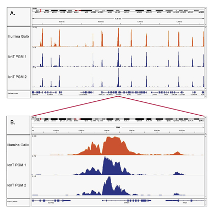
<p><strong>Figure 1A. The high consistency of the iDeal ChIP-seq kit on the Ion Torrent™ PGM™ (Life Technologies) and GAIIx (Illumina<sup>®</sup>)</strong><br /> ChIP was performed on sheared chromatin from 1 million HelaS3 cells using the iDeal ChIP-seq kit and 1 µg of H3K4me3 positive control antibody. Two different biological samples have been analyzed using two different sequencers - GAIIx (Illumina<sup>®</sup>) and PGM™ (Ion Torrent™). The expected ChIP-seq profile for H3K4me3 on the GAPDH promoter region has been obtained.<br /> Image A shows a several hundred bp along chr12 with high similarity of read distribution despite the radically different sequencers. Image B is a close capture focusing on the GAPDH that shows that even the peak structure is similar.</p>
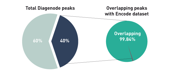
<p class="text-center"><strong>Perfect match between ChIP-seq data obtained with the iDeal ChIP-seq workflow and reference dataset</strong></p>
<p><img src="https://www.diagenode.com/img/product/kits/perfect-match-between-chipseq-data.png" alt="Figure 1B" style="display: block; margin-left: auto; margin-right: auto;" /></p>
<p><strong>Figure 1B.</strong> The ChIP-seq dataset from this experiment has been compared with a reference dataset from the Broad Institute. We observed a perfect match between the top 40% of Diagenode peaks and the reference dataset. Based on the NIH Encode project criterion, ChIP-seq results are considered reproducible between an original and reproduced dataset if the top 40% of peaks have at least an 80% overlap ratio with the compared dataset.</p>
<p><img src="https://www.diagenode.com/img/product/kits/iDeal-kit-C01010053-figure-2.jpg" alt="Figure 2" style="display: block; margin-left: auto; margin-right: auto;" /></p>
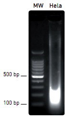
<p><strong>Figure 2. Efficient and easy chromatin shearing using the Bioruptor<sup>®</sup> and Shearing buffer iS1 from the iDeal ChIP-seq kit</strong><br /> Chromatin from 1 million of Hela cells was sheared using the Bioruptor<sup>®</sup> combined with the Bioruptor<sup>®</sup> Water cooler (Cat No. BioAcc-cool) during 3 rounds of 10 cycles of 30 seconds “ON” / 30 seconds “OFF” at HIGH power setting (position H). Diagenode 1.5 ml TPX tubes (Cat No. M-50001) were used for chromatin shearing. Samples were gently vortexed before and after performing each sonication round (rounds of 10 cycles), followed by a short centrifugation at 4°C to recover the sample volume at the bottom of the tube. The sheared chromatin was then decross-linked as described in the kit manual and analyzed by agarose gel electrophoresis.</p>
<p><img src="https://www.diagenode.com/img/product/kits/iDeal-kit-C01010053-figure-3.jpg" alt="Figure 3" style="display: block; margin-left: auto; margin-right: auto;" width="264" height="320" /></p>
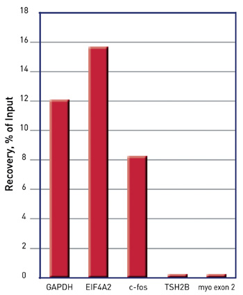
<p><strong>Figure 3. Validation of ChIP by qPCR: reliable results using Diagenode’s ChIP-seq grade H3K4me3 antibody, isotype control and sets of validated primers</strong><br /> Specific enrichment on positive loci (GAPDH, EIF4A2, c-fos promoter regions) comparing to no enrichment on negative loci (TSH2B promoter region and Myoglobin exon 2) was detected by qPCR. Samples were prepared using the Diagenode iDeal ChIP-seq kit. Diagenode ChIP-seq grade antibody against H3K4me3 and the corresponding isotype control IgG were used for immunoprecipitation. qPCR amplification was performed with sets of validated primers.</p>
<h3>ChIP-seq on tissue</h3>
<p><img src="https://www.diagenode.com/img/product/kits/ideal-figure-h3k4me3.jpg" alt="Figure 4A" style="display: block; margin-left: auto; margin-right: auto;" /></p>
<p><strong>Figure 4A.</strong> Chromatin Immunoprecipitation has been performed using chromatin from mouse liver tissue, the iDeal ChIP-seq kit for Histones and the Diagenode ChIP-seq-grade H3K4me3 (Cat. No. C15410003) antibody. The IP'd DNA was subsequently analysed on an Illumina® HiSeq. Library preparation, cluster generation and sequencing were performed according to the manufacturer's instructions. This figure shows the peak distribution in a region surrounding the GAPDH positive control gene.</p>
<p><img src="https://www.diagenode.com/img/product/kits/match-of-the-top40-peaks-2.png" alt="Figure 4B" caption="false" style="display: block; margin-left: auto; margin-right: auto;" width="700" height="280" /></p>
<p><strong>Figure 4B.</strong> The ChIP-seq dataset from this experiment has been compared with a reference dataset from the Broad Institute. We observed a perfect match between the top 40% of Diagenode peaks and the reference dataset. Based on the NIH Encode project criterion, ChIP-seq results are considered reproducible between an original and reproduced dataset if the top 40% of peaks have at least an 80% overlap ratio with the compared dataset.</p>',
'label2' => 'Species, cell lines, tissues tested',
'info2' => '<p>The iDeal ChIP-seq Kit for Histones is compatible with a broad variety of cell lines, tissues and species - some examples are shown below. Other species / cell lines / tissues can be used with this kit.</p>
<p><u>Cell lines:</u></p>
<p>Human: A549, A673, CD8+ T, Blood vascular endothelial cells, Lymphatic endothelial cells, fibroblasts, K562, MDA-MB231</p>
<p>Pig: Alveolar macrophages</p>
<p>Mouse: C2C12, primary HSPC, synovial fibroblasts, HeLa-S3, FACS sorted cells from embryonic kidneys, macrophages, mesodermal cells, myoblasts, NPC, salivary glands, spermatids, spermatocytes, skeletal muscle stem cells, stem cells, Th2</p>
<p>Hamster: CHO</p>
<p>Other cell lines / species: compatible, not tested</p>
<p><u>Tissues</u></p>
<p>Bee – brain</p>
<p>Daphnia – whole animal</p>
<p>Horse – brain, heart, lamina, liver, lung, skeletal muscles, ovary</p>
<p>Human – Erwing sarcoma tumor samples</p>
<p>Other tissues: compatible, not tested</p>
<p>Did you use the iDeal ChIP-seq for Histones Kit on other cell line / tissue / species? <a href="mailto:agnieszka.zelisko@diagenode.com?subject=Species, cell lines, tissues tested with the iDeal ChIP-seq Kit for TF&body=Dear Customer,%0D%0A%0D%0APlease, leave below your feedback about the iDeal ChIP-seq for Transcription Factors (cell / tissue type, species, other information...).%0D%0A%0D%0AThank you for sharing with us your experience !%0D%0A%0D%0ABest regards,%0D%0A%0D%0AAgnieszka Zelisko-Schmidt, PhD">Let us know!</a></p>',
'label3' => ' Additional solutions compatible with iDeal ChIP-seq Kit for Histones',
'info3' => '<p><a href="../p/chromatin-shearing-optimization-kit-low-sds-100-million-cells">Chromatin EasyShear Kit - Ultra Low SDS </a>optimizes chromatin shearing, a critical step for ChIP.</p>
<p> The <a href="https://www.diagenode.com/en/categories/library-preparation-for-ChIP-seq">MicroPlex Library Preparation Kit </a>provides easy and optimal library preparation of ChIPed samples.</p>
<p><a href="../categories/chip-seq-grade-antibodies">ChIP-seq grade anti-histone antibodies</a> provide high yields with excellent specificity and sensitivity.</p>
<p> Plus, for our IP-Star Automation users for automated ChIP, check out our <a href="../p/auto-ideal-chip-seq-kit-for-histones-x24-24-rxns">automated</a> version of this kit.</p>',
'format' => '4 chrom. prep./24 IPs',
'catalog_number' => 'C01010051',
'old_catalog_number' => 'AB-001-0024',
'sf_code' => 'C01010051-',
'type' => 'RFR',
'search_order' => '04-undefined',
'price_EUR' => '915',
'price_USD' => '1130',
'price_GBP' => '840',
'price_JPY' => '143335',
'price_CNY' => '',
'price_AUD' => '2825',
'country' => 'ALL',
'except_countries' => 'None',
'quote' => false,
'in_stock' => false,
'featured' => true,
'no_promo' => false,
'online' => true,
'master' => true,
'last_datasheet_update' => '0000-00-00',
'slug' => 'ideal-chip-seq-kit-x24-24-rxns',
'meta_title' => 'iDeal ChIP-seq kit x24',
'meta_keywords' => '',
'meta_description' => 'iDeal ChIP-seq kit x24',
'modified' => '2023-04-20 16:00:20',
'created' => '2015-06-29 14:08:20',
'locale' => 'eng'
),
'Antibody' => array(
'host' => '*****',
'id' => null,
'name' => null,
'description' => null,
'clonality' => null,
'isotype' => null,
'lot' => null,
'concentration' => null,
'reactivity' => null,
'type' => null,
'purity' => null,
'classification' => null,
'application_table' => null,
'storage_conditions' => null,
'storage_buffer' => null,
'precautions' => null,
'uniprot_acc' => null,
'slug' => null,
'meta_keywords' => null,
'meta_description' => null,
'modified' => null,
'created' => null,
'select_label' => null
),
'Slave' => array(
(int) 0 => array(
[maximum depth reached]
)
),
'Group' => array(
'Group' => array(
[maximum depth reached]
),
'Master' => array(
[maximum depth reached]
),
'Product' => array(
[maximum depth reached]
)
),
'Related' => array(
(int) 0 => array(
[maximum depth reached]
),
(int) 1 => array(
[maximum depth reached]
),
(int) 2 => array(
[maximum depth reached]
),
(int) 3 => array(
[maximum depth reached]
),
(int) 4 => array(
[maximum depth reached]
)
),
'Application' => array(
(int) 0 => array(
[maximum depth reached]
)
),
'Category' => array(
(int) 0 => array(
[maximum depth reached]
)
),
'Document' => array(
(int) 0 => array(
[maximum depth reached]
),
(int) 1 => array(
[maximum depth reached]
),
(int) 2 => array(
[maximum depth reached]
),
(int) 3 => array(
[maximum depth reached]
)
),
'Feature' => array(),
'Image' => array(
(int) 0 => array(
[maximum depth reached]
)
),
'Promotion' => array(),
'Protocol' => array(
(int) 0 => array(
[maximum depth reached]
)
),
'Publication' => array(
(int) 0 => array(
[maximum depth reached]
),
(int) 1 => array(
[maximum depth reached]
),
(int) 2 => array(
[maximum depth reached]
),
(int) 3 => array(
[maximum depth reached]
),
(int) 4 => array(
[maximum depth reached]
),
(int) 5 => array(
[maximum depth reached]
),
(int) 6 => array(
[maximum depth reached]
),
(int) 7 => array(
[maximum depth reached]
),
(int) 8 => array(
[maximum depth reached]
),
(int) 9 => array(
[maximum depth reached]
),
(int) 10 => array(
[maximum depth reached]
),
(int) 11 => array(
[maximum depth reached]
),
(int) 12 => array(
[maximum depth reached]
),
(int) 13 => array(
[maximum depth reached]
),
(int) 14 => array(
[maximum depth reached]
),
(int) 15 => array(
[maximum depth reached]
),
(int) 16 => array(
[maximum depth reached]
),
(int) 17 => array(
[maximum depth reached]
),
(int) 18 => array(
[maximum depth reached]
),
(int) 19 => array(
[maximum depth reached]
),
(int) 20 => array(
[maximum depth reached]
),
(int) 21 => array(
[maximum depth reached]
),
(int) 22 => array(
[maximum depth reached]
),
(int) 23 => array(
[maximum depth reached]
),
(int) 24 => array(
[maximum depth reached]
),
(int) 25 => array(
[maximum depth reached]
),
(int) 26 => array(
[maximum depth reached]
),
(int) 27 => array(
[maximum depth reached]
),
(int) 28 => array(
[maximum depth reached]
),
(int) 29 => array(
[maximum depth reached]
),
(int) 30 => array(
[maximum depth reached]
),
(int) 31 => array(
[maximum depth reached]
),
(int) 32 => array(
[maximum depth reached]
),
(int) 33 => array(
[maximum depth reached]
),
(int) 34 => array(
[maximum depth reached]
),
(int) 35 => array(
[maximum depth reached]
),
(int) 36 => array(
[maximum depth reached]
),
(int) 37 => array(
[maximum depth reached]
),
(int) 38 => array(
[maximum depth reached]
),
(int) 39 => array(
[maximum depth reached]
),
(int) 40 => array(
[maximum depth reached]
),
(int) 41 => array(
[maximum depth reached]
),
(int) 42 => array(
[maximum depth reached]
),
(int) 43 => array(
[maximum depth reached]
),
(int) 44 => array(
[maximum depth reached]
),
(int) 45 => array(
[maximum depth reached]
),
(int) 46 => array(
[maximum depth reached]
),
(int) 47 => array(
[maximum depth reached]
),
(int) 48 => array(
[maximum depth reached]
),
(int) 49 => array(
[maximum depth reached]
),
(int) 50 => array(
[maximum depth reached]
),
(int) 51 => array(
[maximum depth reached]
),
(int) 52 => array(
[maximum depth reached]
),
(int) 53 => array(
[maximum depth reached]
),
(int) 54 => array(
[maximum depth reached]
),
(int) 55 => array(
[maximum depth reached]
),
(int) 56 => array(
[maximum depth reached]
),
(int) 57 => array(
[maximum depth reached]
),
(int) 58 => array(
[maximum depth reached]
),
(int) 59 => array(
[maximum depth reached]
),
(int) 60 => array(
[maximum depth reached]
),
(int) 61 => array(
[maximum depth reached]
),
(int) 62 => array(
[maximum depth reached]
),
(int) 63 => array(
[maximum depth reached]
),
(int) 64 => array(
[maximum depth reached]
),
(int) 65 => array(
[maximum depth reached]
),
(int) 66 => array(
[maximum depth reached]
),
(int) 67 => array(
[maximum depth reached]
),
(int) 68 => array(
[maximum depth reached]
),
(int) 69 => array(
[maximum depth reached]
),
(int) 70 => array(
[maximum depth reached]
),
(int) 71 => array(
[maximum depth reached]
),
(int) 72 => array(
[maximum depth reached]
),
(int) 73 => array(
[maximum depth reached]
),
(int) 74 => array(
[maximum depth reached]
),
(int) 75 => array(
[maximum depth reached]
),
(int) 76 => array(
[maximum depth reached]
),
(int) 77 => array(
[maximum depth reached]
),
(int) 78 => array(
[maximum depth reached]
),
(int) 79 => array(
[maximum depth reached]
),
(int) 80 => array(
[maximum depth reached]
),
(int) 81 => array(
[maximum depth reached]
),
(int) 82 => array(
[maximum depth reached]
),
(int) 83 => array(
[maximum depth reached]
)
),
'Testimonial' => array(
(int) 0 => array(
[maximum depth reached]
),
(int) 1 => array(
[maximum depth reached]
)
),
'Area' => array(),
'SafetySheet' => array(
(int) 0 => array(
[maximum depth reached]
),
(int) 1 => array(
[maximum depth reached]
),
(int) 2 => array(
[maximum depth reached]
),
(int) 3 => array(
[maximum depth reached]
),
(int) 4 => array(
[maximum depth reached]
),
(int) 5 => array(
[maximum depth reached]
),
(int) 6 => array(
[maximum depth reached]
),
(int) 7 => array(
[maximum depth reached]
)
)
),
'meta_canonical' => 'https://www.diagenode.com/en/p/ideal-chip-seq-kit-x24-24-rxns'
)
$language = 'en'
$meta_keywords = ''
$meta_description = 'iDeal ChIP-seq kit x24'
$meta_title = 'iDeal ChIP-seq kit x24'
$product = array(
'Product' => array(
'id' => '1836',
'antibody_id' => null,
'name' => 'iDeal ChIP-seq kit for Histones',
'description' => '<p><a href="https://www.diagenode.com/files/products/kits/ideal-chipseq-for-histones-complete-manual.pdf"><img src="https://www.diagenode.com/img/buttons/bt-manual.png" /></a></p>
<p>Don’t risk wasting your precious sequencing samples. Diagenode’s validated <strong>iDeal ChIP-seq kit for Histones</strong> has everything you need for a successful start-to-finish <strong>ChIP of histones prior to Next-Generation Sequencing</strong>. The complete kit contains all buffers and reagents for cell lysis, chromatin shearing, immunoprecipitation and DNA purification. In addition, unlike competing solutions, the kit contains positive and negative control antibodies (H3K4me3 and IgG, respectively) as well as positive and negative control PCR primers pairs (GAPDH TSS and Myoglobin exon 2, respectively) for your convenience and a guarantee of optimal results. The kit has been validated on multiple histone marks.</p>
<p> The iDeal ChIP-seq kit for Histones<strong> </strong>is perfect for <strong>cells</strong> (<strong>100,000 cells</strong> to <strong>1,000,000 cells</strong> per IP) and has been validated for <strong>tissues</strong> (<strong>1.5 mg</strong> to <strong>5 mg</strong> of tissue per IP).</p>
<p> The iDeal ChIP-seq kit is the only kit on the market validated for the major sequencing systems. Our expertise in ChIP-seq tools allows reproducible and efficient results every time.</p>
<p></p>
<p> <strong></strong></p>
<p></p>',
'label1' => 'Characteristics',
'info1' => '<ul style="list-style-type: disc;">
<li>Highly <strong>optimized</strong> protocol for ChIP-seq from cells and tissues</li>
<li><strong>Validated</strong> for ChIP-seq with multiple histones marks</li>
<li>Most <strong>complete</strong> kit available (covers all steps, including the control antibodies and primers)</li>
<li>Optimized chromatin preparation in combination with the Bioruptor ensuring the best <strong>epitope integrity</strong></li>
<li>Magnetic beads make ChIP easy, fast and more <strong>reproducible</strong></li>
<li>Combination with Diagenode ChIP-seq antibodies provides high yields with excellent <strong>specificity</strong> and <strong>sensitivity</strong></li>
<li>Purified DNA suitable for any downstream application</li>
<li>Easy-to-follow protocol</li>
</ul>
<p>Note: to obtain optimal results, this kit should be used in combination with the DiaMag1.5 - magnetic rack.</p>
<h3>ChIP-seq on cells</h3>
<p><img src="https://www.diagenode.com/img/product/kits/iDeal-kit-C01010053-figure-1.jpg" alt="Figure 1A" style="display: block; margin-left: auto; margin-right: auto;" /></p>
<p><strong>Figure 1A. The high consistency of the iDeal ChIP-seq kit on the Ion Torrent™ PGM™ (Life Technologies) and GAIIx (Illumina<sup>®</sup>)</strong><br /> ChIP was performed on sheared chromatin from 1 million HelaS3 cells using the iDeal ChIP-seq kit and 1 µg of H3K4me3 positive control antibody. Two different biological samples have been analyzed using two different sequencers - GAIIx (Illumina<sup>®</sup>) and PGM™ (Ion Torrent™). The expected ChIP-seq profile for H3K4me3 on the GAPDH promoter region has been obtained.<br /> Image A shows a several hundred bp along chr12 with high similarity of read distribution despite the radically different sequencers. Image B is a close capture focusing on the GAPDH that shows that even the peak structure is similar.</p>
<p class="text-center"><strong>Perfect match between ChIP-seq data obtained with the iDeal ChIP-seq workflow and reference dataset</strong></p>
<p><img src="https://www.diagenode.com/img/product/kits/perfect-match-between-chipseq-data.png" alt="Figure 1B" style="display: block; margin-left: auto; margin-right: auto;" /></p>
<p><strong>Figure 1B.</strong> The ChIP-seq dataset from this experiment has been compared with a reference dataset from the Broad Institute. We observed a perfect match between the top 40% of Diagenode peaks and the reference dataset. Based on the NIH Encode project criterion, ChIP-seq results are considered reproducible between an original and reproduced dataset if the top 40% of peaks have at least an 80% overlap ratio with the compared dataset.</p>
<p><img src="https://www.diagenode.com/img/product/kits/iDeal-kit-C01010053-figure-2.jpg" alt="Figure 2" style="display: block; margin-left: auto; margin-right: auto;" /></p>
<p><strong>Figure 2. Efficient and easy chromatin shearing using the Bioruptor<sup>®</sup> and Shearing buffer iS1 from the iDeal ChIP-seq kit</strong><br /> Chromatin from 1 million of Hela cells was sheared using the Bioruptor<sup>®</sup> combined with the Bioruptor<sup>®</sup> Water cooler (Cat No. BioAcc-cool) during 3 rounds of 10 cycles of 30 seconds “ON” / 30 seconds “OFF” at HIGH power setting (position H). Diagenode 1.5 ml TPX tubes (Cat No. M-50001) were used for chromatin shearing. Samples were gently vortexed before and after performing each sonication round (rounds of 10 cycles), followed by a short centrifugation at 4°C to recover the sample volume at the bottom of the tube. The sheared chromatin was then decross-linked as described in the kit manual and analyzed by agarose gel electrophoresis.</p>
<p><img src="https://www.diagenode.com/img/product/kits/iDeal-kit-C01010053-figure-3.jpg" alt="Figure 3" style="display: block; margin-left: auto; margin-right: auto;" width="264" height="320" /></p>
<p><strong>Figure 3. Validation of ChIP by qPCR: reliable results using Diagenode’s ChIP-seq grade H3K4me3 antibody, isotype control and sets of validated primers</strong><br /> Specific enrichment on positive loci (GAPDH, EIF4A2, c-fos promoter regions) comparing to no enrichment on negative loci (TSH2B promoter region and Myoglobin exon 2) was detected by qPCR. Samples were prepared using the Diagenode iDeal ChIP-seq kit. Diagenode ChIP-seq grade antibody against H3K4me3 and the corresponding isotype control IgG were used for immunoprecipitation. qPCR amplification was performed with sets of validated primers.</p>
<h3>ChIP-seq on tissue</h3>
<p><img src="https://www.diagenode.com/img/product/kits/ideal-figure-h3k4me3.jpg" alt="Figure 4A" style="display: block; margin-left: auto; margin-right: auto;" /></p>
<p><strong>Figure 4A.</strong> Chromatin Immunoprecipitation has been performed using chromatin from mouse liver tissue, the iDeal ChIP-seq kit for Histones and the Diagenode ChIP-seq-grade H3K4me3 (Cat. No. C15410003) antibody. The IP'd DNA was subsequently analysed on an Illumina® HiSeq. Library preparation, cluster generation and sequencing were performed according to the manufacturer's instructions. This figure shows the peak distribution in a region surrounding the GAPDH positive control gene.</p>
<p><img src="https://www.diagenode.com/img/product/kits/match-of-the-top40-peaks-2.png" alt="Figure 4B" caption="false" style="display: block; margin-left: auto; margin-right: auto;" width="700" height="280" /></p>
<p><strong>Figure 4B.</strong> The ChIP-seq dataset from this experiment has been compared with a reference dataset from the Broad Institute. We observed a perfect match between the top 40% of Diagenode peaks and the reference dataset. Based on the NIH Encode project criterion, ChIP-seq results are considered reproducible between an original and reproduced dataset if the top 40% of peaks have at least an 80% overlap ratio with the compared dataset.</p>',
'label2' => 'Species, cell lines, tissues tested',
'info2' => '<p>The iDeal ChIP-seq Kit for Histones is compatible with a broad variety of cell lines, tissues and species - some examples are shown below. Other species / cell lines / tissues can be used with this kit.</p>
<p><u>Cell lines:</u></p>
<p>Human: A549, A673, CD8+ T, Blood vascular endothelial cells, Lymphatic endothelial cells, fibroblasts, K562, MDA-MB231</p>
<p>Pig: Alveolar macrophages</p>
<p>Mouse: C2C12, primary HSPC, synovial fibroblasts, HeLa-S3, FACS sorted cells from embryonic kidneys, macrophages, mesodermal cells, myoblasts, NPC, salivary glands, spermatids, spermatocytes, skeletal muscle stem cells, stem cells, Th2</p>
<p>Hamster: CHO</p>
<p>Other cell lines / species: compatible, not tested</p>
<p><u>Tissues</u></p>
<p>Bee – brain</p>
<p>Daphnia – whole animal</p>
<p>Horse – brain, heart, lamina, liver, lung, skeletal muscles, ovary</p>
<p>Human – Erwing sarcoma tumor samples</p>
<p>Other tissues: compatible, not tested</p>
<p>Did you use the iDeal ChIP-seq for Histones Kit on other cell line / tissue / species? <a href="mailto:agnieszka.zelisko@diagenode.com?subject=Species, cell lines, tissues tested with the iDeal ChIP-seq Kit for TF&body=Dear Customer,%0D%0A%0D%0APlease, leave below your feedback about the iDeal ChIP-seq for Transcription Factors (cell / tissue type, species, other information...).%0D%0A%0D%0AThank you for sharing with us your experience !%0D%0A%0D%0ABest regards,%0D%0A%0D%0AAgnieszka Zelisko-Schmidt, PhD">Let us know!</a></p>',
'label3' => ' Additional solutions compatible with iDeal ChIP-seq Kit for Histones',
'info3' => '<p><a href="../p/chromatin-shearing-optimization-kit-low-sds-100-million-cells">Chromatin EasyShear Kit - Ultra Low SDS </a>optimizes chromatin shearing, a critical step for ChIP.</p>
<p> The <a href="https://www.diagenode.com/en/categories/library-preparation-for-ChIP-seq">MicroPlex Library Preparation Kit </a>provides easy and optimal library preparation of ChIPed samples.</p>
<p><a href="../categories/chip-seq-grade-antibodies">ChIP-seq grade anti-histone antibodies</a> provide high yields with excellent specificity and sensitivity.</p>
<p> Plus, for our IP-Star Automation users for automated ChIP, check out our <a href="../p/auto-ideal-chip-seq-kit-for-histones-x24-24-rxns">automated</a> version of this kit.</p>',
'format' => '4 chrom. prep./24 IPs',
'catalog_number' => 'C01010051',
'old_catalog_number' => 'AB-001-0024',
'sf_code' => 'C01010051-',
'type' => 'RFR',
'search_order' => '04-undefined',
'price_EUR' => '915',
'price_USD' => '1130',
'price_GBP' => '840',
'price_JPY' => '143335',
'price_CNY' => '',
'price_AUD' => '2825',
'country' => 'ALL',
'except_countries' => 'None',
'quote' => false,
'in_stock' => false,
'featured' => true,
'no_promo' => false,
'online' => true,
'master' => true,
'last_datasheet_update' => '0000-00-00',
'slug' => 'ideal-chip-seq-kit-x24-24-rxns',
'meta_title' => 'iDeal ChIP-seq kit x24',
'meta_keywords' => '',
'meta_description' => 'iDeal ChIP-seq kit x24',
'modified' => '2023-04-20 16:00:20',
'created' => '2015-06-29 14:08:20',
'locale' => 'eng'
),
'Antibody' => array(
'host' => '*****',
'id' => null,
'name' => null,
'description' => null,
'clonality' => null,
'isotype' => null,
'lot' => null,
'concentration' => null,
'reactivity' => null,
'type' => null,
'purity' => null,
'classification' => null,
'application_table' => null,
'storage_conditions' => null,
'storage_buffer' => null,
'precautions' => null,
'uniprot_acc' => null,
'slug' => null,
'meta_keywords' => null,
'meta_description' => null,
'modified' => null,
'created' => null,
'select_label' => null
),
'Slave' => array(
(int) 0 => array(
'id' => '9',
'name' => 'C01010051',
'product_id' => '1836',
'modified' => '2016-02-18 17:14:29',
'created' => '2016-02-18 17:14:29'
)
),
'Group' => array(
'Group' => array(
'id' => '9',
'name' => 'C01010051',
'product_id' => '1836',
'modified' => '2016-02-18 17:14:29',
'created' => '2016-02-18 17:14:29'
),
'Master' => array(
'id' => '1836',
'antibody_id' => null,
'name' => 'iDeal ChIP-seq kit for Histones',
'description' => '<p><a href="https://www.diagenode.com/files/products/kits/ideal-chipseq-for-histones-complete-manual.pdf"><img src="https://www.diagenode.com/img/buttons/bt-manual.png" /></a></p>
<p>Don’t risk wasting your precious sequencing samples. Diagenode’s validated <strong>iDeal ChIP-seq kit for Histones</strong> has everything you need for a successful start-to-finish <strong>ChIP of histones prior to Next-Generation Sequencing</strong>. The complete kit contains all buffers and reagents for cell lysis, chromatin shearing, immunoprecipitation and DNA purification. In addition, unlike competing solutions, the kit contains positive and negative control antibodies (H3K4me3 and IgG, respectively) as well as positive and negative control PCR primers pairs (GAPDH TSS and Myoglobin exon 2, respectively) for your convenience and a guarantee of optimal results. The kit has been validated on multiple histone marks.</p>
<p> The iDeal ChIP-seq kit for Histones<strong> </strong>is perfect for <strong>cells</strong> (<strong>100,000 cells</strong> to <strong>1,000,000 cells</strong> per IP) and has been validated for <strong>tissues</strong> (<strong>1.5 mg</strong> to <strong>5 mg</strong> of tissue per IP).</p>
<p> The iDeal ChIP-seq kit is the only kit on the market validated for the major sequencing systems. Our expertise in ChIP-seq tools allows reproducible and efficient results every time.</p>
<p></p>
<p> <strong></strong></p>
<p></p>',
'label1' => 'Characteristics',
'info1' => '<ul style="list-style-type: disc;">
<li>Highly <strong>optimized</strong> protocol for ChIP-seq from cells and tissues</li>
<li><strong>Validated</strong> for ChIP-seq with multiple histones marks</li>
<li>Most <strong>complete</strong> kit available (covers all steps, including the control antibodies and primers)</li>
<li>Optimized chromatin preparation in combination with the Bioruptor ensuring the best <strong>epitope integrity</strong></li>
<li>Magnetic beads make ChIP easy, fast and more <strong>reproducible</strong></li>
<li>Combination with Diagenode ChIP-seq antibodies provides high yields with excellent <strong>specificity</strong> and <strong>sensitivity</strong></li>
<li>Purified DNA suitable for any downstream application</li>
<li>Easy-to-follow protocol</li>
</ul>
<p>Note: to obtain optimal results, this kit should be used in combination with the DiaMag1.5 - magnetic rack.</p>
<h3>ChIP-seq on cells</h3>
<p><img src="https://www.diagenode.com/img/product/kits/iDeal-kit-C01010053-figure-1.jpg" alt="Figure 1A" style="display: block; margin-left: auto; margin-right: auto;" /></p>
<p><strong>Figure 1A. The high consistency of the iDeal ChIP-seq kit on the Ion Torrent™ PGM™ (Life Technologies) and GAIIx (Illumina<sup>®</sup>)</strong><br /> ChIP was performed on sheared chromatin from 1 million HelaS3 cells using the iDeal ChIP-seq kit and 1 µg of H3K4me3 positive control antibody. Two different biological samples have been analyzed using two different sequencers - GAIIx (Illumina<sup>®</sup>) and PGM™ (Ion Torrent™). The expected ChIP-seq profile for H3K4me3 on the GAPDH promoter region has been obtained.<br /> Image A shows a several hundred bp along chr12 with high similarity of read distribution despite the radically different sequencers. Image B is a close capture focusing on the GAPDH that shows that even the peak structure is similar.</p>
<p class="text-center"><strong>Perfect match between ChIP-seq data obtained with the iDeal ChIP-seq workflow and reference dataset</strong></p>
<p><img src="https://www.diagenode.com/img/product/kits/perfect-match-between-chipseq-data.png" alt="Figure 1B" style="display: block; margin-left: auto; margin-right: auto;" /></p>
<p><strong>Figure 1B.</strong> The ChIP-seq dataset from this experiment has been compared with a reference dataset from the Broad Institute. We observed a perfect match between the top 40% of Diagenode peaks and the reference dataset. Based on the NIH Encode project criterion, ChIP-seq results are considered reproducible between an original and reproduced dataset if the top 40% of peaks have at least an 80% overlap ratio with the compared dataset.</p>
<p><img src="https://www.diagenode.com/img/product/kits/iDeal-kit-C01010053-figure-2.jpg" alt="Figure 2" style="display: block; margin-left: auto; margin-right: auto;" /></p>
<p><strong>Figure 2. Efficient and easy chromatin shearing using the Bioruptor<sup>®</sup> and Shearing buffer iS1 from the iDeal ChIP-seq kit</strong><br /> Chromatin from 1 million of Hela cells was sheared using the Bioruptor<sup>®</sup> combined with the Bioruptor<sup>®</sup> Water cooler (Cat No. BioAcc-cool) during 3 rounds of 10 cycles of 30 seconds “ON” / 30 seconds “OFF” at HIGH power setting (position H). Diagenode 1.5 ml TPX tubes (Cat No. M-50001) were used for chromatin shearing. Samples were gently vortexed before and after performing each sonication round (rounds of 10 cycles), followed by a short centrifugation at 4°C to recover the sample volume at the bottom of the tube. The sheared chromatin was then decross-linked as described in the kit manual and analyzed by agarose gel electrophoresis.</p>
<p><img src="https://www.diagenode.com/img/product/kits/iDeal-kit-C01010053-figure-3.jpg" alt="Figure 3" style="display: block; margin-left: auto; margin-right: auto;" width="264" height="320" /></p>
<p><strong>Figure 3. Validation of ChIP by qPCR: reliable results using Diagenode’s ChIP-seq grade H3K4me3 antibody, isotype control and sets of validated primers</strong><br /> Specific enrichment on positive loci (GAPDH, EIF4A2, c-fos promoter regions) comparing to no enrichment on negative loci (TSH2B promoter region and Myoglobin exon 2) was detected by qPCR. Samples were prepared using the Diagenode iDeal ChIP-seq kit. Diagenode ChIP-seq grade antibody against H3K4me3 and the corresponding isotype control IgG were used for immunoprecipitation. qPCR amplification was performed with sets of validated primers.</p>
<h3>ChIP-seq on tissue</h3>
<p><img src="https://www.diagenode.com/img/product/kits/ideal-figure-h3k4me3.jpg" alt="Figure 4A" style="display: block; margin-left: auto; margin-right: auto;" /></p>
<p><strong>Figure 4A.</strong> Chromatin Immunoprecipitation has been performed using chromatin from mouse liver tissue, the iDeal ChIP-seq kit for Histones and the Diagenode ChIP-seq-grade H3K4me3 (Cat. No. C15410003) antibody. The IP'd DNA was subsequently analysed on an Illumina® HiSeq. Library preparation, cluster generation and sequencing were performed according to the manufacturer's instructions. This figure shows the peak distribution in a region surrounding the GAPDH positive control gene.</p>
<p><img src="https://www.diagenode.com/img/product/kits/match-of-the-top40-peaks-2.png" alt="Figure 4B" caption="false" style="display: block; margin-left: auto; margin-right: auto;" width="700" height="280" /></p>
<p><strong>Figure 4B.</strong> The ChIP-seq dataset from this experiment has been compared with a reference dataset from the Broad Institute. We observed a perfect match between the top 40% of Diagenode peaks and the reference dataset. Based on the NIH Encode project criterion, ChIP-seq results are considered reproducible between an original and reproduced dataset if the top 40% of peaks have at least an 80% overlap ratio with the compared dataset.</p>',
'label2' => 'Species, cell lines, tissues tested',
'info2' => '<p>The iDeal ChIP-seq Kit for Histones is compatible with a broad variety of cell lines, tissues and species - some examples are shown below. Other species / cell lines / tissues can be used with this kit.</p>
<p><u>Cell lines:</u></p>
<p>Human: A549, A673, CD8+ T, Blood vascular endothelial cells, Lymphatic endothelial cells, fibroblasts, K562, MDA-MB231</p>
<p>Pig: Alveolar macrophages</p>
<p>Mouse: C2C12, primary HSPC, synovial fibroblasts, HeLa-S3, FACS sorted cells from embryonic kidneys, macrophages, mesodermal cells, myoblasts, NPC, salivary glands, spermatids, spermatocytes, skeletal muscle stem cells, stem cells, Th2</p>
<p>Hamster: CHO</p>
<p>Other cell lines / species: compatible, not tested</p>
<p><u>Tissues</u></p>
<p>Bee – brain</p>
<p>Daphnia – whole animal</p>
<p>Horse – brain, heart, lamina, liver, lung, skeletal muscles, ovary</p>
<p>Human – Erwing sarcoma tumor samples</p>
<p>Other tissues: compatible, not tested</p>
<p>Did you use the iDeal ChIP-seq for Histones Kit on other cell line / tissue / species? <a href="mailto:agnieszka.zelisko@diagenode.com?subject=Species, cell lines, tissues tested with the iDeal ChIP-seq Kit for TF&body=Dear Customer,%0D%0A%0D%0APlease, leave below your feedback about the iDeal ChIP-seq for Transcription Factors (cell / tissue type, species, other information...).%0D%0A%0D%0AThank you for sharing with us your experience !%0D%0A%0D%0ABest regards,%0D%0A%0D%0AAgnieszka Zelisko-Schmidt, PhD">Let us know!</a></p>',
'label3' => ' Additional solutions compatible with iDeal ChIP-seq Kit for Histones',
'info3' => '<p><a href="../p/chromatin-shearing-optimization-kit-low-sds-100-million-cells">Chromatin EasyShear Kit - Ultra Low SDS </a>optimizes chromatin shearing, a critical step for ChIP.</p>
<p> The <a href="https://www.diagenode.com/en/categories/library-preparation-for-ChIP-seq">MicroPlex Library Preparation Kit </a>provides easy and optimal library preparation of ChIPed samples.</p>
<p><a href="../categories/chip-seq-grade-antibodies">ChIP-seq grade anti-histone antibodies</a> provide high yields with excellent specificity and sensitivity.</p>
<p> Plus, for our IP-Star Automation users for automated ChIP, check out our <a href="../p/auto-ideal-chip-seq-kit-for-histones-x24-24-rxns">automated</a> version of this kit.</p>',
'format' => '4 chrom. prep./24 IPs',
'catalog_number' => 'C01010051',
'old_catalog_number' => 'AB-001-0024',
'sf_code' => 'C01010051-',
'type' => 'RFR',
'search_order' => '04-undefined',
'price_EUR' => '915',
'price_USD' => '1130',
'price_GBP' => '840',
'price_JPY' => '143335',
'price_CNY' => '',
'price_AUD' => '2825',
'country' => 'ALL',
'except_countries' => 'None',
'quote' => false,
'in_stock' => false,
'featured' => true,
'no_promo' => false,
'online' => true,
'master' => true,
'last_datasheet_update' => '0000-00-00',
'slug' => 'ideal-chip-seq-kit-x24-24-rxns',
'meta_title' => 'iDeal ChIP-seq kit x24',
'meta_keywords' => '',
'meta_description' => 'iDeal ChIP-seq kit x24',
'modified' => '2023-04-20 16:00:20',
'created' => '2015-06-29 14:08:20'
),
'Product' => array(
(int) 0 => array(
[maximum depth reached]
),
(int) 1 => array(
[maximum depth reached]
)
)
),
'Related' => array(
(int) 0 => array(
'id' => '1839',
'antibody_id' => null,
'name' => 'iDeal ChIP-seq kit for Transcription Factors',
'description' => '<p><a href="https://www.diagenode.com/files/products/kits/ideal-chipseq-transcription-factors-x10-manual.pdf"><img src="https://www.diagenode.com/img/buttons/bt-manual.png" /></a></p>
<div class="row">
<div class="small-12 medium-8 large-8 columns"><br />
<p><span style="font-weight: 400;">Diagenode’s <strong>iDeal ChIP-seq Kit for Transcription Factors</strong> is a highly validated solution for robust transcription factor and other non-histone proteins ChIP-seq results and contains everything you need for start-to-finish </span><b>ChIP </b><span style="font-weight: 400;">prior to </span><b>Next-Generation Sequencing</b><span style="font-weight: 400;">. This complete solution contains all buffers and reagents for cell lysis, chromatin shearing, immunoprecipitation, and DNA purification. In addition, unlike competing solutions, the kit contains positive and negative control antibodies (CTCF and IgG, respectively) as well as positive and negative control PCR primers pairs (H19 and Myoglobin exon 2, respectively) for your convenience and a guarantee of optimal results. <br /></span></p>
</div>
<div class="small-12 medium-4 large-4 columns"><center><br /><br />
<script>// <![CDATA[
var date = new Date(); var heure = date.getHours(); var jour = date.getDay(); var semaine = Math.floor(date.getDate() / 7) + 1; if (jour === 2 && ( (heure >= 9 && heure < 9.5) || (heure >= 18 && heure < 18.5) )) { document.write('<a href="https://us02web.zoom.us/j/85467619762"><img src="https://www.diagenode.com/img/epicafe-ON.gif"></a>'); } else { document.write('<a href="https://go.diagenode.com/l/928883/2023-04-26/3kq1v"><img src="https://www.diagenode.com/img/epicafe-OFF.png"></a>'); }
// ]]></script>
</center></div>
</div>
<p><span style="font-weight: 400;">The </span><b> iDeal ChIP-seq kit for Transcription Factors </b><span style="font-weight: 400;">is compatible for cells or tissues:</span></p>
<table style="width: 419px; margin-left: auto; margin-right: auto;">
<tbody>
<tr>
<td style="width: 144px;"></td>
<td style="width: 267px; text-align: center;"><span style="font-weight: 400;">Amount per IP</span></td>
</tr>
<tr>
<td style="width: 144px;">Cells</td>
<td style="width: 267px; text-align: center;"><strong>4,000,000</strong></td>
</tr>
<tr>
<td style="width: 144px;">Tissues</td>
<td style="width: 267px; text-align: center;"><strong>30 mg</strong></td>
</tr>
</tbody>
</table>
<p><span style="font-weight: 400;">The iDeal ChIP-seq kit is the only kit on the market validated for major sequencing systems. Our expertise in ChIP-seq tools allows reproducible and efficient results every time. </span></p>
<p></p>
<p></p>',
'label1' => 'Characteristics',
'info1' => '<ul>
<li><span style="font-weight: 400;"><strong>Highly optimized protocol</strong> for ChIP-seq from cells and tissues</span></li>
<li><span style="font-weight: 400;"><strong>Validated</strong> for <strong>ChIP-seq</strong> with multiple transcription factors and non-histone targets<br /></span></li>
<li><span style="font-weight: 400;"><strong>Most complete kit</strong> available (covers all steps, including the control antibodies and primers)<br /></span></li>
<li><span style="font-weight: 400;"><strong>Magnetic beads</strong> make ChIP <strong>easy</strong>, <strong>fast</strong> and more <strong>reproducible</strong></span></li>
<li><span style="font-weight: 400;">Combination with Diagenode ChIP-seq antibodies provides <strong>high yields</strong> with excellent <strong>specificity</strong> and <strong>sensitivity</strong><br /></span></li>
<li><span style="font-weight: 400;">Purified DNA suitable for any downstream application</span></li>
<li><span style="font-weight: 400;">Easy-to-follow protocol</span></li>
</ul>
<p><span style="font-weight: 400;"></span></p>
<p> </p>
<h3>ChIP-seq on cells</h3>
<p><img src="https://www.diagenode.com/img/product/kits/ideal-ctcf-diagenode.jpg" alt="CTCF Diagenode" style="display: block; margin-left: auto; margin-right: auto;" /></p>
<p><strong>Figure 1.</strong> (A) Chromatin Immunoprecipitation has been performed using chromatin from HeLa cells, the iDeal ChIP-seq kit for Transcription Factors and the Diagenode ChIP-seq-grade CTCF antibody. The IP'd DNA was subsequently analysed on an Illumina<sup>®</sup> HiSeq. Library preparation, cluster generation and sequencing were performed according to the manufacturer's instructions. This figure shows the peak distribution in a region surrounding the GAPDH positive control gene.</p>
<p><img src="https://www.diagenode.com/img/product/kits/ideal-figure-b-total-diagendoe-peaks.png" alt="CTCF Diagenode" style="display: block; margin-left: auto; margin-right: auto;" /></p>
<p><strong>Figure 1B.</strong> The ChIP-seq dataset from this experiment has been compared with a reference dataset from the Broad Institute. We observed a perfect match between the top 40% of Diagenode peaks and the reference dataset. Based on the NIH Encode project criterion, ChIP-seq results are considered reproducible between an original and reproduced dataset if the top 40% of peaks have at least an 80% overlap ratio with the compared dataset.</p>
<p> </p>
<p><img src="https://www.diagenode.com/img/product/kits/ideal-TF-chip-seq-A.png" alt="ChIP-seq figure A" style="display: block; margin-left: auto; margin-right: auto;" /></p>
<p><img src="https://www.diagenode.com/img/product/kits/ideal-TF-chip-seq-B.png" alt="ChIP-seq figure B" style="display: block; margin-left: auto; margin-right: auto;" /></p>
<p><img src="https://www.diagenode.com/img/product/kits/ideal-TF-chip-seq-C.png" alt="ChIP-seq figure C" style="display: block; margin-left: auto; margin-right: auto;" /></p>
<p><strong>Figure 2.</strong> Chromatin Immunoprecipitation has been performed using chromatin from HeLa cells, the iDeal ChIP-seq kit for Transcription Factors and the Diagenode ChIP-seq-grade HDAC1 (A), LSD1 (B) and p53 antibody (C). The IP'd DNA was subsequently analysed on an Illumina<sup>®</sup> Genome Analyzer. Library preparation, cluster generation and sequencing were performed according to the manufacturer's instructions. This figure shows the peak distribution in regions of chromosome 3 (A), chromosome 12 (B) and chromosome 6 (C) respectively.</p>
<p> </p>
<h3>ChIP-seq on tissue</h3>
<p><img src="https://www.diagenode.com/img/product/kits/ideal-figure-3a.jpg" alt="ChIP-seq figure A" style="display: block; margin-left: auto; margin-right: auto;" /></p>
<p><strong>Figure 3A.</strong> Chromatin Immunoprecipitation has been performed using chromatin from mouse liver tissue, the iDeal ChIP-seq kit for Transcription Factors and the Diagenode ChIP-seq-grade CTCF antibody. The IP'd DNA was subsequently analysed on an Illumina® HiSeq. Library preparation, cluster generation and sequencing were performed according to the manufacturer's instructions. This figure shows the peak distribution in a region surrounding the Vwf positive control gene.</p>
<p><img src="https://www.diagenode.com/img/product/kits/match-of-the-top40-peaks.png" alt="Match of the Top40 peaks" style="display: block; margin-left: auto; margin-right: auto;" /></p>
<p><strong>Figure 3B.</strong> The ChIP-seq dataset from this experiment has been compared with a reference dataset from the Broad Institute. We observed a perfect match between the top 40% of Diagenode peaks and the reference dataset. Based on the NIH Encode project criterion, ChIP-seq results are considered reproducible between an original and reproduced dataset if the top 40% of peaks have at least an 80% overlap ratio with the compared dataset.</p>',
'label2' => 'Species, cell lines, tissues tested',
'info2' => '<p>The iDeal ChIP-seq Kit for Transcription Factors is compatible with a broad variety of cell lines, tissues and species - some examples are shown below. Other species / cell lines / tissues can be used with this kit.</p>
<p><span style="text-decoration: underline;">Cell lines:</span></p>
<p>Human: A549, A673, BT-549, CD4 T, HCC1806, HeLa, HepG2, HFF, HK-GFP-MR, ILC, K562, KYSE-180, LapC4, M14, MCF7, MDA-MB-231, MDA-MB-436, RDES, SKNO1, VCaP, U2-OS, ZR-75-1 </p>
<p>Mouse: ESC, NPCs, BZ, GT1-7, acinar cells, HSPCs, Th2 cells, keratinocytes</p>
<p>Cattle: pbMEC, <span>MAC-T</span></p>
<p><span>Other cell lines / species: compatible, not tested</span></p>
<p><span style="text-decoration: underline;">Tissues:</span></p>
<p>Mouse: kidney, heart, brain, iris, liver, limbs from E10.5 embryos</p>
<p><span>Horse: l</span>iver, brain, heart, lung, skeletal muscle, lamina, ovary</p>
<p>Other tissues: compatible, not tested</p>
<p><span style="text-decoration: underline;">ChIP on yeast</span></p>
<p>The iDeal ChIP-seq kit for TF is compatible with yeast samples. Check out our <strong><a href="https://www.diagenode.com/files/products/kits/Application_Note-ChIP_on_Yeast.pdf">Application Note</a></strong> presenting an optimized detailed protocol for ChIP on yeast.</p>
<p></p>
<p>Did you use the iDeal ChIP-seq for Transcription Factors Kit on other cell line / tissue / species? <a href="mailto:agnieszka.zelisko@diagenode.com?subject=Species, cell lines, tissues tested with the iDeal ChIP-seq Kit for TF&body=Dear Customer,%0D%0A%0D%0APlease, leave below your feedback about the iDeal ChIP-seq for Transcription Factors (cell / tissue type, species, other information...).%0D%0A%0D%0AThank you for sharing with us your experience !%0D%0A%0D%0ABest regards,%0D%0A%0D%0AAgnieszka Zelisko-Schmidt, PhD">Let us know!</a></p>',
'label3' => 'Additional solutions compatible with iDeal ChIP-seq kit for Transcription Factors',
'info3' => '<p><span style="font-weight: 400;">The</span> <a href="https://www.diagenode.com/en/p/chromatin-shearing-optimization-kit-low-sds-for-tfs-25-rxns"><span style="font-weight: 400;">Chromatin EasyShear Kit – Low SDS </span></a><span style="font-weight: 400;">is the kit compatible with the iDeal ChIP-seq kit for TF, recommended for the optimization of chromatin shearing, a critical step for ChIP.</span></p>
<p><a href="https://www.diagenode.com/en/p/chip-cross-link-gold-600-ul"><span style="font-weight: 400;">ChIP Cross-link Gold</span></a> <span style="font-weight: 400;">should be used in combination with formaldehyde when working with higher order and/or dynamic interactions, for efficient protein-protein fixation.</span></p>
<p><span style="font-weight: 400;">For library preparation of immunoprecipitated samples we recommend to use the </span><b> </b><a href="https://www.diagenode.com/en/categories/library-preparation-for-ChIP-seq"><span style="font-weight: 400;">MicroPlex Library Preparation Kit</span></a><span style="font-weight: 400;"> - validated for library preparation from picogram inputs.</span></p>
<p><a href="https://www.diagenode.com/en/categories/chip-seq-grade-antibodies"><span style="font-weight: 400;">ChIP-seq grade antibodies</span></a><span style="font-weight: 400;"> provide high yields with excellent specificity and sensitivity.</span></p>
<p><span style="font-weight: 400;">Check the list of available </span><a href="https://www.diagenode.com/en/categories/primer-pairs"><span style="font-weight: 400;">Primer pairs</span></a><span style="font-weight: 400;"> designed for high specificity to specific genomic regions.</span></p>
<p><span style="font-weight: 400;">Plus, for our <a href="https://www.diagenode.com/en/categories/ip-star">IP-Star Automation</a> users for automated ChIP, check out our <a href="https://www.diagenode.com/en/p/auto-ideal-chip-seq-kit-for-transcription-factors-x24-24-rxns">automated version</a> of this kit.</span></p>',
'format' => '4 chrom. prep./24 IPs',
'catalog_number' => 'C01010055',
'old_catalog_number' => '',
'sf_code' => 'C01010055-',
'type' => 'RFR',
'search_order' => '04-undefined',
'price_EUR' => '915',
'price_USD' => '1130',
'price_GBP' => '840',
'price_JPY' => '143335',
'price_CNY' => '',
'price_AUD' => '2825',
'country' => 'ALL',
'except_countries' => 'None',
'quote' => false,
'in_stock' => false,
'featured' => true,
'no_promo' => false,
'online' => true,
'master' => true,
'last_datasheet_update' => '0000-00-00',
'slug' => 'ideal-chip-seq-kit-for-transcription-factors-x24-24-rxns',
'meta_title' => 'iDeal ChIP-seq kit for Transcription Factors x24',
'meta_keywords' => '',
'meta_description' => 'iDeal ChIP-seq kit for Transcription Factors x24',
'modified' => '2023-06-20 18:27:37',
'created' => '2015-06-29 14:08:20',
'ProductsRelated' => array(
[maximum depth reached]
),
'Image' => array(
[maximum depth reached]
)
),
(int) 1 => array(
'id' => '2173',
'antibody_id' => '115',
'name' => 'H3K4me3 Antibody',
'description' => '<p><span>Polyclonal antibody raised in rabbit against the region of histone <strong>H3 containing the trimethylated lysine 4</strong> (<strong>H3K4me3</strong>), using a KLH-conjugated synthetic peptide.</span></p>',
'label1' => 'Validation data',
'info1' => '<div class="row">
<div class="small-6 columns"><center><img src="https://www.diagenode.com/img/product/antibodies/C15410003-fig1-ChIP.jpg" /></center></div>
<div class="small-6 columns">
<p><small><strong>Figure 1. ChIP results obtained with the Diagenode antibody directed against H3K4me3</strong><br />ChIP assays were performed using human K562 cells, the Diagenode antibody against H3K4me3 (cat. No. C15410003) and optimized PCR primer pairs for qPCR. ChIP was performed with the iDeal ChIP-seq kit (cat. No. C01010051), using sheared chromatin from 500,000 cells. A titration consisting of 0.5, 1, 2 and 5 µg of antibody per ChIP experiment was analyzed. IgG (1 µg/IP) was used as a negative IP control. Quantitative PCR was performed with primers specific for the promoter of the active genes GAPDH and EIF4A2, used as positive controls, and for the inactive MYOD1 gene and the Sat2 satellite repeat, used as negative controls. Figure 1 shows the recovery, expressed as a % of input (the relative amount of immunoprecipitated DNA compared to input DNA after qPCR analysis). </small></p>
</div>
</div>
<p></p>
<div class="row">
<div class="small-12 columns"><center>A.<img src="https://www.diagenode.com/img/product/antibodies/C15410003-fig2a-ChIP-seq.jpg" width="800" /></center><center>B.<img src="https://www.diagenode.com/img/product/antibodies/C15410003-fig2b-ChIP-seq.jpg" width="800" /></center><center>C.<img src="https://www.diagenode.com/img/product/antibodies/C15410003-fig2c-ChIP-seq.jpg" width="800" /></center><center>D.<img src="https://www.diagenode.com/img/product/antibodies/C15410003-fig2d-ChIP-seq.jpg" width="800" /></center></div>
</div>
<div class="row">
<div class="small-12 columns">
<p><small><strong>Figure 2. ChIP-seq results obtained with the Diagenode antibody directed against H3K4me3</strong><br />ChIP was performed on sheared chromatin from 1 million HeLaS3 cells using 1 µg of the Diagenode antibody against H3K4me3 (cat. No. C15410003) as described above. The IP'd DNA was subsequently analysed on an Illumina Genome Analyzer. Library preparation, cluster generation and sequencing were performed according to the manufacturer's instructions. The 36 bp tags were aligned to the human genome using the ELAND algorithm. Figure 2 shows the peak distribution along the complete sequence and a 600 kb region of the X-chromosome (figure 2A and B) and in two regions surrounding the GAPDH and EIF4A2 positive control genes, respectively (figure 2C and D). These results clearly show an enrichment of the H3K4 trimethylation at the promoters of active genes.</small></p>
</div>
</div>
<div class="row">
<div class="small-12 columns"><center>A.<img src="https://www.diagenode.com/img/product/antibodies/C15410003-cuttag-a.png" width="800" /></center></div>
<div class="small-12 columns"><center>B.<img src="https://www.diagenode.com/img/product/antibodies/C15410003-cuttag-b.png" width="800" /></center></div>
</div>
<div class="row">
<div class="small-12 columns">
<p><small><strong>Figure 3. Cut&Tag results obtained with the Diagenode antibody directed against H3K4me3</strong><br />CUT&TAG (Kaya-Okur, H.S., Nat Commun 10, 1930, 2019) was performed on 50,000 K562 cells using 0.5 µg of the Diagenode antibody against H3K4me3 (cat. No. C15410003) and the Diagenode pA-Tn5 transposase (C01070001). The libraries were subsequently analysed on an Illumina NextSeq 500 sequencer (2x75 paired-end reads) according to the manufacturer's instructions. The tags were aligned to the human genome (hg19) using the BWA algorithm. Figure 3 shows the peak distribution in 2 genomic regions surrounding the FOS gene on chromosome 14 and the ACTB gene on chromosome 7 (figure 3A and B, respectively).</small></p>
</div>
</div>
<div class="row">
<div class="small-6 columns"><center><img src="https://www.diagenode.com/img/product/antibodies/C15410003-fig3-ELISA.jpg" width="350" /></center><center></center><center></center><center></center><center></center></div>
<div class="small-6 columns">
<p><small><strong>Figure 4. Determination of the antibody titer</strong><br />To determine the titer of the antibody, an ELISA was performed using a serial dilution of the Diagenode antibody against H3K4me3 (cat. No. C15410003). The antigen used was a peptide containing the histone modification of interest. By plotting the absorbance against the antibody dilution (Figure 4), the titer of the antibody was estimated to be 1:11,000.</small></p>
</div>
</div>
<div class="row">
<div class="small-6 columns"><img src="https://www.diagenode.com/img/product/antibodies/C15410003-fig4-DB.jpg" /></div>
<div class="small-6 columns">
<p><small><strong>Figure 5. Cross reactivity tests using the Diagenode antibody directed against H3K4me3</strong><br />To test the cross reactivity of the Diagenode antibody against H3K4me3 (cat. No. C15410003), a Dot Blot analysis was performed with peptides containing other histone modifications and the unmodified H3K4. One hundred to 0.2 pmol of the respective peptides were spotted on a membrane. The antibody was used at a dilution of 1:2,000. Figure 5A shows a high specificity of the antibody for the modification of interest.</small></p>
</div>
</div>
<div class="row">
<div class="small-4 columns"><img src="https://www.diagenode.com/img/product/antibodies/C15410003-fig5-WB.jpg" /></div>
<div class="small-8 columns">
<p><small><strong>Figure 6. Western blot analysis using the Diagenode antibody directed against H3K4me3</strong><br />Western blot was performed on whole cell extracts (40 µg, lane 1) from HeLa cells, and on 1 µg of recombinant histone H3 (lane 2) using the Diagenode antibody against H3K4me3 (cat. No. C15410003). The antibody was diluted 1:1,000 in TBS-Tween containing 5% skimmed milk. The position of the protein of interest is indicated on the right; the marker (in kDa) is shown on the left.</small></p>
</div>
</div>
<div class="row">
<div class="small-12 columns"><center><img src="https://www.diagenode.com/img/product/antibodies/C15410003-fig6-if.jpg" /></center></div>
</div>
<div class="row">
<div class="small-12 columns">
<p><small><strong>Figure 7. Immunofluorescence using the Diagenode antibody directed against H3K4me3</strong><br />HeLa cells were stained with the Diagenode antibody against H3K4me3 (cat. No. C15410003) and with DAPI. Cells were fixed with 4% formaldehyde for 20’ and blocked with PBS/TX-100 containing 5% normal goat serum. The cells were immunofluorescently labelled with the H3K4me3 antibody (left) diluted 1:200 in blocking solution followed by an anti-rabbit antibody conjugated to Alexa568 or with DAPI (middle), which specifically labels DNA. The right picture shows a merge of both stainings.</small></p>
</div>
</div>
<script src="chrome-extension://hhojmcideegachlhfgfdhailpfhgknjm/web_accessible_resources/index.js"></script>',
'label2' => 'Target Description',
'info2' => '<p>Histones are the main constituents of the protein part of chromosomes of eukaryotic cells. They are rich in the amino acids arginine and lysine and have been greatly conserved during evolution. Histones pack the DNA into tight masses of chromatin. Two core histones of each class H2A, H2B, H3 and H4 assemble and are wrapped by 146 base pairs of DNA to form one octameric nucleosome. Histone tails undergo numerous post-translational modifications, which either directly or indirectly alter chromatin structure to facilitate transcriptional activation or repression or other nuclear processes. In addition to the genetic code, combinations of the different histone modifications reveal the so-called "histone code". Histone methylation and demethylation is dynamically regulated by respectively histone methyl transferases and histone demethylases. Methylation of histone H3K4 is associated with activation of gene transcription.</p>
<script src="chrome-extension://hhojmcideegachlhfgfdhailpfhgknjm/web_accessible_resources/index.js"></script>',
'label3' => '',
'info3' => '<p></p>
<script src="chrome-extension://hhojmcideegachlhfgfdhailpfhgknjm/web_accessible_resources/index.js"></script>',
'format' => '50 µg',
'catalog_number' => 'C15410003',
'old_catalog_number' => 'pAb-003-050',
'sf_code' => 'C15410003-D001-000581',
'type' => 'FRE',
'search_order' => '03-Antibody',
'price_EUR' => '480',
'price_USD' => '470',
'price_GBP' => '430',
'price_JPY' => '75190',
'price_CNY' => '',
'price_AUD' => '1175',
'country' => 'ALL',
'except_countries' => 'None',
'quote' => false,
'in_stock' => false,
'featured' => false,
'no_promo' => false,
'online' => true,
'master' => true,
'last_datasheet_update' => 'January 8, 2021',
'slug' => 'h3k4me3-polyclonal-antibody-premium-50-ug-50-ul',
'meta_title' => 'H3K4me3 Antibody - ChIP-seq Grade (C15410003) | Diagenode',
'meta_keywords' => '',
'meta_description' => 'H3K4me3 (Histone H3 trimethylated at lysine 4) Polyclonal Antibody validated in ChIP-seq, ChIP-qPCR, CUT&Tag, ELISA, DB, WB and IF. Specificity confirmed by Peptide array. Batch-specific data available on the website. Sample size available.',
'modified' => '2024-11-19 16:51:19',
'created' => '2015-06-29 14:08:20',
'ProductsRelated' => array(
[maximum depth reached]
),
'Image' => array(
[maximum depth reached]
)
),
(int) 2 => array(
'id' => '2268',
'antibody_id' => '70',
'name' => 'H3K27me3 Antibody',
'description' => '<p>Polyclonal antibody raised in rabbit against the region of histone <strong>H3 containing the trimethylated lysine 27</strong> (<strong>H3K27me3</strong>), using a KLH-conjugated synthetic peptide.</p>',
'label1' => 'Validation Data',
'info1' => '<div class="row">
<div class="small-6 columns">
<p>A. <img src="https://www.diagenode.com/img/product/antibodies/C15410195-ChIP-Fig1.png" alt="H3K27me3 Antibody ChIP Grade" /></p>
<p>B. <img src="https://www.diagenode.com/img/product/antibodies/C15410195-ChIP-Fig2.png" alt="H3K27me3 Antibody for ChIP" /></p>
</div>
<div class="small-6 columns">
<p><small><strong>Figure 1. ChIP results obtained with the Diagenode antibody directed against H3K27me3</strong><br />ChIP assays were performed using human HeLa cells, the Diagenode antibody against H3K27me3 (Cat. No. C15410195) and optimized PCR primer pairs for qPCR. ChIP was performed with the “iDeal ChIP-seq” kit (Cat. No. C01010051), using sheared chromatin from 1 million cells. The chromatin was spiked with a panel of in vitro assembled nucleosomes, each containing a specific lysine methylation. A titration consisting of 0.5, 1, 2 and 5 µg of antibody per ChIP experiment was analyzed. IgG (1 µg/IP) was used as a negative IP control.</small></p>
<p><small><strong>Figure 1A.</strong> Quantitative PCR was performed with primers specific for the promoter of the active GAPDH and EIF4A2 genes, used as negative controls, and for the inactive TSH2B and MYT1 genes, used as positive controls. The graph shows the recovery, expressed as a % of input (the relative amount of immunoprecipitated DNA compared to input DNA after qPCR analysis).</small></p>
<p><small><strong>Figure 1B.</strong> Recovery of the nucleosomes carrying the H3K27me1, H3K27me2, H3K27me3, H3K4me3, H3K9me3 and H3K36me3 modifications and the unmodified H3K27 as determined by qPCR. The figure clearly shows the antibody is very specific in ChIP for the H3K27me3 modification.</small></p>
</div>
</div>
<div class="row">
<div class="small-12 columns">
<p>A. <img src="https://www.diagenode.com/img/product/antibodies/C15410195-ChIP-Fig2a.png" alt="H3K27me3 Antibody ChIP-seq Grade" /></p>
</div>
</div>
<div class="extra-spaced"></div>
<div class="row">
<div class="small-12 columns">
<p>B. <img src="https://www.diagenode.com/img/product/antibodies/C15410195-ChIP-Fig2b.png" alt="H3K27me3 Antibody for ChIP-seq" /></p>
<p>C. <img src="https://www.diagenode.com/img/product/antibodies/C15410195-ChIP-Fig2c.png" alt="H3K27me3 Antibody for ChIP-seq assay" /></p>
<p>D. <img src="https://www.diagenode.com/img/product/antibodies/C15410195-ChIP-Fig2d.png" alt="H3K27me3 Antibody validated in ChIP-seq" /></p>
</div>
</div>
<div class="row">
<div class="small-12 columns">
<p><small><strong>Figure 2. ChIP-seq results obtained with the Diagenode antibody directed against H3K27me3</strong><br />ChIP was performed on sheared chromatin from 1 million HeLa cells using 1 µg of the Diagenode antibody against H3K27me3 (Cat. No. C15410195) as described above. The IP'd DNA was subsequently analysed on an Illumina HiSeq. Library preparation, cluster generation and sequencing were performed according to the manufacturer's instructions. The 50 bp tags were aligned to the human genome using the BWA algorithm. Figure 2 shows the enrichment in genomic regions of chromosome 6 and 20, surrounding the TSH2B and MYT1 positive control genes (fig 2A and 2B, respectively), and in two genomic regions of chromosome 1 and X (figure 2C and D).</small></p>
</div>
</div>
<div class="extra-spaced"></div>
<div class="row">
<div class="small-12 columns">
<p>A. <img src="https://www.diagenode.com/img/product/antibodies/C15410195-CUTTAG-Fig3A.png" /></p>
<p>B. <img src="https://www.diagenode.com/img/product/antibodies/C15410195-CUTTAG-Fig3B.png" /></p>
</div>
</div>
<div class="row">
<div class="small-12 columns">
<p><small><strong>Figure 3. Cut&Tag results obtained with the Diagenode antibody directed against H3K27me3</strong><br />CUT&TAG (Kaya-Okur, H.S., Nat Commun 10, 1930, 2019) was performed on 50,000 K562 cells using 1 µg of the Diagenode antibody against H3K27me3 (cat. No. C15410195) and the Diagenode pA-Tn5 transposase (C01070001). The libraries were subsequently analysed on an Illumina NextSeq 500 sequencer (2x75 paired-end reads) according to the manufacturer's instructions. The tags were aligned to the human genome (hg19) using the BWA algorithm. Figure 3 shows the peak distribution in 2 genomic regions on chromosome and 13 and 20 (figure 3A and B, respectively).</small></p>
</div>
</div>
<div class="extra-spaced"></div>
<div class="row">
<div class="small-6 columns">
<p><img src="https://www.diagenode.com/img/product/antibodies/C15410195-ELISA-Fig4.png" alt="H3K27me3 Antibody ELISA Validation " /></p>
</div>
<div class="small-6 columns">
<p><small><strong>Figure 4. Determination of the antibody titer</strong><br />To determine the titer of the antibody, an ELISA was performed using a serial dilution of the Diagenode antibody directed against H3K27me3 (Cat. No. C15410195). The antigen used was a peptide containing the histone modification of interest. By plotting the absorbance against the antibody dilution (Figure 4), the titer of the antibody was estimated to be 1:3,000.</small></p>
</div>
</div>
<div class="extra-spaced"></div>
<div class="row">
<div class="small-6 columns">
<p><img src="https://www.diagenode.com/img/product/antibodies/C15410195-DB-Fig5a.png" alt="H3K27me3 Antibody Dot Blot Validation " /></p>
</div>
<div class="small-6 columns">
<p><small><strong>Figure 5. Cross reactivity tests using the Diagenode antibody directed against H3K27me3</strong><br />A Dot Blot analysis was performed to test the cross reactivity of the Diagenode antibody against H3K27me3 (Cat. No. C15410195) with peptides containing other modifications of histone H3 and H4 and the unmodified H3K27 sequence. One hundred to 0.2 pmol of the peptide containing the respective histone modification were spotted on a membrane. The antibody was used at a dilution of 1:5,000. Figure 5 shows a high specificity of the antibody for the modification of interest. Please note that the antibody also recognizes the modification if S28 is phosphorylated.</small></p>
</div>
</div>
<div class="row">
<div class="small-6 columns">
<p><img src="https://www.diagenode.com/img/product/antibodies/C15410195-WB-Fig6.png" alt="H3K27me3 Antibody validated in Western Blot" /></p>
</div>
<div class="small-6 columns">
<p><small><strong>Figure 6. Western blot analysis using the Diagenode antibody directed against H3K27me3</strong><br />Western blot was performed on whole cell (25 µg, lane 1) and histone extracts (15 µg, lane 2) from HeLa cells, and on 1 µg of recombinant histone H2A, H2B, H3 and H4 (lane 3, 4, 5 and 6, respectively) using the Diagenode antibody against H3K27me3 (cat. No. C15410195) diluted 1:500 in TBS-Tween containing 5% skimmed milk. The position of the protein of interest is indicated on the right; the marker (in kDa) is shown on the left.</small></p>
</div>
</div>
<div class="row">
<div class="small-12 columns">
<p><img src="https://www.diagenode.com/img/product/antibodies/C15410195-IF-Fig7.png" alt="H3K27me3 Antibody validated for Immunofluorescence" /></p>
</div>
</div>
<div class="row">
<div class="small-12 columns">
<p><small><strong>Figure 7. Immunofluorescence using the Diagenode antibody directed against H3K27me3</strong><br />Human HeLa cells were stained with the Diagenode antibody against H3K27me3 (Cat. No. C15410195) and with DAPI. Cells were fixed with 4% formaldehyde for 10’ and blocked with PBS/TX-100 containing 5% normal goat serum and 1% BSA. The cells were immunofluorescently labelled with the H3K27me3 antibody (left) diluted 1:200 in blocking solution followed by an anti-rabbit antibody conjugated to Alexa488. The middle panel shows staining of the nuclei with DAPI. A merge of the two stainings is shown on the right.</small></p>
</div>
</div>',
'label2' => 'Target Description',
'info2' => '<p><small>Histones are the main constituents of the protein part of chromosomes of eukaryotic cells. They are rich in the amino acids arginine and lysine and have been greatly conserved during evolution. Histones pack the DNA into tight masses of chromatin. Two core histones of each class H2A, H2B, H3 and H4 assemble and are wrapped by 146 base pairs of DNA to form one octameric nucleosome. Histone tails undergo numerous post-translational modifications, which alter chromatin structure to facilitate transcriptional activation, repression or other nuclear processes. In addition to the genetic code, combinations of the different histone modifications reveal the so-called “histone code”. Histone methylation and demethylation is regulated by histone methyl transferases and histone demethylases. Methylation of histone H3K27 is associated with inactive genomic regions.</small></p>',
'label3' => '',
'info3' => '',
'format' => '50 μg',
'catalog_number' => 'C15410195',
'old_catalog_number' => 'pAb-195-050',
'sf_code' => 'C15410195-D001-000581',
'type' => 'FRE',
'search_order' => '03-Antibody',
'price_EUR' => '480',
'price_USD' => '470',
'price_GBP' => '430',
'price_JPY' => '75190',
'price_CNY' => '',
'price_AUD' => '1175',
'country' => 'ALL',
'except_countries' => 'None',
'quote' => false,
'in_stock' => false,
'featured' => false,
'no_promo' => false,
'online' => true,
'master' => true,
'last_datasheet_update' => 'January 14, 2021',
'slug' => 'h3k27me3-polyclonal-antibody-premium-50-mg-27-ml',
'meta_title' => 'H3K27me3 Antibody - ChIP-seq Grade (C15410195) | Diagenode',
'meta_keywords' => '',
'meta_description' => 'H3K27me3 (Histone H3 trimethylated at lysine 27) Polyclonal Antibody validated in ChIP-seq, ChIP-qPCR, CUT&Tag, ELISA, DB, WB and IF. Specificity confirmed by Peptide array assay. Batch-specific data available on the website. Sample size available.',
'modified' => '2024-01-17 13:55:58',
'created' => '2015-06-29 14:08:20',
'ProductsRelated' => array(
[maximum depth reached]
),
'Image' => array(
[maximum depth reached]
)
),
(int) 3 => array(
'id' => '2264',
'antibody_id' => '121',
'name' => 'H3K9me3 Antibody',
'description' => '<p><span>Polyclonal antibody raised in rabbit against the region of histone<strong> H3 containing the trimethylated lysine 9</strong> (<strong>H3K9me3</strong>), using a KLH-conjugated synthetic peptide.</span></p>',
'label1' => 'Validation Data',
'info1' => '<div class="row">
<div class="small-6 columns"><center><img src="https://www.diagenode.com/img/product/antibodies/C15410193-ChIP-Fig1.png" /></center></div>
<div class="small-6 columns">
<p><small><strong>Figure 1. ChIP results obtained with the Diagenode antibody directed against H3K9me3</strong><br />ChIP assays were performed using human HeLa cells, the Diagenode antibody against H3K9me3 (cat. No. C15410193) and optimized PCR primer sets for qPCR. ChIP was performed on sheared chromatin from 1 million HeLaS3 cells using the “iDeal ChIP-seq” kit (cat. No. C01010051). A titration of the antibody consisting of 0.5, 1, 2, and 5 µg per ChIP experiment was analysed. IgG (1 µg/IP) was used as negative IP control. QPCR was performed with primers for the heterochromatin marker Sat2 and for the ZNF510 gene, used as positive controls, and for the promoters of the active EIF4A2 and GAPDH genes, used as negative controls. Figure 1 shows the recovery, expressed as a % of input (the relative amount of immunoprecipitated DNA compared to input DNA after qPCR analysis).</small></p>
</div>
</div>
<div class="row">
<div class="small-12 columns"><center>A.<img src="https://www.diagenode.com/img/product/antibodies/C15410193-ChIP-Fig2a.png" width="700" /></center><center>B.<img src="https://www.diagenode.com/img/product/antibodies/C15410193-ChIP-Fig2b.png" width="700" /></center><center>C.<img src="https://www.diagenode.com/img/product/antibodies/C15410193-ChIP-Fig2c.png" width="700" /></center><center>D.<img src="https://www.diagenode.com/img/product/antibodies/C15410193-ChIP-Fig2d.png" width="700" /></center></div>
</div>
<div class="row">
<div class="small-12 columns">
<p><small><strong>Figure 2. ChIP-seq results obtained with the Diagenode antibody directed against H3K9me3</strong><br />ChIP was performed with 0.5 µg of the Diagenode antibody against H3K9me3 (cat. No. C15410193) on sheared chromatin from 1,000,000 HeLa cells using the “iDeal ChIP-seq” kit as described above. The IP'd DNA was subsequently analysed on an Illumina HiSeq 2000. Library preparation, cluster generation and sequencing were performed according to the manufacturer's instructions. The 50 bp tags were aligned to the human genome using the BWA algorithm. Figure 2A shows the signal distribution along the long arm of chromosome 19 and a zoomin to an enriched region containing several ZNF repeat genes. The arrows indicate two satellite repeat regions which exhibit a stronger signal. Figures 2B, 2C and 2D show the enrichment along the ZNF510 positive control target and at the H19 and KCNQ1 imprinted genes.</small></p>
</div>
</div>
<div class="row">
<div class="small-12 columns"><center>A.<img src="https://www.diagenode.com/img/product/antibodies/C15410193-CT-Fig3a.png" width="700" /></center><center>B.<img src="https://www.diagenode.com/img/product/antibodies/C15410193-CT-Fig3b.png" width="700" /></center></div>
</div>
<div class="row">
<div class="small-12 columns">
<p><small><strong>Figure 3. Cut&Tag results obtained with the Diagenode antibody directed against H3K9me3</strong><br />CUT&TAG (Kaya-Okur, H.S., Nat Commun 10, 1930, 2019) was performed on 50,000 K562 cells using 1 µg of the Diagenode antibody against H3K9me3 (cat. No. C15410193) and the Diagenode pA-Tn5 transposase (C01070001). The libraries were subsequently analysed on an Illumina NextSeq 500 sequencer (2x75 paired-end reads) according to the manufacturer's instructions. The tags were aligned to the human genome (hg19) using the BWA algorithm. Figure 3 shows the peak distribution in a genomic regions on chromosome 1 containing several ZNF repeat genes and in a genomic region surrounding the KCNQ1 imprinting control gene on chromosome 11 (figure 3A and B, respectively).</small></p>
</div>
</div>
<div class="row">
<div class="small-6 columns"><center><img src="https://www.diagenode.com/img/product/antibodies/C15410193-Elisa-Fig4.png" /></center></div>
<div class="small-6 columns">
<p><small><strong>Figure 4. Determination of the antibody titer</strong><br />To determine the titer of the antibody, an ELISA was performed using a serial dilution of the antibody directed against human H3K9me3 (cat. No. C15410193) in antigen coated wells. The antigen used was a peptide containing the histone modification of interest. By plotting the absorbance against the antibody dilution (Figure 4), the titer of the antibody was estimated to be 1:87,000.</small></p>
</div>
</div>
<div class="row">
<div class="small-4 columns"><center><img src="https://www.diagenode.com/img/product/antibodies/C15410193-DB-Fig5.png" /></center></div>
<div class="small-8 columns">
<p><small><strong>Figure 5. Cross reactivity tests using the Diagenode antibody directed against H3K9me3</strong><br />A Dot Blot analysis was performed to test the cross reactivity of the Diagenode antibody against H3K9me3 (cat. No. C15410193) with peptides containing other modifications and unmodified sequences of histone H3 and H4. One hundred to 0.2 pmol of the peptide containing the respective histone modification were spotted on a membrane. The antibody was used at a dilution of 1:20,000. Figure 5 shows a high specificity of the antibody for the modification of interest.</small></p>
</div>
</div>
<div class="row">
<div class="small-4 columns"><center><img src="https://www.diagenode.com/img/product/antibodies/C15410193-WB-Fig6.png" /></center></div>
<div class="small-8 columns">
<p><small><strong>Figure 6. Western blot analysis using the Diagenode antibody directed against H3K9me3</strong><br />Western blot was performed on whole cell (25 µg, lane 1) and histone extracts (15 µg, lane 2) from HeLa cells, and on 1 µg of recombinant histone H2A, H2B, H3 and H4 (lane 3, 4, 5 and 6, respectively) using the Diagenode antibody against H3K9me3 (cat. No. C15410193). The antibody was diluted 1:1,000 in TBS-Tween containing 5% skimmed milk. The position of the protein of interest is indicated on the right; the marker (in kDa) is shown on the left.</small></p>
</div>
</div>
<div class="row">
<div class="small-12 columns"><center><img src="https://www.diagenode.com/img/product/antibodies/C15410193-IF-Fig7.png" /></center></div>
</div>
<div class="row">
<div class="small-12 columns">
<p><small><strong>Figure 7. Immunofluorescence using the Diagenode antibody directed against H3K9me3</strong><br />HeLa cells were stained with the Diagenode antibody against H3K9me3 (cat. No. C15410193) and with DAPI. Cells were fixed with 4% formaldehyde for 10’ and blocked with PBS/TX-100 containing 5% normal goat serum and 1% BSA. The cells were immunofluorescently labelled with the H3K9me3 antibody (middle) diluted 1:500 in blocking solution followed by an anti-rabbit antibody conjugated to Alexa488. The left panel shows staining of the nuclei with DAPI. A merge of both stainings is shown on the right.</small></p>
</div>
</div>',
'label2' => 'Target Description',
'info2' => '<p>Histones are the main constituents of the protein part of chromosomes of eukaryotic cells. They are rich in the amino acids arginine and lysine and have been greatly conserved during evolution. Histones pack the DNA into tight masses of chromatin. Two core histones of each class H2A, H2B, H3 and H4 assemble and are wrapped by 146 base pairs of DNA to form one octameric nucleosome. Histone tails undergo numerous post-translational modifications, which either directly or indirectly alter chromatin structure to facilitate transcriptional activation or repression or other nuclear processes. In addition to the genetic code, combinations of the different histone modifications reveal the so-called “histone code”. Histone methylation and demethylation is dynamically regulated by respectively histone methyl transferases and histone demethylases. Trimethylation of histone H3K9 is associated with inactive genomic regions, satellite repeats and ZNF gene repeats.</p>',
'label3' => '',
'info3' => '',
'format' => '50 μg',
'catalog_number' => 'C15410193',
'old_catalog_number' => 'pAb-193-050',
'sf_code' => 'C15410193-D001-000581',
'type' => 'FRE',
'search_order' => '03-Antibody',
'price_EUR' => '480',
'price_USD' => '470',
'price_GBP' => '430',
'price_JPY' => '75190',
'price_CNY' => '0',
'price_AUD' => '1175',
'country' => 'ALL',
'except_countries' => 'None',
'quote' => false,
'in_stock' => false,
'featured' => false,
'no_promo' => false,
'online' => true,
'master' => true,
'last_datasheet_update' => 'December 12, 2017',
'slug' => 'h3k9me3-polyclonal-antibody-premium-50-mg',
'meta_title' => 'H3K9me3 Antibody - ChIP-seq Grade (C15410193) | Diagenode',
'meta_keywords' => '',
'meta_description' => 'H3K9me3 (Histone H3 trimethylated at lysine 9) Polyclonal Antibody validated in ChIP-seq, ChIP-qPCR, CUT&Tag, ELISA, DB, WB and IF. Specificity confirmed by Peptide array assay. Batch-specific data available on the website. Sample size available.',
'modified' => '2021-10-20 09:55:53',
'created' => '2015-06-29 14:08:20',
'ProductsRelated' => array(
[maximum depth reached]
),
'Image' => array(
[maximum depth reached]
)
),
(int) 4 => array(
'id' => '2270',
'antibody_id' => '109',
'name' => 'H3K27ac Antibody',
'description' => '<p><span>Polyclonal antibody raised in rabbit against the region of histone <strong>H3 containing the acetylated lysine 27</strong> (<strong>H3K27ac</strong>), using a KLH-conjugated synthetic peptide.</span></p>',
'label1' => 'Validation Data',
'info1' => '<div class="row">
<div class="small-6 columns">A.<img src="https://www.diagenode.com/img/product/antibodies/C15410196-ChIP-Fig1a.png" width="356" /><br /> B.<img src="https://www.diagenode.com/img/product/antibodies/C15410196-ChIP-Fig1b.png" width="356" /></div>
<div class="small-6 columns">
<p><strong>Figure 1. ChIP results obtained with the Diagenode antibody directed against H3K27ac</strong></p>
<p>Figure 1A ChIP assays were performed using human HeLa cells, the Diagenode antibody against H3K27ac (Cat. No. C15410196) and optimized PCR primer pairs for qPCR. ChIP was performed with the “Auto Histone ChIP-seq” kit on the IP-Star automated system, using sheared chromatin from 1,000,000 cells. A titration consisting of 1, 2, 5 and 10 µg of antibody per ChIP experiment was analyzed. IgG (2 µg/IP) was used as a negative IP control. Quantitative PCR was performed with primers for the promoters of the active EIF4A2 and ACTB genes, used as positive controls, and for the inactive TSH2B and MYT1 genes, used as negative controls.</p>
<p>Figure 1B ChIP assays were performed using human K562 cells, the Diagenode antibody against H3K27ac (Cat. No. C15410196)and optimized PCR primer pairs for qPCR. ChIP was performed with the “iDeal ChIP-seq” kit (Cat. No. C01010051), using sheared chromatin from 100,000 cells. A titration consisting of 0.2, 0.5, 1 and 2 µg of antibody per ChIP experiment was analyzed. IgG (1 µg/IP) was used as a negative IP control. Quantitative PCR was performed with primers for the promoters of the active GAPDH and EIF4A2 genes, used as positive controls, and for the coding regions of the inactive MB and MYT1 genes, used as negative controls. Figure 1 shows the recovery, expressed as a % of input (the relative amount of immunoprecipitated DNA compared to input DNA after qPCR analysis)</p>
</div>
</div>
<div class="extra-spaced"></div>
<div class="row">
<div class="small-12 columns"><center>
<p>A.<img src="https://www.diagenode.com/img/product/antibodies/C15410196-ChIP-Fig2a.png" /></p>
</center><center>
<p>B.<img src="https://www.diagenode.com/img/product/antibodies/C15410196-ChIP-Fig2b.png" /></p>
</center><center>
<p>C.<img src="https://www.diagenode.com/img/product/antibodies/C15410196-ChIP-Fig2c.png" /></p>
</center></div>
</div>
<div class="row">
<div class="small-12 columns">
<p><strong>Figure 2. ChIP-seq results obtained with the Diagenode antibody directed against H3K27ac</strong></p>
<p>ChIP was performed on sheared chromatin from 100,000 K562 cells using 1 µg of the Diagenode antibody against H3K27ac (Cat. No. C15410196<span class="label-primary"></span>) as described above. The IP’d DNA was subsequently analysed on an Illumina Genome Analyzer. Library preparation, cluster generation and sequencing were performed according to the manufacturer’s instructions. The 36 bp tags were aligned to the human genome using the ELAND algorithm. Figure 2A shows the peak distribution along the complete human X-chromosome. Figure 2 B and C show the peak distribution in two regions surrounding the EIF4A2 and GAPDH positive control genes, respectively. The position of the PCR amplicon, used for validating the ChIP assay is indicated with an arrow.</p>
</div>
</div>
<div class="row">
<div class="small-12 columns"><center><img src="https://www.diagenode.com/img/product/antibodies/C15410196-fig3.jpg" /></center></div>
</div>
<div class="row">
<div class="small-12 columns">
<p><strong>Figure 3. Cut&Tag results obtained with the Diagenode antibody directed against H3K27ac</strong></p>
<p>CUT&TAG (Kaya-Okur, H.S., Nat Commun 10, 1930, 2019) was performed on 50,000 K562 cells using 1 µg of the Diagenode antibody against H3K27ac (cat. No. C15410196) and the Diagenode pA-Tn5 transposase (C01070001). The libraries were subsequently analysed on an Illumina NextSeq 500 sequencer (2x75 paired-end reads) according to the manufacturer's instructions. The tags were aligned to the human genome (hg19) using the BWA algorithm. Figure 3 shows the peak distribution in 2 genomic regions surrounding the EIF2S3 gene on the X-chromosome and the CCT5 gene on chromosome 5 (figure 3A and B, respectively).</p>
</div>
</div>
<div class="row">
<div class="small-6 columns"><img src="https://www.diagenode.com/img/product/antibodies/C15410196-ELISA-Fig3.png" /></div>
<div class="small-6 columns">
<p><strong>Figure 4. Determination of the antibody titer</strong></p>
<p>To determine the titer of the antibody, an ELISA was performed using a serial dilution of the Diagenode antibody against H3K27ac (Cat. No. C15410196<span class="label-primary"></span>). The antigen used was a peptide containing the histone modification of interest. By plotting the absorbance against the antibody dilution (Figure 4), the titer of the antibody was estimated to be 1:8,300.</p>
</div>
</div>
<div class="row">
<div class="small-4 columns"><center><img src="https://www.diagenode.com/img/product/antibodies/C15410196-DB-Fig4.png" /></center></div>
<div class="small-8 columns">
<p><strong>Figure 5. Cross reactivity tests using the Diagenode antibody directed against H3K27ac</strong><br />To test the cross reactivity of the Diagenode antibody against H3K27ac (Cat. No. C15410196<span class="label-primary"></span>), a Dot Blot analysis was performed with peptides containing other histone modifications and the unmodified H3K27. One hundred to 0.2 pmol of the respective peptides were spotted on a membrane. The antibody was used at a dilution of 1:20,000. Figure 5 shows a high specificity of the antibody for the modification of interest.</p>
</div>
</div>
<div class="row">
<div class="small-4 columns"><center><img src="https://www.diagenode.com/img/product/antibodies/C15410196-WB-Fig5.png" /></center></div>
<div class="small-8 columns">
<p><strong>Figure 6. Western blot analysis using the Diagenode antibody directed against H3K27ac</strong><br />Western blot was performed on whole cell (25 µg, lane 1) and histone extracts (15 µg, lane 2) from HeLa cells, and on 1 µg of recombinant histone H2A, H2B, H3 and H4 (lane 3, 4, 5 and 6, respectively) using the Diagenode antibody against H3K27ac (Cat. No. C1541196). The antibody was diluted 1:1,000 in TBS-Tween containing 5% skimmed milk. The marker (in kDa) is shown on the left.</p>
</div>
</div>
<div class="row">
<div class="small-4 columns"><img src="https://www.diagenode.com/img/product/antibodies/C15410196-IF-Fig6.png" /></div>
<div class="small-8 columns">
<p><strong>Figure 7. Immunofluorescence using the Diagenode antibody directed against H3K27ac</strong></p>
<p>HeLa cells were stained with the Diagenode antibody against H3K27ac (Cat. No. C15410196<span class="label-primary"></span>) and with DAPI. Cells were fixed with 4% formaldehyde for 10’ and blocked with PBS/ TX-100 containing 5% normal goat serum and 1% BSA. The cells were immunofluorescently labeled with the H3K27ac antibody (top) diluted 1:500 in blocking solution followed by an anti-rabbit antibody conjugated to Alexa488. The middle panel shows staining of the nuclei with DAPI. A merge of the two stainings is shown at the bottom.</p>
</div>
</div>',
'label2' => 'Target Description',
'info2' => '<p style="text-align: justify;">Histones are the main constituents of the protein part of chromosomes of eukaryotic cells. They are rich in the amino acids arginine and lysine and have been greatly conserved during evolution. Histones pack the DNA into tight masses of chromatin. Two core histones of each class H2A, H2B, H3 and H4 assemble and are wrapped by 146 base pairs of DNA to form one octameric nucleosome. Histone tails undergo numerous post-translational modifications, which either directly or indirectly alter chromatin structure to facilitate transcriptional activation or repression or other nuclear processes. In addition to the genetic code, combinations of the different histone modifications reveal the so-called “histone code”. Histone methylation and demethylation is dynamically regulated by respectively histone methyl transferases and histone demethylases. Acetylation of histone H3K27 is associated with active promoters and enhancers.</p>',
'label3' => '',
'info3' => '',
'format' => '50 μg',
'catalog_number' => 'C15410196',
'old_catalog_number' => 'pAb-196-050',
'sf_code' => 'C15410196-D001-000581',
'type' => 'FRE',
'search_order' => '03-Antibody',
'price_EUR' => '480',
'price_USD' => '470',
'price_GBP' => '430',
'price_JPY' => '75190',
'price_CNY' => '',
'price_AUD' => '1175',
'country' => 'ALL',
'except_countries' => 'None',
'quote' => false,
'in_stock' => false,
'featured' => false,
'no_promo' => false,
'online' => true,
'master' => true,
'last_datasheet_update' => 'January 11, 2021',
'slug' => 'h3k27ac-polyclonal-antibody-premium-50-mg-18-ml',
'meta_title' => 'H3K27ac Antibody - ChIP-seq Grade (C15410196) | Diagenode',
'meta_keywords' => '',
'meta_description' => 'H3K27ac (Histone H3 acetylated at lysine 27) Polyclonal Antibody validated in ChIP-seq, ChIP-qPCR, CUT&Tag, ELISA, DB, WB and IF. Batch-specific data available on the website. Sample size available. ',
'modified' => '2021-10-20 10:28:57',
'created' => '2015-06-29 14:08:20',
'ProductsRelated' => array(
[maximum depth reached]
),
'Image' => array(
[maximum depth reached]
)
)
),
'Application' => array(
(int) 0 => array(
'id' => '9',
'position' => '10',
'parent_id' => '2',
'name' => 'ChIP-seq',
'description' => '<div class="row">
<div class="large-12 columns">Chromatin Immunoprecipitation (ChIP) coupled with high-throughput massively parallel sequencing as a detection method (ChIP-seq) has become one of the primary methods for epigenomics researchers, namely to investigate protein-DNA interaction on a genome-wide scale. This technique is now used in a variety of life science disciplines including cellular differentiation, tumor suppressor gene silencing, and the effect of histone modifications on gene expression.</div>
<div class="large-12 columns"></div>
<h5 class="large-12 columns"><strong></strong></h5>
<h5 class="large-12 columns"><strong>The ChIP-seq workflow</strong></h5>
<div class="small-12 medium-12 large-12 columns text-center"><br /><img src="https://www.diagenode.com/img/chip-seq-diagram.png" /></div>
<div class="large-12 columns"><br />
<ol>
<li class="large-12 columns"><strong>Chromatin preparation: </strong>Crosslink chromatin-bound proteins (histones or transcription factors) to DNA followed by cell lysis.</li>
<li class="large-12 columns"><strong>Chromatin shearing:</strong> Fragment chromatin by sonication to desired fragment size (100-500 bp)</li>
<li class="large-12 columns"><strong>Chromatin IP</strong>: Capture protein-DNA complexes with <strong><a href="../categories/chip-seq-grade-antibodies">specific ChIP-seq grade antibodies</a></strong> against the histone or transcription factor of interest</li>
<li class="large-12 columns"><strong>DNA purification</strong>: Reverse cross-links, elute, and purify </li>
<li class="large-12 columns"><strong>NGS Library Preparation</strong>: Ligate adapters and amplify IP'd material</li>
<li class="large-12 columns"><strong>Bioinformatic analysis</strong>: Perform r<span style="font-weight: 400;">ead filtering and trimming</span>, r<span style="font-weight: 400;">ead specific alignment, enrichment specific peak calling, QC metrics, multi-sample cross-comparison etc. </span></li>
</ol>
</div>
</div>
<div class="row" style="margin-top: 32px;">
<div class="small-12 medium-10 large-9 small-centered columns">
<div class="radius panel" style="background-color: #fff;">
<h3 class="text-center" style="color: #b21329;">Need guidance?</h3>
<p class="text-justify">Choose our full ChIP kits or simply choose what you need from antibodies, buffers, beads, chromatin shearing and purification reagents. With the ChIP Kit Customizer, you have complete flexibility on which components you want from our validated ChIP kits.</p>
<div class="row">
<div class="small-6 medium-6 large-6 columns"><a href="../pages/which-kit-to-choose"><img alt="" src="https://www.diagenode.com/img/banners/banner-decide.png" /></a></div>
<div class="small-6 medium-6 large-6 columns"><a href="../pages/chip-kit-customizer-1"><img alt="" src="https://www.diagenode.com/img/banners/banner-customizer.png" /></a></div>
</div>
</div>
</div>
</div>',
'in_footer' => false,
'in_menu' => true,
'online' => true,
'tabular' => true,
'slug' => 'chromatin-immunoprecipitation-sequencing',
'meta_keywords' => 'Chromatin Immunoprecipitation Sequencing,ChIP-Seq,ChIP-seq grade antibodies,DNA purification,qPCR,Shearing of chromatin',
'meta_description' => 'Diagenode offers wide range of kits and antibodies for Chromatin Immunoprecipitation Sequencing (ChIP-Seq) and also provides Bioruptor for chromatin shearing',
'meta_title' => 'Chromatin Immunoprecipitation - ChIP-seq Kits - Dna methylation | Diagenode',
'modified' => '2017-11-14 09:57:16',
'created' => '2015-04-12 18:08:46',
'ProductsApplication' => array(
[maximum depth reached]
)
)
),
'Category' => array(
(int) 0 => array(
'id' => '115',
'position' => '2',
'parent_id' => '59',
'name' => 'ChIP-seq kits',
'description' => '',
'no_promo' => false,
'in_menu' => false,
'online' => true,
'tabular' => true,
'hide' => false,
'all_format' => false,
'is_antibody' => false,
'slug' => 'chromatin-ip-chip-seq-kits',
'cookies_tag_id' => null,
'meta_keywords' => '',
'meta_description' => '',
'meta_title' => '',
'modified' => '2017-10-19 12:31:03',
'created' => '2016-07-19 16:52:57',
'ProductsCategory' => array(
[maximum depth reached]
),
'CookiesTag' => array([maximum depth reached])
)
),
'Document' => array(
(int) 0 => array(
'id' => '1004',
'name' => 'iDeal ChIP Seq histones kit x24 x100マニュアル',
'description' => '<p>iDeal ChIP Seq histone kitx24 x100マニュアル</p>',
'image_id' => null,
'type' => 'マニュアル',
'url' => 'files/products/kits/iDeal_ChIP_Seq_histones_kitマニュアル.pdf',
'slug' => 'iDeal-ChIP-Seq-histones-kitマニュアル',
'meta_keywords' => '',
'meta_description' => '',
'modified' => '2018-04-17 16:36:18',
'created' => '2018-04-17 11:43:52',
'ProductsDocument' => array(
[maximum depth reached]
)
),
(int) 1 => array(
'id' => '1008',
'name' => 'iDeal ChIP-seq Kit for Histones manual',
'description' => '<p>The iDeal ChIP-seq Kit for Histones has been validated on IP-Star® Compact Automated System. Two versions of this protocol (manual and automated) are described in this document.</p>',
'image_id' => null,
'type' => 'Manual',
'url' => 'files/products/kits/ideal-chipseq-for-histones-complete-manual.pdf',
'slug' => 'ideal-chipseq-kit-for-histones-manual',
'meta_keywords' => '',
'meta_description' => '',
'modified' => '2020-03-31 10:26:16',
'created' => '2018-06-18 15:02:07',
'ProductsDocument' => array(
[maximum depth reached]
)
),
(int) 2 => array(
'id' => '2',
'name' => 'Bioinformatics pipeline for ChIPseq analyses',
'description' => '<p>Chromatin ImmunoPrecipitation (ChIP) coupled with high-throughput massively parallel sequencing as a detection method (ChIP-seq) has become one of the primary methods for epigenomic researches, namely to investigate protein-DNA interaction on a genome-wide<br />scale.</p>',
'image_id' => '200',
'type' => 'Poster',
'url' => 'files/posters/Bioinformatics_pipeline_for_ChIPseq_analyses.pdf',
'slug' => 'bioinformatics-pipeline-for-chipseq-analyses',
'meta_keywords' => '',
'meta_description' => '',
'modified' => '2015-09-04 19:00:32',
'created' => '2015-07-03 16:05:15',
'ProductsDocument' => array(
[maximum depth reached]
)
),
(int) 3 => array(
'id' => '37',
'name' => 'Chromatin Brochure',
'description' => '<p>Whether you are experienced or new to the field of chromatin immunoprecipitation, Diagenode has everything you need to make ChIP easy and convenient while ensuring consistent data between samples and experiments. As an expert in the field of epigenetics, Diagenode is committed to providing complete solutions from chromatin shearing reagents, shearing instruments such as the Bioruptor® (the gold standard for chromatin shearing), ChIP kits, the largest number of validated and trusted antibodies on the market, and the SX-8G IP-Star® Compact Automated System to achieve unparalleled productivity and reproducibility.</p>',
'image_id' => null,
'type' => 'Brochure',
'url' => 'files/brochures/Chromatin_Immunoprecipitation_Brochure.pdf',
'slug' => 'chromatin-immunoprecipitation-brochure',
'meta_keywords' => '',
'meta_description' => '',
'modified' => '2022-03-24 12:34:11',
'created' => '2015-07-03 16:05:27',
'ProductsDocument' => array(
[maximum depth reached]
)
)
),
'Feature' => array(),
'Image' => array(
(int) 0 => array(
'id' => '1775',
'name' => 'product/kits/chip-kit-icon.png',
'alt' => 'ChIP kit icon',
'modified' => '2018-04-17 11:52:29',
'created' => '2018-03-15 15:50:34',
'ProductsImage' => array(
[maximum depth reached]
)
)
),
'Promotion' => array(),
'Protocol' => array(
(int) 0 => array(
'id' => '1',
'name' => 'Chromatin shearing from tissue protocol using Diagenode’s Chromatin shearing optimization kit - Low SDS and Bioruptor<sup>®</sup> Standard, Plus or Pico',
'description' => '<p>This protocol describes the chromatin preparation from fresh or frozen tissues. The isolated chromatin can be used for chromatin immunoprecipitation assays using Diagenode’s iDeal ChIP-seq kit (C01010051). The following protocol is optimized for 30-40 mg of tissue allowing up to 18 ChIP samples (1.5-2 mg of tissue per sample). However, the exact amount of tissue needed for ChIP may vary depending on protein abundance, antibody affinity etc. and should be determined for each tissue type.</p>',
'image_id' => '222',
'type' => 'Protocol',
'url' => 'files/protocols/Chromatin_shearing_from_tissue_protocol.pdf',
'slug' => 'chromatin-shearing-from-tissue-protocol',
'meta_keywords' => '',
'meta_description' => '',
'modified' => '2015-09-29 22:10:32',
'created' => '2015-07-20 10:35:07',
'ProductsProtocol' => array(
[maximum depth reached]
)
)
),
'Publication' => array(
(int) 0 => array(
'id' => '5000',
'name' => 'Claudin-1 as a potential marker of stress-induced premature senescence in vascular smooth muscle cells',
'authors' => 'Agnieszka Gadecka et al.',
'description' => '<p><span>Cellular senescence, a permanent state of cell cycle arrest, can result either from external stress and is then called stress-induced premature senescence (SIPS), or from the exhaustion of cell division potential giving rise to replicative senescence (RS). Despite numerous biomarkers distinguishing SIPS from RS remains challenging. We propose claudin-1 (CLDN1) as a potential cell-specific marker of SIPS in vascular smooth muscle cells (VSMCs). In our study, VSMCs subjected to RS or SIPS exhibited significantly higher levels of CLDN1 expression exclusively in SIPS. Moreover, nuclear accumulation of this protein was also characteristic only of prematurely senescent cells. ChIP-seq results suggest that higher CLDN1 expression in SIPS might be a result of a more open chromatin state, as evidenced by a broader H3K4me3 peak in the gene promoter region. However, the broad H3K4me3 peak and relatively high </span><em>CLDN1</em><span><span> </span>expression in RS did not translate into protein level, which implies a different regulatory mechanism in this type of senescence. Elevated CLDN1 levels were also observed in VSMCs isolated from atherosclerotic plaques, although this was highly donor dependent. These findings indicate that increased CLDN1 level in prematurely senescent cells may serve as a promising cell-specific marker of SIPS in VSMCs, both in vitro and ex vivo.</span></p>',
'date' => '2024-11-07',
'pmid' => 'https://www.researchsquare.com/article/rs-5192437/v1',
'doi' => 'https://doi.org/10.21203/rs.3.rs-5192437/v1',
'modified' => '2024-11-12 09:27:24',
'created' => '2024-11-12 09:27:24',
'ProductsPublication' => array(
[maximum depth reached]
)
),
(int) 1 => array(
'id' => '4968',
'name' => 'Innate immune training restores pro-reparative myeloid functions to promote remyelination in the aged central nervous system',
'authors' => 'Tiwari V. et al.',
'description' => '<p><span>The reduced ability of the central nervous system to regenerate with increasing age limits functional recovery following demyelinating injury. Previous work has shown that myelin debris can overwhelm the metabolic capacity of microglia, thereby impeding tissue regeneration in aging, but the underlying mechanisms are unknown. In a model of demyelination, we found that a substantial number of genes that were not effectively activated in aged myeloid cells displayed epigenetic modifications associated with restricted chromatin accessibility. Ablation of two class I histone deacetylases in microglia was sufficient to restore the capacity of aged mice to remyelinate lesioned tissue. We used Bacillus Calmette-Guerin (BCG), a live-attenuated vaccine, to train the innate immune system and detected epigenetic reprogramming of brain-resident myeloid cells and functional restoration of myelin debris clearance and lesion recovery. Our results provide insight into aging-associated decline in myeloid function and how this decay can be prevented by innate immune reprogramming.</span></p>',
'date' => '2024-07-24',
'pmid' => 'https://www.cell.com/immunity/fulltext/S1074-7613(24)00348-0',
'doi' => '',
'modified' => '2024-09-02 17:05:54',
'created' => '2024-09-02 17:05:54',
'ProductsPublication' => array(
[maximum depth reached]
)
),
(int) 2 => array(
'id' => '4954',
'name' => 'A multiomic atlas of the aging hippocampus reveals molecular changes in response to environmental enrichment',
'authors' => 'Perez R. F. at al. ',
'description' => '<p><span>Aging involves the deterioration of organismal function, leading to the emergence of multiple pathologies. Environmental stimuli, including lifestyle, can influence the trajectory of this process and may be used as tools in the pursuit of healthy aging. To evaluate the role of epigenetic mechanisms in this context, we have generated bulk tissue and single cell multi-omic maps of the male mouse dorsal hippocampus in young and old animals exposed to environmental stimulation in the form of enriched environments. We present a molecular atlas of the aging process, highlighting two distinct axes, related to inflammation and to the dysregulation of mRNA metabolism, at the functional RNA and protein level. Additionally, we report the alteration of heterochromatin domains, including the loss of bivalent chromatin and the uncovering of a heterochromatin-switch phenomenon whereby constitutive heterochromatin loss is partially mitigated through gains in facultative heterochromatin. Notably, we observed the multi-omic reversal of a great number of aging-associated alterations in the context of environmental enrichment, which was particularly linked to glial and oligodendrocyte pathways. In conclusion, our work describes the epigenomic landscape of environmental stimulation in the context of aging and reveals how lifestyle intervention can lead to the multi-layered reversal of aging-associated decline.</span></p>',
'date' => '2024-07-16',
'pmid' => 'https://www.nature.com/articles/s41467-024-49608-z',
'doi' => 'https://doi.org/10.1038/s41467-024-49608-z',
'modified' => '2024-07-29 11:33:49',
'created' => '2024-07-29 11:33:49',
'ProductsPublication' => array(
[maximum depth reached]
)
),
(int) 3 => array(
'id' => '4946',
'name' => 'The landscape of RNA-chromatin interaction reveals small non-coding RNAs as essential mediators of leukemia maintenance',
'authors' => 'Haiyang Yun et al.',
'description' => '<p><span>RNA constitutes a large fraction of chromatin. Spatial distribution and functional relevance of most of RNA-chromatin interactions remain unknown. We established a landscape analysis of RNA-chromatin interactions in human acute myeloid leukemia (AML). In total more than 50 million interactions were captured in an AML cell line. Protein-coding mRNAs and long non-coding RNAs exhibited a substantial number of interactions with chromatin in </span><i>cis</i><span><span> </span>suggesting transcriptional activity. In contrast, small nucleolar RNAs (snoRNAs) and small nuclear RNAs (snRNAs) associated with chromatin predominantly in<span> </span></span><i>trans</i><span><span> </span>suggesting chromatin specific functions. Of note, snoRNA-chromatin interaction was associated with chromatin modifications and occurred independently of the classical snoRNA-RNP complex. Two C/D box snoRNAs, namely<span> </span></span><i>SNORD118</i><span><span> </span>and<span> </span></span><i>SNORD3A</i><span>, displayed high frequency of<span> </span></span><i>trans</i><span>-association with chromatin. The transcription of<span> </span></span><i>SNORD118</i><span><span> </span>and<span> </span></span><i>SNORD3A</i><span><span> </span>was increased upon leukemia transformation and enriched in leukemia stem cells, but decreased during myeloid differentiation. Suppression of<span> </span></span><i>SNORD118</i><span><span> </span>and<span> </span></span><i>SNORD3A</i><span><span> </span>impaired leukemia cell proliferation and colony forming capacity in AML cell lines and primary patient samples. Notably, this effect was leukemia specific with less impact on healthy CD34+ hematopoietic stem and progenitor cells. These findings highlight the functional importance of chromatin-associated RNAs overall and in particular of<span> </span></span><i>SNORD118</i><span><span> </span>and<span> </span></span><i>SNORD3A</i><span><span> </span>in maintaining leukemia propagation.</span></p>',
'date' => '2024-06-28',
'pmid' => 'https://www.nature.com/articles/s41375-024-02322-7',
'doi' => 'https://doi.org/10.1038/s41375-024-02322-7',
'modified' => '2024-07-04 14:32:41',
'created' => '2024-07-04 14:32:41',
'ProductsPublication' => array(
[maximum depth reached]
)
),
(int) 4 => array(
'id' => '4940',
'name' => 'Positive and negative feedback regulation of the TGF-β1 explains two equilibrium states in skin aging',
'authors' => 'Haga M. et al.',
'description' => '<div class="abstract-content selected" id="eng-abstract">
<p>During aging, skin homeostasis is essential for maintaining appearance, as well as biological defense of the human body. In this study, we identified thrombospondin-1 (THBS1) and fibromodulin (FMOD) as positive and negative regulators, respectively, of the TGF-β1-SMAD4 axis in human skin aging, based on<span> </span><i>in vitro</i><span> </span>and<span> </span><i>in vivo</i><span> </span>omics analyses and mathematical modeling. Using transcriptomic and epigenetic analyses of senescent dermal fibroblasts, TGF-β1 was identified as the key upstream regulator. Bifurcation analysis revealed a binary high-/low-TGF-β1 switch, with THBS1 as the main controller. Computational simulation of the TGF-β1 signaling pathway indicated that THBS1 expression was sensitively regulated, whereas FMOD was regulated robustly. Results of sensitivity analysis and validation showed that inhibition of SMAD4 complex formation was a promising method to control THBS1 production and senescence. Therefore, this study demonstrated the potential of combining data-driven target discovery with mathematical approaches to determine the mechanisms underlying skin aging.</p>
</div>
<p><strong class="sub-title">Keywords:<span> </span></strong>Cell biology; Dermatology; Mathematical biosciences; Omics; Systems biology.</p>',
'date' => '2024-04-10',
'pmid' => 'https://pubmed.ncbi.nlm.nih.gov/38706856/',
'doi' => '10.1016/j.isci.2024.109708',
'modified' => '2024-06-24 10:07:20',
'created' => '2024-06-24 10:07:20',
'ProductsPublication' => array(
[maximum depth reached]
)
),
(int) 5 => array(
'id' => '4925',
'name' => 'Chromatin profiling reveals TFAP4 as a critical transcriptional regulator of bovine satellite cell differentiation',
'authors' => 'Pengcheng Lyu et al.',
'description' => '<h3 class="c-article__sub-heading" data-test="abstract-sub-heading">Background</h3>
<p>Satellite cells are myogenic precursor cells in adult skeletal muscle and play a crucial role in skeletal muscle regeneration, maintenance, and growth. Like embryonic myoblasts, satellite cells have the ability to proliferate, differentiate, and fuse to form multinucleated myofibers. In this study, we aimed to identify additional transcription factors that control gene expression during bovine satellite cell proliferation and differentiation.</p>
<h3 class="c-article__sub-heading" data-test="abstract-sub-heading">Results</h3>
<p>Using chromatin immunoprecipitation followed by sequencing, we identified 56,973 and 54,470 genomic regions marked with both the histone modifications H3K4me1 and H3K27ac, which were considered active enhancers, and 50,956 and 59,174 genomic regions marked with H3K27me3, which were considered repressed enhancers, in proliferating and differentiating bovine satellite cells, respectively. In addition, we identified 1,216 and 1,171 super-enhancers in proliferating and differentiating bovine satellite cells, respectively. Analyzing these enhancers showed that in proliferating bovine satellite cells, active enhancers were associated with genes stimulating cell proliferation or inhibiting myoblast differentiation whereas repressed enhancers were associated with genes essential for myoblast differentiation, and that in differentiating satellite cells, active enhancers were associated with genes essential for myoblast differentiation or muscle contraction whereas repressed enhancers were associated with genes stimulating cell proliferation or inhibiting myoblast differentiation. Active enhancers in proliferating bovine satellite cells were enriched with binding sites for many transcription factors such as MYF5 and the AP-1 family transcription factors; active enhancers in differentiating bovine satellite cells were enriched with binding sites for many transcription factors such as MYOG and TFAP4; and repressed enhancers in both proliferating and differentiating bovine satellite cells were enriched with binding sites for NF-kB, ZEB-1, and several other transcription factors. The role of TFAP4 in satellite cell or myoblast differentiation was previously unknown, and through gene knockdown and overexpression, we experimentally validated a critical role for TFAP4 in the differentiation and fusion of bovine satellite cells into myofibers.</p>
<h3 class="c-article__sub-heading" data-test="abstract-sub-heading">Conclusions</h3>
<p>Satellite cell proliferation and differentiation are controlled by many transcription factors such as AP-1, TFAP4, NF-kB, and ZEB-1 whose roles in these processes were previously unknown in addition to those transcription factors such as MYF5 and MYOG whose roles in these processes are widely known.</p>',
'date' => '2024-03-12',
'pmid' => 'https://bmcgenomics.biomedcentral.com/articles/10.1186/s12864-024-10189-2',
'doi' => 'https://doi.org/10.1186/s12864-024-10189-2',
'modified' => '2024-03-15 15:06:06',
'created' => '2024-03-15 15:06:06',
'ProductsPublication' => array(
[maximum depth reached]
)
),
(int) 6 => array(
'id' => '4881',
'name' => 'LEO1 Is Required for Efficient Entry into Quiescence, Control of H3K9 Methylation and Gene Expression in Human Fibroblasts',
'authors' => 'Laurent M. et al.',
'description' => '<p><span>(1) Background: The LEO1 (Left open reading frame 1) protein is a conserved subunit of the PAF1C complex (RNA polymerase II-associated factor 1 complex). PAF1C has well-established mechanistic functions in elongation of transcription and RNA processing. We previously showed, in fission yeast, that LEO1 controls histone H3K9 methylation levels by affecting the turnover of histone H3 in chromatin, and that it is essential for the proper regulation of gene expression during cellular quiescence. Human fibroblasts enter a reversible quiescence state upon serum deprivation in the growth media. Here we investigate the function of LEO1 in human fibroblasts. (2) Methods: We knocked out the </span><span class="html-italic">LEO1</span><span><span> </span>gene using CRISPR/Cas9 methodology in human fibroblasts and verified that the LEO1 protein was undetectable by Western blot. We characterized the phenotype of the<span> </span></span><span class="html-italic">ΔLEO1</span><span><span> </span>knockout cells with FACS analysis and cell growth assays. We used RNA-sequencing using spike-in controls to measure gene expression and spike-in controlled ChIP-sequencing experiments to measure the histone modification H3K9me2 genome-wide. (3) Results: Gene expression levels are altered in quiescent cells, however factors controlling chromatin and gene expression changes in quiescent human cells are largely unknown. The<span> </span></span><span class="html-italic">ΔLEO1</span><span><span> </span>knockout fibroblasts are viable but have reduced metabolic activity compared to wild-type cells.<span> </span></span><span class="html-italic">ΔLEO1</span><span><span> </span>cells showed a slower entry into quiescence and a different morphology compared to wild-type cells. Gene expression was generally reduced in quiescent wild-type cells. The downregulated genes included genes involved in cell proliferation. A small number of genes were upregulated in quiescent wild-type cells including several genes involved in ERK1/ERK2 and Wnt signaling. In quiescent<span> </span></span><span class="html-italic">ΔLEO1</span><span><span> </span>cells, many genes were mis-regulated compared to wild-type cells. This included genes involved in Calcium ion transport and cell morphogenesis. Finally, spike-in controlled ChIP-sequencing experiments demonstrated that the histone modification H3K9me2 levels are globally increased in quiescent<span> </span></span><span class="html-italic">ΔLEO1</span><span><span> </span>cells. (4) Conclusions: Thus, LEO1 is important for proper entry into cellular quiescence, control of H3K9me2 levels, and gene expression in human fibroblasts.</span></p>',
'date' => '2023-11-17',
'pmid' => 'https://www.mdpi.com/2218-273X/13/11/1662',
'doi' => 'https://doi.org/10.3390/biom13111662',
'modified' => '2023-11-21 12:01:53',
'created' => '2023-11-21 12:01:53',
'ProductsPublication' => array(
[maximum depth reached]
)
),
(int) 7 => array(
'id' => '4852',
'name' => 'In skeletal muscle and neural crest cells, SMCHD1 regulates biologicalpathways relevant for Bosma syndrome and facioscapulohumeral dystrophyphenotype.',
'authors' => 'Laberthonnière C. et al.',
'description' => '<p>Many genetic syndromes are linked to mutations in genes encoding factors that guide chromatin organization. Among them, several distinct rare genetic diseases are linked to mutations in SMCHD1 that encodes the structural maintenance of chromosomes flexible hinge domain containing 1 chromatin-associated factor. In humans, its function as well as the impact of its mutations remains poorly defined. To fill this gap, we determined the episignature associated with heterozygous SMCHD1 variants in primary cells and cell lineages derived from induced pluripotent stem cells for Bosma arhinia and microphthalmia syndrome (BAMS) and type 2 facioscapulohumeral dystrophy (FSHD2). In human tissues, SMCHD1 regulates the distribution of methylated CpGs, H3K27 trimethylation and CTCF at repressed chromatin but also at euchromatin. Based on the exploration of tissues affected either in FSHD or in BAMS, i.e. skeletal muscle fibers and neural crest stem cells, respectively, our results emphasize multiple functions for SMCHD1, in chromatin compaction, chromatin insulation and gene regulation with variable targets or phenotypical outcomes. We concluded that in rare genetic diseases, SMCHD1 variants impact gene expression in two ways: (i) by changing the chromatin context at a number of euchromatin loci or (ii) by directly regulating some loci encoding master transcription factors required for cell fate determination and tissue differentiation.</p>',
'date' => '2023-06-01',
'pmid' => 'https://www.ncbi.nlm.nih.gov/pubmed/37334829',
'doi' => '10.1093/nar/gkad523',
'modified' => '2023-08-01 14:35:38',
'created' => '2023-08-01 15:59:38',
'ProductsPublication' => array(
[maximum depth reached]
)
),
(int) 8 => array(
'id' => '4804',
'name' => 'Neonatal immune challenge poses a sex-specific risk for epigeneticmicroglial reprogramming and behavioral impairment.',
'authors' => 'Schwabenland M. et al.',
'description' => '<p><span>While the precise processes underlying a sex bias in the development of central nervous system (CNS) disorders are unknown, there is growing evidence that an early life immune activation can contribute to the disease pathogenesis. When we mimicked an early systemic viral infection or applied murine cytomegalovirus (MCMV) systemically in neonatal female and male mice, only male adolescent mice presented behavioral deficits, including reduced social behavior and cognition. This was paralleled by an increased amount of infiltrating T cells in the brain parenchyma, enhanced interferon-γ (IFNγ) signaling, and epigenetic reprogramming of microglial cells. These microglial cells showed increased phagocytic activity, which resulted in abnormal loss of excitatory synapses within the hippocampal brain region. None of these alterations were seen in female adolescent mice. Our findings underscore the early postnatal period's susceptibility to cause sex-dependent long-term CNS deficiencies following infections.</span></p>',
'date' => '2023-05-01',
'pmid' => 'https://www.ncbi.nlm.nih.gov/pubmed/37169749',
'doi' => '10.1038/s41467-023-38373-0',
'modified' => '2023-06-15 08:46:33',
'created' => '2023-06-13 21:11:31',
'ProductsPublication' => array(
[maximum depth reached]
)
),
(int) 9 => array(
'id' => '4819',
'name' => 'Reversible transitions between noradrenergic and mesenchymal tumoridentities define cell plasticity in neuroblastoma.',
'authors' => 'Thirant C. et al.',
'description' => '<p><span>Noradrenergic and mesenchymal identities have been characterized in neuroblastoma cell lines according to their epigenetic landscapes and core regulatory circuitries. However, their relationship and relative contribution in patient tumors remain poorly defined. We now document spontaneous and reversible plasticity between the two identities, associated with epigenetic reprogramming, in several neuroblastoma models. Interestingly, xenografts with cells from each identity eventually harbor a noradrenergic phenotype suggesting that the microenvironment provides a powerful pressure towards this phenotype. Accordingly, such a noradrenergic cell identity is systematically observed in single-cell RNA-seq of 18 tumor biopsies and 15 PDX models. Yet, a subpopulation of these noradrenergic tumor cells presents with mesenchymal features that are shared with plasticity models, indicating that the plasticity described in these models has relevance in neuroblastoma patients. This work therefore emphasizes that intrinsic plasticity properties of neuroblastoma cells are dependent upon external cues of the environment to drive cell identity.</span></p>',
'date' => '2023-05-01',
'pmid' => 'https://www.ncbi.nlm.nih.gov/pubmed/37142597',
'doi' => '10.1038/s41467-023-38239-5',
'modified' => '2023-06-19 10:09:07',
'created' => '2023-06-13 21:11:31',
'ProductsPublication' => array(
[maximum depth reached]
)
),
(int) 10 => array(
'id' => '4765',
'name' => 'Epigenetic dosage identifies two major and functionally distinct beta cells ubtypes.',
'authors' => 'Dror E.et al.',
'description' => '<p>The mechanisms that specify and stabilize cell subtypes remain poorly understood. Here, we identify two major subtypes of pancreatic β cells based on histone mark heterogeneity (beta HI and beta LO). Beta HI cells exhibit 4-fold higher levels of H3K27me3, distinct chromatin organization and compaction, and a specific transcriptional pattern. B<span>eta HI and beta LO</span> cells also differ in size, morphology, cytosolic and nuclear ultrastructure, epigenomes, cell surface marker expression, and function, and can be FACS separated into CD24 and CD24 fractions. Functionally, β cells have increased mitochondrial mass, activity, and insulin secretion in vivo and ex vivo. Partial loss of function indicates that H3K27me3 dosage regulates <span>beta HI/beta LO </span>ratio in vivo, suggesting that control of <span>beta HI </span>cell subtype identity and ratio is at least partially uncoupled. Both subtypes are conserved in humans, with <span>beta HI</span> cells enriched in humans with type 2 diabetes. Thus, epigenetic dosage is a novel regulator of cell subtype specification and identifies two functionally distinct beta cell subtypes.</p>',
'date' => '2023-03-01',
'pmid' => 'https://www.ncbi.nlm.nih.gov/pubmed/36948185',
'doi' => '10.1016/j.cmet.2023.03.008',
'modified' => '2023-04-17 09:26:02',
'created' => '2023-04-14 13:41:22',
'ProductsPublication' => array(
[maximum depth reached]
)
),
(int) 11 => array(
'id' => '4725',
'name' => 'Developmental phenomics suggests that H3K4 monomethylation confersmulti-level phenotypic robustness.',
'authors' => 'Gandara Lautaro et al.',
'description' => '<p>How histone modifications affect animal development remains difficult to ascertain. Despite the prevalence of histone 3 lysine 4 monomethylation (H3K4me1) on enhancers, hypomethylation appears to have minor effects on phenotype and viability. Here, we genetically reduce H3K4me1 deposition in Drosophila melanogaster and find that hypomethylation reduces transcription factor enrichment in nuclear microenvironments, disrupts gene expression, and reduces phenotypic robustness. Using a developmental phenomics approach, we find changes in morphology, metabolism, behavior, and offspring production. However, many phenotypic changes are only detected when hypomethylated flies develop outside of standard laboratory environments or with specific genetic backgrounds. Therefore, quantitative phenomics measurements can unravel how pleiotropic modulators of gene expression affect developmental robustness under conditions resembling the natural environments of a species.</p>',
'date' => '2022-12-01',
'pmid' => 'https://www.ncbi.nlm.nih.gov/pubmed/36516782',
'doi' => '10.1016/j.celrep.2022.111832',
'modified' => '2023-03-13 10:50:28',
'created' => '2023-02-28 12:19:11',
'ProductsPublication' => array(
[maximum depth reached]
)
),
(int) 12 => array(
'id' => '4882',
'name' => 'ΔNp63α facilitates proliferation and migration, and modulates the chromatin landscape in intrahepatic cholangiocarcinoma cells',
'authors' => 'Anghui Peng et al.',
'description' => '<p><span>p63 plays a crucial role in epithelia-originating tumours; however, its role in intrahepatic cholangiocarcinoma (iCCA) has not been completely explored. Our study revealed the oncogenic properties of p63 in iCCA and identified the major expressed isoform as ΔNp63α. We collected iCCA clinical data from The Cancer Genome Atlas database and analyzed p63 expression in iCCA tissue samples. We further established genetically modified iCCA cell lines in which p63 was overexpressed or knocked down to study the protein function/function of p63 in iCCA. We found that cells overexpressing p63, but not p63 knockdown counterparts, displayed increased proliferation, migration, and invasion. Transcriptome analysis showed that p63 altered the iCCA transcriptome, particularly by affecting cell adhesion-related genes. Moreover, chromatin accessibility decreased at p63 target sites when p63 binding was lost and increased when p63 binding was gained. The majority of the p63 bound sites were located in the distal intergenic regions and showed strong enhancer marks; however, active histone modifications around the Transcription Start Site changed as p63 expression changed. We also detected an interaction between p63 and the chromatin structural protein YY1. Taken together, our results suggest an oncogenic role for p63 in iCCA.</span></p>',
'date' => '2022-11-27',
'pmid' => 'https://pubmed.ncbi.nlm.nih.gov/38012140/',
'doi' => '10.1038/s41419-023-06309-7',
'modified' => '2023-11-30 08:30:33',
'created' => '2023-11-30 08:30:33',
'ProductsPublication' => array(
[maximum depth reached]
)
),
(int) 13 => array(
'id' => '4490',
'name' => 'Repression and 3D-restructuring resolves regulatory conflicts inevolutionarily rearranged genomes.',
'authors' => 'Ringel A. et al.',
'description' => '<p>Regulatory landscapes drive complex developmental gene expression, but it remains unclear how their integrity is maintained when incorporating novel genes and functions during evolution. Here, we investigated how a placental mammal-specific gene, Zfp42, emerged in an ancient vertebrate topologically associated domain (TAD) without adopting or disrupting the conserved expression of its gene, Fat1. In ESCs, physical TAD partitioning separates Zfp42 and Fat1 with distinct local enhancers that drive their independent expression. This separation is driven by chromatin activity and not CTCF/cohesin. In contrast, in embryonic limbs, inactive Zfp42 shares Fat1's intact TAD without responding to active Fat1 enhancers. However, neither Fat1 enhancer-incompatibility nor nuclear envelope-attachment account for Zfp42's unresponsiveness. Rather, Zfp42's promoter is rendered inert to enhancers by context-dependent DNA methylation. Thus, diverse mechanisms enabled the integration of independent Zfp42 regulation in the Fat1 locus. Critically, such regulatory complexity appears common in evolution as, genome wide, most TADs contain multiple independently expressed genes.</p>',
'date' => '2022-09-01',
'pmid' => 'https://www.ncbi.nlm.nih.gov/pubmed/36179666',
'doi' => '10.1016/j.cell.2022.09.006',
'modified' => '2022-11-18 12:39:16',
'created' => '2022-11-15 09:26:20',
'ProductsPublication' => array(
[maximum depth reached]
)
),
(int) 14 => array(
'id' => '4220',
'name' => 'Effects of GSK-J4 on JMJD3 Histone Demethylase in Mouse Prostate Cancer Xenografts',
'authors' => 'Sanchez A. et al.',
'description' => '<p><strong class="sub-title">Background/aim:<span> </span></strong>Histone methylation status is required to control gene expression. H3K27me3 is an epigenetic tri-methylation modification to histone H3 controlled by the demethylase JMJD3. JMJD3 is dysregulated in a wide range of cancers and has been shown to control the expression of a specific growth-modulatory gene signature, making it an interesting candidate to better understand prostate tumor progression in vivo. This study aimed to identify the impact of JMJD3 inhibition by its inhibitor, GSK4, on prostate tumor growth in vivo.</p>
<p><strong class="sub-title">Materials and methods:<span> </span></strong>Prostate cancer cell lines were implanted into Balb/c nude male mice. The effects of the selective JMJD3 inhibitor GSK-J4 on tumor growth were analyzed by bioluminescence assays and H3K27me3-regulated changes in gene expression were analyzed by ChIP-qPCR and RT-qPCR.</p>
<p><strong class="sub-title">Results:<span> </span></strong>JMJD3 inhibition contributed to an increase in tumor growth in androgen-independent (AR-) xenografts and a decrease in androgen-dependent (AR+). GSK-J4 treatment modulated H3K27me3 enrichment on the gene panel in DU-145-luc xenografts while it had little effect on PC3-luc and no effect on LNCaP-luc. Effects of JMJD3 inhibition affected the panel gene expression.</p>
<p><strong class="sub-title">Conclusion:<span> </span></strong>JMJD3 has a differential effect in prostate tumor progression according to AR status. Our results suggest that JMJD3 is able to play a role independently of its demethylase function in androgen-independent prostate cancer. The effects of GSK-J4 on AR+ prostate xenografts led to a decrease in tumor growth.</p>',
'date' => '2022-05-01',
'pmid' => 'https://pubmed.ncbi.nlm.nih.gov/35430567/',
'doi' => '10.21873/cgp.20324',
'modified' => '2022-04-21 11:54:21',
'created' => '2022-04-21 11:54:21',
'ProductsPublication' => array(
[maximum depth reached]
)
),
(int) 15 => array(
'id' => '4554',
'name' => 'Immune disease variants modulate gene expression in regulatory CD4T cells.',
'authors' => 'Bossini-Castillo L. et al.',
'description' => '<p>Identifying cellular functions dysregulated by disease-associated variants could implicate novel pathways for drug targeting or modulation in cell therapies. However, follow-up studies can be challenging if disease-relevant cell types are difficult to sample. Variants associated with immune diseases point toward the role of CD4 regulatory T cells (Treg cells). We mapped genetic regulation (quantitative trait loci [QTL]) of gene expression and chromatin activity in Treg cells, and we identified 133 colocalizing loci with immune disease variants. Colocalizations of immune disease genome-wide association study (GWAS) variants with expression QTLs (eQTLs) controlling the expression of and , involved in Treg cell activation and interleukin-2 (IL-2) signaling, support the contribution of Treg cells to the pathobiology of immune diseases. Finally, we identified seven known drug targets suitable for drug repurposing and suggested 63 targets with drug tractability evidence among the GWAS signals that colocalized with Treg cell QTLs. Our study is the first in-depth characterization of immune disease variant effects on Treg cell gene expression modulation and dysregulation of Treg cell function.</p>',
'date' => '2022-04-01',
'pmid' => 'https://doi.org/10.1016%2Fj.xgen.2022.100117',
'doi' => '10.1016/j.xgen.2022.100117',
'modified' => '2022-11-24 09:28:15',
'created' => '2022-11-24 08:49:52',
'ProductsPublication' => array(
[maximum depth reached]
)
),
(int) 16 => array(
'id' => '4857',
'name' => 'Broad domains of histone marks in the highly compact macronucleargenome.',
'authors' => 'Drews F. et al.',
'description' => '<p>The unicellular ciliate contains a large vegetative macronucleus with several unusual characteristics, including an extremely high coding density and high polyploidy. As macronculear chromatin is devoid of heterochromatin, our study characterizes the functional epigenomic organization necessary for gene regulation and proper Pol II activity. Histone marks (H3K4me3, H3K9ac, H3K27me3) reveal no narrow peaks but broad domains along gene bodies, whereas intergenic regions are devoid of nucleosomes. Our data implicate H3K4me3 levels inside ORFs to be the main factor associated with gene expression, and H3K27me3 appears in association with H3K4me3 in plastic genes. Silent and lowly expressed genes show low nucleosome occupancy, suggesting that gene inactivation does not involve increased nucleosome occupancy and chromatin condensation. Because of a high occupancy of Pol II along highly expressed ORFs, transcriptional elongation appears to be quite different from that of other species. This is supported by missing heptameric repeats in the C-terminal domain of Pol II and a divergent elongation system. Our data imply that unoccupied DNA is the default state, whereas gene activation requires nucleosome recruitment together with broad domains of H3K4me3. In summary, gene activation and silencing in run counter to the current understanding of chromatin biology.</p>',
'date' => '2022-04-01',
'pmid' => 'https://www.ncbi.nlm.nih.gov/pubmed/35264449',
'doi' => '10.1101/gr.276126.121',
'modified' => '2023-08-01 14:45:37',
'created' => '2023-08-01 15:59:38',
'ProductsPublication' => array(
[maximum depth reached]
)
),
(int) 17 => array(
'id' => '4217',
'name' => 'CREBBP/EP300 acetyltransferase inhibition disrupts FOXA1-bound enhancers to inhibit the proliferation of ER+ breast cancer cells.',
'authors' => 'Bommi-Reddy A. et al.',
'description' => '<p><span>Therapeutic targeting of the estrogen receptor (ER) is a clinically validated approach for estrogen receptor positive breast cancer (ER+ BC), but sustained response is limited by acquired resistance. Targeting the transcriptional coactivators required for estrogen receptor activity represents an alternative approach that is not subject to the same limitations as targeting estrogen receptor itself. In this report we demonstrate that the acetyltransferase activity of coactivator paralogs CREBBP/EP300 represents a promising therapeutic target in ER+ BC. Using the potent and selective inhibitor CPI-1612, we show that CREBBP/EP300 acetyltransferase inhibition potently suppresses in vitro and in vivo growth of breast cancer cell line models and acts in a manner orthogonal to directly targeting ER. CREBBP/EP300 acetyltransferase inhibition suppresses ER-dependent transcription by targeting lineage-specific enhancers defined by the pioneer transcription factor FOXA1. These results validate CREBBP/EP300 acetyltransferase activity as a viable target for clinical development in ER+ breast cancer.</span></p>',
'date' => '2022-03-30',
'pmid' => 'https://pubmed.ncbi.nlm.nih.gov/35353838/',
'doi' => '10.1371/journal.pone.0262378',
'modified' => '2022-04-12 10:56:54',
'created' => '2022-04-12 10:56:54',
'ProductsPublication' => array(
[maximum depth reached]
)
),
(int) 18 => array(
'id' => '4379',
'name' => 'Screening of ETO2-GLIS2-induced Super Enhancers identifiestargetable cooperative dependencies in acute megakaryoblastic leukemia.',
'authors' => 'Benbarche S. et al.',
'description' => '<p>Super Enhancers (SEs) are clusters of regulatory elements associated with cell identity and disease. However, whether these elements are induced by oncogenes and can regulate gene modules cooperating for cancer cell transformation or maintenance remains elusive. To address this question, we conducted a genome-wide CRISPRi-based screening of SEs in ETO2-GLIS2 acute megakaryoblastic leukemia. This approach revealed SEs essential for leukemic cell growth and survival that are induced by ETO2-GLIS2 expression. In particular, we identified a de novo SE specific of this leukemia subtype and regulating expression of tyrosine kinase-associated receptors and . Combined expression of these two receptors was required for leukemic cell growth, and CRISPRi-mediated inhibition of this SE or treatment with tyrosine kinase inhibitors impaired progression of leukemia in vivo in patient-derived xenografts experiments. Our results show that fusion oncogenes, such as ETO2-GLIS2, can induce activation of SEs regulating essential gene modules synergizing for leukemia progression.</p>',
'date' => '2022-02-01',
'pmid' => 'https://www.ncbi.nlm.nih.gov/pubmed/35138899',
'doi' => '10.1126/sciadv.abg9455',
'modified' => '2022-08-04 16:20:06',
'created' => '2022-08-04 14:55:36',
'ProductsPublication' => array(
[maximum depth reached]
)
),
(int) 19 => array(
'id' => '4214',
'name' => 'Comprehensive characterization of the epigenetic landscape in Multiple Myeloma',
'authors' => 'Elina Alaterre et al.',
'description' => '<p>Background: Human multiple myeloma (MM) cell lines (HMCLs) have been widely used to understand the<br />molecular processes that drive MM biology. Epigenetic modifications are involved in MM development,<br />progression, and drug resistance. A comprehensive characterization of the epigenetic landscape of MM would<br />advance our understanding of MM pathophysiology and may attempt to identify new therapeutic targets.<br />Methods: We performed chromatin immunoprecipitation sequencing to analyze histone mark changes<br />(H3K4me1, H3K4me3, H3K9me3, H3K27ac, H3K27me3 and H3K36me3) on 16 HMCLs.<br />Results: Differential analysis of histone modification profiles highlighted links between histone modifications<br />and cytogenetic abnormalities or recurrent mutations. Using histone modifications associated to enhancer<br />regions, we identified super-enhancers (SE) associated with genes involved in MM biology. We also identified<br />promoters of genes enriched in H3K9me3 and H3K27me3 repressive marks associated to potential tumor<br />suppressor functions. The prognostic value of genes associated with repressive domains and SE was used to<br />build two distinct scores identifying high-risk MM patients in two independent cohorts (CoMMpass cohort; n =<br />674 and Montpellier cohort; n = 69). Finally, we explored H3K4me3 marks comparing drug-resistant and<br />-sensitive HMCLs to identify regions involved in drug resistance. From these data, we developed epigenetic<br />biomarkers based on the H3K4me3 modification predicting MM cell response to lenalidomide and histone<br />deacetylase inhibitors (HDACi).<br />Conclusions: The epigenetic landscape of MM cells represents a unique resource for future biological studies.<br />Furthermore, risk-scores based on SE and repressive regions together with epigenetic biomarkers of drug<br />response could represent new tools for precision medicine in MM.</p>',
'date' => '2022-01-16',
'pmid' => 'https://www.thno.org/v12p1715',
'doi' => '10.7150/thno.54453',
'modified' => '2022-01-27 13:17:28',
'created' => '2022-01-27 13:14:17',
'ProductsPublication' => array(
[maximum depth reached]
)
),
(int) 20 => array(
'id' => '4225',
'name' => 'Comprehensive characterization of the epigenetic landscape in Multiple
Myeloma',
'authors' => 'Alaterre, Elina and Ovejero, Sara and Herviou, Laurie and de
Boussac, Hugues and Papadopoulos, Giorgio and Kulis, Marta and
Boireau, Stéphanie and Robert, Nicolas and Requirand, Guilhem
and Bruyer, Angélique and Cartron, Guillaume and Vincent,
Laure and M',
'description' => 'Background: Human multiple myeloma (MM) cell lines (HMCLs) have
been widely used to understand the molecular processes that drive MM
biology. Epigenetic modifications are involved in MM development,
progression, and drug resistance. A comprehensive characterization of the
epigenetic landscape of MM would advance our understanding of MM
pathophysiology and may attempt to identify new therapeutic
targets.
Methods: We performed chromatin immunoprecipitation
sequencing to analyze histone mark changes (H3K4me1, H3K4me3,
H3K9me3, H3K27ac, H3K27me3 and H3K36me3) on 16
HMCLs.
Results: Differential analysis of histone modification
profiles highlighted links between histone modifications and cytogenetic
abnormalities or recurrent mutations. Using histone modifications
associated to enhancer regions, we identified super-enhancers (SE)
associated with genes involved in MM biology. We also identified
promoters of genes enriched in H3K9me3 and H3K27me3 repressive
marks associated to potential tumor suppressor functions. The prognostic
value of genes associated with repressive domains and SE was used to
build two distinct scores identifying high-risk MM patients in two
independent cohorts (CoMMpass cohort; n = 674 and Montpellier cohort;
n = 69). Finally, we explored H3K4me3 marks comparing drug-resistant
and -sensitive HMCLs to identify regions involved in drug resistance.
From these data, we developed epigenetic biomarkers based on the
H3K4me3 modification predicting MM cell response to lenalidomide and
histone deacetylase inhibitors (HDACi).
Conclusions: The epigenetic
landscape of MM cells represents a unique resource for future biological
studies. Furthermore, risk-scores based on SE and repressive regions
together with epigenetic biomarkers of drug response could represent new
tools for precision medicine in MM.',
'date' => '2022-01-01',
'pmid' => 'https://www.thno.org/v12p1715.htm',
'doi' => '10.7150/thno.54453',
'modified' => '2022-05-19 10:41:50',
'created' => '2022-05-19 10:41:50',
'ProductsPublication' => array(
[maximum depth reached]
)
),
(int) 21 => array(
'id' => '4244',
'name' => 'Developmental and Injury-induced Changes in DNA Methylation inRegenerative versus Non-regenerative Regions of the VertebrateCentral Nervous System',
'authors' => 'Reverdatto S. et al.',
'description' => '<p>Background Because some of its CNS neurons (e.g., retinal ganglion cells after optic nerve crush (ONC)) regenerate axons throughout life, whereas others (e.g., hindbrain neurons after spinal cord injury (SCI)) lose this capacity as tadpoles metamorphose into frogs, the South African claw-toed frog, Xenopus laevis, offers unique opportunities for exploring differences between regenerative and non-regenerative responses to CNS injury within the same organism. An earlier, three-way RNA-seq study (frog ONC eye, tadpole SCI hindbrain, frog SCI hindbrain) identified genes that regulate chromatin accessibility among those that were differentially expressed in regenerative vs non-regenerative CNS [11]. The current study used whole genome bisulfite sequencing (WGBS) of DNA collected from these same animals at the peak period of axon regeneration to study the extent to which DNA methylation could potentially underlie differences in chromatin accessibility between regenerative and non-regenerative CNS. Results Consistent with the hypothesis that DNA of regenerative CNS is more accessible than that of non-regenerative CNS, DNA from both the regenerative tadpole hindbrain and frog eye was less methylated than that of the non-regenerative frog hindbrain. Also, consistent with observations of CNS injury in mammals, DNA methylation in non-regenerative frog hindbrain decreased after SCI. However, contrary to expectations that the level of DNA methylation would decrease even further with axotomy in regenerative CNS, DNA methylation in these regions instead increased with injury. Injury-induced differences in CpG methylation in regenerative CNS became especially enriched in gene promoter regions, whereas non-CpG methylation differences were more evenly distributed across promoter regions, intergenic, and intragenic regions. In non-regenerative CNS, tissue-related (i.e., regenerative vs. non-regenerative CNS) and injury-induced decreases in promoter region CpG methylation were significantly correlated with increased RNA expression, but the injury-induced, increased CpG methylation seen in regenerative CNS across promoter regions was not, suggesting it was associated with increased rather than decreased chromatin accessibility. This hypothesis received support from observations that in regenerative CNS, many genes exhibiting increased, injury-induced, promoter-associated CpG-methylation also exhibited increased RNA expression and association with histone markers for active promoters and enhancers. DNA immunoprecipitation for 5hmC in optic nerve regeneration found that the promoter-associated increases seen in CpG methylation were distinct from those exhibiting changes in 5hmC. Conclusions Although seemingly paradoxical, the increased injury-associated DNA methylation seen in regenerative CNS has many parallels in stem cells and cancer. Thus, these axotomy-induced changes in DNA methylation in regenerative CNS provide evidence for a novel epigenetic state favoring successful over unsuccessful CNS axon regeneration. The datasets described in this study should help lay the foundations for future studies of the molecular and cellular mechanisms involved. The insights gained should, in turn, help point the way to novel therapeutic approaches for treating CNS injury in mammals. Supplementary Information The online version contains supplementary material available at 10.1186/s12864-021-08247-0.</p>',
'date' => '2022-01-01',
'pmid' => 'https://www.ncbi.nlm.nih.gov/pubmed/34979916',
'doi' => '10.1186/s12864-021-08247-0',
'modified' => '2022-05-20 09:20:25',
'created' => '2022-05-19 10:41:50',
'ProductsPublication' => array(
[maximum depth reached]
)
),
(int) 22 => array(
'id' => '4409',
'name' => 'Effects of GSK-J4 on JMJD3 Histone Demethylase in MouseProstate Cancer Xenografts.',
'authors' => 'Sanchez A. et al.',
'description' => '<p>BACKGROUND/AIM: Histone methylation status is required to control gene expression. H3K27me3 is an epigenetic tri-methylation modification to histone H3 controlled by the demethylase JMJD3. JMJD3 is dysregulated in a wide range of cancers and has been shown to control the expression of a specific growth-modulatory gene signature, making it an interesting candidate to better understand prostate tumor progression in vivo. This study aimed to identify the impact of JMJD3 inhibition by its inhibitor, GSK4, on prostate tumor growth in vivo. MATERIALS AND METHODS: Prostate cancer cell lines were implanted into Balb/c nude male mice. The effects of the selective JMJD3 inhibitor GSK-J4 on tumor growth were analyzed by bioluminescence assays and H3K27me3-regulated changes in gene expression were analyzed by ChIP-qPCR and RT-qPCR. RESULTS: JMJD3 inhibition contributed to an increase in tumor growth in androgen-independent (AR-) xenografts and a decrease in androgen-dependent (AR+). GSK-J4 treatment modulated H3K27me3 enrichment on the gene panel in DU-145-luc xenografts while it had little effect on PC3-luc and no effect on LNCaP-luc. Effects of JMJD3 inhibition affected the panel gene expression. CONCLUSION: JMJD3 has a differential effect in prostate tumor progression according to AR status. Our results suggest that JMJD3 is able to play a role independently of its demethylase function in androgen-independent prostate cancer. The effects of GSK-J4 on AR+ prostate xenografts led to a decrease in tumor growth.</p>',
'date' => '2022-01-01',
'pmid' => 'https://www.ncbi.nlm.nih.gov/pubmed/35430567',
'doi' => '10.21873/cgp.20324',
'modified' => '2022-08-11 15:11:58',
'created' => '2022-08-11 12:14:50',
'ProductsPublication' => array(
[maximum depth reached]
)
),
(int) 23 => array(
'id' => '4512',
'name' => 'Sp1-Induced SETDB1 Overexpression Transcriptionally InhibitsHPGD in a β-Catenin-Dependent Manner and Promotes theProliferation and Metastasis of Gastric Cancer',
'authors' => 'Fan Y. et al.',
'description' => '<p>Gastric cancer (GC) has high morbidity and mortality, the cure rate of surgical treatment and drug chemotherapy is not ideal. Therefore, development of new treatment strategies is necessary. We aimed to identify the mechanism underlying Sp1 regulation of GC progression.</p>',
'date' => '2022-01-01',
'pmid' => 'https://doi.org/10.5230%2Fjgc.2022.22.e26',
'doi' => '10.5230/jgc.2022.22.e26',
'modified' => '2022-11-24 08:39:02',
'created' => '2022-11-15 09:26:20',
'ProductsPublication' => array(
[maximum depth reached]
)
),
(int) 24 => array(
'id' => '4329',
'name' => 'Epigenetic remodelling of enhancers in response to estrogen deprivationand re-stimulation.',
'authors' => 'Sklias Athena et al.',
'description' => '<p>Estrogen hormones are implicated in a majority of breast cancers and estrogen receptor alpha (ER), the main nuclear factor mediating estrogen signaling, orchestrates a complex molecular circuitry that is not yet fully elucidated. Here, we investigated genome-wide DNA methylation, histone acetylation and transcription after estradiol (E2) deprivation and re-stimulation to better characterize the ability of ER to coordinate gene regulation. We found that E2 deprivation mostly resulted in DNA hypermethylation and histone deacetylation in enhancers. Transcriptome analysis revealed that E2 deprivation leads to a global down-regulation in gene expression, and more specifically of TET2 demethylase that may be involved in the DNA hypermethylation following short-term E2 deprivation. Further enrichment analysis of transcription factor (TF) binding and motif occurrence highlights the importance of ER connection mainly with two partner TF families, AP-1 and FOX. These interactions take place in the proximity of E2 deprivation-mediated differentially methylated and histone acetylated enhancers. Finally, while most deprivation-dependent epigenetic changes were reversed following E2 re-stimulation, DNA hypermethylation and H3K27 deacetylation at certain enhancers were partially retained. Overall, these results show that inactivation of ER mediates rapid and mostly reversible epigenetic changes at enhancers, and bring new insight into early events, which may ultimately lead to endocrine resistance.</p>',
'date' => '2021-09-01',
'pmid' => 'https://doi.org/10.1093%2Fnar%2Fgkab697',
'doi' => '10.1093/nar/gkab697',
'modified' => '2022-06-22 09:25:09',
'created' => '2022-05-19 10:41:50',
'ProductsPublication' => array(
[maximum depth reached]
)
),
(int) 25 => array(
'id' => '4273',
'name' => 'An integrated multi-omics analysis identifies prognostic molecularsubtypes of non-muscle-invasive bladder cancer',
'authors' => 'Lindskrog Sia Viborg et al.',
'description' => '<p>The molecular landscape in non-muscle-invasive bladder cancer (NMIBC) is characterized by large biological heterogeneity with variable clinical outcomes. Here, we perform an integrative multi-omics analysis of patients diagnosed with NMIBC (n = 834). Transcriptomic analysis identifies four classes (1, 2a, 2b and 3) reflecting tumor biology and disease aggressiveness. Both transcriptome-based subtyping and the level of chromosomal instability provide independent prognostic value beyond established prognostic clinicopathological parameters. High chromosomal instability, p53-pathway disruption and APOBEC-related mutations are significantly associated with transcriptomic class 2a and poor outcome. RNA-derived immune cell infiltration is associated with chromosomally unstable tumors and enriched in class 2b. Spatial proteomics analysis confirms the higher infiltration of class 2b tumors and demonstrates an association between higher immune cell infiltration and lower recurrence rates. Finally, the independent prognostic value of the transcriptomic classes is documented in 1228 validation samples using a single sample classification tool. The classifier provides a framework for biomarker discovery and for optimizing treatment and surveillance in next-generation clinical trials.</p>',
'date' => '2021-04-01',
'pmid' => 'https://www.ncbi.nlm.nih.gov/pubmed/33863885',
'doi' => '10.1038/s41467-021-22465-w',
'modified' => '2022-05-23 09:49:43',
'created' => '2022-05-19 10:41:50',
'ProductsPublication' => array(
[maximum depth reached]
)
),
(int) 26 => array(
'id' => '4196',
'name' => 'Functional annotations of three domestic animal genomes provide vitalresources for comparative and agricultural research.',
'authors' => 'Kern C. et al.',
'description' => '<p>Gene regulatory elements are central drivers of phenotypic variation and thus of critical importance towards understanding the genetics of complex traits. The Functional Annotation of Animal Genomes consortium was formed to collaboratively annotate the functional elements in animal genomes, starting with domesticated animals. Here we present an expansive collection of datasets from eight diverse tissues in three important agricultural species: chicken (Gallus gallus), pig (Sus scrofa), and cattle (Bos taurus). Comparative analysis of these datasets and those from the human and mouse Encyclopedia of DNA Elements projects reveal that a core set of regulatory elements are functionally conserved independent of divergence between species, and that tissue-specific transcription factor occupancy at regulatory elements and their predicted target genes are also conserved. These datasets represent a unique opportunity for the emerging field of comparative epigenomics, as well as the agricultural research community, including species that are globally important food resources.</p>',
'date' => '2021-03-01',
'pmid' => 'https://doi.org/10.1038%2Fs41467-021-22100-8',
'doi' => '10.1038/s41467-021-22100-8',
'modified' => '2022-01-06 14:30:41',
'created' => '2021-12-06 15:53:19',
'ProductsPublication' => array(
[maximum depth reached]
)
),
(int) 27 => array(
'id' => '4164',
'name' => 'Chromatin dysregulation associated with NSD1 mutation in head and necksquamous cell carcinoma.',
'authors' => 'Farhangdoost, Nargess et al. ',
'description' => '<p>Chromatin dysregulation has emerged as an important mechanism of oncogenesis. To develop targeted treatments, it is important to understand the transcriptomic consequences of mutations in chromatin modifier genes. Recently, mutations in the histone methyltransferase gene nuclear receptor binding SET domain protein 1 (NSD1) have been identified in a subset of common and deadly head and neck squamous cell carcinomas (HNSCCs). Here, we use genome-wide approaches and genome editing to dissect the downstream effects of loss of NSD1 in HNSCC. We demonstrate that NSD1 mutations are responsible for loss of intergenic H3K36me2 domains, followed by loss of DNA methylation and gain of H3K27me3 in the affected genomic regions. In addition, those regions are enriched in cis-regulatory elements, and subsequent loss of H3K27ac correlates with reduced expression of their target genes. Our analysis identifies genes and pathways affected by the loss of NSD1 and paves the way to further understanding the interplay among chromatin modifications in cancer.</p>',
'date' => '2021-02-01',
'pmid' => 'https://www.ncbi.nlm.nih.gov/pubmed/33626351',
'doi' => '10.1016/j.celrep.2021.108769',
'modified' => '2021-12-21 15:35:45',
'created' => '2021-12-06 15:53:19',
'ProductsPublication' => array(
[maximum depth reached]
)
),
(int) 28 => array(
'id' => '4185',
'name' => 'A distinct metabolic response characterizes sensitivity to EZH2inhibition in multiple myeloma.',
'authors' => 'Nylund P. et al.',
'description' => '<p>Multiple myeloma (MM) is a heterogeneous haematological disease that remains clinically challenging. Increased activity of the epigenetic silencer EZH2 is a common feature in patients with poor prognosis. Previous findings have demonstrated that metabolic profiles can be sensitive markers for response to treatment in cancer. While EZH2 inhibition (EZH2i) has proven efficient in inducing cell death in a number of human MM cell lines, we hereby identified a subset of cell lines that despite a global loss of H3K27me3, remains viable after EZH2i. By coupling liquid chromatography-mass spectrometry with gene and miRNA expression profiling, we found that sensitivity to EZH2i correlated with distinct metabolic signatures resulting from a dysregulation of genes involved in methionine cycling. Specifically, EZH2i resulted in a miRNA-mediated downregulation of methionine cycling-associated genes in responsive cells. This induced metabolite accumulation and DNA damage, leading to G2 arrest and apoptosis. Altogether, we unveiled that sensitivity to EZH2i in human MM cell lines is associated with a specific metabolic and gene expression profile post-treatment.</p>',
'date' => '2021-02-01',
'pmid' => 'https://www.ncbi.nlm.nih.gov/pubmed/33579905',
'doi' => '10.1038/s41419-021-03447-8',
'modified' => '2022-01-05 14:59:39',
'created' => '2021-12-06 15:53:19',
'ProductsPublication' => array(
[maximum depth reached]
)
),
(int) 29 => array(
'id' => '4187',
'name' => 'A brain cyst load-associated antigen is a Toxoplasma gondii biomarker forserodetection of persistent parasites and chronic infection.',
'authors' => 'Dard C. et al.',
'description' => '<p>BACKGROUND: Biomarker discovery remains a major challenge for predictive medicine, in particular, in the context of chronic diseases. This is true for the widespread protozoan Toxoplasma gondii which establishes long-lasting parasitism in metazoans, humans included. This microbe successively unfolds distinct genetic programs that direct the transition from high to low replicative potential inside host cells. As a slow-replicating cell, the T. gondii bradyzoite developmental stage persists enclosed in a cyst compartment within tissues including the nervous system, being held by a sustained immune equilibrium which accounts for the prolonged clinically silent phase of parasitism. Serological surveys indicate that nearly one third of the human population has been exposed to T. gondii and possibly host bradyzoites. Because any disruption of the immune balance drives the reverse transition from bradyzoite to fast replicating tachyzoite and uncontrolled growth of the latter, these people are at risk for life-threatening disease. While serological tests for discriminating recent from past infection are available, there is yet no immunogenic biomarker used in the serological test to allow ascertaining the presence of persistent bradyzoites. RESULTS: Capitalizing on genetically engineered parasites induced to produce mature bradyzoites in vitro, we have identified the BCLA/MAG2 protein being restricted to the bradyzoite and the cyst envelope. Using laboratory mice as relevant T. gondii host models, we demonstrated that BCLA/MAG2 drives the generation of antibodies that recognize bradyzoite and the enveloping cyst structure. We have designed an ELISA assay based on a bacterially produced BCLA recombinant polypeptide, which was validated using a large collection of sera from mice of different genetic backgrounds and infected with bcla+ or bcla-null cystogenic and non-cystogenic T. gondii strains. To refine the design of the ELISA assay, we applied high-resolution BCLA epitope mapping and identified a specific combination of peptides and accordingly set up a selective and sensitive ELISA assay which allowed the detection of anti-BCLA/MAG2 antibodies in the sera of human patients with various forms of toxoplasmosis. CONCLUSIONS: We brought proof of principle that anti-BCLA/MAG2 antibodies serve as specific and sensitive serological markers in the perspective of a combinatorial strategy for detection of persistent T. gondii parasitism.</p>',
'date' => '2021-02-01',
'pmid' => 'https://www.ncbi.nlm.nih.gov/pubmed/33557824',
'doi' => '10.1186/s12915-021-00959-9',
'modified' => '2022-01-05 15:04:11',
'created' => '2021-12-06 15:53:19',
'ProductsPublication' => array(
[maximum depth reached]
)
),
(int) 30 => array(
'id' => '4056',
'name' => 'Multi-omic comparison of Alzheimer's variants in human ESC-derivedmicroglia reveals convergence at APOE.',
'authors' => 'Liu, Tongfei and Zhu, Bing and Liu, Yan and Zhang, Xiaoming and Yin, Junand Li, Xiaoguang and Jiang, LuLin and Hodges, Andrew P and Rosenthal, SaraBrin and Zhou, Lisa and Yancey, Joel and McQuade, Amanda and Blurton-Jones,Mathew and Tanzi, Rudolph E an',
'description' => '<p>Variations in many genes linked to sporadic Alzheimer's disease (AD) show abundant expression in microglia, but relationships among these genes remain largely elusive. Here, we establish isogenic human ESC-derived microglia-like cell lines (hMGLs) harboring AD variants in CD33, INPP5D, SORL1, and TREM2 loci and curate a comprehensive atlas comprising ATAC-seq, ChIP-seq, RNA-seq, and proteomics datasets. AD-like expression signatures are observed in AD mutant SORL1 and TREM2 hMGLs, while integrative multi-omic analysis of combined epigenetic and expression datasets indicates up-regulation of APOE as a convergent pathogenic node. We also observe cross-regulatory relationships between SORL1 and TREM2, in which SORL1R744X hMGLs induce TREM2 expression to enhance APOE expression. AD-associated SORL1 and TREM2 mutations also impaired hMGL Aβ uptake in an APOE-dependent manner in vitro and attenuated Aβ uptake/clearance in mouse AD brain xenotransplants. Using this modeling and analysis platform for human microglia, we provide new insight into epistatic interactions in AD genes and demonstrate convergence of microglial AD genes at the APOE locus.</p>',
'date' => '2020-12-01',
'pmid' => 'https://www.ncbi.nlm.nih.gov/pubmed/32941599',
'doi' => '10.1084/jem.20200474',
'modified' => '2021-02-19 17:18:23',
'created' => '2021-02-18 10:21:53',
'ProductsPublication' => array(
[maximum depth reached]
)
),
(int) 31 => array(
'id' => '4331',
'name' => 'Kaposi’s Sarcoma-Associated Herpesvirus Processivity Factor,ORF59, Binds to Canonical and Linker Histones, and ItsCarboxy Terminus Is Dispensable for Viral DNA Synthesis',
'authors' => 'Gutierrez IV et al.',
'description' => '<p>Kaposi’s sarcoma-associated herpesvirus (KSHV) is an oncogenic virus and the causative agent of potentially fatal malignancies. Lytic replication of KSHV is an essential part of the viral life cycle, allowing for virus dissemination within the infected host and shedding to infect naive hosts.</p>',
'date' => '2020-12-01',
'pmid' => 'https://www.ncbi.nlm.nih.gov/pubmed/33361421',
'doi' => '10.1128/JVI.02169-20',
'modified' => '2022-08-03 17:10:55',
'created' => '2022-05-19 10:41:50',
'ProductsPublication' => array(
[maximum depth reached]
)
),
(int) 32 => array(
'id' => '4040',
'name' => 'Genomic profiling of T-cell activation suggests increased sensitivity ofmemory T cells to CD28 costimulation.',
'authors' => 'Glinos, Dafni A and Soskic, Blagoje and Williams, Cayman and Kennedy, Alanand Jostins, Luke and Sansom, David M and Trynka, Gosia',
'description' => '<p>T-cell activation is a critical driver of immune responses. The CD28 costimulation is an essential regulator of CD4 T-cell responses, however, its relative importance in naive and memory T cells is not fully understood. Using different model systems, we observe that human memory T cells are more sensitive to CD28 costimulation than naive T cells. To deconvolute how the T-cell receptor (TCR) and CD28 orchestrate activation of human T cells, we stimulate cells using varying intensities of TCR and CD28 and profiled gene expression. We show that genes involved in cell cycle progression and division are CD28-driven in memory cells, but under TCR control in naive cells. We further demonstrate that T-helper differentiation and cytokine expression are controlled by CD28. Using chromatin accessibility profiling, we observe that AP1 transcriptional regulation is enriched when both TCR and CD28 are engaged, whereas open chromatin near CD28-sensitive genes is enriched for NF-kB motifs. Lastly, we show that CD28-sensitive genes are enriched in GWAS regions associated with immune diseases, implicating a role for CD28 in disease development. Our study provides important insights into the differential role of costimulation in naive and memory T-cell responses and disease susceptibility.</p>',
'date' => '2020-11-01',
'pmid' => 'https://www.ncbi.nlm.nih.gov/pubmed/33223527',
'doi' => '10.1038/s41435-020-00118-0',
'modified' => '2021-02-19 12:08:04',
'created' => '2021-02-18 10:21:53',
'ProductsPublication' => array(
[maximum depth reached]
)
),
(int) 33 => array(
'id' => '4083',
'name' => 'H3K27M in Gliomas Causes a One-Step Decrease in H3K27 Methylation andReduced Spreading within the Constraints of H3K36 Methylation.',
'authors' => 'Harutyunyan, Ashot S and Chen, Haifen and Lu, Tianyuan and Horth, Cynthiaand Nikbakht, Hamid and Krug, Brian and Russo, Caterina and Bareke, Ericand Marchione, Dylan M and Coradin, Mariel and Garcia, Benjamin A andJabado, Nada and Majewski, Jacek',
'description' => '<p>The discovery of H3K27M mutations in pediatric gliomas marked a new chapter in cancer epigenomics. Numerous studies have investigated the effect of this mutation on H3K27 trimethylation, but only recently have we started to realize its additional effects on the epigenome. Here, we use isogenic glioma H3K27M cell lines to investigate H3K27 methylation and its interaction with H3K36 and H3K9 modifications. We describe a "step down" effect of H3K27M on the distribution of H3K27 methylation: me3 is reduced to me2, me2 is reduced to me1, whereas H3K36me2/3 delineates the boundaries for the spread of H3K27me marks. We also observe a replacement of H3K27me2/3 silencing by H3K9me3. Using a computational simulation, we explain our observations by reduced effectiveness of PRC2 and constraints imposed on the deposition of H3K27me by antagonistic H3K36 modifications. Our work further elucidates the effects of H3K27M in gliomas as well as the general principles of deposition in H3K27 methylation.</p>',
'date' => '2020-11-01',
'pmid' => 'https://www.ncbi.nlm.nih.gov/pubmed/33207202',
'doi' => '10.1016/j.celrep.2020.108390',
'modified' => '2021-03-15 17:05:20',
'created' => '2021-02-18 10:21:53',
'ProductsPublication' => array(
[maximum depth reached]
)
),
(int) 34 => array(
'id' => '4086',
'name' => 'Macrophage Immune Memory Controls Endometriosis in Mice and Humans.',
'authors' => 'Jeljeli, Mohamed and Riccio, Luiza G C and Chouzenoux, Sandrine and Moresi,Fabiana and Toullec, Laurie and Doridot, Ludivine and Nicco, Carole andBourdon, Mathilde and Marcellin, Louis and Santulli, Pietro and Abrão,Mauricio S and Chapron, Charles and ',
'description' => '<p>Endometriosis is a frequent, chronic, inflammatory gynecological disease characterized by the presence of ectopic endometrial tissue causing pain and infertility. Macrophages have a central role in lesion establishment and maintenance by driving chronic inflammation and tissue remodeling. Macrophages can be reprogrammed to acquire memory-like characteristics after antigenic challenge to reinforce or inhibit a subsequent immune response, a phenomenon termed "trained immunity." Here, whereas bacille Calmette-Guérin (BCG) training enhances the lesion growth in a mice model of endometriosis, tolerization with repeated low doses of lipopolysaccharide (LPS) or adoptive transfer of LPS-tolerized macrophages elicits a suppressor effect. LPS-tolerized human macrophages mitigate the fibro-inflammatory phenotype of endometriotic cells in an interleukin-10 (IL-10)-dependent manner. A history of severe Gram-negative infection is associated with reduced infertility duration and alleviated symptoms, in contrast to patients with Gram-positive infection history. Thus, the manipulation of innate immune memory may be effective in dampening hyper-inflammatory conditions, opening the way to promising therapeutic approaches.</p>',
'date' => '2020-11-01',
'pmid' => 'https://www.ncbi.nlm.nih.gov/pubmed/33147452',
'doi' => '10.1016/j.celrep.2020.108325',
'modified' => '2021-03-15 17:14:08',
'created' => '2021-02-18 10:21:53',
'ProductsPublication' => array(
[maximum depth reached]
)
),
(int) 35 => array(
'id' => '4197',
'name' => 'Derivation of Intermediate Pluripotent Stem Cells Amenable to PrimordialGerm Cell Specification.',
'authors' => 'Yu L. et al.',
'description' => '<p>Dynamic pluripotent stem cell (PSC) states are in vitro adaptations of pluripotency continuum in vivo. Previous studies have generated a number of PSCs with distinct properties. To date, however, no known PSCs have demonstrated dual competency for chimera formation and direct responsiveness to primordial germ cell (PGC) specification, a unique functional feature of formative pluripotency. Here, by modulating fibroblast growth factor (FGF), transforming growth factor β (TGF-β), and WNT pathways, we derived PSCs from mice, horses, and humans (designated as XPSCs) that are permissive for direct PGC-like cell induction in vitro and are capable of contributing to intra- or inter-species chimeras in vivo. XPSCs represent a pluripotency state between naive and primed pluripotency and harbor molecular, cellular, and phenotypic features characteristic of formative pluripotency. XPSCs open new avenues for studying mammalian pluripotency and dissecting the molecular mechanisms governing PGC specification. Our method may be broadly applicable for the derivation of analogous stem cells from other mammalian species.</p>',
'date' => '2020-11-01',
'pmid' => 'https://www.ncbi.nlm.nih.gov/pubmed/33271070',
'doi' => '10.1016/j.stem.2020.11.003',
'modified' => '2022-01-06 14:35:44',
'created' => '2021-12-06 15:53:19',
'ProductsPublication' => array(
[maximum depth reached]
)
),
(int) 36 => array(
'id' => '4087',
'name' => 'Radiation-induced H3K9 methylation on E-cadherin promoter mediated byROS/Snail axis : Role of G9a signaling during lung epithelial-mesenchymaltransition.',
'authors' => 'Nagaraja, SunilGowda Sunnaghatta and Subramanian, Umadevi and Nagarajan,Devipriya',
'description' => '<p>Lung cancer patients who have undergone radiotherapy developed severe complications such as pneumonitis and fibrosis. Upon irradiation, epithelial cells acquire mesenchymal phenotype via a process called epithelial to mesenchymal transition (EMT), which plays a vital role in organ fibrosis. Several mechanisms have been studied on EMT, however, the correlation between radiation-induced EMT and epigenetic changes are not well known. In the present study, we investigated the role of histone methyltransferase G9a on radiation-induced EMT signaling. There was an increase in total global histone methylation level in irradiated epithelial cells. Western blot analysis on irradiated cells showed an increased expression of H3K9me2/3. The pre-treatment of G9a inhibitor enhanced E-cadherin expression and decreased the mesenchymal markers like N-cadherin, vimentin in the radiated group. Surprisingly, radiation-induced ROS generation and pERK1/2 levels were also inhibited by G9a inhibitor BIX01294, which is showing its antioxidant potential. The ChIP-qPCR analysis on the E-cadherin promoter suggested that G9a and Snail might have formed complex to enrich suppressive marker H3K9me2/3. On the whole, our present study suggested that 1] ROS could modify H3K9 methylation via G9a and promote radiation-induced lung EMT in Beas2B and A549 cells 2] E-cadherin promoter enrichment with heterochromatin mark H3K9me2 expression upon irradiation could be modified by regulating G9a methyltransferase.</p>',
'date' => '2020-10-01',
'pmid' => 'https://www.ncbi.nlm.nih.gov/pubmed/33148527',
'doi' => '10.1016/j.tiv.2020.105037',
'modified' => '2021-03-15 17:16:05',
'created' => '2021-02-18 10:21:53',
'ProductsPublication' => array(
[maximum depth reached]
)
),
(int) 37 => array(
'id' => '4092',
'name' => 'Formation of the CenH3-Deficient Holocentromere in Lepidoptera AvoidsActive Chromatin.',
'authors' => 'Senaratne, Aruni P and Muller, Héloïse and Fryer, Kelsey A and Kawamoto,Munetaka and Katsuma, Susumu and Drinnenberg, Ines A',
'description' => '<p>Despite the essentiality for faithful chromosome segregation, centromere architectures are diverse among eukaryotes and embody two main configurations: mono- and holocentromeres, referring, respectively, to localized or unrestricted distribution of centromeric activity. Of the two, some holocentromeres offer the curious condition of having arisen independently in multiple insects, most of which have lost the otherwise essential centromere-specifying factor CenH3 (first described as CENP-A in humans). The loss of CenH3 raises intuitive questions about how holocentromeres are organized and regulated in CenH3-lacking insects. Here, we report the first chromatin-level description of CenH3-deficient holocentromeres by leveraging recently identified centromere components and genomics approaches to map and characterize the holocentromeres of the silk moth Bombyx mori, a representative lepidopteran insect lacking CenH3. This uncovered a robust correlation between the distribution of centromere sites and regions of low chromatin activity along B. mori chromosomes. Transcriptional perturbation experiments recapitulated the exclusion of B. mori centromeres from active chromatin. Based on reciprocal centromere occupancy patterns observed along differentially expressed orthologous genes of Lepidoptera, we further found that holocentromere formation in a manner that is recessive to chromatin dynamics is evolutionarily conserved. Our results help us discuss the plasticity of centromeres in the context of a role for the chromosome-wide chromatin landscape in conferring centromere identity rather than the presence of CenH3. Given the co-occurrence of CenH3 loss and holocentricity in insects, we further propose that the evolutionary establishment of holocentromeres in insects was facilitated through the loss of a CenH3-specified centromere.</p>',
'date' => '2020-10-01',
'pmid' => 'https://www.ncbi.nlm.nih.gov/pubmed/33125865',
'doi' => '10.1016/j.cub.2020.09.078',
'modified' => '2021-03-17 17:13:50',
'created' => '2021-02-18 10:21:53',
'ProductsPublication' => array(
[maximum depth reached]
)
),
(int) 38 => array(
'id' => '4091',
'name' => 'Epigenetic regulation of the lineage specificity of primary human dermallymphatic and blood vascular endothelial cells.',
'authors' => 'Tacconi, Carlotta and He, Yuliang and Ducoli, Luca and Detmar, Michael',
'description' => '<p>Lymphatic and blood vascular endothelial cells (ECs) share several molecular and developmental features. However, these two cell types possess distinct phenotypic signatures, reflecting their different biological functions. Despite significant advances in elucidating how the specification of lymphatic and blood vascular ECs is regulated at the transcriptional level during development, the key molecular mechanisms governing their lineage identity under physiological or pathological conditions remain poorly understood. To explore the epigenomic signatures in the maintenance of EC lineage specificity, we compared the transcriptomic landscapes, histone composition (H3K4me3 and H3K27me3) and DNA methylomes of cultured matched human primary dermal lymphatic and blood vascular ECs. Our findings reveal that blood vascular lineage genes manifest a more 'repressed' histone composition in lymphatic ECs, whereas DNA methylation at promoters is less linked to the differential transcriptomes of lymphatic versus blood vascular ECs. Meta-analyses identified two transcriptional regulators, BCL6 and MEF2C, which potentially govern endothelial lineage specificity. Notably, the blood vascular endothelial lineage markers CD34, ESAM and FLT1 and the lymphatic endothelial lineage markers PROX1, PDPN and FLT4 exhibited highly differential epigenetic profiles and responded in distinct manners to epigenetic drug treatments. The perturbation of histone and DNA methylation selectively promoted the expression of blood vascular endothelial markers in lymphatic endothelial cells, but not vice versa. Overall, our study reveals that the fine regulation of lymphatic and blood vascular endothelial transcriptomes is maintained via several epigenetic mechanisms, which are crucial to the maintenance of endothelial cell identity.</p>',
'date' => '2020-09-01',
'pmid' => 'https://www.ncbi.nlm.nih.gov/pubmed/32918672',
'doi' => '10.1007/s10456-020-09743-9',
'modified' => '2021-03-17 17:09:36',
'created' => '2021-02-18 10:21:53',
'ProductsPublication' => array(
[maximum depth reached]
)
),
(int) 39 => array(
'id' => '4027',
'name' => 'N-Methyladenosine co-transcriptionally directs the demethylation of histoneH3K9me2.',
'authors' => 'Li, Y and Xia, L and Tan, K and Ye, X and Zuo, Z and Li, M and Xiao, R andWang, Z and Liu, X and Deng, M and Cui, J and Yang, M and Luo, Q and Liu, Sand Cao, X and Zhu, H and Liu, T and Hu, J and Shi, J and Xiao, S',
'description' => '<p>A dynamic epigenome is critical for appropriate gene expression in development and health. Central to this is the intricate process of transcription, which integrates cellular signaling with chromatin changes, transcriptional machinery and modifications to messenger RNA, such as N-methyladenosine (mA), which is co-transcriptionally incorporated. The integration of these aspects of the dynamic epigenome, however, is not well understood mechanistically. Here we show that the repressive histone mark H3K9me2 is specifically removed by the induction of mA-modified transcripts. We demonstrate that the methyltransferase METTL3/METTL14 regulates H3K9me2 modification. We observe a genome-wide correlation between mA and occupancy by the H3K9me2 demethylase KDM3B, and we find that the mA reader YTHDC1 physically interacts with and recruits KDM3B to mA-associated chromatin regions, promoting H3K9me2 demethylation and gene expression. This study establishes a direct link between mA and dynamic chromatin modification and provides mechanistic insight into the co-transcriptional interplay between RNA modifications and histone modifications.</p>',
'date' => '2020-08-10',
'pmid' => 'http://www.pubmed.gov/32778823',
'doi' => '10.1038/s41588-020-0677-3',
'modified' => '2020-12-16 17:54:08',
'created' => '2020-10-12 14:54:59',
'ProductsPublication' => array(
[maximum depth reached]
)
),
(int) 40 => array(
'id' => '3928',
'name' => 'Combined deletion of Bap1, Nf2, and Cdkn2ab causes rapid onset of malignant mesothelioma in mice.',
'authors' => 'Badhai J, Pandey GK, Song JY, Krijgsman O, Bhaskaran R, Chandrasekaran G, Kwon MC, Bombardelli L, Monkhorst K, Grasso C, Zevenhoven J, van der Vliet J, Cozijnsen M, Krimpenfort P, Peeper D, van Lohuizen M, Berns A',
'description' => '<p>We have generated mouse models of malignant mesothelioma (MM) based upon disruption of the Bap1, Nf2, and Cdkn2ab tumor suppressor loci in various combinations as also frequently observed in human MM. Inactivation of all three loci in the mesothelial lining of the thoracic cavity leads to a highly aggressive MM that recapitulates the histological features and gene expression profile observed in human patients. The tumors also show a similar inflammatory phenotype. Bap1 deletion alone does not cause MM but dramatically accelerates MM development when combined with Nf2 and Cdkn2ab (hereafter BNC) disruption. The accelerated tumor development is accompanied by increased Polycomb repression and EZH2-mediated redistribution of H3K27me3 toward promoter sites with concomitant activation of PI3K and MAPK pathways. Treatment of BNC tumor-bearing mice with cisplatin and pemetrexed, the current frontline treatment, prolongs survival. This makes the autochthonous mouse model described here very well suited to explore the pathogenesis of MM and validate new treatment regimens for MM, including immunotherapy.</p>',
'date' => '2020-06-01',
'pmid' => 'http://www.pubmed.gov/32271879',
'doi' => '10.1084/jem.20191257',
'modified' => '2020-08-17 10:47:22',
'created' => '2020-08-10 12:12:25',
'ProductsPublication' => array(
[maximum depth reached]
)
),
(int) 41 => array(
'id' => '3957',
'name' => 'Restoration of KMT2C/MLL3 in human colorectal cancer cells reinforces genome-wide H3K4me1 profiles and influences cell growth and gene expression.',
'authors' => 'Larsson C, Cordeddu L, Siggens L, Pandzic T, Kundu S, He L, Ali MA, Pristovšek N, Hartman K, Ekwall K, Sjöblom T',
'description' => '<p>BACKGROUND: The histone 3 lysine 4 (H3K4) monomethylase KMT2C is mutated across several cancer types; however, the effects of mutations on epigenome organization, gene expression, and cell growth are not clear. A frequently recurring mutation in colorectal cancer (CRC) with microsatellite instability is a single nucleotide deletion within the exon 38 poly-A(9) repeat (c.8390delA) which results in frameshift preceding the functional carboxy-terminal SET domain. To study effects of KMT2C expression in CRC cells, we restored one allele to wild type KMT2C in the two CRC cell lines RKO and HCT116, which both are homozygous c.8390delA mutant. RESULTS: Gene editing resulted in increased KMT2C expression, increased H3K4me1 levels, altered gene expression profiles, and subtle negative effects on cell growth, where higher dependence and stronger effects of KMT2C expression were observed in RKO compared to HCT116 cells. Surprisingly, we found that the two RKO and HCT116 CRC cell lines have distinct baseline H3K4me1 epigenomic profiles. In RKO cells, a flatter genome-wide H3K4me1 profile was associated with more increased H3K4me1 deposition at enhancers, reduced cell growth, and more differential gene expression relative to HCT116 cells when KMT2C was restored. Profiling of H3K4me1 did not indicate a highly specific regulation of gene expression as KMT2C-induced H3K4me1 deposition was found globally and not at a specific enhancer sub-set in the engineered cells. Although we observed variation in differentially regulated gene sets between cell lines and individual clones, differentially expressed genes in both cell lines included genes linked to known cancer signaling pathways, estrogen response, hypoxia response, and aspects of immune system regulation. CONCLUSIONS: Here, KMT2C restoration reduced CRC cell growth and reinforced genome-wide H3K4me1 deposition at enhancers; however, the effects varied depending upon the H3K4me1 status of KMT2C deficient cells. Results indicate that KMT2C inactivation may promote colorectal cancer development through transcriptional dysregulation in several pathways with known cancer relevance.</p>',
'date' => '2020-05-29',
'pmid' => 'http://www.pubmed.gov/32471474',
'doi' => '10.1186/s13148-020-00863-z',
'modified' => '2020-08-17 09:10:54',
'created' => '2020-08-10 12:12:25',
'ProductsPublication' => array(
[maximum depth reached]
)
),
(int) 42 => array(
'id' => '3922',
'name' => 'Multi-omic analysis of gametogenesis reveals a novel signature at the promoters and distal enhancers of active genes.',
'authors' => 'Crespo M, Damont A, Blanco M, Lastrucci E, Kennani SE, Ialy-Radio C, Khattabi LE, Terrier S, Louwagie M, Kieffer-Jaquinod S, Hesse AM, Bruley C, Chantalat S, Govin J, Fenaille F, Battail C, Cocquet J, Pflieger D',
'description' => '<p>Epigenetic regulation of gene expression is tightly controlled by the dynamic modification of histones by chemical groups, the diversity of which has largely expanded over the past decade with the discovery of lysine acylations, catalyzed from acyl-coenzymes A. We investigated the dynamics of lysine acetylation and crotonylation on histones H3 and H4 during mouse spermatogenesis. Lysine crotonylation appeared to be of significant abundance compared to acetylation, particularly on Lys27 of histone H3 (H3K27cr) that accumulates in sperm in a cleaved form of H3. We identified the genomic localization of H3K27cr and studied its effects on transcription compared to the classical active mark H3K27ac at promoters and distal enhancers. The presence of both marks was strongly associated with highest gene expression. Assessment of their co-localization with transcription regulators (SLY, SOX30) and chromatin-binding proteins (BRD4, BRDT, BORIS and CTCF) indicated systematic highest binding when both active marks were present and different selective binding when present alone at chromatin. H3K27cr and H3K27ac finally mark the building of some sperm super-enhancers. This integrated analysis of omics data provides an unprecedented level of understanding of gene expression regulation by H3K27cr in comparison to H3K27ac, and reveals both synergistic and specific actions of each histone modification.</p>',
'date' => '2020-03-17',
'pmid' => 'http://www.pubmed.gov/32182340',
'doi' => '10.1093/nar/gkaa163',
'modified' => '2020-08-17 10:56:19',
'created' => '2020-08-10 12:12:25',
'ProductsPublication' => array(
[maximum depth reached]
)
),
(int) 43 => array(
'id' => '3884',
'name' => 'A MORC-driven transcriptional switch controls Toxoplasma developmental trajectories and sexual commitment.',
'authors' => 'Farhat DC, Swale C, Dard C, Cannella D, Ortet P, Barakat M, Sindikubwabo F, Belmudes L, De Bock PJ, Couté Y, Bougdour A, Hakimi MA',
'description' => '<p>Toxoplasma gondii has a complex life cycle that is typified by asexual development that takes place in vertebrates, and sexual reproduction, which occurs exclusively in felids and is therefore less studied. The developmental transitions rely on changes in the patterns of gene expression, and recent studies have assigned roles for chromatin shapers, including histone modifications, in establishing specific epigenetic programs for each given stage. Here, we identified the T. gondii microrchidia (MORC) protein as an upstream transcriptional repressor of sexual commitment. MORC, in a complex with Apetala 2 (AP2) transcription factors, was shown to recruit the histone deacetylase HDAC3, thereby impeding the accessibility of chromatin at the genes that are exclusively expressed during sexual stages. We found that MORC-depleted cells underwent marked transcriptional changes, resulting in the expression of a specific repertoire of genes, and revealing a shift from asexual proliferation to sexual differentiation. MORC acts as a master regulator that directs the hierarchical expression of secondary AP2 transcription factors, and these transcription factors potentially contribute to the unidirectionality of the life cycle. Thus, MORC plays a cardinal role in the T. gondii life cycle, and its conditional depletion offers a method to study the sexual development of the parasite in vitro, and is proposed as an alternative to the requirement of T. gondii infections in cats.</p>',
'date' => '2020-02-24',
'pmid' => 'http://www.pubmed.gov/32094587',
'doi' => '10.1038/s41564-020-0674-4',
'modified' => '2020-03-20 17:27:25',
'created' => '2020-03-13 13:45:54',
'ProductsPublication' => array(
[maximum depth reached]
)
),
(int) 44 => array(
'id' => '3882',
'name' => 'MYCN amplification and ATRX mutations are incompatible in neuroblastoma.',
'authors' => 'Zeineldin M, Federico S, Chen X, Fan Y, Xu B, Stewart E, Zhou X, Jeon J, Griffiths L, Nguyen R, Norrie J, Easton J, Mulder H, Yergeau D, Liu Y, Wu J, Van Ryn C, Naranjo A, Hogarty MD, Kamiński MM, Valentine M, Pruett-Miller SM, Pappo A, Zhang J, Clay MR, ',
'description' => '<p>Aggressive cancers often have activating mutations in growth-controlling oncogenes and inactivating mutations in tumor-suppressor genes. In neuroblastoma, amplification of the MYCN oncogene and inactivation of the ATRX tumor-suppressor gene correlate with high-risk disease and poor prognosis. Here we show that ATRX mutations and MYCN amplification are mutually exclusive across all ages and stages in neuroblastoma. Using human cell lines and mouse models, we found that elevated MYCN expression and ATRX mutations are incompatible. Elevated MYCN levels promote metabolic reprogramming, mitochondrial dysfunction, reactive-oxygen species generation, and DNA-replicative stress. The combination of replicative stress caused by defects in the ATRX-histone chaperone complex, and that induced by MYCN-mediated metabolic reprogramming, leads to synthetic lethality. Therefore, ATRX and MYCN represent an unusual example, where inactivation of a tumor-suppressor gene and activation of an oncogene are incompatible. This synthetic lethality may eventually be exploited to improve outcomes for patients with high-risk neuroblastoma.</p>',
'date' => '2020-02-14',
'pmid' => 'http://www.pubmed.gov/32060267',
'doi' => '10.1038/s41467-020-14682-6',
'modified' => '2020-03-20 17:30:52',
'created' => '2020-03-13 13:45:54',
'ProductsPublication' => array(
[maximum depth reached]
)
),
(int) 45 => array(
'id' => '3848',
'name' => 'A comprehensive epigenomic analysis of phenotypically distinguishable, genetically identical female and male Daphnia pulex.',
'authors' => 'Kvist J, Athanàsio CG, Pfrender ME, Brown JB, Colbourne JK, Mirbahai L',
'description' => '<p>BACKGROUND: Daphnia species reproduce by cyclic parthenogenesis involving both sexual and asexual reproduction. The sex of the offspring is environmentally determined and mediated via endocrine signalling by the mother. Interestingly, male and female Daphnia can be genetically identical, yet display large differences in behaviour, morphology, lifespan and metabolic activity. Our goal was to integrate multiple omics datasets, including gene expression, splicing, histone modification and DNA methylation data generated from genetically identical female and male Daphnia pulex under controlled laboratory settings with the aim of achieving a better understanding of the underlying epigenetic factors that may contribute to the phenotypic differences observed between the two genders. RESULTS: In this study we demonstrate that gene expression level is positively correlated with increased DNA methylation, and histone H3 trimethylation at lysine 4 (H3K4me3) at predicted promoter regions. Conversely, elevated histone H3 trimethylation at lysine 27 (H3K27me3), distributed across the entire transcript length, is negatively correlated with gene expression level. Interestingly, male Daphnia are dominated with epigenetic modifications that globally promote elevated gene expression, while female Daphnia are dominated with epigenetic modifications that reduce gene expression globally. For examples, CpG methylation (positively correlated with gene expression level) is significantly higher in almost all differentially methylated sites in male compared to female Daphnia. Furthermore, H3K4me3 modifications are higher in male compared to female Daphnia in more than 3/4 of the differentially regulated promoters. On the other hand, H3K27me3 is higher in female compared to male Daphnia in more than 5/6 of differentially modified sites. However, both sexes demonstrate roughly equal number of genes that are up-regulated in one gender compared to the other sex. Since, gene expression analyses typically assume that most genes are expressed at equal level among samples and different conditions, and thus cannot detect global changes affecting most genes. CONCLUSIONS: The epigenetic differences between male and female in Daphnia pulex are vast and dominated by changes that promote elevated gene expression in male Daphnia. Furthermore, the differences observed in both gene expression changes and epigenetic modifications between the genders relate to pathways that are physiologically relevant to the observed phenotypic differences.</p>',
'date' => '2020-01-06',
'pmid' => 'http://www.pubmed.gov/31906859',
'doi' => '10.1186/s12864-019-6415-5',
'modified' => '2020-02-20 11:34:47',
'created' => '2020-02-13 10:02:44',
'ProductsPublication' => array(
[maximum depth reached]
)
),
(int) 46 => array(
'id' => '4068',
'name' => 'TIP60/P400/H4K12ac Plays a Role as a Heterochromatin Back-up Skeleton inBreast Cancer.',
'authors' => 'Idrissou, Mouhamed and Boisnier, Tiphanie and Sanchez, Anna and Khoufaf,Fatma Zohra Houfaf and Penault-Llorca, Frederique and Bignon, Yves-Jean andBernard-Gallon, Dominique',
'description' => '<p>BACKGROUND/AIM: In breast cancer, initiation of carcinogenesis leads to epigenetic dysregulation, which can lead for example to the loss of the heterochromatin skeleton SUV39H1/H3K9me3/HP1 or the supposed secondary skeleton TIP60/P400/H4K12ac/BRD (2/4), which allows the maintenance of chromatin integrity and plasticity. This study investigated the relationship between TIP60, P400 and H4K12ac and their implications in breast tumors. MATERIALS AND METHODS: Seventy-seven patients diagnosed with breast cancer were included in this study. Chromatin immunoprecipitation (ChIP) assay was used to identify chromatin modifications. Western blot and reverse transcription and quantitative real-time PCR were used to determine protein and gene expression, respectively. RESULTS: We verified the variation in H4K12ac enrichment and the co-localization of H4K12ac and TIP60 on the euchromatin and heterochromatin genes, respectively, by ChIP-qPCR and ChIP-reChIP, which showed an enrichment of H4K12ac on specific genes in tumors compared to the adjacent healthy tissue and a co-localization of H4K12ac with TIP60 in different breast tumor types. Furthermore, RNA and protein expression of TIP60 and P400 was investigated and overexpression of TIP60 and P400 mRNA was associated with tumor aggressiveness. CONCLUSION: There is a potential interaction between H4K12ac and TIP60 in heterochromatin or euchromatin in breast tumors.</p>',
'date' => '2020-01-01',
'pmid' => 'https://www.ncbi.nlm.nih.gov/pubmed/33099470',
'doi' => '10.21873/cgp.20223',
'modified' => '2021-02-19 17:52:18',
'created' => '2021-02-18 10:21:53',
'ProductsPublication' => array(
[maximum depth reached]
)
),
(int) 47 => array(
'id' => '4096',
'name' => 'Changes in H3K27ac at Gene Regulatory Regions in Porcine AlveolarMacrophages Following LPS or PolyIC Exposure.',
'authors' => 'Herrera-Uribe, Juber and Liu, Haibo and Byrne, Kristen A and Bond, Zahra Fand Loving, Crystal L and Tuggle, Christopher K',
'description' => '<p>Changes in chromatin structure, especially in histone modifications (HMs), linked with chromatin accessibility for transcription machinery, are considered to play significant roles in transcriptional regulation. Alveolar macrophages (AM) are important immune cells for protection against pulmonary pathogens, and must readily respond to bacteria and viruses that enter the airways. Mechanism(s) controlling AM innate response to different pathogen-associated molecular patterns (PAMPs) are not well defined in pigs. By combining RNA sequencing (RNA-seq) with chromatin immunoprecipitation and sequencing (ChIP-seq) for four histone marks (H3K4me3, H3K4me1, H3K27ac and H3K27me3), we established a chromatin state map for AM stimulated with two different PAMPs, lipopolysaccharide (LPS) and Poly(I:C), and investigated the potential effect of identified histone modifications on transcription factor binding motif (TFBM) prediction and RNA abundance changes in these AM. The integrative analysis suggests that the differential gene expression between non-stimulated and stimulated AM is significantly associated with changes in the H3K27ac level at active regulatory regions. Although global changes in chromatin states were minor after stimulation, we detected chromatin state changes for differentially expressed genes involved in the TLR4, TLR3 and RIG-I signaling pathways. We found that regions marked by H3K27ac genome-wide were enriched for TFBMs of TF that are involved in the inflammatory response. We further documented that TF whose expression was induced by these stimuli had TFBMs enriched within H3K27ac-marked regions whose chromatin state changed by these same stimuli. Given that the dramatic transcriptomic changes and minor chromatin state changes occurred in response to both stimuli, we conclude that regulatory elements (i.e. active promoters) that contain transcription factor binding motifs were already active/poised in AM for immediate inflammatory response to PAMPs. In summary, our data provides the first chromatin state map of porcine AM in response to bacterial and viral PAMPs, contributing to the Functional Annotation of Animal Genomes (FAANG) project, and demonstrates the role of HMs, especially H3K27ac, in regulating transcription in AM in response to LPS and Poly(I:C).</p>',
'date' => '2020-01-01',
'pmid' => 'https://www.frontiersin.org/articles/10.3389/fgene.2020.00817/full',
'doi' => '10.3389/fgene.2020.00817',
'modified' => '2021-03-17 17:22:56',
'created' => '2021-02-18 10:21:53',
'ProductsPublication' => array(
[maximum depth reached]
)
),
(int) 48 => array(
'id' => '3838',
'name' => 'Unraveling the role of H3K4 trimethylation and lncRNA HOTAIR in SATB1 and DUSP4-dependent survival of virulent Mycobacterium tuberculosis in macrophages',
'authors' => 'Subuddhi Arijita, Kumar Manish, Majumder Debayan, Sarkar Arijita, Ghosh Zhumur, Vasudevan Madavan, Kundu Manikuntala, Basu Joyoti',
'description' => '<p>The modification of chromatin influences host transcriptional programs during bacterial infection, at times skewing the balance in favor of pathogen survival. To test the role of chromatin modifications during Mycobacterium tuberculosis infection, we analysed genome-wide deposition of H3K4me3 marks in macrophages infected with either avirulent M. tuberculosis H37Ra or virulent H37Rv, by chromatin immunoprecipitation, followed by sequencing. We validated differences in association of H3K4me3 at the loci of special AT-rich sequence binding protein 1 (SATB1) and dual specificity MAP kinase phosphatase 4 (DUSP4) between H37Rv and H37Ra-infected macrophages, and demonstrated their role in regulating bacterial survival in macrophages as well as the expression of chemokines. SATB1 repressed gp91phox (an NADPH oxidase subunit) thereby regulating reactive oxygen species (ROS) generation during infection. Long non-coding RNA HOX transcript antisense RNA (HOTAIR) was upregulated in H37Ra-, but downregulated in H37Rv-infected macrophages. HOTAIR overexpression correlated with deposition of repressive H3K27me3 marks around the TSSs of DUSP4 and SATB1, suggesting that its downregulation favors the transcription of SATB1 and DUSP4. In summary, we have delineated histone modification- and lncRNA-dependent mechanisms regulating gene expression patterns facilitating survival of virulent M. tuberculosis. Our observations raise the possibility of harnessing histone-modifying enzymes to develop host-directed therapies for tuberculosis.</p>',
'date' => '2019-12-22',
'pmid' => 'https://doi.org/10.1016/j.tube.2019.101897',
'doi' => '10.1016/j.tube.2019.101897',
'modified' => '2020-02-20 11:22:43',
'created' => '2020-02-13 10:02:44',
'ProductsPublication' => array(
[maximum depth reached]
)
),
(int) 49 => array(
'id' => '3839',
'name' => 'Functionally Annotating Regulatory Elements in the Equine Genome Using Histone Mark ChIP-Seq.',
'authors' => 'Kingsley NB, Kern C, Creppe C, Hales EN, Zhou H, Kalbfleisch TS, MacLeod JN, Petersen JL, Finno CJ, Bellone RR',
'description' => '<p>One of the primary aims of the Functional Annotation of ANimal Genomes (FAANG) initiative is to characterize tissue-specific regulation within animal genomes. To this end, we used chromatin immunoprecipitation followed by sequencing (ChIP-Seq) to map four histone modifications (H3K4me1, H3K4me3, H3K27ac, and H3K27me3) in eight prioritized tissues collected as part of the FAANG equine biobank from two thoroughbred mares. Data were generated according to optimized experimental parameters developed during quality control testing. To ensure that we obtained sufficient ChIP and successful peak-calling, data and peak-calls were assessed using six quality metrics, replicate comparisons, and site-specific evaluations. Tissue specificity was explored by identifying binding motifs within unique active regions, and motifs were further characterized by gene ontology (GO) and protein-protein interaction analyses. The histone marks identified in this study represent some of the first resources for tissue-specific regulation within the equine genome. As such, these publicly available annotation data can be used to advance equine studies investigating health, performance, reproduction, and other traits of economic interest in the horse.</p>',
'date' => '2019-12-18',
'pmid' => 'http://www.pubmed.gov/31861495',
'doi' => '10.3390/genes11010003',
'modified' => '2020-02-20 11:20:25',
'created' => '2020-02-13 10:02:44',
'ProductsPublication' => array(
[maximum depth reached]
)
),
(int) 50 => array(
'id' => '3830',
'name' => 'Trained immunity modulates inflammation-induced fibrosis.',
'authors' => 'Jeljeli M, Riccio LGC, Doridot L, Chêne C, Nicco C, Chouzenoux S, Deletang Q, Allanore Y, Kavian N, Batteux F',
'description' => '<p>Chronic inflammation and fibrosis can result from inappropriately activated immune responses that are mediated by macrophages. Macrophages can acquire memory-like characteristics in response to antigen exposure. Here, we show the effect of BCG or low-dose LPS stimulation on macrophage phenotype, cytokine production, chromatin and metabolic modifications. Low-dose LPS training alleviates fibrosis and inflammation in a mouse model of systemic sclerosis (SSc), whereas BCG-training exacerbates disease in this model. Adoptive transfer of low-dose LPS-trained or BCG-trained macrophages also has beneficial or harmful effects, respectively. Furthermore, coculture with low-dose LPS trained macrophages reduces the fibro-inflammatory profile of fibroblasts from mice and patients with SSc, indicating that trained immunity might be a phenomenon that can be targeted to treat SSc and other autoimmune and inflammatory fibrotic disorders.</p>',
'date' => '2019-12-11',
'pmid' => 'http://www.pubmed.gov/31827093',
'doi' => '10.1038/s41467-019-13636-x',
'modified' => '2020-02-25 13:32:01',
'created' => '2020-02-13 10:02:44',
'ProductsPublication' => array(
[maximum depth reached]
)
),
(int) 51 => array(
'id' => '3777',
'name' => 'Nucleome Dynamics during Retinal Development.',
'authors' => 'Norrie JL, Lupo MS, Xu B, Al Diri I, Valentine M, Putnam D, Griffiths L, Zhang J, Johnson D, Easton J, Shao Y, Honnell V, Frase S, Miller S, Stewart V, Zhou X, Chen X, Dyer MA',
'description' => '<p>More than 8,000 genes are turned on or off as progenitor cells produce the 7 classes of retinal cell types during development. Thousands of enhancers are also active in the developing retinae, many having features of cell- and developmental stage-specific activity. We studied dynamic changes in the 3D chromatin landscape important for precisely orchestrated changes in gene expression during retinal development by ultra-deep in situ Hi-C analysis on murine retinae. We identified developmental-stage-specific changes in chromatin compartments and enhancer-promoter interactions. We developed a machine learning-based algorithm to map euchromatin and heterochromatin domains genome-wide and overlaid it with chromatin compartments identified by Hi-C. Single-cell ATAC-seq and RNA-seq were integrated with our Hi-C and previous ChIP-seq data to identify cell- and developmental-stage-specific super-enhancers (SEs). We identified a bipolar neuron-specific core regulatory circuit SE upstream of Vsx2, whose deletion in mice led to the loss of bipolar neurons.</p>',
'date' => '2019-08-21',
'pmid' => 'http://www.pubmed.gov/31493975',
'doi' => '10.1016/j.neuron.2019.08.002',
'modified' => '2019-10-02 16:58:50',
'created' => '2019-10-02 16:16:55',
'ProductsPublication' => array(
[maximum depth reached]
)
),
(int) 52 => array(
'id' => '3664',
'name' => 'Pervasive H3K27 Acetylation Leads to ERV Expression and a Therapeutic Vulnerability in H3K27M Gliomas.',
'authors' => 'Krug B, De Jay N, Harutyunyan AS, Deshmukh S, Marchione DM, Guilhamon P, Bertrand KC, Mikael LG, McConechy MK, Chen CCL, Khazaei S, Koncar RF, Agnihotri S, Faury D, Ellezam B, Weil AG, Ursini-Siegel J, De Carvalho DD, Dirks PB, Lewis PW, Salomoni P, Lupie',
'description' => '<p>High-grade gliomas defined by histone 3 K27M driver mutations exhibit global loss of H3K27 trimethylation and reciprocal gain of H3K27 acetylation, respectively shaping repressive and active chromatin landscapes. We generated tumor-derived isogenic models bearing this mutation and show that it leads to pervasive H3K27ac deposition across the genome. In turn, active enhancers and promoters are not created de novo and instead reflect the epigenomic landscape of the cell of origin. H3K27ac is enriched at repeat elements, resulting in their increased expression, which in turn can be further amplified by DNA demethylation and histone deacetylase inhibitors providing an exquisite therapeutic vulnerability. These agents may therefore modulate anti-tumor immune responses as a therapeutic modality for this untreatable disease.</p>',
'date' => '2019-05-13',
'pmid' => 'http://www.pubmed.gov/31085178',
'doi' => '10.1016/j.ccell.2019.04.004',
'modified' => '2019-07-01 11:40:39',
'created' => '2019-06-21 14:55:31',
'ProductsPublication' => array(
[maximum depth reached]
)
),
(int) 53 => array(
'id' => '3711',
'name' => 'Long intergenic non-coding RNAs regulate human lung fibroblast function: Implications for idiopathic pulmonary fibrosis.',
'authors' => 'Hadjicharalambous MR, Roux BT, Csomor E, Feghali-Bostwick CA, Murray LA, Clarke DL, Lindsay MA',
'description' => '<p>Phenotypic changes in lung fibroblasts are believed to contribute to the development of Idiopathic Pulmonary Fibrosis (IPF), a progressive and fatal lung disease. Long intergenic non-coding RNAs (lincRNAs) have been identified as novel regulators of gene expression and protein activity. In non-stimulated cells, we observed reduced proliferation and inflammation but no difference in the fibrotic response of IPF fibroblasts. These functional changes in non-stimulated cells were associated with changes in the expression of the histone marks, H3K4me1, H3K4me3 and H3K27ac indicating a possible involvement of epigenetics. Following activation with TGF-β1 and IL-1β, we demonstrated an increased fibrotic but reduced inflammatory response in IPF fibroblasts. There was no significant difference in proliferation following PDGF exposure. The lincRNAs, LINC00960 and LINC01140 were upregulated in IPF fibroblasts. Knockdown studies showed that LINC00960 and LINC01140 were positive regulators of proliferation in both control and IPF fibroblasts but had no effect upon the fibrotic response. Knockdown of LINC01140 but not LINC00960 increased the inflammatory response, which was greater in IPF compared to control fibroblasts. Overall, these studies demonstrate for the first time that lincRNAs are important regulators of proliferation and inflammation in human lung fibroblasts and that these might mediate the reduced inflammatory response observed in IPF-derived fibroblasts.</p>',
'date' => '2019-04-15',
'pmid' => 'http://www.pubmed.gov/30988425',
'doi' => '10.1038/s41598-019-42292-w',
'modified' => '2019-07-05 14:31:28',
'created' => '2019-07-04 10:42:34',
'ProductsPublication' => array(
[maximum depth reached]
)
),
(int) 54 => array(
'id' => '3726',
'name' => 'Chromatin activity at GWAS loci identifies T cell states driving complex immune diseases',
'authors' => 'Blagoje Soskic, Eddie Cano-Gamez, Deborah J. Smyth, Wendy C. Rowan, Nikolina Nakic, Jorge Esparza-Gordillo, Lara Bossini-Castillo, David F. Tough, Christopher G. C. Larminie, Paola G. Bronson, David Wille, Gosia Trynka',
'description' => '<p>Complex immune disease variants are enriched in active chromatin regions of T cells and macrophages. However, whether these variants function in specific cell states or stages of cell activation is unknown. We stimulated T cells and macrophages in the presence of thirteen different cytokine cocktails linked to immune diseases and profiled active enhancers and promoters together with regions of open chromatin. We observed that T cell activation induced major chromatin remodelling, while additional exposure to cytokines fine-tuned the magnitude of these changes. Therefore, we developed a new statistical method that accounts for subtle changes in chromatin landscape to identify SNP enrichment across cell states. Our results point towards the role of immune disease variants in early rather than late activation of memory CD4+ T cells, and with limited differences across polarizing cytokines. Furthermore, we demonstrate that inflammatory bowel disease variants are enriched in chromatin regions active in Th1 cells, while asthma variants overlap regions active in Th2 cells. We also show that Alzheimer’s disease variants are enriched in different macrophage cell states. Our results represent the first in-depth analysis of immune disease variants across a comprehensive panel of activation states of T cells and macrophages.</p>',
'date' => '2019-03-04',
'pmid' => 'https://www.nature.com/articles/s41588-019-0493-9',
'doi' => '10.1101/566810',
'modified' => '2019-11-27 15:34:18',
'created' => '2019-07-31 13:35:50',
'ProductsPublication' => array(
[maximum depth reached]
)
),
(int) 55 => array(
'id' => '3658',
'name' => 'The Wnt-Driven Mll1 Epigenome Regulates Salivary Gland and Head and Neck Cancer.',
'authors' => 'Zhu Q, Fang L, Heuberger J, Kranz A, Schipper J, Scheckenbach K, Vidal RO, Sunaga-Franze DY, Müller M, Wulf-Goldenberg A, Sauer S, Birchmeier W',
'description' => '<p>We identified a regulatory system that acts downstream of Wnt/β-catenin signaling in salivary gland and head and neck carcinomas. We show in a mouse tumor model of K14-Cre-induced Wnt/β-catenin gain-of-function and Bmpr1a loss-of-function mutations that tumor-propagating cells exhibit increased Mll1 activity and genome-wide increased H3K4 tri-methylation at promoters. Null mutations of Mll1 in tumor mice and in xenotransplanted human head and neck tumors resulted in loss of self-renewal of tumor-propagating cells and in block of tumor formation but did not alter normal tissue homeostasis. CRISPR/Cas9 mutagenesis and pharmacological interference of Mll1 at sequences that inhibit essential protein-protein interactions or the SET enzyme active site also blocked the self-renewal of mouse and human tumor-propagating cells. Our work provides strong genetic evidence for a crucial role of Mll1 in solid tumors. Moreover, inhibitors targeting specific Mll1 interactions might offer additional directions for therapies to treat these aggressive tumors.</p>',
'date' => '2019-01-08',
'pmid' => 'http://www.pubmed.gov/30625324',
'doi' => '10.1016/j.celrep.2018.12.059',
'modified' => '2019-06-07 09:00:14',
'created' => '2019-06-06 12:11:18',
'ProductsPublication' => array(
[maximum depth reached]
)
),
(int) 56 => array(
'id' => '3554',
'name' => 'Generation of an equine biobank to be used for Functional Annotation of Animal Genomes project.',
'authors' => 'Burns EN, Bordbari MH, Mienaltowski MJ, Affolter VK, Barro MV, Gianino F, Gianino G, Giulotto E, Kalbfleisch TS, Katzman SA, Lassaline M, Leeb T, Mack M, Müller EJ, MacLeod JN, Ming-Whitfield B, Alanis CR, Raudsepp T, Scott E, Vig S, Zhou H, Petersen JL, ',
'description' => '<p>The Functional Annotation of Animal Genomes (FAANG) project aims to identify genomic regulatory elements in both sexes across multiple stages of development in domesticated animals. This study represents the first stage of the FAANG project for the horse, Equus caballus. A biobank of 80 tissue samples, two cell lines and six body fluids was created from two adult Thoroughbred mares. Ante-mortem assessments included full physical examinations, lameness, ophthalmologic and neurologic evaluations. Complete blood counts and serum biochemistries were also performed. At necropsy, in addition to tissue samples, aliquots of serum, ethylenediaminetetraacetic acid (EDTA) plasma, heparinized plasma, cerebrospinal fluid, synovial fluid, urine and microbiome samples from all regions of the gastrointestinal and urogenital tracts were collected. Epidermal keratinocytes and dermal fibroblasts were cultured from skin samples. All tissues were grossly and histologically evaluated by a board-certified veterinary pathologist. The results of the clinical and pathological evaluations identified subclinical eosinophilic and lymphocytic infiltration throughout the length of the gastrointestinal tract as well as a mild clinical lameness in both animals. Each sample was cryo-preserved in multiple ways, and nuclei were extracted from selected tissues. These samples represent the first published systemically healthy equine-specific biobank with extensive clinical phenotyping ante- and post-mortem. The tissues in the biobank are intended for community-wide use in the functional annotation of the equine genome. The use of the biobank will improve the quality of the reference annotation and allow all equine researchers to elucidate unknown genomic and epigenomic causes of disease.</p>',
'date' => '2018-10-11',
'pmid' => 'http://www.pubmed.org/30311254',
'doi' => '10.1111/age.12717',
'modified' => '2019-03-25 11:03:12',
'created' => '2019-03-21 14:12:08',
'ProductsPublication' => array(
[maximum depth reached]
)
),
(int) 57 => array(
'id' => '3400',
'name' => 'Cellular localization of the cell cycle inhibitor Cdkn1c controls growth arrest of adult skeletal muscle stem cells',
'authors' => 'Despoina Mademtzoglou, Yoko Asakura, Matthew J Borok, Sonia Alonso-Martin, Philippos Mourikis, Yusaku Kodaka, Amrudha Mohan, Atsushi Asakura, Frederic Relaix',
'description' => '<p>Adult skeletal muscle maintenance and regeneration depend on efficient muscle stem cell (MuSC) functions. The mechanisms coordinating cell cycle with activation, renewal, and differentiation of MuSCs remain poorly understood. Here, we investigated how adult MuSCs are regulated by CDKN1c (p57kip2), a cyclin-dependent kinase inhibitor, using mouse molecular genetics. In the absence of CDKN1c, skeletal muscle repair is severely impaired after injury. We show that CDKN1c is not expressed in quiescent MuSCs, while being induced in activated and proliferating myoblasts and maintained in differentiating myogenic cells. In agreement, isolated Cdkn1c-deficient primary myoblasts display differentiation defects and increased proliferation. We further show that the subcellular localization of CDKN1c is dynamic; while CDKN1c is initially localized to the cytoplasm of activated/proliferating myoblasts, progressive nuclear translocation leads to growth arrest during differentiation. We propose that CDKN1c activity is restricted to differentiating myoblasts by regulated cyto-nuclear relocalization, coordinating the balance between proliferation and growth arrest.</p>',
'date' => '2018-10-04',
'pmid' => 'http://www.pubmed.gov/30284969',
'doi' => '10.7554/eLife.33337.001',
'modified' => '2018-11-09 11:33:57',
'created' => '2018-11-08 12:59:45',
'ProductsPublication' => array(
[maximum depth reached]
)
),
(int) 58 => array(
'id' => '3557',
'name' => 'Cellular localization of the cell cycle inhibitor Cdkn1c controls growth arrest of adult skeletal muscle stem cells',
'authors' => 'Despoina Mademtzoglou, Yoko Asakura, Matthew J Borok, Sonia Alonso-Martin, Philippos Mourikis, Yusaku Kodaka, Amrudha Mohan, Atsushi Asakura Is a corresponding author , Frederic Relaix ',
'description' => '<p>Adult skeletal muscle maintenance and regeneration depend on efficient muscle stem cell (MuSC) functions. The mechanisms coordinating cell cycle with activation, renewal, and differentiation of MuSCs remain poorly understood. Here, we investigated how adult MuSCs are regulated by CDKN1c (p57kip2), a cyclin-dependent kinase inhibitor, using mouse molecular genetics. In the absence of CDKN1c, skeletal muscle repair is severely impaired after injury. We show that CDKN1c is not expressed in quiescent MuSCs, while being induced in activated and proliferating myoblasts and maintained in differentiating myogenic cells. In agreement, isolated Cdkn1c-deficient primary myoblasts display differentiation defects and increased proliferation. We further show that the subcellular localization of CDKN1c is dynamic; while CDKN1c is initially localized to the cytoplasm of activated/proliferating myoblasts, progressive nuclear translocation leads to growth arrest during differentiation. We propose that CDKN1c activity is restricted to differentiating myoblasts by regulated cyto-nuclear relocalization, coordinating the balance between proliferation and growth arrest.</p>',
'date' => '2018-10-03',
'pmid' => 'https://www.ncbi.nlm.nih.gov/pubmed/30284969',
'doi' => '10.7554/eLife.33337.001',
'modified' => '2019-03-25 11:08:29',
'created' => '2019-03-21 14:12:08',
'ProductsPublication' => array(
[maximum depth reached]
)
),
(int) 59 => array(
'id' => '3582',
'name' => 'Genome-wide association study identifies multiple new loci associated with Ewing sarcoma susceptibility.',
'authors' => 'Machiela MJ, Grünewald TGP, Surdez D, Reynaud S, Mirabeau O, Karlins E, Rubio RA, Zaidi S, Grossetete-Lalami S, Ballet S, Lapouble E, Laurence V, Michon J, Pierron G, Kovar H, Gaspar N, Kontny U, González-Neira A, Picci P, Alonso J, Patino-Garcia A, Corra',
'description' => '<p>Ewing sarcoma (EWS) is a pediatric cancer characterized by the EWSR1-FLI1 fusion. We performed a genome-wide association study of 733 EWS cases and 1346 unaffected individuals of European ancestry. Our study replicates previously reported susceptibility loci at 1p36.22, 10q21.3 and 15q15.1, and identifies new loci at 6p25.1, 20p11.22 and 20p11.23. Effect estimates exhibit odds ratios in excess of 1.7, which is high for cancer GWAS, and striking in light of the rarity of EWS cases in familial cancer syndromes. Expression quantitative trait locus (eQTL) analyses identify candidate genes at 6p25.1 (RREB1) and 20p11.23 (KIZ). The 20p11.22 locus is near NKX2-2, a highly overexpressed gene in EWS. Interestingly, most loci reside near GGAA repeat sequences and may disrupt binding of the EWSR1-FLI1 fusion protein. The high locus to case discovery ratio from 733 EWS cases suggests a genetic architecture in which moderate risk SNPs constitute a significant fraction of risk.</p>',
'date' => '2018-08-09',
'pmid' => 'http://www.pubmed.gov/30093639',
'doi' => '10.1038/s41467-018-05537-2',
'modified' => '2019-04-17 15:51:49',
'created' => '2019-04-16 12:25:30',
'ProductsPublication' => array(
[maximum depth reached]
)
),
(int) 60 => array(
'id' => '3453',
'name' => 'LSD1-ERRα complex requires NRF1 to positively regulate transcription and cell invasion.',
'authors' => 'Zhang L, Carnesecchi J, Cerutti C, Tribollet V, Périan S, Forcet C, Wong J, Vanacker JM',
'description' => '<p>Lysine-specific demethylase 1 (LSD1) exerts dual effects on histone H3, promoting transcriptional repression via Lys4 (H3K4) demethylation or transcriptional activation through Lys9 (H3K9) demethylation. These activities are often exerted at transcriptional start sites (TSSs) and depend on the type of enhancer-bound transcription factor (TFs) with which LSD1 interacts. In particular, the Estrogen-Receptor Related α (ERRα) TF interacts with LSD1 and switches its activities toward H3K9 demethylation, resulting in transcriptional activation of a set of common target genes. However, how are the LSD1-TF and, in particular LSD1-ERRα, complexes determined to act at TSSs is not understood. Here we show that promoter-bound nuclear respiratory factor 1 (NRF1), but not ERRα, is essential to LSD1 recruitment at the TSSs of positive LSD1-ERRα targets. In contrast to ERRα, NRF1 does not impact on the nature of LSD1 enzymatic activity. We propose a three factor model, in which the LSD1 histone modifier requires a TSS tethering factor (NRF1) as well as an activity inducer (ERRα) to transcriptionally activate common targets. The relevance of this common network is illustrated by functional data, showing that all three factors are required for cell invasion in an MMP1 (Matrix MetalloProtease 1)-dependent manner, the expression of which is regulated by NRF1/LSD1/ERRα-mediated H3K9me2 demethylation.</p>',
'date' => '2018-07-03',
'pmid' => 'http://www.pubmed.gov/29968728',
'doi' => '10.1038/s41598-018-27676-8',
'modified' => '2019-02-15 20:53:08',
'created' => '2019-02-14 15:01:22',
'ProductsPublication' => array(
[maximum depth reached]
)
),
(int) 61 => array(
'id' => '3577',
'name' => 'UTX-mediated enhancer and chromatin remodeling suppresses myeloid leukemogenesis through noncatalytic inverse regulation of ETS and GATA programs.',
'authors' => 'Gozdecka M, Meduri E, Mazan M, Tzelepis K, Dudek M, Knights AJ, Pardo M, Yu L, Choudhary JS, Metzakopian E, Iyer V, Yun H, Park N, Varela I, Bautista R, Collord G, Dovey O, Garyfallos DA, De Braekeleer E, Kondo S, Cooper J, Göttgens B, Bullinger L, Northc',
'description' => '<p>The histone H3 Lys27-specific demethylase UTX (or KDM6A) is targeted by loss-of-function mutations in multiple cancers. Here, we demonstrate that UTX suppresses myeloid leukemogenesis through noncatalytic functions, a property shared with its catalytically inactive Y-chromosome paralog, UTY (or KDM6C). In keeping with this, we demonstrate concomitant loss/mutation of KDM6A (UTX) and UTY in multiple human cancers. Mechanistically, global genomic profiling showed only minor changes in H3K27me3 but significant and bidirectional alterations in H3K27ac and chromatin accessibility; a predominant loss of H3K4me1 modifications; alterations in ETS and GATA-factor binding; and altered gene expression after Utx loss. By integrating proteomic and genomic analyses, we link these changes to UTX regulation of ATP-dependent chromatin remodeling, coordination of the COMPASS complex and enhanced pioneering activity of ETS factors during evolution to AML. Collectively, our findings identify a dual role for UTX in suppressing acute myeloid leukemia via repression of oncogenic ETS and upregulation of tumor-suppressive GATA programs.</p>',
'date' => '2018-06-01',
'pmid' => 'http://www.pubmed.gov/29736013',
'doi' => '10.1038/s41588-018-0114-z',
'modified' => '2019-04-17 15:58:10',
'created' => '2019-04-16 12:25:30',
'ProductsPublication' => array(
[maximum depth reached]
)
),
(int) 62 => array(
'id' => '3595',
'name' => 'Reciprocal signalling by Notch-Collagen V-CALCR retains muscle stem cells in their niche.',
'authors' => 'Baghdadi MB, Castel D, Machado L, Fukada SI, Birk DE, Relaix F, Tajbakhsh S, Mourikis P',
'description' => '<p>The cell microenvironment, which is critical for stem cell maintenance, contains both cellular and non-cellular components, including secreted growth factors and the extracellular matrix. Although Notch and other signalling pathways have previously been reported to regulate quiescence of stem cells, the composition and source of molecules that maintain the stem cell niche remain largely unknown. Here we show that adult muscle satellite (stem) cells in mice produce extracellular matrix collagens to maintain quiescence in a cell-autonomous manner. Using chromatin immunoprecipitation followed by sequencing, we identified NOTCH1/RBPJ-bound regulatory elements adjacent to specific collagen genes, the expression of which is deregulated in Notch-mutant mice. Moreover, we show that Collagen V (COLV) produced by satellite cells is a critical component of the quiescent niche, as depletion of COLV by conditional deletion of the Col5a1 gene leads to anomalous cell cycle entry and gradual diminution of the stem cell pool. Notably, the interaction of COLV with satellite cells is mediated by the Calcitonin receptor, for which COLV acts as a surrogate local ligand. Systemic administration of a calcitonin derivative is sufficient to rescue the quiescence and self-renewal defects found in COLV-null satellite cells. This study reveals a Notch-COLV-Calcitonin receptor signalling cascade that maintains satellite cells in a quiescent state in a cell-autonomous fashion, and raises the possibility that similar reciprocal mechanisms act in diverse stem cell populations.</p>',
'date' => '2018-05-23',
'pmid' => 'http://www.pubmed.gov/29795344',
'doi' => '10.1038/s41586-018-0144-9',
'modified' => '2019-04-17 15:12:55',
'created' => '2019-04-16 12:25:30',
'ProductsPublication' => array(
[maximum depth reached]
)
),
(int) 63 => array(
'id' => '3467',
'name' => 'Bcl11b, a novel GATA3-interacting protein, suppresses Th1 while limiting Th2 cell differentiation.',
'authors' => 'Fang D, Cui K, Hu G, Gurram RK, Zhong C, Oler AJ, Yagi R, Zhao M, Sharma S, Liu P, Sun B, Zhao K, Zhu J',
'description' => '<p>GATA-binding protein 3 (GATA3) acts as the master transcription factor for type 2 T helper (Th2) cell differentiation and function. However, it is still elusive how GATA3 function is precisely regulated in Th2 cells. Here, we show that the transcription factor B cell lymphoma 11b (Bcl11b), a previously unknown component of GATA3 transcriptional complex, is involved in GATA3-mediated gene regulation. Bcl11b binds to GATA3 through protein-protein interaction, and they colocalize at many important cis-regulatory elements in Th2 cells. The expression of type 2 cytokines, including IL-4, IL-5, and IL-13, is up-regulated in -deficient Th2 cells both in vitro and in vivo; such up-regulation is completely GATA3 dependent. Genome-wide analyses of Bcl11b- and GATA3-regulated genes (from RNA sequencing), cobinding patterns (from chromatin immunoprecipitation sequencing), and Bcl11b-modulated epigenetic modification and gene accessibility suggest that GATA3/Bcl11b complex is involved in limiting Th2 gene expression, as well as in inhibiting non-Th2 gene expression. Thus, Bcl11b controls both GATA3-mediated gene activation and repression in Th2 cells.</p>',
'date' => '2018-05-07',
'pmid' => 'http://www.pubmed.gov/29514917',
'doi' => '10.1084/jem.20171127',
'modified' => '2019-02-15 21:10:37',
'created' => '2019-02-14 15:01:22',
'ProductsPublication' => array(
[maximum depth reached]
)
),
(int) 64 => array(
'id' => '3463',
'name' => 'Epigenetic modifiers promote mitochondrial biogenesis and oxidative metabolism leading to enhanced differentiation of neuroprogenitor cells.',
'authors' => 'Martine Uittenbogaard, Christine A. Brantner, Anne Chiaramello1',
'description' => '<p>During neural development, epigenetic modulation of chromatin acetylation is part of a dynamic, sequential and critical process to steer the fate of multipotent neural progenitors toward a specific lineage. Pan-HDAC inhibitors (HDCis) trigger neuronal differentiation by generating an "acetylation" signature and promoting the expression of neurogenic bHLH transcription factors. Our studies and others have revealed a link between neuronal differentiation and increase of mitochondrial mass. However, the neuronal regulation of mitochondrial biogenesis has remained largely unexplored. Here, we show that the HDACi, sodium butyrate (NaBt), promotes mitochondrial biogenesis via the NRF-1/Tfam axis in embryonic hippocampal progenitor cells and neuroprogenitor-like PC12-NeuroD6 cells, thereby enhancing their neuronal differentiation competency. Increased mitochondrial DNA replication by several pan-HDACis indicates a common mechanism by which they regulate mitochondrial biogenesis. NaBt also induces coordinates mitochondrial ultrastructural changes and enhanced OXPHOS metabolism, thereby increasing key mitochondrial bioenergetics parameters in neural progenitor cells. NaBt also endows the neuronal cells with increased mitochondrial spare capacity to confer resistance to oxidative stress associated with neuronal differentiation. We demonstrate that mitochondrial biogenesis is under HDAC-mediated epigenetic regulation, the timing of which is consistent with its integrative role during neuronal differentiation. Thus, our findings add a new facet to our mechanistic understanding of how pan-HDACis induce differentiation of neuronal progenitor cells. Our results reveal the concept that epigenetic modulation of the mitochondrial pool prior to neurotrophic signaling dictates the efficiency of initiation of neuronal differentiation during the transition from progenitor to differentiating neuronal cells. The histone acetyltransferase CREB-binding protein plays a key role in regulating the mitochondrial biomass. By ChIP-seq analysis, we show that NaBt confers an H3K27ac epigenetic signature in several interconnected nodes of nuclear genes vital for neuronal differentiation and mitochondrial reprogramming. Collectively, our study reports a novel developmental epigenetic layer that couples mitochondrial biogenesis to neuronal differentiation.</p>',
'date' => '2018-03-02',
'pmid' => 'http://www.pubmed.gov/29500414',
'doi' => '10.1038/s41419-018-0396-1',
'modified' => '2019-02-15 21:21:45',
'created' => '2019-02-14 15:01:22',
'ProductsPublication' => array(
[maximum depth reached]
)
),
(int) 65 => array(
'id' => '3528',
'name' => 'Selenite and methylseleninic acid epigenetically affects distinct gene sets in myeloid leukemia: A genome wide epigenetic analysis.',
'authors' => 'Khalkar P, Ali HA, Codó P, Argelich ND, Martikainen A, Arzenani MK, Lehmann S, Walfridsson J, Ungerstedt J, Fernandes AP',
'description' => '<p>Selenium compounds have emerged as promising chemotherapeutic agents with proposed epigenetic effects, however the mechanisms and downstream effects are yet to be studied. Here we assessed the effects of the inorganic selenium compound selenite and the organic form methylseleninic acid (MSA) in a leukemic cell line K562, on active (histone H3 lysine 9 acetylation, H3K9ac and histone H3 lysine 4 tri-methylation, H3K4me3) and repressive (histone H3 lysine 9 tri-methylation, H3K9me3) histone marks by Chromatin immunoprecipitation followed by DNA sequencing (ChIP-Seq). Both selenite and MSA had major effects on histone marks but the effects of MSA were more pronounced. Gene ontology analysis revealed that selenite affected genes involved in response to oxygen and hypoxia, whereas MSA affected distinct gene sets associated with cell adhesion and glucocorticoid receptors, also apparent by global gene expression analysis using RNA sequencing. The correlation to adhesion was functionally confirmed by a significantly weakened ability of MSA treated cells to attach to fibronectin and linked to decreased expression of integrin beta 1. A striking loss of cellular adhesion was also confirmed in primary patient AML cells. Recent strategies to enhance the cytotoxicity of chemotherapeutic drugs by disrupting the interaction between leukemic and stromal cells in the bone marrow are of increasing interest; and organic selenium compounds like MSA might be promising candidates. In conclusion, these results provide new insight on the mechanism of action of selenium compounds, and will be of value for the understanding, usage, and development of new selenium compounds as anticancer agents.</p>',
'date' => '2018-03-01',
'pmid' => 'http://www.pubmed.gov/29438720',
'doi' => '10.1016/j.freeradbiomed.2018.02.014',
'modified' => '2019-02-28 10:49:31',
'created' => '2019-02-27 12:54:44',
'ProductsPublication' => array(
[maximum depth reached]
)
),
(int) 66 => array(
'id' => '3326',
'name' => 'BRACHYURY directs histone acetylation to target loci during mesoderm development.',
'authors' => 'Beisaw A. et al.',
'description' => '<p>T-box transcription factors play essential roles in multiple aspects of vertebrate development. Here, we show that cooperative function of BRACHYURY (T) with histone-modifying enzymes is essential for mouse embryogenesis. A single point mutation (T<sup>Y88A</sup>) results in decreased histone 3 lysine 27 acetylation (H3K27ac) at T target sites, including the <i>T</i> locus, suggesting that T autoregulates the maintenance of its expression and functions by recruiting permissive chromatin modifications to putative enhancers during mesoderm specification. Our data indicate that T mediates H3K27ac recruitment through a physical interaction with p300. In addition, we determine that T plays a prominent role in the specification of hematopoietic and endothelial cell types. Hematopoietic and endothelial gene expression programs are disrupted in <i>T</i><sup><i>Y88A</i></sup> mutant embryos, leading to a defect in the differentiation of hematopoietic progenitors. We show that this role of T is mediated, at least in part, through activation of a distal <i>Lmo2</i> enhancer.</p>',
'date' => '2018-01-01',
'pmid' => 'https://www.ncbi.nlm.nih.gov/pubmed/29141987',
'doi' => '',
'modified' => '2018-02-06 09:48:53',
'created' => '2018-02-06 09:48:06',
'ProductsPublication' => array(
[maximum depth reached]
)
),
(int) 67 => array(
'id' => '3322',
'name' => 'In Situ Fixation Redefines Quiescence and Early Activation of Skeletal Muscle Stem Cells',
'authors' => 'Machado L. et al.',
'description' => '<div class="abstract">
<h2 class="sectionTitle" tabindex="0">Summary</h2>
<div class="content">
<p>State of the art techniques have been developed to isolate and analyze cells from various tissues, aiming to capture their <em>in vivo</em> state. However, the majority of cell isolation protocols involve lengthy mechanical and enzymatic dissociation steps followed by flow cytometry, exposing cells to stress and disrupting their physiological niche. Focusing on adult skeletal muscle stem cells, we have developed a protocol that circumvents the impact of isolation procedures and captures cells in their native quiescent state. We show that current isolation protocols induce major transcriptional changes accompanied by specific histone modifications while having negligible effects on DNA methylation. In addition to proposing a protocol to avoid isolation-induced artifacts, our study reveals previously undetected quiescence and early activation genes of potential biological interest.</p>
</div>
</div>',
'date' => '2017-11-14',
'pmid' => 'http://www.cell.com/cell-reports/abstract/S2211-1247(17)31543-7',
'doi' => '',
'modified' => '2022-05-19 16:11:43',
'created' => '2018-02-02 16:36:37',
'ProductsPublication' => array(
[maximum depth reached]
)
),
(int) 68 => array(
'id' => '3211',
'name' => 'The Dynamic Epigenetic Landscape of the Retina During Development, Reprogramming, and Tumorigenesis.',
'authors' => 'Aldiri I. et al.',
'description' => '<p>In the developing retina, multipotent neural progenitors undergo unidirectional differentiation in a precise spatiotemporal order. Here we profile the epigenetic and transcriptional changes that occur during retinogenesis in mice and humans. Although some progenitor genes and cell cycle genes were epigenetically silenced during retinogenesis, the most dramatic change was derepression of cell-type-specific differentiation programs. We identified developmental-stage-specific super-enhancers and showed that most epigenetic changes are conserved in humans and mice. To determine how the epigenome changes during tumorigenesis and reprogramming, we performed integrated epigenetic analysis of murine and human retinoblastomas and induced pluripotent stem cells (iPSCs) derived from murine rod photoreceptors. The retinoblastoma epigenome mapped to the developmental stage when retinal progenitors switch from neurogenic to terminal patterns of cell division. The epigenome of retinoblastomas was more similar to that of the normal retina than that of retina-derived iPSCs, and we identified retina-specific epigenetic memory.</p>',
'date' => '2017-05-03',
'pmid' => 'https://www.ncbi.nlm.nih.gov/pubmed/28472656',
'doi' => '',
'modified' => '2017-07-07 17:04:39',
'created' => '2017-07-07 17:04:39',
'ProductsPublication' => array(
[maximum depth reached]
)
),
(int) 69 => array(
'id' => '3192',
'name' => 'Genomic responses of mouse synovial fibroblasts during TNF-driven arthritogenesis greatly mimic those of human rheumatoid arthritis',
'authors' => 'Ntougkos E. et al.',
'description' => '<h4>OBJECTIVE:</h4>
<p><abstracttext label="OBJECTIVE" nlmcategory="OBJECTIVE">Aberrant activation of synovial fibroblasts (SFs) is a key determinant in the pathogenesis of rheumatoid arthritis (RA). We aimed to produce a map of gene expression and epigenetic changes occurring in this cell type during disease progression in the human TNF-transgenic model of arthritis, and identify commonalities with human SFs.</abstracttext></p>
<h4>METHODS:</h4>
<p><abstracttext label="METHODS" nlmcategory="METHODS">We used deep sequencing to probe the transcriptome, the methylome and the chromatin landscape of cultured mouse arthritogenic SFs at three stages of disease, as well as SFs stimulated with human TNF. We performed bioinformatics analyses at the gene, pathway and network levels, compared mouse and human data, and validated selected genes in both species.</abstracttext></p>
<h4>RESULTS:</h4>
<p><abstracttext label="RESULTS" nlmcategory="RESULTS">We report that SF arthritogenicity is reflected on distinct dynamic patterns of transcriptional deregulation, enriched in pathways of the innate immune response and mesenchymal differentiation. A functionally-representative subset of these changes is associated with methylation, mostly in gene bodies. The arthritogenic state involves highly active promoters, marked by H3K4 trimethylation. There is significant overlap between mouse and human data, at the level of deregulated genes and to an even higher extent at the level of pathways.</abstracttext></p>
<h4>CONCLUSION:</h4>
<p><abstracttext label="CONCLUSION" nlmcategory="CONCLUSIONS">This work presents the first systematic examination of the pathogenic changes that occur in mouse synovial fibroblasts in progressive TNF-driven arthritogenesis. Significant correlations with respective human RA SF data further validate the human TNF-transgenic mouse as a reliable model of the human disease. The resource of data generated here may serve as a framework for the discovery of novel pathogenic mechanisms and disease biomarkers. </abstracttext></p>',
'date' => '2017-04-13',
'pmid' => 'https://www.ncbi.nlm.nih.gov/pubmed/28409894',
'doi' => '',
'modified' => '2017-06-19 13:59:13',
'created' => '2017-06-19 13:59:13',
'ProductsPublication' => array(
[maximum depth reached]
)
),
(int) 70 => array(
'id' => '3187',
'name' => 'Epigenetically-driven anatomical diversity of synovial fibroblasts guides joint-specific fibroblast functions',
'authors' => 'Frank-Bertoncelj M, Trenkmann M, Klein K, Karouzakis E, Rehrauer H, Bratus A, Kolling C, Armaka M, Filer A, Michel BA, Gay RE, Buckley CD, Kollias G, Gay S, Ospelt C',
'description' => '<p>A number of human diseases, such as arthritis and atherosclerosis, include characteristic pathology in specific anatomical locations. Here we show transcriptomic differences in synovial fibroblasts from different joint locations and that HOX gene signatures reflect the joint-specific origins of mouse and human synovial fibroblasts and synovial tissues. Alongside DNA methylation and histone modifications, bromodomain and extra-terminal reader proteins regulate joint-specific HOX gene expression. Anatomical transcriptional diversity translates into joint-specific synovial fibroblast phenotypes with distinct adhesive, proliferative, chemotactic and matrix-degrading characteristics and differential responsiveness to TNF, creating a unique microenvironment in each joint. These findings indicate that local stroma might control positional disease patterns not only in arthritis but in any disease with a prominent stromal component.</p>',
'date' => '2017-03-27',
'pmid' => 'https://www.ncbi.nlm.nih.gov/pubmed/28332497',
'doi' => '',
'modified' => '2017-05-24 17:07:07',
'created' => '2017-05-24 17:07:07',
'ProductsPublication' => array(
[maximum depth reached]
)
),
(int) 71 => array(
'id' => '3179',
'name' => 'Behavioral, transcriptomic and epigenetic responses to social challenge in honey bees',
'authors' => 'Shpigler H.Y. et al.',
'description' => '<p>Understanding how social experiences are represented in the brain and shape future responses is a major challenge in the study of behavior. We addressed this problem by studying behavioral, transcriptomic and epigenetic responses to intrusion in honey bees. Previous research showed that initial exposure to an intruder provokes an immediate attack; we now show that this also leads to longer-term changes in behavior in the response to a second intruder, with increases in the probability of responding aggressively and the intensity of aggression lasting 2 and 1 h, respectively. Previous research also documented the whole-brain transcriptomic response; we now show that in the mushroom bodies (MBs) there are 2 waves of gene expression, the first highlighted by genes related to cytoskeleton remodeling, and the second highlighted by genes related to hormones, stress response and transcription factors (TFs). Overall, 16 of 37 (43%) of the TFs whose cis-motifs were enriched in the promoters of the differentially expressed genes (DEGs) were also predicted from transcriptional regulatory network analysis to regulate the MB transcriptional response, highlighting the strong role played by a relatively small subset of TFs in the MB's transcriptomic response to social challenge. Whole brain histone profiling showed few changes in chromatin accessibility in response to social challenge; most DEGs were 'ready' to be activated. These results show how biological embedding of a social challenge involves temporally dynamic changes in the neurogenomic state of a prominent region of the insect brain that are likely to influence future behavior.</p>',
'date' => '2017-03-22',
'pmid' => 'https://www.ncbi.nlm.nih.gov/pubmed/28328153',
'doi' => '',
'modified' => '2017-05-17 10:53:32',
'created' => '2017-05-17 10:53:32',
'ProductsPublication' => array(
[maximum depth reached]
)
),
(int) 72 => array(
'id' => '3131',
'name' => 'DNA methylation heterogeneity defines a disease spectrum in Ewing sarcoma',
'authors' => 'Sheffield N.C. et al.',
'description' => '<p>Developmental tumors in children and young adults carry few genetic alterations, yet they have diverse clinical presentation. Focusing on Ewing sarcoma, we sought to establish the prevalence and characteristics of epigenetic heterogeneity in genetically homogeneous cancers. We performed genome-scale DNA methylation sequencing for a large cohort of Ewing sarcoma tumors and analyzed epigenetic heterogeneity on three levels: between cancers, between tumors, and within tumors. We observed consistent DNA hypomethylation at enhancers regulated by the disease-defining EWS-FLI1 fusion protein, thus establishing epigenomic enhancer reprogramming as a ubiquitous and characteristic feature of Ewing sarcoma. DNA methylation differences between tumors identified a continuous disease spectrum underlying Ewing sarcoma, which reflected the strength of an EWS-FLI1 regulatory signature and a continuum between mesenchymal and stem cell signatures. There was substantial epigenetic heterogeneity within tumors, particularly in patients with metastatic disease. In summary, our study provides a comprehensive assessment of epigenetic heterogeneity in Ewing sarcoma and thereby highlights the importance of considering nongenetic aspects of tumor heterogeneity in the context of cancer biology and personalized medicine.</p>',
'date' => '2017-01-30',
'pmid' => 'http://www.nature.com/nm/journal/vaop/ncurrent/full/nm.4273.html',
'doi' => '',
'modified' => '2017-03-07 15:33:50',
'created' => '2017-03-07 15:33:50',
'ProductsPublication' => array(
[maximum depth reached]
)
),
(int) 73 => array(
'id' => '3133',
'name' => 'BMP restricts stemness of intestinal Lgr5(+) stem cells by directly suppressing their signature genes',
'authors' => 'Zhen Q. et al.',
'description' => '<p>The intestinal epithelium possesses a remarkable self-renewal ability, which is mediated by actively proliferating Lgr5<sup>+</sup> stem cells. Bone morphogenetic protein (BMP) signalling represents one major counterforce that limits the hyperproliferation of intestinal epithelium, but the exact mechanism remains elusive. Here we demonstrate that epithelial BMP signalling plays an indispensable role in restricting Lgr5<sup>+</sup> stem cell expansion to maintain intestinal homeostasis and prevent premalignant hyperproliferation on damage. Mechanistically, BMP inhibits stemness of Lgr5<sup>+</sup> stem cells through Smad-mediated transcriptional repression of a large number of stem cell signature genes, including <i>Lgr5</i>, and this effect is independent of Wnt/β-catenin signalling. Smad1/Smad4 recruits histone deacetylase HDAC1 to the promoters to repress transcription, and knockout of Smad4 abolishes the negative effects of BMP on stem cells. Our findings therefore demonstrate that epithelial BMP constrains the Lgr5<sup>+</sup> stem cell self-renewal via Smad-mediated repression of stem cell signature genes to ensure proper homeostatic renewal of intestinal epithelium.</p>',
'date' => '2017-01-06',
'pmid' => 'http://www.nature.com/articles/ncomms13824',
'doi' => '',
'modified' => '2017-03-07 15:50:33',
'created' => '2017-03-07 15:50:33',
'ProductsPublication' => array(
[maximum depth reached]
)
),
(int) 74 => array(
'id' => '3134',
'name' => 'HMCan-diff: a method to detect changes in histone modifications in cells with different genetic characteristics',
'authors' => 'Ashoor H. et al.',
'description' => '<p>Comparing histone modification profiles between cancer and normal states, or across different tumor samples, can provide insights into understanding cancer initiation, progression and response to therapy. ChIP-seq histone modification data of cancer samples are distorted by copy number variation innate to any cancer cell. We present HMCan-diff, the first method designed to analyze ChIP-seq data to detect changes in histone modifications between two cancer samples of different genetic backgrounds, or between a cancer sample and a normal control. HMCan-diff explicitly corrects for copy number bias, and for other biases in the ChIP-seq data, which significantly improves prediction accuracy compared to methods that do not consider such corrections. On in silico simulated ChIP-seq data generated using genomes with differences in copy number profiles, HMCan-diff shows a much better performance compared to other methods that have no correction for copy number bias. Additionally, we benchmarked HMCan-diff on four experimental datasets, characterizing two histone marks in two different scenarios. We correlated changes in histone modifications between a cancer and a normal control sample with changes in gene expression. On all experimental datasets, HMCan-diff demonstrated better performance compared to the other methods.</p>',
'date' => '2017-01-03',
'pmid' => 'https://www.ncbi.nlm.nih.gov/pubmed/28053124',
'doi' => '',
'modified' => '2017-03-07 17:25:32',
'created' => '2017-03-07 17:25:32',
'ProductsPublication' => array(
[maximum depth reached]
)
),
(int) 75 => array(
'id' => '3198',
'name' => 'Epigenetic Networks Regulate the Transcriptional Program in Memory and Terminally Differentiated CD8+ T Cells',
'authors' => 'Rodriguez R.M. et al.',
'description' => '<p>Epigenetic mechanisms play a critical role during differentiation of T cells by contributing to the formation of stable and heritable transcriptional patterns. To better understand the mechanisms of memory maintenance in CD8 <sup>+</sup> T cells, we performed genome-wide analysis of DNA methylation, histone marking (acetylated lysine 9 in histone H3 and trimethylated lysine 9 in histone), and gene-expression profiles in naive, effector memory (EM), and terminally differentiated EM (TEMRA) cells. Our results indicate that DNA demethylation and histone acetylation are coordinated to generate the transcriptional program associated with memory cells. Conversely, EM and TEMRA cells share a very similar epigenetic landscape. Nonetheless, the TEMRA transcriptional program predicts an innate immunity phenotype associated with genes never reported in these cells, including several mediators of NK cell activation (VAV3 and LYN) and a large array of NK receptors (e.g., KIR2DL3, KIR2DL4, KIR2DL1, KIR3DL1, KIR2DS5). In addition, we identified up to 161 genes that encode transcriptional regulators, some of unknown function in CD8 <sup>+</sup> T cells, and that were differentially expressed in the course of differentiation. Overall, these results provide new insights into the regulatory networks involved in memory CD8 <sup>+</sup> T cell maintenance and T cell terminal differentiation.</p>',
'date' => '2016-12-14',
'pmid' => 'https://www.ncbi.nlm.nih.gov/labs/articles/27974453/',
'doi' => '',
'modified' => '2017-06-22 13:20:15',
'created' => '2017-06-22 13:20:15',
'ProductsPublication' => array(
[maximum depth reached]
)
),
(int) 76 => array(
'id' => '3114',
'name' => 'Iterative Fragmentation Improves the Detection of ChIP-seq Peaks for Inactive Histone Marks',
'authors' => 'Laczik M. et al.',
'description' => '<p>As chromatin immunoprecipitation (ChIP) sequencing is becoming the dominant technique for studying chromatin modifications, new protocols surface to improve the method. Bioinformatics is also essential to analyze and understand the results, and precise analysis helps us to identify the effects of protocol optimizations. We applied iterative sonication - sending the fragmented DNA after ChIP through additional round(s) of shearing - to a number of samples, testing the effects on different histone marks, aiming to uncover potential benefits of inactive histone marks specifically. We developed an analysis pipeline that utilizes our unique, enrichment-type specific approach to peak calling. With the help of this pipeline, we managed to accurately describe the advantages and disadvantages of the iterative refragmentation technique, and we successfully identified possible fields for its applications, where it enhances the results greatly. In addition to the resonication protocol description, we provide guidelines for peak calling optimization and a freely implementable pipeline for data analysis.</p>',
'date' => '2016-10-25',
'pmid' => 'https://www.ncbi.nlm.nih.gov/pubmed/27812282',
'doi' => '',
'modified' => '2017-01-17 16:07:44',
'created' => '2017-01-17 16:07:44',
'ProductsPublication' => array(
[maximum depth reached]
)
),
(int) 77 => array(
'id' => '2894',
'name' => 'Comprehensive genome and epigenome characterization of CHO cells in response to evolutionary pressures and over time',
'authors' => 'Feichtinger J, Hernández I, Fischer C, Hanscho M, Auer N, Hackl M, Jadhav V, Baumann M, Krempl PM, Schmidl C, Farlik M, Schuster M, Merkel A, Sommer A, Heath S, Rico D, Bock C, Thallinger GG, Borth N',
'description' => '<p>The most striking characteristic of CHO cells is their adaptability, which enables efficient production of proteins as well as growth under a variety of culture conditions, but also results in genomic and phenotypic instability. To investigate the relative contribution of genomic and epigenetic modifications towards phenotype evolution, comprehensive genome and epigenome data are presented for 6 related CHO cell lines, both in response to perturbations (different culture conditions and media as well as selection of a specific phenotype with increased transient productivity) and in steady state (prolonged time in culture under constant conditions). Clear transitions were observed in DNA-methylation patterns upon each perturbation, while few changes occurred over time under constant conditions. Only minor DNA-methylation changes were observed between exponential and stationary growth phase, however, throughout a batch culture the histone modification pattern underwent continuous adaptation. Variation in genome sequence between the 6 cell lines on the level of SNPs, InDels and structural variants is high, both upon perturbation and under constant conditions over time. The here presented comprehensive resource may open the door to improved control and manipulation of gene expression during industrial bioprocesses based on epigenetic mechanisms</p>',
'date' => '2016-04-12',
'pmid' => 'http://www.ncbi.nlm.nih.gov/pubmed/27072894',
'doi' => '10.1002/bit.25990',
'modified' => '2016-04-22 12:53:44',
'created' => '2016-04-22 12:37:44',
'ProductsPublication' => array(
[maximum depth reached]
)
),
(int) 78 => array(
'id' => '2963',
'name' => 'Brg1 coordinates multiple processes during retinogenesis and is a tumor suppressor in retinoblastoma',
'authors' => 'Aldiri I et al.',
'description' => '<p>Retinal development requires precise temporal and spatial coordination of cell cycle exit, cell fate specification, cell migration and differentiation. When this process is disrupted, retinoblastoma, a developmental tumor of the retina, can form. Epigenetic modulators are central to precisely coordinating developmental events, and many epigenetic processes have been implicated in cancer. Studying epigenetic mechanisms in development is challenging because they often regulate multiple cellular processes; therefore, elucidating the primary molecular mechanisms involved can be difficult. Here we explore the role of Brg1 (Smarca4) in retinal development and retinoblastoma in mice using molecular and cellular approaches. Brg1 was found to regulate retinal size by controlling cell cycle length, cell cycle exit and cell survival during development. Brg1 was not required for cell fate specification but was required for photoreceptor differentiation and cell adhesion/polarity programs that contribute to proper retinal lamination during development. The combination of defective cell differentiation and lamination led to retinal degeneration in Brg1-deficient retinae. Despite the hypocellularity, premature cell cycle exit, increased cell death and extended cell cycle length, retinal progenitor cells persisted in Brg1-deficient retinae, making them more susceptible to retinoblastoma. ChIP-Seq analysis suggests that Brg1 might regulate gene expression through multiple mechanisms.</p>',
'date' => '2015-12-01',
'pmid' => 'http://www.ncbi.nlm.nih.gov/pubmed/26628093',
'doi' => '10.1242/dev.124800',
'modified' => '2016-06-24 09:48:45',
'created' => '2016-06-24 09:48:45',
'ProductsPublication' => array(
[maximum depth reached]
)
),
(int) 79 => array(
'id' => '2802',
'name' => 'Epigenetic role of CCAAT box-binding transcription factor NF-Y on ID gene family in human embryonic carcinoma cells',
'authors' => 'Farideh Moeinvaziri and Maryam Shahhoseini',
'description' => '<p><span>Nuclear factor Y (NF-Y) is a histone substitute protein that specifically binds to the CCAAT box of the target genes and thereby promotes their regulation. NF-Y transcription factor, with defined CCAAT element-binding activities, target a gene family that encodes a group of basic helix–loop–helix ID factors (ID1–ID4), with or without CCAAT box at their promoter region. In this study, the expressions of NF-Y in mRNA and protein level were evaluated in a human embryonic carcinoma cell line, named NTera2, before and after 7 days induction of differentiation. We also looked into expression levels of </span><em>ID</em><span> genes in NTera2 cells during differentiation because of their critical role in development. By using chromatin immunoprecipitation coupled with real-time polymerase chain reaction, NF-Y incorporation and acetylation/dimethylation of histone H3 at lysine 9 (H3K9ac/me2) was quantitatively evaluated on the regulatory regions of considered genes to monitor the changes in epigenetic markers at </span><em>ID</em><span> gene promoters throughout differentiation. The results demonstrated a marked down-regulation of </span><em>ID1</em><span>, </span><em>ID2</em><span>, and </span><em>ID3</em><span> genes, parallel to a loss of NF-Y binding to the promoters of these genes. The data show that although the genes encoding NF-Y complex remained expressed at mRNA level, NF-YC is lost at the protein level onset of differentiation. Additionally, the epigenetic marks of H3K9ac and H3K9me2 at the target gene promoters decreased and increased, respectively, after 1 day of differentiation. It is suggested that, in the absence of NF-Y binding, the corresponding regions adopt a heterochromatic nature, whereas when NF-Y comes back after 7 days of differentiation, the </span><em>ID1–3</em><span> promoters become again converted into active chromatin. The</span><em>ID4</em><span> gene, lacking a CCAAT box, behaves differently and does not show any incorporation. This experiment implies for the first time that the presence of NF-Y transcription factor plays a pivotal role in transcriptional regulation of </span><em>ID</em><span> genes in development.</span></p>',
'date' => '2015-10-28',
'pmid' => 'http://www.ncbi.nlm.nih.gov/pubmed/26509926',
'doi' => '10.1002/iub.1443',
'modified' => '2016-01-05 12:35:40',
'created' => '2015-12-07 06:44:45',
'ProductsPublication' => array(
[maximum depth reached]
)
),
(int) 80 => array(
'id' => '2798',
'name' => 'The mycotoxin aflatoxin B1 stimulates Epstein–Barr virus-induced B-cell transformation in in vitro and in vivo experimental models',
'authors' => 'R. Accardi, H. Gruffat, C. Sirand, F. Fusil, T. Gheit, H. Hernandez-Vargas, F. Le Calvez-Kelm, A. Traverse-Glehen, F.-L. Cosset, E. Manet, C. P. Wild and M. Tommasino',
'description' => '<p>Although Epstein–Barr virus (EBV) infection is widely distributed, certain EBV-driven malignancies are geographically restricted. EBV-associated Burkitt’s lymphoma (eBL) is endemic in children living in sub-Saharan Africa. This population is heavily exposed to food contaminated with the mycotoxin aflatoxin B1 (AFB1). Here, we show that exposure to AFB1 in <em>in vitro</em> and <em>in vivo</em> models induces activation of the EBV lytic cycle and increases EBV load, two events that are associated with an increased risk of eBL <em>in vivo</em>. AFB1 treatment leads to the alteration of cellular gene expression, with consequent activations of signalling pathways, e.g. PI3K, that in turn mediate reactivation of the EBV life cycle. Finally, we show that AFB1 triggers EBV-driven cellular transformation both in primary human B cells and in a humanized animal model. In summary, our data provide evidence for a role of AFB1 as a co-factor in EBV-mediated carcinogenesis</p>',
'date' => '2015-09-30',
'pmid' => 'http://carcin.oxfordjournals.org/content/early/2015/09/29/carcin.bgv142.abstract',
'doi' => '10.1093/carcin/bgv142',
'modified' => '2015-11-18 09:48:07',
'created' => '2015-11-03 07:54:56',
'ProductsPublication' => array(
[maximum depth reached]
)
),
(int) 81 => array(
'id' => '2625',
'name' => 'Epigenome mapping reveals distinct modes of gene regulation and widespread enhancer reprogramming by the oncogenic fusion protein EWS-FLI1.',
'authors' => 'Tomazou EM, Sheffield NC, Schmidl C, Schuster M, Schönegger A, Datlinger P, Kubicek S, Bock C, Kovar H',
'description' => '<p>Transcription factor fusion proteins can transform cells by inducing global changes of the transcriptome, often creating a state of oncogene addiction. Here, we investigate the role of epigenetic mechanisms in this process, focusing on Ewing sarcoma cells that are dependent on the EWS-FLI1 fusion protein. We established reference epigenome maps comprising DNA methylation, seven histone marks, open chromatin states, and RNA levels, and we analyzed the epigenome dynamics upon downregulation of the driving oncogene. Reduced EWS-FLI1 expression led to widespread epigenetic changes in promoters, enhancers, and super-enhancers, and we identified histone H3K27 acetylation as the most strongly affected mark. Clustering of epigenetic promoter signatures defined classes of EWS-FLI1-regulated genes that responded differently to low-dose treatment with histone deacetylase inhibitors. Furthermore, we observed strong and opposing enrichment patterns for E2F and AP-1 among EWS-FLI1-correlated and anticorrelated genes. Our data describe extensive genome-wide rewiring of epigenetic cell states driven by an oncogenic fusion protein.</p>',
'date' => '2015-02-24',
'pmid' => 'https://www.ncbi.nlm.nih.gov/pubmed/25704812',
'doi' => '',
'modified' => '2017-02-14 12:53:04',
'created' => '2015-07-24 15:39:05',
'ProductsPublication' => array(
[maximum depth reached]
)
),
(int) 82 => array(
'id' => '2502',
'name' => 'TRIM28 Represses Transcription of Endogenous Retroviruses in Neural Progenitor Cells.',
'authors' => 'Fasching L, Kapopoulou A, Sachdeva R, Petri R, Jönsson ME, Männe C, Turelli P, Jern P, Cammas F, Trono D, Jakobsson J',
'description' => '<p>TRIM28 is a corepressor that mediates transcriptional silencing by establishing local heterochromatin. Here, we show that deletion of TRIM28 in neural progenitor cells (NPCs) results in high-level expression of two groups of endogenous retroviruses (ERVs): IAP1 and MMERVK10C. We find that NPCs use TRIM28-mediated histone modifications to dynamically regulate transcription and silencing of ERVs, which is in contrast to other somatic cell types using DNA methylation. We also show that derepression of ERVs influences transcriptional dynamics in NPCs through the activation of nearby genes and the expression of long noncoding RNAs. These findings demonstrate a unique dynamic transcriptional regulation of ERVs in NPCs. Our results warrant future studies on the role of ERVs in the healthy and diseased brain.</p>',
'date' => '2015-01-06',
'pmid' => 'https://www.ncbi.nlm.nih.gov/pubmed/25543143',
'doi' => '',
'modified' => '2017-06-22 13:34:19',
'created' => '2015-07-24 15:39:04',
'ProductsPublication' => array(
[maximum depth reached]
)
),
(int) 83 => array(
'id' => '1912',
'name' => 'Centromeric histone H2B monoubiquitination promotes noncoding transcription and chromatin integrity.',
'authors' => 'Sadeghi L, Siggens L, Svensson JP, Ekwall K',
'description' => '<p>Functional centromeres are essential for proper cell division. Centromeres are established largely by epigenetic processes resulting in incorporation of the histone H3 variant CENP-A. Here, we demonstrate the direct involvement of H2B monoubiquitination, mediated by RNF20 in humans or Brl1 in Schizosaccharomyces pombe, in centromeric chromatin maintenance. Monoubiquinated H2B (H2Bub1) is needed for this maintenance, promoting noncoding transcription, centromere integrity and accurate chromosomal segregation. A transient pulse of centromeric H2Bub1 leads to RNA polymerase II-mediated transcription of the centromere's central domain, coupled to decreased H3 stability. H2Bub1-deficient cells have centromere cores that, despite their intact centromeric heterochromatin barriers, exhibit characteristics of heterochromatin, such as silencing histone modifications, reduced nucleosome turnover and reduced levels of transcription. In the H2Bub1-deficient cells, centromere functionality is hampered, thus resulting in unequal chromosome segregation. Therefore, centromeric H2Bub1 is essential for maintaining active centromeric chromatin.</p>',
'date' => '2014-03-01',
'pmid' => 'https://www.ncbi.nlm.nih.gov/pubmed/24531659',
'doi' => '',
'modified' => '2017-06-22 13:45:17',
'created' => '2015-07-24 15:39:02',
'ProductsPublication' => array(
[maximum depth reached]
)
)
),
'Testimonial' => array(
(int) 0 => array(
'id' => '63',
'name' => 'iDeal + Abs F. Martinez Real',
'description' => '<p>I have been using Diagenode products to perform ChIP-seq during the last three years and I am very satisfied, with the Bioruptor, the kits and the <a href="../categories/antibodies">antibodies</a>. I have used the<span> </span><a href="../p/ideal-chip-seq-kit-x24-24-rxns">iDeal ChIP-seq kit for Histones</a><span> </span>and the<span> </span><a href="../p/ideal-chip-seq-kit-for-transcription-factors-x24-24-rxns">iDeal ChIP-seq kit for Transcription Factors</a><span> </span>with very successful and reproducible results. Once I tried to ChIP histones with a home-made protocol and it worked much worse in comparison with Diagenode kits. In other occasion, I tried a non-Diagenode antibody for a transcription factor and I also got much poor results, however with the Diagenode antibody I always got very nice results. I strongly recommend the use of Diagenode products.</p>',
'author' => 'Dr. Francisca Martinez Real - Development and Disease Research Group - Max Planck Institute for Molecular Genetics, Berlin, Germany',
'featured' => false,
'slug' => '',
'meta_keywords' => '',
'meta_description' => '',
'modified' => '2018-01-16 09:51:58',
'created' => '2017-03-21 12:56:54',
'ProductsTestimonial' => array(
[maximum depth reached]
)
),
(int) 1 => array(
'id' => '36',
'name' => 'Bioruptor Pico Chromatin Shearing',
'description' => '<p><span lang="EN-GB">The </span><span>new Bioruptor<sup><strong>®</strong></sup> Pico machine has reduced the amount of time spent sonicating Chromatin by a massive amount. Some protocols require quite harsh fixing conditions which meant fragmenting DNA on the old machine was taking many rounds and several times. With the new Bioruptor<sup>®</sup> Pico machine these sonications were taking just one round of 10 cycles thereby reducing the fragmentation time substantially. Following sonication, I have used the new IDeal ChIP-seq kit. This is a nice straight forward kit that if followed with an appropriate chip validated antibody gave amazing chip-seq results that worked time and again with several different transcription factors. I would recommend both kits for good, consistant chromatin work.</span></p>',
'author' => 'Dr. Karen Dawson, RNA Biology Group, Cancer Research UK Manchester Institute at the University of Manchester',
'featured' => false,
'slug' => '',
'meta_keywords' => '',
'meta_description' => '',
'modified' => '2016-03-11 14:20:16',
'created' => '2015-10-13 10:27:20',
'ProductsTestimonial' => array(
[maximum depth reached]
)
)
),
'Area' => array(),
'SafetySheet' => array(
(int) 0 => array(
'id' => '2070',
'name' => 'iDeal ChIP-seq Kit for Histones x10 x24 x100 SDS GB en',
'language' => 'en',
'url' => 'files/SDS/iDeal_ChIP-seq-Kit/SDS-C01010050_C01010051_C01010059-iDeal_ChIP-seq_Kit_for_Histones_x10_x24_x100-GB-en-1_0.pdf',
'countries' => 'GB',
'modified' => '2021-10-12 10:35:48',
'created' => '2021-10-12 10:35:48',
'ProductsSafetySheet' => array(
[maximum depth reached]
)
),
(int) 1 => array(
'id' => '2072',
'name' => 'iDeal ChIP-seq Kit for Histones x10 x24 x100 SDS US en',
'language' => 'en',
'url' => 'files/SDS/iDeal_ChIP-seq-Kit/SDS-C01010050_C01010051_C01010059-iDeal_ChIP-seq_Kit_for_Histones_x10_x24_x100-US-en-1_0.pdf',
'countries' => 'US',
'modified' => '2021-10-12 10:35:48',
'created' => '2021-10-12 10:35:48',
'ProductsSafetySheet' => array(
[maximum depth reached]
)
),
(int) 2 => array(
'id' => '2067',
'name' => 'iDeal ChIP-seq Kit for Histones x10 x24 x100 SDS DE de',
'language' => 'de',
'url' => 'files/SDS/iDeal_ChIP-seq-Kit/SDS-C01010050_C01010051_C01010059-iDeal_ChIP-seq_Kit_for_Histones_x10_x24_x100-DE-de-1_0.pdf',
'countries' => 'DE',
'modified' => '2021-10-12 10:35:48',
'created' => '2021-10-12 10:35:48',
'ProductsSafetySheet' => array(
[maximum depth reached]
)
),
(int) 3 => array(
'id' => '2071',
'name' => 'iDeal ChIP-seq Kit for Histones x10 x24 x100 SDS JP ja',
'language' => 'ja',
'url' => 'files/SDS/iDeal_ChIP-seq-Kit/SDS-C01010050_C01010051_C01010059-iDeal_ChIP-seq_Kit_for_Histones_x10_x24_x100-JP-ja-1_1.pdf',
'countries' => 'JP',
'modified' => '2021-10-12 10:35:48',
'created' => '2021-10-12 10:35:48',
'ProductsSafetySheet' => array(
[maximum depth reached]
)
),
(int) 4 => array(
'id' => '2066',
'name' => 'iDeal ChIP-seq Kit for Histones x10 x24 x100 SDS BE nl',
'language' => 'nl',
'url' => 'files/SDS/iDeal_ChIP-seq-Kit/SDS-C01010050_C01010051_C01010059-iDeal_ChIP-seq_Kit_for_Histones_x10_x24_x100-BE-nl-1_1.pdf',
'countries' => 'BE',
'modified' => '2021-10-12 10:35:48',
'created' => '2021-10-12 10:35:48',
'ProductsSafetySheet' => array(
[maximum depth reached]
)
),
(int) 5 => array(
'id' => '2065',
'name' => 'iDeal ChIP-seq Kit for Histones x10 x24 x100 SDS BE fr',
'language' => 'fr',
'url' => 'files/SDS/iDeal_ChIP-seq-Kit/SDS-C01010050_C01010051_C01010059-iDeal_ChIP-seq_Kit_for_Histones_x10_x24_x100-BE-fr-1_0.pdf',
'countries' => 'BE',
'modified' => '2021-10-12 10:35:48',
'created' => '2021-10-12 10:35:48',
'ProductsSafetySheet' => array(
[maximum depth reached]
)
),
(int) 6 => array(
'id' => '2069',
'name' => 'iDeal ChIP-seq Kit for Histones x10 x24 x100 SDS FR fr',
'language' => 'fr',
'url' => 'files/SDS/iDeal_ChIP-seq-Kit/SDS-C01010050_C01010051_C01010059-iDeal_ChIP-seq_Kit_for_Histones_x10_x24_x100-FR-fr-1_0.pdf',
'countries' => 'FR',
'modified' => '2021-10-12 10:35:48',
'created' => '2021-10-12 10:35:48',
'ProductsSafetySheet' => array(
[maximum depth reached]
)
),
(int) 7 => array(
'id' => '2068',
'name' => 'iDeal ChIP-seq Kit for Histones x10 x24 x100 SDS ES es',
'language' => 'es',
'url' => 'files/SDS/iDeal_ChIP-seq-Kit/SDS-C01010050_C01010051_C01010059-iDeal_ChIP-seq_Kit_for_Histones_x10_x24_x100-ES-es-1_0.pdf',
'countries' => 'ES',
'modified' => '2021-10-12 10:35:48',
'created' => '2021-10-12 10:35:48',
'ProductsSafetySheet' => array(
[maximum depth reached]
)
)
)
)
$meta_canonical = 'https://www.diagenode.com/en/p/ideal-chip-seq-kit-x24-24-rxns'
$country = 'US'
$countries_allowed = array(
(int) 0 => 'CA',
(int) 1 => 'US',
(int) 2 => 'IE',
(int) 3 => 'GB',
(int) 4 => 'DK',
(int) 5 => 'NO',
(int) 6 => 'SE',
(int) 7 => 'FI',
(int) 8 => 'NL',
(int) 9 => 'BE',
(int) 10 => 'LU',
(int) 11 => 'FR',
(int) 12 => 'DE',
(int) 13 => 'CH',
(int) 14 => 'AT',
(int) 15 => 'ES',
(int) 16 => 'IT',
(int) 17 => 'PT'
)
$outsource = false
$other_formats = array(
(int) 0 => array(
'id' => '2683',
'antibody_id' => null,
'name' => 'iDeal ChIP-seq kit for Histones',
'description' => '<p><a href="https://www.diagenode.com/files/products/kits/ideal-chipseq-for-histones-complete-manual.pdf"><img src="https://www.diagenode.com/img/buttons/bt-manual.png" /></a></p>
<p>Don’t risk wasting your precious sequencing samples. Diagenode’s validated <strong>iDeal ChIP-seq kit for Histones</strong> has everything you need for a successful start-to-finish <strong>ChIP of histones prior to Next-Generation Sequencing</strong>. The complete kit contains all buffers and reagents for cell lysis, chromatin shearing, immunoprecipitation and DNA purification. In addition, unlike competing solutions, the kit contains positive and negative control antibodies (H3K4me3 and IgG, respectively) as well as positive and negative control PCR primers pairs (GAPDH TSS and Myoglobin exon 2, respectively) for your convenience and a guarantee of optimal results. The kit has been validated on multiple histone marks.</p>
<p> The iDeal ChIP-seq kit for Histones<strong> </strong>is perfect for <strong>cells</strong> (<strong>100,000 cells</strong> to <strong>1,000,000 cells</strong> per IP) and has been validated for <strong>tissues</strong> (<strong>1.5 mg</strong> to <strong>5 mg</strong> of tissue per IP).</p>
<p> The iDeal ChIP-seq kit is the only kit on the market validated for the major sequencing systems. Our expertise in ChIP-seq tools allows reproducible and efficient results every time.</p>
<p></p>
<p><span> </span></p>',
'label1' => 'Characteristics',
'info1' => '',
'label2' => '',
'info2' => '',
'label3' => '',
'info3' => '',
'format' => '17 chrom. prep./100 IPs',
'catalog_number' => 'C01010059',
'old_catalog_number' => '',
'sf_code' => 'C01010059-',
'type' => 'RFR',
'search_order' => '',
'price_EUR' => '2230',
'price_USD' => '2340',
'price_GBP' => '2040',
'price_JPY' => '349330',
'price_CNY' => '',
'price_AUD' => '5850',
'country' => 'ALL',
'except_countries' => 'None',
'quote' => false,
'in_stock' => false,
'featured' => true,
'no_promo' => false,
'online' => true,
'master' => false,
'last_datasheet_update' => '0000-00-00',
'slug' => 'ideal-chip-seq-kit-for-histones-x100-100-rxns',
'meta_title' => 'iDeal ChIP-seq kit x100 | Diagenode',
'meta_keywords' => 'iDeal ChIP-seq kit x100, Next-Generation Sequencing,ChIP-seq kit ',
'meta_description' => 'Diagenode’s validated iDeal ChIP-seq kit has everything you need for a successful ChIP prior to Next-Generation Sequencing.',
'modified' => '2023-04-20 15:59:21',
'created' => '2015-09-09 10:14:51',
'ProductsGroup' => array(
'id' => '12',
'product_id' => '2683',
'group_id' => '9'
)
)
)
$pro = array(
'id' => '1835',
'antibody_id' => null,
'name' => 'iDeal ChIP-seq kit for Histones',
'description' => '<p><a href="https://www.diagenode.com/files/products/kits/ideal-chipseq-for-histones-complete-manual.pdf"><img src="https://www.diagenode.com/img/buttons/bt-manual.png" /></a></p>
<p>Don’t risk wasting your precious sequencing samples. Diagenode’s validated <strong>iDeal ChIP-seq kit for Histones</strong> has everything you need for a successful start-to-finish <strong>ChIP of histones prior to Next-Generation Sequencing</strong>. The complete kit contains all buffers and reagents for cell lysis, chromatin shearing, immunoprecipitation and DNA purification. In addition, unlike competing solutions, the kit contains positive and negative control antibodies (H3K4me3 and IgG, respectively) as well as positive and negative control PCR primers pairs (GAPDH TSS and Myoglobin exon 2, respectively) for your convenience and a guarantee of optimal results. The kit has been validated on multiple histone marks.</p>
<p> The iDeal ChIP-seq kit for Histones<strong> </strong>is perfect for <strong>cells</strong> (<strong>100,000 cells</strong> to <strong>1,000,000 cells</strong> per IP) and has been validated for <strong>tissues</strong> (<strong>1.5 mg</strong> to <strong>5 mg</strong> of tissue per IP).</p>
<p> The iDeal ChIP-seq kit is the only kit on the market validated for the major sequencing systems. Our expertise in ChIP-seq tools allows reproducible and efficient results every time.</p>
<p></p>',
'label1' => 'Characteristics',
'info1' => '',
'label2' => '',
'info2' => '',
'label3' => '',
'info3' => '',
'format' => '2 chrom. prep./10 IPs',
'catalog_number' => 'C01010050',
'old_catalog_number' => 'AB-001-0010',
'sf_code' => 'C01010050-',
'type' => 'RFR',
'search_order' => '04-undefined',
'price_EUR' => '455',
'price_USD' => '600',
'price_GBP' => '415',
'price_JPY' => '71275',
'price_CNY' => '',
'price_AUD' => '1500',
'country' => 'ALL',
'except_countries' => 'None',
'quote' => false,
'in_stock' => false,
'featured' => false,
'no_promo' => false,
'online' => false,
'master' => false,
'last_datasheet_update' => '0000-00-00',
'slug' => 'ideal-chip-seq-kit-x10-10-rxns',
'meta_title' => 'iDeal ChIP-seq kit x10',
'meta_keywords' => '',
'meta_description' => 'iDeal ChIP-seq kit x10',
'modified' => '2024-03-15 17:20:03',
'created' => '2015-06-29 14:08:20',
'ProductsGroup' => array(
'id' => '13',
'product_id' => '1835',
'group_id' => '9'
)
)
$edit = ''
$testimonials = '<blockquote><p>I have been using Diagenode products to perform ChIP-seq during the last three years and I am very satisfied, with the Bioruptor, the kits and the <a href="../categories/antibodies">antibodies</a>. I have used the<span> </span><a href="../p/ideal-chip-seq-kit-x24-24-rxns">iDeal ChIP-seq kit for Histones</a><span> </span>and the<span> </span><a href="../p/ideal-chip-seq-kit-for-transcription-factors-x24-24-rxns">iDeal ChIP-seq kit for Transcription Factors</a><span> </span>with very successful and reproducible results. Once I tried to ChIP histones with a home-made protocol and it worked much worse in comparison with Diagenode kits. In other occasion, I tried a non-Diagenode antibody for a transcription factor and I also got much poor results, however with the Diagenode antibody I always got very nice results. I strongly recommend the use of Diagenode products.</p><cite>Dr. Francisca Martinez Real - Development and Disease Research Group - Max Planck Institute for Molecular Genetics, Berlin, Germany</cite></blockquote>
<blockquote><p><span lang="EN-GB">The </span><span>new Bioruptor<sup><strong>®</strong></sup> Pico machine has reduced the amount of time spent sonicating Chromatin by a massive amount. Some protocols require quite harsh fixing conditions which meant fragmenting DNA on the old machine was taking many rounds and several times. With the new Bioruptor<sup>®</sup> Pico machine these sonications were taking just one round of 10 cycles thereby reducing the fragmentation time substantially. Following sonication, I have used the new IDeal ChIP-seq kit. This is a nice straight forward kit that if followed with an appropriate chip validated antibody gave amazing chip-seq results that worked time and again with several different transcription factors. I would recommend both kits for good, consistant chromatin work.</span></p><cite>Dr. Karen Dawson, RNA Biology Group, Cancer Research UK Manchester Institute at the University of Manchester</cite></blockquote>
'
$featured_testimonials = ''
$testimonial = array(
'id' => '36',
'name' => 'Bioruptor Pico Chromatin Shearing',
'description' => '<p><span lang="EN-GB">The </span><span>new Bioruptor<sup><strong>®</strong></sup> Pico machine has reduced the amount of time spent sonicating Chromatin by a massive amount. Some protocols require quite harsh fixing conditions which meant fragmenting DNA on the old machine was taking many rounds and several times. With the new Bioruptor<sup>®</sup> Pico machine these sonications were taking just one round of 10 cycles thereby reducing the fragmentation time substantially. Following sonication, I have used the new IDeal ChIP-seq kit. This is a nice straight forward kit that if followed with an appropriate chip validated antibody gave amazing chip-seq results that worked time and again with several different transcription factors. I would recommend both kits for good, consistant chromatin work.</span></p>',
'author' => 'Dr. Karen Dawson, RNA Biology Group, Cancer Research UK Manchester Institute at the University of Manchester',
'featured' => false,
'slug' => '',
'meta_keywords' => '',
'meta_description' => '',
'modified' => '2016-03-11 14:20:16',
'created' => '2015-10-13 10:27:20',
'ProductsTestimonial' => array(
'id' => '49',
'product_id' => '1836',
'testimonial_id' => '36'
)
)
$related_products = '<li>
<div class="row">
<div class="small-12 columns">
<a href="/en/p/ideal-chip-seq-kit-for-transcription-factors-x24-24-rxns"><img src="/img/product/kits/chip-kit-icon.png" alt="ChIP kit icon" class="th"/></a> </div>
<div class="small-12 columns">
<div class="small-6 columns" style="padding-left:0px;padding-right:0px;margin-top:-6px;margin-left:-1px">
<span class="success label" style="">C01010055</span>
</div>
<div class="small-6 columns text-right" style="padding-left:0px;padding-right:0px;margin-top:-6px">
<!--a href="#" style="color:#B21329"><i class="fa fa-info-circle"></i></a-->
<!-- BEGIN: ADD TO CART MODAL --><div id="cartModal-1839" class="reveal-modal small" data-reveal aria-labelledby="modalTitle" aria-hidden="true" role="dialog">
<form action="/en/carts/add/1839" id="CartAdd/1839Form" method="post" accept-charset="utf-8"><div style="display:none;"><input type="hidden" name="_method" value="POST"/></div><input type="hidden" name="data[Cart][product_id]" value="1839" id="CartProductId"/>
<div class="row">
<div class="small-12 medium-12 large-12 columns">
<p>Add <input name="data[Cart][quantity]" placeholder="1" value="1" min="1" style="width:60px;display:inline" type="number" id="CartQuantity" required="required"/> <strong> iDeal ChIP-seq kit for Transcription Factors</strong> to my shopping cart.</p>
<div class="row">
<div class="small-6 medium-6 large-6 columns">
<button class="alert small button expand" onclick="$(this).addToCart('iDeal ChIP-seq kit for Transcription Factors',
'C01010055',
'1130',
$('#CartQuantity').val());" name="checkout" id="checkout" value="checkout" type="submit">Checkout</button> </div>
<div class="small-6 medium-6 large-6 columns">
<button class="alert small button expand" onclick="$(this).addToCart('iDeal ChIP-seq kit for Transcription Factors',
'C01010055',
'1130',
$('#CartQuantity').val());" name="keepshop" id="keepshop" type="submit">Keep shopping</button> </div>
</div>
</div>
</div>
</form><a class="close-reveal-modal" aria-label="Close">×</a></div><!-- END: ADD TO CART MODAL --><a href="#" id="ideal-chip-seq-kit-for-transcription-factors-x24-24-rxns" data-reveal-id="cartModal-1839" class="" style="color:#B21329"><i class="fa fa-cart-plus"></i></a>
</div>
</div>
<div class="small-12 columns" >
<h6 style="height:60px">iDeal ChIP-seq kit for Transcription Factors</h6>
</div>
</div>
</li>
<li>
<div class="row">
<div class="small-12 columns">
<a href="/en/p/h3k4me3-polyclonal-antibody-premium-50-ug-50-ul"><img src="/img/product/antibodies/ab-cuttag-icon.png" alt="cut and tag antibody icon" class="th"/></a> </div>
<div class="small-12 columns">
<div class="small-6 columns" style="padding-left:0px;padding-right:0px;margin-top:-6px;margin-left:-1px">
<span class="success label" style="">C15410003</span>
</div>
<div class="small-6 columns text-right" style="padding-left:0px;padding-right:0px;margin-top:-6px">
<!--a href="#" style="color:#B21329"><i class="fa fa-info-circle"></i></a-->
<!-- BEGIN: ADD TO CART MODAL --><div id="cartModal-2173" class="reveal-modal small" data-reveal aria-labelledby="modalTitle" aria-hidden="true" role="dialog">
<form action="/en/carts/add/2173" id="CartAdd/2173Form" method="post" accept-charset="utf-8"><div style="display:none;"><input type="hidden" name="_method" value="POST"/></div><input type="hidden" name="data[Cart][product_id]" value="2173" id="CartProductId"/>
<div class="row">
<div class="small-12 medium-12 large-12 columns">
<p>Add <input name="data[Cart][quantity]" placeholder="1" value="1" min="1" style="width:60px;display:inline" type="number" id="CartQuantity" required="required"/> <strong> H3K4me3 Antibody</strong> to my shopping cart.</p>
<div class="row">
<div class="small-6 medium-6 large-6 columns">
<button class="alert small button expand" onclick="$(this).addToCart('H3K4me3 Antibody',
'C15410003',
'470',
$('#CartQuantity').val());" name="checkout" id="checkout" value="checkout" type="submit">Checkout</button> </div>
<div class="small-6 medium-6 large-6 columns">
<button class="alert small button expand" onclick="$(this).addToCart('H3K4me3 Antibody',
'C15410003',
'470',
$('#CartQuantity').val());" name="keepshop" id="keepshop" type="submit">Keep shopping</button> </div>
</div>
</div>
</div>
</form><a class="close-reveal-modal" aria-label="Close">×</a></div><!-- END: ADD TO CART MODAL --><a href="#" id="h3k4me3-polyclonal-antibody-premium-50-ug-50-ul" data-reveal-id="cartModal-2173" class="" style="color:#B21329"><i class="fa fa-cart-plus"></i></a>
</div>
</div>
<div class="small-12 columns" >
<h6 style="height:60px">H3K4me3 Antibody</h6>
</div>
</div>
</li>
<li>
<div class="row">
<div class="small-12 columns">
<a href="/en/p/h3k27me3-polyclonal-antibody-premium-50-mg-27-ml"><img src="/img/product/antibodies/ab-cuttag-icon.png" alt="cut and tag antibody icon" class="th"/></a> </div>
<div class="small-12 columns">
<div class="small-6 columns" style="padding-left:0px;padding-right:0px;margin-top:-6px;margin-left:-1px">
<span class="success label" style="">C15410195</span>
</div>
<div class="small-6 columns text-right" style="padding-left:0px;padding-right:0px;margin-top:-6px">
<!--a href="#" style="color:#B21329"><i class="fa fa-info-circle"></i></a-->
<!-- BEGIN: ADD TO CART MODAL --><div id="cartModal-2268" class="reveal-modal small" data-reveal aria-labelledby="modalTitle" aria-hidden="true" role="dialog">
<form action="/en/carts/add/2268" id="CartAdd/2268Form" method="post" accept-charset="utf-8"><div style="display:none;"><input type="hidden" name="_method" value="POST"/></div><input type="hidden" name="data[Cart][product_id]" value="2268" id="CartProductId"/>
<div class="row">
<div class="small-12 medium-12 large-12 columns">
<p>Add <input name="data[Cart][quantity]" placeholder="1" value="1" min="1" style="width:60px;display:inline" type="number" id="CartQuantity" required="required"/> <strong> H3K27me3 Antibody</strong> to my shopping cart.</p>
<div class="row">
<div class="small-6 medium-6 large-6 columns">
<button class="alert small button expand" onclick="$(this).addToCart('H3K27me3 Antibody',
'C15410195',
'470',
$('#CartQuantity').val());" name="checkout" id="checkout" value="checkout" type="submit">Checkout</button> </div>
<div class="small-6 medium-6 large-6 columns">
<button class="alert small button expand" onclick="$(this).addToCart('H3K27me3 Antibody',
'C15410195',
'470',
$('#CartQuantity').val());" name="keepshop" id="keepshop" type="submit">Keep shopping</button> </div>
</div>
</div>
</div>
</form><a class="close-reveal-modal" aria-label="Close">×</a></div><!-- END: ADD TO CART MODAL --><a href="#" id="h3k27me3-polyclonal-antibody-premium-50-mg-27-ml" data-reveal-id="cartModal-2268" class="" style="color:#B21329"><i class="fa fa-cart-plus"></i></a>
</div>
</div>
<div class="small-12 columns" >
<h6 style="height:60px">H3K27me3 Antibody</h6>
</div>
</div>
</li>
<li>
<div class="row">
<div class="small-12 columns">
<a href="/en/p/h3k9me3-polyclonal-antibody-premium-50-mg"><img src="/img/product/antibodies/ab-cuttag-icon.png" alt="cut and tag antibody icon" class="th"/></a> </div>
<div class="small-12 columns">
<div class="small-6 columns" style="padding-left:0px;padding-right:0px;margin-top:-6px;margin-left:-1px">
<span class="success label" style="">C15410193</span>
</div>
<div class="small-6 columns text-right" style="padding-left:0px;padding-right:0px;margin-top:-6px">
<!--a href="#" style="color:#B21329"><i class="fa fa-info-circle"></i></a-->
<!-- BEGIN: ADD TO CART MODAL --><div id="cartModal-2264" class="reveal-modal small" data-reveal aria-labelledby="modalTitle" aria-hidden="true" role="dialog">
<form action="/en/carts/add/2264" id="CartAdd/2264Form" method="post" accept-charset="utf-8"><div style="display:none;"><input type="hidden" name="_method" value="POST"/></div><input type="hidden" name="data[Cart][product_id]" value="2264" id="CartProductId"/>
<div class="row">
<div class="small-12 medium-12 large-12 columns">
<p>Add <input name="data[Cart][quantity]" placeholder="1" value="1" min="1" style="width:60px;display:inline" type="number" id="CartQuantity" required="required"/> <strong> H3K9me3 Antibody</strong> to my shopping cart.</p>
<div class="row">
<div class="small-6 medium-6 large-6 columns">
<button class="alert small button expand" onclick="$(this).addToCart('H3K9me3 Antibody',
'C15410193',
'470',
$('#CartQuantity').val());" name="checkout" id="checkout" value="checkout" type="submit">Checkout</button> </div>
<div class="small-6 medium-6 large-6 columns">
<button class="alert small button expand" onclick="$(this).addToCart('H3K9me3 Antibody',
'C15410193',
'470',
$('#CartQuantity').val());" name="keepshop" id="keepshop" type="submit">Keep shopping</button> </div>
</div>
</div>
</div>
</form><a class="close-reveal-modal" aria-label="Close">×</a></div><!-- END: ADD TO CART MODAL --><a href="#" id="h3k9me3-polyclonal-antibody-premium-50-mg" data-reveal-id="cartModal-2264" class="" style="color:#B21329"><i class="fa fa-cart-plus"></i></a>
</div>
</div>
<div class="small-12 columns" >
<h6 style="height:60px">H3K9me3 Antibody</h6>
</div>
</div>
</li>
<li>
<div class="row">
<div class="small-12 columns">
<a href="/en/p/h3k27ac-polyclonal-antibody-premium-50-mg-18-ml"><img src="/img/product/antibodies/ab-cuttag-icon.png" alt="cut and tag antibody icon" class="th"/></a> </div>
<div class="small-12 columns">
<div class="small-6 columns" style="padding-left:0px;padding-right:0px;margin-top:-6px;margin-left:-1px">
<span class="success label" style="">C15410196</span>
</div>
<div class="small-6 columns text-right" style="padding-left:0px;padding-right:0px;margin-top:-6px">
<!--a href="#" style="color:#B21329"><i class="fa fa-info-circle"></i></a-->
<!-- BEGIN: ADD TO CART MODAL --><div id="cartModal-2270" class="reveal-modal small" data-reveal aria-labelledby="modalTitle" aria-hidden="true" role="dialog">
<form action="/en/carts/add/2270" id="CartAdd/2270Form" method="post" accept-charset="utf-8"><div style="display:none;"><input type="hidden" name="_method" value="POST"/></div><input type="hidden" name="data[Cart][product_id]" value="2270" id="CartProductId"/>
<div class="row">
<div class="small-12 medium-12 large-12 columns">
<p>Add <input name="data[Cart][quantity]" placeholder="1" value="1" min="1" style="width:60px;display:inline" type="number" id="CartQuantity" required="required"/> <strong> H3K27ac Antibody</strong> to my shopping cart.</p>
<div class="row">
<div class="small-6 medium-6 large-6 columns">
<button class="alert small button expand" onclick="$(this).addToCart('H3K27ac Antibody',
'C15410196',
'470',
$('#CartQuantity').val());" name="checkout" id="checkout" value="checkout" type="submit">Checkout</button> </div>
<div class="small-6 medium-6 large-6 columns">
<button class="alert small button expand" onclick="$(this).addToCart('H3K27ac Antibody',
'C15410196',
'470',
$('#CartQuantity').val());" name="keepshop" id="keepshop" type="submit">Keep shopping</button> </div>
</div>
</div>
</div>
</form><a class="close-reveal-modal" aria-label="Close">×</a></div><!-- END: ADD TO CART MODAL --><a href="#" id="h3k27ac-polyclonal-antibody-premium-50-mg-18-ml" data-reveal-id="cartModal-2270" class="" style="color:#B21329"><i class="fa fa-cart-plus"></i></a>
</div>
</div>
<div class="small-12 columns" >
<h6 style="height:60px">H3K27ac Antibody</h6>
</div>
</div>
</li>
'
$related = array(
'id' => '2270',
'antibody_id' => '109',
'name' => 'H3K27ac Antibody',
'description' => '<p><span>Polyclonal antibody raised in rabbit against the region of histone <strong>H3 containing the acetylated lysine 27</strong> (<strong>H3K27ac</strong>), using a KLH-conjugated synthetic peptide.</span></p>',
'label1' => 'Validation Data',
'info1' => '<div class="row">
<div class="small-6 columns">A.<img src="https://www.diagenode.com/img/product/antibodies/C15410196-ChIP-Fig1a.png" width="356" /><br /> B.<img src="https://www.diagenode.com/img/product/antibodies/C15410196-ChIP-Fig1b.png" width="356" /></div>
<div class="small-6 columns">
<p><strong>Figure 1. ChIP results obtained with the Diagenode antibody directed against H3K27ac</strong></p>
<p>Figure 1A ChIP assays were performed using human HeLa cells, the Diagenode antibody against H3K27ac (Cat. No. C15410196) and optimized PCR primer pairs for qPCR. ChIP was performed with the “Auto Histone ChIP-seq” kit on the IP-Star automated system, using sheared chromatin from 1,000,000 cells. A titration consisting of 1, 2, 5 and 10 µg of antibody per ChIP experiment was analyzed. IgG (2 µg/IP) was used as a negative IP control. Quantitative PCR was performed with primers for the promoters of the active EIF4A2 and ACTB genes, used as positive controls, and for the inactive TSH2B and MYT1 genes, used as negative controls.</p>
<p>Figure 1B ChIP assays were performed using human K562 cells, the Diagenode antibody against H3K27ac (Cat. No. C15410196)and optimized PCR primer pairs for qPCR. ChIP was performed with the “iDeal ChIP-seq” kit (Cat. No. C01010051), using sheared chromatin from 100,000 cells. A titration consisting of 0.2, 0.5, 1 and 2 µg of antibody per ChIP experiment was analyzed. IgG (1 µg/IP) was used as a negative IP control. Quantitative PCR was performed with primers for the promoters of the active GAPDH and EIF4A2 genes, used as positive controls, and for the coding regions of the inactive MB and MYT1 genes, used as negative controls. Figure 1 shows the recovery, expressed as a % of input (the relative amount of immunoprecipitated DNA compared to input DNA after qPCR analysis)</p>
</div>
</div>
<div class="extra-spaced"></div>
<div class="row">
<div class="small-12 columns"><center>
<p>A.<img src="https://www.diagenode.com/img/product/antibodies/C15410196-ChIP-Fig2a.png" /></p>
</center><center>
<p>B.<img src="https://www.diagenode.com/img/product/antibodies/C15410196-ChIP-Fig2b.png" /></p>
</center><center>
<p>C.<img src="https://www.diagenode.com/img/product/antibodies/C15410196-ChIP-Fig2c.png" /></p>
</center></div>
</div>
<div class="row">
<div class="small-12 columns">
<p><strong>Figure 2. ChIP-seq results obtained with the Diagenode antibody directed against H3K27ac</strong></p>
<p>ChIP was performed on sheared chromatin from 100,000 K562 cells using 1 µg of the Diagenode antibody against H3K27ac (Cat. No. C15410196<span class="label-primary"></span>) as described above. The IP’d DNA was subsequently analysed on an Illumina Genome Analyzer. Library preparation, cluster generation and sequencing were performed according to the manufacturer’s instructions. The 36 bp tags were aligned to the human genome using the ELAND algorithm. Figure 2A shows the peak distribution along the complete human X-chromosome. Figure 2 B and C show the peak distribution in two regions surrounding the EIF4A2 and GAPDH positive control genes, respectively. The position of the PCR amplicon, used for validating the ChIP assay is indicated with an arrow.</p>
</div>
</div>
<div class="row">
<div class="small-12 columns"><center><img src="https://www.diagenode.com/img/product/antibodies/C15410196-fig3.jpg" /></center></div>
</div>
<div class="row">
<div class="small-12 columns">
<p><strong>Figure 3. Cut&Tag results obtained with the Diagenode antibody directed against H3K27ac</strong></p>
<p>CUT&TAG (Kaya-Okur, H.S., Nat Commun 10, 1930, 2019) was performed on 50,000 K562 cells using 1 µg of the Diagenode antibody against H3K27ac (cat. No. C15410196) and the Diagenode pA-Tn5 transposase (C01070001). The libraries were subsequently analysed on an Illumina NextSeq 500 sequencer (2x75 paired-end reads) according to the manufacturer's instructions. The tags were aligned to the human genome (hg19) using the BWA algorithm. Figure 3 shows the peak distribution in 2 genomic regions surrounding the EIF2S3 gene on the X-chromosome and the CCT5 gene on chromosome 5 (figure 3A and B, respectively).</p>
</div>
</div>
<div class="row">
<div class="small-6 columns"><img src="https://www.diagenode.com/img/product/antibodies/C15410196-ELISA-Fig3.png" /></div>
<div class="small-6 columns">
<p><strong>Figure 4. Determination of the antibody titer</strong></p>
<p>To determine the titer of the antibody, an ELISA was performed using a serial dilution of the Diagenode antibody against H3K27ac (Cat. No. C15410196<span class="label-primary"></span>). The antigen used was a peptide containing the histone modification of interest. By plotting the absorbance against the antibody dilution (Figure 4), the titer of the antibody was estimated to be 1:8,300.</p>
</div>
</div>
<div class="row">
<div class="small-4 columns"><center><img src="https://www.diagenode.com/img/product/antibodies/C15410196-DB-Fig4.png" /></center></div>
<div class="small-8 columns">
<p><strong>Figure 5. Cross reactivity tests using the Diagenode antibody directed against H3K27ac</strong><br />To test the cross reactivity of the Diagenode antibody against H3K27ac (Cat. No. C15410196<span class="label-primary"></span>), a Dot Blot analysis was performed with peptides containing other histone modifications and the unmodified H3K27. One hundred to 0.2 pmol of the respective peptides were spotted on a membrane. The antibody was used at a dilution of 1:20,000. Figure 5 shows a high specificity of the antibody for the modification of interest.</p>
</div>
</div>
<div class="row">
<div class="small-4 columns"><center><img src="https://www.diagenode.com/img/product/antibodies/C15410196-WB-Fig5.png" /></center></div>
<div class="small-8 columns">
<p><strong>Figure 6. Western blot analysis using the Diagenode antibody directed against H3K27ac</strong><br />Western blot was performed on whole cell (25 µg, lane 1) and histone extracts (15 µg, lane 2) from HeLa cells, and on 1 µg of recombinant histone H2A, H2B, H3 and H4 (lane 3, 4, 5 and 6, respectively) using the Diagenode antibody against H3K27ac (Cat. No. C1541196). The antibody was diluted 1:1,000 in TBS-Tween containing 5% skimmed milk. The marker (in kDa) is shown on the left.</p>
</div>
</div>
<div class="row">
<div class="small-4 columns"><img src="https://www.diagenode.com/img/product/antibodies/C15410196-IF-Fig6.png" /></div>
<div class="small-8 columns">
<p><strong>Figure 7. Immunofluorescence using the Diagenode antibody directed against H3K27ac</strong></p>
<p>HeLa cells were stained with the Diagenode antibody against H3K27ac (Cat. No. C15410196<span class="label-primary"></span>) and with DAPI. Cells were fixed with 4% formaldehyde for 10’ and blocked with PBS/ TX-100 containing 5% normal goat serum and 1% BSA. The cells were immunofluorescently labeled with the H3K27ac antibody (top) diluted 1:500 in blocking solution followed by an anti-rabbit antibody conjugated to Alexa488. The middle panel shows staining of the nuclei with DAPI. A merge of the two stainings is shown at the bottom.</p>
</div>
</div>',
'label2' => 'Target Description',
'info2' => '<p style="text-align: justify;">Histones are the main constituents of the protein part of chromosomes of eukaryotic cells. They are rich in the amino acids arginine and lysine and have been greatly conserved during evolution. Histones pack the DNA into tight masses of chromatin. Two core histones of each class H2A, H2B, H3 and H4 assemble and are wrapped by 146 base pairs of DNA to form one octameric nucleosome. Histone tails undergo numerous post-translational modifications, which either directly or indirectly alter chromatin structure to facilitate transcriptional activation or repression or other nuclear processes. In addition to the genetic code, combinations of the different histone modifications reveal the so-called “histone code”. Histone methylation and demethylation is dynamically regulated by respectively histone methyl transferases and histone demethylases. Acetylation of histone H3K27 is associated with active promoters and enhancers.</p>',
'label3' => '',
'info3' => '',
'format' => '50 μg',
'catalog_number' => 'C15410196',
'old_catalog_number' => 'pAb-196-050',
'sf_code' => 'C15410196-D001-000581',
'type' => 'FRE',
'search_order' => '03-Antibody',
'price_EUR' => '480',
'price_USD' => '470',
'price_GBP' => '430',
'price_JPY' => '75190',
'price_CNY' => '',
'price_AUD' => '1175',
'country' => 'ALL',
'except_countries' => 'None',
'quote' => false,
'in_stock' => false,
'featured' => false,
'no_promo' => false,
'online' => true,
'master' => true,
'last_datasheet_update' => 'January 11, 2021',
'slug' => 'h3k27ac-polyclonal-antibody-premium-50-mg-18-ml',
'meta_title' => 'H3K27ac Antibody - ChIP-seq Grade (C15410196) | Diagenode',
'meta_keywords' => '',
'meta_description' => 'H3K27ac (Histone H3 acetylated at lysine 27) Polyclonal Antibody validated in ChIP-seq, ChIP-qPCR, CUT&Tag, ELISA, DB, WB and IF. Batch-specific data available on the website. Sample size available. ',
'modified' => '2021-10-20 10:28:57',
'created' => '2015-06-29 14:08:20',
'ProductsRelated' => array(
'id' => '2668',
'product_id' => '1836',
'related_id' => '2270'
),
'Image' => array(
(int) 0 => array(
'id' => '1815',
'name' => 'product/antibodies/ab-cuttag-icon.png',
'alt' => 'cut and tag antibody icon',
'modified' => '2021-02-11 12:45:34',
'created' => '2021-02-11 12:45:34',
'ProductsImage' => array(
[maximum depth reached]
)
)
)
)
$rrbs_service = array(
(int) 0 => (int) 1894,
(int) 1 => (int) 1895
)
$chipseq_service = array(
(int) 0 => (int) 2683,
(int) 1 => (int) 1835,
(int) 2 => (int) 1836,
(int) 3 => (int) 2684,
(int) 4 => (int) 1838,
(int) 5 => (int) 1839,
(int) 6 => (int) 1856
)
$label = '<img src="/img/banners/banner-customizer-back.png" alt=""/>'
$labelize = object(Closure) {
}
$old_catalog_number = ''
$country_code = 'US'
$other_format = array(
'id' => '2683',
'antibody_id' => null,
'name' => 'iDeal ChIP-seq kit for Histones',
'description' => '<p><a href="https://www.diagenode.com/files/products/kits/ideal-chipseq-for-histones-complete-manual.pdf"><img src="https://www.diagenode.com/img/buttons/bt-manual.png" /></a></p>
<p>Don’t risk wasting your precious sequencing samples. Diagenode’s validated <strong>iDeal ChIP-seq kit for Histones</strong> has everything you need for a successful start-to-finish <strong>ChIP of histones prior to Next-Generation Sequencing</strong>. The complete kit contains all buffers and reagents for cell lysis, chromatin shearing, immunoprecipitation and DNA purification. In addition, unlike competing solutions, the kit contains positive and negative control antibodies (H3K4me3 and IgG, respectively) as well as positive and negative control PCR primers pairs (GAPDH TSS and Myoglobin exon 2, respectively) for your convenience and a guarantee of optimal results. The kit has been validated on multiple histone marks.</p>
<p> The iDeal ChIP-seq kit for Histones<strong> </strong>is perfect for <strong>cells</strong> (<strong>100,000 cells</strong> to <strong>1,000,000 cells</strong> per IP) and has been validated for <strong>tissues</strong> (<strong>1.5 mg</strong> to <strong>5 mg</strong> of tissue per IP).</p>
<p> The iDeal ChIP-seq kit is the only kit on the market validated for the major sequencing systems. Our expertise in ChIP-seq tools allows reproducible and efficient results every time.</p>
<p></p>
<p><span> </span></p>',
'label1' => 'Characteristics',
'info1' => '',
'label2' => '',
'info2' => '',
'label3' => '',
'info3' => '',
'format' => '17 chrom. prep./100 IPs',
'catalog_number' => 'C01010059',
'old_catalog_number' => '',
'sf_code' => 'C01010059-',
'type' => 'RFR',
'search_order' => '',
'price_EUR' => '2230',
'price_USD' => '2340',
'price_GBP' => '2040',
'price_JPY' => '349330',
'price_CNY' => '',
'price_AUD' => '5850',
'country' => 'ALL',
'except_countries' => 'None',
'quote' => false,
'in_stock' => false,
'featured' => true,
'no_promo' => false,
'online' => true,
'master' => false,
'last_datasheet_update' => '0000-00-00',
'slug' => 'ideal-chip-seq-kit-for-histones-x100-100-rxns',
'meta_title' => 'iDeal ChIP-seq kit x100 | Diagenode',
'meta_keywords' => 'iDeal ChIP-seq kit x100, Next-Generation Sequencing,ChIP-seq kit ',
'meta_description' => 'Diagenode’s validated iDeal ChIP-seq kit has everything you need for a successful ChIP prior to Next-Generation Sequencing.',
'modified' => '2023-04-20 15:59:21',
'created' => '2015-09-09 10:14:51',
'ProductsGroup' => array(
'id' => '12',
'product_id' => '2683',
'group_id' => '9'
)
)
$protocol = array(
'id' => '1',
'name' => 'Chromatin shearing from tissue protocol using Diagenode’s Chromatin shearing optimization kit - Low SDS and Bioruptor<sup>®</sup> Standard, Plus or Pico',
'description' => '<p>This protocol describes the chromatin preparation from fresh or frozen tissues. The isolated chromatin can be used for chromatin immunoprecipitation assays using Diagenode’s iDeal ChIP-seq kit (C01010051). The following protocol is optimized for 30-40 mg of tissue allowing up to 18 ChIP samples (1.5-2 mg of tissue per sample). However, the exact amount of tissue needed for ChIP may vary depending on protein abundance, antibody affinity etc. and should be determined for each tissue type.</p>',
'image_id' => '222',
'type' => 'Protocol',
'url' => 'files/protocols/Chromatin_shearing_from_tissue_protocol.pdf',
'slug' => 'chromatin-shearing-from-tissue-protocol',
'meta_keywords' => '',
'meta_description' => '',
'modified' => '2015-09-29 22:10:32',
'created' => '2015-07-20 10:35:07',
'ProductsProtocol' => array(
'id' => '19',
'product_id' => '1836',
'protocol_id' => '1'
)
)
$document = array(
'id' => '37',
'name' => 'Chromatin Brochure',
'description' => '<p>Whether you are experienced or new to the field of chromatin immunoprecipitation, Diagenode has everything you need to make ChIP easy and convenient while ensuring consistent data between samples and experiments. As an expert in the field of epigenetics, Diagenode is committed to providing complete solutions from chromatin shearing reagents, shearing instruments such as the Bioruptor® (the gold standard for chromatin shearing), ChIP kits, the largest number of validated and trusted antibodies on the market, and the SX-8G IP-Star® Compact Automated System to achieve unparalleled productivity and reproducibility.</p>',
'image_id' => null,
'type' => 'Brochure',
'url' => 'files/brochures/Chromatin_Immunoprecipitation_Brochure.pdf',
'slug' => 'chromatin-immunoprecipitation-brochure',
'meta_keywords' => '',
'meta_description' => '',
'modified' => '2022-03-24 12:34:11',
'created' => '2015-07-03 16:05:27',
'ProductsDocument' => array(
'id' => '2642',
'product_id' => '1836',
'document_id' => '37'
)
)
$sds = array(
'id' => '2068',
'name' => 'iDeal ChIP-seq Kit for Histones x10 x24 x100 SDS ES es',
'language' => 'es',
'url' => 'files/SDS/iDeal_ChIP-seq-Kit/SDS-C01010050_C01010051_C01010059-iDeal_ChIP-seq_Kit_for_Histones_x10_x24_x100-ES-es-1_0.pdf',
'countries' => 'ES',
'modified' => '2021-10-12 10:35:48',
'created' => '2021-10-12 10:35:48',
'ProductsSafetySheet' => array(
'id' => '3578',
'product_id' => '1836',
'safety_sheet_id' => '2068'
)
)
$publication = array(
'id' => '1912',
'name' => 'Centromeric histone H2B monoubiquitination promotes noncoding transcription and chromatin integrity.',
'authors' => 'Sadeghi L, Siggens L, Svensson JP, Ekwall K',
'description' => '<p>Functional centromeres are essential for proper cell division. Centromeres are established largely by epigenetic processes resulting in incorporation of the histone H3 variant CENP-A. Here, we demonstrate the direct involvement of H2B monoubiquitination, mediated by RNF20 in humans or Brl1 in Schizosaccharomyces pombe, in centromeric chromatin maintenance. Monoubiquinated H2B (H2Bub1) is needed for this maintenance, promoting noncoding transcription, centromere integrity and accurate chromosomal segregation. A transient pulse of centromeric H2Bub1 leads to RNA polymerase II-mediated transcription of the centromere's central domain, coupled to decreased H3 stability. H2Bub1-deficient cells have centromere cores that, despite their intact centromeric heterochromatin barriers, exhibit characteristics of heterochromatin, such as silencing histone modifications, reduced nucleosome turnover and reduced levels of transcription. In the H2Bub1-deficient cells, centromere functionality is hampered, thus resulting in unequal chromosome segregation. Therefore, centromeric H2Bub1 is essential for maintaining active centromeric chromatin.</p>',
'date' => '2014-03-01',
'pmid' => 'https://www.ncbi.nlm.nih.gov/pubmed/24531659',
'doi' => '',
'modified' => '2017-06-22 13:45:17',
'created' => '2015-07-24 15:39:02',
'ProductsPublication' => array(
'id' => '2157',
'product_id' => '1836',
'publication_id' => '1912'
)
)
$externalLink = ' <a href="https://www.ncbi.nlm.nih.gov/pubmed/24531659" target="_blank"><i class="fa fa-external-link"></i></a>'
include - APP/View/Products/view.ctp, line 755
View::_evaluate() - CORE/Cake/View/View.php, line 971
View::_render() - CORE/Cake/View/View.php, line 933
View::render() - CORE/Cake/View/View.php, line 473
Controller::render() - CORE/Cake/Controller/Controller.php, line 963
ProductsController::slug() - APP/Controller/ProductsController.php, line 1052
ReflectionMethod::invokeArgs() - [internal], line ??
Controller::invokeAction() - CORE/Cake/Controller/Controller.php, line 491
Dispatcher::_invoke() - CORE/Cake/Routing/Dispatcher.php, line 193
Dispatcher::dispatch() - CORE/Cake/Routing/Dispatcher.php, line 167
[main] - APP/webroot/index.php, line 118
Notice (8): Undefined variable: header [APP/View/Products/view.ctp, line 755]Code Context<!-- BEGIN: REQUEST_FORM MODAL -->
<div id="request_formModal" class="reveal-modal medium" data-reveal aria-labelledby="modalTitle" aria-hidden="true" role="dialog">
<?= $this->element('Forms/simple_form', array('solution_of_interest' => $solution_of_interest, 'header' => $header, 'message' => $message, 'campaign_id' => $campaign_id)) ?>
$viewFile = '/home/website-server/www/app/View/Products/view.ctp'
$dataForView = array(
'language' => 'en',
'meta_keywords' => '',
'meta_description' => 'iDeal ChIP-seq kit x24',
'meta_title' => 'iDeal ChIP-seq kit x24',
'product' => array(
'Product' => array(
'id' => '1836',
'antibody_id' => null,
'name' => 'iDeal ChIP-seq kit for Histones',
'description' => '<p><a href="https://www.diagenode.com/files/products/kits/ideal-chipseq-for-histones-complete-manual.pdf"><img src="https://www.diagenode.com/img/buttons/bt-manual.png" /></a></p>
<p>Don’t risk wasting your precious sequencing samples. Diagenode’s validated <strong>iDeal ChIP-seq kit for Histones</strong> has everything you need for a successful start-to-finish <strong>ChIP of histones prior to Next-Generation Sequencing</strong>. The complete kit contains all buffers and reagents for cell lysis, chromatin shearing, immunoprecipitation and DNA purification. In addition, unlike competing solutions, the kit contains positive and negative control antibodies (H3K4me3 and IgG, respectively) as well as positive and negative control PCR primers pairs (GAPDH TSS and Myoglobin exon 2, respectively) for your convenience and a guarantee of optimal results. The kit has been validated on multiple histone marks.</p>
<p> The iDeal ChIP-seq kit for Histones<strong> </strong>is perfect for <strong>cells</strong> (<strong>100,000 cells</strong> to <strong>1,000,000 cells</strong> per IP) and has been validated for <strong>tissues</strong> (<strong>1.5 mg</strong> to <strong>5 mg</strong> of tissue per IP).</p>
<p> The iDeal ChIP-seq kit is the only kit on the market validated for the major sequencing systems. Our expertise in ChIP-seq tools allows reproducible and efficient results every time.</p>
<p></p>
<p> <strong></strong></p>
<p></p>',
'label1' => 'Characteristics',
'info1' => '<ul style="list-style-type: disc;">
<li>Highly <strong>optimized</strong> protocol for ChIP-seq from cells and tissues</li>
<li><strong>Validated</strong> for ChIP-seq with multiple histones marks</li>
<li>Most <strong>complete</strong> kit available (covers all steps, including the control antibodies and primers)</li>
<li>Optimized chromatin preparation in combination with the Bioruptor ensuring the best <strong>epitope integrity</strong></li>
<li>Magnetic beads make ChIP easy, fast and more <strong>reproducible</strong></li>
<li>Combination with Diagenode ChIP-seq antibodies provides high yields with excellent <strong>specificity</strong> and <strong>sensitivity</strong></li>
<li>Purified DNA suitable for any downstream application</li>
<li>Easy-to-follow protocol</li>
</ul>
<p>Note: to obtain optimal results, this kit should be used in combination with the DiaMag1.5 - magnetic rack.</p>
<h3>ChIP-seq on cells</h3>
<p><img src="https://www.diagenode.com/img/product/kits/iDeal-kit-C01010053-figure-1.jpg" alt="Figure 1A" style="display: block; margin-left: auto; margin-right: auto;" /></p>
<p><strong>Figure 1A. The high consistency of the iDeal ChIP-seq kit on the Ion Torrent™ PGM™ (Life Technologies) and GAIIx (Illumina<sup>®</sup>)</strong><br /> ChIP was performed on sheared chromatin from 1 million HelaS3 cells using the iDeal ChIP-seq kit and 1 µg of H3K4me3 positive control antibody. Two different biological samples have been analyzed using two different sequencers - GAIIx (Illumina<sup>®</sup>) and PGM™ (Ion Torrent™). The expected ChIP-seq profile for H3K4me3 on the GAPDH promoter region has been obtained.<br /> Image A shows a several hundred bp along chr12 with high similarity of read distribution despite the radically different sequencers. Image B is a close capture focusing on the GAPDH that shows that even the peak structure is similar.</p>
<p class="text-center"><strong>Perfect match between ChIP-seq data obtained with the iDeal ChIP-seq workflow and reference dataset</strong></p>
<p><img src="https://www.diagenode.com/img/product/kits/perfect-match-between-chipseq-data.png" alt="Figure 1B" style="display: block; margin-left: auto; margin-right: auto;" /></p>
<p><strong>Figure 1B.</strong> The ChIP-seq dataset from this experiment has been compared with a reference dataset from the Broad Institute. We observed a perfect match between the top 40% of Diagenode peaks and the reference dataset. Based on the NIH Encode project criterion, ChIP-seq results are considered reproducible between an original and reproduced dataset if the top 40% of peaks have at least an 80% overlap ratio with the compared dataset.</p>
<p><img src="https://www.diagenode.com/img/product/kits/iDeal-kit-C01010053-figure-2.jpg" alt="Figure 2" style="display: block; margin-left: auto; margin-right: auto;" /></p>
<p><strong>Figure 2. Efficient and easy chromatin shearing using the Bioruptor<sup>®</sup> and Shearing buffer iS1 from the iDeal ChIP-seq kit</strong><br /> Chromatin from 1 million of Hela cells was sheared using the Bioruptor<sup>®</sup> combined with the Bioruptor<sup>®</sup> Water cooler (Cat No. BioAcc-cool) during 3 rounds of 10 cycles of 30 seconds “ON” / 30 seconds “OFF” at HIGH power setting (position H). Diagenode 1.5 ml TPX tubes (Cat No. M-50001) were used for chromatin shearing. Samples were gently vortexed before and after performing each sonication round (rounds of 10 cycles), followed by a short centrifugation at 4°C to recover the sample volume at the bottom of the tube. The sheared chromatin was then decross-linked as described in the kit manual and analyzed by agarose gel electrophoresis.</p>
<p><img src="https://www.diagenode.com/img/product/kits/iDeal-kit-C01010053-figure-3.jpg" alt="Figure 3" style="display: block; margin-left: auto; margin-right: auto;" width="264" height="320" /></p>
<p><strong>Figure 3. Validation of ChIP by qPCR: reliable results using Diagenode’s ChIP-seq grade H3K4me3 antibody, isotype control and sets of validated primers</strong><br /> Specific enrichment on positive loci (GAPDH, EIF4A2, c-fos promoter regions) comparing to no enrichment on negative loci (TSH2B promoter region and Myoglobin exon 2) was detected by qPCR. Samples were prepared using the Diagenode iDeal ChIP-seq kit. Diagenode ChIP-seq grade antibody against H3K4me3 and the corresponding isotype control IgG were used for immunoprecipitation. qPCR amplification was performed with sets of validated primers.</p>
<h3>ChIP-seq on tissue</h3>
<p><img src="https://www.diagenode.com/img/product/kits/ideal-figure-h3k4me3.jpg" alt="Figure 4A" style="display: block; margin-left: auto; margin-right: auto;" /></p>
<p><strong>Figure 4A.</strong> Chromatin Immunoprecipitation has been performed using chromatin from mouse liver tissue, the iDeal ChIP-seq kit for Histones and the Diagenode ChIP-seq-grade H3K4me3 (Cat. No. C15410003) antibody. The IP'd DNA was subsequently analysed on an Illumina® HiSeq. Library preparation, cluster generation and sequencing were performed according to the manufacturer's instructions. This figure shows the peak distribution in a region surrounding the GAPDH positive control gene.</p>
<p><img src="https://www.diagenode.com/img/product/kits/match-of-the-top40-peaks-2.png" alt="Figure 4B" caption="false" style="display: block; margin-left: auto; margin-right: auto;" width="700" height="280" /></p>
<p><strong>Figure 4B.</strong> The ChIP-seq dataset from this experiment has been compared with a reference dataset from the Broad Institute. We observed a perfect match between the top 40% of Diagenode peaks and the reference dataset. Based on the NIH Encode project criterion, ChIP-seq results are considered reproducible between an original and reproduced dataset if the top 40% of peaks have at least an 80% overlap ratio with the compared dataset.</p>',
'label2' => 'Species, cell lines, tissues tested',
'info2' => '<p>The iDeal ChIP-seq Kit for Histones is compatible with a broad variety of cell lines, tissues and species - some examples are shown below. Other species / cell lines / tissues can be used with this kit.</p>
<p><u>Cell lines:</u></p>
<p>Human: A549, A673, CD8+ T, Blood vascular endothelial cells, Lymphatic endothelial cells, fibroblasts, K562, MDA-MB231</p>
<p>Pig: Alveolar macrophages</p>
<p>Mouse: C2C12, primary HSPC, synovial fibroblasts, HeLa-S3, FACS sorted cells from embryonic kidneys, macrophages, mesodermal cells, myoblasts, NPC, salivary glands, spermatids, spermatocytes, skeletal muscle stem cells, stem cells, Th2</p>
<p>Hamster: CHO</p>
<p>Other cell lines / species: compatible, not tested</p>
<p><u>Tissues</u></p>
<p>Bee – brain</p>
<p>Daphnia – whole animal</p>
<p>Horse – brain, heart, lamina, liver, lung, skeletal muscles, ovary</p>
<p>Human – Erwing sarcoma tumor samples</p>
<p>Other tissues: compatible, not tested</p>
<p>Did you use the iDeal ChIP-seq for Histones Kit on other cell line / tissue / species? <a href="mailto:agnieszka.zelisko@diagenode.com?subject=Species, cell lines, tissues tested with the iDeal ChIP-seq Kit for TF&body=Dear Customer,%0D%0A%0D%0APlease, leave below your feedback about the iDeal ChIP-seq for Transcription Factors (cell / tissue type, species, other information...).%0D%0A%0D%0AThank you for sharing with us your experience !%0D%0A%0D%0ABest regards,%0D%0A%0D%0AAgnieszka Zelisko-Schmidt, PhD">Let us know!</a></p>',
'label3' => ' Additional solutions compatible with iDeal ChIP-seq Kit for Histones',
'info3' => '<p><a href="../p/chromatin-shearing-optimization-kit-low-sds-100-million-cells">Chromatin EasyShear Kit - Ultra Low SDS </a>optimizes chromatin shearing, a critical step for ChIP.</p>
<p> The <a href="https://www.diagenode.com/en/categories/library-preparation-for-ChIP-seq">MicroPlex Library Preparation Kit </a>provides easy and optimal library preparation of ChIPed samples.</p>
<p><a href="../categories/chip-seq-grade-antibodies">ChIP-seq grade anti-histone antibodies</a> provide high yields with excellent specificity and sensitivity.</p>
<p> Plus, for our IP-Star Automation users for automated ChIP, check out our <a href="../p/auto-ideal-chip-seq-kit-for-histones-x24-24-rxns">automated</a> version of this kit.</p>',
'format' => '4 chrom. prep./24 IPs',
'catalog_number' => 'C01010051',
'old_catalog_number' => 'AB-001-0024',
'sf_code' => 'C01010051-',
'type' => 'RFR',
'search_order' => '04-undefined',
'price_EUR' => '915',
'price_USD' => '1130',
'price_GBP' => '840',
'price_JPY' => '143335',
'price_CNY' => '',
'price_AUD' => '2825',
'country' => 'ALL',
'except_countries' => 'None',
'quote' => false,
'in_stock' => false,
'featured' => true,
'no_promo' => false,
'online' => true,
'master' => true,
'last_datasheet_update' => '0000-00-00',
'slug' => 'ideal-chip-seq-kit-x24-24-rxns',
'meta_title' => 'iDeal ChIP-seq kit x24',
'meta_keywords' => '',
'meta_description' => 'iDeal ChIP-seq kit x24',
'modified' => '2023-04-20 16:00:20',
'created' => '2015-06-29 14:08:20',
'locale' => 'eng'
),
'Antibody' => array(
'host' => '*****',
'id' => null,
'name' => null,
'description' => null,
'clonality' => null,
'isotype' => null,
'lot' => null,
'concentration' => null,
'reactivity' => null,
'type' => null,
'purity' => null,
'classification' => null,
'application_table' => null,
'storage_conditions' => null,
'storage_buffer' => null,
'precautions' => null,
'uniprot_acc' => null,
'slug' => null,
'meta_keywords' => null,
'meta_description' => null,
'modified' => null,
'created' => null,
'select_label' => null
),
'Slave' => array(
(int) 0 => array(
[maximum depth reached]
)
),
'Group' => array(
'Group' => array(
[maximum depth reached]
),
'Master' => array(
[maximum depth reached]
),
'Product' => array(
[maximum depth reached]
)
),
'Related' => array(
(int) 0 => array(
[maximum depth reached]
),
(int) 1 => array(
[maximum depth reached]
),
(int) 2 => array(
[maximum depth reached]
),
(int) 3 => array(
[maximum depth reached]
),
(int) 4 => array(
[maximum depth reached]
)
),
'Application' => array(
(int) 0 => array(
[maximum depth reached]
)
),
'Category' => array(
(int) 0 => array(
[maximum depth reached]
)
),
'Document' => array(
(int) 0 => array(
[maximum depth reached]
),
(int) 1 => array(
[maximum depth reached]
),
(int) 2 => array(
[maximum depth reached]
),
(int) 3 => array(
[maximum depth reached]
)
),
'Feature' => array(),
'Image' => array(
(int) 0 => array(
[maximum depth reached]
)
),
'Promotion' => array(),
'Protocol' => array(
(int) 0 => array(
[maximum depth reached]
)
),
'Publication' => array(
(int) 0 => array(
[maximum depth reached]
),
(int) 1 => array(
[maximum depth reached]
),
(int) 2 => array(
[maximum depth reached]
),
(int) 3 => array(
[maximum depth reached]
),
(int) 4 => array(
[maximum depth reached]
),
(int) 5 => array(
[maximum depth reached]
),
(int) 6 => array(
[maximum depth reached]
),
(int) 7 => array(
[maximum depth reached]
),
(int) 8 => array(
[maximum depth reached]
),
(int) 9 => array(
[maximum depth reached]
),
(int) 10 => array(
[maximum depth reached]
),
(int) 11 => array(
[maximum depth reached]
),
(int) 12 => array(
[maximum depth reached]
),
(int) 13 => array(
[maximum depth reached]
),
(int) 14 => array(
[maximum depth reached]
),
(int) 15 => array(
[maximum depth reached]
),
(int) 16 => array(
[maximum depth reached]
),
(int) 17 => array(
[maximum depth reached]
),
(int) 18 => array(
[maximum depth reached]
),
(int) 19 => array(
[maximum depth reached]
),
(int) 20 => array(
[maximum depth reached]
),
(int) 21 => array(
[maximum depth reached]
),
(int) 22 => array(
[maximum depth reached]
),
(int) 23 => array(
[maximum depth reached]
),
(int) 24 => array(
[maximum depth reached]
),
(int) 25 => array(
[maximum depth reached]
),
(int) 26 => array(
[maximum depth reached]
),
(int) 27 => array(
[maximum depth reached]
),
(int) 28 => array(
[maximum depth reached]
),
(int) 29 => array(
[maximum depth reached]
),
(int) 30 => array(
[maximum depth reached]
),
(int) 31 => array(
[maximum depth reached]
),
(int) 32 => array(
[maximum depth reached]
),
(int) 33 => array(
[maximum depth reached]
),
(int) 34 => array(
[maximum depth reached]
),
(int) 35 => array(
[maximum depth reached]
),
(int) 36 => array(
[maximum depth reached]
),
(int) 37 => array(
[maximum depth reached]
),
(int) 38 => array(
[maximum depth reached]
),
(int) 39 => array(
[maximum depth reached]
),
(int) 40 => array(
[maximum depth reached]
),
(int) 41 => array(
[maximum depth reached]
),
(int) 42 => array(
[maximum depth reached]
),
(int) 43 => array(
[maximum depth reached]
),
(int) 44 => array(
[maximum depth reached]
),
(int) 45 => array(
[maximum depth reached]
),
(int) 46 => array(
[maximum depth reached]
),
(int) 47 => array(
[maximum depth reached]
),
(int) 48 => array(
[maximum depth reached]
),
(int) 49 => array(
[maximum depth reached]
),
(int) 50 => array(
[maximum depth reached]
),
(int) 51 => array(
[maximum depth reached]
),
(int) 52 => array(
[maximum depth reached]
),
(int) 53 => array(
[maximum depth reached]
),
(int) 54 => array(
[maximum depth reached]
),
(int) 55 => array(
[maximum depth reached]
),
(int) 56 => array(
[maximum depth reached]
),
(int) 57 => array(
[maximum depth reached]
),
(int) 58 => array(
[maximum depth reached]
),
(int) 59 => array(
[maximum depth reached]
),
(int) 60 => array(
[maximum depth reached]
),
(int) 61 => array(
[maximum depth reached]
),
(int) 62 => array(
[maximum depth reached]
),
(int) 63 => array(
[maximum depth reached]
),
(int) 64 => array(
[maximum depth reached]
),
(int) 65 => array(
[maximum depth reached]
),
(int) 66 => array(
[maximum depth reached]
),
(int) 67 => array(
[maximum depth reached]
),
(int) 68 => array(
[maximum depth reached]
),
(int) 69 => array(
[maximum depth reached]
),
(int) 70 => array(
[maximum depth reached]
),
(int) 71 => array(
[maximum depth reached]
),
(int) 72 => array(
[maximum depth reached]
),
(int) 73 => array(
[maximum depth reached]
),
(int) 74 => array(
[maximum depth reached]
),
(int) 75 => array(
[maximum depth reached]
),
(int) 76 => array(
[maximum depth reached]
),
(int) 77 => array(
[maximum depth reached]
),
(int) 78 => array(
[maximum depth reached]
),
(int) 79 => array(
[maximum depth reached]
),
(int) 80 => array(
[maximum depth reached]
),
(int) 81 => array(
[maximum depth reached]
),
(int) 82 => array(
[maximum depth reached]
),
(int) 83 => array(
[maximum depth reached]
)
),
'Testimonial' => array(
(int) 0 => array(
[maximum depth reached]
),
(int) 1 => array(
[maximum depth reached]
)
),
'Area' => array(),
'SafetySheet' => array(
(int) 0 => array(
[maximum depth reached]
),
(int) 1 => array(
[maximum depth reached]
),
(int) 2 => array(
[maximum depth reached]
),
(int) 3 => array(
[maximum depth reached]
),
(int) 4 => array(
[maximum depth reached]
),
(int) 5 => array(
[maximum depth reached]
),
(int) 6 => array(
[maximum depth reached]
),
(int) 7 => array(
[maximum depth reached]
)
)
),
'meta_canonical' => 'https://www.diagenode.com/en/p/ideal-chip-seq-kit-x24-24-rxns'
)
$language = 'en'
$meta_keywords = ''
$meta_description = 'iDeal ChIP-seq kit x24'
$meta_title = 'iDeal ChIP-seq kit x24'
$product = array(
'Product' => array(
'id' => '1836',
'antibody_id' => null,
'name' => 'iDeal ChIP-seq kit for Histones',
'description' => '<p><a href="https://www.diagenode.com/files/products/kits/ideal-chipseq-for-histones-complete-manual.pdf"><img src="https://www.diagenode.com/img/buttons/bt-manual.png" /></a></p>
<p>Don’t risk wasting your precious sequencing samples. Diagenode’s validated <strong>iDeal ChIP-seq kit for Histones</strong> has everything you need for a successful start-to-finish <strong>ChIP of histones prior to Next-Generation Sequencing</strong>. The complete kit contains all buffers and reagents for cell lysis, chromatin shearing, immunoprecipitation and DNA purification. In addition, unlike competing solutions, the kit contains positive and negative control antibodies (H3K4me3 and IgG, respectively) as well as positive and negative control PCR primers pairs (GAPDH TSS and Myoglobin exon 2, respectively) for your convenience and a guarantee of optimal results. The kit has been validated on multiple histone marks.</p>
<p> The iDeal ChIP-seq kit for Histones<strong> </strong>is perfect for <strong>cells</strong> (<strong>100,000 cells</strong> to <strong>1,000,000 cells</strong> per IP) and has been validated for <strong>tissues</strong> (<strong>1.5 mg</strong> to <strong>5 mg</strong> of tissue per IP).</p>
<p> The iDeal ChIP-seq kit is the only kit on the market validated for the major sequencing systems. Our expertise in ChIP-seq tools allows reproducible and efficient results every time.</p>
<p></p>
<p> <strong></strong></p>
<p></p>',
'label1' => 'Characteristics',
'info1' => '<ul style="list-style-type: disc;">
<li>Highly <strong>optimized</strong> protocol for ChIP-seq from cells and tissues</li>
<li><strong>Validated</strong> for ChIP-seq with multiple histones marks</li>
<li>Most <strong>complete</strong> kit available (covers all steps, including the control antibodies and primers)</li>
<li>Optimized chromatin preparation in combination with the Bioruptor ensuring the best <strong>epitope integrity</strong></li>
<li>Magnetic beads make ChIP easy, fast and more <strong>reproducible</strong></li>
<li>Combination with Diagenode ChIP-seq antibodies provides high yields with excellent <strong>specificity</strong> and <strong>sensitivity</strong></li>
<li>Purified DNA suitable for any downstream application</li>
<li>Easy-to-follow protocol</li>
</ul>
<p>Note: to obtain optimal results, this kit should be used in combination with the DiaMag1.5 - magnetic rack.</p>
<h3>ChIP-seq on cells</h3>
<p><img src="https://www.diagenode.com/img/product/kits/iDeal-kit-C01010053-figure-1.jpg" alt="Figure 1A" style="display: block; margin-left: auto; margin-right: auto;" /></p>
<p><strong>Figure 1A. The high consistency of the iDeal ChIP-seq kit on the Ion Torrent™ PGM™ (Life Technologies) and GAIIx (Illumina<sup>®</sup>)</strong><br /> ChIP was performed on sheared chromatin from 1 million HelaS3 cells using the iDeal ChIP-seq kit and 1 µg of H3K4me3 positive control antibody. Two different biological samples have been analyzed using two different sequencers - GAIIx (Illumina<sup>®</sup>) and PGM™ (Ion Torrent™). The expected ChIP-seq profile for H3K4me3 on the GAPDH promoter region has been obtained.<br /> Image A shows a several hundred bp along chr12 with high similarity of read distribution despite the radically different sequencers. Image B is a close capture focusing on the GAPDH that shows that even the peak structure is similar.</p>
<p class="text-center"><strong>Perfect match between ChIP-seq data obtained with the iDeal ChIP-seq workflow and reference dataset</strong></p>
<p><img src="https://www.diagenode.com/img/product/kits/perfect-match-between-chipseq-data.png" alt="Figure 1B" style="display: block; margin-left: auto; margin-right: auto;" /></p>
<p><strong>Figure 1B.</strong> The ChIP-seq dataset from this experiment has been compared with a reference dataset from the Broad Institute. We observed a perfect match between the top 40% of Diagenode peaks and the reference dataset. Based on the NIH Encode project criterion, ChIP-seq results are considered reproducible between an original and reproduced dataset if the top 40% of peaks have at least an 80% overlap ratio with the compared dataset.</p>
<p><img src="https://www.diagenode.com/img/product/kits/iDeal-kit-C01010053-figure-2.jpg" alt="Figure 2" style="display: block; margin-left: auto; margin-right: auto;" /></p>
<p><strong>Figure 2. Efficient and easy chromatin shearing using the Bioruptor<sup>®</sup> and Shearing buffer iS1 from the iDeal ChIP-seq kit</strong><br /> Chromatin from 1 million of Hela cells was sheared using the Bioruptor<sup>®</sup> combined with the Bioruptor<sup>®</sup> Water cooler (Cat No. BioAcc-cool) during 3 rounds of 10 cycles of 30 seconds “ON” / 30 seconds “OFF” at HIGH power setting (position H). Diagenode 1.5 ml TPX tubes (Cat No. M-50001) were used for chromatin shearing. Samples were gently vortexed before and after performing each sonication round (rounds of 10 cycles), followed by a short centrifugation at 4°C to recover the sample volume at the bottom of the tube. The sheared chromatin was then decross-linked as described in the kit manual and analyzed by agarose gel electrophoresis.</p>
<p><img src="https://www.diagenode.com/img/product/kits/iDeal-kit-C01010053-figure-3.jpg" alt="Figure 3" style="display: block; margin-left: auto; margin-right: auto;" width="264" height="320" /></p>
<p><strong>Figure 3. Validation of ChIP by qPCR: reliable results using Diagenode’s ChIP-seq grade H3K4me3 antibody, isotype control and sets of validated primers</strong><br /> Specific enrichment on positive loci (GAPDH, EIF4A2, c-fos promoter regions) comparing to no enrichment on negative loci (TSH2B promoter region and Myoglobin exon 2) was detected by qPCR. Samples were prepared using the Diagenode iDeal ChIP-seq kit. Diagenode ChIP-seq grade antibody against H3K4me3 and the corresponding isotype control IgG were used for immunoprecipitation. qPCR amplification was performed with sets of validated primers.</p>
<h3>ChIP-seq on tissue</h3>
<p><img src="https://www.diagenode.com/img/product/kits/ideal-figure-h3k4me3.jpg" alt="Figure 4A" style="display: block; margin-left: auto; margin-right: auto;" /></p>
<p><strong>Figure 4A.</strong> Chromatin Immunoprecipitation has been performed using chromatin from mouse liver tissue, the iDeal ChIP-seq kit for Histones and the Diagenode ChIP-seq-grade H3K4me3 (Cat. No. C15410003) antibody. The IP'd DNA was subsequently analysed on an Illumina® HiSeq. Library preparation, cluster generation and sequencing were performed according to the manufacturer's instructions. This figure shows the peak distribution in a region surrounding the GAPDH positive control gene.</p>
<p><img src="https://www.diagenode.com/img/product/kits/match-of-the-top40-peaks-2.png" alt="Figure 4B" caption="false" style="display: block; margin-left: auto; margin-right: auto;" width="700" height="280" /></p>
<p><strong>Figure 4B.</strong> The ChIP-seq dataset from this experiment has been compared with a reference dataset from the Broad Institute. We observed a perfect match between the top 40% of Diagenode peaks and the reference dataset. Based on the NIH Encode project criterion, ChIP-seq results are considered reproducible between an original and reproduced dataset if the top 40% of peaks have at least an 80% overlap ratio with the compared dataset.</p>',
'label2' => 'Species, cell lines, tissues tested',
'info2' => '<p>The iDeal ChIP-seq Kit for Histones is compatible with a broad variety of cell lines, tissues and species - some examples are shown below. Other species / cell lines / tissues can be used with this kit.</p>
<p><u>Cell lines:</u></p>
<p>Human: A549, A673, CD8+ T, Blood vascular endothelial cells, Lymphatic endothelial cells, fibroblasts, K562, MDA-MB231</p>
<p>Pig: Alveolar macrophages</p>
<p>Mouse: C2C12, primary HSPC, synovial fibroblasts, HeLa-S3, FACS sorted cells from embryonic kidneys, macrophages, mesodermal cells, myoblasts, NPC, salivary glands, spermatids, spermatocytes, skeletal muscle stem cells, stem cells, Th2</p>
<p>Hamster: CHO</p>
<p>Other cell lines / species: compatible, not tested</p>
<p><u>Tissues</u></p>
<p>Bee – brain</p>
<p>Daphnia – whole animal</p>
<p>Horse – brain, heart, lamina, liver, lung, skeletal muscles, ovary</p>
<p>Human – Erwing sarcoma tumor samples</p>
<p>Other tissues: compatible, not tested</p>
<p>Did you use the iDeal ChIP-seq for Histones Kit on other cell line / tissue / species? <a href="mailto:agnieszka.zelisko@diagenode.com?subject=Species, cell lines, tissues tested with the iDeal ChIP-seq Kit for TF&body=Dear Customer,%0D%0A%0D%0APlease, leave below your feedback about the iDeal ChIP-seq for Transcription Factors (cell / tissue type, species, other information...).%0D%0A%0D%0AThank you for sharing with us your experience !%0D%0A%0D%0ABest regards,%0D%0A%0D%0AAgnieszka Zelisko-Schmidt, PhD">Let us know!</a></p>',
'label3' => ' Additional solutions compatible with iDeal ChIP-seq Kit for Histones',
'info3' => '<p><a href="../p/chromatin-shearing-optimization-kit-low-sds-100-million-cells">Chromatin EasyShear Kit - Ultra Low SDS </a>optimizes chromatin shearing, a critical step for ChIP.</p>
<p> The <a href="https://www.diagenode.com/en/categories/library-preparation-for-ChIP-seq">MicroPlex Library Preparation Kit </a>provides easy and optimal library preparation of ChIPed samples.</p>
<p><a href="../categories/chip-seq-grade-antibodies">ChIP-seq grade anti-histone antibodies</a> provide high yields with excellent specificity and sensitivity.</p>
<p> Plus, for our IP-Star Automation users for automated ChIP, check out our <a href="../p/auto-ideal-chip-seq-kit-for-histones-x24-24-rxns">automated</a> version of this kit.</p>',
'format' => '4 chrom. prep./24 IPs',
'catalog_number' => 'C01010051',
'old_catalog_number' => 'AB-001-0024',
'sf_code' => 'C01010051-',
'type' => 'RFR',
'search_order' => '04-undefined',
'price_EUR' => '915',
'price_USD' => '1130',
'price_GBP' => '840',
'price_JPY' => '143335',
'price_CNY' => '',
'price_AUD' => '2825',
'country' => 'ALL',
'except_countries' => 'None',
'quote' => false,
'in_stock' => false,
'featured' => true,
'no_promo' => false,
'online' => true,
'master' => true,
'last_datasheet_update' => '0000-00-00',
'slug' => 'ideal-chip-seq-kit-x24-24-rxns',
'meta_title' => 'iDeal ChIP-seq kit x24',
'meta_keywords' => '',
'meta_description' => 'iDeal ChIP-seq kit x24',
'modified' => '2023-04-20 16:00:20',
'created' => '2015-06-29 14:08:20',
'locale' => 'eng'
),
'Antibody' => array(
'host' => '*****',
'id' => null,
'name' => null,
'description' => null,
'clonality' => null,
'isotype' => null,
'lot' => null,
'concentration' => null,
'reactivity' => null,
'type' => null,
'purity' => null,
'classification' => null,
'application_table' => null,
'storage_conditions' => null,
'storage_buffer' => null,
'precautions' => null,
'uniprot_acc' => null,
'slug' => null,
'meta_keywords' => null,
'meta_description' => null,
'modified' => null,
'created' => null,
'select_label' => null
),
'Slave' => array(
(int) 0 => array(
'id' => '9',
'name' => 'C01010051',
'product_id' => '1836',
'modified' => '2016-02-18 17:14:29',
'created' => '2016-02-18 17:14:29'
)
),
'Group' => array(
'Group' => array(
'id' => '9',
'name' => 'C01010051',
'product_id' => '1836',
'modified' => '2016-02-18 17:14:29',
'created' => '2016-02-18 17:14:29'
),
'Master' => array(
'id' => '1836',
'antibody_id' => null,
'name' => 'iDeal ChIP-seq kit for Histones',
'description' => '<p><a href="https://www.diagenode.com/files/products/kits/ideal-chipseq-for-histones-complete-manual.pdf"><img src="https://www.diagenode.com/img/buttons/bt-manual.png" /></a></p>
<p>Don’t risk wasting your precious sequencing samples. Diagenode’s validated <strong>iDeal ChIP-seq kit for Histones</strong> has everything you need for a successful start-to-finish <strong>ChIP of histones prior to Next-Generation Sequencing</strong>. The complete kit contains all buffers and reagents for cell lysis, chromatin shearing, immunoprecipitation and DNA purification. In addition, unlike competing solutions, the kit contains positive and negative control antibodies (H3K4me3 and IgG, respectively) as well as positive and negative control PCR primers pairs (GAPDH TSS and Myoglobin exon 2, respectively) for your convenience and a guarantee of optimal results. The kit has been validated on multiple histone marks.</p>
<p> The iDeal ChIP-seq kit for Histones<strong> </strong>is perfect for <strong>cells</strong> (<strong>100,000 cells</strong> to <strong>1,000,000 cells</strong> per IP) and has been validated for <strong>tissues</strong> (<strong>1.5 mg</strong> to <strong>5 mg</strong> of tissue per IP).</p>
<p> The iDeal ChIP-seq kit is the only kit on the market validated for the major sequencing systems. Our expertise in ChIP-seq tools allows reproducible and efficient results every time.</p>
<p></p>
<p> <strong></strong></p>
<p></p>',
'label1' => 'Characteristics',
'info1' => '<ul style="list-style-type: disc;">
<li>Highly <strong>optimized</strong> protocol for ChIP-seq from cells and tissues</li>
<li><strong>Validated</strong> for ChIP-seq with multiple histones marks</li>
<li>Most <strong>complete</strong> kit available (covers all steps, including the control antibodies and primers)</li>
<li>Optimized chromatin preparation in combination with the Bioruptor ensuring the best <strong>epitope integrity</strong></li>
<li>Magnetic beads make ChIP easy, fast and more <strong>reproducible</strong></li>
<li>Combination with Diagenode ChIP-seq antibodies provides high yields with excellent <strong>specificity</strong> and <strong>sensitivity</strong></li>
<li>Purified DNA suitable for any downstream application</li>
<li>Easy-to-follow protocol</li>
</ul>
<p>Note: to obtain optimal results, this kit should be used in combination with the DiaMag1.5 - magnetic rack.</p>
<h3>ChIP-seq on cells</h3>
<p><img src="https://www.diagenode.com/img/product/kits/iDeal-kit-C01010053-figure-1.jpg" alt="Figure 1A" style="display: block; margin-left: auto; margin-right: auto;" /></p>
<p><strong>Figure 1A. The high consistency of the iDeal ChIP-seq kit on the Ion Torrent™ PGM™ (Life Technologies) and GAIIx (Illumina<sup>®</sup>)</strong><br /> ChIP was performed on sheared chromatin from 1 million HelaS3 cells using the iDeal ChIP-seq kit and 1 µg of H3K4me3 positive control antibody. Two different biological samples have been analyzed using two different sequencers - GAIIx (Illumina<sup>®</sup>) and PGM™ (Ion Torrent™). The expected ChIP-seq profile for H3K4me3 on the GAPDH promoter region has been obtained.<br /> Image A shows a several hundred bp along chr12 with high similarity of read distribution despite the radically different sequencers. Image B is a close capture focusing on the GAPDH that shows that even the peak structure is similar.</p>
<p class="text-center"><strong>Perfect match between ChIP-seq data obtained with the iDeal ChIP-seq workflow and reference dataset</strong></p>
<p><img src="https://www.diagenode.com/img/product/kits/perfect-match-between-chipseq-data.png" alt="Figure 1B" style="display: block; margin-left: auto; margin-right: auto;" /></p>
<p><strong>Figure 1B.</strong> The ChIP-seq dataset from this experiment has been compared with a reference dataset from the Broad Institute. We observed a perfect match between the top 40% of Diagenode peaks and the reference dataset. Based on the NIH Encode project criterion, ChIP-seq results are considered reproducible between an original and reproduced dataset if the top 40% of peaks have at least an 80% overlap ratio with the compared dataset.</p>
<p><img src="https://www.diagenode.com/img/product/kits/iDeal-kit-C01010053-figure-2.jpg" alt="Figure 2" style="display: block; margin-left: auto; margin-right: auto;" /></p>
<p><strong>Figure 2. Efficient and easy chromatin shearing using the Bioruptor<sup>®</sup> and Shearing buffer iS1 from the iDeal ChIP-seq kit</strong><br /> Chromatin from 1 million of Hela cells was sheared using the Bioruptor<sup>®</sup> combined with the Bioruptor<sup>®</sup> Water cooler (Cat No. BioAcc-cool) during 3 rounds of 10 cycles of 30 seconds “ON” / 30 seconds “OFF” at HIGH power setting (position H). Diagenode 1.5 ml TPX tubes (Cat No. M-50001) were used for chromatin shearing. Samples were gently vortexed before and after performing each sonication round (rounds of 10 cycles), followed by a short centrifugation at 4°C to recover the sample volume at the bottom of the tube. The sheared chromatin was then decross-linked as described in the kit manual and analyzed by agarose gel electrophoresis.</p>
<p><img src="https://www.diagenode.com/img/product/kits/iDeal-kit-C01010053-figure-3.jpg" alt="Figure 3" style="display: block; margin-left: auto; margin-right: auto;" width="264" height="320" /></p>
<p><strong>Figure 3. Validation of ChIP by qPCR: reliable results using Diagenode’s ChIP-seq grade H3K4me3 antibody, isotype control and sets of validated primers</strong><br /> Specific enrichment on positive loci (GAPDH, EIF4A2, c-fos promoter regions) comparing to no enrichment on negative loci (TSH2B promoter region and Myoglobin exon 2) was detected by qPCR. Samples were prepared using the Diagenode iDeal ChIP-seq kit. Diagenode ChIP-seq grade antibody against H3K4me3 and the corresponding isotype control IgG were used for immunoprecipitation. qPCR amplification was performed with sets of validated primers.</p>
<h3>ChIP-seq on tissue</h3>
<p><img src="https://www.diagenode.com/img/product/kits/ideal-figure-h3k4me3.jpg" alt="Figure 4A" style="display: block; margin-left: auto; margin-right: auto;" /></p>
<p><strong>Figure 4A.</strong> Chromatin Immunoprecipitation has been performed using chromatin from mouse liver tissue, the iDeal ChIP-seq kit for Histones and the Diagenode ChIP-seq-grade H3K4me3 (Cat. No. C15410003) antibody. The IP'd DNA was subsequently analysed on an Illumina® HiSeq. Library preparation, cluster generation and sequencing were performed according to the manufacturer's instructions. This figure shows the peak distribution in a region surrounding the GAPDH positive control gene.</p>
<p><img src="https://www.diagenode.com/img/product/kits/match-of-the-top40-peaks-2.png" alt="Figure 4B" caption="false" style="display: block; margin-left: auto; margin-right: auto;" width="700" height="280" /></p>
<p><strong>Figure 4B.</strong> The ChIP-seq dataset from this experiment has been compared with a reference dataset from the Broad Institute. We observed a perfect match between the top 40% of Diagenode peaks and the reference dataset. Based on the NIH Encode project criterion, ChIP-seq results are considered reproducible between an original and reproduced dataset if the top 40% of peaks have at least an 80% overlap ratio with the compared dataset.</p>',
'label2' => 'Species, cell lines, tissues tested',
'info2' => '<p>The iDeal ChIP-seq Kit for Histones is compatible with a broad variety of cell lines, tissues and species - some examples are shown below. Other species / cell lines / tissues can be used with this kit.</p>
<p><u>Cell lines:</u></p>
<p>Human: A549, A673, CD8+ T, Blood vascular endothelial cells, Lymphatic endothelial cells, fibroblasts, K562, MDA-MB231</p>
<p>Pig: Alveolar macrophages</p>
<p>Mouse: C2C12, primary HSPC, synovial fibroblasts, HeLa-S3, FACS sorted cells from embryonic kidneys, macrophages, mesodermal cells, myoblasts, NPC, salivary glands, spermatids, spermatocytes, skeletal muscle stem cells, stem cells, Th2</p>
<p>Hamster: CHO</p>
<p>Other cell lines / species: compatible, not tested</p>
<p><u>Tissues</u></p>
<p>Bee – brain</p>
<p>Daphnia – whole animal</p>
<p>Horse – brain, heart, lamina, liver, lung, skeletal muscles, ovary</p>
<p>Human – Erwing sarcoma tumor samples</p>
<p>Other tissues: compatible, not tested</p>
<p>Did you use the iDeal ChIP-seq for Histones Kit on other cell line / tissue / species? <a href="mailto:agnieszka.zelisko@diagenode.com?subject=Species, cell lines, tissues tested with the iDeal ChIP-seq Kit for TF&body=Dear Customer,%0D%0A%0D%0APlease, leave below your feedback about the iDeal ChIP-seq for Transcription Factors (cell / tissue type, species, other information...).%0D%0A%0D%0AThank you for sharing with us your experience !%0D%0A%0D%0ABest regards,%0D%0A%0D%0AAgnieszka Zelisko-Schmidt, PhD">Let us know!</a></p>',
'label3' => ' Additional solutions compatible with iDeal ChIP-seq Kit for Histones',
'info3' => '<p><a href="../p/chromatin-shearing-optimization-kit-low-sds-100-million-cells">Chromatin EasyShear Kit - Ultra Low SDS </a>optimizes chromatin shearing, a critical step for ChIP.</p>
<p> The <a href="https://www.diagenode.com/en/categories/library-preparation-for-ChIP-seq">MicroPlex Library Preparation Kit </a>provides easy and optimal library preparation of ChIPed samples.</p>
<p><a href="../categories/chip-seq-grade-antibodies">ChIP-seq grade anti-histone antibodies</a> provide high yields with excellent specificity and sensitivity.</p>
<p> Plus, for our IP-Star Automation users for automated ChIP, check out our <a href="../p/auto-ideal-chip-seq-kit-for-histones-x24-24-rxns">automated</a> version of this kit.</p>',
'format' => '4 chrom. prep./24 IPs',
'catalog_number' => 'C01010051',
'old_catalog_number' => 'AB-001-0024',
'sf_code' => 'C01010051-',
'type' => 'RFR',
'search_order' => '04-undefined',
'price_EUR' => '915',
'price_USD' => '1130',
'price_GBP' => '840',
'price_JPY' => '143335',
'price_CNY' => '',
'price_AUD' => '2825',
'country' => 'ALL',
'except_countries' => 'None',
'quote' => false,
'in_stock' => false,
'featured' => true,
'no_promo' => false,
'online' => true,
'master' => true,
'last_datasheet_update' => '0000-00-00',
'slug' => 'ideal-chip-seq-kit-x24-24-rxns',
'meta_title' => 'iDeal ChIP-seq kit x24',
'meta_keywords' => '',
'meta_description' => 'iDeal ChIP-seq kit x24',
'modified' => '2023-04-20 16:00:20',
'created' => '2015-06-29 14:08:20'
),
'Product' => array(
(int) 0 => array(
[maximum depth reached]
),
(int) 1 => array(
[maximum depth reached]
)
)
),
'Related' => array(
(int) 0 => array(
'id' => '1839',
'antibody_id' => null,
'name' => 'iDeal ChIP-seq kit for Transcription Factors',
'description' => '<p><a href="https://www.diagenode.com/files/products/kits/ideal-chipseq-transcription-factors-x10-manual.pdf"><img src="https://www.diagenode.com/img/buttons/bt-manual.png" /></a></p>
<div class="row">
<div class="small-12 medium-8 large-8 columns"><br />
<p><span style="font-weight: 400;">Diagenode’s <strong>iDeal ChIP-seq Kit for Transcription Factors</strong> is a highly validated solution for robust transcription factor and other non-histone proteins ChIP-seq results and contains everything you need for start-to-finish </span><b>ChIP </b><span style="font-weight: 400;">prior to </span><b>Next-Generation Sequencing</b><span style="font-weight: 400;">. This complete solution contains all buffers and reagents for cell lysis, chromatin shearing, immunoprecipitation, and DNA purification. In addition, unlike competing solutions, the kit contains positive and negative control antibodies (CTCF and IgG, respectively) as well as positive and negative control PCR primers pairs (H19 and Myoglobin exon 2, respectively) for your convenience and a guarantee of optimal results. <br /></span></p>
</div>
<div class="small-12 medium-4 large-4 columns"><center><br /><br />
<script>// <![CDATA[
var date = new Date(); var heure = date.getHours(); var jour = date.getDay(); var semaine = Math.floor(date.getDate() / 7) + 1; if (jour === 2 && ( (heure >= 9 && heure < 9.5) || (heure >= 18 && heure < 18.5) )) { document.write('<a href="https://us02web.zoom.us/j/85467619762"><img src="https://www.diagenode.com/img/epicafe-ON.gif"></a>'); } else { document.write('<a href="https://go.diagenode.com/l/928883/2023-04-26/3kq1v"><img src="https://www.diagenode.com/img/epicafe-OFF.png"></a>'); }
// ]]></script>
</center></div>
</div>
<p><span style="font-weight: 400;">The </span><b> iDeal ChIP-seq kit for Transcription Factors </b><span style="font-weight: 400;">is compatible for cells or tissues:</span></p>
<table style="width: 419px; margin-left: auto; margin-right: auto;">
<tbody>
<tr>
<td style="width: 144px;"></td>
<td style="width: 267px; text-align: center;"><span style="font-weight: 400;">Amount per IP</span></td>
</tr>
<tr>
<td style="width: 144px;">Cells</td>
<td style="width: 267px; text-align: center;"><strong>4,000,000</strong></td>
</tr>
<tr>
<td style="width: 144px;">Tissues</td>
<td style="width: 267px; text-align: center;"><strong>30 mg</strong></td>
</tr>
</tbody>
</table>
<p><span style="font-weight: 400;">The iDeal ChIP-seq kit is the only kit on the market validated for major sequencing systems. Our expertise in ChIP-seq tools allows reproducible and efficient results every time. </span></p>
<p></p>
<p></p>',
'label1' => 'Characteristics',
'info1' => '<ul>
<li><span style="font-weight: 400;"><strong>Highly optimized protocol</strong> for ChIP-seq from cells and tissues</span></li>
<li><span style="font-weight: 400;"><strong>Validated</strong> for <strong>ChIP-seq</strong> with multiple transcription factors and non-histone targets<br /></span></li>
<li><span style="font-weight: 400;"><strong>Most complete kit</strong> available (covers all steps, including the control antibodies and primers)<br /></span></li>
<li><span style="font-weight: 400;"><strong>Magnetic beads</strong> make ChIP <strong>easy</strong>, <strong>fast</strong> and more <strong>reproducible</strong></span></li>
<li><span style="font-weight: 400;">Combination with Diagenode ChIP-seq antibodies provides <strong>high yields</strong> with excellent <strong>specificity</strong> and <strong>sensitivity</strong><br /></span></li>
<li><span style="font-weight: 400;">Purified DNA suitable for any downstream application</span></li>
<li><span style="font-weight: 400;">Easy-to-follow protocol</span></li>
</ul>
<p><span style="font-weight: 400;"></span></p>
<p> </p>
<h3>ChIP-seq on cells</h3>
<p><img src="https://www.diagenode.com/img/product/kits/ideal-ctcf-diagenode.jpg" alt="CTCF Diagenode" style="display: block; margin-left: auto; margin-right: auto;" /></p>
<p><strong>Figure 1.</strong> (A) Chromatin Immunoprecipitation has been performed using chromatin from HeLa cells, the iDeal ChIP-seq kit for Transcription Factors and the Diagenode ChIP-seq-grade CTCF antibody. The IP'd DNA was subsequently analysed on an Illumina<sup>®</sup> HiSeq. Library preparation, cluster generation and sequencing were performed according to the manufacturer's instructions. This figure shows the peak distribution in a region surrounding the GAPDH positive control gene.</p>
<p><img src="https://www.diagenode.com/img/product/kits/ideal-figure-b-total-diagendoe-peaks.png" alt="CTCF Diagenode" style="display: block; margin-left: auto; margin-right: auto;" /></p>
<p><strong>Figure 1B.</strong> The ChIP-seq dataset from this experiment has been compared with a reference dataset from the Broad Institute. We observed a perfect match between the top 40% of Diagenode peaks and the reference dataset. Based on the NIH Encode project criterion, ChIP-seq results are considered reproducible between an original and reproduced dataset if the top 40% of peaks have at least an 80% overlap ratio with the compared dataset.</p>
<p> </p>
<p><img src="https://www.diagenode.com/img/product/kits/ideal-TF-chip-seq-A.png" alt="ChIP-seq figure A" style="display: block; margin-left: auto; margin-right: auto;" /></p>
<p><img src="https://www.diagenode.com/img/product/kits/ideal-TF-chip-seq-B.png" alt="ChIP-seq figure B" style="display: block; margin-left: auto; margin-right: auto;" /></p>
<p><img src="https://www.diagenode.com/img/product/kits/ideal-TF-chip-seq-C.png" alt="ChIP-seq figure C" style="display: block; margin-left: auto; margin-right: auto;" /></p>
<p><strong>Figure 2.</strong> Chromatin Immunoprecipitation has been performed using chromatin from HeLa cells, the iDeal ChIP-seq kit for Transcription Factors and the Diagenode ChIP-seq-grade HDAC1 (A), LSD1 (B) and p53 antibody (C). The IP'd DNA was subsequently analysed on an Illumina<sup>®</sup> Genome Analyzer. Library preparation, cluster generation and sequencing were performed according to the manufacturer's instructions. This figure shows the peak distribution in regions of chromosome 3 (A), chromosome 12 (B) and chromosome 6 (C) respectively.</p>
<p> </p>
<h3>ChIP-seq on tissue</h3>
<p><img src="https://www.diagenode.com/img/product/kits/ideal-figure-3a.jpg" alt="ChIP-seq figure A" style="display: block; margin-left: auto; margin-right: auto;" /></p>
<p><strong>Figure 3A.</strong> Chromatin Immunoprecipitation has been performed using chromatin from mouse liver tissue, the iDeal ChIP-seq kit for Transcription Factors and the Diagenode ChIP-seq-grade CTCF antibody. The IP'd DNA was subsequently analysed on an Illumina® HiSeq. Library preparation, cluster generation and sequencing were performed according to the manufacturer's instructions. This figure shows the peak distribution in a region surrounding the Vwf positive control gene.</p>
<p><img src="https://www.diagenode.com/img/product/kits/match-of-the-top40-peaks.png" alt="Match of the Top40 peaks" style="display: block; margin-left: auto; margin-right: auto;" /></p>
<p><strong>Figure 3B.</strong> The ChIP-seq dataset from this experiment has been compared with a reference dataset from the Broad Institute. We observed a perfect match between the top 40% of Diagenode peaks and the reference dataset. Based on the NIH Encode project criterion, ChIP-seq results are considered reproducible between an original and reproduced dataset if the top 40% of peaks have at least an 80% overlap ratio with the compared dataset.</p>',
'label2' => 'Species, cell lines, tissues tested',
'info2' => '<p>The iDeal ChIP-seq Kit for Transcription Factors is compatible with a broad variety of cell lines, tissues and species - some examples are shown below. Other species / cell lines / tissues can be used with this kit.</p>
<p><span style="text-decoration: underline;">Cell lines:</span></p>
<p>Human: A549, A673, BT-549, CD4 T, HCC1806, HeLa, HepG2, HFF, HK-GFP-MR, ILC, K562, KYSE-180, LapC4, M14, MCF7, MDA-MB-231, MDA-MB-436, RDES, SKNO1, VCaP, U2-OS, ZR-75-1 </p>
<p>Mouse: ESC, NPCs, BZ, GT1-7, acinar cells, HSPCs, Th2 cells, keratinocytes</p>
<p>Cattle: pbMEC, <span>MAC-T</span></p>
<p><span>Other cell lines / species: compatible, not tested</span></p>
<p><span style="text-decoration: underline;">Tissues:</span></p>
<p>Mouse: kidney, heart, brain, iris, liver, limbs from E10.5 embryos</p>
<p><span>Horse: l</span>iver, brain, heart, lung, skeletal muscle, lamina, ovary</p>
<p>Other tissues: compatible, not tested</p>
<p><span style="text-decoration: underline;">ChIP on yeast</span></p>
<p>The iDeal ChIP-seq kit for TF is compatible with yeast samples. Check out our <strong><a href="https://www.diagenode.com/files/products/kits/Application_Note-ChIP_on_Yeast.pdf">Application Note</a></strong> presenting an optimized detailed protocol for ChIP on yeast.</p>
<p></p>
<p>Did you use the iDeal ChIP-seq for Transcription Factors Kit on other cell line / tissue / species? <a href="mailto:agnieszka.zelisko@diagenode.com?subject=Species, cell lines, tissues tested with the iDeal ChIP-seq Kit for TF&body=Dear Customer,%0D%0A%0D%0APlease, leave below your feedback about the iDeal ChIP-seq for Transcription Factors (cell / tissue type, species, other information...).%0D%0A%0D%0AThank you for sharing with us your experience !%0D%0A%0D%0ABest regards,%0D%0A%0D%0AAgnieszka Zelisko-Schmidt, PhD">Let us know!</a></p>',
'label3' => 'Additional solutions compatible with iDeal ChIP-seq kit for Transcription Factors',
'info3' => '<p><span style="font-weight: 400;">The</span> <a href="https://www.diagenode.com/en/p/chromatin-shearing-optimization-kit-low-sds-for-tfs-25-rxns"><span style="font-weight: 400;">Chromatin EasyShear Kit – Low SDS </span></a><span style="font-weight: 400;">is the kit compatible with the iDeal ChIP-seq kit for TF, recommended for the optimization of chromatin shearing, a critical step for ChIP.</span></p>
<p><a href="https://www.diagenode.com/en/p/chip-cross-link-gold-600-ul"><span style="font-weight: 400;">ChIP Cross-link Gold</span></a> <span style="font-weight: 400;">should be used in combination with formaldehyde when working with higher order and/or dynamic interactions, for efficient protein-protein fixation.</span></p>
<p><span style="font-weight: 400;">For library preparation of immunoprecipitated samples we recommend to use the </span><b> </b><a href="https://www.diagenode.com/en/categories/library-preparation-for-ChIP-seq"><span style="font-weight: 400;">MicroPlex Library Preparation Kit</span></a><span style="font-weight: 400;"> - validated for library preparation from picogram inputs.</span></p>
<p><a href="https://www.diagenode.com/en/categories/chip-seq-grade-antibodies"><span style="font-weight: 400;">ChIP-seq grade antibodies</span></a><span style="font-weight: 400;"> provide high yields with excellent specificity and sensitivity.</span></p>
<p><span style="font-weight: 400;">Check the list of available </span><a href="https://www.diagenode.com/en/categories/primer-pairs"><span style="font-weight: 400;">Primer pairs</span></a><span style="font-weight: 400;"> designed for high specificity to specific genomic regions.</span></p>
<p><span style="font-weight: 400;">Plus, for our <a href="https://www.diagenode.com/en/categories/ip-star">IP-Star Automation</a> users for automated ChIP, check out our <a href="https://www.diagenode.com/en/p/auto-ideal-chip-seq-kit-for-transcription-factors-x24-24-rxns">automated version</a> of this kit.</span></p>',
'format' => '4 chrom. prep./24 IPs',
'catalog_number' => 'C01010055',
'old_catalog_number' => '',
'sf_code' => 'C01010055-',
'type' => 'RFR',
'search_order' => '04-undefined',
'price_EUR' => '915',
'price_USD' => '1130',
'price_GBP' => '840',
'price_JPY' => '143335',
'price_CNY' => '',
'price_AUD' => '2825',
'country' => 'ALL',
'except_countries' => 'None',
'quote' => false,
'in_stock' => false,
'featured' => true,
'no_promo' => false,
'online' => true,
'master' => true,
'last_datasheet_update' => '0000-00-00',
'slug' => 'ideal-chip-seq-kit-for-transcription-factors-x24-24-rxns',
'meta_title' => 'iDeal ChIP-seq kit for Transcription Factors x24',
'meta_keywords' => '',
'meta_description' => 'iDeal ChIP-seq kit for Transcription Factors x24',
'modified' => '2023-06-20 18:27:37',
'created' => '2015-06-29 14:08:20',
'ProductsRelated' => array(
[maximum depth reached]
),
'Image' => array(
[maximum depth reached]
)
),
(int) 1 => array(
'id' => '2173',
'antibody_id' => '115',
'name' => 'H3K4me3 Antibody',
'description' => '<p><span>Polyclonal antibody raised in rabbit against the region of histone <strong>H3 containing the trimethylated lysine 4</strong> (<strong>H3K4me3</strong>), using a KLH-conjugated synthetic peptide.</span></p>',
'label1' => 'Validation data',
'info1' => '<div class="row">
<div class="small-6 columns"><center><img src="https://www.diagenode.com/img/product/antibodies/C15410003-fig1-ChIP.jpg" /></center></div>
<div class="small-6 columns">
<p><small><strong>Figure 1. ChIP results obtained with the Diagenode antibody directed against H3K4me3</strong><br />ChIP assays were performed using human K562 cells, the Diagenode antibody against H3K4me3 (cat. No. C15410003) and optimized PCR primer pairs for qPCR. ChIP was performed with the iDeal ChIP-seq kit (cat. No. C01010051), using sheared chromatin from 500,000 cells. A titration consisting of 0.5, 1, 2 and 5 µg of antibody per ChIP experiment was analyzed. IgG (1 µg/IP) was used as a negative IP control. Quantitative PCR was performed with primers specific for the promoter of the active genes GAPDH and EIF4A2, used as positive controls, and for the inactive MYOD1 gene and the Sat2 satellite repeat, used as negative controls. Figure 1 shows the recovery, expressed as a % of input (the relative amount of immunoprecipitated DNA compared to input DNA after qPCR analysis). </small></p>
</div>
</div>
<p></p>
<div class="row">
<div class="small-12 columns"><center>A.<img src="https://www.diagenode.com/img/product/antibodies/C15410003-fig2a-ChIP-seq.jpg" width="800" /></center><center>B.<img src="https://www.diagenode.com/img/product/antibodies/C15410003-fig2b-ChIP-seq.jpg" width="800" /></center><center>C.<img src="https://www.diagenode.com/img/product/antibodies/C15410003-fig2c-ChIP-seq.jpg" width="800" /></center><center>D.<img src="https://www.diagenode.com/img/product/antibodies/C15410003-fig2d-ChIP-seq.jpg" width="800" /></center></div>
</div>
<div class="row">
<div class="small-12 columns">
<p><small><strong>Figure 2. ChIP-seq results obtained with the Diagenode antibody directed against H3K4me3</strong><br />ChIP was performed on sheared chromatin from 1 million HeLaS3 cells using 1 µg of the Diagenode antibody against H3K4me3 (cat. No. C15410003) as described above. The IP'd DNA was subsequently analysed on an Illumina Genome Analyzer. Library preparation, cluster generation and sequencing were performed according to the manufacturer's instructions. The 36 bp tags were aligned to the human genome using the ELAND algorithm. Figure 2 shows the peak distribution along the complete sequence and a 600 kb region of the X-chromosome (figure 2A and B) and in two regions surrounding the GAPDH and EIF4A2 positive control genes, respectively (figure 2C and D). These results clearly show an enrichment of the H3K4 trimethylation at the promoters of active genes.</small></p>
</div>
</div>
<div class="row">
<div class="small-12 columns"><center>A.<img src="https://www.diagenode.com/img/product/antibodies/C15410003-cuttag-a.png" width="800" /></center></div>
<div class="small-12 columns"><center>B.<img src="https://www.diagenode.com/img/product/antibodies/C15410003-cuttag-b.png" width="800" /></center></div>
</div>
<div class="row">
<div class="small-12 columns">
<p><small><strong>Figure 3. Cut&Tag results obtained with the Diagenode antibody directed against H3K4me3</strong><br />CUT&TAG (Kaya-Okur, H.S., Nat Commun 10, 1930, 2019) was performed on 50,000 K562 cells using 0.5 µg of the Diagenode antibody against H3K4me3 (cat. No. C15410003) and the Diagenode pA-Tn5 transposase (C01070001). The libraries were subsequently analysed on an Illumina NextSeq 500 sequencer (2x75 paired-end reads) according to the manufacturer's instructions. The tags were aligned to the human genome (hg19) using the BWA algorithm. Figure 3 shows the peak distribution in 2 genomic regions surrounding the FOS gene on chromosome 14 and the ACTB gene on chromosome 7 (figure 3A and B, respectively).</small></p>
</div>
</div>
<div class="row">
<div class="small-6 columns"><center><img src="https://www.diagenode.com/img/product/antibodies/C15410003-fig3-ELISA.jpg" width="350" /></center><center></center><center></center><center></center><center></center></div>
<div class="small-6 columns">
<p><small><strong>Figure 4. Determination of the antibody titer</strong><br />To determine the titer of the antibody, an ELISA was performed using a serial dilution of the Diagenode antibody against H3K4me3 (cat. No. C15410003). The antigen used was a peptide containing the histone modification of interest. By plotting the absorbance against the antibody dilution (Figure 4), the titer of the antibody was estimated to be 1:11,000.</small></p>
</div>
</div>
<div class="row">
<div class="small-6 columns"><img src="https://www.diagenode.com/img/product/antibodies/C15410003-fig4-DB.jpg" /></div>
<div class="small-6 columns">
<p><small><strong>Figure 5. Cross reactivity tests using the Diagenode antibody directed against H3K4me3</strong><br />To test the cross reactivity of the Diagenode antibody against H3K4me3 (cat. No. C15410003), a Dot Blot analysis was performed with peptides containing other histone modifications and the unmodified H3K4. One hundred to 0.2 pmol of the respective peptides were spotted on a membrane. The antibody was used at a dilution of 1:2,000. Figure 5A shows a high specificity of the antibody for the modification of interest.</small></p>
</div>
</div>
<div class="row">
<div class="small-4 columns"><img src="https://www.diagenode.com/img/product/antibodies/C15410003-fig5-WB.jpg" /></div>
<div class="small-8 columns">
<p><small><strong>Figure 6. Western blot analysis using the Diagenode antibody directed against H3K4me3</strong><br />Western blot was performed on whole cell extracts (40 µg, lane 1) from HeLa cells, and on 1 µg of recombinant histone H3 (lane 2) using the Diagenode antibody against H3K4me3 (cat. No. C15410003). The antibody was diluted 1:1,000 in TBS-Tween containing 5% skimmed milk. The position of the protein of interest is indicated on the right; the marker (in kDa) is shown on the left.</small></p>
</div>
</div>
<div class="row">
<div class="small-12 columns"><center><img src="https://www.diagenode.com/img/product/antibodies/C15410003-fig6-if.jpg" /></center></div>
</div>
<div class="row">
<div class="small-12 columns">
<p><small><strong>Figure 7. Immunofluorescence using the Diagenode antibody directed against H3K4me3</strong><br />HeLa cells were stained with the Diagenode antibody against H3K4me3 (cat. No. C15410003) and with DAPI. Cells were fixed with 4% formaldehyde for 20’ and blocked with PBS/TX-100 containing 5% normal goat serum. The cells were immunofluorescently labelled with the H3K4me3 antibody (left) diluted 1:200 in blocking solution followed by an anti-rabbit antibody conjugated to Alexa568 or with DAPI (middle), which specifically labels DNA. The right picture shows a merge of both stainings.</small></p>
</div>
</div>
<script src="chrome-extension://hhojmcideegachlhfgfdhailpfhgknjm/web_accessible_resources/index.js"></script>',
'label2' => 'Target Description',
'info2' => '<p>Histones are the main constituents of the protein part of chromosomes of eukaryotic cells. They are rich in the amino acids arginine and lysine and have been greatly conserved during evolution. Histones pack the DNA into tight masses of chromatin. Two core histones of each class H2A, H2B, H3 and H4 assemble and are wrapped by 146 base pairs of DNA to form one octameric nucleosome. Histone tails undergo numerous post-translational modifications, which either directly or indirectly alter chromatin structure to facilitate transcriptional activation or repression or other nuclear processes. In addition to the genetic code, combinations of the different histone modifications reveal the so-called "histone code". Histone methylation and demethylation is dynamically regulated by respectively histone methyl transferases and histone demethylases. Methylation of histone H3K4 is associated with activation of gene transcription.</p>
<script src="chrome-extension://hhojmcideegachlhfgfdhailpfhgknjm/web_accessible_resources/index.js"></script>',
'label3' => '',
'info3' => '<p></p>
<script src="chrome-extension://hhojmcideegachlhfgfdhailpfhgknjm/web_accessible_resources/index.js"></script>',
'format' => '50 µg',
'catalog_number' => 'C15410003',
'old_catalog_number' => 'pAb-003-050',
'sf_code' => 'C15410003-D001-000581',
'type' => 'FRE',
'search_order' => '03-Antibody',
'price_EUR' => '480',
'price_USD' => '470',
'price_GBP' => '430',
'price_JPY' => '75190',
'price_CNY' => '',
'price_AUD' => '1175',
'country' => 'ALL',
'except_countries' => 'None',
'quote' => false,
'in_stock' => false,
'featured' => false,
'no_promo' => false,
'online' => true,
'master' => true,
'last_datasheet_update' => 'January 8, 2021',
'slug' => 'h3k4me3-polyclonal-antibody-premium-50-ug-50-ul',
'meta_title' => 'H3K4me3 Antibody - ChIP-seq Grade (C15410003) | Diagenode',
'meta_keywords' => '',
'meta_description' => 'H3K4me3 (Histone H3 trimethylated at lysine 4) Polyclonal Antibody validated in ChIP-seq, ChIP-qPCR, CUT&Tag, ELISA, DB, WB and IF. Specificity confirmed by Peptide array. Batch-specific data available on the website. Sample size available.',
'modified' => '2024-11-19 16:51:19',
'created' => '2015-06-29 14:08:20',
'ProductsRelated' => array(
[maximum depth reached]
),
'Image' => array(
[maximum depth reached]
)
),
(int) 2 => array(
'id' => '2268',
'antibody_id' => '70',
'name' => 'H3K27me3 Antibody',
'description' => '<p>Polyclonal antibody raised in rabbit against the region of histone <strong>H3 containing the trimethylated lysine 27</strong> (<strong>H3K27me3</strong>), using a KLH-conjugated synthetic peptide.</p>',
'label1' => 'Validation Data',
'info1' => '<div class="row">
<div class="small-6 columns">
<p>A. <img src="https://www.diagenode.com/img/product/antibodies/C15410195-ChIP-Fig1.png" alt="H3K27me3 Antibody ChIP Grade" /></p>
<p>B. <img src="https://www.diagenode.com/img/product/antibodies/C15410195-ChIP-Fig2.png" alt="H3K27me3 Antibody for ChIP" /></p>
</div>
<div class="small-6 columns">
<p><small><strong>Figure 1. ChIP results obtained with the Diagenode antibody directed against H3K27me3</strong><br />ChIP assays were performed using human HeLa cells, the Diagenode antibody against H3K27me3 (Cat. No. C15410195) and optimized PCR primer pairs for qPCR. ChIP was performed with the “iDeal ChIP-seq” kit (Cat. No. C01010051), using sheared chromatin from 1 million cells. The chromatin was spiked with a panel of in vitro assembled nucleosomes, each containing a specific lysine methylation. A titration consisting of 0.5, 1, 2 and 5 µg of antibody per ChIP experiment was analyzed. IgG (1 µg/IP) was used as a negative IP control.</small></p>
<p><small><strong>Figure 1A.</strong> Quantitative PCR was performed with primers specific for the promoter of the active GAPDH and EIF4A2 genes, used as negative controls, and for the inactive TSH2B and MYT1 genes, used as positive controls. The graph shows the recovery, expressed as a % of input (the relative amount of immunoprecipitated DNA compared to input DNA after qPCR analysis).</small></p>
<p><small><strong>Figure 1B.</strong> Recovery of the nucleosomes carrying the H3K27me1, H3K27me2, H3K27me3, H3K4me3, H3K9me3 and H3K36me3 modifications and the unmodified H3K27 as determined by qPCR. The figure clearly shows the antibody is very specific in ChIP for the H3K27me3 modification.</small></p>
</div>
</div>
<div class="row">
<div class="small-12 columns">
<p>A. <img src="https://www.diagenode.com/img/product/antibodies/C15410195-ChIP-Fig2a.png" alt="H3K27me3 Antibody ChIP-seq Grade" /></p>
</div>
</div>
<div class="extra-spaced"></div>
<div class="row">
<div class="small-12 columns">
<p>B. <img src="https://www.diagenode.com/img/product/antibodies/C15410195-ChIP-Fig2b.png" alt="H3K27me3 Antibody for ChIP-seq" /></p>
<p>C. <img src="https://www.diagenode.com/img/product/antibodies/C15410195-ChIP-Fig2c.png" alt="H3K27me3 Antibody for ChIP-seq assay" /></p>
<p>D. <img src="https://www.diagenode.com/img/product/antibodies/C15410195-ChIP-Fig2d.png" alt="H3K27me3 Antibody validated in ChIP-seq" /></p>
</div>
</div>
<div class="row">
<div class="small-12 columns">
<p><small><strong>Figure 2. ChIP-seq results obtained with the Diagenode antibody directed against H3K27me3</strong><br />ChIP was performed on sheared chromatin from 1 million HeLa cells using 1 µg of the Diagenode antibody against H3K27me3 (Cat. No. C15410195) as described above. The IP'd DNA was subsequently analysed on an Illumina HiSeq. Library preparation, cluster generation and sequencing were performed according to the manufacturer's instructions. The 50 bp tags were aligned to the human genome using the BWA algorithm. Figure 2 shows the enrichment in genomic regions of chromosome 6 and 20, surrounding the TSH2B and MYT1 positive control genes (fig 2A and 2B, respectively), and in two genomic regions of chromosome 1 and X (figure 2C and D).</small></p>
</div>
</div>
<div class="extra-spaced"></div>
<div class="row">
<div class="small-12 columns">
<p>A. <img src="https://www.diagenode.com/img/product/antibodies/C15410195-CUTTAG-Fig3A.png" /></p>
<p>B. <img src="https://www.diagenode.com/img/product/antibodies/C15410195-CUTTAG-Fig3B.png" /></p>
</div>
</div>
<div class="row">
<div class="small-12 columns">
<p><small><strong>Figure 3. Cut&Tag results obtained with the Diagenode antibody directed against H3K27me3</strong><br />CUT&TAG (Kaya-Okur, H.S., Nat Commun 10, 1930, 2019) was performed on 50,000 K562 cells using 1 µg of the Diagenode antibody against H3K27me3 (cat. No. C15410195) and the Diagenode pA-Tn5 transposase (C01070001). The libraries were subsequently analysed on an Illumina NextSeq 500 sequencer (2x75 paired-end reads) according to the manufacturer's instructions. The tags were aligned to the human genome (hg19) using the BWA algorithm. Figure 3 shows the peak distribution in 2 genomic regions on chromosome and 13 and 20 (figure 3A and B, respectively).</small></p>
</div>
</div>
<div class="extra-spaced"></div>
<div class="row">
<div class="small-6 columns">
<p><img src="https://www.diagenode.com/img/product/antibodies/C15410195-ELISA-Fig4.png" alt="H3K27me3 Antibody ELISA Validation " /></p>
</div>
<div class="small-6 columns">
<p><small><strong>Figure 4. Determination of the antibody titer</strong><br />To determine the titer of the antibody, an ELISA was performed using a serial dilution of the Diagenode antibody directed against H3K27me3 (Cat. No. C15410195). The antigen used was a peptide containing the histone modification of interest. By plotting the absorbance against the antibody dilution (Figure 4), the titer of the antibody was estimated to be 1:3,000.</small></p>
</div>
</div>
<div class="extra-spaced"></div>
<div class="row">
<div class="small-6 columns">
<p><img src="https://www.diagenode.com/img/product/antibodies/C15410195-DB-Fig5a.png" alt="H3K27me3 Antibody Dot Blot Validation " /></p>
</div>
<div class="small-6 columns">
<p><small><strong>Figure 5. Cross reactivity tests using the Diagenode antibody directed against H3K27me3</strong><br />A Dot Blot analysis was performed to test the cross reactivity of the Diagenode antibody against H3K27me3 (Cat. No. C15410195) with peptides containing other modifications of histone H3 and H4 and the unmodified H3K27 sequence. One hundred to 0.2 pmol of the peptide containing the respective histone modification were spotted on a membrane. The antibody was used at a dilution of 1:5,000. Figure 5 shows a high specificity of the antibody for the modification of interest. Please note that the antibody also recognizes the modification if S28 is phosphorylated.</small></p>
</div>
</div>
<div class="row">
<div class="small-6 columns">
<p><img src="https://www.diagenode.com/img/product/antibodies/C15410195-WB-Fig6.png" alt="H3K27me3 Antibody validated in Western Blot" /></p>
</div>
<div class="small-6 columns">
<p><small><strong>Figure 6. Western blot analysis using the Diagenode antibody directed against H3K27me3</strong><br />Western blot was performed on whole cell (25 µg, lane 1) and histone extracts (15 µg, lane 2) from HeLa cells, and on 1 µg of recombinant histone H2A, H2B, H3 and H4 (lane 3, 4, 5 and 6, respectively) using the Diagenode antibody against H3K27me3 (cat. No. C15410195) diluted 1:500 in TBS-Tween containing 5% skimmed milk. The position of the protein of interest is indicated on the right; the marker (in kDa) is shown on the left.</small></p>
</div>
</div>
<div class="row">
<div class="small-12 columns">
<p><img src="https://www.diagenode.com/img/product/antibodies/C15410195-IF-Fig7.png" alt="H3K27me3 Antibody validated for Immunofluorescence" /></p>
</div>
</div>
<div class="row">
<div class="small-12 columns">
<p><small><strong>Figure 7. Immunofluorescence using the Diagenode antibody directed against H3K27me3</strong><br />Human HeLa cells were stained with the Diagenode antibody against H3K27me3 (Cat. No. C15410195) and with DAPI. Cells were fixed with 4% formaldehyde for 10’ and blocked with PBS/TX-100 containing 5% normal goat serum and 1% BSA. The cells were immunofluorescently labelled with the H3K27me3 antibody (left) diluted 1:200 in blocking solution followed by an anti-rabbit antibody conjugated to Alexa488. The middle panel shows staining of the nuclei with DAPI. A merge of the two stainings is shown on the right.</small></p>
</div>
</div>',
'label2' => 'Target Description',
'info2' => '<p><small>Histones are the main constituents of the protein part of chromosomes of eukaryotic cells. They are rich in the amino acids arginine and lysine and have been greatly conserved during evolution. Histones pack the DNA into tight masses of chromatin. Two core histones of each class H2A, H2B, H3 and H4 assemble and are wrapped by 146 base pairs of DNA to form one octameric nucleosome. Histone tails undergo numerous post-translational modifications, which alter chromatin structure to facilitate transcriptional activation, repression or other nuclear processes. In addition to the genetic code, combinations of the different histone modifications reveal the so-called “histone code”. Histone methylation and demethylation is regulated by histone methyl transferases and histone demethylases. Methylation of histone H3K27 is associated with inactive genomic regions.</small></p>',
'label3' => '',
'info3' => '',
'format' => '50 μg',
'catalog_number' => 'C15410195',
'old_catalog_number' => 'pAb-195-050',
'sf_code' => 'C15410195-D001-000581',
'type' => 'FRE',
'search_order' => '03-Antibody',
'price_EUR' => '480',
'price_USD' => '470',
'price_GBP' => '430',
'price_JPY' => '75190',
'price_CNY' => '',
'price_AUD' => '1175',
'country' => 'ALL',
'except_countries' => 'None',
'quote' => false,
'in_stock' => false,
'featured' => false,
'no_promo' => false,
'online' => true,
'master' => true,
'last_datasheet_update' => 'January 14, 2021',
'slug' => 'h3k27me3-polyclonal-antibody-premium-50-mg-27-ml',
'meta_title' => 'H3K27me3 Antibody - ChIP-seq Grade (C15410195) | Diagenode',
'meta_keywords' => '',
'meta_description' => 'H3K27me3 (Histone H3 trimethylated at lysine 27) Polyclonal Antibody validated in ChIP-seq, ChIP-qPCR, CUT&Tag, ELISA, DB, WB and IF. Specificity confirmed by Peptide array assay. Batch-specific data available on the website. Sample size available.',
'modified' => '2024-01-17 13:55:58',
'created' => '2015-06-29 14:08:20',
'ProductsRelated' => array(
[maximum depth reached]
),
'Image' => array(
[maximum depth reached]
)
),
(int) 3 => array(
'id' => '2264',
'antibody_id' => '121',
'name' => 'H3K9me3 Antibody',
'description' => '<p><span>Polyclonal antibody raised in rabbit against the region of histone<strong> H3 containing the trimethylated lysine 9</strong> (<strong>H3K9me3</strong>), using a KLH-conjugated synthetic peptide.</span></p>',
'label1' => 'Validation Data',
'info1' => '<div class="row">
<div class="small-6 columns"><center><img src="https://www.diagenode.com/img/product/antibodies/C15410193-ChIP-Fig1.png" /></center></div>
<div class="small-6 columns">
<p><small><strong>Figure 1. ChIP results obtained with the Diagenode antibody directed against H3K9me3</strong><br />ChIP assays were performed using human HeLa cells, the Diagenode antibody against H3K9me3 (cat. No. C15410193) and optimized PCR primer sets for qPCR. ChIP was performed on sheared chromatin from 1 million HeLaS3 cells using the “iDeal ChIP-seq” kit (cat. No. C01010051). A titration of the antibody consisting of 0.5, 1, 2, and 5 µg per ChIP experiment was analysed. IgG (1 µg/IP) was used as negative IP control. QPCR was performed with primers for the heterochromatin marker Sat2 and for the ZNF510 gene, used as positive controls, and for the promoters of the active EIF4A2 and GAPDH genes, used as negative controls. Figure 1 shows the recovery, expressed as a % of input (the relative amount of immunoprecipitated DNA compared to input DNA after qPCR analysis).</small></p>
</div>
</div>
<div class="row">
<div class="small-12 columns"><center>A.<img src="https://www.diagenode.com/img/product/antibodies/C15410193-ChIP-Fig2a.png" width="700" /></center><center>B.<img src="https://www.diagenode.com/img/product/antibodies/C15410193-ChIP-Fig2b.png" width="700" /></center><center>C.<img src="https://www.diagenode.com/img/product/antibodies/C15410193-ChIP-Fig2c.png" width="700" /></center><center>D.<img src="https://www.diagenode.com/img/product/antibodies/C15410193-ChIP-Fig2d.png" width="700" /></center></div>
</div>
<div class="row">
<div class="small-12 columns">
<p><small><strong>Figure 2. ChIP-seq results obtained with the Diagenode antibody directed against H3K9me3</strong><br />ChIP was performed with 0.5 µg of the Diagenode antibody against H3K9me3 (cat. No. C15410193) on sheared chromatin from 1,000,000 HeLa cells using the “iDeal ChIP-seq” kit as described above. The IP'd DNA was subsequently analysed on an Illumina HiSeq 2000. Library preparation, cluster generation and sequencing were performed according to the manufacturer's instructions. The 50 bp tags were aligned to the human genome using the BWA algorithm. Figure 2A shows the signal distribution along the long arm of chromosome 19 and a zoomin to an enriched region containing several ZNF repeat genes. The arrows indicate two satellite repeat regions which exhibit a stronger signal. Figures 2B, 2C and 2D show the enrichment along the ZNF510 positive control target and at the H19 and KCNQ1 imprinted genes.</small></p>
</div>
</div>
<div class="row">
<div class="small-12 columns"><center>A.<img src="https://www.diagenode.com/img/product/antibodies/C15410193-CT-Fig3a.png" width="700" /></center><center>B.<img src="https://www.diagenode.com/img/product/antibodies/C15410193-CT-Fig3b.png" width="700" /></center></div>
</div>
<div class="row">
<div class="small-12 columns">
<p><small><strong>Figure 3. Cut&Tag results obtained with the Diagenode antibody directed against H3K9me3</strong><br />CUT&TAG (Kaya-Okur, H.S., Nat Commun 10, 1930, 2019) was performed on 50,000 K562 cells using 1 µg of the Diagenode antibody against H3K9me3 (cat. No. C15410193) and the Diagenode pA-Tn5 transposase (C01070001). The libraries were subsequently analysed on an Illumina NextSeq 500 sequencer (2x75 paired-end reads) according to the manufacturer's instructions. The tags were aligned to the human genome (hg19) using the BWA algorithm. Figure 3 shows the peak distribution in a genomic regions on chromosome 1 containing several ZNF repeat genes and in a genomic region surrounding the KCNQ1 imprinting control gene on chromosome 11 (figure 3A and B, respectively).</small></p>
</div>
</div>
<div class="row">
<div class="small-6 columns"><center><img src="https://www.diagenode.com/img/product/antibodies/C15410193-Elisa-Fig4.png" /></center></div>
<div class="small-6 columns">
<p><small><strong>Figure 4. Determination of the antibody titer</strong><br />To determine the titer of the antibody, an ELISA was performed using a serial dilution of the antibody directed against human H3K9me3 (cat. No. C15410193) in antigen coated wells. The antigen used was a peptide containing the histone modification of interest. By plotting the absorbance against the antibody dilution (Figure 4), the titer of the antibody was estimated to be 1:87,000.</small></p>
</div>
</div>
<div class="row">
<div class="small-4 columns"><center><img src="https://www.diagenode.com/img/product/antibodies/C15410193-DB-Fig5.png" /></center></div>
<div class="small-8 columns">
<p><small><strong>Figure 5. Cross reactivity tests using the Diagenode antibody directed against H3K9me3</strong><br />A Dot Blot analysis was performed to test the cross reactivity of the Diagenode antibody against H3K9me3 (cat. No. C15410193) with peptides containing other modifications and unmodified sequences of histone H3 and H4. One hundred to 0.2 pmol of the peptide containing the respective histone modification were spotted on a membrane. The antibody was used at a dilution of 1:20,000. Figure 5 shows a high specificity of the antibody for the modification of interest.</small></p>
</div>
</div>
<div class="row">
<div class="small-4 columns"><center><img src="https://www.diagenode.com/img/product/antibodies/C15410193-WB-Fig6.png" /></center></div>
<div class="small-8 columns">
<p><small><strong>Figure 6. Western blot analysis using the Diagenode antibody directed against H3K9me3</strong><br />Western blot was performed on whole cell (25 µg, lane 1) and histone extracts (15 µg, lane 2) from HeLa cells, and on 1 µg of recombinant histone H2A, H2B, H3 and H4 (lane 3, 4, 5 and 6, respectively) using the Diagenode antibody against H3K9me3 (cat. No. C15410193). The antibody was diluted 1:1,000 in TBS-Tween containing 5% skimmed milk. The position of the protein of interest is indicated on the right; the marker (in kDa) is shown on the left.</small></p>
</div>
</div>
<div class="row">
<div class="small-12 columns"><center><img src="https://www.diagenode.com/img/product/antibodies/C15410193-IF-Fig7.png" /></center></div>
</div>
<div class="row">
<div class="small-12 columns">
<p><small><strong>Figure 7. Immunofluorescence using the Diagenode antibody directed against H3K9me3</strong><br />HeLa cells were stained with the Diagenode antibody against H3K9me3 (cat. No. C15410193) and with DAPI. Cells were fixed with 4% formaldehyde for 10’ and blocked with PBS/TX-100 containing 5% normal goat serum and 1% BSA. The cells were immunofluorescently labelled with the H3K9me3 antibody (middle) diluted 1:500 in blocking solution followed by an anti-rabbit antibody conjugated to Alexa488. The left panel shows staining of the nuclei with DAPI. A merge of both stainings is shown on the right.</small></p>
</div>
</div>',
'label2' => 'Target Description',
'info2' => '<p>Histones are the main constituents of the protein part of chromosomes of eukaryotic cells. They are rich in the amino acids arginine and lysine and have been greatly conserved during evolution. Histones pack the DNA into tight masses of chromatin. Two core histones of each class H2A, H2B, H3 and H4 assemble and are wrapped by 146 base pairs of DNA to form one octameric nucleosome. Histone tails undergo numerous post-translational modifications, which either directly or indirectly alter chromatin structure to facilitate transcriptional activation or repression or other nuclear processes. In addition to the genetic code, combinations of the different histone modifications reveal the so-called “histone code”. Histone methylation and demethylation is dynamically regulated by respectively histone methyl transferases and histone demethylases. Trimethylation of histone H3K9 is associated with inactive genomic regions, satellite repeats and ZNF gene repeats.</p>',
'label3' => '',
'info3' => '',
'format' => '50 μg',
'catalog_number' => 'C15410193',
'old_catalog_number' => 'pAb-193-050',
'sf_code' => 'C15410193-D001-000581',
'type' => 'FRE',
'search_order' => '03-Antibody',
'price_EUR' => '480',
'price_USD' => '470',
'price_GBP' => '430',
'price_JPY' => '75190',
'price_CNY' => '0',
'price_AUD' => '1175',
'country' => 'ALL',
'except_countries' => 'None',
'quote' => false,
'in_stock' => false,
'featured' => false,
'no_promo' => false,
'online' => true,
'master' => true,
'last_datasheet_update' => 'December 12, 2017',
'slug' => 'h3k9me3-polyclonal-antibody-premium-50-mg',
'meta_title' => 'H3K9me3 Antibody - ChIP-seq Grade (C15410193) | Diagenode',
'meta_keywords' => '',
'meta_description' => 'H3K9me3 (Histone H3 trimethylated at lysine 9) Polyclonal Antibody validated in ChIP-seq, ChIP-qPCR, CUT&Tag, ELISA, DB, WB and IF. Specificity confirmed by Peptide array assay. Batch-specific data available on the website. Sample size available.',
'modified' => '2021-10-20 09:55:53',
'created' => '2015-06-29 14:08:20',
'ProductsRelated' => array(
[maximum depth reached]
),
'Image' => array(
[maximum depth reached]
)
),
(int) 4 => array(
'id' => '2270',
'antibody_id' => '109',
'name' => 'H3K27ac Antibody',
'description' => '<p><span>Polyclonal antibody raised in rabbit against the region of histone <strong>H3 containing the acetylated lysine 27</strong> (<strong>H3K27ac</strong>), using a KLH-conjugated synthetic peptide.</span></p>',
'label1' => 'Validation Data',
'info1' => '<div class="row">
<div class="small-6 columns">A.<img src="https://www.diagenode.com/img/product/antibodies/C15410196-ChIP-Fig1a.png" width="356" /><br /> B.<img src="https://www.diagenode.com/img/product/antibodies/C15410196-ChIP-Fig1b.png" width="356" /></div>
<div class="small-6 columns">
<p><strong>Figure 1. ChIP results obtained with the Diagenode antibody directed against H3K27ac</strong></p>
<p>Figure 1A ChIP assays were performed using human HeLa cells, the Diagenode antibody against H3K27ac (Cat. No. C15410196) and optimized PCR primer pairs for qPCR. ChIP was performed with the “Auto Histone ChIP-seq” kit on the IP-Star automated system, using sheared chromatin from 1,000,000 cells. A titration consisting of 1, 2, 5 and 10 µg of antibody per ChIP experiment was analyzed. IgG (2 µg/IP) was used as a negative IP control. Quantitative PCR was performed with primers for the promoters of the active EIF4A2 and ACTB genes, used as positive controls, and for the inactive TSH2B and MYT1 genes, used as negative controls.</p>
<p>Figure 1B ChIP assays were performed using human K562 cells, the Diagenode antibody against H3K27ac (Cat. No. C15410196)and optimized PCR primer pairs for qPCR. ChIP was performed with the “iDeal ChIP-seq” kit (Cat. No. C01010051), using sheared chromatin from 100,000 cells. A titration consisting of 0.2, 0.5, 1 and 2 µg of antibody per ChIP experiment was analyzed. IgG (1 µg/IP) was used as a negative IP control. Quantitative PCR was performed with primers for the promoters of the active GAPDH and EIF4A2 genes, used as positive controls, and for the coding regions of the inactive MB and MYT1 genes, used as negative controls. Figure 1 shows the recovery, expressed as a % of input (the relative amount of immunoprecipitated DNA compared to input DNA after qPCR analysis)</p>
</div>
</div>
<div class="extra-spaced"></div>
<div class="row">
<div class="small-12 columns"><center>
<p>A.<img src="https://www.diagenode.com/img/product/antibodies/C15410196-ChIP-Fig2a.png" /></p>
</center><center>
<p>B.<img src="https://www.diagenode.com/img/product/antibodies/C15410196-ChIP-Fig2b.png" /></p>
</center><center>
<p>C.<img src="https://www.diagenode.com/img/product/antibodies/C15410196-ChIP-Fig2c.png" /></p>
</center></div>
</div>
<div class="row">
<div class="small-12 columns">
<p><strong>Figure 2. ChIP-seq results obtained with the Diagenode antibody directed against H3K27ac</strong></p>
<p>ChIP was performed on sheared chromatin from 100,000 K562 cells using 1 µg of the Diagenode antibody against H3K27ac (Cat. No. C15410196<span class="label-primary"></span>) as described above. The IP’d DNA was subsequently analysed on an Illumina Genome Analyzer. Library preparation, cluster generation and sequencing were performed according to the manufacturer’s instructions. The 36 bp tags were aligned to the human genome using the ELAND algorithm. Figure 2A shows the peak distribution along the complete human X-chromosome. Figure 2 B and C show the peak distribution in two regions surrounding the EIF4A2 and GAPDH positive control genes, respectively. The position of the PCR amplicon, used for validating the ChIP assay is indicated with an arrow.</p>
</div>
</div>
<div class="row">
<div class="small-12 columns"><center><img src="https://www.diagenode.com/img/product/antibodies/C15410196-fig3.jpg" /></center></div>
</div>
<div class="row">
<div class="small-12 columns">
<p><strong>Figure 3. Cut&Tag results obtained with the Diagenode antibody directed against H3K27ac</strong></p>
<p>CUT&TAG (Kaya-Okur, H.S., Nat Commun 10, 1930, 2019) was performed on 50,000 K562 cells using 1 µg of the Diagenode antibody against H3K27ac (cat. No. C15410196) and the Diagenode pA-Tn5 transposase (C01070001). The libraries were subsequently analysed on an Illumina NextSeq 500 sequencer (2x75 paired-end reads) according to the manufacturer's instructions. The tags were aligned to the human genome (hg19) using the BWA algorithm. Figure 3 shows the peak distribution in 2 genomic regions surrounding the EIF2S3 gene on the X-chromosome and the CCT5 gene on chromosome 5 (figure 3A and B, respectively).</p>
</div>
</div>
<div class="row">
<div class="small-6 columns"><img src="https://www.diagenode.com/img/product/antibodies/C15410196-ELISA-Fig3.png" /></div>
<div class="small-6 columns">
<p><strong>Figure 4. Determination of the antibody titer</strong></p>
<p>To determine the titer of the antibody, an ELISA was performed using a serial dilution of the Diagenode antibody against H3K27ac (Cat. No. C15410196<span class="label-primary"></span>). The antigen used was a peptide containing the histone modification of interest. By plotting the absorbance against the antibody dilution (Figure 4), the titer of the antibody was estimated to be 1:8,300.</p>
</div>
</div>
<div class="row">
<div class="small-4 columns"><center><img src="https://www.diagenode.com/img/product/antibodies/C15410196-DB-Fig4.png" /></center></div>
<div class="small-8 columns">
<p><strong>Figure 5. Cross reactivity tests using the Diagenode antibody directed against H3K27ac</strong><br />To test the cross reactivity of the Diagenode antibody against H3K27ac (Cat. No. C15410196<span class="label-primary"></span>), a Dot Blot analysis was performed with peptides containing other histone modifications and the unmodified H3K27. One hundred to 0.2 pmol of the respective peptides were spotted on a membrane. The antibody was used at a dilution of 1:20,000. Figure 5 shows a high specificity of the antibody for the modification of interest.</p>
</div>
</div>
<div class="row">
<div class="small-4 columns"><center><img src="https://www.diagenode.com/img/product/antibodies/C15410196-WB-Fig5.png" /></center></div>
<div class="small-8 columns">
<p><strong>Figure 6. Western blot analysis using the Diagenode antibody directed against H3K27ac</strong><br />Western blot was performed on whole cell (25 µg, lane 1) and histone extracts (15 µg, lane 2) from HeLa cells, and on 1 µg of recombinant histone H2A, H2B, H3 and H4 (lane 3, 4, 5 and 6, respectively) using the Diagenode antibody against H3K27ac (Cat. No. C1541196). The antibody was diluted 1:1,000 in TBS-Tween containing 5% skimmed milk. The marker (in kDa) is shown on the left.</p>
</div>
</div>
<div class="row">
<div class="small-4 columns"><img src="https://www.diagenode.com/img/product/antibodies/C15410196-IF-Fig6.png" /></div>
<div class="small-8 columns">
<p><strong>Figure 7. Immunofluorescence using the Diagenode antibody directed against H3K27ac</strong></p>
<p>HeLa cells were stained with the Diagenode antibody against H3K27ac (Cat. No. C15410196<span class="label-primary"></span>) and with DAPI. Cells were fixed with 4% formaldehyde for 10’ and blocked with PBS/ TX-100 containing 5% normal goat serum and 1% BSA. The cells were immunofluorescently labeled with the H3K27ac antibody (top) diluted 1:500 in blocking solution followed by an anti-rabbit antibody conjugated to Alexa488. The middle panel shows staining of the nuclei with DAPI. A merge of the two stainings is shown at the bottom.</p>
</div>
</div>',
'label2' => 'Target Description',
'info2' => '<p style="text-align: justify;">Histones are the main constituents of the protein part of chromosomes of eukaryotic cells. They are rich in the amino acids arginine and lysine and have been greatly conserved during evolution. Histones pack the DNA into tight masses of chromatin. Two core histones of each class H2A, H2B, H3 and H4 assemble and are wrapped by 146 base pairs of DNA to form one octameric nucleosome. Histone tails undergo numerous post-translational modifications, which either directly or indirectly alter chromatin structure to facilitate transcriptional activation or repression or other nuclear processes. In addition to the genetic code, combinations of the different histone modifications reveal the so-called “histone code”. Histone methylation and demethylation is dynamically regulated by respectively histone methyl transferases and histone demethylases. Acetylation of histone H3K27 is associated with active promoters and enhancers.</p>',
'label3' => '',
'info3' => '',
'format' => '50 μg',
'catalog_number' => 'C15410196',
'old_catalog_number' => 'pAb-196-050',
'sf_code' => 'C15410196-D001-000581',
'type' => 'FRE',
'search_order' => '03-Antibody',
'price_EUR' => '480',
'price_USD' => '470',
'price_GBP' => '430',
'price_JPY' => '75190',
'price_CNY' => '',
'price_AUD' => '1175',
'country' => 'ALL',
'except_countries' => 'None',
'quote' => false,
'in_stock' => false,
'featured' => false,
'no_promo' => false,
'online' => true,
'master' => true,
'last_datasheet_update' => 'January 11, 2021',
'slug' => 'h3k27ac-polyclonal-antibody-premium-50-mg-18-ml',
'meta_title' => 'H3K27ac Antibody - ChIP-seq Grade (C15410196) | Diagenode',
'meta_keywords' => '',
'meta_description' => 'H3K27ac (Histone H3 acetylated at lysine 27) Polyclonal Antibody validated in ChIP-seq, ChIP-qPCR, CUT&Tag, ELISA, DB, WB and IF. Batch-specific data available on the website. Sample size available. ',
'modified' => '2021-10-20 10:28:57',
'created' => '2015-06-29 14:08:20',
'ProductsRelated' => array(
[maximum depth reached]
),
'Image' => array(
[maximum depth reached]
)
)
),
'Application' => array(
(int) 0 => array(
'id' => '9',
'position' => '10',
'parent_id' => '2',
'name' => 'ChIP-seq',
'description' => '<div class="row">
<div class="large-12 columns">Chromatin Immunoprecipitation (ChIP) coupled with high-throughput massively parallel sequencing as a detection method (ChIP-seq) has become one of the primary methods for epigenomics researchers, namely to investigate protein-DNA interaction on a genome-wide scale. This technique is now used in a variety of life science disciplines including cellular differentiation, tumor suppressor gene silencing, and the effect of histone modifications on gene expression.</div>
<div class="large-12 columns"></div>
<h5 class="large-12 columns"><strong></strong></h5>
<h5 class="large-12 columns"><strong>The ChIP-seq workflow</strong></h5>
<div class="small-12 medium-12 large-12 columns text-center"><br /><img src="https://www.diagenode.com/img/chip-seq-diagram.png" /></div>
<div class="large-12 columns"><br />
<ol>
<li class="large-12 columns"><strong>Chromatin preparation: </strong>Crosslink chromatin-bound proteins (histones or transcription factors) to DNA followed by cell lysis.</li>
<li class="large-12 columns"><strong>Chromatin shearing:</strong> Fragment chromatin by sonication to desired fragment size (100-500 bp)</li>
<li class="large-12 columns"><strong>Chromatin IP</strong>: Capture protein-DNA complexes with <strong><a href="../categories/chip-seq-grade-antibodies">specific ChIP-seq grade antibodies</a></strong> against the histone or transcription factor of interest</li>
<li class="large-12 columns"><strong>DNA purification</strong>: Reverse cross-links, elute, and purify </li>
<li class="large-12 columns"><strong>NGS Library Preparation</strong>: Ligate adapters and amplify IP'd material</li>
<li class="large-12 columns"><strong>Bioinformatic analysis</strong>: Perform r<span style="font-weight: 400;">ead filtering and trimming</span>, r<span style="font-weight: 400;">ead specific alignment, enrichment specific peak calling, QC metrics, multi-sample cross-comparison etc. </span></li>
</ol>
</div>
</div>
<div class="row" style="margin-top: 32px;">
<div class="small-12 medium-10 large-9 small-centered columns">
<div class="radius panel" style="background-color: #fff;">
<h3 class="text-center" style="color: #b21329;">Need guidance?</h3>
<p class="text-justify">Choose our full ChIP kits or simply choose what you need from antibodies, buffers, beads, chromatin shearing and purification reagents. With the ChIP Kit Customizer, you have complete flexibility on which components you want from our validated ChIP kits.</p>
<div class="row">
<div class="small-6 medium-6 large-6 columns"><a href="../pages/which-kit-to-choose"><img alt="" src="https://www.diagenode.com/img/banners/banner-decide.png" /></a></div>
<div class="small-6 medium-6 large-6 columns"><a href="../pages/chip-kit-customizer-1"><img alt="" src="https://www.diagenode.com/img/banners/banner-customizer.png" /></a></div>
</div>
</div>
</div>
</div>',
'in_footer' => false,
'in_menu' => true,
'online' => true,
'tabular' => true,
'slug' => 'chromatin-immunoprecipitation-sequencing',
'meta_keywords' => 'Chromatin Immunoprecipitation Sequencing,ChIP-Seq,ChIP-seq grade antibodies,DNA purification,qPCR,Shearing of chromatin',
'meta_description' => 'Diagenode offers wide range of kits and antibodies for Chromatin Immunoprecipitation Sequencing (ChIP-Seq) and also provides Bioruptor for chromatin shearing',
'meta_title' => 'Chromatin Immunoprecipitation - ChIP-seq Kits - Dna methylation | Diagenode',
'modified' => '2017-11-14 09:57:16',
'created' => '2015-04-12 18:08:46',
'ProductsApplication' => array(
[maximum depth reached]
)
)
),
'Category' => array(
(int) 0 => array(
'id' => '115',
'position' => '2',
'parent_id' => '59',
'name' => 'ChIP-seq kits',
'description' => '',
'no_promo' => false,
'in_menu' => false,
'online' => true,
'tabular' => true,
'hide' => false,
'all_format' => false,
'is_antibody' => false,
'slug' => 'chromatin-ip-chip-seq-kits',
'cookies_tag_id' => null,
'meta_keywords' => '',
'meta_description' => '',
'meta_title' => '',
'modified' => '2017-10-19 12:31:03',
'created' => '2016-07-19 16:52:57',
'ProductsCategory' => array(
[maximum depth reached]
),
'CookiesTag' => array([maximum depth reached])
)
),
'Document' => array(
(int) 0 => array(
'id' => '1004',
'name' => 'iDeal ChIP Seq histones kit x24 x100マニュアル',
'description' => '<p>iDeal ChIP Seq histone kitx24 x100マニュアル</p>',
'image_id' => null,
'type' => 'マニュアル',
'url' => 'files/products/kits/iDeal_ChIP_Seq_histones_kitマニュアル.pdf',
'slug' => 'iDeal-ChIP-Seq-histones-kitマニュアル',
'meta_keywords' => '',
'meta_description' => '',
'modified' => '2018-04-17 16:36:18',
'created' => '2018-04-17 11:43:52',
'ProductsDocument' => array(
[maximum depth reached]
)
),
(int) 1 => array(
'id' => '1008',
'name' => 'iDeal ChIP-seq Kit for Histones manual',
'description' => '<p>The iDeal ChIP-seq Kit for Histones has been validated on IP-Star® Compact Automated System. Two versions of this protocol (manual and automated) are described in this document.</p>',
'image_id' => null,
'type' => 'Manual',
'url' => 'files/products/kits/ideal-chipseq-for-histones-complete-manual.pdf',
'slug' => 'ideal-chipseq-kit-for-histones-manual',
'meta_keywords' => '',
'meta_description' => '',
'modified' => '2020-03-31 10:26:16',
'created' => '2018-06-18 15:02:07',
'ProductsDocument' => array(
[maximum depth reached]
)
),
(int) 2 => array(
'id' => '2',
'name' => 'Bioinformatics pipeline for ChIPseq analyses',
'description' => '<p>Chromatin ImmunoPrecipitation (ChIP) coupled with high-throughput massively parallel sequencing as a detection method (ChIP-seq) has become one of the primary methods for epigenomic researches, namely to investigate protein-DNA interaction on a genome-wide<br />scale.</p>',
'image_id' => '200',
'type' => 'Poster',
'url' => 'files/posters/Bioinformatics_pipeline_for_ChIPseq_analyses.pdf',
'slug' => 'bioinformatics-pipeline-for-chipseq-analyses',
'meta_keywords' => '',
'meta_description' => '',
'modified' => '2015-09-04 19:00:32',
'created' => '2015-07-03 16:05:15',
'ProductsDocument' => array(
[maximum depth reached]
)
),
(int) 3 => array(
'id' => '37',
'name' => 'Chromatin Brochure',
'description' => '<p>Whether you are experienced or new to the field of chromatin immunoprecipitation, Diagenode has everything you need to make ChIP easy and convenient while ensuring consistent data between samples and experiments. As an expert in the field of epigenetics, Diagenode is committed to providing complete solutions from chromatin shearing reagents, shearing instruments such as the Bioruptor® (the gold standard for chromatin shearing), ChIP kits, the largest number of validated and trusted antibodies on the market, and the SX-8G IP-Star® Compact Automated System to achieve unparalleled productivity and reproducibility.</p>',
'image_id' => null,
'type' => 'Brochure',
'url' => 'files/brochures/Chromatin_Immunoprecipitation_Brochure.pdf',
'slug' => 'chromatin-immunoprecipitation-brochure',
'meta_keywords' => '',
'meta_description' => '',
'modified' => '2022-03-24 12:34:11',
'created' => '2015-07-03 16:05:27',
'ProductsDocument' => array(
[maximum depth reached]
)
)
),
'Feature' => array(),
'Image' => array(
(int) 0 => array(
'id' => '1775',
'name' => 'product/kits/chip-kit-icon.png',
'alt' => 'ChIP kit icon',
'modified' => '2018-04-17 11:52:29',
'created' => '2018-03-15 15:50:34',
'ProductsImage' => array(
[maximum depth reached]
)
)
),
'Promotion' => array(),
'Protocol' => array(
(int) 0 => array(
'id' => '1',
'name' => 'Chromatin shearing from tissue protocol using Diagenode’s Chromatin shearing optimization kit - Low SDS and Bioruptor<sup>®</sup> Standard, Plus or Pico',
'description' => '<p>This protocol describes the chromatin preparation from fresh or frozen tissues. The isolated chromatin can be used for chromatin immunoprecipitation assays using Diagenode’s iDeal ChIP-seq kit (C01010051). The following protocol is optimized for 30-40 mg of tissue allowing up to 18 ChIP samples (1.5-2 mg of tissue per sample). However, the exact amount of tissue needed for ChIP may vary depending on protein abundance, antibody affinity etc. and should be determined for each tissue type.</p>',
'image_id' => '222',
'type' => 'Protocol',
'url' => 'files/protocols/Chromatin_shearing_from_tissue_protocol.pdf',
'slug' => 'chromatin-shearing-from-tissue-protocol',
'meta_keywords' => '',
'meta_description' => '',
'modified' => '2015-09-29 22:10:32',
'created' => '2015-07-20 10:35:07',
'ProductsProtocol' => array(
[maximum depth reached]
)
)
),
'Publication' => array(
(int) 0 => array(
'id' => '5000',
'name' => 'Claudin-1 as a potential marker of stress-induced premature senescence in vascular smooth muscle cells',
'authors' => 'Agnieszka Gadecka et al.',
'description' => '<p><span>Cellular senescence, a permanent state of cell cycle arrest, can result either from external stress and is then called stress-induced premature senescence (SIPS), or from the exhaustion of cell division potential giving rise to replicative senescence (RS). Despite numerous biomarkers distinguishing SIPS from RS remains challenging. We propose claudin-1 (CLDN1) as a potential cell-specific marker of SIPS in vascular smooth muscle cells (VSMCs). In our study, VSMCs subjected to RS or SIPS exhibited significantly higher levels of CLDN1 expression exclusively in SIPS. Moreover, nuclear accumulation of this protein was also characteristic only of prematurely senescent cells. ChIP-seq results suggest that higher CLDN1 expression in SIPS might be a result of a more open chromatin state, as evidenced by a broader H3K4me3 peak in the gene promoter region. However, the broad H3K4me3 peak and relatively high </span><em>CLDN1</em><span><span> </span>expression in RS did not translate into protein level, which implies a different regulatory mechanism in this type of senescence. Elevated CLDN1 levels were also observed in VSMCs isolated from atherosclerotic plaques, although this was highly donor dependent. These findings indicate that increased CLDN1 level in prematurely senescent cells may serve as a promising cell-specific marker of SIPS in VSMCs, both in vitro and ex vivo.</span></p>',
'date' => '2024-11-07',
'pmid' => 'https://www.researchsquare.com/article/rs-5192437/v1',
'doi' => 'https://doi.org/10.21203/rs.3.rs-5192437/v1',
'modified' => '2024-11-12 09:27:24',
'created' => '2024-11-12 09:27:24',
'ProductsPublication' => array(
[maximum depth reached]
)
),
(int) 1 => array(
'id' => '4968',
'name' => 'Innate immune training restores pro-reparative myeloid functions to promote remyelination in the aged central nervous system',
'authors' => 'Tiwari V. et al.',
'description' => '<p><span>The reduced ability of the central nervous system to regenerate with increasing age limits functional recovery following demyelinating injury. Previous work has shown that myelin debris can overwhelm the metabolic capacity of microglia, thereby impeding tissue regeneration in aging, but the underlying mechanisms are unknown. In a model of demyelination, we found that a substantial number of genes that were not effectively activated in aged myeloid cells displayed epigenetic modifications associated with restricted chromatin accessibility. Ablation of two class I histone deacetylases in microglia was sufficient to restore the capacity of aged mice to remyelinate lesioned tissue. We used Bacillus Calmette-Guerin (BCG), a live-attenuated vaccine, to train the innate immune system and detected epigenetic reprogramming of brain-resident myeloid cells and functional restoration of myelin debris clearance and lesion recovery. Our results provide insight into aging-associated decline in myeloid function and how this decay can be prevented by innate immune reprogramming.</span></p>',
'date' => '2024-07-24',
'pmid' => 'https://www.cell.com/immunity/fulltext/S1074-7613(24)00348-0',
'doi' => '',
'modified' => '2024-09-02 17:05:54',
'created' => '2024-09-02 17:05:54',
'ProductsPublication' => array(
[maximum depth reached]
)
),
(int) 2 => array(
'id' => '4954',
'name' => 'A multiomic atlas of the aging hippocampus reveals molecular changes in response to environmental enrichment',
'authors' => 'Perez R. F. at al. ',
'description' => '<p><span>Aging involves the deterioration of organismal function, leading to the emergence of multiple pathologies. Environmental stimuli, including lifestyle, can influence the trajectory of this process and may be used as tools in the pursuit of healthy aging. To evaluate the role of epigenetic mechanisms in this context, we have generated bulk tissue and single cell multi-omic maps of the male mouse dorsal hippocampus in young and old animals exposed to environmental stimulation in the form of enriched environments. We present a molecular atlas of the aging process, highlighting two distinct axes, related to inflammation and to the dysregulation of mRNA metabolism, at the functional RNA and protein level. Additionally, we report the alteration of heterochromatin domains, including the loss of bivalent chromatin and the uncovering of a heterochromatin-switch phenomenon whereby constitutive heterochromatin loss is partially mitigated through gains in facultative heterochromatin. Notably, we observed the multi-omic reversal of a great number of aging-associated alterations in the context of environmental enrichment, which was particularly linked to glial and oligodendrocyte pathways. In conclusion, our work describes the epigenomic landscape of environmental stimulation in the context of aging and reveals how lifestyle intervention can lead to the multi-layered reversal of aging-associated decline.</span></p>',
'date' => '2024-07-16',
'pmid' => 'https://www.nature.com/articles/s41467-024-49608-z',
'doi' => 'https://doi.org/10.1038/s41467-024-49608-z',
'modified' => '2024-07-29 11:33:49',
'created' => '2024-07-29 11:33:49',
'ProductsPublication' => array(
[maximum depth reached]
)
),
(int) 3 => array(
'id' => '4946',
'name' => 'The landscape of RNA-chromatin interaction reveals small non-coding RNAs as essential mediators of leukemia maintenance',
'authors' => 'Haiyang Yun et al.',
'description' => '<p><span>RNA constitutes a large fraction of chromatin. Spatial distribution and functional relevance of most of RNA-chromatin interactions remain unknown. We established a landscape analysis of RNA-chromatin interactions in human acute myeloid leukemia (AML). In total more than 50 million interactions were captured in an AML cell line. Protein-coding mRNAs and long non-coding RNAs exhibited a substantial number of interactions with chromatin in </span><i>cis</i><span><span> </span>suggesting transcriptional activity. In contrast, small nucleolar RNAs (snoRNAs) and small nuclear RNAs (snRNAs) associated with chromatin predominantly in<span> </span></span><i>trans</i><span><span> </span>suggesting chromatin specific functions. Of note, snoRNA-chromatin interaction was associated with chromatin modifications and occurred independently of the classical snoRNA-RNP complex. Two C/D box snoRNAs, namely<span> </span></span><i>SNORD118</i><span><span> </span>and<span> </span></span><i>SNORD3A</i><span>, displayed high frequency of<span> </span></span><i>trans</i><span>-association with chromatin. The transcription of<span> </span></span><i>SNORD118</i><span><span> </span>and<span> </span></span><i>SNORD3A</i><span><span> </span>was increased upon leukemia transformation and enriched in leukemia stem cells, but decreased during myeloid differentiation. Suppression of<span> </span></span><i>SNORD118</i><span><span> </span>and<span> </span></span><i>SNORD3A</i><span><span> </span>impaired leukemia cell proliferation and colony forming capacity in AML cell lines and primary patient samples. Notably, this effect was leukemia specific with less impact on healthy CD34+ hematopoietic stem and progenitor cells. These findings highlight the functional importance of chromatin-associated RNAs overall and in particular of<span> </span></span><i>SNORD118</i><span><span> </span>and<span> </span></span><i>SNORD3A</i><span><span> </span>in maintaining leukemia propagation.</span></p>',
'date' => '2024-06-28',
'pmid' => 'https://www.nature.com/articles/s41375-024-02322-7',
'doi' => 'https://doi.org/10.1038/s41375-024-02322-7',
'modified' => '2024-07-04 14:32:41',
'created' => '2024-07-04 14:32:41',
'ProductsPublication' => array(
[maximum depth reached]
)
),
(int) 4 => array(
'id' => '4940',
'name' => 'Positive and negative feedback regulation of the TGF-β1 explains two equilibrium states in skin aging',
'authors' => 'Haga M. et al.',
'description' => '<div class="abstract-content selected" id="eng-abstract">
<p>During aging, skin homeostasis is essential for maintaining appearance, as well as biological defense of the human body. In this study, we identified thrombospondin-1 (THBS1) and fibromodulin (FMOD) as positive and negative regulators, respectively, of the TGF-β1-SMAD4 axis in human skin aging, based on<span> </span><i>in vitro</i><span> </span>and<span> </span><i>in vivo</i><span> </span>omics analyses and mathematical modeling. Using transcriptomic and epigenetic analyses of senescent dermal fibroblasts, TGF-β1 was identified as the key upstream regulator. Bifurcation analysis revealed a binary high-/low-TGF-β1 switch, with THBS1 as the main controller. Computational simulation of the TGF-β1 signaling pathway indicated that THBS1 expression was sensitively regulated, whereas FMOD was regulated robustly. Results of sensitivity analysis and validation showed that inhibition of SMAD4 complex formation was a promising method to control THBS1 production and senescence. Therefore, this study demonstrated the potential of combining data-driven target discovery with mathematical approaches to determine the mechanisms underlying skin aging.</p>
</div>
<p><strong class="sub-title">Keywords:<span> </span></strong>Cell biology; Dermatology; Mathematical biosciences; Omics; Systems biology.</p>',
'date' => '2024-04-10',
'pmid' => 'https://pubmed.ncbi.nlm.nih.gov/38706856/',
'doi' => '10.1016/j.isci.2024.109708',
'modified' => '2024-06-24 10:07:20',
'created' => '2024-06-24 10:07:20',
'ProductsPublication' => array(
[maximum depth reached]
)
),
(int) 5 => array(
'id' => '4925',
'name' => 'Chromatin profiling reveals TFAP4 as a critical transcriptional regulator of bovine satellite cell differentiation',
'authors' => 'Pengcheng Lyu et al.',
'description' => '<h3 class="c-article__sub-heading" data-test="abstract-sub-heading">Background</h3>
<p>Satellite cells are myogenic precursor cells in adult skeletal muscle and play a crucial role in skeletal muscle regeneration, maintenance, and growth. Like embryonic myoblasts, satellite cells have the ability to proliferate, differentiate, and fuse to form multinucleated myofibers. In this study, we aimed to identify additional transcription factors that control gene expression during bovine satellite cell proliferation and differentiation.</p>
<h3 class="c-article__sub-heading" data-test="abstract-sub-heading">Results</h3>
<p>Using chromatin immunoprecipitation followed by sequencing, we identified 56,973 and 54,470 genomic regions marked with both the histone modifications H3K4me1 and H3K27ac, which were considered active enhancers, and 50,956 and 59,174 genomic regions marked with H3K27me3, which were considered repressed enhancers, in proliferating and differentiating bovine satellite cells, respectively. In addition, we identified 1,216 and 1,171 super-enhancers in proliferating and differentiating bovine satellite cells, respectively. Analyzing these enhancers showed that in proliferating bovine satellite cells, active enhancers were associated with genes stimulating cell proliferation or inhibiting myoblast differentiation whereas repressed enhancers were associated with genes essential for myoblast differentiation, and that in differentiating satellite cells, active enhancers were associated with genes essential for myoblast differentiation or muscle contraction whereas repressed enhancers were associated with genes stimulating cell proliferation or inhibiting myoblast differentiation. Active enhancers in proliferating bovine satellite cells were enriched with binding sites for many transcription factors such as MYF5 and the AP-1 family transcription factors; active enhancers in differentiating bovine satellite cells were enriched with binding sites for many transcription factors such as MYOG and TFAP4; and repressed enhancers in both proliferating and differentiating bovine satellite cells were enriched with binding sites for NF-kB, ZEB-1, and several other transcription factors. The role of TFAP4 in satellite cell or myoblast differentiation was previously unknown, and through gene knockdown and overexpression, we experimentally validated a critical role for TFAP4 in the differentiation and fusion of bovine satellite cells into myofibers.</p>
<h3 class="c-article__sub-heading" data-test="abstract-sub-heading">Conclusions</h3>
<p>Satellite cell proliferation and differentiation are controlled by many transcription factors such as AP-1, TFAP4, NF-kB, and ZEB-1 whose roles in these processes were previously unknown in addition to those transcription factors such as MYF5 and MYOG whose roles in these processes are widely known.</p>',
'date' => '2024-03-12',
'pmid' => 'https://bmcgenomics.biomedcentral.com/articles/10.1186/s12864-024-10189-2',
'doi' => 'https://doi.org/10.1186/s12864-024-10189-2',
'modified' => '2024-03-15 15:06:06',
'created' => '2024-03-15 15:06:06',
'ProductsPublication' => array(
[maximum depth reached]
)
),
(int) 6 => array(
'id' => '4881',
'name' => 'LEO1 Is Required for Efficient Entry into Quiescence, Control of H3K9 Methylation and Gene Expression in Human Fibroblasts',
'authors' => 'Laurent M. et al.',
'description' => '<p><span>(1) Background: The LEO1 (Left open reading frame 1) protein is a conserved subunit of the PAF1C complex (RNA polymerase II-associated factor 1 complex). PAF1C has well-established mechanistic functions in elongation of transcription and RNA processing. We previously showed, in fission yeast, that LEO1 controls histone H3K9 methylation levels by affecting the turnover of histone H3 in chromatin, and that it is essential for the proper regulation of gene expression during cellular quiescence. Human fibroblasts enter a reversible quiescence state upon serum deprivation in the growth media. Here we investigate the function of LEO1 in human fibroblasts. (2) Methods: We knocked out the </span><span class="html-italic">LEO1</span><span><span> </span>gene using CRISPR/Cas9 methodology in human fibroblasts and verified that the LEO1 protein was undetectable by Western blot. We characterized the phenotype of the<span> </span></span><span class="html-italic">ΔLEO1</span><span><span> </span>knockout cells with FACS analysis and cell growth assays. We used RNA-sequencing using spike-in controls to measure gene expression and spike-in controlled ChIP-sequencing experiments to measure the histone modification H3K9me2 genome-wide. (3) Results: Gene expression levels are altered in quiescent cells, however factors controlling chromatin and gene expression changes in quiescent human cells are largely unknown. The<span> </span></span><span class="html-italic">ΔLEO1</span><span><span> </span>knockout fibroblasts are viable but have reduced metabolic activity compared to wild-type cells.<span> </span></span><span class="html-italic">ΔLEO1</span><span><span> </span>cells showed a slower entry into quiescence and a different morphology compared to wild-type cells. Gene expression was generally reduced in quiescent wild-type cells. The downregulated genes included genes involved in cell proliferation. A small number of genes were upregulated in quiescent wild-type cells including several genes involved in ERK1/ERK2 and Wnt signaling. In quiescent<span> </span></span><span class="html-italic">ΔLEO1</span><span><span> </span>cells, many genes were mis-regulated compared to wild-type cells. This included genes involved in Calcium ion transport and cell morphogenesis. Finally, spike-in controlled ChIP-sequencing experiments demonstrated that the histone modification H3K9me2 levels are globally increased in quiescent<span> </span></span><span class="html-italic">ΔLEO1</span><span><span> </span>cells. (4) Conclusions: Thus, LEO1 is important for proper entry into cellular quiescence, control of H3K9me2 levels, and gene expression in human fibroblasts.</span></p>',
'date' => '2023-11-17',
'pmid' => 'https://www.mdpi.com/2218-273X/13/11/1662',
'doi' => 'https://doi.org/10.3390/biom13111662',
'modified' => '2023-11-21 12:01:53',
'created' => '2023-11-21 12:01:53',
'ProductsPublication' => array(
[maximum depth reached]
)
),
(int) 7 => array(
'id' => '4852',
'name' => 'In skeletal muscle and neural crest cells, SMCHD1 regulates biologicalpathways relevant for Bosma syndrome and facioscapulohumeral dystrophyphenotype.',
'authors' => 'Laberthonnière C. et al.',
'description' => '<p>Many genetic syndromes are linked to mutations in genes encoding factors that guide chromatin organization. Among them, several distinct rare genetic diseases are linked to mutations in SMCHD1 that encodes the structural maintenance of chromosomes flexible hinge domain containing 1 chromatin-associated factor. In humans, its function as well as the impact of its mutations remains poorly defined. To fill this gap, we determined the episignature associated with heterozygous SMCHD1 variants in primary cells and cell lineages derived from induced pluripotent stem cells for Bosma arhinia and microphthalmia syndrome (BAMS) and type 2 facioscapulohumeral dystrophy (FSHD2). In human tissues, SMCHD1 regulates the distribution of methylated CpGs, H3K27 trimethylation and CTCF at repressed chromatin but also at euchromatin. Based on the exploration of tissues affected either in FSHD or in BAMS, i.e. skeletal muscle fibers and neural crest stem cells, respectively, our results emphasize multiple functions for SMCHD1, in chromatin compaction, chromatin insulation and gene regulation with variable targets or phenotypical outcomes. We concluded that in rare genetic diseases, SMCHD1 variants impact gene expression in two ways: (i) by changing the chromatin context at a number of euchromatin loci or (ii) by directly regulating some loci encoding master transcription factors required for cell fate determination and tissue differentiation.</p>',
'date' => '2023-06-01',
'pmid' => 'https://www.ncbi.nlm.nih.gov/pubmed/37334829',
'doi' => '10.1093/nar/gkad523',
'modified' => '2023-08-01 14:35:38',
'created' => '2023-08-01 15:59:38',
'ProductsPublication' => array(
[maximum depth reached]
)
),
(int) 8 => array(
'id' => '4804',
'name' => 'Neonatal immune challenge poses a sex-specific risk for epigeneticmicroglial reprogramming and behavioral impairment.',
'authors' => 'Schwabenland M. et al.',
'description' => '<p><span>While the precise processes underlying a sex bias in the development of central nervous system (CNS) disorders are unknown, there is growing evidence that an early life immune activation can contribute to the disease pathogenesis. When we mimicked an early systemic viral infection or applied murine cytomegalovirus (MCMV) systemically in neonatal female and male mice, only male adolescent mice presented behavioral deficits, including reduced social behavior and cognition. This was paralleled by an increased amount of infiltrating T cells in the brain parenchyma, enhanced interferon-γ (IFNγ) signaling, and epigenetic reprogramming of microglial cells. These microglial cells showed increased phagocytic activity, which resulted in abnormal loss of excitatory synapses within the hippocampal brain region. None of these alterations were seen in female adolescent mice. Our findings underscore the early postnatal period's susceptibility to cause sex-dependent long-term CNS deficiencies following infections.</span></p>',
'date' => '2023-05-01',
'pmid' => 'https://www.ncbi.nlm.nih.gov/pubmed/37169749',
'doi' => '10.1038/s41467-023-38373-0',
'modified' => '2023-06-15 08:46:33',
'created' => '2023-06-13 21:11:31',
'ProductsPublication' => array(
[maximum depth reached]
)
),
(int) 9 => array(
'id' => '4819',
'name' => 'Reversible transitions between noradrenergic and mesenchymal tumoridentities define cell plasticity in neuroblastoma.',
'authors' => 'Thirant C. et al.',
'description' => '<p><span>Noradrenergic and mesenchymal identities have been characterized in neuroblastoma cell lines according to their epigenetic landscapes and core regulatory circuitries. However, their relationship and relative contribution in patient tumors remain poorly defined. We now document spontaneous and reversible plasticity between the two identities, associated with epigenetic reprogramming, in several neuroblastoma models. Interestingly, xenografts with cells from each identity eventually harbor a noradrenergic phenotype suggesting that the microenvironment provides a powerful pressure towards this phenotype. Accordingly, such a noradrenergic cell identity is systematically observed in single-cell RNA-seq of 18 tumor biopsies and 15 PDX models. Yet, a subpopulation of these noradrenergic tumor cells presents with mesenchymal features that are shared with plasticity models, indicating that the plasticity described in these models has relevance in neuroblastoma patients. This work therefore emphasizes that intrinsic plasticity properties of neuroblastoma cells are dependent upon external cues of the environment to drive cell identity.</span></p>',
'date' => '2023-05-01',
'pmid' => 'https://www.ncbi.nlm.nih.gov/pubmed/37142597',
'doi' => '10.1038/s41467-023-38239-5',
'modified' => '2023-06-19 10:09:07',
'created' => '2023-06-13 21:11:31',
'ProductsPublication' => array(
[maximum depth reached]
)
),
(int) 10 => array(
'id' => '4765',
'name' => 'Epigenetic dosage identifies two major and functionally distinct beta cells ubtypes.',
'authors' => 'Dror E.et al.',
'description' => '<p>The mechanisms that specify and stabilize cell subtypes remain poorly understood. Here, we identify two major subtypes of pancreatic β cells based on histone mark heterogeneity (beta HI and beta LO). Beta HI cells exhibit 4-fold higher levels of H3K27me3, distinct chromatin organization and compaction, and a specific transcriptional pattern. B<span>eta HI and beta LO</span> cells also differ in size, morphology, cytosolic and nuclear ultrastructure, epigenomes, cell surface marker expression, and function, and can be FACS separated into CD24 and CD24 fractions. Functionally, β cells have increased mitochondrial mass, activity, and insulin secretion in vivo and ex vivo. Partial loss of function indicates that H3K27me3 dosage regulates <span>beta HI/beta LO </span>ratio in vivo, suggesting that control of <span>beta HI </span>cell subtype identity and ratio is at least partially uncoupled. Both subtypes are conserved in humans, with <span>beta HI</span> cells enriched in humans with type 2 diabetes. Thus, epigenetic dosage is a novel regulator of cell subtype specification and identifies two functionally distinct beta cell subtypes.</p>',
'date' => '2023-03-01',
'pmid' => 'https://www.ncbi.nlm.nih.gov/pubmed/36948185',
'doi' => '10.1016/j.cmet.2023.03.008',
'modified' => '2023-04-17 09:26:02',
'created' => '2023-04-14 13:41:22',
'ProductsPublication' => array(
[maximum depth reached]
)
),
(int) 11 => array(
'id' => '4725',
'name' => 'Developmental phenomics suggests that H3K4 monomethylation confersmulti-level phenotypic robustness.',
'authors' => 'Gandara Lautaro et al.',
'description' => '<p>How histone modifications affect animal development remains difficult to ascertain. Despite the prevalence of histone 3 lysine 4 monomethylation (H3K4me1) on enhancers, hypomethylation appears to have minor effects on phenotype and viability. Here, we genetically reduce H3K4me1 deposition in Drosophila melanogaster and find that hypomethylation reduces transcription factor enrichment in nuclear microenvironments, disrupts gene expression, and reduces phenotypic robustness. Using a developmental phenomics approach, we find changes in morphology, metabolism, behavior, and offspring production. However, many phenotypic changes are only detected when hypomethylated flies develop outside of standard laboratory environments or with specific genetic backgrounds. Therefore, quantitative phenomics measurements can unravel how pleiotropic modulators of gene expression affect developmental robustness under conditions resembling the natural environments of a species.</p>',
'date' => '2022-12-01',
'pmid' => 'https://www.ncbi.nlm.nih.gov/pubmed/36516782',
'doi' => '10.1016/j.celrep.2022.111832',
'modified' => '2023-03-13 10:50:28',
'created' => '2023-02-28 12:19:11',
'ProductsPublication' => array(
[maximum depth reached]
)
),
(int) 12 => array(
'id' => '4882',
'name' => 'ΔNp63α facilitates proliferation and migration, and modulates the chromatin landscape in intrahepatic cholangiocarcinoma cells',
'authors' => 'Anghui Peng et al.',
'description' => '<p><span>p63 plays a crucial role in epithelia-originating tumours; however, its role in intrahepatic cholangiocarcinoma (iCCA) has not been completely explored. Our study revealed the oncogenic properties of p63 in iCCA and identified the major expressed isoform as ΔNp63α. We collected iCCA clinical data from The Cancer Genome Atlas database and analyzed p63 expression in iCCA tissue samples. We further established genetically modified iCCA cell lines in which p63 was overexpressed or knocked down to study the protein function/function of p63 in iCCA. We found that cells overexpressing p63, but not p63 knockdown counterparts, displayed increased proliferation, migration, and invasion. Transcriptome analysis showed that p63 altered the iCCA transcriptome, particularly by affecting cell adhesion-related genes. Moreover, chromatin accessibility decreased at p63 target sites when p63 binding was lost and increased when p63 binding was gained. The majority of the p63 bound sites were located in the distal intergenic regions and showed strong enhancer marks; however, active histone modifications around the Transcription Start Site changed as p63 expression changed. We also detected an interaction between p63 and the chromatin structural protein YY1. Taken together, our results suggest an oncogenic role for p63 in iCCA.</span></p>',
'date' => '2022-11-27',
'pmid' => 'https://pubmed.ncbi.nlm.nih.gov/38012140/',
'doi' => '10.1038/s41419-023-06309-7',
'modified' => '2023-11-30 08:30:33',
'created' => '2023-11-30 08:30:33',
'ProductsPublication' => array(
[maximum depth reached]
)
),
(int) 13 => array(
'id' => '4490',
'name' => 'Repression and 3D-restructuring resolves regulatory conflicts inevolutionarily rearranged genomes.',
'authors' => 'Ringel A. et al.',
'description' => '<p>Regulatory landscapes drive complex developmental gene expression, but it remains unclear how their integrity is maintained when incorporating novel genes and functions during evolution. Here, we investigated how a placental mammal-specific gene, Zfp42, emerged in an ancient vertebrate topologically associated domain (TAD) without adopting or disrupting the conserved expression of its gene, Fat1. In ESCs, physical TAD partitioning separates Zfp42 and Fat1 with distinct local enhancers that drive their independent expression. This separation is driven by chromatin activity and not CTCF/cohesin. In contrast, in embryonic limbs, inactive Zfp42 shares Fat1's intact TAD without responding to active Fat1 enhancers. However, neither Fat1 enhancer-incompatibility nor nuclear envelope-attachment account for Zfp42's unresponsiveness. Rather, Zfp42's promoter is rendered inert to enhancers by context-dependent DNA methylation. Thus, diverse mechanisms enabled the integration of independent Zfp42 regulation in the Fat1 locus. Critically, such regulatory complexity appears common in evolution as, genome wide, most TADs contain multiple independently expressed genes.</p>',
'date' => '2022-09-01',
'pmid' => 'https://www.ncbi.nlm.nih.gov/pubmed/36179666',
'doi' => '10.1016/j.cell.2022.09.006',
'modified' => '2022-11-18 12:39:16',
'created' => '2022-11-15 09:26:20',
'ProductsPublication' => array(
[maximum depth reached]
)
),
(int) 14 => array(
'id' => '4220',
'name' => 'Effects of GSK-J4 on JMJD3 Histone Demethylase in Mouse Prostate Cancer Xenografts',
'authors' => 'Sanchez A. et al.',
'description' => '<p><strong class="sub-title">Background/aim:<span> </span></strong>Histone methylation status is required to control gene expression. H3K27me3 is an epigenetic tri-methylation modification to histone H3 controlled by the demethylase JMJD3. JMJD3 is dysregulated in a wide range of cancers and has been shown to control the expression of a specific growth-modulatory gene signature, making it an interesting candidate to better understand prostate tumor progression in vivo. This study aimed to identify the impact of JMJD3 inhibition by its inhibitor, GSK4, on prostate tumor growth in vivo.</p>
<p><strong class="sub-title">Materials and methods:<span> </span></strong>Prostate cancer cell lines were implanted into Balb/c nude male mice. The effects of the selective JMJD3 inhibitor GSK-J4 on tumor growth were analyzed by bioluminescence assays and H3K27me3-regulated changes in gene expression were analyzed by ChIP-qPCR and RT-qPCR.</p>
<p><strong class="sub-title">Results:<span> </span></strong>JMJD3 inhibition contributed to an increase in tumor growth in androgen-independent (AR-) xenografts and a decrease in androgen-dependent (AR+). GSK-J4 treatment modulated H3K27me3 enrichment on the gene panel in DU-145-luc xenografts while it had little effect on PC3-luc and no effect on LNCaP-luc. Effects of JMJD3 inhibition affected the panel gene expression.</p>
<p><strong class="sub-title">Conclusion:<span> </span></strong>JMJD3 has a differential effect in prostate tumor progression according to AR status. Our results suggest that JMJD3 is able to play a role independently of its demethylase function in androgen-independent prostate cancer. The effects of GSK-J4 on AR+ prostate xenografts led to a decrease in tumor growth.</p>',
'date' => '2022-05-01',
'pmid' => 'https://pubmed.ncbi.nlm.nih.gov/35430567/',
'doi' => '10.21873/cgp.20324',
'modified' => '2022-04-21 11:54:21',
'created' => '2022-04-21 11:54:21',
'ProductsPublication' => array(
[maximum depth reached]
)
),
(int) 15 => array(
'id' => '4554',
'name' => 'Immune disease variants modulate gene expression in regulatory CD4T cells.',
'authors' => 'Bossini-Castillo L. et al.',
'description' => '<p>Identifying cellular functions dysregulated by disease-associated variants could implicate novel pathways for drug targeting or modulation in cell therapies. However, follow-up studies can be challenging if disease-relevant cell types are difficult to sample. Variants associated with immune diseases point toward the role of CD4 regulatory T cells (Treg cells). We mapped genetic regulation (quantitative trait loci [QTL]) of gene expression and chromatin activity in Treg cells, and we identified 133 colocalizing loci with immune disease variants. Colocalizations of immune disease genome-wide association study (GWAS) variants with expression QTLs (eQTLs) controlling the expression of and , involved in Treg cell activation and interleukin-2 (IL-2) signaling, support the contribution of Treg cells to the pathobiology of immune diseases. Finally, we identified seven known drug targets suitable for drug repurposing and suggested 63 targets with drug tractability evidence among the GWAS signals that colocalized with Treg cell QTLs. Our study is the first in-depth characterization of immune disease variant effects on Treg cell gene expression modulation and dysregulation of Treg cell function.</p>',
'date' => '2022-04-01',
'pmid' => 'https://doi.org/10.1016%2Fj.xgen.2022.100117',
'doi' => '10.1016/j.xgen.2022.100117',
'modified' => '2022-11-24 09:28:15',
'created' => '2022-11-24 08:49:52',
'ProductsPublication' => array(
[maximum depth reached]
)
),
(int) 16 => array(
'id' => '4857',
'name' => 'Broad domains of histone marks in the highly compact macronucleargenome.',
'authors' => 'Drews F. et al.',
'description' => '<p>The unicellular ciliate contains a large vegetative macronucleus with several unusual characteristics, including an extremely high coding density and high polyploidy. As macronculear chromatin is devoid of heterochromatin, our study characterizes the functional epigenomic organization necessary for gene regulation and proper Pol II activity. Histone marks (H3K4me3, H3K9ac, H3K27me3) reveal no narrow peaks but broad domains along gene bodies, whereas intergenic regions are devoid of nucleosomes. Our data implicate H3K4me3 levels inside ORFs to be the main factor associated with gene expression, and H3K27me3 appears in association with H3K4me3 in plastic genes. Silent and lowly expressed genes show low nucleosome occupancy, suggesting that gene inactivation does not involve increased nucleosome occupancy and chromatin condensation. Because of a high occupancy of Pol II along highly expressed ORFs, transcriptional elongation appears to be quite different from that of other species. This is supported by missing heptameric repeats in the C-terminal domain of Pol II and a divergent elongation system. Our data imply that unoccupied DNA is the default state, whereas gene activation requires nucleosome recruitment together with broad domains of H3K4me3. In summary, gene activation and silencing in run counter to the current understanding of chromatin biology.</p>',
'date' => '2022-04-01',
'pmid' => 'https://www.ncbi.nlm.nih.gov/pubmed/35264449',
'doi' => '10.1101/gr.276126.121',
'modified' => '2023-08-01 14:45:37',
'created' => '2023-08-01 15:59:38',
'ProductsPublication' => array(
[maximum depth reached]
)
),
(int) 17 => array(
'id' => '4217',
'name' => 'CREBBP/EP300 acetyltransferase inhibition disrupts FOXA1-bound enhancers to inhibit the proliferation of ER+ breast cancer cells.',
'authors' => 'Bommi-Reddy A. et al.',
'description' => '<p><span>Therapeutic targeting of the estrogen receptor (ER) is a clinically validated approach for estrogen receptor positive breast cancer (ER+ BC), but sustained response is limited by acquired resistance. Targeting the transcriptional coactivators required for estrogen receptor activity represents an alternative approach that is not subject to the same limitations as targeting estrogen receptor itself. In this report we demonstrate that the acetyltransferase activity of coactivator paralogs CREBBP/EP300 represents a promising therapeutic target in ER+ BC. Using the potent and selective inhibitor CPI-1612, we show that CREBBP/EP300 acetyltransferase inhibition potently suppresses in vitro and in vivo growth of breast cancer cell line models and acts in a manner orthogonal to directly targeting ER. CREBBP/EP300 acetyltransferase inhibition suppresses ER-dependent transcription by targeting lineage-specific enhancers defined by the pioneer transcription factor FOXA1. These results validate CREBBP/EP300 acetyltransferase activity as a viable target for clinical development in ER+ breast cancer.</span></p>',
'date' => '2022-03-30',
'pmid' => 'https://pubmed.ncbi.nlm.nih.gov/35353838/',
'doi' => '10.1371/journal.pone.0262378',
'modified' => '2022-04-12 10:56:54',
'created' => '2022-04-12 10:56:54',
'ProductsPublication' => array(
[maximum depth reached]
)
),
(int) 18 => array(
'id' => '4379',
'name' => 'Screening of ETO2-GLIS2-induced Super Enhancers identifiestargetable cooperative dependencies in acute megakaryoblastic leukemia.',
'authors' => 'Benbarche S. et al.',
'description' => '<p>Super Enhancers (SEs) are clusters of regulatory elements associated with cell identity and disease. However, whether these elements are induced by oncogenes and can regulate gene modules cooperating for cancer cell transformation or maintenance remains elusive. To address this question, we conducted a genome-wide CRISPRi-based screening of SEs in ETO2-GLIS2 acute megakaryoblastic leukemia. This approach revealed SEs essential for leukemic cell growth and survival that are induced by ETO2-GLIS2 expression. In particular, we identified a de novo SE specific of this leukemia subtype and regulating expression of tyrosine kinase-associated receptors and . Combined expression of these two receptors was required for leukemic cell growth, and CRISPRi-mediated inhibition of this SE or treatment with tyrosine kinase inhibitors impaired progression of leukemia in vivo in patient-derived xenografts experiments. Our results show that fusion oncogenes, such as ETO2-GLIS2, can induce activation of SEs regulating essential gene modules synergizing for leukemia progression.</p>',
'date' => '2022-02-01',
'pmid' => 'https://www.ncbi.nlm.nih.gov/pubmed/35138899',
'doi' => '10.1126/sciadv.abg9455',
'modified' => '2022-08-04 16:20:06',
'created' => '2022-08-04 14:55:36',
'ProductsPublication' => array(
[maximum depth reached]
)
),
(int) 19 => array(
'id' => '4214',
'name' => 'Comprehensive characterization of the epigenetic landscape in Multiple Myeloma',
'authors' => 'Elina Alaterre et al.',
'description' => '<p>Background: Human multiple myeloma (MM) cell lines (HMCLs) have been widely used to understand the<br />molecular processes that drive MM biology. Epigenetic modifications are involved in MM development,<br />progression, and drug resistance. A comprehensive characterization of the epigenetic landscape of MM would<br />advance our understanding of MM pathophysiology and may attempt to identify new therapeutic targets.<br />Methods: We performed chromatin immunoprecipitation sequencing to analyze histone mark changes<br />(H3K4me1, H3K4me3, H3K9me3, H3K27ac, H3K27me3 and H3K36me3) on 16 HMCLs.<br />Results: Differential analysis of histone modification profiles highlighted links between histone modifications<br />and cytogenetic abnormalities or recurrent mutations. Using histone modifications associated to enhancer<br />regions, we identified super-enhancers (SE) associated with genes involved in MM biology. We also identified<br />promoters of genes enriched in H3K9me3 and H3K27me3 repressive marks associated to potential tumor<br />suppressor functions. The prognostic value of genes associated with repressive domains and SE was used to<br />build two distinct scores identifying high-risk MM patients in two independent cohorts (CoMMpass cohort; n =<br />674 and Montpellier cohort; n = 69). Finally, we explored H3K4me3 marks comparing drug-resistant and<br />-sensitive HMCLs to identify regions involved in drug resistance. From these data, we developed epigenetic<br />biomarkers based on the H3K4me3 modification predicting MM cell response to lenalidomide and histone<br />deacetylase inhibitors (HDACi).<br />Conclusions: The epigenetic landscape of MM cells represents a unique resource for future biological studies.<br />Furthermore, risk-scores based on SE and repressive regions together with epigenetic biomarkers of drug<br />response could represent new tools for precision medicine in MM.</p>',
'date' => '2022-01-16',
'pmid' => 'https://www.thno.org/v12p1715',
'doi' => '10.7150/thno.54453',
'modified' => '2022-01-27 13:17:28',
'created' => '2022-01-27 13:14:17',
'ProductsPublication' => array(
[maximum depth reached]
)
),
(int) 20 => array(
'id' => '4225',
'name' => 'Comprehensive characterization of the epigenetic landscape in Multiple
Myeloma',
'authors' => 'Alaterre, Elina and Ovejero, Sara and Herviou, Laurie and de
Boussac, Hugues and Papadopoulos, Giorgio and Kulis, Marta and
Boireau, Stéphanie and Robert, Nicolas and Requirand, Guilhem
and Bruyer, Angélique and Cartron, Guillaume and Vincent,
Laure and M',
'description' => 'Background: Human multiple myeloma (MM) cell lines (HMCLs) have
been widely used to understand the molecular processes that drive MM
biology. Epigenetic modifications are involved in MM development,
progression, and drug resistance. A comprehensive characterization of the
epigenetic landscape of MM would advance our understanding of MM
pathophysiology and may attempt to identify new therapeutic
targets.
Methods: We performed chromatin immunoprecipitation
sequencing to analyze histone mark changes (H3K4me1, H3K4me3,
H3K9me3, H3K27ac, H3K27me3 and H3K36me3) on 16
HMCLs.
Results: Differential analysis of histone modification
profiles highlighted links between histone modifications and cytogenetic
abnormalities or recurrent mutations. Using histone modifications
associated to enhancer regions, we identified super-enhancers (SE)
associated with genes involved in MM biology. We also identified
promoters of genes enriched in H3K9me3 and H3K27me3 repressive
marks associated to potential tumor suppressor functions. The prognostic
value of genes associated with repressive domains and SE was used to
build two distinct scores identifying high-risk MM patients in two
independent cohorts (CoMMpass cohort; n = 674 and Montpellier cohort;
n = 69). Finally, we explored H3K4me3 marks comparing drug-resistant
and -sensitive HMCLs to identify regions involved in drug resistance.
From these data, we developed epigenetic biomarkers based on the
H3K4me3 modification predicting MM cell response to lenalidomide and
histone deacetylase inhibitors (HDACi).
Conclusions: The epigenetic
landscape of MM cells represents a unique resource for future biological
studies. Furthermore, risk-scores based on SE and repressive regions
together with epigenetic biomarkers of drug response could represent new
tools for precision medicine in MM.',
'date' => '2022-01-01',
'pmid' => 'https://www.thno.org/v12p1715.htm',
'doi' => '10.7150/thno.54453',
'modified' => '2022-05-19 10:41:50',
'created' => '2022-05-19 10:41:50',
'ProductsPublication' => array(
[maximum depth reached]
)
),
(int) 21 => array(
'id' => '4244',
'name' => 'Developmental and Injury-induced Changes in DNA Methylation inRegenerative versus Non-regenerative Regions of the VertebrateCentral Nervous System',
'authors' => 'Reverdatto S. et al.',
'description' => '<p>Background Because some of its CNS neurons (e.g., retinal ganglion cells after optic nerve crush (ONC)) regenerate axons throughout life, whereas others (e.g., hindbrain neurons after spinal cord injury (SCI)) lose this capacity as tadpoles metamorphose into frogs, the South African claw-toed frog, Xenopus laevis, offers unique opportunities for exploring differences between regenerative and non-regenerative responses to CNS injury within the same organism. An earlier, three-way RNA-seq study (frog ONC eye, tadpole SCI hindbrain, frog SCI hindbrain) identified genes that regulate chromatin accessibility among those that were differentially expressed in regenerative vs non-regenerative CNS [11]. The current study used whole genome bisulfite sequencing (WGBS) of DNA collected from these same animals at the peak period of axon regeneration to study the extent to which DNA methylation could potentially underlie differences in chromatin accessibility between regenerative and non-regenerative CNS. Results Consistent with the hypothesis that DNA of regenerative CNS is more accessible than that of non-regenerative CNS, DNA from both the regenerative tadpole hindbrain and frog eye was less methylated than that of the non-regenerative frog hindbrain. Also, consistent with observations of CNS injury in mammals, DNA methylation in non-regenerative frog hindbrain decreased after SCI. However, contrary to expectations that the level of DNA methylation would decrease even further with axotomy in regenerative CNS, DNA methylation in these regions instead increased with injury. Injury-induced differences in CpG methylation in regenerative CNS became especially enriched in gene promoter regions, whereas non-CpG methylation differences were more evenly distributed across promoter regions, intergenic, and intragenic regions. In non-regenerative CNS, tissue-related (i.e., regenerative vs. non-regenerative CNS) and injury-induced decreases in promoter region CpG methylation were significantly correlated with increased RNA expression, but the injury-induced, increased CpG methylation seen in regenerative CNS across promoter regions was not, suggesting it was associated with increased rather than decreased chromatin accessibility. This hypothesis received support from observations that in regenerative CNS, many genes exhibiting increased, injury-induced, promoter-associated CpG-methylation also exhibited increased RNA expression and association with histone markers for active promoters and enhancers. DNA immunoprecipitation for 5hmC in optic nerve regeneration found that the promoter-associated increases seen in CpG methylation were distinct from those exhibiting changes in 5hmC. Conclusions Although seemingly paradoxical, the increased injury-associated DNA methylation seen in regenerative CNS has many parallels in stem cells and cancer. Thus, these axotomy-induced changes in DNA methylation in regenerative CNS provide evidence for a novel epigenetic state favoring successful over unsuccessful CNS axon regeneration. The datasets described in this study should help lay the foundations for future studies of the molecular and cellular mechanisms involved. The insights gained should, in turn, help point the way to novel therapeutic approaches for treating CNS injury in mammals. Supplementary Information The online version contains supplementary material available at 10.1186/s12864-021-08247-0.</p>',
'date' => '2022-01-01',
'pmid' => 'https://www.ncbi.nlm.nih.gov/pubmed/34979916',
'doi' => '10.1186/s12864-021-08247-0',
'modified' => '2022-05-20 09:20:25',
'created' => '2022-05-19 10:41:50',
'ProductsPublication' => array(
[maximum depth reached]
)
),
(int) 22 => array(
'id' => '4409',
'name' => 'Effects of GSK-J4 on JMJD3 Histone Demethylase in MouseProstate Cancer Xenografts.',
'authors' => 'Sanchez A. et al.',
'description' => '<p>BACKGROUND/AIM: Histone methylation status is required to control gene expression. H3K27me3 is an epigenetic tri-methylation modification to histone H3 controlled by the demethylase JMJD3. JMJD3 is dysregulated in a wide range of cancers and has been shown to control the expression of a specific growth-modulatory gene signature, making it an interesting candidate to better understand prostate tumor progression in vivo. This study aimed to identify the impact of JMJD3 inhibition by its inhibitor, GSK4, on prostate tumor growth in vivo. MATERIALS AND METHODS: Prostate cancer cell lines were implanted into Balb/c nude male mice. The effects of the selective JMJD3 inhibitor GSK-J4 on tumor growth were analyzed by bioluminescence assays and H3K27me3-regulated changes in gene expression were analyzed by ChIP-qPCR and RT-qPCR. RESULTS: JMJD3 inhibition contributed to an increase in tumor growth in androgen-independent (AR-) xenografts and a decrease in androgen-dependent (AR+). GSK-J4 treatment modulated H3K27me3 enrichment on the gene panel in DU-145-luc xenografts while it had little effect on PC3-luc and no effect on LNCaP-luc. Effects of JMJD3 inhibition affected the panel gene expression. CONCLUSION: JMJD3 has a differential effect in prostate tumor progression according to AR status. Our results suggest that JMJD3 is able to play a role independently of its demethylase function in androgen-independent prostate cancer. The effects of GSK-J4 on AR+ prostate xenografts led to a decrease in tumor growth.</p>',
'date' => '2022-01-01',
'pmid' => 'https://www.ncbi.nlm.nih.gov/pubmed/35430567',
'doi' => '10.21873/cgp.20324',
'modified' => '2022-08-11 15:11:58',
'created' => '2022-08-11 12:14:50',
'ProductsPublication' => array(
[maximum depth reached]
)
),
(int) 23 => array(
'id' => '4512',
'name' => 'Sp1-Induced SETDB1 Overexpression Transcriptionally InhibitsHPGD in a β-Catenin-Dependent Manner and Promotes theProliferation and Metastasis of Gastric Cancer',
'authors' => 'Fan Y. et al.',
'description' => '<p>Gastric cancer (GC) has high morbidity and mortality, the cure rate of surgical treatment and drug chemotherapy is not ideal. Therefore, development of new treatment strategies is necessary. We aimed to identify the mechanism underlying Sp1 regulation of GC progression.</p>',
'date' => '2022-01-01',
'pmid' => 'https://doi.org/10.5230%2Fjgc.2022.22.e26',
'doi' => '10.5230/jgc.2022.22.e26',
'modified' => '2022-11-24 08:39:02',
'created' => '2022-11-15 09:26:20',
'ProductsPublication' => array(
[maximum depth reached]
)
),
(int) 24 => array(
'id' => '4329',
'name' => 'Epigenetic remodelling of enhancers in response to estrogen deprivationand re-stimulation.',
'authors' => 'Sklias Athena et al.',
'description' => '<p>Estrogen hormones are implicated in a majority of breast cancers and estrogen receptor alpha (ER), the main nuclear factor mediating estrogen signaling, orchestrates a complex molecular circuitry that is not yet fully elucidated. Here, we investigated genome-wide DNA methylation, histone acetylation and transcription after estradiol (E2) deprivation and re-stimulation to better characterize the ability of ER to coordinate gene regulation. We found that E2 deprivation mostly resulted in DNA hypermethylation and histone deacetylation in enhancers. Transcriptome analysis revealed that E2 deprivation leads to a global down-regulation in gene expression, and more specifically of TET2 demethylase that may be involved in the DNA hypermethylation following short-term E2 deprivation. Further enrichment analysis of transcription factor (TF) binding and motif occurrence highlights the importance of ER connection mainly with two partner TF families, AP-1 and FOX. These interactions take place in the proximity of E2 deprivation-mediated differentially methylated and histone acetylated enhancers. Finally, while most deprivation-dependent epigenetic changes were reversed following E2 re-stimulation, DNA hypermethylation and H3K27 deacetylation at certain enhancers were partially retained. Overall, these results show that inactivation of ER mediates rapid and mostly reversible epigenetic changes at enhancers, and bring new insight into early events, which may ultimately lead to endocrine resistance.</p>',
'date' => '2021-09-01',
'pmid' => 'https://doi.org/10.1093%2Fnar%2Fgkab697',
'doi' => '10.1093/nar/gkab697',
'modified' => '2022-06-22 09:25:09',
'created' => '2022-05-19 10:41:50',
'ProductsPublication' => array(
[maximum depth reached]
)
),
(int) 25 => array(
'id' => '4273',
'name' => 'An integrated multi-omics analysis identifies prognostic molecularsubtypes of non-muscle-invasive bladder cancer',
'authors' => 'Lindskrog Sia Viborg et al.',
'description' => '<p>The molecular landscape in non-muscle-invasive bladder cancer (NMIBC) is characterized by large biological heterogeneity with variable clinical outcomes. Here, we perform an integrative multi-omics analysis of patients diagnosed with NMIBC (n = 834). Transcriptomic analysis identifies four classes (1, 2a, 2b and 3) reflecting tumor biology and disease aggressiveness. Both transcriptome-based subtyping and the level of chromosomal instability provide independent prognostic value beyond established prognostic clinicopathological parameters. High chromosomal instability, p53-pathway disruption and APOBEC-related mutations are significantly associated with transcriptomic class 2a and poor outcome. RNA-derived immune cell infiltration is associated with chromosomally unstable tumors and enriched in class 2b. Spatial proteomics analysis confirms the higher infiltration of class 2b tumors and demonstrates an association between higher immune cell infiltration and lower recurrence rates. Finally, the independent prognostic value of the transcriptomic classes is documented in 1228 validation samples using a single sample classification tool. The classifier provides a framework for biomarker discovery and for optimizing treatment and surveillance in next-generation clinical trials.</p>',
'date' => '2021-04-01',
'pmid' => 'https://www.ncbi.nlm.nih.gov/pubmed/33863885',
'doi' => '10.1038/s41467-021-22465-w',
'modified' => '2022-05-23 09:49:43',
'created' => '2022-05-19 10:41:50',
'ProductsPublication' => array(
[maximum depth reached]
)
),
(int) 26 => array(
'id' => '4196',
'name' => 'Functional annotations of three domestic animal genomes provide vitalresources for comparative and agricultural research.',
'authors' => 'Kern C. et al.',
'description' => '<p>Gene regulatory elements are central drivers of phenotypic variation and thus of critical importance towards understanding the genetics of complex traits. The Functional Annotation of Animal Genomes consortium was formed to collaboratively annotate the functional elements in animal genomes, starting with domesticated animals. Here we present an expansive collection of datasets from eight diverse tissues in three important agricultural species: chicken (Gallus gallus), pig (Sus scrofa), and cattle (Bos taurus). Comparative analysis of these datasets and those from the human and mouse Encyclopedia of DNA Elements projects reveal that a core set of regulatory elements are functionally conserved independent of divergence between species, and that tissue-specific transcription factor occupancy at regulatory elements and their predicted target genes are also conserved. These datasets represent a unique opportunity for the emerging field of comparative epigenomics, as well as the agricultural research community, including species that are globally important food resources.</p>',
'date' => '2021-03-01',
'pmid' => 'https://doi.org/10.1038%2Fs41467-021-22100-8',
'doi' => '10.1038/s41467-021-22100-8',
'modified' => '2022-01-06 14:30:41',
'created' => '2021-12-06 15:53:19',
'ProductsPublication' => array(
[maximum depth reached]
)
),
(int) 27 => array(
'id' => '4164',
'name' => 'Chromatin dysregulation associated with NSD1 mutation in head and necksquamous cell carcinoma.',
'authors' => 'Farhangdoost, Nargess et al. ',
'description' => '<p>Chromatin dysregulation has emerged as an important mechanism of oncogenesis. To develop targeted treatments, it is important to understand the transcriptomic consequences of mutations in chromatin modifier genes. Recently, mutations in the histone methyltransferase gene nuclear receptor binding SET domain protein 1 (NSD1) have been identified in a subset of common and deadly head and neck squamous cell carcinomas (HNSCCs). Here, we use genome-wide approaches and genome editing to dissect the downstream effects of loss of NSD1 in HNSCC. We demonstrate that NSD1 mutations are responsible for loss of intergenic H3K36me2 domains, followed by loss of DNA methylation and gain of H3K27me3 in the affected genomic regions. In addition, those regions are enriched in cis-regulatory elements, and subsequent loss of H3K27ac correlates with reduced expression of their target genes. Our analysis identifies genes and pathways affected by the loss of NSD1 and paves the way to further understanding the interplay among chromatin modifications in cancer.</p>',
'date' => '2021-02-01',
'pmid' => 'https://www.ncbi.nlm.nih.gov/pubmed/33626351',
'doi' => '10.1016/j.celrep.2021.108769',
'modified' => '2021-12-21 15:35:45',
'created' => '2021-12-06 15:53:19',
'ProductsPublication' => array(
[maximum depth reached]
)
),
(int) 28 => array(
'id' => '4185',
'name' => 'A distinct metabolic response characterizes sensitivity to EZH2inhibition in multiple myeloma.',
'authors' => 'Nylund P. et al.',
'description' => '<p>Multiple myeloma (MM) is a heterogeneous haematological disease that remains clinically challenging. Increased activity of the epigenetic silencer EZH2 is a common feature in patients with poor prognosis. Previous findings have demonstrated that metabolic profiles can be sensitive markers for response to treatment in cancer. While EZH2 inhibition (EZH2i) has proven efficient in inducing cell death in a number of human MM cell lines, we hereby identified a subset of cell lines that despite a global loss of H3K27me3, remains viable after EZH2i. By coupling liquid chromatography-mass spectrometry with gene and miRNA expression profiling, we found that sensitivity to EZH2i correlated with distinct metabolic signatures resulting from a dysregulation of genes involved in methionine cycling. Specifically, EZH2i resulted in a miRNA-mediated downregulation of methionine cycling-associated genes in responsive cells. This induced metabolite accumulation and DNA damage, leading to G2 arrest and apoptosis. Altogether, we unveiled that sensitivity to EZH2i in human MM cell lines is associated with a specific metabolic and gene expression profile post-treatment.</p>',
'date' => '2021-02-01',
'pmid' => 'https://www.ncbi.nlm.nih.gov/pubmed/33579905',
'doi' => '10.1038/s41419-021-03447-8',
'modified' => '2022-01-05 14:59:39',
'created' => '2021-12-06 15:53:19',
'ProductsPublication' => array(
[maximum depth reached]
)
),
(int) 29 => array(
'id' => '4187',
'name' => 'A brain cyst load-associated antigen is a Toxoplasma gondii biomarker forserodetection of persistent parasites and chronic infection.',
'authors' => 'Dard C. et al.',
'description' => '<p>BACKGROUND: Biomarker discovery remains a major challenge for predictive medicine, in particular, in the context of chronic diseases. This is true for the widespread protozoan Toxoplasma gondii which establishes long-lasting parasitism in metazoans, humans included. This microbe successively unfolds distinct genetic programs that direct the transition from high to low replicative potential inside host cells. As a slow-replicating cell, the T. gondii bradyzoite developmental stage persists enclosed in a cyst compartment within tissues including the nervous system, being held by a sustained immune equilibrium which accounts for the prolonged clinically silent phase of parasitism. Serological surveys indicate that nearly one third of the human population has been exposed to T. gondii and possibly host bradyzoites. Because any disruption of the immune balance drives the reverse transition from bradyzoite to fast replicating tachyzoite and uncontrolled growth of the latter, these people are at risk for life-threatening disease. While serological tests for discriminating recent from past infection are available, there is yet no immunogenic biomarker used in the serological test to allow ascertaining the presence of persistent bradyzoites. RESULTS: Capitalizing on genetically engineered parasites induced to produce mature bradyzoites in vitro, we have identified the BCLA/MAG2 protein being restricted to the bradyzoite and the cyst envelope. Using laboratory mice as relevant T. gondii host models, we demonstrated that BCLA/MAG2 drives the generation of antibodies that recognize bradyzoite and the enveloping cyst structure. We have designed an ELISA assay based on a bacterially produced BCLA recombinant polypeptide, which was validated using a large collection of sera from mice of different genetic backgrounds and infected with bcla+ or bcla-null cystogenic and non-cystogenic T. gondii strains. To refine the design of the ELISA assay, we applied high-resolution BCLA epitope mapping and identified a specific combination of peptides and accordingly set up a selective and sensitive ELISA assay which allowed the detection of anti-BCLA/MAG2 antibodies in the sera of human patients with various forms of toxoplasmosis. CONCLUSIONS: We brought proof of principle that anti-BCLA/MAG2 antibodies serve as specific and sensitive serological markers in the perspective of a combinatorial strategy for detection of persistent T. gondii parasitism.</p>',
'date' => '2021-02-01',
'pmid' => 'https://www.ncbi.nlm.nih.gov/pubmed/33557824',
'doi' => '10.1186/s12915-021-00959-9',
'modified' => '2022-01-05 15:04:11',
'created' => '2021-12-06 15:53:19',
'ProductsPublication' => array(
[maximum depth reached]
)
),
(int) 30 => array(
'id' => '4056',
'name' => 'Multi-omic comparison of Alzheimer's variants in human ESC-derivedmicroglia reveals convergence at APOE.',
'authors' => 'Liu, Tongfei and Zhu, Bing and Liu, Yan and Zhang, Xiaoming and Yin, Junand Li, Xiaoguang and Jiang, LuLin and Hodges, Andrew P and Rosenthal, SaraBrin and Zhou, Lisa and Yancey, Joel and McQuade, Amanda and Blurton-Jones,Mathew and Tanzi, Rudolph E an',
'description' => '<p>Variations in many genes linked to sporadic Alzheimer's disease (AD) show abundant expression in microglia, but relationships among these genes remain largely elusive. Here, we establish isogenic human ESC-derived microglia-like cell lines (hMGLs) harboring AD variants in CD33, INPP5D, SORL1, and TREM2 loci and curate a comprehensive atlas comprising ATAC-seq, ChIP-seq, RNA-seq, and proteomics datasets. AD-like expression signatures are observed in AD mutant SORL1 and TREM2 hMGLs, while integrative multi-omic analysis of combined epigenetic and expression datasets indicates up-regulation of APOE as a convergent pathogenic node. We also observe cross-regulatory relationships between SORL1 and TREM2, in which SORL1R744X hMGLs induce TREM2 expression to enhance APOE expression. AD-associated SORL1 and TREM2 mutations also impaired hMGL Aβ uptake in an APOE-dependent manner in vitro and attenuated Aβ uptake/clearance in mouse AD brain xenotransplants. Using this modeling and analysis platform for human microglia, we provide new insight into epistatic interactions in AD genes and demonstrate convergence of microglial AD genes at the APOE locus.</p>',
'date' => '2020-12-01',
'pmid' => 'https://www.ncbi.nlm.nih.gov/pubmed/32941599',
'doi' => '10.1084/jem.20200474',
'modified' => '2021-02-19 17:18:23',
'created' => '2021-02-18 10:21:53',
'ProductsPublication' => array(
[maximum depth reached]
)
),
(int) 31 => array(
'id' => '4331',
'name' => 'Kaposi’s Sarcoma-Associated Herpesvirus Processivity Factor,ORF59, Binds to Canonical and Linker Histones, and ItsCarboxy Terminus Is Dispensable for Viral DNA Synthesis',
'authors' => 'Gutierrez IV et al.',
'description' => '<p>Kaposi’s sarcoma-associated herpesvirus (KSHV) is an oncogenic virus and the causative agent of potentially fatal malignancies. Lytic replication of KSHV is an essential part of the viral life cycle, allowing for virus dissemination within the infected host and shedding to infect naive hosts.</p>',
'date' => '2020-12-01',
'pmid' => 'https://www.ncbi.nlm.nih.gov/pubmed/33361421',
'doi' => '10.1128/JVI.02169-20',
'modified' => '2022-08-03 17:10:55',
'created' => '2022-05-19 10:41:50',
'ProductsPublication' => array(
[maximum depth reached]
)
),
(int) 32 => array(
'id' => '4040',
'name' => 'Genomic profiling of T-cell activation suggests increased sensitivity ofmemory T cells to CD28 costimulation.',
'authors' => 'Glinos, Dafni A and Soskic, Blagoje and Williams, Cayman and Kennedy, Alanand Jostins, Luke and Sansom, David M and Trynka, Gosia',
'description' => '<p>T-cell activation is a critical driver of immune responses. The CD28 costimulation is an essential regulator of CD4 T-cell responses, however, its relative importance in naive and memory T cells is not fully understood. Using different model systems, we observe that human memory T cells are more sensitive to CD28 costimulation than naive T cells. To deconvolute how the T-cell receptor (TCR) and CD28 orchestrate activation of human T cells, we stimulate cells using varying intensities of TCR and CD28 and profiled gene expression. We show that genes involved in cell cycle progression and division are CD28-driven in memory cells, but under TCR control in naive cells. We further demonstrate that T-helper differentiation and cytokine expression are controlled by CD28. Using chromatin accessibility profiling, we observe that AP1 transcriptional regulation is enriched when both TCR and CD28 are engaged, whereas open chromatin near CD28-sensitive genes is enriched for NF-kB motifs. Lastly, we show that CD28-sensitive genes are enriched in GWAS regions associated with immune diseases, implicating a role for CD28 in disease development. Our study provides important insights into the differential role of costimulation in naive and memory T-cell responses and disease susceptibility.</p>',
'date' => '2020-11-01',
'pmid' => 'https://www.ncbi.nlm.nih.gov/pubmed/33223527',
'doi' => '10.1038/s41435-020-00118-0',
'modified' => '2021-02-19 12:08:04',
'created' => '2021-02-18 10:21:53',
'ProductsPublication' => array(
[maximum depth reached]
)
),
(int) 33 => array(
'id' => '4083',
'name' => 'H3K27M in Gliomas Causes a One-Step Decrease in H3K27 Methylation andReduced Spreading within the Constraints of H3K36 Methylation.',
'authors' => 'Harutyunyan, Ashot S and Chen, Haifen and Lu, Tianyuan and Horth, Cynthiaand Nikbakht, Hamid and Krug, Brian and Russo, Caterina and Bareke, Ericand Marchione, Dylan M and Coradin, Mariel and Garcia, Benjamin A andJabado, Nada and Majewski, Jacek',
'description' => '<p>The discovery of H3K27M mutations in pediatric gliomas marked a new chapter in cancer epigenomics. Numerous studies have investigated the effect of this mutation on H3K27 trimethylation, but only recently have we started to realize its additional effects on the epigenome. Here, we use isogenic glioma H3K27M cell lines to investigate H3K27 methylation and its interaction with H3K36 and H3K9 modifications. We describe a "step down" effect of H3K27M on the distribution of H3K27 methylation: me3 is reduced to me2, me2 is reduced to me1, whereas H3K36me2/3 delineates the boundaries for the spread of H3K27me marks. We also observe a replacement of H3K27me2/3 silencing by H3K9me3. Using a computational simulation, we explain our observations by reduced effectiveness of PRC2 and constraints imposed on the deposition of H3K27me by antagonistic H3K36 modifications. Our work further elucidates the effects of H3K27M in gliomas as well as the general principles of deposition in H3K27 methylation.</p>',
'date' => '2020-11-01',
'pmid' => 'https://www.ncbi.nlm.nih.gov/pubmed/33207202',
'doi' => '10.1016/j.celrep.2020.108390',
'modified' => '2021-03-15 17:05:20',
'created' => '2021-02-18 10:21:53',
'ProductsPublication' => array(
[maximum depth reached]
)
),
(int) 34 => array(
'id' => '4086',
'name' => 'Macrophage Immune Memory Controls Endometriosis in Mice and Humans.',
'authors' => 'Jeljeli, Mohamed and Riccio, Luiza G C and Chouzenoux, Sandrine and Moresi,Fabiana and Toullec, Laurie and Doridot, Ludivine and Nicco, Carole andBourdon, Mathilde and Marcellin, Louis and Santulli, Pietro and Abrão,Mauricio S and Chapron, Charles and ',
'description' => '<p>Endometriosis is a frequent, chronic, inflammatory gynecological disease characterized by the presence of ectopic endometrial tissue causing pain and infertility. Macrophages have a central role in lesion establishment and maintenance by driving chronic inflammation and tissue remodeling. Macrophages can be reprogrammed to acquire memory-like characteristics after antigenic challenge to reinforce or inhibit a subsequent immune response, a phenomenon termed "trained immunity." Here, whereas bacille Calmette-Guérin (BCG) training enhances the lesion growth in a mice model of endometriosis, tolerization with repeated low doses of lipopolysaccharide (LPS) or adoptive transfer of LPS-tolerized macrophages elicits a suppressor effect. LPS-tolerized human macrophages mitigate the fibro-inflammatory phenotype of endometriotic cells in an interleukin-10 (IL-10)-dependent manner. A history of severe Gram-negative infection is associated with reduced infertility duration and alleviated symptoms, in contrast to patients with Gram-positive infection history. Thus, the manipulation of innate immune memory may be effective in dampening hyper-inflammatory conditions, opening the way to promising therapeutic approaches.</p>',
'date' => '2020-11-01',
'pmid' => 'https://www.ncbi.nlm.nih.gov/pubmed/33147452',
'doi' => '10.1016/j.celrep.2020.108325',
'modified' => '2021-03-15 17:14:08',
'created' => '2021-02-18 10:21:53',
'ProductsPublication' => array(
[maximum depth reached]
)
),
(int) 35 => array(
'id' => '4197',
'name' => 'Derivation of Intermediate Pluripotent Stem Cells Amenable to PrimordialGerm Cell Specification.',
'authors' => 'Yu L. et al.',
'description' => '<p>Dynamic pluripotent stem cell (PSC) states are in vitro adaptations of pluripotency continuum in vivo. Previous studies have generated a number of PSCs with distinct properties. To date, however, no known PSCs have demonstrated dual competency for chimera formation and direct responsiveness to primordial germ cell (PGC) specification, a unique functional feature of formative pluripotency. Here, by modulating fibroblast growth factor (FGF), transforming growth factor β (TGF-β), and WNT pathways, we derived PSCs from mice, horses, and humans (designated as XPSCs) that are permissive for direct PGC-like cell induction in vitro and are capable of contributing to intra- or inter-species chimeras in vivo. XPSCs represent a pluripotency state between naive and primed pluripotency and harbor molecular, cellular, and phenotypic features characteristic of formative pluripotency. XPSCs open new avenues for studying mammalian pluripotency and dissecting the molecular mechanisms governing PGC specification. Our method may be broadly applicable for the derivation of analogous stem cells from other mammalian species.</p>',
'date' => '2020-11-01',
'pmid' => 'https://www.ncbi.nlm.nih.gov/pubmed/33271070',
'doi' => '10.1016/j.stem.2020.11.003',
'modified' => '2022-01-06 14:35:44',
'created' => '2021-12-06 15:53:19',
'ProductsPublication' => array(
[maximum depth reached]
)
),
(int) 36 => array(
'id' => '4087',
'name' => 'Radiation-induced H3K9 methylation on E-cadherin promoter mediated byROS/Snail axis : Role of G9a signaling during lung epithelial-mesenchymaltransition.',
'authors' => 'Nagaraja, SunilGowda Sunnaghatta and Subramanian, Umadevi and Nagarajan,Devipriya',
'description' => '<p>Lung cancer patients who have undergone radiotherapy developed severe complications such as pneumonitis and fibrosis. Upon irradiation, epithelial cells acquire mesenchymal phenotype via a process called epithelial to mesenchymal transition (EMT), which plays a vital role in organ fibrosis. Several mechanisms have been studied on EMT, however, the correlation between radiation-induced EMT and epigenetic changes are not well known. In the present study, we investigated the role of histone methyltransferase G9a on radiation-induced EMT signaling. There was an increase in total global histone methylation level in irradiated epithelial cells. Western blot analysis on irradiated cells showed an increased expression of H3K9me2/3. The pre-treatment of G9a inhibitor enhanced E-cadherin expression and decreased the mesenchymal markers like N-cadherin, vimentin in the radiated group. Surprisingly, radiation-induced ROS generation and pERK1/2 levels were also inhibited by G9a inhibitor BIX01294, which is showing its antioxidant potential. The ChIP-qPCR analysis on the E-cadherin promoter suggested that G9a and Snail might have formed complex to enrich suppressive marker H3K9me2/3. On the whole, our present study suggested that 1] ROS could modify H3K9 methylation via G9a and promote radiation-induced lung EMT in Beas2B and A549 cells 2] E-cadherin promoter enrichment with heterochromatin mark H3K9me2 expression upon irradiation could be modified by regulating G9a methyltransferase.</p>',
'date' => '2020-10-01',
'pmid' => 'https://www.ncbi.nlm.nih.gov/pubmed/33148527',
'doi' => '10.1016/j.tiv.2020.105037',
'modified' => '2021-03-15 17:16:05',
'created' => '2021-02-18 10:21:53',
'ProductsPublication' => array(
[maximum depth reached]
)
),
(int) 37 => array(
'id' => '4092',
'name' => 'Formation of the CenH3-Deficient Holocentromere in Lepidoptera AvoidsActive Chromatin.',
'authors' => 'Senaratne, Aruni P and Muller, Héloïse and Fryer, Kelsey A and Kawamoto,Munetaka and Katsuma, Susumu and Drinnenberg, Ines A',
'description' => '<p>Despite the essentiality for faithful chromosome segregation, centromere architectures are diverse among eukaryotes and embody two main configurations: mono- and holocentromeres, referring, respectively, to localized or unrestricted distribution of centromeric activity. Of the two, some holocentromeres offer the curious condition of having arisen independently in multiple insects, most of which have lost the otherwise essential centromere-specifying factor CenH3 (first described as CENP-A in humans). The loss of CenH3 raises intuitive questions about how holocentromeres are organized and regulated in CenH3-lacking insects. Here, we report the first chromatin-level description of CenH3-deficient holocentromeres by leveraging recently identified centromere components and genomics approaches to map and characterize the holocentromeres of the silk moth Bombyx mori, a representative lepidopteran insect lacking CenH3. This uncovered a robust correlation between the distribution of centromere sites and regions of low chromatin activity along B. mori chromosomes. Transcriptional perturbation experiments recapitulated the exclusion of B. mori centromeres from active chromatin. Based on reciprocal centromere occupancy patterns observed along differentially expressed orthologous genes of Lepidoptera, we further found that holocentromere formation in a manner that is recessive to chromatin dynamics is evolutionarily conserved. Our results help us discuss the plasticity of centromeres in the context of a role for the chromosome-wide chromatin landscape in conferring centromere identity rather than the presence of CenH3. Given the co-occurrence of CenH3 loss and holocentricity in insects, we further propose that the evolutionary establishment of holocentromeres in insects was facilitated through the loss of a CenH3-specified centromere.</p>',
'date' => '2020-10-01',
'pmid' => 'https://www.ncbi.nlm.nih.gov/pubmed/33125865',
'doi' => '10.1016/j.cub.2020.09.078',
'modified' => '2021-03-17 17:13:50',
'created' => '2021-02-18 10:21:53',
'ProductsPublication' => array(
[maximum depth reached]
)
),
(int) 38 => array(
'id' => '4091',
'name' => 'Epigenetic regulation of the lineage specificity of primary human dermallymphatic and blood vascular endothelial cells.',
'authors' => 'Tacconi, Carlotta and He, Yuliang and Ducoli, Luca and Detmar, Michael',
'description' => '<p>Lymphatic and blood vascular endothelial cells (ECs) share several molecular and developmental features. However, these two cell types possess distinct phenotypic signatures, reflecting their different biological functions. Despite significant advances in elucidating how the specification of lymphatic and blood vascular ECs is regulated at the transcriptional level during development, the key molecular mechanisms governing their lineage identity under physiological or pathological conditions remain poorly understood. To explore the epigenomic signatures in the maintenance of EC lineage specificity, we compared the transcriptomic landscapes, histone composition (H3K4me3 and H3K27me3) and DNA methylomes of cultured matched human primary dermal lymphatic and blood vascular ECs. Our findings reveal that blood vascular lineage genes manifest a more 'repressed' histone composition in lymphatic ECs, whereas DNA methylation at promoters is less linked to the differential transcriptomes of lymphatic versus blood vascular ECs. Meta-analyses identified two transcriptional regulators, BCL6 and MEF2C, which potentially govern endothelial lineage specificity. Notably, the blood vascular endothelial lineage markers CD34, ESAM and FLT1 and the lymphatic endothelial lineage markers PROX1, PDPN and FLT4 exhibited highly differential epigenetic profiles and responded in distinct manners to epigenetic drug treatments. The perturbation of histone and DNA methylation selectively promoted the expression of blood vascular endothelial markers in lymphatic endothelial cells, but not vice versa. Overall, our study reveals that the fine regulation of lymphatic and blood vascular endothelial transcriptomes is maintained via several epigenetic mechanisms, which are crucial to the maintenance of endothelial cell identity.</p>',
'date' => '2020-09-01',
'pmid' => 'https://www.ncbi.nlm.nih.gov/pubmed/32918672',
'doi' => '10.1007/s10456-020-09743-9',
'modified' => '2021-03-17 17:09:36',
'created' => '2021-02-18 10:21:53',
'ProductsPublication' => array(
[maximum depth reached]
)
),
(int) 39 => array(
'id' => '4027',
'name' => 'N-Methyladenosine co-transcriptionally directs the demethylation of histoneH3K9me2.',
'authors' => 'Li, Y and Xia, L and Tan, K and Ye, X and Zuo, Z and Li, M and Xiao, R andWang, Z and Liu, X and Deng, M and Cui, J and Yang, M and Luo, Q and Liu, Sand Cao, X and Zhu, H and Liu, T and Hu, J and Shi, J and Xiao, S',
'description' => '<p>A dynamic epigenome is critical for appropriate gene expression in development and health. Central to this is the intricate process of transcription, which integrates cellular signaling with chromatin changes, transcriptional machinery and modifications to messenger RNA, such as N-methyladenosine (mA), which is co-transcriptionally incorporated. The integration of these aspects of the dynamic epigenome, however, is not well understood mechanistically. Here we show that the repressive histone mark H3K9me2 is specifically removed by the induction of mA-modified transcripts. We demonstrate that the methyltransferase METTL3/METTL14 regulates H3K9me2 modification. We observe a genome-wide correlation between mA and occupancy by the H3K9me2 demethylase KDM3B, and we find that the mA reader YTHDC1 physically interacts with and recruits KDM3B to mA-associated chromatin regions, promoting H3K9me2 demethylation and gene expression. This study establishes a direct link between mA and dynamic chromatin modification and provides mechanistic insight into the co-transcriptional interplay between RNA modifications and histone modifications.</p>',
'date' => '2020-08-10',
'pmid' => 'http://www.pubmed.gov/32778823',
'doi' => '10.1038/s41588-020-0677-3',
'modified' => '2020-12-16 17:54:08',
'created' => '2020-10-12 14:54:59',
'ProductsPublication' => array(
[maximum depth reached]
)
),
(int) 40 => array(
'id' => '3928',
'name' => 'Combined deletion of Bap1, Nf2, and Cdkn2ab causes rapid onset of malignant mesothelioma in mice.',
'authors' => 'Badhai J, Pandey GK, Song JY, Krijgsman O, Bhaskaran R, Chandrasekaran G, Kwon MC, Bombardelli L, Monkhorst K, Grasso C, Zevenhoven J, van der Vliet J, Cozijnsen M, Krimpenfort P, Peeper D, van Lohuizen M, Berns A',
'description' => '<p>We have generated mouse models of malignant mesothelioma (MM) based upon disruption of the Bap1, Nf2, and Cdkn2ab tumor suppressor loci in various combinations as also frequently observed in human MM. Inactivation of all three loci in the mesothelial lining of the thoracic cavity leads to a highly aggressive MM that recapitulates the histological features and gene expression profile observed in human patients. The tumors also show a similar inflammatory phenotype. Bap1 deletion alone does not cause MM but dramatically accelerates MM development when combined with Nf2 and Cdkn2ab (hereafter BNC) disruption. The accelerated tumor development is accompanied by increased Polycomb repression and EZH2-mediated redistribution of H3K27me3 toward promoter sites with concomitant activation of PI3K and MAPK pathways. Treatment of BNC tumor-bearing mice with cisplatin and pemetrexed, the current frontline treatment, prolongs survival. This makes the autochthonous mouse model described here very well suited to explore the pathogenesis of MM and validate new treatment regimens for MM, including immunotherapy.</p>',
'date' => '2020-06-01',
'pmid' => 'http://www.pubmed.gov/32271879',
'doi' => '10.1084/jem.20191257',
'modified' => '2020-08-17 10:47:22',
'created' => '2020-08-10 12:12:25',
'ProductsPublication' => array(
[maximum depth reached]
)
),
(int) 41 => array(
'id' => '3957',
'name' => 'Restoration of KMT2C/MLL3 in human colorectal cancer cells reinforces genome-wide H3K4me1 profiles and influences cell growth and gene expression.',
'authors' => 'Larsson C, Cordeddu L, Siggens L, Pandzic T, Kundu S, He L, Ali MA, Pristovšek N, Hartman K, Ekwall K, Sjöblom T',
'description' => '<p>BACKGROUND: The histone 3 lysine 4 (H3K4) monomethylase KMT2C is mutated across several cancer types; however, the effects of mutations on epigenome organization, gene expression, and cell growth are not clear. A frequently recurring mutation in colorectal cancer (CRC) with microsatellite instability is a single nucleotide deletion within the exon 38 poly-A(9) repeat (c.8390delA) which results in frameshift preceding the functional carboxy-terminal SET domain. To study effects of KMT2C expression in CRC cells, we restored one allele to wild type KMT2C in the two CRC cell lines RKO and HCT116, which both are homozygous c.8390delA mutant. RESULTS: Gene editing resulted in increased KMT2C expression, increased H3K4me1 levels, altered gene expression profiles, and subtle negative effects on cell growth, where higher dependence and stronger effects of KMT2C expression were observed in RKO compared to HCT116 cells. Surprisingly, we found that the two RKO and HCT116 CRC cell lines have distinct baseline H3K4me1 epigenomic profiles. In RKO cells, a flatter genome-wide H3K4me1 profile was associated with more increased H3K4me1 deposition at enhancers, reduced cell growth, and more differential gene expression relative to HCT116 cells when KMT2C was restored. Profiling of H3K4me1 did not indicate a highly specific regulation of gene expression as KMT2C-induced H3K4me1 deposition was found globally and not at a specific enhancer sub-set in the engineered cells. Although we observed variation in differentially regulated gene sets between cell lines and individual clones, differentially expressed genes in both cell lines included genes linked to known cancer signaling pathways, estrogen response, hypoxia response, and aspects of immune system regulation. CONCLUSIONS: Here, KMT2C restoration reduced CRC cell growth and reinforced genome-wide H3K4me1 deposition at enhancers; however, the effects varied depending upon the H3K4me1 status of KMT2C deficient cells. Results indicate that KMT2C inactivation may promote colorectal cancer development through transcriptional dysregulation in several pathways with known cancer relevance.</p>',
'date' => '2020-05-29',
'pmid' => 'http://www.pubmed.gov/32471474',
'doi' => '10.1186/s13148-020-00863-z',
'modified' => '2020-08-17 09:10:54',
'created' => '2020-08-10 12:12:25',
'ProductsPublication' => array(
[maximum depth reached]
)
),
(int) 42 => array(
'id' => '3922',
'name' => 'Multi-omic analysis of gametogenesis reveals a novel signature at the promoters and distal enhancers of active genes.',
'authors' => 'Crespo M, Damont A, Blanco M, Lastrucci E, Kennani SE, Ialy-Radio C, Khattabi LE, Terrier S, Louwagie M, Kieffer-Jaquinod S, Hesse AM, Bruley C, Chantalat S, Govin J, Fenaille F, Battail C, Cocquet J, Pflieger D',
'description' => '<p>Epigenetic regulation of gene expression is tightly controlled by the dynamic modification of histones by chemical groups, the diversity of which has largely expanded over the past decade with the discovery of lysine acylations, catalyzed from acyl-coenzymes A. We investigated the dynamics of lysine acetylation and crotonylation on histones H3 and H4 during mouse spermatogenesis. Lysine crotonylation appeared to be of significant abundance compared to acetylation, particularly on Lys27 of histone H3 (H3K27cr) that accumulates in sperm in a cleaved form of H3. We identified the genomic localization of H3K27cr and studied its effects on transcription compared to the classical active mark H3K27ac at promoters and distal enhancers. The presence of both marks was strongly associated with highest gene expression. Assessment of their co-localization with transcription regulators (SLY, SOX30) and chromatin-binding proteins (BRD4, BRDT, BORIS and CTCF) indicated systematic highest binding when both active marks were present and different selective binding when present alone at chromatin. H3K27cr and H3K27ac finally mark the building of some sperm super-enhancers. This integrated analysis of omics data provides an unprecedented level of understanding of gene expression regulation by H3K27cr in comparison to H3K27ac, and reveals both synergistic and specific actions of each histone modification.</p>',
'date' => '2020-03-17',
'pmid' => 'http://www.pubmed.gov/32182340',
'doi' => '10.1093/nar/gkaa163',
'modified' => '2020-08-17 10:56:19',
'created' => '2020-08-10 12:12:25',
'ProductsPublication' => array(
[maximum depth reached]
)
),
(int) 43 => array(
'id' => '3884',
'name' => 'A MORC-driven transcriptional switch controls Toxoplasma developmental trajectories and sexual commitment.',
'authors' => 'Farhat DC, Swale C, Dard C, Cannella D, Ortet P, Barakat M, Sindikubwabo F, Belmudes L, De Bock PJ, Couté Y, Bougdour A, Hakimi MA',
'description' => '<p>Toxoplasma gondii has a complex life cycle that is typified by asexual development that takes place in vertebrates, and sexual reproduction, which occurs exclusively in felids and is therefore less studied. The developmental transitions rely on changes in the patterns of gene expression, and recent studies have assigned roles for chromatin shapers, including histone modifications, in establishing specific epigenetic programs for each given stage. Here, we identified the T. gondii microrchidia (MORC) protein as an upstream transcriptional repressor of sexual commitment. MORC, in a complex with Apetala 2 (AP2) transcription factors, was shown to recruit the histone deacetylase HDAC3, thereby impeding the accessibility of chromatin at the genes that are exclusively expressed during sexual stages. We found that MORC-depleted cells underwent marked transcriptional changes, resulting in the expression of a specific repertoire of genes, and revealing a shift from asexual proliferation to sexual differentiation. MORC acts as a master regulator that directs the hierarchical expression of secondary AP2 transcription factors, and these transcription factors potentially contribute to the unidirectionality of the life cycle. Thus, MORC plays a cardinal role in the T. gondii life cycle, and its conditional depletion offers a method to study the sexual development of the parasite in vitro, and is proposed as an alternative to the requirement of T. gondii infections in cats.</p>',
'date' => '2020-02-24',
'pmid' => 'http://www.pubmed.gov/32094587',
'doi' => '10.1038/s41564-020-0674-4',
'modified' => '2020-03-20 17:27:25',
'created' => '2020-03-13 13:45:54',
'ProductsPublication' => array(
[maximum depth reached]
)
),
(int) 44 => array(
'id' => '3882',
'name' => 'MYCN amplification and ATRX mutations are incompatible in neuroblastoma.',
'authors' => 'Zeineldin M, Federico S, Chen X, Fan Y, Xu B, Stewart E, Zhou X, Jeon J, Griffiths L, Nguyen R, Norrie J, Easton J, Mulder H, Yergeau D, Liu Y, Wu J, Van Ryn C, Naranjo A, Hogarty MD, Kamiński MM, Valentine M, Pruett-Miller SM, Pappo A, Zhang J, Clay MR, ',
'description' => '<p>Aggressive cancers often have activating mutations in growth-controlling oncogenes and inactivating mutations in tumor-suppressor genes. In neuroblastoma, amplification of the MYCN oncogene and inactivation of the ATRX tumor-suppressor gene correlate with high-risk disease and poor prognosis. Here we show that ATRX mutations and MYCN amplification are mutually exclusive across all ages and stages in neuroblastoma. Using human cell lines and mouse models, we found that elevated MYCN expression and ATRX mutations are incompatible. Elevated MYCN levels promote metabolic reprogramming, mitochondrial dysfunction, reactive-oxygen species generation, and DNA-replicative stress. The combination of replicative stress caused by defects in the ATRX-histone chaperone complex, and that induced by MYCN-mediated metabolic reprogramming, leads to synthetic lethality. Therefore, ATRX and MYCN represent an unusual example, where inactivation of a tumor-suppressor gene and activation of an oncogene are incompatible. This synthetic lethality may eventually be exploited to improve outcomes for patients with high-risk neuroblastoma.</p>',
'date' => '2020-02-14',
'pmid' => 'http://www.pubmed.gov/32060267',
'doi' => '10.1038/s41467-020-14682-6',
'modified' => '2020-03-20 17:30:52',
'created' => '2020-03-13 13:45:54',
'ProductsPublication' => array(
[maximum depth reached]
)
),
(int) 45 => array(
'id' => '3848',
'name' => 'A comprehensive epigenomic analysis of phenotypically distinguishable, genetically identical female and male Daphnia pulex.',
'authors' => 'Kvist J, Athanàsio CG, Pfrender ME, Brown JB, Colbourne JK, Mirbahai L',
'description' => '<p>BACKGROUND: Daphnia species reproduce by cyclic parthenogenesis involving both sexual and asexual reproduction. The sex of the offspring is environmentally determined and mediated via endocrine signalling by the mother. Interestingly, male and female Daphnia can be genetically identical, yet display large differences in behaviour, morphology, lifespan and metabolic activity. Our goal was to integrate multiple omics datasets, including gene expression, splicing, histone modification and DNA methylation data generated from genetically identical female and male Daphnia pulex under controlled laboratory settings with the aim of achieving a better understanding of the underlying epigenetic factors that may contribute to the phenotypic differences observed between the two genders. RESULTS: In this study we demonstrate that gene expression level is positively correlated with increased DNA methylation, and histone H3 trimethylation at lysine 4 (H3K4me3) at predicted promoter regions. Conversely, elevated histone H3 trimethylation at lysine 27 (H3K27me3), distributed across the entire transcript length, is negatively correlated with gene expression level. Interestingly, male Daphnia are dominated with epigenetic modifications that globally promote elevated gene expression, while female Daphnia are dominated with epigenetic modifications that reduce gene expression globally. For examples, CpG methylation (positively correlated with gene expression level) is significantly higher in almost all differentially methylated sites in male compared to female Daphnia. Furthermore, H3K4me3 modifications are higher in male compared to female Daphnia in more than 3/4 of the differentially regulated promoters. On the other hand, H3K27me3 is higher in female compared to male Daphnia in more than 5/6 of differentially modified sites. However, both sexes demonstrate roughly equal number of genes that are up-regulated in one gender compared to the other sex. Since, gene expression analyses typically assume that most genes are expressed at equal level among samples and different conditions, and thus cannot detect global changes affecting most genes. CONCLUSIONS: The epigenetic differences between male and female in Daphnia pulex are vast and dominated by changes that promote elevated gene expression in male Daphnia. Furthermore, the differences observed in both gene expression changes and epigenetic modifications between the genders relate to pathways that are physiologically relevant to the observed phenotypic differences.</p>',
'date' => '2020-01-06',
'pmid' => 'http://www.pubmed.gov/31906859',
'doi' => '10.1186/s12864-019-6415-5',
'modified' => '2020-02-20 11:34:47',
'created' => '2020-02-13 10:02:44',
'ProductsPublication' => array(
[maximum depth reached]
)
),
(int) 46 => array(
'id' => '4068',
'name' => 'TIP60/P400/H4K12ac Plays a Role as a Heterochromatin Back-up Skeleton inBreast Cancer.',
'authors' => 'Idrissou, Mouhamed and Boisnier, Tiphanie and Sanchez, Anna and Khoufaf,Fatma Zohra Houfaf and Penault-Llorca, Frederique and Bignon, Yves-Jean andBernard-Gallon, Dominique',
'description' => '<p>BACKGROUND/AIM: In breast cancer, initiation of carcinogenesis leads to epigenetic dysregulation, which can lead for example to the loss of the heterochromatin skeleton SUV39H1/H3K9me3/HP1 or the supposed secondary skeleton TIP60/P400/H4K12ac/BRD (2/4), which allows the maintenance of chromatin integrity and plasticity. This study investigated the relationship between TIP60, P400 and H4K12ac and their implications in breast tumors. MATERIALS AND METHODS: Seventy-seven patients diagnosed with breast cancer were included in this study. Chromatin immunoprecipitation (ChIP) assay was used to identify chromatin modifications. Western blot and reverse transcription and quantitative real-time PCR were used to determine protein and gene expression, respectively. RESULTS: We verified the variation in H4K12ac enrichment and the co-localization of H4K12ac and TIP60 on the euchromatin and heterochromatin genes, respectively, by ChIP-qPCR and ChIP-reChIP, which showed an enrichment of H4K12ac on specific genes in tumors compared to the adjacent healthy tissue and a co-localization of H4K12ac with TIP60 in different breast tumor types. Furthermore, RNA and protein expression of TIP60 and P400 was investigated and overexpression of TIP60 and P400 mRNA was associated with tumor aggressiveness. CONCLUSION: There is a potential interaction between H4K12ac and TIP60 in heterochromatin or euchromatin in breast tumors.</p>',
'date' => '2020-01-01',
'pmid' => 'https://www.ncbi.nlm.nih.gov/pubmed/33099470',
'doi' => '10.21873/cgp.20223',
'modified' => '2021-02-19 17:52:18',
'created' => '2021-02-18 10:21:53',
'ProductsPublication' => array(
[maximum depth reached]
)
),
(int) 47 => array(
'id' => '4096',
'name' => 'Changes in H3K27ac at Gene Regulatory Regions in Porcine AlveolarMacrophages Following LPS or PolyIC Exposure.',
'authors' => 'Herrera-Uribe, Juber and Liu, Haibo and Byrne, Kristen A and Bond, Zahra Fand Loving, Crystal L and Tuggle, Christopher K',
'description' => '<p>Changes in chromatin structure, especially in histone modifications (HMs), linked with chromatin accessibility for transcription machinery, are considered to play significant roles in transcriptional regulation. Alveolar macrophages (AM) are important immune cells for protection against pulmonary pathogens, and must readily respond to bacteria and viruses that enter the airways. Mechanism(s) controlling AM innate response to different pathogen-associated molecular patterns (PAMPs) are not well defined in pigs. By combining RNA sequencing (RNA-seq) with chromatin immunoprecipitation and sequencing (ChIP-seq) for four histone marks (H3K4me3, H3K4me1, H3K27ac and H3K27me3), we established a chromatin state map for AM stimulated with two different PAMPs, lipopolysaccharide (LPS) and Poly(I:C), and investigated the potential effect of identified histone modifications on transcription factor binding motif (TFBM) prediction and RNA abundance changes in these AM. The integrative analysis suggests that the differential gene expression between non-stimulated and stimulated AM is significantly associated with changes in the H3K27ac level at active regulatory regions. Although global changes in chromatin states were minor after stimulation, we detected chromatin state changes for differentially expressed genes involved in the TLR4, TLR3 and RIG-I signaling pathways. We found that regions marked by H3K27ac genome-wide were enriched for TFBMs of TF that are involved in the inflammatory response. We further documented that TF whose expression was induced by these stimuli had TFBMs enriched within H3K27ac-marked regions whose chromatin state changed by these same stimuli. Given that the dramatic transcriptomic changes and minor chromatin state changes occurred in response to both stimuli, we conclude that regulatory elements (i.e. active promoters) that contain transcription factor binding motifs were already active/poised in AM for immediate inflammatory response to PAMPs. In summary, our data provides the first chromatin state map of porcine AM in response to bacterial and viral PAMPs, contributing to the Functional Annotation of Animal Genomes (FAANG) project, and demonstrates the role of HMs, especially H3K27ac, in regulating transcription in AM in response to LPS and Poly(I:C).</p>',
'date' => '2020-01-01',
'pmid' => 'https://www.frontiersin.org/articles/10.3389/fgene.2020.00817/full',
'doi' => '10.3389/fgene.2020.00817',
'modified' => '2021-03-17 17:22:56',
'created' => '2021-02-18 10:21:53',
'ProductsPublication' => array(
[maximum depth reached]
)
),
(int) 48 => array(
'id' => '3838',
'name' => 'Unraveling the role of H3K4 trimethylation and lncRNA HOTAIR in SATB1 and DUSP4-dependent survival of virulent Mycobacterium tuberculosis in macrophages',
'authors' => 'Subuddhi Arijita, Kumar Manish, Majumder Debayan, Sarkar Arijita, Ghosh Zhumur, Vasudevan Madavan, Kundu Manikuntala, Basu Joyoti',
'description' => '<p>The modification of chromatin influences host transcriptional programs during bacterial infection, at times skewing the balance in favor of pathogen survival. To test the role of chromatin modifications during Mycobacterium tuberculosis infection, we analysed genome-wide deposition of H3K4me3 marks in macrophages infected with either avirulent M. tuberculosis H37Ra or virulent H37Rv, by chromatin immunoprecipitation, followed by sequencing. We validated differences in association of H3K4me3 at the loci of special AT-rich sequence binding protein 1 (SATB1) and dual specificity MAP kinase phosphatase 4 (DUSP4) between H37Rv and H37Ra-infected macrophages, and demonstrated their role in regulating bacterial survival in macrophages as well as the expression of chemokines. SATB1 repressed gp91phox (an NADPH oxidase subunit) thereby regulating reactive oxygen species (ROS) generation during infection. Long non-coding RNA HOX transcript antisense RNA (HOTAIR) was upregulated in H37Ra-, but downregulated in H37Rv-infected macrophages. HOTAIR overexpression correlated with deposition of repressive H3K27me3 marks around the TSSs of DUSP4 and SATB1, suggesting that its downregulation favors the transcription of SATB1 and DUSP4. In summary, we have delineated histone modification- and lncRNA-dependent mechanisms regulating gene expression patterns facilitating survival of virulent M. tuberculosis. Our observations raise the possibility of harnessing histone-modifying enzymes to develop host-directed therapies for tuberculosis.</p>',
'date' => '2019-12-22',
'pmid' => 'https://doi.org/10.1016/j.tube.2019.101897',
'doi' => '10.1016/j.tube.2019.101897',
'modified' => '2020-02-20 11:22:43',
'created' => '2020-02-13 10:02:44',
'ProductsPublication' => array(
[maximum depth reached]
)
),
(int) 49 => array(
'id' => '3839',
'name' => 'Functionally Annotating Regulatory Elements in the Equine Genome Using Histone Mark ChIP-Seq.',
'authors' => 'Kingsley NB, Kern C, Creppe C, Hales EN, Zhou H, Kalbfleisch TS, MacLeod JN, Petersen JL, Finno CJ, Bellone RR',
'description' => '<p>One of the primary aims of the Functional Annotation of ANimal Genomes (FAANG) initiative is to characterize tissue-specific regulation within animal genomes. To this end, we used chromatin immunoprecipitation followed by sequencing (ChIP-Seq) to map four histone modifications (H3K4me1, H3K4me3, H3K27ac, and H3K27me3) in eight prioritized tissues collected as part of the FAANG equine biobank from two thoroughbred mares. Data were generated according to optimized experimental parameters developed during quality control testing. To ensure that we obtained sufficient ChIP and successful peak-calling, data and peak-calls were assessed using six quality metrics, replicate comparisons, and site-specific evaluations. Tissue specificity was explored by identifying binding motifs within unique active regions, and motifs were further characterized by gene ontology (GO) and protein-protein interaction analyses. The histone marks identified in this study represent some of the first resources for tissue-specific regulation within the equine genome. As such, these publicly available annotation data can be used to advance equine studies investigating health, performance, reproduction, and other traits of economic interest in the horse.</p>',
'date' => '2019-12-18',
'pmid' => 'http://www.pubmed.gov/31861495',
'doi' => '10.3390/genes11010003',
'modified' => '2020-02-20 11:20:25',
'created' => '2020-02-13 10:02:44',
'ProductsPublication' => array(
[maximum depth reached]
)
),
(int) 50 => array(
'id' => '3830',
'name' => 'Trained immunity modulates inflammation-induced fibrosis.',
'authors' => 'Jeljeli M, Riccio LGC, Doridot L, Chêne C, Nicco C, Chouzenoux S, Deletang Q, Allanore Y, Kavian N, Batteux F',
'description' => '<p>Chronic inflammation and fibrosis can result from inappropriately activated immune responses that are mediated by macrophages. Macrophages can acquire memory-like characteristics in response to antigen exposure. Here, we show the effect of BCG or low-dose LPS stimulation on macrophage phenotype, cytokine production, chromatin and metabolic modifications. Low-dose LPS training alleviates fibrosis and inflammation in a mouse model of systemic sclerosis (SSc), whereas BCG-training exacerbates disease in this model. Adoptive transfer of low-dose LPS-trained or BCG-trained macrophages also has beneficial or harmful effects, respectively. Furthermore, coculture with low-dose LPS trained macrophages reduces the fibro-inflammatory profile of fibroblasts from mice and patients with SSc, indicating that trained immunity might be a phenomenon that can be targeted to treat SSc and other autoimmune and inflammatory fibrotic disorders.</p>',
'date' => '2019-12-11',
'pmid' => 'http://www.pubmed.gov/31827093',
'doi' => '10.1038/s41467-019-13636-x',
'modified' => '2020-02-25 13:32:01',
'created' => '2020-02-13 10:02:44',
'ProductsPublication' => array(
[maximum depth reached]
)
),
(int) 51 => array(
'id' => '3777',
'name' => 'Nucleome Dynamics during Retinal Development.',
'authors' => 'Norrie JL, Lupo MS, Xu B, Al Diri I, Valentine M, Putnam D, Griffiths L, Zhang J, Johnson D, Easton J, Shao Y, Honnell V, Frase S, Miller S, Stewart V, Zhou X, Chen X, Dyer MA',
'description' => '<p>More than 8,000 genes are turned on or off as progenitor cells produce the 7 classes of retinal cell types during development. Thousands of enhancers are also active in the developing retinae, many having features of cell- and developmental stage-specific activity. We studied dynamic changes in the 3D chromatin landscape important for precisely orchestrated changes in gene expression during retinal development by ultra-deep in situ Hi-C analysis on murine retinae. We identified developmental-stage-specific changes in chromatin compartments and enhancer-promoter interactions. We developed a machine learning-based algorithm to map euchromatin and heterochromatin domains genome-wide and overlaid it with chromatin compartments identified by Hi-C. Single-cell ATAC-seq and RNA-seq were integrated with our Hi-C and previous ChIP-seq data to identify cell- and developmental-stage-specific super-enhancers (SEs). We identified a bipolar neuron-specific core regulatory circuit SE upstream of Vsx2, whose deletion in mice led to the loss of bipolar neurons.</p>',
'date' => '2019-08-21',
'pmid' => 'http://www.pubmed.gov/31493975',
'doi' => '10.1016/j.neuron.2019.08.002',
'modified' => '2019-10-02 16:58:50',
'created' => '2019-10-02 16:16:55',
'ProductsPublication' => array(
[maximum depth reached]
)
),
(int) 52 => array(
'id' => '3664',
'name' => 'Pervasive H3K27 Acetylation Leads to ERV Expression and a Therapeutic Vulnerability in H3K27M Gliomas.',
'authors' => 'Krug B, De Jay N, Harutyunyan AS, Deshmukh S, Marchione DM, Guilhamon P, Bertrand KC, Mikael LG, McConechy MK, Chen CCL, Khazaei S, Koncar RF, Agnihotri S, Faury D, Ellezam B, Weil AG, Ursini-Siegel J, De Carvalho DD, Dirks PB, Lewis PW, Salomoni P, Lupie',
'description' => '<p>High-grade gliomas defined by histone 3 K27M driver mutations exhibit global loss of H3K27 trimethylation and reciprocal gain of H3K27 acetylation, respectively shaping repressive and active chromatin landscapes. We generated tumor-derived isogenic models bearing this mutation and show that it leads to pervasive H3K27ac deposition across the genome. In turn, active enhancers and promoters are not created de novo and instead reflect the epigenomic landscape of the cell of origin. H3K27ac is enriched at repeat elements, resulting in their increased expression, which in turn can be further amplified by DNA demethylation and histone deacetylase inhibitors providing an exquisite therapeutic vulnerability. These agents may therefore modulate anti-tumor immune responses as a therapeutic modality for this untreatable disease.</p>',
'date' => '2019-05-13',
'pmid' => 'http://www.pubmed.gov/31085178',
'doi' => '10.1016/j.ccell.2019.04.004',
'modified' => '2019-07-01 11:40:39',
'created' => '2019-06-21 14:55:31',
'ProductsPublication' => array(
[maximum depth reached]
)
),
(int) 53 => array(
'id' => '3711',
'name' => 'Long intergenic non-coding RNAs regulate human lung fibroblast function: Implications for idiopathic pulmonary fibrosis.',
'authors' => 'Hadjicharalambous MR, Roux BT, Csomor E, Feghali-Bostwick CA, Murray LA, Clarke DL, Lindsay MA',
'description' => '<p>Phenotypic changes in lung fibroblasts are believed to contribute to the development of Idiopathic Pulmonary Fibrosis (IPF), a progressive and fatal lung disease. Long intergenic non-coding RNAs (lincRNAs) have been identified as novel regulators of gene expression and protein activity. In non-stimulated cells, we observed reduced proliferation and inflammation but no difference in the fibrotic response of IPF fibroblasts. These functional changes in non-stimulated cells were associated with changes in the expression of the histone marks, H3K4me1, H3K4me3 and H3K27ac indicating a possible involvement of epigenetics. Following activation with TGF-β1 and IL-1β, we demonstrated an increased fibrotic but reduced inflammatory response in IPF fibroblasts. There was no significant difference in proliferation following PDGF exposure. The lincRNAs, LINC00960 and LINC01140 were upregulated in IPF fibroblasts. Knockdown studies showed that LINC00960 and LINC01140 were positive regulators of proliferation in both control and IPF fibroblasts but had no effect upon the fibrotic response. Knockdown of LINC01140 but not LINC00960 increased the inflammatory response, which was greater in IPF compared to control fibroblasts. Overall, these studies demonstrate for the first time that lincRNAs are important regulators of proliferation and inflammation in human lung fibroblasts and that these might mediate the reduced inflammatory response observed in IPF-derived fibroblasts.</p>',
'date' => '2019-04-15',
'pmid' => 'http://www.pubmed.gov/30988425',
'doi' => '10.1038/s41598-019-42292-w',
'modified' => '2019-07-05 14:31:28',
'created' => '2019-07-04 10:42:34',
'ProductsPublication' => array(
[maximum depth reached]
)
),
(int) 54 => array(
'id' => '3726',
'name' => 'Chromatin activity at GWAS loci identifies T cell states driving complex immune diseases',
'authors' => 'Blagoje Soskic, Eddie Cano-Gamez, Deborah J. Smyth, Wendy C. Rowan, Nikolina Nakic, Jorge Esparza-Gordillo, Lara Bossini-Castillo, David F. Tough, Christopher G. C. Larminie, Paola G. Bronson, David Wille, Gosia Trynka',
'description' => '<p>Complex immune disease variants are enriched in active chromatin regions of T cells and macrophages. However, whether these variants function in specific cell states or stages of cell activation is unknown. We stimulated T cells and macrophages in the presence of thirteen different cytokine cocktails linked to immune diseases and profiled active enhancers and promoters together with regions of open chromatin. We observed that T cell activation induced major chromatin remodelling, while additional exposure to cytokines fine-tuned the magnitude of these changes. Therefore, we developed a new statistical method that accounts for subtle changes in chromatin landscape to identify SNP enrichment across cell states. Our results point towards the role of immune disease variants in early rather than late activation of memory CD4+ T cells, and with limited differences across polarizing cytokines. Furthermore, we demonstrate that inflammatory bowel disease variants are enriched in chromatin regions active in Th1 cells, while asthma variants overlap regions active in Th2 cells. We also show that Alzheimer’s disease variants are enriched in different macrophage cell states. Our results represent the first in-depth analysis of immune disease variants across a comprehensive panel of activation states of T cells and macrophages.</p>',
'date' => '2019-03-04',
'pmid' => 'https://www.nature.com/articles/s41588-019-0493-9',
'doi' => '10.1101/566810',
'modified' => '2019-11-27 15:34:18',
'created' => '2019-07-31 13:35:50',
'ProductsPublication' => array(
[maximum depth reached]
)
),
(int) 55 => array(
'id' => '3658',
'name' => 'The Wnt-Driven Mll1 Epigenome Regulates Salivary Gland and Head and Neck Cancer.',
'authors' => 'Zhu Q, Fang L, Heuberger J, Kranz A, Schipper J, Scheckenbach K, Vidal RO, Sunaga-Franze DY, Müller M, Wulf-Goldenberg A, Sauer S, Birchmeier W',
'description' => '<p>We identified a regulatory system that acts downstream of Wnt/β-catenin signaling in salivary gland and head and neck carcinomas. We show in a mouse tumor model of K14-Cre-induced Wnt/β-catenin gain-of-function and Bmpr1a loss-of-function mutations that tumor-propagating cells exhibit increased Mll1 activity and genome-wide increased H3K4 tri-methylation at promoters. Null mutations of Mll1 in tumor mice and in xenotransplanted human head and neck tumors resulted in loss of self-renewal of tumor-propagating cells and in block of tumor formation but did not alter normal tissue homeostasis. CRISPR/Cas9 mutagenesis and pharmacological interference of Mll1 at sequences that inhibit essential protein-protein interactions or the SET enzyme active site also blocked the self-renewal of mouse and human tumor-propagating cells. Our work provides strong genetic evidence for a crucial role of Mll1 in solid tumors. Moreover, inhibitors targeting specific Mll1 interactions might offer additional directions for therapies to treat these aggressive tumors.</p>',
'date' => '2019-01-08',
'pmid' => 'http://www.pubmed.gov/30625324',
'doi' => '10.1016/j.celrep.2018.12.059',
'modified' => '2019-06-07 09:00:14',
'created' => '2019-06-06 12:11:18',
'ProductsPublication' => array(
[maximum depth reached]
)
),
(int) 56 => array(
'id' => '3554',
'name' => 'Generation of an equine biobank to be used for Functional Annotation of Animal Genomes project.',
'authors' => 'Burns EN, Bordbari MH, Mienaltowski MJ, Affolter VK, Barro MV, Gianino F, Gianino G, Giulotto E, Kalbfleisch TS, Katzman SA, Lassaline M, Leeb T, Mack M, Müller EJ, MacLeod JN, Ming-Whitfield B, Alanis CR, Raudsepp T, Scott E, Vig S, Zhou H, Petersen JL, ',
'description' => '<p>The Functional Annotation of Animal Genomes (FAANG) project aims to identify genomic regulatory elements in both sexes across multiple stages of development in domesticated animals. This study represents the first stage of the FAANG project for the horse, Equus caballus. A biobank of 80 tissue samples, two cell lines and six body fluids was created from two adult Thoroughbred mares. Ante-mortem assessments included full physical examinations, lameness, ophthalmologic and neurologic evaluations. Complete blood counts and serum biochemistries were also performed. At necropsy, in addition to tissue samples, aliquots of serum, ethylenediaminetetraacetic acid (EDTA) plasma, heparinized plasma, cerebrospinal fluid, synovial fluid, urine and microbiome samples from all regions of the gastrointestinal and urogenital tracts were collected. Epidermal keratinocytes and dermal fibroblasts were cultured from skin samples. All tissues were grossly and histologically evaluated by a board-certified veterinary pathologist. The results of the clinical and pathological evaluations identified subclinical eosinophilic and lymphocytic infiltration throughout the length of the gastrointestinal tract as well as a mild clinical lameness in both animals. Each sample was cryo-preserved in multiple ways, and nuclei were extracted from selected tissues. These samples represent the first published systemically healthy equine-specific biobank with extensive clinical phenotyping ante- and post-mortem. The tissues in the biobank are intended for community-wide use in the functional annotation of the equine genome. The use of the biobank will improve the quality of the reference annotation and allow all equine researchers to elucidate unknown genomic and epigenomic causes of disease.</p>',
'date' => '2018-10-11',
'pmid' => 'http://www.pubmed.org/30311254',
'doi' => '10.1111/age.12717',
'modified' => '2019-03-25 11:03:12',
'created' => '2019-03-21 14:12:08',
'ProductsPublication' => array(
[maximum depth reached]
)
),
(int) 57 => array(
'id' => '3400',
'name' => 'Cellular localization of the cell cycle inhibitor Cdkn1c controls growth arrest of adult skeletal muscle stem cells',
'authors' => 'Despoina Mademtzoglou, Yoko Asakura, Matthew J Borok, Sonia Alonso-Martin, Philippos Mourikis, Yusaku Kodaka, Amrudha Mohan, Atsushi Asakura, Frederic Relaix',
'description' => '<p>Adult skeletal muscle maintenance and regeneration depend on efficient muscle stem cell (MuSC) functions. The mechanisms coordinating cell cycle with activation, renewal, and differentiation of MuSCs remain poorly understood. Here, we investigated how adult MuSCs are regulated by CDKN1c (p57kip2), a cyclin-dependent kinase inhibitor, using mouse molecular genetics. In the absence of CDKN1c, skeletal muscle repair is severely impaired after injury. We show that CDKN1c is not expressed in quiescent MuSCs, while being induced in activated and proliferating myoblasts and maintained in differentiating myogenic cells. In agreement, isolated Cdkn1c-deficient primary myoblasts display differentiation defects and increased proliferation. We further show that the subcellular localization of CDKN1c is dynamic; while CDKN1c is initially localized to the cytoplasm of activated/proliferating myoblasts, progressive nuclear translocation leads to growth arrest during differentiation. We propose that CDKN1c activity is restricted to differentiating myoblasts by regulated cyto-nuclear relocalization, coordinating the balance between proliferation and growth arrest.</p>',
'date' => '2018-10-04',
'pmid' => 'http://www.pubmed.gov/30284969',
'doi' => '10.7554/eLife.33337.001',
'modified' => '2018-11-09 11:33:57',
'created' => '2018-11-08 12:59:45',
'ProductsPublication' => array(
[maximum depth reached]
)
),
(int) 58 => array(
'id' => '3557',
'name' => 'Cellular localization of the cell cycle inhibitor Cdkn1c controls growth arrest of adult skeletal muscle stem cells',
'authors' => 'Despoina Mademtzoglou, Yoko Asakura, Matthew J Borok, Sonia Alonso-Martin, Philippos Mourikis, Yusaku Kodaka, Amrudha Mohan, Atsushi Asakura Is a corresponding author , Frederic Relaix ',
'description' => '<p>Adult skeletal muscle maintenance and regeneration depend on efficient muscle stem cell (MuSC) functions. The mechanisms coordinating cell cycle with activation, renewal, and differentiation of MuSCs remain poorly understood. Here, we investigated how adult MuSCs are regulated by CDKN1c (p57kip2), a cyclin-dependent kinase inhibitor, using mouse molecular genetics. In the absence of CDKN1c, skeletal muscle repair is severely impaired after injury. We show that CDKN1c is not expressed in quiescent MuSCs, while being induced in activated and proliferating myoblasts and maintained in differentiating myogenic cells. In agreement, isolated Cdkn1c-deficient primary myoblasts display differentiation defects and increased proliferation. We further show that the subcellular localization of CDKN1c is dynamic; while CDKN1c is initially localized to the cytoplasm of activated/proliferating myoblasts, progressive nuclear translocation leads to growth arrest during differentiation. We propose that CDKN1c activity is restricted to differentiating myoblasts by regulated cyto-nuclear relocalization, coordinating the balance between proliferation and growth arrest.</p>',
'date' => '2018-10-03',
'pmid' => 'https://www.ncbi.nlm.nih.gov/pubmed/30284969',
'doi' => '10.7554/eLife.33337.001',
'modified' => '2019-03-25 11:08:29',
'created' => '2019-03-21 14:12:08',
'ProductsPublication' => array(
[maximum depth reached]
)
),
(int) 59 => array(
'id' => '3582',
'name' => 'Genome-wide association study identifies multiple new loci associated with Ewing sarcoma susceptibility.',
'authors' => 'Machiela MJ, Grünewald TGP, Surdez D, Reynaud S, Mirabeau O, Karlins E, Rubio RA, Zaidi S, Grossetete-Lalami S, Ballet S, Lapouble E, Laurence V, Michon J, Pierron G, Kovar H, Gaspar N, Kontny U, González-Neira A, Picci P, Alonso J, Patino-Garcia A, Corra',
'description' => '<p>Ewing sarcoma (EWS) is a pediatric cancer characterized by the EWSR1-FLI1 fusion. We performed a genome-wide association study of 733 EWS cases and 1346 unaffected individuals of European ancestry. Our study replicates previously reported susceptibility loci at 1p36.22, 10q21.3 and 15q15.1, and identifies new loci at 6p25.1, 20p11.22 and 20p11.23. Effect estimates exhibit odds ratios in excess of 1.7, which is high for cancer GWAS, and striking in light of the rarity of EWS cases in familial cancer syndromes. Expression quantitative trait locus (eQTL) analyses identify candidate genes at 6p25.1 (RREB1) and 20p11.23 (KIZ). The 20p11.22 locus is near NKX2-2, a highly overexpressed gene in EWS. Interestingly, most loci reside near GGAA repeat sequences and may disrupt binding of the EWSR1-FLI1 fusion protein. The high locus to case discovery ratio from 733 EWS cases suggests a genetic architecture in which moderate risk SNPs constitute a significant fraction of risk.</p>',
'date' => '2018-08-09',
'pmid' => 'http://www.pubmed.gov/30093639',
'doi' => '10.1038/s41467-018-05537-2',
'modified' => '2019-04-17 15:51:49',
'created' => '2019-04-16 12:25:30',
'ProductsPublication' => array(
[maximum depth reached]
)
),
(int) 60 => array(
'id' => '3453',
'name' => 'LSD1-ERRα complex requires NRF1 to positively regulate transcription and cell invasion.',
'authors' => 'Zhang L, Carnesecchi J, Cerutti C, Tribollet V, Périan S, Forcet C, Wong J, Vanacker JM',
'description' => '<p>Lysine-specific demethylase 1 (LSD1) exerts dual effects on histone H3, promoting transcriptional repression via Lys4 (H3K4) demethylation or transcriptional activation through Lys9 (H3K9) demethylation. These activities are often exerted at transcriptional start sites (TSSs) and depend on the type of enhancer-bound transcription factor (TFs) with which LSD1 interacts. In particular, the Estrogen-Receptor Related α (ERRα) TF interacts with LSD1 and switches its activities toward H3K9 demethylation, resulting in transcriptional activation of a set of common target genes. However, how are the LSD1-TF and, in particular LSD1-ERRα, complexes determined to act at TSSs is not understood. Here we show that promoter-bound nuclear respiratory factor 1 (NRF1), but not ERRα, is essential to LSD1 recruitment at the TSSs of positive LSD1-ERRα targets. In contrast to ERRα, NRF1 does not impact on the nature of LSD1 enzymatic activity. We propose a three factor model, in which the LSD1 histone modifier requires a TSS tethering factor (NRF1) as well as an activity inducer (ERRα) to transcriptionally activate common targets. The relevance of this common network is illustrated by functional data, showing that all three factors are required for cell invasion in an MMP1 (Matrix MetalloProtease 1)-dependent manner, the expression of which is regulated by NRF1/LSD1/ERRα-mediated H3K9me2 demethylation.</p>',
'date' => '2018-07-03',
'pmid' => 'http://www.pubmed.gov/29968728',
'doi' => '10.1038/s41598-018-27676-8',
'modified' => '2019-02-15 20:53:08',
'created' => '2019-02-14 15:01:22',
'ProductsPublication' => array(
[maximum depth reached]
)
),
(int) 61 => array(
'id' => '3577',
'name' => 'UTX-mediated enhancer and chromatin remodeling suppresses myeloid leukemogenesis through noncatalytic inverse regulation of ETS and GATA programs.',
'authors' => 'Gozdecka M, Meduri E, Mazan M, Tzelepis K, Dudek M, Knights AJ, Pardo M, Yu L, Choudhary JS, Metzakopian E, Iyer V, Yun H, Park N, Varela I, Bautista R, Collord G, Dovey O, Garyfallos DA, De Braekeleer E, Kondo S, Cooper J, Göttgens B, Bullinger L, Northc',
'description' => '<p>The histone H3 Lys27-specific demethylase UTX (or KDM6A) is targeted by loss-of-function mutations in multiple cancers. Here, we demonstrate that UTX suppresses myeloid leukemogenesis through noncatalytic functions, a property shared with its catalytically inactive Y-chromosome paralog, UTY (or KDM6C). In keeping with this, we demonstrate concomitant loss/mutation of KDM6A (UTX) and UTY in multiple human cancers. Mechanistically, global genomic profiling showed only minor changes in H3K27me3 but significant and bidirectional alterations in H3K27ac and chromatin accessibility; a predominant loss of H3K4me1 modifications; alterations in ETS and GATA-factor binding; and altered gene expression after Utx loss. By integrating proteomic and genomic analyses, we link these changes to UTX regulation of ATP-dependent chromatin remodeling, coordination of the COMPASS complex and enhanced pioneering activity of ETS factors during evolution to AML. Collectively, our findings identify a dual role for UTX in suppressing acute myeloid leukemia via repression of oncogenic ETS and upregulation of tumor-suppressive GATA programs.</p>',
'date' => '2018-06-01',
'pmid' => 'http://www.pubmed.gov/29736013',
'doi' => '10.1038/s41588-018-0114-z',
'modified' => '2019-04-17 15:58:10',
'created' => '2019-04-16 12:25:30',
'ProductsPublication' => array(
[maximum depth reached]
)
),
(int) 62 => array(
'id' => '3595',
'name' => 'Reciprocal signalling by Notch-Collagen V-CALCR retains muscle stem cells in their niche.',
'authors' => 'Baghdadi MB, Castel D, Machado L, Fukada SI, Birk DE, Relaix F, Tajbakhsh S, Mourikis P',
'description' => '<p>The cell microenvironment, which is critical for stem cell maintenance, contains both cellular and non-cellular components, including secreted growth factors and the extracellular matrix. Although Notch and other signalling pathways have previously been reported to regulate quiescence of stem cells, the composition and source of molecules that maintain the stem cell niche remain largely unknown. Here we show that adult muscle satellite (stem) cells in mice produce extracellular matrix collagens to maintain quiescence in a cell-autonomous manner. Using chromatin immunoprecipitation followed by sequencing, we identified NOTCH1/RBPJ-bound regulatory elements adjacent to specific collagen genes, the expression of which is deregulated in Notch-mutant mice. Moreover, we show that Collagen V (COLV) produced by satellite cells is a critical component of the quiescent niche, as depletion of COLV by conditional deletion of the Col5a1 gene leads to anomalous cell cycle entry and gradual diminution of the stem cell pool. Notably, the interaction of COLV with satellite cells is mediated by the Calcitonin receptor, for which COLV acts as a surrogate local ligand. Systemic administration of a calcitonin derivative is sufficient to rescue the quiescence and self-renewal defects found in COLV-null satellite cells. This study reveals a Notch-COLV-Calcitonin receptor signalling cascade that maintains satellite cells in a quiescent state in a cell-autonomous fashion, and raises the possibility that similar reciprocal mechanisms act in diverse stem cell populations.</p>',
'date' => '2018-05-23',
'pmid' => 'http://www.pubmed.gov/29795344',
'doi' => '10.1038/s41586-018-0144-9',
'modified' => '2019-04-17 15:12:55',
'created' => '2019-04-16 12:25:30',
'ProductsPublication' => array(
[maximum depth reached]
)
),
(int) 63 => array(
'id' => '3467',
'name' => 'Bcl11b, a novel GATA3-interacting protein, suppresses Th1 while limiting Th2 cell differentiation.',
'authors' => 'Fang D, Cui K, Hu G, Gurram RK, Zhong C, Oler AJ, Yagi R, Zhao M, Sharma S, Liu P, Sun B, Zhao K, Zhu J',
'description' => '<p>GATA-binding protein 3 (GATA3) acts as the master transcription factor for type 2 T helper (Th2) cell differentiation and function. However, it is still elusive how GATA3 function is precisely regulated in Th2 cells. Here, we show that the transcription factor B cell lymphoma 11b (Bcl11b), a previously unknown component of GATA3 transcriptional complex, is involved in GATA3-mediated gene regulation. Bcl11b binds to GATA3 through protein-protein interaction, and they colocalize at many important cis-regulatory elements in Th2 cells. The expression of type 2 cytokines, including IL-4, IL-5, and IL-13, is up-regulated in -deficient Th2 cells both in vitro and in vivo; such up-regulation is completely GATA3 dependent. Genome-wide analyses of Bcl11b- and GATA3-regulated genes (from RNA sequencing), cobinding patterns (from chromatin immunoprecipitation sequencing), and Bcl11b-modulated epigenetic modification and gene accessibility suggest that GATA3/Bcl11b complex is involved in limiting Th2 gene expression, as well as in inhibiting non-Th2 gene expression. Thus, Bcl11b controls both GATA3-mediated gene activation and repression in Th2 cells.</p>',
'date' => '2018-05-07',
'pmid' => 'http://www.pubmed.gov/29514917',
'doi' => '10.1084/jem.20171127',
'modified' => '2019-02-15 21:10:37',
'created' => '2019-02-14 15:01:22',
'ProductsPublication' => array(
[maximum depth reached]
)
),
(int) 64 => array(
'id' => '3463',
'name' => 'Epigenetic modifiers promote mitochondrial biogenesis and oxidative metabolism leading to enhanced differentiation of neuroprogenitor cells.',
'authors' => 'Martine Uittenbogaard, Christine A. Brantner, Anne Chiaramello1',
'description' => '<p>During neural development, epigenetic modulation of chromatin acetylation is part of a dynamic, sequential and critical process to steer the fate of multipotent neural progenitors toward a specific lineage. Pan-HDAC inhibitors (HDCis) trigger neuronal differentiation by generating an "acetylation" signature and promoting the expression of neurogenic bHLH transcription factors. Our studies and others have revealed a link between neuronal differentiation and increase of mitochondrial mass. However, the neuronal regulation of mitochondrial biogenesis has remained largely unexplored. Here, we show that the HDACi, sodium butyrate (NaBt), promotes mitochondrial biogenesis via the NRF-1/Tfam axis in embryonic hippocampal progenitor cells and neuroprogenitor-like PC12-NeuroD6 cells, thereby enhancing their neuronal differentiation competency. Increased mitochondrial DNA replication by several pan-HDACis indicates a common mechanism by which they regulate mitochondrial biogenesis. NaBt also induces coordinates mitochondrial ultrastructural changes and enhanced OXPHOS metabolism, thereby increasing key mitochondrial bioenergetics parameters in neural progenitor cells. NaBt also endows the neuronal cells with increased mitochondrial spare capacity to confer resistance to oxidative stress associated with neuronal differentiation. We demonstrate that mitochondrial biogenesis is under HDAC-mediated epigenetic regulation, the timing of which is consistent with its integrative role during neuronal differentiation. Thus, our findings add a new facet to our mechanistic understanding of how pan-HDACis induce differentiation of neuronal progenitor cells. Our results reveal the concept that epigenetic modulation of the mitochondrial pool prior to neurotrophic signaling dictates the efficiency of initiation of neuronal differentiation during the transition from progenitor to differentiating neuronal cells. The histone acetyltransferase CREB-binding protein plays a key role in regulating the mitochondrial biomass. By ChIP-seq analysis, we show that NaBt confers an H3K27ac epigenetic signature in several interconnected nodes of nuclear genes vital for neuronal differentiation and mitochondrial reprogramming. Collectively, our study reports a novel developmental epigenetic layer that couples mitochondrial biogenesis to neuronal differentiation.</p>',
'date' => '2018-03-02',
'pmid' => 'http://www.pubmed.gov/29500414',
'doi' => '10.1038/s41419-018-0396-1',
'modified' => '2019-02-15 21:21:45',
'created' => '2019-02-14 15:01:22',
'ProductsPublication' => array(
[maximum depth reached]
)
),
(int) 65 => array(
'id' => '3528',
'name' => 'Selenite and methylseleninic acid epigenetically affects distinct gene sets in myeloid leukemia: A genome wide epigenetic analysis.',
'authors' => 'Khalkar P, Ali HA, Codó P, Argelich ND, Martikainen A, Arzenani MK, Lehmann S, Walfridsson J, Ungerstedt J, Fernandes AP',
'description' => '<p>Selenium compounds have emerged as promising chemotherapeutic agents with proposed epigenetic effects, however the mechanisms and downstream effects are yet to be studied. Here we assessed the effects of the inorganic selenium compound selenite and the organic form methylseleninic acid (MSA) in a leukemic cell line K562, on active (histone H3 lysine 9 acetylation, H3K9ac and histone H3 lysine 4 tri-methylation, H3K4me3) and repressive (histone H3 lysine 9 tri-methylation, H3K9me3) histone marks by Chromatin immunoprecipitation followed by DNA sequencing (ChIP-Seq). Both selenite and MSA had major effects on histone marks but the effects of MSA were more pronounced. Gene ontology analysis revealed that selenite affected genes involved in response to oxygen and hypoxia, whereas MSA affected distinct gene sets associated with cell adhesion and glucocorticoid receptors, also apparent by global gene expression analysis using RNA sequencing. The correlation to adhesion was functionally confirmed by a significantly weakened ability of MSA treated cells to attach to fibronectin and linked to decreased expression of integrin beta 1. A striking loss of cellular adhesion was also confirmed in primary patient AML cells. Recent strategies to enhance the cytotoxicity of chemotherapeutic drugs by disrupting the interaction between leukemic and stromal cells in the bone marrow are of increasing interest; and organic selenium compounds like MSA might be promising candidates. In conclusion, these results provide new insight on the mechanism of action of selenium compounds, and will be of value for the understanding, usage, and development of new selenium compounds as anticancer agents.</p>',
'date' => '2018-03-01',
'pmid' => 'http://www.pubmed.gov/29438720',
'doi' => '10.1016/j.freeradbiomed.2018.02.014',
'modified' => '2019-02-28 10:49:31',
'created' => '2019-02-27 12:54:44',
'ProductsPublication' => array(
[maximum depth reached]
)
),
(int) 66 => array(
'id' => '3326',
'name' => 'BRACHYURY directs histone acetylation to target loci during mesoderm development.',
'authors' => 'Beisaw A. et al.',
'description' => '<p>T-box transcription factors play essential roles in multiple aspects of vertebrate development. Here, we show that cooperative function of BRACHYURY (T) with histone-modifying enzymes is essential for mouse embryogenesis. A single point mutation (T<sup>Y88A</sup>) results in decreased histone 3 lysine 27 acetylation (H3K27ac) at T target sites, including the <i>T</i> locus, suggesting that T autoregulates the maintenance of its expression and functions by recruiting permissive chromatin modifications to putative enhancers during mesoderm specification. Our data indicate that T mediates H3K27ac recruitment through a physical interaction with p300. In addition, we determine that T plays a prominent role in the specification of hematopoietic and endothelial cell types. Hematopoietic and endothelial gene expression programs are disrupted in <i>T</i><sup><i>Y88A</i></sup> mutant embryos, leading to a defect in the differentiation of hematopoietic progenitors. We show that this role of T is mediated, at least in part, through activation of a distal <i>Lmo2</i> enhancer.</p>',
'date' => '2018-01-01',
'pmid' => 'https://www.ncbi.nlm.nih.gov/pubmed/29141987',
'doi' => '',
'modified' => '2018-02-06 09:48:53',
'created' => '2018-02-06 09:48:06',
'ProductsPublication' => array(
[maximum depth reached]
)
),
(int) 67 => array(
'id' => '3322',
'name' => 'In Situ Fixation Redefines Quiescence and Early Activation of Skeletal Muscle Stem Cells',
'authors' => 'Machado L. et al.',
'description' => '<div class="abstract">
<h2 class="sectionTitle" tabindex="0">Summary</h2>
<div class="content">
<p>State of the art techniques have been developed to isolate and analyze cells from various tissues, aiming to capture their <em>in vivo</em> state. However, the majority of cell isolation protocols involve lengthy mechanical and enzymatic dissociation steps followed by flow cytometry, exposing cells to stress and disrupting their physiological niche. Focusing on adult skeletal muscle stem cells, we have developed a protocol that circumvents the impact of isolation procedures and captures cells in their native quiescent state. We show that current isolation protocols induce major transcriptional changes accompanied by specific histone modifications while having negligible effects on DNA methylation. In addition to proposing a protocol to avoid isolation-induced artifacts, our study reveals previously undetected quiescence and early activation genes of potential biological interest.</p>
</div>
</div>',
'date' => '2017-11-14',
'pmid' => 'http://www.cell.com/cell-reports/abstract/S2211-1247(17)31543-7',
'doi' => '',
'modified' => '2022-05-19 16:11:43',
'created' => '2018-02-02 16:36:37',
'ProductsPublication' => array(
[maximum depth reached]
)
),
(int) 68 => array(
'id' => '3211',
'name' => 'The Dynamic Epigenetic Landscape of the Retina During Development, Reprogramming, and Tumorigenesis.',
'authors' => 'Aldiri I. et al.',
'description' => '<p>In the developing retina, multipotent neural progenitors undergo unidirectional differentiation in a precise spatiotemporal order. Here we profile the epigenetic and transcriptional changes that occur during retinogenesis in mice and humans. Although some progenitor genes and cell cycle genes were epigenetically silenced during retinogenesis, the most dramatic change was derepression of cell-type-specific differentiation programs. We identified developmental-stage-specific super-enhancers and showed that most epigenetic changes are conserved in humans and mice. To determine how the epigenome changes during tumorigenesis and reprogramming, we performed integrated epigenetic analysis of murine and human retinoblastomas and induced pluripotent stem cells (iPSCs) derived from murine rod photoreceptors. The retinoblastoma epigenome mapped to the developmental stage when retinal progenitors switch from neurogenic to terminal patterns of cell division. The epigenome of retinoblastomas was more similar to that of the normal retina than that of retina-derived iPSCs, and we identified retina-specific epigenetic memory.</p>',
'date' => '2017-05-03',
'pmid' => 'https://www.ncbi.nlm.nih.gov/pubmed/28472656',
'doi' => '',
'modified' => '2017-07-07 17:04:39',
'created' => '2017-07-07 17:04:39',
'ProductsPublication' => array(
[maximum depth reached]
)
),
(int) 69 => array(
'id' => '3192',
'name' => 'Genomic responses of mouse synovial fibroblasts during TNF-driven arthritogenesis greatly mimic those of human rheumatoid arthritis',
'authors' => 'Ntougkos E. et al.',
'description' => '<h4>OBJECTIVE:</h4>
<p><abstracttext label="OBJECTIVE" nlmcategory="OBJECTIVE">Aberrant activation of synovial fibroblasts (SFs) is a key determinant in the pathogenesis of rheumatoid arthritis (RA). We aimed to produce a map of gene expression and epigenetic changes occurring in this cell type during disease progression in the human TNF-transgenic model of arthritis, and identify commonalities with human SFs.</abstracttext></p>
<h4>METHODS:</h4>
<p><abstracttext label="METHODS" nlmcategory="METHODS">We used deep sequencing to probe the transcriptome, the methylome and the chromatin landscape of cultured mouse arthritogenic SFs at three stages of disease, as well as SFs stimulated with human TNF. We performed bioinformatics analyses at the gene, pathway and network levels, compared mouse and human data, and validated selected genes in both species.</abstracttext></p>
<h4>RESULTS:</h4>
<p><abstracttext label="RESULTS" nlmcategory="RESULTS">We report that SF arthritogenicity is reflected on distinct dynamic patterns of transcriptional deregulation, enriched in pathways of the innate immune response and mesenchymal differentiation. A functionally-representative subset of these changes is associated with methylation, mostly in gene bodies. The arthritogenic state involves highly active promoters, marked by H3K4 trimethylation. There is significant overlap between mouse and human data, at the level of deregulated genes and to an even higher extent at the level of pathways.</abstracttext></p>
<h4>CONCLUSION:</h4>
<p><abstracttext label="CONCLUSION" nlmcategory="CONCLUSIONS">This work presents the first systematic examination of the pathogenic changes that occur in mouse synovial fibroblasts in progressive TNF-driven arthritogenesis. Significant correlations with respective human RA SF data further validate the human TNF-transgenic mouse as a reliable model of the human disease. The resource of data generated here may serve as a framework for the discovery of novel pathogenic mechanisms and disease biomarkers. </abstracttext></p>',
'date' => '2017-04-13',
'pmid' => 'https://www.ncbi.nlm.nih.gov/pubmed/28409894',
'doi' => '',
'modified' => '2017-06-19 13:59:13',
'created' => '2017-06-19 13:59:13',
'ProductsPublication' => array(
[maximum depth reached]
)
),
(int) 70 => array(
'id' => '3187',
'name' => 'Epigenetically-driven anatomical diversity of synovial fibroblasts guides joint-specific fibroblast functions',
'authors' => 'Frank-Bertoncelj M, Trenkmann M, Klein K, Karouzakis E, Rehrauer H, Bratus A, Kolling C, Armaka M, Filer A, Michel BA, Gay RE, Buckley CD, Kollias G, Gay S, Ospelt C',
'description' => '<p>A number of human diseases, such as arthritis and atherosclerosis, include characteristic pathology in specific anatomical locations. Here we show transcriptomic differences in synovial fibroblasts from different joint locations and that HOX gene signatures reflect the joint-specific origins of mouse and human synovial fibroblasts and synovial tissues. Alongside DNA methylation and histone modifications, bromodomain and extra-terminal reader proteins regulate joint-specific HOX gene expression. Anatomical transcriptional diversity translates into joint-specific synovial fibroblast phenotypes with distinct adhesive, proliferative, chemotactic and matrix-degrading characteristics and differential responsiveness to TNF, creating a unique microenvironment in each joint. These findings indicate that local stroma might control positional disease patterns not only in arthritis but in any disease with a prominent stromal component.</p>',
'date' => '2017-03-27',
'pmid' => 'https://www.ncbi.nlm.nih.gov/pubmed/28332497',
'doi' => '',
'modified' => '2017-05-24 17:07:07',
'created' => '2017-05-24 17:07:07',
'ProductsPublication' => array(
[maximum depth reached]
)
),
(int) 71 => array(
'id' => '3179',
'name' => 'Behavioral, transcriptomic and epigenetic responses to social challenge in honey bees',
'authors' => 'Shpigler H.Y. et al.',
'description' => '<p>Understanding how social experiences are represented in the brain and shape future responses is a major challenge in the study of behavior. We addressed this problem by studying behavioral, transcriptomic and epigenetic responses to intrusion in honey bees. Previous research showed that initial exposure to an intruder provokes an immediate attack; we now show that this also leads to longer-term changes in behavior in the response to a second intruder, with increases in the probability of responding aggressively and the intensity of aggression lasting 2 and 1 h, respectively. Previous research also documented the whole-brain transcriptomic response; we now show that in the mushroom bodies (MBs) there are 2 waves of gene expression, the first highlighted by genes related to cytoskeleton remodeling, and the second highlighted by genes related to hormones, stress response and transcription factors (TFs). Overall, 16 of 37 (43%) of the TFs whose cis-motifs were enriched in the promoters of the differentially expressed genes (DEGs) were also predicted from transcriptional regulatory network analysis to regulate the MB transcriptional response, highlighting the strong role played by a relatively small subset of TFs in the MB's transcriptomic response to social challenge. Whole brain histone profiling showed few changes in chromatin accessibility in response to social challenge; most DEGs were 'ready' to be activated. These results show how biological embedding of a social challenge involves temporally dynamic changes in the neurogenomic state of a prominent region of the insect brain that are likely to influence future behavior.</p>',
'date' => '2017-03-22',
'pmid' => 'https://www.ncbi.nlm.nih.gov/pubmed/28328153',
'doi' => '',
'modified' => '2017-05-17 10:53:32',
'created' => '2017-05-17 10:53:32',
'ProductsPublication' => array(
[maximum depth reached]
)
),
(int) 72 => array(
'id' => '3131',
'name' => 'DNA methylation heterogeneity defines a disease spectrum in Ewing sarcoma',
'authors' => 'Sheffield N.C. et al.',
'description' => '<p>Developmental tumors in children and young adults carry few genetic alterations, yet they have diverse clinical presentation. Focusing on Ewing sarcoma, we sought to establish the prevalence and characteristics of epigenetic heterogeneity in genetically homogeneous cancers. We performed genome-scale DNA methylation sequencing for a large cohort of Ewing sarcoma tumors and analyzed epigenetic heterogeneity on three levels: between cancers, between tumors, and within tumors. We observed consistent DNA hypomethylation at enhancers regulated by the disease-defining EWS-FLI1 fusion protein, thus establishing epigenomic enhancer reprogramming as a ubiquitous and characteristic feature of Ewing sarcoma. DNA methylation differences between tumors identified a continuous disease spectrum underlying Ewing sarcoma, which reflected the strength of an EWS-FLI1 regulatory signature and a continuum between mesenchymal and stem cell signatures. There was substantial epigenetic heterogeneity within tumors, particularly in patients with metastatic disease. In summary, our study provides a comprehensive assessment of epigenetic heterogeneity in Ewing sarcoma and thereby highlights the importance of considering nongenetic aspects of tumor heterogeneity in the context of cancer biology and personalized medicine.</p>',
'date' => '2017-01-30',
'pmid' => 'http://www.nature.com/nm/journal/vaop/ncurrent/full/nm.4273.html',
'doi' => '',
'modified' => '2017-03-07 15:33:50',
'created' => '2017-03-07 15:33:50',
'ProductsPublication' => array(
[maximum depth reached]
)
),
(int) 73 => array(
'id' => '3133',
'name' => 'BMP restricts stemness of intestinal Lgr5(+) stem cells by directly suppressing their signature genes',
'authors' => 'Zhen Q. et al.',
'description' => '<p>The intestinal epithelium possesses a remarkable self-renewal ability, which is mediated by actively proliferating Lgr5<sup>+</sup> stem cells. Bone morphogenetic protein (BMP) signalling represents one major counterforce that limits the hyperproliferation of intestinal epithelium, but the exact mechanism remains elusive. Here we demonstrate that epithelial BMP signalling plays an indispensable role in restricting Lgr5<sup>+</sup> stem cell expansion to maintain intestinal homeostasis and prevent premalignant hyperproliferation on damage. Mechanistically, BMP inhibits stemness of Lgr5<sup>+</sup> stem cells through Smad-mediated transcriptional repression of a large number of stem cell signature genes, including <i>Lgr5</i>, and this effect is independent of Wnt/β-catenin signalling. Smad1/Smad4 recruits histone deacetylase HDAC1 to the promoters to repress transcription, and knockout of Smad4 abolishes the negative effects of BMP on stem cells. Our findings therefore demonstrate that epithelial BMP constrains the Lgr5<sup>+</sup> stem cell self-renewal via Smad-mediated repression of stem cell signature genes to ensure proper homeostatic renewal of intestinal epithelium.</p>',
'date' => '2017-01-06',
'pmid' => 'http://www.nature.com/articles/ncomms13824',
'doi' => '',
'modified' => '2017-03-07 15:50:33',
'created' => '2017-03-07 15:50:33',
'ProductsPublication' => array(
[maximum depth reached]
)
),
(int) 74 => array(
'id' => '3134',
'name' => 'HMCan-diff: a method to detect changes in histone modifications in cells with different genetic characteristics',
'authors' => 'Ashoor H. et al.',
'description' => '<p>Comparing histone modification profiles between cancer and normal states, or across different tumor samples, can provide insights into understanding cancer initiation, progression and response to therapy. ChIP-seq histone modification data of cancer samples are distorted by copy number variation innate to any cancer cell. We present HMCan-diff, the first method designed to analyze ChIP-seq data to detect changes in histone modifications between two cancer samples of different genetic backgrounds, or between a cancer sample and a normal control. HMCan-diff explicitly corrects for copy number bias, and for other biases in the ChIP-seq data, which significantly improves prediction accuracy compared to methods that do not consider such corrections. On in silico simulated ChIP-seq data generated using genomes with differences in copy number profiles, HMCan-diff shows a much better performance compared to other methods that have no correction for copy number bias. Additionally, we benchmarked HMCan-diff on four experimental datasets, characterizing two histone marks in two different scenarios. We correlated changes in histone modifications between a cancer and a normal control sample with changes in gene expression. On all experimental datasets, HMCan-diff demonstrated better performance compared to the other methods.</p>',
'date' => '2017-01-03',
'pmid' => 'https://www.ncbi.nlm.nih.gov/pubmed/28053124',
'doi' => '',
'modified' => '2017-03-07 17:25:32',
'created' => '2017-03-07 17:25:32',
'ProductsPublication' => array(
[maximum depth reached]
)
),
(int) 75 => array(
'id' => '3198',
'name' => 'Epigenetic Networks Regulate the Transcriptional Program in Memory and Terminally Differentiated CD8+ T Cells',
'authors' => 'Rodriguez R.M. et al.',
'description' => '<p>Epigenetic mechanisms play a critical role during differentiation of T cells by contributing to the formation of stable and heritable transcriptional patterns. To better understand the mechanisms of memory maintenance in CD8 <sup>+</sup> T cells, we performed genome-wide analysis of DNA methylation, histone marking (acetylated lysine 9 in histone H3 and trimethylated lysine 9 in histone), and gene-expression profiles in naive, effector memory (EM), and terminally differentiated EM (TEMRA) cells. Our results indicate that DNA demethylation and histone acetylation are coordinated to generate the transcriptional program associated with memory cells. Conversely, EM and TEMRA cells share a very similar epigenetic landscape. Nonetheless, the TEMRA transcriptional program predicts an innate immunity phenotype associated with genes never reported in these cells, including several mediators of NK cell activation (VAV3 and LYN) and a large array of NK receptors (e.g., KIR2DL3, KIR2DL4, KIR2DL1, KIR3DL1, KIR2DS5). In addition, we identified up to 161 genes that encode transcriptional regulators, some of unknown function in CD8 <sup>+</sup> T cells, and that were differentially expressed in the course of differentiation. Overall, these results provide new insights into the regulatory networks involved in memory CD8 <sup>+</sup> T cell maintenance and T cell terminal differentiation.</p>',
'date' => '2016-12-14',
'pmid' => 'https://www.ncbi.nlm.nih.gov/labs/articles/27974453/',
'doi' => '',
'modified' => '2017-06-22 13:20:15',
'created' => '2017-06-22 13:20:15',
'ProductsPublication' => array(
[maximum depth reached]
)
),
(int) 76 => array(
'id' => '3114',
'name' => 'Iterative Fragmentation Improves the Detection of ChIP-seq Peaks for Inactive Histone Marks',
'authors' => 'Laczik M. et al.',
'description' => '<p>As chromatin immunoprecipitation (ChIP) sequencing is becoming the dominant technique for studying chromatin modifications, new protocols surface to improve the method. Bioinformatics is also essential to analyze and understand the results, and precise analysis helps us to identify the effects of protocol optimizations. We applied iterative sonication - sending the fragmented DNA after ChIP through additional round(s) of shearing - to a number of samples, testing the effects on different histone marks, aiming to uncover potential benefits of inactive histone marks specifically. We developed an analysis pipeline that utilizes our unique, enrichment-type specific approach to peak calling. With the help of this pipeline, we managed to accurately describe the advantages and disadvantages of the iterative refragmentation technique, and we successfully identified possible fields for its applications, where it enhances the results greatly. In addition to the resonication protocol description, we provide guidelines for peak calling optimization and a freely implementable pipeline for data analysis.</p>',
'date' => '2016-10-25',
'pmid' => 'https://www.ncbi.nlm.nih.gov/pubmed/27812282',
'doi' => '',
'modified' => '2017-01-17 16:07:44',
'created' => '2017-01-17 16:07:44',
'ProductsPublication' => array(
[maximum depth reached]
)
),
(int) 77 => array(
'id' => '2894',
'name' => 'Comprehensive genome and epigenome characterization of CHO cells in response to evolutionary pressures and over time',
'authors' => 'Feichtinger J, Hernández I, Fischer C, Hanscho M, Auer N, Hackl M, Jadhav V, Baumann M, Krempl PM, Schmidl C, Farlik M, Schuster M, Merkel A, Sommer A, Heath S, Rico D, Bock C, Thallinger GG, Borth N',
'description' => '<p>The most striking characteristic of CHO cells is their adaptability, which enables efficient production of proteins as well as growth under a variety of culture conditions, but also results in genomic and phenotypic instability. To investigate the relative contribution of genomic and epigenetic modifications towards phenotype evolution, comprehensive genome and epigenome data are presented for 6 related CHO cell lines, both in response to perturbations (different culture conditions and media as well as selection of a specific phenotype with increased transient productivity) and in steady state (prolonged time in culture under constant conditions). Clear transitions were observed in DNA-methylation patterns upon each perturbation, while few changes occurred over time under constant conditions. Only minor DNA-methylation changes were observed between exponential and stationary growth phase, however, throughout a batch culture the histone modification pattern underwent continuous adaptation. Variation in genome sequence between the 6 cell lines on the level of SNPs, InDels and structural variants is high, both upon perturbation and under constant conditions over time. The here presented comprehensive resource may open the door to improved control and manipulation of gene expression during industrial bioprocesses based on epigenetic mechanisms</p>',
'date' => '2016-04-12',
'pmid' => 'http://www.ncbi.nlm.nih.gov/pubmed/27072894',
'doi' => '10.1002/bit.25990',
'modified' => '2016-04-22 12:53:44',
'created' => '2016-04-22 12:37:44',
'ProductsPublication' => array(
[maximum depth reached]
)
),
(int) 78 => array(
'id' => '2963',
'name' => 'Brg1 coordinates multiple processes during retinogenesis and is a tumor suppressor in retinoblastoma',
'authors' => 'Aldiri I et al.',
'description' => '<p>Retinal development requires precise temporal and spatial coordination of cell cycle exit, cell fate specification, cell migration and differentiation. When this process is disrupted, retinoblastoma, a developmental tumor of the retina, can form. Epigenetic modulators are central to precisely coordinating developmental events, and many epigenetic processes have been implicated in cancer. Studying epigenetic mechanisms in development is challenging because they often regulate multiple cellular processes; therefore, elucidating the primary molecular mechanisms involved can be difficult. Here we explore the role of Brg1 (Smarca4) in retinal development and retinoblastoma in mice using molecular and cellular approaches. Brg1 was found to regulate retinal size by controlling cell cycle length, cell cycle exit and cell survival during development. Brg1 was not required for cell fate specification but was required for photoreceptor differentiation and cell adhesion/polarity programs that contribute to proper retinal lamination during development. The combination of defective cell differentiation and lamination led to retinal degeneration in Brg1-deficient retinae. Despite the hypocellularity, premature cell cycle exit, increased cell death and extended cell cycle length, retinal progenitor cells persisted in Brg1-deficient retinae, making them more susceptible to retinoblastoma. ChIP-Seq analysis suggests that Brg1 might regulate gene expression through multiple mechanisms.</p>',
'date' => '2015-12-01',
'pmid' => 'http://www.ncbi.nlm.nih.gov/pubmed/26628093',
'doi' => '10.1242/dev.124800',
'modified' => '2016-06-24 09:48:45',
'created' => '2016-06-24 09:48:45',
'ProductsPublication' => array(
[maximum depth reached]
)
),
(int) 79 => array(
'id' => '2802',
'name' => 'Epigenetic role of CCAAT box-binding transcription factor NF-Y on ID gene family in human embryonic carcinoma cells',
'authors' => 'Farideh Moeinvaziri and Maryam Shahhoseini',
'description' => '<p><span>Nuclear factor Y (NF-Y) is a histone substitute protein that specifically binds to the CCAAT box of the target genes and thereby promotes their regulation. NF-Y transcription factor, with defined CCAAT element-binding activities, target a gene family that encodes a group of basic helix–loop–helix ID factors (ID1–ID4), with or without CCAAT box at their promoter region. In this study, the expressions of NF-Y in mRNA and protein level were evaluated in a human embryonic carcinoma cell line, named NTera2, before and after 7 days induction of differentiation. We also looked into expression levels of </span><em>ID</em><span> genes in NTera2 cells during differentiation because of their critical role in development. By using chromatin immunoprecipitation coupled with real-time polymerase chain reaction, NF-Y incorporation and acetylation/dimethylation of histone H3 at lysine 9 (H3K9ac/me2) was quantitatively evaluated on the regulatory regions of considered genes to monitor the changes in epigenetic markers at </span><em>ID</em><span> gene promoters throughout differentiation. The results demonstrated a marked down-regulation of </span><em>ID1</em><span>, </span><em>ID2</em><span>, and </span><em>ID3</em><span> genes, parallel to a loss of NF-Y binding to the promoters of these genes. The data show that although the genes encoding NF-Y complex remained expressed at mRNA level, NF-YC is lost at the protein level onset of differentiation. Additionally, the epigenetic marks of H3K9ac and H3K9me2 at the target gene promoters decreased and increased, respectively, after 1 day of differentiation. It is suggested that, in the absence of NF-Y binding, the corresponding regions adopt a heterochromatic nature, whereas when NF-Y comes back after 7 days of differentiation, the </span><em>ID1–3</em><span> promoters become again converted into active chromatin. The</span><em>ID4</em><span> gene, lacking a CCAAT box, behaves differently and does not show any incorporation. This experiment implies for the first time that the presence of NF-Y transcription factor plays a pivotal role in transcriptional regulation of </span><em>ID</em><span> genes in development.</span></p>',
'date' => '2015-10-28',
'pmid' => 'http://www.ncbi.nlm.nih.gov/pubmed/26509926',
'doi' => '10.1002/iub.1443',
'modified' => '2016-01-05 12:35:40',
'created' => '2015-12-07 06:44:45',
'ProductsPublication' => array(
[maximum depth reached]
)
),
(int) 80 => array(
'id' => '2798',
'name' => 'The mycotoxin aflatoxin B1 stimulates Epstein–Barr virus-induced B-cell transformation in in vitro and in vivo experimental models',
'authors' => 'R. Accardi, H. Gruffat, C. Sirand, F. Fusil, T. Gheit, H. Hernandez-Vargas, F. Le Calvez-Kelm, A. Traverse-Glehen, F.-L. Cosset, E. Manet, C. P. Wild and M. Tommasino',
'description' => '<p>Although Epstein–Barr virus (EBV) infection is widely distributed, certain EBV-driven malignancies are geographically restricted. EBV-associated Burkitt’s lymphoma (eBL) is endemic in children living in sub-Saharan Africa. This population is heavily exposed to food contaminated with the mycotoxin aflatoxin B1 (AFB1). Here, we show that exposure to AFB1 in <em>in vitro</em> and <em>in vivo</em> models induces activation of the EBV lytic cycle and increases EBV load, two events that are associated with an increased risk of eBL <em>in vivo</em>. AFB1 treatment leads to the alteration of cellular gene expression, with consequent activations of signalling pathways, e.g. PI3K, that in turn mediate reactivation of the EBV life cycle. Finally, we show that AFB1 triggers EBV-driven cellular transformation both in primary human B cells and in a humanized animal model. In summary, our data provide evidence for a role of AFB1 as a co-factor in EBV-mediated carcinogenesis</p>',
'date' => '2015-09-30',
'pmid' => 'http://carcin.oxfordjournals.org/content/early/2015/09/29/carcin.bgv142.abstract',
'doi' => '10.1093/carcin/bgv142',
'modified' => '2015-11-18 09:48:07',
'created' => '2015-11-03 07:54:56',
'ProductsPublication' => array(
[maximum depth reached]
)
),
(int) 81 => array(
'id' => '2625',
'name' => 'Epigenome mapping reveals distinct modes of gene regulation and widespread enhancer reprogramming by the oncogenic fusion protein EWS-FLI1.',
'authors' => 'Tomazou EM, Sheffield NC, Schmidl C, Schuster M, Schönegger A, Datlinger P, Kubicek S, Bock C, Kovar H',
'description' => '<p>Transcription factor fusion proteins can transform cells by inducing global changes of the transcriptome, often creating a state of oncogene addiction. Here, we investigate the role of epigenetic mechanisms in this process, focusing on Ewing sarcoma cells that are dependent on the EWS-FLI1 fusion protein. We established reference epigenome maps comprising DNA methylation, seven histone marks, open chromatin states, and RNA levels, and we analyzed the epigenome dynamics upon downregulation of the driving oncogene. Reduced EWS-FLI1 expression led to widespread epigenetic changes in promoters, enhancers, and super-enhancers, and we identified histone H3K27 acetylation as the most strongly affected mark. Clustering of epigenetic promoter signatures defined classes of EWS-FLI1-regulated genes that responded differently to low-dose treatment with histone deacetylase inhibitors. Furthermore, we observed strong and opposing enrichment patterns for E2F and AP-1 among EWS-FLI1-correlated and anticorrelated genes. Our data describe extensive genome-wide rewiring of epigenetic cell states driven by an oncogenic fusion protein.</p>',
'date' => '2015-02-24',
'pmid' => 'https://www.ncbi.nlm.nih.gov/pubmed/25704812',
'doi' => '',
'modified' => '2017-02-14 12:53:04',
'created' => '2015-07-24 15:39:05',
'ProductsPublication' => array(
[maximum depth reached]
)
),
(int) 82 => array(
'id' => '2502',
'name' => 'TRIM28 Represses Transcription of Endogenous Retroviruses in Neural Progenitor Cells.',
'authors' => 'Fasching L, Kapopoulou A, Sachdeva R, Petri R, Jönsson ME, Männe C, Turelli P, Jern P, Cammas F, Trono D, Jakobsson J',
'description' => '<p>TRIM28 is a corepressor that mediates transcriptional silencing by establishing local heterochromatin. Here, we show that deletion of TRIM28 in neural progenitor cells (NPCs) results in high-level expression of two groups of endogenous retroviruses (ERVs): IAP1 and MMERVK10C. We find that NPCs use TRIM28-mediated histone modifications to dynamically regulate transcription and silencing of ERVs, which is in contrast to other somatic cell types using DNA methylation. We also show that derepression of ERVs influences transcriptional dynamics in NPCs through the activation of nearby genes and the expression of long noncoding RNAs. These findings demonstrate a unique dynamic transcriptional regulation of ERVs in NPCs. Our results warrant future studies on the role of ERVs in the healthy and diseased brain.</p>',
'date' => '2015-01-06',
'pmid' => 'https://www.ncbi.nlm.nih.gov/pubmed/25543143',
'doi' => '',
'modified' => '2017-06-22 13:34:19',
'created' => '2015-07-24 15:39:04',
'ProductsPublication' => array(
[maximum depth reached]
)
),
(int) 83 => array(
'id' => '1912',
'name' => 'Centromeric histone H2B monoubiquitination promotes noncoding transcription and chromatin integrity.',
'authors' => 'Sadeghi L, Siggens L, Svensson JP, Ekwall K',
'description' => '<p>Functional centromeres are essential for proper cell division. Centromeres are established largely by epigenetic processes resulting in incorporation of the histone H3 variant CENP-A. Here, we demonstrate the direct involvement of H2B monoubiquitination, mediated by RNF20 in humans or Brl1 in Schizosaccharomyces pombe, in centromeric chromatin maintenance. Monoubiquinated H2B (H2Bub1) is needed for this maintenance, promoting noncoding transcription, centromere integrity and accurate chromosomal segregation. A transient pulse of centromeric H2Bub1 leads to RNA polymerase II-mediated transcription of the centromere's central domain, coupled to decreased H3 stability. H2Bub1-deficient cells have centromere cores that, despite their intact centromeric heterochromatin barriers, exhibit characteristics of heterochromatin, such as silencing histone modifications, reduced nucleosome turnover and reduced levels of transcription. In the H2Bub1-deficient cells, centromere functionality is hampered, thus resulting in unequal chromosome segregation. Therefore, centromeric H2Bub1 is essential for maintaining active centromeric chromatin.</p>',
'date' => '2014-03-01',
'pmid' => 'https://www.ncbi.nlm.nih.gov/pubmed/24531659',
'doi' => '',
'modified' => '2017-06-22 13:45:17',
'created' => '2015-07-24 15:39:02',
'ProductsPublication' => array(
[maximum depth reached]
)
)
),
'Testimonial' => array(
(int) 0 => array(
'id' => '63',
'name' => 'iDeal + Abs F. Martinez Real',
'description' => '<p>I have been using Diagenode products to perform ChIP-seq during the last three years and I am very satisfied, with the Bioruptor, the kits and the <a href="../categories/antibodies">antibodies</a>. I have used the<span> </span><a href="../p/ideal-chip-seq-kit-x24-24-rxns">iDeal ChIP-seq kit for Histones</a><span> </span>and the<span> </span><a href="../p/ideal-chip-seq-kit-for-transcription-factors-x24-24-rxns">iDeal ChIP-seq kit for Transcription Factors</a><span> </span>with very successful and reproducible results. Once I tried to ChIP histones with a home-made protocol and it worked much worse in comparison with Diagenode kits. In other occasion, I tried a non-Diagenode antibody for a transcription factor and I also got much poor results, however with the Diagenode antibody I always got very nice results. I strongly recommend the use of Diagenode products.</p>',
'author' => 'Dr. Francisca Martinez Real - Development and Disease Research Group - Max Planck Institute for Molecular Genetics, Berlin, Germany',
'featured' => false,
'slug' => '',
'meta_keywords' => '',
'meta_description' => '',
'modified' => '2018-01-16 09:51:58',
'created' => '2017-03-21 12:56:54',
'ProductsTestimonial' => array(
[maximum depth reached]
)
),
(int) 1 => array(
'id' => '36',
'name' => 'Bioruptor Pico Chromatin Shearing',
'description' => '<p><span lang="EN-GB">The </span><span>new Bioruptor<sup><strong>®</strong></sup> Pico machine has reduced the amount of time spent sonicating Chromatin by a massive amount. Some protocols require quite harsh fixing conditions which meant fragmenting DNA on the old machine was taking many rounds and several times. With the new Bioruptor<sup>®</sup> Pico machine these sonications were taking just one round of 10 cycles thereby reducing the fragmentation time substantially. Following sonication, I have used the new IDeal ChIP-seq kit. This is a nice straight forward kit that if followed with an appropriate chip validated antibody gave amazing chip-seq results that worked time and again with several different transcription factors. I would recommend both kits for good, consistant chromatin work.</span></p>',
'author' => 'Dr. Karen Dawson, RNA Biology Group, Cancer Research UK Manchester Institute at the University of Manchester',
'featured' => false,
'slug' => '',
'meta_keywords' => '',
'meta_description' => '',
'modified' => '2016-03-11 14:20:16',
'created' => '2015-10-13 10:27:20',
'ProductsTestimonial' => array(
[maximum depth reached]
)
)
),
'Area' => array(),
'SafetySheet' => array(
(int) 0 => array(
'id' => '2070',
'name' => 'iDeal ChIP-seq Kit for Histones x10 x24 x100 SDS GB en',
'language' => 'en',
'url' => 'files/SDS/iDeal_ChIP-seq-Kit/SDS-C01010050_C01010051_C01010059-iDeal_ChIP-seq_Kit_for_Histones_x10_x24_x100-GB-en-1_0.pdf',
'countries' => 'GB',
'modified' => '2021-10-12 10:35:48',
'created' => '2021-10-12 10:35:48',
'ProductsSafetySheet' => array(
[maximum depth reached]
)
),
(int) 1 => array(
'id' => '2072',
'name' => 'iDeal ChIP-seq Kit for Histones x10 x24 x100 SDS US en',
'language' => 'en',
'url' => 'files/SDS/iDeal_ChIP-seq-Kit/SDS-C01010050_C01010051_C01010059-iDeal_ChIP-seq_Kit_for_Histones_x10_x24_x100-US-en-1_0.pdf',
'countries' => 'US',
'modified' => '2021-10-12 10:35:48',
'created' => '2021-10-12 10:35:48',
'ProductsSafetySheet' => array(
[maximum depth reached]
)
),
(int) 2 => array(
'id' => '2067',
'name' => 'iDeal ChIP-seq Kit for Histones x10 x24 x100 SDS DE de',
'language' => 'de',
'url' => 'files/SDS/iDeal_ChIP-seq-Kit/SDS-C01010050_C01010051_C01010059-iDeal_ChIP-seq_Kit_for_Histones_x10_x24_x100-DE-de-1_0.pdf',
'countries' => 'DE',
'modified' => '2021-10-12 10:35:48',
'created' => '2021-10-12 10:35:48',
'ProductsSafetySheet' => array(
[maximum depth reached]
)
),
(int) 3 => array(
'id' => '2071',
'name' => 'iDeal ChIP-seq Kit for Histones x10 x24 x100 SDS JP ja',
'language' => 'ja',
'url' => 'files/SDS/iDeal_ChIP-seq-Kit/SDS-C01010050_C01010051_C01010059-iDeal_ChIP-seq_Kit_for_Histones_x10_x24_x100-JP-ja-1_1.pdf',
'countries' => 'JP',
'modified' => '2021-10-12 10:35:48',
'created' => '2021-10-12 10:35:48',
'ProductsSafetySheet' => array(
[maximum depth reached]
)
),
(int) 4 => array(
'id' => '2066',
'name' => 'iDeal ChIP-seq Kit for Histones x10 x24 x100 SDS BE nl',
'language' => 'nl',
'url' => 'files/SDS/iDeal_ChIP-seq-Kit/SDS-C01010050_C01010051_C01010059-iDeal_ChIP-seq_Kit_for_Histones_x10_x24_x100-BE-nl-1_1.pdf',
'countries' => 'BE',
'modified' => '2021-10-12 10:35:48',
'created' => '2021-10-12 10:35:48',
'ProductsSafetySheet' => array(
[maximum depth reached]
)
),
(int) 5 => array(
'id' => '2065',
'name' => 'iDeal ChIP-seq Kit for Histones x10 x24 x100 SDS BE fr',
'language' => 'fr',
'url' => 'files/SDS/iDeal_ChIP-seq-Kit/SDS-C01010050_C01010051_C01010059-iDeal_ChIP-seq_Kit_for_Histones_x10_x24_x100-BE-fr-1_0.pdf',
'countries' => 'BE',
'modified' => '2021-10-12 10:35:48',
'created' => '2021-10-12 10:35:48',
'ProductsSafetySheet' => array(
[maximum depth reached]
)
),
(int) 6 => array(
'id' => '2069',
'name' => 'iDeal ChIP-seq Kit for Histones x10 x24 x100 SDS FR fr',
'language' => 'fr',
'url' => 'files/SDS/iDeal_ChIP-seq-Kit/SDS-C01010050_C01010051_C01010059-iDeal_ChIP-seq_Kit_for_Histones_x10_x24_x100-FR-fr-1_0.pdf',
'countries' => 'FR',
'modified' => '2021-10-12 10:35:48',
'created' => '2021-10-12 10:35:48',
'ProductsSafetySheet' => array(
[maximum depth reached]
)
),
(int) 7 => array(
'id' => '2068',
'name' => 'iDeal ChIP-seq Kit for Histones x10 x24 x100 SDS ES es',
'language' => 'es',
'url' => 'files/SDS/iDeal_ChIP-seq-Kit/SDS-C01010050_C01010051_C01010059-iDeal_ChIP-seq_Kit_for_Histones_x10_x24_x100-ES-es-1_0.pdf',
'countries' => 'ES',
'modified' => '2021-10-12 10:35:48',
'created' => '2021-10-12 10:35:48',
'ProductsSafetySheet' => array(
[maximum depth reached]
)
)
)
)
$meta_canonical = 'https://www.diagenode.com/en/p/ideal-chip-seq-kit-x24-24-rxns'
$country = 'US'
$countries_allowed = array(
(int) 0 => 'CA',
(int) 1 => 'US',
(int) 2 => 'IE',
(int) 3 => 'GB',
(int) 4 => 'DK',
(int) 5 => 'NO',
(int) 6 => 'SE',
(int) 7 => 'FI',
(int) 8 => 'NL',
(int) 9 => 'BE',
(int) 10 => 'LU',
(int) 11 => 'FR',
(int) 12 => 'DE',
(int) 13 => 'CH',
(int) 14 => 'AT',
(int) 15 => 'ES',
(int) 16 => 'IT',
(int) 17 => 'PT'
)
$outsource = false
$other_formats = array(
(int) 0 => array(
'id' => '2683',
'antibody_id' => null,
'name' => 'iDeal ChIP-seq kit for Histones',
'description' => '<p><a href="https://www.diagenode.com/files/products/kits/ideal-chipseq-for-histones-complete-manual.pdf"><img src="https://www.diagenode.com/img/buttons/bt-manual.png" /></a></p>
<p>Don’t risk wasting your precious sequencing samples. Diagenode’s validated <strong>iDeal ChIP-seq kit for Histones</strong> has everything you need for a successful start-to-finish <strong>ChIP of histones prior to Next-Generation Sequencing</strong>. The complete kit contains all buffers and reagents for cell lysis, chromatin shearing, immunoprecipitation and DNA purification. In addition, unlike competing solutions, the kit contains positive and negative control antibodies (H3K4me3 and IgG, respectively) as well as positive and negative control PCR primers pairs (GAPDH TSS and Myoglobin exon 2, respectively) for your convenience and a guarantee of optimal results. The kit has been validated on multiple histone marks.</p>
<p> The iDeal ChIP-seq kit for Histones<strong> </strong>is perfect for <strong>cells</strong> (<strong>100,000 cells</strong> to <strong>1,000,000 cells</strong> per IP) and has been validated for <strong>tissues</strong> (<strong>1.5 mg</strong> to <strong>5 mg</strong> of tissue per IP).</p>
<p> The iDeal ChIP-seq kit is the only kit on the market validated for the major sequencing systems. Our expertise in ChIP-seq tools allows reproducible and efficient results every time.</p>
<p></p>
<p><span> </span></p>',
'label1' => 'Characteristics',
'info1' => '',
'label2' => '',
'info2' => '',
'label3' => '',
'info3' => '',
'format' => '17 chrom. prep./100 IPs',
'catalog_number' => 'C01010059',
'old_catalog_number' => '',
'sf_code' => 'C01010059-',
'type' => 'RFR',
'search_order' => '',
'price_EUR' => '2230',
'price_USD' => '2340',
'price_GBP' => '2040',
'price_JPY' => '349330',
'price_CNY' => '',
'price_AUD' => '5850',
'country' => 'ALL',
'except_countries' => 'None',
'quote' => false,
'in_stock' => false,
'featured' => true,
'no_promo' => false,
'online' => true,
'master' => false,
'last_datasheet_update' => '0000-00-00',
'slug' => 'ideal-chip-seq-kit-for-histones-x100-100-rxns',
'meta_title' => 'iDeal ChIP-seq kit x100 | Diagenode',
'meta_keywords' => 'iDeal ChIP-seq kit x100, Next-Generation Sequencing,ChIP-seq kit ',
'meta_description' => 'Diagenode’s validated iDeal ChIP-seq kit has everything you need for a successful ChIP prior to Next-Generation Sequencing.',
'modified' => '2023-04-20 15:59:21',
'created' => '2015-09-09 10:14:51',
'ProductsGroup' => array(
'id' => '12',
'product_id' => '2683',
'group_id' => '9'
)
)
)
$pro = array(
'id' => '1835',
'antibody_id' => null,
'name' => 'iDeal ChIP-seq kit for Histones',
'description' => '<p><a href="https://www.diagenode.com/files/products/kits/ideal-chipseq-for-histones-complete-manual.pdf"><img src="https://www.diagenode.com/img/buttons/bt-manual.png" /></a></p>
<p>Don’t risk wasting your precious sequencing samples. Diagenode’s validated <strong>iDeal ChIP-seq kit for Histones</strong> has everything you need for a successful start-to-finish <strong>ChIP of histones prior to Next-Generation Sequencing</strong>. The complete kit contains all buffers and reagents for cell lysis, chromatin shearing, immunoprecipitation and DNA purification. In addition, unlike competing solutions, the kit contains positive and negative control antibodies (H3K4me3 and IgG, respectively) as well as positive and negative control PCR primers pairs (GAPDH TSS and Myoglobin exon 2, respectively) for your convenience and a guarantee of optimal results. The kit has been validated on multiple histone marks.</p>
<p> The iDeal ChIP-seq kit for Histones<strong> </strong>is perfect for <strong>cells</strong> (<strong>100,000 cells</strong> to <strong>1,000,000 cells</strong> per IP) and has been validated for <strong>tissues</strong> (<strong>1.5 mg</strong> to <strong>5 mg</strong> of tissue per IP).</p>
<p> The iDeal ChIP-seq kit is the only kit on the market validated for the major sequencing systems. Our expertise in ChIP-seq tools allows reproducible and efficient results every time.</p>
<p></p>',
'label1' => 'Characteristics',
'info1' => '',
'label2' => '',
'info2' => '',
'label3' => '',
'info3' => '',
'format' => '2 chrom. prep./10 IPs',
'catalog_number' => 'C01010050',
'old_catalog_number' => 'AB-001-0010',
'sf_code' => 'C01010050-',
'type' => 'RFR',
'search_order' => '04-undefined',
'price_EUR' => '455',
'price_USD' => '600',
'price_GBP' => '415',
'price_JPY' => '71275',
'price_CNY' => '',
'price_AUD' => '1500',
'country' => 'ALL',
'except_countries' => 'None',
'quote' => false,
'in_stock' => false,
'featured' => false,
'no_promo' => false,
'online' => false,
'master' => false,
'last_datasheet_update' => '0000-00-00',
'slug' => 'ideal-chip-seq-kit-x10-10-rxns',
'meta_title' => 'iDeal ChIP-seq kit x10',
'meta_keywords' => '',
'meta_description' => 'iDeal ChIP-seq kit x10',
'modified' => '2024-03-15 17:20:03',
'created' => '2015-06-29 14:08:20',
'ProductsGroup' => array(
'id' => '13',
'product_id' => '1835',
'group_id' => '9'
)
)
$edit = ''
$testimonials = '<blockquote><p>I have been using Diagenode products to perform ChIP-seq during the last three years and I am very satisfied, with the Bioruptor, the kits and the <a href="../categories/antibodies">antibodies</a>. I have used the<span> </span><a href="../p/ideal-chip-seq-kit-x24-24-rxns">iDeal ChIP-seq kit for Histones</a><span> </span>and the<span> </span><a href="../p/ideal-chip-seq-kit-for-transcription-factors-x24-24-rxns">iDeal ChIP-seq kit for Transcription Factors</a><span> </span>with very successful and reproducible results. Once I tried to ChIP histones with a home-made protocol and it worked much worse in comparison with Diagenode kits. In other occasion, I tried a non-Diagenode antibody for a transcription factor and I also got much poor results, however with the Diagenode antibody I always got very nice results. I strongly recommend the use of Diagenode products.</p><cite>Dr. Francisca Martinez Real - Development and Disease Research Group - Max Planck Institute for Molecular Genetics, Berlin, Germany</cite></blockquote>
<blockquote><p><span lang="EN-GB">The </span><span>new Bioruptor<sup><strong>®</strong></sup> Pico machine has reduced the amount of time spent sonicating Chromatin by a massive amount. Some protocols require quite harsh fixing conditions which meant fragmenting DNA on the old machine was taking many rounds and several times. With the new Bioruptor<sup>®</sup> Pico machine these sonications were taking just one round of 10 cycles thereby reducing the fragmentation time substantially. Following sonication, I have used the new IDeal ChIP-seq kit. This is a nice straight forward kit that if followed with an appropriate chip validated antibody gave amazing chip-seq results that worked time and again with several different transcription factors. I would recommend both kits for good, consistant chromatin work.</span></p><cite>Dr. Karen Dawson, RNA Biology Group, Cancer Research UK Manchester Institute at the University of Manchester</cite></blockquote>
'
$featured_testimonials = ''
$testimonial = array(
'id' => '36',
'name' => 'Bioruptor Pico Chromatin Shearing',
'description' => '<p><span lang="EN-GB">The </span><span>new Bioruptor<sup><strong>®</strong></sup> Pico machine has reduced the amount of time spent sonicating Chromatin by a massive amount. Some protocols require quite harsh fixing conditions which meant fragmenting DNA on the old machine was taking many rounds and several times. With the new Bioruptor<sup>®</sup> Pico machine these sonications were taking just one round of 10 cycles thereby reducing the fragmentation time substantially. Following sonication, I have used the new IDeal ChIP-seq kit. This is a nice straight forward kit that if followed with an appropriate chip validated antibody gave amazing chip-seq results that worked time and again with several different transcription factors. I would recommend both kits for good, consistant chromatin work.</span></p>',
'author' => 'Dr. Karen Dawson, RNA Biology Group, Cancer Research UK Manchester Institute at the University of Manchester',
'featured' => false,
'slug' => '',
'meta_keywords' => '',
'meta_description' => '',
'modified' => '2016-03-11 14:20:16',
'created' => '2015-10-13 10:27:20',
'ProductsTestimonial' => array(
'id' => '49',
'product_id' => '1836',
'testimonial_id' => '36'
)
)
$related_products = '<li>
<div class="row">
<div class="small-12 columns">
<a href="/en/p/ideal-chip-seq-kit-for-transcription-factors-x24-24-rxns"><img src="/img/product/kits/chip-kit-icon.png" alt="ChIP kit icon" class="th"/></a> </div>
<div class="small-12 columns">
<div class="small-6 columns" style="padding-left:0px;padding-right:0px;margin-top:-6px;margin-left:-1px">
<span class="success label" style="">C01010055</span>
</div>
<div class="small-6 columns text-right" style="padding-left:0px;padding-right:0px;margin-top:-6px">
<!--a href="#" style="color:#B21329"><i class="fa fa-info-circle"></i></a-->
<!-- BEGIN: ADD TO CART MODAL --><div id="cartModal-1839" class="reveal-modal small" data-reveal aria-labelledby="modalTitle" aria-hidden="true" role="dialog">
<form action="/en/carts/add/1839" id="CartAdd/1839Form" method="post" accept-charset="utf-8"><div style="display:none;"><input type="hidden" name="_method" value="POST"/></div><input type="hidden" name="data[Cart][product_id]" value="1839" id="CartProductId"/>
<div class="row">
<div class="small-12 medium-12 large-12 columns">
<p>Add <input name="data[Cart][quantity]" placeholder="1" value="1" min="1" style="width:60px;display:inline" type="number" id="CartQuantity" required="required"/> <strong> iDeal ChIP-seq kit for Transcription Factors</strong> to my shopping cart.</p>
<div class="row">
<div class="small-6 medium-6 large-6 columns">
<button class="alert small button expand" onclick="$(this).addToCart('iDeal ChIP-seq kit for Transcription Factors',
'C01010055',
'1130',
$('#CartQuantity').val());" name="checkout" id="checkout" value="checkout" type="submit">Checkout</button> </div>
<div class="small-6 medium-6 large-6 columns">
<button class="alert small button expand" onclick="$(this).addToCart('iDeal ChIP-seq kit for Transcription Factors',
'C01010055',
'1130',
$('#CartQuantity').val());" name="keepshop" id="keepshop" type="submit">Keep shopping</button> </div>
</div>
</div>
</div>
</form><a class="close-reveal-modal" aria-label="Close">×</a></div><!-- END: ADD TO CART MODAL --><a href="#" id="ideal-chip-seq-kit-for-transcription-factors-x24-24-rxns" data-reveal-id="cartModal-1839" class="" style="color:#B21329"><i class="fa fa-cart-plus"></i></a>
</div>
</div>
<div class="small-12 columns" >
<h6 style="height:60px">iDeal ChIP-seq kit for Transcription Factors</h6>
</div>
</div>
</li>
<li>
<div class="row">
<div class="small-12 columns">
<a href="/en/p/h3k4me3-polyclonal-antibody-premium-50-ug-50-ul"><img src="/img/product/antibodies/ab-cuttag-icon.png" alt="cut and tag antibody icon" class="th"/></a> </div>
<div class="small-12 columns">
<div class="small-6 columns" style="padding-left:0px;padding-right:0px;margin-top:-6px;margin-left:-1px">
<span class="success label" style="">C15410003</span>
</div>
<div class="small-6 columns text-right" style="padding-left:0px;padding-right:0px;margin-top:-6px">
<!--a href="#" style="color:#B21329"><i class="fa fa-info-circle"></i></a-->
<!-- BEGIN: ADD TO CART MODAL --><div id="cartModal-2173" class="reveal-modal small" data-reveal aria-labelledby="modalTitle" aria-hidden="true" role="dialog">
<form action="/en/carts/add/2173" id="CartAdd/2173Form" method="post" accept-charset="utf-8"><div style="display:none;"><input type="hidden" name="_method" value="POST"/></div><input type="hidden" name="data[Cart][product_id]" value="2173" id="CartProductId"/>
<div class="row">
<div class="small-12 medium-12 large-12 columns">
<p>Add <input name="data[Cart][quantity]" placeholder="1" value="1" min="1" style="width:60px;display:inline" type="number" id="CartQuantity" required="required"/> <strong> H3K4me3 Antibody</strong> to my shopping cart.</p>
<div class="row">
<div class="small-6 medium-6 large-6 columns">
<button class="alert small button expand" onclick="$(this).addToCart('H3K4me3 Antibody',
'C15410003',
'470',
$('#CartQuantity').val());" name="checkout" id="checkout" value="checkout" type="submit">Checkout</button> </div>
<div class="small-6 medium-6 large-6 columns">
<button class="alert small button expand" onclick="$(this).addToCart('H3K4me3 Antibody',
'C15410003',
'470',
$('#CartQuantity').val());" name="keepshop" id="keepshop" type="submit">Keep shopping</button> </div>
</div>
</div>
</div>
</form><a class="close-reveal-modal" aria-label="Close">×</a></div><!-- END: ADD TO CART MODAL --><a href="#" id="h3k4me3-polyclonal-antibody-premium-50-ug-50-ul" data-reveal-id="cartModal-2173" class="" style="color:#B21329"><i class="fa fa-cart-plus"></i></a>
</div>
</div>
<div class="small-12 columns" >
<h6 style="height:60px">H3K4me3 Antibody</h6>
</div>
</div>
</li>
<li>
<div class="row">
<div class="small-12 columns">
<a href="/en/p/h3k27me3-polyclonal-antibody-premium-50-mg-27-ml"><img src="/img/product/antibodies/ab-cuttag-icon.png" alt="cut and tag antibody icon" class="th"/></a> </div>
<div class="small-12 columns">
<div class="small-6 columns" style="padding-left:0px;padding-right:0px;margin-top:-6px;margin-left:-1px">
<span class="success label" style="">C15410195</span>
</div>
<div class="small-6 columns text-right" style="padding-left:0px;padding-right:0px;margin-top:-6px">
<!--a href="#" style="color:#B21329"><i class="fa fa-info-circle"></i></a-->
<!-- BEGIN: ADD TO CART MODAL --><div id="cartModal-2268" class="reveal-modal small" data-reveal aria-labelledby="modalTitle" aria-hidden="true" role="dialog">
<form action="/en/carts/add/2268" id="CartAdd/2268Form" method="post" accept-charset="utf-8"><div style="display:none;"><input type="hidden" name="_method" value="POST"/></div><input type="hidden" name="data[Cart][product_id]" value="2268" id="CartProductId"/>
<div class="row">
<div class="small-12 medium-12 large-12 columns">
<p>Add <input name="data[Cart][quantity]" placeholder="1" value="1" min="1" style="width:60px;display:inline" type="number" id="CartQuantity" required="required"/> <strong> H3K27me3 Antibody</strong> to my shopping cart.</p>
<div class="row">
<div class="small-6 medium-6 large-6 columns">
<button class="alert small button expand" onclick="$(this).addToCart('H3K27me3 Antibody',
'C15410195',
'470',
$('#CartQuantity').val());" name="checkout" id="checkout" value="checkout" type="submit">Checkout</button> </div>
<div class="small-6 medium-6 large-6 columns">
<button class="alert small button expand" onclick="$(this).addToCart('H3K27me3 Antibody',
'C15410195',
'470',
$('#CartQuantity').val());" name="keepshop" id="keepshop" type="submit">Keep shopping</button> </div>
</div>
</div>
</div>
</form><a class="close-reveal-modal" aria-label="Close">×</a></div><!-- END: ADD TO CART MODAL --><a href="#" id="h3k27me3-polyclonal-antibody-premium-50-mg-27-ml" data-reveal-id="cartModal-2268" class="" style="color:#B21329"><i class="fa fa-cart-plus"></i></a>
</div>
</div>
<div class="small-12 columns" >
<h6 style="height:60px">H3K27me3 Antibody</h6>
</div>
</div>
</li>
<li>
<div class="row">
<div class="small-12 columns">
<a href="/en/p/h3k9me3-polyclonal-antibody-premium-50-mg"><img src="/img/product/antibodies/ab-cuttag-icon.png" alt="cut and tag antibody icon" class="th"/></a> </div>
<div class="small-12 columns">
<div class="small-6 columns" style="padding-left:0px;padding-right:0px;margin-top:-6px;margin-left:-1px">
<span class="success label" style="">C15410193</span>
</div>
<div class="small-6 columns text-right" style="padding-left:0px;padding-right:0px;margin-top:-6px">
<!--a href="#" style="color:#B21329"><i class="fa fa-info-circle"></i></a-->
<!-- BEGIN: ADD TO CART MODAL --><div id="cartModal-2264" class="reveal-modal small" data-reveal aria-labelledby="modalTitle" aria-hidden="true" role="dialog">
<form action="/en/carts/add/2264" id="CartAdd/2264Form" method="post" accept-charset="utf-8"><div style="display:none;"><input type="hidden" name="_method" value="POST"/></div><input type="hidden" name="data[Cart][product_id]" value="2264" id="CartProductId"/>
<div class="row">
<div class="small-12 medium-12 large-12 columns">
<p>Add <input name="data[Cart][quantity]" placeholder="1" value="1" min="1" style="width:60px;display:inline" type="number" id="CartQuantity" required="required"/> <strong> H3K9me3 Antibody</strong> to my shopping cart.</p>
<div class="row">
<div class="small-6 medium-6 large-6 columns">
<button class="alert small button expand" onclick="$(this).addToCart('H3K9me3 Antibody',
'C15410193',
'470',
$('#CartQuantity').val());" name="checkout" id="checkout" value="checkout" type="submit">Checkout</button> </div>
<div class="small-6 medium-6 large-6 columns">
<button class="alert small button expand" onclick="$(this).addToCart('H3K9me3 Antibody',
'C15410193',
'470',
$('#CartQuantity').val());" name="keepshop" id="keepshop" type="submit">Keep shopping</button> </div>
</div>
</div>
</div>
</form><a class="close-reveal-modal" aria-label="Close">×</a></div><!-- END: ADD TO CART MODAL --><a href="#" id="h3k9me3-polyclonal-antibody-premium-50-mg" data-reveal-id="cartModal-2264" class="" style="color:#B21329"><i class="fa fa-cart-plus"></i></a>
</div>
</div>
<div class="small-12 columns" >
<h6 style="height:60px">H3K9me3 Antibody</h6>
</div>
</div>
</li>
<li>
<div class="row">
<div class="small-12 columns">
<a href="/en/p/h3k27ac-polyclonal-antibody-premium-50-mg-18-ml"><img src="/img/product/antibodies/ab-cuttag-icon.png" alt="cut and tag antibody icon" class="th"/></a> </div>
<div class="small-12 columns">
<div class="small-6 columns" style="padding-left:0px;padding-right:0px;margin-top:-6px;margin-left:-1px">
<span class="success label" style="">C15410196</span>
</div>
<div class="small-6 columns text-right" style="padding-left:0px;padding-right:0px;margin-top:-6px">
<!--a href="#" style="color:#B21329"><i class="fa fa-info-circle"></i></a-->
<!-- BEGIN: ADD TO CART MODAL --><div id="cartModal-2270" class="reveal-modal small" data-reveal aria-labelledby="modalTitle" aria-hidden="true" role="dialog">
<form action="/en/carts/add/2270" id="CartAdd/2270Form" method="post" accept-charset="utf-8"><div style="display:none;"><input type="hidden" name="_method" value="POST"/></div><input type="hidden" name="data[Cart][product_id]" value="2270" id="CartProductId"/>
<div class="row">
<div class="small-12 medium-12 large-12 columns">
<p>Add <input name="data[Cart][quantity]" placeholder="1" value="1" min="1" style="width:60px;display:inline" type="number" id="CartQuantity" required="required"/> <strong> H3K27ac Antibody</strong> to my shopping cart.</p>
<div class="row">
<div class="small-6 medium-6 large-6 columns">
<button class="alert small button expand" onclick="$(this).addToCart('H3K27ac Antibody',
'C15410196',
'470',
$('#CartQuantity').val());" name="checkout" id="checkout" value="checkout" type="submit">Checkout</button> </div>
<div class="small-6 medium-6 large-6 columns">
<button class="alert small button expand" onclick="$(this).addToCart('H3K27ac Antibody',
'C15410196',
'470',
$('#CartQuantity').val());" name="keepshop" id="keepshop" type="submit">Keep shopping</button> </div>
</div>
</div>
</div>
</form><a class="close-reveal-modal" aria-label="Close">×</a></div><!-- END: ADD TO CART MODAL --><a href="#" id="h3k27ac-polyclonal-antibody-premium-50-mg-18-ml" data-reveal-id="cartModal-2270" class="" style="color:#B21329"><i class="fa fa-cart-plus"></i></a>
</div>
</div>
<div class="small-12 columns" >
<h6 style="height:60px">H3K27ac Antibody</h6>
</div>
</div>
</li>
'
$related = array(
'id' => '2270',
'antibody_id' => '109',
'name' => 'H3K27ac Antibody',
'description' => '<p><span>Polyclonal antibody raised in rabbit against the region of histone <strong>H3 containing the acetylated lysine 27</strong> (<strong>H3K27ac</strong>), using a KLH-conjugated synthetic peptide.</span></p>',
'label1' => 'Validation Data',
'info1' => '<div class="row">
<div class="small-6 columns">A.<img src="https://www.diagenode.com/img/product/antibodies/C15410196-ChIP-Fig1a.png" width="356" /><br /> B.<img src="https://www.diagenode.com/img/product/antibodies/C15410196-ChIP-Fig1b.png" width="356" /></div>
<div class="small-6 columns">
<p><strong>Figure 1. ChIP results obtained with the Diagenode antibody directed against H3K27ac</strong></p>
<p>Figure 1A ChIP assays were performed using human HeLa cells, the Diagenode antibody against H3K27ac (Cat. No. C15410196) and optimized PCR primer pairs for qPCR. ChIP was performed with the “Auto Histone ChIP-seq” kit on the IP-Star automated system, using sheared chromatin from 1,000,000 cells. A titration consisting of 1, 2, 5 and 10 µg of antibody per ChIP experiment was analyzed. IgG (2 µg/IP) was used as a negative IP control. Quantitative PCR was performed with primers for the promoters of the active EIF4A2 and ACTB genes, used as positive controls, and for the inactive TSH2B and MYT1 genes, used as negative controls.</p>
<p>Figure 1B ChIP assays were performed using human K562 cells, the Diagenode antibody against H3K27ac (Cat. No. C15410196)and optimized PCR primer pairs for qPCR. ChIP was performed with the “iDeal ChIP-seq” kit (Cat. No. C01010051), using sheared chromatin from 100,000 cells. A titration consisting of 0.2, 0.5, 1 and 2 µg of antibody per ChIP experiment was analyzed. IgG (1 µg/IP) was used as a negative IP control. Quantitative PCR was performed with primers for the promoters of the active GAPDH and EIF4A2 genes, used as positive controls, and for the coding regions of the inactive MB and MYT1 genes, used as negative controls. Figure 1 shows the recovery, expressed as a % of input (the relative amount of immunoprecipitated DNA compared to input DNA after qPCR analysis)</p>
</div>
</div>
<div class="extra-spaced"></div>
<div class="row">
<div class="small-12 columns"><center>
<p>A.<img src="https://www.diagenode.com/img/product/antibodies/C15410196-ChIP-Fig2a.png" /></p>
</center><center>
<p>B.<img src="https://www.diagenode.com/img/product/antibodies/C15410196-ChIP-Fig2b.png" /></p>
</center><center>
<p>C.<img src="https://www.diagenode.com/img/product/antibodies/C15410196-ChIP-Fig2c.png" /></p>
</center></div>
</div>
<div class="row">
<div class="small-12 columns">
<p><strong>Figure 2. ChIP-seq results obtained with the Diagenode antibody directed against H3K27ac</strong></p>
<p>ChIP was performed on sheared chromatin from 100,000 K562 cells using 1 µg of the Diagenode antibody against H3K27ac (Cat. No. C15410196<span class="label-primary"></span>) as described above. The IP’d DNA was subsequently analysed on an Illumina Genome Analyzer. Library preparation, cluster generation and sequencing were performed according to the manufacturer’s instructions. The 36 bp tags were aligned to the human genome using the ELAND algorithm. Figure 2A shows the peak distribution along the complete human X-chromosome. Figure 2 B and C show the peak distribution in two regions surrounding the EIF4A2 and GAPDH positive control genes, respectively. The position of the PCR amplicon, used for validating the ChIP assay is indicated with an arrow.</p>
</div>
</div>
<div class="row">
<div class="small-12 columns"><center><img src="https://www.diagenode.com/img/product/antibodies/C15410196-fig3.jpg" /></center></div>
</div>
<div class="row">
<div class="small-12 columns">
<p><strong>Figure 3. Cut&Tag results obtained with the Diagenode antibody directed against H3K27ac</strong></p>
<p>CUT&TAG (Kaya-Okur, H.S., Nat Commun 10, 1930, 2019) was performed on 50,000 K562 cells using 1 µg of the Diagenode antibody against H3K27ac (cat. No. C15410196) and the Diagenode pA-Tn5 transposase (C01070001). The libraries were subsequently analysed on an Illumina NextSeq 500 sequencer (2x75 paired-end reads) according to the manufacturer's instructions. The tags were aligned to the human genome (hg19) using the BWA algorithm. Figure 3 shows the peak distribution in 2 genomic regions surrounding the EIF2S3 gene on the X-chromosome and the CCT5 gene on chromosome 5 (figure 3A and B, respectively).</p>
</div>
</div>
<div class="row">
<div class="small-6 columns"><img src="https://www.diagenode.com/img/product/antibodies/C15410196-ELISA-Fig3.png" /></div>
<div class="small-6 columns">
<p><strong>Figure 4. Determination of the antibody titer</strong></p>
<p>To determine the titer of the antibody, an ELISA was performed using a serial dilution of the Diagenode antibody against H3K27ac (Cat. No. C15410196<span class="label-primary"></span>). The antigen used was a peptide containing the histone modification of interest. By plotting the absorbance against the antibody dilution (Figure 4), the titer of the antibody was estimated to be 1:8,300.</p>
</div>
</div>
<div class="row">
<div class="small-4 columns"><center><img src="https://www.diagenode.com/img/product/antibodies/C15410196-DB-Fig4.png" /></center></div>
<div class="small-8 columns">
<p><strong>Figure 5. Cross reactivity tests using the Diagenode antibody directed against H3K27ac</strong><br />To test the cross reactivity of the Diagenode antibody against H3K27ac (Cat. No. C15410196<span class="label-primary"></span>), a Dot Blot analysis was performed with peptides containing other histone modifications and the unmodified H3K27. One hundred to 0.2 pmol of the respective peptides were spotted on a membrane. The antibody was used at a dilution of 1:20,000. Figure 5 shows a high specificity of the antibody for the modification of interest.</p>
</div>
</div>
<div class="row">
<div class="small-4 columns"><center><img src="https://www.diagenode.com/img/product/antibodies/C15410196-WB-Fig5.png" /></center></div>
<div class="small-8 columns">
<p><strong>Figure 6. Western blot analysis using the Diagenode antibody directed against H3K27ac</strong><br />Western blot was performed on whole cell (25 µg, lane 1) and histone extracts (15 µg, lane 2) from HeLa cells, and on 1 µg of recombinant histone H2A, H2B, H3 and H4 (lane 3, 4, 5 and 6, respectively) using the Diagenode antibody against H3K27ac (Cat. No. C1541196). The antibody was diluted 1:1,000 in TBS-Tween containing 5% skimmed milk. The marker (in kDa) is shown on the left.</p>
</div>
</div>
<div class="row">
<div class="small-4 columns"><img src="https://www.diagenode.com/img/product/antibodies/C15410196-IF-Fig6.png" /></div>
<div class="small-8 columns">
<p><strong>Figure 7. Immunofluorescence using the Diagenode antibody directed against H3K27ac</strong></p>
<p>HeLa cells were stained with the Diagenode antibody against H3K27ac (Cat. No. C15410196<span class="label-primary"></span>) and with DAPI. Cells were fixed with 4% formaldehyde for 10’ and blocked with PBS/ TX-100 containing 5% normal goat serum and 1% BSA. The cells were immunofluorescently labeled with the H3K27ac antibody (top) diluted 1:500 in blocking solution followed by an anti-rabbit antibody conjugated to Alexa488. The middle panel shows staining of the nuclei with DAPI. A merge of the two stainings is shown at the bottom.</p>
</div>
</div>',
'label2' => 'Target Description',
'info2' => '<p style="text-align: justify;">Histones are the main constituents of the protein part of chromosomes of eukaryotic cells. They are rich in the amino acids arginine and lysine and have been greatly conserved during evolution. Histones pack the DNA into tight masses of chromatin. Two core histones of each class H2A, H2B, H3 and H4 assemble and are wrapped by 146 base pairs of DNA to form one octameric nucleosome. Histone tails undergo numerous post-translational modifications, which either directly or indirectly alter chromatin structure to facilitate transcriptional activation or repression or other nuclear processes. In addition to the genetic code, combinations of the different histone modifications reveal the so-called “histone code”. Histone methylation and demethylation is dynamically regulated by respectively histone methyl transferases and histone demethylases. Acetylation of histone H3K27 is associated with active promoters and enhancers.</p>',
'label3' => '',
'info3' => '',
'format' => '50 μg',
'catalog_number' => 'C15410196',
'old_catalog_number' => 'pAb-196-050',
'sf_code' => 'C15410196-D001-000581',
'type' => 'FRE',
'search_order' => '03-Antibody',
'price_EUR' => '480',
'price_USD' => '470',
'price_GBP' => '430',
'price_JPY' => '75190',
'price_CNY' => '',
'price_AUD' => '1175',
'country' => 'ALL',
'except_countries' => 'None',
'quote' => false,
'in_stock' => false,
'featured' => false,
'no_promo' => false,
'online' => true,
'master' => true,
'last_datasheet_update' => 'January 11, 2021',
'slug' => 'h3k27ac-polyclonal-antibody-premium-50-mg-18-ml',
'meta_title' => 'H3K27ac Antibody - ChIP-seq Grade (C15410196) | Diagenode',
'meta_keywords' => '',
'meta_description' => 'H3K27ac (Histone H3 acetylated at lysine 27) Polyclonal Antibody validated in ChIP-seq, ChIP-qPCR, CUT&Tag, ELISA, DB, WB and IF. Batch-specific data available on the website. Sample size available. ',
'modified' => '2021-10-20 10:28:57',
'created' => '2015-06-29 14:08:20',
'ProductsRelated' => array(
'id' => '2668',
'product_id' => '1836',
'related_id' => '2270'
),
'Image' => array(
(int) 0 => array(
'id' => '1815',
'name' => 'product/antibodies/ab-cuttag-icon.png',
'alt' => 'cut and tag antibody icon',
'modified' => '2021-02-11 12:45:34',
'created' => '2021-02-11 12:45:34',
'ProductsImage' => array(
[maximum depth reached]
)
)
)
)
$rrbs_service = array(
(int) 0 => (int) 1894,
(int) 1 => (int) 1895
)
$chipseq_service = array(
(int) 0 => (int) 2683,
(int) 1 => (int) 1835,
(int) 2 => (int) 1836,
(int) 3 => (int) 2684,
(int) 4 => (int) 1838,
(int) 5 => (int) 1839,
(int) 6 => (int) 1856
)
$label = '<img src="/img/banners/banner-customizer-back.png" alt=""/>'
$labelize = object(Closure) {
}
$old_catalog_number = ''
$country_code = 'US'
$other_format = array(
'id' => '2683',
'antibody_id' => null,
'name' => 'iDeal ChIP-seq kit for Histones',
'description' => '<p><a href="https://www.diagenode.com/files/products/kits/ideal-chipseq-for-histones-complete-manual.pdf"><img src="https://www.diagenode.com/img/buttons/bt-manual.png" /></a></p>
<p>Don’t risk wasting your precious sequencing samples. Diagenode’s validated <strong>iDeal ChIP-seq kit for Histones</strong> has everything you need for a successful start-to-finish <strong>ChIP of histones prior to Next-Generation Sequencing</strong>. The complete kit contains all buffers and reagents for cell lysis, chromatin shearing, immunoprecipitation and DNA purification. In addition, unlike competing solutions, the kit contains positive and negative control antibodies (H3K4me3 and IgG, respectively) as well as positive and negative control PCR primers pairs (GAPDH TSS and Myoglobin exon 2, respectively) for your convenience and a guarantee of optimal results. The kit has been validated on multiple histone marks.</p>
<p> The iDeal ChIP-seq kit for Histones<strong> </strong>is perfect for <strong>cells</strong> (<strong>100,000 cells</strong> to <strong>1,000,000 cells</strong> per IP) and has been validated for <strong>tissues</strong> (<strong>1.5 mg</strong> to <strong>5 mg</strong> of tissue per IP).</p>
<p> The iDeal ChIP-seq kit is the only kit on the market validated for the major sequencing systems. Our expertise in ChIP-seq tools allows reproducible and efficient results every time.</p>
<p></p>
<p><span> </span></p>',
'label1' => 'Characteristics',
'info1' => '',
'label2' => '',
'info2' => '',
'label3' => '',
'info3' => '',
'format' => '17 chrom. prep./100 IPs',
'catalog_number' => 'C01010059',
'old_catalog_number' => '',
'sf_code' => 'C01010059-',
'type' => 'RFR',
'search_order' => '',
'price_EUR' => '2230',
'price_USD' => '2340',
'price_GBP' => '2040',
'price_JPY' => '349330',
'price_CNY' => '',
'price_AUD' => '5850',
'country' => 'ALL',
'except_countries' => 'None',
'quote' => false,
'in_stock' => false,
'featured' => true,
'no_promo' => false,
'online' => true,
'master' => false,
'last_datasheet_update' => '0000-00-00',
'slug' => 'ideal-chip-seq-kit-for-histones-x100-100-rxns',
'meta_title' => 'iDeal ChIP-seq kit x100 | Diagenode',
'meta_keywords' => 'iDeal ChIP-seq kit x100, Next-Generation Sequencing,ChIP-seq kit ',
'meta_description' => 'Diagenode’s validated iDeal ChIP-seq kit has everything you need for a successful ChIP prior to Next-Generation Sequencing.',
'modified' => '2023-04-20 15:59:21',
'created' => '2015-09-09 10:14:51',
'ProductsGroup' => array(
'id' => '12',
'product_id' => '2683',
'group_id' => '9'
)
)
$protocol = array(
'id' => '1',
'name' => 'Chromatin shearing from tissue protocol using Diagenode’s Chromatin shearing optimization kit - Low SDS and Bioruptor<sup>®</sup> Standard, Plus or Pico',
'description' => '<p>This protocol describes the chromatin preparation from fresh or frozen tissues. The isolated chromatin can be used for chromatin immunoprecipitation assays using Diagenode’s iDeal ChIP-seq kit (C01010051). The following protocol is optimized for 30-40 mg of tissue allowing up to 18 ChIP samples (1.5-2 mg of tissue per sample). However, the exact amount of tissue needed for ChIP may vary depending on protein abundance, antibody affinity etc. and should be determined for each tissue type.</p>',
'image_id' => '222',
'type' => 'Protocol',
'url' => 'files/protocols/Chromatin_shearing_from_tissue_protocol.pdf',
'slug' => 'chromatin-shearing-from-tissue-protocol',
'meta_keywords' => '',
'meta_description' => '',
'modified' => '2015-09-29 22:10:32',
'created' => '2015-07-20 10:35:07',
'ProductsProtocol' => array(
'id' => '19',
'product_id' => '1836',
'protocol_id' => '1'
)
)
$document = array(
'id' => '37',
'name' => 'Chromatin Brochure',
'description' => '<p>Whether you are experienced or new to the field of chromatin immunoprecipitation, Diagenode has everything you need to make ChIP easy and convenient while ensuring consistent data between samples and experiments. As an expert in the field of epigenetics, Diagenode is committed to providing complete solutions from chromatin shearing reagents, shearing instruments such as the Bioruptor® (the gold standard for chromatin shearing), ChIP kits, the largest number of validated and trusted antibodies on the market, and the SX-8G IP-Star® Compact Automated System to achieve unparalleled productivity and reproducibility.</p>',
'image_id' => null,
'type' => 'Brochure',
'url' => 'files/brochures/Chromatin_Immunoprecipitation_Brochure.pdf',
'slug' => 'chromatin-immunoprecipitation-brochure',
'meta_keywords' => '',
'meta_description' => '',
'modified' => '2022-03-24 12:34:11',
'created' => '2015-07-03 16:05:27',
'ProductsDocument' => array(
'id' => '2642',
'product_id' => '1836',
'document_id' => '37'
)
)
$sds = array(
'id' => '2068',
'name' => 'iDeal ChIP-seq Kit for Histones x10 x24 x100 SDS ES es',
'language' => 'es',
'url' => 'files/SDS/iDeal_ChIP-seq-Kit/SDS-C01010050_C01010051_C01010059-iDeal_ChIP-seq_Kit_for_Histones_x10_x24_x100-ES-es-1_0.pdf',
'countries' => 'ES',
'modified' => '2021-10-12 10:35:48',
'created' => '2021-10-12 10:35:48',
'ProductsSafetySheet' => array(
'id' => '3578',
'product_id' => '1836',
'safety_sheet_id' => '2068'
)
)
$publication = array(
'id' => '1912',
'name' => 'Centromeric histone H2B monoubiquitination promotes noncoding transcription and chromatin integrity.',
'authors' => 'Sadeghi L, Siggens L, Svensson JP, Ekwall K',
'description' => '<p>Functional centromeres are essential for proper cell division. Centromeres are established largely by epigenetic processes resulting in incorporation of the histone H3 variant CENP-A. Here, we demonstrate the direct involvement of H2B monoubiquitination, mediated by RNF20 in humans or Brl1 in Schizosaccharomyces pombe, in centromeric chromatin maintenance. Monoubiquinated H2B (H2Bub1) is needed for this maintenance, promoting noncoding transcription, centromere integrity and accurate chromosomal segregation. A transient pulse of centromeric H2Bub1 leads to RNA polymerase II-mediated transcription of the centromere's central domain, coupled to decreased H3 stability. H2Bub1-deficient cells have centromere cores that, despite their intact centromeric heterochromatin barriers, exhibit characteristics of heterochromatin, such as silencing histone modifications, reduced nucleosome turnover and reduced levels of transcription. In the H2Bub1-deficient cells, centromere functionality is hampered, thus resulting in unequal chromosome segregation. Therefore, centromeric H2Bub1 is essential for maintaining active centromeric chromatin.</p>',
'date' => '2014-03-01',
'pmid' => 'https://www.ncbi.nlm.nih.gov/pubmed/24531659',
'doi' => '',
'modified' => '2017-06-22 13:45:17',
'created' => '2015-07-24 15:39:02',
'ProductsPublication' => array(
'id' => '2157',
'product_id' => '1836',
'publication_id' => '1912'
)
)
$externalLink = ' <a href="https://www.ncbi.nlm.nih.gov/pubmed/24531659" target="_blank"><i class="fa fa-external-link"></i></a>'
include - APP/View/Products/view.ctp, line 755
View::_evaluate() - CORE/Cake/View/View.php, line 971
View::_render() - CORE/Cake/View/View.php, line 933
View::render() - CORE/Cake/View/View.php, line 473
Controller::render() - CORE/Cake/Controller/Controller.php, line 963
ProductsController::slug() - APP/Controller/ProductsController.php, line 1052
ReflectionMethod::invokeArgs() - [internal], line ??
Controller::invokeAction() - CORE/Cake/Controller/Controller.php, line 491
Dispatcher::_invoke() - CORE/Cake/Routing/Dispatcher.php, line 193
Dispatcher::dispatch() - CORE/Cake/Routing/Dispatcher.php, line 167
[main] - APP/webroot/index.php, line 118
Notice (8): Undefined variable: message [APP/View/Products/view.ctp, line 755]Code Context<!-- BEGIN: REQUEST_FORM MODAL -->
<div id="request_formModal" class="reveal-modal medium" data-reveal aria-labelledby="modalTitle" aria-hidden="true" role="dialog">
<?= $this->element('Forms/simple_form', array('solution_of_interest' => $solution_of_interest, 'header' => $header, 'message' => $message, 'campaign_id' => $campaign_id)) ?>
$viewFile = '/home/website-server/www/app/View/Products/view.ctp'
$dataForView = array(
'language' => 'en',
'meta_keywords' => '',
'meta_description' => 'iDeal ChIP-seq kit x24',
'meta_title' => 'iDeal ChIP-seq kit x24',
'product' => array(
'Product' => array(
'id' => '1836',
'antibody_id' => null,
'name' => 'iDeal ChIP-seq kit for Histones',
'description' => '<p><a href="https://www.diagenode.com/files/products/kits/ideal-chipseq-for-histones-complete-manual.pdf"><img src="https://www.diagenode.com/img/buttons/bt-manual.png" /></a></p>
<p>Don’t risk wasting your precious sequencing samples. Diagenode’s validated <strong>iDeal ChIP-seq kit for Histones</strong> has everything you need for a successful start-to-finish <strong>ChIP of histones prior to Next-Generation Sequencing</strong>. The complete kit contains all buffers and reagents for cell lysis, chromatin shearing, immunoprecipitation and DNA purification. In addition, unlike competing solutions, the kit contains positive and negative control antibodies (H3K4me3 and IgG, respectively) as well as positive and negative control PCR primers pairs (GAPDH TSS and Myoglobin exon 2, respectively) for your convenience and a guarantee of optimal results. The kit has been validated on multiple histone marks.</p>
<p> The iDeal ChIP-seq kit for Histones<strong> </strong>is perfect for <strong>cells</strong> (<strong>100,000 cells</strong> to <strong>1,000,000 cells</strong> per IP) and has been validated for <strong>tissues</strong> (<strong>1.5 mg</strong> to <strong>5 mg</strong> of tissue per IP).</p>
<p> The iDeal ChIP-seq kit is the only kit on the market validated for the major sequencing systems. Our expertise in ChIP-seq tools allows reproducible and efficient results every time.</p>
<p></p>
<p> <strong></strong></p>
<p></p>',
'label1' => 'Characteristics',
'info1' => '<ul style="list-style-type: disc;">
<li>Highly <strong>optimized</strong> protocol for ChIP-seq from cells and tissues</li>
<li><strong>Validated</strong> for ChIP-seq with multiple histones marks</li>
<li>Most <strong>complete</strong> kit available (covers all steps, including the control antibodies and primers)</li>
<li>Optimized chromatin preparation in combination with the Bioruptor ensuring the best <strong>epitope integrity</strong></li>
<li>Magnetic beads make ChIP easy, fast and more <strong>reproducible</strong></li>
<li>Combination with Diagenode ChIP-seq antibodies provides high yields with excellent <strong>specificity</strong> and <strong>sensitivity</strong></li>
<li>Purified DNA suitable for any downstream application</li>
<li>Easy-to-follow protocol</li>
</ul>
<p>Note: to obtain optimal results, this kit should be used in combination with the DiaMag1.5 - magnetic rack.</p>
<h3>ChIP-seq on cells</h3>
<p><img src="https://www.diagenode.com/img/product/kits/iDeal-kit-C01010053-figure-1.jpg" alt="Figure 1A" style="display: block; margin-left: auto; margin-right: auto;" /></p>
<p><strong>Figure 1A. The high consistency of the iDeal ChIP-seq kit on the Ion Torrent™ PGM™ (Life Technologies) and GAIIx (Illumina<sup>®</sup>)</strong><br /> ChIP was performed on sheared chromatin from 1 million HelaS3 cells using the iDeal ChIP-seq kit and 1 µg of H3K4me3 positive control antibody. Two different biological samples have been analyzed using two different sequencers - GAIIx (Illumina<sup>®</sup>) and PGM™ (Ion Torrent™). The expected ChIP-seq profile for H3K4me3 on the GAPDH promoter region has been obtained.<br /> Image A shows a several hundred bp along chr12 with high similarity of read distribution despite the radically different sequencers. Image B is a close capture focusing on the GAPDH that shows that even the peak structure is similar.</p>
<p class="text-center"><strong>Perfect match between ChIP-seq data obtained with the iDeal ChIP-seq workflow and reference dataset</strong></p>
<p><img src="https://www.diagenode.com/img/product/kits/perfect-match-between-chipseq-data.png" alt="Figure 1B" style="display: block; margin-left: auto; margin-right: auto;" /></p>
<p><strong>Figure 1B.</strong> The ChIP-seq dataset from this experiment has been compared with a reference dataset from the Broad Institute. We observed a perfect match between the top 40% of Diagenode peaks and the reference dataset. Based on the NIH Encode project criterion, ChIP-seq results are considered reproducible between an original and reproduced dataset if the top 40% of peaks have at least an 80% overlap ratio with the compared dataset.</p>
<p><img src="https://www.diagenode.com/img/product/kits/iDeal-kit-C01010053-figure-2.jpg" alt="Figure 2" style="display: block; margin-left: auto; margin-right: auto;" /></p>
<p><strong>Figure 2. Efficient and easy chromatin shearing using the Bioruptor<sup>®</sup> and Shearing buffer iS1 from the iDeal ChIP-seq kit</strong><br /> Chromatin from 1 million of Hela cells was sheared using the Bioruptor<sup>®</sup> combined with the Bioruptor<sup>®</sup> Water cooler (Cat No. BioAcc-cool) during 3 rounds of 10 cycles of 30 seconds “ON” / 30 seconds “OFF” at HIGH power setting (position H). Diagenode 1.5 ml TPX tubes (Cat No. M-50001) were used for chromatin shearing. Samples were gently vortexed before and after performing each sonication round (rounds of 10 cycles), followed by a short centrifugation at 4°C to recover the sample volume at the bottom of the tube. The sheared chromatin was then decross-linked as described in the kit manual and analyzed by agarose gel electrophoresis.</p>
<p><img src="https://www.diagenode.com/img/product/kits/iDeal-kit-C01010053-figure-3.jpg" alt="Figure 3" style="display: block; margin-left: auto; margin-right: auto;" width="264" height="320" /></p>
<p><strong>Figure 3. Validation of ChIP by qPCR: reliable results using Diagenode’s ChIP-seq grade H3K4me3 antibody, isotype control and sets of validated primers</strong><br /> Specific enrichment on positive loci (GAPDH, EIF4A2, c-fos promoter regions) comparing to no enrichment on negative loci (TSH2B promoter region and Myoglobin exon 2) was detected by qPCR. Samples were prepared using the Diagenode iDeal ChIP-seq kit. Diagenode ChIP-seq grade antibody against H3K4me3 and the corresponding isotype control IgG were used for immunoprecipitation. qPCR amplification was performed with sets of validated primers.</p>
<h3>ChIP-seq on tissue</h3>
<p><img src="https://www.diagenode.com/img/product/kits/ideal-figure-h3k4me3.jpg" alt="Figure 4A" style="display: block; margin-left: auto; margin-right: auto;" /></p>
<p><strong>Figure 4A.</strong> Chromatin Immunoprecipitation has been performed using chromatin from mouse liver tissue, the iDeal ChIP-seq kit for Histones and the Diagenode ChIP-seq-grade H3K4me3 (Cat. No. C15410003) antibody. The IP'd DNA was subsequently analysed on an Illumina® HiSeq. Library preparation, cluster generation and sequencing were performed according to the manufacturer's instructions. This figure shows the peak distribution in a region surrounding the GAPDH positive control gene.</p>
<p><img src="https://www.diagenode.com/img/product/kits/match-of-the-top40-peaks-2.png" alt="Figure 4B" caption="false" style="display: block; margin-left: auto; margin-right: auto;" width="700" height="280" /></p>
<p><strong>Figure 4B.</strong> The ChIP-seq dataset from this experiment has been compared with a reference dataset from the Broad Institute. We observed a perfect match between the top 40% of Diagenode peaks and the reference dataset. Based on the NIH Encode project criterion, ChIP-seq results are considered reproducible between an original and reproduced dataset if the top 40% of peaks have at least an 80% overlap ratio with the compared dataset.</p>',
'label2' => 'Species, cell lines, tissues tested',
'info2' => '<p>The iDeal ChIP-seq Kit for Histones is compatible with a broad variety of cell lines, tissues and species - some examples are shown below. Other species / cell lines / tissues can be used with this kit.</p>
<p><u>Cell lines:</u></p>
<p>Human: A549, A673, CD8+ T, Blood vascular endothelial cells, Lymphatic endothelial cells, fibroblasts, K562, MDA-MB231</p>
<p>Pig: Alveolar macrophages</p>
<p>Mouse: C2C12, primary HSPC, synovial fibroblasts, HeLa-S3, FACS sorted cells from embryonic kidneys, macrophages, mesodermal cells, myoblasts, NPC, salivary glands, spermatids, spermatocytes, skeletal muscle stem cells, stem cells, Th2</p>
<p>Hamster: CHO</p>
<p>Other cell lines / species: compatible, not tested</p>
<p><u>Tissues</u></p>
<p>Bee – brain</p>
<p>Daphnia – whole animal</p>
<p>Horse – brain, heart, lamina, liver, lung, skeletal muscles, ovary</p>
<p>Human – Erwing sarcoma tumor samples</p>
<p>Other tissues: compatible, not tested</p>
<p>Did you use the iDeal ChIP-seq for Histones Kit on other cell line / tissue / species? <a href="mailto:agnieszka.zelisko@diagenode.com?subject=Species, cell lines, tissues tested with the iDeal ChIP-seq Kit for TF&body=Dear Customer,%0D%0A%0D%0APlease, leave below your feedback about the iDeal ChIP-seq for Transcription Factors (cell / tissue type, species, other information...).%0D%0A%0D%0AThank you for sharing with us your experience !%0D%0A%0D%0ABest regards,%0D%0A%0D%0AAgnieszka Zelisko-Schmidt, PhD">Let us know!</a></p>',
'label3' => ' Additional solutions compatible with iDeal ChIP-seq Kit for Histones',
'info3' => '<p><a href="../p/chromatin-shearing-optimization-kit-low-sds-100-million-cells">Chromatin EasyShear Kit - Ultra Low SDS </a>optimizes chromatin shearing, a critical step for ChIP.</p>
<p> The <a href="https://www.diagenode.com/en/categories/library-preparation-for-ChIP-seq">MicroPlex Library Preparation Kit </a>provides easy and optimal library preparation of ChIPed samples.</p>
<p><a href="../categories/chip-seq-grade-antibodies">ChIP-seq grade anti-histone antibodies</a> provide high yields with excellent specificity and sensitivity.</p>
<p> Plus, for our IP-Star Automation users for automated ChIP, check out our <a href="../p/auto-ideal-chip-seq-kit-for-histones-x24-24-rxns">automated</a> version of this kit.</p>',
'format' => '4 chrom. prep./24 IPs',
'catalog_number' => 'C01010051',
'old_catalog_number' => 'AB-001-0024',
'sf_code' => 'C01010051-',
'type' => 'RFR',
'search_order' => '04-undefined',
'price_EUR' => '915',
'price_USD' => '1130',
'price_GBP' => '840',
'price_JPY' => '143335',
'price_CNY' => '',
'price_AUD' => '2825',
'country' => 'ALL',
'except_countries' => 'None',
'quote' => false,
'in_stock' => false,
'featured' => true,
'no_promo' => false,
'online' => true,
'master' => true,
'last_datasheet_update' => '0000-00-00',
'slug' => 'ideal-chip-seq-kit-x24-24-rxns',
'meta_title' => 'iDeal ChIP-seq kit x24',
'meta_keywords' => '',
'meta_description' => 'iDeal ChIP-seq kit x24',
'modified' => '2023-04-20 16:00:20',
'created' => '2015-06-29 14:08:20',
'locale' => 'eng'
),
'Antibody' => array(
'host' => '*****',
'id' => null,
'name' => null,
'description' => null,
'clonality' => null,
'isotype' => null,
'lot' => null,
'concentration' => null,
'reactivity' => null,
'type' => null,
'purity' => null,
'classification' => null,
'application_table' => null,
'storage_conditions' => null,
'storage_buffer' => null,
'precautions' => null,
'uniprot_acc' => null,
'slug' => null,
'meta_keywords' => null,
'meta_description' => null,
'modified' => null,
'created' => null,
'select_label' => null
),
'Slave' => array(
(int) 0 => array(
[maximum depth reached]
)
),
'Group' => array(
'Group' => array(
[maximum depth reached]
),
'Master' => array(
[maximum depth reached]
),
'Product' => array(
[maximum depth reached]
)
),
'Related' => array(
(int) 0 => array(
[maximum depth reached]
),
(int) 1 => array(
[maximum depth reached]
),
(int) 2 => array(
[maximum depth reached]
),
(int) 3 => array(
[maximum depth reached]
),
(int) 4 => array(
[maximum depth reached]
)
),
'Application' => array(
(int) 0 => array(
[maximum depth reached]
)
),
'Category' => array(
(int) 0 => array(
[maximum depth reached]
)
),
'Document' => array(
(int) 0 => array(
[maximum depth reached]
),
(int) 1 => array(
[maximum depth reached]
),
(int) 2 => array(
[maximum depth reached]
),
(int) 3 => array(
[maximum depth reached]
)
),
'Feature' => array(),
'Image' => array(
(int) 0 => array(
[maximum depth reached]
)
),
'Promotion' => array(),
'Protocol' => array(
(int) 0 => array(
[maximum depth reached]
)
),
'Publication' => array(
(int) 0 => array(
[maximum depth reached]
),
(int) 1 => array(
[maximum depth reached]
),
(int) 2 => array(
[maximum depth reached]
),
(int) 3 => array(
[maximum depth reached]
),
(int) 4 => array(
[maximum depth reached]
),
(int) 5 => array(
[maximum depth reached]
),
(int) 6 => array(
[maximum depth reached]
),
(int) 7 => array(
[maximum depth reached]
),
(int) 8 => array(
[maximum depth reached]
),
(int) 9 => array(
[maximum depth reached]
),
(int) 10 => array(
[maximum depth reached]
),
(int) 11 => array(
[maximum depth reached]
),
(int) 12 => array(
[maximum depth reached]
),
(int) 13 => array(
[maximum depth reached]
),
(int) 14 => array(
[maximum depth reached]
),
(int) 15 => array(
[maximum depth reached]
),
(int) 16 => array(
[maximum depth reached]
),
(int) 17 => array(
[maximum depth reached]
),
(int) 18 => array(
[maximum depth reached]
),
(int) 19 => array(
[maximum depth reached]
),
(int) 20 => array(
[maximum depth reached]
),
(int) 21 => array(
[maximum depth reached]
),
(int) 22 => array(
[maximum depth reached]
),
(int) 23 => array(
[maximum depth reached]
),
(int) 24 => array(
[maximum depth reached]
),
(int) 25 => array(
[maximum depth reached]
),
(int) 26 => array(
[maximum depth reached]
),
(int) 27 => array(
[maximum depth reached]
),
(int) 28 => array(
[maximum depth reached]
),
(int) 29 => array(
[maximum depth reached]
),
(int) 30 => array(
[maximum depth reached]
),
(int) 31 => array(
[maximum depth reached]
),
(int) 32 => array(
[maximum depth reached]
),
(int) 33 => array(
[maximum depth reached]
),
(int) 34 => array(
[maximum depth reached]
),
(int) 35 => array(
[maximum depth reached]
),
(int) 36 => array(
[maximum depth reached]
),
(int) 37 => array(
[maximum depth reached]
),
(int) 38 => array(
[maximum depth reached]
),
(int) 39 => array(
[maximum depth reached]
),
(int) 40 => array(
[maximum depth reached]
),
(int) 41 => array(
[maximum depth reached]
),
(int) 42 => array(
[maximum depth reached]
),
(int) 43 => array(
[maximum depth reached]
),
(int) 44 => array(
[maximum depth reached]
),
(int) 45 => array(
[maximum depth reached]
),
(int) 46 => array(
[maximum depth reached]
),
(int) 47 => array(
[maximum depth reached]
),
(int) 48 => array(
[maximum depth reached]
),
(int) 49 => array(
[maximum depth reached]
),
(int) 50 => array(
[maximum depth reached]
),
(int) 51 => array(
[maximum depth reached]
),
(int) 52 => array(
[maximum depth reached]
),
(int) 53 => array(
[maximum depth reached]
),
(int) 54 => array(
[maximum depth reached]
),
(int) 55 => array(
[maximum depth reached]
),
(int) 56 => array(
[maximum depth reached]
),
(int) 57 => array(
[maximum depth reached]
),
(int) 58 => array(
[maximum depth reached]
),
(int) 59 => array(
[maximum depth reached]
),
(int) 60 => array(
[maximum depth reached]
),
(int) 61 => array(
[maximum depth reached]
),
(int) 62 => array(
[maximum depth reached]
),
(int) 63 => array(
[maximum depth reached]
),
(int) 64 => array(
[maximum depth reached]
),
(int) 65 => array(
[maximum depth reached]
),
(int) 66 => array(
[maximum depth reached]
),
(int) 67 => array(
[maximum depth reached]
),
(int) 68 => array(
[maximum depth reached]
),
(int) 69 => array(
[maximum depth reached]
),
(int) 70 => array(
[maximum depth reached]
),
(int) 71 => array(
[maximum depth reached]
),
(int) 72 => array(
[maximum depth reached]
),
(int) 73 => array(
[maximum depth reached]
),
(int) 74 => array(
[maximum depth reached]
),
(int) 75 => array(
[maximum depth reached]
),
(int) 76 => array(
[maximum depth reached]
),
(int) 77 => array(
[maximum depth reached]
),
(int) 78 => array(
[maximum depth reached]
),
(int) 79 => array(
[maximum depth reached]
),
(int) 80 => array(
[maximum depth reached]
),
(int) 81 => array(
[maximum depth reached]
),
(int) 82 => array(
[maximum depth reached]
),
(int) 83 => array(
[maximum depth reached]
)
),
'Testimonial' => array(
(int) 0 => array(
[maximum depth reached]
),
(int) 1 => array(
[maximum depth reached]
)
),
'Area' => array(),
'SafetySheet' => array(
(int) 0 => array(
[maximum depth reached]
),
(int) 1 => array(
[maximum depth reached]
),
(int) 2 => array(
[maximum depth reached]
),
(int) 3 => array(
[maximum depth reached]
),
(int) 4 => array(
[maximum depth reached]
),
(int) 5 => array(
[maximum depth reached]
),
(int) 6 => array(
[maximum depth reached]
),
(int) 7 => array(
[maximum depth reached]
)
)
),
'meta_canonical' => 'https://www.diagenode.com/en/p/ideal-chip-seq-kit-x24-24-rxns'
)
$language = 'en'
$meta_keywords = ''
$meta_description = 'iDeal ChIP-seq kit x24'
$meta_title = 'iDeal ChIP-seq kit x24'
$product = array(
'Product' => array(
'id' => '1836',
'antibody_id' => null,
'name' => 'iDeal ChIP-seq kit for Histones',
'description' => '<p><a href="https://www.diagenode.com/files/products/kits/ideal-chipseq-for-histones-complete-manual.pdf"><img src="https://www.diagenode.com/img/buttons/bt-manual.png" /></a></p>
<p>Don’t risk wasting your precious sequencing samples. Diagenode’s validated <strong>iDeal ChIP-seq kit for Histones</strong> has everything you need for a successful start-to-finish <strong>ChIP of histones prior to Next-Generation Sequencing</strong>. The complete kit contains all buffers and reagents for cell lysis, chromatin shearing, immunoprecipitation and DNA purification. In addition, unlike competing solutions, the kit contains positive and negative control antibodies (H3K4me3 and IgG, respectively) as well as positive and negative control PCR primers pairs (GAPDH TSS and Myoglobin exon 2, respectively) for your convenience and a guarantee of optimal results. The kit has been validated on multiple histone marks.</p>
<p> The iDeal ChIP-seq kit for Histones<strong> </strong>is perfect for <strong>cells</strong> (<strong>100,000 cells</strong> to <strong>1,000,000 cells</strong> per IP) and has been validated for <strong>tissues</strong> (<strong>1.5 mg</strong> to <strong>5 mg</strong> of tissue per IP).</p>
<p> The iDeal ChIP-seq kit is the only kit on the market validated for the major sequencing systems. Our expertise in ChIP-seq tools allows reproducible and efficient results every time.</p>
<p></p>
<p> <strong></strong></p>
<p></p>',
'label1' => 'Characteristics',
'info1' => '<ul style="list-style-type: disc;">
<li>Highly <strong>optimized</strong> protocol for ChIP-seq from cells and tissues</li>
<li><strong>Validated</strong> for ChIP-seq with multiple histones marks</li>
<li>Most <strong>complete</strong> kit available (covers all steps, including the control antibodies and primers)</li>
<li>Optimized chromatin preparation in combination with the Bioruptor ensuring the best <strong>epitope integrity</strong></li>
<li>Magnetic beads make ChIP easy, fast and more <strong>reproducible</strong></li>
<li>Combination with Diagenode ChIP-seq antibodies provides high yields with excellent <strong>specificity</strong> and <strong>sensitivity</strong></li>
<li>Purified DNA suitable for any downstream application</li>
<li>Easy-to-follow protocol</li>
</ul>
<p>Note: to obtain optimal results, this kit should be used in combination with the DiaMag1.5 - magnetic rack.</p>
<h3>ChIP-seq on cells</h3>
<p><img src="https://www.diagenode.com/img/product/kits/iDeal-kit-C01010053-figure-1.jpg" alt="Figure 1A" style="display: block; margin-left: auto; margin-right: auto;" /></p>
<p><strong>Figure 1A. The high consistency of the iDeal ChIP-seq kit on the Ion Torrent™ PGM™ (Life Technologies) and GAIIx (Illumina<sup>®</sup>)</strong><br /> ChIP was performed on sheared chromatin from 1 million HelaS3 cells using the iDeal ChIP-seq kit and 1 µg of H3K4me3 positive control antibody. Two different biological samples have been analyzed using two different sequencers - GAIIx (Illumina<sup>®</sup>) and PGM™ (Ion Torrent™). The expected ChIP-seq profile for H3K4me3 on the GAPDH promoter region has been obtained.<br /> Image A shows a several hundred bp along chr12 with high similarity of read distribution despite the radically different sequencers. Image B is a close capture focusing on the GAPDH that shows that even the peak structure is similar.</p>
<p class="text-center"><strong>Perfect match between ChIP-seq data obtained with the iDeal ChIP-seq workflow and reference dataset</strong></p>
<p><img src="https://www.diagenode.com/img/product/kits/perfect-match-between-chipseq-data.png" alt="Figure 1B" style="display: block; margin-left: auto; margin-right: auto;" /></p>
<p><strong>Figure 1B.</strong> The ChIP-seq dataset from this experiment has been compared with a reference dataset from the Broad Institute. We observed a perfect match between the top 40% of Diagenode peaks and the reference dataset. Based on the NIH Encode project criterion, ChIP-seq results are considered reproducible between an original and reproduced dataset if the top 40% of peaks have at least an 80% overlap ratio with the compared dataset.</p>
<p><img src="https://www.diagenode.com/img/product/kits/iDeal-kit-C01010053-figure-2.jpg" alt="Figure 2" style="display: block; margin-left: auto; margin-right: auto;" /></p>
<p><strong>Figure 2. Efficient and easy chromatin shearing using the Bioruptor<sup>®</sup> and Shearing buffer iS1 from the iDeal ChIP-seq kit</strong><br /> Chromatin from 1 million of Hela cells was sheared using the Bioruptor<sup>®</sup> combined with the Bioruptor<sup>®</sup> Water cooler (Cat No. BioAcc-cool) during 3 rounds of 10 cycles of 30 seconds “ON” / 30 seconds “OFF” at HIGH power setting (position H). Diagenode 1.5 ml TPX tubes (Cat No. M-50001) were used for chromatin shearing. Samples were gently vortexed before and after performing each sonication round (rounds of 10 cycles), followed by a short centrifugation at 4°C to recover the sample volume at the bottom of the tube. The sheared chromatin was then decross-linked as described in the kit manual and analyzed by agarose gel electrophoresis.</p>
<p><img src="https://www.diagenode.com/img/product/kits/iDeal-kit-C01010053-figure-3.jpg" alt="Figure 3" style="display: block; margin-left: auto; margin-right: auto;" width="264" height="320" /></p>
<p><strong>Figure 3. Validation of ChIP by qPCR: reliable results using Diagenode’s ChIP-seq grade H3K4me3 antibody, isotype control and sets of validated primers</strong><br /> Specific enrichment on positive loci (GAPDH, EIF4A2, c-fos promoter regions) comparing to no enrichment on negative loci (TSH2B promoter region and Myoglobin exon 2) was detected by qPCR. Samples were prepared using the Diagenode iDeal ChIP-seq kit. Diagenode ChIP-seq grade antibody against H3K4me3 and the corresponding isotype control IgG were used for immunoprecipitation. qPCR amplification was performed with sets of validated primers.</p>
<h3>ChIP-seq on tissue</h3>
<p><img src="https://www.diagenode.com/img/product/kits/ideal-figure-h3k4me3.jpg" alt="Figure 4A" style="display: block; margin-left: auto; margin-right: auto;" /></p>
<p><strong>Figure 4A.</strong> Chromatin Immunoprecipitation has been performed using chromatin from mouse liver tissue, the iDeal ChIP-seq kit for Histones and the Diagenode ChIP-seq-grade H3K4me3 (Cat. No. C15410003) antibody. The IP'd DNA was subsequently analysed on an Illumina® HiSeq. Library preparation, cluster generation and sequencing were performed according to the manufacturer's instructions. This figure shows the peak distribution in a region surrounding the GAPDH positive control gene.</p>
<p><img src="https://www.diagenode.com/img/product/kits/match-of-the-top40-peaks-2.png" alt="Figure 4B" caption="false" style="display: block; margin-left: auto; margin-right: auto;" width="700" height="280" /></p>
<p><strong>Figure 4B.</strong> The ChIP-seq dataset from this experiment has been compared with a reference dataset from the Broad Institute. We observed a perfect match between the top 40% of Diagenode peaks and the reference dataset. Based on the NIH Encode project criterion, ChIP-seq results are considered reproducible between an original and reproduced dataset if the top 40% of peaks have at least an 80% overlap ratio with the compared dataset.</p>',
'label2' => 'Species, cell lines, tissues tested',
'info2' => '<p>The iDeal ChIP-seq Kit for Histones is compatible with a broad variety of cell lines, tissues and species - some examples are shown below. Other species / cell lines / tissues can be used with this kit.</p>
<p><u>Cell lines:</u></p>
<p>Human: A549, A673, CD8+ T, Blood vascular endothelial cells, Lymphatic endothelial cells, fibroblasts, K562, MDA-MB231</p>
<p>Pig: Alveolar macrophages</p>
<p>Mouse: C2C12, primary HSPC, synovial fibroblasts, HeLa-S3, FACS sorted cells from embryonic kidneys, macrophages, mesodermal cells, myoblasts, NPC, salivary glands, spermatids, spermatocytes, skeletal muscle stem cells, stem cells, Th2</p>
<p>Hamster: CHO</p>
<p>Other cell lines / species: compatible, not tested</p>
<p><u>Tissues</u></p>
<p>Bee – brain</p>
<p>Daphnia – whole animal</p>
<p>Horse – brain, heart, lamina, liver, lung, skeletal muscles, ovary</p>
<p>Human – Erwing sarcoma tumor samples</p>
<p>Other tissues: compatible, not tested</p>
<p>Did you use the iDeal ChIP-seq for Histones Kit on other cell line / tissue / species? <a href="mailto:agnieszka.zelisko@diagenode.com?subject=Species, cell lines, tissues tested with the iDeal ChIP-seq Kit for TF&body=Dear Customer,%0D%0A%0D%0APlease, leave below your feedback about the iDeal ChIP-seq for Transcription Factors (cell / tissue type, species, other information...).%0D%0A%0D%0AThank you for sharing with us your experience !%0D%0A%0D%0ABest regards,%0D%0A%0D%0AAgnieszka Zelisko-Schmidt, PhD">Let us know!</a></p>',
'label3' => ' Additional solutions compatible with iDeal ChIP-seq Kit for Histones',
'info3' => '<p><a href="../p/chromatin-shearing-optimization-kit-low-sds-100-million-cells">Chromatin EasyShear Kit - Ultra Low SDS </a>optimizes chromatin shearing, a critical step for ChIP.</p>
<p> The <a href="https://www.diagenode.com/en/categories/library-preparation-for-ChIP-seq">MicroPlex Library Preparation Kit </a>provides easy and optimal library preparation of ChIPed samples.</p>
<p><a href="../categories/chip-seq-grade-antibodies">ChIP-seq grade anti-histone antibodies</a> provide high yields with excellent specificity and sensitivity.</p>
<p> Plus, for our IP-Star Automation users for automated ChIP, check out our <a href="../p/auto-ideal-chip-seq-kit-for-histones-x24-24-rxns">automated</a> version of this kit.</p>',
'format' => '4 chrom. prep./24 IPs',
'catalog_number' => 'C01010051',
'old_catalog_number' => 'AB-001-0024',
'sf_code' => 'C01010051-',
'type' => 'RFR',
'search_order' => '04-undefined',
'price_EUR' => '915',
'price_USD' => '1130',
'price_GBP' => '840',
'price_JPY' => '143335',
'price_CNY' => '',
'price_AUD' => '2825',
'country' => 'ALL',
'except_countries' => 'None',
'quote' => false,
'in_stock' => false,
'featured' => true,
'no_promo' => false,
'online' => true,
'master' => true,
'last_datasheet_update' => '0000-00-00',
'slug' => 'ideal-chip-seq-kit-x24-24-rxns',
'meta_title' => 'iDeal ChIP-seq kit x24',
'meta_keywords' => '',
'meta_description' => 'iDeal ChIP-seq kit x24',
'modified' => '2023-04-20 16:00:20',
'created' => '2015-06-29 14:08:20',
'locale' => 'eng'
),
'Antibody' => array(
'host' => '*****',
'id' => null,
'name' => null,
'description' => null,
'clonality' => null,
'isotype' => null,
'lot' => null,
'concentration' => null,
'reactivity' => null,
'type' => null,
'purity' => null,
'classification' => null,
'application_table' => null,
'storage_conditions' => null,
'storage_buffer' => null,
'precautions' => null,
'uniprot_acc' => null,
'slug' => null,
'meta_keywords' => null,
'meta_description' => null,
'modified' => null,
'created' => null,
'select_label' => null
),
'Slave' => array(
(int) 0 => array(
'id' => '9',
'name' => 'C01010051',
'product_id' => '1836',
'modified' => '2016-02-18 17:14:29',
'created' => '2016-02-18 17:14:29'
)
),
'Group' => array(
'Group' => array(
'id' => '9',
'name' => 'C01010051',
'product_id' => '1836',
'modified' => '2016-02-18 17:14:29',
'created' => '2016-02-18 17:14:29'
),
'Master' => array(
'id' => '1836',
'antibody_id' => null,
'name' => 'iDeal ChIP-seq kit for Histones',
'description' => '<p><a href="https://www.diagenode.com/files/products/kits/ideal-chipseq-for-histones-complete-manual.pdf"><img src="https://www.diagenode.com/img/buttons/bt-manual.png" /></a></p>
<p>Don’t risk wasting your precious sequencing samples. Diagenode’s validated <strong>iDeal ChIP-seq kit for Histones</strong> has everything you need for a successful start-to-finish <strong>ChIP of histones prior to Next-Generation Sequencing</strong>. The complete kit contains all buffers and reagents for cell lysis, chromatin shearing, immunoprecipitation and DNA purification. In addition, unlike competing solutions, the kit contains positive and negative control antibodies (H3K4me3 and IgG, respectively) as well as positive and negative control PCR primers pairs (GAPDH TSS and Myoglobin exon 2, respectively) for your convenience and a guarantee of optimal results. The kit has been validated on multiple histone marks.</p>
<p> The iDeal ChIP-seq kit for Histones<strong> </strong>is perfect for <strong>cells</strong> (<strong>100,000 cells</strong> to <strong>1,000,000 cells</strong> per IP) and has been validated for <strong>tissues</strong> (<strong>1.5 mg</strong> to <strong>5 mg</strong> of tissue per IP).</p>
<p> The iDeal ChIP-seq kit is the only kit on the market validated for the major sequencing systems. Our expertise in ChIP-seq tools allows reproducible and efficient results every time.</p>
<p></p>
<p> <strong></strong></p>
<p></p>',
'label1' => 'Characteristics',
'info1' => '<ul style="list-style-type: disc;">
<li>Highly <strong>optimized</strong> protocol for ChIP-seq from cells and tissues</li>
<li><strong>Validated</strong> for ChIP-seq with multiple histones marks</li>
<li>Most <strong>complete</strong> kit available (covers all steps, including the control antibodies and primers)</li>
<li>Optimized chromatin preparation in combination with the Bioruptor ensuring the best <strong>epitope integrity</strong></li>
<li>Magnetic beads make ChIP easy, fast and more <strong>reproducible</strong></li>
<li>Combination with Diagenode ChIP-seq antibodies provides high yields with excellent <strong>specificity</strong> and <strong>sensitivity</strong></li>
<li>Purified DNA suitable for any downstream application</li>
<li>Easy-to-follow protocol</li>
</ul>
<p>Note: to obtain optimal results, this kit should be used in combination with the DiaMag1.5 - magnetic rack.</p>
<h3>ChIP-seq on cells</h3>
<p><img src="https://www.diagenode.com/img/product/kits/iDeal-kit-C01010053-figure-1.jpg" alt="Figure 1A" style="display: block; margin-left: auto; margin-right: auto;" /></p>
<p><strong>Figure 1A. The high consistency of the iDeal ChIP-seq kit on the Ion Torrent™ PGM™ (Life Technologies) and GAIIx (Illumina<sup>®</sup>)</strong><br /> ChIP was performed on sheared chromatin from 1 million HelaS3 cells using the iDeal ChIP-seq kit and 1 µg of H3K4me3 positive control antibody. Two different biological samples have been analyzed using two different sequencers - GAIIx (Illumina<sup>®</sup>) and PGM™ (Ion Torrent™). The expected ChIP-seq profile for H3K4me3 on the GAPDH promoter region has been obtained.<br /> Image A shows a several hundred bp along chr12 with high similarity of read distribution despite the radically different sequencers. Image B is a close capture focusing on the GAPDH that shows that even the peak structure is similar.</p>
<p class="text-center"><strong>Perfect match between ChIP-seq data obtained with the iDeal ChIP-seq workflow and reference dataset</strong></p>
<p><img src="https://www.diagenode.com/img/product/kits/perfect-match-between-chipseq-data.png" alt="Figure 1B" style="display: block; margin-left: auto; margin-right: auto;" /></p>
<p><strong>Figure 1B.</strong> The ChIP-seq dataset from this experiment has been compared with a reference dataset from the Broad Institute. We observed a perfect match between the top 40% of Diagenode peaks and the reference dataset. Based on the NIH Encode project criterion, ChIP-seq results are considered reproducible between an original and reproduced dataset if the top 40% of peaks have at least an 80% overlap ratio with the compared dataset.</p>
<p><img src="https://www.diagenode.com/img/product/kits/iDeal-kit-C01010053-figure-2.jpg" alt="Figure 2" style="display: block; margin-left: auto; margin-right: auto;" /></p>
<p><strong>Figure 2. Efficient and easy chromatin shearing using the Bioruptor<sup>®</sup> and Shearing buffer iS1 from the iDeal ChIP-seq kit</strong><br /> Chromatin from 1 million of Hela cells was sheared using the Bioruptor<sup>®</sup> combined with the Bioruptor<sup>®</sup> Water cooler (Cat No. BioAcc-cool) during 3 rounds of 10 cycles of 30 seconds “ON” / 30 seconds “OFF” at HIGH power setting (position H). Diagenode 1.5 ml TPX tubes (Cat No. M-50001) were used for chromatin shearing. Samples were gently vortexed before and after performing each sonication round (rounds of 10 cycles), followed by a short centrifugation at 4°C to recover the sample volume at the bottom of the tube. The sheared chromatin was then decross-linked as described in the kit manual and analyzed by agarose gel electrophoresis.</p>
<p><img src="https://www.diagenode.com/img/product/kits/iDeal-kit-C01010053-figure-3.jpg" alt="Figure 3" style="display: block; margin-left: auto; margin-right: auto;" width="264" height="320" /></p>
<p><strong>Figure 3. Validation of ChIP by qPCR: reliable results using Diagenode’s ChIP-seq grade H3K4me3 antibody, isotype control and sets of validated primers</strong><br /> Specific enrichment on positive loci (GAPDH, EIF4A2, c-fos promoter regions) comparing to no enrichment on negative loci (TSH2B promoter region and Myoglobin exon 2) was detected by qPCR. Samples were prepared using the Diagenode iDeal ChIP-seq kit. Diagenode ChIP-seq grade antibody against H3K4me3 and the corresponding isotype control IgG were used for immunoprecipitation. qPCR amplification was performed with sets of validated primers.</p>
<h3>ChIP-seq on tissue</h3>
<p><img src="https://www.diagenode.com/img/product/kits/ideal-figure-h3k4me3.jpg" alt="Figure 4A" style="display: block; margin-left: auto; margin-right: auto;" /></p>
<p><strong>Figure 4A.</strong> Chromatin Immunoprecipitation has been performed using chromatin from mouse liver tissue, the iDeal ChIP-seq kit for Histones and the Diagenode ChIP-seq-grade H3K4me3 (Cat. No. C15410003) antibody. The IP'd DNA was subsequently analysed on an Illumina® HiSeq. Library preparation, cluster generation and sequencing were performed according to the manufacturer's instructions. This figure shows the peak distribution in a region surrounding the GAPDH positive control gene.</p>
<p><img src="https://www.diagenode.com/img/product/kits/match-of-the-top40-peaks-2.png" alt="Figure 4B" caption="false" style="display: block; margin-left: auto; margin-right: auto;" width="700" height="280" /></p>
<p><strong>Figure 4B.</strong> The ChIP-seq dataset from this experiment has been compared with a reference dataset from the Broad Institute. We observed a perfect match between the top 40% of Diagenode peaks and the reference dataset. Based on the NIH Encode project criterion, ChIP-seq results are considered reproducible between an original and reproduced dataset if the top 40% of peaks have at least an 80% overlap ratio with the compared dataset.</p>',
'label2' => 'Species, cell lines, tissues tested',
'info2' => '<p>The iDeal ChIP-seq Kit for Histones is compatible with a broad variety of cell lines, tissues and species - some examples are shown below. Other species / cell lines / tissues can be used with this kit.</p>
<p><u>Cell lines:</u></p>
<p>Human: A549, A673, CD8+ T, Blood vascular endothelial cells, Lymphatic endothelial cells, fibroblasts, K562, MDA-MB231</p>
<p>Pig: Alveolar macrophages</p>
<p>Mouse: C2C12, primary HSPC, synovial fibroblasts, HeLa-S3, FACS sorted cells from embryonic kidneys, macrophages, mesodermal cells, myoblasts, NPC, salivary glands, spermatids, spermatocytes, skeletal muscle stem cells, stem cells, Th2</p>
<p>Hamster: CHO</p>
<p>Other cell lines / species: compatible, not tested</p>
<p><u>Tissues</u></p>
<p>Bee – brain</p>
<p>Daphnia – whole animal</p>
<p>Horse – brain, heart, lamina, liver, lung, skeletal muscles, ovary</p>
<p>Human – Erwing sarcoma tumor samples</p>
<p>Other tissues: compatible, not tested</p>
<p>Did you use the iDeal ChIP-seq for Histones Kit on other cell line / tissue / species? <a href="mailto:agnieszka.zelisko@diagenode.com?subject=Species, cell lines, tissues tested with the iDeal ChIP-seq Kit for TF&body=Dear Customer,%0D%0A%0D%0APlease, leave below your feedback about the iDeal ChIP-seq for Transcription Factors (cell / tissue type, species, other information...).%0D%0A%0D%0AThank you for sharing with us your experience !%0D%0A%0D%0ABest regards,%0D%0A%0D%0AAgnieszka Zelisko-Schmidt, PhD">Let us know!</a></p>',
'label3' => ' Additional solutions compatible with iDeal ChIP-seq Kit for Histones',
'info3' => '<p><a href="../p/chromatin-shearing-optimization-kit-low-sds-100-million-cells">Chromatin EasyShear Kit - Ultra Low SDS </a>optimizes chromatin shearing, a critical step for ChIP.</p>
<p> The <a href="https://www.diagenode.com/en/categories/library-preparation-for-ChIP-seq">MicroPlex Library Preparation Kit </a>provides easy and optimal library preparation of ChIPed samples.</p>
<p><a href="../categories/chip-seq-grade-antibodies">ChIP-seq grade anti-histone antibodies</a> provide high yields with excellent specificity and sensitivity.</p>
<p> Plus, for our IP-Star Automation users for automated ChIP, check out our <a href="../p/auto-ideal-chip-seq-kit-for-histones-x24-24-rxns">automated</a> version of this kit.</p>',
'format' => '4 chrom. prep./24 IPs',
'catalog_number' => 'C01010051',
'old_catalog_number' => 'AB-001-0024',
'sf_code' => 'C01010051-',
'type' => 'RFR',
'search_order' => '04-undefined',
'price_EUR' => '915',
'price_USD' => '1130',
'price_GBP' => '840',
'price_JPY' => '143335',
'price_CNY' => '',
'price_AUD' => '2825',
'country' => 'ALL',
'except_countries' => 'None',
'quote' => false,
'in_stock' => false,
'featured' => true,
'no_promo' => false,
'online' => true,
'master' => true,
'last_datasheet_update' => '0000-00-00',
'slug' => 'ideal-chip-seq-kit-x24-24-rxns',
'meta_title' => 'iDeal ChIP-seq kit x24',
'meta_keywords' => '',
'meta_description' => 'iDeal ChIP-seq kit x24',
'modified' => '2023-04-20 16:00:20',
'created' => '2015-06-29 14:08:20'
),
'Product' => array(
(int) 0 => array(
[maximum depth reached]
),
(int) 1 => array(
[maximum depth reached]
)
)
),
'Related' => array(
(int) 0 => array(
'id' => '1839',
'antibody_id' => null,
'name' => 'iDeal ChIP-seq kit for Transcription Factors',
'description' => '<p><a href="https://www.diagenode.com/files/products/kits/ideal-chipseq-transcription-factors-x10-manual.pdf"><img src="https://www.diagenode.com/img/buttons/bt-manual.png" /></a></p>
<div class="row">
<div class="small-12 medium-8 large-8 columns"><br />
<p><span style="font-weight: 400;">Diagenode’s <strong>iDeal ChIP-seq Kit for Transcription Factors</strong> is a highly validated solution for robust transcription factor and other non-histone proteins ChIP-seq results and contains everything you need for start-to-finish </span><b>ChIP </b><span style="font-weight: 400;">prior to </span><b>Next-Generation Sequencing</b><span style="font-weight: 400;">. This complete solution contains all buffers and reagents for cell lysis, chromatin shearing, immunoprecipitation, and DNA purification. In addition, unlike competing solutions, the kit contains positive and negative control antibodies (CTCF and IgG, respectively) as well as positive and negative control PCR primers pairs (H19 and Myoglobin exon 2, respectively) for your convenience and a guarantee of optimal results. <br /></span></p>
</div>
<div class="small-12 medium-4 large-4 columns"><center><br /><br />
<script>// <![CDATA[
var date = new Date(); var heure = date.getHours(); var jour = date.getDay(); var semaine = Math.floor(date.getDate() / 7) + 1; if (jour === 2 && ( (heure >= 9 && heure < 9.5) || (heure >= 18 && heure < 18.5) )) { document.write('<a href="https://us02web.zoom.us/j/85467619762"><img src="https://www.diagenode.com/img/epicafe-ON.gif"></a>'); } else { document.write('<a href="https://go.diagenode.com/l/928883/2023-04-26/3kq1v"><img src="https://www.diagenode.com/img/epicafe-OFF.png"></a>'); }
// ]]></script>
</center></div>
</div>
<p><span style="font-weight: 400;">The </span><b> iDeal ChIP-seq kit for Transcription Factors </b><span style="font-weight: 400;">is compatible for cells or tissues:</span></p>
<table style="width: 419px; margin-left: auto; margin-right: auto;">
<tbody>
<tr>
<td style="width: 144px;"></td>
<td style="width: 267px; text-align: center;"><span style="font-weight: 400;">Amount per IP</span></td>
</tr>
<tr>
<td style="width: 144px;">Cells</td>
<td style="width: 267px; text-align: center;"><strong>4,000,000</strong></td>
</tr>
<tr>
<td style="width: 144px;">Tissues</td>
<td style="width: 267px; text-align: center;"><strong>30 mg</strong></td>
</tr>
</tbody>
</table>
<p><span style="font-weight: 400;">The iDeal ChIP-seq kit is the only kit on the market validated for major sequencing systems. Our expertise in ChIP-seq tools allows reproducible and efficient results every time. </span></p>
<p></p>
<p></p>',
'label1' => 'Characteristics',
'info1' => '<ul>
<li><span style="font-weight: 400;"><strong>Highly optimized protocol</strong> for ChIP-seq from cells and tissues</span></li>
<li><span style="font-weight: 400;"><strong>Validated</strong> for <strong>ChIP-seq</strong> with multiple transcription factors and non-histone targets<br /></span></li>
<li><span style="font-weight: 400;"><strong>Most complete kit</strong> available (covers all steps, including the control antibodies and primers)<br /></span></li>
<li><span style="font-weight: 400;"><strong>Magnetic beads</strong> make ChIP <strong>easy</strong>, <strong>fast</strong> and more <strong>reproducible</strong></span></li>
<li><span style="font-weight: 400;">Combination with Diagenode ChIP-seq antibodies provides <strong>high yields</strong> with excellent <strong>specificity</strong> and <strong>sensitivity</strong><br /></span></li>
<li><span style="font-weight: 400;">Purified DNA suitable for any downstream application</span></li>
<li><span style="font-weight: 400;">Easy-to-follow protocol</span></li>
</ul>
<p><span style="font-weight: 400;"></span></p>
<p> </p>
<h3>ChIP-seq on cells</h3>
<p><img src="https://www.diagenode.com/img/product/kits/ideal-ctcf-diagenode.jpg" alt="CTCF Diagenode" style="display: block; margin-left: auto; margin-right: auto;" /></p>
<p><strong>Figure 1.</strong> (A) Chromatin Immunoprecipitation has been performed using chromatin from HeLa cells, the iDeal ChIP-seq kit for Transcription Factors and the Diagenode ChIP-seq-grade CTCF antibody. The IP'd DNA was subsequently analysed on an Illumina<sup>®</sup> HiSeq. Library preparation, cluster generation and sequencing were performed according to the manufacturer's instructions. This figure shows the peak distribution in a region surrounding the GAPDH positive control gene.</p>
<p><img src="https://www.diagenode.com/img/product/kits/ideal-figure-b-total-diagendoe-peaks.png" alt="CTCF Diagenode" style="display: block; margin-left: auto; margin-right: auto;" /></p>
<p><strong>Figure 1B.</strong> The ChIP-seq dataset from this experiment has been compared with a reference dataset from the Broad Institute. We observed a perfect match between the top 40% of Diagenode peaks and the reference dataset. Based on the NIH Encode project criterion, ChIP-seq results are considered reproducible between an original and reproduced dataset if the top 40% of peaks have at least an 80% overlap ratio with the compared dataset.</p>
<p> </p>
<p><img src="https://www.diagenode.com/img/product/kits/ideal-TF-chip-seq-A.png" alt="ChIP-seq figure A" style="display: block; margin-left: auto; margin-right: auto;" /></p>
<p><img src="https://www.diagenode.com/img/product/kits/ideal-TF-chip-seq-B.png" alt="ChIP-seq figure B" style="display: block; margin-left: auto; margin-right: auto;" /></p>
<p><img src="https://www.diagenode.com/img/product/kits/ideal-TF-chip-seq-C.png" alt="ChIP-seq figure C" style="display: block; margin-left: auto; margin-right: auto;" /></p>
<p><strong>Figure 2.</strong> Chromatin Immunoprecipitation has been performed using chromatin from HeLa cells, the iDeal ChIP-seq kit for Transcription Factors and the Diagenode ChIP-seq-grade HDAC1 (A), LSD1 (B) and p53 antibody (C). The IP'd DNA was subsequently analysed on an Illumina<sup>®</sup> Genome Analyzer. Library preparation, cluster generation and sequencing were performed according to the manufacturer's instructions. This figure shows the peak distribution in regions of chromosome 3 (A), chromosome 12 (B) and chromosome 6 (C) respectively.</p>
<p> </p>
<h3>ChIP-seq on tissue</h3>
<p><img src="https://www.diagenode.com/img/product/kits/ideal-figure-3a.jpg" alt="ChIP-seq figure A" style="display: block; margin-left: auto; margin-right: auto;" /></p>
<p><strong>Figure 3A.</strong> Chromatin Immunoprecipitation has been performed using chromatin from mouse liver tissue, the iDeal ChIP-seq kit for Transcription Factors and the Diagenode ChIP-seq-grade CTCF antibody. The IP'd DNA was subsequently analysed on an Illumina® HiSeq. Library preparation, cluster generation and sequencing were performed according to the manufacturer's instructions. This figure shows the peak distribution in a region surrounding the Vwf positive control gene.</p>
<p><img src="https://www.diagenode.com/img/product/kits/match-of-the-top40-peaks.png" alt="Match of the Top40 peaks" style="display: block; margin-left: auto; margin-right: auto;" /></p>
<p><strong>Figure 3B.</strong> The ChIP-seq dataset from this experiment has been compared with a reference dataset from the Broad Institute. We observed a perfect match between the top 40% of Diagenode peaks and the reference dataset. Based on the NIH Encode project criterion, ChIP-seq results are considered reproducible between an original and reproduced dataset if the top 40% of peaks have at least an 80% overlap ratio with the compared dataset.</p>',
'label2' => 'Species, cell lines, tissues tested',
'info2' => '<p>The iDeal ChIP-seq Kit for Transcription Factors is compatible with a broad variety of cell lines, tissues and species - some examples are shown below. Other species / cell lines / tissues can be used with this kit.</p>
<p><span style="text-decoration: underline;">Cell lines:</span></p>
<p>Human: A549, A673, BT-549, CD4 T, HCC1806, HeLa, HepG2, HFF, HK-GFP-MR, ILC, K562, KYSE-180, LapC4, M14, MCF7, MDA-MB-231, MDA-MB-436, RDES, SKNO1, VCaP, U2-OS, ZR-75-1 </p>
<p>Mouse: ESC, NPCs, BZ, GT1-7, acinar cells, HSPCs, Th2 cells, keratinocytes</p>
<p>Cattle: pbMEC, <span>MAC-T</span></p>
<p><span>Other cell lines / species: compatible, not tested</span></p>
<p><span style="text-decoration: underline;">Tissues:</span></p>
<p>Mouse: kidney, heart, brain, iris, liver, limbs from E10.5 embryos</p>
<p><span>Horse: l</span>iver, brain, heart, lung, skeletal muscle, lamina, ovary</p>
<p>Other tissues: compatible, not tested</p>
<p><span style="text-decoration: underline;">ChIP on yeast</span></p>
<p>The iDeal ChIP-seq kit for TF is compatible with yeast samples. Check out our <strong><a href="https://www.diagenode.com/files/products/kits/Application_Note-ChIP_on_Yeast.pdf">Application Note</a></strong> presenting an optimized detailed protocol for ChIP on yeast.</p>
<p></p>
<p>Did you use the iDeal ChIP-seq for Transcription Factors Kit on other cell line / tissue / species? <a href="mailto:agnieszka.zelisko@diagenode.com?subject=Species, cell lines, tissues tested with the iDeal ChIP-seq Kit for TF&body=Dear Customer,%0D%0A%0D%0APlease, leave below your feedback about the iDeal ChIP-seq for Transcription Factors (cell / tissue type, species, other information...).%0D%0A%0D%0AThank you for sharing with us your experience !%0D%0A%0D%0ABest regards,%0D%0A%0D%0AAgnieszka Zelisko-Schmidt, PhD">Let us know!</a></p>',
'label3' => 'Additional solutions compatible with iDeal ChIP-seq kit for Transcription Factors',
'info3' => '<p><span style="font-weight: 400;">The</span> <a href="https://www.diagenode.com/en/p/chromatin-shearing-optimization-kit-low-sds-for-tfs-25-rxns"><span style="font-weight: 400;">Chromatin EasyShear Kit – Low SDS </span></a><span style="font-weight: 400;">is the kit compatible with the iDeal ChIP-seq kit for TF, recommended for the optimization of chromatin shearing, a critical step for ChIP.</span></p>
<p><a href="https://www.diagenode.com/en/p/chip-cross-link-gold-600-ul"><span style="font-weight: 400;">ChIP Cross-link Gold</span></a> <span style="font-weight: 400;">should be used in combination with formaldehyde when working with higher order and/or dynamic interactions, for efficient protein-protein fixation.</span></p>
<p><span style="font-weight: 400;">For library preparation of immunoprecipitated samples we recommend to use the </span><b> </b><a href="https://www.diagenode.com/en/categories/library-preparation-for-ChIP-seq"><span style="font-weight: 400;">MicroPlex Library Preparation Kit</span></a><span style="font-weight: 400;"> - validated for library preparation from picogram inputs.</span></p>
<p><a href="https://www.diagenode.com/en/categories/chip-seq-grade-antibodies"><span style="font-weight: 400;">ChIP-seq grade antibodies</span></a><span style="font-weight: 400;"> provide high yields with excellent specificity and sensitivity.</span></p>
<p><span style="font-weight: 400;">Check the list of available </span><a href="https://www.diagenode.com/en/categories/primer-pairs"><span style="font-weight: 400;">Primer pairs</span></a><span style="font-weight: 400;"> designed for high specificity to specific genomic regions.</span></p>
<p><span style="font-weight: 400;">Plus, for our <a href="https://www.diagenode.com/en/categories/ip-star">IP-Star Automation</a> users for automated ChIP, check out our <a href="https://www.diagenode.com/en/p/auto-ideal-chip-seq-kit-for-transcription-factors-x24-24-rxns">automated version</a> of this kit.</span></p>',
'format' => '4 chrom. prep./24 IPs',
'catalog_number' => 'C01010055',
'old_catalog_number' => '',
'sf_code' => 'C01010055-',
'type' => 'RFR',
'search_order' => '04-undefined',
'price_EUR' => '915',
'price_USD' => '1130',
'price_GBP' => '840',
'price_JPY' => '143335',
'price_CNY' => '',
'price_AUD' => '2825',
'country' => 'ALL',
'except_countries' => 'None',
'quote' => false,
'in_stock' => false,
'featured' => true,
'no_promo' => false,
'online' => true,
'master' => true,
'last_datasheet_update' => '0000-00-00',
'slug' => 'ideal-chip-seq-kit-for-transcription-factors-x24-24-rxns',
'meta_title' => 'iDeal ChIP-seq kit for Transcription Factors x24',
'meta_keywords' => '',
'meta_description' => 'iDeal ChIP-seq kit for Transcription Factors x24',
'modified' => '2023-06-20 18:27:37',
'created' => '2015-06-29 14:08:20',
'ProductsRelated' => array(
[maximum depth reached]
),
'Image' => array(
[maximum depth reached]
)
),
(int) 1 => array(
'id' => '2173',
'antibody_id' => '115',
'name' => 'H3K4me3 Antibody',
'description' => '<p><span>Polyclonal antibody raised in rabbit against the region of histone <strong>H3 containing the trimethylated lysine 4</strong> (<strong>H3K4me3</strong>), using a KLH-conjugated synthetic peptide.</span></p>',
'label1' => 'Validation data',
'info1' => '<div class="row">
<div class="small-6 columns"><center><img src="https://www.diagenode.com/img/product/antibodies/C15410003-fig1-ChIP.jpg" /></center></div>
<div class="small-6 columns">
<p><small><strong>Figure 1. ChIP results obtained with the Diagenode antibody directed against H3K4me3</strong><br />ChIP assays were performed using human K562 cells, the Diagenode antibody against H3K4me3 (cat. No. C15410003) and optimized PCR primer pairs for qPCR. ChIP was performed with the iDeal ChIP-seq kit (cat. No. C01010051), using sheared chromatin from 500,000 cells. A titration consisting of 0.5, 1, 2 and 5 µg of antibody per ChIP experiment was analyzed. IgG (1 µg/IP) was used as a negative IP control. Quantitative PCR was performed with primers specific for the promoter of the active genes GAPDH and EIF4A2, used as positive controls, and for the inactive MYOD1 gene and the Sat2 satellite repeat, used as negative controls. Figure 1 shows the recovery, expressed as a % of input (the relative amount of immunoprecipitated DNA compared to input DNA after qPCR analysis). </small></p>
</div>
</div>
<p></p>
<div class="row">
<div class="small-12 columns"><center>A.<img src="https://www.diagenode.com/img/product/antibodies/C15410003-fig2a-ChIP-seq.jpg" width="800" /></center><center>B.<img src="https://www.diagenode.com/img/product/antibodies/C15410003-fig2b-ChIP-seq.jpg" width="800" /></center><center>C.<img src="https://www.diagenode.com/img/product/antibodies/C15410003-fig2c-ChIP-seq.jpg" width="800" /></center><center>D.<img src="https://www.diagenode.com/img/product/antibodies/C15410003-fig2d-ChIP-seq.jpg" width="800" /></center></div>
</div>
<div class="row">
<div class="small-12 columns">
<p><small><strong>Figure 2. ChIP-seq results obtained with the Diagenode antibody directed against H3K4me3</strong><br />ChIP was performed on sheared chromatin from 1 million HeLaS3 cells using 1 µg of the Diagenode antibody against H3K4me3 (cat. No. C15410003) as described above. The IP'd DNA was subsequently analysed on an Illumina Genome Analyzer. Library preparation, cluster generation and sequencing were performed according to the manufacturer's instructions. The 36 bp tags were aligned to the human genome using the ELAND algorithm. Figure 2 shows the peak distribution along the complete sequence and a 600 kb region of the X-chromosome (figure 2A and B) and in two regions surrounding the GAPDH and EIF4A2 positive control genes, respectively (figure 2C and D). These results clearly show an enrichment of the H3K4 trimethylation at the promoters of active genes.</small></p>
</div>
</div>
<div class="row">
<div class="small-12 columns"><center>A.<img src="https://www.diagenode.com/img/product/antibodies/C15410003-cuttag-a.png" width="800" /></center></div>
<div class="small-12 columns"><center>B.<img src="https://www.diagenode.com/img/product/antibodies/C15410003-cuttag-b.png" width="800" /></center></div>
</div>
<div class="row">
<div class="small-12 columns">
<p><small><strong>Figure 3. Cut&Tag results obtained with the Diagenode antibody directed against H3K4me3</strong><br />CUT&TAG (Kaya-Okur, H.S., Nat Commun 10, 1930, 2019) was performed on 50,000 K562 cells using 0.5 µg of the Diagenode antibody against H3K4me3 (cat. No. C15410003) and the Diagenode pA-Tn5 transposase (C01070001). The libraries were subsequently analysed on an Illumina NextSeq 500 sequencer (2x75 paired-end reads) according to the manufacturer's instructions. The tags were aligned to the human genome (hg19) using the BWA algorithm. Figure 3 shows the peak distribution in 2 genomic regions surrounding the FOS gene on chromosome 14 and the ACTB gene on chromosome 7 (figure 3A and B, respectively).</small></p>
</div>
</div>
<div class="row">
<div class="small-6 columns"><center><img src="https://www.diagenode.com/img/product/antibodies/C15410003-fig3-ELISA.jpg" width="350" /></center><center></center><center></center><center></center><center></center></div>
<div class="small-6 columns">
<p><small><strong>Figure 4. Determination of the antibody titer</strong><br />To determine the titer of the antibody, an ELISA was performed using a serial dilution of the Diagenode antibody against H3K4me3 (cat. No. C15410003). The antigen used was a peptide containing the histone modification of interest. By plotting the absorbance against the antibody dilution (Figure 4), the titer of the antibody was estimated to be 1:11,000.</small></p>
</div>
</div>
<div class="row">
<div class="small-6 columns"><img src="https://www.diagenode.com/img/product/antibodies/C15410003-fig4-DB.jpg" /></div>
<div class="small-6 columns">
<p><small><strong>Figure 5. Cross reactivity tests using the Diagenode antibody directed against H3K4me3</strong><br />To test the cross reactivity of the Diagenode antibody against H3K4me3 (cat. No. C15410003), a Dot Blot analysis was performed with peptides containing other histone modifications and the unmodified H3K4. One hundred to 0.2 pmol of the respective peptides were spotted on a membrane. The antibody was used at a dilution of 1:2,000. Figure 5A shows a high specificity of the antibody for the modification of interest.</small></p>
</div>
</div>
<div class="row">
<div class="small-4 columns"><img src="https://www.diagenode.com/img/product/antibodies/C15410003-fig5-WB.jpg" /></div>
<div class="small-8 columns">
<p><small><strong>Figure 6. Western blot analysis using the Diagenode antibody directed against H3K4me3</strong><br />Western blot was performed on whole cell extracts (40 µg, lane 1) from HeLa cells, and on 1 µg of recombinant histone H3 (lane 2) using the Diagenode antibody against H3K4me3 (cat. No. C15410003). The antibody was diluted 1:1,000 in TBS-Tween containing 5% skimmed milk. The position of the protein of interest is indicated on the right; the marker (in kDa) is shown on the left.</small></p>
</div>
</div>
<div class="row">
<div class="small-12 columns"><center><img src="https://www.diagenode.com/img/product/antibodies/C15410003-fig6-if.jpg" /></center></div>
</div>
<div class="row">
<div class="small-12 columns">
<p><small><strong>Figure 7. Immunofluorescence using the Diagenode antibody directed against H3K4me3</strong><br />HeLa cells were stained with the Diagenode antibody against H3K4me3 (cat. No. C15410003) and with DAPI. Cells were fixed with 4% formaldehyde for 20’ and blocked with PBS/TX-100 containing 5% normal goat serum. The cells were immunofluorescently labelled with the H3K4me3 antibody (left) diluted 1:200 in blocking solution followed by an anti-rabbit antibody conjugated to Alexa568 or with DAPI (middle), which specifically labels DNA. The right picture shows a merge of both stainings.</small></p>
</div>
</div>
<script src="chrome-extension://hhojmcideegachlhfgfdhailpfhgknjm/web_accessible_resources/index.js"></script>',
'label2' => 'Target Description',
'info2' => '<p>Histones are the main constituents of the protein part of chromosomes of eukaryotic cells. They are rich in the amino acids arginine and lysine and have been greatly conserved during evolution. Histones pack the DNA into tight masses of chromatin. Two core histones of each class H2A, H2B, H3 and H4 assemble and are wrapped by 146 base pairs of DNA to form one octameric nucleosome. Histone tails undergo numerous post-translational modifications, which either directly or indirectly alter chromatin structure to facilitate transcriptional activation or repression or other nuclear processes. In addition to the genetic code, combinations of the different histone modifications reveal the so-called "histone code". Histone methylation and demethylation is dynamically regulated by respectively histone methyl transferases and histone demethylases. Methylation of histone H3K4 is associated with activation of gene transcription.</p>
<script src="chrome-extension://hhojmcideegachlhfgfdhailpfhgknjm/web_accessible_resources/index.js"></script>',
'label3' => '',
'info3' => '<p></p>
<script src="chrome-extension://hhojmcideegachlhfgfdhailpfhgknjm/web_accessible_resources/index.js"></script>',
'format' => '50 µg',
'catalog_number' => 'C15410003',
'old_catalog_number' => 'pAb-003-050',
'sf_code' => 'C15410003-D001-000581',
'type' => 'FRE',
'search_order' => '03-Antibody',
'price_EUR' => '480',
'price_USD' => '470',
'price_GBP' => '430',
'price_JPY' => '75190',
'price_CNY' => '',
'price_AUD' => '1175',
'country' => 'ALL',
'except_countries' => 'None',
'quote' => false,
'in_stock' => false,
'featured' => false,
'no_promo' => false,
'online' => true,
'master' => true,
'last_datasheet_update' => 'January 8, 2021',
'slug' => 'h3k4me3-polyclonal-antibody-premium-50-ug-50-ul',
'meta_title' => 'H3K4me3 Antibody - ChIP-seq Grade (C15410003) | Diagenode',
'meta_keywords' => '',
'meta_description' => 'H3K4me3 (Histone H3 trimethylated at lysine 4) Polyclonal Antibody validated in ChIP-seq, ChIP-qPCR, CUT&Tag, ELISA, DB, WB and IF. Specificity confirmed by Peptide array. Batch-specific data available on the website. Sample size available.',
'modified' => '2024-11-19 16:51:19',
'created' => '2015-06-29 14:08:20',
'ProductsRelated' => array(
[maximum depth reached]
),
'Image' => array(
[maximum depth reached]
)
),
(int) 2 => array(
'id' => '2268',
'antibody_id' => '70',
'name' => 'H3K27me3 Antibody',
'description' => '<p>Polyclonal antibody raised in rabbit against the region of histone <strong>H3 containing the trimethylated lysine 27</strong> (<strong>H3K27me3</strong>), using a KLH-conjugated synthetic peptide.</p>',
'label1' => 'Validation Data',
'info1' => '<div class="row">
<div class="small-6 columns">
<p>A. <img src="https://www.diagenode.com/img/product/antibodies/C15410195-ChIP-Fig1.png" alt="H3K27me3 Antibody ChIP Grade" /></p>
<p>B. <img src="https://www.diagenode.com/img/product/antibodies/C15410195-ChIP-Fig2.png" alt="H3K27me3 Antibody for ChIP" /></p>
</div>
<div class="small-6 columns">
<p><small><strong>Figure 1. ChIP results obtained with the Diagenode antibody directed against H3K27me3</strong><br />ChIP assays were performed using human HeLa cells, the Diagenode antibody against H3K27me3 (Cat. No. C15410195) and optimized PCR primer pairs for qPCR. ChIP was performed with the “iDeal ChIP-seq” kit (Cat. No. C01010051), using sheared chromatin from 1 million cells. The chromatin was spiked with a panel of in vitro assembled nucleosomes, each containing a specific lysine methylation. A titration consisting of 0.5, 1, 2 and 5 µg of antibody per ChIP experiment was analyzed. IgG (1 µg/IP) was used as a negative IP control.</small></p>
<p><small><strong>Figure 1A.</strong> Quantitative PCR was performed with primers specific for the promoter of the active GAPDH and EIF4A2 genes, used as negative controls, and for the inactive TSH2B and MYT1 genes, used as positive controls. The graph shows the recovery, expressed as a % of input (the relative amount of immunoprecipitated DNA compared to input DNA after qPCR analysis).</small></p>
<p><small><strong>Figure 1B.</strong> Recovery of the nucleosomes carrying the H3K27me1, H3K27me2, H3K27me3, H3K4me3, H3K9me3 and H3K36me3 modifications and the unmodified H3K27 as determined by qPCR. The figure clearly shows the antibody is very specific in ChIP for the H3K27me3 modification.</small></p>
</div>
</div>
<div class="row">
<div class="small-12 columns">
<p>A. <img src="https://www.diagenode.com/img/product/antibodies/C15410195-ChIP-Fig2a.png" alt="H3K27me3 Antibody ChIP-seq Grade" /></p>
</div>
</div>
<div class="extra-spaced"></div>
<div class="row">
<div class="small-12 columns">
<p>B. <img src="https://www.diagenode.com/img/product/antibodies/C15410195-ChIP-Fig2b.png" alt="H3K27me3 Antibody for ChIP-seq" /></p>
<p>C. <img src="https://www.diagenode.com/img/product/antibodies/C15410195-ChIP-Fig2c.png" alt="H3K27me3 Antibody for ChIP-seq assay" /></p>
<p>D. <img src="https://www.diagenode.com/img/product/antibodies/C15410195-ChIP-Fig2d.png" alt="H3K27me3 Antibody validated in ChIP-seq" /></p>
</div>
</div>
<div class="row">
<div class="small-12 columns">
<p><small><strong>Figure 2. ChIP-seq results obtained with the Diagenode antibody directed against H3K27me3</strong><br />ChIP was performed on sheared chromatin from 1 million HeLa cells using 1 µg of the Diagenode antibody against H3K27me3 (Cat. No. C15410195) as described above. The IP'd DNA was subsequently analysed on an Illumina HiSeq. Library preparation, cluster generation and sequencing were performed according to the manufacturer's instructions. The 50 bp tags were aligned to the human genome using the BWA algorithm. Figure 2 shows the enrichment in genomic regions of chromosome 6 and 20, surrounding the TSH2B and MYT1 positive control genes (fig 2A and 2B, respectively), and in two genomic regions of chromosome 1 and X (figure 2C and D).</small></p>
</div>
</div>
<div class="extra-spaced"></div>
<div class="row">
<div class="small-12 columns">
<p>A. <img src="https://www.diagenode.com/img/product/antibodies/C15410195-CUTTAG-Fig3A.png" /></p>
<p>B. <img src="https://www.diagenode.com/img/product/antibodies/C15410195-CUTTAG-Fig3B.png" /></p>
</div>
</div>
<div class="row">
<div class="small-12 columns">
<p><small><strong>Figure 3. Cut&Tag results obtained with the Diagenode antibody directed against H3K27me3</strong><br />CUT&TAG (Kaya-Okur, H.S., Nat Commun 10, 1930, 2019) was performed on 50,000 K562 cells using 1 µg of the Diagenode antibody against H3K27me3 (cat. No. C15410195) and the Diagenode pA-Tn5 transposase (C01070001). The libraries were subsequently analysed on an Illumina NextSeq 500 sequencer (2x75 paired-end reads) according to the manufacturer's instructions. The tags were aligned to the human genome (hg19) using the BWA algorithm. Figure 3 shows the peak distribution in 2 genomic regions on chromosome and 13 and 20 (figure 3A and B, respectively).</small></p>
</div>
</div>
<div class="extra-spaced"></div>
<div class="row">
<div class="small-6 columns">
<p><img src="https://www.diagenode.com/img/product/antibodies/C15410195-ELISA-Fig4.png" alt="H3K27me3 Antibody ELISA Validation " /></p>
</div>
<div class="small-6 columns">
<p><small><strong>Figure 4. Determination of the antibody titer</strong><br />To determine the titer of the antibody, an ELISA was performed using a serial dilution of the Diagenode antibody directed against H3K27me3 (Cat. No. C15410195). The antigen used was a peptide containing the histone modification of interest. By plotting the absorbance against the antibody dilution (Figure 4), the titer of the antibody was estimated to be 1:3,000.</small></p>
</div>
</div>
<div class="extra-spaced"></div>
<div class="row">
<div class="small-6 columns">
<p><img src="https://www.diagenode.com/img/product/antibodies/C15410195-DB-Fig5a.png" alt="H3K27me3 Antibody Dot Blot Validation " /></p>
</div>
<div class="small-6 columns">
<p><small><strong>Figure 5. Cross reactivity tests using the Diagenode antibody directed against H3K27me3</strong><br />A Dot Blot analysis was performed to test the cross reactivity of the Diagenode antibody against H3K27me3 (Cat. No. C15410195) with peptides containing other modifications of histone H3 and H4 and the unmodified H3K27 sequence. One hundred to 0.2 pmol of the peptide containing the respective histone modification were spotted on a membrane. The antibody was used at a dilution of 1:5,000. Figure 5 shows a high specificity of the antibody for the modification of interest. Please note that the antibody also recognizes the modification if S28 is phosphorylated.</small></p>
</div>
</div>
<div class="row">
<div class="small-6 columns">
<p><img src="https://www.diagenode.com/img/product/antibodies/C15410195-WB-Fig6.png" alt="H3K27me3 Antibody validated in Western Blot" /></p>
</div>
<div class="small-6 columns">
<p><small><strong>Figure 6. Western blot analysis using the Diagenode antibody directed against H3K27me3</strong><br />Western blot was performed on whole cell (25 µg, lane 1) and histone extracts (15 µg, lane 2) from HeLa cells, and on 1 µg of recombinant histone H2A, H2B, H3 and H4 (lane 3, 4, 5 and 6, respectively) using the Diagenode antibody against H3K27me3 (cat. No. C15410195) diluted 1:500 in TBS-Tween containing 5% skimmed milk. The position of the protein of interest is indicated on the right; the marker (in kDa) is shown on the left.</small></p>
</div>
</div>
<div class="row">
<div class="small-12 columns">
<p><img src="https://www.diagenode.com/img/product/antibodies/C15410195-IF-Fig7.png" alt="H3K27me3 Antibody validated for Immunofluorescence" /></p>
</div>
</div>
<div class="row">
<div class="small-12 columns">
<p><small><strong>Figure 7. Immunofluorescence using the Diagenode antibody directed against H3K27me3</strong><br />Human HeLa cells were stained with the Diagenode antibody against H3K27me3 (Cat. No. C15410195) and with DAPI. Cells were fixed with 4% formaldehyde for 10’ and blocked with PBS/TX-100 containing 5% normal goat serum and 1% BSA. The cells were immunofluorescently labelled with the H3K27me3 antibody (left) diluted 1:200 in blocking solution followed by an anti-rabbit antibody conjugated to Alexa488. The middle panel shows staining of the nuclei with DAPI. A merge of the two stainings is shown on the right.</small></p>
</div>
</div>',
'label2' => 'Target Description',
'info2' => '<p><small>Histones are the main constituents of the protein part of chromosomes of eukaryotic cells. They are rich in the amino acids arginine and lysine and have been greatly conserved during evolution. Histones pack the DNA into tight masses of chromatin. Two core histones of each class H2A, H2B, H3 and H4 assemble and are wrapped by 146 base pairs of DNA to form one octameric nucleosome. Histone tails undergo numerous post-translational modifications, which alter chromatin structure to facilitate transcriptional activation, repression or other nuclear processes. In addition to the genetic code, combinations of the different histone modifications reveal the so-called “histone code”. Histone methylation and demethylation is regulated by histone methyl transferases and histone demethylases. Methylation of histone H3K27 is associated with inactive genomic regions.</small></p>',
'label3' => '',
'info3' => '',
'format' => '50 μg',
'catalog_number' => 'C15410195',
'old_catalog_number' => 'pAb-195-050',
'sf_code' => 'C15410195-D001-000581',
'type' => 'FRE',
'search_order' => '03-Antibody',
'price_EUR' => '480',
'price_USD' => '470',
'price_GBP' => '430',
'price_JPY' => '75190',
'price_CNY' => '',
'price_AUD' => '1175',
'country' => 'ALL',
'except_countries' => 'None',
'quote' => false,
'in_stock' => false,
'featured' => false,
'no_promo' => false,
'online' => true,
'master' => true,
'last_datasheet_update' => 'January 14, 2021',
'slug' => 'h3k27me3-polyclonal-antibody-premium-50-mg-27-ml',
'meta_title' => 'H3K27me3 Antibody - ChIP-seq Grade (C15410195) | Diagenode',
'meta_keywords' => '',
'meta_description' => 'H3K27me3 (Histone H3 trimethylated at lysine 27) Polyclonal Antibody validated in ChIP-seq, ChIP-qPCR, CUT&Tag, ELISA, DB, WB and IF. Specificity confirmed by Peptide array assay. Batch-specific data available on the website. Sample size available.',
'modified' => '2024-01-17 13:55:58',
'created' => '2015-06-29 14:08:20',
'ProductsRelated' => array(
[maximum depth reached]
),
'Image' => array(
[maximum depth reached]
)
),
(int) 3 => array(
'id' => '2264',
'antibody_id' => '121',
'name' => 'H3K9me3 Antibody',
'description' => '<p><span>Polyclonal antibody raised in rabbit against the region of histone<strong> H3 containing the trimethylated lysine 9</strong> (<strong>H3K9me3</strong>), using a KLH-conjugated synthetic peptide.</span></p>',
'label1' => 'Validation Data',
'info1' => '<div class="row">
<div class="small-6 columns"><center><img src="https://www.diagenode.com/img/product/antibodies/C15410193-ChIP-Fig1.png" /></center></div>
<div class="small-6 columns">
<p><small><strong>Figure 1. ChIP results obtained with the Diagenode antibody directed against H3K9me3</strong><br />ChIP assays were performed using human HeLa cells, the Diagenode antibody against H3K9me3 (cat. No. C15410193) and optimized PCR primer sets for qPCR. ChIP was performed on sheared chromatin from 1 million HeLaS3 cells using the “iDeal ChIP-seq” kit (cat. No. C01010051). A titration of the antibody consisting of 0.5, 1, 2, and 5 µg per ChIP experiment was analysed. IgG (1 µg/IP) was used as negative IP control. QPCR was performed with primers for the heterochromatin marker Sat2 and for the ZNF510 gene, used as positive controls, and for the promoters of the active EIF4A2 and GAPDH genes, used as negative controls. Figure 1 shows the recovery, expressed as a % of input (the relative amount of immunoprecipitated DNA compared to input DNA after qPCR analysis).</small></p>
</div>
</div>
<div class="row">
<div class="small-12 columns"><center>A.<img src="https://www.diagenode.com/img/product/antibodies/C15410193-ChIP-Fig2a.png" width="700" /></center><center>B.<img src="https://www.diagenode.com/img/product/antibodies/C15410193-ChIP-Fig2b.png" width="700" /></center><center>C.<img src="https://www.diagenode.com/img/product/antibodies/C15410193-ChIP-Fig2c.png" width="700" /></center><center>D.<img src="https://www.diagenode.com/img/product/antibodies/C15410193-ChIP-Fig2d.png" width="700" /></center></div>
</div>
<div class="row">
<div class="small-12 columns">
<p><small><strong>Figure 2. ChIP-seq results obtained with the Diagenode antibody directed against H3K9me3</strong><br />ChIP was performed with 0.5 µg of the Diagenode antibody against H3K9me3 (cat. No. C15410193) on sheared chromatin from 1,000,000 HeLa cells using the “iDeal ChIP-seq” kit as described above. The IP'd DNA was subsequently analysed on an Illumina HiSeq 2000. Library preparation, cluster generation and sequencing were performed according to the manufacturer's instructions. The 50 bp tags were aligned to the human genome using the BWA algorithm. Figure 2A shows the signal distribution along the long arm of chromosome 19 and a zoomin to an enriched region containing several ZNF repeat genes. The arrows indicate two satellite repeat regions which exhibit a stronger signal. Figures 2B, 2C and 2D show the enrichment along the ZNF510 positive control target and at the H19 and KCNQ1 imprinted genes.</small></p>
</div>
</div>
<div class="row">
<div class="small-12 columns"><center>A.<img src="https://www.diagenode.com/img/product/antibodies/C15410193-CT-Fig3a.png" width="700" /></center><center>B.<img src="https://www.diagenode.com/img/product/antibodies/C15410193-CT-Fig3b.png" width="700" /></center></div>
</div>
<div class="row">
<div class="small-12 columns">
<p><small><strong>Figure 3. Cut&Tag results obtained with the Diagenode antibody directed against H3K9me3</strong><br />CUT&TAG (Kaya-Okur, H.S., Nat Commun 10, 1930, 2019) was performed on 50,000 K562 cells using 1 µg of the Diagenode antibody against H3K9me3 (cat. No. C15410193) and the Diagenode pA-Tn5 transposase (C01070001). The libraries were subsequently analysed on an Illumina NextSeq 500 sequencer (2x75 paired-end reads) according to the manufacturer's instructions. The tags were aligned to the human genome (hg19) using the BWA algorithm. Figure 3 shows the peak distribution in a genomic regions on chromosome 1 containing several ZNF repeat genes and in a genomic region surrounding the KCNQ1 imprinting control gene on chromosome 11 (figure 3A and B, respectively).</small></p>
</div>
</div>
<div class="row">
<div class="small-6 columns"><center><img src="https://www.diagenode.com/img/product/antibodies/C15410193-Elisa-Fig4.png" /></center></div>
<div class="small-6 columns">
<p><small><strong>Figure 4. Determination of the antibody titer</strong><br />To determine the titer of the antibody, an ELISA was performed using a serial dilution of the antibody directed against human H3K9me3 (cat. No. C15410193) in antigen coated wells. The antigen used was a peptide containing the histone modification of interest. By plotting the absorbance against the antibody dilution (Figure 4), the titer of the antibody was estimated to be 1:87,000.</small></p>
</div>
</div>
<div class="row">
<div class="small-4 columns"><center><img src="https://www.diagenode.com/img/product/antibodies/C15410193-DB-Fig5.png" /></center></div>
<div class="small-8 columns">
<p><small><strong>Figure 5. Cross reactivity tests using the Diagenode antibody directed against H3K9me3</strong><br />A Dot Blot analysis was performed to test the cross reactivity of the Diagenode antibody against H3K9me3 (cat. No. C15410193) with peptides containing other modifications and unmodified sequences of histone H3 and H4. One hundred to 0.2 pmol of the peptide containing the respective histone modification were spotted on a membrane. The antibody was used at a dilution of 1:20,000. Figure 5 shows a high specificity of the antibody for the modification of interest.</small></p>
</div>
</div>
<div class="row">
<div class="small-4 columns"><center><img src="https://www.diagenode.com/img/product/antibodies/C15410193-WB-Fig6.png" /></center></div>
<div class="small-8 columns">
<p><small><strong>Figure 6. Western blot analysis using the Diagenode antibody directed against H3K9me3</strong><br />Western blot was performed on whole cell (25 µg, lane 1) and histone extracts (15 µg, lane 2) from HeLa cells, and on 1 µg of recombinant histone H2A, H2B, H3 and H4 (lane 3, 4, 5 and 6, respectively) using the Diagenode antibody against H3K9me3 (cat. No. C15410193). The antibody was diluted 1:1,000 in TBS-Tween containing 5% skimmed milk. The position of the protein of interest is indicated on the right; the marker (in kDa) is shown on the left.</small></p>
</div>
</div>
<div class="row">
<div class="small-12 columns"><center><img src="https://www.diagenode.com/img/product/antibodies/C15410193-IF-Fig7.png" /></center></div>
</div>
<div class="row">
<div class="small-12 columns">
<p><small><strong>Figure 7. Immunofluorescence using the Diagenode antibody directed against H3K9me3</strong><br />HeLa cells were stained with the Diagenode antibody against H3K9me3 (cat. No. C15410193) and with DAPI. Cells were fixed with 4% formaldehyde for 10’ and blocked with PBS/TX-100 containing 5% normal goat serum and 1% BSA. The cells were immunofluorescently labelled with the H3K9me3 antibody (middle) diluted 1:500 in blocking solution followed by an anti-rabbit antibody conjugated to Alexa488. The left panel shows staining of the nuclei with DAPI. A merge of both stainings is shown on the right.</small></p>
</div>
</div>',
'label2' => 'Target Description',
'info2' => '<p>Histones are the main constituents of the protein part of chromosomes of eukaryotic cells. They are rich in the amino acids arginine and lysine and have been greatly conserved during evolution. Histones pack the DNA into tight masses of chromatin. Two core histones of each class H2A, H2B, H3 and H4 assemble and are wrapped by 146 base pairs of DNA to form one octameric nucleosome. Histone tails undergo numerous post-translational modifications, which either directly or indirectly alter chromatin structure to facilitate transcriptional activation or repression or other nuclear processes. In addition to the genetic code, combinations of the different histone modifications reveal the so-called “histone code”. Histone methylation and demethylation is dynamically regulated by respectively histone methyl transferases and histone demethylases. Trimethylation of histone H3K9 is associated with inactive genomic regions, satellite repeats and ZNF gene repeats.</p>',
'label3' => '',
'info3' => '',
'format' => '50 μg',
'catalog_number' => 'C15410193',
'old_catalog_number' => 'pAb-193-050',
'sf_code' => 'C15410193-D001-000581',
'type' => 'FRE',
'search_order' => '03-Antibody',
'price_EUR' => '480',
'price_USD' => '470',
'price_GBP' => '430',
'price_JPY' => '75190',
'price_CNY' => '0',
'price_AUD' => '1175',
'country' => 'ALL',
'except_countries' => 'None',
'quote' => false,
'in_stock' => false,
'featured' => false,
'no_promo' => false,
'online' => true,
'master' => true,
'last_datasheet_update' => 'December 12, 2017',
'slug' => 'h3k9me3-polyclonal-antibody-premium-50-mg',
'meta_title' => 'H3K9me3 Antibody - ChIP-seq Grade (C15410193) | Diagenode',
'meta_keywords' => '',
'meta_description' => 'H3K9me3 (Histone H3 trimethylated at lysine 9) Polyclonal Antibody validated in ChIP-seq, ChIP-qPCR, CUT&Tag, ELISA, DB, WB and IF. Specificity confirmed by Peptide array assay. Batch-specific data available on the website. Sample size available.',
'modified' => '2021-10-20 09:55:53',
'created' => '2015-06-29 14:08:20',
'ProductsRelated' => array(
[maximum depth reached]
),
'Image' => array(
[maximum depth reached]
)
),
(int) 4 => array(
'id' => '2270',
'antibody_id' => '109',
'name' => 'H3K27ac Antibody',
'description' => '<p><span>Polyclonal antibody raised in rabbit against the region of histone <strong>H3 containing the acetylated lysine 27</strong> (<strong>H3K27ac</strong>), using a KLH-conjugated synthetic peptide.</span></p>',
'label1' => 'Validation Data',
'info1' => '<div class="row">
<div class="small-6 columns">A.<img src="https://www.diagenode.com/img/product/antibodies/C15410196-ChIP-Fig1a.png" width="356" /><br /> B.<img src="https://www.diagenode.com/img/product/antibodies/C15410196-ChIP-Fig1b.png" width="356" /></div>
<div class="small-6 columns">
<p><strong>Figure 1. ChIP results obtained with the Diagenode antibody directed against H3K27ac</strong></p>
<p>Figure 1A ChIP assays were performed using human HeLa cells, the Diagenode antibody against H3K27ac (Cat. No. C15410196) and optimized PCR primer pairs for qPCR. ChIP was performed with the “Auto Histone ChIP-seq” kit on the IP-Star automated system, using sheared chromatin from 1,000,000 cells. A titration consisting of 1, 2, 5 and 10 µg of antibody per ChIP experiment was analyzed. IgG (2 µg/IP) was used as a negative IP control. Quantitative PCR was performed with primers for the promoters of the active EIF4A2 and ACTB genes, used as positive controls, and for the inactive TSH2B and MYT1 genes, used as negative controls.</p>
<p>Figure 1B ChIP assays were performed using human K562 cells, the Diagenode antibody against H3K27ac (Cat. No. C15410196)and optimized PCR primer pairs for qPCR. ChIP was performed with the “iDeal ChIP-seq” kit (Cat. No. C01010051), using sheared chromatin from 100,000 cells. A titration consisting of 0.2, 0.5, 1 and 2 µg of antibody per ChIP experiment was analyzed. IgG (1 µg/IP) was used as a negative IP control. Quantitative PCR was performed with primers for the promoters of the active GAPDH and EIF4A2 genes, used as positive controls, and for the coding regions of the inactive MB and MYT1 genes, used as negative controls. Figure 1 shows the recovery, expressed as a % of input (the relative amount of immunoprecipitated DNA compared to input DNA after qPCR analysis)</p>
</div>
</div>
<div class="extra-spaced"></div>
<div class="row">
<div class="small-12 columns"><center>
<p>A.<img src="https://www.diagenode.com/img/product/antibodies/C15410196-ChIP-Fig2a.png" /></p>
</center><center>
<p>B.<img src="https://www.diagenode.com/img/product/antibodies/C15410196-ChIP-Fig2b.png" /></p>
</center><center>
<p>C.<img src="https://www.diagenode.com/img/product/antibodies/C15410196-ChIP-Fig2c.png" /></p>
</center></div>
</div>
<div class="row">
<div class="small-12 columns">
<p><strong>Figure 2. ChIP-seq results obtained with the Diagenode antibody directed against H3K27ac</strong></p>
<p>ChIP was performed on sheared chromatin from 100,000 K562 cells using 1 µg of the Diagenode antibody against H3K27ac (Cat. No. C15410196<span class="label-primary"></span>) as described above. The IP’d DNA was subsequently analysed on an Illumina Genome Analyzer. Library preparation, cluster generation and sequencing were performed according to the manufacturer’s instructions. The 36 bp tags were aligned to the human genome using the ELAND algorithm. Figure 2A shows the peak distribution along the complete human X-chromosome. Figure 2 B and C show the peak distribution in two regions surrounding the EIF4A2 and GAPDH positive control genes, respectively. The position of the PCR amplicon, used for validating the ChIP assay is indicated with an arrow.</p>
</div>
</div>
<div class="row">
<div class="small-12 columns"><center><img src="https://www.diagenode.com/img/product/antibodies/C15410196-fig3.jpg" /></center></div>
</div>
<div class="row">
<div class="small-12 columns">
<p><strong>Figure 3. Cut&Tag results obtained with the Diagenode antibody directed against H3K27ac</strong></p>
<p>CUT&TAG (Kaya-Okur, H.S., Nat Commun 10, 1930, 2019) was performed on 50,000 K562 cells using 1 µg of the Diagenode antibody against H3K27ac (cat. No. C15410196) and the Diagenode pA-Tn5 transposase (C01070001). The libraries were subsequently analysed on an Illumina NextSeq 500 sequencer (2x75 paired-end reads) according to the manufacturer's instructions. The tags were aligned to the human genome (hg19) using the BWA algorithm. Figure 3 shows the peak distribution in 2 genomic regions surrounding the EIF2S3 gene on the X-chromosome and the CCT5 gene on chromosome 5 (figure 3A and B, respectively).</p>
</div>
</div>
<div class="row">
<div class="small-6 columns"><img src="https://www.diagenode.com/img/product/antibodies/C15410196-ELISA-Fig3.png" /></div>
<div class="small-6 columns">
<p><strong>Figure 4. Determination of the antibody titer</strong></p>
<p>To determine the titer of the antibody, an ELISA was performed using a serial dilution of the Diagenode antibody against H3K27ac (Cat. No. C15410196<span class="label-primary"></span>). The antigen used was a peptide containing the histone modification of interest. By plotting the absorbance against the antibody dilution (Figure 4), the titer of the antibody was estimated to be 1:8,300.</p>
</div>
</div>
<div class="row">
<div class="small-4 columns"><center><img src="https://www.diagenode.com/img/product/antibodies/C15410196-DB-Fig4.png" /></center></div>
<div class="small-8 columns">
<p><strong>Figure 5. Cross reactivity tests using the Diagenode antibody directed against H3K27ac</strong><br />To test the cross reactivity of the Diagenode antibody against H3K27ac (Cat. No. C15410196<span class="label-primary"></span>), a Dot Blot analysis was performed with peptides containing other histone modifications and the unmodified H3K27. One hundred to 0.2 pmol of the respective peptides were spotted on a membrane. The antibody was used at a dilution of 1:20,000. Figure 5 shows a high specificity of the antibody for the modification of interest.</p>
</div>
</div>
<div class="row">
<div class="small-4 columns"><center><img src="https://www.diagenode.com/img/product/antibodies/C15410196-WB-Fig5.png" /></center></div>
<div class="small-8 columns">
<p><strong>Figure 6. Western blot analysis using the Diagenode antibody directed against H3K27ac</strong><br />Western blot was performed on whole cell (25 µg, lane 1) and histone extracts (15 µg, lane 2) from HeLa cells, and on 1 µg of recombinant histone H2A, H2B, H3 and H4 (lane 3, 4, 5 and 6, respectively) using the Diagenode antibody against H3K27ac (Cat. No. C1541196). The antibody was diluted 1:1,000 in TBS-Tween containing 5% skimmed milk. The marker (in kDa) is shown on the left.</p>
</div>
</div>
<div class="row">
<div class="small-4 columns"><img src="https://www.diagenode.com/img/product/antibodies/C15410196-IF-Fig6.png" /></div>
<div class="small-8 columns">
<p><strong>Figure 7. Immunofluorescence using the Diagenode antibody directed against H3K27ac</strong></p>
<p>HeLa cells were stained with the Diagenode antibody against H3K27ac (Cat. No. C15410196<span class="label-primary"></span>) and with DAPI. Cells were fixed with 4% formaldehyde for 10’ and blocked with PBS/ TX-100 containing 5% normal goat serum and 1% BSA. The cells were immunofluorescently labeled with the H3K27ac antibody (top) diluted 1:500 in blocking solution followed by an anti-rabbit antibody conjugated to Alexa488. The middle panel shows staining of the nuclei with DAPI. A merge of the two stainings is shown at the bottom.</p>
</div>
</div>',
'label2' => 'Target Description',
'info2' => '<p style="text-align: justify;">Histones are the main constituents of the protein part of chromosomes of eukaryotic cells. They are rich in the amino acids arginine and lysine and have been greatly conserved during evolution. Histones pack the DNA into tight masses of chromatin. Two core histones of each class H2A, H2B, H3 and H4 assemble and are wrapped by 146 base pairs of DNA to form one octameric nucleosome. Histone tails undergo numerous post-translational modifications, which either directly or indirectly alter chromatin structure to facilitate transcriptional activation or repression or other nuclear processes. In addition to the genetic code, combinations of the different histone modifications reveal the so-called “histone code”. Histone methylation and demethylation is dynamically regulated by respectively histone methyl transferases and histone demethylases. Acetylation of histone H3K27 is associated with active promoters and enhancers.</p>',
'label3' => '',
'info3' => '',
'format' => '50 μg',
'catalog_number' => 'C15410196',
'old_catalog_number' => 'pAb-196-050',
'sf_code' => 'C15410196-D001-000581',
'type' => 'FRE',
'search_order' => '03-Antibody',
'price_EUR' => '480',
'price_USD' => '470',
'price_GBP' => '430',
'price_JPY' => '75190',
'price_CNY' => '',
'price_AUD' => '1175',
'country' => 'ALL',
'except_countries' => 'None',
'quote' => false,
'in_stock' => false,
'featured' => false,
'no_promo' => false,
'online' => true,
'master' => true,
'last_datasheet_update' => 'January 11, 2021',
'slug' => 'h3k27ac-polyclonal-antibody-premium-50-mg-18-ml',
'meta_title' => 'H3K27ac Antibody - ChIP-seq Grade (C15410196) | Diagenode',
'meta_keywords' => '',
'meta_description' => 'H3K27ac (Histone H3 acetylated at lysine 27) Polyclonal Antibody validated in ChIP-seq, ChIP-qPCR, CUT&Tag, ELISA, DB, WB and IF. Batch-specific data available on the website. Sample size available. ',
'modified' => '2021-10-20 10:28:57',
'created' => '2015-06-29 14:08:20',
'ProductsRelated' => array(
[maximum depth reached]
),
'Image' => array(
[maximum depth reached]
)
)
),
'Application' => array(
(int) 0 => array(
'id' => '9',
'position' => '10',
'parent_id' => '2',
'name' => 'ChIP-seq',
'description' => '<div class="row">
<div class="large-12 columns">Chromatin Immunoprecipitation (ChIP) coupled with high-throughput massively parallel sequencing as a detection method (ChIP-seq) has become one of the primary methods for epigenomics researchers, namely to investigate protein-DNA interaction on a genome-wide scale. This technique is now used in a variety of life science disciplines including cellular differentiation, tumor suppressor gene silencing, and the effect of histone modifications on gene expression.</div>
<div class="large-12 columns"></div>
<h5 class="large-12 columns"><strong></strong></h5>
<h5 class="large-12 columns"><strong>The ChIP-seq workflow</strong></h5>
<div class="small-12 medium-12 large-12 columns text-center"><br /><img src="https://www.diagenode.com/img/chip-seq-diagram.png" /></div>
<div class="large-12 columns"><br />
<ol>
<li class="large-12 columns"><strong>Chromatin preparation: </strong>Crosslink chromatin-bound proteins (histones or transcription factors) to DNA followed by cell lysis.</li>
<li class="large-12 columns"><strong>Chromatin shearing:</strong> Fragment chromatin by sonication to desired fragment size (100-500 bp)</li>
<li class="large-12 columns"><strong>Chromatin IP</strong>: Capture protein-DNA complexes with <strong><a href="../categories/chip-seq-grade-antibodies">specific ChIP-seq grade antibodies</a></strong> against the histone or transcription factor of interest</li>
<li class="large-12 columns"><strong>DNA purification</strong>: Reverse cross-links, elute, and purify </li>
<li class="large-12 columns"><strong>NGS Library Preparation</strong>: Ligate adapters and amplify IP'd material</li>
<li class="large-12 columns"><strong>Bioinformatic analysis</strong>: Perform r<span style="font-weight: 400;">ead filtering and trimming</span>, r<span style="font-weight: 400;">ead specific alignment, enrichment specific peak calling, QC metrics, multi-sample cross-comparison etc. </span></li>
</ol>
</div>
</div>
<div class="row" style="margin-top: 32px;">
<div class="small-12 medium-10 large-9 small-centered columns">
<div class="radius panel" style="background-color: #fff;">
<h3 class="text-center" style="color: #b21329;">Need guidance?</h3>
<p class="text-justify">Choose our full ChIP kits or simply choose what you need from antibodies, buffers, beads, chromatin shearing and purification reagents. With the ChIP Kit Customizer, you have complete flexibility on which components you want from our validated ChIP kits.</p>
<div class="row">
<div class="small-6 medium-6 large-6 columns"><a href="../pages/which-kit-to-choose"><img alt="" src="https://www.diagenode.com/img/banners/banner-decide.png" /></a></div>
<div class="small-6 medium-6 large-6 columns"><a href="../pages/chip-kit-customizer-1"><img alt="" src="https://www.diagenode.com/img/banners/banner-customizer.png" /></a></div>
</div>
</div>
</div>
</div>',
'in_footer' => false,
'in_menu' => true,
'online' => true,
'tabular' => true,
'slug' => 'chromatin-immunoprecipitation-sequencing',
'meta_keywords' => 'Chromatin Immunoprecipitation Sequencing,ChIP-Seq,ChIP-seq grade antibodies,DNA purification,qPCR,Shearing of chromatin',
'meta_description' => 'Diagenode offers wide range of kits and antibodies for Chromatin Immunoprecipitation Sequencing (ChIP-Seq) and also provides Bioruptor for chromatin shearing',
'meta_title' => 'Chromatin Immunoprecipitation - ChIP-seq Kits - Dna methylation | Diagenode',
'modified' => '2017-11-14 09:57:16',
'created' => '2015-04-12 18:08:46',
'ProductsApplication' => array(
[maximum depth reached]
)
)
),
'Category' => array(
(int) 0 => array(
'id' => '115',
'position' => '2',
'parent_id' => '59',
'name' => 'ChIP-seq kits',
'description' => '',
'no_promo' => false,
'in_menu' => false,
'online' => true,
'tabular' => true,
'hide' => false,
'all_format' => false,
'is_antibody' => false,
'slug' => 'chromatin-ip-chip-seq-kits',
'cookies_tag_id' => null,
'meta_keywords' => '',
'meta_description' => '',
'meta_title' => '',
'modified' => '2017-10-19 12:31:03',
'created' => '2016-07-19 16:52:57',
'ProductsCategory' => array(
[maximum depth reached]
),
'CookiesTag' => array([maximum depth reached])
)
),
'Document' => array(
(int) 0 => array(
'id' => '1004',
'name' => 'iDeal ChIP Seq histones kit x24 x100マニュアル',
'description' => '<p>iDeal ChIP Seq histone kitx24 x100マニュアル</p>',
'image_id' => null,
'type' => 'マニュアル',
'url' => 'files/products/kits/iDeal_ChIP_Seq_histones_kitマニュアル.pdf',
'slug' => 'iDeal-ChIP-Seq-histones-kitマニュアル',
'meta_keywords' => '',
'meta_description' => '',
'modified' => '2018-04-17 16:36:18',
'created' => '2018-04-17 11:43:52',
'ProductsDocument' => array(
[maximum depth reached]
)
),
(int) 1 => array(
'id' => '1008',
'name' => 'iDeal ChIP-seq Kit for Histones manual',
'description' => '<p>The iDeal ChIP-seq Kit for Histones has been validated on IP-Star® Compact Automated System. Two versions of this protocol (manual and automated) are described in this document.</p>',
'image_id' => null,
'type' => 'Manual',
'url' => 'files/products/kits/ideal-chipseq-for-histones-complete-manual.pdf',
'slug' => 'ideal-chipseq-kit-for-histones-manual',
'meta_keywords' => '',
'meta_description' => '',
'modified' => '2020-03-31 10:26:16',
'created' => '2018-06-18 15:02:07',
'ProductsDocument' => array(
[maximum depth reached]
)
),
(int) 2 => array(
'id' => '2',
'name' => 'Bioinformatics pipeline for ChIPseq analyses',
'description' => '<p>Chromatin ImmunoPrecipitation (ChIP) coupled with high-throughput massively parallel sequencing as a detection method (ChIP-seq) has become one of the primary methods for epigenomic researches, namely to investigate protein-DNA interaction on a genome-wide<br />scale.</p>',
'image_id' => '200',
'type' => 'Poster',
'url' => 'files/posters/Bioinformatics_pipeline_for_ChIPseq_analyses.pdf',
'slug' => 'bioinformatics-pipeline-for-chipseq-analyses',
'meta_keywords' => '',
'meta_description' => '',
'modified' => '2015-09-04 19:00:32',
'created' => '2015-07-03 16:05:15',
'ProductsDocument' => array(
[maximum depth reached]
)
),
(int) 3 => array(
'id' => '37',
'name' => 'Chromatin Brochure',
'description' => '<p>Whether you are experienced or new to the field of chromatin immunoprecipitation, Diagenode has everything you need to make ChIP easy and convenient while ensuring consistent data between samples and experiments. As an expert in the field of epigenetics, Diagenode is committed to providing complete solutions from chromatin shearing reagents, shearing instruments such as the Bioruptor® (the gold standard for chromatin shearing), ChIP kits, the largest number of validated and trusted antibodies on the market, and the SX-8G IP-Star® Compact Automated System to achieve unparalleled productivity and reproducibility.</p>',
'image_id' => null,
'type' => 'Brochure',
'url' => 'files/brochures/Chromatin_Immunoprecipitation_Brochure.pdf',
'slug' => 'chromatin-immunoprecipitation-brochure',
'meta_keywords' => '',
'meta_description' => '',
'modified' => '2022-03-24 12:34:11',
'created' => '2015-07-03 16:05:27',
'ProductsDocument' => array(
[maximum depth reached]
)
)
),
'Feature' => array(),
'Image' => array(
(int) 0 => array(
'id' => '1775',
'name' => 'product/kits/chip-kit-icon.png',
'alt' => 'ChIP kit icon',
'modified' => '2018-04-17 11:52:29',
'created' => '2018-03-15 15:50:34',
'ProductsImage' => array(
[maximum depth reached]
)
)
),
'Promotion' => array(),
'Protocol' => array(
(int) 0 => array(
'id' => '1',
'name' => 'Chromatin shearing from tissue protocol using Diagenode’s Chromatin shearing optimization kit - Low SDS and Bioruptor<sup>®</sup> Standard, Plus or Pico',
'description' => '<p>This protocol describes the chromatin preparation from fresh or frozen tissues. The isolated chromatin can be used for chromatin immunoprecipitation assays using Diagenode’s iDeal ChIP-seq kit (C01010051). The following protocol is optimized for 30-40 mg of tissue allowing up to 18 ChIP samples (1.5-2 mg of tissue per sample). However, the exact amount of tissue needed for ChIP may vary depending on protein abundance, antibody affinity etc. and should be determined for each tissue type.</p>',
'image_id' => '222',
'type' => 'Protocol',
'url' => 'files/protocols/Chromatin_shearing_from_tissue_protocol.pdf',
'slug' => 'chromatin-shearing-from-tissue-protocol',
'meta_keywords' => '',
'meta_description' => '',
'modified' => '2015-09-29 22:10:32',
'created' => '2015-07-20 10:35:07',
'ProductsProtocol' => array(
[maximum depth reached]
)
)
),
'Publication' => array(
(int) 0 => array(
'id' => '5000',
'name' => 'Claudin-1 as a potential marker of stress-induced premature senescence in vascular smooth muscle cells',
'authors' => 'Agnieszka Gadecka et al.',
'description' => '<p><span>Cellular senescence, a permanent state of cell cycle arrest, can result either from external stress and is then called stress-induced premature senescence (SIPS), or from the exhaustion of cell division potential giving rise to replicative senescence (RS). Despite numerous biomarkers distinguishing SIPS from RS remains challenging. We propose claudin-1 (CLDN1) as a potential cell-specific marker of SIPS in vascular smooth muscle cells (VSMCs). In our study, VSMCs subjected to RS or SIPS exhibited significantly higher levels of CLDN1 expression exclusively in SIPS. Moreover, nuclear accumulation of this protein was also characteristic only of prematurely senescent cells. ChIP-seq results suggest that higher CLDN1 expression in SIPS might be a result of a more open chromatin state, as evidenced by a broader H3K4me3 peak in the gene promoter region. However, the broad H3K4me3 peak and relatively high </span><em>CLDN1</em><span><span> </span>expression in RS did not translate into protein level, which implies a different regulatory mechanism in this type of senescence. Elevated CLDN1 levels were also observed in VSMCs isolated from atherosclerotic plaques, although this was highly donor dependent. These findings indicate that increased CLDN1 level in prematurely senescent cells may serve as a promising cell-specific marker of SIPS in VSMCs, both in vitro and ex vivo.</span></p>',
'date' => '2024-11-07',
'pmid' => 'https://www.researchsquare.com/article/rs-5192437/v1',
'doi' => 'https://doi.org/10.21203/rs.3.rs-5192437/v1',
'modified' => '2024-11-12 09:27:24',
'created' => '2024-11-12 09:27:24',
'ProductsPublication' => array(
[maximum depth reached]
)
),
(int) 1 => array(
'id' => '4968',
'name' => 'Innate immune training restores pro-reparative myeloid functions to promote remyelination in the aged central nervous system',
'authors' => 'Tiwari V. et al.',
'description' => '<p><span>The reduced ability of the central nervous system to regenerate with increasing age limits functional recovery following demyelinating injury. Previous work has shown that myelin debris can overwhelm the metabolic capacity of microglia, thereby impeding tissue regeneration in aging, but the underlying mechanisms are unknown. In a model of demyelination, we found that a substantial number of genes that were not effectively activated in aged myeloid cells displayed epigenetic modifications associated with restricted chromatin accessibility. Ablation of two class I histone deacetylases in microglia was sufficient to restore the capacity of aged mice to remyelinate lesioned tissue. We used Bacillus Calmette-Guerin (BCG), a live-attenuated vaccine, to train the innate immune system and detected epigenetic reprogramming of brain-resident myeloid cells and functional restoration of myelin debris clearance and lesion recovery. Our results provide insight into aging-associated decline in myeloid function and how this decay can be prevented by innate immune reprogramming.</span></p>',
'date' => '2024-07-24',
'pmid' => 'https://www.cell.com/immunity/fulltext/S1074-7613(24)00348-0',
'doi' => '',
'modified' => '2024-09-02 17:05:54',
'created' => '2024-09-02 17:05:54',
'ProductsPublication' => array(
[maximum depth reached]
)
),
(int) 2 => array(
'id' => '4954',
'name' => 'A multiomic atlas of the aging hippocampus reveals molecular changes in response to environmental enrichment',
'authors' => 'Perez R. F. at al. ',
'description' => '<p><span>Aging involves the deterioration of organismal function, leading to the emergence of multiple pathologies. Environmental stimuli, including lifestyle, can influence the trajectory of this process and may be used as tools in the pursuit of healthy aging. To evaluate the role of epigenetic mechanisms in this context, we have generated bulk tissue and single cell multi-omic maps of the male mouse dorsal hippocampus in young and old animals exposed to environmental stimulation in the form of enriched environments. We present a molecular atlas of the aging process, highlighting two distinct axes, related to inflammation and to the dysregulation of mRNA metabolism, at the functional RNA and protein level. Additionally, we report the alteration of heterochromatin domains, including the loss of bivalent chromatin and the uncovering of a heterochromatin-switch phenomenon whereby constitutive heterochromatin loss is partially mitigated through gains in facultative heterochromatin. Notably, we observed the multi-omic reversal of a great number of aging-associated alterations in the context of environmental enrichment, which was particularly linked to glial and oligodendrocyte pathways. In conclusion, our work describes the epigenomic landscape of environmental stimulation in the context of aging and reveals how lifestyle intervention can lead to the multi-layered reversal of aging-associated decline.</span></p>',
'date' => '2024-07-16',
'pmid' => 'https://www.nature.com/articles/s41467-024-49608-z',
'doi' => 'https://doi.org/10.1038/s41467-024-49608-z',
'modified' => '2024-07-29 11:33:49',
'created' => '2024-07-29 11:33:49',
'ProductsPublication' => array(
[maximum depth reached]
)
),
(int) 3 => array(
'id' => '4946',
'name' => 'The landscape of RNA-chromatin interaction reveals small non-coding RNAs as essential mediators of leukemia maintenance',
'authors' => 'Haiyang Yun et al.',
'description' => '<p><span>RNA constitutes a large fraction of chromatin. Spatial distribution and functional relevance of most of RNA-chromatin interactions remain unknown. We established a landscape analysis of RNA-chromatin interactions in human acute myeloid leukemia (AML). In total more than 50 million interactions were captured in an AML cell line. Protein-coding mRNAs and long non-coding RNAs exhibited a substantial number of interactions with chromatin in </span><i>cis</i><span><span> </span>suggesting transcriptional activity. In contrast, small nucleolar RNAs (snoRNAs) and small nuclear RNAs (snRNAs) associated with chromatin predominantly in<span> </span></span><i>trans</i><span><span> </span>suggesting chromatin specific functions. Of note, snoRNA-chromatin interaction was associated with chromatin modifications and occurred independently of the classical snoRNA-RNP complex. Two C/D box snoRNAs, namely<span> </span></span><i>SNORD118</i><span><span> </span>and<span> </span></span><i>SNORD3A</i><span>, displayed high frequency of<span> </span></span><i>trans</i><span>-association with chromatin. The transcription of<span> </span></span><i>SNORD118</i><span><span> </span>and<span> </span></span><i>SNORD3A</i><span><span> </span>was increased upon leukemia transformation and enriched in leukemia stem cells, but decreased during myeloid differentiation. Suppression of<span> </span></span><i>SNORD118</i><span><span> </span>and<span> </span></span><i>SNORD3A</i><span><span> </span>impaired leukemia cell proliferation and colony forming capacity in AML cell lines and primary patient samples. Notably, this effect was leukemia specific with less impact on healthy CD34+ hematopoietic stem and progenitor cells. These findings highlight the functional importance of chromatin-associated RNAs overall and in particular of<span> </span></span><i>SNORD118</i><span><span> </span>and<span> </span></span><i>SNORD3A</i><span><span> </span>in maintaining leukemia propagation.</span></p>',
'date' => '2024-06-28',
'pmid' => 'https://www.nature.com/articles/s41375-024-02322-7',
'doi' => 'https://doi.org/10.1038/s41375-024-02322-7',
'modified' => '2024-07-04 14:32:41',
'created' => '2024-07-04 14:32:41',
'ProductsPublication' => array(
[maximum depth reached]
)
),
(int) 4 => array(
'id' => '4940',
'name' => 'Positive and negative feedback regulation of the TGF-β1 explains two equilibrium states in skin aging',
'authors' => 'Haga M. et al.',
'description' => '<div class="abstract-content selected" id="eng-abstract">
<p>During aging, skin homeostasis is essential for maintaining appearance, as well as biological defense of the human body. In this study, we identified thrombospondin-1 (THBS1) and fibromodulin (FMOD) as positive and negative regulators, respectively, of the TGF-β1-SMAD4 axis in human skin aging, based on<span> </span><i>in vitro</i><span> </span>and<span> </span><i>in vivo</i><span> </span>omics analyses and mathematical modeling. Using transcriptomic and epigenetic analyses of senescent dermal fibroblasts, TGF-β1 was identified as the key upstream regulator. Bifurcation analysis revealed a binary high-/low-TGF-β1 switch, with THBS1 as the main controller. Computational simulation of the TGF-β1 signaling pathway indicated that THBS1 expression was sensitively regulated, whereas FMOD was regulated robustly. Results of sensitivity analysis and validation showed that inhibition of SMAD4 complex formation was a promising method to control THBS1 production and senescence. Therefore, this study demonstrated the potential of combining data-driven target discovery with mathematical approaches to determine the mechanisms underlying skin aging.</p>
</div>
<p><strong class="sub-title">Keywords:<span> </span></strong>Cell biology; Dermatology; Mathematical biosciences; Omics; Systems biology.</p>',
'date' => '2024-04-10',
'pmid' => 'https://pubmed.ncbi.nlm.nih.gov/38706856/',
'doi' => '10.1016/j.isci.2024.109708',
'modified' => '2024-06-24 10:07:20',
'created' => '2024-06-24 10:07:20',
'ProductsPublication' => array(
[maximum depth reached]
)
),
(int) 5 => array(
'id' => '4925',
'name' => 'Chromatin profiling reveals TFAP4 as a critical transcriptional regulator of bovine satellite cell differentiation',
'authors' => 'Pengcheng Lyu et al.',
'description' => '<h3 class="c-article__sub-heading" data-test="abstract-sub-heading">Background</h3>
<p>Satellite cells are myogenic precursor cells in adult skeletal muscle and play a crucial role in skeletal muscle regeneration, maintenance, and growth. Like embryonic myoblasts, satellite cells have the ability to proliferate, differentiate, and fuse to form multinucleated myofibers. In this study, we aimed to identify additional transcription factors that control gene expression during bovine satellite cell proliferation and differentiation.</p>
<h3 class="c-article__sub-heading" data-test="abstract-sub-heading">Results</h3>
<p>Using chromatin immunoprecipitation followed by sequencing, we identified 56,973 and 54,470 genomic regions marked with both the histone modifications H3K4me1 and H3K27ac, which were considered active enhancers, and 50,956 and 59,174 genomic regions marked with H3K27me3, which were considered repressed enhancers, in proliferating and differentiating bovine satellite cells, respectively. In addition, we identified 1,216 and 1,171 super-enhancers in proliferating and differentiating bovine satellite cells, respectively. Analyzing these enhancers showed that in proliferating bovine satellite cells, active enhancers were associated with genes stimulating cell proliferation or inhibiting myoblast differentiation whereas repressed enhancers were associated with genes essential for myoblast differentiation, and that in differentiating satellite cells, active enhancers were associated with genes essential for myoblast differentiation or muscle contraction whereas repressed enhancers were associated with genes stimulating cell proliferation or inhibiting myoblast differentiation. Active enhancers in proliferating bovine satellite cells were enriched with binding sites for many transcription factors such as MYF5 and the AP-1 family transcription factors; active enhancers in differentiating bovine satellite cells were enriched with binding sites for many transcription factors such as MYOG and TFAP4; and repressed enhancers in both proliferating and differentiating bovine satellite cells were enriched with binding sites for NF-kB, ZEB-1, and several other transcription factors. The role of TFAP4 in satellite cell or myoblast differentiation was previously unknown, and through gene knockdown and overexpression, we experimentally validated a critical role for TFAP4 in the differentiation and fusion of bovine satellite cells into myofibers.</p>
<h3 class="c-article__sub-heading" data-test="abstract-sub-heading">Conclusions</h3>
<p>Satellite cell proliferation and differentiation are controlled by many transcription factors such as AP-1, TFAP4, NF-kB, and ZEB-1 whose roles in these processes were previously unknown in addition to those transcription factors such as MYF5 and MYOG whose roles in these processes are widely known.</p>',
'date' => '2024-03-12',
'pmid' => 'https://bmcgenomics.biomedcentral.com/articles/10.1186/s12864-024-10189-2',
'doi' => 'https://doi.org/10.1186/s12864-024-10189-2',
'modified' => '2024-03-15 15:06:06',
'created' => '2024-03-15 15:06:06',
'ProductsPublication' => array(
[maximum depth reached]
)
),
(int) 6 => array(
'id' => '4881',
'name' => 'LEO1 Is Required for Efficient Entry into Quiescence, Control of H3K9 Methylation and Gene Expression in Human Fibroblasts',
'authors' => 'Laurent M. et al.',
'description' => '<p><span>(1) Background: The LEO1 (Left open reading frame 1) protein is a conserved subunit of the PAF1C complex (RNA polymerase II-associated factor 1 complex). PAF1C has well-established mechanistic functions in elongation of transcription and RNA processing. We previously showed, in fission yeast, that LEO1 controls histone H3K9 methylation levels by affecting the turnover of histone H3 in chromatin, and that it is essential for the proper regulation of gene expression during cellular quiescence. Human fibroblasts enter a reversible quiescence state upon serum deprivation in the growth media. Here we investigate the function of LEO1 in human fibroblasts. (2) Methods: We knocked out the </span><span class="html-italic">LEO1</span><span><span> </span>gene using CRISPR/Cas9 methodology in human fibroblasts and verified that the LEO1 protein was undetectable by Western blot. We characterized the phenotype of the<span> </span></span><span class="html-italic">ΔLEO1</span><span><span> </span>knockout cells with FACS analysis and cell growth assays. We used RNA-sequencing using spike-in controls to measure gene expression and spike-in controlled ChIP-sequencing experiments to measure the histone modification H3K9me2 genome-wide. (3) Results: Gene expression levels are altered in quiescent cells, however factors controlling chromatin and gene expression changes in quiescent human cells are largely unknown. The<span> </span></span><span class="html-italic">ΔLEO1</span><span><span> </span>knockout fibroblasts are viable but have reduced metabolic activity compared to wild-type cells.<span> </span></span><span class="html-italic">ΔLEO1</span><span><span> </span>cells showed a slower entry into quiescence and a different morphology compared to wild-type cells. Gene expression was generally reduced in quiescent wild-type cells. The downregulated genes included genes involved in cell proliferation. A small number of genes were upregulated in quiescent wild-type cells including several genes involved in ERK1/ERK2 and Wnt signaling. In quiescent<span> </span></span><span class="html-italic">ΔLEO1</span><span><span> </span>cells, many genes were mis-regulated compared to wild-type cells. This included genes involved in Calcium ion transport and cell morphogenesis. Finally, spike-in controlled ChIP-sequencing experiments demonstrated that the histone modification H3K9me2 levels are globally increased in quiescent<span> </span></span><span class="html-italic">ΔLEO1</span><span><span> </span>cells. (4) Conclusions: Thus, LEO1 is important for proper entry into cellular quiescence, control of H3K9me2 levels, and gene expression in human fibroblasts.</span></p>',
'date' => '2023-11-17',
'pmid' => 'https://www.mdpi.com/2218-273X/13/11/1662',
'doi' => 'https://doi.org/10.3390/biom13111662',
'modified' => '2023-11-21 12:01:53',
'created' => '2023-11-21 12:01:53',
'ProductsPublication' => array(
[maximum depth reached]
)
),
(int) 7 => array(
'id' => '4852',
'name' => 'In skeletal muscle and neural crest cells, SMCHD1 regulates biologicalpathways relevant for Bosma syndrome and facioscapulohumeral dystrophyphenotype.',
'authors' => 'Laberthonnière C. et al.',
'description' => '<p>Many genetic syndromes are linked to mutations in genes encoding factors that guide chromatin organization. Among them, several distinct rare genetic diseases are linked to mutations in SMCHD1 that encodes the structural maintenance of chromosomes flexible hinge domain containing 1 chromatin-associated factor. In humans, its function as well as the impact of its mutations remains poorly defined. To fill this gap, we determined the episignature associated with heterozygous SMCHD1 variants in primary cells and cell lineages derived from induced pluripotent stem cells for Bosma arhinia and microphthalmia syndrome (BAMS) and type 2 facioscapulohumeral dystrophy (FSHD2). In human tissues, SMCHD1 regulates the distribution of methylated CpGs, H3K27 trimethylation and CTCF at repressed chromatin but also at euchromatin. Based on the exploration of tissues affected either in FSHD or in BAMS, i.e. skeletal muscle fibers and neural crest stem cells, respectively, our results emphasize multiple functions for SMCHD1, in chromatin compaction, chromatin insulation and gene regulation with variable targets or phenotypical outcomes. We concluded that in rare genetic diseases, SMCHD1 variants impact gene expression in two ways: (i) by changing the chromatin context at a number of euchromatin loci or (ii) by directly regulating some loci encoding master transcription factors required for cell fate determination and tissue differentiation.</p>',
'date' => '2023-06-01',
'pmid' => 'https://www.ncbi.nlm.nih.gov/pubmed/37334829',
'doi' => '10.1093/nar/gkad523',
'modified' => '2023-08-01 14:35:38',
'created' => '2023-08-01 15:59:38',
'ProductsPublication' => array(
[maximum depth reached]
)
),
(int) 8 => array(
'id' => '4804',
'name' => 'Neonatal immune challenge poses a sex-specific risk for epigeneticmicroglial reprogramming and behavioral impairment.',
'authors' => 'Schwabenland M. et al.',
'description' => '<p><span>While the precise processes underlying a sex bias in the development of central nervous system (CNS) disorders are unknown, there is growing evidence that an early life immune activation can contribute to the disease pathogenesis. When we mimicked an early systemic viral infection or applied murine cytomegalovirus (MCMV) systemically in neonatal female and male mice, only male adolescent mice presented behavioral deficits, including reduced social behavior and cognition. This was paralleled by an increased amount of infiltrating T cells in the brain parenchyma, enhanced interferon-γ (IFNγ) signaling, and epigenetic reprogramming of microglial cells. These microglial cells showed increased phagocytic activity, which resulted in abnormal loss of excitatory synapses within the hippocampal brain region. None of these alterations were seen in female adolescent mice. Our findings underscore the early postnatal period's susceptibility to cause sex-dependent long-term CNS deficiencies following infections.</span></p>',
'date' => '2023-05-01',
'pmid' => 'https://www.ncbi.nlm.nih.gov/pubmed/37169749',
'doi' => '10.1038/s41467-023-38373-0',
'modified' => '2023-06-15 08:46:33',
'created' => '2023-06-13 21:11:31',
'ProductsPublication' => array(
[maximum depth reached]
)
),
(int) 9 => array(
'id' => '4819',
'name' => 'Reversible transitions between noradrenergic and mesenchymal tumoridentities define cell plasticity in neuroblastoma.',
'authors' => 'Thirant C. et al.',
'description' => '<p><span>Noradrenergic and mesenchymal identities have been characterized in neuroblastoma cell lines according to their epigenetic landscapes and core regulatory circuitries. However, their relationship and relative contribution in patient tumors remain poorly defined. We now document spontaneous and reversible plasticity between the two identities, associated with epigenetic reprogramming, in several neuroblastoma models. Interestingly, xenografts with cells from each identity eventually harbor a noradrenergic phenotype suggesting that the microenvironment provides a powerful pressure towards this phenotype. Accordingly, such a noradrenergic cell identity is systematically observed in single-cell RNA-seq of 18 tumor biopsies and 15 PDX models. Yet, a subpopulation of these noradrenergic tumor cells presents with mesenchymal features that are shared with plasticity models, indicating that the plasticity described in these models has relevance in neuroblastoma patients. This work therefore emphasizes that intrinsic plasticity properties of neuroblastoma cells are dependent upon external cues of the environment to drive cell identity.</span></p>',
'date' => '2023-05-01',
'pmid' => 'https://www.ncbi.nlm.nih.gov/pubmed/37142597',
'doi' => '10.1038/s41467-023-38239-5',
'modified' => '2023-06-19 10:09:07',
'created' => '2023-06-13 21:11:31',
'ProductsPublication' => array(
[maximum depth reached]
)
),
(int) 10 => array(
'id' => '4765',
'name' => 'Epigenetic dosage identifies two major and functionally distinct beta cells ubtypes.',
'authors' => 'Dror E.et al.',
'description' => '<p>The mechanisms that specify and stabilize cell subtypes remain poorly understood. Here, we identify two major subtypes of pancreatic β cells based on histone mark heterogeneity (beta HI and beta LO). Beta HI cells exhibit 4-fold higher levels of H3K27me3, distinct chromatin organization and compaction, and a specific transcriptional pattern. B<span>eta HI and beta LO</span> cells also differ in size, morphology, cytosolic and nuclear ultrastructure, epigenomes, cell surface marker expression, and function, and can be FACS separated into CD24 and CD24 fractions. Functionally, β cells have increased mitochondrial mass, activity, and insulin secretion in vivo and ex vivo. Partial loss of function indicates that H3K27me3 dosage regulates <span>beta HI/beta LO </span>ratio in vivo, suggesting that control of <span>beta HI </span>cell subtype identity and ratio is at least partially uncoupled. Both subtypes are conserved in humans, with <span>beta HI</span> cells enriched in humans with type 2 diabetes. Thus, epigenetic dosage is a novel regulator of cell subtype specification and identifies two functionally distinct beta cell subtypes.</p>',
'date' => '2023-03-01',
'pmid' => 'https://www.ncbi.nlm.nih.gov/pubmed/36948185',
'doi' => '10.1016/j.cmet.2023.03.008',
'modified' => '2023-04-17 09:26:02',
'created' => '2023-04-14 13:41:22',
'ProductsPublication' => array(
[maximum depth reached]
)
),
(int) 11 => array(
'id' => '4725',
'name' => 'Developmental phenomics suggests that H3K4 monomethylation confersmulti-level phenotypic robustness.',
'authors' => 'Gandara Lautaro et al.',
'description' => '<p>How histone modifications affect animal development remains difficult to ascertain. Despite the prevalence of histone 3 lysine 4 monomethylation (H3K4me1) on enhancers, hypomethylation appears to have minor effects on phenotype and viability. Here, we genetically reduce H3K4me1 deposition in Drosophila melanogaster and find that hypomethylation reduces transcription factor enrichment in nuclear microenvironments, disrupts gene expression, and reduces phenotypic robustness. Using a developmental phenomics approach, we find changes in morphology, metabolism, behavior, and offspring production. However, many phenotypic changes are only detected when hypomethylated flies develop outside of standard laboratory environments or with specific genetic backgrounds. Therefore, quantitative phenomics measurements can unravel how pleiotropic modulators of gene expression affect developmental robustness under conditions resembling the natural environments of a species.</p>',
'date' => '2022-12-01',
'pmid' => 'https://www.ncbi.nlm.nih.gov/pubmed/36516782',
'doi' => '10.1016/j.celrep.2022.111832',
'modified' => '2023-03-13 10:50:28',
'created' => '2023-02-28 12:19:11',
'ProductsPublication' => array(
[maximum depth reached]
)
),
(int) 12 => array(
'id' => '4882',
'name' => 'ΔNp63α facilitates proliferation and migration, and modulates the chromatin landscape in intrahepatic cholangiocarcinoma cells',
'authors' => 'Anghui Peng et al.',
'description' => '<p><span>p63 plays a crucial role in epithelia-originating tumours; however, its role in intrahepatic cholangiocarcinoma (iCCA) has not been completely explored. Our study revealed the oncogenic properties of p63 in iCCA and identified the major expressed isoform as ΔNp63α. We collected iCCA clinical data from The Cancer Genome Atlas database and analyzed p63 expression in iCCA tissue samples. We further established genetically modified iCCA cell lines in which p63 was overexpressed or knocked down to study the protein function/function of p63 in iCCA. We found that cells overexpressing p63, but not p63 knockdown counterparts, displayed increased proliferation, migration, and invasion. Transcriptome analysis showed that p63 altered the iCCA transcriptome, particularly by affecting cell adhesion-related genes. Moreover, chromatin accessibility decreased at p63 target sites when p63 binding was lost and increased when p63 binding was gained. The majority of the p63 bound sites were located in the distal intergenic regions and showed strong enhancer marks; however, active histone modifications around the Transcription Start Site changed as p63 expression changed. We also detected an interaction between p63 and the chromatin structural protein YY1. Taken together, our results suggest an oncogenic role for p63 in iCCA.</span></p>',
'date' => '2022-11-27',
'pmid' => 'https://pubmed.ncbi.nlm.nih.gov/38012140/',
'doi' => '10.1038/s41419-023-06309-7',
'modified' => '2023-11-30 08:30:33',
'created' => '2023-11-30 08:30:33',
'ProductsPublication' => array(
[maximum depth reached]
)
),
(int) 13 => array(
'id' => '4490',
'name' => 'Repression and 3D-restructuring resolves regulatory conflicts inevolutionarily rearranged genomes.',
'authors' => 'Ringel A. et al.',
'description' => '<p>Regulatory landscapes drive complex developmental gene expression, but it remains unclear how their integrity is maintained when incorporating novel genes and functions during evolution. Here, we investigated how a placental mammal-specific gene, Zfp42, emerged in an ancient vertebrate topologically associated domain (TAD) without adopting or disrupting the conserved expression of its gene, Fat1. In ESCs, physical TAD partitioning separates Zfp42 and Fat1 with distinct local enhancers that drive their independent expression. This separation is driven by chromatin activity and not CTCF/cohesin. In contrast, in embryonic limbs, inactive Zfp42 shares Fat1's intact TAD without responding to active Fat1 enhancers. However, neither Fat1 enhancer-incompatibility nor nuclear envelope-attachment account for Zfp42's unresponsiveness. Rather, Zfp42's promoter is rendered inert to enhancers by context-dependent DNA methylation. Thus, diverse mechanisms enabled the integration of independent Zfp42 regulation in the Fat1 locus. Critically, such regulatory complexity appears common in evolution as, genome wide, most TADs contain multiple independently expressed genes.</p>',
'date' => '2022-09-01',
'pmid' => 'https://www.ncbi.nlm.nih.gov/pubmed/36179666',
'doi' => '10.1016/j.cell.2022.09.006',
'modified' => '2022-11-18 12:39:16',
'created' => '2022-11-15 09:26:20',
'ProductsPublication' => array(
[maximum depth reached]
)
),
(int) 14 => array(
'id' => '4220',
'name' => 'Effects of GSK-J4 on JMJD3 Histone Demethylase in Mouse Prostate Cancer Xenografts',
'authors' => 'Sanchez A. et al.',
'description' => '<p><strong class="sub-title">Background/aim:<span> </span></strong>Histone methylation status is required to control gene expression. H3K27me3 is an epigenetic tri-methylation modification to histone H3 controlled by the demethylase JMJD3. JMJD3 is dysregulated in a wide range of cancers and has been shown to control the expression of a specific growth-modulatory gene signature, making it an interesting candidate to better understand prostate tumor progression in vivo. This study aimed to identify the impact of JMJD3 inhibition by its inhibitor, GSK4, on prostate tumor growth in vivo.</p>
<p><strong class="sub-title">Materials and methods:<span> </span></strong>Prostate cancer cell lines were implanted into Balb/c nude male mice. The effects of the selective JMJD3 inhibitor GSK-J4 on tumor growth were analyzed by bioluminescence assays and H3K27me3-regulated changes in gene expression were analyzed by ChIP-qPCR and RT-qPCR.</p>
<p><strong class="sub-title">Results:<span> </span></strong>JMJD3 inhibition contributed to an increase in tumor growth in androgen-independent (AR-) xenografts and a decrease in androgen-dependent (AR+). GSK-J4 treatment modulated H3K27me3 enrichment on the gene panel in DU-145-luc xenografts while it had little effect on PC3-luc and no effect on LNCaP-luc. Effects of JMJD3 inhibition affected the panel gene expression.</p>
<p><strong class="sub-title">Conclusion:<span> </span></strong>JMJD3 has a differential effect in prostate tumor progression according to AR status. Our results suggest that JMJD3 is able to play a role independently of its demethylase function in androgen-independent prostate cancer. The effects of GSK-J4 on AR+ prostate xenografts led to a decrease in tumor growth.</p>',
'date' => '2022-05-01',
'pmid' => 'https://pubmed.ncbi.nlm.nih.gov/35430567/',
'doi' => '10.21873/cgp.20324',
'modified' => '2022-04-21 11:54:21',
'created' => '2022-04-21 11:54:21',
'ProductsPublication' => array(
[maximum depth reached]
)
),
(int) 15 => array(
'id' => '4554',
'name' => 'Immune disease variants modulate gene expression in regulatory CD4T cells.',
'authors' => 'Bossini-Castillo L. et al.',
'description' => '<p>Identifying cellular functions dysregulated by disease-associated variants could implicate novel pathways for drug targeting or modulation in cell therapies. However, follow-up studies can be challenging if disease-relevant cell types are difficult to sample. Variants associated with immune diseases point toward the role of CD4 regulatory T cells (Treg cells). We mapped genetic regulation (quantitative trait loci [QTL]) of gene expression and chromatin activity in Treg cells, and we identified 133 colocalizing loci with immune disease variants. Colocalizations of immune disease genome-wide association study (GWAS) variants with expression QTLs (eQTLs) controlling the expression of and , involved in Treg cell activation and interleukin-2 (IL-2) signaling, support the contribution of Treg cells to the pathobiology of immune diseases. Finally, we identified seven known drug targets suitable for drug repurposing and suggested 63 targets with drug tractability evidence among the GWAS signals that colocalized with Treg cell QTLs. Our study is the first in-depth characterization of immune disease variant effects on Treg cell gene expression modulation and dysregulation of Treg cell function.</p>',
'date' => '2022-04-01',
'pmid' => 'https://doi.org/10.1016%2Fj.xgen.2022.100117',
'doi' => '10.1016/j.xgen.2022.100117',
'modified' => '2022-11-24 09:28:15',
'created' => '2022-11-24 08:49:52',
'ProductsPublication' => array(
[maximum depth reached]
)
),
(int) 16 => array(
'id' => '4857',
'name' => 'Broad domains of histone marks in the highly compact macronucleargenome.',
'authors' => 'Drews F. et al.',
'description' => '<p>The unicellular ciliate contains a large vegetative macronucleus with several unusual characteristics, including an extremely high coding density and high polyploidy. As macronculear chromatin is devoid of heterochromatin, our study characterizes the functional epigenomic organization necessary for gene regulation and proper Pol II activity. Histone marks (H3K4me3, H3K9ac, H3K27me3) reveal no narrow peaks but broad domains along gene bodies, whereas intergenic regions are devoid of nucleosomes. Our data implicate H3K4me3 levels inside ORFs to be the main factor associated with gene expression, and H3K27me3 appears in association with H3K4me3 in plastic genes. Silent and lowly expressed genes show low nucleosome occupancy, suggesting that gene inactivation does not involve increased nucleosome occupancy and chromatin condensation. Because of a high occupancy of Pol II along highly expressed ORFs, transcriptional elongation appears to be quite different from that of other species. This is supported by missing heptameric repeats in the C-terminal domain of Pol II and a divergent elongation system. Our data imply that unoccupied DNA is the default state, whereas gene activation requires nucleosome recruitment together with broad domains of H3K4me3. In summary, gene activation and silencing in run counter to the current understanding of chromatin biology.</p>',
'date' => '2022-04-01',
'pmid' => 'https://www.ncbi.nlm.nih.gov/pubmed/35264449',
'doi' => '10.1101/gr.276126.121',
'modified' => '2023-08-01 14:45:37',
'created' => '2023-08-01 15:59:38',
'ProductsPublication' => array(
[maximum depth reached]
)
),
(int) 17 => array(
'id' => '4217',
'name' => 'CREBBP/EP300 acetyltransferase inhibition disrupts FOXA1-bound enhancers to inhibit the proliferation of ER+ breast cancer cells.',
'authors' => 'Bommi-Reddy A. et al.',
'description' => '<p><span>Therapeutic targeting of the estrogen receptor (ER) is a clinically validated approach for estrogen receptor positive breast cancer (ER+ BC), but sustained response is limited by acquired resistance. Targeting the transcriptional coactivators required for estrogen receptor activity represents an alternative approach that is not subject to the same limitations as targeting estrogen receptor itself. In this report we demonstrate that the acetyltransferase activity of coactivator paralogs CREBBP/EP300 represents a promising therapeutic target in ER+ BC. Using the potent and selective inhibitor CPI-1612, we show that CREBBP/EP300 acetyltransferase inhibition potently suppresses in vitro and in vivo growth of breast cancer cell line models and acts in a manner orthogonal to directly targeting ER. CREBBP/EP300 acetyltransferase inhibition suppresses ER-dependent transcription by targeting lineage-specific enhancers defined by the pioneer transcription factor FOXA1. These results validate CREBBP/EP300 acetyltransferase activity as a viable target for clinical development in ER+ breast cancer.</span></p>',
'date' => '2022-03-30',
'pmid' => 'https://pubmed.ncbi.nlm.nih.gov/35353838/',
'doi' => '10.1371/journal.pone.0262378',
'modified' => '2022-04-12 10:56:54',
'created' => '2022-04-12 10:56:54',
'ProductsPublication' => array(
[maximum depth reached]
)
),
(int) 18 => array(
'id' => '4379',
'name' => 'Screening of ETO2-GLIS2-induced Super Enhancers identifiestargetable cooperative dependencies in acute megakaryoblastic leukemia.',
'authors' => 'Benbarche S. et al.',
'description' => '<p>Super Enhancers (SEs) are clusters of regulatory elements associated with cell identity and disease. However, whether these elements are induced by oncogenes and can regulate gene modules cooperating for cancer cell transformation or maintenance remains elusive. To address this question, we conducted a genome-wide CRISPRi-based screening of SEs in ETO2-GLIS2 acute megakaryoblastic leukemia. This approach revealed SEs essential for leukemic cell growth and survival that are induced by ETO2-GLIS2 expression. In particular, we identified a de novo SE specific of this leukemia subtype and regulating expression of tyrosine kinase-associated receptors and . Combined expression of these two receptors was required for leukemic cell growth, and CRISPRi-mediated inhibition of this SE or treatment with tyrosine kinase inhibitors impaired progression of leukemia in vivo in patient-derived xenografts experiments. Our results show that fusion oncogenes, such as ETO2-GLIS2, can induce activation of SEs regulating essential gene modules synergizing for leukemia progression.</p>',
'date' => '2022-02-01',
'pmid' => 'https://www.ncbi.nlm.nih.gov/pubmed/35138899',
'doi' => '10.1126/sciadv.abg9455',
'modified' => '2022-08-04 16:20:06',
'created' => '2022-08-04 14:55:36',
'ProductsPublication' => array(
[maximum depth reached]
)
),
(int) 19 => array(
'id' => '4214',
'name' => 'Comprehensive characterization of the epigenetic landscape in Multiple Myeloma',
'authors' => 'Elina Alaterre et al.',
'description' => '<p>Background: Human multiple myeloma (MM) cell lines (HMCLs) have been widely used to understand the<br />molecular processes that drive MM biology. Epigenetic modifications are involved in MM development,<br />progression, and drug resistance. A comprehensive characterization of the epigenetic landscape of MM would<br />advance our understanding of MM pathophysiology and may attempt to identify new therapeutic targets.<br />Methods: We performed chromatin immunoprecipitation sequencing to analyze histone mark changes<br />(H3K4me1, H3K4me3, H3K9me3, H3K27ac, H3K27me3 and H3K36me3) on 16 HMCLs.<br />Results: Differential analysis of histone modification profiles highlighted links between histone modifications<br />and cytogenetic abnormalities or recurrent mutations. Using histone modifications associated to enhancer<br />regions, we identified super-enhancers (SE) associated with genes involved in MM biology. We also identified<br />promoters of genes enriched in H3K9me3 and H3K27me3 repressive marks associated to potential tumor<br />suppressor functions. The prognostic value of genes associated with repressive domains and SE was used to<br />build two distinct scores identifying high-risk MM patients in two independent cohorts (CoMMpass cohort; n =<br />674 and Montpellier cohort; n = 69). Finally, we explored H3K4me3 marks comparing drug-resistant and<br />-sensitive HMCLs to identify regions involved in drug resistance. From these data, we developed epigenetic<br />biomarkers based on the H3K4me3 modification predicting MM cell response to lenalidomide and histone<br />deacetylase inhibitors (HDACi).<br />Conclusions: The epigenetic landscape of MM cells represents a unique resource for future biological studies.<br />Furthermore, risk-scores based on SE and repressive regions together with epigenetic biomarkers of drug<br />response could represent new tools for precision medicine in MM.</p>',
'date' => '2022-01-16',
'pmid' => 'https://www.thno.org/v12p1715',
'doi' => '10.7150/thno.54453',
'modified' => '2022-01-27 13:17:28',
'created' => '2022-01-27 13:14:17',
'ProductsPublication' => array(
[maximum depth reached]
)
),
(int) 20 => array(
'id' => '4225',
'name' => 'Comprehensive characterization of the epigenetic landscape in Multiple
Myeloma',
'authors' => 'Alaterre, Elina and Ovejero, Sara and Herviou, Laurie and de
Boussac, Hugues and Papadopoulos, Giorgio and Kulis, Marta and
Boireau, Stéphanie and Robert, Nicolas and Requirand, Guilhem
and Bruyer, Angélique and Cartron, Guillaume and Vincent,
Laure and M',
'description' => 'Background: Human multiple myeloma (MM) cell lines (HMCLs) have
been widely used to understand the molecular processes that drive MM
biology. Epigenetic modifications are involved in MM development,
progression, and drug resistance. A comprehensive characterization of the
epigenetic landscape of MM would advance our understanding of MM
pathophysiology and may attempt to identify new therapeutic
targets.
Methods: We performed chromatin immunoprecipitation
sequencing to analyze histone mark changes (H3K4me1, H3K4me3,
H3K9me3, H3K27ac, H3K27me3 and H3K36me3) on 16
HMCLs.
Results: Differential analysis of histone modification
profiles highlighted links between histone modifications and cytogenetic
abnormalities or recurrent mutations. Using histone modifications
associated to enhancer regions, we identified super-enhancers (SE)
associated with genes involved in MM biology. We also identified
promoters of genes enriched in H3K9me3 and H3K27me3 repressive
marks associated to potential tumor suppressor functions. The prognostic
value of genes associated with repressive domains and SE was used to
build two distinct scores identifying high-risk MM patients in two
independent cohorts (CoMMpass cohort; n = 674 and Montpellier cohort;
n = 69). Finally, we explored H3K4me3 marks comparing drug-resistant
and -sensitive HMCLs to identify regions involved in drug resistance.
From these data, we developed epigenetic biomarkers based on the
H3K4me3 modification predicting MM cell response to lenalidomide and
histone deacetylase inhibitors (HDACi).
Conclusions: The epigenetic
landscape of MM cells represents a unique resource for future biological
studies. Furthermore, risk-scores based on SE and repressive regions
together with epigenetic biomarkers of drug response could represent new
tools for precision medicine in MM.',
'date' => '2022-01-01',
'pmid' => 'https://www.thno.org/v12p1715.htm',
'doi' => '10.7150/thno.54453',
'modified' => '2022-05-19 10:41:50',
'created' => '2022-05-19 10:41:50',
'ProductsPublication' => array(
[maximum depth reached]
)
),
(int) 21 => array(
'id' => '4244',
'name' => 'Developmental and Injury-induced Changes in DNA Methylation inRegenerative versus Non-regenerative Regions of the VertebrateCentral Nervous System',
'authors' => 'Reverdatto S. et al.',
'description' => '<p>Background Because some of its CNS neurons (e.g., retinal ganglion cells after optic nerve crush (ONC)) regenerate axons throughout life, whereas others (e.g., hindbrain neurons after spinal cord injury (SCI)) lose this capacity as tadpoles metamorphose into frogs, the South African claw-toed frog, Xenopus laevis, offers unique opportunities for exploring differences between regenerative and non-regenerative responses to CNS injury within the same organism. An earlier, three-way RNA-seq study (frog ONC eye, tadpole SCI hindbrain, frog SCI hindbrain) identified genes that regulate chromatin accessibility among those that were differentially expressed in regenerative vs non-regenerative CNS [11]. The current study used whole genome bisulfite sequencing (WGBS) of DNA collected from these same animals at the peak period of axon regeneration to study the extent to which DNA methylation could potentially underlie differences in chromatin accessibility between regenerative and non-regenerative CNS. Results Consistent with the hypothesis that DNA of regenerative CNS is more accessible than that of non-regenerative CNS, DNA from both the regenerative tadpole hindbrain and frog eye was less methylated than that of the non-regenerative frog hindbrain. Also, consistent with observations of CNS injury in mammals, DNA methylation in non-regenerative frog hindbrain decreased after SCI. However, contrary to expectations that the level of DNA methylation would decrease even further with axotomy in regenerative CNS, DNA methylation in these regions instead increased with injury. Injury-induced differences in CpG methylation in regenerative CNS became especially enriched in gene promoter regions, whereas non-CpG methylation differences were more evenly distributed across promoter regions, intergenic, and intragenic regions. In non-regenerative CNS, tissue-related (i.e., regenerative vs. non-regenerative CNS) and injury-induced decreases in promoter region CpG methylation were significantly correlated with increased RNA expression, but the injury-induced, increased CpG methylation seen in regenerative CNS across promoter regions was not, suggesting it was associated with increased rather than decreased chromatin accessibility. This hypothesis received support from observations that in regenerative CNS, many genes exhibiting increased, injury-induced, promoter-associated CpG-methylation also exhibited increased RNA expression and association with histone markers for active promoters and enhancers. DNA immunoprecipitation for 5hmC in optic nerve regeneration found that the promoter-associated increases seen in CpG methylation were distinct from those exhibiting changes in 5hmC. Conclusions Although seemingly paradoxical, the increased injury-associated DNA methylation seen in regenerative CNS has many parallels in stem cells and cancer. Thus, these axotomy-induced changes in DNA methylation in regenerative CNS provide evidence for a novel epigenetic state favoring successful over unsuccessful CNS axon regeneration. The datasets described in this study should help lay the foundations for future studies of the molecular and cellular mechanisms involved. The insights gained should, in turn, help point the way to novel therapeutic approaches for treating CNS injury in mammals. Supplementary Information The online version contains supplementary material available at 10.1186/s12864-021-08247-0.</p>',
'date' => '2022-01-01',
'pmid' => 'https://www.ncbi.nlm.nih.gov/pubmed/34979916',
'doi' => '10.1186/s12864-021-08247-0',
'modified' => '2022-05-20 09:20:25',
'created' => '2022-05-19 10:41:50',
'ProductsPublication' => array(
[maximum depth reached]
)
),
(int) 22 => array(
'id' => '4409',
'name' => 'Effects of GSK-J4 on JMJD3 Histone Demethylase in MouseProstate Cancer Xenografts.',
'authors' => 'Sanchez A. et al.',
'description' => '<p>BACKGROUND/AIM: Histone methylation status is required to control gene expression. H3K27me3 is an epigenetic tri-methylation modification to histone H3 controlled by the demethylase JMJD3. JMJD3 is dysregulated in a wide range of cancers and has been shown to control the expression of a specific growth-modulatory gene signature, making it an interesting candidate to better understand prostate tumor progression in vivo. This study aimed to identify the impact of JMJD3 inhibition by its inhibitor, GSK4, on prostate tumor growth in vivo. MATERIALS AND METHODS: Prostate cancer cell lines were implanted into Balb/c nude male mice. The effects of the selective JMJD3 inhibitor GSK-J4 on tumor growth were analyzed by bioluminescence assays and H3K27me3-regulated changes in gene expression were analyzed by ChIP-qPCR and RT-qPCR. RESULTS: JMJD3 inhibition contributed to an increase in tumor growth in androgen-independent (AR-) xenografts and a decrease in androgen-dependent (AR+). GSK-J4 treatment modulated H3K27me3 enrichment on the gene panel in DU-145-luc xenografts while it had little effect on PC3-luc and no effect on LNCaP-luc. Effects of JMJD3 inhibition affected the panel gene expression. CONCLUSION: JMJD3 has a differential effect in prostate tumor progression according to AR status. Our results suggest that JMJD3 is able to play a role independently of its demethylase function in androgen-independent prostate cancer. The effects of GSK-J4 on AR+ prostate xenografts led to a decrease in tumor growth.</p>',
'date' => '2022-01-01',
'pmid' => 'https://www.ncbi.nlm.nih.gov/pubmed/35430567',
'doi' => '10.21873/cgp.20324',
'modified' => '2022-08-11 15:11:58',
'created' => '2022-08-11 12:14:50',
'ProductsPublication' => array(
[maximum depth reached]
)
),
(int) 23 => array(
'id' => '4512',
'name' => 'Sp1-Induced SETDB1 Overexpression Transcriptionally InhibitsHPGD in a β-Catenin-Dependent Manner and Promotes theProliferation and Metastasis of Gastric Cancer',
'authors' => 'Fan Y. et al.',
'description' => '<p>Gastric cancer (GC) has high morbidity and mortality, the cure rate of surgical treatment and drug chemotherapy is not ideal. Therefore, development of new treatment strategies is necessary. We aimed to identify the mechanism underlying Sp1 regulation of GC progression.</p>',
'date' => '2022-01-01',
'pmid' => 'https://doi.org/10.5230%2Fjgc.2022.22.e26',
'doi' => '10.5230/jgc.2022.22.e26',
'modified' => '2022-11-24 08:39:02',
'created' => '2022-11-15 09:26:20',
'ProductsPublication' => array(
[maximum depth reached]
)
),
(int) 24 => array(
'id' => '4329',
'name' => 'Epigenetic remodelling of enhancers in response to estrogen deprivationand re-stimulation.',
'authors' => 'Sklias Athena et al.',
'description' => '<p>Estrogen hormones are implicated in a majority of breast cancers and estrogen receptor alpha (ER), the main nuclear factor mediating estrogen signaling, orchestrates a complex molecular circuitry that is not yet fully elucidated. Here, we investigated genome-wide DNA methylation, histone acetylation and transcription after estradiol (E2) deprivation and re-stimulation to better characterize the ability of ER to coordinate gene regulation. We found that E2 deprivation mostly resulted in DNA hypermethylation and histone deacetylation in enhancers. Transcriptome analysis revealed that E2 deprivation leads to a global down-regulation in gene expression, and more specifically of TET2 demethylase that may be involved in the DNA hypermethylation following short-term E2 deprivation. Further enrichment analysis of transcription factor (TF) binding and motif occurrence highlights the importance of ER connection mainly with two partner TF families, AP-1 and FOX. These interactions take place in the proximity of E2 deprivation-mediated differentially methylated and histone acetylated enhancers. Finally, while most deprivation-dependent epigenetic changes were reversed following E2 re-stimulation, DNA hypermethylation and H3K27 deacetylation at certain enhancers were partially retained. Overall, these results show that inactivation of ER mediates rapid and mostly reversible epigenetic changes at enhancers, and bring new insight into early events, which may ultimately lead to endocrine resistance.</p>',
'date' => '2021-09-01',
'pmid' => 'https://doi.org/10.1093%2Fnar%2Fgkab697',
'doi' => '10.1093/nar/gkab697',
'modified' => '2022-06-22 09:25:09',
'created' => '2022-05-19 10:41:50',
'ProductsPublication' => array(
[maximum depth reached]
)
),
(int) 25 => array(
'id' => '4273',
'name' => 'An integrated multi-omics analysis identifies prognostic molecularsubtypes of non-muscle-invasive bladder cancer',
'authors' => 'Lindskrog Sia Viborg et al.',
'description' => '<p>The molecular landscape in non-muscle-invasive bladder cancer (NMIBC) is characterized by large biological heterogeneity with variable clinical outcomes. Here, we perform an integrative multi-omics analysis of patients diagnosed with NMIBC (n = 834). Transcriptomic analysis identifies four classes (1, 2a, 2b and 3) reflecting tumor biology and disease aggressiveness. Both transcriptome-based subtyping and the level of chromosomal instability provide independent prognostic value beyond established prognostic clinicopathological parameters. High chromosomal instability, p53-pathway disruption and APOBEC-related mutations are significantly associated with transcriptomic class 2a and poor outcome. RNA-derived immune cell infiltration is associated with chromosomally unstable tumors and enriched in class 2b. Spatial proteomics analysis confirms the higher infiltration of class 2b tumors and demonstrates an association between higher immune cell infiltration and lower recurrence rates. Finally, the independent prognostic value of the transcriptomic classes is documented in 1228 validation samples using a single sample classification tool. The classifier provides a framework for biomarker discovery and for optimizing treatment and surveillance in next-generation clinical trials.</p>',
'date' => '2021-04-01',
'pmid' => 'https://www.ncbi.nlm.nih.gov/pubmed/33863885',
'doi' => '10.1038/s41467-021-22465-w',
'modified' => '2022-05-23 09:49:43',
'created' => '2022-05-19 10:41:50',
'ProductsPublication' => array(
[maximum depth reached]
)
),
(int) 26 => array(
'id' => '4196',
'name' => 'Functional annotations of three domestic animal genomes provide vitalresources for comparative and agricultural research.',
'authors' => 'Kern C. et al.',
'description' => '<p>Gene regulatory elements are central drivers of phenotypic variation and thus of critical importance towards understanding the genetics of complex traits. The Functional Annotation of Animal Genomes consortium was formed to collaboratively annotate the functional elements in animal genomes, starting with domesticated animals. Here we present an expansive collection of datasets from eight diverse tissues in three important agricultural species: chicken (Gallus gallus), pig (Sus scrofa), and cattle (Bos taurus). Comparative analysis of these datasets and those from the human and mouse Encyclopedia of DNA Elements projects reveal that a core set of regulatory elements are functionally conserved independent of divergence between species, and that tissue-specific transcription factor occupancy at regulatory elements and their predicted target genes are also conserved. These datasets represent a unique opportunity for the emerging field of comparative epigenomics, as well as the agricultural research community, including species that are globally important food resources.</p>',
'date' => '2021-03-01',
'pmid' => 'https://doi.org/10.1038%2Fs41467-021-22100-8',
'doi' => '10.1038/s41467-021-22100-8',
'modified' => '2022-01-06 14:30:41',
'created' => '2021-12-06 15:53:19',
'ProductsPublication' => array(
[maximum depth reached]
)
),
(int) 27 => array(
'id' => '4164',
'name' => 'Chromatin dysregulation associated with NSD1 mutation in head and necksquamous cell carcinoma.',
'authors' => 'Farhangdoost, Nargess et al. ',
'description' => '<p>Chromatin dysregulation has emerged as an important mechanism of oncogenesis. To develop targeted treatments, it is important to understand the transcriptomic consequences of mutations in chromatin modifier genes. Recently, mutations in the histone methyltransferase gene nuclear receptor binding SET domain protein 1 (NSD1) have been identified in a subset of common and deadly head and neck squamous cell carcinomas (HNSCCs). Here, we use genome-wide approaches and genome editing to dissect the downstream effects of loss of NSD1 in HNSCC. We demonstrate that NSD1 mutations are responsible for loss of intergenic H3K36me2 domains, followed by loss of DNA methylation and gain of H3K27me3 in the affected genomic regions. In addition, those regions are enriched in cis-regulatory elements, and subsequent loss of H3K27ac correlates with reduced expression of their target genes. Our analysis identifies genes and pathways affected by the loss of NSD1 and paves the way to further understanding the interplay among chromatin modifications in cancer.</p>',
'date' => '2021-02-01',
'pmid' => 'https://www.ncbi.nlm.nih.gov/pubmed/33626351',
'doi' => '10.1016/j.celrep.2021.108769',
'modified' => '2021-12-21 15:35:45',
'created' => '2021-12-06 15:53:19',
'ProductsPublication' => array(
[maximum depth reached]
)
),
(int) 28 => array(
'id' => '4185',
'name' => 'A distinct metabolic response characterizes sensitivity to EZH2inhibition in multiple myeloma.',
'authors' => 'Nylund P. et al.',
'description' => '<p>Multiple myeloma (MM) is a heterogeneous haematological disease that remains clinically challenging. Increased activity of the epigenetic silencer EZH2 is a common feature in patients with poor prognosis. Previous findings have demonstrated that metabolic profiles can be sensitive markers for response to treatment in cancer. While EZH2 inhibition (EZH2i) has proven efficient in inducing cell death in a number of human MM cell lines, we hereby identified a subset of cell lines that despite a global loss of H3K27me3, remains viable after EZH2i. By coupling liquid chromatography-mass spectrometry with gene and miRNA expression profiling, we found that sensitivity to EZH2i correlated with distinct metabolic signatures resulting from a dysregulation of genes involved in methionine cycling. Specifically, EZH2i resulted in a miRNA-mediated downregulation of methionine cycling-associated genes in responsive cells. This induced metabolite accumulation and DNA damage, leading to G2 arrest and apoptosis. Altogether, we unveiled that sensitivity to EZH2i in human MM cell lines is associated with a specific metabolic and gene expression profile post-treatment.</p>',
'date' => '2021-02-01',
'pmid' => 'https://www.ncbi.nlm.nih.gov/pubmed/33579905',
'doi' => '10.1038/s41419-021-03447-8',
'modified' => '2022-01-05 14:59:39',
'created' => '2021-12-06 15:53:19',
'ProductsPublication' => array(
[maximum depth reached]
)
),
(int) 29 => array(
'id' => '4187',
'name' => 'A brain cyst load-associated antigen is a Toxoplasma gondii biomarker forserodetection of persistent parasites and chronic infection.',
'authors' => 'Dard C. et al.',
'description' => '<p>BACKGROUND: Biomarker discovery remains a major challenge for predictive medicine, in particular, in the context of chronic diseases. This is true for the widespread protozoan Toxoplasma gondii which establishes long-lasting parasitism in metazoans, humans included. This microbe successively unfolds distinct genetic programs that direct the transition from high to low replicative potential inside host cells. As a slow-replicating cell, the T. gondii bradyzoite developmental stage persists enclosed in a cyst compartment within tissues including the nervous system, being held by a sustained immune equilibrium which accounts for the prolonged clinically silent phase of parasitism. Serological surveys indicate that nearly one third of the human population has been exposed to T. gondii and possibly host bradyzoites. Because any disruption of the immune balance drives the reverse transition from bradyzoite to fast replicating tachyzoite and uncontrolled growth of the latter, these people are at risk for life-threatening disease. While serological tests for discriminating recent from past infection are available, there is yet no immunogenic biomarker used in the serological test to allow ascertaining the presence of persistent bradyzoites. RESULTS: Capitalizing on genetically engineered parasites induced to produce mature bradyzoites in vitro, we have identified the BCLA/MAG2 protein being restricted to the bradyzoite and the cyst envelope. Using laboratory mice as relevant T. gondii host models, we demonstrated that BCLA/MAG2 drives the generation of antibodies that recognize bradyzoite and the enveloping cyst structure. We have designed an ELISA assay based on a bacterially produced BCLA recombinant polypeptide, which was validated using a large collection of sera from mice of different genetic backgrounds and infected with bcla+ or bcla-null cystogenic and non-cystogenic T. gondii strains. To refine the design of the ELISA assay, we applied high-resolution BCLA epitope mapping and identified a specific combination of peptides and accordingly set up a selective and sensitive ELISA assay which allowed the detection of anti-BCLA/MAG2 antibodies in the sera of human patients with various forms of toxoplasmosis. CONCLUSIONS: We brought proof of principle that anti-BCLA/MAG2 antibodies serve as specific and sensitive serological markers in the perspective of a combinatorial strategy for detection of persistent T. gondii parasitism.</p>',
'date' => '2021-02-01',
'pmid' => 'https://www.ncbi.nlm.nih.gov/pubmed/33557824',
'doi' => '10.1186/s12915-021-00959-9',
'modified' => '2022-01-05 15:04:11',
'created' => '2021-12-06 15:53:19',
'ProductsPublication' => array(
[maximum depth reached]
)
),
(int) 30 => array(
'id' => '4056',
'name' => 'Multi-omic comparison of Alzheimer's variants in human ESC-derivedmicroglia reveals convergence at APOE.',
'authors' => 'Liu, Tongfei and Zhu, Bing and Liu, Yan and Zhang, Xiaoming and Yin, Junand Li, Xiaoguang and Jiang, LuLin and Hodges, Andrew P and Rosenthal, SaraBrin and Zhou, Lisa and Yancey, Joel and McQuade, Amanda and Blurton-Jones,Mathew and Tanzi, Rudolph E an',
'description' => '<p>Variations in many genes linked to sporadic Alzheimer's disease (AD) show abundant expression in microglia, but relationships among these genes remain largely elusive. Here, we establish isogenic human ESC-derived microglia-like cell lines (hMGLs) harboring AD variants in CD33, INPP5D, SORL1, and TREM2 loci and curate a comprehensive atlas comprising ATAC-seq, ChIP-seq, RNA-seq, and proteomics datasets. AD-like expression signatures are observed in AD mutant SORL1 and TREM2 hMGLs, while integrative multi-omic analysis of combined epigenetic and expression datasets indicates up-regulation of APOE as a convergent pathogenic node. We also observe cross-regulatory relationships between SORL1 and TREM2, in which SORL1R744X hMGLs induce TREM2 expression to enhance APOE expression. AD-associated SORL1 and TREM2 mutations also impaired hMGL Aβ uptake in an APOE-dependent manner in vitro and attenuated Aβ uptake/clearance in mouse AD brain xenotransplants. Using this modeling and analysis platform for human microglia, we provide new insight into epistatic interactions in AD genes and demonstrate convergence of microglial AD genes at the APOE locus.</p>',
'date' => '2020-12-01',
'pmid' => 'https://www.ncbi.nlm.nih.gov/pubmed/32941599',
'doi' => '10.1084/jem.20200474',
'modified' => '2021-02-19 17:18:23',
'created' => '2021-02-18 10:21:53',
'ProductsPublication' => array(
[maximum depth reached]
)
),
(int) 31 => array(
'id' => '4331',
'name' => 'Kaposi’s Sarcoma-Associated Herpesvirus Processivity Factor,ORF59, Binds to Canonical and Linker Histones, and ItsCarboxy Terminus Is Dispensable for Viral DNA Synthesis',
'authors' => 'Gutierrez IV et al.',
'description' => '<p>Kaposi’s sarcoma-associated herpesvirus (KSHV) is an oncogenic virus and the causative agent of potentially fatal malignancies. Lytic replication of KSHV is an essential part of the viral life cycle, allowing for virus dissemination within the infected host and shedding to infect naive hosts.</p>',
'date' => '2020-12-01',
'pmid' => 'https://www.ncbi.nlm.nih.gov/pubmed/33361421',
'doi' => '10.1128/JVI.02169-20',
'modified' => '2022-08-03 17:10:55',
'created' => '2022-05-19 10:41:50',
'ProductsPublication' => array(
[maximum depth reached]
)
),
(int) 32 => array(
'id' => '4040',
'name' => 'Genomic profiling of T-cell activation suggests increased sensitivity ofmemory T cells to CD28 costimulation.',
'authors' => 'Glinos, Dafni A and Soskic, Blagoje and Williams, Cayman and Kennedy, Alanand Jostins, Luke and Sansom, David M and Trynka, Gosia',
'description' => '<p>T-cell activation is a critical driver of immune responses. The CD28 costimulation is an essential regulator of CD4 T-cell responses, however, its relative importance in naive and memory T cells is not fully understood. Using different model systems, we observe that human memory T cells are more sensitive to CD28 costimulation than naive T cells. To deconvolute how the T-cell receptor (TCR) and CD28 orchestrate activation of human T cells, we stimulate cells using varying intensities of TCR and CD28 and profiled gene expression. We show that genes involved in cell cycle progression and division are CD28-driven in memory cells, but under TCR control in naive cells. We further demonstrate that T-helper differentiation and cytokine expression are controlled by CD28. Using chromatin accessibility profiling, we observe that AP1 transcriptional regulation is enriched when both TCR and CD28 are engaged, whereas open chromatin near CD28-sensitive genes is enriched for NF-kB motifs. Lastly, we show that CD28-sensitive genes are enriched in GWAS regions associated with immune diseases, implicating a role for CD28 in disease development. Our study provides important insights into the differential role of costimulation in naive and memory T-cell responses and disease susceptibility.</p>',
'date' => '2020-11-01',
'pmid' => 'https://www.ncbi.nlm.nih.gov/pubmed/33223527',
'doi' => '10.1038/s41435-020-00118-0',
'modified' => '2021-02-19 12:08:04',
'created' => '2021-02-18 10:21:53',
'ProductsPublication' => array(
[maximum depth reached]
)
),
(int) 33 => array(
'id' => '4083',
'name' => 'H3K27M in Gliomas Causes a One-Step Decrease in H3K27 Methylation andReduced Spreading within the Constraints of H3K36 Methylation.',
'authors' => 'Harutyunyan, Ashot S and Chen, Haifen and Lu, Tianyuan and Horth, Cynthiaand Nikbakht, Hamid and Krug, Brian and Russo, Caterina and Bareke, Ericand Marchione, Dylan M and Coradin, Mariel and Garcia, Benjamin A andJabado, Nada and Majewski, Jacek',
'description' => '<p>The discovery of H3K27M mutations in pediatric gliomas marked a new chapter in cancer epigenomics. Numerous studies have investigated the effect of this mutation on H3K27 trimethylation, but only recently have we started to realize its additional effects on the epigenome. Here, we use isogenic glioma H3K27M cell lines to investigate H3K27 methylation and its interaction with H3K36 and H3K9 modifications. We describe a "step down" effect of H3K27M on the distribution of H3K27 methylation: me3 is reduced to me2, me2 is reduced to me1, whereas H3K36me2/3 delineates the boundaries for the spread of H3K27me marks. We also observe a replacement of H3K27me2/3 silencing by H3K9me3. Using a computational simulation, we explain our observations by reduced effectiveness of PRC2 and constraints imposed on the deposition of H3K27me by antagonistic H3K36 modifications. Our work further elucidates the effects of H3K27M in gliomas as well as the general principles of deposition in H3K27 methylation.</p>',
'date' => '2020-11-01',
'pmid' => 'https://www.ncbi.nlm.nih.gov/pubmed/33207202',
'doi' => '10.1016/j.celrep.2020.108390',
'modified' => '2021-03-15 17:05:20',
'created' => '2021-02-18 10:21:53',
'ProductsPublication' => array(
[maximum depth reached]
)
),
(int) 34 => array(
'id' => '4086',
'name' => 'Macrophage Immune Memory Controls Endometriosis in Mice and Humans.',
'authors' => 'Jeljeli, Mohamed and Riccio, Luiza G C and Chouzenoux, Sandrine and Moresi,Fabiana and Toullec, Laurie and Doridot, Ludivine and Nicco, Carole andBourdon, Mathilde and Marcellin, Louis and Santulli, Pietro and Abrão,Mauricio S and Chapron, Charles and ',
'description' => '<p>Endometriosis is a frequent, chronic, inflammatory gynecological disease characterized by the presence of ectopic endometrial tissue causing pain and infertility. Macrophages have a central role in lesion establishment and maintenance by driving chronic inflammation and tissue remodeling. Macrophages can be reprogrammed to acquire memory-like characteristics after antigenic challenge to reinforce or inhibit a subsequent immune response, a phenomenon termed "trained immunity." Here, whereas bacille Calmette-Guérin (BCG) training enhances the lesion growth in a mice model of endometriosis, tolerization with repeated low doses of lipopolysaccharide (LPS) or adoptive transfer of LPS-tolerized macrophages elicits a suppressor effect. LPS-tolerized human macrophages mitigate the fibro-inflammatory phenotype of endometriotic cells in an interleukin-10 (IL-10)-dependent manner. A history of severe Gram-negative infection is associated with reduced infertility duration and alleviated symptoms, in contrast to patients with Gram-positive infection history. Thus, the manipulation of innate immune memory may be effective in dampening hyper-inflammatory conditions, opening the way to promising therapeutic approaches.</p>',
'date' => '2020-11-01',
'pmid' => 'https://www.ncbi.nlm.nih.gov/pubmed/33147452',
'doi' => '10.1016/j.celrep.2020.108325',
'modified' => '2021-03-15 17:14:08',
'created' => '2021-02-18 10:21:53',
'ProductsPublication' => array(
[maximum depth reached]
)
),
(int) 35 => array(
'id' => '4197',
'name' => 'Derivation of Intermediate Pluripotent Stem Cells Amenable to PrimordialGerm Cell Specification.',
'authors' => 'Yu L. et al.',
'description' => '<p>Dynamic pluripotent stem cell (PSC) states are in vitro adaptations of pluripotency continuum in vivo. Previous studies have generated a number of PSCs with distinct properties. To date, however, no known PSCs have demonstrated dual competency for chimera formation and direct responsiveness to primordial germ cell (PGC) specification, a unique functional feature of formative pluripotency. Here, by modulating fibroblast growth factor (FGF), transforming growth factor β (TGF-β), and WNT pathways, we derived PSCs from mice, horses, and humans (designated as XPSCs) that are permissive for direct PGC-like cell induction in vitro and are capable of contributing to intra- or inter-species chimeras in vivo. XPSCs represent a pluripotency state between naive and primed pluripotency and harbor molecular, cellular, and phenotypic features characteristic of formative pluripotency. XPSCs open new avenues for studying mammalian pluripotency and dissecting the molecular mechanisms governing PGC specification. Our method may be broadly applicable for the derivation of analogous stem cells from other mammalian species.</p>',
'date' => '2020-11-01',
'pmid' => 'https://www.ncbi.nlm.nih.gov/pubmed/33271070',
'doi' => '10.1016/j.stem.2020.11.003',
'modified' => '2022-01-06 14:35:44',
'created' => '2021-12-06 15:53:19',
'ProductsPublication' => array(
[maximum depth reached]
)
),
(int) 36 => array(
'id' => '4087',
'name' => 'Radiation-induced H3K9 methylation on E-cadherin promoter mediated byROS/Snail axis : Role of G9a signaling during lung epithelial-mesenchymaltransition.',
'authors' => 'Nagaraja, SunilGowda Sunnaghatta and Subramanian, Umadevi and Nagarajan,Devipriya',
'description' => '<p>Lung cancer patients who have undergone radiotherapy developed severe complications such as pneumonitis and fibrosis. Upon irradiation, epithelial cells acquire mesenchymal phenotype via a process called epithelial to mesenchymal transition (EMT), which plays a vital role in organ fibrosis. Several mechanisms have been studied on EMT, however, the correlation between radiation-induced EMT and epigenetic changes are not well known. In the present study, we investigated the role of histone methyltransferase G9a on radiation-induced EMT signaling. There was an increase in total global histone methylation level in irradiated epithelial cells. Western blot analysis on irradiated cells showed an increased expression of H3K9me2/3. The pre-treatment of G9a inhibitor enhanced E-cadherin expression and decreased the mesenchymal markers like N-cadherin, vimentin in the radiated group. Surprisingly, radiation-induced ROS generation and pERK1/2 levels were also inhibited by G9a inhibitor BIX01294, which is showing its antioxidant potential. The ChIP-qPCR analysis on the E-cadherin promoter suggested that G9a and Snail might have formed complex to enrich suppressive marker H3K9me2/3. On the whole, our present study suggested that 1] ROS could modify H3K9 methylation via G9a and promote radiation-induced lung EMT in Beas2B and A549 cells 2] E-cadherin promoter enrichment with heterochromatin mark H3K9me2 expression upon irradiation could be modified by regulating G9a methyltransferase.</p>',
'date' => '2020-10-01',
'pmid' => 'https://www.ncbi.nlm.nih.gov/pubmed/33148527',
'doi' => '10.1016/j.tiv.2020.105037',
'modified' => '2021-03-15 17:16:05',
'created' => '2021-02-18 10:21:53',
'ProductsPublication' => array(
[maximum depth reached]
)
),
(int) 37 => array(
'id' => '4092',
'name' => 'Formation of the CenH3-Deficient Holocentromere in Lepidoptera AvoidsActive Chromatin.',
'authors' => 'Senaratne, Aruni P and Muller, Héloïse and Fryer, Kelsey A and Kawamoto,Munetaka and Katsuma, Susumu and Drinnenberg, Ines A',
'description' => '<p>Despite the essentiality for faithful chromosome segregation, centromere architectures are diverse among eukaryotes and embody two main configurations: mono- and holocentromeres, referring, respectively, to localized or unrestricted distribution of centromeric activity. Of the two, some holocentromeres offer the curious condition of having arisen independently in multiple insects, most of which have lost the otherwise essential centromere-specifying factor CenH3 (first described as CENP-A in humans). The loss of CenH3 raises intuitive questions about how holocentromeres are organized and regulated in CenH3-lacking insects. Here, we report the first chromatin-level description of CenH3-deficient holocentromeres by leveraging recently identified centromere components and genomics approaches to map and characterize the holocentromeres of the silk moth Bombyx mori, a representative lepidopteran insect lacking CenH3. This uncovered a robust correlation between the distribution of centromere sites and regions of low chromatin activity along B. mori chromosomes. Transcriptional perturbation experiments recapitulated the exclusion of B. mori centromeres from active chromatin. Based on reciprocal centromere occupancy patterns observed along differentially expressed orthologous genes of Lepidoptera, we further found that holocentromere formation in a manner that is recessive to chromatin dynamics is evolutionarily conserved. Our results help us discuss the plasticity of centromeres in the context of a role for the chromosome-wide chromatin landscape in conferring centromere identity rather than the presence of CenH3. Given the co-occurrence of CenH3 loss and holocentricity in insects, we further propose that the evolutionary establishment of holocentromeres in insects was facilitated through the loss of a CenH3-specified centromere.</p>',
'date' => '2020-10-01',
'pmid' => 'https://www.ncbi.nlm.nih.gov/pubmed/33125865',
'doi' => '10.1016/j.cub.2020.09.078',
'modified' => '2021-03-17 17:13:50',
'created' => '2021-02-18 10:21:53',
'ProductsPublication' => array(
[maximum depth reached]
)
),
(int) 38 => array(
'id' => '4091',
'name' => 'Epigenetic regulation of the lineage specificity of primary human dermallymphatic and blood vascular endothelial cells.',
'authors' => 'Tacconi, Carlotta and He, Yuliang and Ducoli, Luca and Detmar, Michael',
'description' => '<p>Lymphatic and blood vascular endothelial cells (ECs) share several molecular and developmental features. However, these two cell types possess distinct phenotypic signatures, reflecting their different biological functions. Despite significant advances in elucidating how the specification of lymphatic and blood vascular ECs is regulated at the transcriptional level during development, the key molecular mechanisms governing their lineage identity under physiological or pathological conditions remain poorly understood. To explore the epigenomic signatures in the maintenance of EC lineage specificity, we compared the transcriptomic landscapes, histone composition (H3K4me3 and H3K27me3) and DNA methylomes of cultured matched human primary dermal lymphatic and blood vascular ECs. Our findings reveal that blood vascular lineage genes manifest a more 'repressed' histone composition in lymphatic ECs, whereas DNA methylation at promoters is less linked to the differential transcriptomes of lymphatic versus blood vascular ECs. Meta-analyses identified two transcriptional regulators, BCL6 and MEF2C, which potentially govern endothelial lineage specificity. Notably, the blood vascular endothelial lineage markers CD34, ESAM and FLT1 and the lymphatic endothelial lineage markers PROX1, PDPN and FLT4 exhibited highly differential epigenetic profiles and responded in distinct manners to epigenetic drug treatments. The perturbation of histone and DNA methylation selectively promoted the expression of blood vascular endothelial markers in lymphatic endothelial cells, but not vice versa. Overall, our study reveals that the fine regulation of lymphatic and blood vascular endothelial transcriptomes is maintained via several epigenetic mechanisms, which are crucial to the maintenance of endothelial cell identity.</p>',
'date' => '2020-09-01',
'pmid' => 'https://www.ncbi.nlm.nih.gov/pubmed/32918672',
'doi' => '10.1007/s10456-020-09743-9',
'modified' => '2021-03-17 17:09:36',
'created' => '2021-02-18 10:21:53',
'ProductsPublication' => array(
[maximum depth reached]
)
),
(int) 39 => array(
'id' => '4027',
'name' => 'N-Methyladenosine co-transcriptionally directs the demethylation of histoneH3K9me2.',
'authors' => 'Li, Y and Xia, L and Tan, K and Ye, X and Zuo, Z and Li, M and Xiao, R andWang, Z and Liu, X and Deng, M and Cui, J and Yang, M and Luo, Q and Liu, Sand Cao, X and Zhu, H and Liu, T and Hu, J and Shi, J and Xiao, S',
'description' => '<p>A dynamic epigenome is critical for appropriate gene expression in development and health. Central to this is the intricate process of transcription, which integrates cellular signaling with chromatin changes, transcriptional machinery and modifications to messenger RNA, such as N-methyladenosine (mA), which is co-transcriptionally incorporated. The integration of these aspects of the dynamic epigenome, however, is not well understood mechanistically. Here we show that the repressive histone mark H3K9me2 is specifically removed by the induction of mA-modified transcripts. We demonstrate that the methyltransferase METTL3/METTL14 regulates H3K9me2 modification. We observe a genome-wide correlation between mA and occupancy by the H3K9me2 demethylase KDM3B, and we find that the mA reader YTHDC1 physically interacts with and recruits KDM3B to mA-associated chromatin regions, promoting H3K9me2 demethylation and gene expression. This study establishes a direct link between mA and dynamic chromatin modification and provides mechanistic insight into the co-transcriptional interplay between RNA modifications and histone modifications.</p>',
'date' => '2020-08-10',
'pmid' => 'http://www.pubmed.gov/32778823',
'doi' => '10.1038/s41588-020-0677-3',
'modified' => '2020-12-16 17:54:08',
'created' => '2020-10-12 14:54:59',
'ProductsPublication' => array(
[maximum depth reached]
)
),
(int) 40 => array(
'id' => '3928',
'name' => 'Combined deletion of Bap1, Nf2, and Cdkn2ab causes rapid onset of malignant mesothelioma in mice.',
'authors' => 'Badhai J, Pandey GK, Song JY, Krijgsman O, Bhaskaran R, Chandrasekaran G, Kwon MC, Bombardelli L, Monkhorst K, Grasso C, Zevenhoven J, van der Vliet J, Cozijnsen M, Krimpenfort P, Peeper D, van Lohuizen M, Berns A',
'description' => '<p>We have generated mouse models of malignant mesothelioma (MM) based upon disruption of the Bap1, Nf2, and Cdkn2ab tumor suppressor loci in various combinations as also frequently observed in human MM. Inactivation of all three loci in the mesothelial lining of the thoracic cavity leads to a highly aggressive MM that recapitulates the histological features and gene expression profile observed in human patients. The tumors also show a similar inflammatory phenotype. Bap1 deletion alone does not cause MM but dramatically accelerates MM development when combined with Nf2 and Cdkn2ab (hereafter BNC) disruption. The accelerated tumor development is accompanied by increased Polycomb repression and EZH2-mediated redistribution of H3K27me3 toward promoter sites with concomitant activation of PI3K and MAPK pathways. Treatment of BNC tumor-bearing mice with cisplatin and pemetrexed, the current frontline treatment, prolongs survival. This makes the autochthonous mouse model described here very well suited to explore the pathogenesis of MM and validate new treatment regimens for MM, including immunotherapy.</p>',
'date' => '2020-06-01',
'pmid' => 'http://www.pubmed.gov/32271879',
'doi' => '10.1084/jem.20191257',
'modified' => '2020-08-17 10:47:22',
'created' => '2020-08-10 12:12:25',
'ProductsPublication' => array(
[maximum depth reached]
)
),
(int) 41 => array(
'id' => '3957',
'name' => 'Restoration of KMT2C/MLL3 in human colorectal cancer cells reinforces genome-wide H3K4me1 profiles and influences cell growth and gene expression.',
'authors' => 'Larsson C, Cordeddu L, Siggens L, Pandzic T, Kundu S, He L, Ali MA, Pristovšek N, Hartman K, Ekwall K, Sjöblom T',
'description' => '<p>BACKGROUND: The histone 3 lysine 4 (H3K4) monomethylase KMT2C is mutated across several cancer types; however, the effects of mutations on epigenome organization, gene expression, and cell growth are not clear. A frequently recurring mutation in colorectal cancer (CRC) with microsatellite instability is a single nucleotide deletion within the exon 38 poly-A(9) repeat (c.8390delA) which results in frameshift preceding the functional carboxy-terminal SET domain. To study effects of KMT2C expression in CRC cells, we restored one allele to wild type KMT2C in the two CRC cell lines RKO and HCT116, which both are homozygous c.8390delA mutant. RESULTS: Gene editing resulted in increased KMT2C expression, increased H3K4me1 levels, altered gene expression profiles, and subtle negative effects on cell growth, where higher dependence and stronger effects of KMT2C expression were observed in RKO compared to HCT116 cells. Surprisingly, we found that the two RKO and HCT116 CRC cell lines have distinct baseline H3K4me1 epigenomic profiles. In RKO cells, a flatter genome-wide H3K4me1 profile was associated with more increased H3K4me1 deposition at enhancers, reduced cell growth, and more differential gene expression relative to HCT116 cells when KMT2C was restored. Profiling of H3K4me1 did not indicate a highly specific regulation of gene expression as KMT2C-induced H3K4me1 deposition was found globally and not at a specific enhancer sub-set in the engineered cells. Although we observed variation in differentially regulated gene sets between cell lines and individual clones, differentially expressed genes in both cell lines included genes linked to known cancer signaling pathways, estrogen response, hypoxia response, and aspects of immune system regulation. CONCLUSIONS: Here, KMT2C restoration reduced CRC cell growth and reinforced genome-wide H3K4me1 deposition at enhancers; however, the effects varied depending upon the H3K4me1 status of KMT2C deficient cells. Results indicate that KMT2C inactivation may promote colorectal cancer development through transcriptional dysregulation in several pathways with known cancer relevance.</p>',
'date' => '2020-05-29',
'pmid' => 'http://www.pubmed.gov/32471474',
'doi' => '10.1186/s13148-020-00863-z',
'modified' => '2020-08-17 09:10:54',
'created' => '2020-08-10 12:12:25',
'ProductsPublication' => array(
[maximum depth reached]
)
),
(int) 42 => array(
'id' => '3922',
'name' => 'Multi-omic analysis of gametogenesis reveals a novel signature at the promoters and distal enhancers of active genes.',
'authors' => 'Crespo M, Damont A, Blanco M, Lastrucci E, Kennani SE, Ialy-Radio C, Khattabi LE, Terrier S, Louwagie M, Kieffer-Jaquinod S, Hesse AM, Bruley C, Chantalat S, Govin J, Fenaille F, Battail C, Cocquet J, Pflieger D',
'description' => '<p>Epigenetic regulation of gene expression is tightly controlled by the dynamic modification of histones by chemical groups, the diversity of which has largely expanded over the past decade with the discovery of lysine acylations, catalyzed from acyl-coenzymes A. We investigated the dynamics of lysine acetylation and crotonylation on histones H3 and H4 during mouse spermatogenesis. Lysine crotonylation appeared to be of significant abundance compared to acetylation, particularly on Lys27 of histone H3 (H3K27cr) that accumulates in sperm in a cleaved form of H3. We identified the genomic localization of H3K27cr and studied its effects on transcription compared to the classical active mark H3K27ac at promoters and distal enhancers. The presence of both marks was strongly associated with highest gene expression. Assessment of their co-localization with transcription regulators (SLY, SOX30) and chromatin-binding proteins (BRD4, BRDT, BORIS and CTCF) indicated systematic highest binding when both active marks were present and different selective binding when present alone at chromatin. H3K27cr and H3K27ac finally mark the building of some sperm super-enhancers. This integrated analysis of omics data provides an unprecedented level of understanding of gene expression regulation by H3K27cr in comparison to H3K27ac, and reveals both synergistic and specific actions of each histone modification.</p>',
'date' => '2020-03-17',
'pmid' => 'http://www.pubmed.gov/32182340',
'doi' => '10.1093/nar/gkaa163',
'modified' => '2020-08-17 10:56:19',
'created' => '2020-08-10 12:12:25',
'ProductsPublication' => array(
[maximum depth reached]
)
),
(int) 43 => array(
'id' => '3884',
'name' => 'A MORC-driven transcriptional switch controls Toxoplasma developmental trajectories and sexual commitment.',
'authors' => 'Farhat DC, Swale C, Dard C, Cannella D, Ortet P, Barakat M, Sindikubwabo F, Belmudes L, De Bock PJ, Couté Y, Bougdour A, Hakimi MA',
'description' => '<p>Toxoplasma gondii has a complex life cycle that is typified by asexual development that takes place in vertebrates, and sexual reproduction, which occurs exclusively in felids and is therefore less studied. The developmental transitions rely on changes in the patterns of gene expression, and recent studies have assigned roles for chromatin shapers, including histone modifications, in establishing specific epigenetic programs for each given stage. Here, we identified the T. gondii microrchidia (MORC) protein as an upstream transcriptional repressor of sexual commitment. MORC, in a complex with Apetala 2 (AP2) transcription factors, was shown to recruit the histone deacetylase HDAC3, thereby impeding the accessibility of chromatin at the genes that are exclusively expressed during sexual stages. We found that MORC-depleted cells underwent marked transcriptional changes, resulting in the expression of a specific repertoire of genes, and revealing a shift from asexual proliferation to sexual differentiation. MORC acts as a master regulator that directs the hierarchical expression of secondary AP2 transcription factors, and these transcription factors potentially contribute to the unidirectionality of the life cycle. Thus, MORC plays a cardinal role in the T. gondii life cycle, and its conditional depletion offers a method to study the sexual development of the parasite in vitro, and is proposed as an alternative to the requirement of T. gondii infections in cats.</p>',
'date' => '2020-02-24',
'pmid' => 'http://www.pubmed.gov/32094587',
'doi' => '10.1038/s41564-020-0674-4',
'modified' => '2020-03-20 17:27:25',
'created' => '2020-03-13 13:45:54',
'ProductsPublication' => array(
[maximum depth reached]
)
),
(int) 44 => array(
'id' => '3882',
'name' => 'MYCN amplification and ATRX mutations are incompatible in neuroblastoma.',
'authors' => 'Zeineldin M, Federico S, Chen X, Fan Y, Xu B, Stewart E, Zhou X, Jeon J, Griffiths L, Nguyen R, Norrie J, Easton J, Mulder H, Yergeau D, Liu Y, Wu J, Van Ryn C, Naranjo A, Hogarty MD, Kamiński MM, Valentine M, Pruett-Miller SM, Pappo A, Zhang J, Clay MR, ',
'description' => '<p>Aggressive cancers often have activating mutations in growth-controlling oncogenes and inactivating mutations in tumor-suppressor genes. In neuroblastoma, amplification of the MYCN oncogene and inactivation of the ATRX tumor-suppressor gene correlate with high-risk disease and poor prognosis. Here we show that ATRX mutations and MYCN amplification are mutually exclusive across all ages and stages in neuroblastoma. Using human cell lines and mouse models, we found that elevated MYCN expression and ATRX mutations are incompatible. Elevated MYCN levels promote metabolic reprogramming, mitochondrial dysfunction, reactive-oxygen species generation, and DNA-replicative stress. The combination of replicative stress caused by defects in the ATRX-histone chaperone complex, and that induced by MYCN-mediated metabolic reprogramming, leads to synthetic lethality. Therefore, ATRX and MYCN represent an unusual example, where inactivation of a tumor-suppressor gene and activation of an oncogene are incompatible. This synthetic lethality may eventually be exploited to improve outcomes for patients with high-risk neuroblastoma.</p>',
'date' => '2020-02-14',
'pmid' => 'http://www.pubmed.gov/32060267',
'doi' => '10.1038/s41467-020-14682-6',
'modified' => '2020-03-20 17:30:52',
'created' => '2020-03-13 13:45:54',
'ProductsPublication' => array(
[maximum depth reached]
)
),
(int) 45 => array(
'id' => '3848',
'name' => 'A comprehensive epigenomic analysis of phenotypically distinguishable, genetically identical female and male Daphnia pulex.',
'authors' => 'Kvist J, Athanàsio CG, Pfrender ME, Brown JB, Colbourne JK, Mirbahai L',
'description' => '<p>BACKGROUND: Daphnia species reproduce by cyclic parthenogenesis involving both sexual and asexual reproduction. The sex of the offspring is environmentally determined and mediated via endocrine signalling by the mother. Interestingly, male and female Daphnia can be genetically identical, yet display large differences in behaviour, morphology, lifespan and metabolic activity. Our goal was to integrate multiple omics datasets, including gene expression, splicing, histone modification and DNA methylation data generated from genetically identical female and male Daphnia pulex under controlled laboratory settings with the aim of achieving a better understanding of the underlying epigenetic factors that may contribute to the phenotypic differences observed between the two genders. RESULTS: In this study we demonstrate that gene expression level is positively correlated with increased DNA methylation, and histone H3 trimethylation at lysine 4 (H3K4me3) at predicted promoter regions. Conversely, elevated histone H3 trimethylation at lysine 27 (H3K27me3), distributed across the entire transcript length, is negatively correlated with gene expression level. Interestingly, male Daphnia are dominated with epigenetic modifications that globally promote elevated gene expression, while female Daphnia are dominated with epigenetic modifications that reduce gene expression globally. For examples, CpG methylation (positively correlated with gene expression level) is significantly higher in almost all differentially methylated sites in male compared to female Daphnia. Furthermore, H3K4me3 modifications are higher in male compared to female Daphnia in more than 3/4 of the differentially regulated promoters. On the other hand, H3K27me3 is higher in female compared to male Daphnia in more than 5/6 of differentially modified sites. However, both sexes demonstrate roughly equal number of genes that are up-regulated in one gender compared to the other sex. Since, gene expression analyses typically assume that most genes are expressed at equal level among samples and different conditions, and thus cannot detect global changes affecting most genes. CONCLUSIONS: The epigenetic differences between male and female in Daphnia pulex are vast and dominated by changes that promote elevated gene expression in male Daphnia. Furthermore, the differences observed in both gene expression changes and epigenetic modifications between the genders relate to pathways that are physiologically relevant to the observed phenotypic differences.</p>',
'date' => '2020-01-06',
'pmid' => 'http://www.pubmed.gov/31906859',
'doi' => '10.1186/s12864-019-6415-5',
'modified' => '2020-02-20 11:34:47',
'created' => '2020-02-13 10:02:44',
'ProductsPublication' => array(
[maximum depth reached]
)
),
(int) 46 => array(
'id' => '4068',
'name' => 'TIP60/P400/H4K12ac Plays a Role as a Heterochromatin Back-up Skeleton inBreast Cancer.',
'authors' => 'Idrissou, Mouhamed and Boisnier, Tiphanie and Sanchez, Anna and Khoufaf,Fatma Zohra Houfaf and Penault-Llorca, Frederique and Bignon, Yves-Jean andBernard-Gallon, Dominique',
'description' => '<p>BACKGROUND/AIM: In breast cancer, initiation of carcinogenesis leads to epigenetic dysregulation, which can lead for example to the loss of the heterochromatin skeleton SUV39H1/H3K9me3/HP1 or the supposed secondary skeleton TIP60/P400/H4K12ac/BRD (2/4), which allows the maintenance of chromatin integrity and plasticity. This study investigated the relationship between TIP60, P400 and H4K12ac and their implications in breast tumors. MATERIALS AND METHODS: Seventy-seven patients diagnosed with breast cancer were included in this study. Chromatin immunoprecipitation (ChIP) assay was used to identify chromatin modifications. Western blot and reverse transcription and quantitative real-time PCR were used to determine protein and gene expression, respectively. RESULTS: We verified the variation in H4K12ac enrichment and the co-localization of H4K12ac and TIP60 on the euchromatin and heterochromatin genes, respectively, by ChIP-qPCR and ChIP-reChIP, which showed an enrichment of H4K12ac on specific genes in tumors compared to the adjacent healthy tissue and a co-localization of H4K12ac with TIP60 in different breast tumor types. Furthermore, RNA and protein expression of TIP60 and P400 was investigated and overexpression of TIP60 and P400 mRNA was associated with tumor aggressiveness. CONCLUSION: There is a potential interaction between H4K12ac and TIP60 in heterochromatin or euchromatin in breast tumors.</p>',
'date' => '2020-01-01',
'pmid' => 'https://www.ncbi.nlm.nih.gov/pubmed/33099470',
'doi' => '10.21873/cgp.20223',
'modified' => '2021-02-19 17:52:18',
'created' => '2021-02-18 10:21:53',
'ProductsPublication' => array(
[maximum depth reached]
)
),
(int) 47 => array(
'id' => '4096',
'name' => 'Changes in H3K27ac at Gene Regulatory Regions in Porcine AlveolarMacrophages Following LPS or PolyIC Exposure.',
'authors' => 'Herrera-Uribe, Juber and Liu, Haibo and Byrne, Kristen A and Bond, Zahra Fand Loving, Crystal L and Tuggle, Christopher K',
'description' => '<p>Changes in chromatin structure, especially in histone modifications (HMs), linked with chromatin accessibility for transcription machinery, are considered to play significant roles in transcriptional regulation. Alveolar macrophages (AM) are important immune cells for protection against pulmonary pathogens, and must readily respond to bacteria and viruses that enter the airways. Mechanism(s) controlling AM innate response to different pathogen-associated molecular patterns (PAMPs) are not well defined in pigs. By combining RNA sequencing (RNA-seq) with chromatin immunoprecipitation and sequencing (ChIP-seq) for four histone marks (H3K4me3, H3K4me1, H3K27ac and H3K27me3), we established a chromatin state map for AM stimulated with two different PAMPs, lipopolysaccharide (LPS) and Poly(I:C), and investigated the potential effect of identified histone modifications on transcription factor binding motif (TFBM) prediction and RNA abundance changes in these AM. The integrative analysis suggests that the differential gene expression between non-stimulated and stimulated AM is significantly associated with changes in the H3K27ac level at active regulatory regions. Although global changes in chromatin states were minor after stimulation, we detected chromatin state changes for differentially expressed genes involved in the TLR4, TLR3 and RIG-I signaling pathways. We found that regions marked by H3K27ac genome-wide were enriched for TFBMs of TF that are involved in the inflammatory response. We further documented that TF whose expression was induced by these stimuli had TFBMs enriched within H3K27ac-marked regions whose chromatin state changed by these same stimuli. Given that the dramatic transcriptomic changes and minor chromatin state changes occurred in response to both stimuli, we conclude that regulatory elements (i.e. active promoters) that contain transcription factor binding motifs were already active/poised in AM for immediate inflammatory response to PAMPs. In summary, our data provides the first chromatin state map of porcine AM in response to bacterial and viral PAMPs, contributing to the Functional Annotation of Animal Genomes (FAANG) project, and demonstrates the role of HMs, especially H3K27ac, in regulating transcription in AM in response to LPS and Poly(I:C).</p>',
'date' => '2020-01-01',
'pmid' => 'https://www.frontiersin.org/articles/10.3389/fgene.2020.00817/full',
'doi' => '10.3389/fgene.2020.00817',
'modified' => '2021-03-17 17:22:56',
'created' => '2021-02-18 10:21:53',
'ProductsPublication' => array(
[maximum depth reached]
)
),
(int) 48 => array(
'id' => '3838',
'name' => 'Unraveling the role of H3K4 trimethylation and lncRNA HOTAIR in SATB1 and DUSP4-dependent survival of virulent Mycobacterium tuberculosis in macrophages',
'authors' => 'Subuddhi Arijita, Kumar Manish, Majumder Debayan, Sarkar Arijita, Ghosh Zhumur, Vasudevan Madavan, Kundu Manikuntala, Basu Joyoti',
'description' => '<p>The modification of chromatin influences host transcriptional programs during bacterial infection, at times skewing the balance in favor of pathogen survival. To test the role of chromatin modifications during Mycobacterium tuberculosis infection, we analysed genome-wide deposition of H3K4me3 marks in macrophages infected with either avirulent M. tuberculosis H37Ra or virulent H37Rv, by chromatin immunoprecipitation, followed by sequencing. We validated differences in association of H3K4me3 at the loci of special AT-rich sequence binding protein 1 (SATB1) and dual specificity MAP kinase phosphatase 4 (DUSP4) between H37Rv and H37Ra-infected macrophages, and demonstrated their role in regulating bacterial survival in macrophages as well as the expression of chemokines. SATB1 repressed gp91phox (an NADPH oxidase subunit) thereby regulating reactive oxygen species (ROS) generation during infection. Long non-coding RNA HOX transcript antisense RNA (HOTAIR) was upregulated in H37Ra-, but downregulated in H37Rv-infected macrophages. HOTAIR overexpression correlated with deposition of repressive H3K27me3 marks around the TSSs of DUSP4 and SATB1, suggesting that its downregulation favors the transcription of SATB1 and DUSP4. In summary, we have delineated histone modification- and lncRNA-dependent mechanisms regulating gene expression patterns facilitating survival of virulent M. tuberculosis. Our observations raise the possibility of harnessing histone-modifying enzymes to develop host-directed therapies for tuberculosis.</p>',
'date' => '2019-12-22',
'pmid' => 'https://doi.org/10.1016/j.tube.2019.101897',
'doi' => '10.1016/j.tube.2019.101897',
'modified' => '2020-02-20 11:22:43',
'created' => '2020-02-13 10:02:44',
'ProductsPublication' => array(
[maximum depth reached]
)
),
(int) 49 => array(
'id' => '3839',
'name' => 'Functionally Annotating Regulatory Elements in the Equine Genome Using Histone Mark ChIP-Seq.',
'authors' => 'Kingsley NB, Kern C, Creppe C, Hales EN, Zhou H, Kalbfleisch TS, MacLeod JN, Petersen JL, Finno CJ, Bellone RR',
'description' => '<p>One of the primary aims of the Functional Annotation of ANimal Genomes (FAANG) initiative is to characterize tissue-specific regulation within animal genomes. To this end, we used chromatin immunoprecipitation followed by sequencing (ChIP-Seq) to map four histone modifications (H3K4me1, H3K4me3, H3K27ac, and H3K27me3) in eight prioritized tissues collected as part of the FAANG equine biobank from two thoroughbred mares. Data were generated according to optimized experimental parameters developed during quality control testing. To ensure that we obtained sufficient ChIP and successful peak-calling, data and peak-calls were assessed using six quality metrics, replicate comparisons, and site-specific evaluations. Tissue specificity was explored by identifying binding motifs within unique active regions, and motifs were further characterized by gene ontology (GO) and protein-protein interaction analyses. The histone marks identified in this study represent some of the first resources for tissue-specific regulation within the equine genome. As such, these publicly available annotation data can be used to advance equine studies investigating health, performance, reproduction, and other traits of economic interest in the horse.</p>',
'date' => '2019-12-18',
'pmid' => 'http://www.pubmed.gov/31861495',
'doi' => '10.3390/genes11010003',
'modified' => '2020-02-20 11:20:25',
'created' => '2020-02-13 10:02:44',
'ProductsPublication' => array(
[maximum depth reached]
)
),
(int) 50 => array(
'id' => '3830',
'name' => 'Trained immunity modulates inflammation-induced fibrosis.',
'authors' => 'Jeljeli M, Riccio LGC, Doridot L, Chêne C, Nicco C, Chouzenoux S, Deletang Q, Allanore Y, Kavian N, Batteux F',
'description' => '<p>Chronic inflammation and fibrosis can result from inappropriately activated immune responses that are mediated by macrophages. Macrophages can acquire memory-like characteristics in response to antigen exposure. Here, we show the effect of BCG or low-dose LPS stimulation on macrophage phenotype, cytokine production, chromatin and metabolic modifications. Low-dose LPS training alleviates fibrosis and inflammation in a mouse model of systemic sclerosis (SSc), whereas BCG-training exacerbates disease in this model. Adoptive transfer of low-dose LPS-trained or BCG-trained macrophages also has beneficial or harmful effects, respectively. Furthermore, coculture with low-dose LPS trained macrophages reduces the fibro-inflammatory profile of fibroblasts from mice and patients with SSc, indicating that trained immunity might be a phenomenon that can be targeted to treat SSc and other autoimmune and inflammatory fibrotic disorders.</p>',
'date' => '2019-12-11',
'pmid' => 'http://www.pubmed.gov/31827093',
'doi' => '10.1038/s41467-019-13636-x',
'modified' => '2020-02-25 13:32:01',
'created' => '2020-02-13 10:02:44',
'ProductsPublication' => array(
[maximum depth reached]
)
),
(int) 51 => array(
'id' => '3777',
'name' => 'Nucleome Dynamics during Retinal Development.',
'authors' => 'Norrie JL, Lupo MS, Xu B, Al Diri I, Valentine M, Putnam D, Griffiths L, Zhang J, Johnson D, Easton J, Shao Y, Honnell V, Frase S, Miller S, Stewart V, Zhou X, Chen X, Dyer MA',
'description' => '<p>More than 8,000 genes are turned on or off as progenitor cells produce the 7 classes of retinal cell types during development. Thousands of enhancers are also active in the developing retinae, many having features of cell- and developmental stage-specific activity. We studied dynamic changes in the 3D chromatin landscape important for precisely orchestrated changes in gene expression during retinal development by ultra-deep in situ Hi-C analysis on murine retinae. We identified developmental-stage-specific changes in chromatin compartments and enhancer-promoter interactions. We developed a machine learning-based algorithm to map euchromatin and heterochromatin domains genome-wide and overlaid it with chromatin compartments identified by Hi-C. Single-cell ATAC-seq and RNA-seq were integrated with our Hi-C and previous ChIP-seq data to identify cell- and developmental-stage-specific super-enhancers (SEs). We identified a bipolar neuron-specific core regulatory circuit SE upstream of Vsx2, whose deletion in mice led to the loss of bipolar neurons.</p>',
'date' => '2019-08-21',
'pmid' => 'http://www.pubmed.gov/31493975',
'doi' => '10.1016/j.neuron.2019.08.002',
'modified' => '2019-10-02 16:58:50',
'created' => '2019-10-02 16:16:55',
'ProductsPublication' => array(
[maximum depth reached]
)
),
(int) 52 => array(
'id' => '3664',
'name' => 'Pervasive H3K27 Acetylation Leads to ERV Expression and a Therapeutic Vulnerability in H3K27M Gliomas.',
'authors' => 'Krug B, De Jay N, Harutyunyan AS, Deshmukh S, Marchione DM, Guilhamon P, Bertrand KC, Mikael LG, McConechy MK, Chen CCL, Khazaei S, Koncar RF, Agnihotri S, Faury D, Ellezam B, Weil AG, Ursini-Siegel J, De Carvalho DD, Dirks PB, Lewis PW, Salomoni P, Lupie',
'description' => '<p>High-grade gliomas defined by histone 3 K27M driver mutations exhibit global loss of H3K27 trimethylation and reciprocal gain of H3K27 acetylation, respectively shaping repressive and active chromatin landscapes. We generated tumor-derived isogenic models bearing this mutation and show that it leads to pervasive H3K27ac deposition across the genome. In turn, active enhancers and promoters are not created de novo and instead reflect the epigenomic landscape of the cell of origin. H3K27ac is enriched at repeat elements, resulting in their increased expression, which in turn can be further amplified by DNA demethylation and histone deacetylase inhibitors providing an exquisite therapeutic vulnerability. These agents may therefore modulate anti-tumor immune responses as a therapeutic modality for this untreatable disease.</p>',
'date' => '2019-05-13',
'pmid' => 'http://www.pubmed.gov/31085178',
'doi' => '10.1016/j.ccell.2019.04.004',
'modified' => '2019-07-01 11:40:39',
'created' => '2019-06-21 14:55:31',
'ProductsPublication' => array(
[maximum depth reached]
)
),
(int) 53 => array(
'id' => '3711',
'name' => 'Long intergenic non-coding RNAs regulate human lung fibroblast function: Implications for idiopathic pulmonary fibrosis.',
'authors' => 'Hadjicharalambous MR, Roux BT, Csomor E, Feghali-Bostwick CA, Murray LA, Clarke DL, Lindsay MA',
'description' => '<p>Phenotypic changes in lung fibroblasts are believed to contribute to the development of Idiopathic Pulmonary Fibrosis (IPF), a progressive and fatal lung disease. Long intergenic non-coding RNAs (lincRNAs) have been identified as novel regulators of gene expression and protein activity. In non-stimulated cells, we observed reduced proliferation and inflammation but no difference in the fibrotic response of IPF fibroblasts. These functional changes in non-stimulated cells were associated with changes in the expression of the histone marks, H3K4me1, H3K4me3 and H3K27ac indicating a possible involvement of epigenetics. Following activation with TGF-β1 and IL-1β, we demonstrated an increased fibrotic but reduced inflammatory response in IPF fibroblasts. There was no significant difference in proliferation following PDGF exposure. The lincRNAs, LINC00960 and LINC01140 were upregulated in IPF fibroblasts. Knockdown studies showed that LINC00960 and LINC01140 were positive regulators of proliferation in both control and IPF fibroblasts but had no effect upon the fibrotic response. Knockdown of LINC01140 but not LINC00960 increased the inflammatory response, which was greater in IPF compared to control fibroblasts. Overall, these studies demonstrate for the first time that lincRNAs are important regulators of proliferation and inflammation in human lung fibroblasts and that these might mediate the reduced inflammatory response observed in IPF-derived fibroblasts.</p>',
'date' => '2019-04-15',
'pmid' => 'http://www.pubmed.gov/30988425',
'doi' => '10.1038/s41598-019-42292-w',
'modified' => '2019-07-05 14:31:28',
'created' => '2019-07-04 10:42:34',
'ProductsPublication' => array(
[maximum depth reached]
)
),
(int) 54 => array(
'id' => '3726',
'name' => 'Chromatin activity at GWAS loci identifies T cell states driving complex immune diseases',
'authors' => 'Blagoje Soskic, Eddie Cano-Gamez, Deborah J. Smyth, Wendy C. Rowan, Nikolina Nakic, Jorge Esparza-Gordillo, Lara Bossini-Castillo, David F. Tough, Christopher G. C. Larminie, Paola G. Bronson, David Wille, Gosia Trynka',
'description' => '<p>Complex immune disease variants are enriched in active chromatin regions of T cells and macrophages. However, whether these variants function in specific cell states or stages of cell activation is unknown. We stimulated T cells and macrophages in the presence of thirteen different cytokine cocktails linked to immune diseases and profiled active enhancers and promoters together with regions of open chromatin. We observed that T cell activation induced major chromatin remodelling, while additional exposure to cytokines fine-tuned the magnitude of these changes. Therefore, we developed a new statistical method that accounts for subtle changes in chromatin landscape to identify SNP enrichment across cell states. Our results point towards the role of immune disease variants in early rather than late activation of memory CD4+ T cells, and with limited differences across polarizing cytokines. Furthermore, we demonstrate that inflammatory bowel disease variants are enriched in chromatin regions active in Th1 cells, while asthma variants overlap regions active in Th2 cells. We also show that Alzheimer’s disease variants are enriched in different macrophage cell states. Our results represent the first in-depth analysis of immune disease variants across a comprehensive panel of activation states of T cells and macrophages.</p>',
'date' => '2019-03-04',
'pmid' => 'https://www.nature.com/articles/s41588-019-0493-9',
'doi' => '10.1101/566810',
'modified' => '2019-11-27 15:34:18',
'created' => '2019-07-31 13:35:50',
'ProductsPublication' => array(
[maximum depth reached]
)
),
(int) 55 => array(
'id' => '3658',
'name' => 'The Wnt-Driven Mll1 Epigenome Regulates Salivary Gland and Head and Neck Cancer.',
'authors' => 'Zhu Q, Fang L, Heuberger J, Kranz A, Schipper J, Scheckenbach K, Vidal RO, Sunaga-Franze DY, Müller M, Wulf-Goldenberg A, Sauer S, Birchmeier W',
'description' => '<p>We identified a regulatory system that acts downstream of Wnt/β-catenin signaling in salivary gland and head and neck carcinomas. We show in a mouse tumor model of K14-Cre-induced Wnt/β-catenin gain-of-function and Bmpr1a loss-of-function mutations that tumor-propagating cells exhibit increased Mll1 activity and genome-wide increased H3K4 tri-methylation at promoters. Null mutations of Mll1 in tumor mice and in xenotransplanted human head and neck tumors resulted in loss of self-renewal of tumor-propagating cells and in block of tumor formation but did not alter normal tissue homeostasis. CRISPR/Cas9 mutagenesis and pharmacological interference of Mll1 at sequences that inhibit essential protein-protein interactions or the SET enzyme active site also blocked the self-renewal of mouse and human tumor-propagating cells. Our work provides strong genetic evidence for a crucial role of Mll1 in solid tumors. Moreover, inhibitors targeting specific Mll1 interactions might offer additional directions for therapies to treat these aggressive tumors.</p>',
'date' => '2019-01-08',
'pmid' => 'http://www.pubmed.gov/30625324',
'doi' => '10.1016/j.celrep.2018.12.059',
'modified' => '2019-06-07 09:00:14',
'created' => '2019-06-06 12:11:18',
'ProductsPublication' => array(
[maximum depth reached]
)
),
(int) 56 => array(
'id' => '3554',
'name' => 'Generation of an equine biobank to be used for Functional Annotation of Animal Genomes project.',
'authors' => 'Burns EN, Bordbari MH, Mienaltowski MJ, Affolter VK, Barro MV, Gianino F, Gianino G, Giulotto E, Kalbfleisch TS, Katzman SA, Lassaline M, Leeb T, Mack M, Müller EJ, MacLeod JN, Ming-Whitfield B, Alanis CR, Raudsepp T, Scott E, Vig S, Zhou H, Petersen JL, ',
'description' => '<p>The Functional Annotation of Animal Genomes (FAANG) project aims to identify genomic regulatory elements in both sexes across multiple stages of development in domesticated animals. This study represents the first stage of the FAANG project for the horse, Equus caballus. A biobank of 80 tissue samples, two cell lines and six body fluids was created from two adult Thoroughbred mares. Ante-mortem assessments included full physical examinations, lameness, ophthalmologic and neurologic evaluations. Complete blood counts and serum biochemistries were also performed. At necropsy, in addition to tissue samples, aliquots of serum, ethylenediaminetetraacetic acid (EDTA) plasma, heparinized plasma, cerebrospinal fluid, synovial fluid, urine and microbiome samples from all regions of the gastrointestinal and urogenital tracts were collected. Epidermal keratinocytes and dermal fibroblasts were cultured from skin samples. All tissues were grossly and histologically evaluated by a board-certified veterinary pathologist. The results of the clinical and pathological evaluations identified subclinical eosinophilic and lymphocytic infiltration throughout the length of the gastrointestinal tract as well as a mild clinical lameness in both animals. Each sample was cryo-preserved in multiple ways, and nuclei were extracted from selected tissues. These samples represent the first published systemically healthy equine-specific biobank with extensive clinical phenotyping ante- and post-mortem. The tissues in the biobank are intended for community-wide use in the functional annotation of the equine genome. The use of the biobank will improve the quality of the reference annotation and allow all equine researchers to elucidate unknown genomic and epigenomic causes of disease.</p>',
'date' => '2018-10-11',
'pmid' => 'http://www.pubmed.org/30311254',
'doi' => '10.1111/age.12717',
'modified' => '2019-03-25 11:03:12',
'created' => '2019-03-21 14:12:08',
'ProductsPublication' => array(
[maximum depth reached]
)
),
(int) 57 => array(
'id' => '3400',
'name' => 'Cellular localization of the cell cycle inhibitor Cdkn1c controls growth arrest of adult skeletal muscle stem cells',
'authors' => 'Despoina Mademtzoglou, Yoko Asakura, Matthew J Borok, Sonia Alonso-Martin, Philippos Mourikis, Yusaku Kodaka, Amrudha Mohan, Atsushi Asakura, Frederic Relaix',
'description' => '<p>Adult skeletal muscle maintenance and regeneration depend on efficient muscle stem cell (MuSC) functions. The mechanisms coordinating cell cycle with activation, renewal, and differentiation of MuSCs remain poorly understood. Here, we investigated how adult MuSCs are regulated by CDKN1c (p57kip2), a cyclin-dependent kinase inhibitor, using mouse molecular genetics. In the absence of CDKN1c, skeletal muscle repair is severely impaired after injury. We show that CDKN1c is not expressed in quiescent MuSCs, while being induced in activated and proliferating myoblasts and maintained in differentiating myogenic cells. In agreement, isolated Cdkn1c-deficient primary myoblasts display differentiation defects and increased proliferation. We further show that the subcellular localization of CDKN1c is dynamic; while CDKN1c is initially localized to the cytoplasm of activated/proliferating myoblasts, progressive nuclear translocation leads to growth arrest during differentiation. We propose that CDKN1c activity is restricted to differentiating myoblasts by regulated cyto-nuclear relocalization, coordinating the balance between proliferation and growth arrest.</p>',
'date' => '2018-10-04',
'pmid' => 'http://www.pubmed.gov/30284969',
'doi' => '10.7554/eLife.33337.001',
'modified' => '2018-11-09 11:33:57',
'created' => '2018-11-08 12:59:45',
'ProductsPublication' => array(
[maximum depth reached]
)
),
(int) 58 => array(
'id' => '3557',
'name' => 'Cellular localization of the cell cycle inhibitor Cdkn1c controls growth arrest of adult skeletal muscle stem cells',
'authors' => 'Despoina Mademtzoglou, Yoko Asakura, Matthew J Borok, Sonia Alonso-Martin, Philippos Mourikis, Yusaku Kodaka, Amrudha Mohan, Atsushi Asakura Is a corresponding author , Frederic Relaix ',
'description' => '<p>Adult skeletal muscle maintenance and regeneration depend on efficient muscle stem cell (MuSC) functions. The mechanisms coordinating cell cycle with activation, renewal, and differentiation of MuSCs remain poorly understood. Here, we investigated how adult MuSCs are regulated by CDKN1c (p57kip2), a cyclin-dependent kinase inhibitor, using mouse molecular genetics. In the absence of CDKN1c, skeletal muscle repair is severely impaired after injury. We show that CDKN1c is not expressed in quiescent MuSCs, while being induced in activated and proliferating myoblasts and maintained in differentiating myogenic cells. In agreement, isolated Cdkn1c-deficient primary myoblasts display differentiation defects and increased proliferation. We further show that the subcellular localization of CDKN1c is dynamic; while CDKN1c is initially localized to the cytoplasm of activated/proliferating myoblasts, progressive nuclear translocation leads to growth arrest during differentiation. We propose that CDKN1c activity is restricted to differentiating myoblasts by regulated cyto-nuclear relocalization, coordinating the balance between proliferation and growth arrest.</p>',
'date' => '2018-10-03',
'pmid' => 'https://www.ncbi.nlm.nih.gov/pubmed/30284969',
'doi' => '10.7554/eLife.33337.001',
'modified' => '2019-03-25 11:08:29',
'created' => '2019-03-21 14:12:08',
'ProductsPublication' => array(
[maximum depth reached]
)
),
(int) 59 => array(
'id' => '3582',
'name' => 'Genome-wide association study identifies multiple new loci associated with Ewing sarcoma susceptibility.',
'authors' => 'Machiela MJ, Grünewald TGP, Surdez D, Reynaud S, Mirabeau O, Karlins E, Rubio RA, Zaidi S, Grossetete-Lalami S, Ballet S, Lapouble E, Laurence V, Michon J, Pierron G, Kovar H, Gaspar N, Kontny U, González-Neira A, Picci P, Alonso J, Patino-Garcia A, Corra',
'description' => '<p>Ewing sarcoma (EWS) is a pediatric cancer characterized by the EWSR1-FLI1 fusion. We performed a genome-wide association study of 733 EWS cases and 1346 unaffected individuals of European ancestry. Our study replicates previously reported susceptibility loci at 1p36.22, 10q21.3 and 15q15.1, and identifies new loci at 6p25.1, 20p11.22 and 20p11.23. Effect estimates exhibit odds ratios in excess of 1.7, which is high for cancer GWAS, and striking in light of the rarity of EWS cases in familial cancer syndromes. Expression quantitative trait locus (eQTL) analyses identify candidate genes at 6p25.1 (RREB1) and 20p11.23 (KIZ). The 20p11.22 locus is near NKX2-2, a highly overexpressed gene in EWS. Interestingly, most loci reside near GGAA repeat sequences and may disrupt binding of the EWSR1-FLI1 fusion protein. The high locus to case discovery ratio from 733 EWS cases suggests a genetic architecture in which moderate risk SNPs constitute a significant fraction of risk.</p>',
'date' => '2018-08-09',
'pmid' => 'http://www.pubmed.gov/30093639',
'doi' => '10.1038/s41467-018-05537-2',
'modified' => '2019-04-17 15:51:49',
'created' => '2019-04-16 12:25:30',
'ProductsPublication' => array(
[maximum depth reached]
)
),
(int) 60 => array(
'id' => '3453',
'name' => 'LSD1-ERRα complex requires NRF1 to positively regulate transcription and cell invasion.',
'authors' => 'Zhang L, Carnesecchi J, Cerutti C, Tribollet V, Périan S, Forcet C, Wong J, Vanacker JM',
'description' => '<p>Lysine-specific demethylase 1 (LSD1) exerts dual effects on histone H3, promoting transcriptional repression via Lys4 (H3K4) demethylation or transcriptional activation through Lys9 (H3K9) demethylation. These activities are often exerted at transcriptional start sites (TSSs) and depend on the type of enhancer-bound transcription factor (TFs) with which LSD1 interacts. In particular, the Estrogen-Receptor Related α (ERRα) TF interacts with LSD1 and switches its activities toward H3K9 demethylation, resulting in transcriptional activation of a set of common target genes. However, how are the LSD1-TF and, in particular LSD1-ERRα, complexes determined to act at TSSs is not understood. Here we show that promoter-bound nuclear respiratory factor 1 (NRF1), but not ERRα, is essential to LSD1 recruitment at the TSSs of positive LSD1-ERRα targets. In contrast to ERRα, NRF1 does not impact on the nature of LSD1 enzymatic activity. We propose a three factor model, in which the LSD1 histone modifier requires a TSS tethering factor (NRF1) as well as an activity inducer (ERRα) to transcriptionally activate common targets. The relevance of this common network is illustrated by functional data, showing that all three factors are required for cell invasion in an MMP1 (Matrix MetalloProtease 1)-dependent manner, the expression of which is regulated by NRF1/LSD1/ERRα-mediated H3K9me2 demethylation.</p>',
'date' => '2018-07-03',
'pmid' => 'http://www.pubmed.gov/29968728',
'doi' => '10.1038/s41598-018-27676-8',
'modified' => '2019-02-15 20:53:08',
'created' => '2019-02-14 15:01:22',
'ProductsPublication' => array(
[maximum depth reached]
)
),
(int) 61 => array(
'id' => '3577',
'name' => 'UTX-mediated enhancer and chromatin remodeling suppresses myeloid leukemogenesis through noncatalytic inverse regulation of ETS and GATA programs.',
'authors' => 'Gozdecka M, Meduri E, Mazan M, Tzelepis K, Dudek M, Knights AJ, Pardo M, Yu L, Choudhary JS, Metzakopian E, Iyer V, Yun H, Park N, Varela I, Bautista R, Collord G, Dovey O, Garyfallos DA, De Braekeleer E, Kondo S, Cooper J, Göttgens B, Bullinger L, Northc',
'description' => '<p>The histone H3 Lys27-specific demethylase UTX (or KDM6A) is targeted by loss-of-function mutations in multiple cancers. Here, we demonstrate that UTX suppresses myeloid leukemogenesis through noncatalytic functions, a property shared with its catalytically inactive Y-chromosome paralog, UTY (or KDM6C). In keeping with this, we demonstrate concomitant loss/mutation of KDM6A (UTX) and UTY in multiple human cancers. Mechanistically, global genomic profiling showed only minor changes in H3K27me3 but significant and bidirectional alterations in H3K27ac and chromatin accessibility; a predominant loss of H3K4me1 modifications; alterations in ETS and GATA-factor binding; and altered gene expression after Utx loss. By integrating proteomic and genomic analyses, we link these changes to UTX regulation of ATP-dependent chromatin remodeling, coordination of the COMPASS complex and enhanced pioneering activity of ETS factors during evolution to AML. Collectively, our findings identify a dual role for UTX in suppressing acute myeloid leukemia via repression of oncogenic ETS and upregulation of tumor-suppressive GATA programs.</p>',
'date' => '2018-06-01',
'pmid' => 'http://www.pubmed.gov/29736013',
'doi' => '10.1038/s41588-018-0114-z',
'modified' => '2019-04-17 15:58:10',
'created' => '2019-04-16 12:25:30',
'ProductsPublication' => array(
[maximum depth reached]
)
),
(int) 62 => array(
'id' => '3595',
'name' => 'Reciprocal signalling by Notch-Collagen V-CALCR retains muscle stem cells in their niche.',
'authors' => 'Baghdadi MB, Castel D, Machado L, Fukada SI, Birk DE, Relaix F, Tajbakhsh S, Mourikis P',
'description' => '<p>The cell microenvironment, which is critical for stem cell maintenance, contains both cellular and non-cellular components, including secreted growth factors and the extracellular matrix. Although Notch and other signalling pathways have previously been reported to regulate quiescence of stem cells, the composition and source of molecules that maintain the stem cell niche remain largely unknown. Here we show that adult muscle satellite (stem) cells in mice produce extracellular matrix collagens to maintain quiescence in a cell-autonomous manner. Using chromatin immunoprecipitation followed by sequencing, we identified NOTCH1/RBPJ-bound regulatory elements adjacent to specific collagen genes, the expression of which is deregulated in Notch-mutant mice. Moreover, we show that Collagen V (COLV) produced by satellite cells is a critical component of the quiescent niche, as depletion of COLV by conditional deletion of the Col5a1 gene leads to anomalous cell cycle entry and gradual diminution of the stem cell pool. Notably, the interaction of COLV with satellite cells is mediated by the Calcitonin receptor, for which COLV acts as a surrogate local ligand. Systemic administration of a calcitonin derivative is sufficient to rescue the quiescence and self-renewal defects found in COLV-null satellite cells. This study reveals a Notch-COLV-Calcitonin receptor signalling cascade that maintains satellite cells in a quiescent state in a cell-autonomous fashion, and raises the possibility that similar reciprocal mechanisms act in diverse stem cell populations.</p>',
'date' => '2018-05-23',
'pmid' => 'http://www.pubmed.gov/29795344',
'doi' => '10.1038/s41586-018-0144-9',
'modified' => '2019-04-17 15:12:55',
'created' => '2019-04-16 12:25:30',
'ProductsPublication' => array(
[maximum depth reached]
)
),
(int) 63 => array(
'id' => '3467',
'name' => 'Bcl11b, a novel GATA3-interacting protein, suppresses Th1 while limiting Th2 cell differentiation.',
'authors' => 'Fang D, Cui K, Hu G, Gurram RK, Zhong C, Oler AJ, Yagi R, Zhao M, Sharma S, Liu P, Sun B, Zhao K, Zhu J',
'description' => '<p>GATA-binding protein 3 (GATA3) acts as the master transcription factor for type 2 T helper (Th2) cell differentiation and function. However, it is still elusive how GATA3 function is precisely regulated in Th2 cells. Here, we show that the transcription factor B cell lymphoma 11b (Bcl11b), a previously unknown component of GATA3 transcriptional complex, is involved in GATA3-mediated gene regulation. Bcl11b binds to GATA3 through protein-protein interaction, and they colocalize at many important cis-regulatory elements in Th2 cells. The expression of type 2 cytokines, including IL-4, IL-5, and IL-13, is up-regulated in -deficient Th2 cells both in vitro and in vivo; such up-regulation is completely GATA3 dependent. Genome-wide analyses of Bcl11b- and GATA3-regulated genes (from RNA sequencing), cobinding patterns (from chromatin immunoprecipitation sequencing), and Bcl11b-modulated epigenetic modification and gene accessibility suggest that GATA3/Bcl11b complex is involved in limiting Th2 gene expression, as well as in inhibiting non-Th2 gene expression. Thus, Bcl11b controls both GATA3-mediated gene activation and repression in Th2 cells.</p>',
'date' => '2018-05-07',
'pmid' => 'http://www.pubmed.gov/29514917',
'doi' => '10.1084/jem.20171127',
'modified' => '2019-02-15 21:10:37',
'created' => '2019-02-14 15:01:22',
'ProductsPublication' => array(
[maximum depth reached]
)
),
(int) 64 => array(
'id' => '3463',
'name' => 'Epigenetic modifiers promote mitochondrial biogenesis and oxidative metabolism leading to enhanced differentiation of neuroprogenitor cells.',
'authors' => 'Martine Uittenbogaard, Christine A. Brantner, Anne Chiaramello1',
'description' => '<p>During neural development, epigenetic modulation of chromatin acetylation is part of a dynamic, sequential and critical process to steer the fate of multipotent neural progenitors toward a specific lineage. Pan-HDAC inhibitors (HDCis) trigger neuronal differentiation by generating an "acetylation" signature and promoting the expression of neurogenic bHLH transcription factors. Our studies and others have revealed a link between neuronal differentiation and increase of mitochondrial mass. However, the neuronal regulation of mitochondrial biogenesis has remained largely unexplored. Here, we show that the HDACi, sodium butyrate (NaBt), promotes mitochondrial biogenesis via the NRF-1/Tfam axis in embryonic hippocampal progenitor cells and neuroprogenitor-like PC12-NeuroD6 cells, thereby enhancing their neuronal differentiation competency. Increased mitochondrial DNA replication by several pan-HDACis indicates a common mechanism by which they regulate mitochondrial biogenesis. NaBt also induces coordinates mitochondrial ultrastructural changes and enhanced OXPHOS metabolism, thereby increasing key mitochondrial bioenergetics parameters in neural progenitor cells. NaBt also endows the neuronal cells with increased mitochondrial spare capacity to confer resistance to oxidative stress associated with neuronal differentiation. We demonstrate that mitochondrial biogenesis is under HDAC-mediated epigenetic regulation, the timing of which is consistent with its integrative role during neuronal differentiation. Thus, our findings add a new facet to our mechanistic understanding of how pan-HDACis induce differentiation of neuronal progenitor cells. Our results reveal the concept that epigenetic modulation of the mitochondrial pool prior to neurotrophic signaling dictates the efficiency of initiation of neuronal differentiation during the transition from progenitor to differentiating neuronal cells. The histone acetyltransferase CREB-binding protein plays a key role in regulating the mitochondrial biomass. By ChIP-seq analysis, we show that NaBt confers an H3K27ac epigenetic signature in several interconnected nodes of nuclear genes vital for neuronal differentiation and mitochondrial reprogramming. Collectively, our study reports a novel developmental epigenetic layer that couples mitochondrial biogenesis to neuronal differentiation.</p>',
'date' => '2018-03-02',
'pmid' => 'http://www.pubmed.gov/29500414',
'doi' => '10.1038/s41419-018-0396-1',
'modified' => '2019-02-15 21:21:45',
'created' => '2019-02-14 15:01:22',
'ProductsPublication' => array(
[maximum depth reached]
)
),
(int) 65 => array(
'id' => '3528',
'name' => 'Selenite and methylseleninic acid epigenetically affects distinct gene sets in myeloid leukemia: A genome wide epigenetic analysis.',
'authors' => 'Khalkar P, Ali HA, Codó P, Argelich ND, Martikainen A, Arzenani MK, Lehmann S, Walfridsson J, Ungerstedt J, Fernandes AP',
'description' => '<p>Selenium compounds have emerged as promising chemotherapeutic agents with proposed epigenetic effects, however the mechanisms and downstream effects are yet to be studied. Here we assessed the effects of the inorganic selenium compound selenite and the organic form methylseleninic acid (MSA) in a leukemic cell line K562, on active (histone H3 lysine 9 acetylation, H3K9ac and histone H3 lysine 4 tri-methylation, H3K4me3) and repressive (histone H3 lysine 9 tri-methylation, H3K9me3) histone marks by Chromatin immunoprecipitation followed by DNA sequencing (ChIP-Seq). Both selenite and MSA had major effects on histone marks but the effects of MSA were more pronounced. Gene ontology analysis revealed that selenite affected genes involved in response to oxygen and hypoxia, whereas MSA affected distinct gene sets associated with cell adhesion and glucocorticoid receptors, also apparent by global gene expression analysis using RNA sequencing. The correlation to adhesion was functionally confirmed by a significantly weakened ability of MSA treated cells to attach to fibronectin and linked to decreased expression of integrin beta 1. A striking loss of cellular adhesion was also confirmed in primary patient AML cells. Recent strategies to enhance the cytotoxicity of chemotherapeutic drugs by disrupting the interaction between leukemic and stromal cells in the bone marrow are of increasing interest; and organic selenium compounds like MSA might be promising candidates. In conclusion, these results provide new insight on the mechanism of action of selenium compounds, and will be of value for the understanding, usage, and development of new selenium compounds as anticancer agents.</p>',
'date' => '2018-03-01',
'pmid' => 'http://www.pubmed.gov/29438720',
'doi' => '10.1016/j.freeradbiomed.2018.02.014',
'modified' => '2019-02-28 10:49:31',
'created' => '2019-02-27 12:54:44',
'ProductsPublication' => array(
[maximum depth reached]
)
),
(int) 66 => array(
'id' => '3326',
'name' => 'BRACHYURY directs histone acetylation to target loci during mesoderm development.',
'authors' => 'Beisaw A. et al.',
'description' => '<p>T-box transcription factors play essential roles in multiple aspects of vertebrate development. Here, we show that cooperative function of BRACHYURY (T) with histone-modifying enzymes is essential for mouse embryogenesis. A single point mutation (T<sup>Y88A</sup>) results in decreased histone 3 lysine 27 acetylation (H3K27ac) at T target sites, including the <i>T</i> locus, suggesting that T autoregulates the maintenance of its expression and functions by recruiting permissive chromatin modifications to putative enhancers during mesoderm specification. Our data indicate that T mediates H3K27ac recruitment through a physical interaction with p300. In addition, we determine that T plays a prominent role in the specification of hematopoietic and endothelial cell types. Hematopoietic and endothelial gene expression programs are disrupted in <i>T</i><sup><i>Y88A</i></sup> mutant embryos, leading to a defect in the differentiation of hematopoietic progenitors. We show that this role of T is mediated, at least in part, through activation of a distal <i>Lmo2</i> enhancer.</p>',
'date' => '2018-01-01',
'pmid' => 'https://www.ncbi.nlm.nih.gov/pubmed/29141987',
'doi' => '',
'modified' => '2018-02-06 09:48:53',
'created' => '2018-02-06 09:48:06',
'ProductsPublication' => array(
[maximum depth reached]
)
),
(int) 67 => array(
'id' => '3322',
'name' => 'In Situ Fixation Redefines Quiescence and Early Activation of Skeletal Muscle Stem Cells',
'authors' => 'Machado L. et al.',
'description' => '<div class="abstract">
<h2 class="sectionTitle" tabindex="0">Summary</h2>
<div class="content">
<p>State of the art techniques have been developed to isolate and analyze cells from various tissues, aiming to capture their <em>in vivo</em> state. However, the majority of cell isolation protocols involve lengthy mechanical and enzymatic dissociation steps followed by flow cytometry, exposing cells to stress and disrupting their physiological niche. Focusing on adult skeletal muscle stem cells, we have developed a protocol that circumvents the impact of isolation procedures and captures cells in their native quiescent state. We show that current isolation protocols induce major transcriptional changes accompanied by specific histone modifications while having negligible effects on DNA methylation. In addition to proposing a protocol to avoid isolation-induced artifacts, our study reveals previously undetected quiescence and early activation genes of potential biological interest.</p>
</div>
</div>',
'date' => '2017-11-14',
'pmid' => 'http://www.cell.com/cell-reports/abstract/S2211-1247(17)31543-7',
'doi' => '',
'modified' => '2022-05-19 16:11:43',
'created' => '2018-02-02 16:36:37',
'ProductsPublication' => array(
[maximum depth reached]
)
),
(int) 68 => array(
'id' => '3211',
'name' => 'The Dynamic Epigenetic Landscape of the Retina During Development, Reprogramming, and Tumorigenesis.',
'authors' => 'Aldiri I. et al.',
'description' => '<p>In the developing retina, multipotent neural progenitors undergo unidirectional differentiation in a precise spatiotemporal order. Here we profile the epigenetic and transcriptional changes that occur during retinogenesis in mice and humans. Although some progenitor genes and cell cycle genes were epigenetically silenced during retinogenesis, the most dramatic change was derepression of cell-type-specific differentiation programs. We identified developmental-stage-specific super-enhancers and showed that most epigenetic changes are conserved in humans and mice. To determine how the epigenome changes during tumorigenesis and reprogramming, we performed integrated epigenetic analysis of murine and human retinoblastomas and induced pluripotent stem cells (iPSCs) derived from murine rod photoreceptors. The retinoblastoma epigenome mapped to the developmental stage when retinal progenitors switch from neurogenic to terminal patterns of cell division. The epigenome of retinoblastomas was more similar to that of the normal retina than that of retina-derived iPSCs, and we identified retina-specific epigenetic memory.</p>',
'date' => '2017-05-03',
'pmid' => 'https://www.ncbi.nlm.nih.gov/pubmed/28472656',
'doi' => '',
'modified' => '2017-07-07 17:04:39',
'created' => '2017-07-07 17:04:39',
'ProductsPublication' => array(
[maximum depth reached]
)
),
(int) 69 => array(
'id' => '3192',
'name' => 'Genomic responses of mouse synovial fibroblasts during TNF-driven arthritogenesis greatly mimic those of human rheumatoid arthritis',
'authors' => 'Ntougkos E. et al.',
'description' => '<h4>OBJECTIVE:</h4>
<p><abstracttext label="OBJECTIVE" nlmcategory="OBJECTIVE">Aberrant activation of synovial fibroblasts (SFs) is a key determinant in the pathogenesis of rheumatoid arthritis (RA). We aimed to produce a map of gene expression and epigenetic changes occurring in this cell type during disease progression in the human TNF-transgenic model of arthritis, and identify commonalities with human SFs.</abstracttext></p>
<h4>METHODS:</h4>
<p><abstracttext label="METHODS" nlmcategory="METHODS">We used deep sequencing to probe the transcriptome, the methylome and the chromatin landscape of cultured mouse arthritogenic SFs at three stages of disease, as well as SFs stimulated with human TNF. We performed bioinformatics analyses at the gene, pathway and network levels, compared mouse and human data, and validated selected genes in both species.</abstracttext></p>
<h4>RESULTS:</h4>
<p><abstracttext label="RESULTS" nlmcategory="RESULTS">We report that SF arthritogenicity is reflected on distinct dynamic patterns of transcriptional deregulation, enriched in pathways of the innate immune response and mesenchymal differentiation. A functionally-representative subset of these changes is associated with methylation, mostly in gene bodies. The arthritogenic state involves highly active promoters, marked by H3K4 trimethylation. There is significant overlap between mouse and human data, at the level of deregulated genes and to an even higher extent at the level of pathways.</abstracttext></p>
<h4>CONCLUSION:</h4>
<p><abstracttext label="CONCLUSION" nlmcategory="CONCLUSIONS">This work presents the first systematic examination of the pathogenic changes that occur in mouse synovial fibroblasts in progressive TNF-driven arthritogenesis. Significant correlations with respective human RA SF data further validate the human TNF-transgenic mouse as a reliable model of the human disease. The resource of data generated here may serve as a framework for the discovery of novel pathogenic mechanisms and disease biomarkers. </abstracttext></p>',
'date' => '2017-04-13',
'pmid' => 'https://www.ncbi.nlm.nih.gov/pubmed/28409894',
'doi' => '',
'modified' => '2017-06-19 13:59:13',
'created' => '2017-06-19 13:59:13',
'ProductsPublication' => array(
[maximum depth reached]
)
),
(int) 70 => array(
'id' => '3187',
'name' => 'Epigenetically-driven anatomical diversity of synovial fibroblasts guides joint-specific fibroblast functions',
'authors' => 'Frank-Bertoncelj M, Trenkmann M, Klein K, Karouzakis E, Rehrauer H, Bratus A, Kolling C, Armaka M, Filer A, Michel BA, Gay RE, Buckley CD, Kollias G, Gay S, Ospelt C',
'description' => '<p>A number of human diseases, such as arthritis and atherosclerosis, include characteristic pathology in specific anatomical locations. Here we show transcriptomic differences in synovial fibroblasts from different joint locations and that HOX gene signatures reflect the joint-specific origins of mouse and human synovial fibroblasts and synovial tissues. Alongside DNA methylation and histone modifications, bromodomain and extra-terminal reader proteins regulate joint-specific HOX gene expression. Anatomical transcriptional diversity translates into joint-specific synovial fibroblast phenotypes with distinct adhesive, proliferative, chemotactic and matrix-degrading characteristics and differential responsiveness to TNF, creating a unique microenvironment in each joint. These findings indicate that local stroma might control positional disease patterns not only in arthritis but in any disease with a prominent stromal component.</p>',
'date' => '2017-03-27',
'pmid' => 'https://www.ncbi.nlm.nih.gov/pubmed/28332497',
'doi' => '',
'modified' => '2017-05-24 17:07:07',
'created' => '2017-05-24 17:07:07',
'ProductsPublication' => array(
[maximum depth reached]
)
),
(int) 71 => array(
'id' => '3179',
'name' => 'Behavioral, transcriptomic and epigenetic responses to social challenge in honey bees',
'authors' => 'Shpigler H.Y. et al.',
'description' => '<p>Understanding how social experiences are represented in the brain and shape future responses is a major challenge in the study of behavior. We addressed this problem by studying behavioral, transcriptomic and epigenetic responses to intrusion in honey bees. Previous research showed that initial exposure to an intruder provokes an immediate attack; we now show that this also leads to longer-term changes in behavior in the response to a second intruder, with increases in the probability of responding aggressively and the intensity of aggression lasting 2 and 1 h, respectively. Previous research also documented the whole-brain transcriptomic response; we now show that in the mushroom bodies (MBs) there are 2 waves of gene expression, the first highlighted by genes related to cytoskeleton remodeling, and the second highlighted by genes related to hormones, stress response and transcription factors (TFs). Overall, 16 of 37 (43%) of the TFs whose cis-motifs were enriched in the promoters of the differentially expressed genes (DEGs) were also predicted from transcriptional regulatory network analysis to regulate the MB transcriptional response, highlighting the strong role played by a relatively small subset of TFs in the MB's transcriptomic response to social challenge. Whole brain histone profiling showed few changes in chromatin accessibility in response to social challenge; most DEGs were 'ready' to be activated. These results show how biological embedding of a social challenge involves temporally dynamic changes in the neurogenomic state of a prominent region of the insect brain that are likely to influence future behavior.</p>',
'date' => '2017-03-22',
'pmid' => 'https://www.ncbi.nlm.nih.gov/pubmed/28328153',
'doi' => '',
'modified' => '2017-05-17 10:53:32',
'created' => '2017-05-17 10:53:32',
'ProductsPublication' => array(
[maximum depth reached]
)
),
(int) 72 => array(
'id' => '3131',
'name' => 'DNA methylation heterogeneity defines a disease spectrum in Ewing sarcoma',
'authors' => 'Sheffield N.C. et al.',
'description' => '<p>Developmental tumors in children and young adults carry few genetic alterations, yet they have diverse clinical presentation. Focusing on Ewing sarcoma, we sought to establish the prevalence and characteristics of epigenetic heterogeneity in genetically homogeneous cancers. We performed genome-scale DNA methylation sequencing for a large cohort of Ewing sarcoma tumors and analyzed epigenetic heterogeneity on three levels: between cancers, between tumors, and within tumors. We observed consistent DNA hypomethylation at enhancers regulated by the disease-defining EWS-FLI1 fusion protein, thus establishing epigenomic enhancer reprogramming as a ubiquitous and characteristic feature of Ewing sarcoma. DNA methylation differences between tumors identified a continuous disease spectrum underlying Ewing sarcoma, which reflected the strength of an EWS-FLI1 regulatory signature and a continuum between mesenchymal and stem cell signatures. There was substantial epigenetic heterogeneity within tumors, particularly in patients with metastatic disease. In summary, our study provides a comprehensive assessment of epigenetic heterogeneity in Ewing sarcoma and thereby highlights the importance of considering nongenetic aspects of tumor heterogeneity in the context of cancer biology and personalized medicine.</p>',
'date' => '2017-01-30',
'pmid' => 'http://www.nature.com/nm/journal/vaop/ncurrent/full/nm.4273.html',
'doi' => '',
'modified' => '2017-03-07 15:33:50',
'created' => '2017-03-07 15:33:50',
'ProductsPublication' => array(
[maximum depth reached]
)
),
(int) 73 => array(
'id' => '3133',
'name' => 'BMP restricts stemness of intestinal Lgr5(+) stem cells by directly suppressing their signature genes',
'authors' => 'Zhen Q. et al.',
'description' => '<p>The intestinal epithelium possesses a remarkable self-renewal ability, which is mediated by actively proliferating Lgr5<sup>+</sup> stem cells. Bone morphogenetic protein (BMP) signalling represents one major counterforce that limits the hyperproliferation of intestinal epithelium, but the exact mechanism remains elusive. Here we demonstrate that epithelial BMP signalling plays an indispensable role in restricting Lgr5<sup>+</sup> stem cell expansion to maintain intestinal homeostasis and prevent premalignant hyperproliferation on damage. Mechanistically, BMP inhibits stemness of Lgr5<sup>+</sup> stem cells through Smad-mediated transcriptional repression of a large number of stem cell signature genes, including <i>Lgr5</i>, and this effect is independent of Wnt/β-catenin signalling. Smad1/Smad4 recruits histone deacetylase HDAC1 to the promoters to repress transcription, and knockout of Smad4 abolishes the negative effects of BMP on stem cells. Our findings therefore demonstrate that epithelial BMP constrains the Lgr5<sup>+</sup> stem cell self-renewal via Smad-mediated repression of stem cell signature genes to ensure proper homeostatic renewal of intestinal epithelium.</p>',
'date' => '2017-01-06',
'pmid' => 'http://www.nature.com/articles/ncomms13824',
'doi' => '',
'modified' => '2017-03-07 15:50:33',
'created' => '2017-03-07 15:50:33',
'ProductsPublication' => array(
[maximum depth reached]
)
),
(int) 74 => array(
'id' => '3134',
'name' => 'HMCan-diff: a method to detect changes in histone modifications in cells with different genetic characteristics',
'authors' => 'Ashoor H. et al.',
'description' => '<p>Comparing histone modification profiles between cancer and normal states, or across different tumor samples, can provide insights into understanding cancer initiation, progression and response to therapy. ChIP-seq histone modification data of cancer samples are distorted by copy number variation innate to any cancer cell. We present HMCan-diff, the first method designed to analyze ChIP-seq data to detect changes in histone modifications between two cancer samples of different genetic backgrounds, or between a cancer sample and a normal control. HMCan-diff explicitly corrects for copy number bias, and for other biases in the ChIP-seq data, which significantly improves prediction accuracy compared to methods that do not consider such corrections. On in silico simulated ChIP-seq data generated using genomes with differences in copy number profiles, HMCan-diff shows a much better performance compared to other methods that have no correction for copy number bias. Additionally, we benchmarked HMCan-diff on four experimental datasets, characterizing two histone marks in two different scenarios. We correlated changes in histone modifications between a cancer and a normal control sample with changes in gene expression. On all experimental datasets, HMCan-diff demonstrated better performance compared to the other methods.</p>',
'date' => '2017-01-03',
'pmid' => 'https://www.ncbi.nlm.nih.gov/pubmed/28053124',
'doi' => '',
'modified' => '2017-03-07 17:25:32',
'created' => '2017-03-07 17:25:32',
'ProductsPublication' => array(
[maximum depth reached]
)
),
(int) 75 => array(
'id' => '3198',
'name' => 'Epigenetic Networks Regulate the Transcriptional Program in Memory and Terminally Differentiated CD8+ T Cells',
'authors' => 'Rodriguez R.M. et al.',
'description' => '<p>Epigenetic mechanisms play a critical role during differentiation of T cells by contributing to the formation of stable and heritable transcriptional patterns. To better understand the mechanisms of memory maintenance in CD8 <sup>+</sup> T cells, we performed genome-wide analysis of DNA methylation, histone marking (acetylated lysine 9 in histone H3 and trimethylated lysine 9 in histone), and gene-expression profiles in naive, effector memory (EM), and terminally differentiated EM (TEMRA) cells. Our results indicate that DNA demethylation and histone acetylation are coordinated to generate the transcriptional program associated with memory cells. Conversely, EM and TEMRA cells share a very similar epigenetic landscape. Nonetheless, the TEMRA transcriptional program predicts an innate immunity phenotype associated with genes never reported in these cells, including several mediators of NK cell activation (VAV3 and LYN) and a large array of NK receptors (e.g., KIR2DL3, KIR2DL4, KIR2DL1, KIR3DL1, KIR2DS5). In addition, we identified up to 161 genes that encode transcriptional regulators, some of unknown function in CD8 <sup>+</sup> T cells, and that were differentially expressed in the course of differentiation. Overall, these results provide new insights into the regulatory networks involved in memory CD8 <sup>+</sup> T cell maintenance and T cell terminal differentiation.</p>',
'date' => '2016-12-14',
'pmid' => 'https://www.ncbi.nlm.nih.gov/labs/articles/27974453/',
'doi' => '',
'modified' => '2017-06-22 13:20:15',
'created' => '2017-06-22 13:20:15',
'ProductsPublication' => array(
[maximum depth reached]
)
),
(int) 76 => array(
'id' => '3114',
'name' => 'Iterative Fragmentation Improves the Detection of ChIP-seq Peaks for Inactive Histone Marks',
'authors' => 'Laczik M. et al.',
'description' => '<p>As chromatin immunoprecipitation (ChIP) sequencing is becoming the dominant technique for studying chromatin modifications, new protocols surface to improve the method. Bioinformatics is also essential to analyze and understand the results, and precise analysis helps us to identify the effects of protocol optimizations. We applied iterative sonication - sending the fragmented DNA after ChIP through additional round(s) of shearing - to a number of samples, testing the effects on different histone marks, aiming to uncover potential benefits of inactive histone marks specifically. We developed an analysis pipeline that utilizes our unique, enrichment-type specific approach to peak calling. With the help of this pipeline, we managed to accurately describe the advantages and disadvantages of the iterative refragmentation technique, and we successfully identified possible fields for its applications, where it enhances the results greatly. In addition to the resonication protocol description, we provide guidelines for peak calling optimization and a freely implementable pipeline for data analysis.</p>',
'date' => '2016-10-25',
'pmid' => 'https://www.ncbi.nlm.nih.gov/pubmed/27812282',
'doi' => '',
'modified' => '2017-01-17 16:07:44',
'created' => '2017-01-17 16:07:44',
'ProductsPublication' => array(
[maximum depth reached]
)
),
(int) 77 => array(
'id' => '2894',
'name' => 'Comprehensive genome and epigenome characterization of CHO cells in response to evolutionary pressures and over time',
'authors' => 'Feichtinger J, Hernández I, Fischer C, Hanscho M, Auer N, Hackl M, Jadhav V, Baumann M, Krempl PM, Schmidl C, Farlik M, Schuster M, Merkel A, Sommer A, Heath S, Rico D, Bock C, Thallinger GG, Borth N',
'description' => '<p>The most striking characteristic of CHO cells is their adaptability, which enables efficient production of proteins as well as growth under a variety of culture conditions, but also results in genomic and phenotypic instability. To investigate the relative contribution of genomic and epigenetic modifications towards phenotype evolution, comprehensive genome and epigenome data are presented for 6 related CHO cell lines, both in response to perturbations (different culture conditions and media as well as selection of a specific phenotype with increased transient productivity) and in steady state (prolonged time in culture under constant conditions). Clear transitions were observed in DNA-methylation patterns upon each perturbation, while few changes occurred over time under constant conditions. Only minor DNA-methylation changes were observed between exponential and stationary growth phase, however, throughout a batch culture the histone modification pattern underwent continuous adaptation. Variation in genome sequence between the 6 cell lines on the level of SNPs, InDels and structural variants is high, both upon perturbation and under constant conditions over time. The here presented comprehensive resource may open the door to improved control and manipulation of gene expression during industrial bioprocesses based on epigenetic mechanisms</p>',
'date' => '2016-04-12',
'pmid' => 'http://www.ncbi.nlm.nih.gov/pubmed/27072894',
'doi' => '10.1002/bit.25990',
'modified' => '2016-04-22 12:53:44',
'created' => '2016-04-22 12:37:44',
'ProductsPublication' => array(
[maximum depth reached]
)
),
(int) 78 => array(
'id' => '2963',
'name' => 'Brg1 coordinates multiple processes during retinogenesis and is a tumor suppressor in retinoblastoma',
'authors' => 'Aldiri I et al.',
'description' => '<p>Retinal development requires precise temporal and spatial coordination of cell cycle exit, cell fate specification, cell migration and differentiation. When this process is disrupted, retinoblastoma, a developmental tumor of the retina, can form. Epigenetic modulators are central to precisely coordinating developmental events, and many epigenetic processes have been implicated in cancer. Studying epigenetic mechanisms in development is challenging because they often regulate multiple cellular processes; therefore, elucidating the primary molecular mechanisms involved can be difficult. Here we explore the role of Brg1 (Smarca4) in retinal development and retinoblastoma in mice using molecular and cellular approaches. Brg1 was found to regulate retinal size by controlling cell cycle length, cell cycle exit and cell survival during development. Brg1 was not required for cell fate specification but was required for photoreceptor differentiation and cell adhesion/polarity programs that contribute to proper retinal lamination during development. The combination of defective cell differentiation and lamination led to retinal degeneration in Brg1-deficient retinae. Despite the hypocellularity, premature cell cycle exit, increased cell death and extended cell cycle length, retinal progenitor cells persisted in Brg1-deficient retinae, making them more susceptible to retinoblastoma. ChIP-Seq analysis suggests that Brg1 might regulate gene expression through multiple mechanisms.</p>',
'date' => '2015-12-01',
'pmid' => 'http://www.ncbi.nlm.nih.gov/pubmed/26628093',
'doi' => '10.1242/dev.124800',
'modified' => '2016-06-24 09:48:45',
'created' => '2016-06-24 09:48:45',
'ProductsPublication' => array(
[maximum depth reached]
)
),
(int) 79 => array(
'id' => '2802',
'name' => 'Epigenetic role of CCAAT box-binding transcription factor NF-Y on ID gene family in human embryonic carcinoma cells',
'authors' => 'Farideh Moeinvaziri and Maryam Shahhoseini',
'description' => '<p><span>Nuclear factor Y (NF-Y) is a histone substitute protein that specifically binds to the CCAAT box of the target genes and thereby promotes their regulation. NF-Y transcription factor, with defined CCAAT element-binding activities, target a gene family that encodes a group of basic helix–loop–helix ID factors (ID1–ID4), with or without CCAAT box at their promoter region. In this study, the expressions of NF-Y in mRNA and protein level were evaluated in a human embryonic carcinoma cell line, named NTera2, before and after 7 days induction of differentiation. We also looked into expression levels of </span><em>ID</em><span> genes in NTera2 cells during differentiation because of their critical role in development. By using chromatin immunoprecipitation coupled with real-time polymerase chain reaction, NF-Y incorporation and acetylation/dimethylation of histone H3 at lysine 9 (H3K9ac/me2) was quantitatively evaluated on the regulatory regions of considered genes to monitor the changes in epigenetic markers at </span><em>ID</em><span> gene promoters throughout differentiation. The results demonstrated a marked down-regulation of </span><em>ID1</em><span>, </span><em>ID2</em><span>, and </span><em>ID3</em><span> genes, parallel to a loss of NF-Y binding to the promoters of these genes. The data show that although the genes encoding NF-Y complex remained expressed at mRNA level, NF-YC is lost at the protein level onset of differentiation. Additionally, the epigenetic marks of H3K9ac and H3K9me2 at the target gene promoters decreased and increased, respectively, after 1 day of differentiation. It is suggested that, in the absence of NF-Y binding, the corresponding regions adopt a heterochromatic nature, whereas when NF-Y comes back after 7 days of differentiation, the </span><em>ID1–3</em><span> promoters become again converted into active chromatin. The</span><em>ID4</em><span> gene, lacking a CCAAT box, behaves differently and does not show any incorporation. This experiment implies for the first time that the presence of NF-Y transcription factor plays a pivotal role in transcriptional regulation of </span><em>ID</em><span> genes in development.</span></p>',
'date' => '2015-10-28',
'pmid' => 'http://www.ncbi.nlm.nih.gov/pubmed/26509926',
'doi' => '10.1002/iub.1443',
'modified' => '2016-01-05 12:35:40',
'created' => '2015-12-07 06:44:45',
'ProductsPublication' => array(
[maximum depth reached]
)
),
(int) 80 => array(
'id' => '2798',
'name' => 'The mycotoxin aflatoxin B1 stimulates Epstein–Barr virus-induced B-cell transformation in in vitro and in vivo experimental models',
'authors' => 'R. Accardi, H. Gruffat, C. Sirand, F. Fusil, T. Gheit, H. Hernandez-Vargas, F. Le Calvez-Kelm, A. Traverse-Glehen, F.-L. Cosset, E. Manet, C. P. Wild and M. Tommasino',
'description' => '<p>Although Epstein–Barr virus (EBV) infection is widely distributed, certain EBV-driven malignancies are geographically restricted. EBV-associated Burkitt’s lymphoma (eBL) is endemic in children living in sub-Saharan Africa. This population is heavily exposed to food contaminated with the mycotoxin aflatoxin B1 (AFB1). Here, we show that exposure to AFB1 in <em>in vitro</em> and <em>in vivo</em> models induces activation of the EBV lytic cycle and increases EBV load, two events that are associated with an increased risk of eBL <em>in vivo</em>. AFB1 treatment leads to the alteration of cellular gene expression, with consequent activations of signalling pathways, e.g. PI3K, that in turn mediate reactivation of the EBV life cycle. Finally, we show that AFB1 triggers EBV-driven cellular transformation both in primary human B cells and in a humanized animal model. In summary, our data provide evidence for a role of AFB1 as a co-factor in EBV-mediated carcinogenesis</p>',
'date' => '2015-09-30',
'pmid' => 'http://carcin.oxfordjournals.org/content/early/2015/09/29/carcin.bgv142.abstract',
'doi' => '10.1093/carcin/bgv142',
'modified' => '2015-11-18 09:48:07',
'created' => '2015-11-03 07:54:56',
'ProductsPublication' => array(
[maximum depth reached]
)
),
(int) 81 => array(
'id' => '2625',
'name' => 'Epigenome mapping reveals distinct modes of gene regulation and widespread enhancer reprogramming by the oncogenic fusion protein EWS-FLI1.',
'authors' => 'Tomazou EM, Sheffield NC, Schmidl C, Schuster M, Schönegger A, Datlinger P, Kubicek S, Bock C, Kovar H',
'description' => '<p>Transcription factor fusion proteins can transform cells by inducing global changes of the transcriptome, often creating a state of oncogene addiction. Here, we investigate the role of epigenetic mechanisms in this process, focusing on Ewing sarcoma cells that are dependent on the EWS-FLI1 fusion protein. We established reference epigenome maps comprising DNA methylation, seven histone marks, open chromatin states, and RNA levels, and we analyzed the epigenome dynamics upon downregulation of the driving oncogene. Reduced EWS-FLI1 expression led to widespread epigenetic changes in promoters, enhancers, and super-enhancers, and we identified histone H3K27 acetylation as the most strongly affected mark. Clustering of epigenetic promoter signatures defined classes of EWS-FLI1-regulated genes that responded differently to low-dose treatment with histone deacetylase inhibitors. Furthermore, we observed strong and opposing enrichment patterns for E2F and AP-1 among EWS-FLI1-correlated and anticorrelated genes. Our data describe extensive genome-wide rewiring of epigenetic cell states driven by an oncogenic fusion protein.</p>',
'date' => '2015-02-24',
'pmid' => 'https://www.ncbi.nlm.nih.gov/pubmed/25704812',
'doi' => '',
'modified' => '2017-02-14 12:53:04',
'created' => '2015-07-24 15:39:05',
'ProductsPublication' => array(
[maximum depth reached]
)
),
(int) 82 => array(
'id' => '2502',
'name' => 'TRIM28 Represses Transcription of Endogenous Retroviruses in Neural Progenitor Cells.',
'authors' => 'Fasching L, Kapopoulou A, Sachdeva R, Petri R, Jönsson ME, Männe C, Turelli P, Jern P, Cammas F, Trono D, Jakobsson J',
'description' => '<p>TRIM28 is a corepressor that mediates transcriptional silencing by establishing local heterochromatin. Here, we show that deletion of TRIM28 in neural progenitor cells (NPCs) results in high-level expression of two groups of endogenous retroviruses (ERVs): IAP1 and MMERVK10C. We find that NPCs use TRIM28-mediated histone modifications to dynamically regulate transcription and silencing of ERVs, which is in contrast to other somatic cell types using DNA methylation. We also show that derepression of ERVs influences transcriptional dynamics in NPCs through the activation of nearby genes and the expression of long noncoding RNAs. These findings demonstrate a unique dynamic transcriptional regulation of ERVs in NPCs. Our results warrant future studies on the role of ERVs in the healthy and diseased brain.</p>',
'date' => '2015-01-06',
'pmid' => 'https://www.ncbi.nlm.nih.gov/pubmed/25543143',
'doi' => '',
'modified' => '2017-06-22 13:34:19',
'created' => '2015-07-24 15:39:04',
'ProductsPublication' => array(
[maximum depth reached]
)
),
(int) 83 => array(
'id' => '1912',
'name' => 'Centromeric histone H2B monoubiquitination promotes noncoding transcription and chromatin integrity.',
'authors' => 'Sadeghi L, Siggens L, Svensson JP, Ekwall K',
'description' => '<p>Functional centromeres are essential for proper cell division. Centromeres are established largely by epigenetic processes resulting in incorporation of the histone H3 variant CENP-A. Here, we demonstrate the direct involvement of H2B monoubiquitination, mediated by RNF20 in humans or Brl1 in Schizosaccharomyces pombe, in centromeric chromatin maintenance. Monoubiquinated H2B (H2Bub1) is needed for this maintenance, promoting noncoding transcription, centromere integrity and accurate chromosomal segregation. A transient pulse of centromeric H2Bub1 leads to RNA polymerase II-mediated transcription of the centromere's central domain, coupled to decreased H3 stability. H2Bub1-deficient cells have centromere cores that, despite their intact centromeric heterochromatin barriers, exhibit characteristics of heterochromatin, such as silencing histone modifications, reduced nucleosome turnover and reduced levels of transcription. In the H2Bub1-deficient cells, centromere functionality is hampered, thus resulting in unequal chromosome segregation. Therefore, centromeric H2Bub1 is essential for maintaining active centromeric chromatin.</p>',
'date' => '2014-03-01',
'pmid' => 'https://www.ncbi.nlm.nih.gov/pubmed/24531659',
'doi' => '',
'modified' => '2017-06-22 13:45:17',
'created' => '2015-07-24 15:39:02',
'ProductsPublication' => array(
[maximum depth reached]
)
)
),
'Testimonial' => array(
(int) 0 => array(
'id' => '63',
'name' => 'iDeal + Abs F. Martinez Real',
'description' => '<p>I have been using Diagenode products to perform ChIP-seq during the last three years and I am very satisfied, with the Bioruptor, the kits and the <a href="../categories/antibodies">antibodies</a>. I have used the<span> </span><a href="../p/ideal-chip-seq-kit-x24-24-rxns">iDeal ChIP-seq kit for Histones</a><span> </span>and the<span> </span><a href="../p/ideal-chip-seq-kit-for-transcription-factors-x24-24-rxns">iDeal ChIP-seq kit for Transcription Factors</a><span> </span>with very successful and reproducible results. Once I tried to ChIP histones with a home-made protocol and it worked much worse in comparison with Diagenode kits. In other occasion, I tried a non-Diagenode antibody for a transcription factor and I also got much poor results, however with the Diagenode antibody I always got very nice results. I strongly recommend the use of Diagenode products.</p>',
'author' => 'Dr. Francisca Martinez Real - Development and Disease Research Group - Max Planck Institute for Molecular Genetics, Berlin, Germany',
'featured' => false,
'slug' => '',
'meta_keywords' => '',
'meta_description' => '',
'modified' => '2018-01-16 09:51:58',
'created' => '2017-03-21 12:56:54',
'ProductsTestimonial' => array(
[maximum depth reached]
)
),
(int) 1 => array(
'id' => '36',
'name' => 'Bioruptor Pico Chromatin Shearing',
'description' => '<p><span lang="EN-GB">The </span><span>new Bioruptor<sup><strong>®</strong></sup> Pico machine has reduced the amount of time spent sonicating Chromatin by a massive amount. Some protocols require quite harsh fixing conditions which meant fragmenting DNA on the old machine was taking many rounds and several times. With the new Bioruptor<sup>®</sup> Pico machine these sonications were taking just one round of 10 cycles thereby reducing the fragmentation time substantially. Following sonication, I have used the new IDeal ChIP-seq kit. This is a nice straight forward kit that if followed with an appropriate chip validated antibody gave amazing chip-seq results that worked time and again with several different transcription factors. I would recommend both kits for good, consistant chromatin work.</span></p>',
'author' => 'Dr. Karen Dawson, RNA Biology Group, Cancer Research UK Manchester Institute at the University of Manchester',
'featured' => false,
'slug' => '',
'meta_keywords' => '',
'meta_description' => '',
'modified' => '2016-03-11 14:20:16',
'created' => '2015-10-13 10:27:20',
'ProductsTestimonial' => array(
[maximum depth reached]
)
)
),
'Area' => array(),
'SafetySheet' => array(
(int) 0 => array(
'id' => '2070',
'name' => 'iDeal ChIP-seq Kit for Histones x10 x24 x100 SDS GB en',
'language' => 'en',
'url' => 'files/SDS/iDeal_ChIP-seq-Kit/SDS-C01010050_C01010051_C01010059-iDeal_ChIP-seq_Kit_for_Histones_x10_x24_x100-GB-en-1_0.pdf',
'countries' => 'GB',
'modified' => '2021-10-12 10:35:48',
'created' => '2021-10-12 10:35:48',
'ProductsSafetySheet' => array(
[maximum depth reached]
)
),
(int) 1 => array(
'id' => '2072',
'name' => 'iDeal ChIP-seq Kit for Histones x10 x24 x100 SDS US en',
'language' => 'en',
'url' => 'files/SDS/iDeal_ChIP-seq-Kit/SDS-C01010050_C01010051_C01010059-iDeal_ChIP-seq_Kit_for_Histones_x10_x24_x100-US-en-1_0.pdf',
'countries' => 'US',
'modified' => '2021-10-12 10:35:48',
'created' => '2021-10-12 10:35:48',
'ProductsSafetySheet' => array(
[maximum depth reached]
)
),
(int) 2 => array(
'id' => '2067',
'name' => 'iDeal ChIP-seq Kit for Histones x10 x24 x100 SDS DE de',
'language' => 'de',
'url' => 'files/SDS/iDeal_ChIP-seq-Kit/SDS-C01010050_C01010051_C01010059-iDeal_ChIP-seq_Kit_for_Histones_x10_x24_x100-DE-de-1_0.pdf',
'countries' => 'DE',
'modified' => '2021-10-12 10:35:48',
'created' => '2021-10-12 10:35:48',
'ProductsSafetySheet' => array(
[maximum depth reached]
)
),
(int) 3 => array(
'id' => '2071',
'name' => 'iDeal ChIP-seq Kit for Histones x10 x24 x100 SDS JP ja',
'language' => 'ja',
'url' => 'files/SDS/iDeal_ChIP-seq-Kit/SDS-C01010050_C01010051_C01010059-iDeal_ChIP-seq_Kit_for_Histones_x10_x24_x100-JP-ja-1_1.pdf',
'countries' => 'JP',
'modified' => '2021-10-12 10:35:48',
'created' => '2021-10-12 10:35:48',
'ProductsSafetySheet' => array(
[maximum depth reached]
)
),
(int) 4 => array(
'id' => '2066',
'name' => 'iDeal ChIP-seq Kit for Histones x10 x24 x100 SDS BE nl',
'language' => 'nl',
'url' => 'files/SDS/iDeal_ChIP-seq-Kit/SDS-C01010050_C01010051_C01010059-iDeal_ChIP-seq_Kit_for_Histones_x10_x24_x100-BE-nl-1_1.pdf',
'countries' => 'BE',
'modified' => '2021-10-12 10:35:48',
'created' => '2021-10-12 10:35:48',
'ProductsSafetySheet' => array(
[maximum depth reached]
)
),
(int) 5 => array(
'id' => '2065',
'name' => 'iDeal ChIP-seq Kit for Histones x10 x24 x100 SDS BE fr',
'language' => 'fr',
'url' => 'files/SDS/iDeal_ChIP-seq-Kit/SDS-C01010050_C01010051_C01010059-iDeal_ChIP-seq_Kit_for_Histones_x10_x24_x100-BE-fr-1_0.pdf',
'countries' => 'BE',
'modified' => '2021-10-12 10:35:48',
'created' => '2021-10-12 10:35:48',
'ProductsSafetySheet' => array(
[maximum depth reached]
)
),
(int) 6 => array(
'id' => '2069',
'name' => 'iDeal ChIP-seq Kit for Histones x10 x24 x100 SDS FR fr',
'language' => 'fr',
'url' => 'files/SDS/iDeal_ChIP-seq-Kit/SDS-C01010050_C01010051_C01010059-iDeal_ChIP-seq_Kit_for_Histones_x10_x24_x100-FR-fr-1_0.pdf',
'countries' => 'FR',
'modified' => '2021-10-12 10:35:48',
'created' => '2021-10-12 10:35:48',
'ProductsSafetySheet' => array(
[maximum depth reached]
)
),
(int) 7 => array(
'id' => '2068',
'name' => 'iDeal ChIP-seq Kit for Histones x10 x24 x100 SDS ES es',
'language' => 'es',
'url' => 'files/SDS/iDeal_ChIP-seq-Kit/SDS-C01010050_C01010051_C01010059-iDeal_ChIP-seq_Kit_for_Histones_x10_x24_x100-ES-es-1_0.pdf',
'countries' => 'ES',
'modified' => '2021-10-12 10:35:48',
'created' => '2021-10-12 10:35:48',
'ProductsSafetySheet' => array(
[maximum depth reached]
)
)
)
)
$meta_canonical = 'https://www.diagenode.com/en/p/ideal-chip-seq-kit-x24-24-rxns'
$country = 'US'
$countries_allowed = array(
(int) 0 => 'CA',
(int) 1 => 'US',
(int) 2 => 'IE',
(int) 3 => 'GB',
(int) 4 => 'DK',
(int) 5 => 'NO',
(int) 6 => 'SE',
(int) 7 => 'FI',
(int) 8 => 'NL',
(int) 9 => 'BE',
(int) 10 => 'LU',
(int) 11 => 'FR',
(int) 12 => 'DE',
(int) 13 => 'CH',
(int) 14 => 'AT',
(int) 15 => 'ES',
(int) 16 => 'IT',
(int) 17 => 'PT'
)
$outsource = false
$other_formats = array(
(int) 0 => array(
'id' => '2683',
'antibody_id' => null,
'name' => 'iDeal ChIP-seq kit for Histones',
'description' => '<p><a href="https://www.diagenode.com/files/products/kits/ideal-chipseq-for-histones-complete-manual.pdf"><img src="https://www.diagenode.com/img/buttons/bt-manual.png" /></a></p>
<p>Don’t risk wasting your precious sequencing samples. Diagenode’s validated <strong>iDeal ChIP-seq kit for Histones</strong> has everything you need for a successful start-to-finish <strong>ChIP of histones prior to Next-Generation Sequencing</strong>. The complete kit contains all buffers and reagents for cell lysis, chromatin shearing, immunoprecipitation and DNA purification. In addition, unlike competing solutions, the kit contains positive and negative control antibodies (H3K4me3 and IgG, respectively) as well as positive and negative control PCR primers pairs (GAPDH TSS and Myoglobin exon 2, respectively) for your convenience and a guarantee of optimal results. The kit has been validated on multiple histone marks.</p>
<p> The iDeal ChIP-seq kit for Histones<strong> </strong>is perfect for <strong>cells</strong> (<strong>100,000 cells</strong> to <strong>1,000,000 cells</strong> per IP) and has been validated for <strong>tissues</strong> (<strong>1.5 mg</strong> to <strong>5 mg</strong> of tissue per IP).</p>
<p> The iDeal ChIP-seq kit is the only kit on the market validated for the major sequencing systems. Our expertise in ChIP-seq tools allows reproducible and efficient results every time.</p>
<p></p>
<p><span> </span></p>',
'label1' => 'Characteristics',
'info1' => '',
'label2' => '',
'info2' => '',
'label3' => '',
'info3' => '',
'format' => '17 chrom. prep./100 IPs',
'catalog_number' => 'C01010059',
'old_catalog_number' => '',
'sf_code' => 'C01010059-',
'type' => 'RFR',
'search_order' => '',
'price_EUR' => '2230',
'price_USD' => '2340',
'price_GBP' => '2040',
'price_JPY' => '349330',
'price_CNY' => '',
'price_AUD' => '5850',
'country' => 'ALL',
'except_countries' => 'None',
'quote' => false,
'in_stock' => false,
'featured' => true,
'no_promo' => false,
'online' => true,
'master' => false,
'last_datasheet_update' => '0000-00-00',
'slug' => 'ideal-chip-seq-kit-for-histones-x100-100-rxns',
'meta_title' => 'iDeal ChIP-seq kit x100 | Diagenode',
'meta_keywords' => 'iDeal ChIP-seq kit x100, Next-Generation Sequencing,ChIP-seq kit ',
'meta_description' => 'Diagenode’s validated iDeal ChIP-seq kit has everything you need for a successful ChIP prior to Next-Generation Sequencing.',
'modified' => '2023-04-20 15:59:21',
'created' => '2015-09-09 10:14:51',
'ProductsGroup' => array(
'id' => '12',
'product_id' => '2683',
'group_id' => '9'
)
)
)
$pro = array(
'id' => '1835',
'antibody_id' => null,
'name' => 'iDeal ChIP-seq kit for Histones',
'description' => '<p><a href="https://www.diagenode.com/files/products/kits/ideal-chipseq-for-histones-complete-manual.pdf"><img src="https://www.diagenode.com/img/buttons/bt-manual.png" /></a></p>
<p>Don’t risk wasting your precious sequencing samples. Diagenode’s validated <strong>iDeal ChIP-seq kit for Histones</strong> has everything you need for a successful start-to-finish <strong>ChIP of histones prior to Next-Generation Sequencing</strong>. The complete kit contains all buffers and reagents for cell lysis, chromatin shearing, immunoprecipitation and DNA purification. In addition, unlike competing solutions, the kit contains positive and negative control antibodies (H3K4me3 and IgG, respectively) as well as positive and negative control PCR primers pairs (GAPDH TSS and Myoglobin exon 2, respectively) for your convenience and a guarantee of optimal results. The kit has been validated on multiple histone marks.</p>
<p> The iDeal ChIP-seq kit for Histones<strong> </strong>is perfect for <strong>cells</strong> (<strong>100,000 cells</strong> to <strong>1,000,000 cells</strong> per IP) and has been validated for <strong>tissues</strong> (<strong>1.5 mg</strong> to <strong>5 mg</strong> of tissue per IP).</p>
<p> The iDeal ChIP-seq kit is the only kit on the market validated for the major sequencing systems. Our expertise in ChIP-seq tools allows reproducible and efficient results every time.</p>
<p></p>',
'label1' => 'Characteristics',
'info1' => '',
'label2' => '',
'info2' => '',
'label3' => '',
'info3' => '',
'format' => '2 chrom. prep./10 IPs',
'catalog_number' => 'C01010050',
'old_catalog_number' => 'AB-001-0010',
'sf_code' => 'C01010050-',
'type' => 'RFR',
'search_order' => '04-undefined',
'price_EUR' => '455',
'price_USD' => '600',
'price_GBP' => '415',
'price_JPY' => '71275',
'price_CNY' => '',
'price_AUD' => '1500',
'country' => 'ALL',
'except_countries' => 'None',
'quote' => false,
'in_stock' => false,
'featured' => false,
'no_promo' => false,
'online' => false,
'master' => false,
'last_datasheet_update' => '0000-00-00',
'slug' => 'ideal-chip-seq-kit-x10-10-rxns',
'meta_title' => 'iDeal ChIP-seq kit x10',
'meta_keywords' => '',
'meta_description' => 'iDeal ChIP-seq kit x10',
'modified' => '2024-03-15 17:20:03',
'created' => '2015-06-29 14:08:20',
'ProductsGroup' => array(
'id' => '13',
'product_id' => '1835',
'group_id' => '9'
)
)
$edit = ''
$testimonials = '<blockquote><p>I have been using Diagenode products to perform ChIP-seq during the last three years and I am very satisfied, with the Bioruptor, the kits and the <a href="../categories/antibodies">antibodies</a>. I have used the<span> </span><a href="../p/ideal-chip-seq-kit-x24-24-rxns">iDeal ChIP-seq kit for Histones</a><span> </span>and the<span> </span><a href="../p/ideal-chip-seq-kit-for-transcription-factors-x24-24-rxns">iDeal ChIP-seq kit for Transcription Factors</a><span> </span>with very successful and reproducible results. Once I tried to ChIP histones with a home-made protocol and it worked much worse in comparison with Diagenode kits. In other occasion, I tried a non-Diagenode antibody for a transcription factor and I also got much poor results, however with the Diagenode antibody I always got very nice results. I strongly recommend the use of Diagenode products.</p><cite>Dr. Francisca Martinez Real - Development and Disease Research Group - Max Planck Institute for Molecular Genetics, Berlin, Germany</cite></blockquote>
<blockquote><p><span lang="EN-GB">The </span><span>new Bioruptor<sup><strong>®</strong></sup> Pico machine has reduced the amount of time spent sonicating Chromatin by a massive amount. Some protocols require quite harsh fixing conditions which meant fragmenting DNA on the old machine was taking many rounds and several times. With the new Bioruptor<sup>®</sup> Pico machine these sonications were taking just one round of 10 cycles thereby reducing the fragmentation time substantially. Following sonication, I have used the new IDeal ChIP-seq kit. This is a nice straight forward kit that if followed with an appropriate chip validated antibody gave amazing chip-seq results that worked time and again with several different transcription factors. I would recommend both kits for good, consistant chromatin work.</span></p><cite>Dr. Karen Dawson, RNA Biology Group, Cancer Research UK Manchester Institute at the University of Manchester</cite></blockquote>
'
$featured_testimonials = ''
$testimonial = array(
'id' => '36',
'name' => 'Bioruptor Pico Chromatin Shearing',
'description' => '<p><span lang="EN-GB">The </span><span>new Bioruptor<sup><strong>®</strong></sup> Pico machine has reduced the amount of time spent sonicating Chromatin by a massive amount. Some protocols require quite harsh fixing conditions which meant fragmenting DNA on the old machine was taking many rounds and several times. With the new Bioruptor<sup>®</sup> Pico machine these sonications were taking just one round of 10 cycles thereby reducing the fragmentation time substantially. Following sonication, I have used the new IDeal ChIP-seq kit. This is a nice straight forward kit that if followed with an appropriate chip validated antibody gave amazing chip-seq results that worked time and again with several different transcription factors. I would recommend both kits for good, consistant chromatin work.</span></p>',
'author' => 'Dr. Karen Dawson, RNA Biology Group, Cancer Research UK Manchester Institute at the University of Manchester',
'featured' => false,
'slug' => '',
'meta_keywords' => '',
'meta_description' => '',
'modified' => '2016-03-11 14:20:16',
'created' => '2015-10-13 10:27:20',
'ProductsTestimonial' => array(
'id' => '49',
'product_id' => '1836',
'testimonial_id' => '36'
)
)
$related_products = '<li>
<div class="row">
<div class="small-12 columns">
<a href="/en/p/ideal-chip-seq-kit-for-transcription-factors-x24-24-rxns"><img src="/img/product/kits/chip-kit-icon.png" alt="ChIP kit icon" class="th"/></a> </div>
<div class="small-12 columns">
<div class="small-6 columns" style="padding-left:0px;padding-right:0px;margin-top:-6px;margin-left:-1px">
<span class="success label" style="">C01010055</span>
</div>
<div class="small-6 columns text-right" style="padding-left:0px;padding-right:0px;margin-top:-6px">
<!--a href="#" style="color:#B21329"><i class="fa fa-info-circle"></i></a-->
<!-- BEGIN: ADD TO CART MODAL --><div id="cartModal-1839" class="reveal-modal small" data-reveal aria-labelledby="modalTitle" aria-hidden="true" role="dialog">
<form action="/en/carts/add/1839" id="CartAdd/1839Form" method="post" accept-charset="utf-8"><div style="display:none;"><input type="hidden" name="_method" value="POST"/></div><input type="hidden" name="data[Cart][product_id]" value="1839" id="CartProductId"/>
<div class="row">
<div class="small-12 medium-12 large-12 columns">
<p>Add <input name="data[Cart][quantity]" placeholder="1" value="1" min="1" style="width:60px;display:inline" type="number" id="CartQuantity" required="required"/> <strong> iDeal ChIP-seq kit for Transcription Factors</strong> to my shopping cart.</p>
<div class="row">
<div class="small-6 medium-6 large-6 columns">
<button class="alert small button expand" onclick="$(this).addToCart('iDeal ChIP-seq kit for Transcription Factors',
'C01010055',
'1130',
$('#CartQuantity').val());" name="checkout" id="checkout" value="checkout" type="submit">Checkout</button> </div>
<div class="small-6 medium-6 large-6 columns">
<button class="alert small button expand" onclick="$(this).addToCart('iDeal ChIP-seq kit for Transcription Factors',
'C01010055',
'1130',
$('#CartQuantity').val());" name="keepshop" id="keepshop" type="submit">Keep shopping</button> </div>
</div>
</div>
</div>
</form><a class="close-reveal-modal" aria-label="Close">×</a></div><!-- END: ADD TO CART MODAL --><a href="#" id="ideal-chip-seq-kit-for-transcription-factors-x24-24-rxns" data-reveal-id="cartModal-1839" class="" style="color:#B21329"><i class="fa fa-cart-plus"></i></a>
</div>
</div>
<div class="small-12 columns" >
<h6 style="height:60px">iDeal ChIP-seq kit for Transcription Factors</h6>
</div>
</div>
</li>
<li>
<div class="row">
<div class="small-12 columns">
<a href="/en/p/h3k4me3-polyclonal-antibody-premium-50-ug-50-ul"><img src="/img/product/antibodies/ab-cuttag-icon.png" alt="cut and tag antibody icon" class="th"/></a> </div>
<div class="small-12 columns">
<div class="small-6 columns" style="padding-left:0px;padding-right:0px;margin-top:-6px;margin-left:-1px">
<span class="success label" style="">C15410003</span>
</div>
<div class="small-6 columns text-right" style="padding-left:0px;padding-right:0px;margin-top:-6px">
<!--a href="#" style="color:#B21329"><i class="fa fa-info-circle"></i></a-->
<!-- BEGIN: ADD TO CART MODAL --><div id="cartModal-2173" class="reveal-modal small" data-reveal aria-labelledby="modalTitle" aria-hidden="true" role="dialog">
<form action="/en/carts/add/2173" id="CartAdd/2173Form" method="post" accept-charset="utf-8"><div style="display:none;"><input type="hidden" name="_method" value="POST"/></div><input type="hidden" name="data[Cart][product_id]" value="2173" id="CartProductId"/>
<div class="row">
<div class="small-12 medium-12 large-12 columns">
<p>Add <input name="data[Cart][quantity]" placeholder="1" value="1" min="1" style="width:60px;display:inline" type="number" id="CartQuantity" required="required"/> <strong> H3K4me3 Antibody</strong> to my shopping cart.</p>
<div class="row">
<div class="small-6 medium-6 large-6 columns">
<button class="alert small button expand" onclick="$(this).addToCart('H3K4me3 Antibody',
'C15410003',
'470',
$('#CartQuantity').val());" name="checkout" id="checkout" value="checkout" type="submit">Checkout</button> </div>
<div class="small-6 medium-6 large-6 columns">
<button class="alert small button expand" onclick="$(this).addToCart('H3K4me3 Antibody',
'C15410003',
'470',
$('#CartQuantity').val());" name="keepshop" id="keepshop" type="submit">Keep shopping</button> </div>
</div>
</div>
</div>
</form><a class="close-reveal-modal" aria-label="Close">×</a></div><!-- END: ADD TO CART MODAL --><a href="#" id="h3k4me3-polyclonal-antibody-premium-50-ug-50-ul" data-reveal-id="cartModal-2173" class="" style="color:#B21329"><i class="fa fa-cart-plus"></i></a>
</div>
</div>
<div class="small-12 columns" >
<h6 style="height:60px">H3K4me3 Antibody</h6>
</div>
</div>
</li>
<li>
<div class="row">
<div class="small-12 columns">
<a href="/en/p/h3k27me3-polyclonal-antibody-premium-50-mg-27-ml"><img src="/img/product/antibodies/ab-cuttag-icon.png" alt="cut and tag antibody icon" class="th"/></a> </div>
<div class="small-12 columns">
<div class="small-6 columns" style="padding-left:0px;padding-right:0px;margin-top:-6px;margin-left:-1px">
<span class="success label" style="">C15410195</span>
</div>
<div class="small-6 columns text-right" style="padding-left:0px;padding-right:0px;margin-top:-6px">
<!--a href="#" style="color:#B21329"><i class="fa fa-info-circle"></i></a-->
<!-- BEGIN: ADD TO CART MODAL --><div id="cartModal-2268" class="reveal-modal small" data-reveal aria-labelledby="modalTitle" aria-hidden="true" role="dialog">
<form action="/en/carts/add/2268" id="CartAdd/2268Form" method="post" accept-charset="utf-8"><div style="display:none;"><input type="hidden" name="_method" value="POST"/></div><input type="hidden" name="data[Cart][product_id]" value="2268" id="CartProductId"/>
<div class="row">
<div class="small-12 medium-12 large-12 columns">
<p>Add <input name="data[Cart][quantity]" placeholder="1" value="1" min="1" style="width:60px;display:inline" type="number" id="CartQuantity" required="required"/> <strong> H3K27me3 Antibody</strong> to my shopping cart.</p>
<div class="row">
<div class="small-6 medium-6 large-6 columns">
<button class="alert small button expand" onclick="$(this).addToCart('H3K27me3 Antibody',
'C15410195',
'470',
$('#CartQuantity').val());" name="checkout" id="checkout" value="checkout" type="submit">Checkout</button> </div>
<div class="small-6 medium-6 large-6 columns">
<button class="alert small button expand" onclick="$(this).addToCart('H3K27me3 Antibody',
'C15410195',
'470',
$('#CartQuantity').val());" name="keepshop" id="keepshop" type="submit">Keep shopping</button> </div>
</div>
</div>
</div>
</form><a class="close-reveal-modal" aria-label="Close">×</a></div><!-- END: ADD TO CART MODAL --><a href="#" id="h3k27me3-polyclonal-antibody-premium-50-mg-27-ml" data-reveal-id="cartModal-2268" class="" style="color:#B21329"><i class="fa fa-cart-plus"></i></a>
</div>
</div>
<div class="small-12 columns" >
<h6 style="height:60px">H3K27me3 Antibody</h6>
</div>
</div>
</li>
<li>
<div class="row">
<div class="small-12 columns">
<a href="/en/p/h3k9me3-polyclonal-antibody-premium-50-mg"><img src="/img/product/antibodies/ab-cuttag-icon.png" alt="cut and tag antibody icon" class="th"/></a> </div>
<div class="small-12 columns">
<div class="small-6 columns" style="padding-left:0px;padding-right:0px;margin-top:-6px;margin-left:-1px">
<span class="success label" style="">C15410193</span>
</div>
<div class="small-6 columns text-right" style="padding-left:0px;padding-right:0px;margin-top:-6px">
<!--a href="#" style="color:#B21329"><i class="fa fa-info-circle"></i></a-->
<!-- BEGIN: ADD TO CART MODAL --><div id="cartModal-2264" class="reveal-modal small" data-reveal aria-labelledby="modalTitle" aria-hidden="true" role="dialog">
<form action="/en/carts/add/2264" id="CartAdd/2264Form" method="post" accept-charset="utf-8"><div style="display:none;"><input type="hidden" name="_method" value="POST"/></div><input type="hidden" name="data[Cart][product_id]" value="2264" id="CartProductId"/>
<div class="row">
<div class="small-12 medium-12 large-12 columns">
<p>Add <input name="data[Cart][quantity]" placeholder="1" value="1" min="1" style="width:60px;display:inline" type="number" id="CartQuantity" required="required"/> <strong> H3K9me3 Antibody</strong> to my shopping cart.</p>
<div class="row">
<div class="small-6 medium-6 large-6 columns">
<button class="alert small button expand" onclick="$(this).addToCart('H3K9me3 Antibody',
'C15410193',
'470',
$('#CartQuantity').val());" name="checkout" id="checkout" value="checkout" type="submit">Checkout</button> </div>
<div class="small-6 medium-6 large-6 columns">
<button class="alert small button expand" onclick="$(this).addToCart('H3K9me3 Antibody',
'C15410193',
'470',
$('#CartQuantity').val());" name="keepshop" id="keepshop" type="submit">Keep shopping</button> </div>
</div>
</div>
</div>
</form><a class="close-reveal-modal" aria-label="Close">×</a></div><!-- END: ADD TO CART MODAL --><a href="#" id="h3k9me3-polyclonal-antibody-premium-50-mg" data-reveal-id="cartModal-2264" class="" style="color:#B21329"><i class="fa fa-cart-plus"></i></a>
</div>
</div>
<div class="small-12 columns" >
<h6 style="height:60px">H3K9me3 Antibody</h6>
</div>
</div>
</li>
<li>
<div class="row">
<div class="small-12 columns">
<a href="/en/p/h3k27ac-polyclonal-antibody-premium-50-mg-18-ml"><img src="/img/product/antibodies/ab-cuttag-icon.png" alt="cut and tag antibody icon" class="th"/></a> </div>
<div class="small-12 columns">
<div class="small-6 columns" style="padding-left:0px;padding-right:0px;margin-top:-6px;margin-left:-1px">
<span class="success label" style="">C15410196</span>
</div>
<div class="small-6 columns text-right" style="padding-left:0px;padding-right:0px;margin-top:-6px">
<!--a href="#" style="color:#B21329"><i class="fa fa-info-circle"></i></a-->
<!-- BEGIN: ADD TO CART MODAL --><div id="cartModal-2270" class="reveal-modal small" data-reveal aria-labelledby="modalTitle" aria-hidden="true" role="dialog">
<form action="/en/carts/add/2270" id="CartAdd/2270Form" method="post" accept-charset="utf-8"><div style="display:none;"><input type="hidden" name="_method" value="POST"/></div><input type="hidden" name="data[Cart][product_id]" value="2270" id="CartProductId"/>
<div class="row">
<div class="small-12 medium-12 large-12 columns">
<p>Add <input name="data[Cart][quantity]" placeholder="1" value="1" min="1" style="width:60px;display:inline" type="number" id="CartQuantity" required="required"/> <strong> H3K27ac Antibody</strong> to my shopping cart.</p>
<div class="row">
<div class="small-6 medium-6 large-6 columns">
<button class="alert small button expand" onclick="$(this).addToCart('H3K27ac Antibody',
'C15410196',
'470',
$('#CartQuantity').val());" name="checkout" id="checkout" value="checkout" type="submit">Checkout</button> </div>
<div class="small-6 medium-6 large-6 columns">
<button class="alert small button expand" onclick="$(this).addToCart('H3K27ac Antibody',
'C15410196',
'470',
$('#CartQuantity').val());" name="keepshop" id="keepshop" type="submit">Keep shopping</button> </div>
</div>
</div>
</div>
</form><a class="close-reveal-modal" aria-label="Close">×</a></div><!-- END: ADD TO CART MODAL --><a href="#" id="h3k27ac-polyclonal-antibody-premium-50-mg-18-ml" data-reveal-id="cartModal-2270" class="" style="color:#B21329"><i class="fa fa-cart-plus"></i></a>
</div>
</div>
<div class="small-12 columns" >
<h6 style="height:60px">H3K27ac Antibody</h6>
</div>
</div>
</li>
'
$related = array(
'id' => '2270',
'antibody_id' => '109',
'name' => 'H3K27ac Antibody',
'description' => '<p><span>Polyclonal antibody raised in rabbit against the region of histone <strong>H3 containing the acetylated lysine 27</strong> (<strong>H3K27ac</strong>), using a KLH-conjugated synthetic peptide.</span></p>',
'label1' => 'Validation Data',
'info1' => '<div class="row">
<div class="small-6 columns">A.<img src="https://www.diagenode.com/img/product/antibodies/C15410196-ChIP-Fig1a.png" width="356" /><br /> B.<img src="https://www.diagenode.com/img/product/antibodies/C15410196-ChIP-Fig1b.png" width="356" /></div>
<div class="small-6 columns">
<p><strong>Figure 1. ChIP results obtained with the Diagenode antibody directed against H3K27ac</strong></p>
<p>Figure 1A ChIP assays were performed using human HeLa cells, the Diagenode antibody against H3K27ac (Cat. No. C15410196) and optimized PCR primer pairs for qPCR. ChIP was performed with the “Auto Histone ChIP-seq” kit on the IP-Star automated system, using sheared chromatin from 1,000,000 cells. A titration consisting of 1, 2, 5 and 10 µg of antibody per ChIP experiment was analyzed. IgG (2 µg/IP) was used as a negative IP control. Quantitative PCR was performed with primers for the promoters of the active EIF4A2 and ACTB genes, used as positive controls, and for the inactive TSH2B and MYT1 genes, used as negative controls.</p>
<p>Figure 1B ChIP assays were performed using human K562 cells, the Diagenode antibody against H3K27ac (Cat. No. C15410196)and optimized PCR primer pairs for qPCR. ChIP was performed with the “iDeal ChIP-seq” kit (Cat. No. C01010051), using sheared chromatin from 100,000 cells. A titration consisting of 0.2, 0.5, 1 and 2 µg of antibody per ChIP experiment was analyzed. IgG (1 µg/IP) was used as a negative IP control. Quantitative PCR was performed with primers for the promoters of the active GAPDH and EIF4A2 genes, used as positive controls, and for the coding regions of the inactive MB and MYT1 genes, used as negative controls. Figure 1 shows the recovery, expressed as a % of input (the relative amount of immunoprecipitated DNA compared to input DNA after qPCR analysis)</p>
</div>
</div>
<div class="extra-spaced"></div>
<div class="row">
<div class="small-12 columns"><center>
<p>A.<img src="https://www.diagenode.com/img/product/antibodies/C15410196-ChIP-Fig2a.png" /></p>
</center><center>
<p>B.<img src="https://www.diagenode.com/img/product/antibodies/C15410196-ChIP-Fig2b.png" /></p>
</center><center>
<p>C.<img src="https://www.diagenode.com/img/product/antibodies/C15410196-ChIP-Fig2c.png" /></p>
</center></div>
</div>
<div class="row">
<div class="small-12 columns">
<p><strong>Figure 2. ChIP-seq results obtained with the Diagenode antibody directed against H3K27ac</strong></p>
<p>ChIP was performed on sheared chromatin from 100,000 K562 cells using 1 µg of the Diagenode antibody against H3K27ac (Cat. No. C15410196<span class="label-primary"></span>) as described above. The IP’d DNA was subsequently analysed on an Illumina Genome Analyzer. Library preparation, cluster generation and sequencing were performed according to the manufacturer’s instructions. The 36 bp tags were aligned to the human genome using the ELAND algorithm. Figure 2A shows the peak distribution along the complete human X-chromosome. Figure 2 B and C show the peak distribution in two regions surrounding the EIF4A2 and GAPDH positive control genes, respectively. The position of the PCR amplicon, used for validating the ChIP assay is indicated with an arrow.</p>
</div>
</div>
<div class="row">
<div class="small-12 columns"><center><img src="https://www.diagenode.com/img/product/antibodies/C15410196-fig3.jpg" /></center></div>
</div>
<div class="row">
<div class="small-12 columns">
<p><strong>Figure 3. Cut&Tag results obtained with the Diagenode antibody directed against H3K27ac</strong></p>
<p>CUT&TAG (Kaya-Okur, H.S., Nat Commun 10, 1930, 2019) was performed on 50,000 K562 cells using 1 µg of the Diagenode antibody against H3K27ac (cat. No. C15410196) and the Diagenode pA-Tn5 transposase (C01070001). The libraries were subsequently analysed on an Illumina NextSeq 500 sequencer (2x75 paired-end reads) according to the manufacturer's instructions. The tags were aligned to the human genome (hg19) using the BWA algorithm. Figure 3 shows the peak distribution in 2 genomic regions surrounding the EIF2S3 gene on the X-chromosome and the CCT5 gene on chromosome 5 (figure 3A and B, respectively).</p>
</div>
</div>
<div class="row">
<div class="small-6 columns"><img src="https://www.diagenode.com/img/product/antibodies/C15410196-ELISA-Fig3.png" /></div>
<div class="small-6 columns">
<p><strong>Figure 4. Determination of the antibody titer</strong></p>
<p>To determine the titer of the antibody, an ELISA was performed using a serial dilution of the Diagenode antibody against H3K27ac (Cat. No. C15410196<span class="label-primary"></span>). The antigen used was a peptide containing the histone modification of interest. By plotting the absorbance against the antibody dilution (Figure 4), the titer of the antibody was estimated to be 1:8,300.</p>
</div>
</div>
<div class="row">
<div class="small-4 columns"><center><img src="https://www.diagenode.com/img/product/antibodies/C15410196-DB-Fig4.png" /></center></div>
<div class="small-8 columns">
<p><strong>Figure 5. Cross reactivity tests using the Diagenode antibody directed against H3K27ac</strong><br />To test the cross reactivity of the Diagenode antibody against H3K27ac (Cat. No. C15410196<span class="label-primary"></span>), a Dot Blot analysis was performed with peptides containing other histone modifications and the unmodified H3K27. One hundred to 0.2 pmol of the respective peptides were spotted on a membrane. The antibody was used at a dilution of 1:20,000. Figure 5 shows a high specificity of the antibody for the modification of interest.</p>
</div>
</div>
<div class="row">
<div class="small-4 columns"><center><img src="https://www.diagenode.com/img/product/antibodies/C15410196-WB-Fig5.png" /></center></div>
<div class="small-8 columns">
<p><strong>Figure 6. Western blot analysis using the Diagenode antibody directed against H3K27ac</strong><br />Western blot was performed on whole cell (25 µg, lane 1) and histone extracts (15 µg, lane 2) from HeLa cells, and on 1 µg of recombinant histone H2A, H2B, H3 and H4 (lane 3, 4, 5 and 6, respectively) using the Diagenode antibody against H3K27ac (Cat. No. C1541196). The antibody was diluted 1:1,000 in TBS-Tween containing 5% skimmed milk. The marker (in kDa) is shown on the left.</p>
</div>
</div>
<div class="row">
<div class="small-4 columns"><img src="https://www.diagenode.com/img/product/antibodies/C15410196-IF-Fig6.png" /></div>
<div class="small-8 columns">
<p><strong>Figure 7. Immunofluorescence using the Diagenode antibody directed against H3K27ac</strong></p>
<p>HeLa cells were stained with the Diagenode antibody against H3K27ac (Cat. No. C15410196<span class="label-primary"></span>) and with DAPI. Cells were fixed with 4% formaldehyde for 10’ and blocked with PBS/ TX-100 containing 5% normal goat serum and 1% BSA. The cells were immunofluorescently labeled with the H3K27ac antibody (top) diluted 1:500 in blocking solution followed by an anti-rabbit antibody conjugated to Alexa488. The middle panel shows staining of the nuclei with DAPI. A merge of the two stainings is shown at the bottom.</p>
</div>
</div>',
'label2' => 'Target Description',
'info2' => '<p style="text-align: justify;">Histones are the main constituents of the protein part of chromosomes of eukaryotic cells. They are rich in the amino acids arginine and lysine and have been greatly conserved during evolution. Histones pack the DNA into tight masses of chromatin. Two core histones of each class H2A, H2B, H3 and H4 assemble and are wrapped by 146 base pairs of DNA to form one octameric nucleosome. Histone tails undergo numerous post-translational modifications, which either directly or indirectly alter chromatin structure to facilitate transcriptional activation or repression or other nuclear processes. In addition to the genetic code, combinations of the different histone modifications reveal the so-called “histone code”. Histone methylation and demethylation is dynamically regulated by respectively histone methyl transferases and histone demethylases. Acetylation of histone H3K27 is associated with active promoters and enhancers.</p>',
'label3' => '',
'info3' => '',
'format' => '50 μg',
'catalog_number' => 'C15410196',
'old_catalog_number' => 'pAb-196-050',
'sf_code' => 'C15410196-D001-000581',
'type' => 'FRE',
'search_order' => '03-Antibody',
'price_EUR' => '480',
'price_USD' => '470',
'price_GBP' => '430',
'price_JPY' => '75190',
'price_CNY' => '',
'price_AUD' => '1175',
'country' => 'ALL',
'except_countries' => 'None',
'quote' => false,
'in_stock' => false,
'featured' => false,
'no_promo' => false,
'online' => true,
'master' => true,
'last_datasheet_update' => 'January 11, 2021',
'slug' => 'h3k27ac-polyclonal-antibody-premium-50-mg-18-ml',
'meta_title' => 'H3K27ac Antibody - ChIP-seq Grade (C15410196) | Diagenode',
'meta_keywords' => '',
'meta_description' => 'H3K27ac (Histone H3 acetylated at lysine 27) Polyclonal Antibody validated in ChIP-seq, ChIP-qPCR, CUT&Tag, ELISA, DB, WB and IF. Batch-specific data available on the website. Sample size available. ',
'modified' => '2021-10-20 10:28:57',
'created' => '2015-06-29 14:08:20',
'ProductsRelated' => array(
'id' => '2668',
'product_id' => '1836',
'related_id' => '2270'
),
'Image' => array(
(int) 0 => array(
'id' => '1815',
'name' => 'product/antibodies/ab-cuttag-icon.png',
'alt' => 'cut and tag antibody icon',
'modified' => '2021-02-11 12:45:34',
'created' => '2021-02-11 12:45:34',
'ProductsImage' => array(
[maximum depth reached]
)
)
)
)
$rrbs_service = array(
(int) 0 => (int) 1894,
(int) 1 => (int) 1895
)
$chipseq_service = array(
(int) 0 => (int) 2683,
(int) 1 => (int) 1835,
(int) 2 => (int) 1836,
(int) 3 => (int) 2684,
(int) 4 => (int) 1838,
(int) 5 => (int) 1839,
(int) 6 => (int) 1856
)
$label = '<img src="/img/banners/banner-customizer-back.png" alt=""/>'
$labelize = object(Closure) {
}
$old_catalog_number = ''
$country_code = 'US'
$other_format = array(
'id' => '2683',
'antibody_id' => null,
'name' => 'iDeal ChIP-seq kit for Histones',
'description' => '<p><a href="https://www.diagenode.com/files/products/kits/ideal-chipseq-for-histones-complete-manual.pdf"><img src="https://www.diagenode.com/img/buttons/bt-manual.png" /></a></p>
<p>Don’t risk wasting your precious sequencing samples. Diagenode’s validated <strong>iDeal ChIP-seq kit for Histones</strong> has everything you need for a successful start-to-finish <strong>ChIP of histones prior to Next-Generation Sequencing</strong>. The complete kit contains all buffers and reagents for cell lysis, chromatin shearing, immunoprecipitation and DNA purification. In addition, unlike competing solutions, the kit contains positive and negative control antibodies (H3K4me3 and IgG, respectively) as well as positive and negative control PCR primers pairs (GAPDH TSS and Myoglobin exon 2, respectively) for your convenience and a guarantee of optimal results. The kit has been validated on multiple histone marks.</p>
<p> The iDeal ChIP-seq kit for Histones<strong> </strong>is perfect for <strong>cells</strong> (<strong>100,000 cells</strong> to <strong>1,000,000 cells</strong> per IP) and has been validated for <strong>tissues</strong> (<strong>1.5 mg</strong> to <strong>5 mg</strong> of tissue per IP).</p>
<p> The iDeal ChIP-seq kit is the only kit on the market validated for the major sequencing systems. Our expertise in ChIP-seq tools allows reproducible and efficient results every time.</p>
<p></p>
<p><span> </span></p>',
'label1' => 'Characteristics',
'info1' => '',
'label2' => '',
'info2' => '',
'label3' => '',
'info3' => '',
'format' => '17 chrom. prep./100 IPs',
'catalog_number' => 'C01010059',
'old_catalog_number' => '',
'sf_code' => 'C01010059-',
'type' => 'RFR',
'search_order' => '',
'price_EUR' => '2230',
'price_USD' => '2340',
'price_GBP' => '2040',
'price_JPY' => '349330',
'price_CNY' => '',
'price_AUD' => '5850',
'country' => 'ALL',
'except_countries' => 'None',
'quote' => false,
'in_stock' => false,
'featured' => true,
'no_promo' => false,
'online' => true,
'master' => false,
'last_datasheet_update' => '0000-00-00',
'slug' => 'ideal-chip-seq-kit-for-histones-x100-100-rxns',
'meta_title' => 'iDeal ChIP-seq kit x100 | Diagenode',
'meta_keywords' => 'iDeal ChIP-seq kit x100, Next-Generation Sequencing,ChIP-seq kit ',
'meta_description' => 'Diagenode’s validated iDeal ChIP-seq kit has everything you need for a successful ChIP prior to Next-Generation Sequencing.',
'modified' => '2023-04-20 15:59:21',
'created' => '2015-09-09 10:14:51',
'ProductsGroup' => array(
'id' => '12',
'product_id' => '2683',
'group_id' => '9'
)
)
$protocol = array(
'id' => '1',
'name' => 'Chromatin shearing from tissue protocol using Diagenode’s Chromatin shearing optimization kit - Low SDS and Bioruptor<sup>®</sup> Standard, Plus or Pico',
'description' => '<p>This protocol describes the chromatin preparation from fresh or frozen tissues. The isolated chromatin can be used for chromatin immunoprecipitation assays using Diagenode’s iDeal ChIP-seq kit (C01010051). The following protocol is optimized for 30-40 mg of tissue allowing up to 18 ChIP samples (1.5-2 mg of tissue per sample). However, the exact amount of tissue needed for ChIP may vary depending on protein abundance, antibody affinity etc. and should be determined for each tissue type.</p>',
'image_id' => '222',
'type' => 'Protocol',
'url' => 'files/protocols/Chromatin_shearing_from_tissue_protocol.pdf',
'slug' => 'chromatin-shearing-from-tissue-protocol',
'meta_keywords' => '',
'meta_description' => '',
'modified' => '2015-09-29 22:10:32',
'created' => '2015-07-20 10:35:07',
'ProductsProtocol' => array(
'id' => '19',
'product_id' => '1836',
'protocol_id' => '1'
)
)
$document = array(
'id' => '37',
'name' => 'Chromatin Brochure',
'description' => '<p>Whether you are experienced or new to the field of chromatin immunoprecipitation, Diagenode has everything you need to make ChIP easy and convenient while ensuring consistent data between samples and experiments. As an expert in the field of epigenetics, Diagenode is committed to providing complete solutions from chromatin shearing reagents, shearing instruments such as the Bioruptor® (the gold standard for chromatin shearing), ChIP kits, the largest number of validated and trusted antibodies on the market, and the SX-8G IP-Star® Compact Automated System to achieve unparalleled productivity and reproducibility.</p>',
'image_id' => null,
'type' => 'Brochure',
'url' => 'files/brochures/Chromatin_Immunoprecipitation_Brochure.pdf',
'slug' => 'chromatin-immunoprecipitation-brochure',
'meta_keywords' => '',
'meta_description' => '',
'modified' => '2022-03-24 12:34:11',
'created' => '2015-07-03 16:05:27',
'ProductsDocument' => array(
'id' => '2642',
'product_id' => '1836',
'document_id' => '37'
)
)
$sds = array(
'id' => '2068',
'name' => 'iDeal ChIP-seq Kit for Histones x10 x24 x100 SDS ES es',
'language' => 'es',
'url' => 'files/SDS/iDeal_ChIP-seq-Kit/SDS-C01010050_C01010051_C01010059-iDeal_ChIP-seq_Kit_for_Histones_x10_x24_x100-ES-es-1_0.pdf',
'countries' => 'ES',
'modified' => '2021-10-12 10:35:48',
'created' => '2021-10-12 10:35:48',
'ProductsSafetySheet' => array(
'id' => '3578',
'product_id' => '1836',
'safety_sheet_id' => '2068'
)
)
$publication = array(
'id' => '1912',
'name' => 'Centromeric histone H2B monoubiquitination promotes noncoding transcription and chromatin integrity.',
'authors' => 'Sadeghi L, Siggens L, Svensson JP, Ekwall K',
'description' => '<p>Functional centromeres are essential for proper cell division. Centromeres are established largely by epigenetic processes resulting in incorporation of the histone H3 variant CENP-A. Here, we demonstrate the direct involvement of H2B monoubiquitination, mediated by RNF20 in humans or Brl1 in Schizosaccharomyces pombe, in centromeric chromatin maintenance. Monoubiquinated H2B (H2Bub1) is needed for this maintenance, promoting noncoding transcription, centromere integrity and accurate chromosomal segregation. A transient pulse of centromeric H2Bub1 leads to RNA polymerase II-mediated transcription of the centromere's central domain, coupled to decreased H3 stability. H2Bub1-deficient cells have centromere cores that, despite their intact centromeric heterochromatin barriers, exhibit characteristics of heterochromatin, such as silencing histone modifications, reduced nucleosome turnover and reduced levels of transcription. In the H2Bub1-deficient cells, centromere functionality is hampered, thus resulting in unequal chromosome segregation. Therefore, centromeric H2Bub1 is essential for maintaining active centromeric chromatin.</p>',
'date' => '2014-03-01',
'pmid' => 'https://www.ncbi.nlm.nih.gov/pubmed/24531659',
'doi' => '',
'modified' => '2017-06-22 13:45:17',
'created' => '2015-07-24 15:39:02',
'ProductsPublication' => array(
'id' => '2157',
'product_id' => '1836',
'publication_id' => '1912'
)
)
$externalLink = ' <a href="https://www.ncbi.nlm.nih.gov/pubmed/24531659" target="_blank"><i class="fa fa-external-link"></i></a>'
include - APP/View/Products/view.ctp, line 755
View::_evaluate() - CORE/Cake/View/View.php, line 971
View::_render() - CORE/Cake/View/View.php, line 933
View::render() - CORE/Cake/View/View.php, line 473
Controller::render() - CORE/Cake/Controller/Controller.php, line 963
ProductsController::slug() - APP/Controller/ProductsController.php, line 1052
ReflectionMethod::invokeArgs() - [internal], line ??
Controller::invokeAction() - CORE/Cake/Controller/Controller.php, line 491
Dispatcher::_invoke() - CORE/Cake/Routing/Dispatcher.php, line 193
Dispatcher::dispatch() - CORE/Cake/Routing/Dispatcher.php, line 167
[main] - APP/webroot/index.php, line 118
Notice (8): Undefined variable: campaign_id [APP/View/Products/view.ctp, line 755]Code Context<!-- BEGIN: REQUEST_FORM MODAL -->
<div id="request_formModal" class="reveal-modal medium" data-reveal aria-labelledby="modalTitle" aria-hidden="true" role="dialog">
<?= $this->element('Forms/simple_form', array('solution_of_interest' => $solution_of_interest, 'header' => $header, 'message' => $message, 'campaign_id' => $campaign_id)) ?>
$viewFile = '/home/website-server/www/app/View/Products/view.ctp'
$dataForView = array(
'language' => 'en',
'meta_keywords' => '',
'meta_description' => 'iDeal ChIP-seq kit x24',
'meta_title' => 'iDeal ChIP-seq kit x24',
'product' => array(
'Product' => array(
'id' => '1836',
'antibody_id' => null,
'name' => 'iDeal ChIP-seq kit for Histones',
'description' => '<p><a href="https://www.diagenode.com/files/products/kits/ideal-chipseq-for-histones-complete-manual.pdf"><img src="https://www.diagenode.com/img/buttons/bt-manual.png" /></a></p>
<p>Don’t risk wasting your precious sequencing samples. Diagenode’s validated <strong>iDeal ChIP-seq kit for Histones</strong> has everything you need for a successful start-to-finish <strong>ChIP of histones prior to Next-Generation Sequencing</strong>. The complete kit contains all buffers and reagents for cell lysis, chromatin shearing, immunoprecipitation and DNA purification. In addition, unlike competing solutions, the kit contains positive and negative control antibodies (H3K4me3 and IgG, respectively) as well as positive and negative control PCR primers pairs (GAPDH TSS and Myoglobin exon 2, respectively) for your convenience and a guarantee of optimal results. The kit has been validated on multiple histone marks.</p>
<p> The iDeal ChIP-seq kit for Histones<strong> </strong>is perfect for <strong>cells</strong> (<strong>100,000 cells</strong> to <strong>1,000,000 cells</strong> per IP) and has been validated for <strong>tissues</strong> (<strong>1.5 mg</strong> to <strong>5 mg</strong> of tissue per IP).</p>
<p> The iDeal ChIP-seq kit is the only kit on the market validated for the major sequencing systems. Our expertise in ChIP-seq tools allows reproducible and efficient results every time.</p>
<p></p>
<p> <strong></strong></p>
<p></p>',
'label1' => 'Characteristics',
'info1' => '<ul style="list-style-type: disc;">
<li>Highly <strong>optimized</strong> protocol for ChIP-seq from cells and tissues</li>
<li><strong>Validated</strong> for ChIP-seq with multiple histones marks</li>
<li>Most <strong>complete</strong> kit available (covers all steps, including the control antibodies and primers)</li>
<li>Optimized chromatin preparation in combination with the Bioruptor ensuring the best <strong>epitope integrity</strong></li>
<li>Magnetic beads make ChIP easy, fast and more <strong>reproducible</strong></li>
<li>Combination with Diagenode ChIP-seq antibodies provides high yields with excellent <strong>specificity</strong> and <strong>sensitivity</strong></li>
<li>Purified DNA suitable for any downstream application</li>
<li>Easy-to-follow protocol</li>
</ul>
<p>Note: to obtain optimal results, this kit should be used in combination with the DiaMag1.5 - magnetic rack.</p>
<h3>ChIP-seq on cells</h3>
<p><img src="https://www.diagenode.com/img/product/kits/iDeal-kit-C01010053-figure-1.jpg" alt="Figure 1A" style="display: block; margin-left: auto; margin-right: auto;" /></p>
<p><strong>Figure 1A. The high consistency of the iDeal ChIP-seq kit on the Ion Torrent™ PGM™ (Life Technologies) and GAIIx (Illumina<sup>®</sup>)</strong><br /> ChIP was performed on sheared chromatin from 1 million HelaS3 cells using the iDeal ChIP-seq kit and 1 µg of H3K4me3 positive control antibody. Two different biological samples have been analyzed using two different sequencers - GAIIx (Illumina<sup>®</sup>) and PGM™ (Ion Torrent™). The expected ChIP-seq profile for H3K4me3 on the GAPDH promoter region has been obtained.<br /> Image A shows a several hundred bp along chr12 with high similarity of read distribution despite the radically different sequencers. Image B is a close capture focusing on the GAPDH that shows that even the peak structure is similar.</p>
<p class="text-center"><strong>Perfect match between ChIP-seq data obtained with the iDeal ChIP-seq workflow and reference dataset</strong></p>
<p><img src="https://www.diagenode.com/img/product/kits/perfect-match-between-chipseq-data.png" alt="Figure 1B" style="display: block; margin-left: auto; margin-right: auto;" /></p>
<p><strong>Figure 1B.</strong> The ChIP-seq dataset from this experiment has been compared with a reference dataset from the Broad Institute. We observed a perfect match between the top 40% of Diagenode peaks and the reference dataset. Based on the NIH Encode project criterion, ChIP-seq results are considered reproducible between an original and reproduced dataset if the top 40% of peaks have at least an 80% overlap ratio with the compared dataset.</p>
<p><img src="https://www.diagenode.com/img/product/kits/iDeal-kit-C01010053-figure-2.jpg" alt="Figure 2" style="display: block; margin-left: auto; margin-right: auto;" /></p>
<p><strong>Figure 2. Efficient and easy chromatin shearing using the Bioruptor<sup>®</sup> and Shearing buffer iS1 from the iDeal ChIP-seq kit</strong><br /> Chromatin from 1 million of Hela cells was sheared using the Bioruptor<sup>®</sup> combined with the Bioruptor<sup>®</sup> Water cooler (Cat No. BioAcc-cool) during 3 rounds of 10 cycles of 30 seconds “ON” / 30 seconds “OFF” at HIGH power setting (position H). Diagenode 1.5 ml TPX tubes (Cat No. M-50001) were used for chromatin shearing. Samples were gently vortexed before and after performing each sonication round (rounds of 10 cycles), followed by a short centrifugation at 4°C to recover the sample volume at the bottom of the tube. The sheared chromatin was then decross-linked as described in the kit manual and analyzed by agarose gel electrophoresis.</p>
<p><img src="https://www.diagenode.com/img/product/kits/iDeal-kit-C01010053-figure-3.jpg" alt="Figure 3" style="display: block; margin-left: auto; margin-right: auto;" width="264" height="320" /></p>
<p><strong>Figure 3. Validation of ChIP by qPCR: reliable results using Diagenode’s ChIP-seq grade H3K4me3 antibody, isotype control and sets of validated primers</strong><br /> Specific enrichment on positive loci (GAPDH, EIF4A2, c-fos promoter regions) comparing to no enrichment on negative loci (TSH2B promoter region and Myoglobin exon 2) was detected by qPCR. Samples were prepared using the Diagenode iDeal ChIP-seq kit. Diagenode ChIP-seq grade antibody against H3K4me3 and the corresponding isotype control IgG were used for immunoprecipitation. qPCR amplification was performed with sets of validated primers.</p>
<h3>ChIP-seq on tissue</h3>
<p><img src="https://www.diagenode.com/img/product/kits/ideal-figure-h3k4me3.jpg" alt="Figure 4A" style="display: block; margin-left: auto; margin-right: auto;" /></p>
<p><strong>Figure 4A.</strong> Chromatin Immunoprecipitation has been performed using chromatin from mouse liver tissue, the iDeal ChIP-seq kit for Histones and the Diagenode ChIP-seq-grade H3K4me3 (Cat. No. C15410003) antibody. The IP'd DNA was subsequently analysed on an Illumina® HiSeq. Library preparation, cluster generation and sequencing were performed according to the manufacturer's instructions. This figure shows the peak distribution in a region surrounding the GAPDH positive control gene.</p>
<p><img src="https://www.diagenode.com/img/product/kits/match-of-the-top40-peaks-2.png" alt="Figure 4B" caption="false" style="display: block; margin-left: auto; margin-right: auto;" width="700" height="280" /></p>
<p><strong>Figure 4B.</strong> The ChIP-seq dataset from this experiment has been compared with a reference dataset from the Broad Institute. We observed a perfect match between the top 40% of Diagenode peaks and the reference dataset. Based on the NIH Encode project criterion, ChIP-seq results are considered reproducible between an original and reproduced dataset if the top 40% of peaks have at least an 80% overlap ratio with the compared dataset.</p>',
'label2' => 'Species, cell lines, tissues tested',
'info2' => '<p>The iDeal ChIP-seq Kit for Histones is compatible with a broad variety of cell lines, tissues and species - some examples are shown below. Other species / cell lines / tissues can be used with this kit.</p>
<p><u>Cell lines:</u></p>
<p>Human: A549, A673, CD8+ T, Blood vascular endothelial cells, Lymphatic endothelial cells, fibroblasts, K562, MDA-MB231</p>
<p>Pig: Alveolar macrophages</p>
<p>Mouse: C2C12, primary HSPC, synovial fibroblasts, HeLa-S3, FACS sorted cells from embryonic kidneys, macrophages, mesodermal cells, myoblasts, NPC, salivary glands, spermatids, spermatocytes, skeletal muscle stem cells, stem cells, Th2</p>
<p>Hamster: CHO</p>
<p>Other cell lines / species: compatible, not tested</p>
<p><u>Tissues</u></p>
<p>Bee – brain</p>
<p>Daphnia – whole animal</p>
<p>Horse – brain, heart, lamina, liver, lung, skeletal muscles, ovary</p>
<p>Human – Erwing sarcoma tumor samples</p>
<p>Other tissues: compatible, not tested</p>
<p>Did you use the iDeal ChIP-seq for Histones Kit on other cell line / tissue / species? <a href="mailto:agnieszka.zelisko@diagenode.com?subject=Species, cell lines, tissues tested with the iDeal ChIP-seq Kit for TF&body=Dear Customer,%0D%0A%0D%0APlease, leave below your feedback about the iDeal ChIP-seq for Transcription Factors (cell / tissue type, species, other information...).%0D%0A%0D%0AThank you for sharing with us your experience !%0D%0A%0D%0ABest regards,%0D%0A%0D%0AAgnieszka Zelisko-Schmidt, PhD">Let us know!</a></p>',
'label3' => ' Additional solutions compatible with iDeal ChIP-seq Kit for Histones',
'info3' => '<p><a href="../p/chromatin-shearing-optimization-kit-low-sds-100-million-cells">Chromatin EasyShear Kit - Ultra Low SDS </a>optimizes chromatin shearing, a critical step for ChIP.</p>
<p> The <a href="https://www.diagenode.com/en/categories/library-preparation-for-ChIP-seq">MicroPlex Library Preparation Kit </a>provides easy and optimal library preparation of ChIPed samples.</p>
<p><a href="../categories/chip-seq-grade-antibodies">ChIP-seq grade anti-histone antibodies</a> provide high yields with excellent specificity and sensitivity.</p>
<p> Plus, for our IP-Star Automation users for automated ChIP, check out our <a href="../p/auto-ideal-chip-seq-kit-for-histones-x24-24-rxns">automated</a> version of this kit.</p>',
'format' => '4 chrom. prep./24 IPs',
'catalog_number' => 'C01010051',
'old_catalog_number' => 'AB-001-0024',
'sf_code' => 'C01010051-',
'type' => 'RFR',
'search_order' => '04-undefined',
'price_EUR' => '915',
'price_USD' => '1130',
'price_GBP' => '840',
'price_JPY' => '143335',
'price_CNY' => '',
'price_AUD' => '2825',
'country' => 'ALL',
'except_countries' => 'None',
'quote' => false,
'in_stock' => false,
'featured' => true,
'no_promo' => false,
'online' => true,
'master' => true,
'last_datasheet_update' => '0000-00-00',
'slug' => 'ideal-chip-seq-kit-x24-24-rxns',
'meta_title' => 'iDeal ChIP-seq kit x24',
'meta_keywords' => '',
'meta_description' => 'iDeal ChIP-seq kit x24',
'modified' => '2023-04-20 16:00:20',
'created' => '2015-06-29 14:08:20',
'locale' => 'eng'
),
'Antibody' => array(
'host' => '*****',
'id' => null,
'name' => null,
'description' => null,
'clonality' => null,
'isotype' => null,
'lot' => null,
'concentration' => null,
'reactivity' => null,
'type' => null,
'purity' => null,
'classification' => null,
'application_table' => null,
'storage_conditions' => null,
'storage_buffer' => null,
'precautions' => null,
'uniprot_acc' => null,
'slug' => null,
'meta_keywords' => null,
'meta_description' => null,
'modified' => null,
'created' => null,
'select_label' => null
),
'Slave' => array(
(int) 0 => array(
[maximum depth reached]
)
),
'Group' => array(
'Group' => array(
[maximum depth reached]
),
'Master' => array(
[maximum depth reached]
),
'Product' => array(
[maximum depth reached]
)
),
'Related' => array(
(int) 0 => array(
[maximum depth reached]
),
(int) 1 => array(
[maximum depth reached]
),
(int) 2 => array(
[maximum depth reached]
),
(int) 3 => array(
[maximum depth reached]
),
(int) 4 => array(
[maximum depth reached]
)
),
'Application' => array(
(int) 0 => array(
[maximum depth reached]
)
),
'Category' => array(
(int) 0 => array(
[maximum depth reached]
)
),
'Document' => array(
(int) 0 => array(
[maximum depth reached]
),
(int) 1 => array(
[maximum depth reached]
),
(int) 2 => array(
[maximum depth reached]
),
(int) 3 => array(
[maximum depth reached]
)
),
'Feature' => array(),
'Image' => array(
(int) 0 => array(
[maximum depth reached]
)
),
'Promotion' => array(),
'Protocol' => array(
(int) 0 => array(
[maximum depth reached]
)
),
'Publication' => array(
(int) 0 => array(
[maximum depth reached]
),
(int) 1 => array(
[maximum depth reached]
),
(int) 2 => array(
[maximum depth reached]
),
(int) 3 => array(
[maximum depth reached]
),
(int) 4 => array(
[maximum depth reached]
),
(int) 5 => array(
[maximum depth reached]
),
(int) 6 => array(
[maximum depth reached]
),
(int) 7 => array(
[maximum depth reached]
),
(int) 8 => array(
[maximum depth reached]
),
(int) 9 => array(
[maximum depth reached]
),
(int) 10 => array(
[maximum depth reached]
),
(int) 11 => array(
[maximum depth reached]
),
(int) 12 => array(
[maximum depth reached]
),
(int) 13 => array(
[maximum depth reached]
),
(int) 14 => array(
[maximum depth reached]
),
(int) 15 => array(
[maximum depth reached]
),
(int) 16 => array(
[maximum depth reached]
),
(int) 17 => array(
[maximum depth reached]
),
(int) 18 => array(
[maximum depth reached]
),
(int) 19 => array(
[maximum depth reached]
),
(int) 20 => array(
[maximum depth reached]
),
(int) 21 => array(
[maximum depth reached]
),
(int) 22 => array(
[maximum depth reached]
),
(int) 23 => array(
[maximum depth reached]
),
(int) 24 => array(
[maximum depth reached]
),
(int) 25 => array(
[maximum depth reached]
),
(int) 26 => array(
[maximum depth reached]
),
(int) 27 => array(
[maximum depth reached]
),
(int) 28 => array(
[maximum depth reached]
),
(int) 29 => array(
[maximum depth reached]
),
(int) 30 => array(
[maximum depth reached]
),
(int) 31 => array(
[maximum depth reached]
),
(int) 32 => array(
[maximum depth reached]
),
(int) 33 => array(
[maximum depth reached]
),
(int) 34 => array(
[maximum depth reached]
),
(int) 35 => array(
[maximum depth reached]
),
(int) 36 => array(
[maximum depth reached]
),
(int) 37 => array(
[maximum depth reached]
),
(int) 38 => array(
[maximum depth reached]
),
(int) 39 => array(
[maximum depth reached]
),
(int) 40 => array(
[maximum depth reached]
),
(int) 41 => array(
[maximum depth reached]
),
(int) 42 => array(
[maximum depth reached]
),
(int) 43 => array(
[maximum depth reached]
),
(int) 44 => array(
[maximum depth reached]
),
(int) 45 => array(
[maximum depth reached]
),
(int) 46 => array(
[maximum depth reached]
),
(int) 47 => array(
[maximum depth reached]
),
(int) 48 => array(
[maximum depth reached]
),
(int) 49 => array(
[maximum depth reached]
),
(int) 50 => array(
[maximum depth reached]
),
(int) 51 => array(
[maximum depth reached]
),
(int) 52 => array(
[maximum depth reached]
),
(int) 53 => array(
[maximum depth reached]
),
(int) 54 => array(
[maximum depth reached]
),
(int) 55 => array(
[maximum depth reached]
),
(int) 56 => array(
[maximum depth reached]
),
(int) 57 => array(
[maximum depth reached]
),
(int) 58 => array(
[maximum depth reached]
),
(int) 59 => array(
[maximum depth reached]
),
(int) 60 => array(
[maximum depth reached]
),
(int) 61 => array(
[maximum depth reached]
),
(int) 62 => array(
[maximum depth reached]
),
(int) 63 => array(
[maximum depth reached]
),
(int) 64 => array(
[maximum depth reached]
),
(int) 65 => array(
[maximum depth reached]
),
(int) 66 => array(
[maximum depth reached]
),
(int) 67 => array(
[maximum depth reached]
),
(int) 68 => array(
[maximum depth reached]
),
(int) 69 => array(
[maximum depth reached]
),
(int) 70 => array(
[maximum depth reached]
),
(int) 71 => array(
[maximum depth reached]
),
(int) 72 => array(
[maximum depth reached]
),
(int) 73 => array(
[maximum depth reached]
),
(int) 74 => array(
[maximum depth reached]
),
(int) 75 => array(
[maximum depth reached]
),
(int) 76 => array(
[maximum depth reached]
),
(int) 77 => array(
[maximum depth reached]
),
(int) 78 => array(
[maximum depth reached]
),
(int) 79 => array(
[maximum depth reached]
),
(int) 80 => array(
[maximum depth reached]
),
(int) 81 => array(
[maximum depth reached]
),
(int) 82 => array(
[maximum depth reached]
),
(int) 83 => array(
[maximum depth reached]
)
),
'Testimonial' => array(
(int) 0 => array(
[maximum depth reached]
),
(int) 1 => array(
[maximum depth reached]
)
),
'Area' => array(),
'SafetySheet' => array(
(int) 0 => array(
[maximum depth reached]
),
(int) 1 => array(
[maximum depth reached]
),
(int) 2 => array(
[maximum depth reached]
),
(int) 3 => array(
[maximum depth reached]
),
(int) 4 => array(
[maximum depth reached]
),
(int) 5 => array(
[maximum depth reached]
),
(int) 6 => array(
[maximum depth reached]
),
(int) 7 => array(
[maximum depth reached]
)
)
),
'meta_canonical' => 'https://www.diagenode.com/en/p/ideal-chip-seq-kit-x24-24-rxns'
)
$language = 'en'
$meta_keywords = ''
$meta_description = 'iDeal ChIP-seq kit x24'
$meta_title = 'iDeal ChIP-seq kit x24'
$product = array(
'Product' => array(
'id' => '1836',
'antibody_id' => null,
'name' => 'iDeal ChIP-seq kit for Histones',
'description' => '<p><a href="https://www.diagenode.com/files/products/kits/ideal-chipseq-for-histones-complete-manual.pdf"><img src="https://www.diagenode.com/img/buttons/bt-manual.png" /></a></p>
<p>Don’t risk wasting your precious sequencing samples. Diagenode’s validated <strong>iDeal ChIP-seq kit for Histones</strong> has everything you need for a successful start-to-finish <strong>ChIP of histones prior to Next-Generation Sequencing</strong>. The complete kit contains all buffers and reagents for cell lysis, chromatin shearing, immunoprecipitation and DNA purification. In addition, unlike competing solutions, the kit contains positive and negative control antibodies (H3K4me3 and IgG, respectively) as well as positive and negative control PCR primers pairs (GAPDH TSS and Myoglobin exon 2, respectively) for your convenience and a guarantee of optimal results. The kit has been validated on multiple histone marks.</p>
<p> The iDeal ChIP-seq kit for Histones<strong> </strong>is perfect for <strong>cells</strong> (<strong>100,000 cells</strong> to <strong>1,000,000 cells</strong> per IP) and has been validated for <strong>tissues</strong> (<strong>1.5 mg</strong> to <strong>5 mg</strong> of tissue per IP).</p>
<p> The iDeal ChIP-seq kit is the only kit on the market validated for the major sequencing systems. Our expertise in ChIP-seq tools allows reproducible and efficient results every time.</p>
<p></p>
<p> <strong></strong></p>
<p></p>',
'label1' => 'Characteristics',
'info1' => '<ul style="list-style-type: disc;">
<li>Highly <strong>optimized</strong> protocol for ChIP-seq from cells and tissues</li>
<li><strong>Validated</strong> for ChIP-seq with multiple histones marks</li>
<li>Most <strong>complete</strong> kit available (covers all steps, including the control antibodies and primers)</li>
<li>Optimized chromatin preparation in combination with the Bioruptor ensuring the best <strong>epitope integrity</strong></li>
<li>Magnetic beads make ChIP easy, fast and more <strong>reproducible</strong></li>
<li>Combination with Diagenode ChIP-seq antibodies provides high yields with excellent <strong>specificity</strong> and <strong>sensitivity</strong></li>
<li>Purified DNA suitable for any downstream application</li>
<li>Easy-to-follow protocol</li>
</ul>
<p>Note: to obtain optimal results, this kit should be used in combination with the DiaMag1.5 - magnetic rack.</p>
<h3>ChIP-seq on cells</h3>
<p><img src="https://www.diagenode.com/img/product/kits/iDeal-kit-C01010053-figure-1.jpg" alt="Figure 1A" style="display: block; margin-left: auto; margin-right: auto;" /></p>
<p><strong>Figure 1A. The high consistency of the iDeal ChIP-seq kit on the Ion Torrent™ PGM™ (Life Technologies) and GAIIx (Illumina<sup>®</sup>)</strong><br /> ChIP was performed on sheared chromatin from 1 million HelaS3 cells using the iDeal ChIP-seq kit and 1 µg of H3K4me3 positive control antibody. Two different biological samples have been analyzed using two different sequencers - GAIIx (Illumina<sup>®</sup>) and PGM™ (Ion Torrent™). The expected ChIP-seq profile for H3K4me3 on the GAPDH promoter region has been obtained.<br /> Image A shows a several hundred bp along chr12 with high similarity of read distribution despite the radically different sequencers. Image B is a close capture focusing on the GAPDH that shows that even the peak structure is similar.</p>
<p class="text-center"><strong>Perfect match between ChIP-seq data obtained with the iDeal ChIP-seq workflow and reference dataset</strong></p>
<p><img src="https://www.diagenode.com/img/product/kits/perfect-match-between-chipseq-data.png" alt="Figure 1B" style="display: block; margin-left: auto; margin-right: auto;" /></p>
<p><strong>Figure 1B.</strong> The ChIP-seq dataset from this experiment has been compared with a reference dataset from the Broad Institute. We observed a perfect match between the top 40% of Diagenode peaks and the reference dataset. Based on the NIH Encode project criterion, ChIP-seq results are considered reproducible between an original and reproduced dataset if the top 40% of peaks have at least an 80% overlap ratio with the compared dataset.</p>
<p><img src="https://www.diagenode.com/img/product/kits/iDeal-kit-C01010053-figure-2.jpg" alt="Figure 2" style="display: block; margin-left: auto; margin-right: auto;" /></p>
<p><strong>Figure 2. Efficient and easy chromatin shearing using the Bioruptor<sup>®</sup> and Shearing buffer iS1 from the iDeal ChIP-seq kit</strong><br /> Chromatin from 1 million of Hela cells was sheared using the Bioruptor<sup>®</sup> combined with the Bioruptor<sup>®</sup> Water cooler (Cat No. BioAcc-cool) during 3 rounds of 10 cycles of 30 seconds “ON” / 30 seconds “OFF” at HIGH power setting (position H). Diagenode 1.5 ml TPX tubes (Cat No. M-50001) were used for chromatin shearing. Samples were gently vortexed before and after performing each sonication round (rounds of 10 cycles), followed by a short centrifugation at 4°C to recover the sample volume at the bottom of the tube. The sheared chromatin was then decross-linked as described in the kit manual and analyzed by agarose gel electrophoresis.</p>
<p><img src="https://www.diagenode.com/img/product/kits/iDeal-kit-C01010053-figure-3.jpg" alt="Figure 3" style="display: block; margin-left: auto; margin-right: auto;" width="264" height="320" /></p>
<p><strong>Figure 3. Validation of ChIP by qPCR: reliable results using Diagenode’s ChIP-seq grade H3K4me3 antibody, isotype control and sets of validated primers</strong><br /> Specific enrichment on positive loci (GAPDH, EIF4A2, c-fos promoter regions) comparing to no enrichment on negative loci (TSH2B promoter region and Myoglobin exon 2) was detected by qPCR. Samples were prepared using the Diagenode iDeal ChIP-seq kit. Diagenode ChIP-seq grade antibody against H3K4me3 and the corresponding isotype control IgG were used for immunoprecipitation. qPCR amplification was performed with sets of validated primers.</p>
<h3>ChIP-seq on tissue</h3>
<p><img src="https://www.diagenode.com/img/product/kits/ideal-figure-h3k4me3.jpg" alt="Figure 4A" style="display: block; margin-left: auto; margin-right: auto;" /></p>
<p><strong>Figure 4A.</strong> Chromatin Immunoprecipitation has been performed using chromatin from mouse liver tissue, the iDeal ChIP-seq kit for Histones and the Diagenode ChIP-seq-grade H3K4me3 (Cat. No. C15410003) antibody. The IP'd DNA was subsequently analysed on an Illumina® HiSeq. Library preparation, cluster generation and sequencing were performed according to the manufacturer's instructions. This figure shows the peak distribution in a region surrounding the GAPDH positive control gene.</p>
<p><img src="https://www.diagenode.com/img/product/kits/match-of-the-top40-peaks-2.png" alt="Figure 4B" caption="false" style="display: block; margin-left: auto; margin-right: auto;" width="700" height="280" /></p>
<p><strong>Figure 4B.</strong> The ChIP-seq dataset from this experiment has been compared with a reference dataset from the Broad Institute. We observed a perfect match between the top 40% of Diagenode peaks and the reference dataset. Based on the NIH Encode project criterion, ChIP-seq results are considered reproducible between an original and reproduced dataset if the top 40% of peaks have at least an 80% overlap ratio with the compared dataset.</p>',
'label2' => 'Species, cell lines, tissues tested',
'info2' => '<p>The iDeal ChIP-seq Kit for Histones is compatible with a broad variety of cell lines, tissues and species - some examples are shown below. Other species / cell lines / tissues can be used with this kit.</p>
<p><u>Cell lines:</u></p>
<p>Human: A549, A673, CD8+ T, Blood vascular endothelial cells, Lymphatic endothelial cells, fibroblasts, K562, MDA-MB231</p>
<p>Pig: Alveolar macrophages</p>
<p>Mouse: C2C12, primary HSPC, synovial fibroblasts, HeLa-S3, FACS sorted cells from embryonic kidneys, macrophages, mesodermal cells, myoblasts, NPC, salivary glands, spermatids, spermatocytes, skeletal muscle stem cells, stem cells, Th2</p>
<p>Hamster: CHO</p>
<p>Other cell lines / species: compatible, not tested</p>
<p><u>Tissues</u></p>
<p>Bee – brain</p>
<p>Daphnia – whole animal</p>
<p>Horse – brain, heart, lamina, liver, lung, skeletal muscles, ovary</p>
<p>Human – Erwing sarcoma tumor samples</p>
<p>Other tissues: compatible, not tested</p>
<p>Did you use the iDeal ChIP-seq for Histones Kit on other cell line / tissue / species? <a href="mailto:agnieszka.zelisko@diagenode.com?subject=Species, cell lines, tissues tested with the iDeal ChIP-seq Kit for TF&body=Dear Customer,%0D%0A%0D%0APlease, leave below your feedback about the iDeal ChIP-seq for Transcription Factors (cell / tissue type, species, other information...).%0D%0A%0D%0AThank you for sharing with us your experience !%0D%0A%0D%0ABest regards,%0D%0A%0D%0AAgnieszka Zelisko-Schmidt, PhD">Let us know!</a></p>',
'label3' => ' Additional solutions compatible with iDeal ChIP-seq Kit for Histones',
'info3' => '<p><a href="../p/chromatin-shearing-optimization-kit-low-sds-100-million-cells">Chromatin EasyShear Kit - Ultra Low SDS </a>optimizes chromatin shearing, a critical step for ChIP.</p>
<p> The <a href="https://www.diagenode.com/en/categories/library-preparation-for-ChIP-seq">MicroPlex Library Preparation Kit </a>provides easy and optimal library preparation of ChIPed samples.</p>
<p><a href="../categories/chip-seq-grade-antibodies">ChIP-seq grade anti-histone antibodies</a> provide high yields with excellent specificity and sensitivity.</p>
<p> Plus, for our IP-Star Automation users for automated ChIP, check out our <a href="../p/auto-ideal-chip-seq-kit-for-histones-x24-24-rxns">automated</a> version of this kit.</p>',
'format' => '4 chrom. prep./24 IPs',
'catalog_number' => 'C01010051',
'old_catalog_number' => 'AB-001-0024',
'sf_code' => 'C01010051-',
'type' => 'RFR',
'search_order' => '04-undefined',
'price_EUR' => '915',
'price_USD' => '1130',
'price_GBP' => '840',
'price_JPY' => '143335',
'price_CNY' => '',
'price_AUD' => '2825',
'country' => 'ALL',
'except_countries' => 'None',
'quote' => false,
'in_stock' => false,
'featured' => true,
'no_promo' => false,
'online' => true,
'master' => true,
'last_datasheet_update' => '0000-00-00',
'slug' => 'ideal-chip-seq-kit-x24-24-rxns',
'meta_title' => 'iDeal ChIP-seq kit x24',
'meta_keywords' => '',
'meta_description' => 'iDeal ChIP-seq kit x24',
'modified' => '2023-04-20 16:00:20',
'created' => '2015-06-29 14:08:20',
'locale' => 'eng'
),
'Antibody' => array(
'host' => '*****',
'id' => null,
'name' => null,
'description' => null,
'clonality' => null,
'isotype' => null,
'lot' => null,
'concentration' => null,
'reactivity' => null,
'type' => null,
'purity' => null,
'classification' => null,
'application_table' => null,
'storage_conditions' => null,
'storage_buffer' => null,
'precautions' => null,
'uniprot_acc' => null,
'slug' => null,
'meta_keywords' => null,
'meta_description' => null,
'modified' => null,
'created' => null,
'select_label' => null
),
'Slave' => array(
(int) 0 => array(
'id' => '9',
'name' => 'C01010051',
'product_id' => '1836',
'modified' => '2016-02-18 17:14:29',
'created' => '2016-02-18 17:14:29'
)
),
'Group' => array(
'Group' => array(
'id' => '9',
'name' => 'C01010051',
'product_id' => '1836',
'modified' => '2016-02-18 17:14:29',
'created' => '2016-02-18 17:14:29'
),
'Master' => array(
'id' => '1836',
'antibody_id' => null,
'name' => 'iDeal ChIP-seq kit for Histones',
'description' => '<p><a href="https://www.diagenode.com/files/products/kits/ideal-chipseq-for-histones-complete-manual.pdf"><img src="https://www.diagenode.com/img/buttons/bt-manual.png" /></a></p>
<p>Don’t risk wasting your precious sequencing samples. Diagenode’s validated <strong>iDeal ChIP-seq kit for Histones</strong> has everything you need for a successful start-to-finish <strong>ChIP of histones prior to Next-Generation Sequencing</strong>. The complete kit contains all buffers and reagents for cell lysis, chromatin shearing, immunoprecipitation and DNA purification. In addition, unlike competing solutions, the kit contains positive and negative control antibodies (H3K4me3 and IgG, respectively) as well as positive and negative control PCR primers pairs (GAPDH TSS and Myoglobin exon 2, respectively) for your convenience and a guarantee of optimal results. The kit has been validated on multiple histone marks.</p>
<p> The iDeal ChIP-seq kit for Histones<strong> </strong>is perfect for <strong>cells</strong> (<strong>100,000 cells</strong> to <strong>1,000,000 cells</strong> per IP) and has been validated for <strong>tissues</strong> (<strong>1.5 mg</strong> to <strong>5 mg</strong> of tissue per IP).</p>
<p> The iDeal ChIP-seq kit is the only kit on the market validated for the major sequencing systems. Our expertise in ChIP-seq tools allows reproducible and efficient results every time.</p>
<p></p>
<p> <strong></strong></p>
<p></p>',
'label1' => 'Characteristics',
'info1' => '<ul style="list-style-type: disc;">
<li>Highly <strong>optimized</strong> protocol for ChIP-seq from cells and tissues</li>
<li><strong>Validated</strong> for ChIP-seq with multiple histones marks</li>
<li>Most <strong>complete</strong> kit available (covers all steps, including the control antibodies and primers)</li>
<li>Optimized chromatin preparation in combination with the Bioruptor ensuring the best <strong>epitope integrity</strong></li>
<li>Magnetic beads make ChIP easy, fast and more <strong>reproducible</strong></li>
<li>Combination with Diagenode ChIP-seq antibodies provides high yields with excellent <strong>specificity</strong> and <strong>sensitivity</strong></li>
<li>Purified DNA suitable for any downstream application</li>
<li>Easy-to-follow protocol</li>
</ul>
<p>Note: to obtain optimal results, this kit should be used in combination with the DiaMag1.5 - magnetic rack.</p>
<h3>ChIP-seq on cells</h3>
<p><img src="https://www.diagenode.com/img/product/kits/iDeal-kit-C01010053-figure-1.jpg" alt="Figure 1A" style="display: block; margin-left: auto; margin-right: auto;" /></p>
<p><strong>Figure 1A. The high consistency of the iDeal ChIP-seq kit on the Ion Torrent™ PGM™ (Life Technologies) and GAIIx (Illumina<sup>®</sup>)</strong><br /> ChIP was performed on sheared chromatin from 1 million HelaS3 cells using the iDeal ChIP-seq kit and 1 µg of H3K4me3 positive control antibody. Two different biological samples have been analyzed using two different sequencers - GAIIx (Illumina<sup>®</sup>) and PGM™ (Ion Torrent™). The expected ChIP-seq profile for H3K4me3 on the GAPDH promoter region has been obtained.<br /> Image A shows a several hundred bp along chr12 with high similarity of read distribution despite the radically different sequencers. Image B is a close capture focusing on the GAPDH that shows that even the peak structure is similar.</p>
<p class="text-center"><strong>Perfect match between ChIP-seq data obtained with the iDeal ChIP-seq workflow and reference dataset</strong></p>
<p><img src="https://www.diagenode.com/img/product/kits/perfect-match-between-chipseq-data.png" alt="Figure 1B" style="display: block; margin-left: auto; margin-right: auto;" /></p>
<p><strong>Figure 1B.</strong> The ChIP-seq dataset from this experiment has been compared with a reference dataset from the Broad Institute. We observed a perfect match between the top 40% of Diagenode peaks and the reference dataset. Based on the NIH Encode project criterion, ChIP-seq results are considered reproducible between an original and reproduced dataset if the top 40% of peaks have at least an 80% overlap ratio with the compared dataset.</p>
<p><img src="https://www.diagenode.com/img/product/kits/iDeal-kit-C01010053-figure-2.jpg" alt="Figure 2" style="display: block; margin-left: auto; margin-right: auto;" /></p>
<p><strong>Figure 2. Efficient and easy chromatin shearing using the Bioruptor<sup>®</sup> and Shearing buffer iS1 from the iDeal ChIP-seq kit</strong><br /> Chromatin from 1 million of Hela cells was sheared using the Bioruptor<sup>®</sup> combined with the Bioruptor<sup>®</sup> Water cooler (Cat No. BioAcc-cool) during 3 rounds of 10 cycles of 30 seconds “ON” / 30 seconds “OFF” at HIGH power setting (position H). Diagenode 1.5 ml TPX tubes (Cat No. M-50001) were used for chromatin shearing. Samples were gently vortexed before and after performing each sonication round (rounds of 10 cycles), followed by a short centrifugation at 4°C to recover the sample volume at the bottom of the tube. The sheared chromatin was then decross-linked as described in the kit manual and analyzed by agarose gel electrophoresis.</p>
<p><img src="https://www.diagenode.com/img/product/kits/iDeal-kit-C01010053-figure-3.jpg" alt="Figure 3" style="display: block; margin-left: auto; margin-right: auto;" width="264" height="320" /></p>
<p><strong>Figure 3. Validation of ChIP by qPCR: reliable results using Diagenode’s ChIP-seq grade H3K4me3 antibody, isotype control and sets of validated primers</strong><br /> Specific enrichment on positive loci (GAPDH, EIF4A2, c-fos promoter regions) comparing to no enrichment on negative loci (TSH2B promoter region and Myoglobin exon 2) was detected by qPCR. Samples were prepared using the Diagenode iDeal ChIP-seq kit. Diagenode ChIP-seq grade antibody against H3K4me3 and the corresponding isotype control IgG were used for immunoprecipitation. qPCR amplification was performed with sets of validated primers.</p>
<h3>ChIP-seq on tissue</h3>
<p><img src="https://www.diagenode.com/img/product/kits/ideal-figure-h3k4me3.jpg" alt="Figure 4A" style="display: block; margin-left: auto; margin-right: auto;" /></p>
<p><strong>Figure 4A.</strong> Chromatin Immunoprecipitation has been performed using chromatin from mouse liver tissue, the iDeal ChIP-seq kit for Histones and the Diagenode ChIP-seq-grade H3K4me3 (Cat. No. C15410003) antibody. The IP'd DNA was subsequently analysed on an Illumina® HiSeq. Library preparation, cluster generation and sequencing were performed according to the manufacturer's instructions. This figure shows the peak distribution in a region surrounding the GAPDH positive control gene.</p>
<p><img src="https://www.diagenode.com/img/product/kits/match-of-the-top40-peaks-2.png" alt="Figure 4B" caption="false" style="display: block; margin-left: auto; margin-right: auto;" width="700" height="280" /></p>
<p><strong>Figure 4B.</strong> The ChIP-seq dataset from this experiment has been compared with a reference dataset from the Broad Institute. We observed a perfect match between the top 40% of Diagenode peaks and the reference dataset. Based on the NIH Encode project criterion, ChIP-seq results are considered reproducible between an original and reproduced dataset if the top 40% of peaks have at least an 80% overlap ratio with the compared dataset.</p>',
'label2' => 'Species, cell lines, tissues tested',
'info2' => '<p>The iDeal ChIP-seq Kit for Histones is compatible with a broad variety of cell lines, tissues and species - some examples are shown below. Other species / cell lines / tissues can be used with this kit.</p>
<p><u>Cell lines:</u></p>
<p>Human: A549, A673, CD8+ T, Blood vascular endothelial cells, Lymphatic endothelial cells, fibroblasts, K562, MDA-MB231</p>
<p>Pig: Alveolar macrophages</p>
<p>Mouse: C2C12, primary HSPC, synovial fibroblasts, HeLa-S3, FACS sorted cells from embryonic kidneys, macrophages, mesodermal cells, myoblasts, NPC, salivary glands, spermatids, spermatocytes, skeletal muscle stem cells, stem cells, Th2</p>
<p>Hamster: CHO</p>
<p>Other cell lines / species: compatible, not tested</p>
<p><u>Tissues</u></p>
<p>Bee – brain</p>
<p>Daphnia – whole animal</p>
<p>Horse – brain, heart, lamina, liver, lung, skeletal muscles, ovary</p>
<p>Human – Erwing sarcoma tumor samples</p>
<p>Other tissues: compatible, not tested</p>
<p>Did you use the iDeal ChIP-seq for Histones Kit on other cell line / tissue / species? <a href="mailto:agnieszka.zelisko@diagenode.com?subject=Species, cell lines, tissues tested with the iDeal ChIP-seq Kit for TF&body=Dear Customer,%0D%0A%0D%0APlease, leave below your feedback about the iDeal ChIP-seq for Transcription Factors (cell / tissue type, species, other information...).%0D%0A%0D%0AThank you for sharing with us your experience !%0D%0A%0D%0ABest regards,%0D%0A%0D%0AAgnieszka Zelisko-Schmidt, PhD">Let us know!</a></p>',
'label3' => ' Additional solutions compatible with iDeal ChIP-seq Kit for Histones',
'info3' => '<p><a href="../p/chromatin-shearing-optimization-kit-low-sds-100-million-cells">Chromatin EasyShear Kit - Ultra Low SDS </a>optimizes chromatin shearing, a critical step for ChIP.</p>
<p> The <a href="https://www.diagenode.com/en/categories/library-preparation-for-ChIP-seq">MicroPlex Library Preparation Kit </a>provides easy and optimal library preparation of ChIPed samples.</p>
<p><a href="../categories/chip-seq-grade-antibodies">ChIP-seq grade anti-histone antibodies</a> provide high yields with excellent specificity and sensitivity.</p>
<p> Plus, for our IP-Star Automation users for automated ChIP, check out our <a href="../p/auto-ideal-chip-seq-kit-for-histones-x24-24-rxns">automated</a> version of this kit.</p>',
'format' => '4 chrom. prep./24 IPs',
'catalog_number' => 'C01010051',
'old_catalog_number' => 'AB-001-0024',
'sf_code' => 'C01010051-',
'type' => 'RFR',
'search_order' => '04-undefined',
'price_EUR' => '915',
'price_USD' => '1130',
'price_GBP' => '840',
'price_JPY' => '143335',
'price_CNY' => '',
'price_AUD' => '2825',
'country' => 'ALL',
'except_countries' => 'None',
'quote' => false,
'in_stock' => false,
'featured' => true,
'no_promo' => false,
'online' => true,
'master' => true,
'last_datasheet_update' => '0000-00-00',
'slug' => 'ideal-chip-seq-kit-x24-24-rxns',
'meta_title' => 'iDeal ChIP-seq kit x24',
'meta_keywords' => '',
'meta_description' => 'iDeal ChIP-seq kit x24',
'modified' => '2023-04-20 16:00:20',
'created' => '2015-06-29 14:08:20'
),
'Product' => array(
(int) 0 => array(
[maximum depth reached]
),
(int) 1 => array(
[maximum depth reached]
)
)
),
'Related' => array(
(int) 0 => array(
'id' => '1839',
'antibody_id' => null,
'name' => 'iDeal ChIP-seq kit for Transcription Factors',
'description' => '<p><a href="https://www.diagenode.com/files/products/kits/ideal-chipseq-transcription-factors-x10-manual.pdf"><img src="https://www.diagenode.com/img/buttons/bt-manual.png" /></a></p>
<div class="row">
<div class="small-12 medium-8 large-8 columns"><br />
<p><span style="font-weight: 400;">Diagenode’s <strong>iDeal ChIP-seq Kit for Transcription Factors</strong> is a highly validated solution for robust transcription factor and other non-histone proteins ChIP-seq results and contains everything you need for start-to-finish </span><b>ChIP </b><span style="font-weight: 400;">prior to </span><b>Next-Generation Sequencing</b><span style="font-weight: 400;">. This complete solution contains all buffers and reagents for cell lysis, chromatin shearing, immunoprecipitation, and DNA purification. In addition, unlike competing solutions, the kit contains positive and negative control antibodies (CTCF and IgG, respectively) as well as positive and negative control PCR primers pairs (H19 and Myoglobin exon 2, respectively) for your convenience and a guarantee of optimal results. <br /></span></p>
</div>
<div class="small-12 medium-4 large-4 columns"><center><br /><br />
<script>// <![CDATA[
var date = new Date(); var heure = date.getHours(); var jour = date.getDay(); var semaine = Math.floor(date.getDate() / 7) + 1; if (jour === 2 && ( (heure >= 9 && heure < 9.5) || (heure >= 18 && heure < 18.5) )) { document.write('<a href="https://us02web.zoom.us/j/85467619762"><img src="https://www.diagenode.com/img/epicafe-ON.gif"></a>'); } else { document.write('<a href="https://go.diagenode.com/l/928883/2023-04-26/3kq1v"><img src="https://www.diagenode.com/img/epicafe-OFF.png"></a>'); }
// ]]></script>
</center></div>
</div>
<p><span style="font-weight: 400;">The </span><b> iDeal ChIP-seq kit for Transcription Factors </b><span style="font-weight: 400;">is compatible for cells or tissues:</span></p>
<table style="width: 419px; margin-left: auto; margin-right: auto;">
<tbody>
<tr>
<td style="width: 144px;"></td>
<td style="width: 267px; text-align: center;"><span style="font-weight: 400;">Amount per IP</span></td>
</tr>
<tr>
<td style="width: 144px;">Cells</td>
<td style="width: 267px; text-align: center;"><strong>4,000,000</strong></td>
</tr>
<tr>
<td style="width: 144px;">Tissues</td>
<td style="width: 267px; text-align: center;"><strong>30 mg</strong></td>
</tr>
</tbody>
</table>
<p><span style="font-weight: 400;">The iDeal ChIP-seq kit is the only kit on the market validated for major sequencing systems. Our expertise in ChIP-seq tools allows reproducible and efficient results every time. </span></p>
<p></p>
<p></p>',
'label1' => 'Characteristics',
'info1' => '<ul>
<li><span style="font-weight: 400;"><strong>Highly optimized protocol</strong> for ChIP-seq from cells and tissues</span></li>
<li><span style="font-weight: 400;"><strong>Validated</strong> for <strong>ChIP-seq</strong> with multiple transcription factors and non-histone targets<br /></span></li>
<li><span style="font-weight: 400;"><strong>Most complete kit</strong> available (covers all steps, including the control antibodies and primers)<br /></span></li>
<li><span style="font-weight: 400;"><strong>Magnetic beads</strong> make ChIP <strong>easy</strong>, <strong>fast</strong> and more <strong>reproducible</strong></span></li>
<li><span style="font-weight: 400;">Combination with Diagenode ChIP-seq antibodies provides <strong>high yields</strong> with excellent <strong>specificity</strong> and <strong>sensitivity</strong><br /></span></li>
<li><span style="font-weight: 400;">Purified DNA suitable for any downstream application</span></li>
<li><span style="font-weight: 400;">Easy-to-follow protocol</span></li>
</ul>
<p><span style="font-weight: 400;"></span></p>
<p> </p>
<h3>ChIP-seq on cells</h3>
<p><img src="https://www.diagenode.com/img/product/kits/ideal-ctcf-diagenode.jpg" alt="CTCF Diagenode" style="display: block; margin-left: auto; margin-right: auto;" /></p>
<p><strong>Figure 1.</strong> (A) Chromatin Immunoprecipitation has been performed using chromatin from HeLa cells, the iDeal ChIP-seq kit for Transcription Factors and the Diagenode ChIP-seq-grade CTCF antibody. The IP'd DNA was subsequently analysed on an Illumina<sup>®</sup> HiSeq. Library preparation, cluster generation and sequencing were performed according to the manufacturer's instructions. This figure shows the peak distribution in a region surrounding the GAPDH positive control gene.</p>
<p><img src="https://www.diagenode.com/img/product/kits/ideal-figure-b-total-diagendoe-peaks.png" alt="CTCF Diagenode" style="display: block; margin-left: auto; margin-right: auto;" /></p>
<p><strong>Figure 1B.</strong> The ChIP-seq dataset from this experiment has been compared with a reference dataset from the Broad Institute. We observed a perfect match between the top 40% of Diagenode peaks and the reference dataset. Based on the NIH Encode project criterion, ChIP-seq results are considered reproducible between an original and reproduced dataset if the top 40% of peaks have at least an 80% overlap ratio with the compared dataset.</p>
<p> </p>
<p><img src="https://www.diagenode.com/img/product/kits/ideal-TF-chip-seq-A.png" alt="ChIP-seq figure A" style="display: block; margin-left: auto; margin-right: auto;" /></p>
<p><img src="https://www.diagenode.com/img/product/kits/ideal-TF-chip-seq-B.png" alt="ChIP-seq figure B" style="display: block; margin-left: auto; margin-right: auto;" /></p>
<p><img src="https://www.diagenode.com/img/product/kits/ideal-TF-chip-seq-C.png" alt="ChIP-seq figure C" style="display: block; margin-left: auto; margin-right: auto;" /></p>
<p><strong>Figure 2.</strong> Chromatin Immunoprecipitation has been performed using chromatin from HeLa cells, the iDeal ChIP-seq kit for Transcription Factors and the Diagenode ChIP-seq-grade HDAC1 (A), LSD1 (B) and p53 antibody (C). The IP'd DNA was subsequently analysed on an Illumina<sup>®</sup> Genome Analyzer. Library preparation, cluster generation and sequencing were performed according to the manufacturer's instructions. This figure shows the peak distribution in regions of chromosome 3 (A), chromosome 12 (B) and chromosome 6 (C) respectively.</p>
<p> </p>
<h3>ChIP-seq on tissue</h3>
<p><img src="https://www.diagenode.com/img/product/kits/ideal-figure-3a.jpg" alt="ChIP-seq figure A" style="display: block; margin-left: auto; margin-right: auto;" /></p>
<p><strong>Figure 3A.</strong> Chromatin Immunoprecipitation has been performed using chromatin from mouse liver tissue, the iDeal ChIP-seq kit for Transcription Factors and the Diagenode ChIP-seq-grade CTCF antibody. The IP'd DNA was subsequently analysed on an Illumina® HiSeq. Library preparation, cluster generation and sequencing were performed according to the manufacturer's instructions. This figure shows the peak distribution in a region surrounding the Vwf positive control gene.</p>
<p><img src="https://www.diagenode.com/img/product/kits/match-of-the-top40-peaks.png" alt="Match of the Top40 peaks" style="display: block; margin-left: auto; margin-right: auto;" /></p>
<p><strong>Figure 3B.</strong> The ChIP-seq dataset from this experiment has been compared with a reference dataset from the Broad Institute. We observed a perfect match between the top 40% of Diagenode peaks and the reference dataset. Based on the NIH Encode project criterion, ChIP-seq results are considered reproducible between an original and reproduced dataset if the top 40% of peaks have at least an 80% overlap ratio with the compared dataset.</p>',
'label2' => 'Species, cell lines, tissues tested',
'info2' => '<p>The iDeal ChIP-seq Kit for Transcription Factors is compatible with a broad variety of cell lines, tissues and species - some examples are shown below. Other species / cell lines / tissues can be used with this kit.</p>
<p><span style="text-decoration: underline;">Cell lines:</span></p>
<p>Human: A549, A673, BT-549, CD4 T, HCC1806, HeLa, HepG2, HFF, HK-GFP-MR, ILC, K562, KYSE-180, LapC4, M14, MCF7, MDA-MB-231, MDA-MB-436, RDES, SKNO1, VCaP, U2-OS, ZR-75-1 </p>
<p>Mouse: ESC, NPCs, BZ, GT1-7, acinar cells, HSPCs, Th2 cells, keratinocytes</p>
<p>Cattle: pbMEC, <span>MAC-T</span></p>
<p><span>Other cell lines / species: compatible, not tested</span></p>
<p><span style="text-decoration: underline;">Tissues:</span></p>
<p>Mouse: kidney, heart, brain, iris, liver, limbs from E10.5 embryos</p>
<p><span>Horse: l</span>iver, brain, heart, lung, skeletal muscle, lamina, ovary</p>
<p>Other tissues: compatible, not tested</p>
<p><span style="text-decoration: underline;">ChIP on yeast</span></p>
<p>The iDeal ChIP-seq kit for TF is compatible with yeast samples. Check out our <strong><a href="https://www.diagenode.com/files/products/kits/Application_Note-ChIP_on_Yeast.pdf">Application Note</a></strong> presenting an optimized detailed protocol for ChIP on yeast.</p>
<p></p>
<p>Did you use the iDeal ChIP-seq for Transcription Factors Kit on other cell line / tissue / species? <a href="mailto:agnieszka.zelisko@diagenode.com?subject=Species, cell lines, tissues tested with the iDeal ChIP-seq Kit for TF&body=Dear Customer,%0D%0A%0D%0APlease, leave below your feedback about the iDeal ChIP-seq for Transcription Factors (cell / tissue type, species, other information...).%0D%0A%0D%0AThank you for sharing with us your experience !%0D%0A%0D%0ABest regards,%0D%0A%0D%0AAgnieszka Zelisko-Schmidt, PhD">Let us know!</a></p>',
'label3' => 'Additional solutions compatible with iDeal ChIP-seq kit for Transcription Factors',
'info3' => '<p><span style="font-weight: 400;">The</span> <a href="https://www.diagenode.com/en/p/chromatin-shearing-optimization-kit-low-sds-for-tfs-25-rxns"><span style="font-weight: 400;">Chromatin EasyShear Kit – Low SDS </span></a><span style="font-weight: 400;">is the kit compatible with the iDeal ChIP-seq kit for TF, recommended for the optimization of chromatin shearing, a critical step for ChIP.</span></p>
<p><a href="https://www.diagenode.com/en/p/chip-cross-link-gold-600-ul"><span style="font-weight: 400;">ChIP Cross-link Gold</span></a> <span style="font-weight: 400;">should be used in combination with formaldehyde when working with higher order and/or dynamic interactions, for efficient protein-protein fixation.</span></p>
<p><span style="font-weight: 400;">For library preparation of immunoprecipitated samples we recommend to use the </span><b> </b><a href="https://www.diagenode.com/en/categories/library-preparation-for-ChIP-seq"><span style="font-weight: 400;">MicroPlex Library Preparation Kit</span></a><span style="font-weight: 400;"> - validated for library preparation from picogram inputs.</span></p>
<p><a href="https://www.diagenode.com/en/categories/chip-seq-grade-antibodies"><span style="font-weight: 400;">ChIP-seq grade antibodies</span></a><span style="font-weight: 400;"> provide high yields with excellent specificity and sensitivity.</span></p>
<p><span style="font-weight: 400;">Check the list of available </span><a href="https://www.diagenode.com/en/categories/primer-pairs"><span style="font-weight: 400;">Primer pairs</span></a><span style="font-weight: 400;"> designed for high specificity to specific genomic regions.</span></p>
<p><span style="font-weight: 400;">Plus, for our <a href="https://www.diagenode.com/en/categories/ip-star">IP-Star Automation</a> users for automated ChIP, check out our <a href="https://www.diagenode.com/en/p/auto-ideal-chip-seq-kit-for-transcription-factors-x24-24-rxns">automated version</a> of this kit.</span></p>',
'format' => '4 chrom. prep./24 IPs',
'catalog_number' => 'C01010055',
'old_catalog_number' => '',
'sf_code' => 'C01010055-',
'type' => 'RFR',
'search_order' => '04-undefined',
'price_EUR' => '915',
'price_USD' => '1130',
'price_GBP' => '840',
'price_JPY' => '143335',
'price_CNY' => '',
'price_AUD' => '2825',
'country' => 'ALL',
'except_countries' => 'None',
'quote' => false,
'in_stock' => false,
'featured' => true,
'no_promo' => false,
'online' => true,
'master' => true,
'last_datasheet_update' => '0000-00-00',
'slug' => 'ideal-chip-seq-kit-for-transcription-factors-x24-24-rxns',
'meta_title' => 'iDeal ChIP-seq kit for Transcription Factors x24',
'meta_keywords' => '',
'meta_description' => 'iDeal ChIP-seq kit for Transcription Factors x24',
'modified' => '2023-06-20 18:27:37',
'created' => '2015-06-29 14:08:20',
'ProductsRelated' => array(
[maximum depth reached]
),
'Image' => array(
[maximum depth reached]
)
),
(int) 1 => array(
'id' => '2173',
'antibody_id' => '115',
'name' => 'H3K4me3 Antibody',
'description' => '<p><span>Polyclonal antibody raised in rabbit against the region of histone <strong>H3 containing the trimethylated lysine 4</strong> (<strong>H3K4me3</strong>), using a KLH-conjugated synthetic peptide.</span></p>',
'label1' => 'Validation data',
'info1' => '<div class="row">
<div class="small-6 columns"><center><img src="https://www.diagenode.com/img/product/antibodies/C15410003-fig1-ChIP.jpg" /></center></div>
<div class="small-6 columns">
<p><small><strong>Figure 1. ChIP results obtained with the Diagenode antibody directed against H3K4me3</strong><br />ChIP assays were performed using human K562 cells, the Diagenode antibody against H3K4me3 (cat. No. C15410003) and optimized PCR primer pairs for qPCR. ChIP was performed with the iDeal ChIP-seq kit (cat. No. C01010051), using sheared chromatin from 500,000 cells. A titration consisting of 0.5, 1, 2 and 5 µg of antibody per ChIP experiment was analyzed. IgG (1 µg/IP) was used as a negative IP control. Quantitative PCR was performed with primers specific for the promoter of the active genes GAPDH and EIF4A2, used as positive controls, and for the inactive MYOD1 gene and the Sat2 satellite repeat, used as negative controls. Figure 1 shows the recovery, expressed as a % of input (the relative amount of immunoprecipitated DNA compared to input DNA after qPCR analysis). </small></p>
</div>
</div>
<p></p>
<div class="row">
<div class="small-12 columns"><center>A.<img src="https://www.diagenode.com/img/product/antibodies/C15410003-fig2a-ChIP-seq.jpg" width="800" /></center><center>B.<img src="https://www.diagenode.com/img/product/antibodies/C15410003-fig2b-ChIP-seq.jpg" width="800" /></center><center>C.<img src="https://www.diagenode.com/img/product/antibodies/C15410003-fig2c-ChIP-seq.jpg" width="800" /></center><center>D.<img src="https://www.diagenode.com/img/product/antibodies/C15410003-fig2d-ChIP-seq.jpg" width="800" /></center></div>
</div>
<div class="row">
<div class="small-12 columns">
<p><small><strong>Figure 2. ChIP-seq results obtained with the Diagenode antibody directed against H3K4me3</strong><br />ChIP was performed on sheared chromatin from 1 million HeLaS3 cells using 1 µg of the Diagenode antibody against H3K4me3 (cat. No. C15410003) as described above. The IP'd DNA was subsequently analysed on an Illumina Genome Analyzer. Library preparation, cluster generation and sequencing were performed according to the manufacturer's instructions. The 36 bp tags were aligned to the human genome using the ELAND algorithm. Figure 2 shows the peak distribution along the complete sequence and a 600 kb region of the X-chromosome (figure 2A and B) and in two regions surrounding the GAPDH and EIF4A2 positive control genes, respectively (figure 2C and D). These results clearly show an enrichment of the H3K4 trimethylation at the promoters of active genes.</small></p>
</div>
</div>
<div class="row">
<div class="small-12 columns"><center>A.<img src="https://www.diagenode.com/img/product/antibodies/C15410003-cuttag-a.png" width="800" /></center></div>
<div class="small-12 columns"><center>B.<img src="https://www.diagenode.com/img/product/antibodies/C15410003-cuttag-b.png" width="800" /></center></div>
</div>
<div class="row">
<div class="small-12 columns">
<p><small><strong>Figure 3. Cut&Tag results obtained with the Diagenode antibody directed against H3K4me3</strong><br />CUT&TAG (Kaya-Okur, H.S., Nat Commun 10, 1930, 2019) was performed on 50,000 K562 cells using 0.5 µg of the Diagenode antibody against H3K4me3 (cat. No. C15410003) and the Diagenode pA-Tn5 transposase (C01070001). The libraries were subsequently analysed on an Illumina NextSeq 500 sequencer (2x75 paired-end reads) according to the manufacturer's instructions. The tags were aligned to the human genome (hg19) using the BWA algorithm. Figure 3 shows the peak distribution in 2 genomic regions surrounding the FOS gene on chromosome 14 and the ACTB gene on chromosome 7 (figure 3A and B, respectively).</small></p>
</div>
</div>
<div class="row">
<div class="small-6 columns"><center><img src="https://www.diagenode.com/img/product/antibodies/C15410003-fig3-ELISA.jpg" width="350" /></center><center></center><center></center><center></center><center></center></div>
<div class="small-6 columns">
<p><small><strong>Figure 4. Determination of the antibody titer</strong><br />To determine the titer of the antibody, an ELISA was performed using a serial dilution of the Diagenode antibody against H3K4me3 (cat. No. C15410003). The antigen used was a peptide containing the histone modification of interest. By plotting the absorbance against the antibody dilution (Figure 4), the titer of the antibody was estimated to be 1:11,000.</small></p>
</div>
</div>
<div class="row">
<div class="small-6 columns"><img src="https://www.diagenode.com/img/product/antibodies/C15410003-fig4-DB.jpg" /></div>
<div class="small-6 columns">
<p><small><strong>Figure 5. Cross reactivity tests using the Diagenode antibody directed against H3K4me3</strong><br />To test the cross reactivity of the Diagenode antibody against H3K4me3 (cat. No. C15410003), a Dot Blot analysis was performed with peptides containing other histone modifications and the unmodified H3K4. One hundred to 0.2 pmol of the respective peptides were spotted on a membrane. The antibody was used at a dilution of 1:2,000. Figure 5A shows a high specificity of the antibody for the modification of interest.</small></p>
</div>
</div>
<div class="row">
<div class="small-4 columns"><img src="https://www.diagenode.com/img/product/antibodies/C15410003-fig5-WB.jpg" /></div>
<div class="small-8 columns">
<p><small><strong>Figure 6. Western blot analysis using the Diagenode antibody directed against H3K4me3</strong><br />Western blot was performed on whole cell extracts (40 µg, lane 1) from HeLa cells, and on 1 µg of recombinant histone H3 (lane 2) using the Diagenode antibody against H3K4me3 (cat. No. C15410003). The antibody was diluted 1:1,000 in TBS-Tween containing 5% skimmed milk. The position of the protein of interest is indicated on the right; the marker (in kDa) is shown on the left.</small></p>
</div>
</div>
<div class="row">
<div class="small-12 columns"><center><img src="https://www.diagenode.com/img/product/antibodies/C15410003-fig6-if.jpg" /></center></div>
</div>
<div class="row">
<div class="small-12 columns">
<p><small><strong>Figure 7. Immunofluorescence using the Diagenode antibody directed against H3K4me3</strong><br />HeLa cells were stained with the Diagenode antibody against H3K4me3 (cat. No. C15410003) and with DAPI. Cells were fixed with 4% formaldehyde for 20’ and blocked with PBS/TX-100 containing 5% normal goat serum. The cells were immunofluorescently labelled with the H3K4me3 antibody (left) diluted 1:200 in blocking solution followed by an anti-rabbit antibody conjugated to Alexa568 or with DAPI (middle), which specifically labels DNA. The right picture shows a merge of both stainings.</small></p>
</div>
</div>
<script src="chrome-extension://hhojmcideegachlhfgfdhailpfhgknjm/web_accessible_resources/index.js"></script>',
'label2' => 'Target Description',
'info2' => '<p>Histones are the main constituents of the protein part of chromosomes of eukaryotic cells. They are rich in the amino acids arginine and lysine and have been greatly conserved during evolution. Histones pack the DNA into tight masses of chromatin. Two core histones of each class H2A, H2B, H3 and H4 assemble and are wrapped by 146 base pairs of DNA to form one octameric nucleosome. Histone tails undergo numerous post-translational modifications, which either directly or indirectly alter chromatin structure to facilitate transcriptional activation or repression or other nuclear processes. In addition to the genetic code, combinations of the different histone modifications reveal the so-called "histone code". Histone methylation and demethylation is dynamically regulated by respectively histone methyl transferases and histone demethylases. Methylation of histone H3K4 is associated with activation of gene transcription.</p>
<script src="chrome-extension://hhojmcideegachlhfgfdhailpfhgknjm/web_accessible_resources/index.js"></script>',
'label3' => '',
'info3' => '<p></p>
<script src="chrome-extension://hhojmcideegachlhfgfdhailpfhgknjm/web_accessible_resources/index.js"></script>',
'format' => '50 µg',
'catalog_number' => 'C15410003',
'old_catalog_number' => 'pAb-003-050',
'sf_code' => 'C15410003-D001-000581',
'type' => 'FRE',
'search_order' => '03-Antibody',
'price_EUR' => '480',
'price_USD' => '470',
'price_GBP' => '430',
'price_JPY' => '75190',
'price_CNY' => '',
'price_AUD' => '1175',
'country' => 'ALL',
'except_countries' => 'None',
'quote' => false,
'in_stock' => false,
'featured' => false,
'no_promo' => false,
'online' => true,
'master' => true,
'last_datasheet_update' => 'January 8, 2021',
'slug' => 'h3k4me3-polyclonal-antibody-premium-50-ug-50-ul',
'meta_title' => 'H3K4me3 Antibody - ChIP-seq Grade (C15410003) | Diagenode',
'meta_keywords' => '',
'meta_description' => 'H3K4me3 (Histone H3 trimethylated at lysine 4) Polyclonal Antibody validated in ChIP-seq, ChIP-qPCR, CUT&Tag, ELISA, DB, WB and IF. Specificity confirmed by Peptide array. Batch-specific data available on the website. Sample size available.',
'modified' => '2024-11-19 16:51:19',
'created' => '2015-06-29 14:08:20',
'ProductsRelated' => array(
[maximum depth reached]
),
'Image' => array(
[maximum depth reached]
)
),
(int) 2 => array(
'id' => '2268',
'antibody_id' => '70',
'name' => 'H3K27me3 Antibody',
'description' => '<p>Polyclonal antibody raised in rabbit against the region of histone <strong>H3 containing the trimethylated lysine 27</strong> (<strong>H3K27me3</strong>), using a KLH-conjugated synthetic peptide.</p>',
'label1' => 'Validation Data',
'info1' => '<div class="row">
<div class="small-6 columns">
<p>A. <img src="https://www.diagenode.com/img/product/antibodies/C15410195-ChIP-Fig1.png" alt="H3K27me3 Antibody ChIP Grade" /></p>
<p>B. <img src="https://www.diagenode.com/img/product/antibodies/C15410195-ChIP-Fig2.png" alt="H3K27me3 Antibody for ChIP" /></p>
</div>
<div class="small-6 columns">
<p><small><strong>Figure 1. ChIP results obtained with the Diagenode antibody directed against H3K27me3</strong><br />ChIP assays were performed using human HeLa cells, the Diagenode antibody against H3K27me3 (Cat. No. C15410195) and optimized PCR primer pairs for qPCR. ChIP was performed with the “iDeal ChIP-seq” kit (Cat. No. C01010051), using sheared chromatin from 1 million cells. The chromatin was spiked with a panel of in vitro assembled nucleosomes, each containing a specific lysine methylation. A titration consisting of 0.5, 1, 2 and 5 µg of antibody per ChIP experiment was analyzed. IgG (1 µg/IP) was used as a negative IP control.</small></p>
<p><small><strong>Figure 1A.</strong> Quantitative PCR was performed with primers specific for the promoter of the active GAPDH and EIF4A2 genes, used as negative controls, and for the inactive TSH2B and MYT1 genes, used as positive controls. The graph shows the recovery, expressed as a % of input (the relative amount of immunoprecipitated DNA compared to input DNA after qPCR analysis).</small></p>
<p><small><strong>Figure 1B.</strong> Recovery of the nucleosomes carrying the H3K27me1, H3K27me2, H3K27me3, H3K4me3, H3K9me3 and H3K36me3 modifications and the unmodified H3K27 as determined by qPCR. The figure clearly shows the antibody is very specific in ChIP for the H3K27me3 modification.</small></p>
</div>
</div>
<div class="row">
<div class="small-12 columns">
<p>A. <img src="https://www.diagenode.com/img/product/antibodies/C15410195-ChIP-Fig2a.png" alt="H3K27me3 Antibody ChIP-seq Grade" /></p>
</div>
</div>
<div class="extra-spaced"></div>
<div class="row">
<div class="small-12 columns">
<p>B. <img src="https://www.diagenode.com/img/product/antibodies/C15410195-ChIP-Fig2b.png" alt="H3K27me3 Antibody for ChIP-seq" /></p>
<p>C. <img src="https://www.diagenode.com/img/product/antibodies/C15410195-ChIP-Fig2c.png" alt="H3K27me3 Antibody for ChIP-seq assay" /></p>
<p>D. <img src="https://www.diagenode.com/img/product/antibodies/C15410195-ChIP-Fig2d.png" alt="H3K27me3 Antibody validated in ChIP-seq" /></p>
</div>
</div>
<div class="row">
<div class="small-12 columns">
<p><small><strong>Figure 2. ChIP-seq results obtained with the Diagenode antibody directed against H3K27me3</strong><br />ChIP was performed on sheared chromatin from 1 million HeLa cells using 1 µg of the Diagenode antibody against H3K27me3 (Cat. No. C15410195) as described above. The IP'd DNA was subsequently analysed on an Illumina HiSeq. Library preparation, cluster generation and sequencing were performed according to the manufacturer's instructions. The 50 bp tags were aligned to the human genome using the BWA algorithm. Figure 2 shows the enrichment in genomic regions of chromosome 6 and 20, surrounding the TSH2B and MYT1 positive control genes (fig 2A and 2B, respectively), and in two genomic regions of chromosome 1 and X (figure 2C and D).</small></p>
</div>
</div>
<div class="extra-spaced"></div>
<div class="row">
<div class="small-12 columns">
<p>A. <img src="https://www.diagenode.com/img/product/antibodies/C15410195-CUTTAG-Fig3A.png" /></p>
<p>B. <img src="https://www.diagenode.com/img/product/antibodies/C15410195-CUTTAG-Fig3B.png" /></p>
</div>
</div>
<div class="row">
<div class="small-12 columns">
<p><small><strong>Figure 3. Cut&Tag results obtained with the Diagenode antibody directed against H3K27me3</strong><br />CUT&TAG (Kaya-Okur, H.S., Nat Commun 10, 1930, 2019) was performed on 50,000 K562 cells using 1 µg of the Diagenode antibody against H3K27me3 (cat. No. C15410195) and the Diagenode pA-Tn5 transposase (C01070001). The libraries were subsequently analysed on an Illumina NextSeq 500 sequencer (2x75 paired-end reads) according to the manufacturer's instructions. The tags were aligned to the human genome (hg19) using the BWA algorithm. Figure 3 shows the peak distribution in 2 genomic regions on chromosome and 13 and 20 (figure 3A and B, respectively).</small></p>
</div>
</div>
<div class="extra-spaced"></div>
<div class="row">
<div class="small-6 columns">
<p><img src="https://www.diagenode.com/img/product/antibodies/C15410195-ELISA-Fig4.png" alt="H3K27me3 Antibody ELISA Validation " /></p>
</div>
<div class="small-6 columns">
<p><small><strong>Figure 4. Determination of the antibody titer</strong><br />To determine the titer of the antibody, an ELISA was performed using a serial dilution of the Diagenode antibody directed against H3K27me3 (Cat. No. C15410195). The antigen used was a peptide containing the histone modification of interest. By plotting the absorbance against the antibody dilution (Figure 4), the titer of the antibody was estimated to be 1:3,000.</small></p>
</div>
</div>
<div class="extra-spaced"></div>
<div class="row">
<div class="small-6 columns">
<p><img src="https://www.diagenode.com/img/product/antibodies/C15410195-DB-Fig5a.png" alt="H3K27me3 Antibody Dot Blot Validation " /></p>
</div>
<div class="small-6 columns">
<p><small><strong>Figure 5. Cross reactivity tests using the Diagenode antibody directed against H3K27me3</strong><br />A Dot Blot analysis was performed to test the cross reactivity of the Diagenode antibody against H3K27me3 (Cat. No. C15410195) with peptides containing other modifications of histone H3 and H4 and the unmodified H3K27 sequence. One hundred to 0.2 pmol of the peptide containing the respective histone modification were spotted on a membrane. The antibody was used at a dilution of 1:5,000. Figure 5 shows a high specificity of the antibody for the modification of interest. Please note that the antibody also recognizes the modification if S28 is phosphorylated.</small></p>
</div>
</div>
<div class="row">
<div class="small-6 columns">
<p><img src="https://www.diagenode.com/img/product/antibodies/C15410195-WB-Fig6.png" alt="H3K27me3 Antibody validated in Western Blot" /></p>
</div>
<div class="small-6 columns">
<p><small><strong>Figure 6. Western blot analysis using the Diagenode antibody directed against H3K27me3</strong><br />Western blot was performed on whole cell (25 µg, lane 1) and histone extracts (15 µg, lane 2) from HeLa cells, and on 1 µg of recombinant histone H2A, H2B, H3 and H4 (lane 3, 4, 5 and 6, respectively) using the Diagenode antibody against H3K27me3 (cat. No. C15410195) diluted 1:500 in TBS-Tween containing 5% skimmed milk. The position of the protein of interest is indicated on the right; the marker (in kDa) is shown on the left.</small></p>
</div>
</div>
<div class="row">
<div class="small-12 columns">
<p><img src="https://www.diagenode.com/img/product/antibodies/C15410195-IF-Fig7.png" alt="H3K27me3 Antibody validated for Immunofluorescence" /></p>
</div>
</div>
<div class="row">
<div class="small-12 columns">
<p><small><strong>Figure 7. Immunofluorescence using the Diagenode antibody directed against H3K27me3</strong><br />Human HeLa cells were stained with the Diagenode antibody against H3K27me3 (Cat. No. C15410195) and with DAPI. Cells were fixed with 4% formaldehyde for 10’ and blocked with PBS/TX-100 containing 5% normal goat serum and 1% BSA. The cells were immunofluorescently labelled with the H3K27me3 antibody (left) diluted 1:200 in blocking solution followed by an anti-rabbit antibody conjugated to Alexa488. The middle panel shows staining of the nuclei with DAPI. A merge of the two stainings is shown on the right.</small></p>
</div>
</div>',
'label2' => 'Target Description',
'info2' => '<p><small>Histones are the main constituents of the protein part of chromosomes of eukaryotic cells. They are rich in the amino acids arginine and lysine and have been greatly conserved during evolution. Histones pack the DNA into tight masses of chromatin. Two core histones of each class H2A, H2B, H3 and H4 assemble and are wrapped by 146 base pairs of DNA to form one octameric nucleosome. Histone tails undergo numerous post-translational modifications, which alter chromatin structure to facilitate transcriptional activation, repression or other nuclear processes. In addition to the genetic code, combinations of the different histone modifications reveal the so-called “histone code”. Histone methylation and demethylation is regulated by histone methyl transferases and histone demethylases. Methylation of histone H3K27 is associated with inactive genomic regions.</small></p>',
'label3' => '',
'info3' => '',
'format' => '50 μg',
'catalog_number' => 'C15410195',
'old_catalog_number' => 'pAb-195-050',
'sf_code' => 'C15410195-D001-000581',
'type' => 'FRE',
'search_order' => '03-Antibody',
'price_EUR' => '480',
'price_USD' => '470',
'price_GBP' => '430',
'price_JPY' => '75190',
'price_CNY' => '',
'price_AUD' => '1175',
'country' => 'ALL',
'except_countries' => 'None',
'quote' => false,
'in_stock' => false,
'featured' => false,
'no_promo' => false,
'online' => true,
'master' => true,
'last_datasheet_update' => 'January 14, 2021',
'slug' => 'h3k27me3-polyclonal-antibody-premium-50-mg-27-ml',
'meta_title' => 'H3K27me3 Antibody - ChIP-seq Grade (C15410195) | Diagenode',
'meta_keywords' => '',
'meta_description' => 'H3K27me3 (Histone H3 trimethylated at lysine 27) Polyclonal Antibody validated in ChIP-seq, ChIP-qPCR, CUT&Tag, ELISA, DB, WB and IF. Specificity confirmed by Peptide array assay. Batch-specific data available on the website. Sample size available.',
'modified' => '2024-01-17 13:55:58',
'created' => '2015-06-29 14:08:20',
'ProductsRelated' => array(
[maximum depth reached]
),
'Image' => array(
[maximum depth reached]
)
),
(int) 3 => array(
'id' => '2264',
'antibody_id' => '121',
'name' => 'H3K9me3 Antibody',
'description' => '<p><span>Polyclonal antibody raised in rabbit against the region of histone<strong> H3 containing the trimethylated lysine 9</strong> (<strong>H3K9me3</strong>), using a KLH-conjugated synthetic peptide.</span></p>',
'label1' => 'Validation Data',
'info1' => '<div class="row">
<div class="small-6 columns"><center><img src="https://www.diagenode.com/img/product/antibodies/C15410193-ChIP-Fig1.png" /></center></div>
<div class="small-6 columns">
<p><small><strong>Figure 1. ChIP results obtained with the Diagenode antibody directed against H3K9me3</strong><br />ChIP assays were performed using human HeLa cells, the Diagenode antibody against H3K9me3 (cat. No. C15410193) and optimized PCR primer sets for qPCR. ChIP was performed on sheared chromatin from 1 million HeLaS3 cells using the “iDeal ChIP-seq” kit (cat. No. C01010051). A titration of the antibody consisting of 0.5, 1, 2, and 5 µg per ChIP experiment was analysed. IgG (1 µg/IP) was used as negative IP control. QPCR was performed with primers for the heterochromatin marker Sat2 and for the ZNF510 gene, used as positive controls, and for the promoters of the active EIF4A2 and GAPDH genes, used as negative controls. Figure 1 shows the recovery, expressed as a % of input (the relative amount of immunoprecipitated DNA compared to input DNA after qPCR analysis).</small></p>
</div>
</div>
<div class="row">
<div class="small-12 columns"><center>A.<img src="https://www.diagenode.com/img/product/antibodies/C15410193-ChIP-Fig2a.png" width="700" /></center><center>B.<img src="https://www.diagenode.com/img/product/antibodies/C15410193-ChIP-Fig2b.png" width="700" /></center><center>C.<img src="https://www.diagenode.com/img/product/antibodies/C15410193-ChIP-Fig2c.png" width="700" /></center><center>D.<img src="https://www.diagenode.com/img/product/antibodies/C15410193-ChIP-Fig2d.png" width="700" /></center></div>
</div>
<div class="row">
<div class="small-12 columns">
<p><small><strong>Figure 2. ChIP-seq results obtained with the Diagenode antibody directed against H3K9me3</strong><br />ChIP was performed with 0.5 µg of the Diagenode antibody against H3K9me3 (cat. No. C15410193) on sheared chromatin from 1,000,000 HeLa cells using the “iDeal ChIP-seq” kit as described above. The IP'd DNA was subsequently analysed on an Illumina HiSeq 2000. Library preparation, cluster generation and sequencing were performed according to the manufacturer's instructions. The 50 bp tags were aligned to the human genome using the BWA algorithm. Figure 2A shows the signal distribution along the long arm of chromosome 19 and a zoomin to an enriched region containing several ZNF repeat genes. The arrows indicate two satellite repeat regions which exhibit a stronger signal. Figures 2B, 2C and 2D show the enrichment along the ZNF510 positive control target and at the H19 and KCNQ1 imprinted genes.</small></p>
</div>
</div>
<div class="row">
<div class="small-12 columns"><center>A.<img src="https://www.diagenode.com/img/product/antibodies/C15410193-CT-Fig3a.png" width="700" /></center><center>B.<img src="https://www.diagenode.com/img/product/antibodies/C15410193-CT-Fig3b.png" width="700" /></center></div>
</div>
<div class="row">
<div class="small-12 columns">
<p><small><strong>Figure 3. Cut&Tag results obtained with the Diagenode antibody directed against H3K9me3</strong><br />CUT&TAG (Kaya-Okur, H.S., Nat Commun 10, 1930, 2019) was performed on 50,000 K562 cells using 1 µg of the Diagenode antibody against H3K9me3 (cat. No. C15410193) and the Diagenode pA-Tn5 transposase (C01070001). The libraries were subsequently analysed on an Illumina NextSeq 500 sequencer (2x75 paired-end reads) according to the manufacturer's instructions. The tags were aligned to the human genome (hg19) using the BWA algorithm. Figure 3 shows the peak distribution in a genomic regions on chromosome 1 containing several ZNF repeat genes and in a genomic region surrounding the KCNQ1 imprinting control gene on chromosome 11 (figure 3A and B, respectively).</small></p>
</div>
</div>
<div class="row">
<div class="small-6 columns"><center><img src="https://www.diagenode.com/img/product/antibodies/C15410193-Elisa-Fig4.png" /></center></div>
<div class="small-6 columns">
<p><small><strong>Figure 4. Determination of the antibody titer</strong><br />To determine the titer of the antibody, an ELISA was performed using a serial dilution of the antibody directed against human H3K9me3 (cat. No. C15410193) in antigen coated wells. The antigen used was a peptide containing the histone modification of interest. By plotting the absorbance against the antibody dilution (Figure 4), the titer of the antibody was estimated to be 1:87,000.</small></p>
</div>
</div>
<div class="row">
<div class="small-4 columns"><center><img src="https://www.diagenode.com/img/product/antibodies/C15410193-DB-Fig5.png" /></center></div>
<div class="small-8 columns">
<p><small><strong>Figure 5. Cross reactivity tests using the Diagenode antibody directed against H3K9me3</strong><br />A Dot Blot analysis was performed to test the cross reactivity of the Diagenode antibody against H3K9me3 (cat. No. C15410193) with peptides containing other modifications and unmodified sequences of histone H3 and H4. One hundred to 0.2 pmol of the peptide containing the respective histone modification were spotted on a membrane. The antibody was used at a dilution of 1:20,000. Figure 5 shows a high specificity of the antibody for the modification of interest.</small></p>
</div>
</div>
<div class="row">
<div class="small-4 columns"><center><img src="https://www.diagenode.com/img/product/antibodies/C15410193-WB-Fig6.png" /></center></div>
<div class="small-8 columns">
<p><small><strong>Figure 6. Western blot analysis using the Diagenode antibody directed against H3K9me3</strong><br />Western blot was performed on whole cell (25 µg, lane 1) and histone extracts (15 µg, lane 2) from HeLa cells, and on 1 µg of recombinant histone H2A, H2B, H3 and H4 (lane 3, 4, 5 and 6, respectively) using the Diagenode antibody against H3K9me3 (cat. No. C15410193). The antibody was diluted 1:1,000 in TBS-Tween containing 5% skimmed milk. The position of the protein of interest is indicated on the right; the marker (in kDa) is shown on the left.</small></p>
</div>
</div>
<div class="row">
<div class="small-12 columns"><center><img src="https://www.diagenode.com/img/product/antibodies/C15410193-IF-Fig7.png" /></center></div>
</div>
<div class="row">
<div class="small-12 columns">
<p><small><strong>Figure 7. Immunofluorescence using the Diagenode antibody directed against H3K9me3</strong><br />HeLa cells were stained with the Diagenode antibody against H3K9me3 (cat. No. C15410193) and with DAPI. Cells were fixed with 4% formaldehyde for 10’ and blocked with PBS/TX-100 containing 5% normal goat serum and 1% BSA. The cells were immunofluorescently labelled with the H3K9me3 antibody (middle) diluted 1:500 in blocking solution followed by an anti-rabbit antibody conjugated to Alexa488. The left panel shows staining of the nuclei with DAPI. A merge of both stainings is shown on the right.</small></p>
</div>
</div>',
'label2' => 'Target Description',
'info2' => '<p>Histones are the main constituents of the protein part of chromosomes of eukaryotic cells. They are rich in the amino acids arginine and lysine and have been greatly conserved during evolution. Histones pack the DNA into tight masses of chromatin. Two core histones of each class H2A, H2B, H3 and H4 assemble and are wrapped by 146 base pairs of DNA to form one octameric nucleosome. Histone tails undergo numerous post-translational modifications, which either directly or indirectly alter chromatin structure to facilitate transcriptional activation or repression or other nuclear processes. In addition to the genetic code, combinations of the different histone modifications reveal the so-called “histone code”. Histone methylation and demethylation is dynamically regulated by respectively histone methyl transferases and histone demethylases. Trimethylation of histone H3K9 is associated with inactive genomic regions, satellite repeats and ZNF gene repeats.</p>',
'label3' => '',
'info3' => '',
'format' => '50 μg',
'catalog_number' => 'C15410193',
'old_catalog_number' => 'pAb-193-050',
'sf_code' => 'C15410193-D001-000581',
'type' => 'FRE',
'search_order' => '03-Antibody',
'price_EUR' => '480',
'price_USD' => '470',
'price_GBP' => '430',
'price_JPY' => '75190',
'price_CNY' => '0',
'price_AUD' => '1175',
'country' => 'ALL',
'except_countries' => 'None',
'quote' => false,
'in_stock' => false,
'featured' => false,
'no_promo' => false,
'online' => true,
'master' => true,
'last_datasheet_update' => 'December 12, 2017',
'slug' => 'h3k9me3-polyclonal-antibody-premium-50-mg',
'meta_title' => 'H3K9me3 Antibody - ChIP-seq Grade (C15410193) | Diagenode',
'meta_keywords' => '',
'meta_description' => 'H3K9me3 (Histone H3 trimethylated at lysine 9) Polyclonal Antibody validated in ChIP-seq, ChIP-qPCR, CUT&Tag, ELISA, DB, WB and IF. Specificity confirmed by Peptide array assay. Batch-specific data available on the website. Sample size available.',
'modified' => '2021-10-20 09:55:53',
'created' => '2015-06-29 14:08:20',
'ProductsRelated' => array(
[maximum depth reached]
),
'Image' => array(
[maximum depth reached]
)
),
(int) 4 => array(
'id' => '2270',
'antibody_id' => '109',
'name' => 'H3K27ac Antibody',
'description' => '<p><span>Polyclonal antibody raised in rabbit against the region of histone <strong>H3 containing the acetylated lysine 27</strong> (<strong>H3K27ac</strong>), using a KLH-conjugated synthetic peptide.</span></p>',
'label1' => 'Validation Data',
'info1' => '<div class="row">
<div class="small-6 columns">A.<img src="https://www.diagenode.com/img/product/antibodies/C15410196-ChIP-Fig1a.png" width="356" /><br /> B.<img src="https://www.diagenode.com/img/product/antibodies/C15410196-ChIP-Fig1b.png" width="356" /></div>
<div class="small-6 columns">
<p><strong>Figure 1. ChIP results obtained with the Diagenode antibody directed against H3K27ac</strong></p>
<p>Figure 1A ChIP assays were performed using human HeLa cells, the Diagenode antibody against H3K27ac (Cat. No. C15410196) and optimized PCR primer pairs for qPCR. ChIP was performed with the “Auto Histone ChIP-seq” kit on the IP-Star automated system, using sheared chromatin from 1,000,000 cells. A titration consisting of 1, 2, 5 and 10 µg of antibody per ChIP experiment was analyzed. IgG (2 µg/IP) was used as a negative IP control. Quantitative PCR was performed with primers for the promoters of the active EIF4A2 and ACTB genes, used as positive controls, and for the inactive TSH2B and MYT1 genes, used as negative controls.</p>
<p>Figure 1B ChIP assays were performed using human K562 cells, the Diagenode antibody against H3K27ac (Cat. No. C15410196)and optimized PCR primer pairs for qPCR. ChIP was performed with the “iDeal ChIP-seq” kit (Cat. No. C01010051), using sheared chromatin from 100,000 cells. A titration consisting of 0.2, 0.5, 1 and 2 µg of antibody per ChIP experiment was analyzed. IgG (1 µg/IP) was used as a negative IP control. Quantitative PCR was performed with primers for the promoters of the active GAPDH and EIF4A2 genes, used as positive controls, and for the coding regions of the inactive MB and MYT1 genes, used as negative controls. Figure 1 shows the recovery, expressed as a % of input (the relative amount of immunoprecipitated DNA compared to input DNA after qPCR analysis)</p>
</div>
</div>
<div class="extra-spaced"></div>
<div class="row">
<div class="small-12 columns"><center>
<p>A.<img src="https://www.diagenode.com/img/product/antibodies/C15410196-ChIP-Fig2a.png" /></p>
</center><center>
<p>B.<img src="https://www.diagenode.com/img/product/antibodies/C15410196-ChIP-Fig2b.png" /></p>
</center><center>
<p>C.<img src="https://www.diagenode.com/img/product/antibodies/C15410196-ChIP-Fig2c.png" /></p>
</center></div>
</div>
<div class="row">
<div class="small-12 columns">
<p><strong>Figure 2. ChIP-seq results obtained with the Diagenode antibody directed against H3K27ac</strong></p>
<p>ChIP was performed on sheared chromatin from 100,000 K562 cells using 1 µg of the Diagenode antibody against H3K27ac (Cat. No. C15410196<span class="label-primary"></span>) as described above. The IP’d DNA was subsequently analysed on an Illumina Genome Analyzer. Library preparation, cluster generation and sequencing were performed according to the manufacturer’s instructions. The 36 bp tags were aligned to the human genome using the ELAND algorithm. Figure 2A shows the peak distribution along the complete human X-chromosome. Figure 2 B and C show the peak distribution in two regions surrounding the EIF4A2 and GAPDH positive control genes, respectively. The position of the PCR amplicon, used for validating the ChIP assay is indicated with an arrow.</p>
</div>
</div>
<div class="row">
<div class="small-12 columns"><center><img src="https://www.diagenode.com/img/product/antibodies/C15410196-fig3.jpg" /></center></div>
</div>
<div class="row">
<div class="small-12 columns">
<p><strong>Figure 3. Cut&Tag results obtained with the Diagenode antibody directed against H3K27ac</strong></p>
<p>CUT&TAG (Kaya-Okur, H.S., Nat Commun 10, 1930, 2019) was performed on 50,000 K562 cells using 1 µg of the Diagenode antibody against H3K27ac (cat. No. C15410196) and the Diagenode pA-Tn5 transposase (C01070001). The libraries were subsequently analysed on an Illumina NextSeq 500 sequencer (2x75 paired-end reads) according to the manufacturer's instructions. The tags were aligned to the human genome (hg19) using the BWA algorithm. Figure 3 shows the peak distribution in 2 genomic regions surrounding the EIF2S3 gene on the X-chromosome and the CCT5 gene on chromosome 5 (figure 3A and B, respectively).</p>
</div>
</div>
<div class="row">
<div class="small-6 columns"><img src="https://www.diagenode.com/img/product/antibodies/C15410196-ELISA-Fig3.png" /></div>
<div class="small-6 columns">
<p><strong>Figure 4. Determination of the antibody titer</strong></p>
<p>To determine the titer of the antibody, an ELISA was performed using a serial dilution of the Diagenode antibody against H3K27ac (Cat. No. C15410196<span class="label-primary"></span>). The antigen used was a peptide containing the histone modification of interest. By plotting the absorbance against the antibody dilution (Figure 4), the titer of the antibody was estimated to be 1:8,300.</p>
</div>
</div>
<div class="row">
<div class="small-4 columns"><center><img src="https://www.diagenode.com/img/product/antibodies/C15410196-DB-Fig4.png" /></center></div>
<div class="small-8 columns">
<p><strong>Figure 5. Cross reactivity tests using the Diagenode antibody directed against H3K27ac</strong><br />To test the cross reactivity of the Diagenode antibody against H3K27ac (Cat. No. C15410196<span class="label-primary"></span>), a Dot Blot analysis was performed with peptides containing other histone modifications and the unmodified H3K27. One hundred to 0.2 pmol of the respective peptides were spotted on a membrane. The antibody was used at a dilution of 1:20,000. Figure 5 shows a high specificity of the antibody for the modification of interest.</p>
</div>
</div>
<div class="row">
<div class="small-4 columns"><center><img src="https://www.diagenode.com/img/product/antibodies/C15410196-WB-Fig5.png" /></center></div>
<div class="small-8 columns">
<p><strong>Figure 6. Western blot analysis using the Diagenode antibody directed against H3K27ac</strong><br />Western blot was performed on whole cell (25 µg, lane 1) and histone extracts (15 µg, lane 2) from HeLa cells, and on 1 µg of recombinant histone H2A, H2B, H3 and H4 (lane 3, 4, 5 and 6, respectively) using the Diagenode antibody against H3K27ac (Cat. No. C1541196). The antibody was diluted 1:1,000 in TBS-Tween containing 5% skimmed milk. The marker (in kDa) is shown on the left.</p>
</div>
</div>
<div class="row">
<div class="small-4 columns"><img src="https://www.diagenode.com/img/product/antibodies/C15410196-IF-Fig6.png" /></div>
<div class="small-8 columns">
<p><strong>Figure 7. Immunofluorescence using the Diagenode antibody directed against H3K27ac</strong></p>
<p>HeLa cells were stained with the Diagenode antibody against H3K27ac (Cat. No. C15410196<span class="label-primary"></span>) and with DAPI. Cells were fixed with 4% formaldehyde for 10’ and blocked with PBS/ TX-100 containing 5% normal goat serum and 1% BSA. The cells were immunofluorescently labeled with the H3K27ac antibody (top) diluted 1:500 in blocking solution followed by an anti-rabbit antibody conjugated to Alexa488. The middle panel shows staining of the nuclei with DAPI. A merge of the two stainings is shown at the bottom.</p>
</div>
</div>',
'label2' => 'Target Description',
'info2' => '<p style="text-align: justify;">Histones are the main constituents of the protein part of chromosomes of eukaryotic cells. They are rich in the amino acids arginine and lysine and have been greatly conserved during evolution. Histones pack the DNA into tight masses of chromatin. Two core histones of each class H2A, H2B, H3 and H4 assemble and are wrapped by 146 base pairs of DNA to form one octameric nucleosome. Histone tails undergo numerous post-translational modifications, which either directly or indirectly alter chromatin structure to facilitate transcriptional activation or repression or other nuclear processes. In addition to the genetic code, combinations of the different histone modifications reveal the so-called “histone code”. Histone methylation and demethylation is dynamically regulated by respectively histone methyl transferases and histone demethylases. Acetylation of histone H3K27 is associated with active promoters and enhancers.</p>',
'label3' => '',
'info3' => '',
'format' => '50 μg',
'catalog_number' => 'C15410196',
'old_catalog_number' => 'pAb-196-050',
'sf_code' => 'C15410196-D001-000581',
'type' => 'FRE',
'search_order' => '03-Antibody',
'price_EUR' => '480',
'price_USD' => '470',
'price_GBP' => '430',
'price_JPY' => '75190',
'price_CNY' => '',
'price_AUD' => '1175',
'country' => 'ALL',
'except_countries' => 'None',
'quote' => false,
'in_stock' => false,
'featured' => false,
'no_promo' => false,
'online' => true,
'master' => true,
'last_datasheet_update' => 'January 11, 2021',
'slug' => 'h3k27ac-polyclonal-antibody-premium-50-mg-18-ml',
'meta_title' => 'H3K27ac Antibody - ChIP-seq Grade (C15410196) | Diagenode',
'meta_keywords' => '',
'meta_description' => 'H3K27ac (Histone H3 acetylated at lysine 27) Polyclonal Antibody validated in ChIP-seq, ChIP-qPCR, CUT&Tag, ELISA, DB, WB and IF. Batch-specific data available on the website. Sample size available. ',
'modified' => '2021-10-20 10:28:57',
'created' => '2015-06-29 14:08:20',
'ProductsRelated' => array(
[maximum depth reached]
),
'Image' => array(
[maximum depth reached]
)
)
),
'Application' => array(
(int) 0 => array(
'id' => '9',
'position' => '10',
'parent_id' => '2',
'name' => 'ChIP-seq',
'description' => '<div class="row">
<div class="large-12 columns">Chromatin Immunoprecipitation (ChIP) coupled with high-throughput massively parallel sequencing as a detection method (ChIP-seq) has become one of the primary methods for epigenomics researchers, namely to investigate protein-DNA interaction on a genome-wide scale. This technique is now used in a variety of life science disciplines including cellular differentiation, tumor suppressor gene silencing, and the effect of histone modifications on gene expression.</div>
<div class="large-12 columns"></div>
<h5 class="large-12 columns"><strong></strong></h5>
<h5 class="large-12 columns"><strong>The ChIP-seq workflow</strong></h5>
<div class="small-12 medium-12 large-12 columns text-center"><br /><img src="https://www.diagenode.com/img/chip-seq-diagram.png" /></div>
<div class="large-12 columns"><br />
<ol>
<li class="large-12 columns"><strong>Chromatin preparation: </strong>Crosslink chromatin-bound proteins (histones or transcription factors) to DNA followed by cell lysis.</li>
<li class="large-12 columns"><strong>Chromatin shearing:</strong> Fragment chromatin by sonication to desired fragment size (100-500 bp)</li>
<li class="large-12 columns"><strong>Chromatin IP</strong>: Capture protein-DNA complexes with <strong><a href="../categories/chip-seq-grade-antibodies">specific ChIP-seq grade antibodies</a></strong> against the histone or transcription factor of interest</li>
<li class="large-12 columns"><strong>DNA purification</strong>: Reverse cross-links, elute, and purify </li>
<li class="large-12 columns"><strong>NGS Library Preparation</strong>: Ligate adapters and amplify IP'd material</li>
<li class="large-12 columns"><strong>Bioinformatic analysis</strong>: Perform r<span style="font-weight: 400;">ead filtering and trimming</span>, r<span style="font-weight: 400;">ead specific alignment, enrichment specific peak calling, QC metrics, multi-sample cross-comparison etc. </span></li>
</ol>
</div>
</div>
<div class="row" style="margin-top: 32px;">
<div class="small-12 medium-10 large-9 small-centered columns">
<div class="radius panel" style="background-color: #fff;">
<h3 class="text-center" style="color: #b21329;">Need guidance?</h3>
<p class="text-justify">Choose our full ChIP kits or simply choose what you need from antibodies, buffers, beads, chromatin shearing and purification reagents. With the ChIP Kit Customizer, you have complete flexibility on which components you want from our validated ChIP kits.</p>
<div class="row">
<div class="small-6 medium-6 large-6 columns"><a href="../pages/which-kit-to-choose"><img alt="" src="https://www.diagenode.com/img/banners/banner-decide.png" /></a></div>
<div class="small-6 medium-6 large-6 columns"><a href="../pages/chip-kit-customizer-1"><img alt="" src="https://www.diagenode.com/img/banners/banner-customizer.png" /></a></div>
</div>
</div>
</div>
</div>',
'in_footer' => false,
'in_menu' => true,
'online' => true,
'tabular' => true,
'slug' => 'chromatin-immunoprecipitation-sequencing',
'meta_keywords' => 'Chromatin Immunoprecipitation Sequencing,ChIP-Seq,ChIP-seq grade antibodies,DNA purification,qPCR,Shearing of chromatin',
'meta_description' => 'Diagenode offers wide range of kits and antibodies for Chromatin Immunoprecipitation Sequencing (ChIP-Seq) and also provides Bioruptor for chromatin shearing',
'meta_title' => 'Chromatin Immunoprecipitation - ChIP-seq Kits - Dna methylation | Diagenode',
'modified' => '2017-11-14 09:57:16',
'created' => '2015-04-12 18:08:46',
'ProductsApplication' => array(
[maximum depth reached]
)
)
),
'Category' => array(
(int) 0 => array(
'id' => '115',
'position' => '2',
'parent_id' => '59',
'name' => 'ChIP-seq kits',
'description' => '',
'no_promo' => false,
'in_menu' => false,
'online' => true,
'tabular' => true,
'hide' => false,
'all_format' => false,
'is_antibody' => false,
'slug' => 'chromatin-ip-chip-seq-kits',
'cookies_tag_id' => null,
'meta_keywords' => '',
'meta_description' => '',
'meta_title' => '',
'modified' => '2017-10-19 12:31:03',
'created' => '2016-07-19 16:52:57',
'ProductsCategory' => array(
[maximum depth reached]
),
'CookiesTag' => array([maximum depth reached])
)
),
'Document' => array(
(int) 0 => array(
'id' => '1004',
'name' => 'iDeal ChIP Seq histones kit x24 x100マニュアル',
'description' => '<p>iDeal ChIP Seq histone kitx24 x100マニュアル</p>',
'image_id' => null,
'type' => 'マニュアル',
'url' => 'files/products/kits/iDeal_ChIP_Seq_histones_kitマニュアル.pdf',
'slug' => 'iDeal-ChIP-Seq-histones-kitマニュアル',
'meta_keywords' => '',
'meta_description' => '',
'modified' => '2018-04-17 16:36:18',
'created' => '2018-04-17 11:43:52',
'ProductsDocument' => array(
[maximum depth reached]
)
),
(int) 1 => array(
'id' => '1008',
'name' => 'iDeal ChIP-seq Kit for Histones manual',
'description' => '<p>The iDeal ChIP-seq Kit for Histones has been validated on IP-Star® Compact Automated System. Two versions of this protocol (manual and automated) are described in this document.</p>',
'image_id' => null,
'type' => 'Manual',
'url' => 'files/products/kits/ideal-chipseq-for-histones-complete-manual.pdf',
'slug' => 'ideal-chipseq-kit-for-histones-manual',
'meta_keywords' => '',
'meta_description' => '',
'modified' => '2020-03-31 10:26:16',
'created' => '2018-06-18 15:02:07',
'ProductsDocument' => array(
[maximum depth reached]
)
),
(int) 2 => array(
'id' => '2',
'name' => 'Bioinformatics pipeline for ChIPseq analyses',
'description' => '<p>Chromatin ImmunoPrecipitation (ChIP) coupled with high-throughput massively parallel sequencing as a detection method (ChIP-seq) has become one of the primary methods for epigenomic researches, namely to investigate protein-DNA interaction on a genome-wide<br />scale.</p>',
'image_id' => '200',
'type' => 'Poster',
'url' => 'files/posters/Bioinformatics_pipeline_for_ChIPseq_analyses.pdf',
'slug' => 'bioinformatics-pipeline-for-chipseq-analyses',
'meta_keywords' => '',
'meta_description' => '',
'modified' => '2015-09-04 19:00:32',
'created' => '2015-07-03 16:05:15',
'ProductsDocument' => array(
[maximum depth reached]
)
),
(int) 3 => array(
'id' => '37',
'name' => 'Chromatin Brochure',
'description' => '<p>Whether you are experienced or new to the field of chromatin immunoprecipitation, Diagenode has everything you need to make ChIP easy and convenient while ensuring consistent data between samples and experiments. As an expert in the field of epigenetics, Diagenode is committed to providing complete solutions from chromatin shearing reagents, shearing instruments such as the Bioruptor® (the gold standard for chromatin shearing), ChIP kits, the largest number of validated and trusted antibodies on the market, and the SX-8G IP-Star® Compact Automated System to achieve unparalleled productivity and reproducibility.</p>',
'image_id' => null,
'type' => 'Brochure',
'url' => 'files/brochures/Chromatin_Immunoprecipitation_Brochure.pdf',
'slug' => 'chromatin-immunoprecipitation-brochure',
'meta_keywords' => '',
'meta_description' => '',
'modified' => '2022-03-24 12:34:11',
'created' => '2015-07-03 16:05:27',
'ProductsDocument' => array(
[maximum depth reached]
)
)
),
'Feature' => array(),
'Image' => array(
(int) 0 => array(
'id' => '1775',
'name' => 'product/kits/chip-kit-icon.png',
'alt' => 'ChIP kit icon',
'modified' => '2018-04-17 11:52:29',
'created' => '2018-03-15 15:50:34',
'ProductsImage' => array(
[maximum depth reached]
)
)
),
'Promotion' => array(),
'Protocol' => array(
(int) 0 => array(
'id' => '1',
'name' => 'Chromatin shearing from tissue protocol using Diagenode’s Chromatin shearing optimization kit - Low SDS and Bioruptor<sup>®</sup> Standard, Plus or Pico',
'description' => '<p>This protocol describes the chromatin preparation from fresh or frozen tissues. The isolated chromatin can be used for chromatin immunoprecipitation assays using Diagenode’s iDeal ChIP-seq kit (C01010051). The following protocol is optimized for 30-40 mg of tissue allowing up to 18 ChIP samples (1.5-2 mg of tissue per sample). However, the exact amount of tissue needed for ChIP may vary depending on protein abundance, antibody affinity etc. and should be determined for each tissue type.</p>',
'image_id' => '222',
'type' => 'Protocol',
'url' => 'files/protocols/Chromatin_shearing_from_tissue_protocol.pdf',
'slug' => 'chromatin-shearing-from-tissue-protocol',
'meta_keywords' => '',
'meta_description' => '',
'modified' => '2015-09-29 22:10:32',
'created' => '2015-07-20 10:35:07',
'ProductsProtocol' => array(
[maximum depth reached]
)
)
),
'Publication' => array(
(int) 0 => array(
'id' => '5000',
'name' => 'Claudin-1 as a potential marker of stress-induced premature senescence in vascular smooth muscle cells',
'authors' => 'Agnieszka Gadecka et al.',
'description' => '<p><span>Cellular senescence, a permanent state of cell cycle arrest, can result either from external stress and is then called stress-induced premature senescence (SIPS), or from the exhaustion of cell division potential giving rise to replicative senescence (RS). Despite numerous biomarkers distinguishing SIPS from RS remains challenging. We propose claudin-1 (CLDN1) as a potential cell-specific marker of SIPS in vascular smooth muscle cells (VSMCs). In our study, VSMCs subjected to RS or SIPS exhibited significantly higher levels of CLDN1 expression exclusively in SIPS. Moreover, nuclear accumulation of this protein was also characteristic only of prematurely senescent cells. ChIP-seq results suggest that higher CLDN1 expression in SIPS might be a result of a more open chromatin state, as evidenced by a broader H3K4me3 peak in the gene promoter region. However, the broad H3K4me3 peak and relatively high </span><em>CLDN1</em><span><span> </span>expression in RS did not translate into protein level, which implies a different regulatory mechanism in this type of senescence. Elevated CLDN1 levels were also observed in VSMCs isolated from atherosclerotic plaques, although this was highly donor dependent. These findings indicate that increased CLDN1 level in prematurely senescent cells may serve as a promising cell-specific marker of SIPS in VSMCs, both in vitro and ex vivo.</span></p>',
'date' => '2024-11-07',
'pmid' => 'https://www.researchsquare.com/article/rs-5192437/v1',
'doi' => 'https://doi.org/10.21203/rs.3.rs-5192437/v1',
'modified' => '2024-11-12 09:27:24',
'created' => '2024-11-12 09:27:24',
'ProductsPublication' => array(
[maximum depth reached]
)
),
(int) 1 => array(
'id' => '4968',
'name' => 'Innate immune training restores pro-reparative myeloid functions to promote remyelination in the aged central nervous system',
'authors' => 'Tiwari V. et al.',
'description' => '<p><span>The reduced ability of the central nervous system to regenerate with increasing age limits functional recovery following demyelinating injury. Previous work has shown that myelin debris can overwhelm the metabolic capacity of microglia, thereby impeding tissue regeneration in aging, but the underlying mechanisms are unknown. In a model of demyelination, we found that a substantial number of genes that were not effectively activated in aged myeloid cells displayed epigenetic modifications associated with restricted chromatin accessibility. Ablation of two class I histone deacetylases in microglia was sufficient to restore the capacity of aged mice to remyelinate lesioned tissue. We used Bacillus Calmette-Guerin (BCG), a live-attenuated vaccine, to train the innate immune system and detected epigenetic reprogramming of brain-resident myeloid cells and functional restoration of myelin debris clearance and lesion recovery. Our results provide insight into aging-associated decline in myeloid function and how this decay can be prevented by innate immune reprogramming.</span></p>',
'date' => '2024-07-24',
'pmid' => 'https://www.cell.com/immunity/fulltext/S1074-7613(24)00348-0',
'doi' => '',
'modified' => '2024-09-02 17:05:54',
'created' => '2024-09-02 17:05:54',
'ProductsPublication' => array(
[maximum depth reached]
)
),
(int) 2 => array(
'id' => '4954',
'name' => 'A multiomic atlas of the aging hippocampus reveals molecular changes in response to environmental enrichment',
'authors' => 'Perez R. F. at al. ',
'description' => '<p><span>Aging involves the deterioration of organismal function, leading to the emergence of multiple pathologies. Environmental stimuli, including lifestyle, can influence the trajectory of this process and may be used as tools in the pursuit of healthy aging. To evaluate the role of epigenetic mechanisms in this context, we have generated bulk tissue and single cell multi-omic maps of the male mouse dorsal hippocampus in young and old animals exposed to environmental stimulation in the form of enriched environments. We present a molecular atlas of the aging process, highlighting two distinct axes, related to inflammation and to the dysregulation of mRNA metabolism, at the functional RNA and protein level. Additionally, we report the alteration of heterochromatin domains, including the loss of bivalent chromatin and the uncovering of a heterochromatin-switch phenomenon whereby constitutive heterochromatin loss is partially mitigated through gains in facultative heterochromatin. Notably, we observed the multi-omic reversal of a great number of aging-associated alterations in the context of environmental enrichment, which was particularly linked to glial and oligodendrocyte pathways. In conclusion, our work describes the epigenomic landscape of environmental stimulation in the context of aging and reveals how lifestyle intervention can lead to the multi-layered reversal of aging-associated decline.</span></p>',
'date' => '2024-07-16',
'pmid' => 'https://www.nature.com/articles/s41467-024-49608-z',
'doi' => 'https://doi.org/10.1038/s41467-024-49608-z',
'modified' => '2024-07-29 11:33:49',
'created' => '2024-07-29 11:33:49',
'ProductsPublication' => array(
[maximum depth reached]
)
),
(int) 3 => array(
'id' => '4946',
'name' => 'The landscape of RNA-chromatin interaction reveals small non-coding RNAs as essential mediators of leukemia maintenance',
'authors' => 'Haiyang Yun et al.',
'description' => '<p><span>RNA constitutes a large fraction of chromatin. Spatial distribution and functional relevance of most of RNA-chromatin interactions remain unknown. We established a landscape analysis of RNA-chromatin interactions in human acute myeloid leukemia (AML). In total more than 50 million interactions were captured in an AML cell line. Protein-coding mRNAs and long non-coding RNAs exhibited a substantial number of interactions with chromatin in </span><i>cis</i><span><span> </span>suggesting transcriptional activity. In contrast, small nucleolar RNAs (snoRNAs) and small nuclear RNAs (snRNAs) associated with chromatin predominantly in<span> </span></span><i>trans</i><span><span> </span>suggesting chromatin specific functions. Of note, snoRNA-chromatin interaction was associated with chromatin modifications and occurred independently of the classical snoRNA-RNP complex. Two C/D box snoRNAs, namely<span> </span></span><i>SNORD118</i><span><span> </span>and<span> </span></span><i>SNORD3A</i><span>, displayed high frequency of<span> </span></span><i>trans</i><span>-association with chromatin. The transcription of<span> </span></span><i>SNORD118</i><span><span> </span>and<span> </span></span><i>SNORD3A</i><span><span> </span>was increased upon leukemia transformation and enriched in leukemia stem cells, but decreased during myeloid differentiation. Suppression of<span> </span></span><i>SNORD118</i><span><span> </span>and<span> </span></span><i>SNORD3A</i><span><span> </span>impaired leukemia cell proliferation and colony forming capacity in AML cell lines and primary patient samples. Notably, this effect was leukemia specific with less impact on healthy CD34+ hematopoietic stem and progenitor cells. These findings highlight the functional importance of chromatin-associated RNAs overall and in particular of<span> </span></span><i>SNORD118</i><span><span> </span>and<span> </span></span><i>SNORD3A</i><span><span> </span>in maintaining leukemia propagation.</span></p>',
'date' => '2024-06-28',
'pmid' => 'https://www.nature.com/articles/s41375-024-02322-7',
'doi' => 'https://doi.org/10.1038/s41375-024-02322-7',
'modified' => '2024-07-04 14:32:41',
'created' => '2024-07-04 14:32:41',
'ProductsPublication' => array(
[maximum depth reached]
)
),
(int) 4 => array(
'id' => '4940',
'name' => 'Positive and negative feedback regulation of the TGF-β1 explains two equilibrium states in skin aging',
'authors' => 'Haga M. et al.',
'description' => '<div class="abstract-content selected" id="eng-abstract">
<p>During aging, skin homeostasis is essential for maintaining appearance, as well as biological defense of the human body. In this study, we identified thrombospondin-1 (THBS1) and fibromodulin (FMOD) as positive and negative regulators, respectively, of the TGF-β1-SMAD4 axis in human skin aging, based on<span> </span><i>in vitro</i><span> </span>and<span> </span><i>in vivo</i><span> </span>omics analyses and mathematical modeling. Using transcriptomic and epigenetic analyses of senescent dermal fibroblasts, TGF-β1 was identified as the key upstream regulator. Bifurcation analysis revealed a binary high-/low-TGF-β1 switch, with THBS1 as the main controller. Computational simulation of the TGF-β1 signaling pathway indicated that THBS1 expression was sensitively regulated, whereas FMOD was regulated robustly. Results of sensitivity analysis and validation showed that inhibition of SMAD4 complex formation was a promising method to control THBS1 production and senescence. Therefore, this study demonstrated the potential of combining data-driven target discovery with mathematical approaches to determine the mechanisms underlying skin aging.</p>
</div>
<p><strong class="sub-title">Keywords:<span> </span></strong>Cell biology; Dermatology; Mathematical biosciences; Omics; Systems biology.</p>',
'date' => '2024-04-10',
'pmid' => 'https://pubmed.ncbi.nlm.nih.gov/38706856/',
'doi' => '10.1016/j.isci.2024.109708',
'modified' => '2024-06-24 10:07:20',
'created' => '2024-06-24 10:07:20',
'ProductsPublication' => array(
[maximum depth reached]
)
),
(int) 5 => array(
'id' => '4925',
'name' => 'Chromatin profiling reveals TFAP4 as a critical transcriptional regulator of bovine satellite cell differentiation',
'authors' => 'Pengcheng Lyu et al.',
'description' => '<h3 class="c-article__sub-heading" data-test="abstract-sub-heading">Background</h3>
<p>Satellite cells are myogenic precursor cells in adult skeletal muscle and play a crucial role in skeletal muscle regeneration, maintenance, and growth. Like embryonic myoblasts, satellite cells have the ability to proliferate, differentiate, and fuse to form multinucleated myofibers. In this study, we aimed to identify additional transcription factors that control gene expression during bovine satellite cell proliferation and differentiation.</p>
<h3 class="c-article__sub-heading" data-test="abstract-sub-heading">Results</h3>
<p>Using chromatin immunoprecipitation followed by sequencing, we identified 56,973 and 54,470 genomic regions marked with both the histone modifications H3K4me1 and H3K27ac, which were considered active enhancers, and 50,956 and 59,174 genomic regions marked with H3K27me3, which were considered repressed enhancers, in proliferating and differentiating bovine satellite cells, respectively. In addition, we identified 1,216 and 1,171 super-enhancers in proliferating and differentiating bovine satellite cells, respectively. Analyzing these enhancers showed that in proliferating bovine satellite cells, active enhancers were associated with genes stimulating cell proliferation or inhibiting myoblast differentiation whereas repressed enhancers were associated with genes essential for myoblast differentiation, and that in differentiating satellite cells, active enhancers were associated with genes essential for myoblast differentiation or muscle contraction whereas repressed enhancers were associated with genes stimulating cell proliferation or inhibiting myoblast differentiation. Active enhancers in proliferating bovine satellite cells were enriched with binding sites for many transcription factors such as MYF5 and the AP-1 family transcription factors; active enhancers in differentiating bovine satellite cells were enriched with binding sites for many transcription factors such as MYOG and TFAP4; and repressed enhancers in both proliferating and differentiating bovine satellite cells were enriched with binding sites for NF-kB, ZEB-1, and several other transcription factors. The role of TFAP4 in satellite cell or myoblast differentiation was previously unknown, and through gene knockdown and overexpression, we experimentally validated a critical role for TFAP4 in the differentiation and fusion of bovine satellite cells into myofibers.</p>
<h3 class="c-article__sub-heading" data-test="abstract-sub-heading">Conclusions</h3>
<p>Satellite cell proliferation and differentiation are controlled by many transcription factors such as AP-1, TFAP4, NF-kB, and ZEB-1 whose roles in these processes were previously unknown in addition to those transcription factors such as MYF5 and MYOG whose roles in these processes are widely known.</p>',
'date' => '2024-03-12',
'pmid' => 'https://bmcgenomics.biomedcentral.com/articles/10.1186/s12864-024-10189-2',
'doi' => 'https://doi.org/10.1186/s12864-024-10189-2',
'modified' => '2024-03-15 15:06:06',
'created' => '2024-03-15 15:06:06',
'ProductsPublication' => array(
[maximum depth reached]
)
),
(int) 6 => array(
'id' => '4881',
'name' => 'LEO1 Is Required for Efficient Entry into Quiescence, Control of H3K9 Methylation and Gene Expression in Human Fibroblasts',
'authors' => 'Laurent M. et al.',
'description' => '<p><span>(1) Background: The LEO1 (Left open reading frame 1) protein is a conserved subunit of the PAF1C complex (RNA polymerase II-associated factor 1 complex). PAF1C has well-established mechanistic functions in elongation of transcription and RNA processing. We previously showed, in fission yeast, that LEO1 controls histone H3K9 methylation levels by affecting the turnover of histone H3 in chromatin, and that it is essential for the proper regulation of gene expression during cellular quiescence. Human fibroblasts enter a reversible quiescence state upon serum deprivation in the growth media. Here we investigate the function of LEO1 in human fibroblasts. (2) Methods: We knocked out the </span><span class="html-italic">LEO1</span><span><span> </span>gene using CRISPR/Cas9 methodology in human fibroblasts and verified that the LEO1 protein was undetectable by Western blot. We characterized the phenotype of the<span> </span></span><span class="html-italic">ΔLEO1</span><span><span> </span>knockout cells with FACS analysis and cell growth assays. We used RNA-sequencing using spike-in controls to measure gene expression and spike-in controlled ChIP-sequencing experiments to measure the histone modification H3K9me2 genome-wide. (3) Results: Gene expression levels are altered in quiescent cells, however factors controlling chromatin and gene expression changes in quiescent human cells are largely unknown. The<span> </span></span><span class="html-italic">ΔLEO1</span><span><span> </span>knockout fibroblasts are viable but have reduced metabolic activity compared to wild-type cells.<span> </span></span><span class="html-italic">ΔLEO1</span><span><span> </span>cells showed a slower entry into quiescence and a different morphology compared to wild-type cells. Gene expression was generally reduced in quiescent wild-type cells. The downregulated genes included genes involved in cell proliferation. A small number of genes were upregulated in quiescent wild-type cells including several genes involved in ERK1/ERK2 and Wnt signaling. In quiescent<span> </span></span><span class="html-italic">ΔLEO1</span><span><span> </span>cells, many genes were mis-regulated compared to wild-type cells. This included genes involved in Calcium ion transport and cell morphogenesis. Finally, spike-in controlled ChIP-sequencing experiments demonstrated that the histone modification H3K9me2 levels are globally increased in quiescent<span> </span></span><span class="html-italic">ΔLEO1</span><span><span> </span>cells. (4) Conclusions: Thus, LEO1 is important for proper entry into cellular quiescence, control of H3K9me2 levels, and gene expression in human fibroblasts.</span></p>',
'date' => '2023-11-17',
'pmid' => 'https://www.mdpi.com/2218-273X/13/11/1662',
'doi' => 'https://doi.org/10.3390/biom13111662',
'modified' => '2023-11-21 12:01:53',
'created' => '2023-11-21 12:01:53',
'ProductsPublication' => array(
[maximum depth reached]
)
),
(int) 7 => array(
'id' => '4852',
'name' => 'In skeletal muscle and neural crest cells, SMCHD1 regulates biologicalpathways relevant for Bosma syndrome and facioscapulohumeral dystrophyphenotype.',
'authors' => 'Laberthonnière C. et al.',
'description' => '<p>Many genetic syndromes are linked to mutations in genes encoding factors that guide chromatin organization. Among them, several distinct rare genetic diseases are linked to mutations in SMCHD1 that encodes the structural maintenance of chromosomes flexible hinge domain containing 1 chromatin-associated factor. In humans, its function as well as the impact of its mutations remains poorly defined. To fill this gap, we determined the episignature associated with heterozygous SMCHD1 variants in primary cells and cell lineages derived from induced pluripotent stem cells for Bosma arhinia and microphthalmia syndrome (BAMS) and type 2 facioscapulohumeral dystrophy (FSHD2). In human tissues, SMCHD1 regulates the distribution of methylated CpGs, H3K27 trimethylation and CTCF at repressed chromatin but also at euchromatin. Based on the exploration of tissues affected either in FSHD or in BAMS, i.e. skeletal muscle fibers and neural crest stem cells, respectively, our results emphasize multiple functions for SMCHD1, in chromatin compaction, chromatin insulation and gene regulation with variable targets or phenotypical outcomes. We concluded that in rare genetic diseases, SMCHD1 variants impact gene expression in two ways: (i) by changing the chromatin context at a number of euchromatin loci or (ii) by directly regulating some loci encoding master transcription factors required for cell fate determination and tissue differentiation.</p>',
'date' => '2023-06-01',
'pmid' => 'https://www.ncbi.nlm.nih.gov/pubmed/37334829',
'doi' => '10.1093/nar/gkad523',
'modified' => '2023-08-01 14:35:38',
'created' => '2023-08-01 15:59:38',
'ProductsPublication' => array(
[maximum depth reached]
)
),
(int) 8 => array(
'id' => '4804',
'name' => 'Neonatal immune challenge poses a sex-specific risk for epigeneticmicroglial reprogramming and behavioral impairment.',
'authors' => 'Schwabenland M. et al.',
'description' => '<p><span>While the precise processes underlying a sex bias in the development of central nervous system (CNS) disorders are unknown, there is growing evidence that an early life immune activation can contribute to the disease pathogenesis. When we mimicked an early systemic viral infection or applied murine cytomegalovirus (MCMV) systemically in neonatal female and male mice, only male adolescent mice presented behavioral deficits, including reduced social behavior and cognition. This was paralleled by an increased amount of infiltrating T cells in the brain parenchyma, enhanced interferon-γ (IFNγ) signaling, and epigenetic reprogramming of microglial cells. These microglial cells showed increased phagocytic activity, which resulted in abnormal loss of excitatory synapses within the hippocampal brain region. None of these alterations were seen in female adolescent mice. Our findings underscore the early postnatal period's susceptibility to cause sex-dependent long-term CNS deficiencies following infections.</span></p>',
'date' => '2023-05-01',
'pmid' => 'https://www.ncbi.nlm.nih.gov/pubmed/37169749',
'doi' => '10.1038/s41467-023-38373-0',
'modified' => '2023-06-15 08:46:33',
'created' => '2023-06-13 21:11:31',
'ProductsPublication' => array(
[maximum depth reached]
)
),
(int) 9 => array(
'id' => '4819',
'name' => 'Reversible transitions between noradrenergic and mesenchymal tumoridentities define cell plasticity in neuroblastoma.',
'authors' => 'Thirant C. et al.',
'description' => '<p><span>Noradrenergic and mesenchymal identities have been characterized in neuroblastoma cell lines according to their epigenetic landscapes and core regulatory circuitries. However, their relationship and relative contribution in patient tumors remain poorly defined. We now document spontaneous and reversible plasticity between the two identities, associated with epigenetic reprogramming, in several neuroblastoma models. Interestingly, xenografts with cells from each identity eventually harbor a noradrenergic phenotype suggesting that the microenvironment provides a powerful pressure towards this phenotype. Accordingly, such a noradrenergic cell identity is systematically observed in single-cell RNA-seq of 18 tumor biopsies and 15 PDX models. Yet, a subpopulation of these noradrenergic tumor cells presents with mesenchymal features that are shared with plasticity models, indicating that the plasticity described in these models has relevance in neuroblastoma patients. This work therefore emphasizes that intrinsic plasticity properties of neuroblastoma cells are dependent upon external cues of the environment to drive cell identity.</span></p>',
'date' => '2023-05-01',
'pmid' => 'https://www.ncbi.nlm.nih.gov/pubmed/37142597',
'doi' => '10.1038/s41467-023-38239-5',
'modified' => '2023-06-19 10:09:07',
'created' => '2023-06-13 21:11:31',
'ProductsPublication' => array(
[maximum depth reached]
)
),
(int) 10 => array(
'id' => '4765',
'name' => 'Epigenetic dosage identifies two major and functionally distinct beta cells ubtypes.',
'authors' => 'Dror E.et al.',
'description' => '<p>The mechanisms that specify and stabilize cell subtypes remain poorly understood. Here, we identify two major subtypes of pancreatic β cells based on histone mark heterogeneity (beta HI and beta LO). Beta HI cells exhibit 4-fold higher levels of H3K27me3, distinct chromatin organization and compaction, and a specific transcriptional pattern. B<span>eta HI and beta LO</span> cells also differ in size, morphology, cytosolic and nuclear ultrastructure, epigenomes, cell surface marker expression, and function, and can be FACS separated into CD24 and CD24 fractions. Functionally, β cells have increased mitochondrial mass, activity, and insulin secretion in vivo and ex vivo. Partial loss of function indicates that H3K27me3 dosage regulates <span>beta HI/beta LO </span>ratio in vivo, suggesting that control of <span>beta HI </span>cell subtype identity and ratio is at least partially uncoupled. Both subtypes are conserved in humans, with <span>beta HI</span> cells enriched in humans with type 2 diabetes. Thus, epigenetic dosage is a novel regulator of cell subtype specification and identifies two functionally distinct beta cell subtypes.</p>',
'date' => '2023-03-01',
'pmid' => 'https://www.ncbi.nlm.nih.gov/pubmed/36948185',
'doi' => '10.1016/j.cmet.2023.03.008',
'modified' => '2023-04-17 09:26:02',
'created' => '2023-04-14 13:41:22',
'ProductsPublication' => array(
[maximum depth reached]
)
),
(int) 11 => array(
'id' => '4725',
'name' => 'Developmental phenomics suggests that H3K4 monomethylation confersmulti-level phenotypic robustness.',
'authors' => 'Gandara Lautaro et al.',
'description' => '<p>How histone modifications affect animal development remains difficult to ascertain. Despite the prevalence of histone 3 lysine 4 monomethylation (H3K4me1) on enhancers, hypomethylation appears to have minor effects on phenotype and viability. Here, we genetically reduce H3K4me1 deposition in Drosophila melanogaster and find that hypomethylation reduces transcription factor enrichment in nuclear microenvironments, disrupts gene expression, and reduces phenotypic robustness. Using a developmental phenomics approach, we find changes in morphology, metabolism, behavior, and offspring production. However, many phenotypic changes are only detected when hypomethylated flies develop outside of standard laboratory environments or with specific genetic backgrounds. Therefore, quantitative phenomics measurements can unravel how pleiotropic modulators of gene expression affect developmental robustness under conditions resembling the natural environments of a species.</p>',
'date' => '2022-12-01',
'pmid' => 'https://www.ncbi.nlm.nih.gov/pubmed/36516782',
'doi' => '10.1016/j.celrep.2022.111832',
'modified' => '2023-03-13 10:50:28',
'created' => '2023-02-28 12:19:11',
'ProductsPublication' => array(
[maximum depth reached]
)
),
(int) 12 => array(
'id' => '4882',
'name' => 'ΔNp63α facilitates proliferation and migration, and modulates the chromatin landscape in intrahepatic cholangiocarcinoma cells',
'authors' => 'Anghui Peng et al.',
'description' => '<p><span>p63 plays a crucial role in epithelia-originating tumours; however, its role in intrahepatic cholangiocarcinoma (iCCA) has not been completely explored. Our study revealed the oncogenic properties of p63 in iCCA and identified the major expressed isoform as ΔNp63α. We collected iCCA clinical data from The Cancer Genome Atlas database and analyzed p63 expression in iCCA tissue samples. We further established genetically modified iCCA cell lines in which p63 was overexpressed or knocked down to study the protein function/function of p63 in iCCA. We found that cells overexpressing p63, but not p63 knockdown counterparts, displayed increased proliferation, migration, and invasion. Transcriptome analysis showed that p63 altered the iCCA transcriptome, particularly by affecting cell adhesion-related genes. Moreover, chromatin accessibility decreased at p63 target sites when p63 binding was lost and increased when p63 binding was gained. The majority of the p63 bound sites were located in the distal intergenic regions and showed strong enhancer marks; however, active histone modifications around the Transcription Start Site changed as p63 expression changed. We also detected an interaction between p63 and the chromatin structural protein YY1. Taken together, our results suggest an oncogenic role for p63 in iCCA.</span></p>',
'date' => '2022-11-27',
'pmid' => 'https://pubmed.ncbi.nlm.nih.gov/38012140/',
'doi' => '10.1038/s41419-023-06309-7',
'modified' => '2023-11-30 08:30:33',
'created' => '2023-11-30 08:30:33',
'ProductsPublication' => array(
[maximum depth reached]
)
),
(int) 13 => array(
'id' => '4490',
'name' => 'Repression and 3D-restructuring resolves regulatory conflicts inevolutionarily rearranged genomes.',
'authors' => 'Ringel A. et al.',
'description' => '<p>Regulatory landscapes drive complex developmental gene expression, but it remains unclear how their integrity is maintained when incorporating novel genes and functions during evolution. Here, we investigated how a placental mammal-specific gene, Zfp42, emerged in an ancient vertebrate topologically associated domain (TAD) without adopting or disrupting the conserved expression of its gene, Fat1. In ESCs, physical TAD partitioning separates Zfp42 and Fat1 with distinct local enhancers that drive their independent expression. This separation is driven by chromatin activity and not CTCF/cohesin. In contrast, in embryonic limbs, inactive Zfp42 shares Fat1's intact TAD without responding to active Fat1 enhancers. However, neither Fat1 enhancer-incompatibility nor nuclear envelope-attachment account for Zfp42's unresponsiveness. Rather, Zfp42's promoter is rendered inert to enhancers by context-dependent DNA methylation. Thus, diverse mechanisms enabled the integration of independent Zfp42 regulation in the Fat1 locus. Critically, such regulatory complexity appears common in evolution as, genome wide, most TADs contain multiple independently expressed genes.</p>',
'date' => '2022-09-01',
'pmid' => 'https://www.ncbi.nlm.nih.gov/pubmed/36179666',
'doi' => '10.1016/j.cell.2022.09.006',
'modified' => '2022-11-18 12:39:16',
'created' => '2022-11-15 09:26:20',
'ProductsPublication' => array(
[maximum depth reached]
)
),
(int) 14 => array(
'id' => '4220',
'name' => 'Effects of GSK-J4 on JMJD3 Histone Demethylase in Mouse Prostate Cancer Xenografts',
'authors' => 'Sanchez A. et al.',
'description' => '<p><strong class="sub-title">Background/aim:<span> </span></strong>Histone methylation status is required to control gene expression. H3K27me3 is an epigenetic tri-methylation modification to histone H3 controlled by the demethylase JMJD3. JMJD3 is dysregulated in a wide range of cancers and has been shown to control the expression of a specific growth-modulatory gene signature, making it an interesting candidate to better understand prostate tumor progression in vivo. This study aimed to identify the impact of JMJD3 inhibition by its inhibitor, GSK4, on prostate tumor growth in vivo.</p>
<p><strong class="sub-title">Materials and methods:<span> </span></strong>Prostate cancer cell lines were implanted into Balb/c nude male mice. The effects of the selective JMJD3 inhibitor GSK-J4 on tumor growth were analyzed by bioluminescence assays and H3K27me3-regulated changes in gene expression were analyzed by ChIP-qPCR and RT-qPCR.</p>
<p><strong class="sub-title">Results:<span> </span></strong>JMJD3 inhibition contributed to an increase in tumor growth in androgen-independent (AR-) xenografts and a decrease in androgen-dependent (AR+). GSK-J4 treatment modulated H3K27me3 enrichment on the gene panel in DU-145-luc xenografts while it had little effect on PC3-luc and no effect on LNCaP-luc. Effects of JMJD3 inhibition affected the panel gene expression.</p>
<p><strong class="sub-title">Conclusion:<span> </span></strong>JMJD3 has a differential effect in prostate tumor progression according to AR status. Our results suggest that JMJD3 is able to play a role independently of its demethylase function in androgen-independent prostate cancer. The effects of GSK-J4 on AR+ prostate xenografts led to a decrease in tumor growth.</p>',
'date' => '2022-05-01',
'pmid' => 'https://pubmed.ncbi.nlm.nih.gov/35430567/',
'doi' => '10.21873/cgp.20324',
'modified' => '2022-04-21 11:54:21',
'created' => '2022-04-21 11:54:21',
'ProductsPublication' => array(
[maximum depth reached]
)
),
(int) 15 => array(
'id' => '4554',
'name' => 'Immune disease variants modulate gene expression in regulatory CD4T cells.',
'authors' => 'Bossini-Castillo L. et al.',
'description' => '<p>Identifying cellular functions dysregulated by disease-associated variants could implicate novel pathways for drug targeting or modulation in cell therapies. However, follow-up studies can be challenging if disease-relevant cell types are difficult to sample. Variants associated with immune diseases point toward the role of CD4 regulatory T cells (Treg cells). We mapped genetic regulation (quantitative trait loci [QTL]) of gene expression and chromatin activity in Treg cells, and we identified 133 colocalizing loci with immune disease variants. Colocalizations of immune disease genome-wide association study (GWAS) variants with expression QTLs (eQTLs) controlling the expression of and , involved in Treg cell activation and interleukin-2 (IL-2) signaling, support the contribution of Treg cells to the pathobiology of immune diseases. Finally, we identified seven known drug targets suitable for drug repurposing and suggested 63 targets with drug tractability evidence among the GWAS signals that colocalized with Treg cell QTLs. Our study is the first in-depth characterization of immune disease variant effects on Treg cell gene expression modulation and dysregulation of Treg cell function.</p>',
'date' => '2022-04-01',
'pmid' => 'https://doi.org/10.1016%2Fj.xgen.2022.100117',
'doi' => '10.1016/j.xgen.2022.100117',
'modified' => '2022-11-24 09:28:15',
'created' => '2022-11-24 08:49:52',
'ProductsPublication' => array(
[maximum depth reached]
)
),
(int) 16 => array(
'id' => '4857',
'name' => 'Broad domains of histone marks in the highly compact macronucleargenome.',
'authors' => 'Drews F. et al.',
'description' => '<p>The unicellular ciliate contains a large vegetative macronucleus with several unusual characteristics, including an extremely high coding density and high polyploidy. As macronculear chromatin is devoid of heterochromatin, our study characterizes the functional epigenomic organization necessary for gene regulation and proper Pol II activity. Histone marks (H3K4me3, H3K9ac, H3K27me3) reveal no narrow peaks but broad domains along gene bodies, whereas intergenic regions are devoid of nucleosomes. Our data implicate H3K4me3 levels inside ORFs to be the main factor associated with gene expression, and H3K27me3 appears in association with H3K4me3 in plastic genes. Silent and lowly expressed genes show low nucleosome occupancy, suggesting that gene inactivation does not involve increased nucleosome occupancy and chromatin condensation. Because of a high occupancy of Pol II along highly expressed ORFs, transcriptional elongation appears to be quite different from that of other species. This is supported by missing heptameric repeats in the C-terminal domain of Pol II and a divergent elongation system. Our data imply that unoccupied DNA is the default state, whereas gene activation requires nucleosome recruitment together with broad domains of H3K4me3. In summary, gene activation and silencing in run counter to the current understanding of chromatin biology.</p>',
'date' => '2022-04-01',
'pmid' => 'https://www.ncbi.nlm.nih.gov/pubmed/35264449',
'doi' => '10.1101/gr.276126.121',
'modified' => '2023-08-01 14:45:37',
'created' => '2023-08-01 15:59:38',
'ProductsPublication' => array(
[maximum depth reached]
)
),
(int) 17 => array(
'id' => '4217',
'name' => 'CREBBP/EP300 acetyltransferase inhibition disrupts FOXA1-bound enhancers to inhibit the proliferation of ER+ breast cancer cells.',
'authors' => 'Bommi-Reddy A. et al.',
'description' => '<p><span>Therapeutic targeting of the estrogen receptor (ER) is a clinically validated approach for estrogen receptor positive breast cancer (ER+ BC), but sustained response is limited by acquired resistance. Targeting the transcriptional coactivators required for estrogen receptor activity represents an alternative approach that is not subject to the same limitations as targeting estrogen receptor itself. In this report we demonstrate that the acetyltransferase activity of coactivator paralogs CREBBP/EP300 represents a promising therapeutic target in ER+ BC. Using the potent and selective inhibitor CPI-1612, we show that CREBBP/EP300 acetyltransferase inhibition potently suppresses in vitro and in vivo growth of breast cancer cell line models and acts in a manner orthogonal to directly targeting ER. CREBBP/EP300 acetyltransferase inhibition suppresses ER-dependent transcription by targeting lineage-specific enhancers defined by the pioneer transcription factor FOXA1. These results validate CREBBP/EP300 acetyltransferase activity as a viable target for clinical development in ER+ breast cancer.</span></p>',
'date' => '2022-03-30',
'pmid' => 'https://pubmed.ncbi.nlm.nih.gov/35353838/',
'doi' => '10.1371/journal.pone.0262378',
'modified' => '2022-04-12 10:56:54',
'created' => '2022-04-12 10:56:54',
'ProductsPublication' => array(
[maximum depth reached]
)
),
(int) 18 => array(
'id' => '4379',
'name' => 'Screening of ETO2-GLIS2-induced Super Enhancers identifiestargetable cooperative dependencies in acute megakaryoblastic leukemia.',
'authors' => 'Benbarche S. et al.',
'description' => '<p>Super Enhancers (SEs) are clusters of regulatory elements associated with cell identity and disease. However, whether these elements are induced by oncogenes and can regulate gene modules cooperating for cancer cell transformation or maintenance remains elusive. To address this question, we conducted a genome-wide CRISPRi-based screening of SEs in ETO2-GLIS2 acute megakaryoblastic leukemia. This approach revealed SEs essential for leukemic cell growth and survival that are induced by ETO2-GLIS2 expression. In particular, we identified a de novo SE specific of this leukemia subtype and regulating expression of tyrosine kinase-associated receptors and . Combined expression of these two receptors was required for leukemic cell growth, and CRISPRi-mediated inhibition of this SE or treatment with tyrosine kinase inhibitors impaired progression of leukemia in vivo in patient-derived xenografts experiments. Our results show that fusion oncogenes, such as ETO2-GLIS2, can induce activation of SEs regulating essential gene modules synergizing for leukemia progression.</p>',
'date' => '2022-02-01',
'pmid' => 'https://www.ncbi.nlm.nih.gov/pubmed/35138899',
'doi' => '10.1126/sciadv.abg9455',
'modified' => '2022-08-04 16:20:06',
'created' => '2022-08-04 14:55:36',
'ProductsPublication' => array(
[maximum depth reached]
)
),
(int) 19 => array(
'id' => '4214',
'name' => 'Comprehensive characterization of the epigenetic landscape in Multiple Myeloma',
'authors' => 'Elina Alaterre et al.',
'description' => '<p>Background: Human multiple myeloma (MM) cell lines (HMCLs) have been widely used to understand the<br />molecular processes that drive MM biology. Epigenetic modifications are involved in MM development,<br />progression, and drug resistance. A comprehensive characterization of the epigenetic landscape of MM would<br />advance our understanding of MM pathophysiology and may attempt to identify new therapeutic targets.<br />Methods: We performed chromatin immunoprecipitation sequencing to analyze histone mark changes<br />(H3K4me1, H3K4me3, H3K9me3, H3K27ac, H3K27me3 and H3K36me3) on 16 HMCLs.<br />Results: Differential analysis of histone modification profiles highlighted links between histone modifications<br />and cytogenetic abnormalities or recurrent mutations. Using histone modifications associated to enhancer<br />regions, we identified super-enhancers (SE) associated with genes involved in MM biology. We also identified<br />promoters of genes enriched in H3K9me3 and H3K27me3 repressive marks associated to potential tumor<br />suppressor functions. The prognostic value of genes associated with repressive domains and SE was used to<br />build two distinct scores identifying high-risk MM patients in two independent cohorts (CoMMpass cohort; n =<br />674 and Montpellier cohort; n = 69). Finally, we explored H3K4me3 marks comparing drug-resistant and<br />-sensitive HMCLs to identify regions involved in drug resistance. From these data, we developed epigenetic<br />biomarkers based on the H3K4me3 modification predicting MM cell response to lenalidomide and histone<br />deacetylase inhibitors (HDACi).<br />Conclusions: The epigenetic landscape of MM cells represents a unique resource for future biological studies.<br />Furthermore, risk-scores based on SE and repressive regions together with epigenetic biomarkers of drug<br />response could represent new tools for precision medicine in MM.</p>',
'date' => '2022-01-16',
'pmid' => 'https://www.thno.org/v12p1715',
'doi' => '10.7150/thno.54453',
'modified' => '2022-01-27 13:17:28',
'created' => '2022-01-27 13:14:17',
'ProductsPublication' => array(
[maximum depth reached]
)
),
(int) 20 => array(
'id' => '4225',
'name' => 'Comprehensive characterization of the epigenetic landscape in Multiple
Myeloma',
'authors' => 'Alaterre, Elina and Ovejero, Sara and Herviou, Laurie and de
Boussac, Hugues and Papadopoulos, Giorgio and Kulis, Marta and
Boireau, Stéphanie and Robert, Nicolas and Requirand, Guilhem
and Bruyer, Angélique and Cartron, Guillaume and Vincent,
Laure and M',
'description' => 'Background: Human multiple myeloma (MM) cell lines (HMCLs) have
been widely used to understand the molecular processes that drive MM
biology. Epigenetic modifications are involved in MM development,
progression, and drug resistance. A comprehensive characterization of the
epigenetic landscape of MM would advance our understanding of MM
pathophysiology and may attempt to identify new therapeutic
targets.
Methods: We performed chromatin immunoprecipitation
sequencing to analyze histone mark changes (H3K4me1, H3K4me3,
H3K9me3, H3K27ac, H3K27me3 and H3K36me3) on 16
HMCLs.
Results: Differential analysis of histone modification
profiles highlighted links between histone modifications and cytogenetic
abnormalities or recurrent mutations. Using histone modifications
associated to enhancer regions, we identified super-enhancers (SE)
associated with genes involved in MM biology. We also identified
promoters of genes enriched in H3K9me3 and H3K27me3 repressive
marks associated to potential tumor suppressor functions. The prognostic
value of genes associated with repressive domains and SE was used to
build two distinct scores identifying high-risk MM patients in two
independent cohorts (CoMMpass cohort; n = 674 and Montpellier cohort;
n = 69). Finally, we explored H3K4me3 marks comparing drug-resistant
and -sensitive HMCLs to identify regions involved in drug resistance.
From these data, we developed epigenetic biomarkers based on the
H3K4me3 modification predicting MM cell response to lenalidomide and
histone deacetylase inhibitors (HDACi).
Conclusions: The epigenetic
landscape of MM cells represents a unique resource for future biological
studies. Furthermore, risk-scores based on SE and repressive regions
together with epigenetic biomarkers of drug response could represent new
tools for precision medicine in MM.',
'date' => '2022-01-01',
'pmid' => 'https://www.thno.org/v12p1715.htm',
'doi' => '10.7150/thno.54453',
'modified' => '2022-05-19 10:41:50',
'created' => '2022-05-19 10:41:50',
'ProductsPublication' => array(
[maximum depth reached]
)
),
(int) 21 => array(
'id' => '4244',
'name' => 'Developmental and Injury-induced Changes in DNA Methylation inRegenerative versus Non-regenerative Regions of the VertebrateCentral Nervous System',
'authors' => 'Reverdatto S. et al.',
'description' => '<p>Background Because some of its CNS neurons (e.g., retinal ganglion cells after optic nerve crush (ONC)) regenerate axons throughout life, whereas others (e.g., hindbrain neurons after spinal cord injury (SCI)) lose this capacity as tadpoles metamorphose into frogs, the South African claw-toed frog, Xenopus laevis, offers unique opportunities for exploring differences between regenerative and non-regenerative responses to CNS injury within the same organism. An earlier, three-way RNA-seq study (frog ONC eye, tadpole SCI hindbrain, frog SCI hindbrain) identified genes that regulate chromatin accessibility among those that were differentially expressed in regenerative vs non-regenerative CNS [11]. The current study used whole genome bisulfite sequencing (WGBS) of DNA collected from these same animals at the peak period of axon regeneration to study the extent to which DNA methylation could potentially underlie differences in chromatin accessibility between regenerative and non-regenerative CNS. Results Consistent with the hypothesis that DNA of regenerative CNS is more accessible than that of non-regenerative CNS, DNA from both the regenerative tadpole hindbrain and frog eye was less methylated than that of the non-regenerative frog hindbrain. Also, consistent with observations of CNS injury in mammals, DNA methylation in non-regenerative frog hindbrain decreased after SCI. However, contrary to expectations that the level of DNA methylation would decrease even further with axotomy in regenerative CNS, DNA methylation in these regions instead increased with injury. Injury-induced differences in CpG methylation in regenerative CNS became especially enriched in gene promoter regions, whereas non-CpG methylation differences were more evenly distributed across promoter regions, intergenic, and intragenic regions. In non-regenerative CNS, tissue-related (i.e., regenerative vs. non-regenerative CNS) and injury-induced decreases in promoter region CpG methylation were significantly correlated with increased RNA expression, but the injury-induced, increased CpG methylation seen in regenerative CNS across promoter regions was not, suggesting it was associated with increased rather than decreased chromatin accessibility. This hypothesis received support from observations that in regenerative CNS, many genes exhibiting increased, injury-induced, promoter-associated CpG-methylation also exhibited increased RNA expression and association with histone markers for active promoters and enhancers. DNA immunoprecipitation for 5hmC in optic nerve regeneration found that the promoter-associated increases seen in CpG methylation were distinct from those exhibiting changes in 5hmC. Conclusions Although seemingly paradoxical, the increased injury-associated DNA methylation seen in regenerative CNS has many parallels in stem cells and cancer. Thus, these axotomy-induced changes in DNA methylation in regenerative CNS provide evidence for a novel epigenetic state favoring successful over unsuccessful CNS axon regeneration. The datasets described in this study should help lay the foundations for future studies of the molecular and cellular mechanisms involved. The insights gained should, in turn, help point the way to novel therapeutic approaches for treating CNS injury in mammals. Supplementary Information The online version contains supplementary material available at 10.1186/s12864-021-08247-0.</p>',
'date' => '2022-01-01',
'pmid' => 'https://www.ncbi.nlm.nih.gov/pubmed/34979916',
'doi' => '10.1186/s12864-021-08247-0',
'modified' => '2022-05-20 09:20:25',
'created' => '2022-05-19 10:41:50',
'ProductsPublication' => array(
[maximum depth reached]
)
),
(int) 22 => array(
'id' => '4409',
'name' => 'Effects of GSK-J4 on JMJD3 Histone Demethylase in MouseProstate Cancer Xenografts.',
'authors' => 'Sanchez A. et al.',
'description' => '<p>BACKGROUND/AIM: Histone methylation status is required to control gene expression. H3K27me3 is an epigenetic tri-methylation modification to histone H3 controlled by the demethylase JMJD3. JMJD3 is dysregulated in a wide range of cancers and has been shown to control the expression of a specific growth-modulatory gene signature, making it an interesting candidate to better understand prostate tumor progression in vivo. This study aimed to identify the impact of JMJD3 inhibition by its inhibitor, GSK4, on prostate tumor growth in vivo. MATERIALS AND METHODS: Prostate cancer cell lines were implanted into Balb/c nude male mice. The effects of the selective JMJD3 inhibitor GSK-J4 on tumor growth were analyzed by bioluminescence assays and H3K27me3-regulated changes in gene expression were analyzed by ChIP-qPCR and RT-qPCR. RESULTS: JMJD3 inhibition contributed to an increase in tumor growth in androgen-independent (AR-) xenografts and a decrease in androgen-dependent (AR+). GSK-J4 treatment modulated H3K27me3 enrichment on the gene panel in DU-145-luc xenografts while it had little effect on PC3-luc and no effect on LNCaP-luc. Effects of JMJD3 inhibition affected the panel gene expression. CONCLUSION: JMJD3 has a differential effect in prostate tumor progression according to AR status. Our results suggest that JMJD3 is able to play a role independently of its demethylase function in androgen-independent prostate cancer. The effects of GSK-J4 on AR+ prostate xenografts led to a decrease in tumor growth.</p>',
'date' => '2022-01-01',
'pmid' => 'https://www.ncbi.nlm.nih.gov/pubmed/35430567',
'doi' => '10.21873/cgp.20324',
'modified' => '2022-08-11 15:11:58',
'created' => '2022-08-11 12:14:50',
'ProductsPublication' => array(
[maximum depth reached]
)
),
(int) 23 => array(
'id' => '4512',
'name' => 'Sp1-Induced SETDB1 Overexpression Transcriptionally InhibitsHPGD in a β-Catenin-Dependent Manner and Promotes theProliferation and Metastasis of Gastric Cancer',
'authors' => 'Fan Y. et al.',
'description' => '<p>Gastric cancer (GC) has high morbidity and mortality, the cure rate of surgical treatment and drug chemotherapy is not ideal. Therefore, development of new treatment strategies is necessary. We aimed to identify the mechanism underlying Sp1 regulation of GC progression.</p>',
'date' => '2022-01-01',
'pmid' => 'https://doi.org/10.5230%2Fjgc.2022.22.e26',
'doi' => '10.5230/jgc.2022.22.e26',
'modified' => '2022-11-24 08:39:02',
'created' => '2022-11-15 09:26:20',
'ProductsPublication' => array(
[maximum depth reached]
)
),
(int) 24 => array(
'id' => '4329',
'name' => 'Epigenetic remodelling of enhancers in response to estrogen deprivationand re-stimulation.',
'authors' => 'Sklias Athena et al.',
'description' => '<p>Estrogen hormones are implicated in a majority of breast cancers and estrogen receptor alpha (ER), the main nuclear factor mediating estrogen signaling, orchestrates a complex molecular circuitry that is not yet fully elucidated. Here, we investigated genome-wide DNA methylation, histone acetylation and transcription after estradiol (E2) deprivation and re-stimulation to better characterize the ability of ER to coordinate gene regulation. We found that E2 deprivation mostly resulted in DNA hypermethylation and histone deacetylation in enhancers. Transcriptome analysis revealed that E2 deprivation leads to a global down-regulation in gene expression, and more specifically of TET2 demethylase that may be involved in the DNA hypermethylation following short-term E2 deprivation. Further enrichment analysis of transcription factor (TF) binding and motif occurrence highlights the importance of ER connection mainly with two partner TF families, AP-1 and FOX. These interactions take place in the proximity of E2 deprivation-mediated differentially methylated and histone acetylated enhancers. Finally, while most deprivation-dependent epigenetic changes were reversed following E2 re-stimulation, DNA hypermethylation and H3K27 deacetylation at certain enhancers were partially retained. Overall, these results show that inactivation of ER mediates rapid and mostly reversible epigenetic changes at enhancers, and bring new insight into early events, which may ultimately lead to endocrine resistance.</p>',
'date' => '2021-09-01',
'pmid' => 'https://doi.org/10.1093%2Fnar%2Fgkab697',
'doi' => '10.1093/nar/gkab697',
'modified' => '2022-06-22 09:25:09',
'created' => '2022-05-19 10:41:50',
'ProductsPublication' => array(
[maximum depth reached]
)
),
(int) 25 => array(
'id' => '4273',
'name' => 'An integrated multi-omics analysis identifies prognostic molecularsubtypes of non-muscle-invasive bladder cancer',
'authors' => 'Lindskrog Sia Viborg et al.',
'description' => '<p>The molecular landscape in non-muscle-invasive bladder cancer (NMIBC) is characterized by large biological heterogeneity with variable clinical outcomes. Here, we perform an integrative multi-omics analysis of patients diagnosed with NMIBC (n = 834). Transcriptomic analysis identifies four classes (1, 2a, 2b and 3) reflecting tumor biology and disease aggressiveness. Both transcriptome-based subtyping and the level of chromosomal instability provide independent prognostic value beyond established prognostic clinicopathological parameters. High chromosomal instability, p53-pathway disruption and APOBEC-related mutations are significantly associated with transcriptomic class 2a and poor outcome. RNA-derived immune cell infiltration is associated with chromosomally unstable tumors and enriched in class 2b. Spatial proteomics analysis confirms the higher infiltration of class 2b tumors and demonstrates an association between higher immune cell infiltration and lower recurrence rates. Finally, the independent prognostic value of the transcriptomic classes is documented in 1228 validation samples using a single sample classification tool. The classifier provides a framework for biomarker discovery and for optimizing treatment and surveillance in next-generation clinical trials.</p>',
'date' => '2021-04-01',
'pmid' => 'https://www.ncbi.nlm.nih.gov/pubmed/33863885',
'doi' => '10.1038/s41467-021-22465-w',
'modified' => '2022-05-23 09:49:43',
'created' => '2022-05-19 10:41:50',
'ProductsPublication' => array(
[maximum depth reached]
)
),
(int) 26 => array(
'id' => '4196',
'name' => 'Functional annotations of three domestic animal genomes provide vitalresources for comparative and agricultural research.',
'authors' => 'Kern C. et al.',
'description' => '<p>Gene regulatory elements are central drivers of phenotypic variation and thus of critical importance towards understanding the genetics of complex traits. The Functional Annotation of Animal Genomes consortium was formed to collaboratively annotate the functional elements in animal genomes, starting with domesticated animals. Here we present an expansive collection of datasets from eight diverse tissues in three important agricultural species: chicken (Gallus gallus), pig (Sus scrofa), and cattle (Bos taurus). Comparative analysis of these datasets and those from the human and mouse Encyclopedia of DNA Elements projects reveal that a core set of regulatory elements are functionally conserved independent of divergence between species, and that tissue-specific transcription factor occupancy at regulatory elements and their predicted target genes are also conserved. These datasets represent a unique opportunity for the emerging field of comparative epigenomics, as well as the agricultural research community, including species that are globally important food resources.</p>',
'date' => '2021-03-01',
'pmid' => 'https://doi.org/10.1038%2Fs41467-021-22100-8',
'doi' => '10.1038/s41467-021-22100-8',
'modified' => '2022-01-06 14:30:41',
'created' => '2021-12-06 15:53:19',
'ProductsPublication' => array(
[maximum depth reached]
)
),
(int) 27 => array(
'id' => '4164',
'name' => 'Chromatin dysregulation associated with NSD1 mutation in head and necksquamous cell carcinoma.',
'authors' => 'Farhangdoost, Nargess et al. ',
'description' => '<p>Chromatin dysregulation has emerged as an important mechanism of oncogenesis. To develop targeted treatments, it is important to understand the transcriptomic consequences of mutations in chromatin modifier genes. Recently, mutations in the histone methyltransferase gene nuclear receptor binding SET domain protein 1 (NSD1) have been identified in a subset of common and deadly head and neck squamous cell carcinomas (HNSCCs). Here, we use genome-wide approaches and genome editing to dissect the downstream effects of loss of NSD1 in HNSCC. We demonstrate that NSD1 mutations are responsible for loss of intergenic H3K36me2 domains, followed by loss of DNA methylation and gain of H3K27me3 in the affected genomic regions. In addition, those regions are enriched in cis-regulatory elements, and subsequent loss of H3K27ac correlates with reduced expression of their target genes. Our analysis identifies genes and pathways affected by the loss of NSD1 and paves the way to further understanding the interplay among chromatin modifications in cancer.</p>',
'date' => '2021-02-01',
'pmid' => 'https://www.ncbi.nlm.nih.gov/pubmed/33626351',
'doi' => '10.1016/j.celrep.2021.108769',
'modified' => '2021-12-21 15:35:45',
'created' => '2021-12-06 15:53:19',
'ProductsPublication' => array(
[maximum depth reached]
)
),
(int) 28 => array(
'id' => '4185',
'name' => 'A distinct metabolic response characterizes sensitivity to EZH2inhibition in multiple myeloma.',
'authors' => 'Nylund P. et al.',
'description' => '<p>Multiple myeloma (MM) is a heterogeneous haematological disease that remains clinically challenging. Increased activity of the epigenetic silencer EZH2 is a common feature in patients with poor prognosis. Previous findings have demonstrated that metabolic profiles can be sensitive markers for response to treatment in cancer. While EZH2 inhibition (EZH2i) has proven efficient in inducing cell death in a number of human MM cell lines, we hereby identified a subset of cell lines that despite a global loss of H3K27me3, remains viable after EZH2i. By coupling liquid chromatography-mass spectrometry with gene and miRNA expression profiling, we found that sensitivity to EZH2i correlated with distinct metabolic signatures resulting from a dysregulation of genes involved in methionine cycling. Specifically, EZH2i resulted in a miRNA-mediated downregulation of methionine cycling-associated genes in responsive cells. This induced metabolite accumulation and DNA damage, leading to G2 arrest and apoptosis. Altogether, we unveiled that sensitivity to EZH2i in human MM cell lines is associated with a specific metabolic and gene expression profile post-treatment.</p>',
'date' => '2021-02-01',
'pmid' => 'https://www.ncbi.nlm.nih.gov/pubmed/33579905',
'doi' => '10.1038/s41419-021-03447-8',
'modified' => '2022-01-05 14:59:39',
'created' => '2021-12-06 15:53:19',
'ProductsPublication' => array(
[maximum depth reached]
)
),
(int) 29 => array(
'id' => '4187',
'name' => 'A brain cyst load-associated antigen is a Toxoplasma gondii biomarker forserodetection of persistent parasites and chronic infection.',
'authors' => 'Dard C. et al.',
'description' => '<p>BACKGROUND: Biomarker discovery remains a major challenge for predictive medicine, in particular, in the context of chronic diseases. This is true for the widespread protozoan Toxoplasma gondii which establishes long-lasting parasitism in metazoans, humans included. This microbe successively unfolds distinct genetic programs that direct the transition from high to low replicative potential inside host cells. As a slow-replicating cell, the T. gondii bradyzoite developmental stage persists enclosed in a cyst compartment within tissues including the nervous system, being held by a sustained immune equilibrium which accounts for the prolonged clinically silent phase of parasitism. Serological surveys indicate that nearly one third of the human population has been exposed to T. gondii and possibly host bradyzoites. Because any disruption of the immune balance drives the reverse transition from bradyzoite to fast replicating tachyzoite and uncontrolled growth of the latter, these people are at risk for life-threatening disease. While serological tests for discriminating recent from past infection are available, there is yet no immunogenic biomarker used in the serological test to allow ascertaining the presence of persistent bradyzoites. RESULTS: Capitalizing on genetically engineered parasites induced to produce mature bradyzoites in vitro, we have identified the BCLA/MAG2 protein being restricted to the bradyzoite and the cyst envelope. Using laboratory mice as relevant T. gondii host models, we demonstrated that BCLA/MAG2 drives the generation of antibodies that recognize bradyzoite and the enveloping cyst structure. We have designed an ELISA assay based on a bacterially produced BCLA recombinant polypeptide, which was validated using a large collection of sera from mice of different genetic backgrounds and infected with bcla+ or bcla-null cystogenic and non-cystogenic T. gondii strains. To refine the design of the ELISA assay, we applied high-resolution BCLA epitope mapping and identified a specific combination of peptides and accordingly set up a selective and sensitive ELISA assay which allowed the detection of anti-BCLA/MAG2 antibodies in the sera of human patients with various forms of toxoplasmosis. CONCLUSIONS: We brought proof of principle that anti-BCLA/MAG2 antibodies serve as specific and sensitive serological markers in the perspective of a combinatorial strategy for detection of persistent T. gondii parasitism.</p>',
'date' => '2021-02-01',
'pmid' => 'https://www.ncbi.nlm.nih.gov/pubmed/33557824',
'doi' => '10.1186/s12915-021-00959-9',
'modified' => '2022-01-05 15:04:11',
'created' => '2021-12-06 15:53:19',
'ProductsPublication' => array(
[maximum depth reached]
)
),
(int) 30 => array(
'id' => '4056',
'name' => 'Multi-omic comparison of Alzheimer's variants in human ESC-derivedmicroglia reveals convergence at APOE.',
'authors' => 'Liu, Tongfei and Zhu, Bing and Liu, Yan and Zhang, Xiaoming and Yin, Junand Li, Xiaoguang and Jiang, LuLin and Hodges, Andrew P and Rosenthal, SaraBrin and Zhou, Lisa and Yancey, Joel and McQuade, Amanda and Blurton-Jones,Mathew and Tanzi, Rudolph E an',
'description' => '<p>Variations in many genes linked to sporadic Alzheimer's disease (AD) show abundant expression in microglia, but relationships among these genes remain largely elusive. Here, we establish isogenic human ESC-derived microglia-like cell lines (hMGLs) harboring AD variants in CD33, INPP5D, SORL1, and TREM2 loci and curate a comprehensive atlas comprising ATAC-seq, ChIP-seq, RNA-seq, and proteomics datasets. AD-like expression signatures are observed in AD mutant SORL1 and TREM2 hMGLs, while integrative multi-omic analysis of combined epigenetic and expression datasets indicates up-regulation of APOE as a convergent pathogenic node. We also observe cross-regulatory relationships between SORL1 and TREM2, in which SORL1R744X hMGLs induce TREM2 expression to enhance APOE expression. AD-associated SORL1 and TREM2 mutations also impaired hMGL Aβ uptake in an APOE-dependent manner in vitro and attenuated Aβ uptake/clearance in mouse AD brain xenotransplants. Using this modeling and analysis platform for human microglia, we provide new insight into epistatic interactions in AD genes and demonstrate convergence of microglial AD genes at the APOE locus.</p>',
'date' => '2020-12-01',
'pmid' => 'https://www.ncbi.nlm.nih.gov/pubmed/32941599',
'doi' => '10.1084/jem.20200474',
'modified' => '2021-02-19 17:18:23',
'created' => '2021-02-18 10:21:53',
'ProductsPublication' => array(
[maximum depth reached]
)
),
(int) 31 => array(
'id' => '4331',
'name' => 'Kaposi’s Sarcoma-Associated Herpesvirus Processivity Factor,ORF59, Binds to Canonical and Linker Histones, and ItsCarboxy Terminus Is Dispensable for Viral DNA Synthesis',
'authors' => 'Gutierrez IV et al.',
'description' => '<p>Kaposi’s sarcoma-associated herpesvirus (KSHV) is an oncogenic virus and the causative agent of potentially fatal malignancies. Lytic replication of KSHV is an essential part of the viral life cycle, allowing for virus dissemination within the infected host and shedding to infect naive hosts.</p>',
'date' => '2020-12-01',
'pmid' => 'https://www.ncbi.nlm.nih.gov/pubmed/33361421',
'doi' => '10.1128/JVI.02169-20',
'modified' => '2022-08-03 17:10:55',
'created' => '2022-05-19 10:41:50',
'ProductsPublication' => array(
[maximum depth reached]
)
),
(int) 32 => array(
'id' => '4040',
'name' => 'Genomic profiling of T-cell activation suggests increased sensitivity ofmemory T cells to CD28 costimulation.',
'authors' => 'Glinos, Dafni A and Soskic, Blagoje and Williams, Cayman and Kennedy, Alanand Jostins, Luke and Sansom, David M and Trynka, Gosia',
'description' => '<p>T-cell activation is a critical driver of immune responses. The CD28 costimulation is an essential regulator of CD4 T-cell responses, however, its relative importance in naive and memory T cells is not fully understood. Using different model systems, we observe that human memory T cells are more sensitive to CD28 costimulation than naive T cells. To deconvolute how the T-cell receptor (TCR) and CD28 orchestrate activation of human T cells, we stimulate cells using varying intensities of TCR and CD28 and profiled gene expression. We show that genes involved in cell cycle progression and division are CD28-driven in memory cells, but under TCR control in naive cells. We further demonstrate that T-helper differentiation and cytokine expression are controlled by CD28. Using chromatin accessibility profiling, we observe that AP1 transcriptional regulation is enriched when both TCR and CD28 are engaged, whereas open chromatin near CD28-sensitive genes is enriched for NF-kB motifs. Lastly, we show that CD28-sensitive genes are enriched in GWAS regions associated with immune diseases, implicating a role for CD28 in disease development. Our study provides important insights into the differential role of costimulation in naive and memory T-cell responses and disease susceptibility.</p>',
'date' => '2020-11-01',
'pmid' => 'https://www.ncbi.nlm.nih.gov/pubmed/33223527',
'doi' => '10.1038/s41435-020-00118-0',
'modified' => '2021-02-19 12:08:04',
'created' => '2021-02-18 10:21:53',
'ProductsPublication' => array(
[maximum depth reached]
)
),
(int) 33 => array(
'id' => '4083',
'name' => 'H3K27M in Gliomas Causes a One-Step Decrease in H3K27 Methylation andReduced Spreading within the Constraints of H3K36 Methylation.',
'authors' => 'Harutyunyan, Ashot S and Chen, Haifen and Lu, Tianyuan and Horth, Cynthiaand Nikbakht, Hamid and Krug, Brian and Russo, Caterina and Bareke, Ericand Marchione, Dylan M and Coradin, Mariel and Garcia, Benjamin A andJabado, Nada and Majewski, Jacek',
'description' => '<p>The discovery of H3K27M mutations in pediatric gliomas marked a new chapter in cancer epigenomics. Numerous studies have investigated the effect of this mutation on H3K27 trimethylation, but only recently have we started to realize its additional effects on the epigenome. Here, we use isogenic glioma H3K27M cell lines to investigate H3K27 methylation and its interaction with H3K36 and H3K9 modifications. We describe a "step down" effect of H3K27M on the distribution of H3K27 methylation: me3 is reduced to me2, me2 is reduced to me1, whereas H3K36me2/3 delineates the boundaries for the spread of H3K27me marks. We also observe a replacement of H3K27me2/3 silencing by H3K9me3. Using a computational simulation, we explain our observations by reduced effectiveness of PRC2 and constraints imposed on the deposition of H3K27me by antagonistic H3K36 modifications. Our work further elucidates the effects of H3K27M in gliomas as well as the general principles of deposition in H3K27 methylation.</p>',
'date' => '2020-11-01',
'pmid' => 'https://www.ncbi.nlm.nih.gov/pubmed/33207202',
'doi' => '10.1016/j.celrep.2020.108390',
'modified' => '2021-03-15 17:05:20',
'created' => '2021-02-18 10:21:53',
'ProductsPublication' => array(
[maximum depth reached]
)
),
(int) 34 => array(
'id' => '4086',
'name' => 'Macrophage Immune Memory Controls Endometriosis in Mice and Humans.',
'authors' => 'Jeljeli, Mohamed and Riccio, Luiza G C and Chouzenoux, Sandrine and Moresi,Fabiana and Toullec, Laurie and Doridot, Ludivine and Nicco, Carole andBourdon, Mathilde and Marcellin, Louis and Santulli, Pietro and Abrão,Mauricio S and Chapron, Charles and ',
'description' => '<p>Endometriosis is a frequent, chronic, inflammatory gynecological disease characterized by the presence of ectopic endometrial tissue causing pain and infertility. Macrophages have a central role in lesion establishment and maintenance by driving chronic inflammation and tissue remodeling. Macrophages can be reprogrammed to acquire memory-like characteristics after antigenic challenge to reinforce or inhibit a subsequent immune response, a phenomenon termed "trained immunity." Here, whereas bacille Calmette-Guérin (BCG) training enhances the lesion growth in a mice model of endometriosis, tolerization with repeated low doses of lipopolysaccharide (LPS) or adoptive transfer of LPS-tolerized macrophages elicits a suppressor effect. LPS-tolerized human macrophages mitigate the fibro-inflammatory phenotype of endometriotic cells in an interleukin-10 (IL-10)-dependent manner. A history of severe Gram-negative infection is associated with reduced infertility duration and alleviated symptoms, in contrast to patients with Gram-positive infection history. Thus, the manipulation of innate immune memory may be effective in dampening hyper-inflammatory conditions, opening the way to promising therapeutic approaches.</p>',
'date' => '2020-11-01',
'pmid' => 'https://www.ncbi.nlm.nih.gov/pubmed/33147452',
'doi' => '10.1016/j.celrep.2020.108325',
'modified' => '2021-03-15 17:14:08',
'created' => '2021-02-18 10:21:53',
'ProductsPublication' => array(
[maximum depth reached]
)
),
(int) 35 => array(
'id' => '4197',
'name' => 'Derivation of Intermediate Pluripotent Stem Cells Amenable to PrimordialGerm Cell Specification.',
'authors' => 'Yu L. et al.',
'description' => '<p>Dynamic pluripotent stem cell (PSC) states are in vitro adaptations of pluripotency continuum in vivo. Previous studies have generated a number of PSCs with distinct properties. To date, however, no known PSCs have demonstrated dual competency for chimera formation and direct responsiveness to primordial germ cell (PGC) specification, a unique functional feature of formative pluripotency. Here, by modulating fibroblast growth factor (FGF), transforming growth factor β (TGF-β), and WNT pathways, we derived PSCs from mice, horses, and humans (designated as XPSCs) that are permissive for direct PGC-like cell induction in vitro and are capable of contributing to intra- or inter-species chimeras in vivo. XPSCs represent a pluripotency state between naive and primed pluripotency and harbor molecular, cellular, and phenotypic features characteristic of formative pluripotency. XPSCs open new avenues for studying mammalian pluripotency and dissecting the molecular mechanisms governing PGC specification. Our method may be broadly applicable for the derivation of analogous stem cells from other mammalian species.</p>',
'date' => '2020-11-01',
'pmid' => 'https://www.ncbi.nlm.nih.gov/pubmed/33271070',
'doi' => '10.1016/j.stem.2020.11.003',
'modified' => '2022-01-06 14:35:44',
'created' => '2021-12-06 15:53:19',
'ProductsPublication' => array(
[maximum depth reached]
)
),
(int) 36 => array(
'id' => '4087',
'name' => 'Radiation-induced H3K9 methylation on E-cadherin promoter mediated byROS/Snail axis : Role of G9a signaling during lung epithelial-mesenchymaltransition.',
'authors' => 'Nagaraja, SunilGowda Sunnaghatta and Subramanian, Umadevi and Nagarajan,Devipriya',
'description' => '<p>Lung cancer patients who have undergone radiotherapy developed severe complications such as pneumonitis and fibrosis. Upon irradiation, epithelial cells acquire mesenchymal phenotype via a process called epithelial to mesenchymal transition (EMT), which plays a vital role in organ fibrosis. Several mechanisms have been studied on EMT, however, the correlation between radiation-induced EMT and epigenetic changes are not well known. In the present study, we investigated the role of histone methyltransferase G9a on radiation-induced EMT signaling. There was an increase in total global histone methylation level in irradiated epithelial cells. Western blot analysis on irradiated cells showed an increased expression of H3K9me2/3. The pre-treatment of G9a inhibitor enhanced E-cadherin expression and decreased the mesenchymal markers like N-cadherin, vimentin in the radiated group. Surprisingly, radiation-induced ROS generation and pERK1/2 levels were also inhibited by G9a inhibitor BIX01294, which is showing its antioxidant potential. The ChIP-qPCR analysis on the E-cadherin promoter suggested that G9a and Snail might have formed complex to enrich suppressive marker H3K9me2/3. On the whole, our present study suggested that 1] ROS could modify H3K9 methylation via G9a and promote radiation-induced lung EMT in Beas2B and A549 cells 2] E-cadherin promoter enrichment with heterochromatin mark H3K9me2 expression upon irradiation could be modified by regulating G9a methyltransferase.</p>',
'date' => '2020-10-01',
'pmid' => 'https://www.ncbi.nlm.nih.gov/pubmed/33148527',
'doi' => '10.1016/j.tiv.2020.105037',
'modified' => '2021-03-15 17:16:05',
'created' => '2021-02-18 10:21:53',
'ProductsPublication' => array(
[maximum depth reached]
)
),
(int) 37 => array(
'id' => '4092',
'name' => 'Formation of the CenH3-Deficient Holocentromere in Lepidoptera AvoidsActive Chromatin.',
'authors' => 'Senaratne, Aruni P and Muller, Héloïse and Fryer, Kelsey A and Kawamoto,Munetaka and Katsuma, Susumu and Drinnenberg, Ines A',
'description' => '<p>Despite the essentiality for faithful chromosome segregation, centromere architectures are diverse among eukaryotes and embody two main configurations: mono- and holocentromeres, referring, respectively, to localized or unrestricted distribution of centromeric activity. Of the two, some holocentromeres offer the curious condition of having arisen independently in multiple insects, most of which have lost the otherwise essential centromere-specifying factor CenH3 (first described as CENP-A in humans). The loss of CenH3 raises intuitive questions about how holocentromeres are organized and regulated in CenH3-lacking insects. Here, we report the first chromatin-level description of CenH3-deficient holocentromeres by leveraging recently identified centromere components and genomics approaches to map and characterize the holocentromeres of the silk moth Bombyx mori, a representative lepidopteran insect lacking CenH3. This uncovered a robust correlation between the distribution of centromere sites and regions of low chromatin activity along B. mori chromosomes. Transcriptional perturbation experiments recapitulated the exclusion of B. mori centromeres from active chromatin. Based on reciprocal centromere occupancy patterns observed along differentially expressed orthologous genes of Lepidoptera, we further found that holocentromere formation in a manner that is recessive to chromatin dynamics is evolutionarily conserved. Our results help us discuss the plasticity of centromeres in the context of a role for the chromosome-wide chromatin landscape in conferring centromere identity rather than the presence of CenH3. Given the co-occurrence of CenH3 loss and holocentricity in insects, we further propose that the evolutionary establishment of holocentromeres in insects was facilitated through the loss of a CenH3-specified centromere.</p>',
'date' => '2020-10-01',
'pmid' => 'https://www.ncbi.nlm.nih.gov/pubmed/33125865',
'doi' => '10.1016/j.cub.2020.09.078',
'modified' => '2021-03-17 17:13:50',
'created' => '2021-02-18 10:21:53',
'ProductsPublication' => array(
[maximum depth reached]
)
),
(int) 38 => array(
'id' => '4091',
'name' => 'Epigenetic regulation of the lineage specificity of primary human dermallymphatic and blood vascular endothelial cells.',
'authors' => 'Tacconi, Carlotta and He, Yuliang and Ducoli, Luca and Detmar, Michael',
'description' => '<p>Lymphatic and blood vascular endothelial cells (ECs) share several molecular and developmental features. However, these two cell types possess distinct phenotypic signatures, reflecting their different biological functions. Despite significant advances in elucidating how the specification of lymphatic and blood vascular ECs is regulated at the transcriptional level during development, the key molecular mechanisms governing their lineage identity under physiological or pathological conditions remain poorly understood. To explore the epigenomic signatures in the maintenance of EC lineage specificity, we compared the transcriptomic landscapes, histone composition (H3K4me3 and H3K27me3) and DNA methylomes of cultured matched human primary dermal lymphatic and blood vascular ECs. Our findings reveal that blood vascular lineage genes manifest a more 'repressed' histone composition in lymphatic ECs, whereas DNA methylation at promoters is less linked to the differential transcriptomes of lymphatic versus blood vascular ECs. Meta-analyses identified two transcriptional regulators, BCL6 and MEF2C, which potentially govern endothelial lineage specificity. Notably, the blood vascular endothelial lineage markers CD34, ESAM and FLT1 and the lymphatic endothelial lineage markers PROX1, PDPN and FLT4 exhibited highly differential epigenetic profiles and responded in distinct manners to epigenetic drug treatments. The perturbation of histone and DNA methylation selectively promoted the expression of blood vascular endothelial markers in lymphatic endothelial cells, but not vice versa. Overall, our study reveals that the fine regulation of lymphatic and blood vascular endothelial transcriptomes is maintained via several epigenetic mechanisms, which are crucial to the maintenance of endothelial cell identity.</p>',
'date' => '2020-09-01',
'pmid' => 'https://www.ncbi.nlm.nih.gov/pubmed/32918672',
'doi' => '10.1007/s10456-020-09743-9',
'modified' => '2021-03-17 17:09:36',
'created' => '2021-02-18 10:21:53',
'ProductsPublication' => array(
[maximum depth reached]
)
),
(int) 39 => array(
'id' => '4027',
'name' => 'N-Methyladenosine co-transcriptionally directs the demethylation of histoneH3K9me2.',
'authors' => 'Li, Y and Xia, L and Tan, K and Ye, X and Zuo, Z and Li, M and Xiao, R andWang, Z and Liu, X and Deng, M and Cui, J and Yang, M and Luo, Q and Liu, Sand Cao, X and Zhu, H and Liu, T and Hu, J and Shi, J and Xiao, S',
'description' => '<p>A dynamic epigenome is critical for appropriate gene expression in development and health. Central to this is the intricate process of transcription, which integrates cellular signaling with chromatin changes, transcriptional machinery and modifications to messenger RNA, such as N-methyladenosine (mA), which is co-transcriptionally incorporated. The integration of these aspects of the dynamic epigenome, however, is not well understood mechanistically. Here we show that the repressive histone mark H3K9me2 is specifically removed by the induction of mA-modified transcripts. We demonstrate that the methyltransferase METTL3/METTL14 regulates H3K9me2 modification. We observe a genome-wide correlation between mA and occupancy by the H3K9me2 demethylase KDM3B, and we find that the mA reader YTHDC1 physically interacts with and recruits KDM3B to mA-associated chromatin regions, promoting H3K9me2 demethylation and gene expression. This study establishes a direct link between mA and dynamic chromatin modification and provides mechanistic insight into the co-transcriptional interplay between RNA modifications and histone modifications.</p>',
'date' => '2020-08-10',
'pmid' => 'http://www.pubmed.gov/32778823',
'doi' => '10.1038/s41588-020-0677-3',
'modified' => '2020-12-16 17:54:08',
'created' => '2020-10-12 14:54:59',
'ProductsPublication' => array(
[maximum depth reached]
)
),
(int) 40 => array(
'id' => '3928',
'name' => 'Combined deletion of Bap1, Nf2, and Cdkn2ab causes rapid onset of malignant mesothelioma in mice.',
'authors' => 'Badhai J, Pandey GK, Song JY, Krijgsman O, Bhaskaran R, Chandrasekaran G, Kwon MC, Bombardelli L, Monkhorst K, Grasso C, Zevenhoven J, van der Vliet J, Cozijnsen M, Krimpenfort P, Peeper D, van Lohuizen M, Berns A',
'description' => '<p>We have generated mouse models of malignant mesothelioma (MM) based upon disruption of the Bap1, Nf2, and Cdkn2ab tumor suppressor loci in various combinations as also frequently observed in human MM. Inactivation of all three loci in the mesothelial lining of the thoracic cavity leads to a highly aggressive MM that recapitulates the histological features and gene expression profile observed in human patients. The tumors also show a similar inflammatory phenotype. Bap1 deletion alone does not cause MM but dramatically accelerates MM development when combined with Nf2 and Cdkn2ab (hereafter BNC) disruption. The accelerated tumor development is accompanied by increased Polycomb repression and EZH2-mediated redistribution of H3K27me3 toward promoter sites with concomitant activation of PI3K and MAPK pathways. Treatment of BNC tumor-bearing mice with cisplatin and pemetrexed, the current frontline treatment, prolongs survival. This makes the autochthonous mouse model described here very well suited to explore the pathogenesis of MM and validate new treatment regimens for MM, including immunotherapy.</p>',
'date' => '2020-06-01',
'pmid' => 'http://www.pubmed.gov/32271879',
'doi' => '10.1084/jem.20191257',
'modified' => '2020-08-17 10:47:22',
'created' => '2020-08-10 12:12:25',
'ProductsPublication' => array(
[maximum depth reached]
)
),
(int) 41 => array(
'id' => '3957',
'name' => 'Restoration of KMT2C/MLL3 in human colorectal cancer cells reinforces genome-wide H3K4me1 profiles and influences cell growth and gene expression.',
'authors' => 'Larsson C, Cordeddu L, Siggens L, Pandzic T, Kundu S, He L, Ali MA, Pristovšek N, Hartman K, Ekwall K, Sjöblom T',
'description' => '<p>BACKGROUND: The histone 3 lysine 4 (H3K4) monomethylase KMT2C is mutated across several cancer types; however, the effects of mutations on epigenome organization, gene expression, and cell growth are not clear. A frequently recurring mutation in colorectal cancer (CRC) with microsatellite instability is a single nucleotide deletion within the exon 38 poly-A(9) repeat (c.8390delA) which results in frameshift preceding the functional carboxy-terminal SET domain. To study effects of KMT2C expression in CRC cells, we restored one allele to wild type KMT2C in the two CRC cell lines RKO and HCT116, which both are homozygous c.8390delA mutant. RESULTS: Gene editing resulted in increased KMT2C expression, increased H3K4me1 levels, altered gene expression profiles, and subtle negative effects on cell growth, where higher dependence and stronger effects of KMT2C expression were observed in RKO compared to HCT116 cells. Surprisingly, we found that the two RKO and HCT116 CRC cell lines have distinct baseline H3K4me1 epigenomic profiles. In RKO cells, a flatter genome-wide H3K4me1 profile was associated with more increased H3K4me1 deposition at enhancers, reduced cell growth, and more differential gene expression relative to HCT116 cells when KMT2C was restored. Profiling of H3K4me1 did not indicate a highly specific regulation of gene expression as KMT2C-induced H3K4me1 deposition was found globally and not at a specific enhancer sub-set in the engineered cells. Although we observed variation in differentially regulated gene sets between cell lines and individual clones, differentially expressed genes in both cell lines included genes linked to known cancer signaling pathways, estrogen response, hypoxia response, and aspects of immune system regulation. CONCLUSIONS: Here, KMT2C restoration reduced CRC cell growth and reinforced genome-wide H3K4me1 deposition at enhancers; however, the effects varied depending upon the H3K4me1 status of KMT2C deficient cells. Results indicate that KMT2C inactivation may promote colorectal cancer development through transcriptional dysregulation in several pathways with known cancer relevance.</p>',
'date' => '2020-05-29',
'pmid' => 'http://www.pubmed.gov/32471474',
'doi' => '10.1186/s13148-020-00863-z',
'modified' => '2020-08-17 09:10:54',
'created' => '2020-08-10 12:12:25',
'ProductsPublication' => array(
[maximum depth reached]
)
),
(int) 42 => array(
'id' => '3922',
'name' => 'Multi-omic analysis of gametogenesis reveals a novel signature at the promoters and distal enhancers of active genes.',
'authors' => 'Crespo M, Damont A, Blanco M, Lastrucci E, Kennani SE, Ialy-Radio C, Khattabi LE, Terrier S, Louwagie M, Kieffer-Jaquinod S, Hesse AM, Bruley C, Chantalat S, Govin J, Fenaille F, Battail C, Cocquet J, Pflieger D',
'description' => '<p>Epigenetic regulation of gene expression is tightly controlled by the dynamic modification of histones by chemical groups, the diversity of which has largely expanded over the past decade with the discovery of lysine acylations, catalyzed from acyl-coenzymes A. We investigated the dynamics of lysine acetylation and crotonylation on histones H3 and H4 during mouse spermatogenesis. Lysine crotonylation appeared to be of significant abundance compared to acetylation, particularly on Lys27 of histone H3 (H3K27cr) that accumulates in sperm in a cleaved form of H3. We identified the genomic localization of H3K27cr and studied its effects on transcription compared to the classical active mark H3K27ac at promoters and distal enhancers. The presence of both marks was strongly associated with highest gene expression. Assessment of their co-localization with transcription regulators (SLY, SOX30) and chromatin-binding proteins (BRD4, BRDT, BORIS and CTCF) indicated systematic highest binding when both active marks were present and different selective binding when present alone at chromatin. H3K27cr and H3K27ac finally mark the building of some sperm super-enhancers. This integrated analysis of omics data provides an unprecedented level of understanding of gene expression regulation by H3K27cr in comparison to H3K27ac, and reveals both synergistic and specific actions of each histone modification.</p>',
'date' => '2020-03-17',
'pmid' => 'http://www.pubmed.gov/32182340',
'doi' => '10.1093/nar/gkaa163',
'modified' => '2020-08-17 10:56:19',
'created' => '2020-08-10 12:12:25',
'ProductsPublication' => array(
[maximum depth reached]
)
),
(int) 43 => array(
'id' => '3884',
'name' => 'A MORC-driven transcriptional switch controls Toxoplasma developmental trajectories and sexual commitment.',
'authors' => 'Farhat DC, Swale C, Dard C, Cannella D, Ortet P, Barakat M, Sindikubwabo F, Belmudes L, De Bock PJ, Couté Y, Bougdour A, Hakimi MA',
'description' => '<p>Toxoplasma gondii has a complex life cycle that is typified by asexual development that takes place in vertebrates, and sexual reproduction, which occurs exclusively in felids and is therefore less studied. The developmental transitions rely on changes in the patterns of gene expression, and recent studies have assigned roles for chromatin shapers, including histone modifications, in establishing specific epigenetic programs for each given stage. Here, we identified the T. gondii microrchidia (MORC) protein as an upstream transcriptional repressor of sexual commitment. MORC, in a complex with Apetala 2 (AP2) transcription factors, was shown to recruit the histone deacetylase HDAC3, thereby impeding the accessibility of chromatin at the genes that are exclusively expressed during sexual stages. We found that MORC-depleted cells underwent marked transcriptional changes, resulting in the expression of a specific repertoire of genes, and revealing a shift from asexual proliferation to sexual differentiation. MORC acts as a master regulator that directs the hierarchical expression of secondary AP2 transcription factors, and these transcription factors potentially contribute to the unidirectionality of the life cycle. Thus, MORC plays a cardinal role in the T. gondii life cycle, and its conditional depletion offers a method to study the sexual development of the parasite in vitro, and is proposed as an alternative to the requirement of T. gondii infections in cats.</p>',
'date' => '2020-02-24',
'pmid' => 'http://www.pubmed.gov/32094587',
'doi' => '10.1038/s41564-020-0674-4',
'modified' => '2020-03-20 17:27:25',
'created' => '2020-03-13 13:45:54',
'ProductsPublication' => array(
[maximum depth reached]
)
),
(int) 44 => array(
'id' => '3882',
'name' => 'MYCN amplification and ATRX mutations are incompatible in neuroblastoma.',
'authors' => 'Zeineldin M, Federico S, Chen X, Fan Y, Xu B, Stewart E, Zhou X, Jeon J, Griffiths L, Nguyen R, Norrie J, Easton J, Mulder H, Yergeau D, Liu Y, Wu J, Van Ryn C, Naranjo A, Hogarty MD, Kamiński MM, Valentine M, Pruett-Miller SM, Pappo A, Zhang J, Clay MR, ',
'description' => '<p>Aggressive cancers often have activating mutations in growth-controlling oncogenes and inactivating mutations in tumor-suppressor genes. In neuroblastoma, amplification of the MYCN oncogene and inactivation of the ATRX tumor-suppressor gene correlate with high-risk disease and poor prognosis. Here we show that ATRX mutations and MYCN amplification are mutually exclusive across all ages and stages in neuroblastoma. Using human cell lines and mouse models, we found that elevated MYCN expression and ATRX mutations are incompatible. Elevated MYCN levels promote metabolic reprogramming, mitochondrial dysfunction, reactive-oxygen species generation, and DNA-replicative stress. The combination of replicative stress caused by defects in the ATRX-histone chaperone complex, and that induced by MYCN-mediated metabolic reprogramming, leads to synthetic lethality. Therefore, ATRX and MYCN represent an unusual example, where inactivation of a tumor-suppressor gene and activation of an oncogene are incompatible. This synthetic lethality may eventually be exploited to improve outcomes for patients with high-risk neuroblastoma.</p>',
'date' => '2020-02-14',
'pmid' => 'http://www.pubmed.gov/32060267',
'doi' => '10.1038/s41467-020-14682-6',
'modified' => '2020-03-20 17:30:52',
'created' => '2020-03-13 13:45:54',
'ProductsPublication' => array(
[maximum depth reached]
)
),
(int) 45 => array(
'id' => '3848',
'name' => 'A comprehensive epigenomic analysis of phenotypically distinguishable, genetically identical female and male Daphnia pulex.',
'authors' => 'Kvist J, Athanàsio CG, Pfrender ME, Brown JB, Colbourne JK, Mirbahai L',
'description' => '<p>BACKGROUND: Daphnia species reproduce by cyclic parthenogenesis involving both sexual and asexual reproduction. The sex of the offspring is environmentally determined and mediated via endocrine signalling by the mother. Interestingly, male and female Daphnia can be genetically identical, yet display large differences in behaviour, morphology, lifespan and metabolic activity. Our goal was to integrate multiple omics datasets, including gene expression, splicing, histone modification and DNA methylation data generated from genetically identical female and male Daphnia pulex under controlled laboratory settings with the aim of achieving a better understanding of the underlying epigenetic factors that may contribute to the phenotypic differences observed between the two genders. RESULTS: In this study we demonstrate that gene expression level is positively correlated with increased DNA methylation, and histone H3 trimethylation at lysine 4 (H3K4me3) at predicted promoter regions. Conversely, elevated histone H3 trimethylation at lysine 27 (H3K27me3), distributed across the entire transcript length, is negatively correlated with gene expression level. Interestingly, male Daphnia are dominated with epigenetic modifications that globally promote elevated gene expression, while female Daphnia are dominated with epigenetic modifications that reduce gene expression globally. For examples, CpG methylation (positively correlated with gene expression level) is significantly higher in almost all differentially methylated sites in male compared to female Daphnia. Furthermore, H3K4me3 modifications are higher in male compared to female Daphnia in more than 3/4 of the differentially regulated promoters. On the other hand, H3K27me3 is higher in female compared to male Daphnia in more than 5/6 of differentially modified sites. However, both sexes demonstrate roughly equal number of genes that are up-regulated in one gender compared to the other sex. Since, gene expression analyses typically assume that most genes are expressed at equal level among samples and different conditions, and thus cannot detect global changes affecting most genes. CONCLUSIONS: The epigenetic differences between male and female in Daphnia pulex are vast and dominated by changes that promote elevated gene expression in male Daphnia. Furthermore, the differences observed in both gene expression changes and epigenetic modifications between the genders relate to pathways that are physiologically relevant to the observed phenotypic differences.</p>',
'date' => '2020-01-06',
'pmid' => 'http://www.pubmed.gov/31906859',
'doi' => '10.1186/s12864-019-6415-5',
'modified' => '2020-02-20 11:34:47',
'created' => '2020-02-13 10:02:44',
'ProductsPublication' => array(
[maximum depth reached]
)
),
(int) 46 => array(
'id' => '4068',
'name' => 'TIP60/P400/H4K12ac Plays a Role as a Heterochromatin Back-up Skeleton inBreast Cancer.',
'authors' => 'Idrissou, Mouhamed and Boisnier, Tiphanie and Sanchez, Anna and Khoufaf,Fatma Zohra Houfaf and Penault-Llorca, Frederique and Bignon, Yves-Jean andBernard-Gallon, Dominique',
'description' => '<p>BACKGROUND/AIM: In breast cancer, initiation of carcinogenesis leads to epigenetic dysregulation, which can lead for example to the loss of the heterochromatin skeleton SUV39H1/H3K9me3/HP1 or the supposed secondary skeleton TIP60/P400/H4K12ac/BRD (2/4), which allows the maintenance of chromatin integrity and plasticity. This study investigated the relationship between TIP60, P400 and H4K12ac and their implications in breast tumors. MATERIALS AND METHODS: Seventy-seven patients diagnosed with breast cancer were included in this study. Chromatin immunoprecipitation (ChIP) assay was used to identify chromatin modifications. Western blot and reverse transcription and quantitative real-time PCR were used to determine protein and gene expression, respectively. RESULTS: We verified the variation in H4K12ac enrichment and the co-localization of H4K12ac and TIP60 on the euchromatin and heterochromatin genes, respectively, by ChIP-qPCR and ChIP-reChIP, which showed an enrichment of H4K12ac on specific genes in tumors compared to the adjacent healthy tissue and a co-localization of H4K12ac with TIP60 in different breast tumor types. Furthermore, RNA and protein expression of TIP60 and P400 was investigated and overexpression of TIP60 and P400 mRNA was associated with tumor aggressiveness. CONCLUSION: There is a potential interaction between H4K12ac and TIP60 in heterochromatin or euchromatin in breast tumors.</p>',
'date' => '2020-01-01',
'pmid' => 'https://www.ncbi.nlm.nih.gov/pubmed/33099470',
'doi' => '10.21873/cgp.20223',
'modified' => '2021-02-19 17:52:18',
'created' => '2021-02-18 10:21:53',
'ProductsPublication' => array(
[maximum depth reached]
)
),
(int) 47 => array(
'id' => '4096',
'name' => 'Changes in H3K27ac at Gene Regulatory Regions in Porcine AlveolarMacrophages Following LPS or PolyIC Exposure.',
'authors' => 'Herrera-Uribe, Juber and Liu, Haibo and Byrne, Kristen A and Bond, Zahra Fand Loving, Crystal L and Tuggle, Christopher K',
'description' => '<p>Changes in chromatin structure, especially in histone modifications (HMs), linked with chromatin accessibility for transcription machinery, are considered to play significant roles in transcriptional regulation. Alveolar macrophages (AM) are important immune cells for protection against pulmonary pathogens, and must readily respond to bacteria and viruses that enter the airways. Mechanism(s) controlling AM innate response to different pathogen-associated molecular patterns (PAMPs) are not well defined in pigs. By combining RNA sequencing (RNA-seq) with chromatin immunoprecipitation and sequencing (ChIP-seq) for four histone marks (H3K4me3, H3K4me1, H3K27ac and H3K27me3), we established a chromatin state map for AM stimulated with two different PAMPs, lipopolysaccharide (LPS) and Poly(I:C), and investigated the potential effect of identified histone modifications on transcription factor binding motif (TFBM) prediction and RNA abundance changes in these AM. The integrative analysis suggests that the differential gene expression between non-stimulated and stimulated AM is significantly associated with changes in the H3K27ac level at active regulatory regions. Although global changes in chromatin states were minor after stimulation, we detected chromatin state changes for differentially expressed genes involved in the TLR4, TLR3 and RIG-I signaling pathways. We found that regions marked by H3K27ac genome-wide were enriched for TFBMs of TF that are involved in the inflammatory response. We further documented that TF whose expression was induced by these stimuli had TFBMs enriched within H3K27ac-marked regions whose chromatin state changed by these same stimuli. Given that the dramatic transcriptomic changes and minor chromatin state changes occurred in response to both stimuli, we conclude that regulatory elements (i.e. active promoters) that contain transcription factor binding motifs were already active/poised in AM for immediate inflammatory response to PAMPs. In summary, our data provides the first chromatin state map of porcine AM in response to bacterial and viral PAMPs, contributing to the Functional Annotation of Animal Genomes (FAANG) project, and demonstrates the role of HMs, especially H3K27ac, in regulating transcription in AM in response to LPS and Poly(I:C).</p>',
'date' => '2020-01-01',
'pmid' => 'https://www.frontiersin.org/articles/10.3389/fgene.2020.00817/full',
'doi' => '10.3389/fgene.2020.00817',
'modified' => '2021-03-17 17:22:56',
'created' => '2021-02-18 10:21:53',
'ProductsPublication' => array(
[maximum depth reached]
)
),
(int) 48 => array(
'id' => '3838',
'name' => 'Unraveling the role of H3K4 trimethylation and lncRNA HOTAIR in SATB1 and DUSP4-dependent survival of virulent Mycobacterium tuberculosis in macrophages',
'authors' => 'Subuddhi Arijita, Kumar Manish, Majumder Debayan, Sarkar Arijita, Ghosh Zhumur, Vasudevan Madavan, Kundu Manikuntala, Basu Joyoti',
'description' => '<p>The modification of chromatin influences host transcriptional programs during bacterial infection, at times skewing the balance in favor of pathogen survival. To test the role of chromatin modifications during Mycobacterium tuberculosis infection, we analysed genome-wide deposition of H3K4me3 marks in macrophages infected with either avirulent M. tuberculosis H37Ra or virulent H37Rv, by chromatin immunoprecipitation, followed by sequencing. We validated differences in association of H3K4me3 at the loci of special AT-rich sequence binding protein 1 (SATB1) and dual specificity MAP kinase phosphatase 4 (DUSP4) between H37Rv and H37Ra-infected macrophages, and demonstrated their role in regulating bacterial survival in macrophages as well as the expression of chemokines. SATB1 repressed gp91phox (an NADPH oxidase subunit) thereby regulating reactive oxygen species (ROS) generation during infection. Long non-coding RNA HOX transcript antisense RNA (HOTAIR) was upregulated in H37Ra-, but downregulated in H37Rv-infected macrophages. HOTAIR overexpression correlated with deposition of repressive H3K27me3 marks around the TSSs of DUSP4 and SATB1, suggesting that its downregulation favors the transcription of SATB1 and DUSP4. In summary, we have delineated histone modification- and lncRNA-dependent mechanisms regulating gene expression patterns facilitating survival of virulent M. tuberculosis. Our observations raise the possibility of harnessing histone-modifying enzymes to develop host-directed therapies for tuberculosis.</p>',
'date' => '2019-12-22',
'pmid' => 'https://doi.org/10.1016/j.tube.2019.101897',
'doi' => '10.1016/j.tube.2019.101897',
'modified' => '2020-02-20 11:22:43',
'created' => '2020-02-13 10:02:44',
'ProductsPublication' => array(
[maximum depth reached]
)
),
(int) 49 => array(
'id' => '3839',
'name' => 'Functionally Annotating Regulatory Elements in the Equine Genome Using Histone Mark ChIP-Seq.',
'authors' => 'Kingsley NB, Kern C, Creppe C, Hales EN, Zhou H, Kalbfleisch TS, MacLeod JN, Petersen JL, Finno CJ, Bellone RR',
'description' => '<p>One of the primary aims of the Functional Annotation of ANimal Genomes (FAANG) initiative is to characterize tissue-specific regulation within animal genomes. To this end, we used chromatin immunoprecipitation followed by sequencing (ChIP-Seq) to map four histone modifications (H3K4me1, H3K4me3, H3K27ac, and H3K27me3) in eight prioritized tissues collected as part of the FAANG equine biobank from two thoroughbred mares. Data were generated according to optimized experimental parameters developed during quality control testing. To ensure that we obtained sufficient ChIP and successful peak-calling, data and peak-calls were assessed using six quality metrics, replicate comparisons, and site-specific evaluations. Tissue specificity was explored by identifying binding motifs within unique active regions, and motifs were further characterized by gene ontology (GO) and protein-protein interaction analyses. The histone marks identified in this study represent some of the first resources for tissue-specific regulation within the equine genome. As such, these publicly available annotation data can be used to advance equine studies investigating health, performance, reproduction, and other traits of economic interest in the horse.</p>',
'date' => '2019-12-18',
'pmid' => 'http://www.pubmed.gov/31861495',
'doi' => '10.3390/genes11010003',
'modified' => '2020-02-20 11:20:25',
'created' => '2020-02-13 10:02:44',
'ProductsPublication' => array(
[maximum depth reached]
)
),
(int) 50 => array(
'id' => '3830',
'name' => 'Trained immunity modulates inflammation-induced fibrosis.',
'authors' => 'Jeljeli M, Riccio LGC, Doridot L, Chêne C, Nicco C, Chouzenoux S, Deletang Q, Allanore Y, Kavian N, Batteux F',
'description' => '<p>Chronic inflammation and fibrosis can result from inappropriately activated immune responses that are mediated by macrophages. Macrophages can acquire memory-like characteristics in response to antigen exposure. Here, we show the effect of BCG or low-dose LPS stimulation on macrophage phenotype, cytokine production, chromatin and metabolic modifications. Low-dose LPS training alleviates fibrosis and inflammation in a mouse model of systemic sclerosis (SSc), whereas BCG-training exacerbates disease in this model. Adoptive transfer of low-dose LPS-trained or BCG-trained macrophages also has beneficial or harmful effects, respectively. Furthermore, coculture with low-dose LPS trained macrophages reduces the fibro-inflammatory profile of fibroblasts from mice and patients with SSc, indicating that trained immunity might be a phenomenon that can be targeted to treat SSc and other autoimmune and inflammatory fibrotic disorders.</p>',
'date' => '2019-12-11',
'pmid' => 'http://www.pubmed.gov/31827093',
'doi' => '10.1038/s41467-019-13636-x',
'modified' => '2020-02-25 13:32:01',
'created' => '2020-02-13 10:02:44',
'ProductsPublication' => array(
[maximum depth reached]
)
),
(int) 51 => array(
'id' => '3777',
'name' => 'Nucleome Dynamics during Retinal Development.',
'authors' => 'Norrie JL, Lupo MS, Xu B, Al Diri I, Valentine M, Putnam D, Griffiths L, Zhang J, Johnson D, Easton J, Shao Y, Honnell V, Frase S, Miller S, Stewart V, Zhou X, Chen X, Dyer MA',
'description' => '<p>More than 8,000 genes are turned on or off as progenitor cells produce the 7 classes of retinal cell types during development. Thousands of enhancers are also active in the developing retinae, many having features of cell- and developmental stage-specific activity. We studied dynamic changes in the 3D chromatin landscape important for precisely orchestrated changes in gene expression during retinal development by ultra-deep in situ Hi-C analysis on murine retinae. We identified developmental-stage-specific changes in chromatin compartments and enhancer-promoter interactions. We developed a machine learning-based algorithm to map euchromatin and heterochromatin domains genome-wide and overlaid it with chromatin compartments identified by Hi-C. Single-cell ATAC-seq and RNA-seq were integrated with our Hi-C and previous ChIP-seq data to identify cell- and developmental-stage-specific super-enhancers (SEs). We identified a bipolar neuron-specific core regulatory circuit SE upstream of Vsx2, whose deletion in mice led to the loss of bipolar neurons.</p>',
'date' => '2019-08-21',
'pmid' => 'http://www.pubmed.gov/31493975',
'doi' => '10.1016/j.neuron.2019.08.002',
'modified' => '2019-10-02 16:58:50',
'created' => '2019-10-02 16:16:55',
'ProductsPublication' => array(
[maximum depth reached]
)
),
(int) 52 => array(
'id' => '3664',
'name' => 'Pervasive H3K27 Acetylation Leads to ERV Expression and a Therapeutic Vulnerability in H3K27M Gliomas.',
'authors' => 'Krug B, De Jay N, Harutyunyan AS, Deshmukh S, Marchione DM, Guilhamon P, Bertrand KC, Mikael LG, McConechy MK, Chen CCL, Khazaei S, Koncar RF, Agnihotri S, Faury D, Ellezam B, Weil AG, Ursini-Siegel J, De Carvalho DD, Dirks PB, Lewis PW, Salomoni P, Lupie',
'description' => '<p>High-grade gliomas defined by histone 3 K27M driver mutations exhibit global loss of H3K27 trimethylation and reciprocal gain of H3K27 acetylation, respectively shaping repressive and active chromatin landscapes. We generated tumor-derived isogenic models bearing this mutation and show that it leads to pervasive H3K27ac deposition across the genome. In turn, active enhancers and promoters are not created de novo and instead reflect the epigenomic landscape of the cell of origin. H3K27ac is enriched at repeat elements, resulting in their increased expression, which in turn can be further amplified by DNA demethylation and histone deacetylase inhibitors providing an exquisite therapeutic vulnerability. These agents may therefore modulate anti-tumor immune responses as a therapeutic modality for this untreatable disease.</p>',
'date' => '2019-05-13',
'pmid' => 'http://www.pubmed.gov/31085178',
'doi' => '10.1016/j.ccell.2019.04.004',
'modified' => '2019-07-01 11:40:39',
'created' => '2019-06-21 14:55:31',
'ProductsPublication' => array(
[maximum depth reached]
)
),
(int) 53 => array(
'id' => '3711',
'name' => 'Long intergenic non-coding RNAs regulate human lung fibroblast function: Implications for idiopathic pulmonary fibrosis.',
'authors' => 'Hadjicharalambous MR, Roux BT, Csomor E, Feghali-Bostwick CA, Murray LA, Clarke DL, Lindsay MA',
'description' => '<p>Phenotypic changes in lung fibroblasts are believed to contribute to the development of Idiopathic Pulmonary Fibrosis (IPF), a progressive and fatal lung disease. Long intergenic non-coding RNAs (lincRNAs) have been identified as novel regulators of gene expression and protein activity. In non-stimulated cells, we observed reduced proliferation and inflammation but no difference in the fibrotic response of IPF fibroblasts. These functional changes in non-stimulated cells were associated with changes in the expression of the histone marks, H3K4me1, H3K4me3 and H3K27ac indicating a possible involvement of epigenetics. Following activation with TGF-β1 and IL-1β, we demonstrated an increased fibrotic but reduced inflammatory response in IPF fibroblasts. There was no significant difference in proliferation following PDGF exposure. The lincRNAs, LINC00960 and LINC01140 were upregulated in IPF fibroblasts. Knockdown studies showed that LINC00960 and LINC01140 were positive regulators of proliferation in both control and IPF fibroblasts but had no effect upon the fibrotic response. Knockdown of LINC01140 but not LINC00960 increased the inflammatory response, which was greater in IPF compared to control fibroblasts. Overall, these studies demonstrate for the first time that lincRNAs are important regulators of proliferation and inflammation in human lung fibroblasts and that these might mediate the reduced inflammatory response observed in IPF-derived fibroblasts.</p>',
'date' => '2019-04-15',
'pmid' => 'http://www.pubmed.gov/30988425',
'doi' => '10.1038/s41598-019-42292-w',
'modified' => '2019-07-05 14:31:28',
'created' => '2019-07-04 10:42:34',
'ProductsPublication' => array(
[maximum depth reached]
)
),
(int) 54 => array(
'id' => '3726',
'name' => 'Chromatin activity at GWAS loci identifies T cell states driving complex immune diseases',
'authors' => 'Blagoje Soskic, Eddie Cano-Gamez, Deborah J. Smyth, Wendy C. Rowan, Nikolina Nakic, Jorge Esparza-Gordillo, Lara Bossini-Castillo, David F. Tough, Christopher G. C. Larminie, Paola G. Bronson, David Wille, Gosia Trynka',
'description' => '<p>Complex immune disease variants are enriched in active chromatin regions of T cells and macrophages. However, whether these variants function in specific cell states or stages of cell activation is unknown. We stimulated T cells and macrophages in the presence of thirteen different cytokine cocktails linked to immune diseases and profiled active enhancers and promoters together with regions of open chromatin. We observed that T cell activation induced major chromatin remodelling, while additional exposure to cytokines fine-tuned the magnitude of these changes. Therefore, we developed a new statistical method that accounts for subtle changes in chromatin landscape to identify SNP enrichment across cell states. Our results point towards the role of immune disease variants in early rather than late activation of memory CD4+ T cells, and with limited differences across polarizing cytokines. Furthermore, we demonstrate that inflammatory bowel disease variants are enriched in chromatin regions active in Th1 cells, while asthma variants overlap regions active in Th2 cells. We also show that Alzheimer’s disease variants are enriched in different macrophage cell states. Our results represent the first in-depth analysis of immune disease variants across a comprehensive panel of activation states of T cells and macrophages.</p>',
'date' => '2019-03-04',
'pmid' => 'https://www.nature.com/articles/s41588-019-0493-9',
'doi' => '10.1101/566810',
'modified' => '2019-11-27 15:34:18',
'created' => '2019-07-31 13:35:50',
'ProductsPublication' => array(
[maximum depth reached]
)
),
(int) 55 => array(
'id' => '3658',
'name' => 'The Wnt-Driven Mll1 Epigenome Regulates Salivary Gland and Head and Neck Cancer.',
'authors' => 'Zhu Q, Fang L, Heuberger J, Kranz A, Schipper J, Scheckenbach K, Vidal RO, Sunaga-Franze DY, Müller M, Wulf-Goldenberg A, Sauer S, Birchmeier W',
'description' => '<p>We identified a regulatory system that acts downstream of Wnt/β-catenin signaling in salivary gland and head and neck carcinomas. We show in a mouse tumor model of K14-Cre-induced Wnt/β-catenin gain-of-function and Bmpr1a loss-of-function mutations that tumor-propagating cells exhibit increased Mll1 activity and genome-wide increased H3K4 tri-methylation at promoters. Null mutations of Mll1 in tumor mice and in xenotransplanted human head and neck tumors resulted in loss of self-renewal of tumor-propagating cells and in block of tumor formation but did not alter normal tissue homeostasis. CRISPR/Cas9 mutagenesis and pharmacological interference of Mll1 at sequences that inhibit essential protein-protein interactions or the SET enzyme active site also blocked the self-renewal of mouse and human tumor-propagating cells. Our work provides strong genetic evidence for a crucial role of Mll1 in solid tumors. Moreover, inhibitors targeting specific Mll1 interactions might offer additional directions for therapies to treat these aggressive tumors.</p>',
'date' => '2019-01-08',
'pmid' => 'http://www.pubmed.gov/30625324',
'doi' => '10.1016/j.celrep.2018.12.059',
'modified' => '2019-06-07 09:00:14',
'created' => '2019-06-06 12:11:18',
'ProductsPublication' => array(
[maximum depth reached]
)
),
(int) 56 => array(
'id' => '3554',
'name' => 'Generation of an equine biobank to be used for Functional Annotation of Animal Genomes project.',
'authors' => 'Burns EN, Bordbari MH, Mienaltowski MJ, Affolter VK, Barro MV, Gianino F, Gianino G, Giulotto E, Kalbfleisch TS, Katzman SA, Lassaline M, Leeb T, Mack M, Müller EJ, MacLeod JN, Ming-Whitfield B, Alanis CR, Raudsepp T, Scott E, Vig S, Zhou H, Petersen JL, ',
'description' => '<p>The Functional Annotation of Animal Genomes (FAANG) project aims to identify genomic regulatory elements in both sexes across multiple stages of development in domesticated animals. This study represents the first stage of the FAANG project for the horse, Equus caballus. A biobank of 80 tissue samples, two cell lines and six body fluids was created from two adult Thoroughbred mares. Ante-mortem assessments included full physical examinations, lameness, ophthalmologic and neurologic evaluations. Complete blood counts and serum biochemistries were also performed. At necropsy, in addition to tissue samples, aliquots of serum, ethylenediaminetetraacetic acid (EDTA) plasma, heparinized plasma, cerebrospinal fluid, synovial fluid, urine and microbiome samples from all regions of the gastrointestinal and urogenital tracts were collected. Epidermal keratinocytes and dermal fibroblasts were cultured from skin samples. All tissues were grossly and histologically evaluated by a board-certified veterinary pathologist. The results of the clinical and pathological evaluations identified subclinical eosinophilic and lymphocytic infiltration throughout the length of the gastrointestinal tract as well as a mild clinical lameness in both animals. Each sample was cryo-preserved in multiple ways, and nuclei were extracted from selected tissues. These samples represent the first published systemically healthy equine-specific biobank with extensive clinical phenotyping ante- and post-mortem. The tissues in the biobank are intended for community-wide use in the functional annotation of the equine genome. The use of the biobank will improve the quality of the reference annotation and allow all equine researchers to elucidate unknown genomic and epigenomic causes of disease.</p>',
'date' => '2018-10-11',
'pmid' => 'http://www.pubmed.org/30311254',
'doi' => '10.1111/age.12717',
'modified' => '2019-03-25 11:03:12',
'created' => '2019-03-21 14:12:08',
'ProductsPublication' => array(
[maximum depth reached]
)
),
(int) 57 => array(
'id' => '3400',
'name' => 'Cellular localization of the cell cycle inhibitor Cdkn1c controls growth arrest of adult skeletal muscle stem cells',
'authors' => 'Despoina Mademtzoglou, Yoko Asakura, Matthew J Borok, Sonia Alonso-Martin, Philippos Mourikis, Yusaku Kodaka, Amrudha Mohan, Atsushi Asakura, Frederic Relaix',
'description' => '<p>Adult skeletal muscle maintenance and regeneration depend on efficient muscle stem cell (MuSC) functions. The mechanisms coordinating cell cycle with activation, renewal, and differentiation of MuSCs remain poorly understood. Here, we investigated how adult MuSCs are regulated by CDKN1c (p57kip2), a cyclin-dependent kinase inhibitor, using mouse molecular genetics. In the absence of CDKN1c, skeletal muscle repair is severely impaired after injury. We show that CDKN1c is not expressed in quiescent MuSCs, while being induced in activated and proliferating myoblasts and maintained in differentiating myogenic cells. In agreement, isolated Cdkn1c-deficient primary myoblasts display differentiation defects and increased proliferation. We further show that the subcellular localization of CDKN1c is dynamic; while CDKN1c is initially localized to the cytoplasm of activated/proliferating myoblasts, progressive nuclear translocation leads to growth arrest during differentiation. We propose that CDKN1c activity is restricted to differentiating myoblasts by regulated cyto-nuclear relocalization, coordinating the balance between proliferation and growth arrest.</p>',
'date' => '2018-10-04',
'pmid' => 'http://www.pubmed.gov/30284969',
'doi' => '10.7554/eLife.33337.001',
'modified' => '2018-11-09 11:33:57',
'created' => '2018-11-08 12:59:45',
'ProductsPublication' => array(
[maximum depth reached]
)
),
(int) 58 => array(
'id' => '3557',
'name' => 'Cellular localization of the cell cycle inhibitor Cdkn1c controls growth arrest of adult skeletal muscle stem cells',
'authors' => 'Despoina Mademtzoglou, Yoko Asakura, Matthew J Borok, Sonia Alonso-Martin, Philippos Mourikis, Yusaku Kodaka, Amrudha Mohan, Atsushi Asakura Is a corresponding author , Frederic Relaix ',
'description' => '<p>Adult skeletal muscle maintenance and regeneration depend on efficient muscle stem cell (MuSC) functions. The mechanisms coordinating cell cycle with activation, renewal, and differentiation of MuSCs remain poorly understood. Here, we investigated how adult MuSCs are regulated by CDKN1c (p57kip2), a cyclin-dependent kinase inhibitor, using mouse molecular genetics. In the absence of CDKN1c, skeletal muscle repair is severely impaired after injury. We show that CDKN1c is not expressed in quiescent MuSCs, while being induced in activated and proliferating myoblasts and maintained in differentiating myogenic cells. In agreement, isolated Cdkn1c-deficient primary myoblasts display differentiation defects and increased proliferation. We further show that the subcellular localization of CDKN1c is dynamic; while CDKN1c is initially localized to the cytoplasm of activated/proliferating myoblasts, progressive nuclear translocation leads to growth arrest during differentiation. We propose that CDKN1c activity is restricted to differentiating myoblasts by regulated cyto-nuclear relocalization, coordinating the balance between proliferation and growth arrest.</p>',
'date' => '2018-10-03',
'pmid' => 'https://www.ncbi.nlm.nih.gov/pubmed/30284969',
'doi' => '10.7554/eLife.33337.001',
'modified' => '2019-03-25 11:08:29',
'created' => '2019-03-21 14:12:08',
'ProductsPublication' => array(
[maximum depth reached]
)
),
(int) 59 => array(
'id' => '3582',
'name' => 'Genome-wide association study identifies multiple new loci associated with Ewing sarcoma susceptibility.',
'authors' => 'Machiela MJ, Grünewald TGP, Surdez D, Reynaud S, Mirabeau O, Karlins E, Rubio RA, Zaidi S, Grossetete-Lalami S, Ballet S, Lapouble E, Laurence V, Michon J, Pierron G, Kovar H, Gaspar N, Kontny U, González-Neira A, Picci P, Alonso J, Patino-Garcia A, Corra',
'description' => '<p>Ewing sarcoma (EWS) is a pediatric cancer characterized by the EWSR1-FLI1 fusion. We performed a genome-wide association study of 733 EWS cases and 1346 unaffected individuals of European ancestry. Our study replicates previously reported susceptibility loci at 1p36.22, 10q21.3 and 15q15.1, and identifies new loci at 6p25.1, 20p11.22 and 20p11.23. Effect estimates exhibit odds ratios in excess of 1.7, which is high for cancer GWAS, and striking in light of the rarity of EWS cases in familial cancer syndromes. Expression quantitative trait locus (eQTL) analyses identify candidate genes at 6p25.1 (RREB1) and 20p11.23 (KIZ). The 20p11.22 locus is near NKX2-2, a highly overexpressed gene in EWS. Interestingly, most loci reside near GGAA repeat sequences and may disrupt binding of the EWSR1-FLI1 fusion protein. The high locus to case discovery ratio from 733 EWS cases suggests a genetic architecture in which moderate risk SNPs constitute a significant fraction of risk.</p>',
'date' => '2018-08-09',
'pmid' => 'http://www.pubmed.gov/30093639',
'doi' => '10.1038/s41467-018-05537-2',
'modified' => '2019-04-17 15:51:49',
'created' => '2019-04-16 12:25:30',
'ProductsPublication' => array(
[maximum depth reached]
)
),
(int) 60 => array(
'id' => '3453',
'name' => 'LSD1-ERRα complex requires NRF1 to positively regulate transcription and cell invasion.',
'authors' => 'Zhang L, Carnesecchi J, Cerutti C, Tribollet V, Périan S, Forcet C, Wong J, Vanacker JM',
'description' => '<p>Lysine-specific demethylase 1 (LSD1) exerts dual effects on histone H3, promoting transcriptional repression via Lys4 (H3K4) demethylation or transcriptional activation through Lys9 (H3K9) demethylation. These activities are often exerted at transcriptional start sites (TSSs) and depend on the type of enhancer-bound transcription factor (TFs) with which LSD1 interacts. In particular, the Estrogen-Receptor Related α (ERRα) TF interacts with LSD1 and switches its activities toward H3K9 demethylation, resulting in transcriptional activation of a set of common target genes. However, how are the LSD1-TF and, in particular LSD1-ERRα, complexes determined to act at TSSs is not understood. Here we show that promoter-bound nuclear respiratory factor 1 (NRF1), but not ERRα, is essential to LSD1 recruitment at the TSSs of positive LSD1-ERRα targets. In contrast to ERRα, NRF1 does not impact on the nature of LSD1 enzymatic activity. We propose a three factor model, in which the LSD1 histone modifier requires a TSS tethering factor (NRF1) as well as an activity inducer (ERRα) to transcriptionally activate common targets. The relevance of this common network is illustrated by functional data, showing that all three factors are required for cell invasion in an MMP1 (Matrix MetalloProtease 1)-dependent manner, the expression of which is regulated by NRF1/LSD1/ERRα-mediated H3K9me2 demethylation.</p>',
'date' => '2018-07-03',
'pmid' => 'http://www.pubmed.gov/29968728',
'doi' => '10.1038/s41598-018-27676-8',
'modified' => '2019-02-15 20:53:08',
'created' => '2019-02-14 15:01:22',
'ProductsPublication' => array(
[maximum depth reached]
)
),
(int) 61 => array(
'id' => '3577',
'name' => 'UTX-mediated enhancer and chromatin remodeling suppresses myeloid leukemogenesis through noncatalytic inverse regulation of ETS and GATA programs.',
'authors' => 'Gozdecka M, Meduri E, Mazan M, Tzelepis K, Dudek M, Knights AJ, Pardo M, Yu L, Choudhary JS, Metzakopian E, Iyer V, Yun H, Park N, Varela I, Bautista R, Collord G, Dovey O, Garyfallos DA, De Braekeleer E, Kondo S, Cooper J, Göttgens B, Bullinger L, Northc',
'description' => '<p>The histone H3 Lys27-specific demethylase UTX (or KDM6A) is targeted by loss-of-function mutations in multiple cancers. Here, we demonstrate that UTX suppresses myeloid leukemogenesis through noncatalytic functions, a property shared with its catalytically inactive Y-chromosome paralog, UTY (or KDM6C). In keeping with this, we demonstrate concomitant loss/mutation of KDM6A (UTX) and UTY in multiple human cancers. Mechanistically, global genomic profiling showed only minor changes in H3K27me3 but significant and bidirectional alterations in H3K27ac and chromatin accessibility; a predominant loss of H3K4me1 modifications; alterations in ETS and GATA-factor binding; and altered gene expression after Utx loss. By integrating proteomic and genomic analyses, we link these changes to UTX regulation of ATP-dependent chromatin remodeling, coordination of the COMPASS complex and enhanced pioneering activity of ETS factors during evolution to AML. Collectively, our findings identify a dual role for UTX in suppressing acute myeloid leukemia via repression of oncogenic ETS and upregulation of tumor-suppressive GATA programs.</p>',
'date' => '2018-06-01',
'pmid' => 'http://www.pubmed.gov/29736013',
'doi' => '10.1038/s41588-018-0114-z',
'modified' => '2019-04-17 15:58:10',
'created' => '2019-04-16 12:25:30',
'ProductsPublication' => array(
[maximum depth reached]
)
),
(int) 62 => array(
'id' => '3595',
'name' => 'Reciprocal signalling by Notch-Collagen V-CALCR retains muscle stem cells in their niche.',
'authors' => 'Baghdadi MB, Castel D, Machado L, Fukada SI, Birk DE, Relaix F, Tajbakhsh S, Mourikis P',
'description' => '<p>The cell microenvironment, which is critical for stem cell maintenance, contains both cellular and non-cellular components, including secreted growth factors and the extracellular matrix. Although Notch and other signalling pathways have previously been reported to regulate quiescence of stem cells, the composition and source of molecules that maintain the stem cell niche remain largely unknown. Here we show that adult muscle satellite (stem) cells in mice produce extracellular matrix collagens to maintain quiescence in a cell-autonomous manner. Using chromatin immunoprecipitation followed by sequencing, we identified NOTCH1/RBPJ-bound regulatory elements adjacent to specific collagen genes, the expression of which is deregulated in Notch-mutant mice. Moreover, we show that Collagen V (COLV) produced by satellite cells is a critical component of the quiescent niche, as depletion of COLV by conditional deletion of the Col5a1 gene leads to anomalous cell cycle entry and gradual diminution of the stem cell pool. Notably, the interaction of COLV with satellite cells is mediated by the Calcitonin receptor, for which COLV acts as a surrogate local ligand. Systemic administration of a calcitonin derivative is sufficient to rescue the quiescence and self-renewal defects found in COLV-null satellite cells. This study reveals a Notch-COLV-Calcitonin receptor signalling cascade that maintains satellite cells in a quiescent state in a cell-autonomous fashion, and raises the possibility that similar reciprocal mechanisms act in diverse stem cell populations.</p>',
'date' => '2018-05-23',
'pmid' => 'http://www.pubmed.gov/29795344',
'doi' => '10.1038/s41586-018-0144-9',
'modified' => '2019-04-17 15:12:55',
'created' => '2019-04-16 12:25:30',
'ProductsPublication' => array(
[maximum depth reached]
)
),
(int) 63 => array(
'id' => '3467',
'name' => 'Bcl11b, a novel GATA3-interacting protein, suppresses Th1 while limiting Th2 cell differentiation.',
'authors' => 'Fang D, Cui K, Hu G, Gurram RK, Zhong C, Oler AJ, Yagi R, Zhao M, Sharma S, Liu P, Sun B, Zhao K, Zhu J',
'description' => '<p>GATA-binding protein 3 (GATA3) acts as the master transcription factor for type 2 T helper (Th2) cell differentiation and function. However, it is still elusive how GATA3 function is precisely regulated in Th2 cells. Here, we show that the transcription factor B cell lymphoma 11b (Bcl11b), a previously unknown component of GATA3 transcriptional complex, is involved in GATA3-mediated gene regulation. Bcl11b binds to GATA3 through protein-protein interaction, and they colocalize at many important cis-regulatory elements in Th2 cells. The expression of type 2 cytokines, including IL-4, IL-5, and IL-13, is up-regulated in -deficient Th2 cells both in vitro and in vivo; such up-regulation is completely GATA3 dependent. Genome-wide analyses of Bcl11b- and GATA3-regulated genes (from RNA sequencing), cobinding patterns (from chromatin immunoprecipitation sequencing), and Bcl11b-modulated epigenetic modification and gene accessibility suggest that GATA3/Bcl11b complex is involved in limiting Th2 gene expression, as well as in inhibiting non-Th2 gene expression. Thus, Bcl11b controls both GATA3-mediated gene activation and repression in Th2 cells.</p>',
'date' => '2018-05-07',
'pmid' => 'http://www.pubmed.gov/29514917',
'doi' => '10.1084/jem.20171127',
'modified' => '2019-02-15 21:10:37',
'created' => '2019-02-14 15:01:22',
'ProductsPublication' => array(
[maximum depth reached]
)
),
(int) 64 => array(
'id' => '3463',
'name' => 'Epigenetic modifiers promote mitochondrial biogenesis and oxidative metabolism leading to enhanced differentiation of neuroprogenitor cells.',
'authors' => 'Martine Uittenbogaard, Christine A. Brantner, Anne Chiaramello1',
'description' => '<p>During neural development, epigenetic modulation of chromatin acetylation is part of a dynamic, sequential and critical process to steer the fate of multipotent neural progenitors toward a specific lineage. Pan-HDAC inhibitors (HDCis) trigger neuronal differentiation by generating an "acetylation" signature and promoting the expression of neurogenic bHLH transcription factors. Our studies and others have revealed a link between neuronal differentiation and increase of mitochondrial mass. However, the neuronal regulation of mitochondrial biogenesis has remained largely unexplored. Here, we show that the HDACi, sodium butyrate (NaBt), promotes mitochondrial biogenesis via the NRF-1/Tfam axis in embryonic hippocampal progenitor cells and neuroprogenitor-like PC12-NeuroD6 cells, thereby enhancing their neuronal differentiation competency. Increased mitochondrial DNA replication by several pan-HDACis indicates a common mechanism by which they regulate mitochondrial biogenesis. NaBt also induces coordinates mitochondrial ultrastructural changes and enhanced OXPHOS metabolism, thereby increasing key mitochondrial bioenergetics parameters in neural progenitor cells. NaBt also endows the neuronal cells with increased mitochondrial spare capacity to confer resistance to oxidative stress associated with neuronal differentiation. We demonstrate that mitochondrial biogenesis is under HDAC-mediated epigenetic regulation, the timing of which is consistent with its integrative role during neuronal differentiation. Thus, our findings add a new facet to our mechanistic understanding of how pan-HDACis induce differentiation of neuronal progenitor cells. Our results reveal the concept that epigenetic modulation of the mitochondrial pool prior to neurotrophic signaling dictates the efficiency of initiation of neuronal differentiation during the transition from progenitor to differentiating neuronal cells. The histone acetyltransferase CREB-binding protein plays a key role in regulating the mitochondrial biomass. By ChIP-seq analysis, we show that NaBt confers an H3K27ac epigenetic signature in several interconnected nodes of nuclear genes vital for neuronal differentiation and mitochondrial reprogramming. Collectively, our study reports a novel developmental epigenetic layer that couples mitochondrial biogenesis to neuronal differentiation.</p>',
'date' => '2018-03-02',
'pmid' => 'http://www.pubmed.gov/29500414',
'doi' => '10.1038/s41419-018-0396-1',
'modified' => '2019-02-15 21:21:45',
'created' => '2019-02-14 15:01:22',
'ProductsPublication' => array(
[maximum depth reached]
)
),
(int) 65 => array(
'id' => '3528',
'name' => 'Selenite and methylseleninic acid epigenetically affects distinct gene sets in myeloid leukemia: A genome wide epigenetic analysis.',
'authors' => 'Khalkar P, Ali HA, Codó P, Argelich ND, Martikainen A, Arzenani MK, Lehmann S, Walfridsson J, Ungerstedt J, Fernandes AP',
'description' => '<p>Selenium compounds have emerged as promising chemotherapeutic agents with proposed epigenetic effects, however the mechanisms and downstream effects are yet to be studied. Here we assessed the effects of the inorganic selenium compound selenite and the organic form methylseleninic acid (MSA) in a leukemic cell line K562, on active (histone H3 lysine 9 acetylation, H3K9ac and histone H3 lysine 4 tri-methylation, H3K4me3) and repressive (histone H3 lysine 9 tri-methylation, H3K9me3) histone marks by Chromatin immunoprecipitation followed by DNA sequencing (ChIP-Seq). Both selenite and MSA had major effects on histone marks but the effects of MSA were more pronounced. Gene ontology analysis revealed that selenite affected genes involved in response to oxygen and hypoxia, whereas MSA affected distinct gene sets associated with cell adhesion and glucocorticoid receptors, also apparent by global gene expression analysis using RNA sequencing. The correlation to adhesion was functionally confirmed by a significantly weakened ability of MSA treated cells to attach to fibronectin and linked to decreased expression of integrin beta 1. A striking loss of cellular adhesion was also confirmed in primary patient AML cells. Recent strategies to enhance the cytotoxicity of chemotherapeutic drugs by disrupting the interaction between leukemic and stromal cells in the bone marrow are of increasing interest; and organic selenium compounds like MSA might be promising candidates. In conclusion, these results provide new insight on the mechanism of action of selenium compounds, and will be of value for the understanding, usage, and development of new selenium compounds as anticancer agents.</p>',
'date' => '2018-03-01',
'pmid' => 'http://www.pubmed.gov/29438720',
'doi' => '10.1016/j.freeradbiomed.2018.02.014',
'modified' => '2019-02-28 10:49:31',
'created' => '2019-02-27 12:54:44',
'ProductsPublication' => array(
[maximum depth reached]
)
),
(int) 66 => array(
'id' => '3326',
'name' => 'BRACHYURY directs histone acetylation to target loci during mesoderm development.',
'authors' => 'Beisaw A. et al.',
'description' => '<p>T-box transcription factors play essential roles in multiple aspects of vertebrate development. Here, we show that cooperative function of BRACHYURY (T) with histone-modifying enzymes is essential for mouse embryogenesis. A single point mutation (T<sup>Y88A</sup>) results in decreased histone 3 lysine 27 acetylation (H3K27ac) at T target sites, including the <i>T</i> locus, suggesting that T autoregulates the maintenance of its expression and functions by recruiting permissive chromatin modifications to putative enhancers during mesoderm specification. Our data indicate that T mediates H3K27ac recruitment through a physical interaction with p300. In addition, we determine that T plays a prominent role in the specification of hematopoietic and endothelial cell types. Hematopoietic and endothelial gene expression programs are disrupted in <i>T</i><sup><i>Y88A</i></sup> mutant embryos, leading to a defect in the differentiation of hematopoietic progenitors. We show that this role of T is mediated, at least in part, through activation of a distal <i>Lmo2</i> enhancer.</p>',
'date' => '2018-01-01',
'pmid' => 'https://www.ncbi.nlm.nih.gov/pubmed/29141987',
'doi' => '',
'modified' => '2018-02-06 09:48:53',
'created' => '2018-02-06 09:48:06',
'ProductsPublication' => array(
[maximum depth reached]
)
),
(int) 67 => array(
'id' => '3322',
'name' => 'In Situ Fixation Redefines Quiescence and Early Activation of Skeletal Muscle Stem Cells',
'authors' => 'Machado L. et al.',
'description' => '<div class="abstract">
<h2 class="sectionTitle" tabindex="0">Summary</h2>
<div class="content">
<p>State of the art techniques have been developed to isolate and analyze cells from various tissues, aiming to capture their <em>in vivo</em> state. However, the majority of cell isolation protocols involve lengthy mechanical and enzymatic dissociation steps followed by flow cytometry, exposing cells to stress and disrupting their physiological niche. Focusing on adult skeletal muscle stem cells, we have developed a protocol that circumvents the impact of isolation procedures and captures cells in their native quiescent state. We show that current isolation protocols induce major transcriptional changes accompanied by specific histone modifications while having negligible effects on DNA methylation. In addition to proposing a protocol to avoid isolation-induced artifacts, our study reveals previously undetected quiescence and early activation genes of potential biological interest.</p>
</div>
</div>',
'date' => '2017-11-14',
'pmid' => 'http://www.cell.com/cell-reports/abstract/S2211-1247(17)31543-7',
'doi' => '',
'modified' => '2022-05-19 16:11:43',
'created' => '2018-02-02 16:36:37',
'ProductsPublication' => array(
[maximum depth reached]
)
),
(int) 68 => array(
'id' => '3211',
'name' => 'The Dynamic Epigenetic Landscape of the Retina During Development, Reprogramming, and Tumorigenesis.',
'authors' => 'Aldiri I. et al.',
'description' => '<p>In the developing retina, multipotent neural progenitors undergo unidirectional differentiation in a precise spatiotemporal order. Here we profile the epigenetic and transcriptional changes that occur during retinogenesis in mice and humans. Although some progenitor genes and cell cycle genes were epigenetically silenced during retinogenesis, the most dramatic change was derepression of cell-type-specific differentiation programs. We identified developmental-stage-specific super-enhancers and showed that most epigenetic changes are conserved in humans and mice. To determine how the epigenome changes during tumorigenesis and reprogramming, we performed integrated epigenetic analysis of murine and human retinoblastomas and induced pluripotent stem cells (iPSCs) derived from murine rod photoreceptors. The retinoblastoma epigenome mapped to the developmental stage when retinal progenitors switch from neurogenic to terminal patterns of cell division. The epigenome of retinoblastomas was more similar to that of the normal retina than that of retina-derived iPSCs, and we identified retina-specific epigenetic memory.</p>',
'date' => '2017-05-03',
'pmid' => 'https://www.ncbi.nlm.nih.gov/pubmed/28472656',
'doi' => '',
'modified' => '2017-07-07 17:04:39',
'created' => '2017-07-07 17:04:39',
'ProductsPublication' => array(
[maximum depth reached]
)
),
(int) 69 => array(
'id' => '3192',
'name' => 'Genomic responses of mouse synovial fibroblasts during TNF-driven arthritogenesis greatly mimic those of human rheumatoid arthritis',
'authors' => 'Ntougkos E. et al.',
'description' => '<h4>OBJECTIVE:</h4>
<p><abstracttext label="OBJECTIVE" nlmcategory="OBJECTIVE">Aberrant activation of synovial fibroblasts (SFs) is a key determinant in the pathogenesis of rheumatoid arthritis (RA). We aimed to produce a map of gene expression and epigenetic changes occurring in this cell type during disease progression in the human TNF-transgenic model of arthritis, and identify commonalities with human SFs.</abstracttext></p>
<h4>METHODS:</h4>
<p><abstracttext label="METHODS" nlmcategory="METHODS">We used deep sequencing to probe the transcriptome, the methylome and the chromatin landscape of cultured mouse arthritogenic SFs at three stages of disease, as well as SFs stimulated with human TNF. We performed bioinformatics analyses at the gene, pathway and network levels, compared mouse and human data, and validated selected genes in both species.</abstracttext></p>
<h4>RESULTS:</h4>
<p><abstracttext label="RESULTS" nlmcategory="RESULTS">We report that SF arthritogenicity is reflected on distinct dynamic patterns of transcriptional deregulation, enriched in pathways of the innate immune response and mesenchymal differentiation. A functionally-representative subset of these changes is associated with methylation, mostly in gene bodies. The arthritogenic state involves highly active promoters, marked by H3K4 trimethylation. There is significant overlap between mouse and human data, at the level of deregulated genes and to an even higher extent at the level of pathways.</abstracttext></p>
<h4>CONCLUSION:</h4>
<p><abstracttext label="CONCLUSION" nlmcategory="CONCLUSIONS">This work presents the first systematic examination of the pathogenic changes that occur in mouse synovial fibroblasts in progressive TNF-driven arthritogenesis. Significant correlations with respective human RA SF data further validate the human TNF-transgenic mouse as a reliable model of the human disease. The resource of data generated here may serve as a framework for the discovery of novel pathogenic mechanisms and disease biomarkers. </abstracttext></p>',
'date' => '2017-04-13',
'pmid' => 'https://www.ncbi.nlm.nih.gov/pubmed/28409894',
'doi' => '',
'modified' => '2017-06-19 13:59:13',
'created' => '2017-06-19 13:59:13',
'ProductsPublication' => array(
[maximum depth reached]
)
),
(int) 70 => array(
'id' => '3187',
'name' => 'Epigenetically-driven anatomical diversity of synovial fibroblasts guides joint-specific fibroblast functions',
'authors' => 'Frank-Bertoncelj M, Trenkmann M, Klein K, Karouzakis E, Rehrauer H, Bratus A, Kolling C, Armaka M, Filer A, Michel BA, Gay RE, Buckley CD, Kollias G, Gay S, Ospelt C',
'description' => '<p>A number of human diseases, such as arthritis and atherosclerosis, include characteristic pathology in specific anatomical locations. Here we show transcriptomic differences in synovial fibroblasts from different joint locations and that HOX gene signatures reflect the joint-specific origins of mouse and human synovial fibroblasts and synovial tissues. Alongside DNA methylation and histone modifications, bromodomain and extra-terminal reader proteins regulate joint-specific HOX gene expression. Anatomical transcriptional diversity translates into joint-specific synovial fibroblast phenotypes with distinct adhesive, proliferative, chemotactic and matrix-degrading characteristics and differential responsiveness to TNF, creating a unique microenvironment in each joint. These findings indicate that local stroma might control positional disease patterns not only in arthritis but in any disease with a prominent stromal component.</p>',
'date' => '2017-03-27',
'pmid' => 'https://www.ncbi.nlm.nih.gov/pubmed/28332497',
'doi' => '',
'modified' => '2017-05-24 17:07:07',
'created' => '2017-05-24 17:07:07',
'ProductsPublication' => array(
[maximum depth reached]
)
),
(int) 71 => array(
'id' => '3179',
'name' => 'Behavioral, transcriptomic and epigenetic responses to social challenge in honey bees',
'authors' => 'Shpigler H.Y. et al.',
'description' => '<p>Understanding how social experiences are represented in the brain and shape future responses is a major challenge in the study of behavior. We addressed this problem by studying behavioral, transcriptomic and epigenetic responses to intrusion in honey bees. Previous research showed that initial exposure to an intruder provokes an immediate attack; we now show that this also leads to longer-term changes in behavior in the response to a second intruder, with increases in the probability of responding aggressively and the intensity of aggression lasting 2 and 1 h, respectively. Previous research also documented the whole-brain transcriptomic response; we now show that in the mushroom bodies (MBs) there are 2 waves of gene expression, the first highlighted by genes related to cytoskeleton remodeling, and the second highlighted by genes related to hormones, stress response and transcription factors (TFs). Overall, 16 of 37 (43%) of the TFs whose cis-motifs were enriched in the promoters of the differentially expressed genes (DEGs) were also predicted from transcriptional regulatory network analysis to regulate the MB transcriptional response, highlighting the strong role played by a relatively small subset of TFs in the MB's transcriptomic response to social challenge. Whole brain histone profiling showed few changes in chromatin accessibility in response to social challenge; most DEGs were 'ready' to be activated. These results show how biological embedding of a social challenge involves temporally dynamic changes in the neurogenomic state of a prominent region of the insect brain that are likely to influence future behavior.</p>',
'date' => '2017-03-22',
'pmid' => 'https://www.ncbi.nlm.nih.gov/pubmed/28328153',
'doi' => '',
'modified' => '2017-05-17 10:53:32',
'created' => '2017-05-17 10:53:32',
'ProductsPublication' => array(
[maximum depth reached]
)
),
(int) 72 => array(
'id' => '3131',
'name' => 'DNA methylation heterogeneity defines a disease spectrum in Ewing sarcoma',
'authors' => 'Sheffield N.C. et al.',
'description' => '<p>Developmental tumors in children and young adults carry few genetic alterations, yet they have diverse clinical presentation. Focusing on Ewing sarcoma, we sought to establish the prevalence and characteristics of epigenetic heterogeneity in genetically homogeneous cancers. We performed genome-scale DNA methylation sequencing for a large cohort of Ewing sarcoma tumors and analyzed epigenetic heterogeneity on three levels: between cancers, between tumors, and within tumors. We observed consistent DNA hypomethylation at enhancers regulated by the disease-defining EWS-FLI1 fusion protein, thus establishing epigenomic enhancer reprogramming as a ubiquitous and characteristic feature of Ewing sarcoma. DNA methylation differences between tumors identified a continuous disease spectrum underlying Ewing sarcoma, which reflected the strength of an EWS-FLI1 regulatory signature and a continuum between mesenchymal and stem cell signatures. There was substantial epigenetic heterogeneity within tumors, particularly in patients with metastatic disease. In summary, our study provides a comprehensive assessment of epigenetic heterogeneity in Ewing sarcoma and thereby highlights the importance of considering nongenetic aspects of tumor heterogeneity in the context of cancer biology and personalized medicine.</p>',
'date' => '2017-01-30',
'pmid' => 'http://www.nature.com/nm/journal/vaop/ncurrent/full/nm.4273.html',
'doi' => '',
'modified' => '2017-03-07 15:33:50',
'created' => '2017-03-07 15:33:50',
'ProductsPublication' => array(
[maximum depth reached]
)
),
(int) 73 => array(
'id' => '3133',
'name' => 'BMP restricts stemness of intestinal Lgr5(+) stem cells by directly suppressing their signature genes',
'authors' => 'Zhen Q. et al.',
'description' => '<p>The intestinal epithelium possesses a remarkable self-renewal ability, which is mediated by actively proliferating Lgr5<sup>+</sup> stem cells. Bone morphogenetic protein (BMP) signalling represents one major counterforce that limits the hyperproliferation of intestinal epithelium, but the exact mechanism remains elusive. Here we demonstrate that epithelial BMP signalling plays an indispensable role in restricting Lgr5<sup>+</sup> stem cell expansion to maintain intestinal homeostasis and prevent premalignant hyperproliferation on damage. Mechanistically, BMP inhibits stemness of Lgr5<sup>+</sup> stem cells through Smad-mediated transcriptional repression of a large number of stem cell signature genes, including <i>Lgr5</i>, and this effect is independent of Wnt/β-catenin signalling. Smad1/Smad4 recruits histone deacetylase HDAC1 to the promoters to repress transcription, and knockout of Smad4 abolishes the negative effects of BMP on stem cells. Our findings therefore demonstrate that epithelial BMP constrains the Lgr5<sup>+</sup> stem cell self-renewal via Smad-mediated repression of stem cell signature genes to ensure proper homeostatic renewal of intestinal epithelium.</p>',
'date' => '2017-01-06',
'pmid' => 'http://www.nature.com/articles/ncomms13824',
'doi' => '',
'modified' => '2017-03-07 15:50:33',
'created' => '2017-03-07 15:50:33',
'ProductsPublication' => array(
[maximum depth reached]
)
),
(int) 74 => array(
'id' => '3134',
'name' => 'HMCan-diff: a method to detect changes in histone modifications in cells with different genetic characteristics',
'authors' => 'Ashoor H. et al.',
'description' => '<p>Comparing histone modification profiles between cancer and normal states, or across different tumor samples, can provide insights into understanding cancer initiation, progression and response to therapy. ChIP-seq histone modification data of cancer samples are distorted by copy number variation innate to any cancer cell. We present HMCan-diff, the first method designed to analyze ChIP-seq data to detect changes in histone modifications between two cancer samples of different genetic backgrounds, or between a cancer sample and a normal control. HMCan-diff explicitly corrects for copy number bias, and for other biases in the ChIP-seq data, which significantly improves prediction accuracy compared to methods that do not consider such corrections. On in silico simulated ChIP-seq data generated using genomes with differences in copy number profiles, HMCan-diff shows a much better performance compared to other methods that have no correction for copy number bias. Additionally, we benchmarked HMCan-diff on four experimental datasets, characterizing two histone marks in two different scenarios. We correlated changes in histone modifications between a cancer and a normal control sample with changes in gene expression. On all experimental datasets, HMCan-diff demonstrated better performance compared to the other methods.</p>',
'date' => '2017-01-03',
'pmid' => 'https://www.ncbi.nlm.nih.gov/pubmed/28053124',
'doi' => '',
'modified' => '2017-03-07 17:25:32',
'created' => '2017-03-07 17:25:32',
'ProductsPublication' => array(
[maximum depth reached]
)
),
(int) 75 => array(
'id' => '3198',
'name' => 'Epigenetic Networks Regulate the Transcriptional Program in Memory and Terminally Differentiated CD8+ T Cells',
'authors' => 'Rodriguez R.M. et al.',
'description' => '<p>Epigenetic mechanisms play a critical role during differentiation of T cells by contributing to the formation of stable and heritable transcriptional patterns. To better understand the mechanisms of memory maintenance in CD8 <sup>+</sup> T cells, we performed genome-wide analysis of DNA methylation, histone marking (acetylated lysine 9 in histone H3 and trimethylated lysine 9 in histone), and gene-expression profiles in naive, effector memory (EM), and terminally differentiated EM (TEMRA) cells. Our results indicate that DNA demethylation and histone acetylation are coordinated to generate the transcriptional program associated with memory cells. Conversely, EM and TEMRA cells share a very similar epigenetic landscape. Nonetheless, the TEMRA transcriptional program predicts an innate immunity phenotype associated with genes never reported in these cells, including several mediators of NK cell activation (VAV3 and LYN) and a large array of NK receptors (e.g., KIR2DL3, KIR2DL4, KIR2DL1, KIR3DL1, KIR2DS5). In addition, we identified up to 161 genes that encode transcriptional regulators, some of unknown function in CD8 <sup>+</sup> T cells, and that were differentially expressed in the course of differentiation. Overall, these results provide new insights into the regulatory networks involved in memory CD8 <sup>+</sup> T cell maintenance and T cell terminal differentiation.</p>',
'date' => '2016-12-14',
'pmid' => 'https://www.ncbi.nlm.nih.gov/labs/articles/27974453/',
'doi' => '',
'modified' => '2017-06-22 13:20:15',
'created' => '2017-06-22 13:20:15',
'ProductsPublication' => array(
[maximum depth reached]
)
),
(int) 76 => array(
'id' => '3114',
'name' => 'Iterative Fragmentation Improves the Detection of ChIP-seq Peaks for Inactive Histone Marks',
'authors' => 'Laczik M. et al.',
'description' => '<p>As chromatin immunoprecipitation (ChIP) sequencing is becoming the dominant technique for studying chromatin modifications, new protocols surface to improve the method. Bioinformatics is also essential to analyze and understand the results, and precise analysis helps us to identify the effects of protocol optimizations. We applied iterative sonication - sending the fragmented DNA after ChIP through additional round(s) of shearing - to a number of samples, testing the effects on different histone marks, aiming to uncover potential benefits of inactive histone marks specifically. We developed an analysis pipeline that utilizes our unique, enrichment-type specific approach to peak calling. With the help of this pipeline, we managed to accurately describe the advantages and disadvantages of the iterative refragmentation technique, and we successfully identified possible fields for its applications, where it enhances the results greatly. In addition to the resonication protocol description, we provide guidelines for peak calling optimization and a freely implementable pipeline for data analysis.</p>',
'date' => '2016-10-25',
'pmid' => 'https://www.ncbi.nlm.nih.gov/pubmed/27812282',
'doi' => '',
'modified' => '2017-01-17 16:07:44',
'created' => '2017-01-17 16:07:44',
'ProductsPublication' => array(
[maximum depth reached]
)
),
(int) 77 => array(
'id' => '2894',
'name' => 'Comprehensive genome and epigenome characterization of CHO cells in response to evolutionary pressures and over time',
'authors' => 'Feichtinger J, Hernández I, Fischer C, Hanscho M, Auer N, Hackl M, Jadhav V, Baumann M, Krempl PM, Schmidl C, Farlik M, Schuster M, Merkel A, Sommer A, Heath S, Rico D, Bock C, Thallinger GG, Borth N',
'description' => '<p>The most striking characteristic of CHO cells is their adaptability, which enables efficient production of proteins as well as growth under a variety of culture conditions, but also results in genomic and phenotypic instability. To investigate the relative contribution of genomic and epigenetic modifications towards phenotype evolution, comprehensive genome and epigenome data are presented for 6 related CHO cell lines, both in response to perturbations (different culture conditions and media as well as selection of a specific phenotype with increased transient productivity) and in steady state (prolonged time in culture under constant conditions). Clear transitions were observed in DNA-methylation patterns upon each perturbation, while few changes occurred over time under constant conditions. Only minor DNA-methylation changes were observed between exponential and stationary growth phase, however, throughout a batch culture the histone modification pattern underwent continuous adaptation. Variation in genome sequence between the 6 cell lines on the level of SNPs, InDels and structural variants is high, both upon perturbation and under constant conditions over time. The here presented comprehensive resource may open the door to improved control and manipulation of gene expression during industrial bioprocesses based on epigenetic mechanisms</p>',
'date' => '2016-04-12',
'pmid' => 'http://www.ncbi.nlm.nih.gov/pubmed/27072894',
'doi' => '10.1002/bit.25990',
'modified' => '2016-04-22 12:53:44',
'created' => '2016-04-22 12:37:44',
'ProductsPublication' => array(
[maximum depth reached]
)
),
(int) 78 => array(
'id' => '2963',
'name' => 'Brg1 coordinates multiple processes during retinogenesis and is a tumor suppressor in retinoblastoma',
'authors' => 'Aldiri I et al.',
'description' => '<p>Retinal development requires precise temporal and spatial coordination of cell cycle exit, cell fate specification, cell migration and differentiation. When this process is disrupted, retinoblastoma, a developmental tumor of the retina, can form. Epigenetic modulators are central to precisely coordinating developmental events, and many epigenetic processes have been implicated in cancer. Studying epigenetic mechanisms in development is challenging because they often regulate multiple cellular processes; therefore, elucidating the primary molecular mechanisms involved can be difficult. Here we explore the role of Brg1 (Smarca4) in retinal development and retinoblastoma in mice using molecular and cellular approaches. Brg1 was found to regulate retinal size by controlling cell cycle length, cell cycle exit and cell survival during development. Brg1 was not required for cell fate specification but was required for photoreceptor differentiation and cell adhesion/polarity programs that contribute to proper retinal lamination during development. The combination of defective cell differentiation and lamination led to retinal degeneration in Brg1-deficient retinae. Despite the hypocellularity, premature cell cycle exit, increased cell death and extended cell cycle length, retinal progenitor cells persisted in Brg1-deficient retinae, making them more susceptible to retinoblastoma. ChIP-Seq analysis suggests that Brg1 might regulate gene expression through multiple mechanisms.</p>',
'date' => '2015-12-01',
'pmid' => 'http://www.ncbi.nlm.nih.gov/pubmed/26628093',
'doi' => '10.1242/dev.124800',
'modified' => '2016-06-24 09:48:45',
'created' => '2016-06-24 09:48:45',
'ProductsPublication' => array(
[maximum depth reached]
)
),
(int) 79 => array(
'id' => '2802',
'name' => 'Epigenetic role of CCAAT box-binding transcription factor NF-Y on ID gene family in human embryonic carcinoma cells',
'authors' => 'Farideh Moeinvaziri and Maryam Shahhoseini',
'description' => '<p><span>Nuclear factor Y (NF-Y) is a histone substitute protein that specifically binds to the CCAAT box of the target genes and thereby promotes their regulation. NF-Y transcription factor, with defined CCAAT element-binding activities, target a gene family that encodes a group of basic helix–loop–helix ID factors (ID1–ID4), with or without CCAAT box at their promoter region. In this study, the expressions of NF-Y in mRNA and protein level were evaluated in a human embryonic carcinoma cell line, named NTera2, before and after 7 days induction of differentiation. We also looked into expression levels of </span><em>ID</em><span> genes in NTera2 cells during differentiation because of their critical role in development. By using chromatin immunoprecipitation coupled with real-time polymerase chain reaction, NF-Y incorporation and acetylation/dimethylation of histone H3 at lysine 9 (H3K9ac/me2) was quantitatively evaluated on the regulatory regions of considered genes to monitor the changes in epigenetic markers at </span><em>ID</em><span> gene promoters throughout differentiation. The results demonstrated a marked down-regulation of </span><em>ID1</em><span>, </span><em>ID2</em><span>, and </span><em>ID3</em><span> genes, parallel to a loss of NF-Y binding to the promoters of these genes. The data show that although the genes encoding NF-Y complex remained expressed at mRNA level, NF-YC is lost at the protein level onset of differentiation. Additionally, the epigenetic marks of H3K9ac and H3K9me2 at the target gene promoters decreased and increased, respectively, after 1 day of differentiation. It is suggested that, in the absence of NF-Y binding, the corresponding regions adopt a heterochromatic nature, whereas when NF-Y comes back after 7 days of differentiation, the </span><em>ID1–3</em><span> promoters become again converted into active chromatin. The</span><em>ID4</em><span> gene, lacking a CCAAT box, behaves differently and does not show any incorporation. This experiment implies for the first time that the presence of NF-Y transcription factor plays a pivotal role in transcriptional regulation of </span><em>ID</em><span> genes in development.</span></p>',
'date' => '2015-10-28',
'pmid' => 'http://www.ncbi.nlm.nih.gov/pubmed/26509926',
'doi' => '10.1002/iub.1443',
'modified' => '2016-01-05 12:35:40',
'created' => '2015-12-07 06:44:45',
'ProductsPublication' => array(
[maximum depth reached]
)
),
(int) 80 => array(
'id' => '2798',
'name' => 'The mycotoxin aflatoxin B1 stimulates Epstein–Barr virus-induced B-cell transformation in in vitro and in vivo experimental models',
'authors' => 'R. Accardi, H. Gruffat, C. Sirand, F. Fusil, T. Gheit, H. Hernandez-Vargas, F. Le Calvez-Kelm, A. Traverse-Glehen, F.-L. Cosset, E. Manet, C. P. Wild and M. Tommasino',
'description' => '<p>Although Epstein–Barr virus (EBV) infection is widely distributed, certain EBV-driven malignancies are geographically restricted. EBV-associated Burkitt’s lymphoma (eBL) is endemic in children living in sub-Saharan Africa. This population is heavily exposed to food contaminated with the mycotoxin aflatoxin B1 (AFB1). Here, we show that exposure to AFB1 in <em>in vitro</em> and <em>in vivo</em> models induces activation of the EBV lytic cycle and increases EBV load, two events that are associated with an increased risk of eBL <em>in vivo</em>. AFB1 treatment leads to the alteration of cellular gene expression, with consequent activations of signalling pathways, e.g. PI3K, that in turn mediate reactivation of the EBV life cycle. Finally, we show that AFB1 triggers EBV-driven cellular transformation both in primary human B cells and in a humanized animal model. In summary, our data provide evidence for a role of AFB1 as a co-factor in EBV-mediated carcinogenesis</p>',
'date' => '2015-09-30',
'pmid' => 'http://carcin.oxfordjournals.org/content/early/2015/09/29/carcin.bgv142.abstract',
'doi' => '10.1093/carcin/bgv142',
'modified' => '2015-11-18 09:48:07',
'created' => '2015-11-03 07:54:56',
'ProductsPublication' => array(
[maximum depth reached]
)
),
(int) 81 => array(
'id' => '2625',
'name' => 'Epigenome mapping reveals distinct modes of gene regulation and widespread enhancer reprogramming by the oncogenic fusion protein EWS-FLI1.',
'authors' => 'Tomazou EM, Sheffield NC, Schmidl C, Schuster M, Schönegger A, Datlinger P, Kubicek S, Bock C, Kovar H',
'description' => '<p>Transcription factor fusion proteins can transform cells by inducing global changes of the transcriptome, often creating a state of oncogene addiction. Here, we investigate the role of epigenetic mechanisms in this process, focusing on Ewing sarcoma cells that are dependent on the EWS-FLI1 fusion protein. We established reference epigenome maps comprising DNA methylation, seven histone marks, open chromatin states, and RNA levels, and we analyzed the epigenome dynamics upon downregulation of the driving oncogene. Reduced EWS-FLI1 expression led to widespread epigenetic changes in promoters, enhancers, and super-enhancers, and we identified histone H3K27 acetylation as the most strongly affected mark. Clustering of epigenetic promoter signatures defined classes of EWS-FLI1-regulated genes that responded differently to low-dose treatment with histone deacetylase inhibitors. Furthermore, we observed strong and opposing enrichment patterns for E2F and AP-1 among EWS-FLI1-correlated and anticorrelated genes. Our data describe extensive genome-wide rewiring of epigenetic cell states driven by an oncogenic fusion protein.</p>',
'date' => '2015-02-24',
'pmid' => 'https://www.ncbi.nlm.nih.gov/pubmed/25704812',
'doi' => '',
'modified' => '2017-02-14 12:53:04',
'created' => '2015-07-24 15:39:05',
'ProductsPublication' => array(
[maximum depth reached]
)
),
(int) 82 => array(
'id' => '2502',
'name' => 'TRIM28 Represses Transcription of Endogenous Retroviruses in Neural Progenitor Cells.',
'authors' => 'Fasching L, Kapopoulou A, Sachdeva R, Petri R, Jönsson ME, Männe C, Turelli P, Jern P, Cammas F, Trono D, Jakobsson J',
'description' => '<p>TRIM28 is a corepressor that mediates transcriptional silencing by establishing local heterochromatin. Here, we show that deletion of TRIM28 in neural progenitor cells (NPCs) results in high-level expression of two groups of endogenous retroviruses (ERVs): IAP1 and MMERVK10C. We find that NPCs use TRIM28-mediated histone modifications to dynamically regulate transcription and silencing of ERVs, which is in contrast to other somatic cell types using DNA methylation. We also show that derepression of ERVs influences transcriptional dynamics in NPCs through the activation of nearby genes and the expression of long noncoding RNAs. These findings demonstrate a unique dynamic transcriptional regulation of ERVs in NPCs. Our results warrant future studies on the role of ERVs in the healthy and diseased brain.</p>',
'date' => '2015-01-06',
'pmid' => 'https://www.ncbi.nlm.nih.gov/pubmed/25543143',
'doi' => '',
'modified' => '2017-06-22 13:34:19',
'created' => '2015-07-24 15:39:04',
'ProductsPublication' => array(
[maximum depth reached]
)
),
(int) 83 => array(
'id' => '1912',
'name' => 'Centromeric histone H2B monoubiquitination promotes noncoding transcription and chromatin integrity.',
'authors' => 'Sadeghi L, Siggens L, Svensson JP, Ekwall K',
'description' => '<p>Functional centromeres are essential for proper cell division. Centromeres are established largely by epigenetic processes resulting in incorporation of the histone H3 variant CENP-A. Here, we demonstrate the direct involvement of H2B monoubiquitination, mediated by RNF20 in humans or Brl1 in Schizosaccharomyces pombe, in centromeric chromatin maintenance. Monoubiquinated H2B (H2Bub1) is needed for this maintenance, promoting noncoding transcription, centromere integrity and accurate chromosomal segregation. A transient pulse of centromeric H2Bub1 leads to RNA polymerase II-mediated transcription of the centromere's central domain, coupled to decreased H3 stability. H2Bub1-deficient cells have centromere cores that, despite their intact centromeric heterochromatin barriers, exhibit characteristics of heterochromatin, such as silencing histone modifications, reduced nucleosome turnover and reduced levels of transcription. In the H2Bub1-deficient cells, centromere functionality is hampered, thus resulting in unequal chromosome segregation. Therefore, centromeric H2Bub1 is essential for maintaining active centromeric chromatin.</p>',
'date' => '2014-03-01',
'pmid' => 'https://www.ncbi.nlm.nih.gov/pubmed/24531659',
'doi' => '',
'modified' => '2017-06-22 13:45:17',
'created' => '2015-07-24 15:39:02',
'ProductsPublication' => array(
[maximum depth reached]
)
)
),
'Testimonial' => array(
(int) 0 => array(
'id' => '63',
'name' => 'iDeal + Abs F. Martinez Real',
'description' => '<p>I have been using Diagenode products to perform ChIP-seq during the last three years and I am very satisfied, with the Bioruptor, the kits and the <a href="../categories/antibodies">antibodies</a>. I have used the<span> </span><a href="../p/ideal-chip-seq-kit-x24-24-rxns">iDeal ChIP-seq kit for Histones</a><span> </span>and the<span> </span><a href="../p/ideal-chip-seq-kit-for-transcription-factors-x24-24-rxns">iDeal ChIP-seq kit for Transcription Factors</a><span> </span>with very successful and reproducible results. Once I tried to ChIP histones with a home-made protocol and it worked much worse in comparison with Diagenode kits. In other occasion, I tried a non-Diagenode antibody for a transcription factor and I also got much poor results, however with the Diagenode antibody I always got very nice results. I strongly recommend the use of Diagenode products.</p>',
'author' => 'Dr. Francisca Martinez Real - Development and Disease Research Group - Max Planck Institute for Molecular Genetics, Berlin, Germany',
'featured' => false,
'slug' => '',
'meta_keywords' => '',
'meta_description' => '',
'modified' => '2018-01-16 09:51:58',
'created' => '2017-03-21 12:56:54',
'ProductsTestimonial' => array(
[maximum depth reached]
)
),
(int) 1 => array(
'id' => '36',
'name' => 'Bioruptor Pico Chromatin Shearing',
'description' => '<p><span lang="EN-GB">The </span><span>new Bioruptor<sup><strong>®</strong></sup> Pico machine has reduced the amount of time spent sonicating Chromatin by a massive amount. Some protocols require quite harsh fixing conditions which meant fragmenting DNA on the old machine was taking many rounds and several times. With the new Bioruptor<sup>®</sup> Pico machine these sonications were taking just one round of 10 cycles thereby reducing the fragmentation time substantially. Following sonication, I have used the new IDeal ChIP-seq kit. This is a nice straight forward kit that if followed with an appropriate chip validated antibody gave amazing chip-seq results that worked time and again with several different transcription factors. I would recommend both kits for good, consistant chromatin work.</span></p>',
'author' => 'Dr. Karen Dawson, RNA Biology Group, Cancer Research UK Manchester Institute at the University of Manchester',
'featured' => false,
'slug' => '',
'meta_keywords' => '',
'meta_description' => '',
'modified' => '2016-03-11 14:20:16',
'created' => '2015-10-13 10:27:20',
'ProductsTestimonial' => array(
[maximum depth reached]
)
)
),
'Area' => array(),
'SafetySheet' => array(
(int) 0 => array(
'id' => '2070',
'name' => 'iDeal ChIP-seq Kit for Histones x10 x24 x100 SDS GB en',
'language' => 'en',
'url' => 'files/SDS/iDeal_ChIP-seq-Kit/SDS-C01010050_C01010051_C01010059-iDeal_ChIP-seq_Kit_for_Histones_x10_x24_x100-GB-en-1_0.pdf',
'countries' => 'GB',
'modified' => '2021-10-12 10:35:48',
'created' => '2021-10-12 10:35:48',
'ProductsSafetySheet' => array(
[maximum depth reached]
)
),
(int) 1 => array(
'id' => '2072',
'name' => 'iDeal ChIP-seq Kit for Histones x10 x24 x100 SDS US en',
'language' => 'en',
'url' => 'files/SDS/iDeal_ChIP-seq-Kit/SDS-C01010050_C01010051_C01010059-iDeal_ChIP-seq_Kit_for_Histones_x10_x24_x100-US-en-1_0.pdf',
'countries' => 'US',
'modified' => '2021-10-12 10:35:48',
'created' => '2021-10-12 10:35:48',
'ProductsSafetySheet' => array(
[maximum depth reached]
)
),
(int) 2 => array(
'id' => '2067',
'name' => 'iDeal ChIP-seq Kit for Histones x10 x24 x100 SDS DE de',
'language' => 'de',
'url' => 'files/SDS/iDeal_ChIP-seq-Kit/SDS-C01010050_C01010051_C01010059-iDeal_ChIP-seq_Kit_for_Histones_x10_x24_x100-DE-de-1_0.pdf',
'countries' => 'DE',
'modified' => '2021-10-12 10:35:48',
'created' => '2021-10-12 10:35:48',
'ProductsSafetySheet' => array(
[maximum depth reached]
)
),
(int) 3 => array(
'id' => '2071',
'name' => 'iDeal ChIP-seq Kit for Histones x10 x24 x100 SDS JP ja',
'language' => 'ja',
'url' => 'files/SDS/iDeal_ChIP-seq-Kit/SDS-C01010050_C01010051_C01010059-iDeal_ChIP-seq_Kit_for_Histones_x10_x24_x100-JP-ja-1_1.pdf',
'countries' => 'JP',
'modified' => '2021-10-12 10:35:48',
'created' => '2021-10-12 10:35:48',
'ProductsSafetySheet' => array(
[maximum depth reached]
)
),
(int) 4 => array(
'id' => '2066',
'name' => 'iDeal ChIP-seq Kit for Histones x10 x24 x100 SDS BE nl',
'language' => 'nl',
'url' => 'files/SDS/iDeal_ChIP-seq-Kit/SDS-C01010050_C01010051_C01010059-iDeal_ChIP-seq_Kit_for_Histones_x10_x24_x100-BE-nl-1_1.pdf',
'countries' => 'BE',
'modified' => '2021-10-12 10:35:48',
'created' => '2021-10-12 10:35:48',
'ProductsSafetySheet' => array(
[maximum depth reached]
)
),
(int) 5 => array(
'id' => '2065',
'name' => 'iDeal ChIP-seq Kit for Histones x10 x24 x100 SDS BE fr',
'language' => 'fr',
'url' => 'files/SDS/iDeal_ChIP-seq-Kit/SDS-C01010050_C01010051_C01010059-iDeal_ChIP-seq_Kit_for_Histones_x10_x24_x100-BE-fr-1_0.pdf',
'countries' => 'BE',
'modified' => '2021-10-12 10:35:48',
'created' => '2021-10-12 10:35:48',
'ProductsSafetySheet' => array(
[maximum depth reached]
)
),
(int) 6 => array(
'id' => '2069',
'name' => 'iDeal ChIP-seq Kit for Histones x10 x24 x100 SDS FR fr',
'language' => 'fr',
'url' => 'files/SDS/iDeal_ChIP-seq-Kit/SDS-C01010050_C01010051_C01010059-iDeal_ChIP-seq_Kit_for_Histones_x10_x24_x100-FR-fr-1_0.pdf',
'countries' => 'FR',
'modified' => '2021-10-12 10:35:48',
'created' => '2021-10-12 10:35:48',
'ProductsSafetySheet' => array(
[maximum depth reached]
)
),
(int) 7 => array(
'id' => '2068',
'name' => 'iDeal ChIP-seq Kit for Histones x10 x24 x100 SDS ES es',
'language' => 'es',
'url' => 'files/SDS/iDeal_ChIP-seq-Kit/SDS-C01010050_C01010051_C01010059-iDeal_ChIP-seq_Kit_for_Histones_x10_x24_x100-ES-es-1_0.pdf',
'countries' => 'ES',
'modified' => '2021-10-12 10:35:48',
'created' => '2021-10-12 10:35:48',
'ProductsSafetySheet' => array(
[maximum depth reached]
)
)
)
)
$meta_canonical = 'https://www.diagenode.com/en/p/ideal-chip-seq-kit-x24-24-rxns'
$country = 'US'
$countries_allowed = array(
(int) 0 => 'CA',
(int) 1 => 'US',
(int) 2 => 'IE',
(int) 3 => 'GB',
(int) 4 => 'DK',
(int) 5 => 'NO',
(int) 6 => 'SE',
(int) 7 => 'FI',
(int) 8 => 'NL',
(int) 9 => 'BE',
(int) 10 => 'LU',
(int) 11 => 'FR',
(int) 12 => 'DE',
(int) 13 => 'CH',
(int) 14 => 'AT',
(int) 15 => 'ES',
(int) 16 => 'IT',
(int) 17 => 'PT'
)
$outsource = false
$other_formats = array(
(int) 0 => array(
'id' => '2683',
'antibody_id' => null,
'name' => 'iDeal ChIP-seq kit for Histones',
'description' => '<p><a href="https://www.diagenode.com/files/products/kits/ideal-chipseq-for-histones-complete-manual.pdf"><img src="https://www.diagenode.com/img/buttons/bt-manual.png" /></a></p>
<p>Don’t risk wasting your precious sequencing samples. Diagenode’s validated <strong>iDeal ChIP-seq kit for Histones</strong> has everything you need for a successful start-to-finish <strong>ChIP of histones prior to Next-Generation Sequencing</strong>. The complete kit contains all buffers and reagents for cell lysis, chromatin shearing, immunoprecipitation and DNA purification. In addition, unlike competing solutions, the kit contains positive and negative control antibodies (H3K4me3 and IgG, respectively) as well as positive and negative control PCR primers pairs (GAPDH TSS and Myoglobin exon 2, respectively) for your convenience and a guarantee of optimal results. The kit has been validated on multiple histone marks.</p>
<p> The iDeal ChIP-seq kit for Histones<strong> </strong>is perfect for <strong>cells</strong> (<strong>100,000 cells</strong> to <strong>1,000,000 cells</strong> per IP) and has been validated for <strong>tissues</strong> (<strong>1.5 mg</strong> to <strong>5 mg</strong> of tissue per IP).</p>
<p> The iDeal ChIP-seq kit is the only kit on the market validated for the major sequencing systems. Our expertise in ChIP-seq tools allows reproducible and efficient results every time.</p>
<p></p>
<p><span> </span></p>',
'label1' => 'Characteristics',
'info1' => '',
'label2' => '',
'info2' => '',
'label3' => '',
'info3' => '',
'format' => '17 chrom. prep./100 IPs',
'catalog_number' => 'C01010059',
'old_catalog_number' => '',
'sf_code' => 'C01010059-',
'type' => 'RFR',
'search_order' => '',
'price_EUR' => '2230',
'price_USD' => '2340',
'price_GBP' => '2040',
'price_JPY' => '349330',
'price_CNY' => '',
'price_AUD' => '5850',
'country' => 'ALL',
'except_countries' => 'None',
'quote' => false,
'in_stock' => false,
'featured' => true,
'no_promo' => false,
'online' => true,
'master' => false,
'last_datasheet_update' => '0000-00-00',
'slug' => 'ideal-chip-seq-kit-for-histones-x100-100-rxns',
'meta_title' => 'iDeal ChIP-seq kit x100 | Diagenode',
'meta_keywords' => 'iDeal ChIP-seq kit x100, Next-Generation Sequencing,ChIP-seq kit ',
'meta_description' => 'Diagenode’s validated iDeal ChIP-seq kit has everything you need for a successful ChIP prior to Next-Generation Sequencing.',
'modified' => '2023-04-20 15:59:21',
'created' => '2015-09-09 10:14:51',
'ProductsGroup' => array(
'id' => '12',
'product_id' => '2683',
'group_id' => '9'
)
)
)
$pro = array(
'id' => '1835',
'antibody_id' => null,
'name' => 'iDeal ChIP-seq kit for Histones',
'description' => '<p><a href="https://www.diagenode.com/files/products/kits/ideal-chipseq-for-histones-complete-manual.pdf"><img src="https://www.diagenode.com/img/buttons/bt-manual.png" /></a></p>
<p>Don’t risk wasting your precious sequencing samples. Diagenode’s validated <strong>iDeal ChIP-seq kit for Histones</strong> has everything you need for a successful start-to-finish <strong>ChIP of histones prior to Next-Generation Sequencing</strong>. The complete kit contains all buffers and reagents for cell lysis, chromatin shearing, immunoprecipitation and DNA purification. In addition, unlike competing solutions, the kit contains positive and negative control antibodies (H3K4me3 and IgG, respectively) as well as positive and negative control PCR primers pairs (GAPDH TSS and Myoglobin exon 2, respectively) for your convenience and a guarantee of optimal results. The kit has been validated on multiple histone marks.</p>
<p> The iDeal ChIP-seq kit for Histones<strong> </strong>is perfect for <strong>cells</strong> (<strong>100,000 cells</strong> to <strong>1,000,000 cells</strong> per IP) and has been validated for <strong>tissues</strong> (<strong>1.5 mg</strong> to <strong>5 mg</strong> of tissue per IP).</p>
<p> The iDeal ChIP-seq kit is the only kit on the market validated for the major sequencing systems. Our expertise in ChIP-seq tools allows reproducible and efficient results every time.</p>
<p></p>',
'label1' => 'Characteristics',
'info1' => '',
'label2' => '',
'info2' => '',
'label3' => '',
'info3' => '',
'format' => '2 chrom. prep./10 IPs',
'catalog_number' => 'C01010050',
'old_catalog_number' => 'AB-001-0010',
'sf_code' => 'C01010050-',
'type' => 'RFR',
'search_order' => '04-undefined',
'price_EUR' => '455',
'price_USD' => '600',
'price_GBP' => '415',
'price_JPY' => '71275',
'price_CNY' => '',
'price_AUD' => '1500',
'country' => 'ALL',
'except_countries' => 'None',
'quote' => false,
'in_stock' => false,
'featured' => false,
'no_promo' => false,
'online' => false,
'master' => false,
'last_datasheet_update' => '0000-00-00',
'slug' => 'ideal-chip-seq-kit-x10-10-rxns',
'meta_title' => 'iDeal ChIP-seq kit x10',
'meta_keywords' => '',
'meta_description' => 'iDeal ChIP-seq kit x10',
'modified' => '2024-03-15 17:20:03',
'created' => '2015-06-29 14:08:20',
'ProductsGroup' => array(
'id' => '13',
'product_id' => '1835',
'group_id' => '9'
)
)
$edit = ''
$testimonials = '<blockquote><p>I have been using Diagenode products to perform ChIP-seq during the last three years and I am very satisfied, with the Bioruptor, the kits and the <a href="../categories/antibodies">antibodies</a>. I have used the<span> </span><a href="../p/ideal-chip-seq-kit-x24-24-rxns">iDeal ChIP-seq kit for Histones</a><span> </span>and the<span> </span><a href="../p/ideal-chip-seq-kit-for-transcription-factors-x24-24-rxns">iDeal ChIP-seq kit for Transcription Factors</a><span> </span>with very successful and reproducible results. Once I tried to ChIP histones with a home-made protocol and it worked much worse in comparison with Diagenode kits. In other occasion, I tried a non-Diagenode antibody for a transcription factor and I also got much poor results, however with the Diagenode antibody I always got very nice results. I strongly recommend the use of Diagenode products.</p><cite>Dr. Francisca Martinez Real - Development and Disease Research Group - Max Planck Institute for Molecular Genetics, Berlin, Germany</cite></blockquote>
<blockquote><p><span lang="EN-GB">The </span><span>new Bioruptor<sup><strong>®</strong></sup> Pico machine has reduced the amount of time spent sonicating Chromatin by a massive amount. Some protocols require quite harsh fixing conditions which meant fragmenting DNA on the old machine was taking many rounds and several times. With the new Bioruptor<sup>®</sup> Pico machine these sonications were taking just one round of 10 cycles thereby reducing the fragmentation time substantially. Following sonication, I have used the new IDeal ChIP-seq kit. This is a nice straight forward kit that if followed with an appropriate chip validated antibody gave amazing chip-seq results that worked time and again with several different transcription factors. I would recommend both kits for good, consistant chromatin work.</span></p><cite>Dr. Karen Dawson, RNA Biology Group, Cancer Research UK Manchester Institute at the University of Manchester</cite></blockquote>
'
$featured_testimonials = ''
$testimonial = array(
'id' => '36',
'name' => 'Bioruptor Pico Chromatin Shearing',
'description' => '<p><span lang="EN-GB">The </span><span>new Bioruptor<sup><strong>®</strong></sup> Pico machine has reduced the amount of time spent sonicating Chromatin by a massive amount. Some protocols require quite harsh fixing conditions which meant fragmenting DNA on the old machine was taking many rounds and several times. With the new Bioruptor<sup>®</sup> Pico machine these sonications were taking just one round of 10 cycles thereby reducing the fragmentation time substantially. Following sonication, I have used the new IDeal ChIP-seq kit. This is a nice straight forward kit that if followed with an appropriate chip validated antibody gave amazing chip-seq results that worked time and again with several different transcription factors. I would recommend both kits for good, consistant chromatin work.</span></p>',
'author' => 'Dr. Karen Dawson, RNA Biology Group, Cancer Research UK Manchester Institute at the University of Manchester',
'featured' => false,
'slug' => '',
'meta_keywords' => '',
'meta_description' => '',
'modified' => '2016-03-11 14:20:16',
'created' => '2015-10-13 10:27:20',
'ProductsTestimonial' => array(
'id' => '49',
'product_id' => '1836',
'testimonial_id' => '36'
)
)
$related_products = '<li>
<div class="row">
<div class="small-12 columns">
<a href="/en/p/ideal-chip-seq-kit-for-transcription-factors-x24-24-rxns"><img src="/img/product/kits/chip-kit-icon.png" alt="ChIP kit icon" class="th"/></a> </div>
<div class="small-12 columns">
<div class="small-6 columns" style="padding-left:0px;padding-right:0px;margin-top:-6px;margin-left:-1px">
<span class="success label" style="">C01010055</span>
</div>
<div class="small-6 columns text-right" style="padding-left:0px;padding-right:0px;margin-top:-6px">
<!--a href="#" style="color:#B21329"><i class="fa fa-info-circle"></i></a-->
<!-- BEGIN: ADD TO CART MODAL --><div id="cartModal-1839" class="reveal-modal small" data-reveal aria-labelledby="modalTitle" aria-hidden="true" role="dialog">
<form action="/en/carts/add/1839" id="CartAdd/1839Form" method="post" accept-charset="utf-8"><div style="display:none;"><input type="hidden" name="_method" value="POST"/></div><input type="hidden" name="data[Cart][product_id]" value="1839" id="CartProductId"/>
<div class="row">
<div class="small-12 medium-12 large-12 columns">
<p>Add <input name="data[Cart][quantity]" placeholder="1" value="1" min="1" style="width:60px;display:inline" type="number" id="CartQuantity" required="required"/> <strong> iDeal ChIP-seq kit for Transcription Factors</strong> to my shopping cart.</p>
<div class="row">
<div class="small-6 medium-6 large-6 columns">
<button class="alert small button expand" onclick="$(this).addToCart('iDeal ChIP-seq kit for Transcription Factors',
'C01010055',
'1130',
$('#CartQuantity').val());" name="checkout" id="checkout" value="checkout" type="submit">Checkout</button> </div>
<div class="small-6 medium-6 large-6 columns">
<button class="alert small button expand" onclick="$(this).addToCart('iDeal ChIP-seq kit for Transcription Factors',
'C01010055',
'1130',
$('#CartQuantity').val());" name="keepshop" id="keepshop" type="submit">Keep shopping</button> </div>
</div>
</div>
</div>
</form><a class="close-reveal-modal" aria-label="Close">×</a></div><!-- END: ADD TO CART MODAL --><a href="#" id="ideal-chip-seq-kit-for-transcription-factors-x24-24-rxns" data-reveal-id="cartModal-1839" class="" style="color:#B21329"><i class="fa fa-cart-plus"></i></a>
</div>
</div>
<div class="small-12 columns" >
<h6 style="height:60px">iDeal ChIP-seq kit for Transcription Factors</h6>
</div>
</div>
</li>
<li>
<div class="row">
<div class="small-12 columns">
<a href="/en/p/h3k4me3-polyclonal-antibody-premium-50-ug-50-ul"><img src="/img/product/antibodies/ab-cuttag-icon.png" alt="cut and tag antibody icon" class="th"/></a> </div>
<div class="small-12 columns">
<div class="small-6 columns" style="padding-left:0px;padding-right:0px;margin-top:-6px;margin-left:-1px">
<span class="success label" style="">C15410003</span>
</div>
<div class="small-6 columns text-right" style="padding-left:0px;padding-right:0px;margin-top:-6px">
<!--a href="#" style="color:#B21329"><i class="fa fa-info-circle"></i></a-->
<!-- BEGIN: ADD TO CART MODAL --><div id="cartModal-2173" class="reveal-modal small" data-reveal aria-labelledby="modalTitle" aria-hidden="true" role="dialog">
<form action="/en/carts/add/2173" id="CartAdd/2173Form" method="post" accept-charset="utf-8"><div style="display:none;"><input type="hidden" name="_method" value="POST"/></div><input type="hidden" name="data[Cart][product_id]" value="2173" id="CartProductId"/>
<div class="row">
<div class="small-12 medium-12 large-12 columns">
<p>Add <input name="data[Cart][quantity]" placeholder="1" value="1" min="1" style="width:60px;display:inline" type="number" id="CartQuantity" required="required"/> <strong> H3K4me3 Antibody</strong> to my shopping cart.</p>
<div class="row">
<div class="small-6 medium-6 large-6 columns">
<button class="alert small button expand" onclick="$(this).addToCart('H3K4me3 Antibody',
'C15410003',
'470',
$('#CartQuantity').val());" name="checkout" id="checkout" value="checkout" type="submit">Checkout</button> </div>
<div class="small-6 medium-6 large-6 columns">
<button class="alert small button expand" onclick="$(this).addToCart('H3K4me3 Antibody',
'C15410003',
'470',
$('#CartQuantity').val());" name="keepshop" id="keepshop" type="submit">Keep shopping</button> </div>
</div>
</div>
</div>
</form><a class="close-reveal-modal" aria-label="Close">×</a></div><!-- END: ADD TO CART MODAL --><a href="#" id="h3k4me3-polyclonal-antibody-premium-50-ug-50-ul" data-reveal-id="cartModal-2173" class="" style="color:#B21329"><i class="fa fa-cart-plus"></i></a>
</div>
</div>
<div class="small-12 columns" >
<h6 style="height:60px">H3K4me3 Antibody</h6>
</div>
</div>
</li>
<li>
<div class="row">
<div class="small-12 columns">
<a href="/en/p/h3k27me3-polyclonal-antibody-premium-50-mg-27-ml"><img src="/img/product/antibodies/ab-cuttag-icon.png" alt="cut and tag antibody icon" class="th"/></a> </div>
<div class="small-12 columns">
<div class="small-6 columns" style="padding-left:0px;padding-right:0px;margin-top:-6px;margin-left:-1px">
<span class="success label" style="">C15410195</span>
</div>
<div class="small-6 columns text-right" style="padding-left:0px;padding-right:0px;margin-top:-6px">
<!--a href="#" style="color:#B21329"><i class="fa fa-info-circle"></i></a-->
<!-- BEGIN: ADD TO CART MODAL --><div id="cartModal-2268" class="reveal-modal small" data-reveal aria-labelledby="modalTitle" aria-hidden="true" role="dialog">
<form action="/en/carts/add/2268" id="CartAdd/2268Form" method="post" accept-charset="utf-8"><div style="display:none;"><input type="hidden" name="_method" value="POST"/></div><input type="hidden" name="data[Cart][product_id]" value="2268" id="CartProductId"/>
<div class="row">
<div class="small-12 medium-12 large-12 columns">
<p>Add <input name="data[Cart][quantity]" placeholder="1" value="1" min="1" style="width:60px;display:inline" type="number" id="CartQuantity" required="required"/> <strong> H3K27me3 Antibody</strong> to my shopping cart.</p>
<div class="row">
<div class="small-6 medium-6 large-6 columns">
<button class="alert small button expand" onclick="$(this).addToCart('H3K27me3 Antibody',
'C15410195',
'470',
$('#CartQuantity').val());" name="checkout" id="checkout" value="checkout" type="submit">Checkout</button> </div>
<div class="small-6 medium-6 large-6 columns">
<button class="alert small button expand" onclick="$(this).addToCart('H3K27me3 Antibody',
'C15410195',
'470',
$('#CartQuantity').val());" name="keepshop" id="keepshop" type="submit">Keep shopping</button> </div>
</div>
</div>
</div>
</form><a class="close-reveal-modal" aria-label="Close">×</a></div><!-- END: ADD TO CART MODAL --><a href="#" id="h3k27me3-polyclonal-antibody-premium-50-mg-27-ml" data-reveal-id="cartModal-2268" class="" style="color:#B21329"><i class="fa fa-cart-plus"></i></a>
</div>
</div>
<div class="small-12 columns" >
<h6 style="height:60px">H3K27me3 Antibody</h6>
</div>
</div>
</li>
<li>
<div class="row">
<div class="small-12 columns">
<a href="/en/p/h3k9me3-polyclonal-antibody-premium-50-mg"><img src="/img/product/antibodies/ab-cuttag-icon.png" alt="cut and tag antibody icon" class="th"/></a> </div>
<div class="small-12 columns">
<div class="small-6 columns" style="padding-left:0px;padding-right:0px;margin-top:-6px;margin-left:-1px">
<span class="success label" style="">C15410193</span>
</div>
<div class="small-6 columns text-right" style="padding-left:0px;padding-right:0px;margin-top:-6px">
<!--a href="#" style="color:#B21329"><i class="fa fa-info-circle"></i></a-->
<!-- BEGIN: ADD TO CART MODAL --><div id="cartModal-2264" class="reveal-modal small" data-reveal aria-labelledby="modalTitle" aria-hidden="true" role="dialog">
<form action="/en/carts/add/2264" id="CartAdd/2264Form" method="post" accept-charset="utf-8"><div style="display:none;"><input type="hidden" name="_method" value="POST"/></div><input type="hidden" name="data[Cart][product_id]" value="2264" id="CartProductId"/>
<div class="row">
<div class="small-12 medium-12 large-12 columns">
<p>Add <input name="data[Cart][quantity]" placeholder="1" value="1" min="1" style="width:60px;display:inline" type="number" id="CartQuantity" required="required"/> <strong> H3K9me3 Antibody</strong> to my shopping cart.</p>
<div class="row">
<div class="small-6 medium-6 large-6 columns">
<button class="alert small button expand" onclick="$(this).addToCart('H3K9me3 Antibody',
'C15410193',
'470',
$('#CartQuantity').val());" name="checkout" id="checkout" value="checkout" type="submit">Checkout</button> </div>
<div class="small-6 medium-6 large-6 columns">
<button class="alert small button expand" onclick="$(this).addToCart('H3K9me3 Antibody',
'C15410193',
'470',
$('#CartQuantity').val());" name="keepshop" id="keepshop" type="submit">Keep shopping</button> </div>
</div>
</div>
</div>
</form><a class="close-reveal-modal" aria-label="Close">×</a></div><!-- END: ADD TO CART MODAL --><a href="#" id="h3k9me3-polyclonal-antibody-premium-50-mg" data-reveal-id="cartModal-2264" class="" style="color:#B21329"><i class="fa fa-cart-plus"></i></a>
</div>
</div>
<div class="small-12 columns" >
<h6 style="height:60px">H3K9me3 Antibody</h6>
</div>
</div>
</li>
<li>
<div class="row">
<div class="small-12 columns">
<a href="/en/p/h3k27ac-polyclonal-antibody-premium-50-mg-18-ml"><img src="/img/product/antibodies/ab-cuttag-icon.png" alt="cut and tag antibody icon" class="th"/></a> </div>
<div class="small-12 columns">
<div class="small-6 columns" style="padding-left:0px;padding-right:0px;margin-top:-6px;margin-left:-1px">
<span class="success label" style="">C15410196</span>
</div>
<div class="small-6 columns text-right" style="padding-left:0px;padding-right:0px;margin-top:-6px">
<!--a href="#" style="color:#B21329"><i class="fa fa-info-circle"></i></a-->
<!-- BEGIN: ADD TO CART MODAL --><div id="cartModal-2270" class="reveal-modal small" data-reveal aria-labelledby="modalTitle" aria-hidden="true" role="dialog">
<form action="/en/carts/add/2270" id="CartAdd/2270Form" method="post" accept-charset="utf-8"><div style="display:none;"><input type="hidden" name="_method" value="POST"/></div><input type="hidden" name="data[Cart][product_id]" value="2270" id="CartProductId"/>
<div class="row">
<div class="small-12 medium-12 large-12 columns">
<p>Add <input name="data[Cart][quantity]" placeholder="1" value="1" min="1" style="width:60px;display:inline" type="number" id="CartQuantity" required="required"/> <strong> H3K27ac Antibody</strong> to my shopping cart.</p>
<div class="row">
<div class="small-6 medium-6 large-6 columns">
<button class="alert small button expand" onclick="$(this).addToCart('H3K27ac Antibody',
'C15410196',
'470',
$('#CartQuantity').val());" name="checkout" id="checkout" value="checkout" type="submit">Checkout</button> </div>
<div class="small-6 medium-6 large-6 columns">
<button class="alert small button expand" onclick="$(this).addToCart('H3K27ac Antibody',
'C15410196',
'470',
$('#CartQuantity').val());" name="keepshop" id="keepshop" type="submit">Keep shopping</button> </div>
</div>
</div>
</div>
</form><a class="close-reveal-modal" aria-label="Close">×</a></div><!-- END: ADD TO CART MODAL --><a href="#" id="h3k27ac-polyclonal-antibody-premium-50-mg-18-ml" data-reveal-id="cartModal-2270" class="" style="color:#B21329"><i class="fa fa-cart-plus"></i></a>
</div>
</div>
<div class="small-12 columns" >
<h6 style="height:60px">H3K27ac Antibody</h6>
</div>
</div>
</li>
'
$related = array(
'id' => '2270',
'antibody_id' => '109',
'name' => 'H3K27ac Antibody',
'description' => '<p><span>Polyclonal antibody raised in rabbit against the region of histone <strong>H3 containing the acetylated lysine 27</strong> (<strong>H3K27ac</strong>), using a KLH-conjugated synthetic peptide.</span></p>',
'label1' => 'Validation Data',
'info1' => '<div class="row">
<div class="small-6 columns">A.<img src="https://www.diagenode.com/img/product/antibodies/C15410196-ChIP-Fig1a.png" width="356" /><br /> B.<img src="https://www.diagenode.com/img/product/antibodies/C15410196-ChIP-Fig1b.png" width="356" /></div>
<div class="small-6 columns">
<p><strong>Figure 1. ChIP results obtained with the Diagenode antibody directed against H3K27ac</strong></p>
<p>Figure 1A ChIP assays were performed using human HeLa cells, the Diagenode antibody against H3K27ac (Cat. No. C15410196) and optimized PCR primer pairs for qPCR. ChIP was performed with the “Auto Histone ChIP-seq” kit on the IP-Star automated system, using sheared chromatin from 1,000,000 cells. A titration consisting of 1, 2, 5 and 10 µg of antibody per ChIP experiment was analyzed. IgG (2 µg/IP) was used as a negative IP control. Quantitative PCR was performed with primers for the promoters of the active EIF4A2 and ACTB genes, used as positive controls, and for the inactive TSH2B and MYT1 genes, used as negative controls.</p>
<p>Figure 1B ChIP assays were performed using human K562 cells, the Diagenode antibody against H3K27ac (Cat. No. C15410196)and optimized PCR primer pairs for qPCR. ChIP was performed with the “iDeal ChIP-seq” kit (Cat. No. C01010051), using sheared chromatin from 100,000 cells. A titration consisting of 0.2, 0.5, 1 and 2 µg of antibody per ChIP experiment was analyzed. IgG (1 µg/IP) was used as a negative IP control. Quantitative PCR was performed with primers for the promoters of the active GAPDH and EIF4A2 genes, used as positive controls, and for the coding regions of the inactive MB and MYT1 genes, used as negative controls. Figure 1 shows the recovery, expressed as a % of input (the relative amount of immunoprecipitated DNA compared to input DNA after qPCR analysis)</p>
</div>
</div>
<div class="extra-spaced"></div>
<div class="row">
<div class="small-12 columns"><center>
<p>A.<img src="https://www.diagenode.com/img/product/antibodies/C15410196-ChIP-Fig2a.png" /></p>
</center><center>
<p>B.<img src="https://www.diagenode.com/img/product/antibodies/C15410196-ChIP-Fig2b.png" /></p>
</center><center>
<p>C.<img src="https://www.diagenode.com/img/product/antibodies/C15410196-ChIP-Fig2c.png" /></p>
</center></div>
</div>
<div class="row">
<div class="small-12 columns">
<p><strong>Figure 2. ChIP-seq results obtained with the Diagenode antibody directed against H3K27ac</strong></p>
<p>ChIP was performed on sheared chromatin from 100,000 K562 cells using 1 µg of the Diagenode antibody against H3K27ac (Cat. No. C15410196<span class="label-primary"></span>) as described above. The IP’d DNA was subsequently analysed on an Illumina Genome Analyzer. Library preparation, cluster generation and sequencing were performed according to the manufacturer’s instructions. The 36 bp tags were aligned to the human genome using the ELAND algorithm. Figure 2A shows the peak distribution along the complete human X-chromosome. Figure 2 B and C show the peak distribution in two regions surrounding the EIF4A2 and GAPDH positive control genes, respectively. The position of the PCR amplicon, used for validating the ChIP assay is indicated with an arrow.</p>
</div>
</div>
<div class="row">
<div class="small-12 columns"><center><img src="https://www.diagenode.com/img/product/antibodies/C15410196-fig3.jpg" /></center></div>
</div>
<div class="row">
<div class="small-12 columns">
<p><strong>Figure 3. Cut&Tag results obtained with the Diagenode antibody directed against H3K27ac</strong></p>
<p>CUT&TAG (Kaya-Okur, H.S., Nat Commun 10, 1930, 2019) was performed on 50,000 K562 cells using 1 µg of the Diagenode antibody against H3K27ac (cat. No. C15410196) and the Diagenode pA-Tn5 transposase (C01070001). The libraries were subsequently analysed on an Illumina NextSeq 500 sequencer (2x75 paired-end reads) according to the manufacturer's instructions. The tags were aligned to the human genome (hg19) using the BWA algorithm. Figure 3 shows the peak distribution in 2 genomic regions surrounding the EIF2S3 gene on the X-chromosome and the CCT5 gene on chromosome 5 (figure 3A and B, respectively).</p>
</div>
</div>
<div class="row">
<div class="small-6 columns"><img src="https://www.diagenode.com/img/product/antibodies/C15410196-ELISA-Fig3.png" /></div>
<div class="small-6 columns">
<p><strong>Figure 4. Determination of the antibody titer</strong></p>
<p>To determine the titer of the antibody, an ELISA was performed using a serial dilution of the Diagenode antibody against H3K27ac (Cat. No. C15410196<span class="label-primary"></span>). The antigen used was a peptide containing the histone modification of interest. By plotting the absorbance against the antibody dilution (Figure 4), the titer of the antibody was estimated to be 1:8,300.</p>
</div>
</div>
<div class="row">
<div class="small-4 columns"><center><img src="https://www.diagenode.com/img/product/antibodies/C15410196-DB-Fig4.png" /></center></div>
<div class="small-8 columns">
<p><strong>Figure 5. Cross reactivity tests using the Diagenode antibody directed against H3K27ac</strong><br />To test the cross reactivity of the Diagenode antibody against H3K27ac (Cat. No. C15410196<span class="label-primary"></span>), a Dot Blot analysis was performed with peptides containing other histone modifications and the unmodified H3K27. One hundred to 0.2 pmol of the respective peptides were spotted on a membrane. The antibody was used at a dilution of 1:20,000. Figure 5 shows a high specificity of the antibody for the modification of interest.</p>
</div>
</div>
<div class="row">
<div class="small-4 columns"><center><img src="https://www.diagenode.com/img/product/antibodies/C15410196-WB-Fig5.png" /></center></div>
<div class="small-8 columns">
<p><strong>Figure 6. Western blot analysis using the Diagenode antibody directed against H3K27ac</strong><br />Western blot was performed on whole cell (25 µg, lane 1) and histone extracts (15 µg, lane 2) from HeLa cells, and on 1 µg of recombinant histone H2A, H2B, H3 and H4 (lane 3, 4, 5 and 6, respectively) using the Diagenode antibody against H3K27ac (Cat. No. C1541196). The antibody was diluted 1:1,000 in TBS-Tween containing 5% skimmed milk. The marker (in kDa) is shown on the left.</p>
</div>
</div>
<div class="row">
<div class="small-4 columns"><img src="https://www.diagenode.com/img/product/antibodies/C15410196-IF-Fig6.png" /></div>
<div class="small-8 columns">
<p><strong>Figure 7. Immunofluorescence using the Diagenode antibody directed against H3K27ac</strong></p>
<p>HeLa cells were stained with the Diagenode antibody against H3K27ac (Cat. No. C15410196<span class="label-primary"></span>) and with DAPI. Cells were fixed with 4% formaldehyde for 10’ and blocked with PBS/ TX-100 containing 5% normal goat serum and 1% BSA. The cells were immunofluorescently labeled with the H3K27ac antibody (top) diluted 1:500 in blocking solution followed by an anti-rabbit antibody conjugated to Alexa488. The middle panel shows staining of the nuclei with DAPI. A merge of the two stainings is shown at the bottom.</p>
</div>
</div>',
'label2' => 'Target Description',
'info2' => '<p style="text-align: justify;">Histones are the main constituents of the protein part of chromosomes of eukaryotic cells. They are rich in the amino acids arginine and lysine and have been greatly conserved during evolution. Histones pack the DNA into tight masses of chromatin. Two core histones of each class H2A, H2B, H3 and H4 assemble and are wrapped by 146 base pairs of DNA to form one octameric nucleosome. Histone tails undergo numerous post-translational modifications, which either directly or indirectly alter chromatin structure to facilitate transcriptional activation or repression or other nuclear processes. In addition to the genetic code, combinations of the different histone modifications reveal the so-called “histone code”. Histone methylation and demethylation is dynamically regulated by respectively histone methyl transferases and histone demethylases. Acetylation of histone H3K27 is associated with active promoters and enhancers.</p>',
'label3' => '',
'info3' => '',
'format' => '50 μg',
'catalog_number' => 'C15410196',
'old_catalog_number' => 'pAb-196-050',
'sf_code' => 'C15410196-D001-000581',
'type' => 'FRE',
'search_order' => '03-Antibody',
'price_EUR' => '480',
'price_USD' => '470',
'price_GBP' => '430',
'price_JPY' => '75190',
'price_CNY' => '',
'price_AUD' => '1175',
'country' => 'ALL',
'except_countries' => 'None',
'quote' => false,
'in_stock' => false,
'featured' => false,
'no_promo' => false,
'online' => true,
'master' => true,
'last_datasheet_update' => 'January 11, 2021',
'slug' => 'h3k27ac-polyclonal-antibody-premium-50-mg-18-ml',
'meta_title' => 'H3K27ac Antibody - ChIP-seq Grade (C15410196) | Diagenode',
'meta_keywords' => '',
'meta_description' => 'H3K27ac (Histone H3 acetylated at lysine 27) Polyclonal Antibody validated in ChIP-seq, ChIP-qPCR, CUT&Tag, ELISA, DB, WB and IF. Batch-specific data available on the website. Sample size available. ',
'modified' => '2021-10-20 10:28:57',
'created' => '2015-06-29 14:08:20',
'ProductsRelated' => array(
'id' => '2668',
'product_id' => '1836',
'related_id' => '2270'
),
'Image' => array(
(int) 0 => array(
'id' => '1815',
'name' => 'product/antibodies/ab-cuttag-icon.png',
'alt' => 'cut and tag antibody icon',
'modified' => '2021-02-11 12:45:34',
'created' => '2021-02-11 12:45:34',
'ProductsImage' => array(
[maximum depth reached]
)
)
)
)
$rrbs_service = array(
(int) 0 => (int) 1894,
(int) 1 => (int) 1895
)
$chipseq_service = array(
(int) 0 => (int) 2683,
(int) 1 => (int) 1835,
(int) 2 => (int) 1836,
(int) 3 => (int) 2684,
(int) 4 => (int) 1838,
(int) 5 => (int) 1839,
(int) 6 => (int) 1856
)
$label = '<img src="/img/banners/banner-customizer-back.png" alt=""/>'
$labelize = object(Closure) {
}
$old_catalog_number = ''
$country_code = 'US'
$other_format = array(
'id' => '2683',
'antibody_id' => null,
'name' => 'iDeal ChIP-seq kit for Histones',
'description' => '<p><a href="https://www.diagenode.com/files/products/kits/ideal-chipseq-for-histones-complete-manual.pdf"><img src="https://www.diagenode.com/img/buttons/bt-manual.png" /></a></p>
<p>Don’t risk wasting your precious sequencing samples. Diagenode’s validated <strong>iDeal ChIP-seq kit for Histones</strong> has everything you need for a successful start-to-finish <strong>ChIP of histones prior to Next-Generation Sequencing</strong>. The complete kit contains all buffers and reagents for cell lysis, chromatin shearing, immunoprecipitation and DNA purification. In addition, unlike competing solutions, the kit contains positive and negative control antibodies (H3K4me3 and IgG, respectively) as well as positive and negative control PCR primers pairs (GAPDH TSS and Myoglobin exon 2, respectively) for your convenience and a guarantee of optimal results. The kit has been validated on multiple histone marks.</p>
<p> The iDeal ChIP-seq kit for Histones<strong> </strong>is perfect for <strong>cells</strong> (<strong>100,000 cells</strong> to <strong>1,000,000 cells</strong> per IP) and has been validated for <strong>tissues</strong> (<strong>1.5 mg</strong> to <strong>5 mg</strong> of tissue per IP).</p>
<p> The iDeal ChIP-seq kit is the only kit on the market validated for the major sequencing systems. Our expertise in ChIP-seq tools allows reproducible and efficient results every time.</p>
<p></p>
<p><span> </span></p>',
'label1' => 'Characteristics',
'info1' => '',
'label2' => '',
'info2' => '',
'label3' => '',
'info3' => '',
'format' => '17 chrom. prep./100 IPs',
'catalog_number' => 'C01010059',
'old_catalog_number' => '',
'sf_code' => 'C01010059-',
'type' => 'RFR',
'search_order' => '',
'price_EUR' => '2230',
'price_USD' => '2340',
'price_GBP' => '2040',
'price_JPY' => '349330',
'price_CNY' => '',
'price_AUD' => '5850',
'country' => 'ALL',
'except_countries' => 'None',
'quote' => false,
'in_stock' => false,
'featured' => true,
'no_promo' => false,
'online' => true,
'master' => false,
'last_datasheet_update' => '0000-00-00',
'slug' => 'ideal-chip-seq-kit-for-histones-x100-100-rxns',
'meta_title' => 'iDeal ChIP-seq kit x100 | Diagenode',
'meta_keywords' => 'iDeal ChIP-seq kit x100, Next-Generation Sequencing,ChIP-seq kit ',
'meta_description' => 'Diagenode’s validated iDeal ChIP-seq kit has everything you need for a successful ChIP prior to Next-Generation Sequencing.',
'modified' => '2023-04-20 15:59:21',
'created' => '2015-09-09 10:14:51',
'ProductsGroup' => array(
'id' => '12',
'product_id' => '2683',
'group_id' => '9'
)
)
$protocol = array(
'id' => '1',
'name' => 'Chromatin shearing from tissue protocol using Diagenode’s Chromatin shearing optimization kit - Low SDS and Bioruptor<sup>®</sup> Standard, Plus or Pico',
'description' => '<p>This protocol describes the chromatin preparation from fresh or frozen tissues. The isolated chromatin can be used for chromatin immunoprecipitation assays using Diagenode’s iDeal ChIP-seq kit (C01010051). The following protocol is optimized for 30-40 mg of tissue allowing up to 18 ChIP samples (1.5-2 mg of tissue per sample). However, the exact amount of tissue needed for ChIP may vary depending on protein abundance, antibody affinity etc. and should be determined for each tissue type.</p>',
'image_id' => '222',
'type' => 'Protocol',
'url' => 'files/protocols/Chromatin_shearing_from_tissue_protocol.pdf',
'slug' => 'chromatin-shearing-from-tissue-protocol',
'meta_keywords' => '',
'meta_description' => '',
'modified' => '2015-09-29 22:10:32',
'created' => '2015-07-20 10:35:07',
'ProductsProtocol' => array(
'id' => '19',
'product_id' => '1836',
'protocol_id' => '1'
)
)
$document = array(
'id' => '37',
'name' => 'Chromatin Brochure',
'description' => '<p>Whether you are experienced or new to the field of chromatin immunoprecipitation, Diagenode has everything you need to make ChIP easy and convenient while ensuring consistent data between samples and experiments. As an expert in the field of epigenetics, Diagenode is committed to providing complete solutions from chromatin shearing reagents, shearing instruments such as the Bioruptor® (the gold standard for chromatin shearing), ChIP kits, the largest number of validated and trusted antibodies on the market, and the SX-8G IP-Star® Compact Automated System to achieve unparalleled productivity and reproducibility.</p>',
'image_id' => null,
'type' => 'Brochure',
'url' => 'files/brochures/Chromatin_Immunoprecipitation_Brochure.pdf',
'slug' => 'chromatin-immunoprecipitation-brochure',
'meta_keywords' => '',
'meta_description' => '',
'modified' => '2022-03-24 12:34:11',
'created' => '2015-07-03 16:05:27',
'ProductsDocument' => array(
'id' => '2642',
'product_id' => '1836',
'document_id' => '37'
)
)
$sds = array(
'id' => '2068',
'name' => 'iDeal ChIP-seq Kit for Histones x10 x24 x100 SDS ES es',
'language' => 'es',
'url' => 'files/SDS/iDeal_ChIP-seq-Kit/SDS-C01010050_C01010051_C01010059-iDeal_ChIP-seq_Kit_for_Histones_x10_x24_x100-ES-es-1_0.pdf',
'countries' => 'ES',
'modified' => '2021-10-12 10:35:48',
'created' => '2021-10-12 10:35:48',
'ProductsSafetySheet' => array(
'id' => '3578',
'product_id' => '1836',
'safety_sheet_id' => '2068'
)
)
$publication = array(
'id' => '1912',
'name' => 'Centromeric histone H2B monoubiquitination promotes noncoding transcription and chromatin integrity.',
'authors' => 'Sadeghi L, Siggens L, Svensson JP, Ekwall K',
'description' => '<p>Functional centromeres are essential for proper cell division. Centromeres are established largely by epigenetic processes resulting in incorporation of the histone H3 variant CENP-A. Here, we demonstrate the direct involvement of H2B monoubiquitination, mediated by RNF20 in humans or Brl1 in Schizosaccharomyces pombe, in centromeric chromatin maintenance. Monoubiquinated H2B (H2Bub1) is needed for this maintenance, promoting noncoding transcription, centromere integrity and accurate chromosomal segregation. A transient pulse of centromeric H2Bub1 leads to RNA polymerase II-mediated transcription of the centromere's central domain, coupled to decreased H3 stability. H2Bub1-deficient cells have centromere cores that, despite their intact centromeric heterochromatin barriers, exhibit characteristics of heterochromatin, such as silencing histone modifications, reduced nucleosome turnover and reduced levels of transcription. In the H2Bub1-deficient cells, centromere functionality is hampered, thus resulting in unequal chromosome segregation. Therefore, centromeric H2Bub1 is essential for maintaining active centromeric chromatin.</p>',
'date' => '2014-03-01',
'pmid' => 'https://www.ncbi.nlm.nih.gov/pubmed/24531659',
'doi' => '',
'modified' => '2017-06-22 13:45:17',
'created' => '2015-07-24 15:39:02',
'ProductsPublication' => array(
'id' => '2157',
'product_id' => '1836',
'publication_id' => '1912'
)
)
$externalLink = ' <a href="https://www.ncbi.nlm.nih.gov/pubmed/24531659" target="_blank"><i class="fa fa-external-link"></i></a>'
include - APP/View/Products/view.ctp, line 755
View::_evaluate() - CORE/Cake/View/View.php, line 971
View::_render() - CORE/Cake/View/View.php, line 933
View::render() - CORE/Cake/View/View.php, line 473
Controller::render() - CORE/Cake/Controller/Controller.php, line 963
ProductsController::slug() - APP/Controller/ProductsController.php, line 1052
ReflectionMethod::invokeArgs() - [internal], line ??
Controller::invokeAction() - CORE/Cake/Controller/Controller.php, line 491
Dispatcher::_invoke() - CORE/Cake/Routing/Dispatcher.php, line 193
Dispatcher::dispatch() - CORE/Cake/Routing/Dispatcher.php, line 167
[main] - APP/webroot/index.php, line 118
×