
| DB Dot blotting Read more |
| WB Western blot : The quality of antibodies used in this technique is crucial for correct and specific protein identification. Diagenode offers huge selection of highly sensitive and specific western blot-validated antibodies. Learn more about: Load... Read more |
| IF Immunofluorescence: Diagenode offers huge selection of highly sensitive antibodies validated in IF. Immunofluorescence using the Diagenode monoclonal antibody directed against CRISPR/Cas9 HeLa cells transfected with a Cas9 expression vector (... Read more |
| ChIP-qPCR (ab) Read more |
| ChIP-seq (ab) Read more |
H3K27ac monoclonal antibody
目录号
格式
价格

Monoclonal antibody raised in rabbit against the region of histone H3 containing the acetylated lysine 27 (H3K27ac), using a KLH-conjugated synthetic peptide.
| Lot | 006 |
|---|---|
| Concentration | 1 μg/μl |
| Species reactivity | Human, wide range expected. |
| Type | Monoclonal ChIP grade/ChIP-seq grade |
| Purity | Affinity purified polyclonal antibody. |
| Host | Rabbit |
| Storage Conditions | Store at -20°C; for long storage, store at -80°C. Avoid multiple freeze-thaw cycles. |
| Storage Buffer | PBS containing 50% glycerol, 1% BSA and 0.09% azide. |
| Precautions | This product is for research use only. Not for use in diagnostic or therapeutic procedures. |
| Applications | Suggested dilution | References |
|---|---|---|
| ChIP/ChIP-seq * | 0.5 - 1 µg per IP | Fig 1 |
| Dot Blotting | 1:2,000 | Fig 2 |
| Western Blotting | 1:1,000 | Fig 3 |
| Immunofluorescence | 1:1,000 | Fig 4 |
*Please note that the optimal antibody amount per IP should be determined by the end-user. We recommend testing 0.5 - 5 µg per IP.
- Validation data
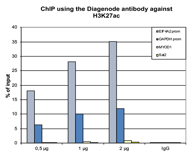
Figure 1. ChIP results obtained with the Diagenode monoclonal antibody directed against H3K27ac
ChIP was performed on sheared chromatin from 500000 HeLa cells using 0.5 µg of the Diagenode antibody against H3K4me3 (cat. No. C15410003) as described above. The IP'd DNA was subsequently analysed on an Illumina NovaSeq. Library preparation, cluster generation and sequencing were performed according to the manufacturer's instructions. The 50 bp tags were aligned to the human genome using the BWA algorithm. Figure 2 shows the peak distribution along the complete sequence and a 2 Mb region of the human X-chromosome (figure 2A and B) and in two regions surrounding the GAPDH and EIF4A2 positive control genes, respectively (figure 2C and D).A.
B.
C.
D.
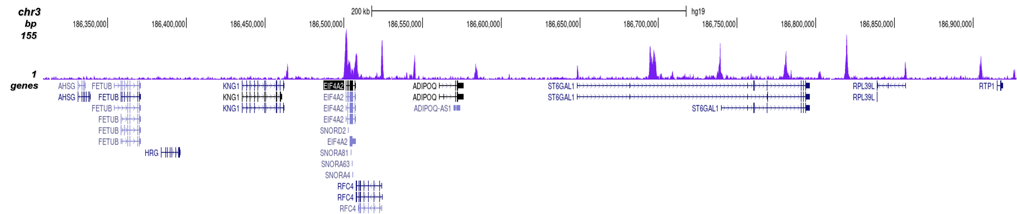
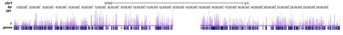
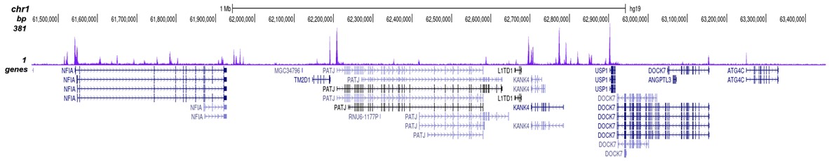
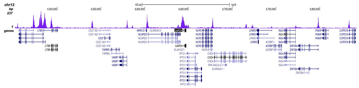
Figure 2. ChIP-seq results obtained with the Diagenode monoclonal antibody directed against H3K27ac
ChIP was performed on HeLa cells using 0.5 µg of the Diagenode antibody against H3K4me2 (cat. No. C15410035). The IP'd DNA was analysed on an Illumina Hiseq. Library preparation, cluster generation and sequencing were performed according to the manufacturer's instructions. The 51 bp tags were aligned to the human genome using the BWA algorithm. Figure 2 shows the peak distribution along the complete sequence and a 1.5 Mb region of the human X-chromosome (figure 2A and 2B) and in 2 chromosomal regions surrounding the ACTB and GAPDH positive control genes (figure 2C and D, respectively).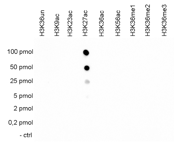
Figure 3. Cross reactivity tests using the Diagenode monoclonal antibody directed against H3K27ac
To test the cross reactivity of the Diagenode antibody against H3K27ac (cat. No. C15210016), a Dot Blot analysis was performed with peptides containing other histone modifications and the unmodified H3K27. One hundred to 0.2 pmol of the respective peptides were spotted on a membrane. The antibody was used at a dilution of 1:2,000. Figure 3 shows a high specificity of the antibody for the modification of interest.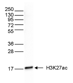
Figure 4. Western blot analysis using the Diagenode monoclonal antibody directed against H3K27ac
Western blot was performed on whole cell extracts (40 µg) from HeLa cells using the Diagenode antibody against H3K27ac (cat. No. C15210016). The antibody was diluted 1:1,000 in TBS-Tween containing 5% skimmed milk. The position of the protein of interest is shown on the right, the marker (in kDa) is shown on the left.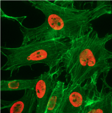
Figure 5. Immunofluorescence using the Diagenode monoclonal antibody directed against H3K27ac
HeLa cells were stained with the Diagenode antibody against H3K27ac (cat. No. C15210016, red) diluted 1:1,000. Actin was stained with fluorescein phalladoin (green). - 出版物

How to properly cite our product/service in your work
We strongly recommend using this: H3K27ac monoclonal antibody (Hologic Diagenode Cat# C15210016 Lot# 006). Click here to copy to clipboard.
Using our products or services in your publication? Let us know!
XPO1-inhibitor Selinexor induces MGMT expression by activating PKA-CREB signaling in IDH wildtype glioblastoma
Mapunda, Josephine A et al.
Purpose: The temozolomide (TMZ) resistance mechanisms in MGMT-promoter methylated IDH wildtype glioblastoma (GBM) tumors are poorly known. This study aimed to identify potential modulators of TMZ resistance in methylated GBM cells. Methods: A genome-wide shRNA library screen was conducted to ide...Legionella pneumophila modulates macrophage functions through epigenetic reprogramming via the C-type lectin receptor Mincle
Stegmann F. et al.
Legionella pneumophila is a pathogen which can lead to a severe form of pneumonia in humans known as Legionnaires disease after replication in alveolar macrophages. Viable L. pneumophila actively secrete effector molecules to modulate the host’s immune response. Here, we report that L....Chromatin profiling reveals TFAP4 as a critical transcriptional regulator of bovine satellite cell differentiation
Pengcheng Lyu et al.
Background Satellite cells are myogenic precursor cells in adult skeletal muscle and play a crucial role in skeletal muscle regeneration, maintenance, and growth. Like embryonic myoblasts, satellite cells have the ability to proliferate, differentiate, and fuse to form multinucleated myofibers. In this study, we ai...Epromoters function as a hub to recruit key transcription factorsrequired for the inflammatory response
Santiago-Algarra D. et al.
Gene expression is controlled by the involvement of gene-proximal (promoters) and distal (enhancers) regulatory elements. Our previous results demonstrated that a subset of gene promoters, termed Epromoters, work as bona fide enhancers and regulate distal gene expression. Here, we hypothesized that Epromoters play a...