Notice (8): Undefined variable: solution_of_interest [APP/View/Products/view.ctp, line 755]Code Context<!-- BEGIN: REQUEST_FORM MODAL -->
<div id="request_formModal" class="reveal-modal medium" data-reveal aria-labelledby="modalTitle" aria-hidden="true" role="dialog">
<?= $this->element('Forms/simple_form', array('solution_of_interest' => $solution_of_interest, 'header' => $header, 'message' => $message, 'campaign_id' => $campaign_id)) ?>
$viewFile = '/home/website-server/www/app/View/Products/view.ctp'
$dataForView = array(
'language' => 'jp',
'meta_keywords' => '',
'meta_description' => 'H3K4me1 polyclonal antibody - Premium',
'meta_title' => 'H3K4me1 polyclonal antibody - Premium',
'product' => array(
'Product' => array(
'id' => '2266',
'antibody_id' => '111',
'name' => 'H3K4me1 polyclonal antibody ',
'description' => '<p><span>Polyclonal antibody raised in rabbit against the region of histone H3 containing the monomethylated lysine 4 (H3K4me1), using a KLH-conjugated synthetic peptide.</span></p>',
'label1' => 'Validation Data',
'info1' => '<div class="row">
<div class="small-6 columns">
<p>A. <img src="https://www.diagenode.com/img/product/antibodies/C15410194-ChIP-1a.png" alt="H3K4me1 Antibody ChIP Grade" caption="false" width="432" /></p>
<p>B. <img src="https://www.diagenode.com/img/product/antibodies/C15410194-ChIP-1b.png" alt="H3K4me1 Antibody for ChIP" caption="false" width="432" /></p>
</div>
<div class="small-6 columns">
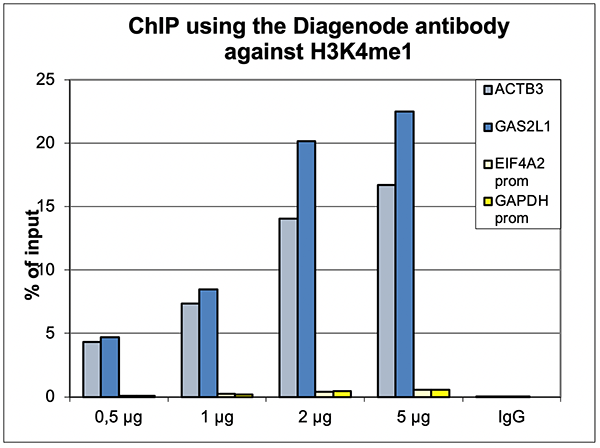
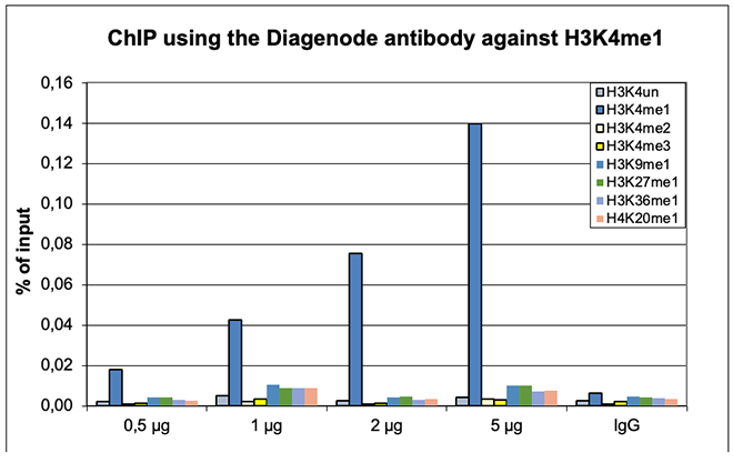
<p><small><strong>Figure 1. ChIP results obtained with the Diagenode antibody directed against H3K4me1</strong><br /> ChIP was performed with the Diagenode antibody against H3K4me1 (Cat. No. C15410194) on sheared chromatin from 500,000 HeLaS3 cells using the “iDeal ChIP-seq” kit (Cat. No. C01010051). The chromatin was spiked with a panel of in vitro assembled nucleosomes, each containing a specific lysine methylation. A titration of the antibody consisting of 0.5, 1, 2 and 5 µg per ChIP experiment was analysed. IgG (2 µg/IP) was used as negative IP control. <strong>Figure 1A.</strong> Quantitative PCR was performed with primers for a region surrounding the ACTB and GAS2L1 genes, used as positive controls, and for the promoters of the GAPDH and EIF4A2 genes, used as negative controls. The graph shows the recovery, expressed as a % of input (the relative amount of immunoprecipitated DNA compared to input DNA after qPCR analysis). <strong>Figure 1B.</strong> Recovery of the nucleosomes carrying the H3K4me1, H3K4me2, H3K4me3, H3K9me1, H3K27me1, H3K36me1, H4K20me1 and the unmodified H3K4 as determined by qPCR. The figure clearly shows the antibody is very specific in ChIP for the H3K4me1 modification. </small></p>
</div>
</div>
<div class="row">
<div class="small-6 columns">
<p><img src="https://www.diagenode.com/img/product/antibodies/C15410194-ChIP.png" alt="H3K4me1 Antibody for ChIP assay" caption="false" width="400" height="317" /></p>
</div>
<div class="small-6 columns">
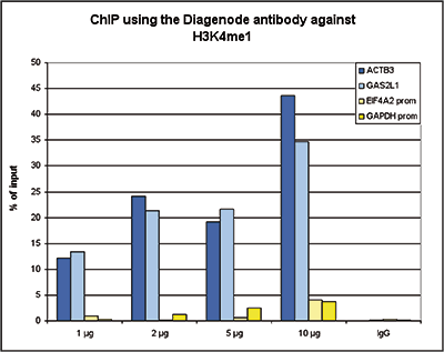
<p><small><strong>Figure 2. ChIP results obtained with the Diagenode antibody directed against H3K4me1</strong><br /> ChIP assays were performed using human K562 cells, the Diagenode antibody against H3K4me1 (Cat. No. C15410194) and optimized PCR primer pairs for qPCR. ChIP was performed with the “iDeal ChIP-seq” kit (Cat. No. C01010051), using sheared chromatin from 100,000 cells. A titration consisting of 1, 2, 5 and 10 µg of antibody per ChIP experiment was analyzed. IgG (1 µg/IP) was used as a negative IP control. Quantitative PCR was performed with primers for a region surrounding the ACTB and GAS2L1 gene, respectively, used as positive controls, and for the promoters of the GAPDH and EIF4A2 genes, used as negative controls. Figure 2 shows the recovery, expressed as a % of input (the relative amount of immunoprecipitated DNA compared to input DNA after qPCR analysis). </small></p>
</div>
</div>
<div class="extra-spaced"></div>
<div class="row">
<div class="small-12 columns"><center>A.<img src="https://www.diagenode.com/img/product/antibodies/C15410194-ChIPSeq-A.png" alt="H3K4me1 Antibody ChIP-seq Grade" caption="false" width="693" /></center><center>B. <img src="https://www.diagenode.com/img/product/antibodies/C15410194-ChIPSeq-B.png" alt="H3K4me1 Antibody for ChIP-seq " caption="false" width="693" /></center><center>C. <img src="https://www.diagenode.com/img/product/antibodies/C15410194-ChIPSeq-C.png" alt="H3K4me1 Antibody for ChIP-seq assay" caption="false" width="693" /></center></div>
</div>
<div class="row">
<div class="small-12 columns">
<p><small><strong>Figure 3. ChIP-seq results obtained with the Diagenode antibody directed against H3K4me1</strong><br /> ChIP was performed on sheared chromatin from 100,000 K562 cells with the “iDeal ChIP-seq” kit (Cat. No. C01010051) using 1 µg of the Diagenode antibody against H3K4me1 (Cat. No. C15410194) as described above. The IP’d DNA was subsequently analysed on an Illumina Genome Analyzer. Library preparation, cluster generation and sequencing were performed according to the manufacturer’s instructions. The 36 bp tags were aligned to the human genome using the ELAND algorithm. Figure 3A and B show the H3K4me1 signal in two genomic regions containing the ACTB and GAS2L1 positive controls. The position of the amplicon used for ChIP-qPCR is indicated by an arrow. Figure 3C shows the H3K4me1 peak distribution along a 1 Mb genomic region of chromosome 5. </small></p>
</div>
</div>
<div class="extra-spaced"></div>
<div class="row">
<div class="small-12 columns"><center>A. <img src="https://www.diagenode.com/img/product/antibodies/C15410194-fig4A-CT.jpg" width="693" /></center><center>B. <img src="https://www.diagenode.com/img/product/antibodies/C15410194-fig4B-CT.jpg" width="693" /></center></div>
</div>
<div class="row">
<div class="small-12 columns">
<p><small><strong>Figure 4. Cut&Tag results obtained with the Diagenode antibody directed against H3K4me1</strong><br /> CUT&TAG (Kaya-Okur, H.S., Nat Commun 10, 1930, 2019) was performed on 50,000 K562 cells using 1 µg of the Diagenode antibody against H3K4me1 (cat. No. C15410194) and the Diagenode pA-Tn5 transposase (C01070001). The libraries were subsequently analysed on an Illumina NextSeq 500 sequencer (2x75 paired-end reads) according to the manufacturer's instructions. The tags were aligned to the human genome (hg19) using the BWA algorithm. Figure 4 shows the peak distribution in 2 genomic regions surrounding the GAPDH gene on chromosome 12 and the FOS gene on chromosome 14 (figure 4A and B, respectively).</small></p>
<div class="row">
<div class="small-6 columns">
<p><img src="https://www.diagenode.com/img/product/antibodies/C15410194-ELISA.png" alt="H3K4me1 Antibody ELISA Validation" caption="false" width="400" height="303" /></p>
</div>
<div class="small-6 columns">
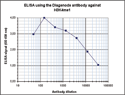
<p><small><strong>Figure 5. Determination of the antibody titer</strong><br /> To determine the titer of the antibody, an ELISA was performed using a serial dilution of the Diagenode antibody against H3K4me1 (Cat. No. C15410194). The antigen used was a peptide containing the histone modification of interest. By plotting the absorbance against the antibody dilution (Figure 5), the titer of the antibody was estimated to be 1:10,300. </small></p>
</div>
</div>
<div class="extra-spaced"></div>
<div class="row">
<div class="small-4 columns">
<p>A.<img src="https://www.diagenode.com/img/product/antibodies/C15410194-DotBlot-A.png" alt="H3K4me1 Antibody Dot Blot Validation" caption="false" width="278" /><br />B.<img src="https://www.diagenode.com/img/product/antibodies/C15410194-DotBlot-B.png" alt="H3K4me1 Antibody Peptide Array Validation" caption="false" width="278" /></p>
</div>
<div class="small-8 columns">
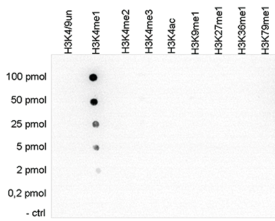
<p><small><strong>Figure 6. Cross reactivity tests using the Diagenode antibody directed against H3K4me1</strong><br /> <strong>Figure 6A.</strong> To test the cross reactivity of the Diagenode antibody against H3K4me1 (Cat. No. C15410194), a Dot Blot analysis was performed with peptides containing other histone modifications and the unmodified H3K4. One hundred to 0.2 pmol of the respective peptides were spotted on a membrane. The antibody was used at a dilution of 1:5,000. Figure 6A shows a high specificity of the antibody for the modification of interest. <br /></small></p>
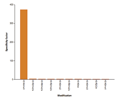
<p><small><strong>Figure 6B.</strong> The specificity of the antibody was further demonstrated by peptide array analyses on an array containing 384 peptides with different combinations of modifications from histone H3, H4, H2A and H2B. The antibody was used at a dilution of 1:2,000. Figure 6B shows the specificity factor, calculated as the ratio of the average intensity of all spots containing the mark, divided by the average intensity of all spots not containing the mark. </small></p>
</div>
</div>
<div class="row">
<div class="small-4 columns">
<p><img src="https://www.diagenode.com/img/product/antibodies/C15410194-WB.png" alt="H3K4me1 Antibody validated in Western blot " caption="false" width="278" height="187" /></p>
</div>
<div class="small-8 columns">
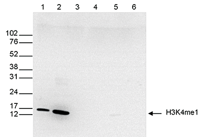
<p><small><strong>Figure 7. Western blot analysis using the Diagenode antibody directed against H3K4me1</strong><br /> Western blot was performed on whole cell (25 µg, lane 1) and histone extracts (15 µg, lane 2) from HeLa cells, and on 1 µg of recombinant histone H2A, H2B, H3 and H4 (lane 3, 4, 5 and 6, respectively) using the Diagenode antibody against H3K4me1 (Cat. No. C15410194). The antibody was diluted 1:500 in TBS-Tween containing 5% skimmed milk. The position of the protein of interest is shown on the right, the marker (in kDa) is shown on the left. </small></p>
</div>
</div>
<div class="row">
<div class="small-12 columns"><center><img src="https://www.diagenode.com/img/product/antibodies/C15410194-IF.png" alt="H3K4me1 Antibody validated for Immunofluorescence " caption="false" width="500" height="122" /></center></div>
</div>
<div class="row">
<div class="small-12 columns">
<p><small><strong>Figure 8. Immunofluorescence using the Diagenode antibody directed against H3K4me1</strong><br /> HeLa cells were stained with the Diagenode antibody against H3K4me1 (Cat. No. C15410194) and with DAPI. Cells were fixed with 4% formaldehyde for 10’ and blocked with PBS/TX-100 containing 5% normal goat serum and 1% BSA. The cells were immunofluorescently labeled with the H3K4me1 antibody (left) diluted 1:200 in blocking solution followed by an anti-rabbit antibody conjugated to Alexa488. The middle panel shows staining of the nuclei with DAPI. A merge of the two stainings is shown on the right. </small></p>
</div>
</div>
</div>
</div>',
'label2' => '',
'info2' => '<p>Histones are the main constituents of the protein part of chromosomes of eukaryotic cells. They are rich in the amino acids arginine and lysine and have been greatly conserved during evolution. Histones pack the DNA into tight masses of chromatin. Two core histones of each class H2A, H2B, H3 and H4 assemble and are wrapped by 146 base pairs of DNA to form one octameric nucleosome. Histone tails undergo numerous post-translational modifications, which either directly or indirectly alter chromatin structure to facilitate transcriptional activation or repression or other nuclear processes. In addition to the genetic code, combinations of the different histone modifications reveal the so-called “histone code”. Histone methylation and demethylation is dynamically regulated by respectively histone methyl transferases and histone demethylases. Methylation of histone H3K4 is associated with active genes.</p>',
'label3' => '',
'info3' => '',
'format' => '50 μg',
'catalog_number' => 'C15410194',
'old_catalog_number' => 'pAb-194-050',
'sf_code' => 'C15410194-D001-000581',
'type' => 'FRE',
'search_order' => '03-Antibody',
'price_EUR' => '480',
'price_USD' => '470',
'price_GBP' => '430',
'price_JPY' => '75190',
'price_CNY' => '',
'price_AUD' => '1175',
'country' => 'ALL',
'except_countries' => 'None',
'quote' => false,
'in_stock' => false,
'featured' => false,
'no_promo' => false,
'online' => true,
'master' => true,
'last_datasheet_update' => 'January 6, 2020',
'slug' => 'h3k4me1-polyclonal-antibody-premium-50-mg',
'meta_title' => 'H3K4me1 polyclonal antibody - Premium',
'meta_keywords' => '',
'meta_description' => 'H3K4me1 polyclonal antibody - Premium',
'modified' => '2021-10-20 09:56:46',
'created' => '2015-06-29 14:08:20',
'locale' => 'jpn'
),
'Antibody' => array(
'host' => '*****',
'id' => '111',
'name' => 'H3K4me1 polyclonal antibody',
'description' => 'Histones are the main constituents of the protein part of chromosomes of eukaryotic cells. They are rich in the amino acids arginine and lysine and have been greatly conserved during evolution. Histones pack the DNA into tight masses of chromatin. Two core histones of each class H2A, H2B, H3 and H4 assemble and are wrapped by 146 base pairs of DNA to form one octameric nucleosome. Histone tails undergo numerous post-translational modifications, which either directly or indirectly alter chromatin structure to facilitate transcriptional activation or repression or other nuclear processes. In addition to the genetic code, combinations of the different histone modifications reveal the so-called “histone code”. Histone methylation and demethylation is dynamically regulated by respectively histone methyl transferases and histone demethylases. Methylation of histone H3K4 is associated with active genes.',
'clonality' => '',
'isotype' => '',
'lot' => 'A1862D',
'concentration' => '1.5 µg/µl',
'reactivity' => 'Human, Mouse, Drosophila, wide range expected',
'type' => 'Polyclonal, <strong>ChIP grade, ChIP-seq grade</strong>',
'purity' => 'Affinity purified polyclonal antibody.',
'classification' => 'Premium',
'application_table' => '<table>
<thead>
<tr>
<th>Applications</th>
<th>Suggested dilution</th>
<th>References</th>
</tr>
</thead>
<tbody>
<tr>
<td>ChIP/ChIP-seq <sup>*</sup></td>
<td>0.5-1 μg/IP</td>
<td>Fig 1, 2, 3</td>
</tr>
<tr>
<td>CUT&TAG</td>
<td>1 μg</td>
<td>Fig 4</td>
</tr>
<tr>
<td>ELISA</td>
<td>1:400</td>
<td>Fig 5</td>
</tr>
<tr>
<td>Dot Blotting/Peptide array</td>
<td>1:5,000/1:2,000</td>
<td>Fig 6</td>
</tr>
<tr>
<td>Western Blotting</td>
<td>1:500</td>
<td>Fig 7</td>
</tr>
<tr>
<td>Immunofluorescence</td>
<td>1:200</td>
<td>Fig 8</td>
</tr>
</tbody>
</table>
<p></p>
<p><small><sup>*</sup> Please note that the optimal antibody amount per IP should be determined by the end-user. We recommend testing 0.5-5 µg per IP.</small></p>',
'storage_conditions' => 'Store at -20°C; for long storage, store at -80°C. Avoid multiple freeze-thaw cycles.',
'storage_buffer' => 'PBS containing 0.05% azide and 0.05% ProClin 300.',
'precautions' => 'This product is for research use only. Not for use in diagnostic or therapeutic procedures.',
'uniprot_acc' => '',
'slug' => '',
'meta_keywords' => '',
'meta_description' => '',
'modified' => '2021-07-28 12:07:24',
'created' => '0000-00-00 00:00:00',
'select_label' => '111 - H3K4me1 polyclonal antibody (A1862D - 1.5 µg/µl - Human, Mouse, Drosophila, wide range expected - Affinity purified polyclonal antibody. - Rabbit)'
),
'Slave' => array(
(int) 0 => array(
[maximum depth reached]
)
),
'Group' => array(
'Group' => array(
[maximum depth reached]
),
'Master' => array(
[maximum depth reached]
),
'Product' => array(
[maximum depth reached]
)
),
'Related' => array(
(int) 0 => array(
[maximum depth reached]
),
(int) 1 => array(
[maximum depth reached]
),
(int) 2 => array(
[maximum depth reached]
),
(int) 3 => array(
[maximum depth reached]
),
(int) 4 => array(
[maximum depth reached]
),
(int) 5 => array(
[maximum depth reached]
),
(int) 6 => array(
[maximum depth reached]
),
(int) 7 => array(
[maximum depth reached]
),
(int) 8 => array(
[maximum depth reached]
)
),
'Application' => array(
(int) 0 => array(
[maximum depth reached]
),
(int) 1 => array(
[maximum depth reached]
),
(int) 2 => array(
[maximum depth reached]
),
(int) 3 => array(
[maximum depth reached]
),
(int) 4 => array(
[maximum depth reached]
),
(int) 5 => array(
[maximum depth reached]
),
(int) 6 => array(
[maximum depth reached]
),
(int) 7 => array(
[maximum depth reached]
)
),
'Category' => array(
(int) 0 => array(
[maximum depth reached]
),
(int) 1 => array(
[maximum depth reached]
),
(int) 2 => array(
[maximum depth reached]
),
(int) 3 => array(
[maximum depth reached]
)
),
'Document' => array(
(int) 0 => array(
[maximum depth reached]
),
(int) 1 => array(
[maximum depth reached]
),
(int) 2 => array(
[maximum depth reached]
)
),
'Feature' => array(),
'Image' => array(
(int) 0 => array(
[maximum depth reached]
)
),
'Promotion' => array(),
'Protocol' => array(),
'Publication' => array(
(int) 0 => array(
[maximum depth reached]
),
(int) 1 => array(
[maximum depth reached]
),
(int) 2 => array(
[maximum depth reached]
),
(int) 3 => array(
[maximum depth reached]
),
(int) 4 => array(
[maximum depth reached]
),
(int) 5 => array(
[maximum depth reached]
),
(int) 6 => array(
[maximum depth reached]
),
(int) 7 => array(
[maximum depth reached]
),
(int) 8 => array(
[maximum depth reached]
),
(int) 9 => array(
[maximum depth reached]
),
(int) 10 => array(
[maximum depth reached]
),
(int) 11 => array(
[maximum depth reached]
),
(int) 12 => array(
[maximum depth reached]
),
(int) 13 => array(
[maximum depth reached]
),
(int) 14 => array(
[maximum depth reached]
),
(int) 15 => array(
[maximum depth reached]
),
(int) 16 => array(
[maximum depth reached]
),
(int) 17 => array(
[maximum depth reached]
),
(int) 18 => array(
[maximum depth reached]
),
(int) 19 => array(
[maximum depth reached]
),
(int) 20 => array(
[maximum depth reached]
),
(int) 21 => array(
[maximum depth reached]
),
(int) 22 => array(
[maximum depth reached]
),
(int) 23 => array(
[maximum depth reached]
),
(int) 24 => array(
[maximum depth reached]
),
(int) 25 => array(
[maximum depth reached]
),
(int) 26 => array(
[maximum depth reached]
),
(int) 27 => array(
[maximum depth reached]
),
(int) 28 => array(
[maximum depth reached]
),
(int) 29 => array(
[maximum depth reached]
),
(int) 30 => array(
[maximum depth reached]
),
(int) 31 => array(
[maximum depth reached]
),
(int) 32 => array(
[maximum depth reached]
),
(int) 33 => array(
[maximum depth reached]
),
(int) 34 => array(
[maximum depth reached]
),
(int) 35 => array(
[maximum depth reached]
),
(int) 36 => array(
[maximum depth reached]
),
(int) 37 => array(
[maximum depth reached]
),
(int) 38 => array(
[maximum depth reached]
),
(int) 39 => array(
[maximum depth reached]
),
(int) 40 => array(
[maximum depth reached]
),
(int) 41 => array(
[maximum depth reached]
),
(int) 42 => array(
[maximum depth reached]
),
(int) 43 => array(
[maximum depth reached]
),
(int) 44 => array(
[maximum depth reached]
),
(int) 45 => array(
[maximum depth reached]
),
(int) 46 => array(
[maximum depth reached]
),
(int) 47 => array(
[maximum depth reached]
),
(int) 48 => array(
[maximum depth reached]
),
(int) 49 => array(
[maximum depth reached]
),
(int) 50 => array(
[maximum depth reached]
),
(int) 51 => array(
[maximum depth reached]
),
(int) 52 => array(
[maximum depth reached]
),
(int) 53 => array(
[maximum depth reached]
),
(int) 54 => array(
[maximum depth reached]
),
(int) 55 => array(
[maximum depth reached]
),
(int) 56 => array(
[maximum depth reached]
),
(int) 57 => array(
[maximum depth reached]
),
(int) 58 => array(
[maximum depth reached]
),
(int) 59 => array(
[maximum depth reached]
),
(int) 60 => array(
[maximum depth reached]
),
(int) 61 => array(
[maximum depth reached]
),
(int) 62 => array(
[maximum depth reached]
),
(int) 63 => array(
[maximum depth reached]
),
(int) 64 => array(
[maximum depth reached]
)
),
'Testimonial' => array(
(int) 0 => array(
[maximum depth reached]
)
),
'Area' => array(),
'SafetySheet' => array(
(int) 0 => array(
[maximum depth reached]
),
(int) 1 => array(
[maximum depth reached]
),
(int) 2 => array(
[maximum depth reached]
),
(int) 3 => array(
[maximum depth reached]
),
(int) 4 => array(
[maximum depth reached]
),
(int) 5 => array(
[maximum depth reached]
),
(int) 6 => array(
[maximum depth reached]
),
(int) 7 => array(
[maximum depth reached]
)
)
),
'meta_canonical' => 'https://www.diagenode.com/jp/p/h3k4me1-polyclonal-antibody-premium-50-mg'
)
$language = 'jp'
$meta_keywords = ''
$meta_description = 'H3K4me1 polyclonal antibody - Premium'
$meta_title = 'H3K4me1 polyclonal antibody - Premium'
$product = array(
'Product' => array(
'id' => '2266',
'antibody_id' => '111',
'name' => 'H3K4me1 polyclonal antibody ',
'description' => '<p><span>Polyclonal antibody raised in rabbit against the region of histone H3 containing the monomethylated lysine 4 (H3K4me1), using a KLH-conjugated synthetic peptide.</span></p>',
'label1' => 'Validation Data',
'info1' => '<div class="row">
<div class="small-6 columns">
<p>A. <img src="https://www.diagenode.com/img/product/antibodies/C15410194-ChIP-1a.png" alt="H3K4me1 Antibody ChIP Grade" caption="false" width="432" /></p>
<p>B. <img src="https://www.diagenode.com/img/product/antibodies/C15410194-ChIP-1b.png" alt="H3K4me1 Antibody for ChIP" caption="false" width="432" /></p>
</div>
<div class="small-6 columns">
<p><small><strong>Figure 1. ChIP results obtained with the Diagenode antibody directed against H3K4me1</strong><br /> ChIP was performed with the Diagenode antibody against H3K4me1 (Cat. No. C15410194) on sheared chromatin from 500,000 HeLaS3 cells using the “iDeal ChIP-seq” kit (Cat. No. C01010051). The chromatin was spiked with a panel of in vitro assembled nucleosomes, each containing a specific lysine methylation. A titration of the antibody consisting of 0.5, 1, 2 and 5 µg per ChIP experiment was analysed. IgG (2 µg/IP) was used as negative IP control. <strong>Figure 1A.</strong> Quantitative PCR was performed with primers for a region surrounding the ACTB and GAS2L1 genes, used as positive controls, and for the promoters of the GAPDH and EIF4A2 genes, used as negative controls. The graph shows the recovery, expressed as a % of input (the relative amount of immunoprecipitated DNA compared to input DNA after qPCR analysis). <strong>Figure 1B.</strong> Recovery of the nucleosomes carrying the H3K4me1, H3K4me2, H3K4me3, H3K9me1, H3K27me1, H3K36me1, H4K20me1 and the unmodified H3K4 as determined by qPCR. The figure clearly shows the antibody is very specific in ChIP for the H3K4me1 modification. </small></p>
</div>
</div>
<div class="row">
<div class="small-6 columns">
<p><img src="https://www.diagenode.com/img/product/antibodies/C15410194-ChIP.png" alt="H3K4me1 Antibody for ChIP assay" caption="false" width="400" height="317" /></p>
</div>
<div class="small-6 columns">
<p><small><strong>Figure 2. ChIP results obtained with the Diagenode antibody directed against H3K4me1</strong><br /> ChIP assays were performed using human K562 cells, the Diagenode antibody against H3K4me1 (Cat. No. C15410194) and optimized PCR primer pairs for qPCR. ChIP was performed with the “iDeal ChIP-seq” kit (Cat. No. C01010051), using sheared chromatin from 100,000 cells. A titration consisting of 1, 2, 5 and 10 µg of antibody per ChIP experiment was analyzed. IgG (1 µg/IP) was used as a negative IP control. Quantitative PCR was performed with primers for a region surrounding the ACTB and GAS2L1 gene, respectively, used as positive controls, and for the promoters of the GAPDH and EIF4A2 genes, used as negative controls. Figure 2 shows the recovery, expressed as a % of input (the relative amount of immunoprecipitated DNA compared to input DNA after qPCR analysis). </small></p>
</div>
</div>
<div class="extra-spaced"></div>
<div class="row">
<div class="small-12 columns"><center>A.<img src="https://www.diagenode.com/img/product/antibodies/C15410194-ChIPSeq-A.png" alt="H3K4me1 Antibody ChIP-seq Grade" caption="false" width="693" /></center><center>B. <img src="https://www.diagenode.com/img/product/antibodies/C15410194-ChIPSeq-B.png" alt="H3K4me1 Antibody for ChIP-seq " caption="false" width="693" /></center><center>C. <img src="https://www.diagenode.com/img/product/antibodies/C15410194-ChIPSeq-C.png" alt="H3K4me1 Antibody for ChIP-seq assay" caption="false" width="693" /></center></div>
</div>
<div class="row">
<div class="small-12 columns">
<p><small><strong>Figure 3. ChIP-seq results obtained with the Diagenode antibody directed against H3K4me1</strong><br /> ChIP was performed on sheared chromatin from 100,000 K562 cells with the “iDeal ChIP-seq” kit (Cat. No. C01010051) using 1 µg of the Diagenode antibody against H3K4me1 (Cat. No. C15410194) as described above. The IP’d DNA was subsequently analysed on an Illumina Genome Analyzer. Library preparation, cluster generation and sequencing were performed according to the manufacturer’s instructions. The 36 bp tags were aligned to the human genome using the ELAND algorithm. Figure 3A and B show the H3K4me1 signal in two genomic regions containing the ACTB and GAS2L1 positive controls. The position of the amplicon used for ChIP-qPCR is indicated by an arrow. Figure 3C shows the H3K4me1 peak distribution along a 1 Mb genomic region of chromosome 5. </small></p>
</div>
</div>
<div class="extra-spaced"></div>
<div class="row">
<div class="small-12 columns"><center>A. <img src="https://www.diagenode.com/img/product/antibodies/C15410194-fig4A-CT.jpg" width="693" /></center><center>B. <img src="https://www.diagenode.com/img/product/antibodies/C15410194-fig4B-CT.jpg" width="693" /></center></div>
</div>
<div class="row">
<div class="small-12 columns">
<p><small><strong>Figure 4. Cut&Tag results obtained with the Diagenode antibody directed against H3K4me1</strong><br /> CUT&TAG (Kaya-Okur, H.S., Nat Commun 10, 1930, 2019) was performed on 50,000 K562 cells using 1 µg of the Diagenode antibody against H3K4me1 (cat. No. C15410194) and the Diagenode pA-Tn5 transposase (C01070001). The libraries were subsequently analysed on an Illumina NextSeq 500 sequencer (2x75 paired-end reads) according to the manufacturer's instructions. The tags were aligned to the human genome (hg19) using the BWA algorithm. Figure 4 shows the peak distribution in 2 genomic regions surrounding the GAPDH gene on chromosome 12 and the FOS gene on chromosome 14 (figure 4A and B, respectively).</small></p>
<div class="row">
<div class="small-6 columns">
<p><img src="https://www.diagenode.com/img/product/antibodies/C15410194-ELISA.png" alt="H3K4me1 Antibody ELISA Validation" caption="false" width="400" height="303" /></p>
</div>
<div class="small-6 columns">
<p><small><strong>Figure 5. Determination of the antibody titer</strong><br /> To determine the titer of the antibody, an ELISA was performed using a serial dilution of the Diagenode antibody against H3K4me1 (Cat. No. C15410194). The antigen used was a peptide containing the histone modification of interest. By plotting the absorbance against the antibody dilution (Figure 5), the titer of the antibody was estimated to be 1:10,300. </small></p>
</div>
</div>
<div class="extra-spaced"></div>
<div class="row">
<div class="small-4 columns">
<p>A.<img src="https://www.diagenode.com/img/product/antibodies/C15410194-DotBlot-A.png" alt="H3K4me1 Antibody Dot Blot Validation" caption="false" width="278" /><br />B.<img src="https://www.diagenode.com/img/product/antibodies/C15410194-DotBlot-B.png" alt="H3K4me1 Antibody Peptide Array Validation" caption="false" width="278" /></p>
</div>
<div class="small-8 columns">
<p><small><strong>Figure 6. Cross reactivity tests using the Diagenode antibody directed against H3K4me1</strong><br /> <strong>Figure 6A.</strong> To test the cross reactivity of the Diagenode antibody against H3K4me1 (Cat. No. C15410194), a Dot Blot analysis was performed with peptides containing other histone modifications and the unmodified H3K4. One hundred to 0.2 pmol of the respective peptides were spotted on a membrane. The antibody was used at a dilution of 1:5,000. Figure 6A shows a high specificity of the antibody for the modification of interest. <br /></small></p>
<p><small><strong>Figure 6B.</strong> The specificity of the antibody was further demonstrated by peptide array analyses on an array containing 384 peptides with different combinations of modifications from histone H3, H4, H2A and H2B. The antibody was used at a dilution of 1:2,000. Figure 6B shows the specificity factor, calculated as the ratio of the average intensity of all spots containing the mark, divided by the average intensity of all spots not containing the mark. </small></p>
</div>
</div>
<div class="row">
<div class="small-4 columns">
<p><img src="https://www.diagenode.com/img/product/antibodies/C15410194-WB.png" alt="H3K4me1 Antibody validated in Western blot " caption="false" width="278" height="187" /></p>
</div>
<div class="small-8 columns">
<p><small><strong>Figure 7. Western blot analysis using the Diagenode antibody directed against H3K4me1</strong><br /> Western blot was performed on whole cell (25 µg, lane 1) and histone extracts (15 µg, lane 2) from HeLa cells, and on 1 µg of recombinant histone H2A, H2B, H3 and H4 (lane 3, 4, 5 and 6, respectively) using the Diagenode antibody against H3K4me1 (Cat. No. C15410194). The antibody was diluted 1:500 in TBS-Tween containing 5% skimmed milk. The position of the protein of interest is shown on the right, the marker (in kDa) is shown on the left. </small></p>
</div>
</div>
<div class="row">
<div class="small-12 columns"><center><img src="https://www.diagenode.com/img/product/antibodies/C15410194-IF.png" alt="H3K4me1 Antibody validated for Immunofluorescence " caption="false" width="500" height="122" /></center></div>
</div>
<div class="row">
<div class="small-12 columns">
<p><small><strong>Figure 8. Immunofluorescence using the Diagenode antibody directed against H3K4me1</strong><br /> HeLa cells were stained with the Diagenode antibody against H3K4me1 (Cat. No. C15410194) and with DAPI. Cells were fixed with 4% formaldehyde for 10’ and blocked with PBS/TX-100 containing 5% normal goat serum and 1% BSA. The cells were immunofluorescently labeled with the H3K4me1 antibody (left) diluted 1:200 in blocking solution followed by an anti-rabbit antibody conjugated to Alexa488. The middle panel shows staining of the nuclei with DAPI. A merge of the two stainings is shown on the right. </small></p>
</div>
</div>
</div>
</div>',
'label2' => 'Target Description',
'info2' => '<p>Histones are the main constituents of the protein part of chromosomes of eukaryotic cells. They are rich in the amino acids arginine and lysine and have been greatly conserved during evolution. Histones pack the DNA into tight masses of chromatin. Two core histones of each class H2A, H2B, H3 and H4 assemble and are wrapped by 146 base pairs of DNA to form one octameric nucleosome. Histone tails undergo numerous post-translational modifications, which either directly or indirectly alter chromatin structure to facilitate transcriptional activation or repression or other nuclear processes. In addition to the genetic code, combinations of the different histone modifications reveal the so-called “histone code”. Histone methylation and demethylation is dynamically regulated by respectively histone methyl transferases and histone demethylases. Methylation of histone H3K4 is associated with active genes.</p>',
'label3' => '',
'info3' => '',
'format' => '50 μg',
'catalog_number' => 'C15410194',
'old_catalog_number' => 'pAb-194-050',
'sf_code' => 'C15410194-D001-000581',
'type' => 'FRE',
'search_order' => '03-Antibody',
'price_EUR' => '480',
'price_USD' => '470',
'price_GBP' => '430',
'price_JPY' => '75190',
'price_CNY' => '',
'price_AUD' => '1175',
'country' => 'ALL',
'except_countries' => 'None',
'quote' => false,
'in_stock' => false,
'featured' => false,
'no_promo' => false,
'online' => true,
'master' => true,
'last_datasheet_update' => 'January 6, 2020',
'slug' => 'h3k4me1-polyclonal-antibody-premium-50-mg',
'meta_title' => 'H3K4me1 polyclonal antibody - Premium',
'meta_keywords' => '',
'meta_description' => 'H3K4me1 polyclonal antibody - Premium',
'modified' => '2021-10-20 09:56:46',
'created' => '2015-06-29 14:08:20',
'locale' => 'jpn'
),
'Antibody' => array(
'host' => '*****',
'id' => '111',
'name' => 'H3K4me1 polyclonal antibody',
'description' => 'Histones are the main constituents of the protein part of chromosomes of eukaryotic cells. They are rich in the amino acids arginine and lysine and have been greatly conserved during evolution. Histones pack the DNA into tight masses of chromatin. Two core histones of each class H2A, H2B, H3 and H4 assemble and are wrapped by 146 base pairs of DNA to form one octameric nucleosome. Histone tails undergo numerous post-translational modifications, which either directly or indirectly alter chromatin structure to facilitate transcriptional activation or repression or other nuclear processes. In addition to the genetic code, combinations of the different histone modifications reveal the so-called “histone code”. Histone methylation and demethylation is dynamically regulated by respectively histone methyl transferases and histone demethylases. Methylation of histone H3K4 is associated with active genes.',
'clonality' => '',
'isotype' => '',
'lot' => 'A1862D',
'concentration' => '1.5 µg/µl',
'reactivity' => 'Human, Mouse, Drosophila, wide range expected',
'type' => 'Polyclonal, <strong>ChIP grade, ChIP-seq grade</strong>',
'purity' => 'Affinity purified polyclonal antibody.',
'classification' => 'Premium',
'application_table' => '<table>
<thead>
<tr>
<th>Applications</th>
<th>Suggested dilution</th>
<th>References</th>
</tr>
</thead>
<tbody>
<tr>
<td>ChIP/ChIP-seq <sup>*</sup></td>
<td>0.5-1 μg/IP</td>
<td>Fig 1, 2, 3</td>
</tr>
<tr>
<td>CUT&TAG</td>
<td>1 μg</td>
<td>Fig 4</td>
</tr>
<tr>
<td>ELISA</td>
<td>1:400</td>
<td>Fig 5</td>
</tr>
<tr>
<td>Dot Blotting/Peptide array</td>
<td>1:5,000/1:2,000</td>
<td>Fig 6</td>
</tr>
<tr>
<td>Western Blotting</td>
<td>1:500</td>
<td>Fig 7</td>
</tr>
<tr>
<td>Immunofluorescence</td>
<td>1:200</td>
<td>Fig 8</td>
</tr>
</tbody>
</table>
<p></p>
<p><small><sup>*</sup> Please note that the optimal antibody amount per IP should be determined by the end-user. We recommend testing 0.5-5 µg per IP.</small></p>',
'storage_conditions' => 'Store at -20°C; for long storage, store at -80°C. Avoid multiple freeze-thaw cycles.',
'storage_buffer' => 'PBS containing 0.05% azide and 0.05% ProClin 300.',
'precautions' => 'This product is for research use only. Not for use in diagnostic or therapeutic procedures.',
'uniprot_acc' => '',
'slug' => '',
'meta_keywords' => '',
'meta_description' => '',
'modified' => '2021-07-28 12:07:24',
'created' => '0000-00-00 00:00:00',
'select_label' => '111 - H3K4me1 polyclonal antibody (A1862D - 1.5 µg/µl - Human, Mouse, Drosophila, wide range expected - Affinity purified polyclonal antibody. - Rabbit)'
),
'Slave' => array(
(int) 0 => array(
'id' => '45',
'name' => 'C15410194',
'product_id' => '2266',
'modified' => '2016-02-18 20:49:43',
'created' => '2016-02-18 20:49:43'
)
),
'Group' => array(
'Group' => array(
'id' => '45',
'name' => 'C15410194',
'product_id' => '2266',
'modified' => '2016-02-18 20:49:43',
'created' => '2016-02-18 20:49:43'
),
'Master' => array(
'id' => '2266',
'antibody_id' => '111',
'name' => 'H3K4me1 Antibody',
'description' => '<p><span>Polyclonal antibody raised in rabbit against the region of histone <strong>H3 containing the monomethylated lysine 4</strong> (<strong>H3K4me1</strong>), using a KLH-conjugated synthetic peptide.</span></p>',
'label1' => 'Validation data',
'info1' => '<div class="row">
<div class="small-6 columns">
<p>A. <img src="https://www.diagenode.com/img/product/antibodies/C15410194-ChIP-1a.png" alt="H3K4me1 Antibody ChIP Grade" caption="false" width="432" /></p>
<p>B. <img src="https://www.diagenode.com/img/product/antibodies/C15410194-ChIP-1b.png" alt="H3K4me1 Antibody for ChIP" caption="false" width="432" /></p>
</div>
<div class="small-6 columns">
<p><small><strong>Figure 1. ChIP results obtained with the Diagenode antibody directed against H3K4me1</strong><br /> ChIP was performed with the Diagenode antibody against H3K4me1 (Cat. No. C15410194) on sheared chromatin from 500,000 HeLaS3 cells using the “iDeal ChIP-seq” kit (Cat. No. C01010051). The chromatin was spiked with a panel of in vitro assembled nucleosomes, each containing a specific lysine methylation. A titration of the antibody consisting of 0.5, 1, 2 and 5 µg per ChIP experiment was analysed. IgG (2 µg/IP) was used as negative IP control. <strong>Figure 1A.</strong> Quantitative PCR was performed with primers for a region surrounding the ACTB and GAS2L1 genes, used as positive controls, and for the promoters of the GAPDH and EIF4A2 genes, used as negative controls. The graph shows the recovery, expressed as a % of input (the relative amount of immunoprecipitated DNA compared to input DNA after qPCR analysis). <strong>Figure 1B.</strong> Recovery of the nucleosomes carrying the H3K4me1, H3K4me2, H3K4me3, H3K9me1, H3K27me1, H3K36me1, H4K20me1 and the unmodified H3K4 as determined by qPCR. The figure clearly shows the antibody is very specific in ChIP for the H3K4me1 modification. </small></p>
</div>
</div>
<div class="row">
<div class="small-6 columns">
<p><img src="https://www.diagenode.com/img/product/antibodies/C15410194-ChIP.png" alt="H3K4me1 Antibody for ChIP assay" caption="false" width="400" height="317" /></p>
</div>
<div class="small-6 columns">
<p><small><strong>Figure 2. ChIP results obtained with the Diagenode antibody directed against H3K4me1</strong><br /> ChIP assays were performed using human K562 cells, the Diagenode antibody against H3K4me1 (Cat. No. C15410194) and optimized PCR primer pairs for qPCR. ChIP was performed with the “iDeal ChIP-seq” kit (Cat. No. C01010051), using sheared chromatin from 100,000 cells. A titration consisting of 1, 2, 5 and 10 µg of antibody per ChIP experiment was analyzed. IgG (1 µg/IP) was used as a negative IP control. Quantitative PCR was performed with primers for a region surrounding the ACTB and GAS2L1 gene, respectively, used as positive controls, and for the promoters of the GAPDH and EIF4A2 genes, used as negative controls. Figure 2 shows the recovery, expressed as a % of input (the relative amount of immunoprecipitated DNA compared to input DNA after qPCR analysis). </small></p>
</div>
</div>
<div class="extra-spaced"></div>
<div class="row">
<div class="small-12 columns"><center>A.<img src="https://www.diagenode.com/img/product/antibodies/C15410194-ChIPSeq-A.png" alt="H3K4me1 Antibody ChIP-seq Grade" caption="false" width="693" /></center><center>B. <img src="https://www.diagenode.com/img/product/antibodies/C15410194-ChIPSeq-B.png" alt="H3K4me1 Antibody for ChIP-seq " caption="false" width="693" /></center><center>C. <img src="https://www.diagenode.com/img/product/antibodies/C15410194-ChIPSeq-C.png" alt="H3K4me1 Antibody for ChIP-seq assay" caption="false" width="693" /></center></div>
</div>
<div class="row">
<div class="small-12 columns">
<p><small><strong>Figure 3. ChIP-seq results obtained with the Diagenode antibody directed against H3K4me1</strong><br /> ChIP was performed on sheared chromatin from 100,000 K562 cells with the “iDeal ChIP-seq” kit (Cat. No. C01010051) using 1 µg of the Diagenode antibody against H3K4me1 (Cat. No. C15410194) as described above. The IP’d DNA was subsequently analysed on an Illumina Genome Analyzer. Library preparation, cluster generation and sequencing were performed according to the manufacturer’s instructions. The 36 bp tags were aligned to the human genome using the ELAND algorithm. Figure 3A and B show the H3K4me1 signal in two genomic regions containing the ACTB and GAS2L1 positive controls. The position of the amplicon used for ChIP-qPCR is indicated by an arrow. Figure 3C shows the H3K4me1 peak distribution along a 1 Mb genomic region of chromosome 5. </small></p>
</div>
</div>
<div class="extra-spaced"></div>
<div class="row">
<div class="small-12 columns"><center>A. <img src="https://www.diagenode.com/img/product/antibodies/C15410194-fig4A-CT.jpg" width="693" /></center><center>B. <img src="https://www.diagenode.com/img/product/antibodies/C15410194-fig4B-CT.jpg" width="693" /></center></div>
</div>
<div class="row">
<div class="small-12 columns">
<p><small><strong>Figure 4. Cut&Tag results obtained with the Diagenode antibody directed against H3K4me1</strong><br /> CUT&TAG (Kaya-Okur, H.S., Nat Commun 10, 1930, 2019) was performed on 50,000 K562 cells using 1 µg of the Diagenode antibody against H3K4me1 (cat. No. C15410194) and the Diagenode pA-Tn5 transposase (C01070001). The libraries were subsequently analysed on an Illumina NextSeq 500 sequencer (2x75 paired-end reads) according to the manufacturer's instructions. The tags were aligned to the human genome (hg19) using the BWA algorithm. Figure 4 shows the peak distribution in 2 genomic regions surrounding the GAPDH gene on chromosome 12 and the FOS gene on chromosome 14 (figure 4A and B, respectively).</small></p>
<div class="row">
<div class="small-6 columns">
<p><img src="https://www.diagenode.com/img/product/antibodies/C15410194-ELISA.png" alt="H3K4me1 Antibody ELISA Validation" caption="false" width="400" height="303" /></p>
</div>
<div class="small-6 columns">
<p><small><strong>Figure 5. Determination of the antibody titer</strong><br /> To determine the titer of the antibody, an ELISA was performed using a serial dilution of the Diagenode antibody against H3K4me1 (Cat. No. C15410194). The antigen used was a peptide containing the histone modification of interest. By plotting the absorbance against the antibody dilution (Figure 5), the titer of the antibody was estimated to be 1:10,300. </small></p>
</div>
</div>
<div class="extra-spaced"></div>
<div class="row">
<div class="small-4 columns">
<p>A.<img src="https://www.diagenode.com/img/product/antibodies/C15410194-DotBlot-A.png" alt="H3K4me1 Antibody Dot Blot Validation" caption="false" width="278" /><br />B.<img src="https://www.diagenode.com/img/product/antibodies/C15410194-DotBlot-B.png" alt="H3K4me1 Antibody Peptide Array Validation" caption="false" width="278" /></p>
</div>
<div class="small-8 columns">
<p><small><strong>Figure 6. Cross reactivity tests using the Diagenode antibody directed against H3K4me1</strong><br /> <strong>Figure 6A.</strong> To test the cross reactivity of the Diagenode antibody against H3K4me1 (Cat. No. C15410194), a Dot Blot analysis was performed with peptides containing other histone modifications and the unmodified H3K4. One hundred to 0.2 pmol of the respective peptides were spotted on a membrane. The antibody was used at a dilution of 1:5,000. Figure 6A shows a high specificity of the antibody for the modification of interest. <br /></small></p>
<p><small><strong>Figure 6B.</strong> The specificity of the antibody was further demonstrated by peptide array analyses on an array containing 384 peptides with different combinations of modifications from histone H3, H4, H2A and H2B. The antibody was used at a dilution of 1:2,000. Figure 6B shows the specificity factor, calculated as the ratio of the average intensity of all spots containing the mark, divided by the average intensity of all spots not containing the mark. </small></p>
</div>
</div>
<div class="row">
<div class="small-4 columns">
<p><img src="https://www.diagenode.com/img/product/antibodies/C15410194-WB.png" alt="H3K4me1 Antibody validated in Western blot " caption="false" width="278" height="187" /></p>
</div>
<div class="small-8 columns">
<p><small><strong>Figure 7. Western blot analysis using the Diagenode antibody directed against H3K4me1</strong><br /> Western blot was performed on whole cell (25 µg, lane 1) and histone extracts (15 µg, lane 2) from HeLa cells, and on 1 µg of recombinant histone H2A, H2B, H3 and H4 (lane 3, 4, 5 and 6, respectively) using the Diagenode antibody against H3K4me1 (Cat. No. C15410194). The antibody was diluted 1:500 in TBS-Tween containing 5% skimmed milk. The position of the protein of interest is shown on the right, the marker (in kDa) is shown on the left. </small></p>
</div>
</div>
<div class="row">
<div class="small-12 columns"><center><img src="https://www.diagenode.com/img/product/antibodies/C15410194-IF.png" alt="H3K4me1 Antibody validated for Immunofluorescence " caption="false" width="500" height="122" /></center></div>
</div>
<div class="row">
<div class="small-12 columns">
<p><small><strong>Figure 8. Immunofluorescence using the Diagenode antibody directed against H3K4me1</strong><br /> HeLa cells were stained with the Diagenode antibody against H3K4me1 (Cat. No. C15410194) and with DAPI. Cells were fixed with 4% formaldehyde for 10’ and blocked with PBS/TX-100 containing 5% normal goat serum and 1% BSA. The cells were immunofluorescently labeled with the H3K4me1 antibody (left) diluted 1:200 in blocking solution followed by an anti-rabbit antibody conjugated to Alexa488. The middle panel shows staining of the nuclei with DAPI. A merge of the two stainings is shown on the right. </small></p>
</div>
</div>
</div>
</div>',
'label2' => 'Target Description',
'info2' => '<p>Histones are the main constituents of the protein part of chromosomes of eukaryotic cells. They are rich in the amino acids arginine and lysine and have been greatly conserved during evolution. Histones pack the DNA into tight masses of chromatin. Two core histones of each class H2A, H2B, H3 and H4 assemble and are wrapped by 146 base pairs of DNA to form one octameric nucleosome. Histone tails undergo numerous post-translational modifications, which either directly or indirectly alter chromatin structure to facilitate transcriptional activation or repression or other nuclear processes. In addition to the genetic code, combinations of the different histone modifications reveal the so-called “histone code”. Histone methylation and demethylation is dynamically regulated by respectively histone methyl transferases and histone demethylases. Methylation of histone H3K4 is associated with active genes.</p>',
'label3' => '',
'info3' => '',
'format' => '50 μg',
'catalog_number' => 'C15410194',
'old_catalog_number' => 'pAb-194-050',
'sf_code' => 'C15410194-D001-000581',
'type' => 'FRE',
'search_order' => '03-Antibody',
'price_EUR' => '480',
'price_USD' => '470',
'price_GBP' => '430',
'price_JPY' => '75190',
'price_CNY' => '',
'price_AUD' => '1175',
'country' => 'ALL',
'except_countries' => 'None',
'quote' => false,
'in_stock' => false,
'featured' => false,
'no_promo' => false,
'online' => true,
'master' => true,
'last_datasheet_update' => 'January 6, 2020',
'slug' => 'h3k4me1-polyclonal-antibody-premium-50-mg',
'meta_title' => 'H3K4me1 Antibody - ChIP-seq Grade (C15410194) | Diagenode',
'meta_keywords' => '',
'meta_description' => 'H3K4me1 (Histone H3 monomethylated at lysine 4) Polyclonal Antibody validated in ChIP-seq, ChIP-qPCR, CUT&Tag, ELISA, DB, WB and IF. Specificity confirmed by Peptide array assay. Batch-specific data available on the website. Sample size available. ',
'modified' => '2021-10-20 09:56:46',
'created' => '2015-06-29 14:08:20'
),
'Product' => array(
(int) 0 => array(
[maximum depth reached]
)
)
),
'Related' => array(
(int) 0 => array(
'id' => '1787',
'antibody_id' => null,
'name' => 'Bioruptor<sup>®</sup> Pico sonication device',
'description' => '<p><a href="https://go.diagenode.com/bioruptor-upgrade"><img src="https://www.diagenode.com/img/banners/banner-br-trade.png" /></a></p>
<p>The Bioruptor® Pico (2013-2019) represented a breakthrough for shearing micro-volumes of 5 μl to larger volumes of up to 2 ml. <span>The new generation keeps the features you like the most and bring even more innovation. Check it now:</span></p>
<center><span></span></center><center><a href="https://www.diagenode.com/p/bioruptorpico2"> <img alt="New Bioruptor Pico" src="https://www.diagenode.com/img/product/shearing_technologies/new-pico-product-banner.jpg" /></a></center>
<p></p>
<p><span>Watch our short video about the Bioruptor Pico and how it can help you accomplish perfect shearing for any application including chromatin shearing, DNA shearing for NGS, unmatched DNA extraction from FFPE samples, RNA shearing, protein extraction, and much more.</span></p>
<p>
<script>// <![CDATA[
(function(){var qs,js,q,s,d=document,gi=d.getElementById,ce=d.createElement,gt=d.getElementsByTagName,id='typef_orm',b='https://s3-eu-west-1.amazonaws.com/share.typeform.com/';if(!gi.call(d,id)){js=ce.call(d,'script');js.id=id;js.src=b+'share.js';q=gt.call(d,'script')[0];q.parentNode.insertBefore(js,q)}id=id+'_';if(!gi.call(d,id)){qs=ce.call(d,'link');qs.rel='stylesheet';qs.id=id;qs.href=b+'share-button.css';s=gt.call(d,'head')[0];s.appendChild(qs,s)}})()
// ]]></script>
</p>
<center><iframe width="560" height="315" src="https://www.youtube.com/embed/ckLc4owudIM" frameborder="0" allowfullscreen="allowfullscreen"></iframe></center><center>
<p></p>
</center><center><a href="https://www.diagenode.com/en/pages/osha"><img src="https://www.diagenode.com/img/banners/banner-osha-580.jpg" width="635" height="243" /></a></center>
<div id="ConnectiveDocSignExtentionInstalled" data-extension-version="1.0.4"></div>
<div id="ConnectiveDocSignExtentionInstalled" data-extension-version="1.0.4"></div>',
'label1' => 'User manual ',
'info1' => '<p><a href="https://www.diagenode.com/files/products/shearing_technology/bioruptor/Bioruptor_pico_cooler_manual.pdf">Download</a></p>
<div id="ConnectiveDocSignExtentionInstalled" data-extension-version="1.0.4"></div>
<script src="chrome-extension://hhojmcideegachlhfgfdhailpfhgknjm/web_accessible_resources/index.js"></script>
<div id="ConnectiveDocSignExtentionInstalled" data-extension-version="1.0.4"></div>
<div id="ConnectiveDocSignExtentionInstalled" data-extension-version="1.0.4"></div>',
'label2' => 'Recommended settings for DNA shearing with Bioruptor® Pico',
'info2' => '<p>Follow our guidelines and find the good parameters for your expected DNA size: <a href="https://pybrevet.typeform.com/to/o8cQfM">DNA shearing with the Bioruptor® Pico</a></p>
<p></p>
<p>
<script>// <![CDATA[
(function(){var qs,js,q,s,d=document,gi=d.getElementById,ce=d.createElement,gt=d.getElementsByTagName,id='typef_orm',b='https://s3-eu-west-1.amazonaws.com/share.typeform.com/';if(!gi.call(d,id)){js=ce.call(d,'script');js.id=id;js.src=b+'share.js';q=gt.call(d,'script')[0];q.parentNode.insertBefore(js,q)}id=id+'_';if(!gi.call(d,id)){qs=ce.call(d,'link');qs.rel='stylesheet';qs.id=id;qs.href=b+'share-button.css';s=gt.call(d,'head')[0];s.appendChild(qs,s)}})()
// ]]></script>
</p>
<div id="ConnectiveDocSignExtentionInstalled" data-extension-version="1.0.4"></div>
<script src="chrome-extension://hhojmcideegachlhfgfdhailpfhgknjm/web_accessible_resources/index.js"></script>
<div id="ConnectiveDocSignExtentionInstalled" data-extension-version="1.0.4"></div>
<div id="ConnectiveDocSignExtentionInstalled" data-extension-version="1.0.4"></div>',
'label3' => 'Available chromatin shearing kits',
'info3' => '<p>It is important to establish optimal conditions to shear crosslinked chromatin to get the correct fragment sizes needed for ChIP. Usually this process requires both optimizing sonication conditions as well as optimizing SDS concentration, which is laborious. With the Chromatin Shearing Optimization Kits, optimization is fast and easy - we provide optimization reagents with varying concentrations of SDS. Moreover, our Chromatin Shearing Optimization Kits can be used for the optimization of chromatin preparation with our kits for ChIP.</p>
<table style="width: 925px;">
<tbody>
<tr valign="middle">
<td style="width: 213px;"></td>
<td style="text-align: center; width: 208px;"><strong><a href="../p/chromatin-shearing-optimization-kit-low-sds-100-million-cells">Chromatin Shearing Kit Low SDS (for Histones)</a></strong></td>
<td style="text-align: center; width: 180px;"><strong><a href="../p/chromatin-shearing-optimization-kit-low-sds-for-tfs-25-rxns">Chromatin Shearing Kit Low SDS (for TF)</a></strong></td>
<td style="text-align: center; width: 154px;"><strong><a href="../p/chromatin-shearing-optimization-kit-high-sds-100-million-cells">Chromatin Shearing Kit High SDS</a></strong></td>
<td style="text-align: center; width: 155px;"><strong><a href="../p/chromatin-shearing-plant-chip-seq-kit">Chromatin Shearing Kit (for Plant)</a></strong></td>
</tr>
<tr style="background-color: #fff;" valign="middle">
<td style="width: 213px;">
<p style="text-align: left;"><strong>SDS concentration</strong></p>
</td>
<td style="text-align: center; width: 208px;">
<p style="text-align: center;">< 0.1%</p>
</td>
<td style="text-align: center; width: 180px;">
<p style="text-align: center;">0.2%</p>
</td>
<td style="text-align: center; width: 154px;">
<p style="text-align: center;">1%</p>
</td>
<td style="text-align: center; width: 155px;">
<p style="text-align: center;">0.5%</p>
</td>
</tr>
<tr style="background-color: #fff;" valign="middle">
<td style="width: 213px;">
<p style="text-align: left;"><strong>Nuclei isolation</strong></p>
</td>
<td style="text-align: center; width: 208px;">
<p style="text-align: center;">Yes</p>
</td>
<td style="text-align: center; width: 180px;">
<p style="text-align: center;">Yes</p>
</td>
<td style="text-align: center; width: 154px;">
<p style="text-align: center;">No</p>
</td>
<td style="text-align: center; width: 155px;">
<p style="text-align: center;">Yes</p>
</td>
</tr>
<tr style="background-color: #fff;" valign="middle">
<td style="width: 213px;">
<p style="text-align: left;"><strong>Allows for shearing of... cells/tissue</strong></p>
</td>
<td style="text-align: center; width: 208px;">
<p style="text-align: center;">100 million cells</p>
</td>
<td style="text-align: center; width: 180px;">
<p style="text-align: center;">100 million cells</p>
</td>
<td style="text-align: center; width: 154px;">
<p style="text-align: center;">100 million cells</p>
</td>
<td style="text-align: center; width: 155px;">
<p style="text-align: center;">up to 25 g of tissue</p>
</td>
</tr>
<tr style="background-color: #fff;" valign="middle">
<td style="width: 213px;">
<p style="text-align: left;"><strong>Corresponding to shearing buffers from</strong></p>
</td>
<td style="text-align: center; width: 208px;">
<p style="text-align: center;"><a href="../p/ideal-chip-seq-kit-x24-24-rxns">iDeal ChIP-seq kit for Histones</a></p>
<p style="text-align: center;"><a href="https://www.diagenode.com/en/p/manual-chipmentation-kit-for-histones-24-rxns">ChIPmentation Kit for Histones</a></p>
</td>
<td style="text-align: center; width: 180px;">
<p style="text-align: center;"><a href="../p/ideal-chip-seq-kit-for-transcription-factors-x24-24-rxns">iDeal ChIP-seq Kit for Transcription Factors</a></p>
<p style="text-align: center;"><a href="../p/ideal-chip-qpcr-kit">iDeal ChIP qPCR kit</a></p>
</td>
<td style="text-align: center; width: 154px;">
<p style="text-align: center;"><a href="../p/true-microchip-kit-x16-16-rxns">True MicroChIP kit</a></p>
</td>
<td style="text-align: center; width: 155px;">
<p style="text-align: center;"><a href="../p/universal-plant-chip-seq-kit-x24-24-rxns">Universal Plant <br />ChIP-seq kit</a></p>
</td>
</tr>
</tbody>
</table>
<div id="ConnectiveDocSignExtentionInstalled" data-extension-version="1.0.4"></div>
<script src="chrome-extension://hhojmcideegachlhfgfdhailpfhgknjm/web_accessible_resources/index.js"></script>
<div id="ConnectiveDocSignExtentionInstalled" data-extension-version="1.0.4"></div>
<div id="ConnectiveDocSignExtentionInstalled" data-extension-version="1.0.4"></div>',
'format' => '1 unit',
'catalog_number' => 'B01060010',
'old_catalog_number' => '',
'sf_code' => 'B01060010-',
'type' => 'ACC',
'search_order' => '00-Machine',
'price_EUR' => '24000',
'price_USD' => '27500',
'price_GBP' => '21000',
'price_JPY' => '3759600',
'price_CNY' => 'Discontinued',
'price_AUD' => '68750',
'country' => 'ALL',
'except_countries' => 'None',
'quote' => false,
'in_stock' => true,
'featured' => false,
'no_promo' => false,
'online' => true,
'master' => true,
'last_datasheet_update' => '0000-00-00',
'slug' => 'bioruptor-pico-sonication-device',
'meta_title' => 'Bioruptor® Pico sonication device for RNA,Chromatin and DNA shearing for Next-Generation-Sequencing | Diagenode',
'meta_keywords' => 'Bioruptor, sonication, Next-Generation-Sequencing,DNA shearing,Protein extraction',
'meta_description' => 'An all-in-one shearing system Ideal for DNA shearing for Next-Generation-Sequencing,Chromatin shearing,RNA shearing,Protein extraction from tissues and cells and FFPE DNA extraction',
'modified' => '2023-12-20 14:21:02',
'created' => '2015-06-29 14:08:20',
'ProductsRelated' => array(
[maximum depth reached]
),
'Image' => array(
[maximum depth reached]
)
),
(int) 1 => array(
'id' => '1836',
'antibody_id' => null,
'name' => 'iDeal ChIP-seq kit for Histones',
'description' => '<p><a href="https://www.diagenode.com/files/products/kits/ideal-chipseq-for-histones-complete-manual.pdf"><img src="https://www.diagenode.com/img/buttons/bt-manual.png" /></a></p>
<p>Don’t risk wasting your precious sequencing samples. Diagenode’s validated <strong>iDeal ChIP-seq kit for Histones</strong> has everything you need for a successful start-to-finish <strong>ChIP of histones prior to Next-Generation Sequencing</strong>. The complete kit contains all buffers and reagents for cell lysis, chromatin shearing, immunoprecipitation and DNA purification. In addition, unlike competing solutions, the kit contains positive and negative control antibodies (H3K4me3 and IgG, respectively) as well as positive and negative control PCR primers pairs (GAPDH TSS and Myoglobin exon 2, respectively) for your convenience and a guarantee of optimal results. The kit has been validated on multiple histone marks.</p>
<p> The iDeal ChIP-seq kit for Histones<strong> </strong>is perfect for <strong>cells</strong> (<strong>100,000 cells</strong> to <strong>1,000,000 cells</strong> per IP) and has been validated for <strong>tissues</strong> (<strong>1.5 mg</strong> to <strong>5 mg</strong> of tissue per IP).</p>
<p> The iDeal ChIP-seq kit is the only kit on the market validated for the major sequencing systems. Our expertise in ChIP-seq tools allows reproducible and efficient results every time.</p>
<p></p>
<p> <strong></strong></p>
<p></p>',
'label1' => 'Characteristics',
'info1' => '<ul style="list-style-type: disc;">
<li>Highly <strong>optimized</strong> protocol for ChIP-seq from cells and tissues</li>
<li><strong>Validated</strong> for ChIP-seq with multiple histones marks</li>
<li>Most <strong>complete</strong> kit available (covers all steps, including the control antibodies and primers)</li>
<li>Optimized chromatin preparation in combination with the Bioruptor ensuring the best <strong>epitope integrity</strong></li>
<li>Magnetic beads make ChIP easy, fast and more <strong>reproducible</strong></li>
<li>Combination with Diagenode ChIP-seq antibodies provides high yields with excellent <strong>specificity</strong> and <strong>sensitivity</strong></li>
<li>Purified DNA suitable for any downstream application</li>
<li>Easy-to-follow protocol</li>
</ul>
<p>Note: to obtain optimal results, this kit should be used in combination with the DiaMag1.5 - magnetic rack.</p>
<h3>ChIP-seq on cells</h3>
<p><img src="https://www.diagenode.com/img/product/kits/iDeal-kit-C01010053-figure-1.jpg" alt="Figure 1A" style="display: block; margin-left: auto; margin-right: auto;" /></p>
<p><strong>Figure 1A. The high consistency of the iDeal ChIP-seq kit on the Ion Torrent™ PGM™ (Life Technologies) and GAIIx (Illumina<sup>®</sup>)</strong><br /> ChIP was performed on sheared chromatin from 1 million HelaS3 cells using the iDeal ChIP-seq kit and 1 µg of H3K4me3 positive control antibody. Two different biological samples have been analyzed using two different sequencers - GAIIx (Illumina<sup>®</sup>) and PGM™ (Ion Torrent™). The expected ChIP-seq profile for H3K4me3 on the GAPDH promoter region has been obtained.<br /> Image A shows a several hundred bp along chr12 with high similarity of read distribution despite the radically different sequencers. Image B is a close capture focusing on the GAPDH that shows that even the peak structure is similar.</p>
<p class="text-center"><strong>Perfect match between ChIP-seq data obtained with the iDeal ChIP-seq workflow and reference dataset</strong></p>
<p><img src="https://www.diagenode.com/img/product/kits/perfect-match-between-chipseq-data.png" alt="Figure 1B" style="display: block; margin-left: auto; margin-right: auto;" /></p>
<p><strong>Figure 1B.</strong> The ChIP-seq dataset from this experiment has been compared with a reference dataset from the Broad Institute. We observed a perfect match between the top 40% of Diagenode peaks and the reference dataset. Based on the NIH Encode project criterion, ChIP-seq results are considered reproducible between an original and reproduced dataset if the top 40% of peaks have at least an 80% overlap ratio with the compared dataset.</p>
<p><img src="https://www.diagenode.com/img/product/kits/iDeal-kit-C01010053-figure-2.jpg" alt="Figure 2" style="display: block; margin-left: auto; margin-right: auto;" /></p>
<p><strong>Figure 2. Efficient and easy chromatin shearing using the Bioruptor<sup>®</sup> and Shearing buffer iS1 from the iDeal ChIP-seq kit</strong><br /> Chromatin from 1 million of Hela cells was sheared using the Bioruptor<sup>®</sup> combined with the Bioruptor<sup>®</sup> Water cooler (Cat No. BioAcc-cool) during 3 rounds of 10 cycles of 30 seconds “ON” / 30 seconds “OFF” at HIGH power setting (position H). Diagenode 1.5 ml TPX tubes (Cat No. M-50001) were used for chromatin shearing. Samples were gently vortexed before and after performing each sonication round (rounds of 10 cycles), followed by a short centrifugation at 4°C to recover the sample volume at the bottom of the tube. The sheared chromatin was then decross-linked as described in the kit manual and analyzed by agarose gel electrophoresis.</p>
<p><img src="https://www.diagenode.com/img/product/kits/iDeal-kit-C01010053-figure-3.jpg" alt="Figure 3" style="display: block; margin-left: auto; margin-right: auto;" width="264" height="320" /></p>
<p><strong>Figure 3. Validation of ChIP by qPCR: reliable results using Diagenode’s ChIP-seq grade H3K4me3 antibody, isotype control and sets of validated primers</strong><br /> Specific enrichment on positive loci (GAPDH, EIF4A2, c-fos promoter regions) comparing to no enrichment on negative loci (TSH2B promoter region and Myoglobin exon 2) was detected by qPCR. Samples were prepared using the Diagenode iDeal ChIP-seq kit. Diagenode ChIP-seq grade antibody against H3K4me3 and the corresponding isotype control IgG were used for immunoprecipitation. qPCR amplification was performed with sets of validated primers.</p>
<h3>ChIP-seq on tissue</h3>
<p><img src="https://www.diagenode.com/img/product/kits/ideal-figure-h3k4me3.jpg" alt="Figure 4A" style="display: block; margin-left: auto; margin-right: auto;" /></p>
<p><strong>Figure 4A.</strong> Chromatin Immunoprecipitation has been performed using chromatin from mouse liver tissue, the iDeal ChIP-seq kit for Histones and the Diagenode ChIP-seq-grade H3K4me3 (Cat. No. C15410003) antibody. The IP'd DNA was subsequently analysed on an Illumina® HiSeq. Library preparation, cluster generation and sequencing were performed according to the manufacturer's instructions. This figure shows the peak distribution in a region surrounding the GAPDH positive control gene.</p>
<p><img src="https://www.diagenode.com/img/product/kits/match-of-the-top40-peaks-2.png" alt="Figure 4B" caption="false" style="display: block; margin-left: auto; margin-right: auto;" width="700" height="280" /></p>
<p><strong>Figure 4B.</strong> The ChIP-seq dataset from this experiment has been compared with a reference dataset from the Broad Institute. We observed a perfect match between the top 40% of Diagenode peaks and the reference dataset. Based on the NIH Encode project criterion, ChIP-seq results are considered reproducible between an original and reproduced dataset if the top 40% of peaks have at least an 80% overlap ratio with the compared dataset.</p>',
'label2' => 'Species, cell lines, tissues tested',
'info2' => '<p>The iDeal ChIP-seq Kit for Histones is compatible with a broad variety of cell lines, tissues and species - some examples are shown below. Other species / cell lines / tissues can be used with this kit.</p>
<p><u>Cell lines:</u></p>
<p>Human: A549, A673, CD8+ T, Blood vascular endothelial cells, Lymphatic endothelial cells, fibroblasts, K562, MDA-MB231</p>
<p>Pig: Alveolar macrophages</p>
<p>Mouse: C2C12, primary HSPC, synovial fibroblasts, HeLa-S3, FACS sorted cells from embryonic kidneys, macrophages, mesodermal cells, myoblasts, NPC, salivary glands, spermatids, spermatocytes, skeletal muscle stem cells, stem cells, Th2</p>
<p>Hamster: CHO</p>
<p>Other cell lines / species: compatible, not tested</p>
<p><u>Tissues</u></p>
<p>Bee – brain</p>
<p>Daphnia – whole animal</p>
<p>Horse – brain, heart, lamina, liver, lung, skeletal muscles, ovary</p>
<p>Human – Erwing sarcoma tumor samples</p>
<p>Other tissues: compatible, not tested</p>
<p>Did you use the iDeal ChIP-seq for Histones Kit on other cell line / tissue / species? <a href="mailto:agnieszka.zelisko@diagenode.com?subject=Species, cell lines, tissues tested with the iDeal ChIP-seq Kit for TF&body=Dear Customer,%0D%0A%0D%0APlease, leave below your feedback about the iDeal ChIP-seq for Transcription Factors (cell / tissue type, species, other information...).%0D%0A%0D%0AThank you for sharing with us your experience !%0D%0A%0D%0ABest regards,%0D%0A%0D%0AAgnieszka Zelisko-Schmidt, PhD">Let us know!</a></p>',
'label3' => ' Additional solutions compatible with iDeal ChIP-seq Kit for Histones',
'info3' => '<p><a href="../p/chromatin-shearing-optimization-kit-low-sds-100-million-cells">Chromatin EasyShear Kit - Ultra Low SDS </a>optimizes chromatin shearing, a critical step for ChIP.</p>
<p> The <a href="https://www.diagenode.com/en/categories/library-preparation-for-ChIP-seq">MicroPlex Library Preparation Kit </a>provides easy and optimal library preparation of ChIPed samples.</p>
<p><a href="../categories/chip-seq-grade-antibodies">ChIP-seq grade anti-histone antibodies</a> provide high yields with excellent specificity and sensitivity.</p>
<p> Plus, for our IP-Star Automation users for automated ChIP, check out our <a href="../p/auto-ideal-chip-seq-kit-for-histones-x24-24-rxns">automated</a> version of this kit.</p>',
'format' => '4 chrom. prep./24 IPs',
'catalog_number' => 'C01010051',
'old_catalog_number' => 'AB-001-0024',
'sf_code' => 'C01010051-',
'type' => 'RFR',
'search_order' => '04-undefined',
'price_EUR' => '915',
'price_USD' => '1130',
'price_GBP' => '840',
'price_JPY' => '143335',
'price_CNY' => '',
'price_AUD' => '2825',
'country' => 'ALL',
'except_countries' => 'None',
'quote' => false,
'in_stock' => false,
'featured' => true,
'no_promo' => false,
'online' => true,
'master' => true,
'last_datasheet_update' => '0000-00-00',
'slug' => 'ideal-chip-seq-kit-x24-24-rxns',
'meta_title' => 'iDeal ChIP-seq kit x24',
'meta_keywords' => '',
'meta_description' => 'iDeal ChIP-seq kit x24',
'modified' => '2023-04-20 16:00:20',
'created' => '2015-06-29 14:08:20',
'ProductsRelated' => array(
[maximum depth reached]
),
'Image' => array(
[maximum depth reached]
)
),
(int) 2 => array(
'id' => '1927',
'antibody_id' => null,
'name' => 'MicroPlex Library Preparation Kit v2 (12 indexes)',
'description' => '<p><a href="https://www.diagenode.com/files/products/kits/MicroPlex-Libary-Prep-Kit-v2-manual.pdf"><img src="https://www.diagenode.com/img/buttons/bt-manual.png" /></a></p>
<p><span><strong>Specifically optimized for ChIP-seq</strong></span><br /><br /><span>The MicroPlex Library Preparation™ kit is the only kit on the market which is validated for ChIP-seq and which allows the preparation of indexed libraries from just picogram inputs. In combination with the </span><a href="./true-microchip-kit-x16-16-rxns">True MicroChIP kit</a><span>, it allows for performing ChIP-seq on as few as 10,000 cells. Less input, fewer steps, fewer supplies, faster time to results! </span></p>
<p>The MicroPlex v2 kit (Cat. No. C05010012) contains all necessary reagents including single indexes for multiplexing up to 12 samples using single barcoding. For higher multiplexing (using dual indexes) check <a href="https://www.diagenode.com/en/p/microplex-lib-prep-kit-v3-48-rxns">MicroPlex Library Preparation Kits v3</a>.</p>',
'label1' => 'Characteristics',
'info1' => '<ul>
<li><strong>1 tube, 2 hours, 3 steps</strong> protocol</li>
<li><strong>Input: </strong>50 pg – 50 ng</li>
<li><strong>Reduce potential bias</strong> - few PCR amplification cycles needed</li>
<li><strong>High sensitivity ChIP-seq</strong> - low PCR duplication rate</li>
<li><strong>Great multiplexing flexibility</strong> with 12 barcodes (8 nt) included</li>
<li><strong>Validated with the <a href="https://www.diagenode.com/p/sx-8g-ip-star-compact-automated-system-1-unit" title="IP-Star Automated System">IP-Star<sup>®</sup> Automated Platform</a></strong></li>
</ul>
<h3>How it works</h3>
<center><img src="https://www.diagenode.com/img/product/kits/microplex-method-overview-v2.png" /></center>
<p style="margin-bottom: 0;"><small><strong>Microplex workflow - protocol with single indexes</strong><br />An input of 50 pg to 50 ng of fragmented dsDNA is converted into sequencing-ready libraries for Illumina® NGS platforms using a fast and simple 3-step protocol</small></p>
<ul class="accordion" data-accordion="" id="readmore" style="margin-left: 0;">
<li class="accordion-navigation"><a href="#first" style="background: #ffffff; padding: 0rem; margin: 0rem; color: #13b2a2;"><small>Read more about MicroPlex workflow</small></a>
<div id="first" class="content">
<p><small><strong>Step 1. Template Preparation</strong> provides efficient repair of the fragmented double-stranded DNA input.</small></p>
<p><small>In this step, the DNA is repaired and yields molecules with blunt ends.</small></p>
<p><small><strong>Step 2. Library Synthesis.</strong> enables ligation of MicroPlex patented stem- loop adapters.</small></p>
<p><small>In the next step, stem-loop adaptors with blocked 5’ ends are ligated with high efficiency to the 5’ end of the genomic DNA, leaving a nick at the 3’ end. The adaptors cannot ligate to each other and do not have single- strand tails, both of which contribute to non-specific background found with many other NGS preparations.</small></p>
<p><small><strong>Step 3. Library Amplification</strong> enables extension of the template, cleavage of the stem-loop adaptors, and amplification of the library. Illumina- compatible indexes are also introduced using a high-fidelity, highly- processive, low-bias DNA polymerase.</small></p>
<p><small>In the final step, the 3’ ends of the genomic DNA are extended to complete library synthesis and Illumina-compatible indexes are added through a high-fidelity amplification. Any remaining free adaptors are destroyed. Hands-on time and the risk of contamination are minimized by using a single tube and eliminating intermediate purifications.</small></p>
<p><small>Obtained libraries are purified, quantified and sized. The libraries pooling can be performed as well before sequencing.</small></p>
</div>
</li>
</ul>
<p></p>
<h3>Reliable detection of enrichments in ChIP-seq</h3>
<p><img src="https://www.diagenode.com/img/product/kits/microplex-library-prep-kit-figure-a.png" alt="Reliable detection of enrichments in ChIP-seq figure 1" style="display: block; margin-left: auto; margin-right: auto;" /></p>
<p><strong>Figure A.</strong> ChIP has been peformed with H3K4me3 antibody, amplification of 17 pg of DNA ChIP'd from 10.000 cells and amplification of 35 pg of DNA ChIP'd from 100.000 cells (control experiment). The IP'd DNA was amplified and transformed into a sequencing-ready preparation for the Illumina plateform with the MicroPlex Library Preparation kit. The library was then analysed on an Illumina<sup>®</sup> Genome Analyzer. Cluster generation and sequencing were performed according to the manufacturer's instructions.</p>
<p><img src="https://www.diagenode.com/img/product/kits/microplex-library-prep-kit-figure-b.png" alt="Reliable detection of enrichments in ChIP-seq figure 2" style="display: block; margin-left: auto; margin-right: auto;" /></p>
<p><strong>Figure B.</strong> We observed a perfect match between the top 40% of True MicroChIP peaks and the reference dataset. Based on the NIH Encode project criterion, ChIP-seq results are considered reproducible between an original and reproduced dataset if the top 40% of peaks have at least an 80% overlap ratio with the compared dataset.</p>',
'label2' => '',
'info2' => '',
'label3' => '',
'info3' => '',
'format' => '12 rxns',
'catalog_number' => 'C05010012',
'old_catalog_number' => 'C05010010',
'sf_code' => 'C05010012-',
'type' => 'FRE',
'search_order' => '04-undefined',
'price_EUR' => '935',
'price_USD' => '1215',
'price_GBP' => '835',
'price_JPY' => '146470',
'price_CNY' => '',
'price_AUD' => '3038',
'country' => 'ALL',
'except_countries' => 'None',
'quote' => false,
'in_stock' => false,
'featured' => false,
'no_promo' => false,
'online' => true,
'master' => true,
'last_datasheet_update' => '0000-00-00',
'slug' => 'microplex-library-preparation-kit-v2-x12-12-indices-12-rxns',
'meta_title' => 'MicroPlex Library Preparation Kit v2 x12 (12 indices)',
'meta_keywords' => '',
'meta_description' => 'MicroPlex Library Preparation Kit v2 x12 (12 indices)',
'modified' => '2023-04-20 15:01:16',
'created' => '2015-06-29 14:08:20',
'ProductsRelated' => array(
[maximum depth reached]
),
'Image' => array(
[maximum depth reached]
)
),
(int) 3 => array(
'id' => '1856',
'antibody_id' => null,
'name' => 'True MicroChIP-seq Kit',
'description' => '<p><a href="https://www.diagenode.com/files/products/kits/truemicrochipseq-kit-manual.pdf"><img src="https://www.diagenode.com/img/buttons/bt-manual.png" /></a></p>
<p>The <b>True </b><b>MicroChIP-seq</b><b> kit </b>provides a robust ChIP protocol suitable for the investigation of histone modifications within chromatin from as few as <b>10 000 cells</b>, including <b>FACS sorted cells</b>. The kit can be used for chromatin preparation for downstream ChIP-qPCR or ChIP-seq analysis. The <b>complete kit</b> contains everything you need for start-to-finish ChIP including all validated buffers and reagents for chromatin shearing, immunoprecipitation and DNA purification for exceptional <strong>ChIP-qPCR</strong> or <strong>ChIP-seq</strong> results. In addition, positive control antibodies and negative control PCR primers are included for your convenience and assurance of result sensitivity and specificity.</p>
<p>The True MicroChIP-seq kit offers unique benefits:</p>
<ul>
<li>An <b>optimized chromatin preparation </b>protocol compatible with low number of cells (<b>10.000</b>) in combination with the Bioruptor™ shearing device</li>
<li>Most <b>complete kit </b>available (covers all steps and includes control antibodies and primers)</li>
<li><b>Magnetic beads </b>make ChIP easy, fast, and more reproducible</li>
<li>MicroChIP DiaPure columns (included in the kit) enable the <b>maximum recovery </b>of immunoprecipitation DNA suitable for any downstream application</li>
<li><b>Excellent </b><b>ChIP</b><b>-seq </b>result when combined with <a href="https://www.diagenode.com/en/categories/library-preparation-for-ChIP-seq">MicroPlex</a><a href="https://www.diagenode.com/en/categories/library-preparation-for-ChIP-seq"> Library Preparation kit </a>adapted for low input</li>
</ul>
<p>For fast ChIP-seq on low input – check out Diagenode’s <a href="https://www.diagenode.com/en/p/uchipmentation-for-histones-24-rxns">µ</a><a href="https://www.diagenode.com/en/p/uchipmentation-for-histones-24-rxns">ChIPmentation</a><a href="https://www.diagenode.com/en/p/uchipmentation-for-histones-24-rxns"> for histones</a>.</p>
<p><sub>The True MicroChIP-seq kit, Cat. No. C01010132 is an upgraded version of the kit True MicroChIP, Cat. No. C01010130, with the new validated protocols (e.g. FACS sorted cells) and MicroChIP DiaPure columns included in the kit.</sub></p>',
'label1' => 'Characteristics',
'info1' => '<ul>
<li><b>Revolutionary:</b> Only 10,000 cells needed for complete ChIP-seq procedure</li>
<li><b>Validated on</b> studies for histone marks</li>
<li><b>Automated protocol </b>for the IP-Star<sup>®</sup> Compact Automated Platform available</li>
</ul>
<p></p>
<p>The True MicroChIP-seq kit protocol has been optimized for the use of 10,000 - 100,000 cells per immunoprecipitation reaction. Regarding chromatin immunoprecipitation, three protocol variants have been optimized:<br />starting with a batch, starting with an individual sample and starting with the FACS-sorted cells.</p>
<div><button id="readmorebtn" style="background-color: #b02736; color: white; border-radius: 5px; border: none; padding: 5px;">Show Workflow</button></div>
<p><br /> <img src="https://www.diagenode.com/img/product/kits/workflow-microchip.png" id="workflowchip" class="hidden" width="600px" /></p>
<p>
<script type="text/javascript">// <![CDATA[
const bouton = document.querySelector('#readmorebtn');
const workflow = document.getElementById('workflowchip');
bouton.addEventListener('click', () => workflow.classList.toggle('hidden'))
// ]]></script>
</p>
<div class="extra-spaced" align="center"></div>
<div class="row">
<div class="carrousel" style="background-position: center;">
<div class="container">
<div class="row" style="background: rgba(255,255,255,0.1);">
<div class="large-12 columns truemicro-slider" id="truemicro-slider">
<div>
<h3>High efficiency ChIP on 10,000 cells</h3>
<div class="large-10 small-12 medium-10 large-centered medium-centered small-centered columns"><img src="https://www.diagenode.com/img/product/kits/true-micro-chip-histone-results.png" width="800px" /></div>
<div class="large-10 small-12 medium-10 large-centered medium-centered small-centered columns"><center>
<p><small><strong>Figure 1. </strong>ChIP efficiency on 10,000 cells. ChIP was performed on human Hela cells using the Diagenode antibodies <a href="https://www.diagenode.com/en/p/h3k4me3-polyclonal-antibody-premium-50-ug-50-ul">H3K4me3</a> (Cat. No. C15410003), <a href="https://www.diagenode.com/en/p/h3k27ac-polyclonal-antibody-classic-50-mg-42-ml">H3K27ac</a> (C15410174), <a href="https://www.diagenode.com/en/p/h3k9me3-polyclonal-antibody-classic-50-ug">H3K9me3</a> (C15410056) and <a href="https://www.diagenode.com/en/p/h3k27me3-polyclonal-antibody-classic-50-mg-34-ml">H3K27me3</a> (C15410069). Sheared chromatin from 10,000 cells and 0.1 µg (H3K27ac), 0.25 µg (H3K4me3 and H3K27me3) or 0.5 µg (H3K9me3) of the antibody were used per IP. Corresponding amount of IgG was used as control. Quantitative PCR was performed with primers for corresponding positive and negative loci. Figure shows the recovery, expressed as a % of input (the relative amount of immunoprecipitated DNA compared to input DNA after qPCR analysis).</small></p>
</center></div>
</div>
<div>
<h3>True MicroChIP-seq protocol in a combination with MicroPlex library preparation kit results in reliable and accurate sequencing data</h3>
<div class="large-10 small-12 medium-10 large-centered medium-centered small-centered columns"><img src="https://www.diagenode.com/img/product/kits/fig2-truemicro.jpg" alt="True MicroChip results" width="800px" /></div>
<div class="large-10 small-12 medium-10 large-centered medium-centered small-centered columns"><center>
<p><small><strong>Figure 2.</strong> Integrative genomics viewer (IGV) visualization of ChIP-seq experiments using 50.000 of K562 cells. ChIP has been performed accordingly to True MicroChIP protocol followed by the library preparation using MicroPlex Library Preparation Kit (C05010001). The above figure shows the peaks from ChIP-seq experiments using the following antibodies: H3K4me1 (C15410194), H3K9/14ac (C15410200), H3K27ac (C15410196) and H3K36me3 (C15410192).</small></p>
</center></div>
</div>
<div>
<h3>Successful chromatin profiling from 10.000 of FACS-sorted cells</h3>
<div class="large-10 small-12 medium-10 large-centered medium-centered small-centered columns"><img src="https://www.diagenode.com/img/product/kits/fig3ab-truemicro.jpg" alt="small non coding RNA" width="800px" /></div>
<div class="large-10 small-12 medium-10 large-centered medium-centered small-centered columns"><center>
<p><small><strong>Figure 3.</strong> (A) Integrative genomics viewer (IGV) visualization of ChIP-seq experiments and heatmap 3kb upstream and downstream of the TSS (B) for H3K4me3. ChIP has been performed using 10.000 of FACS-sorted cells (K562) and H3K4me3 antibody (C15410003) accordingly to True MicroChIP protocol followed by the library preparation using MicroPlex Library Preparation Kit (C05010001). Data were compared to ENCODE standards.</small></p>
</center></div>
</div>
</div>
</div>
</div>
</div>
</div>
<p>
<script type="text/javascript">// <![CDATA[
$('.truemicro-slider').slick({
arrows: true,
dots: true,
autoplay:true,
autoplaySpeed: 3000
});
// ]]></script>
</p>',
'label2' => 'Additional solutions compatible with the True MicroChIP-seq Kit',
'info2' => '<p><span style="font-weight: 400;">The <a href="https://www.diagenode.com/en/p/chromatin-shearing-optimization-kit-high-sds-100-million-cells">Chromatin EasyShear Kit – High SDS</a></span><span style="font-weight: 400;"> Recommended for the optimizing chromatin shearing.</span></p>
<p><a href="https://www.diagenode.com/en/categories/chip-seq-grade-antibodies"><span style="font-weight: 400;">ChIP-seq grade antibodies</span></a><span style="font-weight: 400;"> for high yields, specificity, and sensitivity.</span></p>
<p><span style="font-weight: 400;">Check the list of available </span><a href="https://www.diagenode.com/en/categories/primer-pairs"><span style="font-weight: 400;">primer pairs</span></a><span style="font-weight: 400;"> designed for high specificity to specific genomic regions.</span></p>
<p><span style="font-weight: 400;">For library preparation of immunoprecipitated samples we recommend to use the </span><b> </b><a href="https://www.diagenode.com/en/categories/library-preparation-for-ChIP-seq"><span style="font-weight: 400;">MicroPlex Library Preparation Kit</span></a><span style="font-weight: 400;"> - validated for library preparation from picogram inputs.</span></p>
<p><span style="font-weight: 400;">For IP-Star Automation users, check out the </span><a href="https://www.diagenode.com/en/p/auto-true-microchip-kit-16-rxns"><span style="font-weight: 400;">automated version</span></a><span style="font-weight: 400;"> of this kit.</span></p>
<p><span style="font-weight: 400;">Application note: </span><a href="https://www.diagenode.com/files/application_notes/Diagenode_AATI_Joint.pdf"><span style="font-weight: 400;">Best Workflow Practices for ChIP-seq Analysis with Small Samples</span></a></p>
<p></p>',
'label3' => 'Species, cell lines, tissues tested',
'info3' => '<p>The True MicroChIP-seq kit is compatible with a broad variety of cell lines, tissues and species - some examples are shown below. Other species / cell lines / tissues can be used with this kit.</p>
<p><strong>Cell lines:</strong></p>
<p>Bovine: blastocysts,<br />Drosophila: embryos, salivary glands<br />Human: EndoC-ẞH1 cells, HeLa cells, PBMC, urothelial cells<br />Mouse: adipocytes, B cells, blastocysts, pre-B cells, BMDM cells, chondrocytes, embryonic stem cells, KH2 cells, LSK cells, macrophages, MEP cells, microglia, NK cells, oocytes, pancreatic cells, P19Cl6 cells, RPE cells,</p>
<p>Other cell lines / species: compatible, not tested</p>
<p><strong>Tissues:</strong></p>
<p>Horse: adipose tissue</p>
<p>Mice: intestine tissue</p>
<p>Other tissues: not tested</p>',
'format' => '20 rxns',
'catalog_number' => 'C01010132',
'old_catalog_number' => 'C01010130',
'sf_code' => 'C01010132-',
'type' => 'RFR',
'search_order' => '04-undefined',
'price_EUR' => '625',
'price_USD' => '680',
'price_GBP' => '575',
'price_JPY' => '97905',
'price_CNY' => '',
'price_AUD' => '1700',
'country' => 'ALL',
'except_countries' => 'None',
'quote' => false,
'in_stock' => false,
'featured' => true,
'no_promo' => false,
'online' => true,
'master' => true,
'last_datasheet_update' => '0000-00-00',
'slug' => 'true-microchip-kit-x16-16-rxns',
'meta_title' => 'True MicroChIP-seq Kit | Diagenode C01010132',
'meta_keywords' => '',
'meta_description' => 'True MicroChIP-seq Kit provides a robust ChIP protocol suitable for the investigation of histone modifications within chromatin from as few as 10 000 cells, including FACS sorted cells. Compatible with ChIP-qPCR as well as ChIP-seq.',
'modified' => '2023-04-20 16:06:10',
'created' => '2015-06-29 14:08:20',
'ProductsRelated' => array(
[maximum depth reached]
),
'Image' => array(
[maximum depth reached]
)
),
(int) 4 => array(
'id' => '2173',
'antibody_id' => '115',
'name' => 'H3K4me3 Antibody',
'description' => '<p><span>Polyclonal antibody raised in rabbit against the region of histone <strong>H3 containing the trimethylated lysine 4</strong> (<strong>H3K4me3</strong>), using a KLH-conjugated synthetic peptide.</span></p>',
'label1' => 'Validation data',
'info1' => '<div class="row">
<div class="small-6 columns"><center><img src="https://www.diagenode.com/img/product/antibodies/C15410003-fig1-ChIP.jpg" /></center></div>
<div class="small-6 columns">
<p><small><strong>Figure 1. ChIP results obtained with the Diagenode antibody directed against H3K4me3</strong><br />ChIP assays were performed using human K562 cells, the Diagenode antibody against H3K4me3 (cat. No. C15410003) and optimized PCR primer pairs for qPCR. ChIP was performed with the iDeal ChIP-seq kit (cat. No. C01010051), using sheared chromatin from 500,000 cells. A titration consisting of 0.5, 1, 2 and 5 µg of antibody per ChIP experiment was analyzed. IgG (1 µg/IP) was used as a negative IP control. Quantitative PCR was performed with primers specific for the promoter of the active genes GAPDH and EIF4A2, used as positive controls, and for the inactive MYOD1 gene and the Sat2 satellite repeat, used as negative controls. Figure 1 shows the recovery, expressed as a % of input (the relative amount of immunoprecipitated DNA compared to input DNA after qPCR analysis). </small></p>
</div>
</div>
<p></p>
<div class="row">
<div class="small-12 columns"><center>A.<img src="https://www.diagenode.com/img/product/antibodies/C15410003-fig2a-ChIP-seq.jpg" width="800" /></center><center>B.<img src="https://www.diagenode.com/img/product/antibodies/C15410003-fig2b-ChIP-seq.jpg" width="800" /></center><center>C.<img src="https://www.diagenode.com/img/product/antibodies/C15410003-fig2c-ChIP-seq.jpg" width="800" /></center><center>D.<img src="https://www.diagenode.com/img/product/antibodies/C15410003-fig2d-ChIP-seq.jpg" width="800" /></center></div>
</div>
<div class="row">
<div class="small-12 columns">
<p><small><strong>Figure 2. ChIP-seq results obtained with the Diagenode antibody directed against H3K4me3</strong><br />ChIP was performed on sheared chromatin from 1 million HeLaS3 cells using 1 µg of the Diagenode antibody against H3K4me3 (cat. No. C15410003) as described above. The IP'd DNA was subsequently analysed on an Illumina Genome Analyzer. Library preparation, cluster generation and sequencing were performed according to the manufacturer's instructions. The 36 bp tags were aligned to the human genome using the ELAND algorithm. Figure 2 shows the peak distribution along the complete sequence and a 600 kb region of the X-chromosome (figure 2A and B) and in two regions surrounding the GAPDH and EIF4A2 positive control genes, respectively (figure 2C and D). These results clearly show an enrichment of the H3K4 trimethylation at the promoters of active genes.</small></p>
</div>
</div>
<div class="row">
<div class="small-12 columns"><center>A.<img src="https://www.diagenode.com/img/product/antibodies/C15410003-cuttag-a.png" width="800" /></center></div>
<div class="small-12 columns"><center>B.<img src="https://www.diagenode.com/img/product/antibodies/C15410003-cuttag-b.png" width="800" /></center></div>
</div>
<div class="row">
<div class="small-12 columns">
<p><small><strong>Figure 3. Cut&Tag results obtained with the Diagenode antibody directed against H3K4me3</strong><br />CUT&TAG (Kaya-Okur, H.S., Nat Commun 10, 1930, 2019) was performed on 50,000 K562 cells using 0.5 µg of the Diagenode antibody against H3K4me3 (cat. No. C15410003) and the Diagenode pA-Tn5 transposase (C01070001). The libraries were subsequently analysed on an Illumina NextSeq 500 sequencer (2x75 paired-end reads) according to the manufacturer's instructions. The tags were aligned to the human genome (hg19) using the BWA algorithm. Figure 3 shows the peak distribution in 2 genomic regions surrounding the FOS gene on chromosome 14 and the ACTB gene on chromosome 7 (figure 3A and B, respectively).</small></p>
</div>
</div>
<div class="row">
<div class="small-6 columns"><center><img src="https://www.diagenode.com/img/product/antibodies/C15410003-fig3-ELISA.jpg" width="350" /></center><center></center><center></center><center></center><center></center></div>
<div class="small-6 columns">
<p><small><strong>Figure 4. Determination of the antibody titer</strong><br />To determine the titer of the antibody, an ELISA was performed using a serial dilution of the Diagenode antibody against H3K4me3 (cat. No. C15410003). The antigen used was a peptide containing the histone modification of interest. By plotting the absorbance against the antibody dilution (Figure 4), the titer of the antibody was estimated to be 1:11,000.</small></p>
</div>
</div>
<div class="row">
<div class="small-6 columns"><img src="https://www.diagenode.com/img/product/antibodies/C15410003-fig4-DB.jpg" /></div>
<div class="small-6 columns">
<p><small><strong>Figure 5. Cross reactivity tests using the Diagenode antibody directed against H3K4me3</strong><br />To test the cross reactivity of the Diagenode antibody against H3K4me3 (cat. No. C15410003), a Dot Blot analysis was performed with peptides containing other histone modifications and the unmodified H3K4. One hundred to 0.2 pmol of the respective peptides were spotted on a membrane. The antibody was used at a dilution of 1:2,000. Figure 5A shows a high specificity of the antibody for the modification of interest.</small></p>
</div>
</div>
<div class="row">
<div class="small-4 columns"><img src="https://www.diagenode.com/img/product/antibodies/C15410003-fig5-WB.jpg" /></div>
<div class="small-8 columns">
<p><small><strong>Figure 6. Western blot analysis using the Diagenode antibody directed against H3K4me3</strong><br />Western blot was performed on whole cell extracts (40 µg, lane 1) from HeLa cells, and on 1 µg of recombinant histone H3 (lane 2) using the Diagenode antibody against H3K4me3 (cat. No. C15410003). The antibody was diluted 1:1,000 in TBS-Tween containing 5% skimmed milk. The position of the protein of interest is indicated on the right; the marker (in kDa) is shown on the left.</small></p>
</div>
</div>
<div class="row">
<div class="small-12 columns"><center><img src="https://www.diagenode.com/img/product/antibodies/C15410003-fig6-if.jpg" /></center></div>
</div>
<div class="row">
<div class="small-12 columns">
<p><small><strong>Figure 7. Immunofluorescence using the Diagenode antibody directed against H3K4me3</strong><br />HeLa cells were stained with the Diagenode antibody against H3K4me3 (cat. No. C15410003) and with DAPI. Cells were fixed with 4% formaldehyde for 20’ and blocked with PBS/TX-100 containing 5% normal goat serum. The cells were immunofluorescently labelled with the H3K4me3 antibody (left) diluted 1:200 in blocking solution followed by an anti-rabbit antibody conjugated to Alexa568 or with DAPI (middle), which specifically labels DNA. The right picture shows a merge of both stainings.</small></p>
</div>
</div>
<script src="chrome-extension://hhojmcideegachlhfgfdhailpfhgknjm/web_accessible_resources/index.js"></script>',
'label2' => 'Target Description',
'info2' => '<p>Histones are the main constituents of the protein part of chromosomes of eukaryotic cells. They are rich in the amino acids arginine and lysine and have been greatly conserved during evolution. Histones pack the DNA into tight masses of chromatin. Two core histones of each class H2A, H2B, H3 and H4 assemble and are wrapped by 146 base pairs of DNA to form one octameric nucleosome. Histone tails undergo numerous post-translational modifications, which either directly or indirectly alter chromatin structure to facilitate transcriptional activation or repression or other nuclear processes. In addition to the genetic code, combinations of the different histone modifications reveal the so-called "histone code". Histone methylation and demethylation is dynamically regulated by respectively histone methyl transferases and histone demethylases. Methylation of histone H3K4 is associated with activation of gene transcription.</p>
<script src="chrome-extension://hhojmcideegachlhfgfdhailpfhgknjm/web_accessible_resources/index.js"></script>',
'label3' => '',
'info3' => '<p></p>
<script src="chrome-extension://hhojmcideegachlhfgfdhailpfhgknjm/web_accessible_resources/index.js"></script>',
'format' => '50 µg',
'catalog_number' => 'C15410003-50',
'old_catalog_number' => 'pAb-003-050',
'sf_code' => 'C15410003-D001-000581',
'type' => 'FRE',
'search_order' => '03-Antibody',
'price_EUR' => '480',
'price_USD' => '470',
'price_GBP' => '430',
'price_JPY' => '75190',
'price_CNY' => '',
'price_AUD' => '1175',
'country' => 'ALL',
'except_countries' => 'None',
'quote' => false,
'in_stock' => false,
'featured' => false,
'no_promo' => false,
'online' => true,
'master' => true,
'last_datasheet_update' => 'January 8, 2021',
'slug' => 'h3k4me3-polyclonal-antibody-premium-50-ug-50-ul',
'meta_title' => 'H3K4me3 Antibody - ChIP-seq Grade (C15410003) | Diagenode',
'meta_keywords' => '',
'meta_description' => 'H3K4me3 (Histone H3 trimethylated at lysine 4) Polyclonal Antibody validated in ChIP-seq, ChIP-qPCR, CUT&Tag, ELISA, DB, WB and IF. Specificity confirmed by Peptide array. Batch-specific data available on the website. Sample size available.',
'modified' => '2024-01-28 11:25:44',
'created' => '2015-06-29 14:08:20',
'ProductsRelated' => array(
[maximum depth reached]
),
'Image' => array(
[maximum depth reached]
)
),
(int) 5 => array(
'id' => '2264',
'antibody_id' => '121',
'name' => 'H3K9me3 Antibody',
'description' => '<p><span>Polyclonal antibody raised in rabbit against the region of histone<strong> H3 containing the trimethylated lysine 9</strong> (<strong>H3K9me3</strong>), using a KLH-conjugated synthetic peptide.</span></p>',
'label1' => 'Validation Data',
'info1' => '<div class="row">
<div class="small-6 columns"><center><img src="https://www.diagenode.com/img/product/antibodies/C15410193-ChIP-Fig1.png" /></center></div>
<div class="small-6 columns">
<p><small><strong>Figure 1. ChIP results obtained with the Diagenode antibody directed against H3K9me3</strong><br />ChIP assays were performed using human HeLa cells, the Diagenode antibody against H3K9me3 (cat. No. C15410193) and optimized PCR primer sets for qPCR. ChIP was performed on sheared chromatin from 1 million HeLaS3 cells using the “iDeal ChIP-seq” kit (cat. No. C01010051). A titration of the antibody consisting of 0.5, 1, 2, and 5 µg per ChIP experiment was analysed. IgG (1 µg/IP) was used as negative IP control. QPCR was performed with primers for the heterochromatin marker Sat2 and for the ZNF510 gene, used as positive controls, and for the promoters of the active EIF4A2 and GAPDH genes, used as negative controls. Figure 1 shows the recovery, expressed as a % of input (the relative amount of immunoprecipitated DNA compared to input DNA after qPCR analysis).</small></p>
</div>
</div>
<div class="row">
<div class="small-12 columns"><center>A.<img src="https://www.diagenode.com/img/product/antibodies/C15410193-ChIP-Fig2a.png" width="700" /></center><center>B.<img src="https://www.diagenode.com/img/product/antibodies/C15410193-ChIP-Fig2b.png" width="700" /></center><center>C.<img src="https://www.diagenode.com/img/product/antibodies/C15410193-ChIP-Fig2c.png" width="700" /></center><center>D.<img src="https://www.diagenode.com/img/product/antibodies/C15410193-ChIP-Fig2d.png" width="700" /></center></div>
</div>
<div class="row">
<div class="small-12 columns">
<p><small><strong>Figure 2. ChIP-seq results obtained with the Diagenode antibody directed against H3K9me3</strong><br />ChIP was performed with 0.5 µg of the Diagenode antibody against H3K9me3 (cat. No. C15410193) on sheared chromatin from 1,000,000 HeLa cells using the “iDeal ChIP-seq” kit as described above. The IP'd DNA was subsequently analysed on an Illumina HiSeq 2000. Library preparation, cluster generation and sequencing were performed according to the manufacturer's instructions. The 50 bp tags were aligned to the human genome using the BWA algorithm. Figure 2A shows the signal distribution along the long arm of chromosome 19 and a zoomin to an enriched region containing several ZNF repeat genes. The arrows indicate two satellite repeat regions which exhibit a stronger signal. Figures 2B, 2C and 2D show the enrichment along the ZNF510 positive control target and at the H19 and KCNQ1 imprinted genes.</small></p>
</div>
</div>
<div class="row">
<div class="small-12 columns"><center>A.<img src="https://www.diagenode.com/img/product/antibodies/C15410193-CT-Fig3a.png" width="700" /></center><center>B.<img src="https://www.diagenode.com/img/product/antibodies/C15410193-CT-Fig3b.png" width="700" /></center></div>
</div>
<div class="row">
<div class="small-12 columns">
<p><small><strong>Figure 3. Cut&Tag results obtained with the Diagenode antibody directed against H3K9me3</strong><br />CUT&TAG (Kaya-Okur, H.S., Nat Commun 10, 1930, 2019) was performed on 50,000 K562 cells using 1 µg of the Diagenode antibody against H3K9me3 (cat. No. C15410193) and the Diagenode pA-Tn5 transposase (C01070001). The libraries were subsequently analysed on an Illumina NextSeq 500 sequencer (2x75 paired-end reads) according to the manufacturer's instructions. The tags were aligned to the human genome (hg19) using the BWA algorithm. Figure 3 shows the peak distribution in a genomic regions on chromosome 1 containing several ZNF repeat genes and in a genomic region surrounding the KCNQ1 imprinting control gene on chromosome 11 (figure 3A and B, respectively).</small></p>
</div>
</div>
<div class="row">
<div class="small-6 columns"><center><img src="https://www.diagenode.com/img/product/antibodies/C15410193-Elisa-Fig4.png" /></center></div>
<div class="small-6 columns">
<p><small><strong>Figure 4. Determination of the antibody titer</strong><br />To determine the titer of the antibody, an ELISA was performed using a serial dilution of the antibody directed against human H3K9me3 (cat. No. C15410193) in antigen coated wells. The antigen used was a peptide containing the histone modification of interest. By plotting the absorbance against the antibody dilution (Figure 4), the titer of the antibody was estimated to be 1:87,000.</small></p>
</div>
</div>
<div class="row">
<div class="small-4 columns"><center><img src="https://www.diagenode.com/img/product/antibodies/C15410193-DB-Fig5.png" /></center></div>
<div class="small-8 columns">
<p><small><strong>Figure 5. Cross reactivity tests using the Diagenode antibody directed against H3K9me3</strong><br />A Dot Blot analysis was performed to test the cross reactivity of the Diagenode antibody against H3K9me3 (cat. No. C15410193) with peptides containing other modifications and unmodified sequences of histone H3 and H4. One hundred to 0.2 pmol of the peptide containing the respective histone modification were spotted on a membrane. The antibody was used at a dilution of 1:20,000. Figure 5 shows a high specificity of the antibody for the modification of interest.</small></p>
</div>
</div>
<div class="row">
<div class="small-4 columns"><center><img src="https://www.diagenode.com/img/product/antibodies/C15410193-WB-Fig6.png" /></center></div>
<div class="small-8 columns">
<p><small><strong>Figure 6. Western blot analysis using the Diagenode antibody directed against H3K9me3</strong><br />Western blot was performed on whole cell (25 µg, lane 1) and histone extracts (15 µg, lane 2) from HeLa cells, and on 1 µg of recombinant histone H2A, H2B, H3 and H4 (lane 3, 4, 5 and 6, respectively) using the Diagenode antibody against H3K9me3 (cat. No. C15410193). The antibody was diluted 1:1,000 in TBS-Tween containing 5% skimmed milk. The position of the protein of interest is indicated on the right; the marker (in kDa) is shown on the left.</small></p>
</div>
</div>
<div class="row">
<div class="small-12 columns"><center><img src="https://www.diagenode.com/img/product/antibodies/C15410193-IF-Fig7.png" /></center></div>
</div>
<div class="row">
<div class="small-12 columns">
<p><small><strong>Figure 7. Immunofluorescence using the Diagenode antibody directed against H3K9me3</strong><br />HeLa cells were stained with the Diagenode antibody against H3K9me3 (cat. No. C15410193) and with DAPI. Cells were fixed with 4% formaldehyde for 10’ and blocked with PBS/TX-100 containing 5% normal goat serum and 1% BSA. The cells were immunofluorescently labelled with the H3K9me3 antibody (middle) diluted 1:500 in blocking solution followed by an anti-rabbit antibody conjugated to Alexa488. The left panel shows staining of the nuclei with DAPI. A merge of both stainings is shown on the right.</small></p>
</div>
</div>',
'label2' => 'Target Description',
'info2' => '<p>Histones are the main constituents of the protein part of chromosomes of eukaryotic cells. They are rich in the amino acids arginine and lysine and have been greatly conserved during evolution. Histones pack the DNA into tight masses of chromatin. Two core histones of each class H2A, H2B, H3 and H4 assemble and are wrapped by 146 base pairs of DNA to form one octameric nucleosome. Histone tails undergo numerous post-translational modifications, which either directly or indirectly alter chromatin structure to facilitate transcriptional activation or repression or other nuclear processes. In addition to the genetic code, combinations of the different histone modifications reveal the so-called “histone code”. Histone methylation and demethylation is dynamically regulated by respectively histone methyl transferases and histone demethylases. Trimethylation of histone H3K9 is associated with inactive genomic regions, satellite repeats and ZNF gene repeats.</p>',
'label3' => '',
'info3' => '',
'format' => '50 μg',
'catalog_number' => 'C15410193',
'old_catalog_number' => 'pAb-193-050',
'sf_code' => 'C15410193-D001-000581',
'type' => 'FRE',
'search_order' => '03-Antibody',
'price_EUR' => '480',
'price_USD' => '470',
'price_GBP' => '430',
'price_JPY' => '75190',
'price_CNY' => '0',
'price_AUD' => '1175',
'country' => 'ALL',
'except_countries' => 'None',
'quote' => false,
'in_stock' => false,
'featured' => false,
'no_promo' => false,
'online' => true,
'master' => true,
'last_datasheet_update' => 'December 12, 2017',
'slug' => 'h3k9me3-polyclonal-antibody-premium-50-mg',
'meta_title' => 'H3K9me3 Antibody - ChIP-seq Grade (C15410193) | Diagenode',
'meta_keywords' => '',
'meta_description' => 'H3K9me3 (Histone H3 trimethylated at lysine 9) Polyclonal Antibody validated in ChIP-seq, ChIP-qPCR, CUT&Tag, ELISA, DB, WB and IF. Specificity confirmed by Peptide array assay. Batch-specific data available on the website. Sample size available.',
'modified' => '2021-10-20 09:55:53',
'created' => '2015-06-29 14:08:20',
'ProductsRelated' => array(
[maximum depth reached]
),
'Image' => array(
[maximum depth reached]
)
),
(int) 6 => array(
'id' => '2268',
'antibody_id' => '70',
'name' => 'H3K27me3 Antibody',
'description' => '<p>Polyclonal antibody raised in rabbit against the region of histone <strong>H3 containing the trimethylated lysine 27</strong> (<strong>H3K27me3</strong>), using a KLH-conjugated synthetic peptide.</p>',
'label1' => 'Validation Data',
'info1' => '<div class="row">
<div class="small-6 columns">
<p>A. <img src="https://www.diagenode.com/img/product/antibodies/C15410195-ChIP-Fig1.png" alt="H3K27me3 Antibody ChIP Grade" /></p>
<p>B. <img src="https://www.diagenode.com/img/product/antibodies/C15410195-ChIP-Fig2.png" alt="H3K27me3 Antibody for ChIP" /></p>
</div>
<div class="small-6 columns">
<p><small><strong>Figure 1. ChIP results obtained with the Diagenode antibody directed against H3K27me3</strong><br />ChIP assays were performed using human HeLa cells, the Diagenode antibody against H3K27me3 (Cat. No. C15410195) and optimized PCR primer pairs for qPCR. ChIP was performed with the “iDeal ChIP-seq” kit (Cat. No. C01010051), using sheared chromatin from 1 million cells. The chromatin was spiked with a panel of in vitro assembled nucleosomes, each containing a specific lysine methylation. A titration consisting of 0.5, 1, 2 and 5 µg of antibody per ChIP experiment was analyzed. IgG (1 µg/IP) was used as a negative IP control.</small></p>
<p><small><strong>Figure 1A.</strong> Quantitative PCR was performed with primers specific for the promoter of the active GAPDH and EIF4A2 genes, used as negative controls, and for the inactive TSH2B and MYT1 genes, used as positive controls. The graph shows the recovery, expressed as a % of input (the relative amount of immunoprecipitated DNA compared to input DNA after qPCR analysis).</small></p>
<p><small><strong>Figure 1B.</strong> Recovery of the nucleosomes carrying the H3K27me1, H3K27me2, H3K27me3, H3K4me3, H3K9me3 and H3K36me3 modifications and the unmodified H3K27 as determined by qPCR. The figure clearly shows the antibody is very specific in ChIP for the H3K27me3 modification.</small></p>
</div>
</div>
<div class="row">
<div class="small-12 columns">
<p>A. <img src="https://www.diagenode.com/img/product/antibodies/C15410195-ChIP-Fig2a.png" alt="H3K27me3 Antibody ChIP-seq Grade" /></p>
</div>
</div>
<div class="extra-spaced"></div>
<div class="row">
<div class="small-12 columns">
<p>B. <img src="https://www.diagenode.com/img/product/antibodies/C15410195-ChIP-Fig2b.png" alt="H3K27me3 Antibody for ChIP-seq" /></p>
<p>C. <img src="https://www.diagenode.com/img/product/antibodies/C15410195-ChIP-Fig2c.png" alt="H3K27me3 Antibody for ChIP-seq assay" /></p>
<p>D. <img src="https://www.diagenode.com/img/product/antibodies/C15410195-ChIP-Fig2d.png" alt="H3K27me3 Antibody validated in ChIP-seq" /></p>
</div>
</div>
<div class="row">
<div class="small-12 columns">
<p><small><strong>Figure 2. ChIP-seq results obtained with the Diagenode antibody directed against H3K27me3</strong><br />ChIP was performed on sheared chromatin from 1 million HeLa cells using 1 µg of the Diagenode antibody against H3K27me3 (Cat. No. C15410195) as described above. The IP'd DNA was subsequently analysed on an Illumina HiSeq. Library preparation, cluster generation and sequencing were performed according to the manufacturer's instructions. The 50 bp tags were aligned to the human genome using the BWA algorithm. Figure 2 shows the enrichment in genomic regions of chromosome 6 and 20, surrounding the TSH2B and MYT1 positive control genes (fig 2A and 2B, respectively), and in two genomic regions of chromosome 1 and X (figure 2C and D).</small></p>
</div>
</div>
<div class="extra-spaced"></div>
<div class="row">
<div class="small-12 columns">
<p>A. <img src="https://www.diagenode.com/img/product/antibodies/C15410195-CUTTAG-Fig3A.png" /></p>
<p>B. <img src="https://www.diagenode.com/img/product/antibodies/C15410195-CUTTAG-Fig3B.png" /></p>
</div>
</div>
<div class="row">
<div class="small-12 columns">
<p><small><strong>Figure 3. Cut&Tag results obtained with the Diagenode antibody directed against H3K27me3</strong><br />CUT&TAG (Kaya-Okur, H.S., Nat Commun 10, 1930, 2019) was performed on 50,000 K562 cells using 1 µg of the Diagenode antibody against H3K27me3 (cat. No. C15410195) and the Diagenode pA-Tn5 transposase (C01070001). The libraries were subsequently analysed on an Illumina NextSeq 500 sequencer (2x75 paired-end reads) according to the manufacturer's instructions. The tags were aligned to the human genome (hg19) using the BWA algorithm. Figure 3 shows the peak distribution in 2 genomic regions on chromosome and 13 and 20 (figure 3A and B, respectively).</small></p>
</div>
</div>
<div class="extra-spaced"></div>
<div class="row">
<div class="small-6 columns">
<p><img src="https://www.diagenode.com/img/product/antibodies/C15410195-ELISA-Fig4.png" alt="H3K27me3 Antibody ELISA Validation " /></p>
</div>
<div class="small-6 columns">
<p><small><strong>Figure 4. Determination of the antibody titer</strong><br />To determine the titer of the antibody, an ELISA was performed using a serial dilution of the Diagenode antibody directed against H3K27me3 (Cat. No. C15410195). The antigen used was a peptide containing the histone modification of interest. By plotting the absorbance against the antibody dilution (Figure 4), the titer of the antibody was estimated to be 1:3,000.</small></p>
</div>
</div>
<div class="extra-spaced"></div>
<div class="row">
<div class="small-6 columns">
<p><img src="https://www.diagenode.com/img/product/antibodies/C15410195-DB-Fig5a.png" alt="H3K27me3 Antibody Dot Blot Validation " /></p>
</div>
<div class="small-6 columns">
<p><small><strong>Figure 5. Cross reactivity tests using the Diagenode antibody directed against H3K27me3</strong><br />A Dot Blot analysis was performed to test the cross reactivity of the Diagenode antibody against H3K27me3 (Cat. No. C15410195) with peptides containing other modifications of histone H3 and H4 and the unmodified H3K27 sequence. One hundred to 0.2 pmol of the peptide containing the respective histone modification were spotted on a membrane. The antibody was used at a dilution of 1:5,000. Figure 5 shows a high specificity of the antibody for the modification of interest. Please note that the antibody also recognizes the modification if S28 is phosphorylated.</small></p>
</div>
</div>
<div class="row">
<div class="small-6 columns">
<p><img src="https://www.diagenode.com/img/product/antibodies/C15410195-WB-Fig6.png" alt="H3K27me3 Antibody validated in Western Blot" /></p>
</div>
<div class="small-6 columns">
<p><small><strong>Figure 6. Western blot analysis using the Diagenode antibody directed against H3K27me3</strong><br />Western blot was performed on whole cell (25 µg, lane 1) and histone extracts (15 µg, lane 2) from HeLa cells, and on 1 µg of recombinant histone H2A, H2B, H3 and H4 (lane 3, 4, 5 and 6, respectively) using the Diagenode antibody against H3K27me3 (cat. No. C15410195) diluted 1:500 in TBS-Tween containing 5% skimmed milk. The position of the protein of interest is indicated on the right; the marker (in kDa) is shown on the left.</small></p>
</div>
</div>
<div class="row">
<div class="small-12 columns">
<p><img src="https://www.diagenode.com/img/product/antibodies/C15410195-IF-Fig7.png" alt="H3K27me3 Antibody validated for Immunofluorescence" /></p>
</div>
</div>
<div class="row">
<div class="small-12 columns">
<p><small><strong>Figure 7. Immunofluorescence using the Diagenode antibody directed against H3K27me3</strong><br />Human HeLa cells were stained with the Diagenode antibody against H3K27me3 (Cat. No. C15410195) and with DAPI. Cells were fixed with 4% formaldehyde for 10’ and blocked with PBS/TX-100 containing 5% normal goat serum and 1% BSA. The cells were immunofluorescently labelled with the H3K27me3 antibody (left) diluted 1:200 in blocking solution followed by an anti-rabbit antibody conjugated to Alexa488. The middle panel shows staining of the nuclei with DAPI. A merge of the two stainings is shown on the right.</small></p>
</div>
</div>',
'label2' => 'Target Description',
'info2' => '<p><small>Histones are the main constituents of the protein part of chromosomes of eukaryotic cells. They are rich in the amino acids arginine and lysine and have been greatly conserved during evolution. Histones pack the DNA into tight masses of chromatin. Two core histones of each class H2A, H2B, H3 and H4 assemble and are wrapped by 146 base pairs of DNA to form one octameric nucleosome. Histone tails undergo numerous post-translational modifications, which alter chromatin structure to facilitate transcriptional activation, repression or other nuclear processes. In addition to the genetic code, combinations of the different histone modifications reveal the so-called “histone code”. Histone methylation and demethylation is regulated by histone methyl transferases and histone demethylases. Methylation of histone H3K27 is associated with inactive genomic regions.</small></p>',
'label3' => '',
'info3' => '',
'format' => '50 μg',
'catalog_number' => 'C15410195',
'old_catalog_number' => 'pAb-195-050',
'sf_code' => 'C15410195-D001-000581',
'type' => 'FRE',
'search_order' => '03-Antibody',
'price_EUR' => '480',
'price_USD' => '470',
'price_GBP' => '430',
'price_JPY' => '75190',
'price_CNY' => '',
'price_AUD' => '1175',
'country' => 'ALL',
'except_countries' => 'None',
'quote' => false,
'in_stock' => false,
'featured' => false,
'no_promo' => false,
'online' => true,
'master' => true,
'last_datasheet_update' => 'January 14, 2021',
'slug' => 'h3k27me3-polyclonal-antibody-premium-50-mg-27-ml',
'meta_title' => 'H3K27me3 Antibody - ChIP-seq Grade (C15410195) | Diagenode',
'meta_keywords' => '',
'meta_description' => 'H3K27me3 (Histone H3 trimethylated at lysine 27) Polyclonal Antibody validated in ChIP-seq, ChIP-qPCR, CUT&Tag, ELISA, DB, WB and IF. Specificity confirmed by Peptide array assay. Batch-specific data available on the website. Sample size available.',
'modified' => '2024-01-17 13:55:58',
'created' => '2015-06-29 14:08:20',
'ProductsRelated' => array(
[maximum depth reached]
),
'Image' => array(
[maximum depth reached]
)
),
(int) 7 => array(
'id' => '2270',
'antibody_id' => '109',
'name' => 'H3K27ac Antibody',
'description' => '<p><span>Polyclonal antibody raised in rabbit against the region of histone <strong>H3 containing the acetylated lysine 27</strong> (<strong>H3K27ac</strong>), using a KLH-conjugated synthetic peptide.</span></p>',
'label1' => 'Validation Data',
'info1' => '<div class="row">
<div class="small-6 columns">A.<img src="https://www.diagenode.com/img/product/antibodies/C15410196-ChIP-Fig1a.png" width="356" /><br /> B.<img src="https://www.diagenode.com/img/product/antibodies/C15410196-ChIP-Fig1b.png" width="356" /></div>
<div class="small-6 columns">
<p><strong>Figure 1. ChIP results obtained with the Diagenode antibody directed against H3K27ac</strong></p>
<p>Figure 1A ChIP assays were performed using human HeLa cells, the Diagenode antibody against H3K27ac (Cat. No. C15410196) and optimized PCR primer pairs for qPCR. ChIP was performed with the “Auto Histone ChIP-seq” kit on the IP-Star automated system, using sheared chromatin from 1,000,000 cells. A titration consisting of 1, 2, 5 and 10 µg of antibody per ChIP experiment was analyzed. IgG (2 µg/IP) was used as a negative IP control. Quantitative PCR was performed with primers for the promoters of the active EIF4A2 and ACTB genes, used as positive controls, and for the inactive TSH2B and MYT1 genes, used as negative controls.</p>
<p>Figure 1B ChIP assays were performed using human K562 cells, the Diagenode antibody against H3K27ac (Cat. No. C15410196)and optimized PCR primer pairs for qPCR. ChIP was performed with the “iDeal ChIP-seq” kit (Cat. No. C01010051), using sheared chromatin from 100,000 cells. A titration consisting of 0.2, 0.5, 1 and 2 µg of antibody per ChIP experiment was analyzed. IgG (1 µg/IP) was used as a negative IP control. Quantitative PCR was performed with primers for the promoters of the active GAPDH and EIF4A2 genes, used as positive controls, and for the coding regions of the inactive MB and MYT1 genes, used as negative controls. Figure 1 shows the recovery, expressed as a % of input (the relative amount of immunoprecipitated DNA compared to input DNA after qPCR analysis)</p>
</div>
</div>
<div class="extra-spaced"></div>
<div class="row">
<div class="small-12 columns"><center>
<p>A.<img src="https://www.diagenode.com/img/product/antibodies/C15410196-ChIP-Fig2a.png" /></p>
</center><center>
<p>B.<img src="https://www.diagenode.com/img/product/antibodies/C15410196-ChIP-Fig2b.png" /></p>
</center><center>
<p>C.<img src="https://www.diagenode.com/img/product/antibodies/C15410196-ChIP-Fig2c.png" /></p>
</center></div>
</div>
<div class="row">
<div class="small-12 columns">
<p><strong>Figure 2. ChIP-seq results obtained with the Diagenode antibody directed against H3K27ac</strong></p>
<p>ChIP was performed on sheared chromatin from 100,000 K562 cells using 1 µg of the Diagenode antibody against H3K27ac (Cat. No. C15410196<span class="label-primary"></span>) as described above. The IP’d DNA was subsequently analysed on an Illumina Genome Analyzer. Library preparation, cluster generation and sequencing were performed according to the manufacturer’s instructions. The 36 bp tags were aligned to the human genome using the ELAND algorithm. Figure 2A shows the peak distribution along the complete human X-chromosome. Figure 2 B and C show the peak distribution in two regions surrounding the EIF4A2 and GAPDH positive control genes, respectively. The position of the PCR amplicon, used for validating the ChIP assay is indicated with an arrow.</p>
</div>
</div>
<div class="row">
<div class="small-12 columns"><center><img src="https://www.diagenode.com/img/product/antibodies/C15410196-fig3.jpg" /></center></div>
</div>
<div class="row">
<div class="small-12 columns">
<p><strong>Figure 3. Cut&Tag results obtained with the Diagenode antibody directed against H3K27ac</strong></p>
<p>CUT&TAG (Kaya-Okur, H.S., Nat Commun 10, 1930, 2019) was performed on 50,000 K562 cells using 1 µg of the Diagenode antibody against H3K27ac (cat. No. C15410196) and the Diagenode pA-Tn5 transposase (C01070001). The libraries were subsequently analysed on an Illumina NextSeq 500 sequencer (2x75 paired-end reads) according to the manufacturer's instructions. The tags were aligned to the human genome (hg19) using the BWA algorithm. Figure 3 shows the peak distribution in 2 genomic regions surrounding the EIF2S3 gene on the X-chromosome and the CCT5 gene on chromosome 5 (figure 3A and B, respectively).</p>
</div>
</div>
<div class="row">
<div class="small-6 columns"><img src="https://www.diagenode.com/img/product/antibodies/C15410196-ELISA-Fig3.png" /></div>
<div class="small-6 columns">
<p><strong>Figure 4. Determination of the antibody titer</strong></p>
<p>To determine the titer of the antibody, an ELISA was performed using a serial dilution of the Diagenode antibody against H3K27ac (Cat. No. C15410196<span class="label-primary"></span>). The antigen used was a peptide containing the histone modification of interest. By plotting the absorbance against the antibody dilution (Figure 4), the titer of the antibody was estimated to be 1:8,300.</p>
</div>
</div>
<div class="row">
<div class="small-4 columns"><center><img src="https://www.diagenode.com/img/product/antibodies/C15410196-DB-Fig4.png" /></center></div>
<div class="small-8 columns">
<p><strong>Figure 5. Cross reactivity tests using the Diagenode antibody directed against H3K27ac</strong><br />To test the cross reactivity of the Diagenode antibody against H3K27ac (Cat. No. C15410196<span class="label-primary"></span>), a Dot Blot analysis was performed with peptides containing other histone modifications and the unmodified H3K27. One hundred to 0.2 pmol of the respective peptides were spotted on a membrane. The antibody was used at a dilution of 1:20,000. Figure 5 shows a high specificity of the antibody for the modification of interest.</p>
</div>
</div>
<div class="row">
<div class="small-4 columns"><center><img src="https://www.diagenode.com/img/product/antibodies/C15410196-WB-Fig5.png" /></center></div>
<div class="small-8 columns">
<p><strong>Figure 6. Western blot analysis using the Diagenode antibody directed against H3K27ac</strong><br />Western blot was performed on whole cell (25 µg, lane 1) and histone extracts (15 µg, lane 2) from HeLa cells, and on 1 µg of recombinant histone H2A, H2B, H3 and H4 (lane 3, 4, 5 and 6, respectively) using the Diagenode antibody against H3K27ac (Cat. No. C1541196). The antibody was diluted 1:1,000 in TBS-Tween containing 5% skimmed milk. The marker (in kDa) is shown on the left.</p>
</div>
</div>
<div class="row">
<div class="small-4 columns"><img src="https://www.diagenode.com/img/product/antibodies/C15410196-IF-Fig6.png" /></div>
<div class="small-8 columns">
<p><strong>Figure 7. Immunofluorescence using the Diagenode antibody directed against H3K27ac</strong></p>
<p>HeLa cells were stained with the Diagenode antibody against H3K27ac (Cat. No. C15410196<span class="label-primary"></span>) and with DAPI. Cells were fixed with 4% formaldehyde for 10’ and blocked with PBS/ TX-100 containing 5% normal goat serum and 1% BSA. The cells were immunofluorescently labeled with the H3K27ac antibody (top) diluted 1:500 in blocking solution followed by an anti-rabbit antibody conjugated to Alexa488. The middle panel shows staining of the nuclei with DAPI. A merge of the two stainings is shown at the bottom.</p>
</div>
</div>',
'label2' => 'Target Description',
'info2' => '<p style="text-align: justify;">Histones are the main constituents of the protein part of chromosomes of eukaryotic cells. They are rich in the amino acids arginine and lysine and have been greatly conserved during evolution. Histones pack the DNA into tight masses of chromatin. Two core histones of each class H2A, H2B, H3 and H4 assemble and are wrapped by 146 base pairs of DNA to form one octameric nucleosome. Histone tails undergo numerous post-translational modifications, which either directly or indirectly alter chromatin structure to facilitate transcriptional activation or repression or other nuclear processes. In addition to the genetic code, combinations of the different histone modifications reveal the so-called “histone code”. Histone methylation and demethylation is dynamically regulated by respectively histone methyl transferases and histone demethylases. Acetylation of histone H3K27 is associated with active promoters and enhancers.</p>',
'label3' => '',
'info3' => '',
'format' => '50 μg',
'catalog_number' => 'C15410196',
'old_catalog_number' => 'pAb-196-050',
'sf_code' => 'C15410196-D001-000581',
'type' => 'FRE',
'search_order' => '03-Antibody',
'price_EUR' => '480',
'price_USD' => '470',
'price_GBP' => '430',
'price_JPY' => '75190',
'price_CNY' => '',
'price_AUD' => '1175',
'country' => 'ALL',
'except_countries' => 'None',
'quote' => false,
'in_stock' => false,
'featured' => false,
'no_promo' => false,
'online' => true,
'master' => true,
'last_datasheet_update' => 'January 11, 2021',
'slug' => 'h3k27ac-polyclonal-antibody-premium-50-mg-18-ml',
'meta_title' => 'H3K27ac Antibody - ChIP-seq Grade (C15410196) | Diagenode',
'meta_keywords' => '',
'meta_description' => 'H3K27ac (Histone H3 acetylated at lysine 27) Polyclonal Antibody validated in ChIP-seq, ChIP-qPCR, CUT&Tag, ELISA, DB, WB and IF. Batch-specific data available on the website. Sample size available. ',
'modified' => '2021-10-20 10:28:57',
'created' => '2015-06-29 14:08:20',
'ProductsRelated' => array(
[maximum depth reached]
),
'Image' => array(
[maximum depth reached]
)
),
(int) 8 => array(
'id' => '2914',
'antibody_id' => null,
'name' => 'Auto ChIPmentation Kit for Histones - Replaced by C01011009',
'description' => '<div class="extra-spaced"><center></center></div>
<div class="row">
<div class="small-12 medium-8 large-8 columns"><!--<div class="extra-spaced"><center><a href="https://www.diagenode.com/en/pages/grant"><img src="https://www.diagenode.com/img/banners/banner-chipmentation-grant-580x120.jpg" /></a></center></div>-->
<div align="center"><video width="400" height="250" autoplay="autoplay" muted="" loop="loop" controls="controls" src="https://www.diagenode.com/videos/chipmentation-dgo.mp4" frameborder="0" allowfullscreen="allowfullscreen"></video></div>
<div align="center"><a href="https://www.diagenode.com/pages/form-chipmentation" class="center alert radius button"><i class="fa fa-info"></i> Contact us</a></div>
</div>
<div class="small-12 medium-4 large-4 columns"><center><a href="https://www.citeab.com/awards/2018/innovative-product-of-the-year" target="_blank"><img src="https://www.diagenode.com/emailing/images/citeab.png" width="224" height="200" /></a></center></div>
</div>
<p></p>
<p><strong>This product must be used with the <a href="https://www.diagenode.com/en/p/sx-8g-ip-star-compact-automated-system-1-unit">IP-Star Compact Automated System</a>.</strong></p>
<p>Diagenode’s latest technology for histone ChIP-seq, ChIPmentation, incorporates automation with an ligation-free protocol that integrates chromatin immunoprecipitation with NGS library preparation. Using histone ChIPmentation, you can avoid the multi-step ligation required for traditional histone ChIP-seq protocols, providing an easier protocol, faster time to results, and unmatched efficiency and reproducibility for histone ChIP-seq from challenging and low input samples. The ChIPmentaion Kit for Histones is a complete solution, including all buffers for chromatin preparation, immunoprecipitation, tagmentation and multiplexing using 24 single indexes.</p>',
'label1' => 'Characteristics',
'info1' => '<p>ChIPmentation is based on tagmentation that allows library preparation to be integrated during the ChIP itself using transposase and sequencing-compatible adaptors. ChIPmentation involves much easier and shorter protocol with high efficiency for low input samples using optimized ChIP and NGS sample preparation reagents on the IP-Star Compact Automated System. The IP-Star Compact Automated System ensures the highest reproducibility for challenging samples from limited input amounts.</p>
<h3>Benefits of the ChIPmentation system for histone ChIP-seq</h3>
<ul>
<li>Automates ChIP-seq of histones on the IP-Star® for highest reproducibility</li>
<li>Easier and faster than classical ChIP-seq</li>
<li>Validated for various histone marks</li>
<li>Ideal for analysis of large cohorts of samples</li>
<li>Ideal for analysis of large number of marks on a unique sample</li>
<li>High quality sequencing data</li>
</ul>
<p><img src="https://www.diagenode.com/img/product/chipmentation/chipmentation.jpg" style="display: block; margin-left: auto; margin-right: auto;" /></p>
<h3>ChIPmentation - Highest reliability for even the lowest inputs</h3>
<p>The new Auto ChIPmentation technology has been tested on histone marks and compared to available datasets from the ENCODE project (Figure 1). ChIPmentation generated high quality data with low background. In addition, more than 99% of the top 40% peaks obtained with auto-ChIPmentation overlap with ENCODE datasets, which shows that ChIP-seq data obtained with ChIPmentation are highly reliable.</p>
<div class="spaced"><center>A.<br /> <img src="https://www.diagenode.com/img/categories/kits_chromatin_function/auto-chipmentation.png" alt="Diagenode-Auto-ChIPmentation" /></center><br /><center>B. Match of the Top40 peaks of Auto-ChIPmentation<br /> <img src="https://www.diagenode.com/img/categories/kits_chromatin_function/match-of-the-top40-peaks-of-auto-chipmentation.png" /></center></div>
<ul class="accordion extra-spaced" data-accordion="">
<li class="accordion-navigation"><a href="#figure1"><i class="fa fa-caret-right"></i> Figure 1</a>
<div class="content" id="figure1">
<p><strong>Comparison of ChIPmentation sequencing results and reference ChIP-seq data.</strong><br /> Chromatin preparation has been performed on 7 M K562 cells using the Auto ChIPmentation Kit for Histones (Cat. no. C01011000). Diluted chromatin from 100.000 cells was used for the immunoprecipitation with the Diagenode antibody targeting H3K4me3 (Cat. no. C15410003). The experiment was performed on the IP-Star® Compact Automated System. A. Distribution of the ChIPmentation and ENCODE readsets in a representative region of the genome. B. Comparison of the top 40% peaks from ChIPmentation dataset to the ENCODE dataset.</p>
</div>
</li>
</ul>
<p class="extra-spaced">As ChIPmentation is a ligation-free protocol and requires fewer steps than a classical workflow, it allows the generation of excellent ChIP-seq data from reduced starting amounts. The sequencing data generated from 5,000 and 10,000 cells are very similar to those obtained with 100,000 cells, showing more than 98% overlap of the top 40% peaks (Figure 2).</p>
<center>A.<br /><img class="extra-spaced" style="margin-bottom: 20px;" src="https://www.diagenode.com/img/categories/kits_chromatin_function/chipmentation-sequencing-figure-a.jpg" alt="Diagenode-ChIPmentation-sequencing" /></center>
<div class="row">
<div class="small-6 columns" style="text-align: left;">B. Match of the Top40 peaks of ChIPmentation from 10.000 cells<br /><center><img src="https://www.diagenode.com/img/categories/kits_chromatin_function/match-of-the-top40-peaks-of-chipmentation-from-cent-cells.png" /></center></div>
<div class="small-6 columns">C. Match of the Top40 peaks of ChIPmentation from 5.000 cells<center><img src="https://www.diagenode.com/img/categories/kits_chromatin_function/match-of-the-top40-peaks-of-chipmentation-from-five-cells.png" /></center></div>
</div>
<ul class="accordion extra-spaced" data-accordion="">
<li class="accordion-navigation"><a href="#figure2"><i class="fa fa-caret-right"></i> Figure 2</a>
<div class="content" id="figure2">
<p><strong>ChIPmentation sequencing results obtained from decreasing starting amounts of cells.</strong><br /> Chromatin preparation has been performed on 7 M K562 cells using the Auto ChIPmentation Kit for Histones (Cat. no. C01011000). Diluted chromatin from 100.000, 10.000 and 5.000 cells was used for the immunoprecipitation with the Diagenode antibody targeting H3K4me3 (Cat. no. C15410003). The experiments were performed on the IP-Star® Compact Automated System. A. Distribution of the ChIPmentation readsets in a representative region of the genome. B. and C. Comparison of the top 40% peaks from 10.000 (B.) and 5.000 (C.) cells with dataset generated from 100.000 cells.</p>
</div>
</li>
</ul>
<p class="extra-spaced">Moreover, in order to check the reproducibility of data generated with low cell amounts, correlation between replicates for 10.000 and 5.000 starting cells was performed (Figure 3). The data show very high correlations with Pearson’s coefficients of 0.96 for both conditions.</p>
<div class="row">
<div class="small-6 columns" style="text-align: left;"><center>A. Auto ChIPmentation on 10.000 cells<br /> <img src="https://www.diagenode.com/img/categories/kits_chromatin_function/auto-chipmentation-on-10000cells.jpg" caption="false" width="480" height="420" /></center></div>
<div class="small-6 columns"><center>B. Auto ChIPmentation on 5.000 cells<br /> <img src="https://www.diagenode.com/img/categories/kits_chromatin_function/auto-chipmentation-on-5000cells.jpg" width="480" height="420" /></center></div>
</div>
<ul class="accordion extra-spaced" data-accordion="">
<li class="accordion-navigation"><a href="#figure3"><i class="fa fa-caret-right"></i> Figure 3</a>
<div class="content" id="figure3">
<p><strong>Correlation of replicates of auto-ChIPmentation experiments.</strong> Chromatin preparation has been performed on 7 M K562 cells using the Auto ChIPmentation Kit for Histones (Cat. no. C01011000). Diluted chromatin from 10.000 and 5.000 cells was used for the immunoprecipitation with the Diagenode antibody targeting H3K4me3 (Cat. no. C15410003). The experiments were performed on the IP-Star® Compact Automated System.The scatterplots show the correlation between the enrichments between two replicates. The green line represents an ideal case where the two replicates are identical, while the red curve fitted on the data points by a LOESS regression shows the actual trend. A. Correlation analysis between two replicates of auto-ChIPmentation experiments starting from 10.000 cells. B. Correlation analysis between two replicates of auto-ChIPmentation experiments starting from 5.000 cells.</p>
</div>
</li>
</ul>
<p>The Auto-ChIPmentation workflow is easy and fast, producing high-quality ChIP-seq data even on low starting amounts.</p>
<center><img src="https://www.diagenode.com/img/product/chipmentation/chipmentation-sequencing-results-for-histone.jpg" width="678" height="240" /></center>
<ul class="accordion extra-spaced" data-accordion="">
<li class="accordion-navigation"><a href="#figure4"><i class="fa fa-caret-right"></i> Figure 4</a>
<div class="content" id="figure4">
<p><strong>ChIPmentation sequencing results for four histone marks.</strong> Chromatin preparation has been performed on 7 M K562 cells using the Auto ChIPmentation Kit for Histones (Cat. no. C01011000). Diluted chromatin from 1,000,000 cells was used for the immunoprecipitation with the Diagenode antibodies targeting H3K4me1 (Cat. no. C15410194), H3K27me3 (Cat. no. C15410195), H3K27ac (Cat. no. C15410196) and IgG (Cat. no. C15410206). The experiment was performed on the IP-Star® Compact Automated System.</p>
</div>
</li>
</ul>',
'label2' => 'Additional solutions compatible with Auto ChIPmentation Kit for Histones',
'info2' => '<p><a href="https://www.diagenode.com/en/p/chromatin-shearing-optimization-kit-low-sds-100-million-cells">Chromatin shearing optimization kit - Low SDS (iDeal Kit for Histones)</a> optimizes chromatin shearing, a critical step for ChIP.</p>
<p><a href="https://www.diagenode.com/en/categories/chip-seq-grade-antibodies">ChIP-seq grade anti-histone antibodies</a> provide high yields with excellent specificity and sensitivity</p>',
'label3' => '',
'info3' => '',
'format' => '24 rxns',
'catalog_number' => 'C01011000',
'old_catalog_number' => '',
'sf_code' => '',
'type' => 'RFR',
'search_order' => '',
'price_EUR' => '/',
'price_USD' => '/',
'price_GBP' => '/',
'price_JPY' => '42800',
'price_CNY' => '0',
'price_AUD' => '/',
'country' => 'ALL',
'except_countries' => 'Japan',
'quote' => false,
'in_stock' => true,
'featured' => false,
'no_promo' => false,
'online' => true,
'master' => true,
'last_datasheet_update' => '',
'slug' => 'chipmentation-for-histones-24-rxns',
'meta_title' => 'Auto ChIPmentation Kit for Histones',
'meta_keywords' => '',
'meta_description' => '',
'modified' => '2021-02-16 13:40:05',
'created' => '2017-12-11 16:34:56',
'ProductsRelated' => array(
[maximum depth reached]
),
'Image' => array([maximum depth reached])
)
),
'Application' => array(
(int) 0 => array(
'id' => '20',
'position' => '10',
'parent_id' => '40',
'name' => 'ELISA',
'description' => '<div class="row">
<div class="small-12 medium-12 large-12 columns">Enzyme-linked immunosorbent assay.</div>
</div>',
'in_footer' => false,
'in_menu' => false,
'online' => true,
'tabular' => true,
'slug' => 'elisa-antibodies',
'meta_keywords' => ' ELISA Antibodies,Monoclonal antibody, Polyclonal antibody',
'meta_description' => 'Diagenode offers Monoclonal & Polyclonal antibodies for ELISA applications',
'meta_title' => 'ELISA Antibodies - Monoclonal & Polyclonal antibody | Diagenode',
'modified' => '2016-01-13 12:21:41',
'created' => '2014-07-08 08:13:28',
'ProductsApplication' => array(
[maximum depth reached]
)
),
(int) 1 => array(
'id' => '28',
'position' => '10',
'parent_id' => '40',
'name' => 'DB',
'description' => '<p>Dot blotting</p>',
'in_footer' => false,
'in_menu' => false,
'online' => true,
'tabular' => true,
'slug' => 'dot-blotting',
'meta_keywords' => 'Dot blotting,Monoclonal & Polyclonal antibody,',
'meta_description' => 'Diagenode offers Monoclonal & Polyclonal antibodies for Dot blotting applications',
'meta_title' => 'Dot blotting Antibodies - Monoclonal & Polyclonal antibody | Diagenode',
'modified' => '2016-01-13 14:40:49',
'created' => '2015-07-08 13:45:05',
'ProductsApplication' => array(
[maximum depth reached]
)
),
(int) 2 => array(
'id' => '37',
'position' => '10',
'parent_id' => '40',
'name' => 'Peptide array',
'description' => '<p>Peptide array</p>',
'in_footer' => false,
'in_menu' => false,
'online' => true,
'tabular' => true,
'slug' => 'peptide-arry',
'meta_keywords' => 'Peptide array antibodies,Histone antibodies,policlonal antibodies',
'meta_description' => 'Diagenode offers a wide range of antibodies and technical support for peptide array applications',
'meta_title' => 'Peptide array antibodies | Diagenode',
'modified' => '2016-01-20 12:24:40',
'created' => '2015-07-08 13:55:25',
'ProductsApplication' => array(
[maximum depth reached]
)
),
(int) 3 => array(
'id' => '19',
'position' => '10',
'parent_id' => '40',
'name' => 'WB',
'description' => '<p><strong>Western blot</strong> : The quality of antibodies used in this technique is crucial for correct and specific protein identification. Diagenode offers huge selection of highly sensitive and specific western blot-validated antibodies.</p>
<p>Learn more about: <a href="https://www.diagenode.com/applications/western-blot">Loading control, MW marker visualization</a><em>. <br /></em></p>
<p><em></em>Check our selection of antibodies validated in Western blot.</p>',
'in_footer' => false,
'in_menu' => false,
'online' => true,
'tabular' => true,
'slug' => 'western-blot-antibodies',
'meta_keywords' => ' Western Blot Antibodies ,western blot protocol,Western Blotting Products,Polyclonal antibodies ,monoclonal antibodies ',
'meta_description' => 'Diagenode offers a wide range of antibodies and technical support for western blot applications',
'meta_title' => ' Western Blot - Monoclonal antibody - Polyclonal antibody | Diagenode',
'modified' => '2016-04-26 12:44:51',
'created' => '2015-01-07 09:20:00',
'ProductsApplication' => array(
[maximum depth reached]
)
),
(int) 4 => array(
'id' => '29',
'position' => '10',
'parent_id' => '40',
'name' => 'IF',
'description' => '<p><strong>Immunofluorescence</strong>:</p>
<p>Diagenode offers huge selection of highly sensitive antibodies validated in IF.</p>
<p><img src="https://www.diagenode.com/img/product/antibodies/C15200229-IF.jpg" alt="" height="245" width="256" /></p>
<p><sup><strong>Immunofluorescence using the Diagenode monoclonal antibody directed against CRISPR/Cas9</strong></sup></p>
<p><sup>HeLa cells transfected with a Cas9 expression vector (left) or untransfected cells (right) were fixed in methanol at -20°C, permeabilized with acetone at -20°C and blocked with PBS containing 2% BSA. The cells were stained with the Cas9 C-terminal antibody (Cat. No. C15200229) diluted 1:400, followed by incubation with an anti-mouse secondary antibody coupled to AF488. The bottom images show counter-staining of the nuclei with Hoechst 33342.</sup></p>
<h5><sup>Check our selection of antibodies validated in IF.</sup></h5>',
'in_footer' => false,
'in_menu' => false,
'online' => true,
'tabular' => true,
'slug' => 'immunofluorescence',
'meta_keywords' => 'Immunofluorescence,Monoclonal antibody,Polyclonal antibody',
'meta_description' => 'Diagenode offers a wide range of antibodies and technical support for Immunofluorescence applications',
'meta_title' => 'Immunofluorescence - Monoclonal antibody - Polyclonal antibody | Diagenode',
'modified' => '2016-04-27 16:23:10',
'created' => '2015-07-08 13:46:02',
'ProductsApplication' => array(
[maximum depth reached]
)
),
(int) 5 => array(
'id' => '42',
'position' => '10',
'parent_id' => '40',
'name' => 'ChIP-seq (ab)',
'description' => '',
'in_footer' => false,
'in_menu' => false,
'online' => true,
'tabular' => true,
'slug' => 'chip-seq-antibodies',
'meta_keywords' => 'Chromatin Immunoprecipitation Sequencing,ChIP-Seq,ChIP-seq grade antibodies,DNA purification,qPCR,Shearing of chromatin',
'meta_description' => 'Diagenode offers a wide range of antibodies and technical support for ChIP Sequencing applications',
'meta_title' => 'ChIP Sequencing Antibodies (ChIP-Seq) | Diagenode',
'modified' => '2016-01-20 11:06:19',
'created' => '2015-10-20 11:44:45',
'ProductsApplication' => array(
[maximum depth reached]
)
),
(int) 6 => array(
'id' => '43',
'position' => '10',
'parent_id' => '40',
'name' => 'ChIP-qPCR (ab)',
'description' => '',
'in_footer' => false,
'in_menu' => false,
'online' => true,
'tabular' => true,
'slug' => 'chip-qpcr-antibodies',
'meta_keywords' => 'Chromatin Immunoprecipitation Sequencing,ChIP-Seq,ChIP-seq grade antibodies,DNA purification,qPCR,Shearing of chromatin',
'meta_description' => 'Diagenode offers a wide range of antibodies and technical support for ChIP-qPCR applications',
'meta_title' => 'ChIP Quantitative PCR Antibodies (ChIP-qPCR) | Diagenode',
'modified' => '2016-01-20 11:30:24',
'created' => '2015-10-20 11:45:36',
'ProductsApplication' => array(
[maximum depth reached]
)
),
(int) 7 => array(
'id' => '55',
'position' => '10',
'parent_id' => '40',
'name' => 'CUT&Tag',
'description' => '<p>CUT&Tagアッセイを成功させるための重要な要素の1つは使用される抗体の品質です。 特異性高い抗体は、目的のタンパク質のみをターゲットとした確実な結果を可能にします。 CUT&Tagで検証済みの抗体のセレクションはこちらからご覧ください。</p>
<p>Read more:</p>
<p><a href="https://www.diagenode.com/en/categories/cutandtag">Products for CUT&Tag assay</a></p>
<p><a href="https://www.diagenode.com/en/pages/cut-and-tag">Performance of Diagenode's antibodies in CUT&Tag</a></p>
<script src="chrome-extension://hhojmcideegachlhfgfdhailpfhgknjm/web_accessible_resources/index.js"></script>',
'in_footer' => false,
'in_menu' => false,
'online' => true,
'tabular' => true,
'slug' => 'cut-and-tag',
'meta_keywords' => 'CUT&Tag',
'meta_description' => 'CUT&Tag',
'meta_title' => 'CUT&Tag',
'modified' => '2021-04-27 05:17:46',
'created' => '2020-08-20 10:13:47',
'ProductsApplication' => array(
[maximum depth reached]
)
)
),
'Category' => array(
(int) 0 => array(
'id' => '111',
'position' => '40',
'parent_id' => '4',
'name' => 'Histone antibodies',
'description' => '<p>Histones are the main protein components of chromatin involved in the compaction of DNA into nucleosomes, the basic units of chromatin. A <strong>nucleosome</strong> consists of one pair of each of the core histones (<strong>H2A</strong>, <strong>H2B</strong>, <strong>H3</strong> and <strong>H4</strong>) forming an octameric structure wrapped by 146 base pairs of DNA. The different nucleosomes are linked by the linker histone<strong> H1, </strong>allowing for further condensation of chromatin.</p>
<p>The core histones have a globular structure with large unstructured N-terminal tails protruding from the nucleosome. They can undergo to multiple post-translational modifications (PTM), mainly at the N-terminal tails. These <strong>post-translational modifications </strong>include methylation, acetylation, phosphorylation, ubiquitinylation, citrullination, sumoylation, deamination and crotonylation. The most well characterized PTMs are <strong>methylation,</strong> <strong>acetylation and phosphorylation</strong>. Histone methylation occurs mainly on lysine (K) residues, which can be mono-, di- or tri-methylated, and on arginines (R), which can be mono-methylated and symmetrically or asymmetrically di-methylated. Histone acetylation occurs on lysines and histone phosphorylation mainly on serines (S), threonines (T) and tyrosines (Y).</p>
<p>The PTMs of the different residues are involved in numerous processes such as DNA repair, DNA replication and chromosome condensation. They influence the chromatin organization and can be positively or negatively associated with gene expression. Trimethylation of H3K4, H3K36 and H3K79, and lysine acetylation generally result in an open chromatin configuration (figure below) and are therefore associated with <strong>euchromatin</strong> and gene activation. Trimethylation of H3K9, K3K27 and H4K20, on the other hand, is enriched in <strong>heterochromatin </strong>and associated with gene silencing. The combination of different histone modifications is called the "<strong>histone code</strong>”, analogous to the genetic code.</p>
<p><img src="https://www.diagenode.com/img/categories/antibodies/histone-marks-illustration.png" /></p>
<p>Diagenode is proud to offer a large range of antibodies against histones and histone modifications. Our antibodies are highly specific and have been validated in many applications, including <strong>ChIP</strong> and <strong>ChIP-seq</strong>.</p>
<p>Diagenode’s collection includes antibodies recognizing:</p>
<ul>
<li><strong>Histone H1 variants</strong></li>
<li><strong>Histone H2A, H2A variants and histone H2A</strong> <strong>modifications</strong> (serine phosphorylation, lysine acetylation, lysine ubiquitinylation)</li>
<li><strong>Histone H2B and H2B</strong> <strong>modifications </strong>(serine phosphorylation, lysine acetylation)</li>
<li><strong>Histone H3 and H3 modifications </strong>(lysine methylation (mono-, di- and tri-methylated), lysine acetylation, serine phosphorylation, threonine phosphorylation, arginine methylation (mono-methylated, symmetrically and asymmetrically di-methylated))</li>
<li><strong>Histone H4 and H4 modifications (</strong>lysine methylation (mono-, di- and tri-methylated), lysine acetylation, arginine methylation (mono-methylated and symmetrically di-methylated), serine phosphorylation )</li>
</ul>
<p><span style="font-weight: 400;"><strong>HDAC's HAT's, HMT's and other</strong> <strong>enzymes</strong> which modify histones can be found in the category <a href="../categories/chromatin-modifying-proteins-histone-transferase">Histone modifying enzymes</a><br /></span></p>
<p><span style="font-weight: 400;"> Diagenode’s highly validated antibodies:</span></p>
<ul>
<li><span style="font-weight: 400;"> Highly sensitive and specific</span></li>
<li><span style="font-weight: 400;"> Cost-effective (requires less antibody per reaction)</span></li>
<li><span style="font-weight: 400;"> Batch-specific data is available on the website</span></li>
<li><span style="font-weight: 400;"> Expert technical support</span></li>
<li><span style="font-weight: 400;"> Sample sizes available</span></li>
<li><span style="font-weight: 400;"> 100% satisfaction guarantee</span></li>
</ul>',
'no_promo' => false,
'in_menu' => false,
'online' => true,
'tabular' => false,
'hide' => true,
'all_format' => false,
'is_antibody' => true,
'slug' => 'histone-antibodies',
'cookies_tag_id' => null,
'meta_keywords' => 'Histone, antibody, histone h1, histone h2, histone h3, histone h4',
'meta_description' => 'Polyclonal and Monoclonal Antibodies against Histones and their modifications validated for many applications, including Chromatin Immunoprecipitation (ChIP) and ChIP-Sequencing (ChIP-seq)',
'meta_title' => 'Histone and Modified Histone Antibodies | Diagenode',
'modified' => '2020-09-17 13:34:56',
'created' => '2016-04-01 16:01:32',
'ProductsCategory' => array(
[maximum depth reached]
),
'CookiesTag' => array([maximum depth reached])
),
(int) 1 => array(
'id' => '17',
'position' => '10',
'parent_id' => '4',
'name' => 'ChIP-seq grade antibodies',
'description' => '<p><b>Unparalleled ChIP-Seq results with the most rigorously validated antibodies</b></p>
<p><span style="font-weight: 400;">Diagenode provides leading solutions for epigenetic research. Because ChIP-seq is a widely-used technique, we validate our antibodies in ChIP and ChIP-seq experiments (in addition to conventional methods like Western blot, Dot blot, ELISA, and immunofluorescence) to provide the highest quality antibody. We standardize our validation and production to guarantee high product quality without technical bias. Diagenode guarantees ChIP-seq grade antibody performance under our suggested conditions.</span></p>
<div class="row">
<div class="small-12 medium-9 large-9 columns">
<p><strong>ChIP-seq profile</strong> of active (H3K4me3 and H3K36me3) and inactive (H3K27me3) marks using Diagenode antibodies.</p>
<img src="https://www.diagenode.com/img/categories/antibodies/chip-seq-grade-antibodies.png" /></div>
<div class="small-12 medium-3 large-3 columns">
<p><small> ChIP was performed on sheared chromatin from 100,000 K562 cells using iDeal ChIP-seq kit for Histones (cat. No. C01010051) with 1 µg of the Diagenode antibodies against H3K27me3 (cat. No. C15410195) and H3K4me3 (cat. No. C15410003), and 0.5 µg of the antibody against H3K36me3 (cat. No. C15410192). The IP'd DNA was subsequently analysed on an Illumina Genome Analyzer. Library preparation, cluster generation and sequencing were performed according to the manufacturer's instructions. The 36 bp tags were aligned to the human genome using the ELAND algorithm. The figure shows the signal distribution along the complete sequence of human chromosome 3, a zoomin to a 10 Mb region and a further zoomin to a 1.5 Mb region. </small></p>
</div>
</div>
<p>Diagenode’s highly validated antibodies:</p>
<ul>
<li>Highly sensitive and specific</li>
<li>Cost-effective (requires less antibody per reaction)</li>
<li>Batch-specific data is available on the website</li>
<li>Expert technical support</li>
<li>Sample sizes available</li>
<li>100% satisfaction guarantee</li>
</ul>',
'no_promo' => false,
'in_menu' => true,
'online' => true,
'tabular' => false,
'hide' => true,
'all_format' => false,
'is_antibody' => true,
'slug' => 'chip-seq-grade-antibodies',
'cookies_tag_id' => null,
'meta_keywords' => 'ChIP-seq grade antibodies,polyclonal antibody,WB, ELISA, ChIP-seq (ab), ChIP-qPCR (ab)',
'meta_description' => 'Diagenode Offers Wide Range of Validated ChIP-Seq Grade Antibodies for Unparalleled ChIP-Seq Results',
'meta_title' => 'Chromatin Immunoprecipitation ChIP-Seq Grade Antibodies | Diagenode',
'modified' => '2019-07-03 10:57:22',
'created' => '2015-02-16 02:24:01',
'ProductsCategory' => array(
[maximum depth reached]
),
'CookiesTag' => array([maximum depth reached])
),
(int) 2 => array(
'id' => '103',
'position' => '0',
'parent_id' => '4',
'name' => 'All antibodies',
'description' => '<p><span style="font-weight: 400;">All Diagenode’s antibodies are listed below. Please, use our Quick search field to find the antibody of interest by target name, application, purity.</span></p>
<p><span style="font-weight: 400;">Diagenode’s highly validated antibodies:</span></p>
<ul>
<li>Highly sensitive and specific</li>
<li>Cost-effective (requires less antibody per reaction)</li>
<li>Batch-specific data is available on the website</li>
<li>Expert technical support</li>
<li>Sample sizes available</li>
<li>100% satisfaction guarantee</li>
</ul>',
'no_promo' => false,
'in_menu' => true,
'online' => true,
'tabular' => false,
'hide' => true,
'all_format' => false,
'is_antibody' => true,
'slug' => 'all-antibodies',
'cookies_tag_id' => null,
'meta_keywords' => 'Antibodies,Premium Antibodies,Classic,Pioneer',
'meta_description' => 'Diagenode Offers Strict quality standards with Rigorous QC and validated Antibodies. Classified based on level of validation for flexibility of Application. Comprehensive selection of histone and non-histone Antibodies',
'meta_title' => 'Diagenode's selection of Antibodies is exclusively dedicated for Epigenetic Research | Diagenode',
'modified' => '2019-07-03 10:55:44',
'created' => '2015-11-02 14:49:22',
'ProductsCategory' => array(
[maximum depth reached]
),
'CookiesTag' => array([maximum depth reached])
),
(int) 3 => array(
'id' => '127',
'position' => '10',
'parent_id' => '4',
'name' => 'ChIP-grade antibodies',
'description' => '<div class="row">
<div class="small-10 columns"><center></center>
<p><br />Chromatin immunoprecipitation (<b>ChIP</b>) is a technique to study the associations of proteins with the specific genomic regions in intact cells. One of the most important steps of this protocol is the immunoprecipitation of targeted protein using the antibody specifically recognizing it. The quality of antibodies used in ChIP is essential for the success of the experiment. Diagenode offers extensively validated ChIP-grade antibodies, confirmed for their specificity, and high level of performance in ChIP. Each batch is validated, and batch-specific data are available on the website.</p>
<p></p>
</div>
<div class="small-2 columns"><img src="https://www.diagenode.com/emailing/images/epi-success-guaranteed-icon.png" alt="Epigenetic success guaranteed" /></div>
</div>
<p><strong>ChIP results</strong> obtained with the antibody directed against H3K4me3 (Cat. No. <a href="../p/h3k4me3-polyclonal-antibody-premium-50-ug-50-ul">C15410003</a>). </p>
<div class="row">
<div class="small-12 medium-6 large-6 columns"><img src="https://www.diagenode.com/img/product/antibodies/C15410003-fig1-ChIP.jpg" alt="" width="400" height="315" /> </div>
<div class="small-12 medium-6 large-6 columns">
<p></p>
<p></p>
<p></p>
</div>
</div>
<p></p>
<p>Our aim at Diagenode is to offer the largest collection of highly specific <strong>ChIP-grade antibodies</strong>. We add new antibodies monthly. Find your ChIP-grade antibody in the list below and check more information about tested applications, extensive validation data, and product information.</p>',
'no_promo' => false,
'in_menu' => true,
'online' => true,
'tabular' => false,
'hide' => true,
'all_format' => false,
'is_antibody' => true,
'slug' => 'chip-grade-antibodies',
'cookies_tag_id' => null,
'meta_keywords' => 'ChIP-grade antibodies, polyclonal antibody, monoclonal antibody, Diagenode',
'meta_description' => 'Diagenode Offers Extensively Validated ChIP-Grade Antibodies, Confirmed for their Specificity, and high level of Performance in Chromatin Immunoprecipitation ChIP',
'meta_title' => 'Chromatin immunoprecipitation ChIP-grade antibodies | Diagenode',
'modified' => '2021-07-01 10:22:38',
'created' => '2017-02-14 11:16:04',
'ProductsCategory' => array(
[maximum depth reached]
),
'CookiesTag' => array([maximum depth reached])
)
),
'Document' => array(
(int) 0 => array(
'id' => '299',
'name' => 'Datasheet H3K4me1 pAb-194-050',
'description' => '<p><span>Polyclonal antibody raised in rabbit against the region of histone H3 containing the monomethylated lysine 4 (H3K4me1), using a KLH-conjugated synthetic peptide.</span></p>',
'image_id' => null,
'type' => 'Datasheet',
'url' => 'files/products/antibodies/Datasheet_H3K4me1_pAb-194-050.pdf',
'slug' => 'datasheet-h3k4me1-pab-194-050',
'meta_keywords' => '',
'meta_description' => '',
'modified' => '2015-08-28 23:08:32',
'created' => '2015-07-07 11:47:43',
'ProductsDocument' => array(
[maximum depth reached]
)
),
(int) 1 => array(
'id' => '38',
'name' => 'Epigenetic Antibodies Brochure',
'description' => '<p>More than in any other immuoprecipitation assays, quality antibodies are critical tools in many epigenetics experiments. Since 10 years, Diagenode has developed the most stringent quality production available on the market for antibodies exclusively focused on epigenetic uses. All our antibodies have been qualified to work in epigenetic applications.</p>',
'image_id' => null,
'type' => 'Brochure',
'url' => 'files/brochures/Epigenetic_Antibodies_Brochure.pdf',
'slug' => 'epigenetic-antibodies-brochure',
'meta_keywords' => '',
'meta_description' => '',
'modified' => '2016-06-15 11:24:06',
'created' => '2015-07-03 16:05:27',
'ProductsDocument' => array(
[maximum depth reached]
)
),
(int) 2 => array(
'id' => '11',
'name' => 'Antibodies you can trust',
'description' => '<p style="text-align: justify;"><span>Epigenetic research tools have evolved over time from endpoint PCR to qPCR to the analyses of large sets of genome-wide sequencing data. ChIP sequencing (ChIP-seq) has now become the gold standard method for chromatin studies, given the accuracy and coverage scale of the approach over other methods. Successful ChIP-seq, however, requires a higher level of experimental accuracy and consistency in all steps of ChIP than ever before. Particularly crucial is the quality of ChIP antibodies. </span></p>',
'image_id' => null,
'type' => 'Poster',
'url' => 'files/posters/Antibodies_you_can_trust_Poster.pdf',
'slug' => 'antibodies-you-can-trust-poster',
'meta_keywords' => '',
'meta_description' => '',
'modified' => '2015-10-01 20:18:31',
'created' => '2015-07-03 16:05:15',
'ProductsDocument' => array(
[maximum depth reached]
)
)
),
'Feature' => array(),
'Image' => array(
(int) 0 => array(
'id' => '1815',
'name' => 'product/antibodies/ab-cuttag-icon.png',
'alt' => 'cut and tag antibody icon',
'modified' => '2021-02-11 12:45:34',
'created' => '2021-02-11 12:45:34',
'ProductsImage' => array(
[maximum depth reached]
)
)
),
'Promotion' => array(),
'Protocol' => array(),
'Publication' => array(
(int) 0 => array(
'id' => '4974',
'name' => 'Systematic prioritization of functional variants and effector genes underlying colorectal cancer risk',
'authors' => 'Law P.J. et al.',
'description' => '<p><span>Genome-wide association studies of colorectal cancer (CRC) have identified 170 autosomal risk loci. However, for most of these, the functional variants and their target genes are unknown. Here, we perform statistical fine-mapping incorporating tissue-specific epigenetic annotations and massively parallel reporter assays to systematically prioritize functional variants for each CRC risk locus. We identify plausible causal variants for the 170 risk loci, with a single variant for 40. We link these variants to 208 target genes by analyzing colon-specific quantitative trait loci and implementing the activity-by-contact model, which integrates epigenomic features and Micro-C data, to predict enhancer–gene connections. By deciphering CRC risk loci, we identify direct links between risk variants and target genes, providing further insight into the molecular basis of CRC susceptibility and highlighting potential pharmaceutical targets for prevention and treatment.</span></p>',
'date' => '2024-09-16',
'pmid' => 'https://www.nature.com/articles/s41588-024-01900-w',
'doi' => 'https://doi.org/10.1038/s41588-024-01900-w',
'modified' => '2024-09-23 10:14:18',
'created' => '2024-09-23 10:14:18',
'ProductsPublication' => array(
[maximum depth reached]
)
),
(int) 1 => array(
'id' => '4954',
'name' => 'A multiomic atlas of the aging hippocampus reveals molecular changes in response to environmental enrichment',
'authors' => 'Perez R. F. at al. ',
'description' => '<p><span>Aging involves the deterioration of organismal function, leading to the emergence of multiple pathologies. Environmental stimuli, including lifestyle, can influence the trajectory of this process and may be used as tools in the pursuit of healthy aging. To evaluate the role of epigenetic mechanisms in this context, we have generated bulk tissue and single cell multi-omic maps of the male mouse dorsal hippocampus in young and old animals exposed to environmental stimulation in the form of enriched environments. We present a molecular atlas of the aging process, highlighting two distinct axes, related to inflammation and to the dysregulation of mRNA metabolism, at the functional RNA and protein level. Additionally, we report the alteration of heterochromatin domains, including the loss of bivalent chromatin and the uncovering of a heterochromatin-switch phenomenon whereby constitutive heterochromatin loss is partially mitigated through gains in facultative heterochromatin. Notably, we observed the multi-omic reversal of a great number of aging-associated alterations in the context of environmental enrichment, which was particularly linked to glial and oligodendrocyte pathways. In conclusion, our work describes the epigenomic landscape of environmental stimulation in the context of aging and reveals how lifestyle intervention can lead to the multi-layered reversal of aging-associated decline.</span></p>',
'date' => '2024-07-16',
'pmid' => 'https://www.nature.com/articles/s41467-024-49608-z',
'doi' => 'https://doi.org/10.1038/s41467-024-49608-z',
'modified' => '2024-07-29 11:33:49',
'created' => '2024-07-29 11:33:49',
'ProductsPublication' => array(
[maximum depth reached]
)
),
(int) 2 => array(
'id' => '4842',
'name' => 'Alterations in the hepatocyte epigenetic landscape in steatosis.',
'authors' => 'Maji Ranjan K. et al.',
'description' => '<p>Fatty liver disease or the accumulation of fat in the liver, has been reported to affect the global population. This comes with an increased risk for the development of fibrosis, cirrhosis, and hepatocellular carcinoma. Yet, little is known about the effects of a diet containing high fat and alcohol towards epigenetic aging, with respect to changes in transcriptional and epigenomic profiles. In this study, we took up a multi-omics approach and integrated gene expression, methylation signals, and chromatin signals to study the epigenomic effects of a high-fat and alcohol-containing diet on mouse hepatocytes. We identified four relevant gene network clusters that were associated with relevant pathways that promote steatosis. Using a machine learning approach, we predict specific transcription factors that might be responsible to modulate the functionally relevant clusters. Finally, we discover four additional CpG loci and validate aging-related differential CpG methylation. Differential CpG methylation linked to aging showed minimal overlap with altered methylation in steatosis.</p>',
'date' => '2023-07-01',
'pmid' => 'https://www.ncbi.nlm.nih.gov/pubmed/37415213',
'doi' => '10.1186/s13072-023-00504-8',
'modified' => '2023-08-01 14:08:16',
'created' => '2023-08-01 15:59:38',
'ProductsPublication' => array(
[maximum depth reached]
)
),
(int) 3 => array(
'id' => '4778',
'name' => 'Comprehensive epigenomic profiling reveals the extent of disease-specificchromatin states and informs target discovery in ankylosing spondylitis',
'authors' => 'Brown A.C. et al.',
'description' => '<p>Ankylosing spondylitis (AS) is a common, highly heritable inflammatory arthritis characterized by enthesitis of the spine and sacroiliac joints. Genome-wide association studies (GWASs) have revealed more than 100 genetic associations whose functional effects remain largely unresolved. Here, we present a comprehensive transcriptomic and epigenomic map of disease-relevant blood immune cell subsets from AS patients and healthy controls.We find that, while CD14+ monocytes and CD4+ and CD8+ T cells show disease-specific differences at the RNA level, epigenomic differences are only apparent upon multi-omics integration. The latter reveals enrichment at disease-associated loci in monocytes. We link putative functional SNPs to genes using high-resolution Capture-C at 10 loci, including PTGER4 and ETS1, and show how disease-specific functional genomic data can be integrated with GWASs to enhance therapeutic target discovery. This study combines epigenetic and transcriptional analysis with GWASs to identify disease-relevant cell types and gene regulation of likely pathogenic relevance and prioritize drug targets.</p>',
'date' => '2023-04-01',
'pmid' => 'https://doi.org/10.1016%2Fj.xgen.2023.100306',
'doi' => '10.1016/j.xgen.2023.100306',
'modified' => '2023-06-13 09:14:26',
'created' => '2023-05-05 12:34:24',
'ProductsPublication' => array(
[maximum depth reached]
)
),
(int) 4 => array(
'id' => '4584',
'name' => 'DNA dioxygenases Tet2/3 regulate gene promoter accessibility andchromatin topology in lineage-specific loci to control epithelialdifferentiation.',
'authors' => 'Chen G-D et al.',
'description' => '<p>Execution of lineage-specific differentiation programs requires tight coordination between many regulators including Ten-eleven translocation (TET) family enzymes, catalyzing 5-methylcytosine oxidation in DNA. Here, by using --driven ablation of genes in skin epithelial cells, we demonstrate that ablation of results in marked alterations of hair shape and length followed by hair loss. We show that, through DNA demethylation, control chromatin accessibility and Dlx3 binding and promoter activity of the and genes regulating hair shape, as well as regulate interactions between the gene promoter and distal enhancer. Moreover, also control three-dimensional chromatin topology in Keratin type I/II gene loci via DNA methylation-independent mechanisms. These data demonstrate the essential roles for Tet2/3 in establishment of lineage-specific gene expression program and control of Dlx3/Krt25/Krt28 axis in hair follicle epithelial cells and implicate modulation of DNA methylation as a novel approach for hair growth control.</p>',
'date' => '2023-01-01',
'pmid' => 'https://www.ncbi.nlm.nih.gov/pubmed/36630508',
'doi' => '10.1126/sciadv.abo7605',
'modified' => '2023-04-07 15:01:44',
'created' => '2023-02-21 09:59:46',
'ProductsPublication' => array(
[maximum depth reached]
)
),
(int) 5 => array(
'id' => '4214',
'name' => 'Comprehensive characterization of the epigenetic landscape in Multiple Myeloma',
'authors' => 'Elina Alaterre et al.',
'description' => '<p>Background: Human multiple myeloma (MM) cell lines (HMCLs) have been widely used to understand the<br />molecular processes that drive MM biology. Epigenetic modifications are involved in MM development,<br />progression, and drug resistance. A comprehensive characterization of the epigenetic landscape of MM would<br />advance our understanding of MM pathophysiology and may attempt to identify new therapeutic targets.<br />Methods: We performed chromatin immunoprecipitation sequencing to analyze histone mark changes<br />(H3K4me1, H3K4me3, H3K9me3, H3K27ac, H3K27me3 and H3K36me3) on 16 HMCLs.<br />Results: Differential analysis of histone modification profiles highlighted links between histone modifications<br />and cytogenetic abnormalities or recurrent mutations. Using histone modifications associated to enhancer<br />regions, we identified super-enhancers (SE) associated with genes involved in MM biology. We also identified<br />promoters of genes enriched in H3K9me3 and H3K27me3 repressive marks associated to potential tumor<br />suppressor functions. The prognostic value of genes associated with repressive domains and SE was used to<br />build two distinct scores identifying high-risk MM patients in two independent cohorts (CoMMpass cohort; n =<br />674 and Montpellier cohort; n = 69). Finally, we explored H3K4me3 marks comparing drug-resistant and<br />-sensitive HMCLs to identify regions involved in drug resistance. From these data, we developed epigenetic<br />biomarkers based on the H3K4me3 modification predicting MM cell response to lenalidomide and histone<br />deacetylase inhibitors (HDACi).<br />Conclusions: The epigenetic landscape of MM cells represents a unique resource for future biological studies.<br />Furthermore, risk-scores based on SE and repressive regions together with epigenetic biomarkers of drug<br />response could represent new tools for precision medicine in MM.</p>',
'date' => '2022-01-16',
'pmid' => 'https://www.thno.org/v12p1715',
'doi' => '10.7150/thno.54453',
'modified' => '2022-01-27 13:17:28',
'created' => '2022-01-27 13:14:17',
'ProductsPublication' => array(
[maximum depth reached]
)
),
(int) 6 => array(
'id' => '4225',
'name' => 'Comprehensive characterization of the epigenetic landscape in Multiple
Myeloma',
'authors' => 'Alaterre, Elina and Ovejero, Sara and Herviou, Laurie and de
Boussac, Hugues and Papadopoulos, Giorgio and Kulis, Marta and
Boireau, Stéphanie and Robert, Nicolas and Requirand, Guilhem
and Bruyer, Angélique and Cartron, Guillaume and Vincent,
Laure and M',
'description' => 'Background: Human multiple myeloma (MM) cell lines (HMCLs) have
been widely used to understand the molecular processes that drive MM
biology. Epigenetic modifications are involved in MM development,
progression, and drug resistance. A comprehensive characterization of the
epigenetic landscape of MM would advance our understanding of MM
pathophysiology and may attempt to identify new therapeutic
targets.
Methods: We performed chromatin immunoprecipitation
sequencing to analyze histone mark changes (H3K4me1, H3K4me3,
H3K9me3, H3K27ac, H3K27me3 and H3K36me3) on 16
HMCLs.
Results: Differential analysis of histone modification
profiles highlighted links between histone modifications and cytogenetic
abnormalities or recurrent mutations. Using histone modifications
associated to enhancer regions, we identified super-enhancers (SE)
associated with genes involved in MM biology. We also identified
promoters of genes enriched in H3K9me3 and H3K27me3 repressive
marks associated to potential tumor suppressor functions. The prognostic
value of genes associated with repressive domains and SE was used to
build two distinct scores identifying high-risk MM patients in two
independent cohorts (CoMMpass cohort; n = 674 and Montpellier cohort;
n = 69). Finally, we explored H3K4me3 marks comparing drug-resistant
and -sensitive HMCLs to identify regions involved in drug resistance.
From these data, we developed epigenetic biomarkers based on the
H3K4me3 modification predicting MM cell response to lenalidomide and
histone deacetylase inhibitors (HDACi).
Conclusions: The epigenetic
landscape of MM cells represents a unique resource for future biological
studies. Furthermore, risk-scores based on SE and repressive regions
together with epigenetic biomarkers of drug response could represent new
tools for precision medicine in MM.',
'date' => '2022-01-01',
'pmid' => 'https://www.thno.org/v12p1715.htm',
'doi' => '10.7150/thno.54453',
'modified' => '2022-05-19 10:41:50',
'created' => '2022-05-19 10:41:50',
'ProductsPublication' => array(
[maximum depth reached]
)
),
(int) 7 => array(
'id' => '4239',
'name' => 'Epromoters function as a hub to recruit key transcription factorsrequired for the inflammatory response',
'authors' => 'Santiago-Algarra D. et al. ',
'description' => '<p>Gene expression is controlled by the involvement of gene-proximal (promoters) and distal (enhancers) regulatory elements. Our previous results demonstrated that a subset of gene promoters, termed Epromoters, work as bona fide enhancers and regulate distal gene expression. Here, we hypothesized that Epromoters play a key role in the coordination of rapid gene induction during the inflammatory response. Using a high-throughput reporter assay we explored the function of Epromoters in response to type I interferon. We find that clusters of IFNa-induced genes are frequently associated with Epromoters and that these regulatory elements preferentially recruit the STAT1/2 and IRF transcription factors and distally regulate the activation of interferon-response genes. Consistently, we identified and validated the involvement of Epromoter-containing clusters in the regulation of LPS-stimulated macrophages. Our findings suggest that Epromoters function as a local hub recruiting the key TFs required for coordinated regulation of gene clusters during the inflammatory response.</p>',
'date' => '2021-11-01',
'pmid' => 'https://www.ncbi.nlm.nih.gov/pubmed/34795220',
'doi' => '10.1038/s41467-021-26861-0',
'modified' => '2022-05-19 17:10:30',
'created' => '2022-05-19 10:41:50',
'ProductsPublication' => array(
[maximum depth reached]
)
),
(int) 8 => array(
'id' => '4268',
'name' => 'p300 suppresses the transition of myelodysplastic syndromes to acutemyeloid leukemia',
'authors' => 'Man Na et al.',
'description' => '<p>Myelodysplastic syndromes (MDS) are hematopoietic stem and progenitor cell (HSPC) malignancies characterized by ineffective hematopoiesis and an increased risk of leukemia transformation. Epigenetic regulators are recurrently mutated in MDS, directly implicating epigenetic dysregulation in MDS pathogenesis. Here, we identified a tumor suppressor role of the acetyltransferase p300 in clinically relevant MDS models driven by mutations in the epigenetic regulators TET2, ASXL1, and SRSF2. The loss of p300 enhanced the proliferation and self-renewal capacity of Tet2-deficient HSPCs, resulting in an increased HSPC pool and leukemogenicity in primary and transplantation mouse models. Mechanistically, the loss of p300 in Tet2-deficient HSPCs altered enhancer accessibility and the expression of genes associated with differentiation, proliferation, and leukemia development. Particularly, p300 loss led to an increased expression of Myb, and the depletion of Myb attenuated the proliferation of HSPCs and improved the survival of leukemia-bearing mice. Additionally, we show that chemical inhibition of p300 acetyltransferase activity phenocopied Ep300 deletion in Tet2-deficient HSPCs, whereas activation of p300 activity with a small molecule impaired the self-renewal and leukemogenicity of Tet2-deficient cells. This suggests a potential therapeutic application of p300 activators in the treatment of MDS with TET2 inactivating mutations.</p>',
'date' => '2021-10-01',
'pmid' => 'https://www.ncbi.nlm.nih.gov/pubmed/34622806',
'doi' => '10.1172/jci.insight.138478',
'modified' => '2022-05-23 09:44:16',
'created' => '2022-05-19 10:41:50',
'ProductsPublication' => array(
[maximum depth reached]
)
),
(int) 9 => array(
'id' => '4353',
'name' => 'Epigenetic control of region-specific transcriptional programs in mousecerebellar and cortical astrocytes.',
'authors' => 'Welle Anna et al.',
'description' => '<p>Astrocytes from the cerebral cortex (CTX) and cerebellum (CB) share basic molecular programs, but also form distinct spatial and functional subtypes. The regulatory epigenetic layers controlling such regional diversity have not been comprehensively investigated so far. Here, we present an integrated epigenome analysis of methylomes, open chromatin, and transcriptomes of astroglia populations isolated from the cortex or cerebellum of young adult mice. Besides a basic overall similarity in their epigenomic programs, cortical astrocytes and cerebellar astrocytes exhibit substantial differences in their overall open chromatin structure and in gene-specific DNA methylation. Regional epigenetic differences are linked to differences in transcriptional programs encompassing genes of region-specific transcription factor networks centered around Lhx2/Foxg1 in CTX astrocytes and the Zic/Irx families in CB astrocytes. The distinct epigenetic signatures around these transcription factor networks point to a complex interconnected and combinatorial regulation of region-specific transcriptomes. These findings suggest that key transcription factors, previously linked to temporal, regional, and spatial control of neurogenesis, also form combinatorial networks important for astrocytes. Our study provides a valuable resource for the molecular basis of regional astrocyte identity and physiology.</p>',
'date' => '2021-09-01',
'pmid' => 'https://doi.org/10.1002%2Fglia.24016',
'doi' => '10.1002/glia.24016',
'modified' => '2022-06-21 17:00:12',
'created' => '2022-05-19 10:41:50',
'ProductsPublication' => array(
[maximum depth reached]
)
),
(int) 10 => array(
'id' => '4349',
'name' => 'Lasp1 regulates adherens junction dynamics and fibroblast transformationin destructive arthritis',
'authors' => 'Beckmann D. et al.',
'description' => '<p>The LIM and SH3 domain protein 1 (Lasp1) was originally cloned from metastatic breast cancer and characterised as an adaptor molecule associated with tumourigenesis and cancer cell invasion. However, the regulation of Lasp1 and its function in the aggressive transformation of cells is unclear. Here we use integrative epigenomic profiling of invasive fibroblast-like synoviocytes (FLS) from patients with rheumatoid arthritis (RA) and from mouse models of the disease, to identify Lasp1 as an epigenomically co-modified region in chronic inflammatory arthritis and a functionally important binding partner of the Cadherin-11/β-Catenin complex in zipper-like cell-to-cell contacts. In vitro, loss or blocking of Lasp1 alters pathological tissue formation, migratory behaviour and platelet-derived growth factor response of arthritic FLS. In arthritic human TNF transgenic mice, deletion of Lasp1 reduces arthritic joint destruction. Therefore, we show a function of Lasp1 in cellular junction formation and inflammatory tissue remodelling and identify Lasp1 as a potential target for treating inflammatory joint disorders associated with aggressive cellular transformation.</p>',
'date' => '2021-06-01',
'pmid' => 'https://www.ncbi.nlm.nih.gov/pubmed/34131132',
'doi' => '10.1038/s41467-021-23706-8',
'modified' => '2022-08-03 17:02:30',
'created' => '2022-05-19 10:41:50',
'ProductsPublication' => array(
[maximum depth reached]
)
),
(int) 11 => array(
'id' => '4160',
'name' => 'Sarcomere function activates a p53-dependent DNA damage response that promotes polyploidization and limits in vivo cell engraftment.',
'authors' => 'Pettinato, Anthony M. et al. ',
'description' => '<p>Human cardiac regeneration is limited by low cardiomyocyte replicative rates and progressive polyploidization by unclear mechanisms. To study this process, we engineer a human cardiomyocyte model to track replication and polyploidization using fluorescently tagged cyclin B1 and cardiac troponin T. Using time-lapse imaging, in vitro cardiomyocyte replication patterns recapitulate the progressive mononuclear polyploidization and replicative arrest observed in vivo. Single-cell transcriptomics and chromatin state analyses reveal that polyploidization is preceded by sarcomere assembly, enhanced oxidative metabolism, a DNA damage response, and p53 activation. CRISPR knockout screening reveals p53 as a driver of cell-cycle arrest and polyploidization. Inhibiting sarcomere function, or scavenging ROS, inhibits cell-cycle arrest and polyploidization. Finally, we show that cardiomyocyte engraftment in infarcted rat hearts is enhanced 4-fold by the increased proliferation of troponin-knockout cardiomyocytes. Thus, the sarcomere inhibits cell division through a DNA damage response that can be targeted to improve cardiomyocyte replacement strategies.</p>',
'date' => '2021-05-01',
'pmid' => 'https://www.ncbi.nlm.nih.gov/pubmed/33951429',
'doi' => '10.1016/j.celrep.2021.109088',
'modified' => '2021-12-16 10:58:59',
'created' => '2021-12-06 15:53:19',
'ProductsPublication' => array(
[maximum depth reached]
)
),
(int) 12 => array(
'id' => '4337',
'name' => 'GATA6 defines endoderm fate by controlling chromatin accessibility duringdifferentiation of human-induced pluripotent stem cells',
'authors' => 'Heslop J. A. et al. ',
'description' => '<p>SUMMARY In addition to driving specific gene expression profiles, transcriptional regulators are becoming increasingly recognized for their capacity to modulate chromatin structure. GATA6 is essential for the formation of definitive endoderm; however, the molecular basis defining the importance of GATA6 to endoderm commitment is poorly understood. The members of the GATA family of transcription factors have the capacity to bind and alter the accessibility of chromatin. Using pluripotent stem cells as a model of human development, we reveal that GATA6 is integral to the establishment of the endoderm enhancer network via the induction of chromatin accessibility and histone modifications. We additionally identify the chromatin-modifying complexes that interact with GATA6, defining the putative mechanisms by which GATA6 modulates chromatin architecture. The identified GATA6-dependent processes further our knowledge of the molecular mechanisms that underpin cell-fate decisions during formative development.</p>',
'date' => '2021-05-01',
'pmid' => 'https://www.ncbi.nlm.nih.gov/pubmed/34010638',
'doi' => '10.1016/j.celrep.2021.109145',
'modified' => '2022-08-03 16:31:02',
'created' => '2022-05-19 10:41:50',
'ProductsPublication' => array(
[maximum depth reached]
)
),
(int) 13 => array(
'id' => '4125',
'name' => 'Androgen and glucocorticoid receptor direct distinct transcriptionalprograms by receptor-specific and shared DNA binding sites.',
'authors' => 'Kulik, Marina et al.',
'description' => '<p>The glucocorticoid (GR) and androgen (AR) receptors execute unique functions in vivo, yet have nearly identical DNA binding specificities. To identify mechanisms that facilitate functional diversification among these transcription factor paralogs, we studied them in an equivalent cellular context. Analysis of chromatin and sequence suggest that divergent binding, and corresponding gene regulation, are driven by different abilities of AR and GR to interact with relatively inaccessible chromatin. Divergent genomic binding patterns can also be the result of subtle differences in DNA binding preference between AR and GR. Furthermore, the sequence composition of large regions (>10 kb) surrounding selectively occupied binding sites differs significantly, indicating a role for the sequence environment in guiding AR and GR to distinct binding sites. The comparison of binding sites that are shared shows that the specificity paradox can also be resolved by differences in the events that occur downstream of receptor binding. Specifically, shared binding sites display receptor-specific enhancer activity, cofactor recruitment and changes in histone modifications. Genomic deletion of shared binding sites demonstrates their contribution to directing receptor-specific gene regulation. Together, these data suggest that differences in genomic occupancy as well as divergence in the events that occur downstream of receptor binding direct functional diversification among transcription factor paralogs.</p>',
'date' => '2021-04-01',
'pmid' => 'https://www.ncbi.nlm.nih.gov/pubmed/33751115',
'doi' => '10.1093/nar/gkab185',
'modified' => '2021-12-07 10:05:59',
'created' => '2021-12-06 15:53:19',
'ProductsPublication' => array(
[maximum depth reached]
)
),
(int) 14 => array(
'id' => '4182',
'name' => 'Epigenomic landscape of human colorectal cancer unveils an aberrant core ofpan-cancer enhancers orchestrated by YAP/TAZ.',
'authors' => 'Della Chiara, Giulia et al.',
'description' => '<p>Cancer is characterized by pervasive epigenetic alterations with enhancer dysfunction orchestrating the aberrant cancer transcriptional programs and transcriptional dependencies. Here, we epigenetically characterize human colorectal cancer (CRC) using de novo chromatin state discovery on a library of different patient-derived organoids. By exploring this resource, we unveil a tumor-specific deregulated enhancerome that is cancer cell-intrinsic and independent of interpatient heterogeneity. We show that the transcriptional coactivators YAP/TAZ act as key regulators of the conserved CRC gained enhancers. The same YAP/TAZ-bound enhancers display active chromatin profiles across diverse human tumors, highlighting a pan-cancer epigenetic rewiring which at single-cell level distinguishes malignant from normal cell populations. YAP/TAZ inhibition in established tumor organoids causes extensive cell death unveiling their essential role in tumor maintenance. This work indicates a common layer of YAP/TAZ-fueled enhancer reprogramming that is key for the cancer cell state and can be exploited for the development of improved therapeutic avenues.</p>',
'date' => '2021-04-01',
'pmid' => 'https://www.ncbi.nlm.nih.gov/pubmed/33879786',
'doi' => '10.1038/s41467-021-22544-y',
'modified' => '2021-12-21 16:52:49',
'created' => '2021-12-06 15:53:19',
'ProductsPublication' => array(
[maximum depth reached]
)
),
(int) 15 => array(
'id' => '4162',
'name' => 'Epigenomic tensor predicts disease subtypes and reveals constrained tumorevolution.',
'authors' => 'Leistico, Jacob R et al.',
'description' => '<p>Understanding the epigenomic evolution and specificity of disease subtypes from complex patient data remains a major biomedical problem. We here present DeCET (decomposition and classification of epigenomic tensors), an integrative computational approach for simultaneously analyzing hierarchical heterogeneous data, to identify robust epigenomic differences among tissue types, differentiation states, and disease subtypes. Applying DeCET to our own data from 21 uterine benign tumor (leiomyoma) patients identifies distinct epigenomic features discriminating normal myometrium and leiomyoma subtypes. Leiomyomas possess preponderant alterations in distal enhancers and long-range histone modifications confined to chromatin contact domains that constrain the evolution of pathological epigenomes. Moreover, we demonstrate the power and advantage of DeCET on multiple publicly available epigenomic datasets representing different cancers and cellular states. Epigenomic features extracted by DeCET can thus help improve our understanding of disease states, cellular development, and differentiation, thereby facilitating future therapeutic, diagnostic, and prognostic strategies.</p>',
'date' => '2021-03-01',
'pmid' => 'https://www.ncbi.nlm.nih.gov/pubmed/33789109',
'doi' => '10.1016/j.celrep.2021.108927',
'modified' => '2021-12-21 15:19:13',
'created' => '2021-12-06 15:53:19',
'ProductsPublication' => array(
[maximum depth reached]
)
),
(int) 16 => array(
'id' => '4149',
'name' => 'Restricted nucleation and piRNA-mediated establishment of heterochromatinduring embryogenesis in Drosophila miranda',
'authors' => 'Wei, K. et al.',
'description' => '<p>Heterochromatin is a key architectural feature of eukaryotic genomes, crucial for silencing of repetitive elements and maintaining genome stability. Heterochromatin shows stereotypical enrichment patterns around centromeres and repetitive sequences, but the molecular details of how heterochromatin is established during embryogenesis are poorly understood. Here, we map the genome-wide distribution of H3K9me3-dependent heterochromatin in individual embryos of D. miranda at precisely staged developmental time points. We find that canonical H3K9me3 enrichment patterns are established early on before cellularization, and mature into stable and broad heterochromatin domains through development. Intriguingly, initial nucleation sites of H3K9me3 enrichment appear as early as embryonic stage3 (nuclear cycle 9) over transposable elements (TE) and progressively broaden, consistent with spreading to neighboring nucleosomes. The earliest nucleation sites are limited to specific regions of a small number of TE families and often appear over promoter regions, while late nucleation develops broadly across most TEs. Early nucleating TEs are highly targeted by maternal piRNAs and show early zygotic transcription, consistent with a model of co-transcriptional silencing of TEs by small RNAs. Interestingly, truncated TE insertions lacking nucleation sites show significantly reduced enrichment across development, suggesting that the underlying sequences play an important role in recruiting histone methyltransferases for heterochromatin</p>',
'date' => '2021-02-01',
'pmid' => 'https://doi.org/10.1101%2F2021.02.16.431328',
'doi' => '10.1101/2021.02.16.431328',
'modified' => '2021-12-14 09:28:27',
'created' => '2021-12-06 15:53:19',
'ProductsPublication' => array(
[maximum depth reached]
)
),
(int) 17 => array(
'id' => '4152',
'name' => 'Environmental enrichment induces epigenomic and genome organization changesrelevant for cognitive function',
'authors' => 'Espeso-Gil, S. et al.',
'description' => '<p>In early development, the environment triggers mnemonic epigenomic programs resulting in memory and learning experiences to confer cognitive phenotypes into adulthood. To uncover how environmental stimulation impacts the epigenome and genome organization, we used the paradigm of environmental enrichment (EE) in young mice constantly receiving novel stimulation. We profiled epigenome and chromatin architecture in whole cortex and sorted neurons by deep-sequencing techniques. Specifically, we studied chromatin accessibility, gene and protein regulation, and 3D genome conformation, combined with predicted enhancer and chromatin interactions. We identified increased chromatin accessibility, transcription factor binding including CTCF-mediated insulation, differential occupancy of H3K36me3 and H3K79me2, and changes in transcriptional programs required for neuronal development. EE stimuli led to local genome re-organization by inducing increased contacts between chromosomes 7 and 17 (inter-chromosomal). Our findings support the notion that EE-induced learning and memory processes are directly associated with the epigenome and genome organization.</p>',
'date' => '2021-02-01',
'pmid' => 'https://doi.org/10.1101%2F2021.01.31.428988',
'doi' => '10.1101/2021.01.31.428988',
'modified' => '2021-12-16 09:56:05',
'created' => '2021-12-06 15:53:19',
'ProductsPublication' => array(
[maximum depth reached]
)
),
(int) 18 => array(
'id' => '4165',
'name' => 'Kmt2c mutations enhance HSC self-renewal capacity and convey a selectiveadvantage after chemotherapy.',
'authors' => 'Chen, Ran et al.',
'description' => '<p>The myeloid tumor suppressor KMT2C is recurrently deleted in myelodysplastic syndrome (MDS) and acute myeloid leukemia (AML), particularly therapy-related MDS/AML (t-MDS/t-AML), as part of larger chromosome 7 deletions. Here, we show that KMT2C deletions convey a selective advantage to hematopoietic stem cells (HSCs) after chemotherapy treatment that may precipitate t-MDS/t-AML. Kmt2c deletions markedly enhance murine HSC self-renewal capacity without altering proliferation rates. Haploid Kmt2c deletions convey a selective advantage only when HSCs are driven into cycle by a strong proliferative stimulus, such as chemotherapy. Cycling Kmt2c-deficient HSCs fail to differentiate appropriately, particularly in response to interleukin-1. Kmt2c deletions mitigate histone methylation/acetylation changes that accrue as HSCs cycle after chemotherapy, and they impair enhancer recruitment during HSC differentiation. These findings help explain why Kmt2c deletions are more common in t-MDS/t-AML than in de novo AML or clonal hematopoiesis: they selectively protect cycling HSCs from differentiation without inducing HSC proliferation themselves.</p>',
'date' => '2021-02-01',
'pmid' => 'https://www.ncbi.nlm.nih.gov/pubmed/33596429',
'doi' => '10.1016/j.celrep.2021.108751',
'modified' => '2021-12-21 15:38:44',
'created' => '2021-12-06 15:53:19',
'ProductsPublication' => array(
[maximum depth reached]
)
),
(int) 19 => array(
'id' => '4166',
'name' => 'The glucocorticoid receptor recruits the COMPASS complex to regulateinflammatory transcription at macrophage enhancers.',
'authors' => 'Greulich, Franziska et al.',
'description' => '<p>Glucocorticoids (GCs) are effective anti-inflammatory drugs; yet, their mechanisms of action are poorly understood. GCs bind to the glucocorticoid receptor (GR), a ligand-gated transcription factor controlling gene expression in numerous cell types. Here, we characterize GR's protein interactome and find the SETD1A (SET domain containing 1A)/COMPASS (complex of proteins associated with Set1) histone H3 lysine 4 (H3K4) methyltransferase complex highly enriched in activated mouse macrophages. We show that SETD1A/COMPASS is recruited by GR to specific cis-regulatory elements, coinciding with H3K4 methylation dynamics at subsets of sites, upon treatment with lipopolysaccharide (LPS) and GCs. By chromatin immunoprecipitation sequencing (ChIP-seq) and RNA-seq, we identify subsets of GR target loci that display SETD1A occupancy, H3K4 mono-, di-, or tri-methylation patterns, and transcriptional changes. However, our data on methylation status and COMPASS recruitment suggest that SETD1A has additional transcriptional functions. Setd1a loss-of-function studies reveal that SETD1A/COMPASS is required for GR-controlled transcription of subsets of macrophage target genes. We demonstrate that the SETD1A/COMPASS complex cooperates with GR to mediate anti-inflammatory effects.</p>',
'date' => '2021-02-01',
'pmid' => 'https://www.ncbi.nlm.nih.gov/pubmed/33567280',
'doi' => '10.1016/j.celrep.2021.108742',
'modified' => '2021-12-21 15:42:49',
'created' => '2021-12-06 15:53:19',
'ProductsPublication' => array(
[maximum depth reached]
)
),
(int) 20 => array(
'id' => '3802',
'name' => 'Analysis of Histone Modifications in Rodent Pancreatic Islets by Native Chromatin Immunoprecipitation.',
'authors' => 'Sandovici I, Nicholas LM, O'Neill LP',
'description' => '<p>The islets of Langerhans are clusters of cells dispersed throughout the pancreas that produce several hormones essential for controlling a variety of metabolic processes, including glucose homeostasis and lipid metabolism. Studying the transcriptional control of pancreatic islet cells has important implications for understanding the mechanisms that control their normal development, as well as the pathogenesis of metabolic diseases such as diabetes. Histones represent the main protein components of the chromatin and undergo diverse covalent modifications that are very important for gene regulation. Here we describe the isolation of pancreatic islets from rodents and subsequently outline the methods used to immunoprecipitate and analyze the native chromatin obtained from these cells.</p>',
'date' => '2020-01-01',
'pmid' => 'http://www.pubmed.gov/31586329',
'doi' => '10.1007/978-1-4939-9882-1',
'modified' => '2019-12-05 11:28:01',
'created' => '2019-12-02 15:25:44',
'ProductsPublication' => array(
[maximum depth reached]
)
),
(int) 21 => array(
'id' => '4096',
'name' => 'Changes in H3K27ac at Gene Regulatory Regions in Porcine AlveolarMacrophages Following LPS or PolyIC Exposure.',
'authors' => 'Herrera-Uribe, Juber and Liu, Haibo and Byrne, Kristen A and Bond, Zahra Fand Loving, Crystal L and Tuggle, Christopher K',
'description' => '<p>Changes in chromatin structure, especially in histone modifications (HMs), linked with chromatin accessibility for transcription machinery, are considered to play significant roles in transcriptional regulation. Alveolar macrophages (AM) are important immune cells for protection against pulmonary pathogens, and must readily respond to bacteria and viruses that enter the airways. Mechanism(s) controlling AM innate response to different pathogen-associated molecular patterns (PAMPs) are not well defined in pigs. By combining RNA sequencing (RNA-seq) with chromatin immunoprecipitation and sequencing (ChIP-seq) for four histone marks (H3K4me3, H3K4me1, H3K27ac and H3K27me3), we established a chromatin state map for AM stimulated with two different PAMPs, lipopolysaccharide (LPS) and Poly(I:C), and investigated the potential effect of identified histone modifications on transcription factor binding motif (TFBM) prediction and RNA abundance changes in these AM. The integrative analysis suggests that the differential gene expression between non-stimulated and stimulated AM is significantly associated with changes in the H3K27ac level at active regulatory regions. Although global changes in chromatin states were minor after stimulation, we detected chromatin state changes for differentially expressed genes involved in the TLR4, TLR3 and RIG-I signaling pathways. We found that regions marked by H3K27ac genome-wide were enriched for TFBMs of TF that are involved in the inflammatory response. We further documented that TF whose expression was induced by these stimuli had TFBMs enriched within H3K27ac-marked regions whose chromatin state changed by these same stimuli. Given that the dramatic transcriptomic changes and minor chromatin state changes occurred in response to both stimuli, we conclude that regulatory elements (i.e. active promoters) that contain transcription factor binding motifs were already active/poised in AM for immediate inflammatory response to PAMPs. In summary, our data provides the first chromatin state map of porcine AM in response to bacterial and viral PAMPs, contributing to the Functional Annotation of Animal Genomes (FAANG) project, and demonstrates the role of HMs, especially H3K27ac, in regulating transcription in AM in response to LPS and Poly(I:C).</p>',
'date' => '2020-01-01',
'pmid' => 'https://www.frontiersin.org/articles/10.3389/fgene.2020.00817/full',
'doi' => '10.3389/fgene.2020.00817',
'modified' => '2021-03-17 17:22:56',
'created' => '2021-02-18 10:21:53',
'ProductsPublication' => array(
[maximum depth reached]
)
),
(int) 22 => array(
'id' => '3844',
'name' => 'Charting the cis-regulome of activated B cells by coupling structural and functional genomics.',
'authors' => 'Chaudhri VK, Dienger-Stambaugh K, Wu Z, Shrestha M, Singh H',
'description' => '<p>Cis-regulomes underlying immune-cell-specific genomic states have been extensively analyzed by structure-based chromatin profiling. By coupling such approaches with a high-throughput enhancer screen (self-transcribing active regulatory region sequencing (STARR-seq)), we assembled a functional cis-regulome for lipopolysaccharide-activated B cells. Functional enhancers, in contrast with accessible chromatin regions that lack enhancer activity, were enriched for enhancer RNAs (eRNAs) and preferentially interacted in vivo with B cell lineage-determining transcription factors. Interestingly, preferential combinatorial binding by these transcription factors was not associated with differential enrichment of their sites. Instead, active enhancers were resolved by principal component analysis (PCA) from all accessible regions by co-varying transcription factor motif scores involving a distinct set of signaling-induced transcription factors. High-resolution chromosome conformation capture (Hi-C) analysis revealed multiplex, activated enhancer-promoter configurations encompassing numerous multi-enhancer genes and multi-genic enhancers engaged in the control of divergent molecular pathways. Motif analysis of pathway-specific enhancers provides a catalog of diverse transcription factor codes for biological processes encompassing B cell activation, cycling and differentiation.</p>',
'date' => '2019-12-23',
'pmid' => 'http://www.pubmed.gov/31873292',
'doi' => '10.1038/s41590-019-0565-0',
'modified' => '2020-02-20 11:14:31',
'created' => '2020-02-13 10:02:44',
'ProductsPublication' => array(
[maximum depth reached]
)
),
(int) 23 => array(
'id' => '3839',
'name' => 'Functionally Annotating Regulatory Elements in the Equine Genome Using Histone Mark ChIP-Seq.',
'authors' => 'Kingsley NB, Kern C, Creppe C, Hales EN, Zhou H, Kalbfleisch TS, MacLeod JN, Petersen JL, Finno CJ, Bellone RR',
'description' => '<p>One of the primary aims of the Functional Annotation of ANimal Genomes (FAANG) initiative is to characterize tissue-specific regulation within animal genomes. To this end, we used chromatin immunoprecipitation followed by sequencing (ChIP-Seq) to map four histone modifications (H3K4me1, H3K4me3, H3K27ac, and H3K27me3) in eight prioritized tissues collected as part of the FAANG equine biobank from two thoroughbred mares. Data were generated according to optimized experimental parameters developed during quality control testing. To ensure that we obtained sufficient ChIP and successful peak-calling, data and peak-calls were assessed using six quality metrics, replicate comparisons, and site-specific evaluations. Tissue specificity was explored by identifying binding motifs within unique active regions, and motifs were further characterized by gene ontology (GO) and protein-protein interaction analyses. The histone marks identified in this study represent some of the first resources for tissue-specific regulation within the equine genome. As such, these publicly available annotation data can be used to advance equine studies investigating health, performance, reproduction, and other traits of economic interest in the horse.</p>',
'date' => '2019-12-18',
'pmid' => 'http://www.pubmed.gov/31861495',
'doi' => '10.3390/genes11010003',
'modified' => '2020-02-20 11:20:25',
'created' => '2020-02-13 10:02:44',
'ProductsPublication' => array(
[maximum depth reached]
)
),
(int) 24 => array(
'id' => '3837',
'name' => 'H3K4me1 Supports Memory-like NK Cells Induced by Systemic Inflammation.',
'authors' => 'Rasid O, Chevalier C, Camarasa TM, Fitting C, Cavaillon JM, Hamon MA',
'description' => '<p>Natural killer (NK) cells are unique players in innate immunity and, as such, an attractive target for immunotherapy. NK cells display immune memory properties in certain models, but the long-term status of NK cells following systemic inflammation is unknown. Here we show that following LPS-induced endotoxemia in mice, NK cells acquire cell-intrinsic memory-like properties, showing increased production of IFNγ upon specific secondary stimulation. The NK cell memory response is detectable for at least 9 weeks and contributes to protection from E. coli infection upon adoptive transfer. Importantly, we reveal a mechanism essential for NK cell memory, whereby an H3K4me1-marked latent enhancer is uncovered at the ifng locus. Chemical inhibition of histone methyltransferase activity erases the enhancer and abolishes NK cell memory. Thus, NK cell memory develops after endotoxemia in a histone methylation-dependent manner, ensuring a heightened response to secondary stimulation.</p>',
'date' => '2019-12-17',
'pmid' => 'http://www.pubmed.gov/31851924',
'doi' => '10.1016/j.celrep.2019.11.043',
'modified' => '2020-02-20 11:24:10',
'created' => '2020-02-13 10:02:44',
'ProductsPublication' => array(
[maximum depth reached]
)
),
(int) 25 => array(
'id' => '3826',
'name' => 'MicroRNA-708 is a novel regulator of the Hoxa9 program in myeloid cells.',
'authors' => 'Schneider E, Pochert N, Ruess C, MacPhee L, Escano L, Miller C, Krowiorz K, Delsing Malmberg E, Heravi-Moussavi A, Lorzadeh A, Ashouri A, Grasedieck S, Sperb N, Kumar Kopparapu P, Iben S, Staffas A, Xiang P, Rösler R, Kanduri M, Larsson E, Fogelstrand L, ',
'description' => '<p>MicroRNAs (miRNAs) are commonly deregulated in acute myeloid leukemia (AML), affecting critical genes not only through direct targeting, but also through modulation of downstream effectors. Homeobox (Hox) genes balance self-renewal, proliferation, cell death, and differentiation in many tissues and aberrant Hox gene expression can create a predisposition to leukemogenesis in hematopoietic cells. However, possible linkages between the regulatory pathways of Hox genes and miRNAs are not yet fully resolved. We identified miR-708 to be upregulated in Hoxa9/Meis1 AML inducing cell lines as well as in AML patients. We further showed Meis1 directly targeting miR-708 and modulating its expression through epigenetic transcriptional regulation. CRISPR/Cas9 mediated knockout of miR-708 in Hoxa9/Meis1 cells delayed disease onset in vivo, demonstrating for the first time a pro-leukemic contribution of miR-708 in this context. Overexpression of miR-708 however strongly impeded Hoxa9 mediated transformation and homing capacity in vivo through modulation of adhesion factors and induction of myeloid differentiation. Taken together, we reveal miR-708, a putative tumor suppressor miRNA and direct target of Meis1, as a potent antagonist of the Hoxa9 phenotype but an effector of transformation in Hoxa9/Meis1. This unexpected finding highlights the yet unexplored role of miRNAs as indirect regulators of the Hox program during normal and aberrant hematopoiesis.</p>',
'date' => '2019-11-25',
'pmid' => 'http://www.pubmed.gov/31768018',
'doi' => '10.1038/s41375-019-0651-1',
'modified' => '2020-02-25 13:36:10',
'created' => '2020-02-13 10:02:44',
'ProductsPublication' => array(
[maximum depth reached]
)
),
(int) 26 => array(
'id' => '3801',
'name' => 'TET2 Regulates the Neuroinflammatory Response in Microglia.',
'authors' => 'Carrillo-Jimenez A, Deniz Ö, Niklison-Chirou MV, Ruiz R, Bezerra-Salomão K, Stratoulias V, Amouroux R, Yip PK, Vilalta A, Cheray M, Scott-Egerton AM, Rivas E, Tayara K, García-Domínguez I, Garcia-Revilla J, Fernandez-Martin JC, Espinosa-Oliva AM, Shen X, ',
'description' => '<p>Epigenomic mechanisms regulate distinct aspects of the inflammatory response in immune cells. Despite the central role for microglia in neuroinflammation and neurodegeneration, little is known about their epigenomic regulation of the inflammatory response. Here, we show that Ten-eleven translocation 2 (TET2) methylcytosine dioxygenase expression is increased in microglia upon stimulation with various inflammogens through a NF-κB-dependent pathway. We found that TET2 regulates early gene transcriptional changes, leading to early metabolic alterations, as well as a later inflammatory response independently of its enzymatic activity. We further show that TET2 regulates the proinflammatory response in microglia of mice intraperitoneally injected with LPS. We observed that microglia associated with amyloid β plaques expressed TET2 in brain tissue from individuals with Alzheimer's disease (AD) and in 5xFAD mice. Collectively, our findings show that TET2 plays an important role in the microglial inflammatory response and suggest TET2 as a potential target to combat neurodegenerative brain disorders.</p>',
'date' => '2019-10-15',
'pmid' => 'http://www.pubmed.gov/31618637',
'doi' => '10.1016/j.celrep.2019.09.013',
'modified' => '2019-12-05 11:29:07',
'created' => '2019-12-02 15:25:44',
'ProductsPublication' => array(
[maximum depth reached]
)
),
(int) 27 => array(
'id' => '3776',
'name' => 'β-Glucan-Induced Trained Immunity Protects against Leishmania braziliensis Infection: a Crucial Role for IL-32.',
'authors' => 'Dos Santos JC, Barroso de Figueiredo AM, Teodoro Silva MV, Cirovic B, de Bree LCJ, Damen MSMA, Moorlag SJCFM, Gomes RS, Helsen MM, Oosting M, Keating ST, Schlitzer A, Netea MG, Ribeiro-Dias F, Joosten LAB',
'description' => '<p>American tegumentary leishmaniasis is a vector-borne parasitic disease caused by Leishmania protozoans. Innate immune cells undergo long-term functional reprogramming in response to infection or Bacillus Calmette-Guérin (BCG) vaccination via a process called trained immunity, conferring non-specific protection from secondary infections. Here, we demonstrate that monocytes trained with the fungal cell wall component β-glucan confer enhanced protection against infections caused by Leishmania braziliensis through the enhanced production of proinflammatory cytokines. Mechanistically, this augmented immunological response is dependent on increased expression of interleukin 32 (IL-32). Studies performed using a humanized IL-32 transgenic mouse highlight the clinical implications of these findings in vivo. This study represents a definitive characterization of the role of IL-32γ in the trained phenotype induced by β-glucan or BCG, the results of which improve our understanding of the molecular mechanisms governing trained immunity and Leishmania infection control.</p>',
'date' => '2019-09-03',
'pmid' => 'http://www.pubmed.gov/31484076',
'doi' => '10.1016/j.celrep.2019.08.004',
'modified' => '2019-10-02 17:00:49',
'created' => '2019-10-02 16:16:55',
'ProductsPublication' => array(
[maximum depth reached]
)
),
(int) 28 => array(
'id' => '3774',
'name' => 'Reactivation of super-enhancers by KLF4 in human Head and Neck Squamous Cell Carcinoma.',
'authors' => 'Tsompana M, Gluck C, Sethi I, Joshi I, Bard J, Nowak NJ, Sinha S, Buck MJ',
'description' => '<p>Head and neck squamous cell carcinoma (HNSCC) is a disease of significant morbidity and mortality and rarely diagnosed in early stages. Despite extensive genetic and genomic characterization, targeted therapeutics and diagnostic markers of HNSCC are lacking due to the inherent heterogeneity and complexity of the disease. Herein, we have generated the global histone mark based epigenomic and transcriptomic cartogram of SCC25, a representative cell type of mesenchymal HNSCC and its normal oral keratinocyte counterpart. Examination of genomic regions marked by differential chromatin states and associated with misregulated gene expression led us to identify SCC25 enriched regulatory sequences and transcription factors (TF) motifs. These findings were further strengthened by ATAC-seq based open chromatin and TF footprint analysis which unearthed Krüppel-like Factor 4 (KLF4) as a potential key regulator of the SCC25 cistrome. We reaffirm the results obtained from in silico and chromatin studies in SCC25 by ChIP-seq of KLF4 and identify ΔNp63 as a co-oncogenic driver of the cancer-specific gene expression milieu. Taken together, our results lead us to propose a model where elevated KLF4 levels sustains the oncogenic state of HNSCC by reactivating repressed chromatin domains at key downstream genes, often by targeting super-enhancers.</p>',
'date' => '2019-09-02',
'pmid' => 'http://www.pubmed.gov/31477832',
'doi' => '10.1038/s41388-019-0990-4',
'modified' => '2019-10-02 17:05:36',
'created' => '2019-10-02 16:16:55',
'ProductsPublication' => array(
[maximum depth reached]
)
),
(int) 29 => array(
'id' => '3742',
'name' => 'Development and epigenetic plasticity of murine Müller glia.',
'authors' => 'Dvoriantchikova G, Seemungal RJ, Ivanov D',
'description' => '<p>The ability to regenerate the entire retina and restore lost sight after injury is found in some species and relies mostly on the epigenetic plasticity of Müller glia. To understand the role of mammalian Müller glia as a source of progenitors for retinal regeneration, we investigated changes in gene expression during differentiation of retinal progenitor cells (RPCs) into Müller glia. We also analyzed the global epigenetic profile of adult Müller glia. We observed significant changes in gene expression during differentiation of RPCs into Müller glia in only a small group of genes. We found a high similarity between RPCs and Müller glia on the transcriptomic and epigenomic levels. Our findings also indicate that Müller glia are epigenetically very close to late-born retinal neurons, but not early-born retinal neurons. Importantly, we found that key genes required for phototransduction were highly methylated. Thus, our data suggest that Müller glia are epigenetically very similar to late RPCs. Meanwhile, obstacles for regeneration of the entire mammalian retina from Müller glia may consist of repressive chromatin and highly methylated DNA in the promoter regions of many genes required for the development of early-born retinal neurons. In addition, DNA demethylation may be required for proper reprogramming and differentiation of Müller glia into rod photoreceptors.</p>
<div id="ConnectiveDocSignExtentionInstalled" data-extension-version="1.0.4"></div>
<div id="ConnectiveDocSignExtentionInstalled" data-extension-version="1.0.4"></div>',
'date' => '2019-07-02',
'pmid' => 'http://www.pubmed.gov/31276697',
'doi' => '10.1016/j.bbamcr.2019.06.019',
'modified' => '2019-08-13 10:50:24',
'created' => '2019-07-31 13:35:50',
'ProductsPublication' => array(
[maximum depth reached]
)
),
(int) 30 => array(
'id' => '3754',
'name' => 'The alarmin S100A9 hampers osteoclast differentiation from human circulating precursors by reducing the expression of RANK.',
'authors' => 'Di Ceglie I, Blom AB, Davar R, Logie C, Martens JHA, Habibi E, Böttcher LM, Roth J, Vogl T, Goodyear CS, van der Kraan PM, van Lent PL, van den Bosch MH',
'description' => '<p>The alarmin S100A8/A9 is implicated in sterile inflammation-induced bone resorption and has been shown to increase the bone-resorptive capacity of mature osteoclasts. Here, we investigated the effects of S100A9 on osteoclast differentiation from human CD14 circulating precursors. Hereto, human CD14 monocytes were isolated and differentiated toward osteoclasts with M-CSF and receptor activator of NF-κB (RANK) ligand (RANKL) in the presence or absence of S100A9. Tartrate-resistant acid phosphatase staining showed that exposure to S100A9 during monocyte-to-osteoclast differentiation strongly decreased the numbers of multinucleated osteoclasts. This was underlined by a decreased resorption of a hydroxyapatite-like coating. The thus differentiated cells showed a high mRNA and protein production of proinflammatory factors after 16 h of exposure. In contrast, at d 4, the cells showed a decreased production of the osteoclast-promoting protein TNF-α. Interestingly, S100A9 exposure during the first 16 h of culture only was sufficient to reduce osteoclastogenesis. Using fluorescently labeled RANKL, we showed that, within this time frame, S100A9 inhibited the M-CSF-mediated induction of RANK. Chromatin immunoprecipitation showed that this was associated with changes in various histone marks at the epigenetic level. This S100A9-induced reduction in RANK was in part recovered by blocking TNF-α but not IL-1. Together, our data show that S100A9 impedes monocyte-to-osteoclast differentiation, probably a reduction in RANK expression.-Di Ceglie, I., Blom, A. B., Davar, R., Logie, C., Martens, J. H. A., Habibi, E., Böttcher, L.-M., Roth, J., Vogl, T., Goodyear, C. S., van der Kraan, P. M., van Lent, P. L., van den Bosch, M. H. The alarmin S100A9 hampers osteoclast differentiation from human circulating precursors by reducing the expression of RANK.</p>',
'date' => '2019-06-14',
'pmid' => 'http://www.pubmed.gov/31199668',
'doi' => '10.1096/fj.201802691RR',
'modified' => '2019-10-03 12:20:02',
'created' => '2019-10-02 16:16:55',
'ProductsPublication' => array(
[maximum depth reached]
)
),
(int) 31 => array(
'id' => '3733',
'name' => 'Bromodomain inhibition of the coactivators CBP/EP300 facilitate cellular reprogramming.',
'authors' => 'Ebrahimi A, Sevinç K, Gürhan Sevinç G, Cribbs AP, Philpott M, Uyulur F, Morova T, Dunford JE, Göklemez S, Arı Ş, Oppermann U, Önder TT',
'description' => '<p>Silencing of the somatic cell type-specific genes is a critical yet poorly understood step in reprogramming. To uncover pathways that maintain cell identity, we performed a reprogramming screen using inhibitors of chromatin factors. Here, we identify acetyl-lysine competitive inhibitors targeting the bromodomains of coactivators CREB (cyclic-AMP response element binding protein) binding protein (CBP) and E1A binding protein of 300 kDa (EP300) as potent enhancers of reprogramming. These inhibitors accelerate reprogramming, are critical during its early stages and, when combined with DOT1L inhibition, enable efficient derivation of human induced pluripotent stem cells (iPSCs) with OCT4 and SOX2. In contrast, catalytic inhibition of CBP/EP300 prevents iPSC formation, suggesting distinct functions for different coactivator domains in reprogramming. CBP/EP300 bromodomain inhibition decreases somatic-specific gene expression, histone H3 lysine 27 acetylation (H3K27Ac) and chromatin accessibility at target promoters and enhancers. The master mesenchymal transcription factor PRRX1 is one such functionally important target of CBP/EP300 bromodomain inhibition. Collectively, these results show that CBP/EP300 bromodomains sustain cell-type-specific gene expression and maintain cell identity.</p>',
'date' => '2019-05-01',
'pmid' => 'http://www.pubmed.gov/30962627',
'doi' => '10.1038/s41589-019-0264-z',
'modified' => '2019-08-06 17:04:38',
'created' => '2019-07-31 13:35:50',
'ProductsPublication' => array(
[maximum depth reached]
)
),
(int) 32 => array(
'id' => '4039',
'name' => 'ChIP-seq of plasma cell-free nucleosomes identifies cell-of-origin geneexpression programs',
'authors' => 'Sadeh, Ronen and Sharkia, Israa and Fialkoff, Gavriel and Rahat, Ayelet andGutin, Jenia and Chappleboim, Alon and Nitzan, Mor and Fox-Fisher, Ilanaand Neiman, Daniel and Meler, Guy and Kamari, Zahala and Yaish, Dayana andPeretz, Tamar and Hubert, Ayala',
'description' => '<p>Blood cell-free DNA (cfDNA) is derived from fragmented chromatin in dying cells. As such, it remains associated with histones that may retain the covalent modifications present in the cell of origin. Until now this rich epigenetic information carried by cell-free nucleosomes has not been explored at the genome level. Here, we perform ChIP-seq of cell free nucleosomes (cfChIP-seq) directly from human blood plasma to sequence DNA fragments from nucleosomes carrying specific chromatin marks. We assay a cohort of healthy subjects and patients and use cfChIP-seq to generate rich sequencing libraries from low volumes of blood. We find that cfChIP-seq of chromatin marks associated with active transcription recapitulates ChIP-seq profiles of the same marks in the tissue of origin, and reflects gene activity in these cells of origin. We demonstrate that cfChIP-seq detects changes in expression programs in patients with heart and liver injury or cancer. cfChIP-seq opens a new window into normal and pathologic tissue dynamics with far-reaching implications for biology and medicine.</p>',
'date' => '2019-05-01',
'pmid' => 'https://www.biorxiv.org/content/10.1101/638643v1.full',
'doi' => '10.1101/638643',
'modified' => '2021-02-19 13:49:32',
'created' => '2021-02-18 10:21:53',
'ProductsPublication' => array(
[maximum depth reached]
)
),
(int) 33 => array(
'id' => '3711',
'name' => 'Long intergenic non-coding RNAs regulate human lung fibroblast function: Implications for idiopathic pulmonary fibrosis.',
'authors' => 'Hadjicharalambous MR, Roux BT, Csomor E, Feghali-Bostwick CA, Murray LA, Clarke DL, Lindsay MA',
'description' => '<p>Phenotypic changes in lung fibroblasts are believed to contribute to the development of Idiopathic Pulmonary Fibrosis (IPF), a progressive and fatal lung disease. Long intergenic non-coding RNAs (lincRNAs) have been identified as novel regulators of gene expression and protein activity. In non-stimulated cells, we observed reduced proliferation and inflammation but no difference in the fibrotic response of IPF fibroblasts. These functional changes in non-stimulated cells were associated with changes in the expression of the histone marks, H3K4me1, H3K4me3 and H3K27ac indicating a possible involvement of epigenetics. Following activation with TGF-β1 and IL-1β, we demonstrated an increased fibrotic but reduced inflammatory response in IPF fibroblasts. There was no significant difference in proliferation following PDGF exposure. The lincRNAs, LINC00960 and LINC01140 were upregulated in IPF fibroblasts. Knockdown studies showed that LINC00960 and LINC01140 were positive regulators of proliferation in both control and IPF fibroblasts but had no effect upon the fibrotic response. Knockdown of LINC01140 but not LINC00960 increased the inflammatory response, which was greater in IPF compared to control fibroblasts. Overall, these studies demonstrate for the first time that lincRNAs are important regulators of proliferation and inflammation in human lung fibroblasts and that these might mediate the reduced inflammatory response observed in IPF-derived fibroblasts.</p>',
'date' => '2019-04-15',
'pmid' => 'http://www.pubmed.gov/30988425',
'doi' => '10.1038/s41598-019-42292-w',
'modified' => '2019-07-05 14:31:28',
'created' => '2019-07-04 10:42:34',
'ProductsPublication' => array(
[maximum depth reached]
)
),
(int) 34 => array(
'id' => '3611',
'name' => 'Extensive Recovery of Embryonic Enhancer and Gene Memory Stored in Hypomethylated Enhancer DNA.',
'authors' => 'Jadhav U, Cavazza A, Banerjee KK, Xie H, O'Neill NK, Saenz-Vash V, Herbert Z, Madha S, Orkin SH, Zhai H, Shivdasani RA',
'description' => '<p>Developing and adult tissues use different cis-regulatory elements. Although DNA at some decommissioned embryonic enhancers is hypomethylated in adult cells, it is unknown whether this putative epigenetic memory is complete and recoverable. We find that, in adult mouse cells, hypomethylated CpG dinucleotides preserve a nearly complete archive of tissue-specific developmental enhancers. Sites that carry the active histone mark H3K4me1, and are therefore considered "primed," are mainly cis elements that act late in organogenesis. In contrast, sites decommissioned early in development retain hypomethylated DNA as a singular property. In adult intestinal and blood cells, sustained absence of polycomb repressive complex 2 indirectly reactivates most-and only-hypomethylated developmental enhancers. Embryonic and fetal transcriptional programs re-emerge as a result, in reverse chronology to cis element inactivation during development. Thus, hypomethylated DNA in adult cells preserves a "fossil record" of tissue-specific developmental enhancers, stably marking decommissioned sites and enabling recovery of this epigenetic memory.</p>',
'date' => '2019-03-15',
'pmid' => 'http://www.pubmed.gov/30905509',
'doi' => '10.1016/j.molcel.2019.02.024',
'modified' => '2019-04-17 14:46:15',
'created' => '2019-04-16 12:25:30',
'ProductsPublication' => array(
[maximum depth reached]
)
),
(int) 35 => array(
'id' => '3569',
'name' => 'The epigenetic basis for the impaired ability of adult murine retinal pigment epithelium cells to regenerate retinal tissue.',
'authors' => 'Dvoriantchikova G, Seemungal RJ, Ivanov D',
'description' => '<p>The epigenetic plasticity of amphibian retinal pigment epithelium (RPE) allows them to regenerate the entire retina, a trait known to be absent in mammals. In this study, we investigated the epigenetic plasticity of adult murine RPE to identify possible mechanisms that prevent mammalian RPE from regenerating retinal tissue. RPE were analyzed using microarray, ChIP-seq, and whole-genome bisulfite sequencing approaches. We found that the majority of key genes required for progenitor phenotypes were in a permissive chromatin state and unmethylated in RPE. We observed that the majority of non-photoreceptor genes had promoters in a repressive chromatin state, but these promoters were in unmethylated or low-methylated regions. Meanwhile, the majority of promoters for photoreceptor genes were found in a permissive chromatin state, but were highly-methylated. Methylome states of photoreceptor-related genes in adult RPE and embryonic retina (which mostly contain progenitors) were very similar. However, promoters of these genes were demethylated and activated during retinal development. Our data suggest that, epigenetically, adult murine RPE cells are a progenitor-like cell type. Most likely two mechanisms prevent adult RPE from reprogramming and differentiating into retinal neurons: 1) repressive chromatin in the promoter regions of non-photoreceptor retinal neuron genes; 2) highly-methylated promoters of photoreceptor-related genes.</p>',
'date' => '2019-03-07',
'pmid' => 'http://www.pubmed.gov/30846751',
'doi' => '10.1038/s41598-019-40262-w',
'modified' => '2019-05-09 17:33:09',
'created' => '2019-03-21 14:12:08',
'ProductsPublication' => array(
[maximum depth reached]
)
),
(int) 36 => array(
'id' => '3671',
'name' => 'Chromatin-Based Classification of Genetically Heterogeneous AMLs into Two Distinct Subtypes with Diverse Stemness Phenotypes.',
'authors' => 'Yi G, Wierenga ATJ, Petraglia F, Narang P, Janssen-Megens EM, Mandoli A, Merkel A, Berentsen K, Kim B, Matarese F, Singh AA, Habibi E, Prange KHM, Mulder AB, Jansen JH, Clarke L, Heath S, van der Reijden BA, Flicek P, Yaspo ML, Gut I, Bock C, Schuringa JJ',
'description' => '<p>Global investigation of histone marks in acute myeloid leukemia (AML) remains limited. Analyses of 38 AML samples through integrated transcriptional and chromatin mark analysis exposes 2 major subtypes. One subtype is dominated by patients with NPM1 mutations or MLL-fusion genes, shows activation of the regulatory pathways involving HOX-family genes as targets, and displays high self-renewal capacity and stemness. The second subtype is enriched for RUNX1 or spliceosome mutations, suggesting potential interplay between the 2 aberrations, and mainly depends on IRF family regulators. Cellular consequences in prognosis predict a relatively worse outcome for the first subtype. Our integrated profiling establishes a rich resource to probe AML subtypes on the basis of expression and chromatin data.</p>',
'date' => '2019-01-22',
'pmid' => 'http://www.pubmed.gov/30673601',
'doi' => '10.1016/j.celrep.2018.12.098',
'modified' => '2019-07-01 11:30:31',
'created' => '2019-06-21 14:55:31',
'ProductsPublication' => array(
[maximum depth reached]
)
),
(int) 37 => array(
'id' => '3658',
'name' => 'The Wnt-Driven Mll1 Epigenome Regulates Salivary Gland and Head and Neck Cancer.',
'authors' => 'Zhu Q, Fang L, Heuberger J, Kranz A, Schipper J, Scheckenbach K, Vidal RO, Sunaga-Franze DY, Müller M, Wulf-Goldenberg A, Sauer S, Birchmeier W',
'description' => '<p>We identified a regulatory system that acts downstream of Wnt/β-catenin signaling in salivary gland and head and neck carcinomas. We show in a mouse tumor model of K14-Cre-induced Wnt/β-catenin gain-of-function and Bmpr1a loss-of-function mutations that tumor-propagating cells exhibit increased Mll1 activity and genome-wide increased H3K4 tri-methylation at promoters. Null mutations of Mll1 in tumor mice and in xenotransplanted human head and neck tumors resulted in loss of self-renewal of tumor-propagating cells and in block of tumor formation but did not alter normal tissue homeostasis. CRISPR/Cas9 mutagenesis and pharmacological interference of Mll1 at sequences that inhibit essential protein-protein interactions or the SET enzyme active site also blocked the self-renewal of mouse and human tumor-propagating cells. Our work provides strong genetic evidence for a crucial role of Mll1 in solid tumors. Moreover, inhibitors targeting specific Mll1 interactions might offer additional directions for therapies to treat these aggressive tumors.</p>',
'date' => '2019-01-08',
'pmid' => 'http://www.pubmed.gov/30625324',
'doi' => '10.1016/j.celrep.2018.12.059',
'modified' => '2019-06-07 09:00:14',
'created' => '2019-06-06 12:11:18',
'ProductsPublication' => array(
[maximum depth reached]
)
),
(int) 38 => array(
'id' => '3575',
'name' => 'MIWI2 targets RNAs transcribed from piRNA-dependent regions to drive DNA methylation in mouse prospermatogonia.',
'authors' => 'Watanabe T, Cui X, Yuan Z, Qi H, Lin H',
'description' => '<p>Argonaute/Piwi proteins can regulate gene expression via RNA degradation and translational regulation using small RNAs as guides. They also promote the establishment of suppressive epigenetic marks on repeat sequences in diverse organisms. In mice, the nuclear Piwi protein MIWI2 and Piwi-interacting RNAs (piRNAs) are required for DNA methylation of retrotransposon sequences and some other sequences. However, its underlying molecular mechanisms remain unclear. Here, we show that piRNA-dependent regions are transcribed at the stage when piRNA-mediated DNA methylation takes place. MIWI2 specifically interacts with RNAs from these regions. In addition, we generated mice with deletion of a retrotransposon sequence either in a representative piRNA-dependent region or in a piRNA cluster. Both deleted regions were required for the establishment of DNA methylation of the piRNA-dependent region, indicating that piRNAs determine the target specificity of MIWI2-mediated DNA methylation. Our results indicate that MIWI2 affects the chromatin state through base-pairing between piRNAs and nascent RNAs, as observed in other organisms possessing small RNA-mediated epigenetic regulation.</p>',
'date' => '2018-09-14',
'pmid' => 'http://www.pubmed.gov/30108053',
'doi' => '10.15252/embj.201695329',
'modified' => '2019-03-25 11:09:38',
'created' => '2019-03-21 14:12:08',
'ProductsPublication' => array(
[maximum depth reached]
)
),
(int) 39 => array(
'id' => '3566',
'name' => 'Mapping molecular landmarks of human skeletal ontogeny and pluripotent stem cell-derived articular chondrocytes.',
'authors' => 'Ferguson GB, Van Handel B, Bay M, Fiziev P, Org T, Lee S, Shkhyan R, Banks NW, Scheinberg M, Wu L, Saitta B, Elphingstone J, Larson AN, Riester SM, Pyle AD, Bernthal NM, Mikkola HK, Ernst J, van Wijnen AJ, Bonaguidi M, Evseenko D',
'description' => '<p>Tissue-specific gene expression defines cellular identity and function, but knowledge of early human development is limited, hampering application of cell-based therapies. Here we profiled 5 distinct cell types at a single fetal stage, as well as chondrocytes at 4 stages in vivo and 2 stages during in vitro differentiation. Network analysis delineated five tissue-specific gene modules; these modules and chromatin state analysis defined broad similarities in gene expression during cartilage specification and maturation in vitro and in vivo, including early expression and progressive silencing of muscle- and bone-specific genes. Finally, ontogenetic analysis of freshly isolated and pluripotent stem cell-derived articular chondrocytes identified that integrin alpha 4 defines 2 subsets of functionally and molecularly distinct chondrocytes characterized by their gene expression, osteochondral potential in vitro and proliferative signature in vivo. These analyses provide new insight into human musculoskeletal development and provide an essential comparative resource for disease modeling and regenerative medicine.</p>',
'date' => '2018-09-07',
'pmid' => 'http://www.pubmed.gov/30194383',
'doi' => '10.1038/s41467-018-05573-y',
'modified' => '2019-03-25 11:14:45',
'created' => '2019-03-21 14:12:08',
'ProductsPublication' => array(
[maximum depth reached]
)
),
(int) 40 => array(
'id' => '3380',
'name' => 'The reference epigenome and regulatory chromatin landscape of chronic lymphocytic leukemia',
'authors' => 'Beekman R. et al.',
'description' => '<p>Chronic lymphocytic leukemia (CLL) is a frequent hematological neoplasm in which underlying epigenetic alterations are only partially understood. Here, we analyze the reference epigenome of seven primary CLLs and the regulatory chromatin landscape of 107 primary cases in the context of normal B cell differentiation. We identify that the CLL chromatin landscape is largely influenced by distinct dynamics during normal B cell maturation. Beyond this, we define extensive catalogues of regulatory elements de novo reprogrammed in CLL as a whole and in its major clinico-biological subtypes classified by IGHV somatic hypermutation levels. We uncover that IGHV-unmutated CLLs harbor more active and open chromatin than IGHV-mutated cases. Furthermore, we show that de novo active regions in CLL are enriched for NFAT, FOX and TCF/LEF transcription factor family binding sites. Although most genetic alterations are not associated with consistent epigenetic profiles, CLLs with MYD88 mutations and trisomy 12 show distinct chromatin configurations. Furthermore, we observe that non-coding mutations in IGHV-mutated CLLs are enriched in H3K27ac-associated regulatory elements outside accessible chromatin. Overall, this study provides an integrative portrait of the CLL epigenome, identifies extensive networks of altered regulatory elements and sheds light on the relationship between the genetic and epigenetic architecture of the disease.</p>',
'date' => '2018-06-01',
'pmid' => 'https://www.ncbi.nlm.nih.gov/pubmed/29785028',
'doi' => '',
'modified' => '2018-07-27 17:10:43',
'created' => '2018-07-27 17:10:43',
'ProductsPublication' => array(
[maximum depth reached]
)
),
(int) 41 => array(
'id' => '3577',
'name' => 'UTX-mediated enhancer and chromatin remodeling suppresses myeloid leukemogenesis through noncatalytic inverse regulation of ETS and GATA programs.',
'authors' => 'Gozdecka M, Meduri E, Mazan M, Tzelepis K, Dudek M, Knights AJ, Pardo M, Yu L, Choudhary JS, Metzakopian E, Iyer V, Yun H, Park N, Varela I, Bautista R, Collord G, Dovey O, Garyfallos DA, De Braekeleer E, Kondo S, Cooper J, Göttgens B, Bullinger L, Northc',
'description' => '<p>The histone H3 Lys27-specific demethylase UTX (or KDM6A) is targeted by loss-of-function mutations in multiple cancers. Here, we demonstrate that UTX suppresses myeloid leukemogenesis through noncatalytic functions, a property shared with its catalytically inactive Y-chromosome paralog, UTY (or KDM6C). In keeping with this, we demonstrate concomitant loss/mutation of KDM6A (UTX) and UTY in multiple human cancers. Mechanistically, global genomic profiling showed only minor changes in H3K27me3 but significant and bidirectional alterations in H3K27ac and chromatin accessibility; a predominant loss of H3K4me1 modifications; alterations in ETS and GATA-factor binding; and altered gene expression after Utx loss. By integrating proteomic and genomic analyses, we link these changes to UTX regulation of ATP-dependent chromatin remodeling, coordination of the COMPASS complex and enhanced pioneering activity of ETS factors during evolution to AML. Collectively, our findings identify a dual role for UTX in suppressing acute myeloid leukemia via repression of oncogenic ETS and upregulation of tumor-suppressive GATA programs.</p>',
'date' => '2018-06-01',
'pmid' => 'http://www.pubmed.gov/29736013',
'doi' => '10.1038/s41588-018-0114-z',
'modified' => '2019-04-17 15:58:10',
'created' => '2019-04-16 12:25:30',
'ProductsPublication' => array(
[maximum depth reached]
)
),
(int) 42 => array(
'id' => '3361',
'name' => 'Micro-ribonucleic acid-155 is a direct target of Meis1, but not a driver in acute myeloid leukemia',
'authors' => 'Schneider E. et al.',
'description' => '<p>Micro-ribonucleic acid-155 (miR-155) is one of the first described oncogenic miRNAs. Although multiple direct targets of miR-155 have been identified, it is not clear how it contributes to the pathogenesis of acute myeloid leukemia. We found miR-155 to be a direct target of Meis1 in murine Hoxa9/Meis1 induced acute myeloid leukemia. The additional overexpression of miR-155 accelerated the formation of acute myeloid leukemia in Hoxa9 as well as in Hoxa9/Meis1 cells <i>in vivo</i> However, in the absence or following the removal of miR-155, leukemia onset and progression were unaffected. Although miR-155 accelerated growth and homing in addition to impairing differentiation, our data underscore the pathophysiological relevance of miR-155 as an accelerator rather than a driver of leukemogenesis. This further highlights the complexity of the oncogenic program of Meis1 to compensate for the loss of a potent oncogene such as miR-155. These findings are highly relevant to current and developing approaches for targeting miR-155 in acute myeloid leukemia.</p>',
'date' => '2018-02-01',
'pmid' => 'https://www.ncbi.nlm.nih.gov/pubmed/29217774',
'doi' => '',
'modified' => '2018-04-06 15:39:36',
'created' => '2018-04-06 15:39:36',
'ProductsPublication' => array(
[maximum depth reached]
)
),
(int) 43 => array(
'id' => '3326',
'name' => 'BRACHYURY directs histone acetylation to target loci during mesoderm development.',
'authors' => 'Beisaw A. et al.',
'description' => '<p>T-box transcription factors play essential roles in multiple aspects of vertebrate development. Here, we show that cooperative function of BRACHYURY (T) with histone-modifying enzymes is essential for mouse embryogenesis. A single point mutation (T<sup>Y88A</sup>) results in decreased histone 3 lysine 27 acetylation (H3K27ac) at T target sites, including the <i>T</i> locus, suggesting that T autoregulates the maintenance of its expression and functions by recruiting permissive chromatin modifications to putative enhancers during mesoderm specification. Our data indicate that T mediates H3K27ac recruitment through a physical interaction with p300. In addition, we determine that T plays a prominent role in the specification of hematopoietic and endothelial cell types. Hematopoietic and endothelial gene expression programs are disrupted in <i>T</i><sup><i>Y88A</i></sup> mutant embryos, leading to a defect in the differentiation of hematopoietic progenitors. We show that this role of T is mediated, at least in part, through activation of a distal <i>Lmo2</i> enhancer.</p>',
'date' => '2018-01-01',
'pmid' => 'https://www.ncbi.nlm.nih.gov/pubmed/29141987',
'doi' => '',
'modified' => '2018-02-06 09:48:53',
'created' => '2018-02-06 09:48:06',
'ProductsPublication' => array(
[maximum depth reached]
)
),
(int) 44 => array(
'id' => '3303',
'name' => 'Genetic Predisposition to Multiple Myeloma at 5q15 Is Mediated by an ELL2 Enhancer Polymorphism',
'authors' => 'Li N. et al.',
'description' => '<p>Multiple myeloma (MM) is a malignancy of plasma cells. Genome-wide association studies have shown that variation at 5q15 influences MM risk. Here, we have sought to decipher the causal variant at 5q15 and the mechanism by which it influences tumorigenesis. We show that rs6877329 G > C resides in a predicted enhancer element that physically interacts with the transcription start site of ELL2. The rs6877329-C risk allele is associated with reduced enhancer activity and lowered ELL2 expression. Since ELL2 is critical to the B cell differentiation process, reduced ELL2 expression is consistent with inherited genetic variation contributing to arrest of plasma cell development, facilitating MM clonal expansion. These data provide evidence for a biological mechanism underlying a hereditary risk of MM at 5q15.</p>',
'date' => '2017-09-12',
'pmid' => 'https://www.ncbi.nlm.nih.gov/pubmed/28903037',
'doi' => '',
'modified' => '2018-01-02 17:58:38',
'created' => '2018-01-02 17:58:38',
'ProductsPublication' => array(
[maximum depth reached]
)
),
(int) 45 => array(
'id' => '3298',
'name' => 'Chromosome contacts in activated T cells identify autoimmune disease candidate genes',
'authors' => 'Burren OS et al.',
'description' => '<div class="abstr">
<div class="">
<h4>BACKGROUND:</h4>
<p><abstracttext label="BACKGROUND" nlmcategory="BACKGROUND">Autoimmune disease-associated variants are preferentially found in regulatory regions in immune cells, particularly CD4<sup>+</sup> T cells. Linking such regulatory regions to gene promoters in disease-relevant cell contexts facilitates identification of candidate disease genes.</abstracttext></p>
<h4>RESULTS:</h4>
<p><abstracttext label="RESULTS" nlmcategory="RESULTS">Within 4 h, activation of CD4<sup>+</sup> T cells invokes changes in histone modifications and enhancer RNA transcription that correspond to altered expression of the interacting genes identified by promoter capture Hi-C. By integrating promoter capture Hi-C data with genetic associations for five autoimmune diseases, we prioritised 245 candidate genes with a median distance from peak signal to prioritised gene of 153 kb. Just under half (108/245) prioritised genes related to activation-sensitive interactions. This included IL2RA, where allele-specific expression analyses were consistent with its interaction-mediated regulation, illustrating the utility of the approach.</abstracttext></p>
<h4>CONCLUSIONS:</h4>
<p><abstracttext label="CONCLUSIONS" nlmcategory="CONCLUSIONS">Our systematic experimental framework offers an alternative approach to candidate causal gene identification for variants with cell state-specific functional effects, with achievable sample sizes.</abstracttext></p>
</div>
</div>',
'date' => '2017-09-04',
'pmid' => 'https://www.ncbi.nlm.nih.gov/pubmed/28870212',
'doi' => '',
'modified' => '2017-12-04 11:25:15',
'created' => '2017-12-04 11:25:15',
'ProductsPublication' => array(
[maximum depth reached]
)
),
(int) 46 => array(
'id' => '3339',
'name' => 'Platelet function is modified by common sequence variation in megakaryocyte super enhancers',
'authors' => 'Petersen R. et al.',
'description' => '<p>Linking non-coding genetic variants associated with the risk of diseases or disease-relevant traits to target genes is a crucial step to realize GWAS potential in the introduction of precision medicine. Here we set out to determine the mechanisms underpinning variant association with platelet quantitative traits using cell type-matched epigenomic data and promoter long-range interactions. We identify potential regulatory functions for 423 of 565 (75%) non-coding variants associated with platelet traits and we demonstrate, through <em>ex vivo</em> and proof of principle genome editing validation, that variants in super enhancers play an important role in controlling archetypical platelet functions.</p>',
'date' => '2017-07-13',
'pmid' => 'https://www.ncbi.nlm.nih.gov/pmc/articles/PMC5511350/#S1',
'doi' => '',
'modified' => '2018-02-15 10:25:39',
'created' => '2018-02-15 10:25:39',
'ProductsPublication' => array(
[maximum depth reached]
)
),
(int) 47 => array(
'id' => '3232',
'name' => 'Dynamic Reorganization of Chromatin Accessibility Signatures during Dedifferentiation of Secretory Precursors into Lgr5+ Intestinal Stem Cells',
'authors' => 'Jadhav U. et al.',
'description' => '<p>Replicating Lgr5<sup>+</sup> stem cells and quiescent Bmi1<sup>+</sup> cells behave as intestinal stem cells (ISCs) in vivo. Disrupting Lgr5<sup>+</sup> ISCs triggers epithelial renewal from Bmi1<sup>+</sup> cells, from secretory or absorptive progenitors, and from Paneth cell precursors, revealing a high degree of plasticity within intestinal crypts. Here, we show that GFP<sup>+</sup> cells from <em>Bmi1</em><sup><em>GFP</em></sup> mice are preterminal enteroendocrine cells and we identify CD69<sup>+</sup>CD274<sup>+</sup> cells as related goblet cell precursors. Upon loss of native Lgr5<sup>+</sup> ISCs, both populations revert toward an Lgr5<sup>+</sup> cell identity. While active histone marks are distributed similarly between Lgr5<sup>+</sup> ISCs and progenitors of both major lineages, thousands of <em>cis</em> elements that control expression of lineage-restricted genes are selectively open in secretory cells. This accessibility signature dynamically converts to that of Lgr5<sup>+</sup> ISCs during crypt regeneration. Beyond establishing the nature of Bmi1<sup>GFP+</sup> cells, these findings reveal how chromatin status underlies intestinal cell diversity and dedifferentiation to restore ISC function and intestinal homeostasis.</p>',
'date' => '2017-07-06',
'pmid' => 'http://www.cell.com/cell-stem-cell/abstract/S1934-5909(17)30166-2',
'doi' => '',
'modified' => '2017-08-24 09:46:09',
'created' => '2017-08-24 09:46:09',
'ProductsPublication' => array(
[maximum depth reached]
)
),
(int) 48 => array(
'id' => '3241',
'name' => 'Evolutionary re-wiring of p63 and the epigenomic regulatory landscape in keratinocytes and its potential implications on species-specific gene expression and phenotypes',
'authors' => 'Sethi I. et al.',
'description' => '<p>Although epidermal keratinocyte development and differentiation proceeds in similar fashion between humans and mice, evolutionary pressures have also wrought significant species-specific physiological differences. These differences between species could arise in part, by the rewiring of regulatory network due to changes in the global targets of lineage-specific transcriptional master regulators such as p63. Here we have performed a systematic and comparative analysis of the p63 target gene network within the integrated framework of the transcriptomic and epigenomic landscape of mouse and human keratinocytes. We determined that there exists a core set of ∼1600 genomic regions distributed among enhancers and super-enhancers, which are conserved and occupied by p63 in keratinocytes from both species. Notably, these DNA segments are typified by consensus p63 binding motifs under purifying selection and are associated with genes involved in key keratinocyte and skin-centric biological processes. However, the majority of the p63-bound mouse target regions consist of either murine-specific DNA elements that are not alignable to the human genome or exhibit no p63 binding in the orthologous syntenic regions, typifying an occupancy lost subset. Our results suggest that these evolutionarily divergent regions have undergone significant turnover of p63 binding sites and are associated with an underlying inactive and inaccessible chromatin state, indicative of their selective functional activity in the transcriptional regulatory network in mouse but not human. Furthermore, we demonstrate that this selective targeting of genes by p63 correlates with subtle, but measurable transcriptional differences in mouse and human keratinocytes that converges on major metabolic processes, which often exhibit species-specific trends. Collectively our study offers possible molecular explanation for the observable phenotypic differences between the mouse and human skin and broadly informs on the prevailing principles that govern the tug-of-war between evolutionary forces of rigidity and plasticity over transcriptional regulatory programs.</p>',
'date' => '2017-05-13',
'pmid' => 'https://www.ncbi.nlm.nih.gov/pubmed/28505376',
'doi' => '',
'modified' => '2017-08-29 12:01:20',
'created' => '2017-08-29 12:01:20',
'ProductsPublication' => array(
[maximum depth reached]
)
),
(int) 49 => array(
'id' => '3131',
'name' => 'DNA methylation heterogeneity defines a disease spectrum in Ewing sarcoma',
'authors' => 'Sheffield N.C. et al.',
'description' => '<p>Developmental tumors in children and young adults carry few genetic alterations, yet they have diverse clinical presentation. Focusing on Ewing sarcoma, we sought to establish the prevalence and characteristics of epigenetic heterogeneity in genetically homogeneous cancers. We performed genome-scale DNA methylation sequencing for a large cohort of Ewing sarcoma tumors and analyzed epigenetic heterogeneity on three levels: between cancers, between tumors, and within tumors. We observed consistent DNA hypomethylation at enhancers regulated by the disease-defining EWS-FLI1 fusion protein, thus establishing epigenomic enhancer reprogramming as a ubiquitous and characteristic feature of Ewing sarcoma. DNA methylation differences between tumors identified a continuous disease spectrum underlying Ewing sarcoma, which reflected the strength of an EWS-FLI1 regulatory signature and a continuum between mesenchymal and stem cell signatures. There was substantial epigenetic heterogeneity within tumors, particularly in patients with metastatic disease. In summary, our study provides a comprehensive assessment of epigenetic heterogeneity in Ewing sarcoma and thereby highlights the importance of considering nongenetic aspects of tumor heterogeneity in the context of cancer biology and personalized medicine.</p>',
'date' => '2017-01-30',
'pmid' => 'http://www.nature.com/nm/journal/vaop/ncurrent/full/nm.4273.html',
'doi' => '',
'modified' => '2017-03-07 15:33:50',
'created' => '2017-03-07 15:33:50',
'ProductsPublication' => array(
[maximum depth reached]
)
),
(int) 50 => array(
'id' => '3075',
'name' => 'Genetic Drivers of Epigenetic and Transcriptional Variation in Human Immune Cells',
'authors' => 'Chen L. et al.',
'description' => '<section id="abs0020" class="articleHighlights"></section>
<section class="graphical"></section>
<div class="abstract">
<p>Characterizing the multifaceted contribution of genetic and epigenetic factors to disease phenotypes is a major challenge in human genetics and medicine. We carried out high-resolution genetic, epigenetic, and transcriptomic profiling in three major human immune cell types (CD14<sup>+</sup> monocytes, CD16<sup>+</sup> neutrophils, and naive CD4<sup>+</sup> T cells) from up to 197 individuals. We assess, quantitatively, the relative contribution of <em>cis</em>-genetic and epigenetic factors to transcription and evaluate their impact as potential sources of confounding in epigenome-wide association studies. Further, we characterize highly coordinated genetic effects on gene expression, methylation, and histone variation through quantitative trait locus (QTL) mapping and allele-specific (AS) analyses. Finally, we demonstrate colocalization of molecular trait QTLs at 345 unique immune disease loci. This expansive, high-resolution atlas of multi-omics changes yields insights into cell-type-specific correlation between diverse genomic inputs, more generalizable correlations between these inputs, and defines molecular events that may underpin complex disease risk.</p>
</div>',
'date' => '2016-11-17',
'pmid' => 'http://www.cell.com/cell/abstract/S0092-8674(16)31446-5',
'doi' => '',
'modified' => '2016-11-28 10:38:18',
'created' => '2016-11-28 10:36:27',
'ProductsPublication' => array(
[maximum depth reached]
)
),
(int) 51 => array(
'id' => '3087',
'name' => 'The Hematopoietic Transcription Factors RUNX1 and ERG Prevent AML1-ETO Oncogene Overexpression and Onset of the Apoptosis Program in t(8;21) AMLs',
'authors' => 'Mandoli A. et al.',
'description' => '<p>The t(8;21) acute myeloid leukemia (AML)-associated oncoprotein AML1-ETO disrupts normal hematopoietic differentiation. Here, we have investigated its effects on the transcriptome and epigenome in t(8,21) patient cells. AML1-ETO binding was found at promoter regions of active genes with high levels of histone acetylation but also at distal elements characterized by low acetylation levels and binding of the hematopoietic transcription factors LYL1 and LMO2. In contrast, ERG, FLI1, TAL1, and RUNX1 bind at all AML1-ETO-occupied regulatory regions, including those of the AML1-ETO gene itself, suggesting their involvement in regulating AML1-ETO expression levels. While expression of AML1-ETO in myeloid differentiated induced pluripotent stem cells (iPSCs) induces leukemic characteristics, overexpression increases cell death. We find that expression of wild-type transcription factors RUNX1 and ERG in AML is required to prevent this oncogene overexpression. Together our results show that the interplay of the epigenome and transcription factors prevents apoptosis in t(8;21) AML cells.</p>',
'date' => '2016-11-15',
'pmid' => 'https://www.ncbi.nlm.nih.gov/pubmed/27851970',
'doi' => '',
'modified' => '2017-01-02 11:07:24',
'created' => '2017-01-02 11:07:24',
'ProductsPublication' => array(
[maximum depth reached]
)
),
(int) 52 => array(
'id' => '3114',
'name' => 'Iterative Fragmentation Improves the Detection of ChIP-seq Peaks for Inactive Histone Marks',
'authors' => 'Laczik M. et al.',
'description' => '<p>As chromatin immunoprecipitation (ChIP) sequencing is becoming the dominant technique for studying chromatin modifications, new protocols surface to improve the method. Bioinformatics is also essential to analyze and understand the results, and precise analysis helps us to identify the effects of protocol optimizations. We applied iterative sonication - sending the fragmented DNA after ChIP through additional round(s) of shearing - to a number of samples, testing the effects on different histone marks, aiming to uncover potential benefits of inactive histone marks specifically. We developed an analysis pipeline that utilizes our unique, enrichment-type specific approach to peak calling. With the help of this pipeline, we managed to accurately describe the advantages and disadvantages of the iterative refragmentation technique, and we successfully identified possible fields for its applications, where it enhances the results greatly. In addition to the resonication protocol description, we provide guidelines for peak calling optimization and a freely implementable pipeline for data analysis.</p>',
'date' => '2016-10-25',
'pmid' => 'https://www.ncbi.nlm.nih.gov/pubmed/27812282',
'doi' => '',
'modified' => '2017-01-17 16:07:44',
'created' => '2017-01-17 16:07:44',
'ProductsPublication' => array(
[maximum depth reached]
)
),
(int) 53 => array(
'id' => '3032',
'name' => 'Neonatal monocytes exhibit a unique histone modification landscape',
'authors' => 'Bermick JR et al.',
'description' => '<div xmlns="http://www.w3.org/1999/xhtml" class="AbstractSection" id="ASec1">
<h3 xmlns="" class="Heading">Background</h3>
<p id="Par1" class="Para">Neonates have dampened expression of pro-inflammatory cytokines and difficulty clearing pathogens. This makes them uniquely susceptible to infections, but the factors regulating neonatal-specific immune responses are poorly understood. Epigenetics, including histone modifications, can activate or silence gene transcription by modulating chromatin structure and stability without affecting the DNA sequence itself and are potentially modifiable. Histone modifications are known to regulate immune cell differentiation and function in adults but have not been well studied in neonates.</p>
</div>
<div xmlns="http://www.w3.org/1999/xhtml" class="AbstractSection" id="ASec2">
<h3 xmlns="" class="Heading">Results</h3>
<p id="Par2" class="Para">To elucidate the role of histone modifications in neonatal immune function, we performed chromatin immunoprecipitation on mononuclear cells from 45 healthy neonates (gestational ages 23–40 weeks). As gestation approached term, there was increased activating H3K4me3 on the pro-inflammatory <em xmlns="" class="EmphasisTypeItalic">IL1B</em>, <em xmlns="" class="EmphasisTypeItalic">IL6</em>, <em xmlns="" class="EmphasisTypeItalic">IL12B</em>, and <em xmlns="" class="EmphasisTypeItalic">TNF</em> cytokine promoters (<em xmlns="" class="EmphasisTypeItalic">p</em>  < 0.01) with no change in repressive H3K27me3, suggesting that these promoters in preterm neonates are less open and accessible to transcription factors than in term neonates. Chromatin immunoprecipitation with massively parallel DNA sequencing (ChIP-seq) was then performed to establish the H3K4me3, H3K9me3, H3K27me3, H3K4me1, H3K27ac, and H3K36me3 landscapes in neonatal and adult CD14+ monocytes. As development progressed from neonate to adult, monocytes lost the poised enhancer mark H3K4me1 and gained the activating mark H3K4me3, without a change in additional histone modifications. This decreased H3K4me3 abundance at immunologically important neonatal monocyte gene promoters, including <em xmlns="" class="EmphasisTypeItalic">CCR2</em>, <em xmlns="" class="EmphasisTypeItalic">CD300C</em>, <em xmlns="" class="EmphasisTypeItalic">ILF2</em>, <em xmlns="" class="EmphasisTypeItalic">IL1B</em>, and <em xmlns="" class="EmphasisTypeItalic">TNF</em> was associated with reduced gene expression.</p>
</div>
<div xmlns="http://www.w3.org/1999/xhtml" class="AbstractSection" id="ASec3">
<h3 xmlns="" class="Heading">Conclusions</h3>
<p id="Par3" class="Para">These results provide evidence that neonatal immune cells exist in an epigenetic state that is distinctly different from adults and that this state contributes to neonatal-specific immune responses that leaves them particularly vulnerable to infections.</p>
</div>',
'date' => '2016-09-20',
'pmid' => 'http://clinicalepigeneticsjournal.biomedcentral.com/articles/10.1186/s13148-016-0265-7',
'doi' => '',
'modified' => '2016-09-20 15:19:10',
'created' => '2016-09-20 15:19:10',
'ProductsPublication' => array(
[maximum depth reached]
)
),
(int) 54 => array(
'id' => '3003',
'name' => 'Epigenetic dynamics of monocyte-to-macrophage differentiation',
'authors' => 'Wallner S et al.',
'description' => '<div class="">
<h4>BACKGROUND:</h4>
<p><abstracttext label="BACKGROUND" nlmcategory="BACKGROUND">Monocyte-to-macrophage differentiation involves major biochemical and structural changes. In order to elucidate the role of gene regulatory changes during this process, we used high-throughput sequencing to analyze the complete transcriptome and epigenome of human monocytes that were differentiated in vitro by addition of colony-stimulating factor 1 in serum-free medium.</abstracttext></p>
<h4>RESULTS:</h4>
<p><abstracttext label="RESULTS" nlmcategory="RESULTS">Numerous mRNAs and miRNAs were significantly up- or down-regulated. More than 100 discrete DNA regions, most often far away from transcription start sites, were rapidly demethylated by the ten eleven translocation enzymes, became nucleosome-free and gained histone marks indicative of active enhancers. These regions were unique for macrophages and associated with genes involved in the regulation of the actin cytoskeleton, phagocytosis and innate immune response.</abstracttext></p>
<h4>CONCLUSIONS:</h4>
<p><abstracttext label="CONCLUSIONS" nlmcategory="CONCLUSIONS">In summary, we have discovered a phagocytic gene network that is repressed by DNA methylation in monocytes and rapidly de-repressed after the onset of macrophage differentiation.</abstracttext></p>
</div>',
'date' => '2016-07-29',
'pmid' => 'http://www.ncbi.nlm.nih.gov/pubmed/27478504',
'doi' => '10.1186/s13072-016-0079-z',
'modified' => '2016-08-26 11:59:54',
'created' => '2016-08-26 10:20:34',
'ProductsPublication' => array(
[maximum depth reached]
)
),
(int) 55 => array(
'id' => '2974',
'name' => 'Chromatin accessibility maps of chronic lymphocytic leukaemia identify subtype-specific epigenome signatures and transcription regulatory networks',
'authors' => 'Rendeiro AF et al.',
'description' => '<p>Chronic lymphocytic leukaemia (CLL) is characterized by substantial clinical heterogeneity, despite relatively few genetic alterations. To provide a basis for studying epigenome deregulation in CLL, here we present genome-wide chromatin accessibility maps for 88 CLL samples from 55 patients measured by the ATAC-seq assay. We also performed ChIPmentation and RNA-seq profiling for ten representative samples. Based on the resulting data set, we devised and applied a bioinformatic method that links chromatin profiles to clinical annotations. Our analysis identified sample-specific variation on top of a shared core of CLL regulatory regions. IGHV mutation status-which distinguishes the two major subtypes of CLL-was accurately predicted by the chromatin profiles and gene regulatory networks inferred for IGHV-mutated versus IGHV-unmutated samples identified characteristic differences between these two disease subtypes. In summary, we discovered widespread heterogeneity in the chromatin landscape of CLL, established a community resource for studying epigenome deregulation in leukaemia and demonstrated the feasibility of large-scale chromatin accessibility mapping in cancer cohorts and clinical research.</p>',
'date' => '2016-06-27',
'pmid' => 'http://www.ncbi.nlm.nih.gov/pubmed/27346425',
'doi' => '10.1038/ncomms11938',
'modified' => '2016-07-06 09:42:59',
'created' => '2016-07-06 09:42:59',
'ProductsPublication' => array(
[maximum depth reached]
)
),
(int) 56 => array(
'id' => '2914',
'name' => 'Chromatin immunoprecipitation from fixed clinical tissues reveals tumor-specific enhancer profiles.',
'authors' => 'Cejas P et al.',
'description' => '<p>Extensive cross-linking introduced during routine tissue fixation of clinical pathology specimens severely hampers chromatin immunoprecipitation followed by next-generation sequencing (ChIP-seq) analysis from archived tissue samples. This limits the ability to study the epigenomes of valuable, clinically annotated tissue resources. Here we describe fixed-tissue chromatin immunoprecipitation sequencing (FiT-seq), a method that enables reliable extraction of soluble chromatin from formalin-fixed paraffin-embedded (FFPE) tissue samples for accurate detection of histone marks. We demonstrate that FiT-seq data from FFPE specimens are concordant with ChIP-seq data from fresh-frozen samples of the same tumors. By using multiple histone marks, we generate chromatin-state maps and identify cis-regulatory elements in clinical samples from various tumor types that can readily allow us to distinguish between cancers by the tissue of origin. Tumor-specific enhancers and superenhancers that are elucidated by FiT-seq analysis correlate with known oncogenic drivers in different tissues and can assist in the understanding of how chromatin states affect gene regulation.</p>',
'date' => '2016-04-25',
'pmid' => 'http://www.ncbi.nlm.nih.gov/pubmed/27111282',
'doi' => '10.1038/nm.4085',
'modified' => '2016-05-11 17:34:25',
'created' => '2016-05-11 17:34:25',
'ProductsPublication' => array(
[maximum depth reached]
)
),
(int) 57 => array(
'id' => '2894',
'name' => 'Comprehensive genome and epigenome characterization of CHO cells in response to evolutionary pressures and over time',
'authors' => 'Feichtinger J, Hernández I, Fischer C, Hanscho M, Auer N, Hackl M, Jadhav V, Baumann M, Krempl PM, Schmidl C, Farlik M, Schuster M, Merkel A, Sommer A, Heath S, Rico D, Bock C, Thallinger GG, Borth N',
'description' => '<p>The most striking characteristic of CHO cells is their adaptability, which enables efficient production of proteins as well as growth under a variety of culture conditions, but also results in genomic and phenotypic instability. To investigate the relative contribution of genomic and epigenetic modifications towards phenotype evolution, comprehensive genome and epigenome data are presented for 6 related CHO cell lines, both in response to perturbations (different culture conditions and media as well as selection of a specific phenotype with increased transient productivity) and in steady state (prolonged time in culture under constant conditions). Clear transitions were observed in DNA-methylation patterns upon each perturbation, while few changes occurred over time under constant conditions. Only minor DNA-methylation changes were observed between exponential and stationary growth phase, however, throughout a batch culture the histone modification pattern underwent continuous adaptation. Variation in genome sequence between the 6 cell lines on the level of SNPs, InDels and structural variants is high, both upon perturbation and under constant conditions over time. The here presented comprehensive resource may open the door to improved control and manipulation of gene expression during industrial bioprocesses based on epigenetic mechanisms</p>',
'date' => '2016-04-12',
'pmid' => 'http://www.ncbi.nlm.nih.gov/pubmed/27072894',
'doi' => '10.1002/bit.25990',
'modified' => '2016-04-22 12:53:44',
'created' => '2016-04-22 12:37:44',
'ProductsPublication' => array(
[maximum depth reached]
)
),
(int) 58 => array(
'id' => '3039',
'name' => 'KMT2D regulates specific programs in heart development via histone H3 lysine 4 di-methylation',
'authors' => 'Ang SY et al.',
'description' => '<p>KMT2D, which encodes a histone H3K4 methyltransferase, has been implicated in human congenital heart disease in the context of Kabuki syndrome. However, its role in heart development is not understood. Here, we demonstrate a requirement for KMT2D in cardiac precursors and cardiomyocytes during cardiogenesis in mice. Gene expression analysis revealed downregulation of ion transport and cell cycle genes, leading to altered calcium handling and cell cycle defects. We further determined that myocardial Kmt2d deletion led to decreased H3K4me1 and H3K4me2 at enhancers and promoters. Finally, we identified KMT2D-bound regions in cardiomyocytes, of which a subset was associated with decreased gene expression and decreased H3K4me2 in mutant hearts. This subset included genes related to ion transport, hypoxia-reoxygenation and cell cycle regulation, suggesting that KMT2D is important for these processes. Our findings indicate that KMT2D is essential for regulating cardiac gene expression during heart development primarily via H3K4 di-methylation.</p>',
'date' => '2016-03-01',
'pmid' => 'https://www.ncbi.nlm.nih.gov/pubmed/26932671',
'doi' => '',
'modified' => '2016-10-07 10:53:33',
'created' => '2016-10-07 10:53:33',
'ProductsPublication' => array(
[maximum depth reached]
)
),
(int) 59 => array(
'id' => '2849',
'name' => 'MLL-Rearranged Acute Lymphoblastic Leukemias Activate BCL-2 through H3K79 Methylation and Are Sensitive to the BCL-2-Specific Antagonist ABT-199',
'authors' => 'Benito JM et al.',
'description' => '<p>Targeted therapies designed to exploit specific molecular pathways in aggressive cancers are an exciting area of current research. <em>Mixed Lineage Leukemia</em> (<em>MLL</em>) mutations such as the t(4;11) translocation cause aggressive leukemias that are refractory to conventional treatment. The t(4;11) translocation produces an MLL/AF4 fusion protein that activates key target genes through both epigenetic and transcriptional elongation mechanisms. In this study, we show that t(4;11) patient cells express high levels of BCL-2 and are highly sensitive to treatment with the BCL-2-specific BH3 mimetic ABT-199. We demonstrate that MLL/AF4 specifically upregulates the <em>BCL-2</em> gene but not other BCL-2 family members via DOT1L-mediated H3K79me2/3. We use this information to show that a t(4;11) cell line is sensitive to a combination of ABT-199 and DOT1L inhibitors. In addition, ABT-199 synergizes with standard induction-type therapy in a xenotransplant model, advocating for the introduction of ABT-199 into therapeutic regimens for MLL-rearranged leukemias.</p>',
'date' => '2015-12-29',
'pmid' => 'http://www.cell.com/cell-reports/abstract/S2211-1247%2815%2901415-1',
'doi' => ' http://dx.doi.org/10.1016/j.celrep.2015.12.003',
'modified' => '2016-03-11 17:31:23',
'created' => '2016-03-11 17:11:09',
'ProductsPublication' => array(
[maximum depth reached]
)
),
(int) 60 => array(
'id' => '2964',
'name' => 'Glucocorticoid receptor and nuclear factor kappa-b affect three-dimensional chromatin organization',
'authors' => 'Kuznetsova T et al.',
'description' => '<div class="">
<h4>BACKGROUND:</h4>
<p><abstracttext label="BACKGROUND" nlmcategory="BACKGROUND">The impact of signal-dependent transcription factors, such as glucocorticoid receptor and nuclear factor kappa-b, on the three-dimensional organization of chromatin remains a topic of discussion. The possible scenarios range from remodeling of higher order chromatin architecture by activated transcription factors to recruitment of activated transcription factors to pre-established long-range interactions.</abstracttext></p>
<h4>RESULTS:</h4>
<p><abstracttext label="RESULTS" nlmcategory="RESULTS">Using circular chromosome conformation capture coupled with next generation sequencing and high-resolution chromatin interaction analysis by paired-end tag sequencing of P300, we observed agonist-induced changes in long-range chromatin interactions, and uncovered interconnected enhancer-enhancer hubs spanning up to one megabase. The vast majority of activated glucocorticoid receptor and nuclear factor kappa-b appeared to join pre-existing P300 enhancer hubs without affecting the chromatin conformation. In contrast, binding of the activated transcription factors to loci with their consensus response elements led to the increased formation of an active epigenetic state of enhancers and a significant increase in long-range interactions within pre-existing enhancer networks. De novo enhancers or ligand-responsive enhancer hubs preferentially interacted with ligand-induced genes.</abstracttext></p>
<h4>CONCLUSIONS:</h4>
<p><abstracttext label="CONCLUSIONS" nlmcategory="CONCLUSIONS">We demonstrate that, at a subset of genomic loci, ligand-mediated induction leads to active enhancer formation and an increase in long-range interactions, facilitating efficient regulation of target genes. Therefore, our data suggest an active role of signal-dependent transcription factors in chromatin and long-range interaction remodeling.</abstracttext></p>
</div>',
'date' => '2015-12-01',
'pmid' => 'http://www.ncbi.nlm.nih.gov/pubmed/26619937',
'doi' => '10.1186/s13059-015-0832-9',
'modified' => '2016-06-24 10:02:16',
'created' => '2016-06-24 10:02:16',
'ProductsPublication' => array(
[maximum depth reached]
)
),
(int) 61 => array(
'id' => '2925',
'name' => 'Cell-Cycle-Dependent Reconfiguration of the DNA Methylome during Terminal Differentiation of Human B Cells into Plasma Cells',
'authors' => 'Caron G et al.',
'description' => '<p>Molecular mechanisms underlying terminal differentiation of B cells into plasma cells are major determinants of adaptive immunity but remain only partially understood. Here we present the transcriptional and epigenomic landscapes of cell subsets arising from activation of human naive B cells and differentiation into plasmablasts. Cell proliferation of activated B cells was linked to a slight decrease in DNA methylation levels, but followed by a committal step in which an S phase-synchronized differentiation switch was associated with an extensive DNA demethylation and local acquisition of 5-hydroxymethylcytosine at enhancers and genes related to plasma cell identity. Downregulation of both TGF-?1/SMAD3 signaling and p53 pathway supported this final step, allowing the emergence of a CD23-negative subpopulation in transition from B cells to plasma cells. Remarkably, hydroxymethylation of PRDM1, a gene essential for plasma cell fate, was coupled to progression in S phase, revealing an intricate connection among cell cycle, DNA (hydroxy)methylation, and cell fate determination.</p>',
'date' => '2015-11-03',
'pmid' => 'http://www.cell.com/action/showExperimentalProcedures?pii=S2211-1247%2815%2901076-1',
'doi' => 'http://dx.doi.org/10.1016/j.celrep.2015.09.051',
'modified' => '2016-05-15 15:16:30',
'created' => '2016-05-15 15:16:30',
'ProductsPublication' => array(
[maximum depth reached]
)
),
(int) 62 => array(
'id' => '2816',
'name' => 'Non-coding recurrent mutations in chronic lymphocytic leukaemia.',
'authors' => 'Xose S. Puente, Silvia Beà, Rafael Valdés-Mas, Neus Villamor, Jesús Gutiérrez-Abril et al.',
'description' => '<p><span>Chronic lymphocytic leukaemia (CLL) is a frequent disease in which the genetic alterations determining the clinicobiological behaviour are not fully understood. Here we describe a comprehensive evaluation of the genomic landscape of 452 CLL cases and 54 patients with monoclonal B-lymphocytosis, a precursor disorder. We extend the number of CLL driver alterations, including changes in ZNF292, ZMYM3, ARID1A and PTPN11. We also identify novel recurrent mutations in non-coding regions, including the 3' region of NOTCH1, which cause aberrant splicing events, increase NOTCH1 activity and result in a more aggressive disease. In addition, mutations in an enhancer located on chromosome 9p13 result in reduced expression of the B-cell-specific transcription factor PAX5. The accumulative number of driver alterations (0 to ≥4) discriminated between patients with differences in clinical behaviour. This study provides an integrated portrait of the CLL genomic landscape, identifies new recurrent driver mutations of the disease, and suggests clinical interventions that may improve the management of this neoplasia.</span></p>',
'date' => '2015-07-22',
'pmid' => 'http://www.ncbi.nlm.nih.gov/pubmed/26200345',
'doi' => '10.1038/nature14666',
'modified' => '2016-02-10 16:17:29',
'created' => '2016-02-10 16:17:29',
'ProductsPublication' => array(
[maximum depth reached]
)
),
(int) 63 => array(
'id' => '2717',
'name' => 'Human disease modeling reveals integrated transcriptional and epigenetic mechanisms of NOTCH1 haploinsufficiency.',
'authors' => 'Theodoris CV, Li M, White MP, Liu L, He D, Pollard KS, Bruneau BG, Srivastava D',
'description' => 'The mechanisms by which transcription factor haploinsufficiency alters the epigenetic and transcriptional landscape in human cells to cause disease are unknown. Here, we utilized human induced pluripotent stem cell (iPSC)-derived endothelial cells (ECs) to show that heterozygous nonsense mutations in NOTCH1 that cause aortic valve calcification disrupt the epigenetic architecture, resulting in derepression of latent pro-osteogenic and -inflammatory gene networks. Hemodynamic shear stress, which protects valves from calcification in vivo, activated anti-osteogenic and anti-inflammatory networks in NOTCH1(+/+), but not NOTCH1(+/-), iPSC-derived ECs. NOTCH1 haploinsufficiency altered H3K27ac at NOTCH1-bound enhancers, dysregulating downstream transcription of more than 1,000 genes involved in osteogenesis, inflammation, and oxidative stress. Computational predictions of the disrupted NOTCH1-dependent gene network revealed regulatory nodes that, when modulated, restored the network toward the NOTCH1(+/+) state. Our results highlight how alterations in transcription factor dosage affect gene networks leading to human disease and reveal nodes for potential therapeutic intervention.',
'date' => '2015-03-12',
'pmid' => 'https://www.ncbi.nlm.nih.gov/pubmed/25768904',
'doi' => '',
'modified' => '2015-07-24 15:39:05',
'created' => '2015-07-24 15:39:05',
'ProductsPublication' => array(
[maximum depth reached]
)
),
(int) 64 => array(
'id' => '2625',
'name' => 'Epigenome mapping reveals distinct modes of gene regulation and widespread enhancer reprogramming by the oncogenic fusion protein EWS-FLI1.',
'authors' => 'Tomazou EM, Sheffield NC, Schmidl C, Schuster M, Schönegger A, Datlinger P, Kubicek S, Bock C, Kovar H',
'description' => '<p>Transcription factor fusion proteins can transform cells by inducing global changes of the transcriptome, often creating a state of oncogene addiction. Here, we investigate the role of epigenetic mechanisms in this process, focusing on Ewing sarcoma cells that are dependent on the EWS-FLI1 fusion protein. We established reference epigenome maps comprising DNA methylation, seven histone marks, open chromatin states, and RNA levels, and we analyzed the epigenome dynamics upon downregulation of the driving oncogene. Reduced EWS-FLI1 expression led to widespread epigenetic changes in promoters, enhancers, and super-enhancers, and we identified histone H3K27 acetylation as the most strongly affected mark. Clustering of epigenetic promoter signatures defined classes of EWS-FLI1-regulated genes that responded differently to low-dose treatment with histone deacetylase inhibitors. Furthermore, we observed strong and opposing enrichment patterns for E2F and AP-1 among EWS-FLI1-correlated and anticorrelated genes. Our data describe extensive genome-wide rewiring of epigenetic cell states driven by an oncogenic fusion protein.</p>',
'date' => '2015-02-24',
'pmid' => 'https://www.ncbi.nlm.nih.gov/pubmed/25704812',
'doi' => '',
'modified' => '2017-02-14 12:53:04',
'created' => '2015-07-24 15:39:05',
'ProductsPublication' => array(
[maximum depth reached]
)
)
),
'Testimonial' => array(
(int) 0 => array(
'id' => '53',
'name' => 'antibodies-florian-heidelberg',
'description' => '<p>In life sciences, epigenetics is nowadays the most rapid developing field with new astonishing discoveries made every day. To keep pace with this field, we are in need of reliable tools to foster our research - tools Diagenode provides us with. From <strong>antibodies</strong> to <strong>automated solutions</strong> - all from one source and with robust support. Antibodies used in our lab: H3K27me3 polyclonal antibody – Premium, H3K4me3 polyclonal antibody – Premium, H3K9me3 polyclonal antibody – Premium, H3K4me1 polyclonal antibody – Premium, CTCF polyclonal antibody – Classic, Rabbit IgG.</p>',
'author' => 'Dr. Florian Uhle, Dept. of Anesthesiology, Heidelberg University Hospital, Germany',
'featured' => false,
'slug' => 'antibodies-florian-heidelberg',
'meta_keywords' => '',
'meta_description' => '',
'modified' => '2016-03-11 10:43:28',
'created' => '2016-03-10 16:56:56',
'ProductsTestimonial' => array(
[maximum depth reached]
)
)
),
'Area' => array(),
'SafetySheet' => array(
(int) 0 => array(
'id' => '94',
'name' => 'H3K4me1 antibody SDS GB en',
'language' => 'en',
'url' => 'files/SDS/H3K4me1/SDS-C15410194-H3K4me1_Antibody-GB-en-GHS_2_0.pdf',
'countries' => 'GB',
'modified' => '2020-03-06 15:58:36',
'created' => '2020-03-06 15:58:36',
'ProductsSafetySheet' => array(
[maximum depth reached]
)
),
(int) 1 => array(
'id' => '96',
'name' => 'H3K4me1 antibody SDS US en',
'language' => 'en',
'url' => 'files/SDS/H3K4me1/SDS-C15410194-H3K4me1_Antibody-US-en-GHS_2_0.pdf',
'countries' => 'US',
'modified' => '2020-03-06 15:59:56',
'created' => '2020-03-06 15:59:56',
'ProductsSafetySheet' => array(
[maximum depth reached]
)
),
(int) 2 => array(
'id' => '91',
'name' => 'H3K4me1 antibody SDS DE de',
'language' => 'de',
'url' => 'files/SDS/H3K4me1/SDS-C15410194-H3K4me1_Antibody-DE-de-GHS_2_0.pdf',
'countries' => 'DE',
'modified' => '2020-03-06 15:55:24',
'created' => '2020-03-06 15:55:24',
'ProductsSafetySheet' => array(
[maximum depth reached]
)
),
(int) 3 => array(
'id' => '95',
'name' => 'H3K4me1 antibody SDS JP ja',
'language' => 'ja',
'url' => 'files/SDS/H3K4me1/SDS-C15410194-H3K4me1_Antibody-JP-ja-GHS_3_0.pdf',
'countries' => 'JP',
'modified' => '2020-03-06 15:59:19',
'created' => '2020-03-06 15:59:19',
'ProductsSafetySheet' => array(
[maximum depth reached]
)
),
(int) 4 => array(
'id' => '90',
'name' => 'H3K4me1 antibody SDS BE nl',
'language' => 'nl',
'url' => 'files/SDS/H3K4me1/SDS-C15410194-H3K4me1_Antibody-BE-nl-GHS_2_0.pdf',
'countries' => 'BE',
'modified' => '2020-03-06 15:54:42',
'created' => '2020-03-06 15:54:42',
'ProductsSafetySheet' => array(
[maximum depth reached]
)
),
(int) 5 => array(
'id' => '89',
'name' => 'H3K4me1 antibody SDS BE fr',
'language' => 'fr',
'url' => 'files/SDS/H3K4me1/SDS-C15410194-H3K4me1_Antibody-BE-fr-GHS_2_0.pdf',
'countries' => 'BE',
'modified' => '2020-03-06 15:53:49',
'created' => '2020-03-06 15:53:49',
'ProductsSafetySheet' => array(
[maximum depth reached]
)
),
(int) 6 => array(
'id' => '93',
'name' => 'H3K4me1 antibody SDS FR fr',
'language' => 'fr',
'url' => 'files/SDS/H3K4me1/SDS-C15410194-H3K4me1_Antibody-FR-fr-GHS_2_0.pdf',
'countries' => 'FR',
'modified' => '2020-03-06 15:57:54',
'created' => '2020-03-06 15:57:54',
'ProductsSafetySheet' => array(
[maximum depth reached]
)
),
(int) 7 => array(
'id' => '92',
'name' => 'H3K4me1 antibody SDS ES es',
'language' => 'es',
'url' => 'files/SDS/H3K4me1/SDS-C15410194-H3K4me1_Antibody-ES-es-GHS_2_0.pdf',
'countries' => 'ES',
'modified' => '2020-03-06 15:56:10',
'created' => '2020-03-06 15:56:10',
'ProductsSafetySheet' => array(
[maximum depth reached]
)
)
)
)
$meta_canonical = 'https://www.diagenode.com/jp/p/h3k4me1-polyclonal-antibody-premium-50-mg'
$country = 'US'
$countries_allowed = array(
(int) 0 => 'CA',
(int) 1 => 'US',
(int) 2 => 'IE',
(int) 3 => 'GB',
(int) 4 => 'DK',
(int) 5 => 'NO',
(int) 6 => 'SE',
(int) 7 => 'FI',
(int) 8 => 'NL',
(int) 9 => 'BE',
(int) 10 => 'LU',
(int) 11 => 'FR',
(int) 12 => 'DE',
(int) 13 => 'CH',
(int) 14 => 'AT',
(int) 15 => 'ES',
(int) 16 => 'IT',
(int) 17 => 'PT'
)
$outsource = true
$other_formats = array(
(int) 0 => array(
'id' => '2267',
'antibody_id' => '111',
'name' => 'H3K4me1 Antibody (sample size)',
'description' => '<p><span>Polyclonal antibody raised in rabbit against the region of histone H3 containing the monomethylated lysine 4 (<strong>H3K4me1</strong>), using a KLH-conjugated synthetic peptide.</span></p>',
'label1' => 'Validation Data',
'info1' => '<div class="row">
<div class="small-4 columns">
<p><img src="https://www.diagenode.com/img/product/antibodies/C15410194-ChIP.png" alt="H3K4me1 Antibody for ChIP Grade" caption="false" width="278" height="220" /></p>
</div>
<div class="small-8 columns">
<p><small> <strong>Figure 1. ChIP results obtained with the Diagenode antibody directed against H3K4me1</strong><br /> ChIP assays were performed using human K562 cells, the Diagenode antibody against H3K4me1 (Cat. No. C15410194) and optimized PCR primer pairs for qPCR. ChIP was performed with the “iDeal ChIP-seq” kit (Cat. No. AB-001-0024), using sheared chromatin from 100,000 cells. A titration consisting of 1, 2, 5 and 10 μg of antibody per ChIP experiment was analyzed. IgG (1 μg/IP) was used as a negative IP control. Quantitative PCR was performed with primers for a region surrounding the ACTB and GAS2L1 gene, respectively, used as positive controls, and for the promoters of the GAPDH and EIF4A2 genes, used as negative controls. Figure 1 shows the recovery, expressed as a % of input (the relative amount of immunoprecipitated DNA compared to input DNA after qPCR analysis). </small></p>
</div>
</div>
<div class="row">
<div class="small-6 columns">
<p><img src="https://www.diagenode.com/img/product/antibodies/C15410194-ChIPSeq-A.png" alt="H3K4me1 Antibody ChIP-seq Grade" caption="false" width="432" height="78" /> <img src="https://www.diagenode.com/img/product/antibodies/C15410194-ChIPSeq-B.png" alt="H3K4me1 Antibody ChIP-seq assay" caption="false" width="432" height="89" /> <img src="https://www.diagenode.com/img/product/antibodies/C15410194-ChIPSeq-C.png" alt="H3K4me1 Antibody Validation in ChIP-seq " caption="false" width="432" height="84" /></p>
</div>
<div class="small-6 columns">
<p><small> <strong>Figure 2. ChIP-seq results obtained with the Diagenode antibody directed against H3K4me1</strong><br /> ChIP was performed on sheared chromatin from 100,000 K562 cells with the “iDeal ChIP-seq” kit (Cat. No. AB-001-0024) using 1 μg of the Diagenode antibody against H3K4me1 (Cat. No. C15410194) as described above. The IP’d DNA was subsequently analysed on an Illumina Genome Analyzer. Library preparation, cluster generation and sequencing were performed according to the manufacturer’s instructions. The 36 bp tags were aligned to the human genome using the ELAND algorithm. Figure 2A and B show the H3K4me1 signal in two genomic regions containing the ACTB and GAS2L1 positive controls. The position of the amplicon used for ChIP-qPCR is indicated by an arrow. Figure 2C shows the H3K4me1 peak distribution along a 1 Mb genomic region of chromosome 5. </small></p>
</div>
</div>
<div class="row">
<div class="small-4 columns">
<p><img src="https://www.diagenode.com/img/product/antibodies/C15410194-ELISA.png" alt="H3K4me1 Antibody ELISA Validation" caption="false" width="278" height="211" /></p>
</div>
<div class="small-8 columns">
<p><small> <strong>Figure 3. Determination of the antibody titer</strong><br /> To determine the titer of the antibody, an ELISA was performed using a serial dilution of the Diagenode antibody against H3K4me1 (Cat. No. C15410194). The antigen used was a peptide containing the histone modification of interest. By plotting the absorbance against the antibody dilution (Figure 3), the titer of the antibody was estimated to be 1:10,300. </small></p>
</div>
</div>
<div class="row">
<div class="small-4 columns">
<p><img src="https://www.diagenode.com/img/product/antibodies/C15410194-DotBlot-A.png" alt="H3K4me1 Antibody Dot Blot Validation" caption="false" width="278" height="224" /><br /> <img src="https://www.diagenode.com/img/product/antibodies/C15410194-DotBlot-B.png" alt="H3K4me1 Antibody Peptide Array Validation" caption="false" width="278" height="236" /></p>
</div>
<div class="small-8 columns">
<p><small> <strong>Figure 4. Cross reactivity tests using the Diagenode antibody directed against H3K4me1</strong><br /> Figure 4A To test the cross reactivity of the Diagenode antibody against H3K4me1 (Cat. No. C15410194), a Dot Blot analysis was performed with peptides containing other histone modifications and the unmodified H3K4. One hundred to 0.2 pmol of the respective peptides were spotted on a membrane. The antibody was used at a dilution of 1:5,000. Figure 4A shows a high specificity of the antibody for the modification of interest. Figure 4B The specificity of the antibody was further demonstrated by peptide array analyses on an array containing 384 peptides with different combinations of modifications from histone H3, H4, H2A and H2B. The antibody was used at a dilution of 1:2,000. Figure 4B shows the specificity factor, calculated as the ratio of the average intensity of all spots containing the mark, divided by the average intensity of all spots not containing the mark. </small></p>
</div>
</div>
<div class="row">
<div class="small-4 columns">
<p><img src="https://www.diagenode.com/img/product/antibodies/C15410194-WB.png" alt="H3K4me1 Antibody Western Blot Validation" caption="false" width="400" height="269" /></p>
</div>
<div class="small-8 columns">
<p><small> <strong>Figure 5. Western blot analysis using the Diagenode antibody directed against H3K4me1</strong><br /> Western blot was performed on whole cell (25 μg, lane 1) and histone extracts (15 μg, lane 2) from HeLa cells, and on 1 μg of recombinant histone H2A, H2B, H3 and H4 (lane 3, 4, 5 and 6, respectively) using the Diagenode antibody against H3K4me1 (Cat. No. C15410194). The antibody was diluted 1:500 in TBS-Tween containing 5% skimmed milk. The position of the protein of interest is shown on the right, the marker (in kDa) is shown on the left. </small></p>
</div>
</div>
<div class="row">
<div class="small-6 columns">
<p><img src="https://www.diagenode.com/img/product/antibodies/C15410194-IF.png" alt="H3K4me1 Antibody for Immunofluorescence" caption="false" width="432" height="106" /></p>
</div>
<div class="small-6 columns">
<p><small> <strong>Figure 6. Immunofluorescence using the Diagenode antibody directed against H3K4me1</strong><br /> HeLa cells were stained with the Diagenode antibody against H3K4me1 (Cat. No. C15410194) and with DAPI. Cells were fixed with 4% formaldehyde for 10’ and blocked with PBS/TX-100 containing 5% normal goat serum and 1% BSA. The cells were immunofluorescently labeled with the H3K4me1 antibody (left) diluted 1:200 in blocking solution followed by an anti-rabbit antibody conjugated to Alexa488. The middle panel shows staining of the nuclei with DAPI. A merge of the two stainings is shown on the right. </small></p>
</div>
</div>',
'label2' => '',
'info2' => '',
'label3' => '',
'info3' => '',
'format' => '10 µg',
'catalog_number' => 'C15410194-10',
'old_catalog_number' => 'pAb-194-050',
'sf_code' => 'C15410194-D001-000582',
'type' => 'FRE',
'search_order' => '03-Antibody',
'price_EUR' => '125',
'price_USD' => '115',
'price_GBP' => '115',
'price_JPY' => '19580',
'price_CNY' => '',
'price_AUD' => '288',
'country' => 'ALL',
'except_countries' => 'None',
'quote' => false,
'in_stock' => false,
'featured' => false,
'no_promo' => false,
'online' => true,
'master' => false,
'last_datasheet_update' => '0000-00-00',
'slug' => 'h3k4me1-polyclonal-antibody-premium-sample-size-10-ug',
'meta_title' => 'H3K4me1 Antibody - ChIP-seq Grade () | Diagenode',
'meta_keywords' => '',
'meta_description' => 'H3K4me1 (Histone H3 monomethylated at lysine 1) Polyclonal Antibody validated in ChIP-seq, ChIP-qPCR, ELISA, DB, WB and IF. Specificity confirmed by Peptide array. Batch-specific data available on the website. Sample size available',
'modified' => '2021-10-20 09:57:06',
'created' => '2015-06-29 14:08:20',
'ProductsGroup' => array(
'id' => '52',
'product_id' => '2267',
'group_id' => '45'
)
)
)
$pro = array(
'id' => '2267',
'antibody_id' => '111',
'name' => 'H3K4me1 Antibody (sample size)',
'description' => '<p><span>Polyclonal antibody raised in rabbit against the region of histone H3 containing the monomethylated lysine 4 (<strong>H3K4me1</strong>), using a KLH-conjugated synthetic peptide.</span></p>',
'label1' => 'Validation Data',
'info1' => '<div class="row">
<div class="small-4 columns">
<p><img src="https://www.diagenode.com/img/product/antibodies/C15410194-ChIP.png" alt="H3K4me1 Antibody for ChIP Grade" caption="false" width="278" height="220" /></p>
</div>
<div class="small-8 columns">
<p><small> <strong>Figure 1. ChIP results obtained with the Diagenode antibody directed against H3K4me1</strong><br /> ChIP assays were performed using human K562 cells, the Diagenode antibody against H3K4me1 (Cat. No. C15410194) and optimized PCR primer pairs for qPCR. ChIP was performed with the “iDeal ChIP-seq” kit (Cat. No. AB-001-0024), using sheared chromatin from 100,000 cells. A titration consisting of 1, 2, 5 and 10 μg of antibody per ChIP experiment was analyzed. IgG (1 μg/IP) was used as a negative IP control. Quantitative PCR was performed with primers for a region surrounding the ACTB and GAS2L1 gene, respectively, used as positive controls, and for the promoters of the GAPDH and EIF4A2 genes, used as negative controls. Figure 1 shows the recovery, expressed as a % of input (the relative amount of immunoprecipitated DNA compared to input DNA after qPCR analysis). </small></p>
</div>
</div>
<div class="row">
<div class="small-6 columns">
<p><img src="https://www.diagenode.com/img/product/antibodies/C15410194-ChIPSeq-A.png" alt="H3K4me1 Antibody ChIP-seq Grade" caption="false" width="432" height="78" /> <img src="https://www.diagenode.com/img/product/antibodies/C15410194-ChIPSeq-B.png" alt="H3K4me1 Antibody ChIP-seq assay" caption="false" width="432" height="89" /> <img src="https://www.diagenode.com/img/product/antibodies/C15410194-ChIPSeq-C.png" alt="H3K4me1 Antibody Validation in ChIP-seq " caption="false" width="432" height="84" /></p>
</div>
<div class="small-6 columns">
<p><small> <strong>Figure 2. ChIP-seq results obtained with the Diagenode antibody directed against H3K4me1</strong><br /> ChIP was performed on sheared chromatin from 100,000 K562 cells with the “iDeal ChIP-seq” kit (Cat. No. AB-001-0024) using 1 μg of the Diagenode antibody against H3K4me1 (Cat. No. C15410194) as described above. The IP’d DNA was subsequently analysed on an Illumina Genome Analyzer. Library preparation, cluster generation and sequencing were performed according to the manufacturer’s instructions. The 36 bp tags were aligned to the human genome using the ELAND algorithm. Figure 2A and B show the H3K4me1 signal in two genomic regions containing the ACTB and GAS2L1 positive controls. The position of the amplicon used for ChIP-qPCR is indicated by an arrow. Figure 2C shows the H3K4me1 peak distribution along a 1 Mb genomic region of chromosome 5. </small></p>
</div>
</div>
<div class="row">
<div class="small-4 columns">
<p><img src="https://www.diagenode.com/img/product/antibodies/C15410194-ELISA.png" alt="H3K4me1 Antibody ELISA Validation" caption="false" width="278" height="211" /></p>
</div>
<div class="small-8 columns">
<p><small> <strong>Figure 3. Determination of the antibody titer</strong><br /> To determine the titer of the antibody, an ELISA was performed using a serial dilution of the Diagenode antibody against H3K4me1 (Cat. No. C15410194). The antigen used was a peptide containing the histone modification of interest. By plotting the absorbance against the antibody dilution (Figure 3), the titer of the antibody was estimated to be 1:10,300. </small></p>
</div>
</div>
<div class="row">
<div class="small-4 columns">
<p><img src="https://www.diagenode.com/img/product/antibodies/C15410194-DotBlot-A.png" alt="H3K4me1 Antibody Dot Blot Validation" caption="false" width="278" height="224" /><br /> <img src="https://www.diagenode.com/img/product/antibodies/C15410194-DotBlot-B.png" alt="H3K4me1 Antibody Peptide Array Validation" caption="false" width="278" height="236" /></p>
</div>
<div class="small-8 columns">
<p><small> <strong>Figure 4. Cross reactivity tests using the Diagenode antibody directed against H3K4me1</strong><br /> Figure 4A To test the cross reactivity of the Diagenode antibody against H3K4me1 (Cat. No. C15410194), a Dot Blot analysis was performed with peptides containing other histone modifications and the unmodified H3K4. One hundred to 0.2 pmol of the respective peptides were spotted on a membrane. The antibody was used at a dilution of 1:5,000. Figure 4A shows a high specificity of the antibody for the modification of interest. Figure 4B The specificity of the antibody was further demonstrated by peptide array analyses on an array containing 384 peptides with different combinations of modifications from histone H3, H4, H2A and H2B. The antibody was used at a dilution of 1:2,000. Figure 4B shows the specificity factor, calculated as the ratio of the average intensity of all spots containing the mark, divided by the average intensity of all spots not containing the mark. </small></p>
</div>
</div>
<div class="row">
<div class="small-4 columns">
<p><img src="https://www.diagenode.com/img/product/antibodies/C15410194-WB.png" alt="H3K4me1 Antibody Western Blot Validation" caption="false" width="400" height="269" /></p>
</div>
<div class="small-8 columns">
<p><small> <strong>Figure 5. Western blot analysis using the Diagenode antibody directed against H3K4me1</strong><br /> Western blot was performed on whole cell (25 μg, lane 1) and histone extracts (15 μg, lane 2) from HeLa cells, and on 1 μg of recombinant histone H2A, H2B, H3 and H4 (lane 3, 4, 5 and 6, respectively) using the Diagenode antibody against H3K4me1 (Cat. No. C15410194). The antibody was diluted 1:500 in TBS-Tween containing 5% skimmed milk. The position of the protein of interest is shown on the right, the marker (in kDa) is shown on the left. </small></p>
</div>
</div>
<div class="row">
<div class="small-6 columns">
<p><img src="https://www.diagenode.com/img/product/antibodies/C15410194-IF.png" alt="H3K4me1 Antibody for Immunofluorescence" caption="false" width="432" height="106" /></p>
</div>
<div class="small-6 columns">
<p><small> <strong>Figure 6. Immunofluorescence using the Diagenode antibody directed against H3K4me1</strong><br /> HeLa cells were stained with the Diagenode antibody against H3K4me1 (Cat. No. C15410194) and with DAPI. Cells were fixed with 4% formaldehyde for 10’ and blocked with PBS/TX-100 containing 5% normal goat serum and 1% BSA. The cells were immunofluorescently labeled with the H3K4me1 antibody (left) diluted 1:200 in blocking solution followed by an anti-rabbit antibody conjugated to Alexa488. The middle panel shows staining of the nuclei with DAPI. A merge of the two stainings is shown on the right. </small></p>
</div>
</div>',
'label2' => '',
'info2' => '',
'label3' => '',
'info3' => '',
'format' => '10 µg',
'catalog_number' => 'C15410194-10',
'old_catalog_number' => 'pAb-194-050',
'sf_code' => 'C15410194-D001-000582',
'type' => 'FRE',
'search_order' => '03-Antibody',
'price_EUR' => '125',
'price_USD' => '115',
'price_GBP' => '115',
'price_JPY' => '19580',
'price_CNY' => '',
'price_AUD' => '288',
'country' => 'ALL',
'except_countries' => 'None',
'quote' => false,
'in_stock' => false,
'featured' => false,
'no_promo' => false,
'online' => true,
'master' => false,
'last_datasheet_update' => '0000-00-00',
'slug' => 'h3k4me1-polyclonal-antibody-premium-sample-size-10-ug',
'meta_title' => 'H3K4me1 Antibody - ChIP-seq Grade () | Diagenode',
'meta_keywords' => '',
'meta_description' => 'H3K4me1 (Histone H3 monomethylated at lysine 1) Polyclonal Antibody validated in ChIP-seq, ChIP-qPCR, ELISA, DB, WB and IF. Specificity confirmed by Peptide array. Batch-specific data available on the website. Sample size available',
'modified' => '2021-10-20 09:57:06',
'created' => '2015-06-29 14:08:20',
'ProductsGroup' => array(
'id' => '52',
'product_id' => '2267',
'group_id' => '45'
)
)
$edit = ''
$testimonials = '<blockquote><p>In life sciences, epigenetics is nowadays the most rapid developing field with new astonishing discoveries made every day. To keep pace with this field, we are in need of reliable tools to foster our research - tools Diagenode provides us with. From <strong>antibodies</strong> to <strong>automated solutions</strong> - all from one source and with robust support. Antibodies used in our lab: H3K27me3 polyclonal antibody – Premium, H3K4me3 polyclonal antibody – Premium, H3K9me3 polyclonal antibody – Premium, H3K4me1 polyclonal antibody – Premium, CTCF polyclonal antibody – Classic, Rabbit IgG.</p><cite>Dr. Florian Uhle, Dept. of Anesthesiology, Heidelberg University Hospital, Germany</cite></blockquote>
'
$featured_testimonials = ''
$testimonial = array(
'id' => '53',
'name' => 'antibodies-florian-heidelberg',
'description' => '<p>In life sciences, epigenetics is nowadays the most rapid developing field with new astonishing discoveries made every day. To keep pace with this field, we are in need of reliable tools to foster our research - tools Diagenode provides us with. From <strong>antibodies</strong> to <strong>automated solutions</strong> - all from one source and with robust support. Antibodies used in our lab: H3K27me3 polyclonal antibody – Premium, H3K4me3 polyclonal antibody – Premium, H3K9me3 polyclonal antibody – Premium, H3K4me1 polyclonal antibody – Premium, CTCF polyclonal antibody – Classic, Rabbit IgG.</p>',
'author' => 'Dr. Florian Uhle, Dept. of Anesthesiology, Heidelberg University Hospital, Germany',
'featured' => false,
'slug' => 'antibodies-florian-heidelberg',
'meta_keywords' => '',
'meta_description' => '',
'modified' => '2016-03-11 10:43:28',
'created' => '2016-03-10 16:56:56',
'ProductsTestimonial' => array(
'id' => '120',
'product_id' => '2266',
'testimonial_id' => '53'
)
)
$related_products = '<li>
<div class="row">
<div class="small-12 columns">
<a href="/jp/p/bioruptor-pico-sonication-device"><img src="/img/product/shearing_technologies/bioruptor_pico.jpg" alt="Bioruptor pico next gen sequencing " class="th"/></a> </div>
<div class="small-12 columns">
<div class="small-6 columns" style="padding-left:0px;padding-right:0px;margin-top:-6px;margin-left:-1px">
<span class="success label" style="">B01060010</span>
</div>
<div class="small-6 columns text-right" style="padding-left:0px;padding-right:0px;margin-top:-6px">
<!--a href="#" style="color:#B21329"><i class="fa fa-info-circle"></i></a-->
</div>
</div>
<div class="small-12 columns" >
<h6 style="height:60px">Picoruptor Sonicator</h6>
</div>
</div>
</li>
<li>
<div class="row">
<div class="small-12 columns">
<a href="/jp/p/ideal-chip-seq-kit-x24-24-rxns"><img src="/img/product/kits/chip-kit-icon.png" alt="ChIP kit icon" class="th"/></a> </div>
<div class="small-12 columns">
<div class="small-6 columns" style="padding-left:0px;padding-right:0px;margin-top:-6px;margin-left:-1px">
<span class="success label" style="">C01010051</span>
</div>
<div class="small-6 columns text-right" style="padding-left:0px;padding-right:0px;margin-top:-6px">
<!--a href="#" style="color:#B21329"><i class="fa fa-info-circle"></i></a-->
<!-- BEGIN: ADD TO CART MODAL --><div id="cartModal-1836" class="reveal-modal small" data-reveal aria-labelledby="modalTitle" aria-hidden="true" role="dialog">
<form action="/jp/carts/add/1836" id="CartAdd/1836Form" method="post" accept-charset="utf-8"><div style="display:none;"><input type="hidden" name="_method" value="POST"/></div><input type="hidden" name="data[Cart][product_id]" value="1836" id="CartProductId"/>
<div class="row">
<div class="small-12 medium-12 large-12 columns">
<p><strong><input name="data[Cart][quantity]" placeholder="1" value="1" min="1" style="width:60px;display:inline" type="number" id="CartQuantity" required="required"/></strong>iDeal ChIP-seq kit for Histones個カートに追加。</p>
<div class="row">
<div class="small-6 medium-6 large-6 columns">
<button class="alert small button expand" onclick="$(this).addToCart('iDeal ChIP-seq kit for Histones',
'C01010051',
'1130',
$('#CartQuantity').val());" name="checkout" id="checkout" value="checkout" type="submit">お会計</button> </div>
<div class="small-6 medium-6 large-6 columns">
<button class="alert small button expand" onclick="$(this).addToCart('iDeal ChIP-seq kit for Histones',
'C01010051',
'1130',
$('#CartQuantity').val());" name="keepshop" id="keepshop" type="submit">お買い物を続ける</button> </div>
</div>
</div>
</div>
</form><a class="close-reveal-modal" aria-label="Close">×</a></div><!-- END: ADD TO CART MODAL --><a href="#" id="ideal-chip-seq-kit-x24-24-rxns" data-reveal-id="cartModal-1836" class="" style="color:#B21329"><i class="fa fa-cart-plus"></i></a>
</div>
</div>
<div class="small-12 columns" >
<h6 style="height:60px">iDeal ChIP-seq kit for Histones</h6>
</div>
</div>
</li>
<li>
<div class="row">
<div class="small-12 columns">
<a href="/jp/p/microplex-library-preparation-kit-v2-x12-12-indices-12-rxns"><img src="/img/product/kits/chip-kit-icon.png" alt="ChIP kit icon" class="th"/></a> </div>
<div class="small-12 columns">
<div class="small-6 columns" style="padding-left:0px;padding-right:0px;margin-top:-6px;margin-left:-1px">
<span class="success label" style="">C05010012</span>
</div>
<div class="small-6 columns text-right" style="padding-left:0px;padding-right:0px;margin-top:-6px">
<!--a href="#" style="color:#B21329"><i class="fa fa-info-circle"></i></a-->
<!-- BEGIN: ADD TO CART MODAL --><div id="cartModal-1927" class="reveal-modal small" data-reveal aria-labelledby="modalTitle" aria-hidden="true" role="dialog">
<form action="/jp/carts/add/1927" id="CartAdd/1927Form" method="post" accept-charset="utf-8"><div style="display:none;"><input type="hidden" name="_method" value="POST"/></div><input type="hidden" name="data[Cart][product_id]" value="1927" id="CartProductId"/>
<div class="row">
<div class="small-12 medium-12 large-12 columns">
<p><strong><input name="data[Cart][quantity]" placeholder="1" value="1" min="1" style="width:60px;display:inline" type="number" id="CartQuantity" required="required"/></strong>MicroPlex Library Preparation Kit v2 (12 indexes)個カートに追加。</p>
<div class="row">
<div class="small-6 medium-6 large-6 columns">
<button class="alert small button expand" onclick="$(this).addToCart('MicroPlex Library Preparation Kit v2 (12 indexes)',
'C05010012',
'1215',
$('#CartQuantity').val());" name="checkout" id="checkout" value="checkout" type="submit">お会計</button> </div>
<div class="small-6 medium-6 large-6 columns">
<button class="alert small button expand" onclick="$(this).addToCart('MicroPlex Library Preparation Kit v2 (12 indexes)',
'C05010012',
'1215',
$('#CartQuantity').val());" name="keepshop" id="keepshop" type="submit">お買い物を続ける</button> </div>
</div>
</div>
</div>
</form><a class="close-reveal-modal" aria-label="Close">×</a></div><!-- END: ADD TO CART MODAL --><a href="#" id="microplex-library-preparation-kit-v2-x12-12-indices-12-rxns" data-reveal-id="cartModal-1927" class="" style="color:#B21329"><i class="fa fa-cart-plus"></i></a>
</div>
</div>
<div class="small-12 columns" >
<h6 style="height:60px">MicroPlex Library Preparation Kit v2 (12 indices)</h6>
</div>
</div>
</li>
<li>
<div class="row">
<div class="small-12 columns">
<a href="/jp/p/true-microchip-kit-x16-16-rxns"><img src="/img/product/kits/chip-kit-icon.png" alt="ChIP kit icon" class="th"/></a> </div>
<div class="small-12 columns">
<div class="small-6 columns" style="padding-left:0px;padding-right:0px;margin-top:-6px;margin-left:-1px">
<span class="success label" style="">C01010132</span>
</div>
<div class="small-6 columns text-right" style="padding-left:0px;padding-right:0px;margin-top:-6px">
<!--a href="#" style="color:#B21329"><i class="fa fa-info-circle"></i></a-->
<!-- BEGIN: ADD TO CART MODAL --><div id="cartModal-1856" class="reveal-modal small" data-reveal aria-labelledby="modalTitle" aria-hidden="true" role="dialog">
<form action="/jp/carts/add/1856" id="CartAdd/1856Form" method="post" accept-charset="utf-8"><div style="display:none;"><input type="hidden" name="_method" value="POST"/></div><input type="hidden" name="data[Cart][product_id]" value="1856" id="CartProductId"/>
<div class="row">
<div class="small-12 medium-12 large-12 columns">
<p><strong><input name="data[Cart][quantity]" placeholder="1" value="1" min="1" style="width:60px;display:inline" type="number" id="CartQuantity" required="required"/></strong>True MicroChIP-seq Kit個カートに追加。</p>
<div class="row">
<div class="small-6 medium-6 large-6 columns">
<button class="alert small button expand" onclick="$(this).addToCart('True MicroChIP-seq Kit',
'C01010132',
'680',
$('#CartQuantity').val());" name="checkout" id="checkout" value="checkout" type="submit">お会計</button> </div>
<div class="small-6 medium-6 large-6 columns">
<button class="alert small button expand" onclick="$(this).addToCart('True MicroChIP-seq Kit',
'C01010132',
'680',
$('#CartQuantity').val());" name="keepshop" id="keepshop" type="submit">お買い物を続ける</button> </div>
</div>
</div>
</div>
</form><a class="close-reveal-modal" aria-label="Close">×</a></div><!-- END: ADD TO CART MODAL --><a href="#" id="true-microchip-kit-x16-16-rxns" data-reveal-id="cartModal-1856" class="" style="color:#B21329"><i class="fa fa-cart-plus"></i></a>
</div>
</div>
<div class="small-12 columns" >
<h6 style="height:60px">True MicroChIP kit</h6>
</div>
</div>
</li>
<li>
<div class="row">
<div class="small-12 columns">
<a href="/jp/p/h3k4me3-polyclonal-antibody-premium-50-ug-50-ul"><img src="/img/product/antibodies/ab-cuttag-icon.png" alt="cut and tag antibody icon" class="th"/></a> </div>
<div class="small-12 columns">
<div class="small-6 columns" style="padding-left:0px;padding-right:0px;margin-top:-6px;margin-left:-1px">
<span class="success label" style="">C15410003-50</span>
</div>
<div class="small-6 columns text-right" style="padding-left:0px;padding-right:0px;margin-top:-6px">
<!--a href="#" style="color:#B21329"><i class="fa fa-info-circle"></i></a-->
<!-- BEGIN: ADD TO CART MODAL --><div id="cartModal-2173" class="reveal-modal small" data-reveal aria-labelledby="modalTitle" aria-hidden="true" role="dialog">
<form action="/jp/carts/add/2173" id="CartAdd/2173Form" method="post" accept-charset="utf-8"><div style="display:none;"><input type="hidden" name="_method" value="POST"/></div><input type="hidden" name="data[Cart][product_id]" value="2173" id="CartProductId"/>
<div class="row">
<div class="small-12 medium-12 large-12 columns">
<p><strong><input name="data[Cart][quantity]" placeholder="1" value="1" min="1" style="width:60px;display:inline" type="number" id="CartQuantity" required="required"/></strong>H3K4me3 Antibody個カートに追加。</p>
<div class="row">
<div class="small-6 medium-6 large-6 columns">
<button class="alert small button expand" onclick="$(this).addToCart('H3K4me3 Antibody',
'C15410003-50',
'470',
$('#CartQuantity').val());" name="checkout" id="checkout" value="checkout" type="submit">お会計</button> </div>
<div class="small-6 medium-6 large-6 columns">
<button class="alert small button expand" onclick="$(this).addToCart('H3K4me3 Antibody',
'C15410003-50',
'470',
$('#CartQuantity').val());" name="keepshop" id="keepshop" type="submit">お買い物を続ける</button> </div>
</div>
</div>
</div>
</form><a class="close-reveal-modal" aria-label="Close">×</a></div><!-- END: ADD TO CART MODAL --><a href="#" id="h3k4me3-polyclonal-antibody-premium-50-ug-50-ul" data-reveal-id="cartModal-2173" class="" style="color:#B21329"><i class="fa fa-cart-plus"></i></a>
</div>
</div>
<div class="small-12 columns" >
<h6 style="height:60px">H3K4me3 polyclonal antibody </h6>
</div>
</div>
</li>
<li>
<div class="row">
<div class="small-12 columns">
<a href="/jp/p/h3k9me3-polyclonal-antibody-premium-50-mg"><img src="/img/product/antibodies/ab-cuttag-icon.png" alt="cut and tag antibody icon" class="th"/></a> </div>
<div class="small-12 columns">
<div class="small-6 columns" style="padding-left:0px;padding-right:0px;margin-top:-6px;margin-left:-1px">
<span class="success label" style="">C15410193</span>
</div>
<div class="small-6 columns text-right" style="padding-left:0px;padding-right:0px;margin-top:-6px">
<!--a href="#" style="color:#B21329"><i class="fa fa-info-circle"></i></a-->
<!-- BEGIN: ADD TO CART MODAL --><div id="cartModal-2264" class="reveal-modal small" data-reveal aria-labelledby="modalTitle" aria-hidden="true" role="dialog">
<form action="/jp/carts/add/2264" id="CartAdd/2264Form" method="post" accept-charset="utf-8"><div style="display:none;"><input type="hidden" name="_method" value="POST"/></div><input type="hidden" name="data[Cart][product_id]" value="2264" id="CartProductId"/>
<div class="row">
<div class="small-12 medium-12 large-12 columns">
<p><strong><input name="data[Cart][quantity]" placeholder="1" value="1" min="1" style="width:60px;display:inline" type="number" id="CartQuantity" required="required"/></strong>H3K9me3 Antibody個カートに追加。</p>
<div class="row">
<div class="small-6 medium-6 large-6 columns">
<button class="alert small button expand" onclick="$(this).addToCart('H3K9me3 Antibody',
'C15410193',
'470',
$('#CartQuantity').val());" name="checkout" id="checkout" value="checkout" type="submit">お会計</button> </div>
<div class="small-6 medium-6 large-6 columns">
<button class="alert small button expand" onclick="$(this).addToCart('H3K9me3 Antibody',
'C15410193',
'470',
$('#CartQuantity').val());" name="keepshop" id="keepshop" type="submit">お買い物を続ける</button> </div>
</div>
</div>
</div>
</form><a class="close-reveal-modal" aria-label="Close">×</a></div><!-- END: ADD TO CART MODAL --><a href="#" id="h3k9me3-polyclonal-antibody-premium-50-mg" data-reveal-id="cartModal-2264" class="" style="color:#B21329"><i class="fa fa-cart-plus"></i></a>
</div>
</div>
<div class="small-12 columns" >
<h6 style="height:60px">H3K9me3 polyclonal antibody </h6>
</div>
</div>
</li>
<li>
<div class="row">
<div class="small-12 columns">
<a href="/jp/p/h3k27me3-polyclonal-antibody-premium-50-mg-27-ml"><img src="/img/product/antibodies/ab-cuttag-icon.png" alt="cut and tag antibody icon" class="th"/></a> </div>
<div class="small-12 columns">
<div class="small-6 columns" style="padding-left:0px;padding-right:0px;margin-top:-6px;margin-left:-1px">
<span class="success label" style="">C15410195</span>
</div>
<div class="small-6 columns text-right" style="padding-left:0px;padding-right:0px;margin-top:-6px">
<!--a href="#" style="color:#B21329"><i class="fa fa-info-circle"></i></a-->
<!-- BEGIN: ADD TO CART MODAL --><div id="cartModal-2268" class="reveal-modal small" data-reveal aria-labelledby="modalTitle" aria-hidden="true" role="dialog">
<form action="/jp/carts/add/2268" id="CartAdd/2268Form" method="post" accept-charset="utf-8"><div style="display:none;"><input type="hidden" name="_method" value="POST"/></div><input type="hidden" name="data[Cart][product_id]" value="2268" id="CartProductId"/>
<div class="row">
<div class="small-12 medium-12 large-12 columns">
<p><strong><input name="data[Cart][quantity]" placeholder="1" value="1" min="1" style="width:60px;display:inline" type="number" id="CartQuantity" required="required"/></strong>H3K27me3 Antibody個カートに追加。</p>
<div class="row">
<div class="small-6 medium-6 large-6 columns">
<button class="alert small button expand" onclick="$(this).addToCart('H3K27me3 Antibody',
'C15410195',
'470',
$('#CartQuantity').val());" name="checkout" id="checkout" value="checkout" type="submit">お会計</button> </div>
<div class="small-6 medium-6 large-6 columns">
<button class="alert small button expand" onclick="$(this).addToCart('H3K27me3 Antibody',
'C15410195',
'470',
$('#CartQuantity').val());" name="keepshop" id="keepshop" type="submit">お買い物を続ける</button> </div>
</div>
</div>
</div>
</form><a class="close-reveal-modal" aria-label="Close">×</a></div><!-- END: ADD TO CART MODAL --><a href="#" id="h3k27me3-polyclonal-antibody-premium-50-mg-27-ml" data-reveal-id="cartModal-2268" class="" style="color:#B21329"><i class="fa fa-cart-plus"></i></a>
</div>
</div>
<div class="small-12 columns" >
<h6 style="height:60px">H3K27me3 polyclonal antibody </h6>
</div>
</div>
</li>
<li>
<div class="row">
<div class="small-12 columns">
<a href="/jp/p/h3k27ac-polyclonal-antibody-premium-50-mg-18-ml"><img src="/img/product/antibodies/ab-cuttag-icon.png" alt="cut and tag antibody icon" class="th"/></a> </div>
<div class="small-12 columns">
<div class="small-6 columns" style="padding-left:0px;padding-right:0px;margin-top:-6px;margin-left:-1px">
<span class="success label" style="">C15410196</span>
</div>
<div class="small-6 columns text-right" style="padding-left:0px;padding-right:0px;margin-top:-6px">
<!--a href="#" style="color:#B21329"><i class="fa fa-info-circle"></i></a-->
<!-- BEGIN: ADD TO CART MODAL --><div id="cartModal-2270" class="reveal-modal small" data-reveal aria-labelledby="modalTitle" aria-hidden="true" role="dialog">
<form action="/jp/carts/add/2270" id="CartAdd/2270Form" method="post" accept-charset="utf-8"><div style="display:none;"><input type="hidden" name="_method" value="POST"/></div><input type="hidden" name="data[Cart][product_id]" value="2270" id="CartProductId"/>
<div class="row">
<div class="small-12 medium-12 large-12 columns">
<p><strong><input name="data[Cart][quantity]" placeholder="1" value="1" min="1" style="width:60px;display:inline" type="number" id="CartQuantity" required="required"/></strong>H3K27ac Antibody個カートに追加。</p>
<div class="row">
<div class="small-6 medium-6 large-6 columns">
<button class="alert small button expand" onclick="$(this).addToCart('H3K27ac Antibody',
'C15410196',
'470',
$('#CartQuantity').val());" name="checkout" id="checkout" value="checkout" type="submit">お会計</button> </div>
<div class="small-6 medium-6 large-6 columns">
<button class="alert small button expand" onclick="$(this).addToCart('H3K27ac Antibody',
'C15410196',
'470',
$('#CartQuantity').val());" name="keepshop" id="keepshop" type="submit">お買い物を続ける</button> </div>
</div>
</div>
</div>
</form><a class="close-reveal-modal" aria-label="Close">×</a></div><!-- END: ADD TO CART MODAL --><a href="#" id="h3k27ac-polyclonal-antibody-premium-50-mg-18-ml" data-reveal-id="cartModal-2270" class="" style="color:#B21329"><i class="fa fa-cart-plus"></i></a>
</div>
</div>
<div class="small-12 columns" >
<h6 style="height:60px">H3K27ac polyclonal antibody </h6>
</div>
</div>
</li>
<li>
<div class="row">
<div class="small-12 columns">
<a href="/jp/p/chipmentation-for-histones-24-rxns"><img src="/img/grey-logo.jpg" alt="default alt" class="th"/></a> </div>
<div class="small-12 columns">
<div class="small-6 columns" style="padding-left:0px;padding-right:0px;margin-top:-6px;margin-left:-1px">
<span class="success label" style="">C01011000</span>
</div>
<div class="small-6 columns text-right" style="padding-left:0px;padding-right:0px;margin-top:-6px">
<!--a href="#" style="color:#B21329"><i class="fa fa-info-circle"></i></a-->
</div>
</div>
<div class="small-12 columns" >
<h6 style="height:60px">Auto ChIPmentation Kit for Histones</h6>
</div>
</div>
</li>
'
$related = array(
'id' => '2914',
'antibody_id' => null,
'name' => 'Auto ChIPmentation Kit for Histones - Replaced by C01011009',
'description' => '<div class="extra-spaced"><center></center></div>
<div class="row">
<div class="small-12 medium-8 large-8 columns"><!--<div class="extra-spaced"><center><a href="https://www.diagenode.com/en/pages/grant"><img src="https://www.diagenode.com/img/banners/banner-chipmentation-grant-580x120.jpg" /></a></center></div>-->
<div align="center"><video width="400" height="250" autoplay="autoplay" muted="" loop="loop" controls="controls" src="https://www.diagenode.com/videos/chipmentation-dgo.mp4" frameborder="0" allowfullscreen="allowfullscreen"></video></div>
<div align="center"><a href="https://www.diagenode.com/pages/form-chipmentation" class="center alert radius button"><i class="fa fa-info"></i> Contact us</a></div>
</div>
<div class="small-12 medium-4 large-4 columns"><center><a href="https://www.citeab.com/awards/2018/innovative-product-of-the-year" target="_blank"><img src="https://www.diagenode.com/emailing/images/citeab.png" width="224" height="200" /></a></center></div>
</div>
<p></p>
<p><strong>This product must be used with the <a href="https://www.diagenode.com/en/p/sx-8g-ip-star-compact-automated-system-1-unit">IP-Star Compact Automated System</a>.</strong></p>
<p>Diagenode’s latest technology for histone ChIP-seq, ChIPmentation, incorporates automation with an ligation-free protocol that integrates chromatin immunoprecipitation with NGS library preparation. Using histone ChIPmentation, you can avoid the multi-step ligation required for traditional histone ChIP-seq protocols, providing an easier protocol, faster time to results, and unmatched efficiency and reproducibility for histone ChIP-seq from challenging and low input samples. The ChIPmentaion Kit for Histones is a complete solution, including all buffers for chromatin preparation, immunoprecipitation, tagmentation and multiplexing using 24 single indexes.</p>',
'label1' => 'Characteristics',
'info1' => '<p>ChIPmentation is based on tagmentation that allows library preparation to be integrated during the ChIP itself using transposase and sequencing-compatible adaptors. ChIPmentation involves much easier and shorter protocol with high efficiency for low input samples using optimized ChIP and NGS sample preparation reagents on the IP-Star Compact Automated System. The IP-Star Compact Automated System ensures the highest reproducibility for challenging samples from limited input amounts.</p>
<h3>Benefits of the ChIPmentation system for histone ChIP-seq</h3>
<ul>
<li>Automates ChIP-seq of histones on the IP-Star® for highest reproducibility</li>
<li>Easier and faster than classical ChIP-seq</li>
<li>Validated for various histone marks</li>
<li>Ideal for analysis of large cohorts of samples</li>
<li>Ideal for analysis of large number of marks on a unique sample</li>
<li>High quality sequencing data</li>
</ul>
<p><img src="https://www.diagenode.com/img/product/chipmentation/chipmentation.jpg" style="display: block; margin-left: auto; margin-right: auto;" /></p>
<h3>ChIPmentation - Highest reliability for even the lowest inputs</h3>
<p>The new Auto ChIPmentation technology has been tested on histone marks and compared to available datasets from the ENCODE project (Figure 1). ChIPmentation generated high quality data with low background. In addition, more than 99% of the top 40% peaks obtained with auto-ChIPmentation overlap with ENCODE datasets, which shows that ChIP-seq data obtained with ChIPmentation are highly reliable.</p>
<div class="spaced"><center>A.<br /> <img src="https://www.diagenode.com/img/categories/kits_chromatin_function/auto-chipmentation.png" alt="Diagenode-Auto-ChIPmentation" /></center><br /><center>B. Match of the Top40 peaks of Auto-ChIPmentation<br /> <img src="https://www.diagenode.com/img/categories/kits_chromatin_function/match-of-the-top40-peaks-of-auto-chipmentation.png" /></center></div>
<ul class="accordion extra-spaced" data-accordion="">
<li class="accordion-navigation"><a href="#figure1"><i class="fa fa-caret-right"></i> Figure 1</a>
<div class="content" id="figure1">
<p><strong>Comparison of ChIPmentation sequencing results and reference ChIP-seq data.</strong><br /> Chromatin preparation has been performed on 7 M K562 cells using the Auto ChIPmentation Kit for Histones (Cat. no. C01011000). Diluted chromatin from 100.000 cells was used for the immunoprecipitation with the Diagenode antibody targeting H3K4me3 (Cat. no. C15410003). The experiment was performed on the IP-Star® Compact Automated System. A. Distribution of the ChIPmentation and ENCODE readsets in a representative region of the genome. B. Comparison of the top 40% peaks from ChIPmentation dataset to the ENCODE dataset.</p>
</div>
</li>
</ul>
<p class="extra-spaced">As ChIPmentation is a ligation-free protocol and requires fewer steps than a classical workflow, it allows the generation of excellent ChIP-seq data from reduced starting amounts. The sequencing data generated from 5,000 and 10,000 cells are very similar to those obtained with 100,000 cells, showing more than 98% overlap of the top 40% peaks (Figure 2).</p>
<center>A.<br /><img class="extra-spaced" style="margin-bottom: 20px;" src="https://www.diagenode.com/img/categories/kits_chromatin_function/chipmentation-sequencing-figure-a.jpg" alt="Diagenode-ChIPmentation-sequencing" /></center>
<div class="row">
<div class="small-6 columns" style="text-align: left;">B. Match of the Top40 peaks of ChIPmentation from 10.000 cells<br /><center><img src="https://www.diagenode.com/img/categories/kits_chromatin_function/match-of-the-top40-peaks-of-chipmentation-from-cent-cells.png" /></center></div>
<div class="small-6 columns">C. Match of the Top40 peaks of ChIPmentation from 5.000 cells<center><img src="https://www.diagenode.com/img/categories/kits_chromatin_function/match-of-the-top40-peaks-of-chipmentation-from-five-cells.png" /></center></div>
</div>
<ul class="accordion extra-spaced" data-accordion="">
<li class="accordion-navigation"><a href="#figure2"><i class="fa fa-caret-right"></i> Figure 2</a>
<div class="content" id="figure2">
<p><strong>ChIPmentation sequencing results obtained from decreasing starting amounts of cells.</strong><br /> Chromatin preparation has been performed on 7 M K562 cells using the Auto ChIPmentation Kit for Histones (Cat. no. C01011000). Diluted chromatin from 100.000, 10.000 and 5.000 cells was used for the immunoprecipitation with the Diagenode antibody targeting H3K4me3 (Cat. no. C15410003). The experiments were performed on the IP-Star® Compact Automated System. A. Distribution of the ChIPmentation readsets in a representative region of the genome. B. and C. Comparison of the top 40% peaks from 10.000 (B.) and 5.000 (C.) cells with dataset generated from 100.000 cells.</p>
</div>
</li>
</ul>
<p class="extra-spaced">Moreover, in order to check the reproducibility of data generated with low cell amounts, correlation between replicates for 10.000 and 5.000 starting cells was performed (Figure 3). The data show very high correlations with Pearson’s coefficients of 0.96 for both conditions.</p>
<div class="row">
<div class="small-6 columns" style="text-align: left;"><center>A. Auto ChIPmentation on 10.000 cells<br /> <img src="https://www.diagenode.com/img/categories/kits_chromatin_function/auto-chipmentation-on-10000cells.jpg" caption="false" width="480" height="420" /></center></div>
<div class="small-6 columns"><center>B. Auto ChIPmentation on 5.000 cells<br /> <img src="https://www.diagenode.com/img/categories/kits_chromatin_function/auto-chipmentation-on-5000cells.jpg" width="480" height="420" /></center></div>
</div>
<ul class="accordion extra-spaced" data-accordion="">
<li class="accordion-navigation"><a href="#figure3"><i class="fa fa-caret-right"></i> Figure 3</a>
<div class="content" id="figure3">
<p><strong>Correlation of replicates of auto-ChIPmentation experiments.</strong> Chromatin preparation has been performed on 7 M K562 cells using the Auto ChIPmentation Kit for Histones (Cat. no. C01011000). Diluted chromatin from 10.000 and 5.000 cells was used for the immunoprecipitation with the Diagenode antibody targeting H3K4me3 (Cat. no. C15410003). The experiments were performed on the IP-Star® Compact Automated System.The scatterplots show the correlation between the enrichments between two replicates. The green line represents an ideal case where the two replicates are identical, while the red curve fitted on the data points by a LOESS regression shows the actual trend. A. Correlation analysis between two replicates of auto-ChIPmentation experiments starting from 10.000 cells. B. Correlation analysis between two replicates of auto-ChIPmentation experiments starting from 5.000 cells.</p>
</div>
</li>
</ul>
<p>The Auto-ChIPmentation workflow is easy and fast, producing high-quality ChIP-seq data even on low starting amounts.</p>
<center><img src="https://www.diagenode.com/img/product/chipmentation/chipmentation-sequencing-results-for-histone.jpg" width="678" height="240" /></center>
<ul class="accordion extra-spaced" data-accordion="">
<li class="accordion-navigation"><a href="#figure4"><i class="fa fa-caret-right"></i> Figure 4</a>
<div class="content" id="figure4">
<p><strong>ChIPmentation sequencing results for four histone marks.</strong> Chromatin preparation has been performed on 7 M K562 cells using the Auto ChIPmentation Kit for Histones (Cat. no. C01011000). Diluted chromatin from 1,000,000 cells was used for the immunoprecipitation with the Diagenode antibodies targeting H3K4me1 (Cat. no. C15410194), H3K27me3 (Cat. no. C15410195), H3K27ac (Cat. no. C15410196) and IgG (Cat. no. C15410206). The experiment was performed on the IP-Star® Compact Automated System.</p>
</div>
</li>
</ul>',
'label2' => 'Additional solutions compatible with Auto ChIPmentation Kit for Histones',
'info2' => '<p><a href="https://www.diagenode.com/en/p/chromatin-shearing-optimization-kit-low-sds-100-million-cells">Chromatin shearing optimization kit - Low SDS (iDeal Kit for Histones)</a> optimizes chromatin shearing, a critical step for ChIP.</p>
<p><a href="https://www.diagenode.com/en/categories/chip-seq-grade-antibodies">ChIP-seq grade anti-histone antibodies</a> provide high yields with excellent specificity and sensitivity</p>',
'label3' => '',
'info3' => '',
'format' => '24 rxns',
'catalog_number' => 'C01011000',
'old_catalog_number' => '',
'sf_code' => '',
'type' => 'RFR',
'search_order' => '',
'price_EUR' => '/',
'price_USD' => '/',
'price_GBP' => '/',
'price_JPY' => '42800',
'price_CNY' => '0',
'price_AUD' => '/',
'country' => 'ALL',
'except_countries' => 'Japan',
'quote' => false,
'in_stock' => true,
'featured' => false,
'no_promo' => false,
'online' => true,
'master' => true,
'last_datasheet_update' => '',
'slug' => 'chipmentation-for-histones-24-rxns',
'meta_title' => 'Auto ChIPmentation Kit for Histones',
'meta_keywords' => '',
'meta_description' => '',
'modified' => '2021-02-16 13:40:05',
'created' => '2017-12-11 16:34:56',
'ProductsRelated' => array(
'id' => '4385',
'product_id' => '2266',
'related_id' => '2914'
),
'Image' => array()
)
$rrbs_service = array(
(int) 0 => (int) 1894,
(int) 1 => (int) 1895
)
$chipseq_service = array(
(int) 0 => (int) 2683,
(int) 1 => (int) 1835,
(int) 2 => (int) 1836,
(int) 3 => (int) 2684,
(int) 4 => (int) 1838,
(int) 5 => (int) 1839,
(int) 6 => (int) 1856
)
$labelize = object(Closure) {
}
$old_catalog_number = ' <span style="color:#CCC">(pAb-194-050)</span>'
$country_code = 'US'
$other_format = array(
'id' => '2267',
'antibody_id' => '111',
'name' => 'H3K4me1 Antibody (sample size)',
'description' => '<p><span>Polyclonal antibody raised in rabbit against the region of histone H3 containing the monomethylated lysine 4 (<strong>H3K4me1</strong>), using a KLH-conjugated synthetic peptide.</span></p>',
'label1' => 'Validation Data',
'info1' => '<div class="row">
<div class="small-4 columns">
<p><img src="https://www.diagenode.com/img/product/antibodies/C15410194-ChIP.png" alt="H3K4me1 Antibody for ChIP Grade" caption="false" width="278" height="220" /></p>
</div>
<div class="small-8 columns">
<p><small> <strong>Figure 1. ChIP results obtained with the Diagenode antibody directed against H3K4me1</strong><br /> ChIP assays were performed using human K562 cells, the Diagenode antibody against H3K4me1 (Cat. No. C15410194) and optimized PCR primer pairs for qPCR. ChIP was performed with the “iDeal ChIP-seq” kit (Cat. No. AB-001-0024), using sheared chromatin from 100,000 cells. A titration consisting of 1, 2, 5 and 10 μg of antibody per ChIP experiment was analyzed. IgG (1 μg/IP) was used as a negative IP control. Quantitative PCR was performed with primers for a region surrounding the ACTB and GAS2L1 gene, respectively, used as positive controls, and for the promoters of the GAPDH and EIF4A2 genes, used as negative controls. Figure 1 shows the recovery, expressed as a % of input (the relative amount of immunoprecipitated DNA compared to input DNA after qPCR analysis). </small></p>
</div>
</div>
<div class="row">
<div class="small-6 columns">
<p><img src="https://www.diagenode.com/img/product/antibodies/C15410194-ChIPSeq-A.png" alt="H3K4me1 Antibody ChIP-seq Grade" caption="false" width="432" height="78" /> <img src="https://www.diagenode.com/img/product/antibodies/C15410194-ChIPSeq-B.png" alt="H3K4me1 Antibody ChIP-seq assay" caption="false" width="432" height="89" /> <img src="https://www.diagenode.com/img/product/antibodies/C15410194-ChIPSeq-C.png" alt="H3K4me1 Antibody Validation in ChIP-seq " caption="false" width="432" height="84" /></p>
</div>
<div class="small-6 columns">
<p><small> <strong>Figure 2. ChIP-seq results obtained with the Diagenode antibody directed against H3K4me1</strong><br /> ChIP was performed on sheared chromatin from 100,000 K562 cells with the “iDeal ChIP-seq” kit (Cat. No. AB-001-0024) using 1 μg of the Diagenode antibody against H3K4me1 (Cat. No. C15410194) as described above. The IP’d DNA was subsequently analysed on an Illumina Genome Analyzer. Library preparation, cluster generation and sequencing were performed according to the manufacturer’s instructions. The 36 bp tags were aligned to the human genome using the ELAND algorithm. Figure 2A and B show the H3K4me1 signal in two genomic regions containing the ACTB and GAS2L1 positive controls. The position of the amplicon used for ChIP-qPCR is indicated by an arrow. Figure 2C shows the H3K4me1 peak distribution along a 1 Mb genomic region of chromosome 5. </small></p>
</div>
</div>
<div class="row">
<div class="small-4 columns">
<p><img src="https://www.diagenode.com/img/product/antibodies/C15410194-ELISA.png" alt="H3K4me1 Antibody ELISA Validation" caption="false" width="278" height="211" /></p>
</div>
<div class="small-8 columns">
<p><small> <strong>Figure 3. Determination of the antibody titer</strong><br /> To determine the titer of the antibody, an ELISA was performed using a serial dilution of the Diagenode antibody against H3K4me1 (Cat. No. C15410194). The antigen used was a peptide containing the histone modification of interest. By plotting the absorbance against the antibody dilution (Figure 3), the titer of the antibody was estimated to be 1:10,300. </small></p>
</div>
</div>
<div class="row">
<div class="small-4 columns">
<p><img src="https://www.diagenode.com/img/product/antibodies/C15410194-DotBlot-A.png" alt="H3K4me1 Antibody Dot Blot Validation" caption="false" width="278" height="224" /><br /> <img src="https://www.diagenode.com/img/product/antibodies/C15410194-DotBlot-B.png" alt="H3K4me1 Antibody Peptide Array Validation" caption="false" width="278" height="236" /></p>
</div>
<div class="small-8 columns">
<p><small> <strong>Figure 4. Cross reactivity tests using the Diagenode antibody directed against H3K4me1</strong><br /> Figure 4A To test the cross reactivity of the Diagenode antibody against H3K4me1 (Cat. No. C15410194), a Dot Blot analysis was performed with peptides containing other histone modifications and the unmodified H3K4. One hundred to 0.2 pmol of the respective peptides were spotted on a membrane. The antibody was used at a dilution of 1:5,000. Figure 4A shows a high specificity of the antibody for the modification of interest. Figure 4B The specificity of the antibody was further demonstrated by peptide array analyses on an array containing 384 peptides with different combinations of modifications from histone H3, H4, H2A and H2B. The antibody was used at a dilution of 1:2,000. Figure 4B shows the specificity factor, calculated as the ratio of the average intensity of all spots containing the mark, divided by the average intensity of all spots not containing the mark. </small></p>
</div>
</div>
<div class="row">
<div class="small-4 columns">
<p><img src="https://www.diagenode.com/img/product/antibodies/C15410194-WB.png" alt="H3K4me1 Antibody Western Blot Validation" caption="false" width="400" height="269" /></p>
</div>
<div class="small-8 columns">
<p><small> <strong>Figure 5. Western blot analysis using the Diagenode antibody directed against H3K4me1</strong><br /> Western blot was performed on whole cell (25 μg, lane 1) and histone extracts (15 μg, lane 2) from HeLa cells, and on 1 μg of recombinant histone H2A, H2B, H3 and H4 (lane 3, 4, 5 and 6, respectively) using the Diagenode antibody against H3K4me1 (Cat. No. C15410194). The antibody was diluted 1:500 in TBS-Tween containing 5% skimmed milk. The position of the protein of interest is shown on the right, the marker (in kDa) is shown on the left. </small></p>
</div>
</div>
<div class="row">
<div class="small-6 columns">
<p><img src="https://www.diagenode.com/img/product/antibodies/C15410194-IF.png" alt="H3K4me1 Antibody for Immunofluorescence" caption="false" width="432" height="106" /></p>
</div>
<div class="small-6 columns">
<p><small> <strong>Figure 6. Immunofluorescence using the Diagenode antibody directed against H3K4me1</strong><br /> HeLa cells were stained with the Diagenode antibody against H3K4me1 (Cat. No. C15410194) and with DAPI. Cells were fixed with 4% formaldehyde for 10’ and blocked with PBS/TX-100 containing 5% normal goat serum and 1% BSA. The cells were immunofluorescently labeled with the H3K4me1 antibody (left) diluted 1:200 in blocking solution followed by an anti-rabbit antibody conjugated to Alexa488. The middle panel shows staining of the nuclei with DAPI. A merge of the two stainings is shown on the right. </small></p>
</div>
</div>',
'label2' => '',
'info2' => '',
'label3' => '',
'info3' => '',
'format' => '10 µg',
'catalog_number' => 'C15410194-10',
'old_catalog_number' => 'pAb-194-050',
'sf_code' => 'C15410194-D001-000582',
'type' => 'FRE',
'search_order' => '03-Antibody',
'price_EUR' => '125',
'price_USD' => '115',
'price_GBP' => '115',
'price_JPY' => '19580',
'price_CNY' => '',
'price_AUD' => '288',
'country' => 'ALL',
'except_countries' => 'None',
'quote' => false,
'in_stock' => false,
'featured' => false,
'no_promo' => false,
'online' => true,
'master' => false,
'last_datasheet_update' => '0000-00-00',
'slug' => 'h3k4me1-polyclonal-antibody-premium-sample-size-10-ug',
'meta_title' => 'H3K4me1 Antibody - ChIP-seq Grade () | Diagenode',
'meta_keywords' => '',
'meta_description' => 'H3K4me1 (Histone H3 monomethylated at lysine 1) Polyclonal Antibody validated in ChIP-seq, ChIP-qPCR, ELISA, DB, WB and IF. Specificity confirmed by Peptide array. Batch-specific data available on the website. Sample size available',
'modified' => '2021-10-20 09:57:06',
'created' => '2015-06-29 14:08:20',
'ProductsGroup' => array(
'id' => '52',
'product_id' => '2267',
'group_id' => '45'
)
)
$img = 'banners/banner-cut_tag-chipmentation-500.jpg'
$label = '<img src="/img/banners/banner-customizer-back.png" alt=""/>'
$application = array(
'id' => '55',
'position' => '10',
'parent_id' => '40',
'name' => 'CUT&Tag',
'description' => '<p>CUT&Tagアッセイを成功させるための重要な要素の1つは使用される抗体の品質です。 特異性高い抗体は、目的のタンパク質のみをターゲットとした確実な結果を可能にします。 CUT&Tagで検証済みの抗体のセレクションはこちらからご覧ください。</p>
<p>Read more:</p>
<p><a href="https://www.diagenode.com/en/categories/cutandtag">Products for CUT&Tag assay</a></p>
<p><a href="https://www.diagenode.com/en/pages/cut-and-tag">Performance of Diagenode's antibodies in CUT&Tag</a></p>
<script src="chrome-extension://hhojmcideegachlhfgfdhailpfhgknjm/web_accessible_resources/index.js"></script>',
'in_footer' => false,
'in_menu' => false,
'online' => true,
'tabular' => true,
'slug' => 'cut-and-tag',
'meta_keywords' => 'CUT&Tag',
'meta_description' => 'CUT&Tag',
'meta_title' => 'CUT&Tag',
'modified' => '2021-04-27 05:17:46',
'created' => '2020-08-20 10:13:47',
'ProductsApplication' => array(
'id' => '5435',
'product_id' => '2266',
'application_id' => '55'
)
)
$slugs = array(
(int) 0 => 'cut-and-tag'
)
$applications = array(
'id' => '55',
'position' => '10',
'parent_id' => '40',
'name' => 'CUT&Tag',
'description' => '<p>CUT&Tagアッセイを成功させるための重要な要素の1つは使用される抗体の品質です。 特異性高い抗体は、目的のタンパク質のみをターゲットとした確実な結果を可能にします。 CUT&Tagで検証済みの抗体のセレクションはこちらからご覧ください。</p>
<p>Read more:</p>
<p><a href="https://www.diagenode.com/en/categories/cutandtag">Products for CUT&Tag assay</a></p>
<p><a href="https://www.diagenode.com/en/pages/cut-and-tag">Performance of Diagenode's antibodies in CUT&Tag</a></p>
<script src="chrome-extension://hhojmcideegachlhfgfdhailpfhgknjm/web_accessible_resources/index.js"></script>',
'in_footer' => false,
'in_menu' => false,
'online' => true,
'tabular' => true,
'slug' => 'cut-and-tag',
'meta_keywords' => 'CUT&Tag',
'meta_description' => 'CUT&Tag',
'meta_title' => 'CUT&Tag',
'modified' => '2021-04-27 05:17:46',
'created' => '2020-08-20 10:13:47',
'locale' => 'jpn'
)
$description = '<p>CUT&Tagアッセイを成功させるための重要な要素の1つは使用される抗体の品質です。 特異性高い抗体は、目的のタンパク質のみをターゲットとした確実な結果を可能にします。 CUT&Tagで検証済みの抗体のセレクションはこちらからご覧ください。</p>
<p>Read more:</p>
<p><a href="https://www.diagenode.com/en/categories/cutandtag">Products for CUT&Tag assay</a></p>
<p><a href="https://www.diagenode.com/en/pages/cut-and-tag">Performance of Diagenode's antibodies in CUT&Tag</a></p>
<script src="chrome-extension://hhojmcideegachlhfgfdhailpfhgknjm/web_accessible_resources/index.js"></script>'
$name = 'CUT&Tag'
$document = array(
'id' => '11',
'name' => 'Antibodies you can trust',
'description' => '<p style="text-align: justify;"><span>Epigenetic research tools have evolved over time from endpoint PCR to qPCR to the analyses of large sets of genome-wide sequencing data. ChIP sequencing (ChIP-seq) has now become the gold standard method for chromatin studies, given the accuracy and coverage scale of the approach over other methods. Successful ChIP-seq, however, requires a higher level of experimental accuracy and consistency in all steps of ChIP than ever before. Particularly crucial is the quality of ChIP antibodies. </span></p>',
'image_id' => null,
'type' => 'Poster',
'url' => 'files/posters/Antibodies_you_can_trust_Poster.pdf',
'slug' => 'antibodies-you-can-trust-poster',
'meta_keywords' => '',
'meta_description' => '',
'modified' => '2015-10-01 20:18:31',
'created' => '2015-07-03 16:05:15',
'ProductsDocument' => array(
'id' => '1193',
'product_id' => '2266',
'document_id' => '11'
)
)
$sds = array(
'id' => '92',
'name' => 'H3K4me1 antibody SDS ES es',
'language' => 'es',
'url' => 'files/SDS/H3K4me1/SDS-C15410194-H3K4me1_Antibody-ES-es-GHS_2_0.pdf',
'countries' => 'ES',
'modified' => '2020-03-06 15:56:10',
'created' => '2020-03-06 15:56:10',
'ProductsSafetySheet' => array(
'id' => '175',
'product_id' => '2266',
'safety_sheet_id' => '92'
)
)
$publication = array(
'id' => '2625',
'name' => 'Epigenome mapping reveals distinct modes of gene regulation and widespread enhancer reprogramming by the oncogenic fusion protein EWS-FLI1.',
'authors' => 'Tomazou EM, Sheffield NC, Schmidl C, Schuster M, Schönegger A, Datlinger P, Kubicek S, Bock C, Kovar H',
'description' => '<p>Transcription factor fusion proteins can transform cells by inducing global changes of the transcriptome, often creating a state of oncogene addiction. Here, we investigate the role of epigenetic mechanisms in this process, focusing on Ewing sarcoma cells that are dependent on the EWS-FLI1 fusion protein. We established reference epigenome maps comprising DNA methylation, seven histone marks, open chromatin states, and RNA levels, and we analyzed the epigenome dynamics upon downregulation of the driving oncogene. Reduced EWS-FLI1 expression led to widespread epigenetic changes in promoters, enhancers, and super-enhancers, and we identified histone H3K27 acetylation as the most strongly affected mark. Clustering of epigenetic promoter signatures defined classes of EWS-FLI1-regulated genes that responded differently to low-dose treatment with histone deacetylase inhibitors. Furthermore, we observed strong and opposing enrichment patterns for E2F and AP-1 among EWS-FLI1-correlated and anticorrelated genes. Our data describe extensive genome-wide rewiring of epigenetic cell states driven by an oncogenic fusion protein.</p>',
'date' => '2015-02-24',
'pmid' => 'https://www.ncbi.nlm.nih.gov/pubmed/25704812',
'doi' => '',
'modified' => '2017-02-14 12:53:04',
'created' => '2015-07-24 15:39:05',
'ProductsPublication' => array(
'id' => '288',
'product_id' => '2266',
'publication_id' => '2625'
)
)
$externalLink = ' <a href="https://www.ncbi.nlm.nih.gov/pubmed/25704812" target="_blank"><i class="fa fa-external-link"></i></a>'
include - APP/View/Products/view.ctp, line 755
View::_evaluate() - CORE/Cake/View/View.php, line 971
View::_render() - CORE/Cake/View/View.php, line 933
View::render() - CORE/Cake/View/View.php, line 473
Controller::render() - CORE/Cake/Controller/Controller.php, line 963
ProductsController::slug() - APP/Controller/ProductsController.php, line 1052
ReflectionMethod::invokeArgs() - [internal], line ??
Controller::invokeAction() - CORE/Cake/Controller/Controller.php, line 491
Dispatcher::_invoke() - CORE/Cake/Routing/Dispatcher.php, line 193
Dispatcher::dispatch() - CORE/Cake/Routing/Dispatcher.php, line 167
[main] - APP/webroot/index.php, line 118
Notice (8): Undefined variable: header [APP/View/Products/view.ctp, line 755]Code Context<!-- BEGIN: REQUEST_FORM MODAL -->
<div id="request_formModal" class="reveal-modal medium" data-reveal aria-labelledby="modalTitle" aria-hidden="true" role="dialog">
<?= $this->element('Forms/simple_form', array('solution_of_interest' => $solution_of_interest, 'header' => $header, 'message' => $message, 'campaign_id' => $campaign_id)) ?>
$viewFile = '/home/website-server/www/app/View/Products/view.ctp'
$dataForView = array(
'language' => 'jp',
'meta_keywords' => '',
'meta_description' => 'H3K4me1 polyclonal antibody - Premium',
'meta_title' => 'H3K4me1 polyclonal antibody - Premium',
'product' => array(
'Product' => array(
'id' => '2266',
'antibody_id' => '111',
'name' => 'H3K4me1 polyclonal antibody ',
'description' => '<p><span>Polyclonal antibody raised in rabbit against the region of histone H3 containing the monomethylated lysine 4 (H3K4me1), using a KLH-conjugated synthetic peptide.</span></p>',
'label1' => 'Validation Data',
'info1' => '<div class="row">
<div class="small-6 columns">
<p>A. <img src="https://www.diagenode.com/img/product/antibodies/C15410194-ChIP-1a.png" alt="H3K4me1 Antibody ChIP Grade" caption="false" width="432" /></p>
<p>B. <img src="https://www.diagenode.com/img/product/antibodies/C15410194-ChIP-1b.png" alt="H3K4me1 Antibody for ChIP" caption="false" width="432" /></p>
</div>
<div class="small-6 columns">
<p><small><strong>Figure 1. ChIP results obtained with the Diagenode antibody directed against H3K4me1</strong><br /> ChIP was performed with the Diagenode antibody against H3K4me1 (Cat. No. C15410194) on sheared chromatin from 500,000 HeLaS3 cells using the “iDeal ChIP-seq” kit (Cat. No. C01010051). The chromatin was spiked with a panel of in vitro assembled nucleosomes, each containing a specific lysine methylation. A titration of the antibody consisting of 0.5, 1, 2 and 5 µg per ChIP experiment was analysed. IgG (2 µg/IP) was used as negative IP control. <strong>Figure 1A.</strong> Quantitative PCR was performed with primers for a region surrounding the ACTB and GAS2L1 genes, used as positive controls, and for the promoters of the GAPDH and EIF4A2 genes, used as negative controls. The graph shows the recovery, expressed as a % of input (the relative amount of immunoprecipitated DNA compared to input DNA after qPCR analysis). <strong>Figure 1B.</strong> Recovery of the nucleosomes carrying the H3K4me1, H3K4me2, H3K4me3, H3K9me1, H3K27me1, H3K36me1, H4K20me1 and the unmodified H3K4 as determined by qPCR. The figure clearly shows the antibody is very specific in ChIP for the H3K4me1 modification. </small></p>
</div>
</div>
<div class="row">
<div class="small-6 columns">
<p><img src="https://www.diagenode.com/img/product/antibodies/C15410194-ChIP.png" alt="H3K4me1 Antibody for ChIP assay" caption="false" width="400" height="317" /></p>
</div>
<div class="small-6 columns">
<p><small><strong>Figure 2. ChIP results obtained with the Diagenode antibody directed against H3K4me1</strong><br /> ChIP assays were performed using human K562 cells, the Diagenode antibody against H3K4me1 (Cat. No. C15410194) and optimized PCR primer pairs for qPCR. ChIP was performed with the “iDeal ChIP-seq” kit (Cat. No. C01010051), using sheared chromatin from 100,000 cells. A titration consisting of 1, 2, 5 and 10 µg of antibody per ChIP experiment was analyzed. IgG (1 µg/IP) was used as a negative IP control. Quantitative PCR was performed with primers for a region surrounding the ACTB and GAS2L1 gene, respectively, used as positive controls, and for the promoters of the GAPDH and EIF4A2 genes, used as negative controls. Figure 2 shows the recovery, expressed as a % of input (the relative amount of immunoprecipitated DNA compared to input DNA after qPCR analysis). </small></p>
</div>
</div>
<div class="extra-spaced"></div>
<div class="row">
<div class="small-12 columns"><center>A.<img src="https://www.diagenode.com/img/product/antibodies/C15410194-ChIPSeq-A.png" alt="H3K4me1 Antibody ChIP-seq Grade" caption="false" width="693" /></center><center>B. <img src="https://www.diagenode.com/img/product/antibodies/C15410194-ChIPSeq-B.png" alt="H3K4me1 Antibody for ChIP-seq " caption="false" width="693" /></center><center>C. <img src="https://www.diagenode.com/img/product/antibodies/C15410194-ChIPSeq-C.png" alt="H3K4me1 Antibody for ChIP-seq assay" caption="false" width="693" /></center></div>
</div>
<div class="row">
<div class="small-12 columns">
<p><small><strong>Figure 3. ChIP-seq results obtained with the Diagenode antibody directed against H3K4me1</strong><br /> ChIP was performed on sheared chromatin from 100,000 K562 cells with the “iDeal ChIP-seq” kit (Cat. No. C01010051) using 1 µg of the Diagenode antibody against H3K4me1 (Cat. No. C15410194) as described above. The IP’d DNA was subsequently analysed on an Illumina Genome Analyzer. Library preparation, cluster generation and sequencing were performed according to the manufacturer’s instructions. The 36 bp tags were aligned to the human genome using the ELAND algorithm. Figure 3A and B show the H3K4me1 signal in two genomic regions containing the ACTB and GAS2L1 positive controls. The position of the amplicon used for ChIP-qPCR is indicated by an arrow. Figure 3C shows the H3K4me1 peak distribution along a 1 Mb genomic region of chromosome 5. </small></p>
</div>
</div>
<div class="extra-spaced"></div>
<div class="row">
<div class="small-12 columns"><center>A. <img src="https://www.diagenode.com/img/product/antibodies/C15410194-fig4A-CT.jpg" width="693" /></center><center>B. <img src="https://www.diagenode.com/img/product/antibodies/C15410194-fig4B-CT.jpg" width="693" /></center></div>
</div>
<div class="row">
<div class="small-12 columns">
<p><small><strong>Figure 4. Cut&Tag results obtained with the Diagenode antibody directed against H3K4me1</strong><br /> CUT&TAG (Kaya-Okur, H.S., Nat Commun 10, 1930, 2019) was performed on 50,000 K562 cells using 1 µg of the Diagenode antibody against H3K4me1 (cat. No. C15410194) and the Diagenode pA-Tn5 transposase (C01070001). The libraries were subsequently analysed on an Illumina NextSeq 500 sequencer (2x75 paired-end reads) according to the manufacturer's instructions. The tags were aligned to the human genome (hg19) using the BWA algorithm. Figure 4 shows the peak distribution in 2 genomic regions surrounding the GAPDH gene on chromosome 12 and the FOS gene on chromosome 14 (figure 4A and B, respectively).</small></p>
<div class="row">
<div class="small-6 columns">
<p><img src="https://www.diagenode.com/img/product/antibodies/C15410194-ELISA.png" alt="H3K4me1 Antibody ELISA Validation" caption="false" width="400" height="303" /></p>
</div>
<div class="small-6 columns">
<p><small><strong>Figure 5. Determination of the antibody titer</strong><br /> To determine the titer of the antibody, an ELISA was performed using a serial dilution of the Diagenode antibody against H3K4me1 (Cat. No. C15410194). The antigen used was a peptide containing the histone modification of interest. By plotting the absorbance against the antibody dilution (Figure 5), the titer of the antibody was estimated to be 1:10,300. </small></p>
</div>
</div>
<div class="extra-spaced"></div>
<div class="row">
<div class="small-4 columns">
<p>A.<img src="https://www.diagenode.com/img/product/antibodies/C15410194-DotBlot-A.png" alt="H3K4me1 Antibody Dot Blot Validation" caption="false" width="278" /><br />B.<img src="https://www.diagenode.com/img/product/antibodies/C15410194-DotBlot-B.png" alt="H3K4me1 Antibody Peptide Array Validation" caption="false" width="278" /></p>
</div>
<div class="small-8 columns">
<p><small><strong>Figure 6. Cross reactivity tests using the Diagenode antibody directed against H3K4me1</strong><br /> <strong>Figure 6A.</strong> To test the cross reactivity of the Diagenode antibody against H3K4me1 (Cat. No. C15410194), a Dot Blot analysis was performed with peptides containing other histone modifications and the unmodified H3K4. One hundred to 0.2 pmol of the respective peptides were spotted on a membrane. The antibody was used at a dilution of 1:5,000. Figure 6A shows a high specificity of the antibody for the modification of interest. <br /></small></p>
<p><small><strong>Figure 6B.</strong> The specificity of the antibody was further demonstrated by peptide array analyses on an array containing 384 peptides with different combinations of modifications from histone H3, H4, H2A and H2B. The antibody was used at a dilution of 1:2,000. Figure 6B shows the specificity factor, calculated as the ratio of the average intensity of all spots containing the mark, divided by the average intensity of all spots not containing the mark. </small></p>
</div>
</div>
<div class="row">
<div class="small-4 columns">
<p><img src="https://www.diagenode.com/img/product/antibodies/C15410194-WB.png" alt="H3K4me1 Antibody validated in Western blot " caption="false" width="278" height="187" /></p>
</div>
<div class="small-8 columns">
<p><small><strong>Figure 7. Western blot analysis using the Diagenode antibody directed against H3K4me1</strong><br /> Western blot was performed on whole cell (25 µg, lane 1) and histone extracts (15 µg, lane 2) from HeLa cells, and on 1 µg of recombinant histone H2A, H2B, H3 and H4 (lane 3, 4, 5 and 6, respectively) using the Diagenode antibody against H3K4me1 (Cat. No. C15410194). The antibody was diluted 1:500 in TBS-Tween containing 5% skimmed milk. The position of the protein of interest is shown on the right, the marker (in kDa) is shown on the left. </small></p>
</div>
</div>
<div class="row">
<div class="small-12 columns"><center><img src="https://www.diagenode.com/img/product/antibodies/C15410194-IF.png" alt="H3K4me1 Antibody validated for Immunofluorescence " caption="false" width="500" height="122" /></center></div>
</div>
<div class="row">
<div class="small-12 columns">
<p><small><strong>Figure 8. Immunofluorescence using the Diagenode antibody directed against H3K4me1</strong><br /> HeLa cells were stained with the Diagenode antibody against H3K4me1 (Cat. No. C15410194) and with DAPI. Cells were fixed with 4% formaldehyde for 10’ and blocked with PBS/TX-100 containing 5% normal goat serum and 1% BSA. The cells were immunofluorescently labeled with the H3K4me1 antibody (left) diluted 1:200 in blocking solution followed by an anti-rabbit antibody conjugated to Alexa488. The middle panel shows staining of the nuclei with DAPI. A merge of the two stainings is shown on the right. </small></p>
</div>
</div>
</div>
</div>',
'label2' => '',
'info2' => '<p>Histones are the main constituents of the protein part of chromosomes of eukaryotic cells. They are rich in the amino acids arginine and lysine and have been greatly conserved during evolution. Histones pack the DNA into tight masses of chromatin. Two core histones of each class H2A, H2B, H3 and H4 assemble and are wrapped by 146 base pairs of DNA to form one octameric nucleosome. Histone tails undergo numerous post-translational modifications, which either directly or indirectly alter chromatin structure to facilitate transcriptional activation or repression or other nuclear processes. In addition to the genetic code, combinations of the different histone modifications reveal the so-called “histone code”. Histone methylation and demethylation is dynamically regulated by respectively histone methyl transferases and histone demethylases. Methylation of histone H3K4 is associated with active genes.</p>',
'label3' => '',
'info3' => '',
'format' => '50 μg',
'catalog_number' => 'C15410194',
'old_catalog_number' => 'pAb-194-050',
'sf_code' => 'C15410194-D001-000581',
'type' => 'FRE',
'search_order' => '03-Antibody',
'price_EUR' => '480',
'price_USD' => '470',
'price_GBP' => '430',
'price_JPY' => '75190',
'price_CNY' => '',
'price_AUD' => '1175',
'country' => 'ALL',
'except_countries' => 'None',
'quote' => false,
'in_stock' => false,
'featured' => false,
'no_promo' => false,
'online' => true,
'master' => true,
'last_datasheet_update' => 'January 6, 2020',
'slug' => 'h3k4me1-polyclonal-antibody-premium-50-mg',
'meta_title' => 'H3K4me1 polyclonal antibody - Premium',
'meta_keywords' => '',
'meta_description' => 'H3K4me1 polyclonal antibody - Premium',
'modified' => '2021-10-20 09:56:46',
'created' => '2015-06-29 14:08:20',
'locale' => 'jpn'
),
'Antibody' => array(
'host' => '*****',
'id' => '111',
'name' => 'H3K4me1 polyclonal antibody',
'description' => 'Histones are the main constituents of the protein part of chromosomes of eukaryotic cells. They are rich in the amino acids arginine and lysine and have been greatly conserved during evolution. Histones pack the DNA into tight masses of chromatin. Two core histones of each class H2A, H2B, H3 and H4 assemble and are wrapped by 146 base pairs of DNA to form one octameric nucleosome. Histone tails undergo numerous post-translational modifications, which either directly or indirectly alter chromatin structure to facilitate transcriptional activation or repression or other nuclear processes. In addition to the genetic code, combinations of the different histone modifications reveal the so-called “histone code”. Histone methylation and demethylation is dynamically regulated by respectively histone methyl transferases and histone demethylases. Methylation of histone H3K4 is associated with active genes.',
'clonality' => '',
'isotype' => '',
'lot' => 'A1862D',
'concentration' => '1.5 µg/µl',
'reactivity' => 'Human, Mouse, Drosophila, wide range expected',
'type' => 'Polyclonal, <strong>ChIP grade, ChIP-seq grade</strong>',
'purity' => 'Affinity purified polyclonal antibody.',
'classification' => 'Premium',
'application_table' => '<table>
<thead>
<tr>
<th>Applications</th>
<th>Suggested dilution</th>
<th>References</th>
</tr>
</thead>
<tbody>
<tr>
<td>ChIP/ChIP-seq <sup>*</sup></td>
<td>0.5-1 μg/IP</td>
<td>Fig 1, 2, 3</td>
</tr>
<tr>
<td>CUT&TAG</td>
<td>1 μg</td>
<td>Fig 4</td>
</tr>
<tr>
<td>ELISA</td>
<td>1:400</td>
<td>Fig 5</td>
</tr>
<tr>
<td>Dot Blotting/Peptide array</td>
<td>1:5,000/1:2,000</td>
<td>Fig 6</td>
</tr>
<tr>
<td>Western Blotting</td>
<td>1:500</td>
<td>Fig 7</td>
</tr>
<tr>
<td>Immunofluorescence</td>
<td>1:200</td>
<td>Fig 8</td>
</tr>
</tbody>
</table>
<p></p>
<p><small><sup>*</sup> Please note that the optimal antibody amount per IP should be determined by the end-user. We recommend testing 0.5-5 µg per IP.</small></p>',
'storage_conditions' => 'Store at -20°C; for long storage, store at -80°C. Avoid multiple freeze-thaw cycles.',
'storage_buffer' => 'PBS containing 0.05% azide and 0.05% ProClin 300.',
'precautions' => 'This product is for research use only. Not for use in diagnostic or therapeutic procedures.',
'uniprot_acc' => '',
'slug' => '',
'meta_keywords' => '',
'meta_description' => '',
'modified' => '2021-07-28 12:07:24',
'created' => '0000-00-00 00:00:00',
'select_label' => '111 - H3K4me1 polyclonal antibody (A1862D - 1.5 µg/µl - Human, Mouse, Drosophila, wide range expected - Affinity purified polyclonal antibody. - Rabbit)'
),
'Slave' => array(
(int) 0 => array(
[maximum depth reached]
)
),
'Group' => array(
'Group' => array(
[maximum depth reached]
),
'Master' => array(
[maximum depth reached]
),
'Product' => array(
[maximum depth reached]
)
),
'Related' => array(
(int) 0 => array(
[maximum depth reached]
),
(int) 1 => array(
[maximum depth reached]
),
(int) 2 => array(
[maximum depth reached]
),
(int) 3 => array(
[maximum depth reached]
),
(int) 4 => array(
[maximum depth reached]
),
(int) 5 => array(
[maximum depth reached]
),
(int) 6 => array(
[maximum depth reached]
),
(int) 7 => array(
[maximum depth reached]
),
(int) 8 => array(
[maximum depth reached]
)
),
'Application' => array(
(int) 0 => array(
[maximum depth reached]
),
(int) 1 => array(
[maximum depth reached]
),
(int) 2 => array(
[maximum depth reached]
),
(int) 3 => array(
[maximum depth reached]
),
(int) 4 => array(
[maximum depth reached]
),
(int) 5 => array(
[maximum depth reached]
),
(int) 6 => array(
[maximum depth reached]
),
(int) 7 => array(
[maximum depth reached]
)
),
'Category' => array(
(int) 0 => array(
[maximum depth reached]
),
(int) 1 => array(
[maximum depth reached]
),
(int) 2 => array(
[maximum depth reached]
),
(int) 3 => array(
[maximum depth reached]
)
),
'Document' => array(
(int) 0 => array(
[maximum depth reached]
),
(int) 1 => array(
[maximum depth reached]
),
(int) 2 => array(
[maximum depth reached]
)
),
'Feature' => array(),
'Image' => array(
(int) 0 => array(
[maximum depth reached]
)
),
'Promotion' => array(),
'Protocol' => array(),
'Publication' => array(
(int) 0 => array(
[maximum depth reached]
),
(int) 1 => array(
[maximum depth reached]
),
(int) 2 => array(
[maximum depth reached]
),
(int) 3 => array(
[maximum depth reached]
),
(int) 4 => array(
[maximum depth reached]
),
(int) 5 => array(
[maximum depth reached]
),
(int) 6 => array(
[maximum depth reached]
),
(int) 7 => array(
[maximum depth reached]
),
(int) 8 => array(
[maximum depth reached]
),
(int) 9 => array(
[maximum depth reached]
),
(int) 10 => array(
[maximum depth reached]
),
(int) 11 => array(
[maximum depth reached]
),
(int) 12 => array(
[maximum depth reached]
),
(int) 13 => array(
[maximum depth reached]
),
(int) 14 => array(
[maximum depth reached]
),
(int) 15 => array(
[maximum depth reached]
),
(int) 16 => array(
[maximum depth reached]
),
(int) 17 => array(
[maximum depth reached]
),
(int) 18 => array(
[maximum depth reached]
),
(int) 19 => array(
[maximum depth reached]
),
(int) 20 => array(
[maximum depth reached]
),
(int) 21 => array(
[maximum depth reached]
),
(int) 22 => array(
[maximum depth reached]
),
(int) 23 => array(
[maximum depth reached]
),
(int) 24 => array(
[maximum depth reached]
),
(int) 25 => array(
[maximum depth reached]
),
(int) 26 => array(
[maximum depth reached]
),
(int) 27 => array(
[maximum depth reached]
),
(int) 28 => array(
[maximum depth reached]
),
(int) 29 => array(
[maximum depth reached]
),
(int) 30 => array(
[maximum depth reached]
),
(int) 31 => array(
[maximum depth reached]
),
(int) 32 => array(
[maximum depth reached]
),
(int) 33 => array(
[maximum depth reached]
),
(int) 34 => array(
[maximum depth reached]
),
(int) 35 => array(
[maximum depth reached]
),
(int) 36 => array(
[maximum depth reached]
),
(int) 37 => array(
[maximum depth reached]
),
(int) 38 => array(
[maximum depth reached]
),
(int) 39 => array(
[maximum depth reached]
),
(int) 40 => array(
[maximum depth reached]
),
(int) 41 => array(
[maximum depth reached]
),
(int) 42 => array(
[maximum depth reached]
),
(int) 43 => array(
[maximum depth reached]
),
(int) 44 => array(
[maximum depth reached]
),
(int) 45 => array(
[maximum depth reached]
),
(int) 46 => array(
[maximum depth reached]
),
(int) 47 => array(
[maximum depth reached]
),
(int) 48 => array(
[maximum depth reached]
),
(int) 49 => array(
[maximum depth reached]
),
(int) 50 => array(
[maximum depth reached]
),
(int) 51 => array(
[maximum depth reached]
),
(int) 52 => array(
[maximum depth reached]
),
(int) 53 => array(
[maximum depth reached]
),
(int) 54 => array(
[maximum depth reached]
),
(int) 55 => array(
[maximum depth reached]
),
(int) 56 => array(
[maximum depth reached]
),
(int) 57 => array(
[maximum depth reached]
),
(int) 58 => array(
[maximum depth reached]
),
(int) 59 => array(
[maximum depth reached]
),
(int) 60 => array(
[maximum depth reached]
),
(int) 61 => array(
[maximum depth reached]
),
(int) 62 => array(
[maximum depth reached]
),
(int) 63 => array(
[maximum depth reached]
),
(int) 64 => array(
[maximum depth reached]
)
),
'Testimonial' => array(
(int) 0 => array(
[maximum depth reached]
)
),
'Area' => array(),
'SafetySheet' => array(
(int) 0 => array(
[maximum depth reached]
),
(int) 1 => array(
[maximum depth reached]
),
(int) 2 => array(
[maximum depth reached]
),
(int) 3 => array(
[maximum depth reached]
),
(int) 4 => array(
[maximum depth reached]
),
(int) 5 => array(
[maximum depth reached]
),
(int) 6 => array(
[maximum depth reached]
),
(int) 7 => array(
[maximum depth reached]
)
)
),
'meta_canonical' => 'https://www.diagenode.com/jp/p/h3k4me1-polyclonal-antibody-premium-50-mg'
)
$language = 'jp'
$meta_keywords = ''
$meta_description = 'H3K4me1 polyclonal antibody - Premium'
$meta_title = 'H3K4me1 polyclonal antibody - Premium'
$product = array(
'Product' => array(
'id' => '2266',
'antibody_id' => '111',
'name' => 'H3K4me1 polyclonal antibody ',
'description' => '<p><span>Polyclonal antibody raised in rabbit against the region of histone H3 containing the monomethylated lysine 4 (H3K4me1), using a KLH-conjugated synthetic peptide.</span></p>',
'label1' => 'Validation Data',
'info1' => '<div class="row">
<div class="small-6 columns">
<p>A. <img src="https://www.diagenode.com/img/product/antibodies/C15410194-ChIP-1a.png" alt="H3K4me1 Antibody ChIP Grade" caption="false" width="432" /></p>
<p>B. <img src="https://www.diagenode.com/img/product/antibodies/C15410194-ChIP-1b.png" alt="H3K4me1 Antibody for ChIP" caption="false" width="432" /></p>
</div>
<div class="small-6 columns">
<p><small><strong>Figure 1. ChIP results obtained with the Diagenode antibody directed against H3K4me1</strong><br /> ChIP was performed with the Diagenode antibody against H3K4me1 (Cat. No. C15410194) on sheared chromatin from 500,000 HeLaS3 cells using the “iDeal ChIP-seq” kit (Cat. No. C01010051). The chromatin was spiked with a panel of in vitro assembled nucleosomes, each containing a specific lysine methylation. A titration of the antibody consisting of 0.5, 1, 2 and 5 µg per ChIP experiment was analysed. IgG (2 µg/IP) was used as negative IP control. <strong>Figure 1A.</strong> Quantitative PCR was performed with primers for a region surrounding the ACTB and GAS2L1 genes, used as positive controls, and for the promoters of the GAPDH and EIF4A2 genes, used as negative controls. The graph shows the recovery, expressed as a % of input (the relative amount of immunoprecipitated DNA compared to input DNA after qPCR analysis). <strong>Figure 1B.</strong> Recovery of the nucleosomes carrying the H3K4me1, H3K4me2, H3K4me3, H3K9me1, H3K27me1, H3K36me1, H4K20me1 and the unmodified H3K4 as determined by qPCR. The figure clearly shows the antibody is very specific in ChIP for the H3K4me1 modification. </small></p>
</div>
</div>
<div class="row">
<div class="small-6 columns">
<p><img src="https://www.diagenode.com/img/product/antibodies/C15410194-ChIP.png" alt="H3K4me1 Antibody for ChIP assay" caption="false" width="400" height="317" /></p>
</div>
<div class="small-6 columns">
<p><small><strong>Figure 2. ChIP results obtained with the Diagenode antibody directed against H3K4me1</strong><br /> ChIP assays were performed using human K562 cells, the Diagenode antibody against H3K4me1 (Cat. No. C15410194) and optimized PCR primer pairs for qPCR. ChIP was performed with the “iDeal ChIP-seq” kit (Cat. No. C01010051), using sheared chromatin from 100,000 cells. A titration consisting of 1, 2, 5 and 10 µg of antibody per ChIP experiment was analyzed. IgG (1 µg/IP) was used as a negative IP control. Quantitative PCR was performed with primers for a region surrounding the ACTB and GAS2L1 gene, respectively, used as positive controls, and for the promoters of the GAPDH and EIF4A2 genes, used as negative controls. Figure 2 shows the recovery, expressed as a % of input (the relative amount of immunoprecipitated DNA compared to input DNA after qPCR analysis). </small></p>
</div>
</div>
<div class="extra-spaced"></div>
<div class="row">
<div class="small-12 columns"><center>A.<img src="https://www.diagenode.com/img/product/antibodies/C15410194-ChIPSeq-A.png" alt="H3K4me1 Antibody ChIP-seq Grade" caption="false" width="693" /></center><center>B. <img src="https://www.diagenode.com/img/product/antibodies/C15410194-ChIPSeq-B.png" alt="H3K4me1 Antibody for ChIP-seq " caption="false" width="693" /></center><center>C. <img src="https://www.diagenode.com/img/product/antibodies/C15410194-ChIPSeq-C.png" alt="H3K4me1 Antibody for ChIP-seq assay" caption="false" width="693" /></center></div>
</div>
<div class="row">
<div class="small-12 columns">
<p><small><strong>Figure 3. ChIP-seq results obtained with the Diagenode antibody directed against H3K4me1</strong><br /> ChIP was performed on sheared chromatin from 100,000 K562 cells with the “iDeal ChIP-seq” kit (Cat. No. C01010051) using 1 µg of the Diagenode antibody against H3K4me1 (Cat. No. C15410194) as described above. The IP’d DNA was subsequently analysed on an Illumina Genome Analyzer. Library preparation, cluster generation and sequencing were performed according to the manufacturer’s instructions. The 36 bp tags were aligned to the human genome using the ELAND algorithm. Figure 3A and B show the H3K4me1 signal in two genomic regions containing the ACTB and GAS2L1 positive controls. The position of the amplicon used for ChIP-qPCR is indicated by an arrow. Figure 3C shows the H3K4me1 peak distribution along a 1 Mb genomic region of chromosome 5. </small></p>
</div>
</div>
<div class="extra-spaced"></div>
<div class="row">
<div class="small-12 columns"><center>A. <img src="https://www.diagenode.com/img/product/antibodies/C15410194-fig4A-CT.jpg" width="693" /></center><center>B. <img src="https://www.diagenode.com/img/product/antibodies/C15410194-fig4B-CT.jpg" width="693" /></center></div>
</div>
<div class="row">
<div class="small-12 columns">
<p><small><strong>Figure 4. Cut&Tag results obtained with the Diagenode antibody directed against H3K4me1</strong><br /> CUT&TAG (Kaya-Okur, H.S., Nat Commun 10, 1930, 2019) was performed on 50,000 K562 cells using 1 µg of the Diagenode antibody against H3K4me1 (cat. No. C15410194) and the Diagenode pA-Tn5 transposase (C01070001). The libraries were subsequently analysed on an Illumina NextSeq 500 sequencer (2x75 paired-end reads) according to the manufacturer's instructions. The tags were aligned to the human genome (hg19) using the BWA algorithm. Figure 4 shows the peak distribution in 2 genomic regions surrounding the GAPDH gene on chromosome 12 and the FOS gene on chromosome 14 (figure 4A and B, respectively).</small></p>
<div class="row">
<div class="small-6 columns">
<p><img src="https://www.diagenode.com/img/product/antibodies/C15410194-ELISA.png" alt="H3K4me1 Antibody ELISA Validation" caption="false" width="400" height="303" /></p>
</div>
<div class="small-6 columns">
<p><small><strong>Figure 5. Determination of the antibody titer</strong><br /> To determine the titer of the antibody, an ELISA was performed using a serial dilution of the Diagenode antibody against H3K4me1 (Cat. No. C15410194). The antigen used was a peptide containing the histone modification of interest. By plotting the absorbance against the antibody dilution (Figure 5), the titer of the antibody was estimated to be 1:10,300. </small></p>
</div>
</div>
<div class="extra-spaced"></div>
<div class="row">
<div class="small-4 columns">
<p>A.<img src="https://www.diagenode.com/img/product/antibodies/C15410194-DotBlot-A.png" alt="H3K4me1 Antibody Dot Blot Validation" caption="false" width="278" /><br />B.<img src="https://www.diagenode.com/img/product/antibodies/C15410194-DotBlot-B.png" alt="H3K4me1 Antibody Peptide Array Validation" caption="false" width="278" /></p>
</div>
<div class="small-8 columns">
<p><small><strong>Figure 6. Cross reactivity tests using the Diagenode antibody directed against H3K4me1</strong><br /> <strong>Figure 6A.</strong> To test the cross reactivity of the Diagenode antibody against H3K4me1 (Cat. No. C15410194), a Dot Blot analysis was performed with peptides containing other histone modifications and the unmodified H3K4. One hundred to 0.2 pmol of the respective peptides were spotted on a membrane. The antibody was used at a dilution of 1:5,000. Figure 6A shows a high specificity of the antibody for the modification of interest. <br /></small></p>
<p><small><strong>Figure 6B.</strong> The specificity of the antibody was further demonstrated by peptide array analyses on an array containing 384 peptides with different combinations of modifications from histone H3, H4, H2A and H2B. The antibody was used at a dilution of 1:2,000. Figure 6B shows the specificity factor, calculated as the ratio of the average intensity of all spots containing the mark, divided by the average intensity of all spots not containing the mark. </small></p>
</div>
</div>
<div class="row">
<div class="small-4 columns">
<p><img src="https://www.diagenode.com/img/product/antibodies/C15410194-WB.png" alt="H3K4me1 Antibody validated in Western blot " caption="false" width="278" height="187" /></p>
</div>
<div class="small-8 columns">
<p><small><strong>Figure 7. Western blot analysis using the Diagenode antibody directed against H3K4me1</strong><br /> Western blot was performed on whole cell (25 µg, lane 1) and histone extracts (15 µg, lane 2) from HeLa cells, and on 1 µg of recombinant histone H2A, H2B, H3 and H4 (lane 3, 4, 5 and 6, respectively) using the Diagenode antibody against H3K4me1 (Cat. No. C15410194). The antibody was diluted 1:500 in TBS-Tween containing 5% skimmed milk. The position of the protein of interest is shown on the right, the marker (in kDa) is shown on the left. </small></p>
</div>
</div>
<div class="row">
<div class="small-12 columns"><center><img src="https://www.diagenode.com/img/product/antibodies/C15410194-IF.png" alt="H3K4me1 Antibody validated for Immunofluorescence " caption="false" width="500" height="122" /></center></div>
</div>
<div class="row">
<div class="small-12 columns">
<p><small><strong>Figure 8. Immunofluorescence using the Diagenode antibody directed against H3K4me1</strong><br /> HeLa cells were stained with the Diagenode antibody against H3K4me1 (Cat. No. C15410194) and with DAPI. Cells were fixed with 4% formaldehyde for 10’ and blocked with PBS/TX-100 containing 5% normal goat serum and 1% BSA. The cells were immunofluorescently labeled with the H3K4me1 antibody (left) diluted 1:200 in blocking solution followed by an anti-rabbit antibody conjugated to Alexa488. The middle panel shows staining of the nuclei with DAPI. A merge of the two stainings is shown on the right. </small></p>
</div>
</div>
</div>
</div>',
'label2' => 'Target Description',
'info2' => '<p>Histones are the main constituents of the protein part of chromosomes of eukaryotic cells. They are rich in the amino acids arginine and lysine and have been greatly conserved during evolution. Histones pack the DNA into tight masses of chromatin. Two core histones of each class H2A, H2B, H3 and H4 assemble and are wrapped by 146 base pairs of DNA to form one octameric nucleosome. Histone tails undergo numerous post-translational modifications, which either directly or indirectly alter chromatin structure to facilitate transcriptional activation or repression or other nuclear processes. In addition to the genetic code, combinations of the different histone modifications reveal the so-called “histone code”. Histone methylation and demethylation is dynamically regulated by respectively histone methyl transferases and histone demethylases. Methylation of histone H3K4 is associated with active genes.</p>',
'label3' => '',
'info3' => '',
'format' => '50 μg',
'catalog_number' => 'C15410194',
'old_catalog_number' => 'pAb-194-050',
'sf_code' => 'C15410194-D001-000581',
'type' => 'FRE',
'search_order' => '03-Antibody',
'price_EUR' => '480',
'price_USD' => '470',
'price_GBP' => '430',
'price_JPY' => '75190',
'price_CNY' => '',
'price_AUD' => '1175',
'country' => 'ALL',
'except_countries' => 'None',
'quote' => false,
'in_stock' => false,
'featured' => false,
'no_promo' => false,
'online' => true,
'master' => true,
'last_datasheet_update' => 'January 6, 2020',
'slug' => 'h3k4me1-polyclonal-antibody-premium-50-mg',
'meta_title' => 'H3K4me1 polyclonal antibody - Premium',
'meta_keywords' => '',
'meta_description' => 'H3K4me1 polyclonal antibody - Premium',
'modified' => '2021-10-20 09:56:46',
'created' => '2015-06-29 14:08:20',
'locale' => 'jpn'
),
'Antibody' => array(
'host' => '*****',
'id' => '111',
'name' => 'H3K4me1 polyclonal antibody',
'description' => 'Histones are the main constituents of the protein part of chromosomes of eukaryotic cells. They are rich in the amino acids arginine and lysine and have been greatly conserved during evolution. Histones pack the DNA into tight masses of chromatin. Two core histones of each class H2A, H2B, H3 and H4 assemble and are wrapped by 146 base pairs of DNA to form one octameric nucleosome. Histone tails undergo numerous post-translational modifications, which either directly or indirectly alter chromatin structure to facilitate transcriptional activation or repression or other nuclear processes. In addition to the genetic code, combinations of the different histone modifications reveal the so-called “histone code”. Histone methylation and demethylation is dynamically regulated by respectively histone methyl transferases and histone demethylases. Methylation of histone H3K4 is associated with active genes.',
'clonality' => '',
'isotype' => '',
'lot' => 'A1862D',
'concentration' => '1.5 µg/µl',
'reactivity' => 'Human, Mouse, Drosophila, wide range expected',
'type' => 'Polyclonal, <strong>ChIP grade, ChIP-seq grade</strong>',
'purity' => 'Affinity purified polyclonal antibody.',
'classification' => 'Premium',
'application_table' => '<table>
<thead>
<tr>
<th>Applications</th>
<th>Suggested dilution</th>
<th>References</th>
</tr>
</thead>
<tbody>
<tr>
<td>ChIP/ChIP-seq <sup>*</sup></td>
<td>0.5-1 μg/IP</td>
<td>Fig 1, 2, 3</td>
</tr>
<tr>
<td>CUT&TAG</td>
<td>1 μg</td>
<td>Fig 4</td>
</tr>
<tr>
<td>ELISA</td>
<td>1:400</td>
<td>Fig 5</td>
</tr>
<tr>
<td>Dot Blotting/Peptide array</td>
<td>1:5,000/1:2,000</td>
<td>Fig 6</td>
</tr>
<tr>
<td>Western Blotting</td>
<td>1:500</td>
<td>Fig 7</td>
</tr>
<tr>
<td>Immunofluorescence</td>
<td>1:200</td>
<td>Fig 8</td>
</tr>
</tbody>
</table>
<p></p>
<p><small><sup>*</sup> Please note that the optimal antibody amount per IP should be determined by the end-user. We recommend testing 0.5-5 µg per IP.</small></p>',
'storage_conditions' => 'Store at -20°C; for long storage, store at -80°C. Avoid multiple freeze-thaw cycles.',
'storage_buffer' => 'PBS containing 0.05% azide and 0.05% ProClin 300.',
'precautions' => 'This product is for research use only. Not for use in diagnostic or therapeutic procedures.',
'uniprot_acc' => '',
'slug' => '',
'meta_keywords' => '',
'meta_description' => '',
'modified' => '2021-07-28 12:07:24',
'created' => '0000-00-00 00:00:00',
'select_label' => '111 - H3K4me1 polyclonal antibody (A1862D - 1.5 µg/µl - Human, Mouse, Drosophila, wide range expected - Affinity purified polyclonal antibody. - Rabbit)'
),
'Slave' => array(
(int) 0 => array(
'id' => '45',
'name' => 'C15410194',
'product_id' => '2266',
'modified' => '2016-02-18 20:49:43',
'created' => '2016-02-18 20:49:43'
)
),
'Group' => array(
'Group' => array(
'id' => '45',
'name' => 'C15410194',
'product_id' => '2266',
'modified' => '2016-02-18 20:49:43',
'created' => '2016-02-18 20:49:43'
),
'Master' => array(
'id' => '2266',
'antibody_id' => '111',
'name' => 'H3K4me1 Antibody',
'description' => '<p><span>Polyclonal antibody raised in rabbit against the region of histone <strong>H3 containing the monomethylated lysine 4</strong> (<strong>H3K4me1</strong>), using a KLH-conjugated synthetic peptide.</span></p>',
'label1' => 'Validation data',
'info1' => '<div class="row">
<div class="small-6 columns">
<p>A. <img src="https://www.diagenode.com/img/product/antibodies/C15410194-ChIP-1a.png" alt="H3K4me1 Antibody ChIP Grade" caption="false" width="432" /></p>
<p>B. <img src="https://www.diagenode.com/img/product/antibodies/C15410194-ChIP-1b.png" alt="H3K4me1 Antibody for ChIP" caption="false" width="432" /></p>
</div>
<div class="small-6 columns">
<p><small><strong>Figure 1. ChIP results obtained with the Diagenode antibody directed against H3K4me1</strong><br /> ChIP was performed with the Diagenode antibody against H3K4me1 (Cat. No. C15410194) on sheared chromatin from 500,000 HeLaS3 cells using the “iDeal ChIP-seq” kit (Cat. No. C01010051). The chromatin was spiked with a panel of in vitro assembled nucleosomes, each containing a specific lysine methylation. A titration of the antibody consisting of 0.5, 1, 2 and 5 µg per ChIP experiment was analysed. IgG (2 µg/IP) was used as negative IP control. <strong>Figure 1A.</strong> Quantitative PCR was performed with primers for a region surrounding the ACTB and GAS2L1 genes, used as positive controls, and for the promoters of the GAPDH and EIF4A2 genes, used as negative controls. The graph shows the recovery, expressed as a % of input (the relative amount of immunoprecipitated DNA compared to input DNA after qPCR analysis). <strong>Figure 1B.</strong> Recovery of the nucleosomes carrying the H3K4me1, H3K4me2, H3K4me3, H3K9me1, H3K27me1, H3K36me1, H4K20me1 and the unmodified H3K4 as determined by qPCR. The figure clearly shows the antibody is very specific in ChIP for the H3K4me1 modification. </small></p>
</div>
</div>
<div class="row">
<div class="small-6 columns">
<p><img src="https://www.diagenode.com/img/product/antibodies/C15410194-ChIP.png" alt="H3K4me1 Antibody for ChIP assay" caption="false" width="400" height="317" /></p>
</div>
<div class="small-6 columns">
<p><small><strong>Figure 2. ChIP results obtained with the Diagenode antibody directed against H3K4me1</strong><br /> ChIP assays were performed using human K562 cells, the Diagenode antibody against H3K4me1 (Cat. No. C15410194) and optimized PCR primer pairs for qPCR. ChIP was performed with the “iDeal ChIP-seq” kit (Cat. No. C01010051), using sheared chromatin from 100,000 cells. A titration consisting of 1, 2, 5 and 10 µg of antibody per ChIP experiment was analyzed. IgG (1 µg/IP) was used as a negative IP control. Quantitative PCR was performed with primers for a region surrounding the ACTB and GAS2L1 gene, respectively, used as positive controls, and for the promoters of the GAPDH and EIF4A2 genes, used as negative controls. Figure 2 shows the recovery, expressed as a % of input (the relative amount of immunoprecipitated DNA compared to input DNA after qPCR analysis). </small></p>
</div>
</div>
<div class="extra-spaced"></div>
<div class="row">
<div class="small-12 columns"><center>A.<img src="https://www.diagenode.com/img/product/antibodies/C15410194-ChIPSeq-A.png" alt="H3K4me1 Antibody ChIP-seq Grade" caption="false" width="693" /></center><center>B. <img src="https://www.diagenode.com/img/product/antibodies/C15410194-ChIPSeq-B.png" alt="H3K4me1 Antibody for ChIP-seq " caption="false" width="693" /></center><center>C. <img src="https://www.diagenode.com/img/product/antibodies/C15410194-ChIPSeq-C.png" alt="H3K4me1 Antibody for ChIP-seq assay" caption="false" width="693" /></center></div>
</div>
<div class="row">
<div class="small-12 columns">
<p><small><strong>Figure 3. ChIP-seq results obtained with the Diagenode antibody directed against H3K4me1</strong><br /> ChIP was performed on sheared chromatin from 100,000 K562 cells with the “iDeal ChIP-seq” kit (Cat. No. C01010051) using 1 µg of the Diagenode antibody against H3K4me1 (Cat. No. C15410194) as described above. The IP’d DNA was subsequently analysed on an Illumina Genome Analyzer. Library preparation, cluster generation and sequencing were performed according to the manufacturer’s instructions. The 36 bp tags were aligned to the human genome using the ELAND algorithm. Figure 3A and B show the H3K4me1 signal in two genomic regions containing the ACTB and GAS2L1 positive controls. The position of the amplicon used for ChIP-qPCR is indicated by an arrow. Figure 3C shows the H3K4me1 peak distribution along a 1 Mb genomic region of chromosome 5. </small></p>
</div>
</div>
<div class="extra-spaced"></div>
<div class="row">
<div class="small-12 columns"><center>A. <img src="https://www.diagenode.com/img/product/antibodies/C15410194-fig4A-CT.jpg" width="693" /></center><center>B. <img src="https://www.diagenode.com/img/product/antibodies/C15410194-fig4B-CT.jpg" width="693" /></center></div>
</div>
<div class="row">
<div class="small-12 columns">
<p><small><strong>Figure 4. Cut&Tag results obtained with the Diagenode antibody directed against H3K4me1</strong><br /> CUT&TAG (Kaya-Okur, H.S., Nat Commun 10, 1930, 2019) was performed on 50,000 K562 cells using 1 µg of the Diagenode antibody against H3K4me1 (cat. No. C15410194) and the Diagenode pA-Tn5 transposase (C01070001). The libraries were subsequently analysed on an Illumina NextSeq 500 sequencer (2x75 paired-end reads) according to the manufacturer's instructions. The tags were aligned to the human genome (hg19) using the BWA algorithm. Figure 4 shows the peak distribution in 2 genomic regions surrounding the GAPDH gene on chromosome 12 and the FOS gene on chromosome 14 (figure 4A and B, respectively).</small></p>
<div class="row">
<div class="small-6 columns">
<p><img src="https://www.diagenode.com/img/product/antibodies/C15410194-ELISA.png" alt="H3K4me1 Antibody ELISA Validation" caption="false" width="400" height="303" /></p>
</div>
<div class="small-6 columns">
<p><small><strong>Figure 5. Determination of the antibody titer</strong><br /> To determine the titer of the antibody, an ELISA was performed using a serial dilution of the Diagenode antibody against H3K4me1 (Cat. No. C15410194). The antigen used was a peptide containing the histone modification of interest. By plotting the absorbance against the antibody dilution (Figure 5), the titer of the antibody was estimated to be 1:10,300. </small></p>
</div>
</div>
<div class="extra-spaced"></div>
<div class="row">
<div class="small-4 columns">
<p>A.<img src="https://www.diagenode.com/img/product/antibodies/C15410194-DotBlot-A.png" alt="H3K4me1 Antibody Dot Blot Validation" caption="false" width="278" /><br />B.<img src="https://www.diagenode.com/img/product/antibodies/C15410194-DotBlot-B.png" alt="H3K4me1 Antibody Peptide Array Validation" caption="false" width="278" /></p>
</div>
<div class="small-8 columns">
<p><small><strong>Figure 6. Cross reactivity tests using the Diagenode antibody directed against H3K4me1</strong><br /> <strong>Figure 6A.</strong> To test the cross reactivity of the Diagenode antibody against H3K4me1 (Cat. No. C15410194), a Dot Blot analysis was performed with peptides containing other histone modifications and the unmodified H3K4. One hundred to 0.2 pmol of the respective peptides were spotted on a membrane. The antibody was used at a dilution of 1:5,000. Figure 6A shows a high specificity of the antibody for the modification of interest. <br /></small></p>
<p><small><strong>Figure 6B.</strong> The specificity of the antibody was further demonstrated by peptide array analyses on an array containing 384 peptides with different combinations of modifications from histone H3, H4, H2A and H2B. The antibody was used at a dilution of 1:2,000. Figure 6B shows the specificity factor, calculated as the ratio of the average intensity of all spots containing the mark, divided by the average intensity of all spots not containing the mark. </small></p>
</div>
</div>
<div class="row">
<div class="small-4 columns">
<p><img src="https://www.diagenode.com/img/product/antibodies/C15410194-WB.png" alt="H3K4me1 Antibody validated in Western blot " caption="false" width="278" height="187" /></p>
</div>
<div class="small-8 columns">
<p><small><strong>Figure 7. Western blot analysis using the Diagenode antibody directed against H3K4me1</strong><br /> Western blot was performed on whole cell (25 µg, lane 1) and histone extracts (15 µg, lane 2) from HeLa cells, and on 1 µg of recombinant histone H2A, H2B, H3 and H4 (lane 3, 4, 5 and 6, respectively) using the Diagenode antibody against H3K4me1 (Cat. No. C15410194). The antibody was diluted 1:500 in TBS-Tween containing 5% skimmed milk. The position of the protein of interest is shown on the right, the marker (in kDa) is shown on the left. </small></p>
</div>
</div>
<div class="row">
<div class="small-12 columns"><center><img src="https://www.diagenode.com/img/product/antibodies/C15410194-IF.png" alt="H3K4me1 Antibody validated for Immunofluorescence " caption="false" width="500" height="122" /></center></div>
</div>
<div class="row">
<div class="small-12 columns">
<p><small><strong>Figure 8. Immunofluorescence using the Diagenode antibody directed against H3K4me1</strong><br /> HeLa cells were stained with the Diagenode antibody against H3K4me1 (Cat. No. C15410194) and with DAPI. Cells were fixed with 4% formaldehyde for 10’ and blocked with PBS/TX-100 containing 5% normal goat serum and 1% BSA. The cells were immunofluorescently labeled with the H3K4me1 antibody (left) diluted 1:200 in blocking solution followed by an anti-rabbit antibody conjugated to Alexa488. The middle panel shows staining of the nuclei with DAPI. A merge of the two stainings is shown on the right. </small></p>
</div>
</div>
</div>
</div>',
'label2' => 'Target Description',
'info2' => '<p>Histones are the main constituents of the protein part of chromosomes of eukaryotic cells. They are rich in the amino acids arginine and lysine and have been greatly conserved during evolution. Histones pack the DNA into tight masses of chromatin. Two core histones of each class H2A, H2B, H3 and H4 assemble and are wrapped by 146 base pairs of DNA to form one octameric nucleosome. Histone tails undergo numerous post-translational modifications, which either directly or indirectly alter chromatin structure to facilitate transcriptional activation or repression or other nuclear processes. In addition to the genetic code, combinations of the different histone modifications reveal the so-called “histone code”. Histone methylation and demethylation is dynamically regulated by respectively histone methyl transferases and histone demethylases. Methylation of histone H3K4 is associated with active genes.</p>',
'label3' => '',
'info3' => '',
'format' => '50 μg',
'catalog_number' => 'C15410194',
'old_catalog_number' => 'pAb-194-050',
'sf_code' => 'C15410194-D001-000581',
'type' => 'FRE',
'search_order' => '03-Antibody',
'price_EUR' => '480',
'price_USD' => '470',
'price_GBP' => '430',
'price_JPY' => '75190',
'price_CNY' => '',
'price_AUD' => '1175',
'country' => 'ALL',
'except_countries' => 'None',
'quote' => false,
'in_stock' => false,
'featured' => false,
'no_promo' => false,
'online' => true,
'master' => true,
'last_datasheet_update' => 'January 6, 2020',
'slug' => 'h3k4me1-polyclonal-antibody-premium-50-mg',
'meta_title' => 'H3K4me1 Antibody - ChIP-seq Grade (C15410194) | Diagenode',
'meta_keywords' => '',
'meta_description' => 'H3K4me1 (Histone H3 monomethylated at lysine 4) Polyclonal Antibody validated in ChIP-seq, ChIP-qPCR, CUT&Tag, ELISA, DB, WB and IF. Specificity confirmed by Peptide array assay. Batch-specific data available on the website. Sample size available. ',
'modified' => '2021-10-20 09:56:46',
'created' => '2015-06-29 14:08:20'
),
'Product' => array(
(int) 0 => array(
[maximum depth reached]
)
)
),
'Related' => array(
(int) 0 => array(
'id' => '1787',
'antibody_id' => null,
'name' => 'Bioruptor<sup>®</sup> Pico sonication device',
'description' => '<p><a href="https://go.diagenode.com/bioruptor-upgrade"><img src="https://www.diagenode.com/img/banners/banner-br-trade.png" /></a></p>
<p>The Bioruptor® Pico (2013-2019) represented a breakthrough for shearing micro-volumes of 5 μl to larger volumes of up to 2 ml. <span>The new generation keeps the features you like the most and bring even more innovation. Check it now:</span></p>
<center><span></span></center><center><a href="https://www.diagenode.com/p/bioruptorpico2"> <img alt="New Bioruptor Pico" src="https://www.diagenode.com/img/product/shearing_technologies/new-pico-product-banner.jpg" /></a></center>
<p></p>
<p><span>Watch our short video about the Bioruptor Pico and how it can help you accomplish perfect shearing for any application including chromatin shearing, DNA shearing for NGS, unmatched DNA extraction from FFPE samples, RNA shearing, protein extraction, and much more.</span></p>
<p>
<script>// <![CDATA[
(function(){var qs,js,q,s,d=document,gi=d.getElementById,ce=d.createElement,gt=d.getElementsByTagName,id='typef_orm',b='https://s3-eu-west-1.amazonaws.com/share.typeform.com/';if(!gi.call(d,id)){js=ce.call(d,'script');js.id=id;js.src=b+'share.js';q=gt.call(d,'script')[0];q.parentNode.insertBefore(js,q)}id=id+'_';if(!gi.call(d,id)){qs=ce.call(d,'link');qs.rel='stylesheet';qs.id=id;qs.href=b+'share-button.css';s=gt.call(d,'head')[0];s.appendChild(qs,s)}})()
// ]]></script>
</p>
<center><iframe width="560" height="315" src="https://www.youtube.com/embed/ckLc4owudIM" frameborder="0" allowfullscreen="allowfullscreen"></iframe></center><center>
<p></p>
</center><center><a href="https://www.diagenode.com/en/pages/osha"><img src="https://www.diagenode.com/img/banners/banner-osha-580.jpg" width="635" height="243" /></a></center>
<div id="ConnectiveDocSignExtentionInstalled" data-extension-version="1.0.4"></div>
<div id="ConnectiveDocSignExtentionInstalled" data-extension-version="1.0.4"></div>',
'label1' => 'User manual ',
'info1' => '<p><a href="https://www.diagenode.com/files/products/shearing_technology/bioruptor/Bioruptor_pico_cooler_manual.pdf">Download</a></p>
<div id="ConnectiveDocSignExtentionInstalled" data-extension-version="1.0.4"></div>
<script src="chrome-extension://hhojmcideegachlhfgfdhailpfhgknjm/web_accessible_resources/index.js"></script>
<div id="ConnectiveDocSignExtentionInstalled" data-extension-version="1.0.4"></div>
<div id="ConnectiveDocSignExtentionInstalled" data-extension-version="1.0.4"></div>',
'label2' => 'Recommended settings for DNA shearing with Bioruptor® Pico',
'info2' => '<p>Follow our guidelines and find the good parameters for your expected DNA size: <a href="https://pybrevet.typeform.com/to/o8cQfM">DNA shearing with the Bioruptor® Pico</a></p>
<p></p>
<p>
<script>// <![CDATA[
(function(){var qs,js,q,s,d=document,gi=d.getElementById,ce=d.createElement,gt=d.getElementsByTagName,id='typef_orm',b='https://s3-eu-west-1.amazonaws.com/share.typeform.com/';if(!gi.call(d,id)){js=ce.call(d,'script');js.id=id;js.src=b+'share.js';q=gt.call(d,'script')[0];q.parentNode.insertBefore(js,q)}id=id+'_';if(!gi.call(d,id)){qs=ce.call(d,'link');qs.rel='stylesheet';qs.id=id;qs.href=b+'share-button.css';s=gt.call(d,'head')[0];s.appendChild(qs,s)}})()
// ]]></script>
</p>
<div id="ConnectiveDocSignExtentionInstalled" data-extension-version="1.0.4"></div>
<script src="chrome-extension://hhojmcideegachlhfgfdhailpfhgknjm/web_accessible_resources/index.js"></script>
<div id="ConnectiveDocSignExtentionInstalled" data-extension-version="1.0.4"></div>
<div id="ConnectiveDocSignExtentionInstalled" data-extension-version="1.0.4"></div>',
'label3' => 'Available chromatin shearing kits',
'info3' => '<p>It is important to establish optimal conditions to shear crosslinked chromatin to get the correct fragment sizes needed for ChIP. Usually this process requires both optimizing sonication conditions as well as optimizing SDS concentration, which is laborious. With the Chromatin Shearing Optimization Kits, optimization is fast and easy - we provide optimization reagents with varying concentrations of SDS. Moreover, our Chromatin Shearing Optimization Kits can be used for the optimization of chromatin preparation with our kits for ChIP.</p>
<table style="width: 925px;">
<tbody>
<tr valign="middle">
<td style="width: 213px;"></td>
<td style="text-align: center; width: 208px;"><strong><a href="../p/chromatin-shearing-optimization-kit-low-sds-100-million-cells">Chromatin Shearing Kit Low SDS (for Histones)</a></strong></td>
<td style="text-align: center; width: 180px;"><strong><a href="../p/chromatin-shearing-optimization-kit-low-sds-for-tfs-25-rxns">Chromatin Shearing Kit Low SDS (for TF)</a></strong></td>
<td style="text-align: center; width: 154px;"><strong><a href="../p/chromatin-shearing-optimization-kit-high-sds-100-million-cells">Chromatin Shearing Kit High SDS</a></strong></td>
<td style="text-align: center; width: 155px;"><strong><a href="../p/chromatin-shearing-plant-chip-seq-kit">Chromatin Shearing Kit (for Plant)</a></strong></td>
</tr>
<tr style="background-color: #fff;" valign="middle">
<td style="width: 213px;">
<p style="text-align: left;"><strong>SDS concentration</strong></p>
</td>
<td style="text-align: center; width: 208px;">
<p style="text-align: center;">< 0.1%</p>
</td>
<td style="text-align: center; width: 180px;">
<p style="text-align: center;">0.2%</p>
</td>
<td style="text-align: center; width: 154px;">
<p style="text-align: center;">1%</p>
</td>
<td style="text-align: center; width: 155px;">
<p style="text-align: center;">0.5%</p>
</td>
</tr>
<tr style="background-color: #fff;" valign="middle">
<td style="width: 213px;">
<p style="text-align: left;"><strong>Nuclei isolation</strong></p>
</td>
<td style="text-align: center; width: 208px;">
<p style="text-align: center;">Yes</p>
</td>
<td style="text-align: center; width: 180px;">
<p style="text-align: center;">Yes</p>
</td>
<td style="text-align: center; width: 154px;">
<p style="text-align: center;">No</p>
</td>
<td style="text-align: center; width: 155px;">
<p style="text-align: center;">Yes</p>
</td>
</tr>
<tr style="background-color: #fff;" valign="middle">
<td style="width: 213px;">
<p style="text-align: left;"><strong>Allows for shearing of... cells/tissue</strong></p>
</td>
<td style="text-align: center; width: 208px;">
<p style="text-align: center;">100 million cells</p>
</td>
<td style="text-align: center; width: 180px;">
<p style="text-align: center;">100 million cells</p>
</td>
<td style="text-align: center; width: 154px;">
<p style="text-align: center;">100 million cells</p>
</td>
<td style="text-align: center; width: 155px;">
<p style="text-align: center;">up to 25 g of tissue</p>
</td>
</tr>
<tr style="background-color: #fff;" valign="middle">
<td style="width: 213px;">
<p style="text-align: left;"><strong>Corresponding to shearing buffers from</strong></p>
</td>
<td style="text-align: center; width: 208px;">
<p style="text-align: center;"><a href="../p/ideal-chip-seq-kit-x24-24-rxns">iDeal ChIP-seq kit for Histones</a></p>
<p style="text-align: center;"><a href="https://www.diagenode.com/en/p/manual-chipmentation-kit-for-histones-24-rxns">ChIPmentation Kit for Histones</a></p>
</td>
<td style="text-align: center; width: 180px;">
<p style="text-align: center;"><a href="../p/ideal-chip-seq-kit-for-transcription-factors-x24-24-rxns">iDeal ChIP-seq Kit for Transcription Factors</a></p>
<p style="text-align: center;"><a href="../p/ideal-chip-qpcr-kit">iDeal ChIP qPCR kit</a></p>
</td>
<td style="text-align: center; width: 154px;">
<p style="text-align: center;"><a href="../p/true-microchip-kit-x16-16-rxns">True MicroChIP kit</a></p>
</td>
<td style="text-align: center; width: 155px;">
<p style="text-align: center;"><a href="../p/universal-plant-chip-seq-kit-x24-24-rxns">Universal Plant <br />ChIP-seq kit</a></p>
</td>
</tr>
</tbody>
</table>
<div id="ConnectiveDocSignExtentionInstalled" data-extension-version="1.0.4"></div>
<script src="chrome-extension://hhojmcideegachlhfgfdhailpfhgknjm/web_accessible_resources/index.js"></script>
<div id="ConnectiveDocSignExtentionInstalled" data-extension-version="1.0.4"></div>
<div id="ConnectiveDocSignExtentionInstalled" data-extension-version="1.0.4"></div>',
'format' => '1 unit',
'catalog_number' => 'B01060010',
'old_catalog_number' => '',
'sf_code' => 'B01060010-',
'type' => 'ACC',
'search_order' => '00-Machine',
'price_EUR' => '24000',
'price_USD' => '27500',
'price_GBP' => '21000',
'price_JPY' => '3759600',
'price_CNY' => 'Discontinued',
'price_AUD' => '68750',
'country' => 'ALL',
'except_countries' => 'None',
'quote' => false,
'in_stock' => true,
'featured' => false,
'no_promo' => false,
'online' => true,
'master' => true,
'last_datasheet_update' => '0000-00-00',
'slug' => 'bioruptor-pico-sonication-device',
'meta_title' => 'Bioruptor® Pico sonication device for RNA,Chromatin and DNA shearing for Next-Generation-Sequencing | Diagenode',
'meta_keywords' => 'Bioruptor, sonication, Next-Generation-Sequencing,DNA shearing,Protein extraction',
'meta_description' => 'An all-in-one shearing system Ideal for DNA shearing for Next-Generation-Sequencing,Chromatin shearing,RNA shearing,Protein extraction from tissues and cells and FFPE DNA extraction',
'modified' => '2023-12-20 14:21:02',
'created' => '2015-06-29 14:08:20',
'ProductsRelated' => array(
[maximum depth reached]
),
'Image' => array(
[maximum depth reached]
)
),
(int) 1 => array(
'id' => '1836',
'antibody_id' => null,
'name' => 'iDeal ChIP-seq kit for Histones',
'description' => '<p><a href="https://www.diagenode.com/files/products/kits/ideal-chipseq-for-histones-complete-manual.pdf"><img src="https://www.diagenode.com/img/buttons/bt-manual.png" /></a></p>
<p>Don’t risk wasting your precious sequencing samples. Diagenode’s validated <strong>iDeal ChIP-seq kit for Histones</strong> has everything you need for a successful start-to-finish <strong>ChIP of histones prior to Next-Generation Sequencing</strong>. The complete kit contains all buffers and reagents for cell lysis, chromatin shearing, immunoprecipitation and DNA purification. In addition, unlike competing solutions, the kit contains positive and negative control antibodies (H3K4me3 and IgG, respectively) as well as positive and negative control PCR primers pairs (GAPDH TSS and Myoglobin exon 2, respectively) for your convenience and a guarantee of optimal results. The kit has been validated on multiple histone marks.</p>
<p> The iDeal ChIP-seq kit for Histones<strong> </strong>is perfect for <strong>cells</strong> (<strong>100,000 cells</strong> to <strong>1,000,000 cells</strong> per IP) and has been validated for <strong>tissues</strong> (<strong>1.5 mg</strong> to <strong>5 mg</strong> of tissue per IP).</p>
<p> The iDeal ChIP-seq kit is the only kit on the market validated for the major sequencing systems. Our expertise in ChIP-seq tools allows reproducible and efficient results every time.</p>
<p></p>
<p> <strong></strong></p>
<p></p>',
'label1' => 'Characteristics',
'info1' => '<ul style="list-style-type: disc;">
<li>Highly <strong>optimized</strong> protocol for ChIP-seq from cells and tissues</li>
<li><strong>Validated</strong> for ChIP-seq with multiple histones marks</li>
<li>Most <strong>complete</strong> kit available (covers all steps, including the control antibodies and primers)</li>
<li>Optimized chromatin preparation in combination with the Bioruptor ensuring the best <strong>epitope integrity</strong></li>
<li>Magnetic beads make ChIP easy, fast and more <strong>reproducible</strong></li>
<li>Combination with Diagenode ChIP-seq antibodies provides high yields with excellent <strong>specificity</strong> and <strong>sensitivity</strong></li>
<li>Purified DNA suitable for any downstream application</li>
<li>Easy-to-follow protocol</li>
</ul>
<p>Note: to obtain optimal results, this kit should be used in combination with the DiaMag1.5 - magnetic rack.</p>
<h3>ChIP-seq on cells</h3>
<p><img src="https://www.diagenode.com/img/product/kits/iDeal-kit-C01010053-figure-1.jpg" alt="Figure 1A" style="display: block; margin-left: auto; margin-right: auto;" /></p>
<p><strong>Figure 1A. The high consistency of the iDeal ChIP-seq kit on the Ion Torrent™ PGM™ (Life Technologies) and GAIIx (Illumina<sup>®</sup>)</strong><br /> ChIP was performed on sheared chromatin from 1 million HelaS3 cells using the iDeal ChIP-seq kit and 1 µg of H3K4me3 positive control antibody. Two different biological samples have been analyzed using two different sequencers - GAIIx (Illumina<sup>®</sup>) and PGM™ (Ion Torrent™). The expected ChIP-seq profile for H3K4me3 on the GAPDH promoter region has been obtained.<br /> Image A shows a several hundred bp along chr12 with high similarity of read distribution despite the radically different sequencers. Image B is a close capture focusing on the GAPDH that shows that even the peak structure is similar.</p>
<p class="text-center"><strong>Perfect match between ChIP-seq data obtained with the iDeal ChIP-seq workflow and reference dataset</strong></p>
<p><img src="https://www.diagenode.com/img/product/kits/perfect-match-between-chipseq-data.png" alt="Figure 1B" style="display: block; margin-left: auto; margin-right: auto;" /></p>
<p><strong>Figure 1B.</strong> The ChIP-seq dataset from this experiment has been compared with a reference dataset from the Broad Institute. We observed a perfect match between the top 40% of Diagenode peaks and the reference dataset. Based on the NIH Encode project criterion, ChIP-seq results are considered reproducible between an original and reproduced dataset if the top 40% of peaks have at least an 80% overlap ratio with the compared dataset.</p>
<p><img src="https://www.diagenode.com/img/product/kits/iDeal-kit-C01010053-figure-2.jpg" alt="Figure 2" style="display: block; margin-left: auto; margin-right: auto;" /></p>
<p><strong>Figure 2. Efficient and easy chromatin shearing using the Bioruptor<sup>®</sup> and Shearing buffer iS1 from the iDeal ChIP-seq kit</strong><br /> Chromatin from 1 million of Hela cells was sheared using the Bioruptor<sup>®</sup> combined with the Bioruptor<sup>®</sup> Water cooler (Cat No. BioAcc-cool) during 3 rounds of 10 cycles of 30 seconds “ON” / 30 seconds “OFF” at HIGH power setting (position H). Diagenode 1.5 ml TPX tubes (Cat No. M-50001) were used for chromatin shearing. Samples were gently vortexed before and after performing each sonication round (rounds of 10 cycles), followed by a short centrifugation at 4°C to recover the sample volume at the bottom of the tube. The sheared chromatin was then decross-linked as described in the kit manual and analyzed by agarose gel electrophoresis.</p>
<p><img src="https://www.diagenode.com/img/product/kits/iDeal-kit-C01010053-figure-3.jpg" alt="Figure 3" style="display: block; margin-left: auto; margin-right: auto;" width="264" height="320" /></p>
<p><strong>Figure 3. Validation of ChIP by qPCR: reliable results using Diagenode’s ChIP-seq grade H3K4me3 antibody, isotype control and sets of validated primers</strong><br /> Specific enrichment on positive loci (GAPDH, EIF4A2, c-fos promoter regions) comparing to no enrichment on negative loci (TSH2B promoter region and Myoglobin exon 2) was detected by qPCR. Samples were prepared using the Diagenode iDeal ChIP-seq kit. Diagenode ChIP-seq grade antibody against H3K4me3 and the corresponding isotype control IgG were used for immunoprecipitation. qPCR amplification was performed with sets of validated primers.</p>
<h3>ChIP-seq on tissue</h3>
<p><img src="https://www.diagenode.com/img/product/kits/ideal-figure-h3k4me3.jpg" alt="Figure 4A" style="display: block; margin-left: auto; margin-right: auto;" /></p>
<p><strong>Figure 4A.</strong> Chromatin Immunoprecipitation has been performed using chromatin from mouse liver tissue, the iDeal ChIP-seq kit for Histones and the Diagenode ChIP-seq-grade H3K4me3 (Cat. No. C15410003) antibody. The IP'd DNA was subsequently analysed on an Illumina® HiSeq. Library preparation, cluster generation and sequencing were performed according to the manufacturer's instructions. This figure shows the peak distribution in a region surrounding the GAPDH positive control gene.</p>
<p><img src="https://www.diagenode.com/img/product/kits/match-of-the-top40-peaks-2.png" alt="Figure 4B" caption="false" style="display: block; margin-left: auto; margin-right: auto;" width="700" height="280" /></p>
<p><strong>Figure 4B.</strong> The ChIP-seq dataset from this experiment has been compared with a reference dataset from the Broad Institute. We observed a perfect match between the top 40% of Diagenode peaks and the reference dataset. Based on the NIH Encode project criterion, ChIP-seq results are considered reproducible between an original and reproduced dataset if the top 40% of peaks have at least an 80% overlap ratio with the compared dataset.</p>',
'label2' => 'Species, cell lines, tissues tested',
'info2' => '<p>The iDeal ChIP-seq Kit for Histones is compatible with a broad variety of cell lines, tissues and species - some examples are shown below. Other species / cell lines / tissues can be used with this kit.</p>
<p><u>Cell lines:</u></p>
<p>Human: A549, A673, CD8+ T, Blood vascular endothelial cells, Lymphatic endothelial cells, fibroblasts, K562, MDA-MB231</p>
<p>Pig: Alveolar macrophages</p>
<p>Mouse: C2C12, primary HSPC, synovial fibroblasts, HeLa-S3, FACS sorted cells from embryonic kidneys, macrophages, mesodermal cells, myoblasts, NPC, salivary glands, spermatids, spermatocytes, skeletal muscle stem cells, stem cells, Th2</p>
<p>Hamster: CHO</p>
<p>Other cell lines / species: compatible, not tested</p>
<p><u>Tissues</u></p>
<p>Bee – brain</p>
<p>Daphnia – whole animal</p>
<p>Horse – brain, heart, lamina, liver, lung, skeletal muscles, ovary</p>
<p>Human – Erwing sarcoma tumor samples</p>
<p>Other tissues: compatible, not tested</p>
<p>Did you use the iDeal ChIP-seq for Histones Kit on other cell line / tissue / species? <a href="mailto:agnieszka.zelisko@diagenode.com?subject=Species, cell lines, tissues tested with the iDeal ChIP-seq Kit for TF&body=Dear Customer,%0D%0A%0D%0APlease, leave below your feedback about the iDeal ChIP-seq for Transcription Factors (cell / tissue type, species, other information...).%0D%0A%0D%0AThank you for sharing with us your experience !%0D%0A%0D%0ABest regards,%0D%0A%0D%0AAgnieszka Zelisko-Schmidt, PhD">Let us know!</a></p>',
'label3' => ' Additional solutions compatible with iDeal ChIP-seq Kit for Histones',
'info3' => '<p><a href="../p/chromatin-shearing-optimization-kit-low-sds-100-million-cells">Chromatin EasyShear Kit - Ultra Low SDS </a>optimizes chromatin shearing, a critical step for ChIP.</p>
<p> The <a href="https://www.diagenode.com/en/categories/library-preparation-for-ChIP-seq">MicroPlex Library Preparation Kit </a>provides easy and optimal library preparation of ChIPed samples.</p>
<p><a href="../categories/chip-seq-grade-antibodies">ChIP-seq grade anti-histone antibodies</a> provide high yields with excellent specificity and sensitivity.</p>
<p> Plus, for our IP-Star Automation users for automated ChIP, check out our <a href="../p/auto-ideal-chip-seq-kit-for-histones-x24-24-rxns">automated</a> version of this kit.</p>',
'format' => '4 chrom. prep./24 IPs',
'catalog_number' => 'C01010051',
'old_catalog_number' => 'AB-001-0024',
'sf_code' => 'C01010051-',
'type' => 'RFR',
'search_order' => '04-undefined',
'price_EUR' => '915',
'price_USD' => '1130',
'price_GBP' => '840',
'price_JPY' => '143335',
'price_CNY' => '',
'price_AUD' => '2825',
'country' => 'ALL',
'except_countries' => 'None',
'quote' => false,
'in_stock' => false,
'featured' => true,
'no_promo' => false,
'online' => true,
'master' => true,
'last_datasheet_update' => '0000-00-00',
'slug' => 'ideal-chip-seq-kit-x24-24-rxns',
'meta_title' => 'iDeal ChIP-seq kit x24',
'meta_keywords' => '',
'meta_description' => 'iDeal ChIP-seq kit x24',
'modified' => '2023-04-20 16:00:20',
'created' => '2015-06-29 14:08:20',
'ProductsRelated' => array(
[maximum depth reached]
),
'Image' => array(
[maximum depth reached]
)
),
(int) 2 => array(
'id' => '1927',
'antibody_id' => null,
'name' => 'MicroPlex Library Preparation Kit v2 (12 indexes)',
'description' => '<p><a href="https://www.diagenode.com/files/products/kits/MicroPlex-Libary-Prep-Kit-v2-manual.pdf"><img src="https://www.diagenode.com/img/buttons/bt-manual.png" /></a></p>
<p><span><strong>Specifically optimized for ChIP-seq</strong></span><br /><br /><span>The MicroPlex Library Preparation™ kit is the only kit on the market which is validated for ChIP-seq and which allows the preparation of indexed libraries from just picogram inputs. In combination with the </span><a href="./true-microchip-kit-x16-16-rxns">True MicroChIP kit</a><span>, it allows for performing ChIP-seq on as few as 10,000 cells. Less input, fewer steps, fewer supplies, faster time to results! </span></p>
<p>The MicroPlex v2 kit (Cat. No. C05010012) contains all necessary reagents including single indexes for multiplexing up to 12 samples using single barcoding. For higher multiplexing (using dual indexes) check <a href="https://www.diagenode.com/en/p/microplex-lib-prep-kit-v3-48-rxns">MicroPlex Library Preparation Kits v3</a>.</p>',
'label1' => 'Characteristics',
'info1' => '<ul>
<li><strong>1 tube, 2 hours, 3 steps</strong> protocol</li>
<li><strong>Input: </strong>50 pg – 50 ng</li>
<li><strong>Reduce potential bias</strong> - few PCR amplification cycles needed</li>
<li><strong>High sensitivity ChIP-seq</strong> - low PCR duplication rate</li>
<li><strong>Great multiplexing flexibility</strong> with 12 barcodes (8 nt) included</li>
<li><strong>Validated with the <a href="https://www.diagenode.com/p/sx-8g-ip-star-compact-automated-system-1-unit" title="IP-Star Automated System">IP-Star<sup>®</sup> Automated Platform</a></strong></li>
</ul>
<h3>How it works</h3>
<center><img src="https://www.diagenode.com/img/product/kits/microplex-method-overview-v2.png" /></center>
<p style="margin-bottom: 0;"><small><strong>Microplex workflow - protocol with single indexes</strong><br />An input of 50 pg to 50 ng of fragmented dsDNA is converted into sequencing-ready libraries for Illumina® NGS platforms using a fast and simple 3-step protocol</small></p>
<ul class="accordion" data-accordion="" id="readmore" style="margin-left: 0;">
<li class="accordion-navigation"><a href="#first" style="background: #ffffff; padding: 0rem; margin: 0rem; color: #13b2a2;"><small>Read more about MicroPlex workflow</small></a>
<div id="first" class="content">
<p><small><strong>Step 1. Template Preparation</strong> provides efficient repair of the fragmented double-stranded DNA input.</small></p>
<p><small>In this step, the DNA is repaired and yields molecules with blunt ends.</small></p>
<p><small><strong>Step 2. Library Synthesis.</strong> enables ligation of MicroPlex patented stem- loop adapters.</small></p>
<p><small>In the next step, stem-loop adaptors with blocked 5’ ends are ligated with high efficiency to the 5’ end of the genomic DNA, leaving a nick at the 3’ end. The adaptors cannot ligate to each other and do not have single- strand tails, both of which contribute to non-specific background found with many other NGS preparations.</small></p>
<p><small><strong>Step 3. Library Amplification</strong> enables extension of the template, cleavage of the stem-loop adaptors, and amplification of the library. Illumina- compatible indexes are also introduced using a high-fidelity, highly- processive, low-bias DNA polymerase.</small></p>
<p><small>In the final step, the 3’ ends of the genomic DNA are extended to complete library synthesis and Illumina-compatible indexes are added through a high-fidelity amplification. Any remaining free adaptors are destroyed. Hands-on time and the risk of contamination are minimized by using a single tube and eliminating intermediate purifications.</small></p>
<p><small>Obtained libraries are purified, quantified and sized. The libraries pooling can be performed as well before sequencing.</small></p>
</div>
</li>
</ul>
<p></p>
<h3>Reliable detection of enrichments in ChIP-seq</h3>
<p><img src="https://www.diagenode.com/img/product/kits/microplex-library-prep-kit-figure-a.png" alt="Reliable detection of enrichments in ChIP-seq figure 1" style="display: block; margin-left: auto; margin-right: auto;" /></p>
<p><strong>Figure A.</strong> ChIP has been peformed with H3K4me3 antibody, amplification of 17 pg of DNA ChIP'd from 10.000 cells and amplification of 35 pg of DNA ChIP'd from 100.000 cells (control experiment). The IP'd DNA was amplified and transformed into a sequencing-ready preparation for the Illumina plateform with the MicroPlex Library Preparation kit. The library was then analysed on an Illumina<sup>®</sup> Genome Analyzer. Cluster generation and sequencing were performed according to the manufacturer's instructions.</p>
<p><img src="https://www.diagenode.com/img/product/kits/microplex-library-prep-kit-figure-b.png" alt="Reliable detection of enrichments in ChIP-seq figure 2" style="display: block; margin-left: auto; margin-right: auto;" /></p>
<p><strong>Figure B.</strong> We observed a perfect match between the top 40% of True MicroChIP peaks and the reference dataset. Based on the NIH Encode project criterion, ChIP-seq results are considered reproducible between an original and reproduced dataset if the top 40% of peaks have at least an 80% overlap ratio with the compared dataset.</p>',
'label2' => '',
'info2' => '',
'label3' => '',
'info3' => '',
'format' => '12 rxns',
'catalog_number' => 'C05010012',
'old_catalog_number' => 'C05010010',
'sf_code' => 'C05010012-',
'type' => 'FRE',
'search_order' => '04-undefined',
'price_EUR' => '935',
'price_USD' => '1215',
'price_GBP' => '835',
'price_JPY' => '146470',
'price_CNY' => '',
'price_AUD' => '3038',
'country' => 'ALL',
'except_countries' => 'None',
'quote' => false,
'in_stock' => false,
'featured' => false,
'no_promo' => false,
'online' => true,
'master' => true,
'last_datasheet_update' => '0000-00-00',
'slug' => 'microplex-library-preparation-kit-v2-x12-12-indices-12-rxns',
'meta_title' => 'MicroPlex Library Preparation Kit v2 x12 (12 indices)',
'meta_keywords' => '',
'meta_description' => 'MicroPlex Library Preparation Kit v2 x12 (12 indices)',
'modified' => '2023-04-20 15:01:16',
'created' => '2015-06-29 14:08:20',
'ProductsRelated' => array(
[maximum depth reached]
),
'Image' => array(
[maximum depth reached]
)
),
(int) 3 => array(
'id' => '1856',
'antibody_id' => null,
'name' => 'True MicroChIP-seq Kit',
'description' => '<p><a href="https://www.diagenode.com/files/products/kits/truemicrochipseq-kit-manual.pdf"><img src="https://www.diagenode.com/img/buttons/bt-manual.png" /></a></p>
<p>The <b>True </b><b>MicroChIP-seq</b><b> kit </b>provides a robust ChIP protocol suitable for the investigation of histone modifications within chromatin from as few as <b>10 000 cells</b>, including <b>FACS sorted cells</b>. The kit can be used for chromatin preparation for downstream ChIP-qPCR or ChIP-seq analysis. The <b>complete kit</b> contains everything you need for start-to-finish ChIP including all validated buffers and reagents for chromatin shearing, immunoprecipitation and DNA purification for exceptional <strong>ChIP-qPCR</strong> or <strong>ChIP-seq</strong> results. In addition, positive control antibodies and negative control PCR primers are included for your convenience and assurance of result sensitivity and specificity.</p>
<p>The True MicroChIP-seq kit offers unique benefits:</p>
<ul>
<li>An <b>optimized chromatin preparation </b>protocol compatible with low number of cells (<b>10.000</b>) in combination with the Bioruptor™ shearing device</li>
<li>Most <b>complete kit </b>available (covers all steps and includes control antibodies and primers)</li>
<li><b>Magnetic beads </b>make ChIP easy, fast, and more reproducible</li>
<li>MicroChIP DiaPure columns (included in the kit) enable the <b>maximum recovery </b>of immunoprecipitation DNA suitable for any downstream application</li>
<li><b>Excellent </b><b>ChIP</b><b>-seq </b>result when combined with <a href="https://www.diagenode.com/en/categories/library-preparation-for-ChIP-seq">MicroPlex</a><a href="https://www.diagenode.com/en/categories/library-preparation-for-ChIP-seq"> Library Preparation kit </a>adapted for low input</li>
</ul>
<p>For fast ChIP-seq on low input – check out Diagenode’s <a href="https://www.diagenode.com/en/p/uchipmentation-for-histones-24-rxns">µ</a><a href="https://www.diagenode.com/en/p/uchipmentation-for-histones-24-rxns">ChIPmentation</a><a href="https://www.diagenode.com/en/p/uchipmentation-for-histones-24-rxns"> for histones</a>.</p>
<p><sub>The True MicroChIP-seq kit, Cat. No. C01010132 is an upgraded version of the kit True MicroChIP, Cat. No. C01010130, with the new validated protocols (e.g. FACS sorted cells) and MicroChIP DiaPure columns included in the kit.</sub></p>',
'label1' => 'Characteristics',
'info1' => '<ul>
<li><b>Revolutionary:</b> Only 10,000 cells needed for complete ChIP-seq procedure</li>
<li><b>Validated on</b> studies for histone marks</li>
<li><b>Automated protocol </b>for the IP-Star<sup>®</sup> Compact Automated Platform available</li>
</ul>
<p></p>
<p>The True MicroChIP-seq kit protocol has been optimized for the use of 10,000 - 100,000 cells per immunoprecipitation reaction. Regarding chromatin immunoprecipitation, three protocol variants have been optimized:<br />starting with a batch, starting with an individual sample and starting with the FACS-sorted cells.</p>
<div><button id="readmorebtn" style="background-color: #b02736; color: white; border-radius: 5px; border: none; padding: 5px;">Show Workflow</button></div>
<p><br /> <img src="https://www.diagenode.com/img/product/kits/workflow-microchip.png" id="workflowchip" class="hidden" width="600px" /></p>
<p>
<script type="text/javascript">// <![CDATA[
const bouton = document.querySelector('#readmorebtn');
const workflow = document.getElementById('workflowchip');
bouton.addEventListener('click', () => workflow.classList.toggle('hidden'))
// ]]></script>
</p>
<div class="extra-spaced" align="center"></div>
<div class="row">
<div class="carrousel" style="background-position: center;">
<div class="container">
<div class="row" style="background: rgba(255,255,255,0.1);">
<div class="large-12 columns truemicro-slider" id="truemicro-slider">
<div>
<h3>High efficiency ChIP on 10,000 cells</h3>
<div class="large-10 small-12 medium-10 large-centered medium-centered small-centered columns"><img src="https://www.diagenode.com/img/product/kits/true-micro-chip-histone-results.png" width="800px" /></div>
<div class="large-10 small-12 medium-10 large-centered medium-centered small-centered columns"><center>
<p><small><strong>Figure 1. </strong>ChIP efficiency on 10,000 cells. ChIP was performed on human Hela cells using the Diagenode antibodies <a href="https://www.diagenode.com/en/p/h3k4me3-polyclonal-antibody-premium-50-ug-50-ul">H3K4me3</a> (Cat. No. C15410003), <a href="https://www.diagenode.com/en/p/h3k27ac-polyclonal-antibody-classic-50-mg-42-ml">H3K27ac</a> (C15410174), <a href="https://www.diagenode.com/en/p/h3k9me3-polyclonal-antibody-classic-50-ug">H3K9me3</a> (C15410056) and <a href="https://www.diagenode.com/en/p/h3k27me3-polyclonal-antibody-classic-50-mg-34-ml">H3K27me3</a> (C15410069). Sheared chromatin from 10,000 cells and 0.1 µg (H3K27ac), 0.25 µg (H3K4me3 and H3K27me3) or 0.5 µg (H3K9me3) of the antibody were used per IP. Corresponding amount of IgG was used as control. Quantitative PCR was performed with primers for corresponding positive and negative loci. Figure shows the recovery, expressed as a % of input (the relative amount of immunoprecipitated DNA compared to input DNA after qPCR analysis).</small></p>
</center></div>
</div>
<div>
<h3>True MicroChIP-seq protocol in a combination with MicroPlex library preparation kit results in reliable and accurate sequencing data</h3>
<div class="large-10 small-12 medium-10 large-centered medium-centered small-centered columns"><img src="https://www.diagenode.com/img/product/kits/fig2-truemicro.jpg" alt="True MicroChip results" width="800px" /></div>
<div class="large-10 small-12 medium-10 large-centered medium-centered small-centered columns"><center>
<p><small><strong>Figure 2.</strong> Integrative genomics viewer (IGV) visualization of ChIP-seq experiments using 50.000 of K562 cells. ChIP has been performed accordingly to True MicroChIP protocol followed by the library preparation using MicroPlex Library Preparation Kit (C05010001). The above figure shows the peaks from ChIP-seq experiments using the following antibodies: H3K4me1 (C15410194), H3K9/14ac (C15410200), H3K27ac (C15410196) and H3K36me3 (C15410192).</small></p>
</center></div>
</div>
<div>
<h3>Successful chromatin profiling from 10.000 of FACS-sorted cells</h3>
<div class="large-10 small-12 medium-10 large-centered medium-centered small-centered columns"><img src="https://www.diagenode.com/img/product/kits/fig3ab-truemicro.jpg" alt="small non coding RNA" width="800px" /></div>
<div class="large-10 small-12 medium-10 large-centered medium-centered small-centered columns"><center>
<p><small><strong>Figure 3.</strong> (A) Integrative genomics viewer (IGV) visualization of ChIP-seq experiments and heatmap 3kb upstream and downstream of the TSS (B) for H3K4me3. ChIP has been performed using 10.000 of FACS-sorted cells (K562) and H3K4me3 antibody (C15410003) accordingly to True MicroChIP protocol followed by the library preparation using MicroPlex Library Preparation Kit (C05010001). Data were compared to ENCODE standards.</small></p>
</center></div>
</div>
</div>
</div>
</div>
</div>
</div>
<p>
<script type="text/javascript">// <![CDATA[
$('.truemicro-slider').slick({
arrows: true,
dots: true,
autoplay:true,
autoplaySpeed: 3000
});
// ]]></script>
</p>',
'label2' => 'Additional solutions compatible with the True MicroChIP-seq Kit',
'info2' => '<p><span style="font-weight: 400;">The <a href="https://www.diagenode.com/en/p/chromatin-shearing-optimization-kit-high-sds-100-million-cells">Chromatin EasyShear Kit – High SDS</a></span><span style="font-weight: 400;"> Recommended for the optimizing chromatin shearing.</span></p>
<p><a href="https://www.diagenode.com/en/categories/chip-seq-grade-antibodies"><span style="font-weight: 400;">ChIP-seq grade antibodies</span></a><span style="font-weight: 400;"> for high yields, specificity, and sensitivity.</span></p>
<p><span style="font-weight: 400;">Check the list of available </span><a href="https://www.diagenode.com/en/categories/primer-pairs"><span style="font-weight: 400;">primer pairs</span></a><span style="font-weight: 400;"> designed for high specificity to specific genomic regions.</span></p>
<p><span style="font-weight: 400;">For library preparation of immunoprecipitated samples we recommend to use the </span><b> </b><a href="https://www.diagenode.com/en/categories/library-preparation-for-ChIP-seq"><span style="font-weight: 400;">MicroPlex Library Preparation Kit</span></a><span style="font-weight: 400;"> - validated for library preparation from picogram inputs.</span></p>
<p><span style="font-weight: 400;">For IP-Star Automation users, check out the </span><a href="https://www.diagenode.com/en/p/auto-true-microchip-kit-16-rxns"><span style="font-weight: 400;">automated version</span></a><span style="font-weight: 400;"> of this kit.</span></p>
<p><span style="font-weight: 400;">Application note: </span><a href="https://www.diagenode.com/files/application_notes/Diagenode_AATI_Joint.pdf"><span style="font-weight: 400;">Best Workflow Practices for ChIP-seq Analysis with Small Samples</span></a></p>
<p></p>',
'label3' => 'Species, cell lines, tissues tested',
'info3' => '<p>The True MicroChIP-seq kit is compatible with a broad variety of cell lines, tissues and species - some examples are shown below. Other species / cell lines / tissues can be used with this kit.</p>
<p><strong>Cell lines:</strong></p>
<p>Bovine: blastocysts,<br />Drosophila: embryos, salivary glands<br />Human: EndoC-ẞH1 cells, HeLa cells, PBMC, urothelial cells<br />Mouse: adipocytes, B cells, blastocysts, pre-B cells, BMDM cells, chondrocytes, embryonic stem cells, KH2 cells, LSK cells, macrophages, MEP cells, microglia, NK cells, oocytes, pancreatic cells, P19Cl6 cells, RPE cells,</p>
<p>Other cell lines / species: compatible, not tested</p>
<p><strong>Tissues:</strong></p>
<p>Horse: adipose tissue</p>
<p>Mice: intestine tissue</p>
<p>Other tissues: not tested</p>',
'format' => '20 rxns',
'catalog_number' => 'C01010132',
'old_catalog_number' => 'C01010130',
'sf_code' => 'C01010132-',
'type' => 'RFR',
'search_order' => '04-undefined',
'price_EUR' => '625',
'price_USD' => '680',
'price_GBP' => '575',
'price_JPY' => '97905',
'price_CNY' => '',
'price_AUD' => '1700',
'country' => 'ALL',
'except_countries' => 'None',
'quote' => false,
'in_stock' => false,
'featured' => true,
'no_promo' => false,
'online' => true,
'master' => true,
'last_datasheet_update' => '0000-00-00',
'slug' => 'true-microchip-kit-x16-16-rxns',
'meta_title' => 'True MicroChIP-seq Kit | Diagenode C01010132',
'meta_keywords' => '',
'meta_description' => 'True MicroChIP-seq Kit provides a robust ChIP protocol suitable for the investigation of histone modifications within chromatin from as few as 10 000 cells, including FACS sorted cells. Compatible with ChIP-qPCR as well as ChIP-seq.',
'modified' => '2023-04-20 16:06:10',
'created' => '2015-06-29 14:08:20',
'ProductsRelated' => array(
[maximum depth reached]
),
'Image' => array(
[maximum depth reached]
)
),
(int) 4 => array(
'id' => '2173',
'antibody_id' => '115',
'name' => 'H3K4me3 Antibody',
'description' => '<p><span>Polyclonal antibody raised in rabbit against the region of histone <strong>H3 containing the trimethylated lysine 4</strong> (<strong>H3K4me3</strong>), using a KLH-conjugated synthetic peptide.</span></p>',
'label1' => 'Validation data',
'info1' => '<div class="row">
<div class="small-6 columns"><center><img src="https://www.diagenode.com/img/product/antibodies/C15410003-fig1-ChIP.jpg" /></center></div>
<div class="small-6 columns">
<p><small><strong>Figure 1. ChIP results obtained with the Diagenode antibody directed against H3K4me3</strong><br />ChIP assays were performed using human K562 cells, the Diagenode antibody against H3K4me3 (cat. No. C15410003) and optimized PCR primer pairs for qPCR. ChIP was performed with the iDeal ChIP-seq kit (cat. No. C01010051), using sheared chromatin from 500,000 cells. A titration consisting of 0.5, 1, 2 and 5 µg of antibody per ChIP experiment was analyzed. IgG (1 µg/IP) was used as a negative IP control. Quantitative PCR was performed with primers specific for the promoter of the active genes GAPDH and EIF4A2, used as positive controls, and for the inactive MYOD1 gene and the Sat2 satellite repeat, used as negative controls. Figure 1 shows the recovery, expressed as a % of input (the relative amount of immunoprecipitated DNA compared to input DNA after qPCR analysis). </small></p>
</div>
</div>
<p></p>
<div class="row">
<div class="small-12 columns"><center>A.<img src="https://www.diagenode.com/img/product/antibodies/C15410003-fig2a-ChIP-seq.jpg" width="800" /></center><center>B.<img src="https://www.diagenode.com/img/product/antibodies/C15410003-fig2b-ChIP-seq.jpg" width="800" /></center><center>C.<img src="https://www.diagenode.com/img/product/antibodies/C15410003-fig2c-ChIP-seq.jpg" width="800" /></center><center>D.<img src="https://www.diagenode.com/img/product/antibodies/C15410003-fig2d-ChIP-seq.jpg" width="800" /></center></div>
</div>
<div class="row">
<div class="small-12 columns">
<p><small><strong>Figure 2. ChIP-seq results obtained with the Diagenode antibody directed against H3K4me3</strong><br />ChIP was performed on sheared chromatin from 1 million HeLaS3 cells using 1 µg of the Diagenode antibody against H3K4me3 (cat. No. C15410003) as described above. The IP'd DNA was subsequently analysed on an Illumina Genome Analyzer. Library preparation, cluster generation and sequencing were performed according to the manufacturer's instructions. The 36 bp tags were aligned to the human genome using the ELAND algorithm. Figure 2 shows the peak distribution along the complete sequence and a 600 kb region of the X-chromosome (figure 2A and B) and in two regions surrounding the GAPDH and EIF4A2 positive control genes, respectively (figure 2C and D). These results clearly show an enrichment of the H3K4 trimethylation at the promoters of active genes.</small></p>
</div>
</div>
<div class="row">
<div class="small-12 columns"><center>A.<img src="https://www.diagenode.com/img/product/antibodies/C15410003-cuttag-a.png" width="800" /></center></div>
<div class="small-12 columns"><center>B.<img src="https://www.diagenode.com/img/product/antibodies/C15410003-cuttag-b.png" width="800" /></center></div>
</div>
<div class="row">
<div class="small-12 columns">
<p><small><strong>Figure 3. Cut&Tag results obtained with the Diagenode antibody directed against H3K4me3</strong><br />CUT&TAG (Kaya-Okur, H.S., Nat Commun 10, 1930, 2019) was performed on 50,000 K562 cells using 0.5 µg of the Diagenode antibody against H3K4me3 (cat. No. C15410003) and the Diagenode pA-Tn5 transposase (C01070001). The libraries were subsequently analysed on an Illumina NextSeq 500 sequencer (2x75 paired-end reads) according to the manufacturer's instructions. The tags were aligned to the human genome (hg19) using the BWA algorithm. Figure 3 shows the peak distribution in 2 genomic regions surrounding the FOS gene on chromosome 14 and the ACTB gene on chromosome 7 (figure 3A and B, respectively).</small></p>
</div>
</div>
<div class="row">
<div class="small-6 columns"><center><img src="https://www.diagenode.com/img/product/antibodies/C15410003-fig3-ELISA.jpg" width="350" /></center><center></center><center></center><center></center><center></center></div>
<div class="small-6 columns">
<p><small><strong>Figure 4. Determination of the antibody titer</strong><br />To determine the titer of the antibody, an ELISA was performed using a serial dilution of the Diagenode antibody against H3K4me3 (cat. No. C15410003). The antigen used was a peptide containing the histone modification of interest. By plotting the absorbance against the antibody dilution (Figure 4), the titer of the antibody was estimated to be 1:11,000.</small></p>
</div>
</div>
<div class="row">
<div class="small-6 columns"><img src="https://www.diagenode.com/img/product/antibodies/C15410003-fig4-DB.jpg" /></div>
<div class="small-6 columns">
<p><small><strong>Figure 5. Cross reactivity tests using the Diagenode antibody directed against H3K4me3</strong><br />To test the cross reactivity of the Diagenode antibody against H3K4me3 (cat. No. C15410003), a Dot Blot analysis was performed with peptides containing other histone modifications and the unmodified H3K4. One hundred to 0.2 pmol of the respective peptides were spotted on a membrane. The antibody was used at a dilution of 1:2,000. Figure 5A shows a high specificity of the antibody for the modification of interest.</small></p>
</div>
</div>
<div class="row">
<div class="small-4 columns"><img src="https://www.diagenode.com/img/product/antibodies/C15410003-fig5-WB.jpg" /></div>
<div class="small-8 columns">
<p><small><strong>Figure 6. Western blot analysis using the Diagenode antibody directed against H3K4me3</strong><br />Western blot was performed on whole cell extracts (40 µg, lane 1) from HeLa cells, and on 1 µg of recombinant histone H3 (lane 2) using the Diagenode antibody against H3K4me3 (cat. No. C15410003). The antibody was diluted 1:1,000 in TBS-Tween containing 5% skimmed milk. The position of the protein of interest is indicated on the right; the marker (in kDa) is shown on the left.</small></p>
</div>
</div>
<div class="row">
<div class="small-12 columns"><center><img src="https://www.diagenode.com/img/product/antibodies/C15410003-fig6-if.jpg" /></center></div>
</div>
<div class="row">
<div class="small-12 columns">
<p><small><strong>Figure 7. Immunofluorescence using the Diagenode antibody directed against H3K4me3</strong><br />HeLa cells were stained with the Diagenode antibody against H3K4me3 (cat. No. C15410003) and with DAPI. Cells were fixed with 4% formaldehyde for 20’ and blocked with PBS/TX-100 containing 5% normal goat serum. The cells were immunofluorescently labelled with the H3K4me3 antibody (left) diluted 1:200 in blocking solution followed by an anti-rabbit antibody conjugated to Alexa568 or with DAPI (middle), which specifically labels DNA. The right picture shows a merge of both stainings.</small></p>
</div>
</div>
<script src="chrome-extension://hhojmcideegachlhfgfdhailpfhgknjm/web_accessible_resources/index.js"></script>',
'label2' => 'Target Description',
'info2' => '<p>Histones are the main constituents of the protein part of chromosomes of eukaryotic cells. They are rich in the amino acids arginine and lysine and have been greatly conserved during evolution. Histones pack the DNA into tight masses of chromatin. Two core histones of each class H2A, H2B, H3 and H4 assemble and are wrapped by 146 base pairs of DNA to form one octameric nucleosome. Histone tails undergo numerous post-translational modifications, which either directly or indirectly alter chromatin structure to facilitate transcriptional activation or repression or other nuclear processes. In addition to the genetic code, combinations of the different histone modifications reveal the so-called "histone code". Histone methylation and demethylation is dynamically regulated by respectively histone methyl transferases and histone demethylases. Methylation of histone H3K4 is associated with activation of gene transcription.</p>
<script src="chrome-extension://hhojmcideegachlhfgfdhailpfhgknjm/web_accessible_resources/index.js"></script>',
'label3' => '',
'info3' => '<p></p>
<script src="chrome-extension://hhojmcideegachlhfgfdhailpfhgknjm/web_accessible_resources/index.js"></script>',
'format' => '50 µg',
'catalog_number' => 'C15410003-50',
'old_catalog_number' => 'pAb-003-050',
'sf_code' => 'C15410003-D001-000581',
'type' => 'FRE',
'search_order' => '03-Antibody',
'price_EUR' => '480',
'price_USD' => '470',
'price_GBP' => '430',
'price_JPY' => '75190',
'price_CNY' => '',
'price_AUD' => '1175',
'country' => 'ALL',
'except_countries' => 'None',
'quote' => false,
'in_stock' => false,
'featured' => false,
'no_promo' => false,
'online' => true,
'master' => true,
'last_datasheet_update' => 'January 8, 2021',
'slug' => 'h3k4me3-polyclonal-antibody-premium-50-ug-50-ul',
'meta_title' => 'H3K4me3 Antibody - ChIP-seq Grade (C15410003) | Diagenode',
'meta_keywords' => '',
'meta_description' => 'H3K4me3 (Histone H3 trimethylated at lysine 4) Polyclonal Antibody validated in ChIP-seq, ChIP-qPCR, CUT&Tag, ELISA, DB, WB and IF. Specificity confirmed by Peptide array. Batch-specific data available on the website. Sample size available.',
'modified' => '2024-01-28 11:25:44',
'created' => '2015-06-29 14:08:20',
'ProductsRelated' => array(
[maximum depth reached]
),
'Image' => array(
[maximum depth reached]
)
),
(int) 5 => array(
'id' => '2264',
'antibody_id' => '121',
'name' => 'H3K9me3 Antibody',
'description' => '<p><span>Polyclonal antibody raised in rabbit against the region of histone<strong> H3 containing the trimethylated lysine 9</strong> (<strong>H3K9me3</strong>), using a KLH-conjugated synthetic peptide.</span></p>',
'label1' => 'Validation Data',
'info1' => '<div class="row">
<div class="small-6 columns"><center><img src="https://www.diagenode.com/img/product/antibodies/C15410193-ChIP-Fig1.png" /></center></div>
<div class="small-6 columns">
<p><small><strong>Figure 1. ChIP results obtained with the Diagenode antibody directed against H3K9me3</strong><br />ChIP assays were performed using human HeLa cells, the Diagenode antibody against H3K9me3 (cat. No. C15410193) and optimized PCR primer sets for qPCR. ChIP was performed on sheared chromatin from 1 million HeLaS3 cells using the “iDeal ChIP-seq” kit (cat. No. C01010051). A titration of the antibody consisting of 0.5, 1, 2, and 5 µg per ChIP experiment was analysed. IgG (1 µg/IP) was used as negative IP control. QPCR was performed with primers for the heterochromatin marker Sat2 and for the ZNF510 gene, used as positive controls, and for the promoters of the active EIF4A2 and GAPDH genes, used as negative controls. Figure 1 shows the recovery, expressed as a % of input (the relative amount of immunoprecipitated DNA compared to input DNA after qPCR analysis).</small></p>
</div>
</div>
<div class="row">
<div class="small-12 columns"><center>A.<img src="https://www.diagenode.com/img/product/antibodies/C15410193-ChIP-Fig2a.png" width="700" /></center><center>B.<img src="https://www.diagenode.com/img/product/antibodies/C15410193-ChIP-Fig2b.png" width="700" /></center><center>C.<img src="https://www.diagenode.com/img/product/antibodies/C15410193-ChIP-Fig2c.png" width="700" /></center><center>D.<img src="https://www.diagenode.com/img/product/antibodies/C15410193-ChIP-Fig2d.png" width="700" /></center></div>
</div>
<div class="row">
<div class="small-12 columns">
<p><small><strong>Figure 2. ChIP-seq results obtained with the Diagenode antibody directed against H3K9me3</strong><br />ChIP was performed with 0.5 µg of the Diagenode antibody against H3K9me3 (cat. No. C15410193) on sheared chromatin from 1,000,000 HeLa cells using the “iDeal ChIP-seq” kit as described above. The IP'd DNA was subsequently analysed on an Illumina HiSeq 2000. Library preparation, cluster generation and sequencing were performed according to the manufacturer's instructions. The 50 bp tags were aligned to the human genome using the BWA algorithm. Figure 2A shows the signal distribution along the long arm of chromosome 19 and a zoomin to an enriched region containing several ZNF repeat genes. The arrows indicate two satellite repeat regions which exhibit a stronger signal. Figures 2B, 2C and 2D show the enrichment along the ZNF510 positive control target and at the H19 and KCNQ1 imprinted genes.</small></p>
</div>
</div>
<div class="row">
<div class="small-12 columns"><center>A.<img src="https://www.diagenode.com/img/product/antibodies/C15410193-CT-Fig3a.png" width="700" /></center><center>B.<img src="https://www.diagenode.com/img/product/antibodies/C15410193-CT-Fig3b.png" width="700" /></center></div>
</div>
<div class="row">
<div class="small-12 columns">
<p><small><strong>Figure 3. Cut&Tag results obtained with the Diagenode antibody directed against H3K9me3</strong><br />CUT&TAG (Kaya-Okur, H.S., Nat Commun 10, 1930, 2019) was performed on 50,000 K562 cells using 1 µg of the Diagenode antibody against H3K9me3 (cat. No. C15410193) and the Diagenode pA-Tn5 transposase (C01070001). The libraries were subsequently analysed on an Illumina NextSeq 500 sequencer (2x75 paired-end reads) according to the manufacturer's instructions. The tags were aligned to the human genome (hg19) using the BWA algorithm. Figure 3 shows the peak distribution in a genomic regions on chromosome 1 containing several ZNF repeat genes and in a genomic region surrounding the KCNQ1 imprinting control gene on chromosome 11 (figure 3A and B, respectively).</small></p>
</div>
</div>
<div class="row">
<div class="small-6 columns"><center><img src="https://www.diagenode.com/img/product/antibodies/C15410193-Elisa-Fig4.png" /></center></div>
<div class="small-6 columns">
<p><small><strong>Figure 4. Determination of the antibody titer</strong><br />To determine the titer of the antibody, an ELISA was performed using a serial dilution of the antibody directed against human H3K9me3 (cat. No. C15410193) in antigen coated wells. The antigen used was a peptide containing the histone modification of interest. By plotting the absorbance against the antibody dilution (Figure 4), the titer of the antibody was estimated to be 1:87,000.</small></p>
</div>
</div>
<div class="row">
<div class="small-4 columns"><center><img src="https://www.diagenode.com/img/product/antibodies/C15410193-DB-Fig5.png" /></center></div>
<div class="small-8 columns">
<p><small><strong>Figure 5. Cross reactivity tests using the Diagenode antibody directed against H3K9me3</strong><br />A Dot Blot analysis was performed to test the cross reactivity of the Diagenode antibody against H3K9me3 (cat. No. C15410193) with peptides containing other modifications and unmodified sequences of histone H3 and H4. One hundred to 0.2 pmol of the peptide containing the respective histone modification were spotted on a membrane. The antibody was used at a dilution of 1:20,000. Figure 5 shows a high specificity of the antibody for the modification of interest.</small></p>
</div>
</div>
<div class="row">
<div class="small-4 columns"><center><img src="https://www.diagenode.com/img/product/antibodies/C15410193-WB-Fig6.png" /></center></div>
<div class="small-8 columns">
<p><small><strong>Figure 6. Western blot analysis using the Diagenode antibody directed against H3K9me3</strong><br />Western blot was performed on whole cell (25 µg, lane 1) and histone extracts (15 µg, lane 2) from HeLa cells, and on 1 µg of recombinant histone H2A, H2B, H3 and H4 (lane 3, 4, 5 and 6, respectively) using the Diagenode antibody against H3K9me3 (cat. No. C15410193). The antibody was diluted 1:1,000 in TBS-Tween containing 5% skimmed milk. The position of the protein of interest is indicated on the right; the marker (in kDa) is shown on the left.</small></p>
</div>
</div>
<div class="row">
<div class="small-12 columns"><center><img src="https://www.diagenode.com/img/product/antibodies/C15410193-IF-Fig7.png" /></center></div>
</div>
<div class="row">
<div class="small-12 columns">
<p><small><strong>Figure 7. Immunofluorescence using the Diagenode antibody directed against H3K9me3</strong><br />HeLa cells were stained with the Diagenode antibody against H3K9me3 (cat. No. C15410193) and with DAPI. Cells were fixed with 4% formaldehyde for 10’ and blocked with PBS/TX-100 containing 5% normal goat serum and 1% BSA. The cells were immunofluorescently labelled with the H3K9me3 antibody (middle) diluted 1:500 in blocking solution followed by an anti-rabbit antibody conjugated to Alexa488. The left panel shows staining of the nuclei with DAPI. A merge of both stainings is shown on the right.</small></p>
</div>
</div>',
'label2' => 'Target Description',
'info2' => '<p>Histones are the main constituents of the protein part of chromosomes of eukaryotic cells. They are rich in the amino acids arginine and lysine and have been greatly conserved during evolution. Histones pack the DNA into tight masses of chromatin. Two core histones of each class H2A, H2B, H3 and H4 assemble and are wrapped by 146 base pairs of DNA to form one octameric nucleosome. Histone tails undergo numerous post-translational modifications, which either directly or indirectly alter chromatin structure to facilitate transcriptional activation or repression or other nuclear processes. In addition to the genetic code, combinations of the different histone modifications reveal the so-called “histone code”. Histone methylation and demethylation is dynamically regulated by respectively histone methyl transferases and histone demethylases. Trimethylation of histone H3K9 is associated with inactive genomic regions, satellite repeats and ZNF gene repeats.</p>',
'label3' => '',
'info3' => '',
'format' => '50 μg',
'catalog_number' => 'C15410193',
'old_catalog_number' => 'pAb-193-050',
'sf_code' => 'C15410193-D001-000581',
'type' => 'FRE',
'search_order' => '03-Antibody',
'price_EUR' => '480',
'price_USD' => '470',
'price_GBP' => '430',
'price_JPY' => '75190',
'price_CNY' => '0',
'price_AUD' => '1175',
'country' => 'ALL',
'except_countries' => 'None',
'quote' => false,
'in_stock' => false,
'featured' => false,
'no_promo' => false,
'online' => true,
'master' => true,
'last_datasheet_update' => 'December 12, 2017',
'slug' => 'h3k9me3-polyclonal-antibody-premium-50-mg',
'meta_title' => 'H3K9me3 Antibody - ChIP-seq Grade (C15410193) | Diagenode',
'meta_keywords' => '',
'meta_description' => 'H3K9me3 (Histone H3 trimethylated at lysine 9) Polyclonal Antibody validated in ChIP-seq, ChIP-qPCR, CUT&Tag, ELISA, DB, WB and IF. Specificity confirmed by Peptide array assay. Batch-specific data available on the website. Sample size available.',
'modified' => '2021-10-20 09:55:53',
'created' => '2015-06-29 14:08:20',
'ProductsRelated' => array(
[maximum depth reached]
),
'Image' => array(
[maximum depth reached]
)
),
(int) 6 => array(
'id' => '2268',
'antibody_id' => '70',
'name' => 'H3K27me3 Antibody',
'description' => '<p>Polyclonal antibody raised in rabbit against the region of histone <strong>H3 containing the trimethylated lysine 27</strong> (<strong>H3K27me3</strong>), using a KLH-conjugated synthetic peptide.</p>',
'label1' => 'Validation Data',
'info1' => '<div class="row">
<div class="small-6 columns">
<p>A. <img src="https://www.diagenode.com/img/product/antibodies/C15410195-ChIP-Fig1.png" alt="H3K27me3 Antibody ChIP Grade" /></p>
<p>B. <img src="https://www.diagenode.com/img/product/antibodies/C15410195-ChIP-Fig2.png" alt="H3K27me3 Antibody for ChIP" /></p>
</div>
<div class="small-6 columns">
<p><small><strong>Figure 1. ChIP results obtained with the Diagenode antibody directed against H3K27me3</strong><br />ChIP assays were performed using human HeLa cells, the Diagenode antibody against H3K27me3 (Cat. No. C15410195) and optimized PCR primer pairs for qPCR. ChIP was performed with the “iDeal ChIP-seq” kit (Cat. No. C01010051), using sheared chromatin from 1 million cells. The chromatin was spiked with a panel of in vitro assembled nucleosomes, each containing a specific lysine methylation. A titration consisting of 0.5, 1, 2 and 5 µg of antibody per ChIP experiment was analyzed. IgG (1 µg/IP) was used as a negative IP control.</small></p>
<p><small><strong>Figure 1A.</strong> Quantitative PCR was performed with primers specific for the promoter of the active GAPDH and EIF4A2 genes, used as negative controls, and for the inactive TSH2B and MYT1 genes, used as positive controls. The graph shows the recovery, expressed as a % of input (the relative amount of immunoprecipitated DNA compared to input DNA after qPCR analysis).</small></p>
<p><small><strong>Figure 1B.</strong> Recovery of the nucleosomes carrying the H3K27me1, H3K27me2, H3K27me3, H3K4me3, H3K9me3 and H3K36me3 modifications and the unmodified H3K27 as determined by qPCR. The figure clearly shows the antibody is very specific in ChIP for the H3K27me3 modification.</small></p>
</div>
</div>
<div class="row">
<div class="small-12 columns">
<p>A. <img src="https://www.diagenode.com/img/product/antibodies/C15410195-ChIP-Fig2a.png" alt="H3K27me3 Antibody ChIP-seq Grade" /></p>
</div>
</div>
<div class="extra-spaced"></div>
<div class="row">
<div class="small-12 columns">
<p>B. <img src="https://www.diagenode.com/img/product/antibodies/C15410195-ChIP-Fig2b.png" alt="H3K27me3 Antibody for ChIP-seq" /></p>
<p>C. <img src="https://www.diagenode.com/img/product/antibodies/C15410195-ChIP-Fig2c.png" alt="H3K27me3 Antibody for ChIP-seq assay" /></p>
<p>D. <img src="https://www.diagenode.com/img/product/antibodies/C15410195-ChIP-Fig2d.png" alt="H3K27me3 Antibody validated in ChIP-seq" /></p>
</div>
</div>
<div class="row">
<div class="small-12 columns">
<p><small><strong>Figure 2. ChIP-seq results obtained with the Diagenode antibody directed against H3K27me3</strong><br />ChIP was performed on sheared chromatin from 1 million HeLa cells using 1 µg of the Diagenode antibody against H3K27me3 (Cat. No. C15410195) as described above. The IP'd DNA was subsequently analysed on an Illumina HiSeq. Library preparation, cluster generation and sequencing were performed according to the manufacturer's instructions. The 50 bp tags were aligned to the human genome using the BWA algorithm. Figure 2 shows the enrichment in genomic regions of chromosome 6 and 20, surrounding the TSH2B and MYT1 positive control genes (fig 2A and 2B, respectively), and in two genomic regions of chromosome 1 and X (figure 2C and D).</small></p>
</div>
</div>
<div class="extra-spaced"></div>
<div class="row">
<div class="small-12 columns">
<p>A. <img src="https://www.diagenode.com/img/product/antibodies/C15410195-CUTTAG-Fig3A.png" /></p>
<p>B. <img src="https://www.diagenode.com/img/product/antibodies/C15410195-CUTTAG-Fig3B.png" /></p>
</div>
</div>
<div class="row">
<div class="small-12 columns">
<p><small><strong>Figure 3. Cut&Tag results obtained with the Diagenode antibody directed against H3K27me3</strong><br />CUT&TAG (Kaya-Okur, H.S., Nat Commun 10, 1930, 2019) was performed on 50,000 K562 cells using 1 µg of the Diagenode antibody against H3K27me3 (cat. No. C15410195) and the Diagenode pA-Tn5 transposase (C01070001). The libraries were subsequently analysed on an Illumina NextSeq 500 sequencer (2x75 paired-end reads) according to the manufacturer's instructions. The tags were aligned to the human genome (hg19) using the BWA algorithm. Figure 3 shows the peak distribution in 2 genomic regions on chromosome and 13 and 20 (figure 3A and B, respectively).</small></p>
</div>
</div>
<div class="extra-spaced"></div>
<div class="row">
<div class="small-6 columns">
<p><img src="https://www.diagenode.com/img/product/antibodies/C15410195-ELISA-Fig4.png" alt="H3K27me3 Antibody ELISA Validation " /></p>
</div>
<div class="small-6 columns">
<p><small><strong>Figure 4. Determination of the antibody titer</strong><br />To determine the titer of the antibody, an ELISA was performed using a serial dilution of the Diagenode antibody directed against H3K27me3 (Cat. No. C15410195). The antigen used was a peptide containing the histone modification of interest. By plotting the absorbance against the antibody dilution (Figure 4), the titer of the antibody was estimated to be 1:3,000.</small></p>
</div>
</div>
<div class="extra-spaced"></div>
<div class="row">
<div class="small-6 columns">
<p><img src="https://www.diagenode.com/img/product/antibodies/C15410195-DB-Fig5a.png" alt="H3K27me3 Antibody Dot Blot Validation " /></p>
</div>
<div class="small-6 columns">
<p><small><strong>Figure 5. Cross reactivity tests using the Diagenode antibody directed against H3K27me3</strong><br />A Dot Blot analysis was performed to test the cross reactivity of the Diagenode antibody against H3K27me3 (Cat. No. C15410195) with peptides containing other modifications of histone H3 and H4 and the unmodified H3K27 sequence. One hundred to 0.2 pmol of the peptide containing the respective histone modification were spotted on a membrane. The antibody was used at a dilution of 1:5,000. Figure 5 shows a high specificity of the antibody for the modification of interest. Please note that the antibody also recognizes the modification if S28 is phosphorylated.</small></p>
</div>
</div>
<div class="row">
<div class="small-6 columns">
<p><img src="https://www.diagenode.com/img/product/antibodies/C15410195-WB-Fig6.png" alt="H3K27me3 Antibody validated in Western Blot" /></p>
</div>
<div class="small-6 columns">
<p><small><strong>Figure 6. Western blot analysis using the Diagenode antibody directed against H3K27me3</strong><br />Western blot was performed on whole cell (25 µg, lane 1) and histone extracts (15 µg, lane 2) from HeLa cells, and on 1 µg of recombinant histone H2A, H2B, H3 and H4 (lane 3, 4, 5 and 6, respectively) using the Diagenode antibody against H3K27me3 (cat. No. C15410195) diluted 1:500 in TBS-Tween containing 5% skimmed milk. The position of the protein of interest is indicated on the right; the marker (in kDa) is shown on the left.</small></p>
</div>
</div>
<div class="row">
<div class="small-12 columns">
<p><img src="https://www.diagenode.com/img/product/antibodies/C15410195-IF-Fig7.png" alt="H3K27me3 Antibody validated for Immunofluorescence" /></p>
</div>
</div>
<div class="row">
<div class="small-12 columns">
<p><small><strong>Figure 7. Immunofluorescence using the Diagenode antibody directed against H3K27me3</strong><br />Human HeLa cells were stained with the Diagenode antibody against H3K27me3 (Cat. No. C15410195) and with DAPI. Cells were fixed with 4% formaldehyde for 10’ and blocked with PBS/TX-100 containing 5% normal goat serum and 1% BSA. The cells were immunofluorescently labelled with the H3K27me3 antibody (left) diluted 1:200 in blocking solution followed by an anti-rabbit antibody conjugated to Alexa488. The middle panel shows staining of the nuclei with DAPI. A merge of the two stainings is shown on the right.</small></p>
</div>
</div>',
'label2' => 'Target Description',
'info2' => '<p><small>Histones are the main constituents of the protein part of chromosomes of eukaryotic cells. They are rich in the amino acids arginine and lysine and have been greatly conserved during evolution. Histones pack the DNA into tight masses of chromatin. Two core histones of each class H2A, H2B, H3 and H4 assemble and are wrapped by 146 base pairs of DNA to form one octameric nucleosome. Histone tails undergo numerous post-translational modifications, which alter chromatin structure to facilitate transcriptional activation, repression or other nuclear processes. In addition to the genetic code, combinations of the different histone modifications reveal the so-called “histone code”. Histone methylation and demethylation is regulated by histone methyl transferases and histone demethylases. Methylation of histone H3K27 is associated with inactive genomic regions.</small></p>',
'label3' => '',
'info3' => '',
'format' => '50 μg',
'catalog_number' => 'C15410195',
'old_catalog_number' => 'pAb-195-050',
'sf_code' => 'C15410195-D001-000581',
'type' => 'FRE',
'search_order' => '03-Antibody',
'price_EUR' => '480',
'price_USD' => '470',
'price_GBP' => '430',
'price_JPY' => '75190',
'price_CNY' => '',
'price_AUD' => '1175',
'country' => 'ALL',
'except_countries' => 'None',
'quote' => false,
'in_stock' => false,
'featured' => false,
'no_promo' => false,
'online' => true,
'master' => true,
'last_datasheet_update' => 'January 14, 2021',
'slug' => 'h3k27me3-polyclonal-antibody-premium-50-mg-27-ml',
'meta_title' => 'H3K27me3 Antibody - ChIP-seq Grade (C15410195) | Diagenode',
'meta_keywords' => '',
'meta_description' => 'H3K27me3 (Histone H3 trimethylated at lysine 27) Polyclonal Antibody validated in ChIP-seq, ChIP-qPCR, CUT&Tag, ELISA, DB, WB and IF. Specificity confirmed by Peptide array assay. Batch-specific data available on the website. Sample size available.',
'modified' => '2024-01-17 13:55:58',
'created' => '2015-06-29 14:08:20',
'ProductsRelated' => array(
[maximum depth reached]
),
'Image' => array(
[maximum depth reached]
)
),
(int) 7 => array(
'id' => '2270',
'antibody_id' => '109',
'name' => 'H3K27ac Antibody',
'description' => '<p><span>Polyclonal antibody raised in rabbit against the region of histone <strong>H3 containing the acetylated lysine 27</strong> (<strong>H3K27ac</strong>), using a KLH-conjugated synthetic peptide.</span></p>',
'label1' => 'Validation Data',
'info1' => '<div class="row">
<div class="small-6 columns">A.<img src="https://www.diagenode.com/img/product/antibodies/C15410196-ChIP-Fig1a.png" width="356" /><br /> B.<img src="https://www.diagenode.com/img/product/antibodies/C15410196-ChIP-Fig1b.png" width="356" /></div>
<div class="small-6 columns">
<p><strong>Figure 1. ChIP results obtained with the Diagenode antibody directed against H3K27ac</strong></p>
<p>Figure 1A ChIP assays were performed using human HeLa cells, the Diagenode antibody against H3K27ac (Cat. No. C15410196) and optimized PCR primer pairs for qPCR. ChIP was performed with the “Auto Histone ChIP-seq” kit on the IP-Star automated system, using sheared chromatin from 1,000,000 cells. A titration consisting of 1, 2, 5 and 10 µg of antibody per ChIP experiment was analyzed. IgG (2 µg/IP) was used as a negative IP control. Quantitative PCR was performed with primers for the promoters of the active EIF4A2 and ACTB genes, used as positive controls, and for the inactive TSH2B and MYT1 genes, used as negative controls.</p>
<p>Figure 1B ChIP assays were performed using human K562 cells, the Diagenode antibody against H3K27ac (Cat. No. C15410196)and optimized PCR primer pairs for qPCR. ChIP was performed with the “iDeal ChIP-seq” kit (Cat. No. C01010051), using sheared chromatin from 100,000 cells. A titration consisting of 0.2, 0.5, 1 and 2 µg of antibody per ChIP experiment was analyzed. IgG (1 µg/IP) was used as a negative IP control. Quantitative PCR was performed with primers for the promoters of the active GAPDH and EIF4A2 genes, used as positive controls, and for the coding regions of the inactive MB and MYT1 genes, used as negative controls. Figure 1 shows the recovery, expressed as a % of input (the relative amount of immunoprecipitated DNA compared to input DNA after qPCR analysis)</p>
</div>
</div>
<div class="extra-spaced"></div>
<div class="row">
<div class="small-12 columns"><center>
<p>A.<img src="https://www.diagenode.com/img/product/antibodies/C15410196-ChIP-Fig2a.png" /></p>
</center><center>
<p>B.<img src="https://www.diagenode.com/img/product/antibodies/C15410196-ChIP-Fig2b.png" /></p>
</center><center>
<p>C.<img src="https://www.diagenode.com/img/product/antibodies/C15410196-ChIP-Fig2c.png" /></p>
</center></div>
</div>
<div class="row">
<div class="small-12 columns">
<p><strong>Figure 2. ChIP-seq results obtained with the Diagenode antibody directed against H3K27ac</strong></p>
<p>ChIP was performed on sheared chromatin from 100,000 K562 cells using 1 µg of the Diagenode antibody against H3K27ac (Cat. No. C15410196<span class="label-primary"></span>) as described above. The IP’d DNA was subsequently analysed on an Illumina Genome Analyzer. Library preparation, cluster generation and sequencing were performed according to the manufacturer’s instructions. The 36 bp tags were aligned to the human genome using the ELAND algorithm. Figure 2A shows the peak distribution along the complete human X-chromosome. Figure 2 B and C show the peak distribution in two regions surrounding the EIF4A2 and GAPDH positive control genes, respectively. The position of the PCR amplicon, used for validating the ChIP assay is indicated with an arrow.</p>
</div>
</div>
<div class="row">
<div class="small-12 columns"><center><img src="https://www.diagenode.com/img/product/antibodies/C15410196-fig3.jpg" /></center></div>
</div>
<div class="row">
<div class="small-12 columns">
<p><strong>Figure 3. Cut&Tag results obtained with the Diagenode antibody directed against H3K27ac</strong></p>
<p>CUT&TAG (Kaya-Okur, H.S., Nat Commun 10, 1930, 2019) was performed on 50,000 K562 cells using 1 µg of the Diagenode antibody against H3K27ac (cat. No. C15410196) and the Diagenode pA-Tn5 transposase (C01070001). The libraries were subsequently analysed on an Illumina NextSeq 500 sequencer (2x75 paired-end reads) according to the manufacturer's instructions. The tags were aligned to the human genome (hg19) using the BWA algorithm. Figure 3 shows the peak distribution in 2 genomic regions surrounding the EIF2S3 gene on the X-chromosome and the CCT5 gene on chromosome 5 (figure 3A and B, respectively).</p>
</div>
</div>
<div class="row">
<div class="small-6 columns"><img src="https://www.diagenode.com/img/product/antibodies/C15410196-ELISA-Fig3.png" /></div>
<div class="small-6 columns">
<p><strong>Figure 4. Determination of the antibody titer</strong></p>
<p>To determine the titer of the antibody, an ELISA was performed using a serial dilution of the Diagenode antibody against H3K27ac (Cat. No. C15410196<span class="label-primary"></span>). The antigen used was a peptide containing the histone modification of interest. By plotting the absorbance against the antibody dilution (Figure 4), the titer of the antibody was estimated to be 1:8,300.</p>
</div>
</div>
<div class="row">
<div class="small-4 columns"><center><img src="https://www.diagenode.com/img/product/antibodies/C15410196-DB-Fig4.png" /></center></div>
<div class="small-8 columns">
<p><strong>Figure 5. Cross reactivity tests using the Diagenode antibody directed against H3K27ac</strong><br />To test the cross reactivity of the Diagenode antibody against H3K27ac (Cat. No. C15410196<span class="label-primary"></span>), a Dot Blot analysis was performed with peptides containing other histone modifications and the unmodified H3K27. One hundred to 0.2 pmol of the respective peptides were spotted on a membrane. The antibody was used at a dilution of 1:20,000. Figure 5 shows a high specificity of the antibody for the modification of interest.</p>
</div>
</div>
<div class="row">
<div class="small-4 columns"><center><img src="https://www.diagenode.com/img/product/antibodies/C15410196-WB-Fig5.png" /></center></div>
<div class="small-8 columns">
<p><strong>Figure 6. Western blot analysis using the Diagenode antibody directed against H3K27ac</strong><br />Western blot was performed on whole cell (25 µg, lane 1) and histone extracts (15 µg, lane 2) from HeLa cells, and on 1 µg of recombinant histone H2A, H2B, H3 and H4 (lane 3, 4, 5 and 6, respectively) using the Diagenode antibody against H3K27ac (Cat. No. C1541196). The antibody was diluted 1:1,000 in TBS-Tween containing 5% skimmed milk. The marker (in kDa) is shown on the left.</p>
</div>
</div>
<div class="row">
<div class="small-4 columns"><img src="https://www.diagenode.com/img/product/antibodies/C15410196-IF-Fig6.png" /></div>
<div class="small-8 columns">
<p><strong>Figure 7. Immunofluorescence using the Diagenode antibody directed against H3K27ac</strong></p>
<p>HeLa cells were stained with the Diagenode antibody against H3K27ac (Cat. No. C15410196<span class="label-primary"></span>) and with DAPI. Cells were fixed with 4% formaldehyde for 10’ and blocked with PBS/ TX-100 containing 5% normal goat serum and 1% BSA. The cells were immunofluorescently labeled with the H3K27ac antibody (top) diluted 1:500 in blocking solution followed by an anti-rabbit antibody conjugated to Alexa488. The middle panel shows staining of the nuclei with DAPI. A merge of the two stainings is shown at the bottom.</p>
</div>
</div>',
'label2' => 'Target Description',
'info2' => '<p style="text-align: justify;">Histones are the main constituents of the protein part of chromosomes of eukaryotic cells. They are rich in the amino acids arginine and lysine and have been greatly conserved during evolution. Histones pack the DNA into tight masses of chromatin. Two core histones of each class H2A, H2B, H3 and H4 assemble and are wrapped by 146 base pairs of DNA to form one octameric nucleosome. Histone tails undergo numerous post-translational modifications, which either directly or indirectly alter chromatin structure to facilitate transcriptional activation or repression or other nuclear processes. In addition to the genetic code, combinations of the different histone modifications reveal the so-called “histone code”. Histone methylation and demethylation is dynamically regulated by respectively histone methyl transferases and histone demethylases. Acetylation of histone H3K27 is associated with active promoters and enhancers.</p>',
'label3' => '',
'info3' => '',
'format' => '50 μg',
'catalog_number' => 'C15410196',
'old_catalog_number' => 'pAb-196-050',
'sf_code' => 'C15410196-D001-000581',
'type' => 'FRE',
'search_order' => '03-Antibody',
'price_EUR' => '480',
'price_USD' => '470',
'price_GBP' => '430',
'price_JPY' => '75190',
'price_CNY' => '',
'price_AUD' => '1175',
'country' => 'ALL',
'except_countries' => 'None',
'quote' => false,
'in_stock' => false,
'featured' => false,
'no_promo' => false,
'online' => true,
'master' => true,
'last_datasheet_update' => 'January 11, 2021',
'slug' => 'h3k27ac-polyclonal-antibody-premium-50-mg-18-ml',
'meta_title' => 'H3K27ac Antibody - ChIP-seq Grade (C15410196) | Diagenode',
'meta_keywords' => '',
'meta_description' => 'H3K27ac (Histone H3 acetylated at lysine 27) Polyclonal Antibody validated in ChIP-seq, ChIP-qPCR, CUT&Tag, ELISA, DB, WB and IF. Batch-specific data available on the website. Sample size available. ',
'modified' => '2021-10-20 10:28:57',
'created' => '2015-06-29 14:08:20',
'ProductsRelated' => array(
[maximum depth reached]
),
'Image' => array(
[maximum depth reached]
)
),
(int) 8 => array(
'id' => '2914',
'antibody_id' => null,
'name' => 'Auto ChIPmentation Kit for Histones - Replaced by C01011009',
'description' => '<div class="extra-spaced"><center></center></div>
<div class="row">
<div class="small-12 medium-8 large-8 columns"><!--<div class="extra-spaced"><center><a href="https://www.diagenode.com/en/pages/grant"><img src="https://www.diagenode.com/img/banners/banner-chipmentation-grant-580x120.jpg" /></a></center></div>-->
<div align="center"><video width="400" height="250" autoplay="autoplay" muted="" loop="loop" controls="controls" src="https://www.diagenode.com/videos/chipmentation-dgo.mp4" frameborder="0" allowfullscreen="allowfullscreen"></video></div>
<div align="center"><a href="https://www.diagenode.com/pages/form-chipmentation" class="center alert radius button"><i class="fa fa-info"></i> Contact us</a></div>
</div>
<div class="small-12 medium-4 large-4 columns"><center><a href="https://www.citeab.com/awards/2018/innovative-product-of-the-year" target="_blank"><img src="https://www.diagenode.com/emailing/images/citeab.png" width="224" height="200" /></a></center></div>
</div>
<p></p>
<p><strong>This product must be used with the <a href="https://www.diagenode.com/en/p/sx-8g-ip-star-compact-automated-system-1-unit">IP-Star Compact Automated System</a>.</strong></p>
<p>Diagenode’s latest technology for histone ChIP-seq, ChIPmentation, incorporates automation with an ligation-free protocol that integrates chromatin immunoprecipitation with NGS library preparation. Using histone ChIPmentation, you can avoid the multi-step ligation required for traditional histone ChIP-seq protocols, providing an easier protocol, faster time to results, and unmatched efficiency and reproducibility for histone ChIP-seq from challenging and low input samples. The ChIPmentaion Kit for Histones is a complete solution, including all buffers for chromatin preparation, immunoprecipitation, tagmentation and multiplexing using 24 single indexes.</p>',
'label1' => 'Characteristics',
'info1' => '<p>ChIPmentation is based on tagmentation that allows library preparation to be integrated during the ChIP itself using transposase and sequencing-compatible adaptors. ChIPmentation involves much easier and shorter protocol with high efficiency for low input samples using optimized ChIP and NGS sample preparation reagents on the IP-Star Compact Automated System. The IP-Star Compact Automated System ensures the highest reproducibility for challenging samples from limited input amounts.</p>
<h3>Benefits of the ChIPmentation system for histone ChIP-seq</h3>
<ul>
<li>Automates ChIP-seq of histones on the IP-Star® for highest reproducibility</li>
<li>Easier and faster than classical ChIP-seq</li>
<li>Validated for various histone marks</li>
<li>Ideal for analysis of large cohorts of samples</li>
<li>Ideal for analysis of large number of marks on a unique sample</li>
<li>High quality sequencing data</li>
</ul>
<p><img src="https://www.diagenode.com/img/product/chipmentation/chipmentation.jpg" style="display: block; margin-left: auto; margin-right: auto;" /></p>
<h3>ChIPmentation - Highest reliability for even the lowest inputs</h3>
<p>The new Auto ChIPmentation technology has been tested on histone marks and compared to available datasets from the ENCODE project (Figure 1). ChIPmentation generated high quality data with low background. In addition, more than 99% of the top 40% peaks obtained with auto-ChIPmentation overlap with ENCODE datasets, which shows that ChIP-seq data obtained with ChIPmentation are highly reliable.</p>
<div class="spaced"><center>A.<br /> <img src="https://www.diagenode.com/img/categories/kits_chromatin_function/auto-chipmentation.png" alt="Diagenode-Auto-ChIPmentation" /></center><br /><center>B. Match of the Top40 peaks of Auto-ChIPmentation<br /> <img src="https://www.diagenode.com/img/categories/kits_chromatin_function/match-of-the-top40-peaks-of-auto-chipmentation.png" /></center></div>
<ul class="accordion extra-spaced" data-accordion="">
<li class="accordion-navigation"><a href="#figure1"><i class="fa fa-caret-right"></i> Figure 1</a>
<div class="content" id="figure1">
<p><strong>Comparison of ChIPmentation sequencing results and reference ChIP-seq data.</strong><br /> Chromatin preparation has been performed on 7 M K562 cells using the Auto ChIPmentation Kit for Histones (Cat. no. C01011000). Diluted chromatin from 100.000 cells was used for the immunoprecipitation with the Diagenode antibody targeting H3K4me3 (Cat. no. C15410003). The experiment was performed on the IP-Star® Compact Automated System. A. Distribution of the ChIPmentation and ENCODE readsets in a representative region of the genome. B. Comparison of the top 40% peaks from ChIPmentation dataset to the ENCODE dataset.</p>
</div>
</li>
</ul>
<p class="extra-spaced">As ChIPmentation is a ligation-free protocol and requires fewer steps than a classical workflow, it allows the generation of excellent ChIP-seq data from reduced starting amounts. The sequencing data generated from 5,000 and 10,000 cells are very similar to those obtained with 100,000 cells, showing more than 98% overlap of the top 40% peaks (Figure 2).</p>
<center>A.<br /><img class="extra-spaced" style="margin-bottom: 20px;" src="https://www.diagenode.com/img/categories/kits_chromatin_function/chipmentation-sequencing-figure-a.jpg" alt="Diagenode-ChIPmentation-sequencing" /></center>
<div class="row">
<div class="small-6 columns" style="text-align: left;">B. Match of the Top40 peaks of ChIPmentation from 10.000 cells<br /><center><img src="https://www.diagenode.com/img/categories/kits_chromatin_function/match-of-the-top40-peaks-of-chipmentation-from-cent-cells.png" /></center></div>
<div class="small-6 columns">C. Match of the Top40 peaks of ChIPmentation from 5.000 cells<center><img src="https://www.diagenode.com/img/categories/kits_chromatin_function/match-of-the-top40-peaks-of-chipmentation-from-five-cells.png" /></center></div>
</div>
<ul class="accordion extra-spaced" data-accordion="">
<li class="accordion-navigation"><a href="#figure2"><i class="fa fa-caret-right"></i> Figure 2</a>
<div class="content" id="figure2">
<p><strong>ChIPmentation sequencing results obtained from decreasing starting amounts of cells.</strong><br /> Chromatin preparation has been performed on 7 M K562 cells using the Auto ChIPmentation Kit for Histones (Cat. no. C01011000). Diluted chromatin from 100.000, 10.000 and 5.000 cells was used for the immunoprecipitation with the Diagenode antibody targeting H3K4me3 (Cat. no. C15410003). The experiments were performed on the IP-Star® Compact Automated System. A. Distribution of the ChIPmentation readsets in a representative region of the genome. B. and C. Comparison of the top 40% peaks from 10.000 (B.) and 5.000 (C.) cells with dataset generated from 100.000 cells.</p>
</div>
</li>
</ul>
<p class="extra-spaced">Moreover, in order to check the reproducibility of data generated with low cell amounts, correlation between replicates for 10.000 and 5.000 starting cells was performed (Figure 3). The data show very high correlations with Pearson’s coefficients of 0.96 for both conditions.</p>
<div class="row">
<div class="small-6 columns" style="text-align: left;"><center>A. Auto ChIPmentation on 10.000 cells<br /> <img src="https://www.diagenode.com/img/categories/kits_chromatin_function/auto-chipmentation-on-10000cells.jpg" caption="false" width="480" height="420" /></center></div>
<div class="small-6 columns"><center>B. Auto ChIPmentation on 5.000 cells<br /> <img src="https://www.diagenode.com/img/categories/kits_chromatin_function/auto-chipmentation-on-5000cells.jpg" width="480" height="420" /></center></div>
</div>
<ul class="accordion extra-spaced" data-accordion="">
<li class="accordion-navigation"><a href="#figure3"><i class="fa fa-caret-right"></i> Figure 3</a>
<div class="content" id="figure3">
<p><strong>Correlation of replicates of auto-ChIPmentation experiments.</strong> Chromatin preparation has been performed on 7 M K562 cells using the Auto ChIPmentation Kit for Histones (Cat. no. C01011000). Diluted chromatin from 10.000 and 5.000 cells was used for the immunoprecipitation with the Diagenode antibody targeting H3K4me3 (Cat. no. C15410003). The experiments were performed on the IP-Star® Compact Automated System.The scatterplots show the correlation between the enrichments between two replicates. The green line represents an ideal case where the two replicates are identical, while the red curve fitted on the data points by a LOESS regression shows the actual trend. A. Correlation analysis between two replicates of auto-ChIPmentation experiments starting from 10.000 cells. B. Correlation analysis between two replicates of auto-ChIPmentation experiments starting from 5.000 cells.</p>
</div>
</li>
</ul>
<p>The Auto-ChIPmentation workflow is easy and fast, producing high-quality ChIP-seq data even on low starting amounts.</p>
<center><img src="https://www.diagenode.com/img/product/chipmentation/chipmentation-sequencing-results-for-histone.jpg" width="678" height="240" /></center>
<ul class="accordion extra-spaced" data-accordion="">
<li class="accordion-navigation"><a href="#figure4"><i class="fa fa-caret-right"></i> Figure 4</a>
<div class="content" id="figure4">
<p><strong>ChIPmentation sequencing results for four histone marks.</strong> Chromatin preparation has been performed on 7 M K562 cells using the Auto ChIPmentation Kit for Histones (Cat. no. C01011000). Diluted chromatin from 1,000,000 cells was used for the immunoprecipitation with the Diagenode antibodies targeting H3K4me1 (Cat. no. C15410194), H3K27me3 (Cat. no. C15410195), H3K27ac (Cat. no. C15410196) and IgG (Cat. no. C15410206). The experiment was performed on the IP-Star® Compact Automated System.</p>
</div>
</li>
</ul>',
'label2' => 'Additional solutions compatible with Auto ChIPmentation Kit for Histones',
'info2' => '<p><a href="https://www.diagenode.com/en/p/chromatin-shearing-optimization-kit-low-sds-100-million-cells">Chromatin shearing optimization kit - Low SDS (iDeal Kit for Histones)</a> optimizes chromatin shearing, a critical step for ChIP.</p>
<p><a href="https://www.diagenode.com/en/categories/chip-seq-grade-antibodies">ChIP-seq grade anti-histone antibodies</a> provide high yields with excellent specificity and sensitivity</p>',
'label3' => '',
'info3' => '',
'format' => '24 rxns',
'catalog_number' => 'C01011000',
'old_catalog_number' => '',
'sf_code' => '',
'type' => 'RFR',
'search_order' => '',
'price_EUR' => '/',
'price_USD' => '/',
'price_GBP' => '/',
'price_JPY' => '42800',
'price_CNY' => '0',
'price_AUD' => '/',
'country' => 'ALL',
'except_countries' => 'Japan',
'quote' => false,
'in_stock' => true,
'featured' => false,
'no_promo' => false,
'online' => true,
'master' => true,
'last_datasheet_update' => '',
'slug' => 'chipmentation-for-histones-24-rxns',
'meta_title' => 'Auto ChIPmentation Kit for Histones',
'meta_keywords' => '',
'meta_description' => '',
'modified' => '2021-02-16 13:40:05',
'created' => '2017-12-11 16:34:56',
'ProductsRelated' => array(
[maximum depth reached]
),
'Image' => array([maximum depth reached])
)
),
'Application' => array(
(int) 0 => array(
'id' => '20',
'position' => '10',
'parent_id' => '40',
'name' => 'ELISA',
'description' => '<div class="row">
<div class="small-12 medium-12 large-12 columns">Enzyme-linked immunosorbent assay.</div>
</div>',
'in_footer' => false,
'in_menu' => false,
'online' => true,
'tabular' => true,
'slug' => 'elisa-antibodies',
'meta_keywords' => ' ELISA Antibodies,Monoclonal antibody, Polyclonal antibody',
'meta_description' => 'Diagenode offers Monoclonal & Polyclonal antibodies for ELISA applications',
'meta_title' => 'ELISA Antibodies - Monoclonal & Polyclonal antibody | Diagenode',
'modified' => '2016-01-13 12:21:41',
'created' => '2014-07-08 08:13:28',
'ProductsApplication' => array(
[maximum depth reached]
)
),
(int) 1 => array(
'id' => '28',
'position' => '10',
'parent_id' => '40',
'name' => 'DB',
'description' => '<p>Dot blotting</p>',
'in_footer' => false,
'in_menu' => false,
'online' => true,
'tabular' => true,
'slug' => 'dot-blotting',
'meta_keywords' => 'Dot blotting,Monoclonal & Polyclonal antibody,',
'meta_description' => 'Diagenode offers Monoclonal & Polyclonal antibodies for Dot blotting applications',
'meta_title' => 'Dot blotting Antibodies - Monoclonal & Polyclonal antibody | Diagenode',
'modified' => '2016-01-13 14:40:49',
'created' => '2015-07-08 13:45:05',
'ProductsApplication' => array(
[maximum depth reached]
)
),
(int) 2 => array(
'id' => '37',
'position' => '10',
'parent_id' => '40',
'name' => 'Peptide array',
'description' => '<p>Peptide array</p>',
'in_footer' => false,
'in_menu' => false,
'online' => true,
'tabular' => true,
'slug' => 'peptide-arry',
'meta_keywords' => 'Peptide array antibodies,Histone antibodies,policlonal antibodies',
'meta_description' => 'Diagenode offers a wide range of antibodies and technical support for peptide array applications',
'meta_title' => 'Peptide array antibodies | Diagenode',
'modified' => '2016-01-20 12:24:40',
'created' => '2015-07-08 13:55:25',
'ProductsApplication' => array(
[maximum depth reached]
)
),
(int) 3 => array(
'id' => '19',
'position' => '10',
'parent_id' => '40',
'name' => 'WB',
'description' => '<p><strong>Western blot</strong> : The quality of antibodies used in this technique is crucial for correct and specific protein identification. Diagenode offers huge selection of highly sensitive and specific western blot-validated antibodies.</p>
<p>Learn more about: <a href="https://www.diagenode.com/applications/western-blot">Loading control, MW marker visualization</a><em>. <br /></em></p>
<p><em></em>Check our selection of antibodies validated in Western blot.</p>',
'in_footer' => false,
'in_menu' => false,
'online' => true,
'tabular' => true,
'slug' => 'western-blot-antibodies',
'meta_keywords' => ' Western Blot Antibodies ,western blot protocol,Western Blotting Products,Polyclonal antibodies ,monoclonal antibodies ',
'meta_description' => 'Diagenode offers a wide range of antibodies and technical support for western blot applications',
'meta_title' => ' Western Blot - Monoclonal antibody - Polyclonal antibody | Diagenode',
'modified' => '2016-04-26 12:44:51',
'created' => '2015-01-07 09:20:00',
'ProductsApplication' => array(
[maximum depth reached]
)
),
(int) 4 => array(
'id' => '29',
'position' => '10',
'parent_id' => '40',
'name' => 'IF',
'description' => '<p><strong>Immunofluorescence</strong>:</p>
<p>Diagenode offers huge selection of highly sensitive antibodies validated in IF.</p>
<p><img src="https://www.diagenode.com/img/product/antibodies/C15200229-IF.jpg" alt="" height="245" width="256" /></p>
<p><sup><strong>Immunofluorescence using the Diagenode monoclonal antibody directed against CRISPR/Cas9</strong></sup></p>
<p><sup>HeLa cells transfected with a Cas9 expression vector (left) or untransfected cells (right) were fixed in methanol at -20°C, permeabilized with acetone at -20°C and blocked with PBS containing 2% BSA. The cells were stained with the Cas9 C-terminal antibody (Cat. No. C15200229) diluted 1:400, followed by incubation with an anti-mouse secondary antibody coupled to AF488. The bottom images show counter-staining of the nuclei with Hoechst 33342.</sup></p>
<h5><sup>Check our selection of antibodies validated in IF.</sup></h5>',
'in_footer' => false,
'in_menu' => false,
'online' => true,
'tabular' => true,
'slug' => 'immunofluorescence',
'meta_keywords' => 'Immunofluorescence,Monoclonal antibody,Polyclonal antibody',
'meta_description' => 'Diagenode offers a wide range of antibodies and technical support for Immunofluorescence applications',
'meta_title' => 'Immunofluorescence - Monoclonal antibody - Polyclonal antibody | Diagenode',
'modified' => '2016-04-27 16:23:10',
'created' => '2015-07-08 13:46:02',
'ProductsApplication' => array(
[maximum depth reached]
)
),
(int) 5 => array(
'id' => '42',
'position' => '10',
'parent_id' => '40',
'name' => 'ChIP-seq (ab)',
'description' => '',
'in_footer' => false,
'in_menu' => false,
'online' => true,
'tabular' => true,
'slug' => 'chip-seq-antibodies',
'meta_keywords' => 'Chromatin Immunoprecipitation Sequencing,ChIP-Seq,ChIP-seq grade antibodies,DNA purification,qPCR,Shearing of chromatin',
'meta_description' => 'Diagenode offers a wide range of antibodies and technical support for ChIP Sequencing applications',
'meta_title' => 'ChIP Sequencing Antibodies (ChIP-Seq) | Diagenode',
'modified' => '2016-01-20 11:06:19',
'created' => '2015-10-20 11:44:45',
'ProductsApplication' => array(
[maximum depth reached]
)
),
(int) 6 => array(
'id' => '43',
'position' => '10',
'parent_id' => '40',
'name' => 'ChIP-qPCR (ab)',
'description' => '',
'in_footer' => false,
'in_menu' => false,
'online' => true,
'tabular' => true,
'slug' => 'chip-qpcr-antibodies',
'meta_keywords' => 'Chromatin Immunoprecipitation Sequencing,ChIP-Seq,ChIP-seq grade antibodies,DNA purification,qPCR,Shearing of chromatin',
'meta_description' => 'Diagenode offers a wide range of antibodies and technical support for ChIP-qPCR applications',
'meta_title' => 'ChIP Quantitative PCR Antibodies (ChIP-qPCR) | Diagenode',
'modified' => '2016-01-20 11:30:24',
'created' => '2015-10-20 11:45:36',
'ProductsApplication' => array(
[maximum depth reached]
)
),
(int) 7 => array(
'id' => '55',
'position' => '10',
'parent_id' => '40',
'name' => 'CUT&Tag',
'description' => '<p>CUT&Tagアッセイを成功させるための重要な要素の1つは使用される抗体の品質です。 特異性高い抗体は、目的のタンパク質のみをターゲットとした確実な結果を可能にします。 CUT&Tagで検証済みの抗体のセレクションはこちらからご覧ください。</p>
<p>Read more:</p>
<p><a href="https://www.diagenode.com/en/categories/cutandtag">Products for CUT&Tag assay</a></p>
<p><a href="https://www.diagenode.com/en/pages/cut-and-tag">Performance of Diagenode's antibodies in CUT&Tag</a></p>
<script src="chrome-extension://hhojmcideegachlhfgfdhailpfhgknjm/web_accessible_resources/index.js"></script>',
'in_footer' => false,
'in_menu' => false,
'online' => true,
'tabular' => true,
'slug' => 'cut-and-tag',
'meta_keywords' => 'CUT&Tag',
'meta_description' => 'CUT&Tag',
'meta_title' => 'CUT&Tag',
'modified' => '2021-04-27 05:17:46',
'created' => '2020-08-20 10:13:47',
'ProductsApplication' => array(
[maximum depth reached]
)
)
),
'Category' => array(
(int) 0 => array(
'id' => '111',
'position' => '40',
'parent_id' => '4',
'name' => 'Histone antibodies',
'description' => '<p>Histones are the main protein components of chromatin involved in the compaction of DNA into nucleosomes, the basic units of chromatin. A <strong>nucleosome</strong> consists of one pair of each of the core histones (<strong>H2A</strong>, <strong>H2B</strong>, <strong>H3</strong> and <strong>H4</strong>) forming an octameric structure wrapped by 146 base pairs of DNA. The different nucleosomes are linked by the linker histone<strong> H1, </strong>allowing for further condensation of chromatin.</p>
<p>The core histones have a globular structure with large unstructured N-terminal tails protruding from the nucleosome. They can undergo to multiple post-translational modifications (PTM), mainly at the N-terminal tails. These <strong>post-translational modifications </strong>include methylation, acetylation, phosphorylation, ubiquitinylation, citrullination, sumoylation, deamination and crotonylation. The most well characterized PTMs are <strong>methylation,</strong> <strong>acetylation and phosphorylation</strong>. Histone methylation occurs mainly on lysine (K) residues, which can be mono-, di- or tri-methylated, and on arginines (R), which can be mono-methylated and symmetrically or asymmetrically di-methylated. Histone acetylation occurs on lysines and histone phosphorylation mainly on serines (S), threonines (T) and tyrosines (Y).</p>
<p>The PTMs of the different residues are involved in numerous processes such as DNA repair, DNA replication and chromosome condensation. They influence the chromatin organization and can be positively or negatively associated with gene expression. Trimethylation of H3K4, H3K36 and H3K79, and lysine acetylation generally result in an open chromatin configuration (figure below) and are therefore associated with <strong>euchromatin</strong> and gene activation. Trimethylation of H3K9, K3K27 and H4K20, on the other hand, is enriched in <strong>heterochromatin </strong>and associated with gene silencing. The combination of different histone modifications is called the "<strong>histone code</strong>”, analogous to the genetic code.</p>
<p><img src="https://www.diagenode.com/img/categories/antibodies/histone-marks-illustration.png" /></p>
<p>Diagenode is proud to offer a large range of antibodies against histones and histone modifications. Our antibodies are highly specific and have been validated in many applications, including <strong>ChIP</strong> and <strong>ChIP-seq</strong>.</p>
<p>Diagenode’s collection includes antibodies recognizing:</p>
<ul>
<li><strong>Histone H1 variants</strong></li>
<li><strong>Histone H2A, H2A variants and histone H2A</strong> <strong>modifications</strong> (serine phosphorylation, lysine acetylation, lysine ubiquitinylation)</li>
<li><strong>Histone H2B and H2B</strong> <strong>modifications </strong>(serine phosphorylation, lysine acetylation)</li>
<li><strong>Histone H3 and H3 modifications </strong>(lysine methylation (mono-, di- and tri-methylated), lysine acetylation, serine phosphorylation, threonine phosphorylation, arginine methylation (mono-methylated, symmetrically and asymmetrically di-methylated))</li>
<li><strong>Histone H4 and H4 modifications (</strong>lysine methylation (mono-, di- and tri-methylated), lysine acetylation, arginine methylation (mono-methylated and symmetrically di-methylated), serine phosphorylation )</li>
</ul>
<p><span style="font-weight: 400;"><strong>HDAC's HAT's, HMT's and other</strong> <strong>enzymes</strong> which modify histones can be found in the category <a href="../categories/chromatin-modifying-proteins-histone-transferase">Histone modifying enzymes</a><br /></span></p>
<p><span style="font-weight: 400;"> Diagenode’s highly validated antibodies:</span></p>
<ul>
<li><span style="font-weight: 400;"> Highly sensitive and specific</span></li>
<li><span style="font-weight: 400;"> Cost-effective (requires less antibody per reaction)</span></li>
<li><span style="font-weight: 400;"> Batch-specific data is available on the website</span></li>
<li><span style="font-weight: 400;"> Expert technical support</span></li>
<li><span style="font-weight: 400;"> Sample sizes available</span></li>
<li><span style="font-weight: 400;"> 100% satisfaction guarantee</span></li>
</ul>',
'no_promo' => false,
'in_menu' => false,
'online' => true,
'tabular' => false,
'hide' => true,
'all_format' => false,
'is_antibody' => true,
'slug' => 'histone-antibodies',
'cookies_tag_id' => null,
'meta_keywords' => 'Histone, antibody, histone h1, histone h2, histone h3, histone h4',
'meta_description' => 'Polyclonal and Monoclonal Antibodies against Histones and their modifications validated for many applications, including Chromatin Immunoprecipitation (ChIP) and ChIP-Sequencing (ChIP-seq)',
'meta_title' => 'Histone and Modified Histone Antibodies | Diagenode',
'modified' => '2020-09-17 13:34:56',
'created' => '2016-04-01 16:01:32',
'ProductsCategory' => array(
[maximum depth reached]
),
'CookiesTag' => array([maximum depth reached])
),
(int) 1 => array(
'id' => '17',
'position' => '10',
'parent_id' => '4',
'name' => 'ChIP-seq grade antibodies',
'description' => '<p><b>Unparalleled ChIP-Seq results with the most rigorously validated antibodies</b></p>
<p><span style="font-weight: 400;">Diagenode provides leading solutions for epigenetic research. Because ChIP-seq is a widely-used technique, we validate our antibodies in ChIP and ChIP-seq experiments (in addition to conventional methods like Western blot, Dot blot, ELISA, and immunofluorescence) to provide the highest quality antibody. We standardize our validation and production to guarantee high product quality without technical bias. Diagenode guarantees ChIP-seq grade antibody performance under our suggested conditions.</span></p>
<div class="row">
<div class="small-12 medium-9 large-9 columns">
<p><strong>ChIP-seq profile</strong> of active (H3K4me3 and H3K36me3) and inactive (H3K27me3) marks using Diagenode antibodies.</p>
<img src="https://www.diagenode.com/img/categories/antibodies/chip-seq-grade-antibodies.png" /></div>
<div class="small-12 medium-3 large-3 columns">
<p><small> ChIP was performed on sheared chromatin from 100,000 K562 cells using iDeal ChIP-seq kit for Histones (cat. No. C01010051) with 1 µg of the Diagenode antibodies against H3K27me3 (cat. No. C15410195) and H3K4me3 (cat. No. C15410003), and 0.5 µg of the antibody against H3K36me3 (cat. No. C15410192). The IP'd DNA was subsequently analysed on an Illumina Genome Analyzer. Library preparation, cluster generation and sequencing were performed according to the manufacturer's instructions. The 36 bp tags were aligned to the human genome using the ELAND algorithm. The figure shows the signal distribution along the complete sequence of human chromosome 3, a zoomin to a 10 Mb region and a further zoomin to a 1.5 Mb region. </small></p>
</div>
</div>
<p>Diagenode’s highly validated antibodies:</p>
<ul>
<li>Highly sensitive and specific</li>
<li>Cost-effective (requires less antibody per reaction)</li>
<li>Batch-specific data is available on the website</li>
<li>Expert technical support</li>
<li>Sample sizes available</li>
<li>100% satisfaction guarantee</li>
</ul>',
'no_promo' => false,
'in_menu' => true,
'online' => true,
'tabular' => false,
'hide' => true,
'all_format' => false,
'is_antibody' => true,
'slug' => 'chip-seq-grade-antibodies',
'cookies_tag_id' => null,
'meta_keywords' => 'ChIP-seq grade antibodies,polyclonal antibody,WB, ELISA, ChIP-seq (ab), ChIP-qPCR (ab)',
'meta_description' => 'Diagenode Offers Wide Range of Validated ChIP-Seq Grade Antibodies for Unparalleled ChIP-Seq Results',
'meta_title' => 'Chromatin Immunoprecipitation ChIP-Seq Grade Antibodies | Diagenode',
'modified' => '2019-07-03 10:57:22',
'created' => '2015-02-16 02:24:01',
'ProductsCategory' => array(
[maximum depth reached]
),
'CookiesTag' => array([maximum depth reached])
),
(int) 2 => array(
'id' => '103',
'position' => '0',
'parent_id' => '4',
'name' => 'All antibodies',
'description' => '<p><span style="font-weight: 400;">All Diagenode’s antibodies are listed below. Please, use our Quick search field to find the antibody of interest by target name, application, purity.</span></p>
<p><span style="font-weight: 400;">Diagenode’s highly validated antibodies:</span></p>
<ul>
<li>Highly sensitive and specific</li>
<li>Cost-effective (requires less antibody per reaction)</li>
<li>Batch-specific data is available on the website</li>
<li>Expert technical support</li>
<li>Sample sizes available</li>
<li>100% satisfaction guarantee</li>
</ul>',
'no_promo' => false,
'in_menu' => true,
'online' => true,
'tabular' => false,
'hide' => true,
'all_format' => false,
'is_antibody' => true,
'slug' => 'all-antibodies',
'cookies_tag_id' => null,
'meta_keywords' => 'Antibodies,Premium Antibodies,Classic,Pioneer',
'meta_description' => 'Diagenode Offers Strict quality standards with Rigorous QC and validated Antibodies. Classified based on level of validation for flexibility of Application. Comprehensive selection of histone and non-histone Antibodies',
'meta_title' => 'Diagenode's selection of Antibodies is exclusively dedicated for Epigenetic Research | Diagenode',
'modified' => '2019-07-03 10:55:44',
'created' => '2015-11-02 14:49:22',
'ProductsCategory' => array(
[maximum depth reached]
),
'CookiesTag' => array([maximum depth reached])
),
(int) 3 => array(
'id' => '127',
'position' => '10',
'parent_id' => '4',
'name' => 'ChIP-grade antibodies',
'description' => '<div class="row">
<div class="small-10 columns"><center></center>
<p><br />Chromatin immunoprecipitation (<b>ChIP</b>) is a technique to study the associations of proteins with the specific genomic regions in intact cells. One of the most important steps of this protocol is the immunoprecipitation of targeted protein using the antibody specifically recognizing it. The quality of antibodies used in ChIP is essential for the success of the experiment. Diagenode offers extensively validated ChIP-grade antibodies, confirmed for their specificity, and high level of performance in ChIP. Each batch is validated, and batch-specific data are available on the website.</p>
<p></p>
</div>
<div class="small-2 columns"><img src="https://www.diagenode.com/emailing/images/epi-success-guaranteed-icon.png" alt="Epigenetic success guaranteed" /></div>
</div>
<p><strong>ChIP results</strong> obtained with the antibody directed against H3K4me3 (Cat. No. <a href="../p/h3k4me3-polyclonal-antibody-premium-50-ug-50-ul">C15410003</a>). </p>
<div class="row">
<div class="small-12 medium-6 large-6 columns"><img src="https://www.diagenode.com/img/product/antibodies/C15410003-fig1-ChIP.jpg" alt="" width="400" height="315" /> </div>
<div class="small-12 medium-6 large-6 columns">
<p></p>
<p></p>
<p></p>
</div>
</div>
<p></p>
<p>Our aim at Diagenode is to offer the largest collection of highly specific <strong>ChIP-grade antibodies</strong>. We add new antibodies monthly. Find your ChIP-grade antibody in the list below and check more information about tested applications, extensive validation data, and product information.</p>',
'no_promo' => false,
'in_menu' => true,
'online' => true,
'tabular' => false,
'hide' => true,
'all_format' => false,
'is_antibody' => true,
'slug' => 'chip-grade-antibodies',
'cookies_tag_id' => null,
'meta_keywords' => 'ChIP-grade antibodies, polyclonal antibody, monoclonal antibody, Diagenode',
'meta_description' => 'Diagenode Offers Extensively Validated ChIP-Grade Antibodies, Confirmed for their Specificity, and high level of Performance in Chromatin Immunoprecipitation ChIP',
'meta_title' => 'Chromatin immunoprecipitation ChIP-grade antibodies | Diagenode',
'modified' => '2021-07-01 10:22:38',
'created' => '2017-02-14 11:16:04',
'ProductsCategory' => array(
[maximum depth reached]
),
'CookiesTag' => array([maximum depth reached])
)
),
'Document' => array(
(int) 0 => array(
'id' => '299',
'name' => 'Datasheet H3K4me1 pAb-194-050',
'description' => '<p><span>Polyclonal antibody raised in rabbit against the region of histone H3 containing the monomethylated lysine 4 (H3K4me1), using a KLH-conjugated synthetic peptide.</span></p>',
'image_id' => null,
'type' => 'Datasheet',
'url' => 'files/products/antibodies/Datasheet_H3K4me1_pAb-194-050.pdf',
'slug' => 'datasheet-h3k4me1-pab-194-050',
'meta_keywords' => '',
'meta_description' => '',
'modified' => '2015-08-28 23:08:32',
'created' => '2015-07-07 11:47:43',
'ProductsDocument' => array(
[maximum depth reached]
)
),
(int) 1 => array(
'id' => '38',
'name' => 'Epigenetic Antibodies Brochure',
'description' => '<p>More than in any other immuoprecipitation assays, quality antibodies are critical tools in many epigenetics experiments. Since 10 years, Diagenode has developed the most stringent quality production available on the market for antibodies exclusively focused on epigenetic uses. All our antibodies have been qualified to work in epigenetic applications.</p>',
'image_id' => null,
'type' => 'Brochure',
'url' => 'files/brochures/Epigenetic_Antibodies_Brochure.pdf',
'slug' => 'epigenetic-antibodies-brochure',
'meta_keywords' => '',
'meta_description' => '',
'modified' => '2016-06-15 11:24:06',
'created' => '2015-07-03 16:05:27',
'ProductsDocument' => array(
[maximum depth reached]
)
),
(int) 2 => array(
'id' => '11',
'name' => 'Antibodies you can trust',
'description' => '<p style="text-align: justify;"><span>Epigenetic research tools have evolved over time from endpoint PCR to qPCR to the analyses of large sets of genome-wide sequencing data. ChIP sequencing (ChIP-seq) has now become the gold standard method for chromatin studies, given the accuracy and coverage scale of the approach over other methods. Successful ChIP-seq, however, requires a higher level of experimental accuracy and consistency in all steps of ChIP than ever before. Particularly crucial is the quality of ChIP antibodies. </span></p>',
'image_id' => null,
'type' => 'Poster',
'url' => 'files/posters/Antibodies_you_can_trust_Poster.pdf',
'slug' => 'antibodies-you-can-trust-poster',
'meta_keywords' => '',
'meta_description' => '',
'modified' => '2015-10-01 20:18:31',
'created' => '2015-07-03 16:05:15',
'ProductsDocument' => array(
[maximum depth reached]
)
)
),
'Feature' => array(),
'Image' => array(
(int) 0 => array(
'id' => '1815',
'name' => 'product/antibodies/ab-cuttag-icon.png',
'alt' => 'cut and tag antibody icon',
'modified' => '2021-02-11 12:45:34',
'created' => '2021-02-11 12:45:34',
'ProductsImage' => array(
[maximum depth reached]
)
)
),
'Promotion' => array(),
'Protocol' => array(),
'Publication' => array(
(int) 0 => array(
'id' => '4974',
'name' => 'Systematic prioritization of functional variants and effector genes underlying colorectal cancer risk',
'authors' => 'Law P.J. et al.',
'description' => '<p><span>Genome-wide association studies of colorectal cancer (CRC) have identified 170 autosomal risk loci. However, for most of these, the functional variants and their target genes are unknown. Here, we perform statistical fine-mapping incorporating tissue-specific epigenetic annotations and massively parallel reporter assays to systematically prioritize functional variants for each CRC risk locus. We identify plausible causal variants for the 170 risk loci, with a single variant for 40. We link these variants to 208 target genes by analyzing colon-specific quantitative trait loci and implementing the activity-by-contact model, which integrates epigenomic features and Micro-C data, to predict enhancer–gene connections. By deciphering CRC risk loci, we identify direct links between risk variants and target genes, providing further insight into the molecular basis of CRC susceptibility and highlighting potential pharmaceutical targets for prevention and treatment.</span></p>',
'date' => '2024-09-16',
'pmid' => 'https://www.nature.com/articles/s41588-024-01900-w',
'doi' => 'https://doi.org/10.1038/s41588-024-01900-w',
'modified' => '2024-09-23 10:14:18',
'created' => '2024-09-23 10:14:18',
'ProductsPublication' => array(
[maximum depth reached]
)
),
(int) 1 => array(
'id' => '4954',
'name' => 'A multiomic atlas of the aging hippocampus reveals molecular changes in response to environmental enrichment',
'authors' => 'Perez R. F. at al. ',
'description' => '<p><span>Aging involves the deterioration of organismal function, leading to the emergence of multiple pathologies. Environmental stimuli, including lifestyle, can influence the trajectory of this process and may be used as tools in the pursuit of healthy aging. To evaluate the role of epigenetic mechanisms in this context, we have generated bulk tissue and single cell multi-omic maps of the male mouse dorsal hippocampus in young and old animals exposed to environmental stimulation in the form of enriched environments. We present a molecular atlas of the aging process, highlighting two distinct axes, related to inflammation and to the dysregulation of mRNA metabolism, at the functional RNA and protein level. Additionally, we report the alteration of heterochromatin domains, including the loss of bivalent chromatin and the uncovering of a heterochromatin-switch phenomenon whereby constitutive heterochromatin loss is partially mitigated through gains in facultative heterochromatin. Notably, we observed the multi-omic reversal of a great number of aging-associated alterations in the context of environmental enrichment, which was particularly linked to glial and oligodendrocyte pathways. In conclusion, our work describes the epigenomic landscape of environmental stimulation in the context of aging and reveals how lifestyle intervention can lead to the multi-layered reversal of aging-associated decline.</span></p>',
'date' => '2024-07-16',
'pmid' => 'https://www.nature.com/articles/s41467-024-49608-z',
'doi' => 'https://doi.org/10.1038/s41467-024-49608-z',
'modified' => '2024-07-29 11:33:49',
'created' => '2024-07-29 11:33:49',
'ProductsPublication' => array(
[maximum depth reached]
)
),
(int) 2 => array(
'id' => '4842',
'name' => 'Alterations in the hepatocyte epigenetic landscape in steatosis.',
'authors' => 'Maji Ranjan K. et al.',
'description' => '<p>Fatty liver disease or the accumulation of fat in the liver, has been reported to affect the global population. This comes with an increased risk for the development of fibrosis, cirrhosis, and hepatocellular carcinoma. Yet, little is known about the effects of a diet containing high fat and alcohol towards epigenetic aging, with respect to changes in transcriptional and epigenomic profiles. In this study, we took up a multi-omics approach and integrated gene expression, methylation signals, and chromatin signals to study the epigenomic effects of a high-fat and alcohol-containing diet on mouse hepatocytes. We identified four relevant gene network clusters that were associated with relevant pathways that promote steatosis. Using a machine learning approach, we predict specific transcription factors that might be responsible to modulate the functionally relevant clusters. Finally, we discover four additional CpG loci and validate aging-related differential CpG methylation. Differential CpG methylation linked to aging showed minimal overlap with altered methylation in steatosis.</p>',
'date' => '2023-07-01',
'pmid' => 'https://www.ncbi.nlm.nih.gov/pubmed/37415213',
'doi' => '10.1186/s13072-023-00504-8',
'modified' => '2023-08-01 14:08:16',
'created' => '2023-08-01 15:59:38',
'ProductsPublication' => array(
[maximum depth reached]
)
),
(int) 3 => array(
'id' => '4778',
'name' => 'Comprehensive epigenomic profiling reveals the extent of disease-specificchromatin states and informs target discovery in ankylosing spondylitis',
'authors' => 'Brown A.C. et al.',
'description' => '<p>Ankylosing spondylitis (AS) is a common, highly heritable inflammatory arthritis characterized by enthesitis of the spine and sacroiliac joints. Genome-wide association studies (GWASs) have revealed more than 100 genetic associations whose functional effects remain largely unresolved. Here, we present a comprehensive transcriptomic and epigenomic map of disease-relevant blood immune cell subsets from AS patients and healthy controls.We find that, while CD14+ monocytes and CD4+ and CD8+ T cells show disease-specific differences at the RNA level, epigenomic differences are only apparent upon multi-omics integration. The latter reveals enrichment at disease-associated loci in monocytes. We link putative functional SNPs to genes using high-resolution Capture-C at 10 loci, including PTGER4 and ETS1, and show how disease-specific functional genomic data can be integrated with GWASs to enhance therapeutic target discovery. This study combines epigenetic and transcriptional analysis with GWASs to identify disease-relevant cell types and gene regulation of likely pathogenic relevance and prioritize drug targets.</p>',
'date' => '2023-04-01',
'pmid' => 'https://doi.org/10.1016%2Fj.xgen.2023.100306',
'doi' => '10.1016/j.xgen.2023.100306',
'modified' => '2023-06-13 09:14:26',
'created' => '2023-05-05 12:34:24',
'ProductsPublication' => array(
[maximum depth reached]
)
),
(int) 4 => array(
'id' => '4584',
'name' => 'DNA dioxygenases Tet2/3 regulate gene promoter accessibility andchromatin topology in lineage-specific loci to control epithelialdifferentiation.',
'authors' => 'Chen G-D et al.',
'description' => '<p>Execution of lineage-specific differentiation programs requires tight coordination between many regulators including Ten-eleven translocation (TET) family enzymes, catalyzing 5-methylcytosine oxidation in DNA. Here, by using --driven ablation of genes in skin epithelial cells, we demonstrate that ablation of results in marked alterations of hair shape and length followed by hair loss. We show that, through DNA demethylation, control chromatin accessibility and Dlx3 binding and promoter activity of the and genes regulating hair shape, as well as regulate interactions between the gene promoter and distal enhancer. Moreover, also control three-dimensional chromatin topology in Keratin type I/II gene loci via DNA methylation-independent mechanisms. These data demonstrate the essential roles for Tet2/3 in establishment of lineage-specific gene expression program and control of Dlx3/Krt25/Krt28 axis in hair follicle epithelial cells and implicate modulation of DNA methylation as a novel approach for hair growth control.</p>',
'date' => '2023-01-01',
'pmid' => 'https://www.ncbi.nlm.nih.gov/pubmed/36630508',
'doi' => '10.1126/sciadv.abo7605',
'modified' => '2023-04-07 15:01:44',
'created' => '2023-02-21 09:59:46',
'ProductsPublication' => array(
[maximum depth reached]
)
),
(int) 5 => array(
'id' => '4214',
'name' => 'Comprehensive characterization of the epigenetic landscape in Multiple Myeloma',
'authors' => 'Elina Alaterre et al.',
'description' => '<p>Background: Human multiple myeloma (MM) cell lines (HMCLs) have been widely used to understand the<br />molecular processes that drive MM biology. Epigenetic modifications are involved in MM development,<br />progression, and drug resistance. A comprehensive characterization of the epigenetic landscape of MM would<br />advance our understanding of MM pathophysiology and may attempt to identify new therapeutic targets.<br />Methods: We performed chromatin immunoprecipitation sequencing to analyze histone mark changes<br />(H3K4me1, H3K4me3, H3K9me3, H3K27ac, H3K27me3 and H3K36me3) on 16 HMCLs.<br />Results: Differential analysis of histone modification profiles highlighted links between histone modifications<br />and cytogenetic abnormalities or recurrent mutations. Using histone modifications associated to enhancer<br />regions, we identified super-enhancers (SE) associated with genes involved in MM biology. We also identified<br />promoters of genes enriched in H3K9me3 and H3K27me3 repressive marks associated to potential tumor<br />suppressor functions. The prognostic value of genes associated with repressive domains and SE was used to<br />build two distinct scores identifying high-risk MM patients in two independent cohorts (CoMMpass cohort; n =<br />674 and Montpellier cohort; n = 69). Finally, we explored H3K4me3 marks comparing drug-resistant and<br />-sensitive HMCLs to identify regions involved in drug resistance. From these data, we developed epigenetic<br />biomarkers based on the H3K4me3 modification predicting MM cell response to lenalidomide and histone<br />deacetylase inhibitors (HDACi).<br />Conclusions: The epigenetic landscape of MM cells represents a unique resource for future biological studies.<br />Furthermore, risk-scores based on SE and repressive regions together with epigenetic biomarkers of drug<br />response could represent new tools for precision medicine in MM.</p>',
'date' => '2022-01-16',
'pmid' => 'https://www.thno.org/v12p1715',
'doi' => '10.7150/thno.54453',
'modified' => '2022-01-27 13:17:28',
'created' => '2022-01-27 13:14:17',
'ProductsPublication' => array(
[maximum depth reached]
)
),
(int) 6 => array(
'id' => '4225',
'name' => 'Comprehensive characterization of the epigenetic landscape in Multiple
Myeloma',
'authors' => 'Alaterre, Elina and Ovejero, Sara and Herviou, Laurie and de
Boussac, Hugues and Papadopoulos, Giorgio and Kulis, Marta and
Boireau, Stéphanie and Robert, Nicolas and Requirand, Guilhem
and Bruyer, Angélique and Cartron, Guillaume and Vincent,
Laure and M',
'description' => 'Background: Human multiple myeloma (MM) cell lines (HMCLs) have
been widely used to understand the molecular processes that drive MM
biology. Epigenetic modifications are involved in MM development,
progression, and drug resistance. A comprehensive characterization of the
epigenetic landscape of MM would advance our understanding of MM
pathophysiology and may attempt to identify new therapeutic
targets.
Methods: We performed chromatin immunoprecipitation
sequencing to analyze histone mark changes (H3K4me1, H3K4me3,
H3K9me3, H3K27ac, H3K27me3 and H3K36me3) on 16
HMCLs.
Results: Differential analysis of histone modification
profiles highlighted links between histone modifications and cytogenetic
abnormalities or recurrent mutations. Using histone modifications
associated to enhancer regions, we identified super-enhancers (SE)
associated with genes involved in MM biology. We also identified
promoters of genes enriched in H3K9me3 and H3K27me3 repressive
marks associated to potential tumor suppressor functions. The prognostic
value of genes associated with repressive domains and SE was used to
build two distinct scores identifying high-risk MM patients in two
independent cohorts (CoMMpass cohort; n = 674 and Montpellier cohort;
n = 69). Finally, we explored H3K4me3 marks comparing drug-resistant
and -sensitive HMCLs to identify regions involved in drug resistance.
From these data, we developed epigenetic biomarkers based on the
H3K4me3 modification predicting MM cell response to lenalidomide and
histone deacetylase inhibitors (HDACi).
Conclusions: The epigenetic
landscape of MM cells represents a unique resource for future biological
studies. Furthermore, risk-scores based on SE and repressive regions
together with epigenetic biomarkers of drug response could represent new
tools for precision medicine in MM.',
'date' => '2022-01-01',
'pmid' => 'https://www.thno.org/v12p1715.htm',
'doi' => '10.7150/thno.54453',
'modified' => '2022-05-19 10:41:50',
'created' => '2022-05-19 10:41:50',
'ProductsPublication' => array(
[maximum depth reached]
)
),
(int) 7 => array(
'id' => '4239',
'name' => 'Epromoters function as a hub to recruit key transcription factorsrequired for the inflammatory response',
'authors' => 'Santiago-Algarra D. et al. ',
'description' => '<p>Gene expression is controlled by the involvement of gene-proximal (promoters) and distal (enhancers) regulatory elements. Our previous results demonstrated that a subset of gene promoters, termed Epromoters, work as bona fide enhancers and regulate distal gene expression. Here, we hypothesized that Epromoters play a key role in the coordination of rapid gene induction during the inflammatory response. Using a high-throughput reporter assay we explored the function of Epromoters in response to type I interferon. We find that clusters of IFNa-induced genes are frequently associated with Epromoters and that these regulatory elements preferentially recruit the STAT1/2 and IRF transcription factors and distally regulate the activation of interferon-response genes. Consistently, we identified and validated the involvement of Epromoter-containing clusters in the regulation of LPS-stimulated macrophages. Our findings suggest that Epromoters function as a local hub recruiting the key TFs required for coordinated regulation of gene clusters during the inflammatory response.</p>',
'date' => '2021-11-01',
'pmid' => 'https://www.ncbi.nlm.nih.gov/pubmed/34795220',
'doi' => '10.1038/s41467-021-26861-0',
'modified' => '2022-05-19 17:10:30',
'created' => '2022-05-19 10:41:50',
'ProductsPublication' => array(
[maximum depth reached]
)
),
(int) 8 => array(
'id' => '4268',
'name' => 'p300 suppresses the transition of myelodysplastic syndromes to acutemyeloid leukemia',
'authors' => 'Man Na et al.',
'description' => '<p>Myelodysplastic syndromes (MDS) are hematopoietic stem and progenitor cell (HSPC) malignancies characterized by ineffective hematopoiesis and an increased risk of leukemia transformation. Epigenetic regulators are recurrently mutated in MDS, directly implicating epigenetic dysregulation in MDS pathogenesis. Here, we identified a tumor suppressor role of the acetyltransferase p300 in clinically relevant MDS models driven by mutations in the epigenetic regulators TET2, ASXL1, and SRSF2. The loss of p300 enhanced the proliferation and self-renewal capacity of Tet2-deficient HSPCs, resulting in an increased HSPC pool and leukemogenicity in primary and transplantation mouse models. Mechanistically, the loss of p300 in Tet2-deficient HSPCs altered enhancer accessibility and the expression of genes associated with differentiation, proliferation, and leukemia development. Particularly, p300 loss led to an increased expression of Myb, and the depletion of Myb attenuated the proliferation of HSPCs and improved the survival of leukemia-bearing mice. Additionally, we show that chemical inhibition of p300 acetyltransferase activity phenocopied Ep300 deletion in Tet2-deficient HSPCs, whereas activation of p300 activity with a small molecule impaired the self-renewal and leukemogenicity of Tet2-deficient cells. This suggests a potential therapeutic application of p300 activators in the treatment of MDS with TET2 inactivating mutations.</p>',
'date' => '2021-10-01',
'pmid' => 'https://www.ncbi.nlm.nih.gov/pubmed/34622806',
'doi' => '10.1172/jci.insight.138478',
'modified' => '2022-05-23 09:44:16',
'created' => '2022-05-19 10:41:50',
'ProductsPublication' => array(
[maximum depth reached]
)
),
(int) 9 => array(
'id' => '4353',
'name' => 'Epigenetic control of region-specific transcriptional programs in mousecerebellar and cortical astrocytes.',
'authors' => 'Welle Anna et al.',
'description' => '<p>Astrocytes from the cerebral cortex (CTX) and cerebellum (CB) share basic molecular programs, but also form distinct spatial and functional subtypes. The regulatory epigenetic layers controlling such regional diversity have not been comprehensively investigated so far. Here, we present an integrated epigenome analysis of methylomes, open chromatin, and transcriptomes of astroglia populations isolated from the cortex or cerebellum of young adult mice. Besides a basic overall similarity in their epigenomic programs, cortical astrocytes and cerebellar astrocytes exhibit substantial differences in their overall open chromatin structure and in gene-specific DNA methylation. Regional epigenetic differences are linked to differences in transcriptional programs encompassing genes of region-specific transcription factor networks centered around Lhx2/Foxg1 in CTX astrocytes and the Zic/Irx families in CB astrocytes. The distinct epigenetic signatures around these transcription factor networks point to a complex interconnected and combinatorial regulation of region-specific transcriptomes. These findings suggest that key transcription factors, previously linked to temporal, regional, and spatial control of neurogenesis, also form combinatorial networks important for astrocytes. Our study provides a valuable resource for the molecular basis of regional astrocyte identity and physiology.</p>',
'date' => '2021-09-01',
'pmid' => 'https://doi.org/10.1002%2Fglia.24016',
'doi' => '10.1002/glia.24016',
'modified' => '2022-06-21 17:00:12',
'created' => '2022-05-19 10:41:50',
'ProductsPublication' => array(
[maximum depth reached]
)
),
(int) 10 => array(
'id' => '4349',
'name' => 'Lasp1 regulates adherens junction dynamics and fibroblast transformationin destructive arthritis',
'authors' => 'Beckmann D. et al.',
'description' => '<p>The LIM and SH3 domain protein 1 (Lasp1) was originally cloned from metastatic breast cancer and characterised as an adaptor molecule associated with tumourigenesis and cancer cell invasion. However, the regulation of Lasp1 and its function in the aggressive transformation of cells is unclear. Here we use integrative epigenomic profiling of invasive fibroblast-like synoviocytes (FLS) from patients with rheumatoid arthritis (RA) and from mouse models of the disease, to identify Lasp1 as an epigenomically co-modified region in chronic inflammatory arthritis and a functionally important binding partner of the Cadherin-11/β-Catenin complex in zipper-like cell-to-cell contacts. In vitro, loss or blocking of Lasp1 alters pathological tissue formation, migratory behaviour and platelet-derived growth factor response of arthritic FLS. In arthritic human TNF transgenic mice, deletion of Lasp1 reduces arthritic joint destruction. Therefore, we show a function of Lasp1 in cellular junction formation and inflammatory tissue remodelling and identify Lasp1 as a potential target for treating inflammatory joint disorders associated with aggressive cellular transformation.</p>',
'date' => '2021-06-01',
'pmid' => 'https://www.ncbi.nlm.nih.gov/pubmed/34131132',
'doi' => '10.1038/s41467-021-23706-8',
'modified' => '2022-08-03 17:02:30',
'created' => '2022-05-19 10:41:50',
'ProductsPublication' => array(
[maximum depth reached]
)
),
(int) 11 => array(
'id' => '4160',
'name' => 'Sarcomere function activates a p53-dependent DNA damage response that promotes polyploidization and limits in vivo cell engraftment.',
'authors' => 'Pettinato, Anthony M. et al. ',
'description' => '<p>Human cardiac regeneration is limited by low cardiomyocyte replicative rates and progressive polyploidization by unclear mechanisms. To study this process, we engineer a human cardiomyocyte model to track replication and polyploidization using fluorescently tagged cyclin B1 and cardiac troponin T. Using time-lapse imaging, in vitro cardiomyocyte replication patterns recapitulate the progressive mononuclear polyploidization and replicative arrest observed in vivo. Single-cell transcriptomics and chromatin state analyses reveal that polyploidization is preceded by sarcomere assembly, enhanced oxidative metabolism, a DNA damage response, and p53 activation. CRISPR knockout screening reveals p53 as a driver of cell-cycle arrest and polyploidization. Inhibiting sarcomere function, or scavenging ROS, inhibits cell-cycle arrest and polyploidization. Finally, we show that cardiomyocyte engraftment in infarcted rat hearts is enhanced 4-fold by the increased proliferation of troponin-knockout cardiomyocytes. Thus, the sarcomere inhibits cell division through a DNA damage response that can be targeted to improve cardiomyocyte replacement strategies.</p>',
'date' => '2021-05-01',
'pmid' => 'https://www.ncbi.nlm.nih.gov/pubmed/33951429',
'doi' => '10.1016/j.celrep.2021.109088',
'modified' => '2021-12-16 10:58:59',
'created' => '2021-12-06 15:53:19',
'ProductsPublication' => array(
[maximum depth reached]
)
),
(int) 12 => array(
'id' => '4337',
'name' => 'GATA6 defines endoderm fate by controlling chromatin accessibility duringdifferentiation of human-induced pluripotent stem cells',
'authors' => 'Heslop J. A. et al. ',
'description' => '<p>SUMMARY In addition to driving specific gene expression profiles, transcriptional regulators are becoming increasingly recognized for their capacity to modulate chromatin structure. GATA6 is essential for the formation of definitive endoderm; however, the molecular basis defining the importance of GATA6 to endoderm commitment is poorly understood. The members of the GATA family of transcription factors have the capacity to bind and alter the accessibility of chromatin. Using pluripotent stem cells as a model of human development, we reveal that GATA6 is integral to the establishment of the endoderm enhancer network via the induction of chromatin accessibility and histone modifications. We additionally identify the chromatin-modifying complexes that interact with GATA6, defining the putative mechanisms by which GATA6 modulates chromatin architecture. The identified GATA6-dependent processes further our knowledge of the molecular mechanisms that underpin cell-fate decisions during formative development.</p>',
'date' => '2021-05-01',
'pmid' => 'https://www.ncbi.nlm.nih.gov/pubmed/34010638',
'doi' => '10.1016/j.celrep.2021.109145',
'modified' => '2022-08-03 16:31:02',
'created' => '2022-05-19 10:41:50',
'ProductsPublication' => array(
[maximum depth reached]
)
),
(int) 13 => array(
'id' => '4125',
'name' => 'Androgen and glucocorticoid receptor direct distinct transcriptionalprograms by receptor-specific and shared DNA binding sites.',
'authors' => 'Kulik, Marina et al.',
'description' => '<p>The glucocorticoid (GR) and androgen (AR) receptors execute unique functions in vivo, yet have nearly identical DNA binding specificities. To identify mechanisms that facilitate functional diversification among these transcription factor paralogs, we studied them in an equivalent cellular context. Analysis of chromatin and sequence suggest that divergent binding, and corresponding gene regulation, are driven by different abilities of AR and GR to interact with relatively inaccessible chromatin. Divergent genomic binding patterns can also be the result of subtle differences in DNA binding preference between AR and GR. Furthermore, the sequence composition of large regions (>10 kb) surrounding selectively occupied binding sites differs significantly, indicating a role for the sequence environment in guiding AR and GR to distinct binding sites. The comparison of binding sites that are shared shows that the specificity paradox can also be resolved by differences in the events that occur downstream of receptor binding. Specifically, shared binding sites display receptor-specific enhancer activity, cofactor recruitment and changes in histone modifications. Genomic deletion of shared binding sites demonstrates their contribution to directing receptor-specific gene regulation. Together, these data suggest that differences in genomic occupancy as well as divergence in the events that occur downstream of receptor binding direct functional diversification among transcription factor paralogs.</p>',
'date' => '2021-04-01',
'pmid' => 'https://www.ncbi.nlm.nih.gov/pubmed/33751115',
'doi' => '10.1093/nar/gkab185',
'modified' => '2021-12-07 10:05:59',
'created' => '2021-12-06 15:53:19',
'ProductsPublication' => array(
[maximum depth reached]
)
),
(int) 14 => array(
'id' => '4182',
'name' => 'Epigenomic landscape of human colorectal cancer unveils an aberrant core ofpan-cancer enhancers orchestrated by YAP/TAZ.',
'authors' => 'Della Chiara, Giulia et al.',
'description' => '<p>Cancer is characterized by pervasive epigenetic alterations with enhancer dysfunction orchestrating the aberrant cancer transcriptional programs and transcriptional dependencies. Here, we epigenetically characterize human colorectal cancer (CRC) using de novo chromatin state discovery on a library of different patient-derived organoids. By exploring this resource, we unveil a tumor-specific deregulated enhancerome that is cancer cell-intrinsic and independent of interpatient heterogeneity. We show that the transcriptional coactivators YAP/TAZ act as key regulators of the conserved CRC gained enhancers. The same YAP/TAZ-bound enhancers display active chromatin profiles across diverse human tumors, highlighting a pan-cancer epigenetic rewiring which at single-cell level distinguishes malignant from normal cell populations. YAP/TAZ inhibition in established tumor organoids causes extensive cell death unveiling their essential role in tumor maintenance. This work indicates a common layer of YAP/TAZ-fueled enhancer reprogramming that is key for the cancer cell state and can be exploited for the development of improved therapeutic avenues.</p>',
'date' => '2021-04-01',
'pmid' => 'https://www.ncbi.nlm.nih.gov/pubmed/33879786',
'doi' => '10.1038/s41467-021-22544-y',
'modified' => '2021-12-21 16:52:49',
'created' => '2021-12-06 15:53:19',
'ProductsPublication' => array(
[maximum depth reached]
)
),
(int) 15 => array(
'id' => '4162',
'name' => 'Epigenomic tensor predicts disease subtypes and reveals constrained tumorevolution.',
'authors' => 'Leistico, Jacob R et al.',
'description' => '<p>Understanding the epigenomic evolution and specificity of disease subtypes from complex patient data remains a major biomedical problem. We here present DeCET (decomposition and classification of epigenomic tensors), an integrative computational approach for simultaneously analyzing hierarchical heterogeneous data, to identify robust epigenomic differences among tissue types, differentiation states, and disease subtypes. Applying DeCET to our own data from 21 uterine benign tumor (leiomyoma) patients identifies distinct epigenomic features discriminating normal myometrium and leiomyoma subtypes. Leiomyomas possess preponderant alterations in distal enhancers and long-range histone modifications confined to chromatin contact domains that constrain the evolution of pathological epigenomes. Moreover, we demonstrate the power and advantage of DeCET on multiple publicly available epigenomic datasets representing different cancers and cellular states. Epigenomic features extracted by DeCET can thus help improve our understanding of disease states, cellular development, and differentiation, thereby facilitating future therapeutic, diagnostic, and prognostic strategies.</p>',
'date' => '2021-03-01',
'pmid' => 'https://www.ncbi.nlm.nih.gov/pubmed/33789109',
'doi' => '10.1016/j.celrep.2021.108927',
'modified' => '2021-12-21 15:19:13',
'created' => '2021-12-06 15:53:19',
'ProductsPublication' => array(
[maximum depth reached]
)
),
(int) 16 => array(
'id' => '4149',
'name' => 'Restricted nucleation and piRNA-mediated establishment of heterochromatinduring embryogenesis in Drosophila miranda',
'authors' => 'Wei, K. et al.',
'description' => '<p>Heterochromatin is a key architectural feature of eukaryotic genomes, crucial for silencing of repetitive elements and maintaining genome stability. Heterochromatin shows stereotypical enrichment patterns around centromeres and repetitive sequences, but the molecular details of how heterochromatin is established during embryogenesis are poorly understood. Here, we map the genome-wide distribution of H3K9me3-dependent heterochromatin in individual embryos of D. miranda at precisely staged developmental time points. We find that canonical H3K9me3 enrichment patterns are established early on before cellularization, and mature into stable and broad heterochromatin domains through development. Intriguingly, initial nucleation sites of H3K9me3 enrichment appear as early as embryonic stage3 (nuclear cycle 9) over transposable elements (TE) and progressively broaden, consistent with spreading to neighboring nucleosomes. The earliest nucleation sites are limited to specific regions of a small number of TE families and often appear over promoter regions, while late nucleation develops broadly across most TEs. Early nucleating TEs are highly targeted by maternal piRNAs and show early zygotic transcription, consistent with a model of co-transcriptional silencing of TEs by small RNAs. Interestingly, truncated TE insertions lacking nucleation sites show significantly reduced enrichment across development, suggesting that the underlying sequences play an important role in recruiting histone methyltransferases for heterochromatin</p>',
'date' => '2021-02-01',
'pmid' => 'https://doi.org/10.1101%2F2021.02.16.431328',
'doi' => '10.1101/2021.02.16.431328',
'modified' => '2021-12-14 09:28:27',
'created' => '2021-12-06 15:53:19',
'ProductsPublication' => array(
[maximum depth reached]
)
),
(int) 17 => array(
'id' => '4152',
'name' => 'Environmental enrichment induces epigenomic and genome organization changesrelevant for cognitive function',
'authors' => 'Espeso-Gil, S. et al.',
'description' => '<p>In early development, the environment triggers mnemonic epigenomic programs resulting in memory and learning experiences to confer cognitive phenotypes into adulthood. To uncover how environmental stimulation impacts the epigenome and genome organization, we used the paradigm of environmental enrichment (EE) in young mice constantly receiving novel stimulation. We profiled epigenome and chromatin architecture in whole cortex and sorted neurons by deep-sequencing techniques. Specifically, we studied chromatin accessibility, gene and protein regulation, and 3D genome conformation, combined with predicted enhancer and chromatin interactions. We identified increased chromatin accessibility, transcription factor binding including CTCF-mediated insulation, differential occupancy of H3K36me3 and H3K79me2, and changes in transcriptional programs required for neuronal development. EE stimuli led to local genome re-organization by inducing increased contacts between chromosomes 7 and 17 (inter-chromosomal). Our findings support the notion that EE-induced learning and memory processes are directly associated with the epigenome and genome organization.</p>',
'date' => '2021-02-01',
'pmid' => 'https://doi.org/10.1101%2F2021.01.31.428988',
'doi' => '10.1101/2021.01.31.428988',
'modified' => '2021-12-16 09:56:05',
'created' => '2021-12-06 15:53:19',
'ProductsPublication' => array(
[maximum depth reached]
)
),
(int) 18 => array(
'id' => '4165',
'name' => 'Kmt2c mutations enhance HSC self-renewal capacity and convey a selectiveadvantage after chemotherapy.',
'authors' => 'Chen, Ran et al.',
'description' => '<p>The myeloid tumor suppressor KMT2C is recurrently deleted in myelodysplastic syndrome (MDS) and acute myeloid leukemia (AML), particularly therapy-related MDS/AML (t-MDS/t-AML), as part of larger chromosome 7 deletions. Here, we show that KMT2C deletions convey a selective advantage to hematopoietic stem cells (HSCs) after chemotherapy treatment that may precipitate t-MDS/t-AML. Kmt2c deletions markedly enhance murine HSC self-renewal capacity without altering proliferation rates. Haploid Kmt2c deletions convey a selective advantage only when HSCs are driven into cycle by a strong proliferative stimulus, such as chemotherapy. Cycling Kmt2c-deficient HSCs fail to differentiate appropriately, particularly in response to interleukin-1. Kmt2c deletions mitigate histone methylation/acetylation changes that accrue as HSCs cycle after chemotherapy, and they impair enhancer recruitment during HSC differentiation. These findings help explain why Kmt2c deletions are more common in t-MDS/t-AML than in de novo AML or clonal hematopoiesis: they selectively protect cycling HSCs from differentiation without inducing HSC proliferation themselves.</p>',
'date' => '2021-02-01',
'pmid' => 'https://www.ncbi.nlm.nih.gov/pubmed/33596429',
'doi' => '10.1016/j.celrep.2021.108751',
'modified' => '2021-12-21 15:38:44',
'created' => '2021-12-06 15:53:19',
'ProductsPublication' => array(
[maximum depth reached]
)
),
(int) 19 => array(
'id' => '4166',
'name' => 'The glucocorticoid receptor recruits the COMPASS complex to regulateinflammatory transcription at macrophage enhancers.',
'authors' => 'Greulich, Franziska et al.',
'description' => '<p>Glucocorticoids (GCs) are effective anti-inflammatory drugs; yet, their mechanisms of action are poorly understood. GCs bind to the glucocorticoid receptor (GR), a ligand-gated transcription factor controlling gene expression in numerous cell types. Here, we characterize GR's protein interactome and find the SETD1A (SET domain containing 1A)/COMPASS (complex of proteins associated with Set1) histone H3 lysine 4 (H3K4) methyltransferase complex highly enriched in activated mouse macrophages. We show that SETD1A/COMPASS is recruited by GR to specific cis-regulatory elements, coinciding with H3K4 methylation dynamics at subsets of sites, upon treatment with lipopolysaccharide (LPS) and GCs. By chromatin immunoprecipitation sequencing (ChIP-seq) and RNA-seq, we identify subsets of GR target loci that display SETD1A occupancy, H3K4 mono-, di-, or tri-methylation patterns, and transcriptional changes. However, our data on methylation status and COMPASS recruitment suggest that SETD1A has additional transcriptional functions. Setd1a loss-of-function studies reveal that SETD1A/COMPASS is required for GR-controlled transcription of subsets of macrophage target genes. We demonstrate that the SETD1A/COMPASS complex cooperates with GR to mediate anti-inflammatory effects.</p>',
'date' => '2021-02-01',
'pmid' => 'https://www.ncbi.nlm.nih.gov/pubmed/33567280',
'doi' => '10.1016/j.celrep.2021.108742',
'modified' => '2021-12-21 15:42:49',
'created' => '2021-12-06 15:53:19',
'ProductsPublication' => array(
[maximum depth reached]
)
),
(int) 20 => array(
'id' => '3802',
'name' => 'Analysis of Histone Modifications in Rodent Pancreatic Islets by Native Chromatin Immunoprecipitation.',
'authors' => 'Sandovici I, Nicholas LM, O'Neill LP',
'description' => '<p>The islets of Langerhans are clusters of cells dispersed throughout the pancreas that produce several hormones essential for controlling a variety of metabolic processes, including glucose homeostasis and lipid metabolism. Studying the transcriptional control of pancreatic islet cells has important implications for understanding the mechanisms that control their normal development, as well as the pathogenesis of metabolic diseases such as diabetes. Histones represent the main protein components of the chromatin and undergo diverse covalent modifications that are very important for gene regulation. Here we describe the isolation of pancreatic islets from rodents and subsequently outline the methods used to immunoprecipitate and analyze the native chromatin obtained from these cells.</p>',
'date' => '2020-01-01',
'pmid' => 'http://www.pubmed.gov/31586329',
'doi' => '10.1007/978-1-4939-9882-1',
'modified' => '2019-12-05 11:28:01',
'created' => '2019-12-02 15:25:44',
'ProductsPublication' => array(
[maximum depth reached]
)
),
(int) 21 => array(
'id' => '4096',
'name' => 'Changes in H3K27ac at Gene Regulatory Regions in Porcine AlveolarMacrophages Following LPS or PolyIC Exposure.',
'authors' => 'Herrera-Uribe, Juber and Liu, Haibo and Byrne, Kristen A and Bond, Zahra Fand Loving, Crystal L and Tuggle, Christopher K',
'description' => '<p>Changes in chromatin structure, especially in histone modifications (HMs), linked with chromatin accessibility for transcription machinery, are considered to play significant roles in transcriptional regulation. Alveolar macrophages (AM) are important immune cells for protection against pulmonary pathogens, and must readily respond to bacteria and viruses that enter the airways. Mechanism(s) controlling AM innate response to different pathogen-associated molecular patterns (PAMPs) are not well defined in pigs. By combining RNA sequencing (RNA-seq) with chromatin immunoprecipitation and sequencing (ChIP-seq) for four histone marks (H3K4me3, H3K4me1, H3K27ac and H3K27me3), we established a chromatin state map for AM stimulated with two different PAMPs, lipopolysaccharide (LPS) and Poly(I:C), and investigated the potential effect of identified histone modifications on transcription factor binding motif (TFBM) prediction and RNA abundance changes in these AM. The integrative analysis suggests that the differential gene expression between non-stimulated and stimulated AM is significantly associated with changes in the H3K27ac level at active regulatory regions. Although global changes in chromatin states were minor after stimulation, we detected chromatin state changes for differentially expressed genes involved in the TLR4, TLR3 and RIG-I signaling pathways. We found that regions marked by H3K27ac genome-wide were enriched for TFBMs of TF that are involved in the inflammatory response. We further documented that TF whose expression was induced by these stimuli had TFBMs enriched within H3K27ac-marked regions whose chromatin state changed by these same stimuli. Given that the dramatic transcriptomic changes and minor chromatin state changes occurred in response to both stimuli, we conclude that regulatory elements (i.e. active promoters) that contain transcription factor binding motifs were already active/poised in AM for immediate inflammatory response to PAMPs. In summary, our data provides the first chromatin state map of porcine AM in response to bacterial and viral PAMPs, contributing to the Functional Annotation of Animal Genomes (FAANG) project, and demonstrates the role of HMs, especially H3K27ac, in regulating transcription in AM in response to LPS and Poly(I:C).</p>',
'date' => '2020-01-01',
'pmid' => 'https://www.frontiersin.org/articles/10.3389/fgene.2020.00817/full',
'doi' => '10.3389/fgene.2020.00817',
'modified' => '2021-03-17 17:22:56',
'created' => '2021-02-18 10:21:53',
'ProductsPublication' => array(
[maximum depth reached]
)
),
(int) 22 => array(
'id' => '3844',
'name' => 'Charting the cis-regulome of activated B cells by coupling structural and functional genomics.',
'authors' => 'Chaudhri VK, Dienger-Stambaugh K, Wu Z, Shrestha M, Singh H',
'description' => '<p>Cis-regulomes underlying immune-cell-specific genomic states have been extensively analyzed by structure-based chromatin profiling. By coupling such approaches with a high-throughput enhancer screen (self-transcribing active regulatory region sequencing (STARR-seq)), we assembled a functional cis-regulome for lipopolysaccharide-activated B cells. Functional enhancers, in contrast with accessible chromatin regions that lack enhancer activity, were enriched for enhancer RNAs (eRNAs) and preferentially interacted in vivo with B cell lineage-determining transcription factors. Interestingly, preferential combinatorial binding by these transcription factors was not associated with differential enrichment of their sites. Instead, active enhancers were resolved by principal component analysis (PCA) from all accessible regions by co-varying transcription factor motif scores involving a distinct set of signaling-induced transcription factors. High-resolution chromosome conformation capture (Hi-C) analysis revealed multiplex, activated enhancer-promoter configurations encompassing numerous multi-enhancer genes and multi-genic enhancers engaged in the control of divergent molecular pathways. Motif analysis of pathway-specific enhancers provides a catalog of diverse transcription factor codes for biological processes encompassing B cell activation, cycling and differentiation.</p>',
'date' => '2019-12-23',
'pmid' => 'http://www.pubmed.gov/31873292',
'doi' => '10.1038/s41590-019-0565-0',
'modified' => '2020-02-20 11:14:31',
'created' => '2020-02-13 10:02:44',
'ProductsPublication' => array(
[maximum depth reached]
)
),
(int) 23 => array(
'id' => '3839',
'name' => 'Functionally Annotating Regulatory Elements in the Equine Genome Using Histone Mark ChIP-Seq.',
'authors' => 'Kingsley NB, Kern C, Creppe C, Hales EN, Zhou H, Kalbfleisch TS, MacLeod JN, Petersen JL, Finno CJ, Bellone RR',
'description' => '<p>One of the primary aims of the Functional Annotation of ANimal Genomes (FAANG) initiative is to characterize tissue-specific regulation within animal genomes. To this end, we used chromatin immunoprecipitation followed by sequencing (ChIP-Seq) to map four histone modifications (H3K4me1, H3K4me3, H3K27ac, and H3K27me3) in eight prioritized tissues collected as part of the FAANG equine biobank from two thoroughbred mares. Data were generated according to optimized experimental parameters developed during quality control testing. To ensure that we obtained sufficient ChIP and successful peak-calling, data and peak-calls were assessed using six quality metrics, replicate comparisons, and site-specific evaluations. Tissue specificity was explored by identifying binding motifs within unique active regions, and motifs were further characterized by gene ontology (GO) and protein-protein interaction analyses. The histone marks identified in this study represent some of the first resources for tissue-specific regulation within the equine genome. As such, these publicly available annotation data can be used to advance equine studies investigating health, performance, reproduction, and other traits of economic interest in the horse.</p>',
'date' => '2019-12-18',
'pmid' => 'http://www.pubmed.gov/31861495',
'doi' => '10.3390/genes11010003',
'modified' => '2020-02-20 11:20:25',
'created' => '2020-02-13 10:02:44',
'ProductsPublication' => array(
[maximum depth reached]
)
),
(int) 24 => array(
'id' => '3837',
'name' => 'H3K4me1 Supports Memory-like NK Cells Induced by Systemic Inflammation.',
'authors' => 'Rasid O, Chevalier C, Camarasa TM, Fitting C, Cavaillon JM, Hamon MA',
'description' => '<p>Natural killer (NK) cells are unique players in innate immunity and, as such, an attractive target for immunotherapy. NK cells display immune memory properties in certain models, but the long-term status of NK cells following systemic inflammation is unknown. Here we show that following LPS-induced endotoxemia in mice, NK cells acquire cell-intrinsic memory-like properties, showing increased production of IFNγ upon specific secondary stimulation. The NK cell memory response is detectable for at least 9 weeks and contributes to protection from E. coli infection upon adoptive transfer. Importantly, we reveal a mechanism essential for NK cell memory, whereby an H3K4me1-marked latent enhancer is uncovered at the ifng locus. Chemical inhibition of histone methyltransferase activity erases the enhancer and abolishes NK cell memory. Thus, NK cell memory develops after endotoxemia in a histone methylation-dependent manner, ensuring a heightened response to secondary stimulation.</p>',
'date' => '2019-12-17',
'pmid' => 'http://www.pubmed.gov/31851924',
'doi' => '10.1016/j.celrep.2019.11.043',
'modified' => '2020-02-20 11:24:10',
'created' => '2020-02-13 10:02:44',
'ProductsPublication' => array(
[maximum depth reached]
)
),
(int) 25 => array(
'id' => '3826',
'name' => 'MicroRNA-708 is a novel regulator of the Hoxa9 program in myeloid cells.',
'authors' => 'Schneider E, Pochert N, Ruess C, MacPhee L, Escano L, Miller C, Krowiorz K, Delsing Malmberg E, Heravi-Moussavi A, Lorzadeh A, Ashouri A, Grasedieck S, Sperb N, Kumar Kopparapu P, Iben S, Staffas A, Xiang P, Rösler R, Kanduri M, Larsson E, Fogelstrand L, ',
'description' => '<p>MicroRNAs (miRNAs) are commonly deregulated in acute myeloid leukemia (AML), affecting critical genes not only through direct targeting, but also through modulation of downstream effectors. Homeobox (Hox) genes balance self-renewal, proliferation, cell death, and differentiation in many tissues and aberrant Hox gene expression can create a predisposition to leukemogenesis in hematopoietic cells. However, possible linkages between the regulatory pathways of Hox genes and miRNAs are not yet fully resolved. We identified miR-708 to be upregulated in Hoxa9/Meis1 AML inducing cell lines as well as in AML patients. We further showed Meis1 directly targeting miR-708 and modulating its expression through epigenetic transcriptional regulation. CRISPR/Cas9 mediated knockout of miR-708 in Hoxa9/Meis1 cells delayed disease onset in vivo, demonstrating for the first time a pro-leukemic contribution of miR-708 in this context. Overexpression of miR-708 however strongly impeded Hoxa9 mediated transformation and homing capacity in vivo through modulation of adhesion factors and induction of myeloid differentiation. Taken together, we reveal miR-708, a putative tumor suppressor miRNA and direct target of Meis1, as a potent antagonist of the Hoxa9 phenotype but an effector of transformation in Hoxa9/Meis1. This unexpected finding highlights the yet unexplored role of miRNAs as indirect regulators of the Hox program during normal and aberrant hematopoiesis.</p>',
'date' => '2019-11-25',
'pmid' => 'http://www.pubmed.gov/31768018',
'doi' => '10.1038/s41375-019-0651-1',
'modified' => '2020-02-25 13:36:10',
'created' => '2020-02-13 10:02:44',
'ProductsPublication' => array(
[maximum depth reached]
)
),
(int) 26 => array(
'id' => '3801',
'name' => 'TET2 Regulates the Neuroinflammatory Response in Microglia.',
'authors' => 'Carrillo-Jimenez A, Deniz Ö, Niklison-Chirou MV, Ruiz R, Bezerra-Salomão K, Stratoulias V, Amouroux R, Yip PK, Vilalta A, Cheray M, Scott-Egerton AM, Rivas E, Tayara K, García-Domínguez I, Garcia-Revilla J, Fernandez-Martin JC, Espinosa-Oliva AM, Shen X, ',
'description' => '<p>Epigenomic mechanisms regulate distinct aspects of the inflammatory response in immune cells. Despite the central role for microglia in neuroinflammation and neurodegeneration, little is known about their epigenomic regulation of the inflammatory response. Here, we show that Ten-eleven translocation 2 (TET2) methylcytosine dioxygenase expression is increased in microglia upon stimulation with various inflammogens through a NF-κB-dependent pathway. We found that TET2 regulates early gene transcriptional changes, leading to early metabolic alterations, as well as a later inflammatory response independently of its enzymatic activity. We further show that TET2 regulates the proinflammatory response in microglia of mice intraperitoneally injected with LPS. We observed that microglia associated with amyloid β plaques expressed TET2 in brain tissue from individuals with Alzheimer's disease (AD) and in 5xFAD mice. Collectively, our findings show that TET2 plays an important role in the microglial inflammatory response and suggest TET2 as a potential target to combat neurodegenerative brain disorders.</p>',
'date' => '2019-10-15',
'pmid' => 'http://www.pubmed.gov/31618637',
'doi' => '10.1016/j.celrep.2019.09.013',
'modified' => '2019-12-05 11:29:07',
'created' => '2019-12-02 15:25:44',
'ProductsPublication' => array(
[maximum depth reached]
)
),
(int) 27 => array(
'id' => '3776',
'name' => 'β-Glucan-Induced Trained Immunity Protects against Leishmania braziliensis Infection: a Crucial Role for IL-32.',
'authors' => 'Dos Santos JC, Barroso de Figueiredo AM, Teodoro Silva MV, Cirovic B, de Bree LCJ, Damen MSMA, Moorlag SJCFM, Gomes RS, Helsen MM, Oosting M, Keating ST, Schlitzer A, Netea MG, Ribeiro-Dias F, Joosten LAB',
'description' => '<p>American tegumentary leishmaniasis is a vector-borne parasitic disease caused by Leishmania protozoans. Innate immune cells undergo long-term functional reprogramming in response to infection or Bacillus Calmette-Guérin (BCG) vaccination via a process called trained immunity, conferring non-specific protection from secondary infections. Here, we demonstrate that monocytes trained with the fungal cell wall component β-glucan confer enhanced protection against infections caused by Leishmania braziliensis through the enhanced production of proinflammatory cytokines. Mechanistically, this augmented immunological response is dependent on increased expression of interleukin 32 (IL-32). Studies performed using a humanized IL-32 transgenic mouse highlight the clinical implications of these findings in vivo. This study represents a definitive characterization of the role of IL-32γ in the trained phenotype induced by β-glucan or BCG, the results of which improve our understanding of the molecular mechanisms governing trained immunity and Leishmania infection control.</p>',
'date' => '2019-09-03',
'pmid' => 'http://www.pubmed.gov/31484076',
'doi' => '10.1016/j.celrep.2019.08.004',
'modified' => '2019-10-02 17:00:49',
'created' => '2019-10-02 16:16:55',
'ProductsPublication' => array(
[maximum depth reached]
)
),
(int) 28 => array(
'id' => '3774',
'name' => 'Reactivation of super-enhancers by KLF4 in human Head and Neck Squamous Cell Carcinoma.',
'authors' => 'Tsompana M, Gluck C, Sethi I, Joshi I, Bard J, Nowak NJ, Sinha S, Buck MJ',
'description' => '<p>Head and neck squamous cell carcinoma (HNSCC) is a disease of significant morbidity and mortality and rarely diagnosed in early stages. Despite extensive genetic and genomic characterization, targeted therapeutics and diagnostic markers of HNSCC are lacking due to the inherent heterogeneity and complexity of the disease. Herein, we have generated the global histone mark based epigenomic and transcriptomic cartogram of SCC25, a representative cell type of mesenchymal HNSCC and its normal oral keratinocyte counterpart. Examination of genomic regions marked by differential chromatin states and associated with misregulated gene expression led us to identify SCC25 enriched regulatory sequences and transcription factors (TF) motifs. These findings were further strengthened by ATAC-seq based open chromatin and TF footprint analysis which unearthed Krüppel-like Factor 4 (KLF4) as a potential key regulator of the SCC25 cistrome. We reaffirm the results obtained from in silico and chromatin studies in SCC25 by ChIP-seq of KLF4 and identify ΔNp63 as a co-oncogenic driver of the cancer-specific gene expression milieu. Taken together, our results lead us to propose a model where elevated KLF4 levels sustains the oncogenic state of HNSCC by reactivating repressed chromatin domains at key downstream genes, often by targeting super-enhancers.</p>',
'date' => '2019-09-02',
'pmid' => 'http://www.pubmed.gov/31477832',
'doi' => '10.1038/s41388-019-0990-4',
'modified' => '2019-10-02 17:05:36',
'created' => '2019-10-02 16:16:55',
'ProductsPublication' => array(
[maximum depth reached]
)
),
(int) 29 => array(
'id' => '3742',
'name' => 'Development and epigenetic plasticity of murine Müller glia.',
'authors' => 'Dvoriantchikova G, Seemungal RJ, Ivanov D',
'description' => '<p>The ability to regenerate the entire retina and restore lost sight after injury is found in some species and relies mostly on the epigenetic plasticity of Müller glia. To understand the role of mammalian Müller glia as a source of progenitors for retinal regeneration, we investigated changes in gene expression during differentiation of retinal progenitor cells (RPCs) into Müller glia. We also analyzed the global epigenetic profile of adult Müller glia. We observed significant changes in gene expression during differentiation of RPCs into Müller glia in only a small group of genes. We found a high similarity between RPCs and Müller glia on the transcriptomic and epigenomic levels. Our findings also indicate that Müller glia are epigenetically very close to late-born retinal neurons, but not early-born retinal neurons. Importantly, we found that key genes required for phototransduction were highly methylated. Thus, our data suggest that Müller glia are epigenetically very similar to late RPCs. Meanwhile, obstacles for regeneration of the entire mammalian retina from Müller glia may consist of repressive chromatin and highly methylated DNA in the promoter regions of many genes required for the development of early-born retinal neurons. In addition, DNA demethylation may be required for proper reprogramming and differentiation of Müller glia into rod photoreceptors.</p>
<div id="ConnectiveDocSignExtentionInstalled" data-extension-version="1.0.4"></div>
<div id="ConnectiveDocSignExtentionInstalled" data-extension-version="1.0.4"></div>',
'date' => '2019-07-02',
'pmid' => 'http://www.pubmed.gov/31276697',
'doi' => '10.1016/j.bbamcr.2019.06.019',
'modified' => '2019-08-13 10:50:24',
'created' => '2019-07-31 13:35:50',
'ProductsPublication' => array(
[maximum depth reached]
)
),
(int) 30 => array(
'id' => '3754',
'name' => 'The alarmin S100A9 hampers osteoclast differentiation from human circulating precursors by reducing the expression of RANK.',
'authors' => 'Di Ceglie I, Blom AB, Davar R, Logie C, Martens JHA, Habibi E, Böttcher LM, Roth J, Vogl T, Goodyear CS, van der Kraan PM, van Lent PL, van den Bosch MH',
'description' => '<p>The alarmin S100A8/A9 is implicated in sterile inflammation-induced bone resorption and has been shown to increase the bone-resorptive capacity of mature osteoclasts. Here, we investigated the effects of S100A9 on osteoclast differentiation from human CD14 circulating precursors. Hereto, human CD14 monocytes were isolated and differentiated toward osteoclasts with M-CSF and receptor activator of NF-κB (RANK) ligand (RANKL) in the presence or absence of S100A9. Tartrate-resistant acid phosphatase staining showed that exposure to S100A9 during monocyte-to-osteoclast differentiation strongly decreased the numbers of multinucleated osteoclasts. This was underlined by a decreased resorption of a hydroxyapatite-like coating. The thus differentiated cells showed a high mRNA and protein production of proinflammatory factors after 16 h of exposure. In contrast, at d 4, the cells showed a decreased production of the osteoclast-promoting protein TNF-α. Interestingly, S100A9 exposure during the first 16 h of culture only was sufficient to reduce osteoclastogenesis. Using fluorescently labeled RANKL, we showed that, within this time frame, S100A9 inhibited the M-CSF-mediated induction of RANK. Chromatin immunoprecipitation showed that this was associated with changes in various histone marks at the epigenetic level. This S100A9-induced reduction in RANK was in part recovered by blocking TNF-α but not IL-1. Together, our data show that S100A9 impedes monocyte-to-osteoclast differentiation, probably a reduction in RANK expression.-Di Ceglie, I., Blom, A. B., Davar, R., Logie, C., Martens, J. H. A., Habibi, E., Böttcher, L.-M., Roth, J., Vogl, T., Goodyear, C. S., van der Kraan, P. M., van Lent, P. L., van den Bosch, M. H. The alarmin S100A9 hampers osteoclast differentiation from human circulating precursors by reducing the expression of RANK.</p>',
'date' => '2019-06-14',
'pmid' => 'http://www.pubmed.gov/31199668',
'doi' => '10.1096/fj.201802691RR',
'modified' => '2019-10-03 12:20:02',
'created' => '2019-10-02 16:16:55',
'ProductsPublication' => array(
[maximum depth reached]
)
),
(int) 31 => array(
'id' => '3733',
'name' => 'Bromodomain inhibition of the coactivators CBP/EP300 facilitate cellular reprogramming.',
'authors' => 'Ebrahimi A, Sevinç K, Gürhan Sevinç G, Cribbs AP, Philpott M, Uyulur F, Morova T, Dunford JE, Göklemez S, Arı Ş, Oppermann U, Önder TT',
'description' => '<p>Silencing of the somatic cell type-specific genes is a critical yet poorly understood step in reprogramming. To uncover pathways that maintain cell identity, we performed a reprogramming screen using inhibitors of chromatin factors. Here, we identify acetyl-lysine competitive inhibitors targeting the bromodomains of coactivators CREB (cyclic-AMP response element binding protein) binding protein (CBP) and E1A binding protein of 300 kDa (EP300) as potent enhancers of reprogramming. These inhibitors accelerate reprogramming, are critical during its early stages and, when combined with DOT1L inhibition, enable efficient derivation of human induced pluripotent stem cells (iPSCs) with OCT4 and SOX2. In contrast, catalytic inhibition of CBP/EP300 prevents iPSC formation, suggesting distinct functions for different coactivator domains in reprogramming. CBP/EP300 bromodomain inhibition decreases somatic-specific gene expression, histone H3 lysine 27 acetylation (H3K27Ac) and chromatin accessibility at target promoters and enhancers. The master mesenchymal transcription factor PRRX1 is one such functionally important target of CBP/EP300 bromodomain inhibition. Collectively, these results show that CBP/EP300 bromodomains sustain cell-type-specific gene expression and maintain cell identity.</p>',
'date' => '2019-05-01',
'pmid' => 'http://www.pubmed.gov/30962627',
'doi' => '10.1038/s41589-019-0264-z',
'modified' => '2019-08-06 17:04:38',
'created' => '2019-07-31 13:35:50',
'ProductsPublication' => array(
[maximum depth reached]
)
),
(int) 32 => array(
'id' => '4039',
'name' => 'ChIP-seq of plasma cell-free nucleosomes identifies cell-of-origin geneexpression programs',
'authors' => 'Sadeh, Ronen and Sharkia, Israa and Fialkoff, Gavriel and Rahat, Ayelet andGutin, Jenia and Chappleboim, Alon and Nitzan, Mor and Fox-Fisher, Ilanaand Neiman, Daniel and Meler, Guy and Kamari, Zahala and Yaish, Dayana andPeretz, Tamar and Hubert, Ayala',
'description' => '<p>Blood cell-free DNA (cfDNA) is derived from fragmented chromatin in dying cells. As such, it remains associated with histones that may retain the covalent modifications present in the cell of origin. Until now this rich epigenetic information carried by cell-free nucleosomes has not been explored at the genome level. Here, we perform ChIP-seq of cell free nucleosomes (cfChIP-seq) directly from human blood plasma to sequence DNA fragments from nucleosomes carrying specific chromatin marks. We assay a cohort of healthy subjects and patients and use cfChIP-seq to generate rich sequencing libraries from low volumes of blood. We find that cfChIP-seq of chromatin marks associated with active transcription recapitulates ChIP-seq profiles of the same marks in the tissue of origin, and reflects gene activity in these cells of origin. We demonstrate that cfChIP-seq detects changes in expression programs in patients with heart and liver injury or cancer. cfChIP-seq opens a new window into normal and pathologic tissue dynamics with far-reaching implications for biology and medicine.</p>',
'date' => '2019-05-01',
'pmid' => 'https://www.biorxiv.org/content/10.1101/638643v1.full',
'doi' => '10.1101/638643',
'modified' => '2021-02-19 13:49:32',
'created' => '2021-02-18 10:21:53',
'ProductsPublication' => array(
[maximum depth reached]
)
),
(int) 33 => array(
'id' => '3711',
'name' => 'Long intergenic non-coding RNAs regulate human lung fibroblast function: Implications for idiopathic pulmonary fibrosis.',
'authors' => 'Hadjicharalambous MR, Roux BT, Csomor E, Feghali-Bostwick CA, Murray LA, Clarke DL, Lindsay MA',
'description' => '<p>Phenotypic changes in lung fibroblasts are believed to contribute to the development of Idiopathic Pulmonary Fibrosis (IPF), a progressive and fatal lung disease. Long intergenic non-coding RNAs (lincRNAs) have been identified as novel regulators of gene expression and protein activity. In non-stimulated cells, we observed reduced proliferation and inflammation but no difference in the fibrotic response of IPF fibroblasts. These functional changes in non-stimulated cells were associated with changes in the expression of the histone marks, H3K4me1, H3K4me3 and H3K27ac indicating a possible involvement of epigenetics. Following activation with TGF-β1 and IL-1β, we demonstrated an increased fibrotic but reduced inflammatory response in IPF fibroblasts. There was no significant difference in proliferation following PDGF exposure. The lincRNAs, LINC00960 and LINC01140 were upregulated in IPF fibroblasts. Knockdown studies showed that LINC00960 and LINC01140 were positive regulators of proliferation in both control and IPF fibroblasts but had no effect upon the fibrotic response. Knockdown of LINC01140 but not LINC00960 increased the inflammatory response, which was greater in IPF compared to control fibroblasts. Overall, these studies demonstrate for the first time that lincRNAs are important regulators of proliferation and inflammation in human lung fibroblasts and that these might mediate the reduced inflammatory response observed in IPF-derived fibroblasts.</p>',
'date' => '2019-04-15',
'pmid' => 'http://www.pubmed.gov/30988425',
'doi' => '10.1038/s41598-019-42292-w',
'modified' => '2019-07-05 14:31:28',
'created' => '2019-07-04 10:42:34',
'ProductsPublication' => array(
[maximum depth reached]
)
),
(int) 34 => array(
'id' => '3611',
'name' => 'Extensive Recovery of Embryonic Enhancer and Gene Memory Stored in Hypomethylated Enhancer DNA.',
'authors' => 'Jadhav U, Cavazza A, Banerjee KK, Xie H, O'Neill NK, Saenz-Vash V, Herbert Z, Madha S, Orkin SH, Zhai H, Shivdasani RA',
'description' => '<p>Developing and adult tissues use different cis-regulatory elements. Although DNA at some decommissioned embryonic enhancers is hypomethylated in adult cells, it is unknown whether this putative epigenetic memory is complete and recoverable. We find that, in adult mouse cells, hypomethylated CpG dinucleotides preserve a nearly complete archive of tissue-specific developmental enhancers. Sites that carry the active histone mark H3K4me1, and are therefore considered "primed," are mainly cis elements that act late in organogenesis. In contrast, sites decommissioned early in development retain hypomethylated DNA as a singular property. In adult intestinal and blood cells, sustained absence of polycomb repressive complex 2 indirectly reactivates most-and only-hypomethylated developmental enhancers. Embryonic and fetal transcriptional programs re-emerge as a result, in reverse chronology to cis element inactivation during development. Thus, hypomethylated DNA in adult cells preserves a "fossil record" of tissue-specific developmental enhancers, stably marking decommissioned sites and enabling recovery of this epigenetic memory.</p>',
'date' => '2019-03-15',
'pmid' => 'http://www.pubmed.gov/30905509',
'doi' => '10.1016/j.molcel.2019.02.024',
'modified' => '2019-04-17 14:46:15',
'created' => '2019-04-16 12:25:30',
'ProductsPublication' => array(
[maximum depth reached]
)
),
(int) 35 => array(
'id' => '3569',
'name' => 'The epigenetic basis for the impaired ability of adult murine retinal pigment epithelium cells to regenerate retinal tissue.',
'authors' => 'Dvoriantchikova G, Seemungal RJ, Ivanov D',
'description' => '<p>The epigenetic plasticity of amphibian retinal pigment epithelium (RPE) allows them to regenerate the entire retina, a trait known to be absent in mammals. In this study, we investigated the epigenetic plasticity of adult murine RPE to identify possible mechanisms that prevent mammalian RPE from regenerating retinal tissue. RPE were analyzed using microarray, ChIP-seq, and whole-genome bisulfite sequencing approaches. We found that the majority of key genes required for progenitor phenotypes were in a permissive chromatin state and unmethylated in RPE. We observed that the majority of non-photoreceptor genes had promoters in a repressive chromatin state, but these promoters were in unmethylated or low-methylated regions. Meanwhile, the majority of promoters for photoreceptor genes were found in a permissive chromatin state, but were highly-methylated. Methylome states of photoreceptor-related genes in adult RPE and embryonic retina (which mostly contain progenitors) were very similar. However, promoters of these genes were demethylated and activated during retinal development. Our data suggest that, epigenetically, adult murine RPE cells are a progenitor-like cell type. Most likely two mechanisms prevent adult RPE from reprogramming and differentiating into retinal neurons: 1) repressive chromatin in the promoter regions of non-photoreceptor retinal neuron genes; 2) highly-methylated promoters of photoreceptor-related genes.</p>',
'date' => '2019-03-07',
'pmid' => 'http://www.pubmed.gov/30846751',
'doi' => '10.1038/s41598-019-40262-w',
'modified' => '2019-05-09 17:33:09',
'created' => '2019-03-21 14:12:08',
'ProductsPublication' => array(
[maximum depth reached]
)
),
(int) 36 => array(
'id' => '3671',
'name' => 'Chromatin-Based Classification of Genetically Heterogeneous AMLs into Two Distinct Subtypes with Diverse Stemness Phenotypes.',
'authors' => 'Yi G, Wierenga ATJ, Petraglia F, Narang P, Janssen-Megens EM, Mandoli A, Merkel A, Berentsen K, Kim B, Matarese F, Singh AA, Habibi E, Prange KHM, Mulder AB, Jansen JH, Clarke L, Heath S, van der Reijden BA, Flicek P, Yaspo ML, Gut I, Bock C, Schuringa JJ',
'description' => '<p>Global investigation of histone marks in acute myeloid leukemia (AML) remains limited. Analyses of 38 AML samples through integrated transcriptional and chromatin mark analysis exposes 2 major subtypes. One subtype is dominated by patients with NPM1 mutations or MLL-fusion genes, shows activation of the regulatory pathways involving HOX-family genes as targets, and displays high self-renewal capacity and stemness. The second subtype is enriched for RUNX1 or spliceosome mutations, suggesting potential interplay between the 2 aberrations, and mainly depends on IRF family regulators. Cellular consequences in prognosis predict a relatively worse outcome for the first subtype. Our integrated profiling establishes a rich resource to probe AML subtypes on the basis of expression and chromatin data.</p>',
'date' => '2019-01-22',
'pmid' => 'http://www.pubmed.gov/30673601',
'doi' => '10.1016/j.celrep.2018.12.098',
'modified' => '2019-07-01 11:30:31',
'created' => '2019-06-21 14:55:31',
'ProductsPublication' => array(
[maximum depth reached]
)
),
(int) 37 => array(
'id' => '3658',
'name' => 'The Wnt-Driven Mll1 Epigenome Regulates Salivary Gland and Head and Neck Cancer.',
'authors' => 'Zhu Q, Fang L, Heuberger J, Kranz A, Schipper J, Scheckenbach K, Vidal RO, Sunaga-Franze DY, Müller M, Wulf-Goldenberg A, Sauer S, Birchmeier W',
'description' => '<p>We identified a regulatory system that acts downstream of Wnt/β-catenin signaling in salivary gland and head and neck carcinomas. We show in a mouse tumor model of K14-Cre-induced Wnt/β-catenin gain-of-function and Bmpr1a loss-of-function mutations that tumor-propagating cells exhibit increased Mll1 activity and genome-wide increased H3K4 tri-methylation at promoters. Null mutations of Mll1 in tumor mice and in xenotransplanted human head and neck tumors resulted in loss of self-renewal of tumor-propagating cells and in block of tumor formation but did not alter normal tissue homeostasis. CRISPR/Cas9 mutagenesis and pharmacological interference of Mll1 at sequences that inhibit essential protein-protein interactions or the SET enzyme active site also blocked the self-renewal of mouse and human tumor-propagating cells. Our work provides strong genetic evidence for a crucial role of Mll1 in solid tumors. Moreover, inhibitors targeting specific Mll1 interactions might offer additional directions for therapies to treat these aggressive tumors.</p>',
'date' => '2019-01-08',
'pmid' => 'http://www.pubmed.gov/30625324',
'doi' => '10.1016/j.celrep.2018.12.059',
'modified' => '2019-06-07 09:00:14',
'created' => '2019-06-06 12:11:18',
'ProductsPublication' => array(
[maximum depth reached]
)
),
(int) 38 => array(
'id' => '3575',
'name' => 'MIWI2 targets RNAs transcribed from piRNA-dependent regions to drive DNA methylation in mouse prospermatogonia.',
'authors' => 'Watanabe T, Cui X, Yuan Z, Qi H, Lin H',
'description' => '<p>Argonaute/Piwi proteins can regulate gene expression via RNA degradation and translational regulation using small RNAs as guides. They also promote the establishment of suppressive epigenetic marks on repeat sequences in diverse organisms. In mice, the nuclear Piwi protein MIWI2 and Piwi-interacting RNAs (piRNAs) are required for DNA methylation of retrotransposon sequences and some other sequences. However, its underlying molecular mechanisms remain unclear. Here, we show that piRNA-dependent regions are transcribed at the stage when piRNA-mediated DNA methylation takes place. MIWI2 specifically interacts with RNAs from these regions. In addition, we generated mice with deletion of a retrotransposon sequence either in a representative piRNA-dependent region or in a piRNA cluster. Both deleted regions were required for the establishment of DNA methylation of the piRNA-dependent region, indicating that piRNAs determine the target specificity of MIWI2-mediated DNA methylation. Our results indicate that MIWI2 affects the chromatin state through base-pairing between piRNAs and nascent RNAs, as observed in other organisms possessing small RNA-mediated epigenetic regulation.</p>',
'date' => '2018-09-14',
'pmid' => 'http://www.pubmed.gov/30108053',
'doi' => '10.15252/embj.201695329',
'modified' => '2019-03-25 11:09:38',
'created' => '2019-03-21 14:12:08',
'ProductsPublication' => array(
[maximum depth reached]
)
),
(int) 39 => array(
'id' => '3566',
'name' => 'Mapping molecular landmarks of human skeletal ontogeny and pluripotent stem cell-derived articular chondrocytes.',
'authors' => 'Ferguson GB, Van Handel B, Bay M, Fiziev P, Org T, Lee S, Shkhyan R, Banks NW, Scheinberg M, Wu L, Saitta B, Elphingstone J, Larson AN, Riester SM, Pyle AD, Bernthal NM, Mikkola HK, Ernst J, van Wijnen AJ, Bonaguidi M, Evseenko D',
'description' => '<p>Tissue-specific gene expression defines cellular identity and function, but knowledge of early human development is limited, hampering application of cell-based therapies. Here we profiled 5 distinct cell types at a single fetal stage, as well as chondrocytes at 4 stages in vivo and 2 stages during in vitro differentiation. Network analysis delineated five tissue-specific gene modules; these modules and chromatin state analysis defined broad similarities in gene expression during cartilage specification and maturation in vitro and in vivo, including early expression and progressive silencing of muscle- and bone-specific genes. Finally, ontogenetic analysis of freshly isolated and pluripotent stem cell-derived articular chondrocytes identified that integrin alpha 4 defines 2 subsets of functionally and molecularly distinct chondrocytes characterized by their gene expression, osteochondral potential in vitro and proliferative signature in vivo. These analyses provide new insight into human musculoskeletal development and provide an essential comparative resource for disease modeling and regenerative medicine.</p>',
'date' => '2018-09-07',
'pmid' => 'http://www.pubmed.gov/30194383',
'doi' => '10.1038/s41467-018-05573-y',
'modified' => '2019-03-25 11:14:45',
'created' => '2019-03-21 14:12:08',
'ProductsPublication' => array(
[maximum depth reached]
)
),
(int) 40 => array(
'id' => '3380',
'name' => 'The reference epigenome and regulatory chromatin landscape of chronic lymphocytic leukemia',
'authors' => 'Beekman R. et al.',
'description' => '<p>Chronic lymphocytic leukemia (CLL) is a frequent hematological neoplasm in which underlying epigenetic alterations are only partially understood. Here, we analyze the reference epigenome of seven primary CLLs and the regulatory chromatin landscape of 107 primary cases in the context of normal B cell differentiation. We identify that the CLL chromatin landscape is largely influenced by distinct dynamics during normal B cell maturation. Beyond this, we define extensive catalogues of regulatory elements de novo reprogrammed in CLL as a whole and in its major clinico-biological subtypes classified by IGHV somatic hypermutation levels. We uncover that IGHV-unmutated CLLs harbor more active and open chromatin than IGHV-mutated cases. Furthermore, we show that de novo active regions in CLL are enriched for NFAT, FOX and TCF/LEF transcription factor family binding sites. Although most genetic alterations are not associated with consistent epigenetic profiles, CLLs with MYD88 mutations and trisomy 12 show distinct chromatin configurations. Furthermore, we observe that non-coding mutations in IGHV-mutated CLLs are enriched in H3K27ac-associated regulatory elements outside accessible chromatin. Overall, this study provides an integrative portrait of the CLL epigenome, identifies extensive networks of altered regulatory elements and sheds light on the relationship between the genetic and epigenetic architecture of the disease.</p>',
'date' => '2018-06-01',
'pmid' => 'https://www.ncbi.nlm.nih.gov/pubmed/29785028',
'doi' => '',
'modified' => '2018-07-27 17:10:43',
'created' => '2018-07-27 17:10:43',
'ProductsPublication' => array(
[maximum depth reached]
)
),
(int) 41 => array(
'id' => '3577',
'name' => 'UTX-mediated enhancer and chromatin remodeling suppresses myeloid leukemogenesis through noncatalytic inverse regulation of ETS and GATA programs.',
'authors' => 'Gozdecka M, Meduri E, Mazan M, Tzelepis K, Dudek M, Knights AJ, Pardo M, Yu L, Choudhary JS, Metzakopian E, Iyer V, Yun H, Park N, Varela I, Bautista R, Collord G, Dovey O, Garyfallos DA, De Braekeleer E, Kondo S, Cooper J, Göttgens B, Bullinger L, Northc',
'description' => '<p>The histone H3 Lys27-specific demethylase UTX (or KDM6A) is targeted by loss-of-function mutations in multiple cancers. Here, we demonstrate that UTX suppresses myeloid leukemogenesis through noncatalytic functions, a property shared with its catalytically inactive Y-chromosome paralog, UTY (or KDM6C). In keeping with this, we demonstrate concomitant loss/mutation of KDM6A (UTX) and UTY in multiple human cancers. Mechanistically, global genomic profiling showed only minor changes in H3K27me3 but significant and bidirectional alterations in H3K27ac and chromatin accessibility; a predominant loss of H3K4me1 modifications; alterations in ETS and GATA-factor binding; and altered gene expression after Utx loss. By integrating proteomic and genomic analyses, we link these changes to UTX regulation of ATP-dependent chromatin remodeling, coordination of the COMPASS complex and enhanced pioneering activity of ETS factors during evolution to AML. Collectively, our findings identify a dual role for UTX in suppressing acute myeloid leukemia via repression of oncogenic ETS and upregulation of tumor-suppressive GATA programs.</p>',
'date' => '2018-06-01',
'pmid' => 'http://www.pubmed.gov/29736013',
'doi' => '10.1038/s41588-018-0114-z',
'modified' => '2019-04-17 15:58:10',
'created' => '2019-04-16 12:25:30',
'ProductsPublication' => array(
[maximum depth reached]
)
),
(int) 42 => array(
'id' => '3361',
'name' => 'Micro-ribonucleic acid-155 is a direct target of Meis1, but not a driver in acute myeloid leukemia',
'authors' => 'Schneider E. et al.',
'description' => '<p>Micro-ribonucleic acid-155 (miR-155) is one of the first described oncogenic miRNAs. Although multiple direct targets of miR-155 have been identified, it is not clear how it contributes to the pathogenesis of acute myeloid leukemia. We found miR-155 to be a direct target of Meis1 in murine Hoxa9/Meis1 induced acute myeloid leukemia. The additional overexpression of miR-155 accelerated the formation of acute myeloid leukemia in Hoxa9 as well as in Hoxa9/Meis1 cells <i>in vivo</i> However, in the absence or following the removal of miR-155, leukemia onset and progression were unaffected. Although miR-155 accelerated growth and homing in addition to impairing differentiation, our data underscore the pathophysiological relevance of miR-155 as an accelerator rather than a driver of leukemogenesis. This further highlights the complexity of the oncogenic program of Meis1 to compensate for the loss of a potent oncogene such as miR-155. These findings are highly relevant to current and developing approaches for targeting miR-155 in acute myeloid leukemia.</p>',
'date' => '2018-02-01',
'pmid' => 'https://www.ncbi.nlm.nih.gov/pubmed/29217774',
'doi' => '',
'modified' => '2018-04-06 15:39:36',
'created' => '2018-04-06 15:39:36',
'ProductsPublication' => array(
[maximum depth reached]
)
),
(int) 43 => array(
'id' => '3326',
'name' => 'BRACHYURY directs histone acetylation to target loci during mesoderm development.',
'authors' => 'Beisaw A. et al.',
'description' => '<p>T-box transcription factors play essential roles in multiple aspects of vertebrate development. Here, we show that cooperative function of BRACHYURY (T) with histone-modifying enzymes is essential for mouse embryogenesis. A single point mutation (T<sup>Y88A</sup>) results in decreased histone 3 lysine 27 acetylation (H3K27ac) at T target sites, including the <i>T</i> locus, suggesting that T autoregulates the maintenance of its expression and functions by recruiting permissive chromatin modifications to putative enhancers during mesoderm specification. Our data indicate that T mediates H3K27ac recruitment through a physical interaction with p300. In addition, we determine that T plays a prominent role in the specification of hematopoietic and endothelial cell types. Hematopoietic and endothelial gene expression programs are disrupted in <i>T</i><sup><i>Y88A</i></sup> mutant embryos, leading to a defect in the differentiation of hematopoietic progenitors. We show that this role of T is mediated, at least in part, through activation of a distal <i>Lmo2</i> enhancer.</p>',
'date' => '2018-01-01',
'pmid' => 'https://www.ncbi.nlm.nih.gov/pubmed/29141987',
'doi' => '',
'modified' => '2018-02-06 09:48:53',
'created' => '2018-02-06 09:48:06',
'ProductsPublication' => array(
[maximum depth reached]
)
),
(int) 44 => array(
'id' => '3303',
'name' => 'Genetic Predisposition to Multiple Myeloma at 5q15 Is Mediated by an ELL2 Enhancer Polymorphism',
'authors' => 'Li N. et al.',
'description' => '<p>Multiple myeloma (MM) is a malignancy of plasma cells. Genome-wide association studies have shown that variation at 5q15 influences MM risk. Here, we have sought to decipher the causal variant at 5q15 and the mechanism by which it influences tumorigenesis. We show that rs6877329 G > C resides in a predicted enhancer element that physically interacts with the transcription start site of ELL2. The rs6877329-C risk allele is associated with reduced enhancer activity and lowered ELL2 expression. Since ELL2 is critical to the B cell differentiation process, reduced ELL2 expression is consistent with inherited genetic variation contributing to arrest of plasma cell development, facilitating MM clonal expansion. These data provide evidence for a biological mechanism underlying a hereditary risk of MM at 5q15.</p>',
'date' => '2017-09-12',
'pmid' => 'https://www.ncbi.nlm.nih.gov/pubmed/28903037',
'doi' => '',
'modified' => '2018-01-02 17:58:38',
'created' => '2018-01-02 17:58:38',
'ProductsPublication' => array(
[maximum depth reached]
)
),
(int) 45 => array(
'id' => '3298',
'name' => 'Chromosome contacts in activated T cells identify autoimmune disease candidate genes',
'authors' => 'Burren OS et al.',
'description' => '<div class="abstr">
<div class="">
<h4>BACKGROUND:</h4>
<p><abstracttext label="BACKGROUND" nlmcategory="BACKGROUND">Autoimmune disease-associated variants are preferentially found in regulatory regions in immune cells, particularly CD4<sup>+</sup> T cells. Linking such regulatory regions to gene promoters in disease-relevant cell contexts facilitates identification of candidate disease genes.</abstracttext></p>
<h4>RESULTS:</h4>
<p><abstracttext label="RESULTS" nlmcategory="RESULTS">Within 4 h, activation of CD4<sup>+</sup> T cells invokes changes in histone modifications and enhancer RNA transcription that correspond to altered expression of the interacting genes identified by promoter capture Hi-C. By integrating promoter capture Hi-C data with genetic associations for five autoimmune diseases, we prioritised 245 candidate genes with a median distance from peak signal to prioritised gene of 153 kb. Just under half (108/245) prioritised genes related to activation-sensitive interactions. This included IL2RA, where allele-specific expression analyses were consistent with its interaction-mediated regulation, illustrating the utility of the approach.</abstracttext></p>
<h4>CONCLUSIONS:</h4>
<p><abstracttext label="CONCLUSIONS" nlmcategory="CONCLUSIONS">Our systematic experimental framework offers an alternative approach to candidate causal gene identification for variants with cell state-specific functional effects, with achievable sample sizes.</abstracttext></p>
</div>
</div>',
'date' => '2017-09-04',
'pmid' => 'https://www.ncbi.nlm.nih.gov/pubmed/28870212',
'doi' => '',
'modified' => '2017-12-04 11:25:15',
'created' => '2017-12-04 11:25:15',
'ProductsPublication' => array(
[maximum depth reached]
)
),
(int) 46 => array(
'id' => '3339',
'name' => 'Platelet function is modified by common sequence variation in megakaryocyte super enhancers',
'authors' => 'Petersen R. et al.',
'description' => '<p>Linking non-coding genetic variants associated with the risk of diseases or disease-relevant traits to target genes is a crucial step to realize GWAS potential in the introduction of precision medicine. Here we set out to determine the mechanisms underpinning variant association with platelet quantitative traits using cell type-matched epigenomic data and promoter long-range interactions. We identify potential regulatory functions for 423 of 565 (75%) non-coding variants associated with platelet traits and we demonstrate, through <em>ex vivo</em> and proof of principle genome editing validation, that variants in super enhancers play an important role in controlling archetypical platelet functions.</p>',
'date' => '2017-07-13',
'pmid' => 'https://www.ncbi.nlm.nih.gov/pmc/articles/PMC5511350/#S1',
'doi' => '',
'modified' => '2018-02-15 10:25:39',
'created' => '2018-02-15 10:25:39',
'ProductsPublication' => array(
[maximum depth reached]
)
),
(int) 47 => array(
'id' => '3232',
'name' => 'Dynamic Reorganization of Chromatin Accessibility Signatures during Dedifferentiation of Secretory Precursors into Lgr5+ Intestinal Stem Cells',
'authors' => 'Jadhav U. et al.',
'description' => '<p>Replicating Lgr5<sup>+</sup> stem cells and quiescent Bmi1<sup>+</sup> cells behave as intestinal stem cells (ISCs) in vivo. Disrupting Lgr5<sup>+</sup> ISCs triggers epithelial renewal from Bmi1<sup>+</sup> cells, from secretory or absorptive progenitors, and from Paneth cell precursors, revealing a high degree of plasticity within intestinal crypts. Here, we show that GFP<sup>+</sup> cells from <em>Bmi1</em><sup><em>GFP</em></sup> mice are preterminal enteroendocrine cells and we identify CD69<sup>+</sup>CD274<sup>+</sup> cells as related goblet cell precursors. Upon loss of native Lgr5<sup>+</sup> ISCs, both populations revert toward an Lgr5<sup>+</sup> cell identity. While active histone marks are distributed similarly between Lgr5<sup>+</sup> ISCs and progenitors of both major lineages, thousands of <em>cis</em> elements that control expression of lineage-restricted genes are selectively open in secretory cells. This accessibility signature dynamically converts to that of Lgr5<sup>+</sup> ISCs during crypt regeneration. Beyond establishing the nature of Bmi1<sup>GFP+</sup> cells, these findings reveal how chromatin status underlies intestinal cell diversity and dedifferentiation to restore ISC function and intestinal homeostasis.</p>',
'date' => '2017-07-06',
'pmid' => 'http://www.cell.com/cell-stem-cell/abstract/S1934-5909(17)30166-2',
'doi' => '',
'modified' => '2017-08-24 09:46:09',
'created' => '2017-08-24 09:46:09',
'ProductsPublication' => array(
[maximum depth reached]
)
),
(int) 48 => array(
'id' => '3241',
'name' => 'Evolutionary re-wiring of p63 and the epigenomic regulatory landscape in keratinocytes and its potential implications on species-specific gene expression and phenotypes',
'authors' => 'Sethi I. et al.',
'description' => '<p>Although epidermal keratinocyte development and differentiation proceeds in similar fashion between humans and mice, evolutionary pressures have also wrought significant species-specific physiological differences. These differences between species could arise in part, by the rewiring of regulatory network due to changes in the global targets of lineage-specific transcriptional master regulators such as p63. Here we have performed a systematic and comparative analysis of the p63 target gene network within the integrated framework of the transcriptomic and epigenomic landscape of mouse and human keratinocytes. We determined that there exists a core set of ∼1600 genomic regions distributed among enhancers and super-enhancers, which are conserved and occupied by p63 in keratinocytes from both species. Notably, these DNA segments are typified by consensus p63 binding motifs under purifying selection and are associated with genes involved in key keratinocyte and skin-centric biological processes. However, the majority of the p63-bound mouse target regions consist of either murine-specific DNA elements that are not alignable to the human genome or exhibit no p63 binding in the orthologous syntenic regions, typifying an occupancy lost subset. Our results suggest that these evolutionarily divergent regions have undergone significant turnover of p63 binding sites and are associated with an underlying inactive and inaccessible chromatin state, indicative of their selective functional activity in the transcriptional regulatory network in mouse but not human. Furthermore, we demonstrate that this selective targeting of genes by p63 correlates with subtle, but measurable transcriptional differences in mouse and human keratinocytes that converges on major metabolic processes, which often exhibit species-specific trends. Collectively our study offers possible molecular explanation for the observable phenotypic differences between the mouse and human skin and broadly informs on the prevailing principles that govern the tug-of-war between evolutionary forces of rigidity and plasticity over transcriptional regulatory programs.</p>',
'date' => '2017-05-13',
'pmid' => 'https://www.ncbi.nlm.nih.gov/pubmed/28505376',
'doi' => '',
'modified' => '2017-08-29 12:01:20',
'created' => '2017-08-29 12:01:20',
'ProductsPublication' => array(
[maximum depth reached]
)
),
(int) 49 => array(
'id' => '3131',
'name' => 'DNA methylation heterogeneity defines a disease spectrum in Ewing sarcoma',
'authors' => 'Sheffield N.C. et al.',
'description' => '<p>Developmental tumors in children and young adults carry few genetic alterations, yet they have diverse clinical presentation. Focusing on Ewing sarcoma, we sought to establish the prevalence and characteristics of epigenetic heterogeneity in genetically homogeneous cancers. We performed genome-scale DNA methylation sequencing for a large cohort of Ewing sarcoma tumors and analyzed epigenetic heterogeneity on three levels: between cancers, between tumors, and within tumors. We observed consistent DNA hypomethylation at enhancers regulated by the disease-defining EWS-FLI1 fusion protein, thus establishing epigenomic enhancer reprogramming as a ubiquitous and characteristic feature of Ewing sarcoma. DNA methylation differences between tumors identified a continuous disease spectrum underlying Ewing sarcoma, which reflected the strength of an EWS-FLI1 regulatory signature and a continuum between mesenchymal and stem cell signatures. There was substantial epigenetic heterogeneity within tumors, particularly in patients with metastatic disease. In summary, our study provides a comprehensive assessment of epigenetic heterogeneity in Ewing sarcoma and thereby highlights the importance of considering nongenetic aspects of tumor heterogeneity in the context of cancer biology and personalized medicine.</p>',
'date' => '2017-01-30',
'pmid' => 'http://www.nature.com/nm/journal/vaop/ncurrent/full/nm.4273.html',
'doi' => '',
'modified' => '2017-03-07 15:33:50',
'created' => '2017-03-07 15:33:50',
'ProductsPublication' => array(
[maximum depth reached]
)
),
(int) 50 => array(
'id' => '3075',
'name' => 'Genetic Drivers of Epigenetic and Transcriptional Variation in Human Immune Cells',
'authors' => 'Chen L. et al.',
'description' => '<section id="abs0020" class="articleHighlights"></section>
<section class="graphical"></section>
<div class="abstract">
<p>Characterizing the multifaceted contribution of genetic and epigenetic factors to disease phenotypes is a major challenge in human genetics and medicine. We carried out high-resolution genetic, epigenetic, and transcriptomic profiling in three major human immune cell types (CD14<sup>+</sup> monocytes, CD16<sup>+</sup> neutrophils, and naive CD4<sup>+</sup> T cells) from up to 197 individuals. We assess, quantitatively, the relative contribution of <em>cis</em>-genetic and epigenetic factors to transcription and evaluate their impact as potential sources of confounding in epigenome-wide association studies. Further, we characterize highly coordinated genetic effects on gene expression, methylation, and histone variation through quantitative trait locus (QTL) mapping and allele-specific (AS) analyses. Finally, we demonstrate colocalization of molecular trait QTLs at 345 unique immune disease loci. This expansive, high-resolution atlas of multi-omics changes yields insights into cell-type-specific correlation between diverse genomic inputs, more generalizable correlations between these inputs, and defines molecular events that may underpin complex disease risk.</p>
</div>',
'date' => '2016-11-17',
'pmid' => 'http://www.cell.com/cell/abstract/S0092-8674(16)31446-5',
'doi' => '',
'modified' => '2016-11-28 10:38:18',
'created' => '2016-11-28 10:36:27',
'ProductsPublication' => array(
[maximum depth reached]
)
),
(int) 51 => array(
'id' => '3087',
'name' => 'The Hematopoietic Transcription Factors RUNX1 and ERG Prevent AML1-ETO Oncogene Overexpression and Onset of the Apoptosis Program in t(8;21) AMLs',
'authors' => 'Mandoli A. et al.',
'description' => '<p>The t(8;21) acute myeloid leukemia (AML)-associated oncoprotein AML1-ETO disrupts normal hematopoietic differentiation. Here, we have investigated its effects on the transcriptome and epigenome in t(8,21) patient cells. AML1-ETO binding was found at promoter regions of active genes with high levels of histone acetylation but also at distal elements characterized by low acetylation levels and binding of the hematopoietic transcription factors LYL1 and LMO2. In contrast, ERG, FLI1, TAL1, and RUNX1 bind at all AML1-ETO-occupied regulatory regions, including those of the AML1-ETO gene itself, suggesting their involvement in regulating AML1-ETO expression levels. While expression of AML1-ETO in myeloid differentiated induced pluripotent stem cells (iPSCs) induces leukemic characteristics, overexpression increases cell death. We find that expression of wild-type transcription factors RUNX1 and ERG in AML is required to prevent this oncogene overexpression. Together our results show that the interplay of the epigenome and transcription factors prevents apoptosis in t(8;21) AML cells.</p>',
'date' => '2016-11-15',
'pmid' => 'https://www.ncbi.nlm.nih.gov/pubmed/27851970',
'doi' => '',
'modified' => '2017-01-02 11:07:24',
'created' => '2017-01-02 11:07:24',
'ProductsPublication' => array(
[maximum depth reached]
)
),
(int) 52 => array(
'id' => '3114',
'name' => 'Iterative Fragmentation Improves the Detection of ChIP-seq Peaks for Inactive Histone Marks',
'authors' => 'Laczik M. et al.',
'description' => '<p>As chromatin immunoprecipitation (ChIP) sequencing is becoming the dominant technique for studying chromatin modifications, new protocols surface to improve the method. Bioinformatics is also essential to analyze and understand the results, and precise analysis helps us to identify the effects of protocol optimizations. We applied iterative sonication - sending the fragmented DNA after ChIP through additional round(s) of shearing - to a number of samples, testing the effects on different histone marks, aiming to uncover potential benefits of inactive histone marks specifically. We developed an analysis pipeline that utilizes our unique, enrichment-type specific approach to peak calling. With the help of this pipeline, we managed to accurately describe the advantages and disadvantages of the iterative refragmentation technique, and we successfully identified possible fields for its applications, where it enhances the results greatly. In addition to the resonication protocol description, we provide guidelines for peak calling optimization and a freely implementable pipeline for data analysis.</p>',
'date' => '2016-10-25',
'pmid' => 'https://www.ncbi.nlm.nih.gov/pubmed/27812282',
'doi' => '',
'modified' => '2017-01-17 16:07:44',
'created' => '2017-01-17 16:07:44',
'ProductsPublication' => array(
[maximum depth reached]
)
),
(int) 53 => array(
'id' => '3032',
'name' => 'Neonatal monocytes exhibit a unique histone modification landscape',
'authors' => 'Bermick JR et al.',
'description' => '<div xmlns="http://www.w3.org/1999/xhtml" class="AbstractSection" id="ASec1">
<h3 xmlns="" class="Heading">Background</h3>
<p id="Par1" class="Para">Neonates have dampened expression of pro-inflammatory cytokines and difficulty clearing pathogens. This makes them uniquely susceptible to infections, but the factors regulating neonatal-specific immune responses are poorly understood. Epigenetics, including histone modifications, can activate or silence gene transcription by modulating chromatin structure and stability without affecting the DNA sequence itself and are potentially modifiable. Histone modifications are known to regulate immune cell differentiation and function in adults but have not been well studied in neonates.</p>
</div>
<div xmlns="http://www.w3.org/1999/xhtml" class="AbstractSection" id="ASec2">
<h3 xmlns="" class="Heading">Results</h3>
<p id="Par2" class="Para">To elucidate the role of histone modifications in neonatal immune function, we performed chromatin immunoprecipitation on mononuclear cells from 45 healthy neonates (gestational ages 23–40 weeks). As gestation approached term, there was increased activating H3K4me3 on the pro-inflammatory <em xmlns="" class="EmphasisTypeItalic">IL1B</em>, <em xmlns="" class="EmphasisTypeItalic">IL6</em>, <em xmlns="" class="EmphasisTypeItalic">IL12B</em>, and <em xmlns="" class="EmphasisTypeItalic">TNF</em> cytokine promoters (<em xmlns="" class="EmphasisTypeItalic">p</em>  < 0.01) with no change in repressive H3K27me3, suggesting that these promoters in preterm neonates are less open and accessible to transcription factors than in term neonates. Chromatin immunoprecipitation with massively parallel DNA sequencing (ChIP-seq) was then performed to establish the H3K4me3, H3K9me3, H3K27me3, H3K4me1, H3K27ac, and H3K36me3 landscapes in neonatal and adult CD14+ monocytes. As development progressed from neonate to adult, monocytes lost the poised enhancer mark H3K4me1 and gained the activating mark H3K4me3, without a change in additional histone modifications. This decreased H3K4me3 abundance at immunologically important neonatal monocyte gene promoters, including <em xmlns="" class="EmphasisTypeItalic">CCR2</em>, <em xmlns="" class="EmphasisTypeItalic">CD300C</em>, <em xmlns="" class="EmphasisTypeItalic">ILF2</em>, <em xmlns="" class="EmphasisTypeItalic">IL1B</em>, and <em xmlns="" class="EmphasisTypeItalic">TNF</em> was associated with reduced gene expression.</p>
</div>
<div xmlns="http://www.w3.org/1999/xhtml" class="AbstractSection" id="ASec3">
<h3 xmlns="" class="Heading">Conclusions</h3>
<p id="Par3" class="Para">These results provide evidence that neonatal immune cells exist in an epigenetic state that is distinctly different from adults and that this state contributes to neonatal-specific immune responses that leaves them particularly vulnerable to infections.</p>
</div>',
'date' => '2016-09-20',
'pmid' => 'http://clinicalepigeneticsjournal.biomedcentral.com/articles/10.1186/s13148-016-0265-7',
'doi' => '',
'modified' => '2016-09-20 15:19:10',
'created' => '2016-09-20 15:19:10',
'ProductsPublication' => array(
[maximum depth reached]
)
),
(int) 54 => array(
'id' => '3003',
'name' => 'Epigenetic dynamics of monocyte-to-macrophage differentiation',
'authors' => 'Wallner S et al.',
'description' => '<div class="">
<h4>BACKGROUND:</h4>
<p><abstracttext label="BACKGROUND" nlmcategory="BACKGROUND">Monocyte-to-macrophage differentiation involves major biochemical and structural changes. In order to elucidate the role of gene regulatory changes during this process, we used high-throughput sequencing to analyze the complete transcriptome and epigenome of human monocytes that were differentiated in vitro by addition of colony-stimulating factor 1 in serum-free medium.</abstracttext></p>
<h4>RESULTS:</h4>
<p><abstracttext label="RESULTS" nlmcategory="RESULTS">Numerous mRNAs and miRNAs were significantly up- or down-regulated. More than 100 discrete DNA regions, most often far away from transcription start sites, were rapidly demethylated by the ten eleven translocation enzymes, became nucleosome-free and gained histone marks indicative of active enhancers. These regions were unique for macrophages and associated with genes involved in the regulation of the actin cytoskeleton, phagocytosis and innate immune response.</abstracttext></p>
<h4>CONCLUSIONS:</h4>
<p><abstracttext label="CONCLUSIONS" nlmcategory="CONCLUSIONS">In summary, we have discovered a phagocytic gene network that is repressed by DNA methylation in monocytes and rapidly de-repressed after the onset of macrophage differentiation.</abstracttext></p>
</div>',
'date' => '2016-07-29',
'pmid' => 'http://www.ncbi.nlm.nih.gov/pubmed/27478504',
'doi' => '10.1186/s13072-016-0079-z',
'modified' => '2016-08-26 11:59:54',
'created' => '2016-08-26 10:20:34',
'ProductsPublication' => array(
[maximum depth reached]
)
),
(int) 55 => array(
'id' => '2974',
'name' => 'Chromatin accessibility maps of chronic lymphocytic leukaemia identify subtype-specific epigenome signatures and transcription regulatory networks',
'authors' => 'Rendeiro AF et al.',
'description' => '<p>Chronic lymphocytic leukaemia (CLL) is characterized by substantial clinical heterogeneity, despite relatively few genetic alterations. To provide a basis for studying epigenome deregulation in CLL, here we present genome-wide chromatin accessibility maps for 88 CLL samples from 55 patients measured by the ATAC-seq assay. We also performed ChIPmentation and RNA-seq profiling for ten representative samples. Based on the resulting data set, we devised and applied a bioinformatic method that links chromatin profiles to clinical annotations. Our analysis identified sample-specific variation on top of a shared core of CLL regulatory regions. IGHV mutation status-which distinguishes the two major subtypes of CLL-was accurately predicted by the chromatin profiles and gene regulatory networks inferred for IGHV-mutated versus IGHV-unmutated samples identified characteristic differences between these two disease subtypes. In summary, we discovered widespread heterogeneity in the chromatin landscape of CLL, established a community resource for studying epigenome deregulation in leukaemia and demonstrated the feasibility of large-scale chromatin accessibility mapping in cancer cohorts and clinical research.</p>',
'date' => '2016-06-27',
'pmid' => 'http://www.ncbi.nlm.nih.gov/pubmed/27346425',
'doi' => '10.1038/ncomms11938',
'modified' => '2016-07-06 09:42:59',
'created' => '2016-07-06 09:42:59',
'ProductsPublication' => array(
[maximum depth reached]
)
),
(int) 56 => array(
'id' => '2914',
'name' => 'Chromatin immunoprecipitation from fixed clinical tissues reveals tumor-specific enhancer profiles.',
'authors' => 'Cejas P et al.',
'description' => '<p>Extensive cross-linking introduced during routine tissue fixation of clinical pathology specimens severely hampers chromatin immunoprecipitation followed by next-generation sequencing (ChIP-seq) analysis from archived tissue samples. This limits the ability to study the epigenomes of valuable, clinically annotated tissue resources. Here we describe fixed-tissue chromatin immunoprecipitation sequencing (FiT-seq), a method that enables reliable extraction of soluble chromatin from formalin-fixed paraffin-embedded (FFPE) tissue samples for accurate detection of histone marks. We demonstrate that FiT-seq data from FFPE specimens are concordant with ChIP-seq data from fresh-frozen samples of the same tumors. By using multiple histone marks, we generate chromatin-state maps and identify cis-regulatory elements in clinical samples from various tumor types that can readily allow us to distinguish between cancers by the tissue of origin. Tumor-specific enhancers and superenhancers that are elucidated by FiT-seq analysis correlate with known oncogenic drivers in different tissues and can assist in the understanding of how chromatin states affect gene regulation.</p>',
'date' => '2016-04-25',
'pmid' => 'http://www.ncbi.nlm.nih.gov/pubmed/27111282',
'doi' => '10.1038/nm.4085',
'modified' => '2016-05-11 17:34:25',
'created' => '2016-05-11 17:34:25',
'ProductsPublication' => array(
[maximum depth reached]
)
),
(int) 57 => array(
'id' => '2894',
'name' => 'Comprehensive genome and epigenome characterization of CHO cells in response to evolutionary pressures and over time',
'authors' => 'Feichtinger J, Hernández I, Fischer C, Hanscho M, Auer N, Hackl M, Jadhav V, Baumann M, Krempl PM, Schmidl C, Farlik M, Schuster M, Merkel A, Sommer A, Heath S, Rico D, Bock C, Thallinger GG, Borth N',
'description' => '<p>The most striking characteristic of CHO cells is their adaptability, which enables efficient production of proteins as well as growth under a variety of culture conditions, but also results in genomic and phenotypic instability. To investigate the relative contribution of genomic and epigenetic modifications towards phenotype evolution, comprehensive genome and epigenome data are presented for 6 related CHO cell lines, both in response to perturbations (different culture conditions and media as well as selection of a specific phenotype with increased transient productivity) and in steady state (prolonged time in culture under constant conditions). Clear transitions were observed in DNA-methylation patterns upon each perturbation, while few changes occurred over time under constant conditions. Only minor DNA-methylation changes were observed between exponential and stationary growth phase, however, throughout a batch culture the histone modification pattern underwent continuous adaptation. Variation in genome sequence between the 6 cell lines on the level of SNPs, InDels and structural variants is high, both upon perturbation and under constant conditions over time. The here presented comprehensive resource may open the door to improved control and manipulation of gene expression during industrial bioprocesses based on epigenetic mechanisms</p>',
'date' => '2016-04-12',
'pmid' => 'http://www.ncbi.nlm.nih.gov/pubmed/27072894',
'doi' => '10.1002/bit.25990',
'modified' => '2016-04-22 12:53:44',
'created' => '2016-04-22 12:37:44',
'ProductsPublication' => array(
[maximum depth reached]
)
),
(int) 58 => array(
'id' => '3039',
'name' => 'KMT2D regulates specific programs in heart development via histone H3 lysine 4 di-methylation',
'authors' => 'Ang SY et al.',
'description' => '<p>KMT2D, which encodes a histone H3K4 methyltransferase, has been implicated in human congenital heart disease in the context of Kabuki syndrome. However, its role in heart development is not understood. Here, we demonstrate a requirement for KMT2D in cardiac precursors and cardiomyocytes during cardiogenesis in mice. Gene expression analysis revealed downregulation of ion transport and cell cycle genes, leading to altered calcium handling and cell cycle defects. We further determined that myocardial Kmt2d deletion led to decreased H3K4me1 and H3K4me2 at enhancers and promoters. Finally, we identified KMT2D-bound regions in cardiomyocytes, of which a subset was associated with decreased gene expression and decreased H3K4me2 in mutant hearts. This subset included genes related to ion transport, hypoxia-reoxygenation and cell cycle regulation, suggesting that KMT2D is important for these processes. Our findings indicate that KMT2D is essential for regulating cardiac gene expression during heart development primarily via H3K4 di-methylation.</p>',
'date' => '2016-03-01',
'pmid' => 'https://www.ncbi.nlm.nih.gov/pubmed/26932671',
'doi' => '',
'modified' => '2016-10-07 10:53:33',
'created' => '2016-10-07 10:53:33',
'ProductsPublication' => array(
[maximum depth reached]
)
),
(int) 59 => array(
'id' => '2849',
'name' => 'MLL-Rearranged Acute Lymphoblastic Leukemias Activate BCL-2 through H3K79 Methylation and Are Sensitive to the BCL-2-Specific Antagonist ABT-199',
'authors' => 'Benito JM et al.',
'description' => '<p>Targeted therapies designed to exploit specific molecular pathways in aggressive cancers are an exciting area of current research. <em>Mixed Lineage Leukemia</em> (<em>MLL</em>) mutations such as the t(4;11) translocation cause aggressive leukemias that are refractory to conventional treatment. The t(4;11) translocation produces an MLL/AF4 fusion protein that activates key target genes through both epigenetic and transcriptional elongation mechanisms. In this study, we show that t(4;11) patient cells express high levels of BCL-2 and are highly sensitive to treatment with the BCL-2-specific BH3 mimetic ABT-199. We demonstrate that MLL/AF4 specifically upregulates the <em>BCL-2</em> gene but not other BCL-2 family members via DOT1L-mediated H3K79me2/3. We use this information to show that a t(4;11) cell line is sensitive to a combination of ABT-199 and DOT1L inhibitors. In addition, ABT-199 synergizes with standard induction-type therapy in a xenotransplant model, advocating for the introduction of ABT-199 into therapeutic regimens for MLL-rearranged leukemias.</p>',
'date' => '2015-12-29',
'pmid' => 'http://www.cell.com/cell-reports/abstract/S2211-1247%2815%2901415-1',
'doi' => ' http://dx.doi.org/10.1016/j.celrep.2015.12.003',
'modified' => '2016-03-11 17:31:23',
'created' => '2016-03-11 17:11:09',
'ProductsPublication' => array(
[maximum depth reached]
)
),
(int) 60 => array(
'id' => '2964',
'name' => 'Glucocorticoid receptor and nuclear factor kappa-b affect three-dimensional chromatin organization',
'authors' => 'Kuznetsova T et al.',
'description' => '<div class="">
<h4>BACKGROUND:</h4>
<p><abstracttext label="BACKGROUND" nlmcategory="BACKGROUND">The impact of signal-dependent transcription factors, such as glucocorticoid receptor and nuclear factor kappa-b, on the three-dimensional organization of chromatin remains a topic of discussion. The possible scenarios range from remodeling of higher order chromatin architecture by activated transcription factors to recruitment of activated transcription factors to pre-established long-range interactions.</abstracttext></p>
<h4>RESULTS:</h4>
<p><abstracttext label="RESULTS" nlmcategory="RESULTS">Using circular chromosome conformation capture coupled with next generation sequencing and high-resolution chromatin interaction analysis by paired-end tag sequencing of P300, we observed agonist-induced changes in long-range chromatin interactions, and uncovered interconnected enhancer-enhancer hubs spanning up to one megabase. The vast majority of activated glucocorticoid receptor and nuclear factor kappa-b appeared to join pre-existing P300 enhancer hubs without affecting the chromatin conformation. In contrast, binding of the activated transcription factors to loci with their consensus response elements led to the increased formation of an active epigenetic state of enhancers and a significant increase in long-range interactions within pre-existing enhancer networks. De novo enhancers or ligand-responsive enhancer hubs preferentially interacted with ligand-induced genes.</abstracttext></p>
<h4>CONCLUSIONS:</h4>
<p><abstracttext label="CONCLUSIONS" nlmcategory="CONCLUSIONS">We demonstrate that, at a subset of genomic loci, ligand-mediated induction leads to active enhancer formation and an increase in long-range interactions, facilitating efficient regulation of target genes. Therefore, our data suggest an active role of signal-dependent transcription factors in chromatin and long-range interaction remodeling.</abstracttext></p>
</div>',
'date' => '2015-12-01',
'pmid' => 'http://www.ncbi.nlm.nih.gov/pubmed/26619937',
'doi' => '10.1186/s13059-015-0832-9',
'modified' => '2016-06-24 10:02:16',
'created' => '2016-06-24 10:02:16',
'ProductsPublication' => array(
[maximum depth reached]
)
),
(int) 61 => array(
'id' => '2925',
'name' => 'Cell-Cycle-Dependent Reconfiguration of the DNA Methylome during Terminal Differentiation of Human B Cells into Plasma Cells',
'authors' => 'Caron G et al.',
'description' => '<p>Molecular mechanisms underlying terminal differentiation of B cells into plasma cells are major determinants of adaptive immunity but remain only partially understood. Here we present the transcriptional and epigenomic landscapes of cell subsets arising from activation of human naive B cells and differentiation into plasmablasts. Cell proliferation of activated B cells was linked to a slight decrease in DNA methylation levels, but followed by a committal step in which an S phase-synchronized differentiation switch was associated with an extensive DNA demethylation and local acquisition of 5-hydroxymethylcytosine at enhancers and genes related to plasma cell identity. Downregulation of both TGF-?1/SMAD3 signaling and p53 pathway supported this final step, allowing the emergence of a CD23-negative subpopulation in transition from B cells to plasma cells. Remarkably, hydroxymethylation of PRDM1, a gene essential for plasma cell fate, was coupled to progression in S phase, revealing an intricate connection among cell cycle, DNA (hydroxy)methylation, and cell fate determination.</p>',
'date' => '2015-11-03',
'pmid' => 'http://www.cell.com/action/showExperimentalProcedures?pii=S2211-1247%2815%2901076-1',
'doi' => 'http://dx.doi.org/10.1016/j.celrep.2015.09.051',
'modified' => '2016-05-15 15:16:30',
'created' => '2016-05-15 15:16:30',
'ProductsPublication' => array(
[maximum depth reached]
)
),
(int) 62 => array(
'id' => '2816',
'name' => 'Non-coding recurrent mutations in chronic lymphocytic leukaemia.',
'authors' => 'Xose S. Puente, Silvia Beà, Rafael Valdés-Mas, Neus Villamor, Jesús Gutiérrez-Abril et al.',
'description' => '<p><span>Chronic lymphocytic leukaemia (CLL) is a frequent disease in which the genetic alterations determining the clinicobiological behaviour are not fully understood. Here we describe a comprehensive evaluation of the genomic landscape of 452 CLL cases and 54 patients with monoclonal B-lymphocytosis, a precursor disorder. We extend the number of CLL driver alterations, including changes in ZNF292, ZMYM3, ARID1A and PTPN11. We also identify novel recurrent mutations in non-coding regions, including the 3' region of NOTCH1, which cause aberrant splicing events, increase NOTCH1 activity and result in a more aggressive disease. In addition, mutations in an enhancer located on chromosome 9p13 result in reduced expression of the B-cell-specific transcription factor PAX5. The accumulative number of driver alterations (0 to ≥4) discriminated between patients with differences in clinical behaviour. This study provides an integrated portrait of the CLL genomic landscape, identifies new recurrent driver mutations of the disease, and suggests clinical interventions that may improve the management of this neoplasia.</span></p>',
'date' => '2015-07-22',
'pmid' => 'http://www.ncbi.nlm.nih.gov/pubmed/26200345',
'doi' => '10.1038/nature14666',
'modified' => '2016-02-10 16:17:29',
'created' => '2016-02-10 16:17:29',
'ProductsPublication' => array(
[maximum depth reached]
)
),
(int) 63 => array(
'id' => '2717',
'name' => 'Human disease modeling reveals integrated transcriptional and epigenetic mechanisms of NOTCH1 haploinsufficiency.',
'authors' => 'Theodoris CV, Li M, White MP, Liu L, He D, Pollard KS, Bruneau BG, Srivastava D',
'description' => 'The mechanisms by which transcription factor haploinsufficiency alters the epigenetic and transcriptional landscape in human cells to cause disease are unknown. Here, we utilized human induced pluripotent stem cell (iPSC)-derived endothelial cells (ECs) to show that heterozygous nonsense mutations in NOTCH1 that cause aortic valve calcification disrupt the epigenetic architecture, resulting in derepression of latent pro-osteogenic and -inflammatory gene networks. Hemodynamic shear stress, which protects valves from calcification in vivo, activated anti-osteogenic and anti-inflammatory networks in NOTCH1(+/+), but not NOTCH1(+/-), iPSC-derived ECs. NOTCH1 haploinsufficiency altered H3K27ac at NOTCH1-bound enhancers, dysregulating downstream transcription of more than 1,000 genes involved in osteogenesis, inflammation, and oxidative stress. Computational predictions of the disrupted NOTCH1-dependent gene network revealed regulatory nodes that, when modulated, restored the network toward the NOTCH1(+/+) state. Our results highlight how alterations in transcription factor dosage affect gene networks leading to human disease and reveal nodes for potential therapeutic intervention.',
'date' => '2015-03-12',
'pmid' => 'https://www.ncbi.nlm.nih.gov/pubmed/25768904',
'doi' => '',
'modified' => '2015-07-24 15:39:05',
'created' => '2015-07-24 15:39:05',
'ProductsPublication' => array(
[maximum depth reached]
)
),
(int) 64 => array(
'id' => '2625',
'name' => 'Epigenome mapping reveals distinct modes of gene regulation and widespread enhancer reprogramming by the oncogenic fusion protein EWS-FLI1.',
'authors' => 'Tomazou EM, Sheffield NC, Schmidl C, Schuster M, Schönegger A, Datlinger P, Kubicek S, Bock C, Kovar H',
'description' => '<p>Transcription factor fusion proteins can transform cells by inducing global changes of the transcriptome, often creating a state of oncogene addiction. Here, we investigate the role of epigenetic mechanisms in this process, focusing on Ewing sarcoma cells that are dependent on the EWS-FLI1 fusion protein. We established reference epigenome maps comprising DNA methylation, seven histone marks, open chromatin states, and RNA levels, and we analyzed the epigenome dynamics upon downregulation of the driving oncogene. Reduced EWS-FLI1 expression led to widespread epigenetic changes in promoters, enhancers, and super-enhancers, and we identified histone H3K27 acetylation as the most strongly affected mark. Clustering of epigenetic promoter signatures defined classes of EWS-FLI1-regulated genes that responded differently to low-dose treatment with histone deacetylase inhibitors. Furthermore, we observed strong and opposing enrichment patterns for E2F and AP-1 among EWS-FLI1-correlated and anticorrelated genes. Our data describe extensive genome-wide rewiring of epigenetic cell states driven by an oncogenic fusion protein.</p>',
'date' => '2015-02-24',
'pmid' => 'https://www.ncbi.nlm.nih.gov/pubmed/25704812',
'doi' => '',
'modified' => '2017-02-14 12:53:04',
'created' => '2015-07-24 15:39:05',
'ProductsPublication' => array(
[maximum depth reached]
)
)
),
'Testimonial' => array(
(int) 0 => array(
'id' => '53',
'name' => 'antibodies-florian-heidelberg',
'description' => '<p>In life sciences, epigenetics is nowadays the most rapid developing field with new astonishing discoveries made every day. To keep pace with this field, we are in need of reliable tools to foster our research - tools Diagenode provides us with. From <strong>antibodies</strong> to <strong>automated solutions</strong> - all from one source and with robust support. Antibodies used in our lab: H3K27me3 polyclonal antibody – Premium, H3K4me3 polyclonal antibody – Premium, H3K9me3 polyclonal antibody – Premium, H3K4me1 polyclonal antibody – Premium, CTCF polyclonal antibody – Classic, Rabbit IgG.</p>',
'author' => 'Dr. Florian Uhle, Dept. of Anesthesiology, Heidelberg University Hospital, Germany',
'featured' => false,
'slug' => 'antibodies-florian-heidelberg',
'meta_keywords' => '',
'meta_description' => '',
'modified' => '2016-03-11 10:43:28',
'created' => '2016-03-10 16:56:56',
'ProductsTestimonial' => array(
[maximum depth reached]
)
)
),
'Area' => array(),
'SafetySheet' => array(
(int) 0 => array(
'id' => '94',
'name' => 'H3K4me1 antibody SDS GB en',
'language' => 'en',
'url' => 'files/SDS/H3K4me1/SDS-C15410194-H3K4me1_Antibody-GB-en-GHS_2_0.pdf',
'countries' => 'GB',
'modified' => '2020-03-06 15:58:36',
'created' => '2020-03-06 15:58:36',
'ProductsSafetySheet' => array(
[maximum depth reached]
)
),
(int) 1 => array(
'id' => '96',
'name' => 'H3K4me1 antibody SDS US en',
'language' => 'en',
'url' => 'files/SDS/H3K4me1/SDS-C15410194-H3K4me1_Antibody-US-en-GHS_2_0.pdf',
'countries' => 'US',
'modified' => '2020-03-06 15:59:56',
'created' => '2020-03-06 15:59:56',
'ProductsSafetySheet' => array(
[maximum depth reached]
)
),
(int) 2 => array(
'id' => '91',
'name' => 'H3K4me1 antibody SDS DE de',
'language' => 'de',
'url' => 'files/SDS/H3K4me1/SDS-C15410194-H3K4me1_Antibody-DE-de-GHS_2_0.pdf',
'countries' => 'DE',
'modified' => '2020-03-06 15:55:24',
'created' => '2020-03-06 15:55:24',
'ProductsSafetySheet' => array(
[maximum depth reached]
)
),
(int) 3 => array(
'id' => '95',
'name' => 'H3K4me1 antibody SDS JP ja',
'language' => 'ja',
'url' => 'files/SDS/H3K4me1/SDS-C15410194-H3K4me1_Antibody-JP-ja-GHS_3_0.pdf',
'countries' => 'JP',
'modified' => '2020-03-06 15:59:19',
'created' => '2020-03-06 15:59:19',
'ProductsSafetySheet' => array(
[maximum depth reached]
)
),
(int) 4 => array(
'id' => '90',
'name' => 'H3K4me1 antibody SDS BE nl',
'language' => 'nl',
'url' => 'files/SDS/H3K4me1/SDS-C15410194-H3K4me1_Antibody-BE-nl-GHS_2_0.pdf',
'countries' => 'BE',
'modified' => '2020-03-06 15:54:42',
'created' => '2020-03-06 15:54:42',
'ProductsSafetySheet' => array(
[maximum depth reached]
)
),
(int) 5 => array(
'id' => '89',
'name' => 'H3K4me1 antibody SDS BE fr',
'language' => 'fr',
'url' => 'files/SDS/H3K4me1/SDS-C15410194-H3K4me1_Antibody-BE-fr-GHS_2_0.pdf',
'countries' => 'BE',
'modified' => '2020-03-06 15:53:49',
'created' => '2020-03-06 15:53:49',
'ProductsSafetySheet' => array(
[maximum depth reached]
)
),
(int) 6 => array(
'id' => '93',
'name' => 'H3K4me1 antibody SDS FR fr',
'language' => 'fr',
'url' => 'files/SDS/H3K4me1/SDS-C15410194-H3K4me1_Antibody-FR-fr-GHS_2_0.pdf',
'countries' => 'FR',
'modified' => '2020-03-06 15:57:54',
'created' => '2020-03-06 15:57:54',
'ProductsSafetySheet' => array(
[maximum depth reached]
)
),
(int) 7 => array(
'id' => '92',
'name' => 'H3K4me1 antibody SDS ES es',
'language' => 'es',
'url' => 'files/SDS/H3K4me1/SDS-C15410194-H3K4me1_Antibody-ES-es-GHS_2_0.pdf',
'countries' => 'ES',
'modified' => '2020-03-06 15:56:10',
'created' => '2020-03-06 15:56:10',
'ProductsSafetySheet' => array(
[maximum depth reached]
)
)
)
)
$meta_canonical = 'https://www.diagenode.com/jp/p/h3k4me1-polyclonal-antibody-premium-50-mg'
$country = 'US'
$countries_allowed = array(
(int) 0 => 'CA',
(int) 1 => 'US',
(int) 2 => 'IE',
(int) 3 => 'GB',
(int) 4 => 'DK',
(int) 5 => 'NO',
(int) 6 => 'SE',
(int) 7 => 'FI',
(int) 8 => 'NL',
(int) 9 => 'BE',
(int) 10 => 'LU',
(int) 11 => 'FR',
(int) 12 => 'DE',
(int) 13 => 'CH',
(int) 14 => 'AT',
(int) 15 => 'ES',
(int) 16 => 'IT',
(int) 17 => 'PT'
)
$outsource = true
$other_formats = array(
(int) 0 => array(
'id' => '2267',
'antibody_id' => '111',
'name' => 'H3K4me1 Antibody (sample size)',
'description' => '<p><span>Polyclonal antibody raised in rabbit against the region of histone H3 containing the monomethylated lysine 4 (<strong>H3K4me1</strong>), using a KLH-conjugated synthetic peptide.</span></p>',
'label1' => 'Validation Data',
'info1' => '<div class="row">
<div class="small-4 columns">
<p><img src="https://www.diagenode.com/img/product/antibodies/C15410194-ChIP.png" alt="H3K4me1 Antibody for ChIP Grade" caption="false" width="278" height="220" /></p>
</div>
<div class="small-8 columns">
<p><small> <strong>Figure 1. ChIP results obtained with the Diagenode antibody directed against H3K4me1</strong><br /> ChIP assays were performed using human K562 cells, the Diagenode antibody against H3K4me1 (Cat. No. C15410194) and optimized PCR primer pairs for qPCR. ChIP was performed with the “iDeal ChIP-seq” kit (Cat. No. AB-001-0024), using sheared chromatin from 100,000 cells. A titration consisting of 1, 2, 5 and 10 μg of antibody per ChIP experiment was analyzed. IgG (1 μg/IP) was used as a negative IP control. Quantitative PCR was performed with primers for a region surrounding the ACTB and GAS2L1 gene, respectively, used as positive controls, and for the promoters of the GAPDH and EIF4A2 genes, used as negative controls. Figure 1 shows the recovery, expressed as a % of input (the relative amount of immunoprecipitated DNA compared to input DNA after qPCR analysis). </small></p>
</div>
</div>
<div class="row">
<div class="small-6 columns">
<p><img src="https://www.diagenode.com/img/product/antibodies/C15410194-ChIPSeq-A.png" alt="H3K4me1 Antibody ChIP-seq Grade" caption="false" width="432" height="78" /> <img src="https://www.diagenode.com/img/product/antibodies/C15410194-ChIPSeq-B.png" alt="H3K4me1 Antibody ChIP-seq assay" caption="false" width="432" height="89" /> <img src="https://www.diagenode.com/img/product/antibodies/C15410194-ChIPSeq-C.png" alt="H3K4me1 Antibody Validation in ChIP-seq " caption="false" width="432" height="84" /></p>
</div>
<div class="small-6 columns">
<p><small> <strong>Figure 2. ChIP-seq results obtained with the Diagenode antibody directed against H3K4me1</strong><br /> ChIP was performed on sheared chromatin from 100,000 K562 cells with the “iDeal ChIP-seq” kit (Cat. No. AB-001-0024) using 1 μg of the Diagenode antibody against H3K4me1 (Cat. No. C15410194) as described above. The IP’d DNA was subsequently analysed on an Illumina Genome Analyzer. Library preparation, cluster generation and sequencing were performed according to the manufacturer’s instructions. The 36 bp tags were aligned to the human genome using the ELAND algorithm. Figure 2A and B show the H3K4me1 signal in two genomic regions containing the ACTB and GAS2L1 positive controls. The position of the amplicon used for ChIP-qPCR is indicated by an arrow. Figure 2C shows the H3K4me1 peak distribution along a 1 Mb genomic region of chromosome 5. </small></p>
</div>
</div>
<div class="row">
<div class="small-4 columns">
<p><img src="https://www.diagenode.com/img/product/antibodies/C15410194-ELISA.png" alt="H3K4me1 Antibody ELISA Validation" caption="false" width="278" height="211" /></p>
</div>
<div class="small-8 columns">
<p><small> <strong>Figure 3. Determination of the antibody titer</strong><br /> To determine the titer of the antibody, an ELISA was performed using a serial dilution of the Diagenode antibody against H3K4me1 (Cat. No. C15410194). The antigen used was a peptide containing the histone modification of interest. By plotting the absorbance against the antibody dilution (Figure 3), the titer of the antibody was estimated to be 1:10,300. </small></p>
</div>
</div>
<div class="row">
<div class="small-4 columns">
<p><img src="https://www.diagenode.com/img/product/antibodies/C15410194-DotBlot-A.png" alt="H3K4me1 Antibody Dot Blot Validation" caption="false" width="278" height="224" /><br /> <img src="https://www.diagenode.com/img/product/antibodies/C15410194-DotBlot-B.png" alt="H3K4me1 Antibody Peptide Array Validation" caption="false" width="278" height="236" /></p>
</div>
<div class="small-8 columns">
<p><small> <strong>Figure 4. Cross reactivity tests using the Diagenode antibody directed against H3K4me1</strong><br /> Figure 4A To test the cross reactivity of the Diagenode antibody against H3K4me1 (Cat. No. C15410194), a Dot Blot analysis was performed with peptides containing other histone modifications and the unmodified H3K4. One hundred to 0.2 pmol of the respective peptides were spotted on a membrane. The antibody was used at a dilution of 1:5,000. Figure 4A shows a high specificity of the antibody for the modification of interest. Figure 4B The specificity of the antibody was further demonstrated by peptide array analyses on an array containing 384 peptides with different combinations of modifications from histone H3, H4, H2A and H2B. The antibody was used at a dilution of 1:2,000. Figure 4B shows the specificity factor, calculated as the ratio of the average intensity of all spots containing the mark, divided by the average intensity of all spots not containing the mark. </small></p>
</div>
</div>
<div class="row">
<div class="small-4 columns">
<p><img src="https://www.diagenode.com/img/product/antibodies/C15410194-WB.png" alt="H3K4me1 Antibody Western Blot Validation" caption="false" width="400" height="269" /></p>
</div>
<div class="small-8 columns">
<p><small> <strong>Figure 5. Western blot analysis using the Diagenode antibody directed against H3K4me1</strong><br /> Western blot was performed on whole cell (25 μg, lane 1) and histone extracts (15 μg, lane 2) from HeLa cells, and on 1 μg of recombinant histone H2A, H2B, H3 and H4 (lane 3, 4, 5 and 6, respectively) using the Diagenode antibody against H3K4me1 (Cat. No. C15410194). The antibody was diluted 1:500 in TBS-Tween containing 5% skimmed milk. The position of the protein of interest is shown on the right, the marker (in kDa) is shown on the left. </small></p>
</div>
</div>
<div class="row">
<div class="small-6 columns">
<p><img src="https://www.diagenode.com/img/product/antibodies/C15410194-IF.png" alt="H3K4me1 Antibody for Immunofluorescence" caption="false" width="432" height="106" /></p>
</div>
<div class="small-6 columns">
<p><small> <strong>Figure 6. Immunofluorescence using the Diagenode antibody directed against H3K4me1</strong><br /> HeLa cells were stained with the Diagenode antibody against H3K4me1 (Cat. No. C15410194) and with DAPI. Cells were fixed with 4% formaldehyde for 10’ and blocked with PBS/TX-100 containing 5% normal goat serum and 1% BSA. The cells were immunofluorescently labeled with the H3K4me1 antibody (left) diluted 1:200 in blocking solution followed by an anti-rabbit antibody conjugated to Alexa488. The middle panel shows staining of the nuclei with DAPI. A merge of the two stainings is shown on the right. </small></p>
</div>
</div>',
'label2' => '',
'info2' => '',
'label3' => '',
'info3' => '',
'format' => '10 µg',
'catalog_number' => 'C15410194-10',
'old_catalog_number' => 'pAb-194-050',
'sf_code' => 'C15410194-D001-000582',
'type' => 'FRE',
'search_order' => '03-Antibody',
'price_EUR' => '125',
'price_USD' => '115',
'price_GBP' => '115',
'price_JPY' => '19580',
'price_CNY' => '',
'price_AUD' => '288',
'country' => 'ALL',
'except_countries' => 'None',
'quote' => false,
'in_stock' => false,
'featured' => false,
'no_promo' => false,
'online' => true,
'master' => false,
'last_datasheet_update' => '0000-00-00',
'slug' => 'h3k4me1-polyclonal-antibody-premium-sample-size-10-ug',
'meta_title' => 'H3K4me1 Antibody - ChIP-seq Grade () | Diagenode',
'meta_keywords' => '',
'meta_description' => 'H3K4me1 (Histone H3 monomethylated at lysine 1) Polyclonal Antibody validated in ChIP-seq, ChIP-qPCR, ELISA, DB, WB and IF. Specificity confirmed by Peptide array. Batch-specific data available on the website. Sample size available',
'modified' => '2021-10-20 09:57:06',
'created' => '2015-06-29 14:08:20',
'ProductsGroup' => array(
'id' => '52',
'product_id' => '2267',
'group_id' => '45'
)
)
)
$pro = array(
'id' => '2267',
'antibody_id' => '111',
'name' => 'H3K4me1 Antibody (sample size)',
'description' => '<p><span>Polyclonal antibody raised in rabbit against the region of histone H3 containing the monomethylated lysine 4 (<strong>H3K4me1</strong>), using a KLH-conjugated synthetic peptide.</span></p>',
'label1' => 'Validation Data',
'info1' => '<div class="row">
<div class="small-4 columns">
<p><img src="https://www.diagenode.com/img/product/antibodies/C15410194-ChIP.png" alt="H3K4me1 Antibody for ChIP Grade" caption="false" width="278" height="220" /></p>
</div>
<div class="small-8 columns">
<p><small> <strong>Figure 1. ChIP results obtained with the Diagenode antibody directed against H3K4me1</strong><br /> ChIP assays were performed using human K562 cells, the Diagenode antibody against H3K4me1 (Cat. No. C15410194) and optimized PCR primer pairs for qPCR. ChIP was performed with the “iDeal ChIP-seq” kit (Cat. No. AB-001-0024), using sheared chromatin from 100,000 cells. A titration consisting of 1, 2, 5 and 10 μg of antibody per ChIP experiment was analyzed. IgG (1 μg/IP) was used as a negative IP control. Quantitative PCR was performed with primers for a region surrounding the ACTB and GAS2L1 gene, respectively, used as positive controls, and for the promoters of the GAPDH and EIF4A2 genes, used as negative controls. Figure 1 shows the recovery, expressed as a % of input (the relative amount of immunoprecipitated DNA compared to input DNA after qPCR analysis). </small></p>
</div>
</div>
<div class="row">
<div class="small-6 columns">
<p><img src="https://www.diagenode.com/img/product/antibodies/C15410194-ChIPSeq-A.png" alt="H3K4me1 Antibody ChIP-seq Grade" caption="false" width="432" height="78" /> <img src="https://www.diagenode.com/img/product/antibodies/C15410194-ChIPSeq-B.png" alt="H3K4me1 Antibody ChIP-seq assay" caption="false" width="432" height="89" /> <img src="https://www.diagenode.com/img/product/antibodies/C15410194-ChIPSeq-C.png" alt="H3K4me1 Antibody Validation in ChIP-seq " caption="false" width="432" height="84" /></p>
</div>
<div class="small-6 columns">
<p><small> <strong>Figure 2. ChIP-seq results obtained with the Diagenode antibody directed against H3K4me1</strong><br /> ChIP was performed on sheared chromatin from 100,000 K562 cells with the “iDeal ChIP-seq” kit (Cat. No. AB-001-0024) using 1 μg of the Diagenode antibody against H3K4me1 (Cat. No. C15410194) as described above. The IP’d DNA was subsequently analysed on an Illumina Genome Analyzer. Library preparation, cluster generation and sequencing were performed according to the manufacturer’s instructions. The 36 bp tags were aligned to the human genome using the ELAND algorithm. Figure 2A and B show the H3K4me1 signal in two genomic regions containing the ACTB and GAS2L1 positive controls. The position of the amplicon used for ChIP-qPCR is indicated by an arrow. Figure 2C shows the H3K4me1 peak distribution along a 1 Mb genomic region of chromosome 5. </small></p>
</div>
</div>
<div class="row">
<div class="small-4 columns">
<p><img src="https://www.diagenode.com/img/product/antibodies/C15410194-ELISA.png" alt="H3K4me1 Antibody ELISA Validation" caption="false" width="278" height="211" /></p>
</div>
<div class="small-8 columns">
<p><small> <strong>Figure 3. Determination of the antibody titer</strong><br /> To determine the titer of the antibody, an ELISA was performed using a serial dilution of the Diagenode antibody against H3K4me1 (Cat. No. C15410194). The antigen used was a peptide containing the histone modification of interest. By plotting the absorbance against the antibody dilution (Figure 3), the titer of the antibody was estimated to be 1:10,300. </small></p>
</div>
</div>
<div class="row">
<div class="small-4 columns">
<p><img src="https://www.diagenode.com/img/product/antibodies/C15410194-DotBlot-A.png" alt="H3K4me1 Antibody Dot Blot Validation" caption="false" width="278" height="224" /><br /> <img src="https://www.diagenode.com/img/product/antibodies/C15410194-DotBlot-B.png" alt="H3K4me1 Antibody Peptide Array Validation" caption="false" width="278" height="236" /></p>
</div>
<div class="small-8 columns">
<p><small> <strong>Figure 4. Cross reactivity tests using the Diagenode antibody directed against H3K4me1</strong><br /> Figure 4A To test the cross reactivity of the Diagenode antibody against H3K4me1 (Cat. No. C15410194), a Dot Blot analysis was performed with peptides containing other histone modifications and the unmodified H3K4. One hundred to 0.2 pmol of the respective peptides were spotted on a membrane. The antibody was used at a dilution of 1:5,000. Figure 4A shows a high specificity of the antibody for the modification of interest. Figure 4B The specificity of the antibody was further demonstrated by peptide array analyses on an array containing 384 peptides with different combinations of modifications from histone H3, H4, H2A and H2B. The antibody was used at a dilution of 1:2,000. Figure 4B shows the specificity factor, calculated as the ratio of the average intensity of all spots containing the mark, divided by the average intensity of all spots not containing the mark. </small></p>
</div>
</div>
<div class="row">
<div class="small-4 columns">
<p><img src="https://www.diagenode.com/img/product/antibodies/C15410194-WB.png" alt="H3K4me1 Antibody Western Blot Validation" caption="false" width="400" height="269" /></p>
</div>
<div class="small-8 columns">
<p><small> <strong>Figure 5. Western blot analysis using the Diagenode antibody directed against H3K4me1</strong><br /> Western blot was performed on whole cell (25 μg, lane 1) and histone extracts (15 μg, lane 2) from HeLa cells, and on 1 μg of recombinant histone H2A, H2B, H3 and H4 (lane 3, 4, 5 and 6, respectively) using the Diagenode antibody against H3K4me1 (Cat. No. C15410194). The antibody was diluted 1:500 in TBS-Tween containing 5% skimmed milk. The position of the protein of interest is shown on the right, the marker (in kDa) is shown on the left. </small></p>
</div>
</div>
<div class="row">
<div class="small-6 columns">
<p><img src="https://www.diagenode.com/img/product/antibodies/C15410194-IF.png" alt="H3K4me1 Antibody for Immunofluorescence" caption="false" width="432" height="106" /></p>
</div>
<div class="small-6 columns">
<p><small> <strong>Figure 6. Immunofluorescence using the Diagenode antibody directed against H3K4me1</strong><br /> HeLa cells were stained with the Diagenode antibody against H3K4me1 (Cat. No. C15410194) and with DAPI. Cells were fixed with 4% formaldehyde for 10’ and blocked with PBS/TX-100 containing 5% normal goat serum and 1% BSA. The cells were immunofluorescently labeled with the H3K4me1 antibody (left) diluted 1:200 in blocking solution followed by an anti-rabbit antibody conjugated to Alexa488. The middle panel shows staining of the nuclei with DAPI. A merge of the two stainings is shown on the right. </small></p>
</div>
</div>',
'label2' => '',
'info2' => '',
'label3' => '',
'info3' => '',
'format' => '10 µg',
'catalog_number' => 'C15410194-10',
'old_catalog_number' => 'pAb-194-050',
'sf_code' => 'C15410194-D001-000582',
'type' => 'FRE',
'search_order' => '03-Antibody',
'price_EUR' => '125',
'price_USD' => '115',
'price_GBP' => '115',
'price_JPY' => '19580',
'price_CNY' => '',
'price_AUD' => '288',
'country' => 'ALL',
'except_countries' => 'None',
'quote' => false,
'in_stock' => false,
'featured' => false,
'no_promo' => false,
'online' => true,
'master' => false,
'last_datasheet_update' => '0000-00-00',
'slug' => 'h3k4me1-polyclonal-antibody-premium-sample-size-10-ug',
'meta_title' => 'H3K4me1 Antibody - ChIP-seq Grade () | Diagenode',
'meta_keywords' => '',
'meta_description' => 'H3K4me1 (Histone H3 monomethylated at lysine 1) Polyclonal Antibody validated in ChIP-seq, ChIP-qPCR, ELISA, DB, WB and IF. Specificity confirmed by Peptide array. Batch-specific data available on the website. Sample size available',
'modified' => '2021-10-20 09:57:06',
'created' => '2015-06-29 14:08:20',
'ProductsGroup' => array(
'id' => '52',
'product_id' => '2267',
'group_id' => '45'
)
)
$edit = ''
$testimonials = '<blockquote><p>In life sciences, epigenetics is nowadays the most rapid developing field with new astonishing discoveries made every day. To keep pace with this field, we are in need of reliable tools to foster our research - tools Diagenode provides us with. From <strong>antibodies</strong> to <strong>automated solutions</strong> - all from one source and with robust support. Antibodies used in our lab: H3K27me3 polyclonal antibody – Premium, H3K4me3 polyclonal antibody – Premium, H3K9me3 polyclonal antibody – Premium, H3K4me1 polyclonal antibody – Premium, CTCF polyclonal antibody – Classic, Rabbit IgG.</p><cite>Dr. Florian Uhle, Dept. of Anesthesiology, Heidelberg University Hospital, Germany</cite></blockquote>
'
$featured_testimonials = ''
$testimonial = array(
'id' => '53',
'name' => 'antibodies-florian-heidelberg',
'description' => '<p>In life sciences, epigenetics is nowadays the most rapid developing field with new astonishing discoveries made every day. To keep pace with this field, we are in need of reliable tools to foster our research - tools Diagenode provides us with. From <strong>antibodies</strong> to <strong>automated solutions</strong> - all from one source and with robust support. Antibodies used in our lab: H3K27me3 polyclonal antibody – Premium, H3K4me3 polyclonal antibody – Premium, H3K9me3 polyclonal antibody – Premium, H3K4me1 polyclonal antibody – Premium, CTCF polyclonal antibody – Classic, Rabbit IgG.</p>',
'author' => 'Dr. Florian Uhle, Dept. of Anesthesiology, Heidelberg University Hospital, Germany',
'featured' => false,
'slug' => 'antibodies-florian-heidelberg',
'meta_keywords' => '',
'meta_description' => '',
'modified' => '2016-03-11 10:43:28',
'created' => '2016-03-10 16:56:56',
'ProductsTestimonial' => array(
'id' => '120',
'product_id' => '2266',
'testimonial_id' => '53'
)
)
$related_products = '<li>
<div class="row">
<div class="small-12 columns">
<a href="/jp/p/bioruptor-pico-sonication-device"><img src="/img/product/shearing_technologies/bioruptor_pico.jpg" alt="Bioruptor pico next gen sequencing " class="th"/></a> </div>
<div class="small-12 columns">
<div class="small-6 columns" style="padding-left:0px;padding-right:0px;margin-top:-6px;margin-left:-1px">
<span class="success label" style="">B01060010</span>
</div>
<div class="small-6 columns text-right" style="padding-left:0px;padding-right:0px;margin-top:-6px">
<!--a href="#" style="color:#B21329"><i class="fa fa-info-circle"></i></a-->
</div>
</div>
<div class="small-12 columns" >
<h6 style="height:60px">Picoruptor Sonicator</h6>
</div>
</div>
</li>
<li>
<div class="row">
<div class="small-12 columns">
<a href="/jp/p/ideal-chip-seq-kit-x24-24-rxns"><img src="/img/product/kits/chip-kit-icon.png" alt="ChIP kit icon" class="th"/></a> </div>
<div class="small-12 columns">
<div class="small-6 columns" style="padding-left:0px;padding-right:0px;margin-top:-6px;margin-left:-1px">
<span class="success label" style="">C01010051</span>
</div>
<div class="small-6 columns text-right" style="padding-left:0px;padding-right:0px;margin-top:-6px">
<!--a href="#" style="color:#B21329"><i class="fa fa-info-circle"></i></a-->
<!-- BEGIN: ADD TO CART MODAL --><div id="cartModal-1836" class="reveal-modal small" data-reveal aria-labelledby="modalTitle" aria-hidden="true" role="dialog">
<form action="/jp/carts/add/1836" id="CartAdd/1836Form" method="post" accept-charset="utf-8"><div style="display:none;"><input type="hidden" name="_method" value="POST"/></div><input type="hidden" name="data[Cart][product_id]" value="1836" id="CartProductId"/>
<div class="row">
<div class="small-12 medium-12 large-12 columns">
<p><strong><input name="data[Cart][quantity]" placeholder="1" value="1" min="1" style="width:60px;display:inline" type="number" id="CartQuantity" required="required"/></strong>iDeal ChIP-seq kit for Histones個カートに追加。</p>
<div class="row">
<div class="small-6 medium-6 large-6 columns">
<button class="alert small button expand" onclick="$(this).addToCart('iDeal ChIP-seq kit for Histones',
'C01010051',
'1130',
$('#CartQuantity').val());" name="checkout" id="checkout" value="checkout" type="submit">お会計</button> </div>
<div class="small-6 medium-6 large-6 columns">
<button class="alert small button expand" onclick="$(this).addToCart('iDeal ChIP-seq kit for Histones',
'C01010051',
'1130',
$('#CartQuantity').val());" name="keepshop" id="keepshop" type="submit">お買い物を続ける</button> </div>
</div>
</div>
</div>
</form><a class="close-reveal-modal" aria-label="Close">×</a></div><!-- END: ADD TO CART MODAL --><a href="#" id="ideal-chip-seq-kit-x24-24-rxns" data-reveal-id="cartModal-1836" class="" style="color:#B21329"><i class="fa fa-cart-plus"></i></a>
</div>
</div>
<div class="small-12 columns" >
<h6 style="height:60px">iDeal ChIP-seq kit for Histones</h6>
</div>
</div>
</li>
<li>
<div class="row">
<div class="small-12 columns">
<a href="/jp/p/microplex-library-preparation-kit-v2-x12-12-indices-12-rxns"><img src="/img/product/kits/chip-kit-icon.png" alt="ChIP kit icon" class="th"/></a> </div>
<div class="small-12 columns">
<div class="small-6 columns" style="padding-left:0px;padding-right:0px;margin-top:-6px;margin-left:-1px">
<span class="success label" style="">C05010012</span>
</div>
<div class="small-6 columns text-right" style="padding-left:0px;padding-right:0px;margin-top:-6px">
<!--a href="#" style="color:#B21329"><i class="fa fa-info-circle"></i></a-->
<!-- BEGIN: ADD TO CART MODAL --><div id="cartModal-1927" class="reveal-modal small" data-reveal aria-labelledby="modalTitle" aria-hidden="true" role="dialog">
<form action="/jp/carts/add/1927" id="CartAdd/1927Form" method="post" accept-charset="utf-8"><div style="display:none;"><input type="hidden" name="_method" value="POST"/></div><input type="hidden" name="data[Cart][product_id]" value="1927" id="CartProductId"/>
<div class="row">
<div class="small-12 medium-12 large-12 columns">
<p><strong><input name="data[Cart][quantity]" placeholder="1" value="1" min="1" style="width:60px;display:inline" type="number" id="CartQuantity" required="required"/></strong>MicroPlex Library Preparation Kit v2 (12 indexes)個カートに追加。</p>
<div class="row">
<div class="small-6 medium-6 large-6 columns">
<button class="alert small button expand" onclick="$(this).addToCart('MicroPlex Library Preparation Kit v2 (12 indexes)',
'C05010012',
'1215',
$('#CartQuantity').val());" name="checkout" id="checkout" value="checkout" type="submit">お会計</button> </div>
<div class="small-6 medium-6 large-6 columns">
<button class="alert small button expand" onclick="$(this).addToCart('MicroPlex Library Preparation Kit v2 (12 indexes)',
'C05010012',
'1215',
$('#CartQuantity').val());" name="keepshop" id="keepshop" type="submit">お買い物を続ける</button> </div>
</div>
</div>
</div>
</form><a class="close-reveal-modal" aria-label="Close">×</a></div><!-- END: ADD TO CART MODAL --><a href="#" id="microplex-library-preparation-kit-v2-x12-12-indices-12-rxns" data-reveal-id="cartModal-1927" class="" style="color:#B21329"><i class="fa fa-cart-plus"></i></a>
</div>
</div>
<div class="small-12 columns" >
<h6 style="height:60px">MicroPlex Library Preparation Kit v2 (12 indices)</h6>
</div>
</div>
</li>
<li>
<div class="row">
<div class="small-12 columns">
<a href="/jp/p/true-microchip-kit-x16-16-rxns"><img src="/img/product/kits/chip-kit-icon.png" alt="ChIP kit icon" class="th"/></a> </div>
<div class="small-12 columns">
<div class="small-6 columns" style="padding-left:0px;padding-right:0px;margin-top:-6px;margin-left:-1px">
<span class="success label" style="">C01010132</span>
</div>
<div class="small-6 columns text-right" style="padding-left:0px;padding-right:0px;margin-top:-6px">
<!--a href="#" style="color:#B21329"><i class="fa fa-info-circle"></i></a-->
<!-- BEGIN: ADD TO CART MODAL --><div id="cartModal-1856" class="reveal-modal small" data-reveal aria-labelledby="modalTitle" aria-hidden="true" role="dialog">
<form action="/jp/carts/add/1856" id="CartAdd/1856Form" method="post" accept-charset="utf-8"><div style="display:none;"><input type="hidden" name="_method" value="POST"/></div><input type="hidden" name="data[Cart][product_id]" value="1856" id="CartProductId"/>
<div class="row">
<div class="small-12 medium-12 large-12 columns">
<p><strong><input name="data[Cart][quantity]" placeholder="1" value="1" min="1" style="width:60px;display:inline" type="number" id="CartQuantity" required="required"/></strong>True MicroChIP-seq Kit個カートに追加。</p>
<div class="row">
<div class="small-6 medium-6 large-6 columns">
<button class="alert small button expand" onclick="$(this).addToCart('True MicroChIP-seq Kit',
'C01010132',
'680',
$('#CartQuantity').val());" name="checkout" id="checkout" value="checkout" type="submit">お会計</button> </div>
<div class="small-6 medium-6 large-6 columns">
<button class="alert small button expand" onclick="$(this).addToCart('True MicroChIP-seq Kit',
'C01010132',
'680',
$('#CartQuantity').val());" name="keepshop" id="keepshop" type="submit">お買い物を続ける</button> </div>
</div>
</div>
</div>
</form><a class="close-reveal-modal" aria-label="Close">×</a></div><!-- END: ADD TO CART MODAL --><a href="#" id="true-microchip-kit-x16-16-rxns" data-reveal-id="cartModal-1856" class="" style="color:#B21329"><i class="fa fa-cart-plus"></i></a>
</div>
</div>
<div class="small-12 columns" >
<h6 style="height:60px">True MicroChIP kit</h6>
</div>
</div>
</li>
<li>
<div class="row">
<div class="small-12 columns">
<a href="/jp/p/h3k4me3-polyclonal-antibody-premium-50-ug-50-ul"><img src="/img/product/antibodies/ab-cuttag-icon.png" alt="cut and tag antibody icon" class="th"/></a> </div>
<div class="small-12 columns">
<div class="small-6 columns" style="padding-left:0px;padding-right:0px;margin-top:-6px;margin-left:-1px">
<span class="success label" style="">C15410003-50</span>
</div>
<div class="small-6 columns text-right" style="padding-left:0px;padding-right:0px;margin-top:-6px">
<!--a href="#" style="color:#B21329"><i class="fa fa-info-circle"></i></a-->
<!-- BEGIN: ADD TO CART MODAL --><div id="cartModal-2173" class="reveal-modal small" data-reveal aria-labelledby="modalTitle" aria-hidden="true" role="dialog">
<form action="/jp/carts/add/2173" id="CartAdd/2173Form" method="post" accept-charset="utf-8"><div style="display:none;"><input type="hidden" name="_method" value="POST"/></div><input type="hidden" name="data[Cart][product_id]" value="2173" id="CartProductId"/>
<div class="row">
<div class="small-12 medium-12 large-12 columns">
<p><strong><input name="data[Cart][quantity]" placeholder="1" value="1" min="1" style="width:60px;display:inline" type="number" id="CartQuantity" required="required"/></strong>H3K4me3 Antibody個カートに追加。</p>
<div class="row">
<div class="small-6 medium-6 large-6 columns">
<button class="alert small button expand" onclick="$(this).addToCart('H3K4me3 Antibody',
'C15410003-50',
'470',
$('#CartQuantity').val());" name="checkout" id="checkout" value="checkout" type="submit">お会計</button> </div>
<div class="small-6 medium-6 large-6 columns">
<button class="alert small button expand" onclick="$(this).addToCart('H3K4me3 Antibody',
'C15410003-50',
'470',
$('#CartQuantity').val());" name="keepshop" id="keepshop" type="submit">お買い物を続ける</button> </div>
</div>
</div>
</div>
</form><a class="close-reveal-modal" aria-label="Close">×</a></div><!-- END: ADD TO CART MODAL --><a href="#" id="h3k4me3-polyclonal-antibody-premium-50-ug-50-ul" data-reveal-id="cartModal-2173" class="" style="color:#B21329"><i class="fa fa-cart-plus"></i></a>
</div>
</div>
<div class="small-12 columns" >
<h6 style="height:60px">H3K4me3 polyclonal antibody </h6>
</div>
</div>
</li>
<li>
<div class="row">
<div class="small-12 columns">
<a href="/jp/p/h3k9me3-polyclonal-antibody-premium-50-mg"><img src="/img/product/antibodies/ab-cuttag-icon.png" alt="cut and tag antibody icon" class="th"/></a> </div>
<div class="small-12 columns">
<div class="small-6 columns" style="padding-left:0px;padding-right:0px;margin-top:-6px;margin-left:-1px">
<span class="success label" style="">C15410193</span>
</div>
<div class="small-6 columns text-right" style="padding-left:0px;padding-right:0px;margin-top:-6px">
<!--a href="#" style="color:#B21329"><i class="fa fa-info-circle"></i></a-->
<!-- BEGIN: ADD TO CART MODAL --><div id="cartModal-2264" class="reveal-modal small" data-reveal aria-labelledby="modalTitle" aria-hidden="true" role="dialog">
<form action="/jp/carts/add/2264" id="CartAdd/2264Form" method="post" accept-charset="utf-8"><div style="display:none;"><input type="hidden" name="_method" value="POST"/></div><input type="hidden" name="data[Cart][product_id]" value="2264" id="CartProductId"/>
<div class="row">
<div class="small-12 medium-12 large-12 columns">
<p><strong><input name="data[Cart][quantity]" placeholder="1" value="1" min="1" style="width:60px;display:inline" type="number" id="CartQuantity" required="required"/></strong>H3K9me3 Antibody個カートに追加。</p>
<div class="row">
<div class="small-6 medium-6 large-6 columns">
<button class="alert small button expand" onclick="$(this).addToCart('H3K9me3 Antibody',
'C15410193',
'470',
$('#CartQuantity').val());" name="checkout" id="checkout" value="checkout" type="submit">お会計</button> </div>
<div class="small-6 medium-6 large-6 columns">
<button class="alert small button expand" onclick="$(this).addToCart('H3K9me3 Antibody',
'C15410193',
'470',
$('#CartQuantity').val());" name="keepshop" id="keepshop" type="submit">お買い物を続ける</button> </div>
</div>
</div>
</div>
</form><a class="close-reveal-modal" aria-label="Close">×</a></div><!-- END: ADD TO CART MODAL --><a href="#" id="h3k9me3-polyclonal-antibody-premium-50-mg" data-reveal-id="cartModal-2264" class="" style="color:#B21329"><i class="fa fa-cart-plus"></i></a>
</div>
</div>
<div class="small-12 columns" >
<h6 style="height:60px">H3K9me3 polyclonal antibody </h6>
</div>
</div>
</li>
<li>
<div class="row">
<div class="small-12 columns">
<a href="/jp/p/h3k27me3-polyclonal-antibody-premium-50-mg-27-ml"><img src="/img/product/antibodies/ab-cuttag-icon.png" alt="cut and tag antibody icon" class="th"/></a> </div>
<div class="small-12 columns">
<div class="small-6 columns" style="padding-left:0px;padding-right:0px;margin-top:-6px;margin-left:-1px">
<span class="success label" style="">C15410195</span>
</div>
<div class="small-6 columns text-right" style="padding-left:0px;padding-right:0px;margin-top:-6px">
<!--a href="#" style="color:#B21329"><i class="fa fa-info-circle"></i></a-->
<!-- BEGIN: ADD TO CART MODAL --><div id="cartModal-2268" class="reveal-modal small" data-reveal aria-labelledby="modalTitle" aria-hidden="true" role="dialog">
<form action="/jp/carts/add/2268" id="CartAdd/2268Form" method="post" accept-charset="utf-8"><div style="display:none;"><input type="hidden" name="_method" value="POST"/></div><input type="hidden" name="data[Cart][product_id]" value="2268" id="CartProductId"/>
<div class="row">
<div class="small-12 medium-12 large-12 columns">
<p><strong><input name="data[Cart][quantity]" placeholder="1" value="1" min="1" style="width:60px;display:inline" type="number" id="CartQuantity" required="required"/></strong>H3K27me3 Antibody個カートに追加。</p>
<div class="row">
<div class="small-6 medium-6 large-6 columns">
<button class="alert small button expand" onclick="$(this).addToCart('H3K27me3 Antibody',
'C15410195',
'470',
$('#CartQuantity').val());" name="checkout" id="checkout" value="checkout" type="submit">お会計</button> </div>
<div class="small-6 medium-6 large-6 columns">
<button class="alert small button expand" onclick="$(this).addToCart('H3K27me3 Antibody',
'C15410195',
'470',
$('#CartQuantity').val());" name="keepshop" id="keepshop" type="submit">お買い物を続ける</button> </div>
</div>
</div>
</div>
</form><a class="close-reveal-modal" aria-label="Close">×</a></div><!-- END: ADD TO CART MODAL --><a href="#" id="h3k27me3-polyclonal-antibody-premium-50-mg-27-ml" data-reveal-id="cartModal-2268" class="" style="color:#B21329"><i class="fa fa-cart-plus"></i></a>
</div>
</div>
<div class="small-12 columns" >
<h6 style="height:60px">H3K27me3 polyclonal antibody </h6>
</div>
</div>
</li>
<li>
<div class="row">
<div class="small-12 columns">
<a href="/jp/p/h3k27ac-polyclonal-antibody-premium-50-mg-18-ml"><img src="/img/product/antibodies/ab-cuttag-icon.png" alt="cut and tag antibody icon" class="th"/></a> </div>
<div class="small-12 columns">
<div class="small-6 columns" style="padding-left:0px;padding-right:0px;margin-top:-6px;margin-left:-1px">
<span class="success label" style="">C15410196</span>
</div>
<div class="small-6 columns text-right" style="padding-left:0px;padding-right:0px;margin-top:-6px">
<!--a href="#" style="color:#B21329"><i class="fa fa-info-circle"></i></a-->
<!-- BEGIN: ADD TO CART MODAL --><div id="cartModal-2270" class="reveal-modal small" data-reveal aria-labelledby="modalTitle" aria-hidden="true" role="dialog">
<form action="/jp/carts/add/2270" id="CartAdd/2270Form" method="post" accept-charset="utf-8"><div style="display:none;"><input type="hidden" name="_method" value="POST"/></div><input type="hidden" name="data[Cart][product_id]" value="2270" id="CartProductId"/>
<div class="row">
<div class="small-12 medium-12 large-12 columns">
<p><strong><input name="data[Cart][quantity]" placeholder="1" value="1" min="1" style="width:60px;display:inline" type="number" id="CartQuantity" required="required"/></strong>H3K27ac Antibody個カートに追加。</p>
<div class="row">
<div class="small-6 medium-6 large-6 columns">
<button class="alert small button expand" onclick="$(this).addToCart('H3K27ac Antibody',
'C15410196',
'470',
$('#CartQuantity').val());" name="checkout" id="checkout" value="checkout" type="submit">お会計</button> </div>
<div class="small-6 medium-6 large-6 columns">
<button class="alert small button expand" onclick="$(this).addToCart('H3K27ac Antibody',
'C15410196',
'470',
$('#CartQuantity').val());" name="keepshop" id="keepshop" type="submit">お買い物を続ける</button> </div>
</div>
</div>
</div>
</form><a class="close-reveal-modal" aria-label="Close">×</a></div><!-- END: ADD TO CART MODAL --><a href="#" id="h3k27ac-polyclonal-antibody-premium-50-mg-18-ml" data-reveal-id="cartModal-2270" class="" style="color:#B21329"><i class="fa fa-cart-plus"></i></a>
</div>
</div>
<div class="small-12 columns" >
<h6 style="height:60px">H3K27ac polyclonal antibody </h6>
</div>
</div>
</li>
<li>
<div class="row">
<div class="small-12 columns">
<a href="/jp/p/chipmentation-for-histones-24-rxns"><img src="/img/grey-logo.jpg" alt="default alt" class="th"/></a> </div>
<div class="small-12 columns">
<div class="small-6 columns" style="padding-left:0px;padding-right:0px;margin-top:-6px;margin-left:-1px">
<span class="success label" style="">C01011000</span>
</div>
<div class="small-6 columns text-right" style="padding-left:0px;padding-right:0px;margin-top:-6px">
<!--a href="#" style="color:#B21329"><i class="fa fa-info-circle"></i></a-->
</div>
</div>
<div class="small-12 columns" >
<h6 style="height:60px">Auto ChIPmentation Kit for Histones</h6>
</div>
</div>
</li>
'
$related = array(
'id' => '2914',
'antibody_id' => null,
'name' => 'Auto ChIPmentation Kit for Histones - Replaced by C01011009',
'description' => '<div class="extra-spaced"><center></center></div>
<div class="row">
<div class="small-12 medium-8 large-8 columns"><!--<div class="extra-spaced"><center><a href="https://www.diagenode.com/en/pages/grant"><img src="https://www.diagenode.com/img/banners/banner-chipmentation-grant-580x120.jpg" /></a></center></div>-->
<div align="center"><video width="400" height="250" autoplay="autoplay" muted="" loop="loop" controls="controls" src="https://www.diagenode.com/videos/chipmentation-dgo.mp4" frameborder="0" allowfullscreen="allowfullscreen"></video></div>
<div align="center"><a href="https://www.diagenode.com/pages/form-chipmentation" class="center alert radius button"><i class="fa fa-info"></i> Contact us</a></div>
</div>
<div class="small-12 medium-4 large-4 columns"><center><a href="https://www.citeab.com/awards/2018/innovative-product-of-the-year" target="_blank"><img src="https://www.diagenode.com/emailing/images/citeab.png" width="224" height="200" /></a></center></div>
</div>
<p></p>
<p><strong>This product must be used with the <a href="https://www.diagenode.com/en/p/sx-8g-ip-star-compact-automated-system-1-unit">IP-Star Compact Automated System</a>.</strong></p>
<p>Diagenode’s latest technology for histone ChIP-seq, ChIPmentation, incorporates automation with an ligation-free protocol that integrates chromatin immunoprecipitation with NGS library preparation. Using histone ChIPmentation, you can avoid the multi-step ligation required for traditional histone ChIP-seq protocols, providing an easier protocol, faster time to results, and unmatched efficiency and reproducibility for histone ChIP-seq from challenging and low input samples. The ChIPmentaion Kit for Histones is a complete solution, including all buffers for chromatin preparation, immunoprecipitation, tagmentation and multiplexing using 24 single indexes.</p>',
'label1' => 'Characteristics',
'info1' => '<p>ChIPmentation is based on tagmentation that allows library preparation to be integrated during the ChIP itself using transposase and sequencing-compatible adaptors. ChIPmentation involves much easier and shorter protocol with high efficiency for low input samples using optimized ChIP and NGS sample preparation reagents on the IP-Star Compact Automated System. The IP-Star Compact Automated System ensures the highest reproducibility for challenging samples from limited input amounts.</p>
<h3>Benefits of the ChIPmentation system for histone ChIP-seq</h3>
<ul>
<li>Automates ChIP-seq of histones on the IP-Star® for highest reproducibility</li>
<li>Easier and faster than classical ChIP-seq</li>
<li>Validated for various histone marks</li>
<li>Ideal for analysis of large cohorts of samples</li>
<li>Ideal for analysis of large number of marks on a unique sample</li>
<li>High quality sequencing data</li>
</ul>
<p><img src="https://www.diagenode.com/img/product/chipmentation/chipmentation.jpg" style="display: block; margin-left: auto; margin-right: auto;" /></p>
<h3>ChIPmentation - Highest reliability for even the lowest inputs</h3>
<p>The new Auto ChIPmentation technology has been tested on histone marks and compared to available datasets from the ENCODE project (Figure 1). ChIPmentation generated high quality data with low background. In addition, more than 99% of the top 40% peaks obtained with auto-ChIPmentation overlap with ENCODE datasets, which shows that ChIP-seq data obtained with ChIPmentation are highly reliable.</p>
<div class="spaced"><center>A.<br /> <img src="https://www.diagenode.com/img/categories/kits_chromatin_function/auto-chipmentation.png" alt="Diagenode-Auto-ChIPmentation" /></center><br /><center>B. Match of the Top40 peaks of Auto-ChIPmentation<br /> <img src="https://www.diagenode.com/img/categories/kits_chromatin_function/match-of-the-top40-peaks-of-auto-chipmentation.png" /></center></div>
<ul class="accordion extra-spaced" data-accordion="">
<li class="accordion-navigation"><a href="#figure1"><i class="fa fa-caret-right"></i> Figure 1</a>
<div class="content" id="figure1">
<p><strong>Comparison of ChIPmentation sequencing results and reference ChIP-seq data.</strong><br /> Chromatin preparation has been performed on 7 M K562 cells using the Auto ChIPmentation Kit for Histones (Cat. no. C01011000). Diluted chromatin from 100.000 cells was used for the immunoprecipitation with the Diagenode antibody targeting H3K4me3 (Cat. no. C15410003). The experiment was performed on the IP-Star® Compact Automated System. A. Distribution of the ChIPmentation and ENCODE readsets in a representative region of the genome. B. Comparison of the top 40% peaks from ChIPmentation dataset to the ENCODE dataset.</p>
</div>
</li>
</ul>
<p class="extra-spaced">As ChIPmentation is a ligation-free protocol and requires fewer steps than a classical workflow, it allows the generation of excellent ChIP-seq data from reduced starting amounts. The sequencing data generated from 5,000 and 10,000 cells are very similar to those obtained with 100,000 cells, showing more than 98% overlap of the top 40% peaks (Figure 2).</p>
<center>A.<br /><img class="extra-spaced" style="margin-bottom: 20px;" src="https://www.diagenode.com/img/categories/kits_chromatin_function/chipmentation-sequencing-figure-a.jpg" alt="Diagenode-ChIPmentation-sequencing" /></center>
<div class="row">
<div class="small-6 columns" style="text-align: left;">B. Match of the Top40 peaks of ChIPmentation from 10.000 cells<br /><center><img src="https://www.diagenode.com/img/categories/kits_chromatin_function/match-of-the-top40-peaks-of-chipmentation-from-cent-cells.png" /></center></div>
<div class="small-6 columns">C. Match of the Top40 peaks of ChIPmentation from 5.000 cells<center><img src="https://www.diagenode.com/img/categories/kits_chromatin_function/match-of-the-top40-peaks-of-chipmentation-from-five-cells.png" /></center></div>
</div>
<ul class="accordion extra-spaced" data-accordion="">
<li class="accordion-navigation"><a href="#figure2"><i class="fa fa-caret-right"></i> Figure 2</a>
<div class="content" id="figure2">
<p><strong>ChIPmentation sequencing results obtained from decreasing starting amounts of cells.</strong><br /> Chromatin preparation has been performed on 7 M K562 cells using the Auto ChIPmentation Kit for Histones (Cat. no. C01011000). Diluted chromatin from 100.000, 10.000 and 5.000 cells was used for the immunoprecipitation with the Diagenode antibody targeting H3K4me3 (Cat. no. C15410003). The experiments were performed on the IP-Star® Compact Automated System. A. Distribution of the ChIPmentation readsets in a representative region of the genome. B. and C. Comparison of the top 40% peaks from 10.000 (B.) and 5.000 (C.) cells with dataset generated from 100.000 cells.</p>
</div>
</li>
</ul>
<p class="extra-spaced">Moreover, in order to check the reproducibility of data generated with low cell amounts, correlation between replicates for 10.000 and 5.000 starting cells was performed (Figure 3). The data show very high correlations with Pearson’s coefficients of 0.96 for both conditions.</p>
<div class="row">
<div class="small-6 columns" style="text-align: left;"><center>A. Auto ChIPmentation on 10.000 cells<br /> <img src="https://www.diagenode.com/img/categories/kits_chromatin_function/auto-chipmentation-on-10000cells.jpg" caption="false" width="480" height="420" /></center></div>
<div class="small-6 columns"><center>B. Auto ChIPmentation on 5.000 cells<br /> <img src="https://www.diagenode.com/img/categories/kits_chromatin_function/auto-chipmentation-on-5000cells.jpg" width="480" height="420" /></center></div>
</div>
<ul class="accordion extra-spaced" data-accordion="">
<li class="accordion-navigation"><a href="#figure3"><i class="fa fa-caret-right"></i> Figure 3</a>
<div class="content" id="figure3">
<p><strong>Correlation of replicates of auto-ChIPmentation experiments.</strong> Chromatin preparation has been performed on 7 M K562 cells using the Auto ChIPmentation Kit for Histones (Cat. no. C01011000). Diluted chromatin from 10.000 and 5.000 cells was used for the immunoprecipitation with the Diagenode antibody targeting H3K4me3 (Cat. no. C15410003). The experiments were performed on the IP-Star® Compact Automated System.The scatterplots show the correlation between the enrichments between two replicates. The green line represents an ideal case where the two replicates are identical, while the red curve fitted on the data points by a LOESS regression shows the actual trend. A. Correlation analysis between two replicates of auto-ChIPmentation experiments starting from 10.000 cells. B. Correlation analysis between two replicates of auto-ChIPmentation experiments starting from 5.000 cells.</p>
</div>
</li>
</ul>
<p>The Auto-ChIPmentation workflow is easy and fast, producing high-quality ChIP-seq data even on low starting amounts.</p>
<center><img src="https://www.diagenode.com/img/product/chipmentation/chipmentation-sequencing-results-for-histone.jpg" width="678" height="240" /></center>
<ul class="accordion extra-spaced" data-accordion="">
<li class="accordion-navigation"><a href="#figure4"><i class="fa fa-caret-right"></i> Figure 4</a>
<div class="content" id="figure4">
<p><strong>ChIPmentation sequencing results for four histone marks.</strong> Chromatin preparation has been performed on 7 M K562 cells using the Auto ChIPmentation Kit for Histones (Cat. no. C01011000). Diluted chromatin from 1,000,000 cells was used for the immunoprecipitation with the Diagenode antibodies targeting H3K4me1 (Cat. no. C15410194), H3K27me3 (Cat. no. C15410195), H3K27ac (Cat. no. C15410196) and IgG (Cat. no. C15410206). The experiment was performed on the IP-Star® Compact Automated System.</p>
</div>
</li>
</ul>',
'label2' => 'Additional solutions compatible with Auto ChIPmentation Kit for Histones',
'info2' => '<p><a href="https://www.diagenode.com/en/p/chromatin-shearing-optimization-kit-low-sds-100-million-cells">Chromatin shearing optimization kit - Low SDS (iDeal Kit for Histones)</a> optimizes chromatin shearing, a critical step for ChIP.</p>
<p><a href="https://www.diagenode.com/en/categories/chip-seq-grade-antibodies">ChIP-seq grade anti-histone antibodies</a> provide high yields with excellent specificity and sensitivity</p>',
'label3' => '',
'info3' => '',
'format' => '24 rxns',
'catalog_number' => 'C01011000',
'old_catalog_number' => '',
'sf_code' => '',
'type' => 'RFR',
'search_order' => '',
'price_EUR' => '/',
'price_USD' => '/',
'price_GBP' => '/',
'price_JPY' => '42800',
'price_CNY' => '0',
'price_AUD' => '/',
'country' => 'ALL',
'except_countries' => 'Japan',
'quote' => false,
'in_stock' => true,
'featured' => false,
'no_promo' => false,
'online' => true,
'master' => true,
'last_datasheet_update' => '',
'slug' => 'chipmentation-for-histones-24-rxns',
'meta_title' => 'Auto ChIPmentation Kit for Histones',
'meta_keywords' => '',
'meta_description' => '',
'modified' => '2021-02-16 13:40:05',
'created' => '2017-12-11 16:34:56',
'ProductsRelated' => array(
'id' => '4385',
'product_id' => '2266',
'related_id' => '2914'
),
'Image' => array()
)
$rrbs_service = array(
(int) 0 => (int) 1894,
(int) 1 => (int) 1895
)
$chipseq_service = array(
(int) 0 => (int) 2683,
(int) 1 => (int) 1835,
(int) 2 => (int) 1836,
(int) 3 => (int) 2684,
(int) 4 => (int) 1838,
(int) 5 => (int) 1839,
(int) 6 => (int) 1856
)
$labelize = object(Closure) {
}
$old_catalog_number = ' <span style="color:#CCC">(pAb-194-050)</span>'
$country_code = 'US'
$other_format = array(
'id' => '2267',
'antibody_id' => '111',
'name' => 'H3K4me1 Antibody (sample size)',
'description' => '<p><span>Polyclonal antibody raised in rabbit against the region of histone H3 containing the monomethylated lysine 4 (<strong>H3K4me1</strong>), using a KLH-conjugated synthetic peptide.</span></p>',
'label1' => 'Validation Data',
'info1' => '<div class="row">
<div class="small-4 columns">
<p><img src="https://www.diagenode.com/img/product/antibodies/C15410194-ChIP.png" alt="H3K4me1 Antibody for ChIP Grade" caption="false" width="278" height="220" /></p>
</div>
<div class="small-8 columns">
<p><small> <strong>Figure 1. ChIP results obtained with the Diagenode antibody directed against H3K4me1</strong><br /> ChIP assays were performed using human K562 cells, the Diagenode antibody against H3K4me1 (Cat. No. C15410194) and optimized PCR primer pairs for qPCR. ChIP was performed with the “iDeal ChIP-seq” kit (Cat. No. AB-001-0024), using sheared chromatin from 100,000 cells. A titration consisting of 1, 2, 5 and 10 μg of antibody per ChIP experiment was analyzed. IgG (1 μg/IP) was used as a negative IP control. Quantitative PCR was performed with primers for a region surrounding the ACTB and GAS2L1 gene, respectively, used as positive controls, and for the promoters of the GAPDH and EIF4A2 genes, used as negative controls. Figure 1 shows the recovery, expressed as a % of input (the relative amount of immunoprecipitated DNA compared to input DNA after qPCR analysis). </small></p>
</div>
</div>
<div class="row">
<div class="small-6 columns">
<p><img src="https://www.diagenode.com/img/product/antibodies/C15410194-ChIPSeq-A.png" alt="H3K4me1 Antibody ChIP-seq Grade" caption="false" width="432" height="78" /> <img src="https://www.diagenode.com/img/product/antibodies/C15410194-ChIPSeq-B.png" alt="H3K4me1 Antibody ChIP-seq assay" caption="false" width="432" height="89" /> <img src="https://www.diagenode.com/img/product/antibodies/C15410194-ChIPSeq-C.png" alt="H3K4me1 Antibody Validation in ChIP-seq " caption="false" width="432" height="84" /></p>
</div>
<div class="small-6 columns">
<p><small> <strong>Figure 2. ChIP-seq results obtained with the Diagenode antibody directed against H3K4me1</strong><br /> ChIP was performed on sheared chromatin from 100,000 K562 cells with the “iDeal ChIP-seq” kit (Cat. No. AB-001-0024) using 1 μg of the Diagenode antibody against H3K4me1 (Cat. No. C15410194) as described above. The IP’d DNA was subsequently analysed on an Illumina Genome Analyzer. Library preparation, cluster generation and sequencing were performed according to the manufacturer’s instructions. The 36 bp tags were aligned to the human genome using the ELAND algorithm. Figure 2A and B show the H3K4me1 signal in two genomic regions containing the ACTB and GAS2L1 positive controls. The position of the amplicon used for ChIP-qPCR is indicated by an arrow. Figure 2C shows the H3K4me1 peak distribution along a 1 Mb genomic region of chromosome 5. </small></p>
</div>
</div>
<div class="row">
<div class="small-4 columns">
<p><img src="https://www.diagenode.com/img/product/antibodies/C15410194-ELISA.png" alt="H3K4me1 Antibody ELISA Validation" caption="false" width="278" height="211" /></p>
</div>
<div class="small-8 columns">
<p><small> <strong>Figure 3. Determination of the antibody titer</strong><br /> To determine the titer of the antibody, an ELISA was performed using a serial dilution of the Diagenode antibody against H3K4me1 (Cat. No. C15410194). The antigen used was a peptide containing the histone modification of interest. By plotting the absorbance against the antibody dilution (Figure 3), the titer of the antibody was estimated to be 1:10,300. </small></p>
</div>
</div>
<div class="row">
<div class="small-4 columns">
<p><img src="https://www.diagenode.com/img/product/antibodies/C15410194-DotBlot-A.png" alt="H3K4me1 Antibody Dot Blot Validation" caption="false" width="278" height="224" /><br /> <img src="https://www.diagenode.com/img/product/antibodies/C15410194-DotBlot-B.png" alt="H3K4me1 Antibody Peptide Array Validation" caption="false" width="278" height="236" /></p>
</div>
<div class="small-8 columns">
<p><small> <strong>Figure 4. Cross reactivity tests using the Diagenode antibody directed against H3K4me1</strong><br /> Figure 4A To test the cross reactivity of the Diagenode antibody against H3K4me1 (Cat. No. C15410194), a Dot Blot analysis was performed with peptides containing other histone modifications and the unmodified H3K4. One hundred to 0.2 pmol of the respective peptides were spotted on a membrane. The antibody was used at a dilution of 1:5,000. Figure 4A shows a high specificity of the antibody for the modification of interest. Figure 4B The specificity of the antibody was further demonstrated by peptide array analyses on an array containing 384 peptides with different combinations of modifications from histone H3, H4, H2A and H2B. The antibody was used at a dilution of 1:2,000. Figure 4B shows the specificity factor, calculated as the ratio of the average intensity of all spots containing the mark, divided by the average intensity of all spots not containing the mark. </small></p>
</div>
</div>
<div class="row">
<div class="small-4 columns">
<p><img src="https://www.diagenode.com/img/product/antibodies/C15410194-WB.png" alt="H3K4me1 Antibody Western Blot Validation" caption="false" width="400" height="269" /></p>
</div>
<div class="small-8 columns">
<p><small> <strong>Figure 5. Western blot analysis using the Diagenode antibody directed against H3K4me1</strong><br /> Western blot was performed on whole cell (25 μg, lane 1) and histone extracts (15 μg, lane 2) from HeLa cells, and on 1 μg of recombinant histone H2A, H2B, H3 and H4 (lane 3, 4, 5 and 6, respectively) using the Diagenode antibody against H3K4me1 (Cat. No. C15410194). The antibody was diluted 1:500 in TBS-Tween containing 5% skimmed milk. The position of the protein of interest is shown on the right, the marker (in kDa) is shown on the left. </small></p>
</div>
</div>
<div class="row">
<div class="small-6 columns">
<p><img src="https://www.diagenode.com/img/product/antibodies/C15410194-IF.png" alt="H3K4me1 Antibody for Immunofluorescence" caption="false" width="432" height="106" /></p>
</div>
<div class="small-6 columns">
<p><small> <strong>Figure 6. Immunofluorescence using the Diagenode antibody directed against H3K4me1</strong><br /> HeLa cells were stained with the Diagenode antibody against H3K4me1 (Cat. No. C15410194) and with DAPI. Cells were fixed with 4% formaldehyde for 10’ and blocked with PBS/TX-100 containing 5% normal goat serum and 1% BSA. The cells were immunofluorescently labeled with the H3K4me1 antibody (left) diluted 1:200 in blocking solution followed by an anti-rabbit antibody conjugated to Alexa488. The middle panel shows staining of the nuclei with DAPI. A merge of the two stainings is shown on the right. </small></p>
</div>
</div>',
'label2' => '',
'info2' => '',
'label3' => '',
'info3' => '',
'format' => '10 µg',
'catalog_number' => 'C15410194-10',
'old_catalog_number' => 'pAb-194-050',
'sf_code' => 'C15410194-D001-000582',
'type' => 'FRE',
'search_order' => '03-Antibody',
'price_EUR' => '125',
'price_USD' => '115',
'price_GBP' => '115',
'price_JPY' => '19580',
'price_CNY' => '',
'price_AUD' => '288',
'country' => 'ALL',
'except_countries' => 'None',
'quote' => false,
'in_stock' => false,
'featured' => false,
'no_promo' => false,
'online' => true,
'master' => false,
'last_datasheet_update' => '0000-00-00',
'slug' => 'h3k4me1-polyclonal-antibody-premium-sample-size-10-ug',
'meta_title' => 'H3K4me1 Antibody - ChIP-seq Grade () | Diagenode',
'meta_keywords' => '',
'meta_description' => 'H3K4me1 (Histone H3 monomethylated at lysine 1) Polyclonal Antibody validated in ChIP-seq, ChIP-qPCR, ELISA, DB, WB and IF. Specificity confirmed by Peptide array. Batch-specific data available on the website. Sample size available',
'modified' => '2021-10-20 09:57:06',
'created' => '2015-06-29 14:08:20',
'ProductsGroup' => array(
'id' => '52',
'product_id' => '2267',
'group_id' => '45'
)
)
$img = 'banners/banner-cut_tag-chipmentation-500.jpg'
$label = '<img src="/img/banners/banner-customizer-back.png" alt=""/>'
$application = array(
'id' => '55',
'position' => '10',
'parent_id' => '40',
'name' => 'CUT&Tag',
'description' => '<p>CUT&Tagアッセイを成功させるための重要な要素の1つは使用される抗体の品質です。 特異性高い抗体は、目的のタンパク質のみをターゲットとした確実な結果を可能にします。 CUT&Tagで検証済みの抗体のセレクションはこちらからご覧ください。</p>
<p>Read more:</p>
<p><a href="https://www.diagenode.com/en/categories/cutandtag">Products for CUT&Tag assay</a></p>
<p><a href="https://www.diagenode.com/en/pages/cut-and-tag">Performance of Diagenode's antibodies in CUT&Tag</a></p>
<script src="chrome-extension://hhojmcideegachlhfgfdhailpfhgknjm/web_accessible_resources/index.js"></script>',
'in_footer' => false,
'in_menu' => false,
'online' => true,
'tabular' => true,
'slug' => 'cut-and-tag',
'meta_keywords' => 'CUT&Tag',
'meta_description' => 'CUT&Tag',
'meta_title' => 'CUT&Tag',
'modified' => '2021-04-27 05:17:46',
'created' => '2020-08-20 10:13:47',
'ProductsApplication' => array(
'id' => '5435',
'product_id' => '2266',
'application_id' => '55'
)
)
$slugs = array(
(int) 0 => 'cut-and-tag'
)
$applications = array(
'id' => '55',
'position' => '10',
'parent_id' => '40',
'name' => 'CUT&Tag',
'description' => '<p>CUT&Tagアッセイを成功させるための重要な要素の1つは使用される抗体の品質です。 特異性高い抗体は、目的のタンパク質のみをターゲットとした確実な結果を可能にします。 CUT&Tagで検証済みの抗体のセレクションはこちらからご覧ください。</p>
<p>Read more:</p>
<p><a href="https://www.diagenode.com/en/categories/cutandtag">Products for CUT&Tag assay</a></p>
<p><a href="https://www.diagenode.com/en/pages/cut-and-tag">Performance of Diagenode's antibodies in CUT&Tag</a></p>
<script src="chrome-extension://hhojmcideegachlhfgfdhailpfhgknjm/web_accessible_resources/index.js"></script>',
'in_footer' => false,
'in_menu' => false,
'online' => true,
'tabular' => true,
'slug' => 'cut-and-tag',
'meta_keywords' => 'CUT&Tag',
'meta_description' => 'CUT&Tag',
'meta_title' => 'CUT&Tag',
'modified' => '2021-04-27 05:17:46',
'created' => '2020-08-20 10:13:47',
'locale' => 'jpn'
)
$description = '<p>CUT&Tagアッセイを成功させるための重要な要素の1つは使用される抗体の品質です。 特異性高い抗体は、目的のタンパク質のみをターゲットとした確実な結果を可能にします。 CUT&Tagで検証済みの抗体のセレクションはこちらからご覧ください。</p>
<p>Read more:</p>
<p><a href="https://www.diagenode.com/en/categories/cutandtag">Products for CUT&Tag assay</a></p>
<p><a href="https://www.diagenode.com/en/pages/cut-and-tag">Performance of Diagenode's antibodies in CUT&Tag</a></p>
<script src="chrome-extension://hhojmcideegachlhfgfdhailpfhgknjm/web_accessible_resources/index.js"></script>'
$name = 'CUT&Tag'
$document = array(
'id' => '11',
'name' => 'Antibodies you can trust',
'description' => '<p style="text-align: justify;"><span>Epigenetic research tools have evolved over time from endpoint PCR to qPCR to the analyses of large sets of genome-wide sequencing data. ChIP sequencing (ChIP-seq) has now become the gold standard method for chromatin studies, given the accuracy and coverage scale of the approach over other methods. Successful ChIP-seq, however, requires a higher level of experimental accuracy and consistency in all steps of ChIP than ever before. Particularly crucial is the quality of ChIP antibodies. </span></p>',
'image_id' => null,
'type' => 'Poster',
'url' => 'files/posters/Antibodies_you_can_trust_Poster.pdf',
'slug' => 'antibodies-you-can-trust-poster',
'meta_keywords' => '',
'meta_description' => '',
'modified' => '2015-10-01 20:18:31',
'created' => '2015-07-03 16:05:15',
'ProductsDocument' => array(
'id' => '1193',
'product_id' => '2266',
'document_id' => '11'
)
)
$sds = array(
'id' => '92',
'name' => 'H3K4me1 antibody SDS ES es',
'language' => 'es',
'url' => 'files/SDS/H3K4me1/SDS-C15410194-H3K4me1_Antibody-ES-es-GHS_2_0.pdf',
'countries' => 'ES',
'modified' => '2020-03-06 15:56:10',
'created' => '2020-03-06 15:56:10',
'ProductsSafetySheet' => array(
'id' => '175',
'product_id' => '2266',
'safety_sheet_id' => '92'
)
)
$publication = array(
'id' => '2625',
'name' => 'Epigenome mapping reveals distinct modes of gene regulation and widespread enhancer reprogramming by the oncogenic fusion protein EWS-FLI1.',
'authors' => 'Tomazou EM, Sheffield NC, Schmidl C, Schuster M, Schönegger A, Datlinger P, Kubicek S, Bock C, Kovar H',
'description' => '<p>Transcription factor fusion proteins can transform cells by inducing global changes of the transcriptome, often creating a state of oncogene addiction. Here, we investigate the role of epigenetic mechanisms in this process, focusing on Ewing sarcoma cells that are dependent on the EWS-FLI1 fusion protein. We established reference epigenome maps comprising DNA methylation, seven histone marks, open chromatin states, and RNA levels, and we analyzed the epigenome dynamics upon downregulation of the driving oncogene. Reduced EWS-FLI1 expression led to widespread epigenetic changes in promoters, enhancers, and super-enhancers, and we identified histone H3K27 acetylation as the most strongly affected mark. Clustering of epigenetic promoter signatures defined classes of EWS-FLI1-regulated genes that responded differently to low-dose treatment with histone deacetylase inhibitors. Furthermore, we observed strong and opposing enrichment patterns for E2F and AP-1 among EWS-FLI1-correlated and anticorrelated genes. Our data describe extensive genome-wide rewiring of epigenetic cell states driven by an oncogenic fusion protein.</p>',
'date' => '2015-02-24',
'pmid' => 'https://www.ncbi.nlm.nih.gov/pubmed/25704812',
'doi' => '',
'modified' => '2017-02-14 12:53:04',
'created' => '2015-07-24 15:39:05',
'ProductsPublication' => array(
'id' => '288',
'product_id' => '2266',
'publication_id' => '2625'
)
)
$externalLink = ' <a href="https://www.ncbi.nlm.nih.gov/pubmed/25704812" target="_blank"><i class="fa fa-external-link"></i></a>'
include - APP/View/Products/view.ctp, line 755
View::_evaluate() - CORE/Cake/View/View.php, line 971
View::_render() - CORE/Cake/View/View.php, line 933
View::render() - CORE/Cake/View/View.php, line 473
Controller::render() - CORE/Cake/Controller/Controller.php, line 963
ProductsController::slug() - APP/Controller/ProductsController.php, line 1052
ReflectionMethod::invokeArgs() - [internal], line ??
Controller::invokeAction() - CORE/Cake/Controller/Controller.php, line 491
Dispatcher::_invoke() - CORE/Cake/Routing/Dispatcher.php, line 193
Dispatcher::dispatch() - CORE/Cake/Routing/Dispatcher.php, line 167
[main] - APP/webroot/index.php, line 118
Notice (8): Undefined variable: message [APP/View/Products/view.ctp, line 755]Code Context<!-- BEGIN: REQUEST_FORM MODAL -->
<div id="request_formModal" class="reveal-modal medium" data-reveal aria-labelledby="modalTitle" aria-hidden="true" role="dialog">
<?= $this->element('Forms/simple_form', array('solution_of_interest' => $solution_of_interest, 'header' => $header, 'message' => $message, 'campaign_id' => $campaign_id)) ?>
$viewFile = '/home/website-server/www/app/View/Products/view.ctp'
$dataForView = array(
'language' => 'jp',
'meta_keywords' => '',
'meta_description' => 'H3K4me1 polyclonal antibody - Premium',
'meta_title' => 'H3K4me1 polyclonal antibody - Premium',
'product' => array(
'Product' => array(
'id' => '2266',
'antibody_id' => '111',
'name' => 'H3K4me1 polyclonal antibody ',
'description' => '<p><span>Polyclonal antibody raised in rabbit against the region of histone H3 containing the monomethylated lysine 4 (H3K4me1), using a KLH-conjugated synthetic peptide.</span></p>',
'label1' => 'Validation Data',
'info1' => '<div class="row">
<div class="small-6 columns">
<p>A. <img src="https://www.diagenode.com/img/product/antibodies/C15410194-ChIP-1a.png" alt="H3K4me1 Antibody ChIP Grade" caption="false" width="432" /></p>
<p>B. <img src="https://www.diagenode.com/img/product/antibodies/C15410194-ChIP-1b.png" alt="H3K4me1 Antibody for ChIP" caption="false" width="432" /></p>
</div>
<div class="small-6 columns">
<p><small><strong>Figure 1. ChIP results obtained with the Diagenode antibody directed against H3K4me1</strong><br /> ChIP was performed with the Diagenode antibody against H3K4me1 (Cat. No. C15410194) on sheared chromatin from 500,000 HeLaS3 cells using the “iDeal ChIP-seq” kit (Cat. No. C01010051). The chromatin was spiked with a panel of in vitro assembled nucleosomes, each containing a specific lysine methylation. A titration of the antibody consisting of 0.5, 1, 2 and 5 µg per ChIP experiment was analysed. IgG (2 µg/IP) was used as negative IP control. <strong>Figure 1A.</strong> Quantitative PCR was performed with primers for a region surrounding the ACTB and GAS2L1 genes, used as positive controls, and for the promoters of the GAPDH and EIF4A2 genes, used as negative controls. The graph shows the recovery, expressed as a % of input (the relative amount of immunoprecipitated DNA compared to input DNA after qPCR analysis). <strong>Figure 1B.</strong> Recovery of the nucleosomes carrying the H3K4me1, H3K4me2, H3K4me3, H3K9me1, H3K27me1, H3K36me1, H4K20me1 and the unmodified H3K4 as determined by qPCR. The figure clearly shows the antibody is very specific in ChIP for the H3K4me1 modification. </small></p>
</div>
</div>
<div class="row">
<div class="small-6 columns">
<p><img src="https://www.diagenode.com/img/product/antibodies/C15410194-ChIP.png" alt="H3K4me1 Antibody for ChIP assay" caption="false" width="400" height="317" /></p>
</div>
<div class="small-6 columns">
<p><small><strong>Figure 2. ChIP results obtained with the Diagenode antibody directed against H3K4me1</strong><br /> ChIP assays were performed using human K562 cells, the Diagenode antibody against H3K4me1 (Cat. No. C15410194) and optimized PCR primer pairs for qPCR. ChIP was performed with the “iDeal ChIP-seq” kit (Cat. No. C01010051), using sheared chromatin from 100,000 cells. A titration consisting of 1, 2, 5 and 10 µg of antibody per ChIP experiment was analyzed. IgG (1 µg/IP) was used as a negative IP control. Quantitative PCR was performed with primers for a region surrounding the ACTB and GAS2L1 gene, respectively, used as positive controls, and for the promoters of the GAPDH and EIF4A2 genes, used as negative controls. Figure 2 shows the recovery, expressed as a % of input (the relative amount of immunoprecipitated DNA compared to input DNA after qPCR analysis). </small></p>
</div>
</div>
<div class="extra-spaced"></div>
<div class="row">
<div class="small-12 columns"><center>A.<img src="https://www.diagenode.com/img/product/antibodies/C15410194-ChIPSeq-A.png" alt="H3K4me1 Antibody ChIP-seq Grade" caption="false" width="693" /></center><center>B. <img src="https://www.diagenode.com/img/product/antibodies/C15410194-ChIPSeq-B.png" alt="H3K4me1 Antibody for ChIP-seq " caption="false" width="693" /></center><center>C. <img src="https://www.diagenode.com/img/product/antibodies/C15410194-ChIPSeq-C.png" alt="H3K4me1 Antibody for ChIP-seq assay" caption="false" width="693" /></center></div>
</div>
<div class="row">
<div class="small-12 columns">
<p><small><strong>Figure 3. ChIP-seq results obtained with the Diagenode antibody directed against H3K4me1</strong><br /> ChIP was performed on sheared chromatin from 100,000 K562 cells with the “iDeal ChIP-seq” kit (Cat. No. C01010051) using 1 µg of the Diagenode antibody against H3K4me1 (Cat. No. C15410194) as described above. The IP’d DNA was subsequently analysed on an Illumina Genome Analyzer. Library preparation, cluster generation and sequencing were performed according to the manufacturer’s instructions. The 36 bp tags were aligned to the human genome using the ELAND algorithm. Figure 3A and B show the H3K4me1 signal in two genomic regions containing the ACTB and GAS2L1 positive controls. The position of the amplicon used for ChIP-qPCR is indicated by an arrow. Figure 3C shows the H3K4me1 peak distribution along a 1 Mb genomic region of chromosome 5. </small></p>
</div>
</div>
<div class="extra-spaced"></div>
<div class="row">
<div class="small-12 columns"><center>A. <img src="https://www.diagenode.com/img/product/antibodies/C15410194-fig4A-CT.jpg" width="693" /></center><center>B. <img src="https://www.diagenode.com/img/product/antibodies/C15410194-fig4B-CT.jpg" width="693" /></center></div>
</div>
<div class="row">
<div class="small-12 columns">
<p><small><strong>Figure 4. Cut&Tag results obtained with the Diagenode antibody directed against H3K4me1</strong><br /> CUT&TAG (Kaya-Okur, H.S., Nat Commun 10, 1930, 2019) was performed on 50,000 K562 cells using 1 µg of the Diagenode antibody against H3K4me1 (cat. No. C15410194) and the Diagenode pA-Tn5 transposase (C01070001). The libraries were subsequently analysed on an Illumina NextSeq 500 sequencer (2x75 paired-end reads) according to the manufacturer's instructions. The tags were aligned to the human genome (hg19) using the BWA algorithm. Figure 4 shows the peak distribution in 2 genomic regions surrounding the GAPDH gene on chromosome 12 and the FOS gene on chromosome 14 (figure 4A and B, respectively).</small></p>
<div class="row">
<div class="small-6 columns">
<p><img src="https://www.diagenode.com/img/product/antibodies/C15410194-ELISA.png" alt="H3K4me1 Antibody ELISA Validation" caption="false" width="400" height="303" /></p>
</div>
<div class="small-6 columns">
<p><small><strong>Figure 5. Determination of the antibody titer</strong><br /> To determine the titer of the antibody, an ELISA was performed using a serial dilution of the Diagenode antibody against H3K4me1 (Cat. No. C15410194). The antigen used was a peptide containing the histone modification of interest. By plotting the absorbance against the antibody dilution (Figure 5), the titer of the antibody was estimated to be 1:10,300. </small></p>
</div>
</div>
<div class="extra-spaced"></div>
<div class="row">
<div class="small-4 columns">
<p>A.<img src="https://www.diagenode.com/img/product/antibodies/C15410194-DotBlot-A.png" alt="H3K4me1 Antibody Dot Blot Validation" caption="false" width="278" /><br />B.<img src="https://www.diagenode.com/img/product/antibodies/C15410194-DotBlot-B.png" alt="H3K4me1 Antibody Peptide Array Validation" caption="false" width="278" /></p>
</div>
<div class="small-8 columns">
<p><small><strong>Figure 6. Cross reactivity tests using the Diagenode antibody directed against H3K4me1</strong><br /> <strong>Figure 6A.</strong> To test the cross reactivity of the Diagenode antibody against H3K4me1 (Cat. No. C15410194), a Dot Blot analysis was performed with peptides containing other histone modifications and the unmodified H3K4. One hundred to 0.2 pmol of the respective peptides were spotted on a membrane. The antibody was used at a dilution of 1:5,000. Figure 6A shows a high specificity of the antibody for the modification of interest. <br /></small></p>
<p><small><strong>Figure 6B.</strong> The specificity of the antibody was further demonstrated by peptide array analyses on an array containing 384 peptides with different combinations of modifications from histone H3, H4, H2A and H2B. The antibody was used at a dilution of 1:2,000. Figure 6B shows the specificity factor, calculated as the ratio of the average intensity of all spots containing the mark, divided by the average intensity of all spots not containing the mark. </small></p>
</div>
</div>
<div class="row">
<div class="small-4 columns">
<p><img src="https://www.diagenode.com/img/product/antibodies/C15410194-WB.png" alt="H3K4me1 Antibody validated in Western blot " caption="false" width="278" height="187" /></p>
</div>
<div class="small-8 columns">
<p><small><strong>Figure 7. Western blot analysis using the Diagenode antibody directed against H3K4me1</strong><br /> Western blot was performed on whole cell (25 µg, lane 1) and histone extracts (15 µg, lane 2) from HeLa cells, and on 1 µg of recombinant histone H2A, H2B, H3 and H4 (lane 3, 4, 5 and 6, respectively) using the Diagenode antibody against H3K4me1 (Cat. No. C15410194). The antibody was diluted 1:500 in TBS-Tween containing 5% skimmed milk. The position of the protein of interest is shown on the right, the marker (in kDa) is shown on the left. </small></p>
</div>
</div>
<div class="row">
<div class="small-12 columns"><center><img src="https://www.diagenode.com/img/product/antibodies/C15410194-IF.png" alt="H3K4me1 Antibody validated for Immunofluorescence " caption="false" width="500" height="122" /></center></div>
</div>
<div class="row">
<div class="small-12 columns">
<p><small><strong>Figure 8. Immunofluorescence using the Diagenode antibody directed against H3K4me1</strong><br /> HeLa cells were stained with the Diagenode antibody against H3K4me1 (Cat. No. C15410194) and with DAPI. Cells were fixed with 4% formaldehyde for 10’ and blocked with PBS/TX-100 containing 5% normal goat serum and 1% BSA. The cells were immunofluorescently labeled with the H3K4me1 antibody (left) diluted 1:200 in blocking solution followed by an anti-rabbit antibody conjugated to Alexa488. The middle panel shows staining of the nuclei with DAPI. A merge of the two stainings is shown on the right. </small></p>
</div>
</div>
</div>
</div>',
'label2' => '',
'info2' => '<p>Histones are the main constituents of the protein part of chromosomes of eukaryotic cells. They are rich in the amino acids arginine and lysine and have been greatly conserved during evolution. Histones pack the DNA into tight masses of chromatin. Two core histones of each class H2A, H2B, H3 and H4 assemble and are wrapped by 146 base pairs of DNA to form one octameric nucleosome. Histone tails undergo numerous post-translational modifications, which either directly or indirectly alter chromatin structure to facilitate transcriptional activation or repression or other nuclear processes. In addition to the genetic code, combinations of the different histone modifications reveal the so-called “histone code”. Histone methylation and demethylation is dynamically regulated by respectively histone methyl transferases and histone demethylases. Methylation of histone H3K4 is associated with active genes.</p>',
'label3' => '',
'info3' => '',
'format' => '50 μg',
'catalog_number' => 'C15410194',
'old_catalog_number' => 'pAb-194-050',
'sf_code' => 'C15410194-D001-000581',
'type' => 'FRE',
'search_order' => '03-Antibody',
'price_EUR' => '480',
'price_USD' => '470',
'price_GBP' => '430',
'price_JPY' => '75190',
'price_CNY' => '',
'price_AUD' => '1175',
'country' => 'ALL',
'except_countries' => 'None',
'quote' => false,
'in_stock' => false,
'featured' => false,
'no_promo' => false,
'online' => true,
'master' => true,
'last_datasheet_update' => 'January 6, 2020',
'slug' => 'h3k4me1-polyclonal-antibody-premium-50-mg',
'meta_title' => 'H3K4me1 polyclonal antibody - Premium',
'meta_keywords' => '',
'meta_description' => 'H3K4me1 polyclonal antibody - Premium',
'modified' => '2021-10-20 09:56:46',
'created' => '2015-06-29 14:08:20',
'locale' => 'jpn'
),
'Antibody' => array(
'host' => '*****',
'id' => '111',
'name' => 'H3K4me1 polyclonal antibody',
'description' => 'Histones are the main constituents of the protein part of chromosomes of eukaryotic cells. They are rich in the amino acids arginine and lysine and have been greatly conserved during evolution. Histones pack the DNA into tight masses of chromatin. Two core histones of each class H2A, H2B, H3 and H4 assemble and are wrapped by 146 base pairs of DNA to form one octameric nucleosome. Histone tails undergo numerous post-translational modifications, which either directly or indirectly alter chromatin structure to facilitate transcriptional activation or repression or other nuclear processes. In addition to the genetic code, combinations of the different histone modifications reveal the so-called “histone code”. Histone methylation and demethylation is dynamically regulated by respectively histone methyl transferases and histone demethylases. Methylation of histone H3K4 is associated with active genes.',
'clonality' => '',
'isotype' => '',
'lot' => 'A1862D',
'concentration' => '1.5 µg/µl',
'reactivity' => 'Human, Mouse, Drosophila, wide range expected',
'type' => 'Polyclonal, <strong>ChIP grade, ChIP-seq grade</strong>',
'purity' => 'Affinity purified polyclonal antibody.',
'classification' => 'Premium',
'application_table' => '<table>
<thead>
<tr>
<th>Applications</th>
<th>Suggested dilution</th>
<th>References</th>
</tr>
</thead>
<tbody>
<tr>
<td>ChIP/ChIP-seq <sup>*</sup></td>
<td>0.5-1 μg/IP</td>
<td>Fig 1, 2, 3</td>
</tr>
<tr>
<td>CUT&TAG</td>
<td>1 μg</td>
<td>Fig 4</td>
</tr>
<tr>
<td>ELISA</td>
<td>1:400</td>
<td>Fig 5</td>
</tr>
<tr>
<td>Dot Blotting/Peptide array</td>
<td>1:5,000/1:2,000</td>
<td>Fig 6</td>
</tr>
<tr>
<td>Western Blotting</td>
<td>1:500</td>
<td>Fig 7</td>
</tr>
<tr>
<td>Immunofluorescence</td>
<td>1:200</td>
<td>Fig 8</td>
</tr>
</tbody>
</table>
<p></p>
<p><small><sup>*</sup> Please note that the optimal antibody amount per IP should be determined by the end-user. We recommend testing 0.5-5 µg per IP.</small></p>',
'storage_conditions' => 'Store at -20°C; for long storage, store at -80°C. Avoid multiple freeze-thaw cycles.',
'storage_buffer' => 'PBS containing 0.05% azide and 0.05% ProClin 300.',
'precautions' => 'This product is for research use only. Not for use in diagnostic or therapeutic procedures.',
'uniprot_acc' => '',
'slug' => '',
'meta_keywords' => '',
'meta_description' => '',
'modified' => '2021-07-28 12:07:24',
'created' => '0000-00-00 00:00:00',
'select_label' => '111 - H3K4me1 polyclonal antibody (A1862D - 1.5 µg/µl - Human, Mouse, Drosophila, wide range expected - Affinity purified polyclonal antibody. - Rabbit)'
),
'Slave' => array(
(int) 0 => array(
[maximum depth reached]
)
),
'Group' => array(
'Group' => array(
[maximum depth reached]
),
'Master' => array(
[maximum depth reached]
),
'Product' => array(
[maximum depth reached]
)
),
'Related' => array(
(int) 0 => array(
[maximum depth reached]
),
(int) 1 => array(
[maximum depth reached]
),
(int) 2 => array(
[maximum depth reached]
),
(int) 3 => array(
[maximum depth reached]
),
(int) 4 => array(
[maximum depth reached]
),
(int) 5 => array(
[maximum depth reached]
),
(int) 6 => array(
[maximum depth reached]
),
(int) 7 => array(
[maximum depth reached]
),
(int) 8 => array(
[maximum depth reached]
)
),
'Application' => array(
(int) 0 => array(
[maximum depth reached]
),
(int) 1 => array(
[maximum depth reached]
),
(int) 2 => array(
[maximum depth reached]
),
(int) 3 => array(
[maximum depth reached]
),
(int) 4 => array(
[maximum depth reached]
),
(int) 5 => array(
[maximum depth reached]
),
(int) 6 => array(
[maximum depth reached]
),
(int) 7 => array(
[maximum depth reached]
)
),
'Category' => array(
(int) 0 => array(
[maximum depth reached]
),
(int) 1 => array(
[maximum depth reached]
),
(int) 2 => array(
[maximum depth reached]
),
(int) 3 => array(
[maximum depth reached]
)
),
'Document' => array(
(int) 0 => array(
[maximum depth reached]
),
(int) 1 => array(
[maximum depth reached]
),
(int) 2 => array(
[maximum depth reached]
)
),
'Feature' => array(),
'Image' => array(
(int) 0 => array(
[maximum depth reached]
)
),
'Promotion' => array(),
'Protocol' => array(),
'Publication' => array(
(int) 0 => array(
[maximum depth reached]
),
(int) 1 => array(
[maximum depth reached]
),
(int) 2 => array(
[maximum depth reached]
),
(int) 3 => array(
[maximum depth reached]
),
(int) 4 => array(
[maximum depth reached]
),
(int) 5 => array(
[maximum depth reached]
),
(int) 6 => array(
[maximum depth reached]
),
(int) 7 => array(
[maximum depth reached]
),
(int) 8 => array(
[maximum depth reached]
),
(int) 9 => array(
[maximum depth reached]
),
(int) 10 => array(
[maximum depth reached]
),
(int) 11 => array(
[maximum depth reached]
),
(int) 12 => array(
[maximum depth reached]
),
(int) 13 => array(
[maximum depth reached]
),
(int) 14 => array(
[maximum depth reached]
),
(int) 15 => array(
[maximum depth reached]
),
(int) 16 => array(
[maximum depth reached]
),
(int) 17 => array(
[maximum depth reached]
),
(int) 18 => array(
[maximum depth reached]
),
(int) 19 => array(
[maximum depth reached]
),
(int) 20 => array(
[maximum depth reached]
),
(int) 21 => array(
[maximum depth reached]
),
(int) 22 => array(
[maximum depth reached]
),
(int) 23 => array(
[maximum depth reached]
),
(int) 24 => array(
[maximum depth reached]
),
(int) 25 => array(
[maximum depth reached]
),
(int) 26 => array(
[maximum depth reached]
),
(int) 27 => array(
[maximum depth reached]
),
(int) 28 => array(
[maximum depth reached]
),
(int) 29 => array(
[maximum depth reached]
),
(int) 30 => array(
[maximum depth reached]
),
(int) 31 => array(
[maximum depth reached]
),
(int) 32 => array(
[maximum depth reached]
),
(int) 33 => array(
[maximum depth reached]
),
(int) 34 => array(
[maximum depth reached]
),
(int) 35 => array(
[maximum depth reached]
),
(int) 36 => array(
[maximum depth reached]
),
(int) 37 => array(
[maximum depth reached]
),
(int) 38 => array(
[maximum depth reached]
),
(int) 39 => array(
[maximum depth reached]
),
(int) 40 => array(
[maximum depth reached]
),
(int) 41 => array(
[maximum depth reached]
),
(int) 42 => array(
[maximum depth reached]
),
(int) 43 => array(
[maximum depth reached]
),
(int) 44 => array(
[maximum depth reached]
),
(int) 45 => array(
[maximum depth reached]
),
(int) 46 => array(
[maximum depth reached]
),
(int) 47 => array(
[maximum depth reached]
),
(int) 48 => array(
[maximum depth reached]
),
(int) 49 => array(
[maximum depth reached]
),
(int) 50 => array(
[maximum depth reached]
),
(int) 51 => array(
[maximum depth reached]
),
(int) 52 => array(
[maximum depth reached]
),
(int) 53 => array(
[maximum depth reached]
),
(int) 54 => array(
[maximum depth reached]
),
(int) 55 => array(
[maximum depth reached]
),
(int) 56 => array(
[maximum depth reached]
),
(int) 57 => array(
[maximum depth reached]
),
(int) 58 => array(
[maximum depth reached]
),
(int) 59 => array(
[maximum depth reached]
),
(int) 60 => array(
[maximum depth reached]
),
(int) 61 => array(
[maximum depth reached]
),
(int) 62 => array(
[maximum depth reached]
),
(int) 63 => array(
[maximum depth reached]
),
(int) 64 => array(
[maximum depth reached]
)
),
'Testimonial' => array(
(int) 0 => array(
[maximum depth reached]
)
),
'Area' => array(),
'SafetySheet' => array(
(int) 0 => array(
[maximum depth reached]
),
(int) 1 => array(
[maximum depth reached]
),
(int) 2 => array(
[maximum depth reached]
),
(int) 3 => array(
[maximum depth reached]
),
(int) 4 => array(
[maximum depth reached]
),
(int) 5 => array(
[maximum depth reached]
),
(int) 6 => array(
[maximum depth reached]
),
(int) 7 => array(
[maximum depth reached]
)
)
),
'meta_canonical' => 'https://www.diagenode.com/jp/p/h3k4me1-polyclonal-antibody-premium-50-mg'
)
$language = 'jp'
$meta_keywords = ''
$meta_description = 'H3K4me1 polyclonal antibody - Premium'
$meta_title = 'H3K4me1 polyclonal antibody - Premium'
$product = array(
'Product' => array(
'id' => '2266',
'antibody_id' => '111',
'name' => 'H3K4me1 polyclonal antibody ',
'description' => '<p><span>Polyclonal antibody raised in rabbit against the region of histone H3 containing the monomethylated lysine 4 (H3K4me1), using a KLH-conjugated synthetic peptide.</span></p>',
'label1' => 'Validation Data',
'info1' => '<div class="row">
<div class="small-6 columns">
<p>A. <img src="https://www.diagenode.com/img/product/antibodies/C15410194-ChIP-1a.png" alt="H3K4me1 Antibody ChIP Grade" caption="false" width="432" /></p>
<p>B. <img src="https://www.diagenode.com/img/product/antibodies/C15410194-ChIP-1b.png" alt="H3K4me1 Antibody for ChIP" caption="false" width="432" /></p>
</div>
<div class="small-6 columns">
<p><small><strong>Figure 1. ChIP results obtained with the Diagenode antibody directed against H3K4me1</strong><br /> ChIP was performed with the Diagenode antibody against H3K4me1 (Cat. No. C15410194) on sheared chromatin from 500,000 HeLaS3 cells using the “iDeal ChIP-seq” kit (Cat. No. C01010051). The chromatin was spiked with a panel of in vitro assembled nucleosomes, each containing a specific lysine methylation. A titration of the antibody consisting of 0.5, 1, 2 and 5 µg per ChIP experiment was analysed. IgG (2 µg/IP) was used as negative IP control. <strong>Figure 1A.</strong> Quantitative PCR was performed with primers for a region surrounding the ACTB and GAS2L1 genes, used as positive controls, and for the promoters of the GAPDH and EIF4A2 genes, used as negative controls. The graph shows the recovery, expressed as a % of input (the relative amount of immunoprecipitated DNA compared to input DNA after qPCR analysis). <strong>Figure 1B.</strong> Recovery of the nucleosomes carrying the H3K4me1, H3K4me2, H3K4me3, H3K9me1, H3K27me1, H3K36me1, H4K20me1 and the unmodified H3K4 as determined by qPCR. The figure clearly shows the antibody is very specific in ChIP for the H3K4me1 modification. </small></p>
</div>
</div>
<div class="row">
<div class="small-6 columns">
<p><img src="https://www.diagenode.com/img/product/antibodies/C15410194-ChIP.png" alt="H3K4me1 Antibody for ChIP assay" caption="false" width="400" height="317" /></p>
</div>
<div class="small-6 columns">
<p><small><strong>Figure 2. ChIP results obtained with the Diagenode antibody directed against H3K4me1</strong><br /> ChIP assays were performed using human K562 cells, the Diagenode antibody against H3K4me1 (Cat. No. C15410194) and optimized PCR primer pairs for qPCR. ChIP was performed with the “iDeal ChIP-seq” kit (Cat. No. C01010051), using sheared chromatin from 100,000 cells. A titration consisting of 1, 2, 5 and 10 µg of antibody per ChIP experiment was analyzed. IgG (1 µg/IP) was used as a negative IP control. Quantitative PCR was performed with primers for a region surrounding the ACTB and GAS2L1 gene, respectively, used as positive controls, and for the promoters of the GAPDH and EIF4A2 genes, used as negative controls. Figure 2 shows the recovery, expressed as a % of input (the relative amount of immunoprecipitated DNA compared to input DNA after qPCR analysis). </small></p>
</div>
</div>
<div class="extra-spaced"></div>
<div class="row">
<div class="small-12 columns"><center>A.<img src="https://www.diagenode.com/img/product/antibodies/C15410194-ChIPSeq-A.png" alt="H3K4me1 Antibody ChIP-seq Grade" caption="false" width="693" /></center><center>B. <img src="https://www.diagenode.com/img/product/antibodies/C15410194-ChIPSeq-B.png" alt="H3K4me1 Antibody for ChIP-seq " caption="false" width="693" /></center><center>C. <img src="https://www.diagenode.com/img/product/antibodies/C15410194-ChIPSeq-C.png" alt="H3K4me1 Antibody for ChIP-seq assay" caption="false" width="693" /></center></div>
</div>
<div class="row">
<div class="small-12 columns">
<p><small><strong>Figure 3. ChIP-seq results obtained with the Diagenode antibody directed against H3K4me1</strong><br /> ChIP was performed on sheared chromatin from 100,000 K562 cells with the “iDeal ChIP-seq” kit (Cat. No. C01010051) using 1 µg of the Diagenode antibody against H3K4me1 (Cat. No. C15410194) as described above. The IP’d DNA was subsequently analysed on an Illumina Genome Analyzer. Library preparation, cluster generation and sequencing were performed according to the manufacturer’s instructions. The 36 bp tags were aligned to the human genome using the ELAND algorithm. Figure 3A and B show the H3K4me1 signal in two genomic regions containing the ACTB and GAS2L1 positive controls. The position of the amplicon used for ChIP-qPCR is indicated by an arrow. Figure 3C shows the H3K4me1 peak distribution along a 1 Mb genomic region of chromosome 5. </small></p>
</div>
</div>
<div class="extra-spaced"></div>
<div class="row">
<div class="small-12 columns"><center>A. <img src="https://www.diagenode.com/img/product/antibodies/C15410194-fig4A-CT.jpg" width="693" /></center><center>B. <img src="https://www.diagenode.com/img/product/antibodies/C15410194-fig4B-CT.jpg" width="693" /></center></div>
</div>
<div class="row">
<div class="small-12 columns">
<p><small><strong>Figure 4. Cut&Tag results obtained with the Diagenode antibody directed against H3K4me1</strong><br /> CUT&TAG (Kaya-Okur, H.S., Nat Commun 10, 1930, 2019) was performed on 50,000 K562 cells using 1 µg of the Diagenode antibody against H3K4me1 (cat. No. C15410194) and the Diagenode pA-Tn5 transposase (C01070001). The libraries were subsequently analysed on an Illumina NextSeq 500 sequencer (2x75 paired-end reads) according to the manufacturer's instructions. The tags were aligned to the human genome (hg19) using the BWA algorithm. Figure 4 shows the peak distribution in 2 genomic regions surrounding the GAPDH gene on chromosome 12 and the FOS gene on chromosome 14 (figure 4A and B, respectively).</small></p>
<div class="row">
<div class="small-6 columns">
<p><img src="https://www.diagenode.com/img/product/antibodies/C15410194-ELISA.png" alt="H3K4me1 Antibody ELISA Validation" caption="false" width="400" height="303" /></p>
</div>
<div class="small-6 columns">
<p><small><strong>Figure 5. Determination of the antibody titer</strong><br /> To determine the titer of the antibody, an ELISA was performed using a serial dilution of the Diagenode antibody against H3K4me1 (Cat. No. C15410194). The antigen used was a peptide containing the histone modification of interest. By plotting the absorbance against the antibody dilution (Figure 5), the titer of the antibody was estimated to be 1:10,300. </small></p>
</div>
</div>
<div class="extra-spaced"></div>
<div class="row">
<div class="small-4 columns">
<p>A.<img src="https://www.diagenode.com/img/product/antibodies/C15410194-DotBlot-A.png" alt="H3K4me1 Antibody Dot Blot Validation" caption="false" width="278" /><br />B.<img src="https://www.diagenode.com/img/product/antibodies/C15410194-DotBlot-B.png" alt="H3K4me1 Antibody Peptide Array Validation" caption="false" width="278" /></p>
</div>
<div class="small-8 columns">
<p><small><strong>Figure 6. Cross reactivity tests using the Diagenode antibody directed against H3K4me1</strong><br /> <strong>Figure 6A.</strong> To test the cross reactivity of the Diagenode antibody against H3K4me1 (Cat. No. C15410194), a Dot Blot analysis was performed with peptides containing other histone modifications and the unmodified H3K4. One hundred to 0.2 pmol of the respective peptides were spotted on a membrane. The antibody was used at a dilution of 1:5,000. Figure 6A shows a high specificity of the antibody for the modification of interest. <br /></small></p>
<p><small><strong>Figure 6B.</strong> The specificity of the antibody was further demonstrated by peptide array analyses on an array containing 384 peptides with different combinations of modifications from histone H3, H4, H2A and H2B. The antibody was used at a dilution of 1:2,000. Figure 6B shows the specificity factor, calculated as the ratio of the average intensity of all spots containing the mark, divided by the average intensity of all spots not containing the mark. </small></p>
</div>
</div>
<div class="row">
<div class="small-4 columns">
<p><img src="https://www.diagenode.com/img/product/antibodies/C15410194-WB.png" alt="H3K4me1 Antibody validated in Western blot " caption="false" width="278" height="187" /></p>
</div>
<div class="small-8 columns">
<p><small><strong>Figure 7. Western blot analysis using the Diagenode antibody directed against H3K4me1</strong><br /> Western blot was performed on whole cell (25 µg, lane 1) and histone extracts (15 µg, lane 2) from HeLa cells, and on 1 µg of recombinant histone H2A, H2B, H3 and H4 (lane 3, 4, 5 and 6, respectively) using the Diagenode antibody against H3K4me1 (Cat. No. C15410194). The antibody was diluted 1:500 in TBS-Tween containing 5% skimmed milk. The position of the protein of interest is shown on the right, the marker (in kDa) is shown on the left. </small></p>
</div>
</div>
<div class="row">
<div class="small-12 columns"><center><img src="https://www.diagenode.com/img/product/antibodies/C15410194-IF.png" alt="H3K4me1 Antibody validated for Immunofluorescence " caption="false" width="500" height="122" /></center></div>
</div>
<div class="row">
<div class="small-12 columns">
<p><small><strong>Figure 8. Immunofluorescence using the Diagenode antibody directed against H3K4me1</strong><br /> HeLa cells were stained with the Diagenode antibody against H3K4me1 (Cat. No. C15410194) and with DAPI. Cells were fixed with 4% formaldehyde for 10’ and blocked with PBS/TX-100 containing 5% normal goat serum and 1% BSA. The cells were immunofluorescently labeled with the H3K4me1 antibody (left) diluted 1:200 in blocking solution followed by an anti-rabbit antibody conjugated to Alexa488. The middle panel shows staining of the nuclei with DAPI. A merge of the two stainings is shown on the right. </small></p>
</div>
</div>
</div>
</div>',
'label2' => 'Target Description',
'info2' => '<p>Histones are the main constituents of the protein part of chromosomes of eukaryotic cells. They are rich in the amino acids arginine and lysine and have been greatly conserved during evolution. Histones pack the DNA into tight masses of chromatin. Two core histones of each class H2A, H2B, H3 and H4 assemble and are wrapped by 146 base pairs of DNA to form one octameric nucleosome. Histone tails undergo numerous post-translational modifications, which either directly or indirectly alter chromatin structure to facilitate transcriptional activation or repression or other nuclear processes. In addition to the genetic code, combinations of the different histone modifications reveal the so-called “histone code”. Histone methylation and demethylation is dynamically regulated by respectively histone methyl transferases and histone demethylases. Methylation of histone H3K4 is associated with active genes.</p>',
'label3' => '',
'info3' => '',
'format' => '50 μg',
'catalog_number' => 'C15410194',
'old_catalog_number' => 'pAb-194-050',
'sf_code' => 'C15410194-D001-000581',
'type' => 'FRE',
'search_order' => '03-Antibody',
'price_EUR' => '480',
'price_USD' => '470',
'price_GBP' => '430',
'price_JPY' => '75190',
'price_CNY' => '',
'price_AUD' => '1175',
'country' => 'ALL',
'except_countries' => 'None',
'quote' => false,
'in_stock' => false,
'featured' => false,
'no_promo' => false,
'online' => true,
'master' => true,
'last_datasheet_update' => 'January 6, 2020',
'slug' => 'h3k4me1-polyclonal-antibody-premium-50-mg',
'meta_title' => 'H3K4me1 polyclonal antibody - Premium',
'meta_keywords' => '',
'meta_description' => 'H3K4me1 polyclonal antibody - Premium',
'modified' => '2021-10-20 09:56:46',
'created' => '2015-06-29 14:08:20',
'locale' => 'jpn'
),
'Antibody' => array(
'host' => '*****',
'id' => '111',
'name' => 'H3K4me1 polyclonal antibody',
'description' => 'Histones are the main constituents of the protein part of chromosomes of eukaryotic cells. They are rich in the amino acids arginine and lysine and have been greatly conserved during evolution. Histones pack the DNA into tight masses of chromatin. Two core histones of each class H2A, H2B, H3 and H4 assemble and are wrapped by 146 base pairs of DNA to form one octameric nucleosome. Histone tails undergo numerous post-translational modifications, which either directly or indirectly alter chromatin structure to facilitate transcriptional activation or repression or other nuclear processes. In addition to the genetic code, combinations of the different histone modifications reveal the so-called “histone code”. Histone methylation and demethylation is dynamically regulated by respectively histone methyl transferases and histone demethylases. Methylation of histone H3K4 is associated with active genes.',
'clonality' => '',
'isotype' => '',
'lot' => 'A1862D',
'concentration' => '1.5 µg/µl',
'reactivity' => 'Human, Mouse, Drosophila, wide range expected',
'type' => 'Polyclonal, <strong>ChIP grade, ChIP-seq grade</strong>',
'purity' => 'Affinity purified polyclonal antibody.',
'classification' => 'Premium',
'application_table' => '<table>
<thead>
<tr>
<th>Applications</th>
<th>Suggested dilution</th>
<th>References</th>
</tr>
</thead>
<tbody>
<tr>
<td>ChIP/ChIP-seq <sup>*</sup></td>
<td>0.5-1 μg/IP</td>
<td>Fig 1, 2, 3</td>
</tr>
<tr>
<td>CUT&TAG</td>
<td>1 μg</td>
<td>Fig 4</td>
</tr>
<tr>
<td>ELISA</td>
<td>1:400</td>
<td>Fig 5</td>
</tr>
<tr>
<td>Dot Blotting/Peptide array</td>
<td>1:5,000/1:2,000</td>
<td>Fig 6</td>
</tr>
<tr>
<td>Western Blotting</td>
<td>1:500</td>
<td>Fig 7</td>
</tr>
<tr>
<td>Immunofluorescence</td>
<td>1:200</td>
<td>Fig 8</td>
</tr>
</tbody>
</table>
<p></p>
<p><small><sup>*</sup> Please note that the optimal antibody amount per IP should be determined by the end-user. We recommend testing 0.5-5 µg per IP.</small></p>',
'storage_conditions' => 'Store at -20°C; for long storage, store at -80°C. Avoid multiple freeze-thaw cycles.',
'storage_buffer' => 'PBS containing 0.05% azide and 0.05% ProClin 300.',
'precautions' => 'This product is for research use only. Not for use in diagnostic or therapeutic procedures.',
'uniprot_acc' => '',
'slug' => '',
'meta_keywords' => '',
'meta_description' => '',
'modified' => '2021-07-28 12:07:24',
'created' => '0000-00-00 00:00:00',
'select_label' => '111 - H3K4me1 polyclonal antibody (A1862D - 1.5 µg/µl - Human, Mouse, Drosophila, wide range expected - Affinity purified polyclonal antibody. - Rabbit)'
),
'Slave' => array(
(int) 0 => array(
'id' => '45',
'name' => 'C15410194',
'product_id' => '2266',
'modified' => '2016-02-18 20:49:43',
'created' => '2016-02-18 20:49:43'
)
),
'Group' => array(
'Group' => array(
'id' => '45',
'name' => 'C15410194',
'product_id' => '2266',
'modified' => '2016-02-18 20:49:43',
'created' => '2016-02-18 20:49:43'
),
'Master' => array(
'id' => '2266',
'antibody_id' => '111',
'name' => 'H3K4me1 Antibody',
'description' => '<p><span>Polyclonal antibody raised in rabbit against the region of histone <strong>H3 containing the monomethylated lysine 4</strong> (<strong>H3K4me1</strong>), using a KLH-conjugated synthetic peptide.</span></p>',
'label1' => 'Validation data',
'info1' => '<div class="row">
<div class="small-6 columns">
<p>A. <img src="https://www.diagenode.com/img/product/antibodies/C15410194-ChIP-1a.png" alt="H3K4me1 Antibody ChIP Grade" caption="false" width="432" /></p>
<p>B. <img src="https://www.diagenode.com/img/product/antibodies/C15410194-ChIP-1b.png" alt="H3K4me1 Antibody for ChIP" caption="false" width="432" /></p>
</div>
<div class="small-6 columns">
<p><small><strong>Figure 1. ChIP results obtained with the Diagenode antibody directed against H3K4me1</strong><br /> ChIP was performed with the Diagenode antibody against H3K4me1 (Cat. No. C15410194) on sheared chromatin from 500,000 HeLaS3 cells using the “iDeal ChIP-seq” kit (Cat. No. C01010051). The chromatin was spiked with a panel of in vitro assembled nucleosomes, each containing a specific lysine methylation. A titration of the antibody consisting of 0.5, 1, 2 and 5 µg per ChIP experiment was analysed. IgG (2 µg/IP) was used as negative IP control. <strong>Figure 1A.</strong> Quantitative PCR was performed with primers for a region surrounding the ACTB and GAS2L1 genes, used as positive controls, and for the promoters of the GAPDH and EIF4A2 genes, used as negative controls. The graph shows the recovery, expressed as a % of input (the relative amount of immunoprecipitated DNA compared to input DNA after qPCR analysis). <strong>Figure 1B.</strong> Recovery of the nucleosomes carrying the H3K4me1, H3K4me2, H3K4me3, H3K9me1, H3K27me1, H3K36me1, H4K20me1 and the unmodified H3K4 as determined by qPCR. The figure clearly shows the antibody is very specific in ChIP for the H3K4me1 modification. </small></p>
</div>
</div>
<div class="row">
<div class="small-6 columns">
<p><img src="https://www.diagenode.com/img/product/antibodies/C15410194-ChIP.png" alt="H3K4me1 Antibody for ChIP assay" caption="false" width="400" height="317" /></p>
</div>
<div class="small-6 columns">
<p><small><strong>Figure 2. ChIP results obtained with the Diagenode antibody directed against H3K4me1</strong><br /> ChIP assays were performed using human K562 cells, the Diagenode antibody against H3K4me1 (Cat. No. C15410194) and optimized PCR primer pairs for qPCR. ChIP was performed with the “iDeal ChIP-seq” kit (Cat. No. C01010051), using sheared chromatin from 100,000 cells. A titration consisting of 1, 2, 5 and 10 µg of antibody per ChIP experiment was analyzed. IgG (1 µg/IP) was used as a negative IP control. Quantitative PCR was performed with primers for a region surrounding the ACTB and GAS2L1 gene, respectively, used as positive controls, and for the promoters of the GAPDH and EIF4A2 genes, used as negative controls. Figure 2 shows the recovery, expressed as a % of input (the relative amount of immunoprecipitated DNA compared to input DNA after qPCR analysis). </small></p>
</div>
</div>
<div class="extra-spaced"></div>
<div class="row">
<div class="small-12 columns"><center>A.<img src="https://www.diagenode.com/img/product/antibodies/C15410194-ChIPSeq-A.png" alt="H3K4me1 Antibody ChIP-seq Grade" caption="false" width="693" /></center><center>B. <img src="https://www.diagenode.com/img/product/antibodies/C15410194-ChIPSeq-B.png" alt="H3K4me1 Antibody for ChIP-seq " caption="false" width="693" /></center><center>C. <img src="https://www.diagenode.com/img/product/antibodies/C15410194-ChIPSeq-C.png" alt="H3K4me1 Antibody for ChIP-seq assay" caption="false" width="693" /></center></div>
</div>
<div class="row">
<div class="small-12 columns">
<p><small><strong>Figure 3. ChIP-seq results obtained with the Diagenode antibody directed against H3K4me1</strong><br /> ChIP was performed on sheared chromatin from 100,000 K562 cells with the “iDeal ChIP-seq” kit (Cat. No. C01010051) using 1 µg of the Diagenode antibody against H3K4me1 (Cat. No. C15410194) as described above. The IP’d DNA was subsequently analysed on an Illumina Genome Analyzer. Library preparation, cluster generation and sequencing were performed according to the manufacturer’s instructions. The 36 bp tags were aligned to the human genome using the ELAND algorithm. Figure 3A and B show the H3K4me1 signal in two genomic regions containing the ACTB and GAS2L1 positive controls. The position of the amplicon used for ChIP-qPCR is indicated by an arrow. Figure 3C shows the H3K4me1 peak distribution along a 1 Mb genomic region of chromosome 5. </small></p>
</div>
</div>
<div class="extra-spaced"></div>
<div class="row">
<div class="small-12 columns"><center>A. <img src="https://www.diagenode.com/img/product/antibodies/C15410194-fig4A-CT.jpg" width="693" /></center><center>B. <img src="https://www.diagenode.com/img/product/antibodies/C15410194-fig4B-CT.jpg" width="693" /></center></div>
</div>
<div class="row">
<div class="small-12 columns">
<p><small><strong>Figure 4. Cut&Tag results obtained with the Diagenode antibody directed against H3K4me1</strong><br /> CUT&TAG (Kaya-Okur, H.S., Nat Commun 10, 1930, 2019) was performed on 50,000 K562 cells using 1 µg of the Diagenode antibody against H3K4me1 (cat. No. C15410194) and the Diagenode pA-Tn5 transposase (C01070001). The libraries were subsequently analysed on an Illumina NextSeq 500 sequencer (2x75 paired-end reads) according to the manufacturer's instructions. The tags were aligned to the human genome (hg19) using the BWA algorithm. Figure 4 shows the peak distribution in 2 genomic regions surrounding the GAPDH gene on chromosome 12 and the FOS gene on chromosome 14 (figure 4A and B, respectively).</small></p>
<div class="row">
<div class="small-6 columns">
<p><img src="https://www.diagenode.com/img/product/antibodies/C15410194-ELISA.png" alt="H3K4me1 Antibody ELISA Validation" caption="false" width="400" height="303" /></p>
</div>
<div class="small-6 columns">
<p><small><strong>Figure 5. Determination of the antibody titer</strong><br /> To determine the titer of the antibody, an ELISA was performed using a serial dilution of the Diagenode antibody against H3K4me1 (Cat. No. C15410194). The antigen used was a peptide containing the histone modification of interest. By plotting the absorbance against the antibody dilution (Figure 5), the titer of the antibody was estimated to be 1:10,300. </small></p>
</div>
</div>
<div class="extra-spaced"></div>
<div class="row">
<div class="small-4 columns">
<p>A.<img src="https://www.diagenode.com/img/product/antibodies/C15410194-DotBlot-A.png" alt="H3K4me1 Antibody Dot Blot Validation" caption="false" width="278" /><br />B.<img src="https://www.diagenode.com/img/product/antibodies/C15410194-DotBlot-B.png" alt="H3K4me1 Antibody Peptide Array Validation" caption="false" width="278" /></p>
</div>
<div class="small-8 columns">
<p><small><strong>Figure 6. Cross reactivity tests using the Diagenode antibody directed against H3K4me1</strong><br /> <strong>Figure 6A.</strong> To test the cross reactivity of the Diagenode antibody against H3K4me1 (Cat. No. C15410194), a Dot Blot analysis was performed with peptides containing other histone modifications and the unmodified H3K4. One hundred to 0.2 pmol of the respective peptides were spotted on a membrane. The antibody was used at a dilution of 1:5,000. Figure 6A shows a high specificity of the antibody for the modification of interest. <br /></small></p>
<p><small><strong>Figure 6B.</strong> The specificity of the antibody was further demonstrated by peptide array analyses on an array containing 384 peptides with different combinations of modifications from histone H3, H4, H2A and H2B. The antibody was used at a dilution of 1:2,000. Figure 6B shows the specificity factor, calculated as the ratio of the average intensity of all spots containing the mark, divided by the average intensity of all spots not containing the mark. </small></p>
</div>
</div>
<div class="row">
<div class="small-4 columns">
<p><img src="https://www.diagenode.com/img/product/antibodies/C15410194-WB.png" alt="H3K4me1 Antibody validated in Western blot " caption="false" width="278" height="187" /></p>
</div>
<div class="small-8 columns">
<p><small><strong>Figure 7. Western blot analysis using the Diagenode antibody directed against H3K4me1</strong><br /> Western blot was performed on whole cell (25 µg, lane 1) and histone extracts (15 µg, lane 2) from HeLa cells, and on 1 µg of recombinant histone H2A, H2B, H3 and H4 (lane 3, 4, 5 and 6, respectively) using the Diagenode antibody against H3K4me1 (Cat. No. C15410194). The antibody was diluted 1:500 in TBS-Tween containing 5% skimmed milk. The position of the protein of interest is shown on the right, the marker (in kDa) is shown on the left. </small></p>
</div>
</div>
<div class="row">
<div class="small-12 columns"><center><img src="https://www.diagenode.com/img/product/antibodies/C15410194-IF.png" alt="H3K4me1 Antibody validated for Immunofluorescence " caption="false" width="500" height="122" /></center></div>
</div>
<div class="row">
<div class="small-12 columns">
<p><small><strong>Figure 8. Immunofluorescence using the Diagenode antibody directed against H3K4me1</strong><br /> HeLa cells were stained with the Diagenode antibody against H3K4me1 (Cat. No. C15410194) and with DAPI. Cells were fixed with 4% formaldehyde for 10’ and blocked with PBS/TX-100 containing 5% normal goat serum and 1% BSA. The cells were immunofluorescently labeled with the H3K4me1 antibody (left) diluted 1:200 in blocking solution followed by an anti-rabbit antibody conjugated to Alexa488. The middle panel shows staining of the nuclei with DAPI. A merge of the two stainings is shown on the right. </small></p>
</div>
</div>
</div>
</div>',
'label2' => 'Target Description',
'info2' => '<p>Histones are the main constituents of the protein part of chromosomes of eukaryotic cells. They are rich in the amino acids arginine and lysine and have been greatly conserved during evolution. Histones pack the DNA into tight masses of chromatin. Two core histones of each class H2A, H2B, H3 and H4 assemble and are wrapped by 146 base pairs of DNA to form one octameric nucleosome. Histone tails undergo numerous post-translational modifications, which either directly or indirectly alter chromatin structure to facilitate transcriptional activation or repression or other nuclear processes. In addition to the genetic code, combinations of the different histone modifications reveal the so-called “histone code”. Histone methylation and demethylation is dynamically regulated by respectively histone methyl transferases and histone demethylases. Methylation of histone H3K4 is associated with active genes.</p>',
'label3' => '',
'info3' => '',
'format' => '50 μg',
'catalog_number' => 'C15410194',
'old_catalog_number' => 'pAb-194-050',
'sf_code' => 'C15410194-D001-000581',
'type' => 'FRE',
'search_order' => '03-Antibody',
'price_EUR' => '480',
'price_USD' => '470',
'price_GBP' => '430',
'price_JPY' => '75190',
'price_CNY' => '',
'price_AUD' => '1175',
'country' => 'ALL',
'except_countries' => 'None',
'quote' => false,
'in_stock' => false,
'featured' => false,
'no_promo' => false,
'online' => true,
'master' => true,
'last_datasheet_update' => 'January 6, 2020',
'slug' => 'h3k4me1-polyclonal-antibody-premium-50-mg',
'meta_title' => 'H3K4me1 Antibody - ChIP-seq Grade (C15410194) | Diagenode',
'meta_keywords' => '',
'meta_description' => 'H3K4me1 (Histone H3 monomethylated at lysine 4) Polyclonal Antibody validated in ChIP-seq, ChIP-qPCR, CUT&Tag, ELISA, DB, WB and IF. Specificity confirmed by Peptide array assay. Batch-specific data available on the website. Sample size available. ',
'modified' => '2021-10-20 09:56:46',
'created' => '2015-06-29 14:08:20'
),
'Product' => array(
(int) 0 => array(
[maximum depth reached]
)
)
),
'Related' => array(
(int) 0 => array(
'id' => '1787',
'antibody_id' => null,
'name' => 'Bioruptor<sup>®</sup> Pico sonication device',
'description' => '<p><a href="https://go.diagenode.com/bioruptor-upgrade"><img src="https://www.diagenode.com/img/banners/banner-br-trade.png" /></a></p>
<p>The Bioruptor® Pico (2013-2019) represented a breakthrough for shearing micro-volumes of 5 μl to larger volumes of up to 2 ml. <span>The new generation keeps the features you like the most and bring even more innovation. Check it now:</span></p>
<center><span></span></center><center><a href="https://www.diagenode.com/p/bioruptorpico2"> <img alt="New Bioruptor Pico" src="https://www.diagenode.com/img/product/shearing_technologies/new-pico-product-banner.jpg" /></a></center>
<p></p>
<p><span>Watch our short video about the Bioruptor Pico and how it can help you accomplish perfect shearing for any application including chromatin shearing, DNA shearing for NGS, unmatched DNA extraction from FFPE samples, RNA shearing, protein extraction, and much more.</span></p>
<p>
<script>// <![CDATA[
(function(){var qs,js,q,s,d=document,gi=d.getElementById,ce=d.createElement,gt=d.getElementsByTagName,id='typef_orm',b='https://s3-eu-west-1.amazonaws.com/share.typeform.com/';if(!gi.call(d,id)){js=ce.call(d,'script');js.id=id;js.src=b+'share.js';q=gt.call(d,'script')[0];q.parentNode.insertBefore(js,q)}id=id+'_';if(!gi.call(d,id)){qs=ce.call(d,'link');qs.rel='stylesheet';qs.id=id;qs.href=b+'share-button.css';s=gt.call(d,'head')[0];s.appendChild(qs,s)}})()
// ]]></script>
</p>
<center><iframe width="560" height="315" src="https://www.youtube.com/embed/ckLc4owudIM" frameborder="0" allowfullscreen="allowfullscreen"></iframe></center><center>
<p></p>
</center><center><a href="https://www.diagenode.com/en/pages/osha"><img src="https://www.diagenode.com/img/banners/banner-osha-580.jpg" width="635" height="243" /></a></center>
<div id="ConnectiveDocSignExtentionInstalled" data-extension-version="1.0.4"></div>
<div id="ConnectiveDocSignExtentionInstalled" data-extension-version="1.0.4"></div>',
'label1' => 'User manual ',
'info1' => '<p><a href="https://www.diagenode.com/files/products/shearing_technology/bioruptor/Bioruptor_pico_cooler_manual.pdf">Download</a></p>
<div id="ConnectiveDocSignExtentionInstalled" data-extension-version="1.0.4"></div>
<script src="chrome-extension://hhojmcideegachlhfgfdhailpfhgknjm/web_accessible_resources/index.js"></script>
<div id="ConnectiveDocSignExtentionInstalled" data-extension-version="1.0.4"></div>
<div id="ConnectiveDocSignExtentionInstalled" data-extension-version="1.0.4"></div>',
'label2' => 'Recommended settings for DNA shearing with Bioruptor® Pico',
'info2' => '<p>Follow our guidelines and find the good parameters for your expected DNA size: <a href="https://pybrevet.typeform.com/to/o8cQfM">DNA shearing with the Bioruptor® Pico</a></p>
<p></p>
<p>
<script>// <![CDATA[
(function(){var qs,js,q,s,d=document,gi=d.getElementById,ce=d.createElement,gt=d.getElementsByTagName,id='typef_orm',b='https://s3-eu-west-1.amazonaws.com/share.typeform.com/';if(!gi.call(d,id)){js=ce.call(d,'script');js.id=id;js.src=b+'share.js';q=gt.call(d,'script')[0];q.parentNode.insertBefore(js,q)}id=id+'_';if(!gi.call(d,id)){qs=ce.call(d,'link');qs.rel='stylesheet';qs.id=id;qs.href=b+'share-button.css';s=gt.call(d,'head')[0];s.appendChild(qs,s)}})()
// ]]></script>
</p>
<div id="ConnectiveDocSignExtentionInstalled" data-extension-version="1.0.4"></div>
<script src="chrome-extension://hhojmcideegachlhfgfdhailpfhgknjm/web_accessible_resources/index.js"></script>
<div id="ConnectiveDocSignExtentionInstalled" data-extension-version="1.0.4"></div>
<div id="ConnectiveDocSignExtentionInstalled" data-extension-version="1.0.4"></div>',
'label3' => 'Available chromatin shearing kits',
'info3' => '<p>It is important to establish optimal conditions to shear crosslinked chromatin to get the correct fragment sizes needed for ChIP. Usually this process requires both optimizing sonication conditions as well as optimizing SDS concentration, which is laborious. With the Chromatin Shearing Optimization Kits, optimization is fast and easy - we provide optimization reagents with varying concentrations of SDS. Moreover, our Chromatin Shearing Optimization Kits can be used for the optimization of chromatin preparation with our kits for ChIP.</p>
<table style="width: 925px;">
<tbody>
<tr valign="middle">
<td style="width: 213px;"></td>
<td style="text-align: center; width: 208px;"><strong><a href="../p/chromatin-shearing-optimization-kit-low-sds-100-million-cells">Chromatin Shearing Kit Low SDS (for Histones)</a></strong></td>
<td style="text-align: center; width: 180px;"><strong><a href="../p/chromatin-shearing-optimization-kit-low-sds-for-tfs-25-rxns">Chromatin Shearing Kit Low SDS (for TF)</a></strong></td>
<td style="text-align: center; width: 154px;"><strong><a href="../p/chromatin-shearing-optimization-kit-high-sds-100-million-cells">Chromatin Shearing Kit High SDS</a></strong></td>
<td style="text-align: center; width: 155px;"><strong><a href="../p/chromatin-shearing-plant-chip-seq-kit">Chromatin Shearing Kit (for Plant)</a></strong></td>
</tr>
<tr style="background-color: #fff;" valign="middle">
<td style="width: 213px;">
<p style="text-align: left;"><strong>SDS concentration</strong></p>
</td>
<td style="text-align: center; width: 208px;">
<p style="text-align: center;">< 0.1%</p>
</td>
<td style="text-align: center; width: 180px;">
<p style="text-align: center;">0.2%</p>
</td>
<td style="text-align: center; width: 154px;">
<p style="text-align: center;">1%</p>
</td>
<td style="text-align: center; width: 155px;">
<p style="text-align: center;">0.5%</p>
</td>
</tr>
<tr style="background-color: #fff;" valign="middle">
<td style="width: 213px;">
<p style="text-align: left;"><strong>Nuclei isolation</strong></p>
</td>
<td style="text-align: center; width: 208px;">
<p style="text-align: center;">Yes</p>
</td>
<td style="text-align: center; width: 180px;">
<p style="text-align: center;">Yes</p>
</td>
<td style="text-align: center; width: 154px;">
<p style="text-align: center;">No</p>
</td>
<td style="text-align: center; width: 155px;">
<p style="text-align: center;">Yes</p>
</td>
</tr>
<tr style="background-color: #fff;" valign="middle">
<td style="width: 213px;">
<p style="text-align: left;"><strong>Allows for shearing of... cells/tissue</strong></p>
</td>
<td style="text-align: center; width: 208px;">
<p style="text-align: center;">100 million cells</p>
</td>
<td style="text-align: center; width: 180px;">
<p style="text-align: center;">100 million cells</p>
</td>
<td style="text-align: center; width: 154px;">
<p style="text-align: center;">100 million cells</p>
</td>
<td style="text-align: center; width: 155px;">
<p style="text-align: center;">up to 25 g of tissue</p>
</td>
</tr>
<tr style="background-color: #fff;" valign="middle">
<td style="width: 213px;">
<p style="text-align: left;"><strong>Corresponding to shearing buffers from</strong></p>
</td>
<td style="text-align: center; width: 208px;">
<p style="text-align: center;"><a href="../p/ideal-chip-seq-kit-x24-24-rxns">iDeal ChIP-seq kit for Histones</a></p>
<p style="text-align: center;"><a href="https://www.diagenode.com/en/p/manual-chipmentation-kit-for-histones-24-rxns">ChIPmentation Kit for Histones</a></p>
</td>
<td style="text-align: center; width: 180px;">
<p style="text-align: center;"><a href="../p/ideal-chip-seq-kit-for-transcription-factors-x24-24-rxns">iDeal ChIP-seq Kit for Transcription Factors</a></p>
<p style="text-align: center;"><a href="../p/ideal-chip-qpcr-kit">iDeal ChIP qPCR kit</a></p>
</td>
<td style="text-align: center; width: 154px;">
<p style="text-align: center;"><a href="../p/true-microchip-kit-x16-16-rxns">True MicroChIP kit</a></p>
</td>
<td style="text-align: center; width: 155px;">
<p style="text-align: center;"><a href="../p/universal-plant-chip-seq-kit-x24-24-rxns">Universal Plant <br />ChIP-seq kit</a></p>
</td>
</tr>
</tbody>
</table>
<div id="ConnectiveDocSignExtentionInstalled" data-extension-version="1.0.4"></div>
<script src="chrome-extension://hhojmcideegachlhfgfdhailpfhgknjm/web_accessible_resources/index.js"></script>
<div id="ConnectiveDocSignExtentionInstalled" data-extension-version="1.0.4"></div>
<div id="ConnectiveDocSignExtentionInstalled" data-extension-version="1.0.4"></div>',
'format' => '1 unit',
'catalog_number' => 'B01060010',
'old_catalog_number' => '',
'sf_code' => 'B01060010-',
'type' => 'ACC',
'search_order' => '00-Machine',
'price_EUR' => '24000',
'price_USD' => '27500',
'price_GBP' => '21000',
'price_JPY' => '3759600',
'price_CNY' => 'Discontinued',
'price_AUD' => '68750',
'country' => 'ALL',
'except_countries' => 'None',
'quote' => false,
'in_stock' => true,
'featured' => false,
'no_promo' => false,
'online' => true,
'master' => true,
'last_datasheet_update' => '0000-00-00',
'slug' => 'bioruptor-pico-sonication-device',
'meta_title' => 'Bioruptor® Pico sonication device for RNA,Chromatin and DNA shearing for Next-Generation-Sequencing | Diagenode',
'meta_keywords' => 'Bioruptor, sonication, Next-Generation-Sequencing,DNA shearing,Protein extraction',
'meta_description' => 'An all-in-one shearing system Ideal for DNA shearing for Next-Generation-Sequencing,Chromatin shearing,RNA shearing,Protein extraction from tissues and cells and FFPE DNA extraction',
'modified' => '2023-12-20 14:21:02',
'created' => '2015-06-29 14:08:20',
'ProductsRelated' => array(
[maximum depth reached]
),
'Image' => array(
[maximum depth reached]
)
),
(int) 1 => array(
'id' => '1836',
'antibody_id' => null,
'name' => 'iDeal ChIP-seq kit for Histones',
'description' => '<p><a href="https://www.diagenode.com/files/products/kits/ideal-chipseq-for-histones-complete-manual.pdf"><img src="https://www.diagenode.com/img/buttons/bt-manual.png" /></a></p>
<p>Don’t risk wasting your precious sequencing samples. Diagenode’s validated <strong>iDeal ChIP-seq kit for Histones</strong> has everything you need for a successful start-to-finish <strong>ChIP of histones prior to Next-Generation Sequencing</strong>. The complete kit contains all buffers and reagents for cell lysis, chromatin shearing, immunoprecipitation and DNA purification. In addition, unlike competing solutions, the kit contains positive and negative control antibodies (H3K4me3 and IgG, respectively) as well as positive and negative control PCR primers pairs (GAPDH TSS and Myoglobin exon 2, respectively) for your convenience and a guarantee of optimal results. The kit has been validated on multiple histone marks.</p>
<p> The iDeal ChIP-seq kit for Histones<strong> </strong>is perfect for <strong>cells</strong> (<strong>100,000 cells</strong> to <strong>1,000,000 cells</strong> per IP) and has been validated for <strong>tissues</strong> (<strong>1.5 mg</strong> to <strong>5 mg</strong> of tissue per IP).</p>
<p> The iDeal ChIP-seq kit is the only kit on the market validated for the major sequencing systems. Our expertise in ChIP-seq tools allows reproducible and efficient results every time.</p>
<p></p>
<p> <strong></strong></p>
<p></p>',
'label1' => 'Characteristics',
'info1' => '<ul style="list-style-type: disc;">
<li>Highly <strong>optimized</strong> protocol for ChIP-seq from cells and tissues</li>
<li><strong>Validated</strong> for ChIP-seq with multiple histones marks</li>
<li>Most <strong>complete</strong> kit available (covers all steps, including the control antibodies and primers)</li>
<li>Optimized chromatin preparation in combination with the Bioruptor ensuring the best <strong>epitope integrity</strong></li>
<li>Magnetic beads make ChIP easy, fast and more <strong>reproducible</strong></li>
<li>Combination with Diagenode ChIP-seq antibodies provides high yields with excellent <strong>specificity</strong> and <strong>sensitivity</strong></li>
<li>Purified DNA suitable for any downstream application</li>
<li>Easy-to-follow protocol</li>
</ul>
<p>Note: to obtain optimal results, this kit should be used in combination with the DiaMag1.5 - magnetic rack.</p>
<h3>ChIP-seq on cells</h3>
<p><img src="https://www.diagenode.com/img/product/kits/iDeal-kit-C01010053-figure-1.jpg" alt="Figure 1A" style="display: block; margin-left: auto; margin-right: auto;" /></p>
<p><strong>Figure 1A. The high consistency of the iDeal ChIP-seq kit on the Ion Torrent™ PGM™ (Life Technologies) and GAIIx (Illumina<sup>®</sup>)</strong><br /> ChIP was performed on sheared chromatin from 1 million HelaS3 cells using the iDeal ChIP-seq kit and 1 µg of H3K4me3 positive control antibody. Two different biological samples have been analyzed using two different sequencers - GAIIx (Illumina<sup>®</sup>) and PGM™ (Ion Torrent™). The expected ChIP-seq profile for H3K4me3 on the GAPDH promoter region has been obtained.<br /> Image A shows a several hundred bp along chr12 with high similarity of read distribution despite the radically different sequencers. Image B is a close capture focusing on the GAPDH that shows that even the peak structure is similar.</p>
<p class="text-center"><strong>Perfect match between ChIP-seq data obtained with the iDeal ChIP-seq workflow and reference dataset</strong></p>
<p><img src="https://www.diagenode.com/img/product/kits/perfect-match-between-chipseq-data.png" alt="Figure 1B" style="display: block; margin-left: auto; margin-right: auto;" /></p>
<p><strong>Figure 1B.</strong> The ChIP-seq dataset from this experiment has been compared with a reference dataset from the Broad Institute. We observed a perfect match between the top 40% of Diagenode peaks and the reference dataset. Based on the NIH Encode project criterion, ChIP-seq results are considered reproducible between an original and reproduced dataset if the top 40% of peaks have at least an 80% overlap ratio with the compared dataset.</p>
<p><img src="https://www.diagenode.com/img/product/kits/iDeal-kit-C01010053-figure-2.jpg" alt="Figure 2" style="display: block; margin-left: auto; margin-right: auto;" /></p>
<p><strong>Figure 2. Efficient and easy chromatin shearing using the Bioruptor<sup>®</sup> and Shearing buffer iS1 from the iDeal ChIP-seq kit</strong><br /> Chromatin from 1 million of Hela cells was sheared using the Bioruptor<sup>®</sup> combined with the Bioruptor<sup>®</sup> Water cooler (Cat No. BioAcc-cool) during 3 rounds of 10 cycles of 30 seconds “ON” / 30 seconds “OFF” at HIGH power setting (position H). Diagenode 1.5 ml TPX tubes (Cat No. M-50001) were used for chromatin shearing. Samples were gently vortexed before and after performing each sonication round (rounds of 10 cycles), followed by a short centrifugation at 4°C to recover the sample volume at the bottom of the tube. The sheared chromatin was then decross-linked as described in the kit manual and analyzed by agarose gel electrophoresis.</p>
<p><img src="https://www.diagenode.com/img/product/kits/iDeal-kit-C01010053-figure-3.jpg" alt="Figure 3" style="display: block; margin-left: auto; margin-right: auto;" width="264" height="320" /></p>
<p><strong>Figure 3. Validation of ChIP by qPCR: reliable results using Diagenode’s ChIP-seq grade H3K4me3 antibody, isotype control and sets of validated primers</strong><br /> Specific enrichment on positive loci (GAPDH, EIF4A2, c-fos promoter regions) comparing to no enrichment on negative loci (TSH2B promoter region and Myoglobin exon 2) was detected by qPCR. Samples were prepared using the Diagenode iDeal ChIP-seq kit. Diagenode ChIP-seq grade antibody against H3K4me3 and the corresponding isotype control IgG were used for immunoprecipitation. qPCR amplification was performed with sets of validated primers.</p>
<h3>ChIP-seq on tissue</h3>
<p><img src="https://www.diagenode.com/img/product/kits/ideal-figure-h3k4me3.jpg" alt="Figure 4A" style="display: block; margin-left: auto; margin-right: auto;" /></p>
<p><strong>Figure 4A.</strong> Chromatin Immunoprecipitation has been performed using chromatin from mouse liver tissue, the iDeal ChIP-seq kit for Histones and the Diagenode ChIP-seq-grade H3K4me3 (Cat. No. C15410003) antibody. The IP'd DNA was subsequently analysed on an Illumina® HiSeq. Library preparation, cluster generation and sequencing were performed according to the manufacturer's instructions. This figure shows the peak distribution in a region surrounding the GAPDH positive control gene.</p>
<p><img src="https://www.diagenode.com/img/product/kits/match-of-the-top40-peaks-2.png" alt="Figure 4B" caption="false" style="display: block; margin-left: auto; margin-right: auto;" width="700" height="280" /></p>
<p><strong>Figure 4B.</strong> The ChIP-seq dataset from this experiment has been compared with a reference dataset from the Broad Institute. We observed a perfect match between the top 40% of Diagenode peaks and the reference dataset. Based on the NIH Encode project criterion, ChIP-seq results are considered reproducible between an original and reproduced dataset if the top 40% of peaks have at least an 80% overlap ratio with the compared dataset.</p>',
'label2' => 'Species, cell lines, tissues tested',
'info2' => '<p>The iDeal ChIP-seq Kit for Histones is compatible with a broad variety of cell lines, tissues and species - some examples are shown below. Other species / cell lines / tissues can be used with this kit.</p>
<p><u>Cell lines:</u></p>
<p>Human: A549, A673, CD8+ T, Blood vascular endothelial cells, Lymphatic endothelial cells, fibroblasts, K562, MDA-MB231</p>
<p>Pig: Alveolar macrophages</p>
<p>Mouse: C2C12, primary HSPC, synovial fibroblasts, HeLa-S3, FACS sorted cells from embryonic kidneys, macrophages, mesodermal cells, myoblasts, NPC, salivary glands, spermatids, spermatocytes, skeletal muscle stem cells, stem cells, Th2</p>
<p>Hamster: CHO</p>
<p>Other cell lines / species: compatible, not tested</p>
<p><u>Tissues</u></p>
<p>Bee – brain</p>
<p>Daphnia – whole animal</p>
<p>Horse – brain, heart, lamina, liver, lung, skeletal muscles, ovary</p>
<p>Human – Erwing sarcoma tumor samples</p>
<p>Other tissues: compatible, not tested</p>
<p>Did you use the iDeal ChIP-seq for Histones Kit on other cell line / tissue / species? <a href="mailto:agnieszka.zelisko@diagenode.com?subject=Species, cell lines, tissues tested with the iDeal ChIP-seq Kit for TF&body=Dear Customer,%0D%0A%0D%0APlease, leave below your feedback about the iDeal ChIP-seq for Transcription Factors (cell / tissue type, species, other information...).%0D%0A%0D%0AThank you for sharing with us your experience !%0D%0A%0D%0ABest regards,%0D%0A%0D%0AAgnieszka Zelisko-Schmidt, PhD">Let us know!</a></p>',
'label3' => ' Additional solutions compatible with iDeal ChIP-seq Kit for Histones',
'info3' => '<p><a href="../p/chromatin-shearing-optimization-kit-low-sds-100-million-cells">Chromatin EasyShear Kit - Ultra Low SDS </a>optimizes chromatin shearing, a critical step for ChIP.</p>
<p> The <a href="https://www.diagenode.com/en/categories/library-preparation-for-ChIP-seq">MicroPlex Library Preparation Kit </a>provides easy and optimal library preparation of ChIPed samples.</p>
<p><a href="../categories/chip-seq-grade-antibodies">ChIP-seq grade anti-histone antibodies</a> provide high yields with excellent specificity and sensitivity.</p>
<p> Plus, for our IP-Star Automation users for automated ChIP, check out our <a href="../p/auto-ideal-chip-seq-kit-for-histones-x24-24-rxns">automated</a> version of this kit.</p>',
'format' => '4 chrom. prep./24 IPs',
'catalog_number' => 'C01010051',
'old_catalog_number' => 'AB-001-0024',
'sf_code' => 'C01010051-',
'type' => 'RFR',
'search_order' => '04-undefined',
'price_EUR' => '915',
'price_USD' => '1130',
'price_GBP' => '840',
'price_JPY' => '143335',
'price_CNY' => '',
'price_AUD' => '2825',
'country' => 'ALL',
'except_countries' => 'None',
'quote' => false,
'in_stock' => false,
'featured' => true,
'no_promo' => false,
'online' => true,
'master' => true,
'last_datasheet_update' => '0000-00-00',
'slug' => 'ideal-chip-seq-kit-x24-24-rxns',
'meta_title' => 'iDeal ChIP-seq kit x24',
'meta_keywords' => '',
'meta_description' => 'iDeal ChIP-seq kit x24',
'modified' => '2023-04-20 16:00:20',
'created' => '2015-06-29 14:08:20',
'ProductsRelated' => array(
[maximum depth reached]
),
'Image' => array(
[maximum depth reached]
)
),
(int) 2 => array(
'id' => '1927',
'antibody_id' => null,
'name' => 'MicroPlex Library Preparation Kit v2 (12 indexes)',
'description' => '<p><a href="https://www.diagenode.com/files/products/kits/MicroPlex-Libary-Prep-Kit-v2-manual.pdf"><img src="https://www.diagenode.com/img/buttons/bt-manual.png" /></a></p>
<p><span><strong>Specifically optimized for ChIP-seq</strong></span><br /><br /><span>The MicroPlex Library Preparation™ kit is the only kit on the market which is validated for ChIP-seq and which allows the preparation of indexed libraries from just picogram inputs. In combination with the </span><a href="./true-microchip-kit-x16-16-rxns">True MicroChIP kit</a><span>, it allows for performing ChIP-seq on as few as 10,000 cells. Less input, fewer steps, fewer supplies, faster time to results! </span></p>
<p>The MicroPlex v2 kit (Cat. No. C05010012) contains all necessary reagents including single indexes for multiplexing up to 12 samples using single barcoding. For higher multiplexing (using dual indexes) check <a href="https://www.diagenode.com/en/p/microplex-lib-prep-kit-v3-48-rxns">MicroPlex Library Preparation Kits v3</a>.</p>',
'label1' => 'Characteristics',
'info1' => '<ul>
<li><strong>1 tube, 2 hours, 3 steps</strong> protocol</li>
<li><strong>Input: </strong>50 pg – 50 ng</li>
<li><strong>Reduce potential bias</strong> - few PCR amplification cycles needed</li>
<li><strong>High sensitivity ChIP-seq</strong> - low PCR duplication rate</li>
<li><strong>Great multiplexing flexibility</strong> with 12 barcodes (8 nt) included</li>
<li><strong>Validated with the <a href="https://www.diagenode.com/p/sx-8g-ip-star-compact-automated-system-1-unit" title="IP-Star Automated System">IP-Star<sup>®</sup> Automated Platform</a></strong></li>
</ul>
<h3>How it works</h3>
<center><img src="https://www.diagenode.com/img/product/kits/microplex-method-overview-v2.png" /></center>
<p style="margin-bottom: 0;"><small><strong>Microplex workflow - protocol with single indexes</strong><br />An input of 50 pg to 50 ng of fragmented dsDNA is converted into sequencing-ready libraries for Illumina® NGS platforms using a fast and simple 3-step protocol</small></p>
<ul class="accordion" data-accordion="" id="readmore" style="margin-left: 0;">
<li class="accordion-navigation"><a href="#first" style="background: #ffffff; padding: 0rem; margin: 0rem; color: #13b2a2;"><small>Read more about MicroPlex workflow</small></a>
<div id="first" class="content">
<p><small><strong>Step 1. Template Preparation</strong> provides efficient repair of the fragmented double-stranded DNA input.</small></p>
<p><small>In this step, the DNA is repaired and yields molecules with blunt ends.</small></p>
<p><small><strong>Step 2. Library Synthesis.</strong> enables ligation of MicroPlex patented stem- loop adapters.</small></p>
<p><small>In the next step, stem-loop adaptors with blocked 5’ ends are ligated with high efficiency to the 5’ end of the genomic DNA, leaving a nick at the 3’ end. The adaptors cannot ligate to each other and do not have single- strand tails, both of which contribute to non-specific background found with many other NGS preparations.</small></p>
<p><small><strong>Step 3. Library Amplification</strong> enables extension of the template, cleavage of the stem-loop adaptors, and amplification of the library. Illumina- compatible indexes are also introduced using a high-fidelity, highly- processive, low-bias DNA polymerase.</small></p>
<p><small>In the final step, the 3’ ends of the genomic DNA are extended to complete library synthesis and Illumina-compatible indexes are added through a high-fidelity amplification. Any remaining free adaptors are destroyed. Hands-on time and the risk of contamination are minimized by using a single tube and eliminating intermediate purifications.</small></p>
<p><small>Obtained libraries are purified, quantified and sized. The libraries pooling can be performed as well before sequencing.</small></p>
</div>
</li>
</ul>
<p></p>
<h3>Reliable detection of enrichments in ChIP-seq</h3>
<p><img src="https://www.diagenode.com/img/product/kits/microplex-library-prep-kit-figure-a.png" alt="Reliable detection of enrichments in ChIP-seq figure 1" style="display: block; margin-left: auto; margin-right: auto;" /></p>
<p><strong>Figure A.</strong> ChIP has been peformed with H3K4me3 antibody, amplification of 17 pg of DNA ChIP'd from 10.000 cells and amplification of 35 pg of DNA ChIP'd from 100.000 cells (control experiment). The IP'd DNA was amplified and transformed into a sequencing-ready preparation for the Illumina plateform with the MicroPlex Library Preparation kit. The library was then analysed on an Illumina<sup>®</sup> Genome Analyzer. Cluster generation and sequencing were performed according to the manufacturer's instructions.</p>
<p><img src="https://www.diagenode.com/img/product/kits/microplex-library-prep-kit-figure-b.png" alt="Reliable detection of enrichments in ChIP-seq figure 2" style="display: block; margin-left: auto; margin-right: auto;" /></p>
<p><strong>Figure B.</strong> We observed a perfect match between the top 40% of True MicroChIP peaks and the reference dataset. Based on the NIH Encode project criterion, ChIP-seq results are considered reproducible between an original and reproduced dataset if the top 40% of peaks have at least an 80% overlap ratio with the compared dataset.</p>',
'label2' => '',
'info2' => '',
'label3' => '',
'info3' => '',
'format' => '12 rxns',
'catalog_number' => 'C05010012',
'old_catalog_number' => 'C05010010',
'sf_code' => 'C05010012-',
'type' => 'FRE',
'search_order' => '04-undefined',
'price_EUR' => '935',
'price_USD' => '1215',
'price_GBP' => '835',
'price_JPY' => '146470',
'price_CNY' => '',
'price_AUD' => '3038',
'country' => 'ALL',
'except_countries' => 'None',
'quote' => false,
'in_stock' => false,
'featured' => false,
'no_promo' => false,
'online' => true,
'master' => true,
'last_datasheet_update' => '0000-00-00',
'slug' => 'microplex-library-preparation-kit-v2-x12-12-indices-12-rxns',
'meta_title' => 'MicroPlex Library Preparation Kit v2 x12 (12 indices)',
'meta_keywords' => '',
'meta_description' => 'MicroPlex Library Preparation Kit v2 x12 (12 indices)',
'modified' => '2023-04-20 15:01:16',
'created' => '2015-06-29 14:08:20',
'ProductsRelated' => array(
[maximum depth reached]
),
'Image' => array(
[maximum depth reached]
)
),
(int) 3 => array(
'id' => '1856',
'antibody_id' => null,
'name' => 'True MicroChIP-seq Kit',
'description' => '<p><a href="https://www.diagenode.com/files/products/kits/truemicrochipseq-kit-manual.pdf"><img src="https://www.diagenode.com/img/buttons/bt-manual.png" /></a></p>
<p>The <b>True </b><b>MicroChIP-seq</b><b> kit </b>provides a robust ChIP protocol suitable for the investigation of histone modifications within chromatin from as few as <b>10 000 cells</b>, including <b>FACS sorted cells</b>. The kit can be used for chromatin preparation for downstream ChIP-qPCR or ChIP-seq analysis. The <b>complete kit</b> contains everything you need for start-to-finish ChIP including all validated buffers and reagents for chromatin shearing, immunoprecipitation and DNA purification for exceptional <strong>ChIP-qPCR</strong> or <strong>ChIP-seq</strong> results. In addition, positive control antibodies and negative control PCR primers are included for your convenience and assurance of result sensitivity and specificity.</p>
<p>The True MicroChIP-seq kit offers unique benefits:</p>
<ul>
<li>An <b>optimized chromatin preparation </b>protocol compatible with low number of cells (<b>10.000</b>) in combination with the Bioruptor™ shearing device</li>
<li>Most <b>complete kit </b>available (covers all steps and includes control antibodies and primers)</li>
<li><b>Magnetic beads </b>make ChIP easy, fast, and more reproducible</li>
<li>MicroChIP DiaPure columns (included in the kit) enable the <b>maximum recovery </b>of immunoprecipitation DNA suitable for any downstream application</li>
<li><b>Excellent </b><b>ChIP</b><b>-seq </b>result when combined with <a href="https://www.diagenode.com/en/categories/library-preparation-for-ChIP-seq">MicroPlex</a><a href="https://www.diagenode.com/en/categories/library-preparation-for-ChIP-seq"> Library Preparation kit </a>adapted for low input</li>
</ul>
<p>For fast ChIP-seq on low input – check out Diagenode’s <a href="https://www.diagenode.com/en/p/uchipmentation-for-histones-24-rxns">µ</a><a href="https://www.diagenode.com/en/p/uchipmentation-for-histones-24-rxns">ChIPmentation</a><a href="https://www.diagenode.com/en/p/uchipmentation-for-histones-24-rxns"> for histones</a>.</p>
<p><sub>The True MicroChIP-seq kit, Cat. No. C01010132 is an upgraded version of the kit True MicroChIP, Cat. No. C01010130, with the new validated protocols (e.g. FACS sorted cells) and MicroChIP DiaPure columns included in the kit.</sub></p>',
'label1' => 'Characteristics',
'info1' => '<ul>
<li><b>Revolutionary:</b> Only 10,000 cells needed for complete ChIP-seq procedure</li>
<li><b>Validated on</b> studies for histone marks</li>
<li><b>Automated protocol </b>for the IP-Star<sup>®</sup> Compact Automated Platform available</li>
</ul>
<p></p>
<p>The True MicroChIP-seq kit protocol has been optimized for the use of 10,000 - 100,000 cells per immunoprecipitation reaction. Regarding chromatin immunoprecipitation, three protocol variants have been optimized:<br />starting with a batch, starting with an individual sample and starting with the FACS-sorted cells.</p>
<div><button id="readmorebtn" style="background-color: #b02736; color: white; border-radius: 5px; border: none; padding: 5px;">Show Workflow</button></div>
<p><br /> <img src="https://www.diagenode.com/img/product/kits/workflow-microchip.png" id="workflowchip" class="hidden" width="600px" /></p>
<p>
<script type="text/javascript">// <![CDATA[
const bouton = document.querySelector('#readmorebtn');
const workflow = document.getElementById('workflowchip');
bouton.addEventListener('click', () => workflow.classList.toggle('hidden'))
// ]]></script>
</p>
<div class="extra-spaced" align="center"></div>
<div class="row">
<div class="carrousel" style="background-position: center;">
<div class="container">
<div class="row" style="background: rgba(255,255,255,0.1);">
<div class="large-12 columns truemicro-slider" id="truemicro-slider">
<div>
<h3>High efficiency ChIP on 10,000 cells</h3>
<div class="large-10 small-12 medium-10 large-centered medium-centered small-centered columns"><img src="https://www.diagenode.com/img/product/kits/true-micro-chip-histone-results.png" width="800px" /></div>
<div class="large-10 small-12 medium-10 large-centered medium-centered small-centered columns"><center>
<p><small><strong>Figure 1. </strong>ChIP efficiency on 10,000 cells. ChIP was performed on human Hela cells using the Diagenode antibodies <a href="https://www.diagenode.com/en/p/h3k4me3-polyclonal-antibody-premium-50-ug-50-ul">H3K4me3</a> (Cat. No. C15410003), <a href="https://www.diagenode.com/en/p/h3k27ac-polyclonal-antibody-classic-50-mg-42-ml">H3K27ac</a> (C15410174), <a href="https://www.diagenode.com/en/p/h3k9me3-polyclonal-antibody-classic-50-ug">H3K9me3</a> (C15410056) and <a href="https://www.diagenode.com/en/p/h3k27me3-polyclonal-antibody-classic-50-mg-34-ml">H3K27me3</a> (C15410069). Sheared chromatin from 10,000 cells and 0.1 µg (H3K27ac), 0.25 µg (H3K4me3 and H3K27me3) or 0.5 µg (H3K9me3) of the antibody were used per IP. Corresponding amount of IgG was used as control. Quantitative PCR was performed with primers for corresponding positive and negative loci. Figure shows the recovery, expressed as a % of input (the relative amount of immunoprecipitated DNA compared to input DNA after qPCR analysis).</small></p>
</center></div>
</div>
<div>
<h3>True MicroChIP-seq protocol in a combination with MicroPlex library preparation kit results in reliable and accurate sequencing data</h3>
<div class="large-10 small-12 medium-10 large-centered medium-centered small-centered columns"><img src="https://www.diagenode.com/img/product/kits/fig2-truemicro.jpg" alt="True MicroChip results" width="800px" /></div>
<div class="large-10 small-12 medium-10 large-centered medium-centered small-centered columns"><center>
<p><small><strong>Figure 2.</strong> Integrative genomics viewer (IGV) visualization of ChIP-seq experiments using 50.000 of K562 cells. ChIP has been performed accordingly to True MicroChIP protocol followed by the library preparation using MicroPlex Library Preparation Kit (C05010001). The above figure shows the peaks from ChIP-seq experiments using the following antibodies: H3K4me1 (C15410194), H3K9/14ac (C15410200), H3K27ac (C15410196) and H3K36me3 (C15410192).</small></p>
</center></div>
</div>
<div>
<h3>Successful chromatin profiling from 10.000 of FACS-sorted cells</h3>
<div class="large-10 small-12 medium-10 large-centered medium-centered small-centered columns"><img src="https://www.diagenode.com/img/product/kits/fig3ab-truemicro.jpg" alt="small non coding RNA" width="800px" /></div>
<div class="large-10 small-12 medium-10 large-centered medium-centered small-centered columns"><center>
<p><small><strong>Figure 3.</strong> (A) Integrative genomics viewer (IGV) visualization of ChIP-seq experiments and heatmap 3kb upstream and downstream of the TSS (B) for H3K4me3. ChIP has been performed using 10.000 of FACS-sorted cells (K562) and H3K4me3 antibody (C15410003) accordingly to True MicroChIP protocol followed by the library preparation using MicroPlex Library Preparation Kit (C05010001). Data were compared to ENCODE standards.</small></p>
</center></div>
</div>
</div>
</div>
</div>
</div>
</div>
<p>
<script type="text/javascript">// <![CDATA[
$('.truemicro-slider').slick({
arrows: true,
dots: true,
autoplay:true,
autoplaySpeed: 3000
});
// ]]></script>
</p>',
'label2' => 'Additional solutions compatible with the True MicroChIP-seq Kit',
'info2' => '<p><span style="font-weight: 400;">The <a href="https://www.diagenode.com/en/p/chromatin-shearing-optimization-kit-high-sds-100-million-cells">Chromatin EasyShear Kit – High SDS</a></span><span style="font-weight: 400;"> Recommended for the optimizing chromatin shearing.</span></p>
<p><a href="https://www.diagenode.com/en/categories/chip-seq-grade-antibodies"><span style="font-weight: 400;">ChIP-seq grade antibodies</span></a><span style="font-weight: 400;"> for high yields, specificity, and sensitivity.</span></p>
<p><span style="font-weight: 400;">Check the list of available </span><a href="https://www.diagenode.com/en/categories/primer-pairs"><span style="font-weight: 400;">primer pairs</span></a><span style="font-weight: 400;"> designed for high specificity to specific genomic regions.</span></p>
<p><span style="font-weight: 400;">For library preparation of immunoprecipitated samples we recommend to use the </span><b> </b><a href="https://www.diagenode.com/en/categories/library-preparation-for-ChIP-seq"><span style="font-weight: 400;">MicroPlex Library Preparation Kit</span></a><span style="font-weight: 400;"> - validated for library preparation from picogram inputs.</span></p>
<p><span style="font-weight: 400;">For IP-Star Automation users, check out the </span><a href="https://www.diagenode.com/en/p/auto-true-microchip-kit-16-rxns"><span style="font-weight: 400;">automated version</span></a><span style="font-weight: 400;"> of this kit.</span></p>
<p><span style="font-weight: 400;">Application note: </span><a href="https://www.diagenode.com/files/application_notes/Diagenode_AATI_Joint.pdf"><span style="font-weight: 400;">Best Workflow Practices for ChIP-seq Analysis with Small Samples</span></a></p>
<p></p>',
'label3' => 'Species, cell lines, tissues tested',
'info3' => '<p>The True MicroChIP-seq kit is compatible with a broad variety of cell lines, tissues and species - some examples are shown below. Other species / cell lines / tissues can be used with this kit.</p>
<p><strong>Cell lines:</strong></p>
<p>Bovine: blastocysts,<br />Drosophila: embryos, salivary glands<br />Human: EndoC-ẞH1 cells, HeLa cells, PBMC, urothelial cells<br />Mouse: adipocytes, B cells, blastocysts, pre-B cells, BMDM cells, chondrocytes, embryonic stem cells, KH2 cells, LSK cells, macrophages, MEP cells, microglia, NK cells, oocytes, pancreatic cells, P19Cl6 cells, RPE cells,</p>
<p>Other cell lines / species: compatible, not tested</p>
<p><strong>Tissues:</strong></p>
<p>Horse: adipose tissue</p>
<p>Mice: intestine tissue</p>
<p>Other tissues: not tested</p>',
'format' => '20 rxns',
'catalog_number' => 'C01010132',
'old_catalog_number' => 'C01010130',
'sf_code' => 'C01010132-',
'type' => 'RFR',
'search_order' => '04-undefined',
'price_EUR' => '625',
'price_USD' => '680',
'price_GBP' => '575',
'price_JPY' => '97905',
'price_CNY' => '',
'price_AUD' => '1700',
'country' => 'ALL',
'except_countries' => 'None',
'quote' => false,
'in_stock' => false,
'featured' => true,
'no_promo' => false,
'online' => true,
'master' => true,
'last_datasheet_update' => '0000-00-00',
'slug' => 'true-microchip-kit-x16-16-rxns',
'meta_title' => 'True MicroChIP-seq Kit | Diagenode C01010132',
'meta_keywords' => '',
'meta_description' => 'True MicroChIP-seq Kit provides a robust ChIP protocol suitable for the investigation of histone modifications within chromatin from as few as 10 000 cells, including FACS sorted cells. Compatible with ChIP-qPCR as well as ChIP-seq.',
'modified' => '2023-04-20 16:06:10',
'created' => '2015-06-29 14:08:20',
'ProductsRelated' => array(
[maximum depth reached]
),
'Image' => array(
[maximum depth reached]
)
),
(int) 4 => array(
'id' => '2173',
'antibody_id' => '115',
'name' => 'H3K4me3 Antibody',
'description' => '<p><span>Polyclonal antibody raised in rabbit against the region of histone <strong>H3 containing the trimethylated lysine 4</strong> (<strong>H3K4me3</strong>), using a KLH-conjugated synthetic peptide.</span></p>',
'label1' => 'Validation data',
'info1' => '<div class="row">
<div class="small-6 columns"><center><img src="https://www.diagenode.com/img/product/antibodies/C15410003-fig1-ChIP.jpg" /></center></div>
<div class="small-6 columns">
<p><small><strong>Figure 1. ChIP results obtained with the Diagenode antibody directed against H3K4me3</strong><br />ChIP assays were performed using human K562 cells, the Diagenode antibody against H3K4me3 (cat. No. C15410003) and optimized PCR primer pairs for qPCR. ChIP was performed with the iDeal ChIP-seq kit (cat. No. C01010051), using sheared chromatin from 500,000 cells. A titration consisting of 0.5, 1, 2 and 5 µg of antibody per ChIP experiment was analyzed. IgG (1 µg/IP) was used as a negative IP control. Quantitative PCR was performed with primers specific for the promoter of the active genes GAPDH and EIF4A2, used as positive controls, and for the inactive MYOD1 gene and the Sat2 satellite repeat, used as negative controls. Figure 1 shows the recovery, expressed as a % of input (the relative amount of immunoprecipitated DNA compared to input DNA after qPCR analysis). </small></p>
</div>
</div>
<p></p>
<div class="row">
<div class="small-12 columns"><center>A.<img src="https://www.diagenode.com/img/product/antibodies/C15410003-fig2a-ChIP-seq.jpg" width="800" /></center><center>B.<img src="https://www.diagenode.com/img/product/antibodies/C15410003-fig2b-ChIP-seq.jpg" width="800" /></center><center>C.<img src="https://www.diagenode.com/img/product/antibodies/C15410003-fig2c-ChIP-seq.jpg" width="800" /></center><center>D.<img src="https://www.diagenode.com/img/product/antibodies/C15410003-fig2d-ChIP-seq.jpg" width="800" /></center></div>
</div>
<div class="row">
<div class="small-12 columns">
<p><small><strong>Figure 2. ChIP-seq results obtained with the Diagenode antibody directed against H3K4me3</strong><br />ChIP was performed on sheared chromatin from 1 million HeLaS3 cells using 1 µg of the Diagenode antibody against H3K4me3 (cat. No. C15410003) as described above. The IP'd DNA was subsequently analysed on an Illumina Genome Analyzer. Library preparation, cluster generation and sequencing were performed according to the manufacturer's instructions. The 36 bp tags were aligned to the human genome using the ELAND algorithm. Figure 2 shows the peak distribution along the complete sequence and a 600 kb region of the X-chromosome (figure 2A and B) and in two regions surrounding the GAPDH and EIF4A2 positive control genes, respectively (figure 2C and D). These results clearly show an enrichment of the H3K4 trimethylation at the promoters of active genes.</small></p>
</div>
</div>
<div class="row">
<div class="small-12 columns"><center>A.<img src="https://www.diagenode.com/img/product/antibodies/C15410003-cuttag-a.png" width="800" /></center></div>
<div class="small-12 columns"><center>B.<img src="https://www.diagenode.com/img/product/antibodies/C15410003-cuttag-b.png" width="800" /></center></div>
</div>
<div class="row">
<div class="small-12 columns">
<p><small><strong>Figure 3. Cut&Tag results obtained with the Diagenode antibody directed against H3K4me3</strong><br />CUT&TAG (Kaya-Okur, H.S., Nat Commun 10, 1930, 2019) was performed on 50,000 K562 cells using 0.5 µg of the Diagenode antibody against H3K4me3 (cat. No. C15410003) and the Diagenode pA-Tn5 transposase (C01070001). The libraries were subsequently analysed on an Illumina NextSeq 500 sequencer (2x75 paired-end reads) according to the manufacturer's instructions. The tags were aligned to the human genome (hg19) using the BWA algorithm. Figure 3 shows the peak distribution in 2 genomic regions surrounding the FOS gene on chromosome 14 and the ACTB gene on chromosome 7 (figure 3A and B, respectively).</small></p>
</div>
</div>
<div class="row">
<div class="small-6 columns"><center><img src="https://www.diagenode.com/img/product/antibodies/C15410003-fig3-ELISA.jpg" width="350" /></center><center></center><center></center><center></center><center></center></div>
<div class="small-6 columns">
<p><small><strong>Figure 4. Determination of the antibody titer</strong><br />To determine the titer of the antibody, an ELISA was performed using a serial dilution of the Diagenode antibody against H3K4me3 (cat. No. C15410003). The antigen used was a peptide containing the histone modification of interest. By plotting the absorbance against the antibody dilution (Figure 4), the titer of the antibody was estimated to be 1:11,000.</small></p>
</div>
</div>
<div class="row">
<div class="small-6 columns"><img src="https://www.diagenode.com/img/product/antibodies/C15410003-fig4-DB.jpg" /></div>
<div class="small-6 columns">
<p><small><strong>Figure 5. Cross reactivity tests using the Diagenode antibody directed against H3K4me3</strong><br />To test the cross reactivity of the Diagenode antibody against H3K4me3 (cat. No. C15410003), a Dot Blot analysis was performed with peptides containing other histone modifications and the unmodified H3K4. One hundred to 0.2 pmol of the respective peptides were spotted on a membrane. The antibody was used at a dilution of 1:2,000. Figure 5A shows a high specificity of the antibody for the modification of interest.</small></p>
</div>
</div>
<div class="row">
<div class="small-4 columns"><img src="https://www.diagenode.com/img/product/antibodies/C15410003-fig5-WB.jpg" /></div>
<div class="small-8 columns">
<p><small><strong>Figure 6. Western blot analysis using the Diagenode antibody directed against H3K4me3</strong><br />Western blot was performed on whole cell extracts (40 µg, lane 1) from HeLa cells, and on 1 µg of recombinant histone H3 (lane 2) using the Diagenode antibody against H3K4me3 (cat. No. C15410003). The antibody was diluted 1:1,000 in TBS-Tween containing 5% skimmed milk. The position of the protein of interest is indicated on the right; the marker (in kDa) is shown on the left.</small></p>
</div>
</div>
<div class="row">
<div class="small-12 columns"><center><img src="https://www.diagenode.com/img/product/antibodies/C15410003-fig6-if.jpg" /></center></div>
</div>
<div class="row">
<div class="small-12 columns">
<p><small><strong>Figure 7. Immunofluorescence using the Diagenode antibody directed against H3K4me3</strong><br />HeLa cells were stained with the Diagenode antibody against H3K4me3 (cat. No. C15410003) and with DAPI. Cells were fixed with 4% formaldehyde for 20’ and blocked with PBS/TX-100 containing 5% normal goat serum. The cells were immunofluorescently labelled with the H3K4me3 antibody (left) diluted 1:200 in blocking solution followed by an anti-rabbit antibody conjugated to Alexa568 or with DAPI (middle), which specifically labels DNA. The right picture shows a merge of both stainings.</small></p>
</div>
</div>
<script src="chrome-extension://hhojmcideegachlhfgfdhailpfhgknjm/web_accessible_resources/index.js"></script>',
'label2' => 'Target Description',
'info2' => '<p>Histones are the main constituents of the protein part of chromosomes of eukaryotic cells. They are rich in the amino acids arginine and lysine and have been greatly conserved during evolution. Histones pack the DNA into tight masses of chromatin. Two core histones of each class H2A, H2B, H3 and H4 assemble and are wrapped by 146 base pairs of DNA to form one octameric nucleosome. Histone tails undergo numerous post-translational modifications, which either directly or indirectly alter chromatin structure to facilitate transcriptional activation or repression or other nuclear processes. In addition to the genetic code, combinations of the different histone modifications reveal the so-called "histone code". Histone methylation and demethylation is dynamically regulated by respectively histone methyl transferases and histone demethylases. Methylation of histone H3K4 is associated with activation of gene transcription.</p>
<script src="chrome-extension://hhojmcideegachlhfgfdhailpfhgknjm/web_accessible_resources/index.js"></script>',
'label3' => '',
'info3' => '<p></p>
<script src="chrome-extension://hhojmcideegachlhfgfdhailpfhgknjm/web_accessible_resources/index.js"></script>',
'format' => '50 µg',
'catalog_number' => 'C15410003-50',
'old_catalog_number' => 'pAb-003-050',
'sf_code' => 'C15410003-D001-000581',
'type' => 'FRE',
'search_order' => '03-Antibody',
'price_EUR' => '480',
'price_USD' => '470',
'price_GBP' => '430',
'price_JPY' => '75190',
'price_CNY' => '',
'price_AUD' => '1175',
'country' => 'ALL',
'except_countries' => 'None',
'quote' => false,
'in_stock' => false,
'featured' => false,
'no_promo' => false,
'online' => true,
'master' => true,
'last_datasheet_update' => 'January 8, 2021',
'slug' => 'h3k4me3-polyclonal-antibody-premium-50-ug-50-ul',
'meta_title' => 'H3K4me3 Antibody - ChIP-seq Grade (C15410003) | Diagenode',
'meta_keywords' => '',
'meta_description' => 'H3K4me3 (Histone H3 trimethylated at lysine 4) Polyclonal Antibody validated in ChIP-seq, ChIP-qPCR, CUT&Tag, ELISA, DB, WB and IF. Specificity confirmed by Peptide array. Batch-specific data available on the website. Sample size available.',
'modified' => '2024-01-28 11:25:44',
'created' => '2015-06-29 14:08:20',
'ProductsRelated' => array(
[maximum depth reached]
),
'Image' => array(
[maximum depth reached]
)
),
(int) 5 => array(
'id' => '2264',
'antibody_id' => '121',
'name' => 'H3K9me3 Antibody',
'description' => '<p><span>Polyclonal antibody raised in rabbit against the region of histone<strong> H3 containing the trimethylated lysine 9</strong> (<strong>H3K9me3</strong>), using a KLH-conjugated synthetic peptide.</span></p>',
'label1' => 'Validation Data',
'info1' => '<div class="row">
<div class="small-6 columns"><center><img src="https://www.diagenode.com/img/product/antibodies/C15410193-ChIP-Fig1.png" /></center></div>
<div class="small-6 columns">
<p><small><strong>Figure 1. ChIP results obtained with the Diagenode antibody directed against H3K9me3</strong><br />ChIP assays were performed using human HeLa cells, the Diagenode antibody against H3K9me3 (cat. No. C15410193) and optimized PCR primer sets for qPCR. ChIP was performed on sheared chromatin from 1 million HeLaS3 cells using the “iDeal ChIP-seq” kit (cat. No. C01010051). A titration of the antibody consisting of 0.5, 1, 2, and 5 µg per ChIP experiment was analysed. IgG (1 µg/IP) was used as negative IP control. QPCR was performed with primers for the heterochromatin marker Sat2 and for the ZNF510 gene, used as positive controls, and for the promoters of the active EIF4A2 and GAPDH genes, used as negative controls. Figure 1 shows the recovery, expressed as a % of input (the relative amount of immunoprecipitated DNA compared to input DNA after qPCR analysis).</small></p>
</div>
</div>
<div class="row">
<div class="small-12 columns"><center>A.<img src="https://www.diagenode.com/img/product/antibodies/C15410193-ChIP-Fig2a.png" width="700" /></center><center>B.<img src="https://www.diagenode.com/img/product/antibodies/C15410193-ChIP-Fig2b.png" width="700" /></center><center>C.<img src="https://www.diagenode.com/img/product/antibodies/C15410193-ChIP-Fig2c.png" width="700" /></center><center>D.<img src="https://www.diagenode.com/img/product/antibodies/C15410193-ChIP-Fig2d.png" width="700" /></center></div>
</div>
<div class="row">
<div class="small-12 columns">
<p><small><strong>Figure 2. ChIP-seq results obtained with the Diagenode antibody directed against H3K9me3</strong><br />ChIP was performed with 0.5 µg of the Diagenode antibody against H3K9me3 (cat. No. C15410193) on sheared chromatin from 1,000,000 HeLa cells using the “iDeal ChIP-seq” kit as described above. The IP'd DNA was subsequently analysed on an Illumina HiSeq 2000. Library preparation, cluster generation and sequencing were performed according to the manufacturer's instructions. The 50 bp tags were aligned to the human genome using the BWA algorithm. Figure 2A shows the signal distribution along the long arm of chromosome 19 and a zoomin to an enriched region containing several ZNF repeat genes. The arrows indicate two satellite repeat regions which exhibit a stronger signal. Figures 2B, 2C and 2D show the enrichment along the ZNF510 positive control target and at the H19 and KCNQ1 imprinted genes.</small></p>
</div>
</div>
<div class="row">
<div class="small-12 columns"><center>A.<img src="https://www.diagenode.com/img/product/antibodies/C15410193-CT-Fig3a.png" width="700" /></center><center>B.<img src="https://www.diagenode.com/img/product/antibodies/C15410193-CT-Fig3b.png" width="700" /></center></div>
</div>
<div class="row">
<div class="small-12 columns">
<p><small><strong>Figure 3. Cut&Tag results obtained with the Diagenode antibody directed against H3K9me3</strong><br />CUT&TAG (Kaya-Okur, H.S., Nat Commun 10, 1930, 2019) was performed on 50,000 K562 cells using 1 µg of the Diagenode antibody against H3K9me3 (cat. No. C15410193) and the Diagenode pA-Tn5 transposase (C01070001). The libraries were subsequently analysed on an Illumina NextSeq 500 sequencer (2x75 paired-end reads) according to the manufacturer's instructions. The tags were aligned to the human genome (hg19) using the BWA algorithm. Figure 3 shows the peak distribution in a genomic regions on chromosome 1 containing several ZNF repeat genes and in a genomic region surrounding the KCNQ1 imprinting control gene on chromosome 11 (figure 3A and B, respectively).</small></p>
</div>
</div>
<div class="row">
<div class="small-6 columns"><center><img src="https://www.diagenode.com/img/product/antibodies/C15410193-Elisa-Fig4.png" /></center></div>
<div class="small-6 columns">
<p><small><strong>Figure 4. Determination of the antibody titer</strong><br />To determine the titer of the antibody, an ELISA was performed using a serial dilution of the antibody directed against human H3K9me3 (cat. No. C15410193) in antigen coated wells. The antigen used was a peptide containing the histone modification of interest. By plotting the absorbance against the antibody dilution (Figure 4), the titer of the antibody was estimated to be 1:87,000.</small></p>
</div>
</div>
<div class="row">
<div class="small-4 columns"><center><img src="https://www.diagenode.com/img/product/antibodies/C15410193-DB-Fig5.png" /></center></div>
<div class="small-8 columns">
<p><small><strong>Figure 5. Cross reactivity tests using the Diagenode antibody directed against H3K9me3</strong><br />A Dot Blot analysis was performed to test the cross reactivity of the Diagenode antibody against H3K9me3 (cat. No. C15410193) with peptides containing other modifications and unmodified sequences of histone H3 and H4. One hundred to 0.2 pmol of the peptide containing the respective histone modification were spotted on a membrane. The antibody was used at a dilution of 1:20,000. Figure 5 shows a high specificity of the antibody for the modification of interest.</small></p>
</div>
</div>
<div class="row">
<div class="small-4 columns"><center><img src="https://www.diagenode.com/img/product/antibodies/C15410193-WB-Fig6.png" /></center></div>
<div class="small-8 columns">
<p><small><strong>Figure 6. Western blot analysis using the Diagenode antibody directed against H3K9me3</strong><br />Western blot was performed on whole cell (25 µg, lane 1) and histone extracts (15 µg, lane 2) from HeLa cells, and on 1 µg of recombinant histone H2A, H2B, H3 and H4 (lane 3, 4, 5 and 6, respectively) using the Diagenode antibody against H3K9me3 (cat. No. C15410193). The antibody was diluted 1:1,000 in TBS-Tween containing 5% skimmed milk. The position of the protein of interest is indicated on the right; the marker (in kDa) is shown on the left.</small></p>
</div>
</div>
<div class="row">
<div class="small-12 columns"><center><img src="https://www.diagenode.com/img/product/antibodies/C15410193-IF-Fig7.png" /></center></div>
</div>
<div class="row">
<div class="small-12 columns">
<p><small><strong>Figure 7. Immunofluorescence using the Diagenode antibody directed against H3K9me3</strong><br />HeLa cells were stained with the Diagenode antibody against H3K9me3 (cat. No. C15410193) and with DAPI. Cells were fixed with 4% formaldehyde for 10’ and blocked with PBS/TX-100 containing 5% normal goat serum and 1% BSA. The cells were immunofluorescently labelled with the H3K9me3 antibody (middle) diluted 1:500 in blocking solution followed by an anti-rabbit antibody conjugated to Alexa488. The left panel shows staining of the nuclei with DAPI. A merge of both stainings is shown on the right.</small></p>
</div>
</div>',
'label2' => 'Target Description',
'info2' => '<p>Histones are the main constituents of the protein part of chromosomes of eukaryotic cells. They are rich in the amino acids arginine and lysine and have been greatly conserved during evolution. Histones pack the DNA into tight masses of chromatin. Two core histones of each class H2A, H2B, H3 and H4 assemble and are wrapped by 146 base pairs of DNA to form one octameric nucleosome. Histone tails undergo numerous post-translational modifications, which either directly or indirectly alter chromatin structure to facilitate transcriptional activation or repression or other nuclear processes. In addition to the genetic code, combinations of the different histone modifications reveal the so-called “histone code”. Histone methylation and demethylation is dynamically regulated by respectively histone methyl transferases and histone demethylases. Trimethylation of histone H3K9 is associated with inactive genomic regions, satellite repeats and ZNF gene repeats.</p>',
'label3' => '',
'info3' => '',
'format' => '50 μg',
'catalog_number' => 'C15410193',
'old_catalog_number' => 'pAb-193-050',
'sf_code' => 'C15410193-D001-000581',
'type' => 'FRE',
'search_order' => '03-Antibody',
'price_EUR' => '480',
'price_USD' => '470',
'price_GBP' => '430',
'price_JPY' => '75190',
'price_CNY' => '0',
'price_AUD' => '1175',
'country' => 'ALL',
'except_countries' => 'None',
'quote' => false,
'in_stock' => false,
'featured' => false,
'no_promo' => false,
'online' => true,
'master' => true,
'last_datasheet_update' => 'December 12, 2017',
'slug' => 'h3k9me3-polyclonal-antibody-premium-50-mg',
'meta_title' => 'H3K9me3 Antibody - ChIP-seq Grade (C15410193) | Diagenode',
'meta_keywords' => '',
'meta_description' => 'H3K9me3 (Histone H3 trimethylated at lysine 9) Polyclonal Antibody validated in ChIP-seq, ChIP-qPCR, CUT&Tag, ELISA, DB, WB and IF. Specificity confirmed by Peptide array assay. Batch-specific data available on the website. Sample size available.',
'modified' => '2021-10-20 09:55:53',
'created' => '2015-06-29 14:08:20',
'ProductsRelated' => array(
[maximum depth reached]
),
'Image' => array(
[maximum depth reached]
)
),
(int) 6 => array(
'id' => '2268',
'antibody_id' => '70',
'name' => 'H3K27me3 Antibody',
'description' => '<p>Polyclonal antibody raised in rabbit against the region of histone <strong>H3 containing the trimethylated lysine 27</strong> (<strong>H3K27me3</strong>), using a KLH-conjugated synthetic peptide.</p>',
'label1' => 'Validation Data',
'info1' => '<div class="row">
<div class="small-6 columns">
<p>A. <img src="https://www.diagenode.com/img/product/antibodies/C15410195-ChIP-Fig1.png" alt="H3K27me3 Antibody ChIP Grade" /></p>
<p>B. <img src="https://www.diagenode.com/img/product/antibodies/C15410195-ChIP-Fig2.png" alt="H3K27me3 Antibody for ChIP" /></p>
</div>
<div class="small-6 columns">
<p><small><strong>Figure 1. ChIP results obtained with the Diagenode antibody directed against H3K27me3</strong><br />ChIP assays were performed using human HeLa cells, the Diagenode antibody against H3K27me3 (Cat. No. C15410195) and optimized PCR primer pairs for qPCR. ChIP was performed with the “iDeal ChIP-seq” kit (Cat. No. C01010051), using sheared chromatin from 1 million cells. The chromatin was spiked with a panel of in vitro assembled nucleosomes, each containing a specific lysine methylation. A titration consisting of 0.5, 1, 2 and 5 µg of antibody per ChIP experiment was analyzed. IgG (1 µg/IP) was used as a negative IP control.</small></p>
<p><small><strong>Figure 1A.</strong> Quantitative PCR was performed with primers specific for the promoter of the active GAPDH and EIF4A2 genes, used as negative controls, and for the inactive TSH2B and MYT1 genes, used as positive controls. The graph shows the recovery, expressed as a % of input (the relative amount of immunoprecipitated DNA compared to input DNA after qPCR analysis).</small></p>
<p><small><strong>Figure 1B.</strong> Recovery of the nucleosomes carrying the H3K27me1, H3K27me2, H3K27me3, H3K4me3, H3K9me3 and H3K36me3 modifications and the unmodified H3K27 as determined by qPCR. The figure clearly shows the antibody is very specific in ChIP for the H3K27me3 modification.</small></p>
</div>
</div>
<div class="row">
<div class="small-12 columns">
<p>A. <img src="https://www.diagenode.com/img/product/antibodies/C15410195-ChIP-Fig2a.png" alt="H3K27me3 Antibody ChIP-seq Grade" /></p>
</div>
</div>
<div class="extra-spaced"></div>
<div class="row">
<div class="small-12 columns">
<p>B. <img src="https://www.diagenode.com/img/product/antibodies/C15410195-ChIP-Fig2b.png" alt="H3K27me3 Antibody for ChIP-seq" /></p>
<p>C. <img src="https://www.diagenode.com/img/product/antibodies/C15410195-ChIP-Fig2c.png" alt="H3K27me3 Antibody for ChIP-seq assay" /></p>
<p>D. <img src="https://www.diagenode.com/img/product/antibodies/C15410195-ChIP-Fig2d.png" alt="H3K27me3 Antibody validated in ChIP-seq" /></p>
</div>
</div>
<div class="row">
<div class="small-12 columns">
<p><small><strong>Figure 2. ChIP-seq results obtained with the Diagenode antibody directed against H3K27me3</strong><br />ChIP was performed on sheared chromatin from 1 million HeLa cells using 1 µg of the Diagenode antibody against H3K27me3 (Cat. No. C15410195) as described above. The IP'd DNA was subsequently analysed on an Illumina HiSeq. Library preparation, cluster generation and sequencing were performed according to the manufacturer's instructions. The 50 bp tags were aligned to the human genome using the BWA algorithm. Figure 2 shows the enrichment in genomic regions of chromosome 6 and 20, surrounding the TSH2B and MYT1 positive control genes (fig 2A and 2B, respectively), and in two genomic regions of chromosome 1 and X (figure 2C and D).</small></p>
</div>
</div>
<div class="extra-spaced"></div>
<div class="row">
<div class="small-12 columns">
<p>A. <img src="https://www.diagenode.com/img/product/antibodies/C15410195-CUTTAG-Fig3A.png" /></p>
<p>B. <img src="https://www.diagenode.com/img/product/antibodies/C15410195-CUTTAG-Fig3B.png" /></p>
</div>
</div>
<div class="row">
<div class="small-12 columns">
<p><small><strong>Figure 3. Cut&Tag results obtained with the Diagenode antibody directed against H3K27me3</strong><br />CUT&TAG (Kaya-Okur, H.S., Nat Commun 10, 1930, 2019) was performed on 50,000 K562 cells using 1 µg of the Diagenode antibody against H3K27me3 (cat. No. C15410195) and the Diagenode pA-Tn5 transposase (C01070001). The libraries were subsequently analysed on an Illumina NextSeq 500 sequencer (2x75 paired-end reads) according to the manufacturer's instructions. The tags were aligned to the human genome (hg19) using the BWA algorithm. Figure 3 shows the peak distribution in 2 genomic regions on chromosome and 13 and 20 (figure 3A and B, respectively).</small></p>
</div>
</div>
<div class="extra-spaced"></div>
<div class="row">
<div class="small-6 columns">
<p><img src="https://www.diagenode.com/img/product/antibodies/C15410195-ELISA-Fig4.png" alt="H3K27me3 Antibody ELISA Validation " /></p>
</div>
<div class="small-6 columns">
<p><small><strong>Figure 4. Determination of the antibody titer</strong><br />To determine the titer of the antibody, an ELISA was performed using a serial dilution of the Diagenode antibody directed against H3K27me3 (Cat. No. C15410195). The antigen used was a peptide containing the histone modification of interest. By plotting the absorbance against the antibody dilution (Figure 4), the titer of the antibody was estimated to be 1:3,000.</small></p>
</div>
</div>
<div class="extra-spaced"></div>
<div class="row">
<div class="small-6 columns">
<p><img src="https://www.diagenode.com/img/product/antibodies/C15410195-DB-Fig5a.png" alt="H3K27me3 Antibody Dot Blot Validation " /></p>
</div>
<div class="small-6 columns">
<p><small><strong>Figure 5. Cross reactivity tests using the Diagenode antibody directed against H3K27me3</strong><br />A Dot Blot analysis was performed to test the cross reactivity of the Diagenode antibody against H3K27me3 (Cat. No. C15410195) with peptides containing other modifications of histone H3 and H4 and the unmodified H3K27 sequence. One hundred to 0.2 pmol of the peptide containing the respective histone modification were spotted on a membrane. The antibody was used at a dilution of 1:5,000. Figure 5 shows a high specificity of the antibody for the modification of interest. Please note that the antibody also recognizes the modification if S28 is phosphorylated.</small></p>
</div>
</div>
<div class="row">
<div class="small-6 columns">
<p><img src="https://www.diagenode.com/img/product/antibodies/C15410195-WB-Fig6.png" alt="H3K27me3 Antibody validated in Western Blot" /></p>
</div>
<div class="small-6 columns">
<p><small><strong>Figure 6. Western blot analysis using the Diagenode antibody directed against H3K27me3</strong><br />Western blot was performed on whole cell (25 µg, lane 1) and histone extracts (15 µg, lane 2) from HeLa cells, and on 1 µg of recombinant histone H2A, H2B, H3 and H4 (lane 3, 4, 5 and 6, respectively) using the Diagenode antibody against H3K27me3 (cat. No. C15410195) diluted 1:500 in TBS-Tween containing 5% skimmed milk. The position of the protein of interest is indicated on the right; the marker (in kDa) is shown on the left.</small></p>
</div>
</div>
<div class="row">
<div class="small-12 columns">
<p><img src="https://www.diagenode.com/img/product/antibodies/C15410195-IF-Fig7.png" alt="H3K27me3 Antibody validated for Immunofluorescence" /></p>
</div>
</div>
<div class="row">
<div class="small-12 columns">
<p><small><strong>Figure 7. Immunofluorescence using the Diagenode antibody directed against H3K27me3</strong><br />Human HeLa cells were stained with the Diagenode antibody against H3K27me3 (Cat. No. C15410195) and with DAPI. Cells were fixed with 4% formaldehyde for 10’ and blocked with PBS/TX-100 containing 5% normal goat serum and 1% BSA. The cells were immunofluorescently labelled with the H3K27me3 antibody (left) diluted 1:200 in blocking solution followed by an anti-rabbit antibody conjugated to Alexa488. The middle panel shows staining of the nuclei with DAPI. A merge of the two stainings is shown on the right.</small></p>
</div>
</div>',
'label2' => 'Target Description',
'info2' => '<p><small>Histones are the main constituents of the protein part of chromosomes of eukaryotic cells. They are rich in the amino acids arginine and lysine and have been greatly conserved during evolution. Histones pack the DNA into tight masses of chromatin. Two core histones of each class H2A, H2B, H3 and H4 assemble and are wrapped by 146 base pairs of DNA to form one octameric nucleosome. Histone tails undergo numerous post-translational modifications, which alter chromatin structure to facilitate transcriptional activation, repression or other nuclear processes. In addition to the genetic code, combinations of the different histone modifications reveal the so-called “histone code”. Histone methylation and demethylation is regulated by histone methyl transferases and histone demethylases. Methylation of histone H3K27 is associated with inactive genomic regions.</small></p>',
'label3' => '',
'info3' => '',
'format' => '50 μg',
'catalog_number' => 'C15410195',
'old_catalog_number' => 'pAb-195-050',
'sf_code' => 'C15410195-D001-000581',
'type' => 'FRE',
'search_order' => '03-Antibody',
'price_EUR' => '480',
'price_USD' => '470',
'price_GBP' => '430',
'price_JPY' => '75190',
'price_CNY' => '',
'price_AUD' => '1175',
'country' => 'ALL',
'except_countries' => 'None',
'quote' => false,
'in_stock' => false,
'featured' => false,
'no_promo' => false,
'online' => true,
'master' => true,
'last_datasheet_update' => 'January 14, 2021',
'slug' => 'h3k27me3-polyclonal-antibody-premium-50-mg-27-ml',
'meta_title' => 'H3K27me3 Antibody - ChIP-seq Grade (C15410195) | Diagenode',
'meta_keywords' => '',
'meta_description' => 'H3K27me3 (Histone H3 trimethylated at lysine 27) Polyclonal Antibody validated in ChIP-seq, ChIP-qPCR, CUT&Tag, ELISA, DB, WB and IF. Specificity confirmed by Peptide array assay. Batch-specific data available on the website. Sample size available.',
'modified' => '2024-01-17 13:55:58',
'created' => '2015-06-29 14:08:20',
'ProductsRelated' => array(
[maximum depth reached]
),
'Image' => array(
[maximum depth reached]
)
),
(int) 7 => array(
'id' => '2270',
'antibody_id' => '109',
'name' => 'H3K27ac Antibody',
'description' => '<p><span>Polyclonal antibody raised in rabbit against the region of histone <strong>H3 containing the acetylated lysine 27</strong> (<strong>H3K27ac</strong>), using a KLH-conjugated synthetic peptide.</span></p>',
'label1' => 'Validation Data',
'info1' => '<div class="row">
<div class="small-6 columns">A.<img src="https://www.diagenode.com/img/product/antibodies/C15410196-ChIP-Fig1a.png" width="356" /><br /> B.<img src="https://www.diagenode.com/img/product/antibodies/C15410196-ChIP-Fig1b.png" width="356" /></div>
<div class="small-6 columns">
<p><strong>Figure 1. ChIP results obtained with the Diagenode antibody directed against H3K27ac</strong></p>
<p>Figure 1A ChIP assays were performed using human HeLa cells, the Diagenode antibody against H3K27ac (Cat. No. C15410196) and optimized PCR primer pairs for qPCR. ChIP was performed with the “Auto Histone ChIP-seq” kit on the IP-Star automated system, using sheared chromatin from 1,000,000 cells. A titration consisting of 1, 2, 5 and 10 µg of antibody per ChIP experiment was analyzed. IgG (2 µg/IP) was used as a negative IP control. Quantitative PCR was performed with primers for the promoters of the active EIF4A2 and ACTB genes, used as positive controls, and for the inactive TSH2B and MYT1 genes, used as negative controls.</p>
<p>Figure 1B ChIP assays were performed using human K562 cells, the Diagenode antibody against H3K27ac (Cat. No. C15410196)and optimized PCR primer pairs for qPCR. ChIP was performed with the “iDeal ChIP-seq” kit (Cat. No. C01010051), using sheared chromatin from 100,000 cells. A titration consisting of 0.2, 0.5, 1 and 2 µg of antibody per ChIP experiment was analyzed. IgG (1 µg/IP) was used as a negative IP control. Quantitative PCR was performed with primers for the promoters of the active GAPDH and EIF4A2 genes, used as positive controls, and for the coding regions of the inactive MB and MYT1 genes, used as negative controls. Figure 1 shows the recovery, expressed as a % of input (the relative amount of immunoprecipitated DNA compared to input DNA after qPCR analysis)</p>
</div>
</div>
<div class="extra-spaced"></div>
<div class="row">
<div class="small-12 columns"><center>
<p>A.<img src="https://www.diagenode.com/img/product/antibodies/C15410196-ChIP-Fig2a.png" /></p>
</center><center>
<p>B.<img src="https://www.diagenode.com/img/product/antibodies/C15410196-ChIP-Fig2b.png" /></p>
</center><center>
<p>C.<img src="https://www.diagenode.com/img/product/antibodies/C15410196-ChIP-Fig2c.png" /></p>
</center></div>
</div>
<div class="row">
<div class="small-12 columns">
<p><strong>Figure 2. ChIP-seq results obtained with the Diagenode antibody directed against H3K27ac</strong></p>
<p>ChIP was performed on sheared chromatin from 100,000 K562 cells using 1 µg of the Diagenode antibody against H3K27ac (Cat. No. C15410196<span class="label-primary"></span>) as described above. The IP’d DNA was subsequently analysed on an Illumina Genome Analyzer. Library preparation, cluster generation and sequencing were performed according to the manufacturer’s instructions. The 36 bp tags were aligned to the human genome using the ELAND algorithm. Figure 2A shows the peak distribution along the complete human X-chromosome. Figure 2 B and C show the peak distribution in two regions surrounding the EIF4A2 and GAPDH positive control genes, respectively. The position of the PCR amplicon, used for validating the ChIP assay is indicated with an arrow.</p>
</div>
</div>
<div class="row">
<div class="small-12 columns"><center><img src="https://www.diagenode.com/img/product/antibodies/C15410196-fig3.jpg" /></center></div>
</div>
<div class="row">
<div class="small-12 columns">
<p><strong>Figure 3. Cut&Tag results obtained with the Diagenode antibody directed against H3K27ac</strong></p>
<p>CUT&TAG (Kaya-Okur, H.S., Nat Commun 10, 1930, 2019) was performed on 50,000 K562 cells using 1 µg of the Diagenode antibody against H3K27ac (cat. No. C15410196) and the Diagenode pA-Tn5 transposase (C01070001). The libraries were subsequently analysed on an Illumina NextSeq 500 sequencer (2x75 paired-end reads) according to the manufacturer's instructions. The tags were aligned to the human genome (hg19) using the BWA algorithm. Figure 3 shows the peak distribution in 2 genomic regions surrounding the EIF2S3 gene on the X-chromosome and the CCT5 gene on chromosome 5 (figure 3A and B, respectively).</p>
</div>
</div>
<div class="row">
<div class="small-6 columns"><img src="https://www.diagenode.com/img/product/antibodies/C15410196-ELISA-Fig3.png" /></div>
<div class="small-6 columns">
<p><strong>Figure 4. Determination of the antibody titer</strong></p>
<p>To determine the titer of the antibody, an ELISA was performed using a serial dilution of the Diagenode antibody against H3K27ac (Cat. No. C15410196<span class="label-primary"></span>). The antigen used was a peptide containing the histone modification of interest. By plotting the absorbance against the antibody dilution (Figure 4), the titer of the antibody was estimated to be 1:8,300.</p>
</div>
</div>
<div class="row">
<div class="small-4 columns"><center><img src="https://www.diagenode.com/img/product/antibodies/C15410196-DB-Fig4.png" /></center></div>
<div class="small-8 columns">
<p><strong>Figure 5. Cross reactivity tests using the Diagenode antibody directed against H3K27ac</strong><br />To test the cross reactivity of the Diagenode antibody against H3K27ac (Cat. No. C15410196<span class="label-primary"></span>), a Dot Blot analysis was performed with peptides containing other histone modifications and the unmodified H3K27. One hundred to 0.2 pmol of the respective peptides were spotted on a membrane. The antibody was used at a dilution of 1:20,000. Figure 5 shows a high specificity of the antibody for the modification of interest.</p>
</div>
</div>
<div class="row">
<div class="small-4 columns"><center><img src="https://www.diagenode.com/img/product/antibodies/C15410196-WB-Fig5.png" /></center></div>
<div class="small-8 columns">
<p><strong>Figure 6. Western blot analysis using the Diagenode antibody directed against H3K27ac</strong><br />Western blot was performed on whole cell (25 µg, lane 1) and histone extracts (15 µg, lane 2) from HeLa cells, and on 1 µg of recombinant histone H2A, H2B, H3 and H4 (lane 3, 4, 5 and 6, respectively) using the Diagenode antibody against H3K27ac (Cat. No. C1541196). The antibody was diluted 1:1,000 in TBS-Tween containing 5% skimmed milk. The marker (in kDa) is shown on the left.</p>
</div>
</div>
<div class="row">
<div class="small-4 columns"><img src="https://www.diagenode.com/img/product/antibodies/C15410196-IF-Fig6.png" /></div>
<div class="small-8 columns">
<p><strong>Figure 7. Immunofluorescence using the Diagenode antibody directed against H3K27ac</strong></p>
<p>HeLa cells were stained with the Diagenode antibody against H3K27ac (Cat. No. C15410196<span class="label-primary"></span>) and with DAPI. Cells were fixed with 4% formaldehyde for 10’ and blocked with PBS/ TX-100 containing 5% normal goat serum and 1% BSA. The cells were immunofluorescently labeled with the H3K27ac antibody (top) diluted 1:500 in blocking solution followed by an anti-rabbit antibody conjugated to Alexa488. The middle panel shows staining of the nuclei with DAPI. A merge of the two stainings is shown at the bottom.</p>
</div>
</div>',
'label2' => 'Target Description',
'info2' => '<p style="text-align: justify;">Histones are the main constituents of the protein part of chromosomes of eukaryotic cells. They are rich in the amino acids arginine and lysine and have been greatly conserved during evolution. Histones pack the DNA into tight masses of chromatin. Two core histones of each class H2A, H2B, H3 and H4 assemble and are wrapped by 146 base pairs of DNA to form one octameric nucleosome. Histone tails undergo numerous post-translational modifications, which either directly or indirectly alter chromatin structure to facilitate transcriptional activation or repression or other nuclear processes. In addition to the genetic code, combinations of the different histone modifications reveal the so-called “histone code”. Histone methylation and demethylation is dynamically regulated by respectively histone methyl transferases and histone demethylases. Acetylation of histone H3K27 is associated with active promoters and enhancers.</p>',
'label3' => '',
'info3' => '',
'format' => '50 μg',
'catalog_number' => 'C15410196',
'old_catalog_number' => 'pAb-196-050',
'sf_code' => 'C15410196-D001-000581',
'type' => 'FRE',
'search_order' => '03-Antibody',
'price_EUR' => '480',
'price_USD' => '470',
'price_GBP' => '430',
'price_JPY' => '75190',
'price_CNY' => '',
'price_AUD' => '1175',
'country' => 'ALL',
'except_countries' => 'None',
'quote' => false,
'in_stock' => false,
'featured' => false,
'no_promo' => false,
'online' => true,
'master' => true,
'last_datasheet_update' => 'January 11, 2021',
'slug' => 'h3k27ac-polyclonal-antibody-premium-50-mg-18-ml',
'meta_title' => 'H3K27ac Antibody - ChIP-seq Grade (C15410196) | Diagenode',
'meta_keywords' => '',
'meta_description' => 'H3K27ac (Histone H3 acetylated at lysine 27) Polyclonal Antibody validated in ChIP-seq, ChIP-qPCR, CUT&Tag, ELISA, DB, WB and IF. Batch-specific data available on the website. Sample size available. ',
'modified' => '2021-10-20 10:28:57',
'created' => '2015-06-29 14:08:20',
'ProductsRelated' => array(
[maximum depth reached]
),
'Image' => array(
[maximum depth reached]
)
),
(int) 8 => array(
'id' => '2914',
'antibody_id' => null,
'name' => 'Auto ChIPmentation Kit for Histones - Replaced by C01011009',
'description' => '<div class="extra-spaced"><center></center></div>
<div class="row">
<div class="small-12 medium-8 large-8 columns"><!--<div class="extra-spaced"><center><a href="https://www.diagenode.com/en/pages/grant"><img src="https://www.diagenode.com/img/banners/banner-chipmentation-grant-580x120.jpg" /></a></center></div>-->
<div align="center"><video width="400" height="250" autoplay="autoplay" muted="" loop="loop" controls="controls" src="https://www.diagenode.com/videos/chipmentation-dgo.mp4" frameborder="0" allowfullscreen="allowfullscreen"></video></div>
<div align="center"><a href="https://www.diagenode.com/pages/form-chipmentation" class="center alert radius button"><i class="fa fa-info"></i> Contact us</a></div>
</div>
<div class="small-12 medium-4 large-4 columns"><center><a href="https://www.citeab.com/awards/2018/innovative-product-of-the-year" target="_blank"><img src="https://www.diagenode.com/emailing/images/citeab.png" width="224" height="200" /></a></center></div>
</div>
<p></p>
<p><strong>This product must be used with the <a href="https://www.diagenode.com/en/p/sx-8g-ip-star-compact-automated-system-1-unit">IP-Star Compact Automated System</a>.</strong></p>
<p>Diagenode’s latest technology for histone ChIP-seq, ChIPmentation, incorporates automation with an ligation-free protocol that integrates chromatin immunoprecipitation with NGS library preparation. Using histone ChIPmentation, you can avoid the multi-step ligation required for traditional histone ChIP-seq protocols, providing an easier protocol, faster time to results, and unmatched efficiency and reproducibility for histone ChIP-seq from challenging and low input samples. The ChIPmentaion Kit for Histones is a complete solution, including all buffers for chromatin preparation, immunoprecipitation, tagmentation and multiplexing using 24 single indexes.</p>',
'label1' => 'Characteristics',
'info1' => '<p>ChIPmentation is based on tagmentation that allows library preparation to be integrated during the ChIP itself using transposase and sequencing-compatible adaptors. ChIPmentation involves much easier and shorter protocol with high efficiency for low input samples using optimized ChIP and NGS sample preparation reagents on the IP-Star Compact Automated System. The IP-Star Compact Automated System ensures the highest reproducibility for challenging samples from limited input amounts.</p>
<h3>Benefits of the ChIPmentation system for histone ChIP-seq</h3>
<ul>
<li>Automates ChIP-seq of histones on the IP-Star® for highest reproducibility</li>
<li>Easier and faster than classical ChIP-seq</li>
<li>Validated for various histone marks</li>
<li>Ideal for analysis of large cohorts of samples</li>
<li>Ideal for analysis of large number of marks on a unique sample</li>
<li>High quality sequencing data</li>
</ul>
<p><img src="https://www.diagenode.com/img/product/chipmentation/chipmentation.jpg" style="display: block; margin-left: auto; margin-right: auto;" /></p>
<h3>ChIPmentation - Highest reliability for even the lowest inputs</h3>
<p>The new Auto ChIPmentation technology has been tested on histone marks and compared to available datasets from the ENCODE project (Figure 1). ChIPmentation generated high quality data with low background. In addition, more than 99% of the top 40% peaks obtained with auto-ChIPmentation overlap with ENCODE datasets, which shows that ChIP-seq data obtained with ChIPmentation are highly reliable.</p>
<div class="spaced"><center>A.<br /> <img src="https://www.diagenode.com/img/categories/kits_chromatin_function/auto-chipmentation.png" alt="Diagenode-Auto-ChIPmentation" /></center><br /><center>B. Match of the Top40 peaks of Auto-ChIPmentation<br /> <img src="https://www.diagenode.com/img/categories/kits_chromatin_function/match-of-the-top40-peaks-of-auto-chipmentation.png" /></center></div>
<ul class="accordion extra-spaced" data-accordion="">
<li class="accordion-navigation"><a href="#figure1"><i class="fa fa-caret-right"></i> Figure 1</a>
<div class="content" id="figure1">
<p><strong>Comparison of ChIPmentation sequencing results and reference ChIP-seq data.</strong><br /> Chromatin preparation has been performed on 7 M K562 cells using the Auto ChIPmentation Kit for Histones (Cat. no. C01011000). Diluted chromatin from 100.000 cells was used for the immunoprecipitation with the Diagenode antibody targeting H3K4me3 (Cat. no. C15410003). The experiment was performed on the IP-Star® Compact Automated System. A. Distribution of the ChIPmentation and ENCODE readsets in a representative region of the genome. B. Comparison of the top 40% peaks from ChIPmentation dataset to the ENCODE dataset.</p>
</div>
</li>
</ul>
<p class="extra-spaced">As ChIPmentation is a ligation-free protocol and requires fewer steps than a classical workflow, it allows the generation of excellent ChIP-seq data from reduced starting amounts. The sequencing data generated from 5,000 and 10,000 cells are very similar to those obtained with 100,000 cells, showing more than 98% overlap of the top 40% peaks (Figure 2).</p>
<center>A.<br /><img class="extra-spaced" style="margin-bottom: 20px;" src="https://www.diagenode.com/img/categories/kits_chromatin_function/chipmentation-sequencing-figure-a.jpg" alt="Diagenode-ChIPmentation-sequencing" /></center>
<div class="row">
<div class="small-6 columns" style="text-align: left;">B. Match of the Top40 peaks of ChIPmentation from 10.000 cells<br /><center><img src="https://www.diagenode.com/img/categories/kits_chromatin_function/match-of-the-top40-peaks-of-chipmentation-from-cent-cells.png" /></center></div>
<div class="small-6 columns">C. Match of the Top40 peaks of ChIPmentation from 5.000 cells<center><img src="https://www.diagenode.com/img/categories/kits_chromatin_function/match-of-the-top40-peaks-of-chipmentation-from-five-cells.png" /></center></div>
</div>
<ul class="accordion extra-spaced" data-accordion="">
<li class="accordion-navigation"><a href="#figure2"><i class="fa fa-caret-right"></i> Figure 2</a>
<div class="content" id="figure2">
<p><strong>ChIPmentation sequencing results obtained from decreasing starting amounts of cells.</strong><br /> Chromatin preparation has been performed on 7 M K562 cells using the Auto ChIPmentation Kit for Histones (Cat. no. C01011000). Diluted chromatin from 100.000, 10.000 and 5.000 cells was used for the immunoprecipitation with the Diagenode antibody targeting H3K4me3 (Cat. no. C15410003). The experiments were performed on the IP-Star® Compact Automated System. A. Distribution of the ChIPmentation readsets in a representative region of the genome. B. and C. Comparison of the top 40% peaks from 10.000 (B.) and 5.000 (C.) cells with dataset generated from 100.000 cells.</p>
</div>
</li>
</ul>
<p class="extra-spaced">Moreover, in order to check the reproducibility of data generated with low cell amounts, correlation between replicates for 10.000 and 5.000 starting cells was performed (Figure 3). The data show very high correlations with Pearson’s coefficients of 0.96 for both conditions.</p>
<div class="row">
<div class="small-6 columns" style="text-align: left;"><center>A. Auto ChIPmentation on 10.000 cells<br /> <img src="https://www.diagenode.com/img/categories/kits_chromatin_function/auto-chipmentation-on-10000cells.jpg" caption="false" width="480" height="420" /></center></div>
<div class="small-6 columns"><center>B. Auto ChIPmentation on 5.000 cells<br /> <img src="https://www.diagenode.com/img/categories/kits_chromatin_function/auto-chipmentation-on-5000cells.jpg" width="480" height="420" /></center></div>
</div>
<ul class="accordion extra-spaced" data-accordion="">
<li class="accordion-navigation"><a href="#figure3"><i class="fa fa-caret-right"></i> Figure 3</a>
<div class="content" id="figure3">
<p><strong>Correlation of replicates of auto-ChIPmentation experiments.</strong> Chromatin preparation has been performed on 7 M K562 cells using the Auto ChIPmentation Kit for Histones (Cat. no. C01011000). Diluted chromatin from 10.000 and 5.000 cells was used for the immunoprecipitation with the Diagenode antibody targeting H3K4me3 (Cat. no. C15410003). The experiments were performed on the IP-Star® Compact Automated System.The scatterplots show the correlation between the enrichments between two replicates. The green line represents an ideal case where the two replicates are identical, while the red curve fitted on the data points by a LOESS regression shows the actual trend. A. Correlation analysis between two replicates of auto-ChIPmentation experiments starting from 10.000 cells. B. Correlation analysis between two replicates of auto-ChIPmentation experiments starting from 5.000 cells.</p>
</div>
</li>
</ul>
<p>The Auto-ChIPmentation workflow is easy and fast, producing high-quality ChIP-seq data even on low starting amounts.</p>
<center><img src="https://www.diagenode.com/img/product/chipmentation/chipmentation-sequencing-results-for-histone.jpg" width="678" height="240" /></center>
<ul class="accordion extra-spaced" data-accordion="">
<li class="accordion-navigation"><a href="#figure4"><i class="fa fa-caret-right"></i> Figure 4</a>
<div class="content" id="figure4">
<p><strong>ChIPmentation sequencing results for four histone marks.</strong> Chromatin preparation has been performed on 7 M K562 cells using the Auto ChIPmentation Kit for Histones (Cat. no. C01011000). Diluted chromatin from 1,000,000 cells was used for the immunoprecipitation with the Diagenode antibodies targeting H3K4me1 (Cat. no. C15410194), H3K27me3 (Cat. no. C15410195), H3K27ac (Cat. no. C15410196) and IgG (Cat. no. C15410206). The experiment was performed on the IP-Star® Compact Automated System.</p>
</div>
</li>
</ul>',
'label2' => 'Additional solutions compatible with Auto ChIPmentation Kit for Histones',
'info2' => '<p><a href="https://www.diagenode.com/en/p/chromatin-shearing-optimization-kit-low-sds-100-million-cells">Chromatin shearing optimization kit - Low SDS (iDeal Kit for Histones)</a> optimizes chromatin shearing, a critical step for ChIP.</p>
<p><a href="https://www.diagenode.com/en/categories/chip-seq-grade-antibodies">ChIP-seq grade anti-histone antibodies</a> provide high yields with excellent specificity and sensitivity</p>',
'label3' => '',
'info3' => '',
'format' => '24 rxns',
'catalog_number' => 'C01011000',
'old_catalog_number' => '',
'sf_code' => '',
'type' => 'RFR',
'search_order' => '',
'price_EUR' => '/',
'price_USD' => '/',
'price_GBP' => '/',
'price_JPY' => '42800',
'price_CNY' => '0',
'price_AUD' => '/',
'country' => 'ALL',
'except_countries' => 'Japan',
'quote' => false,
'in_stock' => true,
'featured' => false,
'no_promo' => false,
'online' => true,
'master' => true,
'last_datasheet_update' => '',
'slug' => 'chipmentation-for-histones-24-rxns',
'meta_title' => 'Auto ChIPmentation Kit for Histones',
'meta_keywords' => '',
'meta_description' => '',
'modified' => '2021-02-16 13:40:05',
'created' => '2017-12-11 16:34:56',
'ProductsRelated' => array(
[maximum depth reached]
),
'Image' => array([maximum depth reached])
)
),
'Application' => array(
(int) 0 => array(
'id' => '20',
'position' => '10',
'parent_id' => '40',
'name' => 'ELISA',
'description' => '<div class="row">
<div class="small-12 medium-12 large-12 columns">Enzyme-linked immunosorbent assay.</div>
</div>',
'in_footer' => false,
'in_menu' => false,
'online' => true,
'tabular' => true,
'slug' => 'elisa-antibodies',
'meta_keywords' => ' ELISA Antibodies,Monoclonal antibody, Polyclonal antibody',
'meta_description' => 'Diagenode offers Monoclonal & Polyclonal antibodies for ELISA applications',
'meta_title' => 'ELISA Antibodies - Monoclonal & Polyclonal antibody | Diagenode',
'modified' => '2016-01-13 12:21:41',
'created' => '2014-07-08 08:13:28',
'ProductsApplication' => array(
[maximum depth reached]
)
),
(int) 1 => array(
'id' => '28',
'position' => '10',
'parent_id' => '40',
'name' => 'DB',
'description' => '<p>Dot blotting</p>',
'in_footer' => false,
'in_menu' => false,
'online' => true,
'tabular' => true,
'slug' => 'dot-blotting',
'meta_keywords' => 'Dot blotting,Monoclonal & Polyclonal antibody,',
'meta_description' => 'Diagenode offers Monoclonal & Polyclonal antibodies for Dot blotting applications',
'meta_title' => 'Dot blotting Antibodies - Monoclonal & Polyclonal antibody | Diagenode',
'modified' => '2016-01-13 14:40:49',
'created' => '2015-07-08 13:45:05',
'ProductsApplication' => array(
[maximum depth reached]
)
),
(int) 2 => array(
'id' => '37',
'position' => '10',
'parent_id' => '40',
'name' => 'Peptide array',
'description' => '<p>Peptide array</p>',
'in_footer' => false,
'in_menu' => false,
'online' => true,
'tabular' => true,
'slug' => 'peptide-arry',
'meta_keywords' => 'Peptide array antibodies,Histone antibodies,policlonal antibodies',
'meta_description' => 'Diagenode offers a wide range of antibodies and technical support for peptide array applications',
'meta_title' => 'Peptide array antibodies | Diagenode',
'modified' => '2016-01-20 12:24:40',
'created' => '2015-07-08 13:55:25',
'ProductsApplication' => array(
[maximum depth reached]
)
),
(int) 3 => array(
'id' => '19',
'position' => '10',
'parent_id' => '40',
'name' => 'WB',
'description' => '<p><strong>Western blot</strong> : The quality of antibodies used in this technique is crucial for correct and specific protein identification. Diagenode offers huge selection of highly sensitive and specific western blot-validated antibodies.</p>
<p>Learn more about: <a href="https://www.diagenode.com/applications/western-blot">Loading control, MW marker visualization</a><em>. <br /></em></p>
<p><em></em>Check our selection of antibodies validated in Western blot.</p>',
'in_footer' => false,
'in_menu' => false,
'online' => true,
'tabular' => true,
'slug' => 'western-blot-antibodies',
'meta_keywords' => ' Western Blot Antibodies ,western blot protocol,Western Blotting Products,Polyclonal antibodies ,monoclonal antibodies ',
'meta_description' => 'Diagenode offers a wide range of antibodies and technical support for western blot applications',
'meta_title' => ' Western Blot - Monoclonal antibody - Polyclonal antibody | Diagenode',
'modified' => '2016-04-26 12:44:51',
'created' => '2015-01-07 09:20:00',
'ProductsApplication' => array(
[maximum depth reached]
)
),
(int) 4 => array(
'id' => '29',
'position' => '10',
'parent_id' => '40',
'name' => 'IF',
'description' => '<p><strong>Immunofluorescence</strong>:</p>
<p>Diagenode offers huge selection of highly sensitive antibodies validated in IF.</p>
<p><img src="https://www.diagenode.com/img/product/antibodies/C15200229-IF.jpg" alt="" height="245" width="256" /></p>
<p><sup><strong>Immunofluorescence using the Diagenode monoclonal antibody directed against CRISPR/Cas9</strong></sup></p>
<p><sup>HeLa cells transfected with a Cas9 expression vector (left) or untransfected cells (right) were fixed in methanol at -20°C, permeabilized with acetone at -20°C and blocked with PBS containing 2% BSA. The cells were stained with the Cas9 C-terminal antibody (Cat. No. C15200229) diluted 1:400, followed by incubation with an anti-mouse secondary antibody coupled to AF488. The bottom images show counter-staining of the nuclei with Hoechst 33342.</sup></p>
<h5><sup>Check our selection of antibodies validated in IF.</sup></h5>',
'in_footer' => false,
'in_menu' => false,
'online' => true,
'tabular' => true,
'slug' => 'immunofluorescence',
'meta_keywords' => 'Immunofluorescence,Monoclonal antibody,Polyclonal antibody',
'meta_description' => 'Diagenode offers a wide range of antibodies and technical support for Immunofluorescence applications',
'meta_title' => 'Immunofluorescence - Monoclonal antibody - Polyclonal antibody | Diagenode',
'modified' => '2016-04-27 16:23:10',
'created' => '2015-07-08 13:46:02',
'ProductsApplication' => array(
[maximum depth reached]
)
),
(int) 5 => array(
'id' => '42',
'position' => '10',
'parent_id' => '40',
'name' => 'ChIP-seq (ab)',
'description' => '',
'in_footer' => false,
'in_menu' => false,
'online' => true,
'tabular' => true,
'slug' => 'chip-seq-antibodies',
'meta_keywords' => 'Chromatin Immunoprecipitation Sequencing,ChIP-Seq,ChIP-seq grade antibodies,DNA purification,qPCR,Shearing of chromatin',
'meta_description' => 'Diagenode offers a wide range of antibodies and technical support for ChIP Sequencing applications',
'meta_title' => 'ChIP Sequencing Antibodies (ChIP-Seq) | Diagenode',
'modified' => '2016-01-20 11:06:19',
'created' => '2015-10-20 11:44:45',
'ProductsApplication' => array(
[maximum depth reached]
)
),
(int) 6 => array(
'id' => '43',
'position' => '10',
'parent_id' => '40',
'name' => 'ChIP-qPCR (ab)',
'description' => '',
'in_footer' => false,
'in_menu' => false,
'online' => true,
'tabular' => true,
'slug' => 'chip-qpcr-antibodies',
'meta_keywords' => 'Chromatin Immunoprecipitation Sequencing,ChIP-Seq,ChIP-seq grade antibodies,DNA purification,qPCR,Shearing of chromatin',
'meta_description' => 'Diagenode offers a wide range of antibodies and technical support for ChIP-qPCR applications',
'meta_title' => 'ChIP Quantitative PCR Antibodies (ChIP-qPCR) | Diagenode',
'modified' => '2016-01-20 11:30:24',
'created' => '2015-10-20 11:45:36',
'ProductsApplication' => array(
[maximum depth reached]
)
),
(int) 7 => array(
'id' => '55',
'position' => '10',
'parent_id' => '40',
'name' => 'CUT&Tag',
'description' => '<p>CUT&Tagアッセイを成功させるための重要な要素の1つは使用される抗体の品質です。 特異性高い抗体は、目的のタンパク質のみをターゲットとした確実な結果を可能にします。 CUT&Tagで検証済みの抗体のセレクションはこちらからご覧ください。</p>
<p>Read more:</p>
<p><a href="https://www.diagenode.com/en/categories/cutandtag">Products for CUT&Tag assay</a></p>
<p><a href="https://www.diagenode.com/en/pages/cut-and-tag">Performance of Diagenode's antibodies in CUT&Tag</a></p>
<script src="chrome-extension://hhojmcideegachlhfgfdhailpfhgknjm/web_accessible_resources/index.js"></script>',
'in_footer' => false,
'in_menu' => false,
'online' => true,
'tabular' => true,
'slug' => 'cut-and-tag',
'meta_keywords' => 'CUT&Tag',
'meta_description' => 'CUT&Tag',
'meta_title' => 'CUT&Tag',
'modified' => '2021-04-27 05:17:46',
'created' => '2020-08-20 10:13:47',
'ProductsApplication' => array(
[maximum depth reached]
)
)
),
'Category' => array(
(int) 0 => array(
'id' => '111',
'position' => '40',
'parent_id' => '4',
'name' => 'Histone antibodies',
'description' => '<p>Histones are the main protein components of chromatin involved in the compaction of DNA into nucleosomes, the basic units of chromatin. A <strong>nucleosome</strong> consists of one pair of each of the core histones (<strong>H2A</strong>, <strong>H2B</strong>, <strong>H3</strong> and <strong>H4</strong>) forming an octameric structure wrapped by 146 base pairs of DNA. The different nucleosomes are linked by the linker histone<strong> H1, </strong>allowing for further condensation of chromatin.</p>
<p>The core histones have a globular structure with large unstructured N-terminal tails protruding from the nucleosome. They can undergo to multiple post-translational modifications (PTM), mainly at the N-terminal tails. These <strong>post-translational modifications </strong>include methylation, acetylation, phosphorylation, ubiquitinylation, citrullination, sumoylation, deamination and crotonylation. The most well characterized PTMs are <strong>methylation,</strong> <strong>acetylation and phosphorylation</strong>. Histone methylation occurs mainly on lysine (K) residues, which can be mono-, di- or tri-methylated, and on arginines (R), which can be mono-methylated and symmetrically or asymmetrically di-methylated. Histone acetylation occurs on lysines and histone phosphorylation mainly on serines (S), threonines (T) and tyrosines (Y).</p>
<p>The PTMs of the different residues are involved in numerous processes such as DNA repair, DNA replication and chromosome condensation. They influence the chromatin organization and can be positively or negatively associated with gene expression. Trimethylation of H3K4, H3K36 and H3K79, and lysine acetylation generally result in an open chromatin configuration (figure below) and are therefore associated with <strong>euchromatin</strong> and gene activation. Trimethylation of H3K9, K3K27 and H4K20, on the other hand, is enriched in <strong>heterochromatin </strong>and associated with gene silencing. The combination of different histone modifications is called the "<strong>histone code</strong>”, analogous to the genetic code.</p>
<p><img src="https://www.diagenode.com/img/categories/antibodies/histone-marks-illustration.png" /></p>
<p>Diagenode is proud to offer a large range of antibodies against histones and histone modifications. Our antibodies are highly specific and have been validated in many applications, including <strong>ChIP</strong> and <strong>ChIP-seq</strong>.</p>
<p>Diagenode’s collection includes antibodies recognizing:</p>
<ul>
<li><strong>Histone H1 variants</strong></li>
<li><strong>Histone H2A, H2A variants and histone H2A</strong> <strong>modifications</strong> (serine phosphorylation, lysine acetylation, lysine ubiquitinylation)</li>
<li><strong>Histone H2B and H2B</strong> <strong>modifications </strong>(serine phosphorylation, lysine acetylation)</li>
<li><strong>Histone H3 and H3 modifications </strong>(lysine methylation (mono-, di- and tri-methylated), lysine acetylation, serine phosphorylation, threonine phosphorylation, arginine methylation (mono-methylated, symmetrically and asymmetrically di-methylated))</li>
<li><strong>Histone H4 and H4 modifications (</strong>lysine methylation (mono-, di- and tri-methylated), lysine acetylation, arginine methylation (mono-methylated and symmetrically di-methylated), serine phosphorylation )</li>
</ul>
<p><span style="font-weight: 400;"><strong>HDAC's HAT's, HMT's and other</strong> <strong>enzymes</strong> which modify histones can be found in the category <a href="../categories/chromatin-modifying-proteins-histone-transferase">Histone modifying enzymes</a><br /></span></p>
<p><span style="font-weight: 400;"> Diagenode’s highly validated antibodies:</span></p>
<ul>
<li><span style="font-weight: 400;"> Highly sensitive and specific</span></li>
<li><span style="font-weight: 400;"> Cost-effective (requires less antibody per reaction)</span></li>
<li><span style="font-weight: 400;"> Batch-specific data is available on the website</span></li>
<li><span style="font-weight: 400;"> Expert technical support</span></li>
<li><span style="font-weight: 400;"> Sample sizes available</span></li>
<li><span style="font-weight: 400;"> 100% satisfaction guarantee</span></li>
</ul>',
'no_promo' => false,
'in_menu' => false,
'online' => true,
'tabular' => false,
'hide' => true,
'all_format' => false,
'is_antibody' => true,
'slug' => 'histone-antibodies',
'cookies_tag_id' => null,
'meta_keywords' => 'Histone, antibody, histone h1, histone h2, histone h3, histone h4',
'meta_description' => 'Polyclonal and Monoclonal Antibodies against Histones and their modifications validated for many applications, including Chromatin Immunoprecipitation (ChIP) and ChIP-Sequencing (ChIP-seq)',
'meta_title' => 'Histone and Modified Histone Antibodies | Diagenode',
'modified' => '2020-09-17 13:34:56',
'created' => '2016-04-01 16:01:32',
'ProductsCategory' => array(
[maximum depth reached]
),
'CookiesTag' => array([maximum depth reached])
),
(int) 1 => array(
'id' => '17',
'position' => '10',
'parent_id' => '4',
'name' => 'ChIP-seq grade antibodies',
'description' => '<p><b>Unparalleled ChIP-Seq results with the most rigorously validated antibodies</b></p>
<p><span style="font-weight: 400;">Diagenode provides leading solutions for epigenetic research. Because ChIP-seq is a widely-used technique, we validate our antibodies in ChIP and ChIP-seq experiments (in addition to conventional methods like Western blot, Dot blot, ELISA, and immunofluorescence) to provide the highest quality antibody. We standardize our validation and production to guarantee high product quality without technical bias. Diagenode guarantees ChIP-seq grade antibody performance under our suggested conditions.</span></p>
<div class="row">
<div class="small-12 medium-9 large-9 columns">
<p><strong>ChIP-seq profile</strong> of active (H3K4me3 and H3K36me3) and inactive (H3K27me3) marks using Diagenode antibodies.</p>
<img src="https://www.diagenode.com/img/categories/antibodies/chip-seq-grade-antibodies.png" /></div>
<div class="small-12 medium-3 large-3 columns">
<p><small> ChIP was performed on sheared chromatin from 100,000 K562 cells using iDeal ChIP-seq kit for Histones (cat. No. C01010051) with 1 µg of the Diagenode antibodies against H3K27me3 (cat. No. C15410195) and H3K4me3 (cat. No. C15410003), and 0.5 µg of the antibody against H3K36me3 (cat. No. C15410192). The IP'd DNA was subsequently analysed on an Illumina Genome Analyzer. Library preparation, cluster generation and sequencing were performed according to the manufacturer's instructions. The 36 bp tags were aligned to the human genome using the ELAND algorithm. The figure shows the signal distribution along the complete sequence of human chromosome 3, a zoomin to a 10 Mb region and a further zoomin to a 1.5 Mb region. </small></p>
</div>
</div>
<p>Diagenode’s highly validated antibodies:</p>
<ul>
<li>Highly sensitive and specific</li>
<li>Cost-effective (requires less antibody per reaction)</li>
<li>Batch-specific data is available on the website</li>
<li>Expert technical support</li>
<li>Sample sizes available</li>
<li>100% satisfaction guarantee</li>
</ul>',
'no_promo' => false,
'in_menu' => true,
'online' => true,
'tabular' => false,
'hide' => true,
'all_format' => false,
'is_antibody' => true,
'slug' => 'chip-seq-grade-antibodies',
'cookies_tag_id' => null,
'meta_keywords' => 'ChIP-seq grade antibodies,polyclonal antibody,WB, ELISA, ChIP-seq (ab), ChIP-qPCR (ab)',
'meta_description' => 'Diagenode Offers Wide Range of Validated ChIP-Seq Grade Antibodies for Unparalleled ChIP-Seq Results',
'meta_title' => 'Chromatin Immunoprecipitation ChIP-Seq Grade Antibodies | Diagenode',
'modified' => '2019-07-03 10:57:22',
'created' => '2015-02-16 02:24:01',
'ProductsCategory' => array(
[maximum depth reached]
),
'CookiesTag' => array([maximum depth reached])
),
(int) 2 => array(
'id' => '103',
'position' => '0',
'parent_id' => '4',
'name' => 'All antibodies',
'description' => '<p><span style="font-weight: 400;">All Diagenode’s antibodies are listed below. Please, use our Quick search field to find the antibody of interest by target name, application, purity.</span></p>
<p><span style="font-weight: 400;">Diagenode’s highly validated antibodies:</span></p>
<ul>
<li>Highly sensitive and specific</li>
<li>Cost-effective (requires less antibody per reaction)</li>
<li>Batch-specific data is available on the website</li>
<li>Expert technical support</li>
<li>Sample sizes available</li>
<li>100% satisfaction guarantee</li>
</ul>',
'no_promo' => false,
'in_menu' => true,
'online' => true,
'tabular' => false,
'hide' => true,
'all_format' => false,
'is_antibody' => true,
'slug' => 'all-antibodies',
'cookies_tag_id' => null,
'meta_keywords' => 'Antibodies,Premium Antibodies,Classic,Pioneer',
'meta_description' => 'Diagenode Offers Strict quality standards with Rigorous QC and validated Antibodies. Classified based on level of validation for flexibility of Application. Comprehensive selection of histone and non-histone Antibodies',
'meta_title' => 'Diagenode's selection of Antibodies is exclusively dedicated for Epigenetic Research | Diagenode',
'modified' => '2019-07-03 10:55:44',
'created' => '2015-11-02 14:49:22',
'ProductsCategory' => array(
[maximum depth reached]
),
'CookiesTag' => array([maximum depth reached])
),
(int) 3 => array(
'id' => '127',
'position' => '10',
'parent_id' => '4',
'name' => 'ChIP-grade antibodies',
'description' => '<div class="row">
<div class="small-10 columns"><center></center>
<p><br />Chromatin immunoprecipitation (<b>ChIP</b>) is a technique to study the associations of proteins with the specific genomic regions in intact cells. One of the most important steps of this protocol is the immunoprecipitation of targeted protein using the antibody specifically recognizing it. The quality of antibodies used in ChIP is essential for the success of the experiment. Diagenode offers extensively validated ChIP-grade antibodies, confirmed for their specificity, and high level of performance in ChIP. Each batch is validated, and batch-specific data are available on the website.</p>
<p></p>
</div>
<div class="small-2 columns"><img src="https://www.diagenode.com/emailing/images/epi-success-guaranteed-icon.png" alt="Epigenetic success guaranteed" /></div>
</div>
<p><strong>ChIP results</strong> obtained with the antibody directed against H3K4me3 (Cat. No. <a href="../p/h3k4me3-polyclonal-antibody-premium-50-ug-50-ul">C15410003</a>). </p>
<div class="row">
<div class="small-12 medium-6 large-6 columns"><img src="https://www.diagenode.com/img/product/antibodies/C15410003-fig1-ChIP.jpg" alt="" width="400" height="315" /> </div>
<div class="small-12 medium-6 large-6 columns">
<p></p>
<p></p>
<p></p>
</div>
</div>
<p></p>
<p>Our aim at Diagenode is to offer the largest collection of highly specific <strong>ChIP-grade antibodies</strong>. We add new antibodies monthly. Find your ChIP-grade antibody in the list below and check more information about tested applications, extensive validation data, and product information.</p>',
'no_promo' => false,
'in_menu' => true,
'online' => true,
'tabular' => false,
'hide' => true,
'all_format' => false,
'is_antibody' => true,
'slug' => 'chip-grade-antibodies',
'cookies_tag_id' => null,
'meta_keywords' => 'ChIP-grade antibodies, polyclonal antibody, monoclonal antibody, Diagenode',
'meta_description' => 'Diagenode Offers Extensively Validated ChIP-Grade Antibodies, Confirmed for their Specificity, and high level of Performance in Chromatin Immunoprecipitation ChIP',
'meta_title' => 'Chromatin immunoprecipitation ChIP-grade antibodies | Diagenode',
'modified' => '2021-07-01 10:22:38',
'created' => '2017-02-14 11:16:04',
'ProductsCategory' => array(
[maximum depth reached]
),
'CookiesTag' => array([maximum depth reached])
)
),
'Document' => array(
(int) 0 => array(
'id' => '299',
'name' => 'Datasheet H3K4me1 pAb-194-050',
'description' => '<p><span>Polyclonal antibody raised in rabbit against the region of histone H3 containing the monomethylated lysine 4 (H3K4me1), using a KLH-conjugated synthetic peptide.</span></p>',
'image_id' => null,
'type' => 'Datasheet',
'url' => 'files/products/antibodies/Datasheet_H3K4me1_pAb-194-050.pdf',
'slug' => 'datasheet-h3k4me1-pab-194-050',
'meta_keywords' => '',
'meta_description' => '',
'modified' => '2015-08-28 23:08:32',
'created' => '2015-07-07 11:47:43',
'ProductsDocument' => array(
[maximum depth reached]
)
),
(int) 1 => array(
'id' => '38',
'name' => 'Epigenetic Antibodies Brochure',
'description' => '<p>More than in any other immuoprecipitation assays, quality antibodies are critical tools in many epigenetics experiments. Since 10 years, Diagenode has developed the most stringent quality production available on the market for antibodies exclusively focused on epigenetic uses. All our antibodies have been qualified to work in epigenetic applications.</p>',
'image_id' => null,
'type' => 'Brochure',
'url' => 'files/brochures/Epigenetic_Antibodies_Brochure.pdf',
'slug' => 'epigenetic-antibodies-brochure',
'meta_keywords' => '',
'meta_description' => '',
'modified' => '2016-06-15 11:24:06',
'created' => '2015-07-03 16:05:27',
'ProductsDocument' => array(
[maximum depth reached]
)
),
(int) 2 => array(
'id' => '11',
'name' => 'Antibodies you can trust',
'description' => '<p style="text-align: justify;"><span>Epigenetic research tools have evolved over time from endpoint PCR to qPCR to the analyses of large sets of genome-wide sequencing data. ChIP sequencing (ChIP-seq) has now become the gold standard method for chromatin studies, given the accuracy and coverage scale of the approach over other methods. Successful ChIP-seq, however, requires a higher level of experimental accuracy and consistency in all steps of ChIP than ever before. Particularly crucial is the quality of ChIP antibodies. </span></p>',
'image_id' => null,
'type' => 'Poster',
'url' => 'files/posters/Antibodies_you_can_trust_Poster.pdf',
'slug' => 'antibodies-you-can-trust-poster',
'meta_keywords' => '',
'meta_description' => '',
'modified' => '2015-10-01 20:18:31',
'created' => '2015-07-03 16:05:15',
'ProductsDocument' => array(
[maximum depth reached]
)
)
),
'Feature' => array(),
'Image' => array(
(int) 0 => array(
'id' => '1815',
'name' => 'product/antibodies/ab-cuttag-icon.png',
'alt' => 'cut and tag antibody icon',
'modified' => '2021-02-11 12:45:34',
'created' => '2021-02-11 12:45:34',
'ProductsImage' => array(
[maximum depth reached]
)
)
),
'Promotion' => array(),
'Protocol' => array(),
'Publication' => array(
(int) 0 => array(
'id' => '4974',
'name' => 'Systematic prioritization of functional variants and effector genes underlying colorectal cancer risk',
'authors' => 'Law P.J. et al.',
'description' => '<p><span>Genome-wide association studies of colorectal cancer (CRC) have identified 170 autosomal risk loci. However, for most of these, the functional variants and their target genes are unknown. Here, we perform statistical fine-mapping incorporating tissue-specific epigenetic annotations and massively parallel reporter assays to systematically prioritize functional variants for each CRC risk locus. We identify plausible causal variants for the 170 risk loci, with a single variant for 40. We link these variants to 208 target genes by analyzing colon-specific quantitative trait loci and implementing the activity-by-contact model, which integrates epigenomic features and Micro-C data, to predict enhancer–gene connections. By deciphering CRC risk loci, we identify direct links between risk variants and target genes, providing further insight into the molecular basis of CRC susceptibility and highlighting potential pharmaceutical targets for prevention and treatment.</span></p>',
'date' => '2024-09-16',
'pmid' => 'https://www.nature.com/articles/s41588-024-01900-w',
'doi' => 'https://doi.org/10.1038/s41588-024-01900-w',
'modified' => '2024-09-23 10:14:18',
'created' => '2024-09-23 10:14:18',
'ProductsPublication' => array(
[maximum depth reached]
)
),
(int) 1 => array(
'id' => '4954',
'name' => 'A multiomic atlas of the aging hippocampus reveals molecular changes in response to environmental enrichment',
'authors' => 'Perez R. F. at al. ',
'description' => '<p><span>Aging involves the deterioration of organismal function, leading to the emergence of multiple pathologies. Environmental stimuli, including lifestyle, can influence the trajectory of this process and may be used as tools in the pursuit of healthy aging. To evaluate the role of epigenetic mechanisms in this context, we have generated bulk tissue and single cell multi-omic maps of the male mouse dorsal hippocampus in young and old animals exposed to environmental stimulation in the form of enriched environments. We present a molecular atlas of the aging process, highlighting two distinct axes, related to inflammation and to the dysregulation of mRNA metabolism, at the functional RNA and protein level. Additionally, we report the alteration of heterochromatin domains, including the loss of bivalent chromatin and the uncovering of a heterochromatin-switch phenomenon whereby constitutive heterochromatin loss is partially mitigated through gains in facultative heterochromatin. Notably, we observed the multi-omic reversal of a great number of aging-associated alterations in the context of environmental enrichment, which was particularly linked to glial and oligodendrocyte pathways. In conclusion, our work describes the epigenomic landscape of environmental stimulation in the context of aging and reveals how lifestyle intervention can lead to the multi-layered reversal of aging-associated decline.</span></p>',
'date' => '2024-07-16',
'pmid' => 'https://www.nature.com/articles/s41467-024-49608-z',
'doi' => 'https://doi.org/10.1038/s41467-024-49608-z',
'modified' => '2024-07-29 11:33:49',
'created' => '2024-07-29 11:33:49',
'ProductsPublication' => array(
[maximum depth reached]
)
),
(int) 2 => array(
'id' => '4842',
'name' => 'Alterations in the hepatocyte epigenetic landscape in steatosis.',
'authors' => 'Maji Ranjan K. et al.',
'description' => '<p>Fatty liver disease or the accumulation of fat in the liver, has been reported to affect the global population. This comes with an increased risk for the development of fibrosis, cirrhosis, and hepatocellular carcinoma. Yet, little is known about the effects of a diet containing high fat and alcohol towards epigenetic aging, with respect to changes in transcriptional and epigenomic profiles. In this study, we took up a multi-omics approach and integrated gene expression, methylation signals, and chromatin signals to study the epigenomic effects of a high-fat and alcohol-containing diet on mouse hepatocytes. We identified four relevant gene network clusters that were associated with relevant pathways that promote steatosis. Using a machine learning approach, we predict specific transcription factors that might be responsible to modulate the functionally relevant clusters. Finally, we discover four additional CpG loci and validate aging-related differential CpG methylation. Differential CpG methylation linked to aging showed minimal overlap with altered methylation in steatosis.</p>',
'date' => '2023-07-01',
'pmid' => 'https://www.ncbi.nlm.nih.gov/pubmed/37415213',
'doi' => '10.1186/s13072-023-00504-8',
'modified' => '2023-08-01 14:08:16',
'created' => '2023-08-01 15:59:38',
'ProductsPublication' => array(
[maximum depth reached]
)
),
(int) 3 => array(
'id' => '4778',
'name' => 'Comprehensive epigenomic profiling reveals the extent of disease-specificchromatin states and informs target discovery in ankylosing spondylitis',
'authors' => 'Brown A.C. et al.',
'description' => '<p>Ankylosing spondylitis (AS) is a common, highly heritable inflammatory arthritis characterized by enthesitis of the spine and sacroiliac joints. Genome-wide association studies (GWASs) have revealed more than 100 genetic associations whose functional effects remain largely unresolved. Here, we present a comprehensive transcriptomic and epigenomic map of disease-relevant blood immune cell subsets from AS patients and healthy controls.We find that, while CD14+ monocytes and CD4+ and CD8+ T cells show disease-specific differences at the RNA level, epigenomic differences are only apparent upon multi-omics integration. The latter reveals enrichment at disease-associated loci in monocytes. We link putative functional SNPs to genes using high-resolution Capture-C at 10 loci, including PTGER4 and ETS1, and show how disease-specific functional genomic data can be integrated with GWASs to enhance therapeutic target discovery. This study combines epigenetic and transcriptional analysis with GWASs to identify disease-relevant cell types and gene regulation of likely pathogenic relevance and prioritize drug targets.</p>',
'date' => '2023-04-01',
'pmid' => 'https://doi.org/10.1016%2Fj.xgen.2023.100306',
'doi' => '10.1016/j.xgen.2023.100306',
'modified' => '2023-06-13 09:14:26',
'created' => '2023-05-05 12:34:24',
'ProductsPublication' => array(
[maximum depth reached]
)
),
(int) 4 => array(
'id' => '4584',
'name' => 'DNA dioxygenases Tet2/3 regulate gene promoter accessibility andchromatin topology in lineage-specific loci to control epithelialdifferentiation.',
'authors' => 'Chen G-D et al.',
'description' => '<p>Execution of lineage-specific differentiation programs requires tight coordination between many regulators including Ten-eleven translocation (TET) family enzymes, catalyzing 5-methylcytosine oxidation in DNA. Here, by using --driven ablation of genes in skin epithelial cells, we demonstrate that ablation of results in marked alterations of hair shape and length followed by hair loss. We show that, through DNA demethylation, control chromatin accessibility and Dlx3 binding and promoter activity of the and genes regulating hair shape, as well as regulate interactions between the gene promoter and distal enhancer. Moreover, also control three-dimensional chromatin topology in Keratin type I/II gene loci via DNA methylation-independent mechanisms. These data demonstrate the essential roles for Tet2/3 in establishment of lineage-specific gene expression program and control of Dlx3/Krt25/Krt28 axis in hair follicle epithelial cells and implicate modulation of DNA methylation as a novel approach for hair growth control.</p>',
'date' => '2023-01-01',
'pmid' => 'https://www.ncbi.nlm.nih.gov/pubmed/36630508',
'doi' => '10.1126/sciadv.abo7605',
'modified' => '2023-04-07 15:01:44',
'created' => '2023-02-21 09:59:46',
'ProductsPublication' => array(
[maximum depth reached]
)
),
(int) 5 => array(
'id' => '4214',
'name' => 'Comprehensive characterization of the epigenetic landscape in Multiple Myeloma',
'authors' => 'Elina Alaterre et al.',
'description' => '<p>Background: Human multiple myeloma (MM) cell lines (HMCLs) have been widely used to understand the<br />molecular processes that drive MM biology. Epigenetic modifications are involved in MM development,<br />progression, and drug resistance. A comprehensive characterization of the epigenetic landscape of MM would<br />advance our understanding of MM pathophysiology and may attempt to identify new therapeutic targets.<br />Methods: We performed chromatin immunoprecipitation sequencing to analyze histone mark changes<br />(H3K4me1, H3K4me3, H3K9me3, H3K27ac, H3K27me3 and H3K36me3) on 16 HMCLs.<br />Results: Differential analysis of histone modification profiles highlighted links between histone modifications<br />and cytogenetic abnormalities or recurrent mutations. Using histone modifications associated to enhancer<br />regions, we identified super-enhancers (SE) associated with genes involved in MM biology. We also identified<br />promoters of genes enriched in H3K9me3 and H3K27me3 repressive marks associated to potential tumor<br />suppressor functions. The prognostic value of genes associated with repressive domains and SE was used to<br />build two distinct scores identifying high-risk MM patients in two independent cohorts (CoMMpass cohort; n =<br />674 and Montpellier cohort; n = 69). Finally, we explored H3K4me3 marks comparing drug-resistant and<br />-sensitive HMCLs to identify regions involved in drug resistance. From these data, we developed epigenetic<br />biomarkers based on the H3K4me3 modification predicting MM cell response to lenalidomide and histone<br />deacetylase inhibitors (HDACi).<br />Conclusions: The epigenetic landscape of MM cells represents a unique resource for future biological studies.<br />Furthermore, risk-scores based on SE and repressive regions together with epigenetic biomarkers of drug<br />response could represent new tools for precision medicine in MM.</p>',
'date' => '2022-01-16',
'pmid' => 'https://www.thno.org/v12p1715',
'doi' => '10.7150/thno.54453',
'modified' => '2022-01-27 13:17:28',
'created' => '2022-01-27 13:14:17',
'ProductsPublication' => array(
[maximum depth reached]
)
),
(int) 6 => array(
'id' => '4225',
'name' => 'Comprehensive characterization of the epigenetic landscape in Multiple
Myeloma',
'authors' => 'Alaterre, Elina and Ovejero, Sara and Herviou, Laurie and de
Boussac, Hugues and Papadopoulos, Giorgio and Kulis, Marta and
Boireau, Stéphanie and Robert, Nicolas and Requirand, Guilhem
and Bruyer, Angélique and Cartron, Guillaume and Vincent,
Laure and M',
'description' => 'Background: Human multiple myeloma (MM) cell lines (HMCLs) have
been widely used to understand the molecular processes that drive MM
biology. Epigenetic modifications are involved in MM development,
progression, and drug resistance. A comprehensive characterization of the
epigenetic landscape of MM would advance our understanding of MM
pathophysiology and may attempt to identify new therapeutic
targets.
Methods: We performed chromatin immunoprecipitation
sequencing to analyze histone mark changes (H3K4me1, H3K4me3,
H3K9me3, H3K27ac, H3K27me3 and H3K36me3) on 16
HMCLs.
Results: Differential analysis of histone modification
profiles highlighted links between histone modifications and cytogenetic
abnormalities or recurrent mutations. Using histone modifications
associated to enhancer regions, we identified super-enhancers (SE)
associated with genes involved in MM biology. We also identified
promoters of genes enriched in H3K9me3 and H3K27me3 repressive
marks associated to potential tumor suppressor functions. The prognostic
value of genes associated with repressive domains and SE was used to
build two distinct scores identifying high-risk MM patients in two
independent cohorts (CoMMpass cohort; n = 674 and Montpellier cohort;
n = 69). Finally, we explored H3K4me3 marks comparing drug-resistant
and -sensitive HMCLs to identify regions involved in drug resistance.
From these data, we developed epigenetic biomarkers based on the
H3K4me3 modification predicting MM cell response to lenalidomide and
histone deacetylase inhibitors (HDACi).
Conclusions: The epigenetic
landscape of MM cells represents a unique resource for future biological
studies. Furthermore, risk-scores based on SE and repressive regions
together with epigenetic biomarkers of drug response could represent new
tools for precision medicine in MM.',
'date' => '2022-01-01',
'pmid' => 'https://www.thno.org/v12p1715.htm',
'doi' => '10.7150/thno.54453',
'modified' => '2022-05-19 10:41:50',
'created' => '2022-05-19 10:41:50',
'ProductsPublication' => array(
[maximum depth reached]
)
),
(int) 7 => array(
'id' => '4239',
'name' => 'Epromoters function as a hub to recruit key transcription factorsrequired for the inflammatory response',
'authors' => 'Santiago-Algarra D. et al. ',
'description' => '<p>Gene expression is controlled by the involvement of gene-proximal (promoters) and distal (enhancers) regulatory elements. Our previous results demonstrated that a subset of gene promoters, termed Epromoters, work as bona fide enhancers and regulate distal gene expression. Here, we hypothesized that Epromoters play a key role in the coordination of rapid gene induction during the inflammatory response. Using a high-throughput reporter assay we explored the function of Epromoters in response to type I interferon. We find that clusters of IFNa-induced genes are frequently associated with Epromoters and that these regulatory elements preferentially recruit the STAT1/2 and IRF transcription factors and distally regulate the activation of interferon-response genes. Consistently, we identified and validated the involvement of Epromoter-containing clusters in the regulation of LPS-stimulated macrophages. Our findings suggest that Epromoters function as a local hub recruiting the key TFs required for coordinated regulation of gene clusters during the inflammatory response.</p>',
'date' => '2021-11-01',
'pmid' => 'https://www.ncbi.nlm.nih.gov/pubmed/34795220',
'doi' => '10.1038/s41467-021-26861-0',
'modified' => '2022-05-19 17:10:30',
'created' => '2022-05-19 10:41:50',
'ProductsPublication' => array(
[maximum depth reached]
)
),
(int) 8 => array(
'id' => '4268',
'name' => 'p300 suppresses the transition of myelodysplastic syndromes to acutemyeloid leukemia',
'authors' => 'Man Na et al.',
'description' => '<p>Myelodysplastic syndromes (MDS) are hematopoietic stem and progenitor cell (HSPC) malignancies characterized by ineffective hematopoiesis and an increased risk of leukemia transformation. Epigenetic regulators are recurrently mutated in MDS, directly implicating epigenetic dysregulation in MDS pathogenesis. Here, we identified a tumor suppressor role of the acetyltransferase p300 in clinically relevant MDS models driven by mutations in the epigenetic regulators TET2, ASXL1, and SRSF2. The loss of p300 enhanced the proliferation and self-renewal capacity of Tet2-deficient HSPCs, resulting in an increased HSPC pool and leukemogenicity in primary and transplantation mouse models. Mechanistically, the loss of p300 in Tet2-deficient HSPCs altered enhancer accessibility and the expression of genes associated with differentiation, proliferation, and leukemia development. Particularly, p300 loss led to an increased expression of Myb, and the depletion of Myb attenuated the proliferation of HSPCs and improved the survival of leukemia-bearing mice. Additionally, we show that chemical inhibition of p300 acetyltransferase activity phenocopied Ep300 deletion in Tet2-deficient HSPCs, whereas activation of p300 activity with a small molecule impaired the self-renewal and leukemogenicity of Tet2-deficient cells. This suggests a potential therapeutic application of p300 activators in the treatment of MDS with TET2 inactivating mutations.</p>',
'date' => '2021-10-01',
'pmid' => 'https://www.ncbi.nlm.nih.gov/pubmed/34622806',
'doi' => '10.1172/jci.insight.138478',
'modified' => '2022-05-23 09:44:16',
'created' => '2022-05-19 10:41:50',
'ProductsPublication' => array(
[maximum depth reached]
)
),
(int) 9 => array(
'id' => '4353',
'name' => 'Epigenetic control of region-specific transcriptional programs in mousecerebellar and cortical astrocytes.',
'authors' => 'Welle Anna et al.',
'description' => '<p>Astrocytes from the cerebral cortex (CTX) and cerebellum (CB) share basic molecular programs, but also form distinct spatial and functional subtypes. The regulatory epigenetic layers controlling such regional diversity have not been comprehensively investigated so far. Here, we present an integrated epigenome analysis of methylomes, open chromatin, and transcriptomes of astroglia populations isolated from the cortex or cerebellum of young adult mice. Besides a basic overall similarity in their epigenomic programs, cortical astrocytes and cerebellar astrocytes exhibit substantial differences in their overall open chromatin structure and in gene-specific DNA methylation. Regional epigenetic differences are linked to differences in transcriptional programs encompassing genes of region-specific transcription factor networks centered around Lhx2/Foxg1 in CTX astrocytes and the Zic/Irx families in CB astrocytes. The distinct epigenetic signatures around these transcription factor networks point to a complex interconnected and combinatorial regulation of region-specific transcriptomes. These findings suggest that key transcription factors, previously linked to temporal, regional, and spatial control of neurogenesis, also form combinatorial networks important for astrocytes. Our study provides a valuable resource for the molecular basis of regional astrocyte identity and physiology.</p>',
'date' => '2021-09-01',
'pmid' => 'https://doi.org/10.1002%2Fglia.24016',
'doi' => '10.1002/glia.24016',
'modified' => '2022-06-21 17:00:12',
'created' => '2022-05-19 10:41:50',
'ProductsPublication' => array(
[maximum depth reached]
)
),
(int) 10 => array(
'id' => '4349',
'name' => 'Lasp1 regulates adherens junction dynamics and fibroblast transformationin destructive arthritis',
'authors' => 'Beckmann D. et al.',
'description' => '<p>The LIM and SH3 domain protein 1 (Lasp1) was originally cloned from metastatic breast cancer and characterised as an adaptor molecule associated with tumourigenesis and cancer cell invasion. However, the regulation of Lasp1 and its function in the aggressive transformation of cells is unclear. Here we use integrative epigenomic profiling of invasive fibroblast-like synoviocytes (FLS) from patients with rheumatoid arthritis (RA) and from mouse models of the disease, to identify Lasp1 as an epigenomically co-modified region in chronic inflammatory arthritis and a functionally important binding partner of the Cadherin-11/β-Catenin complex in zipper-like cell-to-cell contacts. In vitro, loss or blocking of Lasp1 alters pathological tissue formation, migratory behaviour and platelet-derived growth factor response of arthritic FLS. In arthritic human TNF transgenic mice, deletion of Lasp1 reduces arthritic joint destruction. Therefore, we show a function of Lasp1 in cellular junction formation and inflammatory tissue remodelling and identify Lasp1 as a potential target for treating inflammatory joint disorders associated with aggressive cellular transformation.</p>',
'date' => '2021-06-01',
'pmid' => 'https://www.ncbi.nlm.nih.gov/pubmed/34131132',
'doi' => '10.1038/s41467-021-23706-8',
'modified' => '2022-08-03 17:02:30',
'created' => '2022-05-19 10:41:50',
'ProductsPublication' => array(
[maximum depth reached]
)
),
(int) 11 => array(
'id' => '4160',
'name' => 'Sarcomere function activates a p53-dependent DNA damage response that promotes polyploidization and limits in vivo cell engraftment.',
'authors' => 'Pettinato, Anthony M. et al. ',
'description' => '<p>Human cardiac regeneration is limited by low cardiomyocyte replicative rates and progressive polyploidization by unclear mechanisms. To study this process, we engineer a human cardiomyocyte model to track replication and polyploidization using fluorescently tagged cyclin B1 and cardiac troponin T. Using time-lapse imaging, in vitro cardiomyocyte replication patterns recapitulate the progressive mononuclear polyploidization and replicative arrest observed in vivo. Single-cell transcriptomics and chromatin state analyses reveal that polyploidization is preceded by sarcomere assembly, enhanced oxidative metabolism, a DNA damage response, and p53 activation. CRISPR knockout screening reveals p53 as a driver of cell-cycle arrest and polyploidization. Inhibiting sarcomere function, or scavenging ROS, inhibits cell-cycle arrest and polyploidization. Finally, we show that cardiomyocyte engraftment in infarcted rat hearts is enhanced 4-fold by the increased proliferation of troponin-knockout cardiomyocytes. Thus, the sarcomere inhibits cell division through a DNA damage response that can be targeted to improve cardiomyocyte replacement strategies.</p>',
'date' => '2021-05-01',
'pmid' => 'https://www.ncbi.nlm.nih.gov/pubmed/33951429',
'doi' => '10.1016/j.celrep.2021.109088',
'modified' => '2021-12-16 10:58:59',
'created' => '2021-12-06 15:53:19',
'ProductsPublication' => array(
[maximum depth reached]
)
),
(int) 12 => array(
'id' => '4337',
'name' => 'GATA6 defines endoderm fate by controlling chromatin accessibility duringdifferentiation of human-induced pluripotent stem cells',
'authors' => 'Heslop J. A. et al. ',
'description' => '<p>SUMMARY In addition to driving specific gene expression profiles, transcriptional regulators are becoming increasingly recognized for their capacity to modulate chromatin structure. GATA6 is essential for the formation of definitive endoderm; however, the molecular basis defining the importance of GATA6 to endoderm commitment is poorly understood. The members of the GATA family of transcription factors have the capacity to bind and alter the accessibility of chromatin. Using pluripotent stem cells as a model of human development, we reveal that GATA6 is integral to the establishment of the endoderm enhancer network via the induction of chromatin accessibility and histone modifications. We additionally identify the chromatin-modifying complexes that interact with GATA6, defining the putative mechanisms by which GATA6 modulates chromatin architecture. The identified GATA6-dependent processes further our knowledge of the molecular mechanisms that underpin cell-fate decisions during formative development.</p>',
'date' => '2021-05-01',
'pmid' => 'https://www.ncbi.nlm.nih.gov/pubmed/34010638',
'doi' => '10.1016/j.celrep.2021.109145',
'modified' => '2022-08-03 16:31:02',
'created' => '2022-05-19 10:41:50',
'ProductsPublication' => array(
[maximum depth reached]
)
),
(int) 13 => array(
'id' => '4125',
'name' => 'Androgen and glucocorticoid receptor direct distinct transcriptionalprograms by receptor-specific and shared DNA binding sites.',
'authors' => 'Kulik, Marina et al.',
'description' => '<p>The glucocorticoid (GR) and androgen (AR) receptors execute unique functions in vivo, yet have nearly identical DNA binding specificities. To identify mechanisms that facilitate functional diversification among these transcription factor paralogs, we studied them in an equivalent cellular context. Analysis of chromatin and sequence suggest that divergent binding, and corresponding gene regulation, are driven by different abilities of AR and GR to interact with relatively inaccessible chromatin. Divergent genomic binding patterns can also be the result of subtle differences in DNA binding preference between AR and GR. Furthermore, the sequence composition of large regions (>10 kb) surrounding selectively occupied binding sites differs significantly, indicating a role for the sequence environment in guiding AR and GR to distinct binding sites. The comparison of binding sites that are shared shows that the specificity paradox can also be resolved by differences in the events that occur downstream of receptor binding. Specifically, shared binding sites display receptor-specific enhancer activity, cofactor recruitment and changes in histone modifications. Genomic deletion of shared binding sites demonstrates their contribution to directing receptor-specific gene regulation. Together, these data suggest that differences in genomic occupancy as well as divergence in the events that occur downstream of receptor binding direct functional diversification among transcription factor paralogs.</p>',
'date' => '2021-04-01',
'pmid' => 'https://www.ncbi.nlm.nih.gov/pubmed/33751115',
'doi' => '10.1093/nar/gkab185',
'modified' => '2021-12-07 10:05:59',
'created' => '2021-12-06 15:53:19',
'ProductsPublication' => array(
[maximum depth reached]
)
),
(int) 14 => array(
'id' => '4182',
'name' => 'Epigenomic landscape of human colorectal cancer unveils an aberrant core ofpan-cancer enhancers orchestrated by YAP/TAZ.',
'authors' => 'Della Chiara, Giulia et al.',
'description' => '<p>Cancer is characterized by pervasive epigenetic alterations with enhancer dysfunction orchestrating the aberrant cancer transcriptional programs and transcriptional dependencies. Here, we epigenetically characterize human colorectal cancer (CRC) using de novo chromatin state discovery on a library of different patient-derived organoids. By exploring this resource, we unveil a tumor-specific deregulated enhancerome that is cancer cell-intrinsic and independent of interpatient heterogeneity. We show that the transcriptional coactivators YAP/TAZ act as key regulators of the conserved CRC gained enhancers. The same YAP/TAZ-bound enhancers display active chromatin profiles across diverse human tumors, highlighting a pan-cancer epigenetic rewiring which at single-cell level distinguishes malignant from normal cell populations. YAP/TAZ inhibition in established tumor organoids causes extensive cell death unveiling their essential role in tumor maintenance. This work indicates a common layer of YAP/TAZ-fueled enhancer reprogramming that is key for the cancer cell state and can be exploited for the development of improved therapeutic avenues.</p>',
'date' => '2021-04-01',
'pmid' => 'https://www.ncbi.nlm.nih.gov/pubmed/33879786',
'doi' => '10.1038/s41467-021-22544-y',
'modified' => '2021-12-21 16:52:49',
'created' => '2021-12-06 15:53:19',
'ProductsPublication' => array(
[maximum depth reached]
)
),
(int) 15 => array(
'id' => '4162',
'name' => 'Epigenomic tensor predicts disease subtypes and reveals constrained tumorevolution.',
'authors' => 'Leistico, Jacob R et al.',
'description' => '<p>Understanding the epigenomic evolution and specificity of disease subtypes from complex patient data remains a major biomedical problem. We here present DeCET (decomposition and classification of epigenomic tensors), an integrative computational approach for simultaneously analyzing hierarchical heterogeneous data, to identify robust epigenomic differences among tissue types, differentiation states, and disease subtypes. Applying DeCET to our own data from 21 uterine benign tumor (leiomyoma) patients identifies distinct epigenomic features discriminating normal myometrium and leiomyoma subtypes. Leiomyomas possess preponderant alterations in distal enhancers and long-range histone modifications confined to chromatin contact domains that constrain the evolution of pathological epigenomes. Moreover, we demonstrate the power and advantage of DeCET on multiple publicly available epigenomic datasets representing different cancers and cellular states. Epigenomic features extracted by DeCET can thus help improve our understanding of disease states, cellular development, and differentiation, thereby facilitating future therapeutic, diagnostic, and prognostic strategies.</p>',
'date' => '2021-03-01',
'pmid' => 'https://www.ncbi.nlm.nih.gov/pubmed/33789109',
'doi' => '10.1016/j.celrep.2021.108927',
'modified' => '2021-12-21 15:19:13',
'created' => '2021-12-06 15:53:19',
'ProductsPublication' => array(
[maximum depth reached]
)
),
(int) 16 => array(
'id' => '4149',
'name' => 'Restricted nucleation and piRNA-mediated establishment of heterochromatinduring embryogenesis in Drosophila miranda',
'authors' => 'Wei, K. et al.',
'description' => '<p>Heterochromatin is a key architectural feature of eukaryotic genomes, crucial for silencing of repetitive elements and maintaining genome stability. Heterochromatin shows stereotypical enrichment patterns around centromeres and repetitive sequences, but the molecular details of how heterochromatin is established during embryogenesis are poorly understood. Here, we map the genome-wide distribution of H3K9me3-dependent heterochromatin in individual embryos of D. miranda at precisely staged developmental time points. We find that canonical H3K9me3 enrichment patterns are established early on before cellularization, and mature into stable and broad heterochromatin domains through development. Intriguingly, initial nucleation sites of H3K9me3 enrichment appear as early as embryonic stage3 (nuclear cycle 9) over transposable elements (TE) and progressively broaden, consistent with spreading to neighboring nucleosomes. The earliest nucleation sites are limited to specific regions of a small number of TE families and often appear over promoter regions, while late nucleation develops broadly across most TEs. Early nucleating TEs are highly targeted by maternal piRNAs and show early zygotic transcription, consistent with a model of co-transcriptional silencing of TEs by small RNAs. Interestingly, truncated TE insertions lacking nucleation sites show significantly reduced enrichment across development, suggesting that the underlying sequences play an important role in recruiting histone methyltransferases for heterochromatin</p>',
'date' => '2021-02-01',
'pmid' => 'https://doi.org/10.1101%2F2021.02.16.431328',
'doi' => '10.1101/2021.02.16.431328',
'modified' => '2021-12-14 09:28:27',
'created' => '2021-12-06 15:53:19',
'ProductsPublication' => array(
[maximum depth reached]
)
),
(int) 17 => array(
'id' => '4152',
'name' => 'Environmental enrichment induces epigenomic and genome organization changesrelevant for cognitive function',
'authors' => 'Espeso-Gil, S. et al.',
'description' => '<p>In early development, the environment triggers mnemonic epigenomic programs resulting in memory and learning experiences to confer cognitive phenotypes into adulthood. To uncover how environmental stimulation impacts the epigenome and genome organization, we used the paradigm of environmental enrichment (EE) in young mice constantly receiving novel stimulation. We profiled epigenome and chromatin architecture in whole cortex and sorted neurons by deep-sequencing techniques. Specifically, we studied chromatin accessibility, gene and protein regulation, and 3D genome conformation, combined with predicted enhancer and chromatin interactions. We identified increased chromatin accessibility, transcription factor binding including CTCF-mediated insulation, differential occupancy of H3K36me3 and H3K79me2, and changes in transcriptional programs required for neuronal development. EE stimuli led to local genome re-organization by inducing increased contacts between chromosomes 7 and 17 (inter-chromosomal). Our findings support the notion that EE-induced learning and memory processes are directly associated with the epigenome and genome organization.</p>',
'date' => '2021-02-01',
'pmid' => 'https://doi.org/10.1101%2F2021.01.31.428988',
'doi' => '10.1101/2021.01.31.428988',
'modified' => '2021-12-16 09:56:05',
'created' => '2021-12-06 15:53:19',
'ProductsPublication' => array(
[maximum depth reached]
)
),
(int) 18 => array(
'id' => '4165',
'name' => 'Kmt2c mutations enhance HSC self-renewal capacity and convey a selectiveadvantage after chemotherapy.',
'authors' => 'Chen, Ran et al.',
'description' => '<p>The myeloid tumor suppressor KMT2C is recurrently deleted in myelodysplastic syndrome (MDS) and acute myeloid leukemia (AML), particularly therapy-related MDS/AML (t-MDS/t-AML), as part of larger chromosome 7 deletions. Here, we show that KMT2C deletions convey a selective advantage to hematopoietic stem cells (HSCs) after chemotherapy treatment that may precipitate t-MDS/t-AML. Kmt2c deletions markedly enhance murine HSC self-renewal capacity without altering proliferation rates. Haploid Kmt2c deletions convey a selective advantage only when HSCs are driven into cycle by a strong proliferative stimulus, such as chemotherapy. Cycling Kmt2c-deficient HSCs fail to differentiate appropriately, particularly in response to interleukin-1. Kmt2c deletions mitigate histone methylation/acetylation changes that accrue as HSCs cycle after chemotherapy, and they impair enhancer recruitment during HSC differentiation. These findings help explain why Kmt2c deletions are more common in t-MDS/t-AML than in de novo AML or clonal hematopoiesis: they selectively protect cycling HSCs from differentiation without inducing HSC proliferation themselves.</p>',
'date' => '2021-02-01',
'pmid' => 'https://www.ncbi.nlm.nih.gov/pubmed/33596429',
'doi' => '10.1016/j.celrep.2021.108751',
'modified' => '2021-12-21 15:38:44',
'created' => '2021-12-06 15:53:19',
'ProductsPublication' => array(
[maximum depth reached]
)
),
(int) 19 => array(
'id' => '4166',
'name' => 'The glucocorticoid receptor recruits the COMPASS complex to regulateinflammatory transcription at macrophage enhancers.',
'authors' => 'Greulich, Franziska et al.',
'description' => '<p>Glucocorticoids (GCs) are effective anti-inflammatory drugs; yet, their mechanisms of action are poorly understood. GCs bind to the glucocorticoid receptor (GR), a ligand-gated transcription factor controlling gene expression in numerous cell types. Here, we characterize GR's protein interactome and find the SETD1A (SET domain containing 1A)/COMPASS (complex of proteins associated with Set1) histone H3 lysine 4 (H3K4) methyltransferase complex highly enriched in activated mouse macrophages. We show that SETD1A/COMPASS is recruited by GR to specific cis-regulatory elements, coinciding with H3K4 methylation dynamics at subsets of sites, upon treatment with lipopolysaccharide (LPS) and GCs. By chromatin immunoprecipitation sequencing (ChIP-seq) and RNA-seq, we identify subsets of GR target loci that display SETD1A occupancy, H3K4 mono-, di-, or tri-methylation patterns, and transcriptional changes. However, our data on methylation status and COMPASS recruitment suggest that SETD1A has additional transcriptional functions. Setd1a loss-of-function studies reveal that SETD1A/COMPASS is required for GR-controlled transcription of subsets of macrophage target genes. We demonstrate that the SETD1A/COMPASS complex cooperates with GR to mediate anti-inflammatory effects.</p>',
'date' => '2021-02-01',
'pmid' => 'https://www.ncbi.nlm.nih.gov/pubmed/33567280',
'doi' => '10.1016/j.celrep.2021.108742',
'modified' => '2021-12-21 15:42:49',
'created' => '2021-12-06 15:53:19',
'ProductsPublication' => array(
[maximum depth reached]
)
),
(int) 20 => array(
'id' => '3802',
'name' => 'Analysis of Histone Modifications in Rodent Pancreatic Islets by Native Chromatin Immunoprecipitation.',
'authors' => 'Sandovici I, Nicholas LM, O'Neill LP',
'description' => '<p>The islets of Langerhans are clusters of cells dispersed throughout the pancreas that produce several hormones essential for controlling a variety of metabolic processes, including glucose homeostasis and lipid metabolism. Studying the transcriptional control of pancreatic islet cells has important implications for understanding the mechanisms that control their normal development, as well as the pathogenesis of metabolic diseases such as diabetes. Histones represent the main protein components of the chromatin and undergo diverse covalent modifications that are very important for gene regulation. Here we describe the isolation of pancreatic islets from rodents and subsequently outline the methods used to immunoprecipitate and analyze the native chromatin obtained from these cells.</p>',
'date' => '2020-01-01',
'pmid' => 'http://www.pubmed.gov/31586329',
'doi' => '10.1007/978-1-4939-9882-1',
'modified' => '2019-12-05 11:28:01',
'created' => '2019-12-02 15:25:44',
'ProductsPublication' => array(
[maximum depth reached]
)
),
(int) 21 => array(
'id' => '4096',
'name' => 'Changes in H3K27ac at Gene Regulatory Regions in Porcine AlveolarMacrophages Following LPS or PolyIC Exposure.',
'authors' => 'Herrera-Uribe, Juber and Liu, Haibo and Byrne, Kristen A and Bond, Zahra Fand Loving, Crystal L and Tuggle, Christopher K',
'description' => '<p>Changes in chromatin structure, especially in histone modifications (HMs), linked with chromatin accessibility for transcription machinery, are considered to play significant roles in transcriptional regulation. Alveolar macrophages (AM) are important immune cells for protection against pulmonary pathogens, and must readily respond to bacteria and viruses that enter the airways. Mechanism(s) controlling AM innate response to different pathogen-associated molecular patterns (PAMPs) are not well defined in pigs. By combining RNA sequencing (RNA-seq) with chromatin immunoprecipitation and sequencing (ChIP-seq) for four histone marks (H3K4me3, H3K4me1, H3K27ac and H3K27me3), we established a chromatin state map for AM stimulated with two different PAMPs, lipopolysaccharide (LPS) and Poly(I:C), and investigated the potential effect of identified histone modifications on transcription factor binding motif (TFBM) prediction and RNA abundance changes in these AM. The integrative analysis suggests that the differential gene expression between non-stimulated and stimulated AM is significantly associated with changes in the H3K27ac level at active regulatory regions. Although global changes in chromatin states were minor after stimulation, we detected chromatin state changes for differentially expressed genes involved in the TLR4, TLR3 and RIG-I signaling pathways. We found that regions marked by H3K27ac genome-wide were enriched for TFBMs of TF that are involved in the inflammatory response. We further documented that TF whose expression was induced by these stimuli had TFBMs enriched within H3K27ac-marked regions whose chromatin state changed by these same stimuli. Given that the dramatic transcriptomic changes and minor chromatin state changes occurred in response to both stimuli, we conclude that regulatory elements (i.e. active promoters) that contain transcription factor binding motifs were already active/poised in AM for immediate inflammatory response to PAMPs. In summary, our data provides the first chromatin state map of porcine AM in response to bacterial and viral PAMPs, contributing to the Functional Annotation of Animal Genomes (FAANG) project, and demonstrates the role of HMs, especially H3K27ac, in regulating transcription in AM in response to LPS and Poly(I:C).</p>',
'date' => '2020-01-01',
'pmid' => 'https://www.frontiersin.org/articles/10.3389/fgene.2020.00817/full',
'doi' => '10.3389/fgene.2020.00817',
'modified' => '2021-03-17 17:22:56',
'created' => '2021-02-18 10:21:53',
'ProductsPublication' => array(
[maximum depth reached]
)
),
(int) 22 => array(
'id' => '3844',
'name' => 'Charting the cis-regulome of activated B cells by coupling structural and functional genomics.',
'authors' => 'Chaudhri VK, Dienger-Stambaugh K, Wu Z, Shrestha M, Singh H',
'description' => '<p>Cis-regulomes underlying immune-cell-specific genomic states have been extensively analyzed by structure-based chromatin profiling. By coupling such approaches with a high-throughput enhancer screen (self-transcribing active regulatory region sequencing (STARR-seq)), we assembled a functional cis-regulome for lipopolysaccharide-activated B cells. Functional enhancers, in contrast with accessible chromatin regions that lack enhancer activity, were enriched for enhancer RNAs (eRNAs) and preferentially interacted in vivo with B cell lineage-determining transcription factors. Interestingly, preferential combinatorial binding by these transcription factors was not associated with differential enrichment of their sites. Instead, active enhancers were resolved by principal component analysis (PCA) from all accessible regions by co-varying transcription factor motif scores involving a distinct set of signaling-induced transcription factors. High-resolution chromosome conformation capture (Hi-C) analysis revealed multiplex, activated enhancer-promoter configurations encompassing numerous multi-enhancer genes and multi-genic enhancers engaged in the control of divergent molecular pathways. Motif analysis of pathway-specific enhancers provides a catalog of diverse transcription factor codes for biological processes encompassing B cell activation, cycling and differentiation.</p>',
'date' => '2019-12-23',
'pmid' => 'http://www.pubmed.gov/31873292',
'doi' => '10.1038/s41590-019-0565-0',
'modified' => '2020-02-20 11:14:31',
'created' => '2020-02-13 10:02:44',
'ProductsPublication' => array(
[maximum depth reached]
)
),
(int) 23 => array(
'id' => '3839',
'name' => 'Functionally Annotating Regulatory Elements in the Equine Genome Using Histone Mark ChIP-Seq.',
'authors' => 'Kingsley NB, Kern C, Creppe C, Hales EN, Zhou H, Kalbfleisch TS, MacLeod JN, Petersen JL, Finno CJ, Bellone RR',
'description' => '<p>One of the primary aims of the Functional Annotation of ANimal Genomes (FAANG) initiative is to characterize tissue-specific regulation within animal genomes. To this end, we used chromatin immunoprecipitation followed by sequencing (ChIP-Seq) to map four histone modifications (H3K4me1, H3K4me3, H3K27ac, and H3K27me3) in eight prioritized tissues collected as part of the FAANG equine biobank from two thoroughbred mares. Data were generated according to optimized experimental parameters developed during quality control testing. To ensure that we obtained sufficient ChIP and successful peak-calling, data and peak-calls were assessed using six quality metrics, replicate comparisons, and site-specific evaluations. Tissue specificity was explored by identifying binding motifs within unique active regions, and motifs were further characterized by gene ontology (GO) and protein-protein interaction analyses. The histone marks identified in this study represent some of the first resources for tissue-specific regulation within the equine genome. As such, these publicly available annotation data can be used to advance equine studies investigating health, performance, reproduction, and other traits of economic interest in the horse.</p>',
'date' => '2019-12-18',
'pmid' => 'http://www.pubmed.gov/31861495',
'doi' => '10.3390/genes11010003',
'modified' => '2020-02-20 11:20:25',
'created' => '2020-02-13 10:02:44',
'ProductsPublication' => array(
[maximum depth reached]
)
),
(int) 24 => array(
'id' => '3837',
'name' => 'H3K4me1 Supports Memory-like NK Cells Induced by Systemic Inflammation.',
'authors' => 'Rasid O, Chevalier C, Camarasa TM, Fitting C, Cavaillon JM, Hamon MA',
'description' => '<p>Natural killer (NK) cells are unique players in innate immunity and, as such, an attractive target for immunotherapy. NK cells display immune memory properties in certain models, but the long-term status of NK cells following systemic inflammation is unknown. Here we show that following LPS-induced endotoxemia in mice, NK cells acquire cell-intrinsic memory-like properties, showing increased production of IFNγ upon specific secondary stimulation. The NK cell memory response is detectable for at least 9 weeks and contributes to protection from E. coli infection upon adoptive transfer. Importantly, we reveal a mechanism essential for NK cell memory, whereby an H3K4me1-marked latent enhancer is uncovered at the ifng locus. Chemical inhibition of histone methyltransferase activity erases the enhancer and abolishes NK cell memory. Thus, NK cell memory develops after endotoxemia in a histone methylation-dependent manner, ensuring a heightened response to secondary stimulation.</p>',
'date' => '2019-12-17',
'pmid' => 'http://www.pubmed.gov/31851924',
'doi' => '10.1016/j.celrep.2019.11.043',
'modified' => '2020-02-20 11:24:10',
'created' => '2020-02-13 10:02:44',
'ProductsPublication' => array(
[maximum depth reached]
)
),
(int) 25 => array(
'id' => '3826',
'name' => 'MicroRNA-708 is a novel regulator of the Hoxa9 program in myeloid cells.',
'authors' => 'Schneider E, Pochert N, Ruess C, MacPhee L, Escano L, Miller C, Krowiorz K, Delsing Malmberg E, Heravi-Moussavi A, Lorzadeh A, Ashouri A, Grasedieck S, Sperb N, Kumar Kopparapu P, Iben S, Staffas A, Xiang P, Rösler R, Kanduri M, Larsson E, Fogelstrand L, ',
'description' => '<p>MicroRNAs (miRNAs) are commonly deregulated in acute myeloid leukemia (AML), affecting critical genes not only through direct targeting, but also through modulation of downstream effectors. Homeobox (Hox) genes balance self-renewal, proliferation, cell death, and differentiation in many tissues and aberrant Hox gene expression can create a predisposition to leukemogenesis in hematopoietic cells. However, possible linkages between the regulatory pathways of Hox genes and miRNAs are not yet fully resolved. We identified miR-708 to be upregulated in Hoxa9/Meis1 AML inducing cell lines as well as in AML patients. We further showed Meis1 directly targeting miR-708 and modulating its expression through epigenetic transcriptional regulation. CRISPR/Cas9 mediated knockout of miR-708 in Hoxa9/Meis1 cells delayed disease onset in vivo, demonstrating for the first time a pro-leukemic contribution of miR-708 in this context. Overexpression of miR-708 however strongly impeded Hoxa9 mediated transformation and homing capacity in vivo through modulation of adhesion factors and induction of myeloid differentiation. Taken together, we reveal miR-708, a putative tumor suppressor miRNA and direct target of Meis1, as a potent antagonist of the Hoxa9 phenotype but an effector of transformation in Hoxa9/Meis1. This unexpected finding highlights the yet unexplored role of miRNAs as indirect regulators of the Hox program during normal and aberrant hematopoiesis.</p>',
'date' => '2019-11-25',
'pmid' => 'http://www.pubmed.gov/31768018',
'doi' => '10.1038/s41375-019-0651-1',
'modified' => '2020-02-25 13:36:10',
'created' => '2020-02-13 10:02:44',
'ProductsPublication' => array(
[maximum depth reached]
)
),
(int) 26 => array(
'id' => '3801',
'name' => 'TET2 Regulates the Neuroinflammatory Response in Microglia.',
'authors' => 'Carrillo-Jimenez A, Deniz Ö, Niklison-Chirou MV, Ruiz R, Bezerra-Salomão K, Stratoulias V, Amouroux R, Yip PK, Vilalta A, Cheray M, Scott-Egerton AM, Rivas E, Tayara K, García-Domínguez I, Garcia-Revilla J, Fernandez-Martin JC, Espinosa-Oliva AM, Shen X, ',
'description' => '<p>Epigenomic mechanisms regulate distinct aspects of the inflammatory response in immune cells. Despite the central role for microglia in neuroinflammation and neurodegeneration, little is known about their epigenomic regulation of the inflammatory response. Here, we show that Ten-eleven translocation 2 (TET2) methylcytosine dioxygenase expression is increased in microglia upon stimulation with various inflammogens through a NF-κB-dependent pathway. We found that TET2 regulates early gene transcriptional changes, leading to early metabolic alterations, as well as a later inflammatory response independently of its enzymatic activity. We further show that TET2 regulates the proinflammatory response in microglia of mice intraperitoneally injected with LPS. We observed that microglia associated with amyloid β plaques expressed TET2 in brain tissue from individuals with Alzheimer's disease (AD) and in 5xFAD mice. Collectively, our findings show that TET2 plays an important role in the microglial inflammatory response and suggest TET2 as a potential target to combat neurodegenerative brain disorders.</p>',
'date' => '2019-10-15',
'pmid' => 'http://www.pubmed.gov/31618637',
'doi' => '10.1016/j.celrep.2019.09.013',
'modified' => '2019-12-05 11:29:07',
'created' => '2019-12-02 15:25:44',
'ProductsPublication' => array(
[maximum depth reached]
)
),
(int) 27 => array(
'id' => '3776',
'name' => 'β-Glucan-Induced Trained Immunity Protects against Leishmania braziliensis Infection: a Crucial Role for IL-32.',
'authors' => 'Dos Santos JC, Barroso de Figueiredo AM, Teodoro Silva MV, Cirovic B, de Bree LCJ, Damen MSMA, Moorlag SJCFM, Gomes RS, Helsen MM, Oosting M, Keating ST, Schlitzer A, Netea MG, Ribeiro-Dias F, Joosten LAB',
'description' => '<p>American tegumentary leishmaniasis is a vector-borne parasitic disease caused by Leishmania protozoans. Innate immune cells undergo long-term functional reprogramming in response to infection or Bacillus Calmette-Guérin (BCG) vaccination via a process called trained immunity, conferring non-specific protection from secondary infections. Here, we demonstrate that monocytes trained with the fungal cell wall component β-glucan confer enhanced protection against infections caused by Leishmania braziliensis through the enhanced production of proinflammatory cytokines. Mechanistically, this augmented immunological response is dependent on increased expression of interleukin 32 (IL-32). Studies performed using a humanized IL-32 transgenic mouse highlight the clinical implications of these findings in vivo. This study represents a definitive characterization of the role of IL-32γ in the trained phenotype induced by β-glucan or BCG, the results of which improve our understanding of the molecular mechanisms governing trained immunity and Leishmania infection control.</p>',
'date' => '2019-09-03',
'pmid' => 'http://www.pubmed.gov/31484076',
'doi' => '10.1016/j.celrep.2019.08.004',
'modified' => '2019-10-02 17:00:49',
'created' => '2019-10-02 16:16:55',
'ProductsPublication' => array(
[maximum depth reached]
)
),
(int) 28 => array(
'id' => '3774',
'name' => 'Reactivation of super-enhancers by KLF4 in human Head and Neck Squamous Cell Carcinoma.',
'authors' => 'Tsompana M, Gluck C, Sethi I, Joshi I, Bard J, Nowak NJ, Sinha S, Buck MJ',
'description' => '<p>Head and neck squamous cell carcinoma (HNSCC) is a disease of significant morbidity and mortality and rarely diagnosed in early stages. Despite extensive genetic and genomic characterization, targeted therapeutics and diagnostic markers of HNSCC are lacking due to the inherent heterogeneity and complexity of the disease. Herein, we have generated the global histone mark based epigenomic and transcriptomic cartogram of SCC25, a representative cell type of mesenchymal HNSCC and its normal oral keratinocyte counterpart. Examination of genomic regions marked by differential chromatin states and associated with misregulated gene expression led us to identify SCC25 enriched regulatory sequences and transcription factors (TF) motifs. These findings were further strengthened by ATAC-seq based open chromatin and TF footprint analysis which unearthed Krüppel-like Factor 4 (KLF4) as a potential key regulator of the SCC25 cistrome. We reaffirm the results obtained from in silico and chromatin studies in SCC25 by ChIP-seq of KLF4 and identify ΔNp63 as a co-oncogenic driver of the cancer-specific gene expression milieu. Taken together, our results lead us to propose a model where elevated KLF4 levels sustains the oncogenic state of HNSCC by reactivating repressed chromatin domains at key downstream genes, often by targeting super-enhancers.</p>',
'date' => '2019-09-02',
'pmid' => 'http://www.pubmed.gov/31477832',
'doi' => '10.1038/s41388-019-0990-4',
'modified' => '2019-10-02 17:05:36',
'created' => '2019-10-02 16:16:55',
'ProductsPublication' => array(
[maximum depth reached]
)
),
(int) 29 => array(
'id' => '3742',
'name' => 'Development and epigenetic plasticity of murine Müller glia.',
'authors' => 'Dvoriantchikova G, Seemungal RJ, Ivanov D',
'description' => '<p>The ability to regenerate the entire retina and restore lost sight after injury is found in some species and relies mostly on the epigenetic plasticity of Müller glia. To understand the role of mammalian Müller glia as a source of progenitors for retinal regeneration, we investigated changes in gene expression during differentiation of retinal progenitor cells (RPCs) into Müller glia. We also analyzed the global epigenetic profile of adult Müller glia. We observed significant changes in gene expression during differentiation of RPCs into Müller glia in only a small group of genes. We found a high similarity between RPCs and Müller glia on the transcriptomic and epigenomic levels. Our findings also indicate that Müller glia are epigenetically very close to late-born retinal neurons, but not early-born retinal neurons. Importantly, we found that key genes required for phototransduction were highly methylated. Thus, our data suggest that Müller glia are epigenetically very similar to late RPCs. Meanwhile, obstacles for regeneration of the entire mammalian retina from Müller glia may consist of repressive chromatin and highly methylated DNA in the promoter regions of many genes required for the development of early-born retinal neurons. In addition, DNA demethylation may be required for proper reprogramming and differentiation of Müller glia into rod photoreceptors.</p>
<div id="ConnectiveDocSignExtentionInstalled" data-extension-version="1.0.4"></div>
<div id="ConnectiveDocSignExtentionInstalled" data-extension-version="1.0.4"></div>',
'date' => '2019-07-02',
'pmid' => 'http://www.pubmed.gov/31276697',
'doi' => '10.1016/j.bbamcr.2019.06.019',
'modified' => '2019-08-13 10:50:24',
'created' => '2019-07-31 13:35:50',
'ProductsPublication' => array(
[maximum depth reached]
)
),
(int) 30 => array(
'id' => '3754',
'name' => 'The alarmin S100A9 hampers osteoclast differentiation from human circulating precursors by reducing the expression of RANK.',
'authors' => 'Di Ceglie I, Blom AB, Davar R, Logie C, Martens JHA, Habibi E, Böttcher LM, Roth J, Vogl T, Goodyear CS, van der Kraan PM, van Lent PL, van den Bosch MH',
'description' => '<p>The alarmin S100A8/A9 is implicated in sterile inflammation-induced bone resorption and has been shown to increase the bone-resorptive capacity of mature osteoclasts. Here, we investigated the effects of S100A9 on osteoclast differentiation from human CD14 circulating precursors. Hereto, human CD14 monocytes were isolated and differentiated toward osteoclasts with M-CSF and receptor activator of NF-κB (RANK) ligand (RANKL) in the presence or absence of S100A9. Tartrate-resistant acid phosphatase staining showed that exposure to S100A9 during monocyte-to-osteoclast differentiation strongly decreased the numbers of multinucleated osteoclasts. This was underlined by a decreased resorption of a hydroxyapatite-like coating. The thus differentiated cells showed a high mRNA and protein production of proinflammatory factors after 16 h of exposure. In contrast, at d 4, the cells showed a decreased production of the osteoclast-promoting protein TNF-α. Interestingly, S100A9 exposure during the first 16 h of culture only was sufficient to reduce osteoclastogenesis. Using fluorescently labeled RANKL, we showed that, within this time frame, S100A9 inhibited the M-CSF-mediated induction of RANK. Chromatin immunoprecipitation showed that this was associated with changes in various histone marks at the epigenetic level. This S100A9-induced reduction in RANK was in part recovered by blocking TNF-α but not IL-1. Together, our data show that S100A9 impedes monocyte-to-osteoclast differentiation, probably a reduction in RANK expression.-Di Ceglie, I., Blom, A. B., Davar, R., Logie, C., Martens, J. H. A., Habibi, E., Böttcher, L.-M., Roth, J., Vogl, T., Goodyear, C. S., van der Kraan, P. M., van Lent, P. L., van den Bosch, M. H. The alarmin S100A9 hampers osteoclast differentiation from human circulating precursors by reducing the expression of RANK.</p>',
'date' => '2019-06-14',
'pmid' => 'http://www.pubmed.gov/31199668',
'doi' => '10.1096/fj.201802691RR',
'modified' => '2019-10-03 12:20:02',
'created' => '2019-10-02 16:16:55',
'ProductsPublication' => array(
[maximum depth reached]
)
),
(int) 31 => array(
'id' => '3733',
'name' => 'Bromodomain inhibition of the coactivators CBP/EP300 facilitate cellular reprogramming.',
'authors' => 'Ebrahimi A, Sevinç K, Gürhan Sevinç G, Cribbs AP, Philpott M, Uyulur F, Morova T, Dunford JE, Göklemez S, Arı Ş, Oppermann U, Önder TT',
'description' => '<p>Silencing of the somatic cell type-specific genes is a critical yet poorly understood step in reprogramming. To uncover pathways that maintain cell identity, we performed a reprogramming screen using inhibitors of chromatin factors. Here, we identify acetyl-lysine competitive inhibitors targeting the bromodomains of coactivators CREB (cyclic-AMP response element binding protein) binding protein (CBP) and E1A binding protein of 300 kDa (EP300) as potent enhancers of reprogramming. These inhibitors accelerate reprogramming, are critical during its early stages and, when combined with DOT1L inhibition, enable efficient derivation of human induced pluripotent stem cells (iPSCs) with OCT4 and SOX2. In contrast, catalytic inhibition of CBP/EP300 prevents iPSC formation, suggesting distinct functions for different coactivator domains in reprogramming. CBP/EP300 bromodomain inhibition decreases somatic-specific gene expression, histone H3 lysine 27 acetylation (H3K27Ac) and chromatin accessibility at target promoters and enhancers. The master mesenchymal transcription factor PRRX1 is one such functionally important target of CBP/EP300 bromodomain inhibition. Collectively, these results show that CBP/EP300 bromodomains sustain cell-type-specific gene expression and maintain cell identity.</p>',
'date' => '2019-05-01',
'pmid' => 'http://www.pubmed.gov/30962627',
'doi' => '10.1038/s41589-019-0264-z',
'modified' => '2019-08-06 17:04:38',
'created' => '2019-07-31 13:35:50',
'ProductsPublication' => array(
[maximum depth reached]
)
),
(int) 32 => array(
'id' => '4039',
'name' => 'ChIP-seq of plasma cell-free nucleosomes identifies cell-of-origin geneexpression programs',
'authors' => 'Sadeh, Ronen and Sharkia, Israa and Fialkoff, Gavriel and Rahat, Ayelet andGutin, Jenia and Chappleboim, Alon and Nitzan, Mor and Fox-Fisher, Ilanaand Neiman, Daniel and Meler, Guy and Kamari, Zahala and Yaish, Dayana andPeretz, Tamar and Hubert, Ayala',
'description' => '<p>Blood cell-free DNA (cfDNA) is derived from fragmented chromatin in dying cells. As such, it remains associated with histones that may retain the covalent modifications present in the cell of origin. Until now this rich epigenetic information carried by cell-free nucleosomes has not been explored at the genome level. Here, we perform ChIP-seq of cell free nucleosomes (cfChIP-seq) directly from human blood plasma to sequence DNA fragments from nucleosomes carrying specific chromatin marks. We assay a cohort of healthy subjects and patients and use cfChIP-seq to generate rich sequencing libraries from low volumes of blood. We find that cfChIP-seq of chromatin marks associated with active transcription recapitulates ChIP-seq profiles of the same marks in the tissue of origin, and reflects gene activity in these cells of origin. We demonstrate that cfChIP-seq detects changes in expression programs in patients with heart and liver injury or cancer. cfChIP-seq opens a new window into normal and pathologic tissue dynamics with far-reaching implications for biology and medicine.</p>',
'date' => '2019-05-01',
'pmid' => 'https://www.biorxiv.org/content/10.1101/638643v1.full',
'doi' => '10.1101/638643',
'modified' => '2021-02-19 13:49:32',
'created' => '2021-02-18 10:21:53',
'ProductsPublication' => array(
[maximum depth reached]
)
),
(int) 33 => array(
'id' => '3711',
'name' => 'Long intergenic non-coding RNAs regulate human lung fibroblast function: Implications for idiopathic pulmonary fibrosis.',
'authors' => 'Hadjicharalambous MR, Roux BT, Csomor E, Feghali-Bostwick CA, Murray LA, Clarke DL, Lindsay MA',
'description' => '<p>Phenotypic changes in lung fibroblasts are believed to contribute to the development of Idiopathic Pulmonary Fibrosis (IPF), a progressive and fatal lung disease. Long intergenic non-coding RNAs (lincRNAs) have been identified as novel regulators of gene expression and protein activity. In non-stimulated cells, we observed reduced proliferation and inflammation but no difference in the fibrotic response of IPF fibroblasts. These functional changes in non-stimulated cells were associated with changes in the expression of the histone marks, H3K4me1, H3K4me3 and H3K27ac indicating a possible involvement of epigenetics. Following activation with TGF-β1 and IL-1β, we demonstrated an increased fibrotic but reduced inflammatory response in IPF fibroblasts. There was no significant difference in proliferation following PDGF exposure. The lincRNAs, LINC00960 and LINC01140 were upregulated in IPF fibroblasts. Knockdown studies showed that LINC00960 and LINC01140 were positive regulators of proliferation in both control and IPF fibroblasts but had no effect upon the fibrotic response. Knockdown of LINC01140 but not LINC00960 increased the inflammatory response, which was greater in IPF compared to control fibroblasts. Overall, these studies demonstrate for the first time that lincRNAs are important regulators of proliferation and inflammation in human lung fibroblasts and that these might mediate the reduced inflammatory response observed in IPF-derived fibroblasts.</p>',
'date' => '2019-04-15',
'pmid' => 'http://www.pubmed.gov/30988425',
'doi' => '10.1038/s41598-019-42292-w',
'modified' => '2019-07-05 14:31:28',
'created' => '2019-07-04 10:42:34',
'ProductsPublication' => array(
[maximum depth reached]
)
),
(int) 34 => array(
'id' => '3611',
'name' => 'Extensive Recovery of Embryonic Enhancer and Gene Memory Stored in Hypomethylated Enhancer DNA.',
'authors' => 'Jadhav U, Cavazza A, Banerjee KK, Xie H, O'Neill NK, Saenz-Vash V, Herbert Z, Madha S, Orkin SH, Zhai H, Shivdasani RA',
'description' => '<p>Developing and adult tissues use different cis-regulatory elements. Although DNA at some decommissioned embryonic enhancers is hypomethylated in adult cells, it is unknown whether this putative epigenetic memory is complete and recoverable. We find that, in adult mouse cells, hypomethylated CpG dinucleotides preserve a nearly complete archive of tissue-specific developmental enhancers. Sites that carry the active histone mark H3K4me1, and are therefore considered "primed," are mainly cis elements that act late in organogenesis. In contrast, sites decommissioned early in development retain hypomethylated DNA as a singular property. In adult intestinal and blood cells, sustained absence of polycomb repressive complex 2 indirectly reactivates most-and only-hypomethylated developmental enhancers. Embryonic and fetal transcriptional programs re-emerge as a result, in reverse chronology to cis element inactivation during development. Thus, hypomethylated DNA in adult cells preserves a "fossil record" of tissue-specific developmental enhancers, stably marking decommissioned sites and enabling recovery of this epigenetic memory.</p>',
'date' => '2019-03-15',
'pmid' => 'http://www.pubmed.gov/30905509',
'doi' => '10.1016/j.molcel.2019.02.024',
'modified' => '2019-04-17 14:46:15',
'created' => '2019-04-16 12:25:30',
'ProductsPublication' => array(
[maximum depth reached]
)
),
(int) 35 => array(
'id' => '3569',
'name' => 'The epigenetic basis for the impaired ability of adult murine retinal pigment epithelium cells to regenerate retinal tissue.',
'authors' => 'Dvoriantchikova G, Seemungal RJ, Ivanov D',
'description' => '<p>The epigenetic plasticity of amphibian retinal pigment epithelium (RPE) allows them to regenerate the entire retina, a trait known to be absent in mammals. In this study, we investigated the epigenetic plasticity of adult murine RPE to identify possible mechanisms that prevent mammalian RPE from regenerating retinal tissue. RPE were analyzed using microarray, ChIP-seq, and whole-genome bisulfite sequencing approaches. We found that the majority of key genes required for progenitor phenotypes were in a permissive chromatin state and unmethylated in RPE. We observed that the majority of non-photoreceptor genes had promoters in a repressive chromatin state, but these promoters were in unmethylated or low-methylated regions. Meanwhile, the majority of promoters for photoreceptor genes were found in a permissive chromatin state, but were highly-methylated. Methylome states of photoreceptor-related genes in adult RPE and embryonic retina (which mostly contain progenitors) were very similar. However, promoters of these genes were demethylated and activated during retinal development. Our data suggest that, epigenetically, adult murine RPE cells are a progenitor-like cell type. Most likely two mechanisms prevent adult RPE from reprogramming and differentiating into retinal neurons: 1) repressive chromatin in the promoter regions of non-photoreceptor retinal neuron genes; 2) highly-methylated promoters of photoreceptor-related genes.</p>',
'date' => '2019-03-07',
'pmid' => 'http://www.pubmed.gov/30846751',
'doi' => '10.1038/s41598-019-40262-w',
'modified' => '2019-05-09 17:33:09',
'created' => '2019-03-21 14:12:08',
'ProductsPublication' => array(
[maximum depth reached]
)
),
(int) 36 => array(
'id' => '3671',
'name' => 'Chromatin-Based Classification of Genetically Heterogeneous AMLs into Two Distinct Subtypes with Diverse Stemness Phenotypes.',
'authors' => 'Yi G, Wierenga ATJ, Petraglia F, Narang P, Janssen-Megens EM, Mandoli A, Merkel A, Berentsen K, Kim B, Matarese F, Singh AA, Habibi E, Prange KHM, Mulder AB, Jansen JH, Clarke L, Heath S, van der Reijden BA, Flicek P, Yaspo ML, Gut I, Bock C, Schuringa JJ',
'description' => '<p>Global investigation of histone marks in acute myeloid leukemia (AML) remains limited. Analyses of 38 AML samples through integrated transcriptional and chromatin mark analysis exposes 2 major subtypes. One subtype is dominated by patients with NPM1 mutations or MLL-fusion genes, shows activation of the regulatory pathways involving HOX-family genes as targets, and displays high self-renewal capacity and stemness. The second subtype is enriched for RUNX1 or spliceosome mutations, suggesting potential interplay between the 2 aberrations, and mainly depends on IRF family regulators. Cellular consequences in prognosis predict a relatively worse outcome for the first subtype. Our integrated profiling establishes a rich resource to probe AML subtypes on the basis of expression and chromatin data.</p>',
'date' => '2019-01-22',
'pmid' => 'http://www.pubmed.gov/30673601',
'doi' => '10.1016/j.celrep.2018.12.098',
'modified' => '2019-07-01 11:30:31',
'created' => '2019-06-21 14:55:31',
'ProductsPublication' => array(
[maximum depth reached]
)
),
(int) 37 => array(
'id' => '3658',
'name' => 'The Wnt-Driven Mll1 Epigenome Regulates Salivary Gland and Head and Neck Cancer.',
'authors' => 'Zhu Q, Fang L, Heuberger J, Kranz A, Schipper J, Scheckenbach K, Vidal RO, Sunaga-Franze DY, Müller M, Wulf-Goldenberg A, Sauer S, Birchmeier W',
'description' => '<p>We identified a regulatory system that acts downstream of Wnt/β-catenin signaling in salivary gland and head and neck carcinomas. We show in a mouse tumor model of K14-Cre-induced Wnt/β-catenin gain-of-function and Bmpr1a loss-of-function mutations that tumor-propagating cells exhibit increased Mll1 activity and genome-wide increased H3K4 tri-methylation at promoters. Null mutations of Mll1 in tumor mice and in xenotransplanted human head and neck tumors resulted in loss of self-renewal of tumor-propagating cells and in block of tumor formation but did not alter normal tissue homeostasis. CRISPR/Cas9 mutagenesis and pharmacological interference of Mll1 at sequences that inhibit essential protein-protein interactions or the SET enzyme active site also blocked the self-renewal of mouse and human tumor-propagating cells. Our work provides strong genetic evidence for a crucial role of Mll1 in solid tumors. Moreover, inhibitors targeting specific Mll1 interactions might offer additional directions for therapies to treat these aggressive tumors.</p>',
'date' => '2019-01-08',
'pmid' => 'http://www.pubmed.gov/30625324',
'doi' => '10.1016/j.celrep.2018.12.059',
'modified' => '2019-06-07 09:00:14',
'created' => '2019-06-06 12:11:18',
'ProductsPublication' => array(
[maximum depth reached]
)
),
(int) 38 => array(
'id' => '3575',
'name' => 'MIWI2 targets RNAs transcribed from piRNA-dependent regions to drive DNA methylation in mouse prospermatogonia.',
'authors' => 'Watanabe T, Cui X, Yuan Z, Qi H, Lin H',
'description' => '<p>Argonaute/Piwi proteins can regulate gene expression via RNA degradation and translational regulation using small RNAs as guides. They also promote the establishment of suppressive epigenetic marks on repeat sequences in diverse organisms. In mice, the nuclear Piwi protein MIWI2 and Piwi-interacting RNAs (piRNAs) are required for DNA methylation of retrotransposon sequences and some other sequences. However, its underlying molecular mechanisms remain unclear. Here, we show that piRNA-dependent regions are transcribed at the stage when piRNA-mediated DNA methylation takes place. MIWI2 specifically interacts with RNAs from these regions. In addition, we generated mice with deletion of a retrotransposon sequence either in a representative piRNA-dependent region or in a piRNA cluster. Both deleted regions were required for the establishment of DNA methylation of the piRNA-dependent region, indicating that piRNAs determine the target specificity of MIWI2-mediated DNA methylation. Our results indicate that MIWI2 affects the chromatin state through base-pairing between piRNAs and nascent RNAs, as observed in other organisms possessing small RNA-mediated epigenetic regulation.</p>',
'date' => '2018-09-14',
'pmid' => 'http://www.pubmed.gov/30108053',
'doi' => '10.15252/embj.201695329',
'modified' => '2019-03-25 11:09:38',
'created' => '2019-03-21 14:12:08',
'ProductsPublication' => array(
[maximum depth reached]
)
),
(int) 39 => array(
'id' => '3566',
'name' => 'Mapping molecular landmarks of human skeletal ontogeny and pluripotent stem cell-derived articular chondrocytes.',
'authors' => 'Ferguson GB, Van Handel B, Bay M, Fiziev P, Org T, Lee S, Shkhyan R, Banks NW, Scheinberg M, Wu L, Saitta B, Elphingstone J, Larson AN, Riester SM, Pyle AD, Bernthal NM, Mikkola HK, Ernst J, van Wijnen AJ, Bonaguidi M, Evseenko D',
'description' => '<p>Tissue-specific gene expression defines cellular identity and function, but knowledge of early human development is limited, hampering application of cell-based therapies. Here we profiled 5 distinct cell types at a single fetal stage, as well as chondrocytes at 4 stages in vivo and 2 stages during in vitro differentiation. Network analysis delineated five tissue-specific gene modules; these modules and chromatin state analysis defined broad similarities in gene expression during cartilage specification and maturation in vitro and in vivo, including early expression and progressive silencing of muscle- and bone-specific genes. Finally, ontogenetic analysis of freshly isolated and pluripotent stem cell-derived articular chondrocytes identified that integrin alpha 4 defines 2 subsets of functionally and molecularly distinct chondrocytes characterized by their gene expression, osteochondral potential in vitro and proliferative signature in vivo. These analyses provide new insight into human musculoskeletal development and provide an essential comparative resource for disease modeling and regenerative medicine.</p>',
'date' => '2018-09-07',
'pmid' => 'http://www.pubmed.gov/30194383',
'doi' => '10.1038/s41467-018-05573-y',
'modified' => '2019-03-25 11:14:45',
'created' => '2019-03-21 14:12:08',
'ProductsPublication' => array(
[maximum depth reached]
)
),
(int) 40 => array(
'id' => '3380',
'name' => 'The reference epigenome and regulatory chromatin landscape of chronic lymphocytic leukemia',
'authors' => 'Beekman R. et al.',
'description' => '<p>Chronic lymphocytic leukemia (CLL) is a frequent hematological neoplasm in which underlying epigenetic alterations are only partially understood. Here, we analyze the reference epigenome of seven primary CLLs and the regulatory chromatin landscape of 107 primary cases in the context of normal B cell differentiation. We identify that the CLL chromatin landscape is largely influenced by distinct dynamics during normal B cell maturation. Beyond this, we define extensive catalogues of regulatory elements de novo reprogrammed in CLL as a whole and in its major clinico-biological subtypes classified by IGHV somatic hypermutation levels. We uncover that IGHV-unmutated CLLs harbor more active and open chromatin than IGHV-mutated cases. Furthermore, we show that de novo active regions in CLL are enriched for NFAT, FOX and TCF/LEF transcription factor family binding sites. Although most genetic alterations are not associated with consistent epigenetic profiles, CLLs with MYD88 mutations and trisomy 12 show distinct chromatin configurations. Furthermore, we observe that non-coding mutations in IGHV-mutated CLLs are enriched in H3K27ac-associated regulatory elements outside accessible chromatin. Overall, this study provides an integrative portrait of the CLL epigenome, identifies extensive networks of altered regulatory elements and sheds light on the relationship between the genetic and epigenetic architecture of the disease.</p>',
'date' => '2018-06-01',
'pmid' => 'https://www.ncbi.nlm.nih.gov/pubmed/29785028',
'doi' => '',
'modified' => '2018-07-27 17:10:43',
'created' => '2018-07-27 17:10:43',
'ProductsPublication' => array(
[maximum depth reached]
)
),
(int) 41 => array(
'id' => '3577',
'name' => 'UTX-mediated enhancer and chromatin remodeling suppresses myeloid leukemogenesis through noncatalytic inverse regulation of ETS and GATA programs.',
'authors' => 'Gozdecka M, Meduri E, Mazan M, Tzelepis K, Dudek M, Knights AJ, Pardo M, Yu L, Choudhary JS, Metzakopian E, Iyer V, Yun H, Park N, Varela I, Bautista R, Collord G, Dovey O, Garyfallos DA, De Braekeleer E, Kondo S, Cooper J, Göttgens B, Bullinger L, Northc',
'description' => '<p>The histone H3 Lys27-specific demethylase UTX (or KDM6A) is targeted by loss-of-function mutations in multiple cancers. Here, we demonstrate that UTX suppresses myeloid leukemogenesis through noncatalytic functions, a property shared with its catalytically inactive Y-chromosome paralog, UTY (or KDM6C). In keeping with this, we demonstrate concomitant loss/mutation of KDM6A (UTX) and UTY in multiple human cancers. Mechanistically, global genomic profiling showed only minor changes in H3K27me3 but significant and bidirectional alterations in H3K27ac and chromatin accessibility; a predominant loss of H3K4me1 modifications; alterations in ETS and GATA-factor binding; and altered gene expression after Utx loss. By integrating proteomic and genomic analyses, we link these changes to UTX regulation of ATP-dependent chromatin remodeling, coordination of the COMPASS complex and enhanced pioneering activity of ETS factors during evolution to AML. Collectively, our findings identify a dual role for UTX in suppressing acute myeloid leukemia via repression of oncogenic ETS and upregulation of tumor-suppressive GATA programs.</p>',
'date' => '2018-06-01',
'pmid' => 'http://www.pubmed.gov/29736013',
'doi' => '10.1038/s41588-018-0114-z',
'modified' => '2019-04-17 15:58:10',
'created' => '2019-04-16 12:25:30',
'ProductsPublication' => array(
[maximum depth reached]
)
),
(int) 42 => array(
'id' => '3361',
'name' => 'Micro-ribonucleic acid-155 is a direct target of Meis1, but not a driver in acute myeloid leukemia',
'authors' => 'Schneider E. et al.',
'description' => '<p>Micro-ribonucleic acid-155 (miR-155) is one of the first described oncogenic miRNAs. Although multiple direct targets of miR-155 have been identified, it is not clear how it contributes to the pathogenesis of acute myeloid leukemia. We found miR-155 to be a direct target of Meis1 in murine Hoxa9/Meis1 induced acute myeloid leukemia. The additional overexpression of miR-155 accelerated the formation of acute myeloid leukemia in Hoxa9 as well as in Hoxa9/Meis1 cells <i>in vivo</i> However, in the absence or following the removal of miR-155, leukemia onset and progression were unaffected. Although miR-155 accelerated growth and homing in addition to impairing differentiation, our data underscore the pathophysiological relevance of miR-155 as an accelerator rather than a driver of leukemogenesis. This further highlights the complexity of the oncogenic program of Meis1 to compensate for the loss of a potent oncogene such as miR-155. These findings are highly relevant to current and developing approaches for targeting miR-155 in acute myeloid leukemia.</p>',
'date' => '2018-02-01',
'pmid' => 'https://www.ncbi.nlm.nih.gov/pubmed/29217774',
'doi' => '',
'modified' => '2018-04-06 15:39:36',
'created' => '2018-04-06 15:39:36',
'ProductsPublication' => array(
[maximum depth reached]
)
),
(int) 43 => array(
'id' => '3326',
'name' => 'BRACHYURY directs histone acetylation to target loci during mesoderm development.',
'authors' => 'Beisaw A. et al.',
'description' => '<p>T-box transcription factors play essential roles in multiple aspects of vertebrate development. Here, we show that cooperative function of BRACHYURY (T) with histone-modifying enzymes is essential for mouse embryogenesis. A single point mutation (T<sup>Y88A</sup>) results in decreased histone 3 lysine 27 acetylation (H3K27ac) at T target sites, including the <i>T</i> locus, suggesting that T autoregulates the maintenance of its expression and functions by recruiting permissive chromatin modifications to putative enhancers during mesoderm specification. Our data indicate that T mediates H3K27ac recruitment through a physical interaction with p300. In addition, we determine that T plays a prominent role in the specification of hematopoietic and endothelial cell types. Hematopoietic and endothelial gene expression programs are disrupted in <i>T</i><sup><i>Y88A</i></sup> mutant embryos, leading to a defect in the differentiation of hematopoietic progenitors. We show that this role of T is mediated, at least in part, through activation of a distal <i>Lmo2</i> enhancer.</p>',
'date' => '2018-01-01',
'pmid' => 'https://www.ncbi.nlm.nih.gov/pubmed/29141987',
'doi' => '',
'modified' => '2018-02-06 09:48:53',
'created' => '2018-02-06 09:48:06',
'ProductsPublication' => array(
[maximum depth reached]
)
),
(int) 44 => array(
'id' => '3303',
'name' => 'Genetic Predisposition to Multiple Myeloma at 5q15 Is Mediated by an ELL2 Enhancer Polymorphism',
'authors' => 'Li N. et al.',
'description' => '<p>Multiple myeloma (MM) is a malignancy of plasma cells. Genome-wide association studies have shown that variation at 5q15 influences MM risk. Here, we have sought to decipher the causal variant at 5q15 and the mechanism by which it influences tumorigenesis. We show that rs6877329 G > C resides in a predicted enhancer element that physically interacts with the transcription start site of ELL2. The rs6877329-C risk allele is associated with reduced enhancer activity and lowered ELL2 expression. Since ELL2 is critical to the B cell differentiation process, reduced ELL2 expression is consistent with inherited genetic variation contributing to arrest of plasma cell development, facilitating MM clonal expansion. These data provide evidence for a biological mechanism underlying a hereditary risk of MM at 5q15.</p>',
'date' => '2017-09-12',
'pmid' => 'https://www.ncbi.nlm.nih.gov/pubmed/28903037',
'doi' => '',
'modified' => '2018-01-02 17:58:38',
'created' => '2018-01-02 17:58:38',
'ProductsPublication' => array(
[maximum depth reached]
)
),
(int) 45 => array(
'id' => '3298',
'name' => 'Chromosome contacts in activated T cells identify autoimmune disease candidate genes',
'authors' => 'Burren OS et al.',
'description' => '<div class="abstr">
<div class="">
<h4>BACKGROUND:</h4>
<p><abstracttext label="BACKGROUND" nlmcategory="BACKGROUND">Autoimmune disease-associated variants are preferentially found in regulatory regions in immune cells, particularly CD4<sup>+</sup> T cells. Linking such regulatory regions to gene promoters in disease-relevant cell contexts facilitates identification of candidate disease genes.</abstracttext></p>
<h4>RESULTS:</h4>
<p><abstracttext label="RESULTS" nlmcategory="RESULTS">Within 4 h, activation of CD4<sup>+</sup> T cells invokes changes in histone modifications and enhancer RNA transcription that correspond to altered expression of the interacting genes identified by promoter capture Hi-C. By integrating promoter capture Hi-C data with genetic associations for five autoimmune diseases, we prioritised 245 candidate genes with a median distance from peak signal to prioritised gene of 153 kb. Just under half (108/245) prioritised genes related to activation-sensitive interactions. This included IL2RA, where allele-specific expression analyses were consistent with its interaction-mediated regulation, illustrating the utility of the approach.</abstracttext></p>
<h4>CONCLUSIONS:</h4>
<p><abstracttext label="CONCLUSIONS" nlmcategory="CONCLUSIONS">Our systematic experimental framework offers an alternative approach to candidate causal gene identification for variants with cell state-specific functional effects, with achievable sample sizes.</abstracttext></p>
</div>
</div>',
'date' => '2017-09-04',
'pmid' => 'https://www.ncbi.nlm.nih.gov/pubmed/28870212',
'doi' => '',
'modified' => '2017-12-04 11:25:15',
'created' => '2017-12-04 11:25:15',
'ProductsPublication' => array(
[maximum depth reached]
)
),
(int) 46 => array(
'id' => '3339',
'name' => 'Platelet function is modified by common sequence variation in megakaryocyte super enhancers',
'authors' => 'Petersen R. et al.',
'description' => '<p>Linking non-coding genetic variants associated with the risk of diseases or disease-relevant traits to target genes is a crucial step to realize GWAS potential in the introduction of precision medicine. Here we set out to determine the mechanisms underpinning variant association with platelet quantitative traits using cell type-matched epigenomic data and promoter long-range interactions. We identify potential regulatory functions for 423 of 565 (75%) non-coding variants associated with platelet traits and we demonstrate, through <em>ex vivo</em> and proof of principle genome editing validation, that variants in super enhancers play an important role in controlling archetypical platelet functions.</p>',
'date' => '2017-07-13',
'pmid' => 'https://www.ncbi.nlm.nih.gov/pmc/articles/PMC5511350/#S1',
'doi' => '',
'modified' => '2018-02-15 10:25:39',
'created' => '2018-02-15 10:25:39',
'ProductsPublication' => array(
[maximum depth reached]
)
),
(int) 47 => array(
'id' => '3232',
'name' => 'Dynamic Reorganization of Chromatin Accessibility Signatures during Dedifferentiation of Secretory Precursors into Lgr5+ Intestinal Stem Cells',
'authors' => 'Jadhav U. et al.',
'description' => '<p>Replicating Lgr5<sup>+</sup> stem cells and quiescent Bmi1<sup>+</sup> cells behave as intestinal stem cells (ISCs) in vivo. Disrupting Lgr5<sup>+</sup> ISCs triggers epithelial renewal from Bmi1<sup>+</sup> cells, from secretory or absorptive progenitors, and from Paneth cell precursors, revealing a high degree of plasticity within intestinal crypts. Here, we show that GFP<sup>+</sup> cells from <em>Bmi1</em><sup><em>GFP</em></sup> mice are preterminal enteroendocrine cells and we identify CD69<sup>+</sup>CD274<sup>+</sup> cells as related goblet cell precursors. Upon loss of native Lgr5<sup>+</sup> ISCs, both populations revert toward an Lgr5<sup>+</sup> cell identity. While active histone marks are distributed similarly between Lgr5<sup>+</sup> ISCs and progenitors of both major lineages, thousands of <em>cis</em> elements that control expression of lineage-restricted genes are selectively open in secretory cells. This accessibility signature dynamically converts to that of Lgr5<sup>+</sup> ISCs during crypt regeneration. Beyond establishing the nature of Bmi1<sup>GFP+</sup> cells, these findings reveal how chromatin status underlies intestinal cell diversity and dedifferentiation to restore ISC function and intestinal homeostasis.</p>',
'date' => '2017-07-06',
'pmid' => 'http://www.cell.com/cell-stem-cell/abstract/S1934-5909(17)30166-2',
'doi' => '',
'modified' => '2017-08-24 09:46:09',
'created' => '2017-08-24 09:46:09',
'ProductsPublication' => array(
[maximum depth reached]
)
),
(int) 48 => array(
'id' => '3241',
'name' => 'Evolutionary re-wiring of p63 and the epigenomic regulatory landscape in keratinocytes and its potential implications on species-specific gene expression and phenotypes',
'authors' => 'Sethi I. et al.',
'description' => '<p>Although epidermal keratinocyte development and differentiation proceeds in similar fashion between humans and mice, evolutionary pressures have also wrought significant species-specific physiological differences. These differences between species could arise in part, by the rewiring of regulatory network due to changes in the global targets of lineage-specific transcriptional master regulators such as p63. Here we have performed a systematic and comparative analysis of the p63 target gene network within the integrated framework of the transcriptomic and epigenomic landscape of mouse and human keratinocytes. We determined that there exists a core set of ∼1600 genomic regions distributed among enhancers and super-enhancers, which are conserved and occupied by p63 in keratinocytes from both species. Notably, these DNA segments are typified by consensus p63 binding motifs under purifying selection and are associated with genes involved in key keratinocyte and skin-centric biological processes. However, the majority of the p63-bound mouse target regions consist of either murine-specific DNA elements that are not alignable to the human genome or exhibit no p63 binding in the orthologous syntenic regions, typifying an occupancy lost subset. Our results suggest that these evolutionarily divergent regions have undergone significant turnover of p63 binding sites and are associated with an underlying inactive and inaccessible chromatin state, indicative of their selective functional activity in the transcriptional regulatory network in mouse but not human. Furthermore, we demonstrate that this selective targeting of genes by p63 correlates with subtle, but measurable transcriptional differences in mouse and human keratinocytes that converges on major metabolic processes, which often exhibit species-specific trends. Collectively our study offers possible molecular explanation for the observable phenotypic differences between the mouse and human skin and broadly informs on the prevailing principles that govern the tug-of-war between evolutionary forces of rigidity and plasticity over transcriptional regulatory programs.</p>',
'date' => '2017-05-13',
'pmid' => 'https://www.ncbi.nlm.nih.gov/pubmed/28505376',
'doi' => '',
'modified' => '2017-08-29 12:01:20',
'created' => '2017-08-29 12:01:20',
'ProductsPublication' => array(
[maximum depth reached]
)
),
(int) 49 => array(
'id' => '3131',
'name' => 'DNA methylation heterogeneity defines a disease spectrum in Ewing sarcoma',
'authors' => 'Sheffield N.C. et al.',
'description' => '<p>Developmental tumors in children and young adults carry few genetic alterations, yet they have diverse clinical presentation. Focusing on Ewing sarcoma, we sought to establish the prevalence and characteristics of epigenetic heterogeneity in genetically homogeneous cancers. We performed genome-scale DNA methylation sequencing for a large cohort of Ewing sarcoma tumors and analyzed epigenetic heterogeneity on three levels: between cancers, between tumors, and within tumors. We observed consistent DNA hypomethylation at enhancers regulated by the disease-defining EWS-FLI1 fusion protein, thus establishing epigenomic enhancer reprogramming as a ubiquitous and characteristic feature of Ewing sarcoma. DNA methylation differences between tumors identified a continuous disease spectrum underlying Ewing sarcoma, which reflected the strength of an EWS-FLI1 regulatory signature and a continuum between mesenchymal and stem cell signatures. There was substantial epigenetic heterogeneity within tumors, particularly in patients with metastatic disease. In summary, our study provides a comprehensive assessment of epigenetic heterogeneity in Ewing sarcoma and thereby highlights the importance of considering nongenetic aspects of tumor heterogeneity in the context of cancer biology and personalized medicine.</p>',
'date' => '2017-01-30',
'pmid' => 'http://www.nature.com/nm/journal/vaop/ncurrent/full/nm.4273.html',
'doi' => '',
'modified' => '2017-03-07 15:33:50',
'created' => '2017-03-07 15:33:50',
'ProductsPublication' => array(
[maximum depth reached]
)
),
(int) 50 => array(
'id' => '3075',
'name' => 'Genetic Drivers of Epigenetic and Transcriptional Variation in Human Immune Cells',
'authors' => 'Chen L. et al.',
'description' => '<section id="abs0020" class="articleHighlights"></section>
<section class="graphical"></section>
<div class="abstract">
<p>Characterizing the multifaceted contribution of genetic and epigenetic factors to disease phenotypes is a major challenge in human genetics and medicine. We carried out high-resolution genetic, epigenetic, and transcriptomic profiling in three major human immune cell types (CD14<sup>+</sup> monocytes, CD16<sup>+</sup> neutrophils, and naive CD4<sup>+</sup> T cells) from up to 197 individuals. We assess, quantitatively, the relative contribution of <em>cis</em>-genetic and epigenetic factors to transcription and evaluate their impact as potential sources of confounding in epigenome-wide association studies. Further, we characterize highly coordinated genetic effects on gene expression, methylation, and histone variation through quantitative trait locus (QTL) mapping and allele-specific (AS) analyses. Finally, we demonstrate colocalization of molecular trait QTLs at 345 unique immune disease loci. This expansive, high-resolution atlas of multi-omics changes yields insights into cell-type-specific correlation between diverse genomic inputs, more generalizable correlations between these inputs, and defines molecular events that may underpin complex disease risk.</p>
</div>',
'date' => '2016-11-17',
'pmid' => 'http://www.cell.com/cell/abstract/S0092-8674(16)31446-5',
'doi' => '',
'modified' => '2016-11-28 10:38:18',
'created' => '2016-11-28 10:36:27',
'ProductsPublication' => array(
[maximum depth reached]
)
),
(int) 51 => array(
'id' => '3087',
'name' => 'The Hematopoietic Transcription Factors RUNX1 and ERG Prevent AML1-ETO Oncogene Overexpression and Onset of the Apoptosis Program in t(8;21) AMLs',
'authors' => 'Mandoli A. et al.',
'description' => '<p>The t(8;21) acute myeloid leukemia (AML)-associated oncoprotein AML1-ETO disrupts normal hematopoietic differentiation. Here, we have investigated its effects on the transcriptome and epigenome in t(8,21) patient cells. AML1-ETO binding was found at promoter regions of active genes with high levels of histone acetylation but also at distal elements characterized by low acetylation levels and binding of the hematopoietic transcription factors LYL1 and LMO2. In contrast, ERG, FLI1, TAL1, and RUNX1 bind at all AML1-ETO-occupied regulatory regions, including those of the AML1-ETO gene itself, suggesting their involvement in regulating AML1-ETO expression levels. While expression of AML1-ETO in myeloid differentiated induced pluripotent stem cells (iPSCs) induces leukemic characteristics, overexpression increases cell death. We find that expression of wild-type transcription factors RUNX1 and ERG in AML is required to prevent this oncogene overexpression. Together our results show that the interplay of the epigenome and transcription factors prevents apoptosis in t(8;21) AML cells.</p>',
'date' => '2016-11-15',
'pmid' => 'https://www.ncbi.nlm.nih.gov/pubmed/27851970',
'doi' => '',
'modified' => '2017-01-02 11:07:24',
'created' => '2017-01-02 11:07:24',
'ProductsPublication' => array(
[maximum depth reached]
)
),
(int) 52 => array(
'id' => '3114',
'name' => 'Iterative Fragmentation Improves the Detection of ChIP-seq Peaks for Inactive Histone Marks',
'authors' => 'Laczik M. et al.',
'description' => '<p>As chromatin immunoprecipitation (ChIP) sequencing is becoming the dominant technique for studying chromatin modifications, new protocols surface to improve the method. Bioinformatics is also essential to analyze and understand the results, and precise analysis helps us to identify the effects of protocol optimizations. We applied iterative sonication - sending the fragmented DNA after ChIP through additional round(s) of shearing - to a number of samples, testing the effects on different histone marks, aiming to uncover potential benefits of inactive histone marks specifically. We developed an analysis pipeline that utilizes our unique, enrichment-type specific approach to peak calling. With the help of this pipeline, we managed to accurately describe the advantages and disadvantages of the iterative refragmentation technique, and we successfully identified possible fields for its applications, where it enhances the results greatly. In addition to the resonication protocol description, we provide guidelines for peak calling optimization and a freely implementable pipeline for data analysis.</p>',
'date' => '2016-10-25',
'pmid' => 'https://www.ncbi.nlm.nih.gov/pubmed/27812282',
'doi' => '',
'modified' => '2017-01-17 16:07:44',
'created' => '2017-01-17 16:07:44',
'ProductsPublication' => array(
[maximum depth reached]
)
),
(int) 53 => array(
'id' => '3032',
'name' => 'Neonatal monocytes exhibit a unique histone modification landscape',
'authors' => 'Bermick JR et al.',
'description' => '<div xmlns="http://www.w3.org/1999/xhtml" class="AbstractSection" id="ASec1">
<h3 xmlns="" class="Heading">Background</h3>
<p id="Par1" class="Para">Neonates have dampened expression of pro-inflammatory cytokines and difficulty clearing pathogens. This makes them uniquely susceptible to infections, but the factors regulating neonatal-specific immune responses are poorly understood. Epigenetics, including histone modifications, can activate or silence gene transcription by modulating chromatin structure and stability without affecting the DNA sequence itself and are potentially modifiable. Histone modifications are known to regulate immune cell differentiation and function in adults but have not been well studied in neonates.</p>
</div>
<div xmlns="http://www.w3.org/1999/xhtml" class="AbstractSection" id="ASec2">
<h3 xmlns="" class="Heading">Results</h3>
<p id="Par2" class="Para">To elucidate the role of histone modifications in neonatal immune function, we performed chromatin immunoprecipitation on mononuclear cells from 45 healthy neonates (gestational ages 23–40 weeks). As gestation approached term, there was increased activating H3K4me3 on the pro-inflammatory <em xmlns="" class="EmphasisTypeItalic">IL1B</em>, <em xmlns="" class="EmphasisTypeItalic">IL6</em>, <em xmlns="" class="EmphasisTypeItalic">IL12B</em>, and <em xmlns="" class="EmphasisTypeItalic">TNF</em> cytokine promoters (<em xmlns="" class="EmphasisTypeItalic">p</em>  < 0.01) with no change in repressive H3K27me3, suggesting that these promoters in preterm neonates are less open and accessible to transcription factors than in term neonates. Chromatin immunoprecipitation with massively parallel DNA sequencing (ChIP-seq) was then performed to establish the H3K4me3, H3K9me3, H3K27me3, H3K4me1, H3K27ac, and H3K36me3 landscapes in neonatal and adult CD14+ monocytes. As development progressed from neonate to adult, monocytes lost the poised enhancer mark H3K4me1 and gained the activating mark H3K4me3, without a change in additional histone modifications. This decreased H3K4me3 abundance at immunologically important neonatal monocyte gene promoters, including <em xmlns="" class="EmphasisTypeItalic">CCR2</em>, <em xmlns="" class="EmphasisTypeItalic">CD300C</em>, <em xmlns="" class="EmphasisTypeItalic">ILF2</em>, <em xmlns="" class="EmphasisTypeItalic">IL1B</em>, and <em xmlns="" class="EmphasisTypeItalic">TNF</em> was associated with reduced gene expression.</p>
</div>
<div xmlns="http://www.w3.org/1999/xhtml" class="AbstractSection" id="ASec3">
<h3 xmlns="" class="Heading">Conclusions</h3>
<p id="Par3" class="Para">These results provide evidence that neonatal immune cells exist in an epigenetic state that is distinctly different from adults and that this state contributes to neonatal-specific immune responses that leaves them particularly vulnerable to infections.</p>
</div>',
'date' => '2016-09-20',
'pmid' => 'http://clinicalepigeneticsjournal.biomedcentral.com/articles/10.1186/s13148-016-0265-7',
'doi' => '',
'modified' => '2016-09-20 15:19:10',
'created' => '2016-09-20 15:19:10',
'ProductsPublication' => array(
[maximum depth reached]
)
),
(int) 54 => array(
'id' => '3003',
'name' => 'Epigenetic dynamics of monocyte-to-macrophage differentiation',
'authors' => 'Wallner S et al.',
'description' => '<div class="">
<h4>BACKGROUND:</h4>
<p><abstracttext label="BACKGROUND" nlmcategory="BACKGROUND">Monocyte-to-macrophage differentiation involves major biochemical and structural changes. In order to elucidate the role of gene regulatory changes during this process, we used high-throughput sequencing to analyze the complete transcriptome and epigenome of human monocytes that were differentiated in vitro by addition of colony-stimulating factor 1 in serum-free medium.</abstracttext></p>
<h4>RESULTS:</h4>
<p><abstracttext label="RESULTS" nlmcategory="RESULTS">Numerous mRNAs and miRNAs were significantly up- or down-regulated. More than 100 discrete DNA regions, most often far away from transcription start sites, were rapidly demethylated by the ten eleven translocation enzymes, became nucleosome-free and gained histone marks indicative of active enhancers. These regions were unique for macrophages and associated with genes involved in the regulation of the actin cytoskeleton, phagocytosis and innate immune response.</abstracttext></p>
<h4>CONCLUSIONS:</h4>
<p><abstracttext label="CONCLUSIONS" nlmcategory="CONCLUSIONS">In summary, we have discovered a phagocytic gene network that is repressed by DNA methylation in monocytes and rapidly de-repressed after the onset of macrophage differentiation.</abstracttext></p>
</div>',
'date' => '2016-07-29',
'pmid' => 'http://www.ncbi.nlm.nih.gov/pubmed/27478504',
'doi' => '10.1186/s13072-016-0079-z',
'modified' => '2016-08-26 11:59:54',
'created' => '2016-08-26 10:20:34',
'ProductsPublication' => array(
[maximum depth reached]
)
),
(int) 55 => array(
'id' => '2974',
'name' => 'Chromatin accessibility maps of chronic lymphocytic leukaemia identify subtype-specific epigenome signatures and transcription regulatory networks',
'authors' => 'Rendeiro AF et al.',
'description' => '<p>Chronic lymphocytic leukaemia (CLL) is characterized by substantial clinical heterogeneity, despite relatively few genetic alterations. To provide a basis for studying epigenome deregulation in CLL, here we present genome-wide chromatin accessibility maps for 88 CLL samples from 55 patients measured by the ATAC-seq assay. We also performed ChIPmentation and RNA-seq profiling for ten representative samples. Based on the resulting data set, we devised and applied a bioinformatic method that links chromatin profiles to clinical annotations. Our analysis identified sample-specific variation on top of a shared core of CLL regulatory regions. IGHV mutation status-which distinguishes the two major subtypes of CLL-was accurately predicted by the chromatin profiles and gene regulatory networks inferred for IGHV-mutated versus IGHV-unmutated samples identified characteristic differences between these two disease subtypes. In summary, we discovered widespread heterogeneity in the chromatin landscape of CLL, established a community resource for studying epigenome deregulation in leukaemia and demonstrated the feasibility of large-scale chromatin accessibility mapping in cancer cohorts and clinical research.</p>',
'date' => '2016-06-27',
'pmid' => 'http://www.ncbi.nlm.nih.gov/pubmed/27346425',
'doi' => '10.1038/ncomms11938',
'modified' => '2016-07-06 09:42:59',
'created' => '2016-07-06 09:42:59',
'ProductsPublication' => array(
[maximum depth reached]
)
),
(int) 56 => array(
'id' => '2914',
'name' => 'Chromatin immunoprecipitation from fixed clinical tissues reveals tumor-specific enhancer profiles.',
'authors' => 'Cejas P et al.',
'description' => '<p>Extensive cross-linking introduced during routine tissue fixation of clinical pathology specimens severely hampers chromatin immunoprecipitation followed by next-generation sequencing (ChIP-seq) analysis from archived tissue samples. This limits the ability to study the epigenomes of valuable, clinically annotated tissue resources. Here we describe fixed-tissue chromatin immunoprecipitation sequencing (FiT-seq), a method that enables reliable extraction of soluble chromatin from formalin-fixed paraffin-embedded (FFPE) tissue samples for accurate detection of histone marks. We demonstrate that FiT-seq data from FFPE specimens are concordant with ChIP-seq data from fresh-frozen samples of the same tumors. By using multiple histone marks, we generate chromatin-state maps and identify cis-regulatory elements in clinical samples from various tumor types that can readily allow us to distinguish between cancers by the tissue of origin. Tumor-specific enhancers and superenhancers that are elucidated by FiT-seq analysis correlate with known oncogenic drivers in different tissues and can assist in the understanding of how chromatin states affect gene regulation.</p>',
'date' => '2016-04-25',
'pmid' => 'http://www.ncbi.nlm.nih.gov/pubmed/27111282',
'doi' => '10.1038/nm.4085',
'modified' => '2016-05-11 17:34:25',
'created' => '2016-05-11 17:34:25',
'ProductsPublication' => array(
[maximum depth reached]
)
),
(int) 57 => array(
'id' => '2894',
'name' => 'Comprehensive genome and epigenome characterization of CHO cells in response to evolutionary pressures and over time',
'authors' => 'Feichtinger J, Hernández I, Fischer C, Hanscho M, Auer N, Hackl M, Jadhav V, Baumann M, Krempl PM, Schmidl C, Farlik M, Schuster M, Merkel A, Sommer A, Heath S, Rico D, Bock C, Thallinger GG, Borth N',
'description' => '<p>The most striking characteristic of CHO cells is their adaptability, which enables efficient production of proteins as well as growth under a variety of culture conditions, but also results in genomic and phenotypic instability. To investigate the relative contribution of genomic and epigenetic modifications towards phenotype evolution, comprehensive genome and epigenome data are presented for 6 related CHO cell lines, both in response to perturbations (different culture conditions and media as well as selection of a specific phenotype with increased transient productivity) and in steady state (prolonged time in culture under constant conditions). Clear transitions were observed in DNA-methylation patterns upon each perturbation, while few changes occurred over time under constant conditions. Only minor DNA-methylation changes were observed between exponential and stationary growth phase, however, throughout a batch culture the histone modification pattern underwent continuous adaptation. Variation in genome sequence between the 6 cell lines on the level of SNPs, InDels and structural variants is high, both upon perturbation and under constant conditions over time. The here presented comprehensive resource may open the door to improved control and manipulation of gene expression during industrial bioprocesses based on epigenetic mechanisms</p>',
'date' => '2016-04-12',
'pmid' => 'http://www.ncbi.nlm.nih.gov/pubmed/27072894',
'doi' => '10.1002/bit.25990',
'modified' => '2016-04-22 12:53:44',
'created' => '2016-04-22 12:37:44',
'ProductsPublication' => array(
[maximum depth reached]
)
),
(int) 58 => array(
'id' => '3039',
'name' => 'KMT2D regulates specific programs in heart development via histone H3 lysine 4 di-methylation',
'authors' => 'Ang SY et al.',
'description' => '<p>KMT2D, which encodes a histone H3K4 methyltransferase, has been implicated in human congenital heart disease in the context of Kabuki syndrome. However, its role in heart development is not understood. Here, we demonstrate a requirement for KMT2D in cardiac precursors and cardiomyocytes during cardiogenesis in mice. Gene expression analysis revealed downregulation of ion transport and cell cycle genes, leading to altered calcium handling and cell cycle defects. We further determined that myocardial Kmt2d deletion led to decreased H3K4me1 and H3K4me2 at enhancers and promoters. Finally, we identified KMT2D-bound regions in cardiomyocytes, of which a subset was associated with decreased gene expression and decreased H3K4me2 in mutant hearts. This subset included genes related to ion transport, hypoxia-reoxygenation and cell cycle regulation, suggesting that KMT2D is important for these processes. Our findings indicate that KMT2D is essential for regulating cardiac gene expression during heart development primarily via H3K4 di-methylation.</p>',
'date' => '2016-03-01',
'pmid' => 'https://www.ncbi.nlm.nih.gov/pubmed/26932671',
'doi' => '',
'modified' => '2016-10-07 10:53:33',
'created' => '2016-10-07 10:53:33',
'ProductsPublication' => array(
[maximum depth reached]
)
),
(int) 59 => array(
'id' => '2849',
'name' => 'MLL-Rearranged Acute Lymphoblastic Leukemias Activate BCL-2 through H3K79 Methylation and Are Sensitive to the BCL-2-Specific Antagonist ABT-199',
'authors' => 'Benito JM et al.',
'description' => '<p>Targeted therapies designed to exploit specific molecular pathways in aggressive cancers are an exciting area of current research. <em>Mixed Lineage Leukemia</em> (<em>MLL</em>) mutations such as the t(4;11) translocation cause aggressive leukemias that are refractory to conventional treatment. The t(4;11) translocation produces an MLL/AF4 fusion protein that activates key target genes through both epigenetic and transcriptional elongation mechanisms. In this study, we show that t(4;11) patient cells express high levels of BCL-2 and are highly sensitive to treatment with the BCL-2-specific BH3 mimetic ABT-199. We demonstrate that MLL/AF4 specifically upregulates the <em>BCL-2</em> gene but not other BCL-2 family members via DOT1L-mediated H3K79me2/3. We use this information to show that a t(4;11) cell line is sensitive to a combination of ABT-199 and DOT1L inhibitors. In addition, ABT-199 synergizes with standard induction-type therapy in a xenotransplant model, advocating for the introduction of ABT-199 into therapeutic regimens for MLL-rearranged leukemias.</p>',
'date' => '2015-12-29',
'pmid' => 'http://www.cell.com/cell-reports/abstract/S2211-1247%2815%2901415-1',
'doi' => ' http://dx.doi.org/10.1016/j.celrep.2015.12.003',
'modified' => '2016-03-11 17:31:23',
'created' => '2016-03-11 17:11:09',
'ProductsPublication' => array(
[maximum depth reached]
)
),
(int) 60 => array(
'id' => '2964',
'name' => 'Glucocorticoid receptor and nuclear factor kappa-b affect three-dimensional chromatin organization',
'authors' => 'Kuznetsova T et al.',
'description' => '<div class="">
<h4>BACKGROUND:</h4>
<p><abstracttext label="BACKGROUND" nlmcategory="BACKGROUND">The impact of signal-dependent transcription factors, such as glucocorticoid receptor and nuclear factor kappa-b, on the three-dimensional organization of chromatin remains a topic of discussion. The possible scenarios range from remodeling of higher order chromatin architecture by activated transcription factors to recruitment of activated transcription factors to pre-established long-range interactions.</abstracttext></p>
<h4>RESULTS:</h4>
<p><abstracttext label="RESULTS" nlmcategory="RESULTS">Using circular chromosome conformation capture coupled with next generation sequencing and high-resolution chromatin interaction analysis by paired-end tag sequencing of P300, we observed agonist-induced changes in long-range chromatin interactions, and uncovered interconnected enhancer-enhancer hubs spanning up to one megabase. The vast majority of activated glucocorticoid receptor and nuclear factor kappa-b appeared to join pre-existing P300 enhancer hubs without affecting the chromatin conformation. In contrast, binding of the activated transcription factors to loci with their consensus response elements led to the increased formation of an active epigenetic state of enhancers and a significant increase in long-range interactions within pre-existing enhancer networks. De novo enhancers or ligand-responsive enhancer hubs preferentially interacted with ligand-induced genes.</abstracttext></p>
<h4>CONCLUSIONS:</h4>
<p><abstracttext label="CONCLUSIONS" nlmcategory="CONCLUSIONS">We demonstrate that, at a subset of genomic loci, ligand-mediated induction leads to active enhancer formation and an increase in long-range interactions, facilitating efficient regulation of target genes. Therefore, our data suggest an active role of signal-dependent transcription factors in chromatin and long-range interaction remodeling.</abstracttext></p>
</div>',
'date' => '2015-12-01',
'pmid' => 'http://www.ncbi.nlm.nih.gov/pubmed/26619937',
'doi' => '10.1186/s13059-015-0832-9',
'modified' => '2016-06-24 10:02:16',
'created' => '2016-06-24 10:02:16',
'ProductsPublication' => array(
[maximum depth reached]
)
),
(int) 61 => array(
'id' => '2925',
'name' => 'Cell-Cycle-Dependent Reconfiguration of the DNA Methylome during Terminal Differentiation of Human B Cells into Plasma Cells',
'authors' => 'Caron G et al.',
'description' => '<p>Molecular mechanisms underlying terminal differentiation of B cells into plasma cells are major determinants of adaptive immunity but remain only partially understood. Here we present the transcriptional and epigenomic landscapes of cell subsets arising from activation of human naive B cells and differentiation into plasmablasts. Cell proliferation of activated B cells was linked to a slight decrease in DNA methylation levels, but followed by a committal step in which an S phase-synchronized differentiation switch was associated with an extensive DNA demethylation and local acquisition of 5-hydroxymethylcytosine at enhancers and genes related to plasma cell identity. Downregulation of both TGF-?1/SMAD3 signaling and p53 pathway supported this final step, allowing the emergence of a CD23-negative subpopulation in transition from B cells to plasma cells. Remarkably, hydroxymethylation of PRDM1, a gene essential for plasma cell fate, was coupled to progression in S phase, revealing an intricate connection among cell cycle, DNA (hydroxy)methylation, and cell fate determination.</p>',
'date' => '2015-11-03',
'pmid' => 'http://www.cell.com/action/showExperimentalProcedures?pii=S2211-1247%2815%2901076-1',
'doi' => 'http://dx.doi.org/10.1016/j.celrep.2015.09.051',
'modified' => '2016-05-15 15:16:30',
'created' => '2016-05-15 15:16:30',
'ProductsPublication' => array(
[maximum depth reached]
)
),
(int) 62 => array(
'id' => '2816',
'name' => 'Non-coding recurrent mutations in chronic lymphocytic leukaemia.',
'authors' => 'Xose S. Puente, Silvia Beà, Rafael Valdés-Mas, Neus Villamor, Jesús Gutiérrez-Abril et al.',
'description' => '<p><span>Chronic lymphocytic leukaemia (CLL) is a frequent disease in which the genetic alterations determining the clinicobiological behaviour are not fully understood. Here we describe a comprehensive evaluation of the genomic landscape of 452 CLL cases and 54 patients with monoclonal B-lymphocytosis, a precursor disorder. We extend the number of CLL driver alterations, including changes in ZNF292, ZMYM3, ARID1A and PTPN11. We also identify novel recurrent mutations in non-coding regions, including the 3' region of NOTCH1, which cause aberrant splicing events, increase NOTCH1 activity and result in a more aggressive disease. In addition, mutations in an enhancer located on chromosome 9p13 result in reduced expression of the B-cell-specific transcription factor PAX5. The accumulative number of driver alterations (0 to ≥4) discriminated between patients with differences in clinical behaviour. This study provides an integrated portrait of the CLL genomic landscape, identifies new recurrent driver mutations of the disease, and suggests clinical interventions that may improve the management of this neoplasia.</span></p>',
'date' => '2015-07-22',
'pmid' => 'http://www.ncbi.nlm.nih.gov/pubmed/26200345',
'doi' => '10.1038/nature14666',
'modified' => '2016-02-10 16:17:29',
'created' => '2016-02-10 16:17:29',
'ProductsPublication' => array(
[maximum depth reached]
)
),
(int) 63 => array(
'id' => '2717',
'name' => 'Human disease modeling reveals integrated transcriptional and epigenetic mechanisms of NOTCH1 haploinsufficiency.',
'authors' => 'Theodoris CV, Li M, White MP, Liu L, He D, Pollard KS, Bruneau BG, Srivastava D',
'description' => 'The mechanisms by which transcription factor haploinsufficiency alters the epigenetic and transcriptional landscape in human cells to cause disease are unknown. Here, we utilized human induced pluripotent stem cell (iPSC)-derived endothelial cells (ECs) to show that heterozygous nonsense mutations in NOTCH1 that cause aortic valve calcification disrupt the epigenetic architecture, resulting in derepression of latent pro-osteogenic and -inflammatory gene networks. Hemodynamic shear stress, which protects valves from calcification in vivo, activated anti-osteogenic and anti-inflammatory networks in NOTCH1(+/+), but not NOTCH1(+/-), iPSC-derived ECs. NOTCH1 haploinsufficiency altered H3K27ac at NOTCH1-bound enhancers, dysregulating downstream transcription of more than 1,000 genes involved in osteogenesis, inflammation, and oxidative stress. Computational predictions of the disrupted NOTCH1-dependent gene network revealed regulatory nodes that, when modulated, restored the network toward the NOTCH1(+/+) state. Our results highlight how alterations in transcription factor dosage affect gene networks leading to human disease and reveal nodes for potential therapeutic intervention.',
'date' => '2015-03-12',
'pmid' => 'https://www.ncbi.nlm.nih.gov/pubmed/25768904',
'doi' => '',
'modified' => '2015-07-24 15:39:05',
'created' => '2015-07-24 15:39:05',
'ProductsPublication' => array(
[maximum depth reached]
)
),
(int) 64 => array(
'id' => '2625',
'name' => 'Epigenome mapping reveals distinct modes of gene regulation and widespread enhancer reprogramming by the oncogenic fusion protein EWS-FLI1.',
'authors' => 'Tomazou EM, Sheffield NC, Schmidl C, Schuster M, Schönegger A, Datlinger P, Kubicek S, Bock C, Kovar H',
'description' => '<p>Transcription factor fusion proteins can transform cells by inducing global changes of the transcriptome, often creating a state of oncogene addiction. Here, we investigate the role of epigenetic mechanisms in this process, focusing on Ewing sarcoma cells that are dependent on the EWS-FLI1 fusion protein. We established reference epigenome maps comprising DNA methylation, seven histone marks, open chromatin states, and RNA levels, and we analyzed the epigenome dynamics upon downregulation of the driving oncogene. Reduced EWS-FLI1 expression led to widespread epigenetic changes in promoters, enhancers, and super-enhancers, and we identified histone H3K27 acetylation as the most strongly affected mark. Clustering of epigenetic promoter signatures defined classes of EWS-FLI1-regulated genes that responded differently to low-dose treatment with histone deacetylase inhibitors. Furthermore, we observed strong and opposing enrichment patterns for E2F and AP-1 among EWS-FLI1-correlated and anticorrelated genes. Our data describe extensive genome-wide rewiring of epigenetic cell states driven by an oncogenic fusion protein.</p>',
'date' => '2015-02-24',
'pmid' => 'https://www.ncbi.nlm.nih.gov/pubmed/25704812',
'doi' => '',
'modified' => '2017-02-14 12:53:04',
'created' => '2015-07-24 15:39:05',
'ProductsPublication' => array(
[maximum depth reached]
)
)
),
'Testimonial' => array(
(int) 0 => array(
'id' => '53',
'name' => 'antibodies-florian-heidelberg',
'description' => '<p>In life sciences, epigenetics is nowadays the most rapid developing field with new astonishing discoveries made every day. To keep pace with this field, we are in need of reliable tools to foster our research - tools Diagenode provides us with. From <strong>antibodies</strong> to <strong>automated solutions</strong> - all from one source and with robust support. Antibodies used in our lab: H3K27me3 polyclonal antibody – Premium, H3K4me3 polyclonal antibody – Premium, H3K9me3 polyclonal antibody – Premium, H3K4me1 polyclonal antibody – Premium, CTCF polyclonal antibody – Classic, Rabbit IgG.</p>',
'author' => 'Dr. Florian Uhle, Dept. of Anesthesiology, Heidelberg University Hospital, Germany',
'featured' => false,
'slug' => 'antibodies-florian-heidelberg',
'meta_keywords' => '',
'meta_description' => '',
'modified' => '2016-03-11 10:43:28',
'created' => '2016-03-10 16:56:56',
'ProductsTestimonial' => array(
[maximum depth reached]
)
)
),
'Area' => array(),
'SafetySheet' => array(
(int) 0 => array(
'id' => '94',
'name' => 'H3K4me1 antibody SDS GB en',
'language' => 'en',
'url' => 'files/SDS/H3K4me1/SDS-C15410194-H3K4me1_Antibody-GB-en-GHS_2_0.pdf',
'countries' => 'GB',
'modified' => '2020-03-06 15:58:36',
'created' => '2020-03-06 15:58:36',
'ProductsSafetySheet' => array(
[maximum depth reached]
)
),
(int) 1 => array(
'id' => '96',
'name' => 'H3K4me1 antibody SDS US en',
'language' => 'en',
'url' => 'files/SDS/H3K4me1/SDS-C15410194-H3K4me1_Antibody-US-en-GHS_2_0.pdf',
'countries' => 'US',
'modified' => '2020-03-06 15:59:56',
'created' => '2020-03-06 15:59:56',
'ProductsSafetySheet' => array(
[maximum depth reached]
)
),
(int) 2 => array(
'id' => '91',
'name' => 'H3K4me1 antibody SDS DE de',
'language' => 'de',
'url' => 'files/SDS/H3K4me1/SDS-C15410194-H3K4me1_Antibody-DE-de-GHS_2_0.pdf',
'countries' => 'DE',
'modified' => '2020-03-06 15:55:24',
'created' => '2020-03-06 15:55:24',
'ProductsSafetySheet' => array(
[maximum depth reached]
)
),
(int) 3 => array(
'id' => '95',
'name' => 'H3K4me1 antibody SDS JP ja',
'language' => 'ja',
'url' => 'files/SDS/H3K4me1/SDS-C15410194-H3K4me1_Antibody-JP-ja-GHS_3_0.pdf',
'countries' => 'JP',
'modified' => '2020-03-06 15:59:19',
'created' => '2020-03-06 15:59:19',
'ProductsSafetySheet' => array(
[maximum depth reached]
)
),
(int) 4 => array(
'id' => '90',
'name' => 'H3K4me1 antibody SDS BE nl',
'language' => 'nl',
'url' => 'files/SDS/H3K4me1/SDS-C15410194-H3K4me1_Antibody-BE-nl-GHS_2_0.pdf',
'countries' => 'BE',
'modified' => '2020-03-06 15:54:42',
'created' => '2020-03-06 15:54:42',
'ProductsSafetySheet' => array(
[maximum depth reached]
)
),
(int) 5 => array(
'id' => '89',
'name' => 'H3K4me1 antibody SDS BE fr',
'language' => 'fr',
'url' => 'files/SDS/H3K4me1/SDS-C15410194-H3K4me1_Antibody-BE-fr-GHS_2_0.pdf',
'countries' => 'BE',
'modified' => '2020-03-06 15:53:49',
'created' => '2020-03-06 15:53:49',
'ProductsSafetySheet' => array(
[maximum depth reached]
)
),
(int) 6 => array(
'id' => '93',
'name' => 'H3K4me1 antibody SDS FR fr',
'language' => 'fr',
'url' => 'files/SDS/H3K4me1/SDS-C15410194-H3K4me1_Antibody-FR-fr-GHS_2_0.pdf',
'countries' => 'FR',
'modified' => '2020-03-06 15:57:54',
'created' => '2020-03-06 15:57:54',
'ProductsSafetySheet' => array(
[maximum depth reached]
)
),
(int) 7 => array(
'id' => '92',
'name' => 'H3K4me1 antibody SDS ES es',
'language' => 'es',
'url' => 'files/SDS/H3K4me1/SDS-C15410194-H3K4me1_Antibody-ES-es-GHS_2_0.pdf',
'countries' => 'ES',
'modified' => '2020-03-06 15:56:10',
'created' => '2020-03-06 15:56:10',
'ProductsSafetySheet' => array(
[maximum depth reached]
)
)
)
)
$meta_canonical = 'https://www.diagenode.com/jp/p/h3k4me1-polyclonal-antibody-premium-50-mg'
$country = 'US'
$countries_allowed = array(
(int) 0 => 'CA',
(int) 1 => 'US',
(int) 2 => 'IE',
(int) 3 => 'GB',
(int) 4 => 'DK',
(int) 5 => 'NO',
(int) 6 => 'SE',
(int) 7 => 'FI',
(int) 8 => 'NL',
(int) 9 => 'BE',
(int) 10 => 'LU',
(int) 11 => 'FR',
(int) 12 => 'DE',
(int) 13 => 'CH',
(int) 14 => 'AT',
(int) 15 => 'ES',
(int) 16 => 'IT',
(int) 17 => 'PT'
)
$outsource = true
$other_formats = array(
(int) 0 => array(
'id' => '2267',
'antibody_id' => '111',
'name' => 'H3K4me1 Antibody (sample size)',
'description' => '<p><span>Polyclonal antibody raised in rabbit against the region of histone H3 containing the monomethylated lysine 4 (<strong>H3K4me1</strong>), using a KLH-conjugated synthetic peptide.</span></p>',
'label1' => 'Validation Data',
'info1' => '<div class="row">
<div class="small-4 columns">
<p><img src="https://www.diagenode.com/img/product/antibodies/C15410194-ChIP.png" alt="H3K4me1 Antibody for ChIP Grade" caption="false" width="278" height="220" /></p>
</div>
<div class="small-8 columns">
<p><small> <strong>Figure 1. ChIP results obtained with the Diagenode antibody directed against H3K4me1</strong><br /> ChIP assays were performed using human K562 cells, the Diagenode antibody against H3K4me1 (Cat. No. C15410194) and optimized PCR primer pairs for qPCR. ChIP was performed with the “iDeal ChIP-seq” kit (Cat. No. AB-001-0024), using sheared chromatin from 100,000 cells. A titration consisting of 1, 2, 5 and 10 μg of antibody per ChIP experiment was analyzed. IgG (1 μg/IP) was used as a negative IP control. Quantitative PCR was performed with primers for a region surrounding the ACTB and GAS2L1 gene, respectively, used as positive controls, and for the promoters of the GAPDH and EIF4A2 genes, used as negative controls. Figure 1 shows the recovery, expressed as a % of input (the relative amount of immunoprecipitated DNA compared to input DNA after qPCR analysis). </small></p>
</div>
</div>
<div class="row">
<div class="small-6 columns">
<p><img src="https://www.diagenode.com/img/product/antibodies/C15410194-ChIPSeq-A.png" alt="H3K4me1 Antibody ChIP-seq Grade" caption="false" width="432" height="78" /> <img src="https://www.diagenode.com/img/product/antibodies/C15410194-ChIPSeq-B.png" alt="H3K4me1 Antibody ChIP-seq assay" caption="false" width="432" height="89" /> <img src="https://www.diagenode.com/img/product/antibodies/C15410194-ChIPSeq-C.png" alt="H3K4me1 Antibody Validation in ChIP-seq " caption="false" width="432" height="84" /></p>
</div>
<div class="small-6 columns">
<p><small> <strong>Figure 2. ChIP-seq results obtained with the Diagenode antibody directed against H3K4me1</strong><br /> ChIP was performed on sheared chromatin from 100,000 K562 cells with the “iDeal ChIP-seq” kit (Cat. No. AB-001-0024) using 1 μg of the Diagenode antibody against H3K4me1 (Cat. No. C15410194) as described above. The IP’d DNA was subsequently analysed on an Illumina Genome Analyzer. Library preparation, cluster generation and sequencing were performed according to the manufacturer’s instructions. The 36 bp tags were aligned to the human genome using the ELAND algorithm. Figure 2A and B show the H3K4me1 signal in two genomic regions containing the ACTB and GAS2L1 positive controls. The position of the amplicon used for ChIP-qPCR is indicated by an arrow. Figure 2C shows the H3K4me1 peak distribution along a 1 Mb genomic region of chromosome 5. </small></p>
</div>
</div>
<div class="row">
<div class="small-4 columns">
<p><img src="https://www.diagenode.com/img/product/antibodies/C15410194-ELISA.png" alt="H3K4me1 Antibody ELISA Validation" caption="false" width="278" height="211" /></p>
</div>
<div class="small-8 columns">
<p><small> <strong>Figure 3. Determination of the antibody titer</strong><br /> To determine the titer of the antibody, an ELISA was performed using a serial dilution of the Diagenode antibody against H3K4me1 (Cat. No. C15410194). The antigen used was a peptide containing the histone modification of interest. By plotting the absorbance against the antibody dilution (Figure 3), the titer of the antibody was estimated to be 1:10,300. </small></p>
</div>
</div>
<div class="row">
<div class="small-4 columns">
<p><img src="https://www.diagenode.com/img/product/antibodies/C15410194-DotBlot-A.png" alt="H3K4me1 Antibody Dot Blot Validation" caption="false" width="278" height="224" /><br /> <img src="https://www.diagenode.com/img/product/antibodies/C15410194-DotBlot-B.png" alt="H3K4me1 Antibody Peptide Array Validation" caption="false" width="278" height="236" /></p>
</div>
<div class="small-8 columns">
<p><small> <strong>Figure 4. Cross reactivity tests using the Diagenode antibody directed against H3K4me1</strong><br /> Figure 4A To test the cross reactivity of the Diagenode antibody against H3K4me1 (Cat. No. C15410194), a Dot Blot analysis was performed with peptides containing other histone modifications and the unmodified H3K4. One hundred to 0.2 pmol of the respective peptides were spotted on a membrane. The antibody was used at a dilution of 1:5,000. Figure 4A shows a high specificity of the antibody for the modification of interest. Figure 4B The specificity of the antibody was further demonstrated by peptide array analyses on an array containing 384 peptides with different combinations of modifications from histone H3, H4, H2A and H2B. The antibody was used at a dilution of 1:2,000. Figure 4B shows the specificity factor, calculated as the ratio of the average intensity of all spots containing the mark, divided by the average intensity of all spots not containing the mark. </small></p>
</div>
</div>
<div class="row">
<div class="small-4 columns">
<p><img src="https://www.diagenode.com/img/product/antibodies/C15410194-WB.png" alt="H3K4me1 Antibody Western Blot Validation" caption="false" width="400" height="269" /></p>
</div>
<div class="small-8 columns">
<p><small> <strong>Figure 5. Western blot analysis using the Diagenode antibody directed against H3K4me1</strong><br /> Western blot was performed on whole cell (25 μg, lane 1) and histone extracts (15 μg, lane 2) from HeLa cells, and on 1 μg of recombinant histone H2A, H2B, H3 and H4 (lane 3, 4, 5 and 6, respectively) using the Diagenode antibody against H3K4me1 (Cat. No. C15410194). The antibody was diluted 1:500 in TBS-Tween containing 5% skimmed milk. The position of the protein of interest is shown on the right, the marker (in kDa) is shown on the left. </small></p>
</div>
</div>
<div class="row">
<div class="small-6 columns">
<p><img src="https://www.diagenode.com/img/product/antibodies/C15410194-IF.png" alt="H3K4me1 Antibody for Immunofluorescence" caption="false" width="432" height="106" /></p>
</div>
<div class="small-6 columns">
<p><small> <strong>Figure 6. Immunofluorescence using the Diagenode antibody directed against H3K4me1</strong><br /> HeLa cells were stained with the Diagenode antibody against H3K4me1 (Cat. No. C15410194) and with DAPI. Cells were fixed with 4% formaldehyde for 10’ and blocked with PBS/TX-100 containing 5% normal goat serum and 1% BSA. The cells were immunofluorescently labeled with the H3K4me1 antibody (left) diluted 1:200 in blocking solution followed by an anti-rabbit antibody conjugated to Alexa488. The middle panel shows staining of the nuclei with DAPI. A merge of the two stainings is shown on the right. </small></p>
</div>
</div>',
'label2' => '',
'info2' => '',
'label3' => '',
'info3' => '',
'format' => '10 µg',
'catalog_number' => 'C15410194-10',
'old_catalog_number' => 'pAb-194-050',
'sf_code' => 'C15410194-D001-000582',
'type' => 'FRE',
'search_order' => '03-Antibody',
'price_EUR' => '125',
'price_USD' => '115',
'price_GBP' => '115',
'price_JPY' => '19580',
'price_CNY' => '',
'price_AUD' => '288',
'country' => 'ALL',
'except_countries' => 'None',
'quote' => false,
'in_stock' => false,
'featured' => false,
'no_promo' => false,
'online' => true,
'master' => false,
'last_datasheet_update' => '0000-00-00',
'slug' => 'h3k4me1-polyclonal-antibody-premium-sample-size-10-ug',
'meta_title' => 'H3K4me1 Antibody - ChIP-seq Grade () | Diagenode',
'meta_keywords' => '',
'meta_description' => 'H3K4me1 (Histone H3 monomethylated at lysine 1) Polyclonal Antibody validated in ChIP-seq, ChIP-qPCR, ELISA, DB, WB and IF. Specificity confirmed by Peptide array. Batch-specific data available on the website. Sample size available',
'modified' => '2021-10-20 09:57:06',
'created' => '2015-06-29 14:08:20',
'ProductsGroup' => array(
'id' => '52',
'product_id' => '2267',
'group_id' => '45'
)
)
)
$pro = array(
'id' => '2267',
'antibody_id' => '111',
'name' => 'H3K4me1 Antibody (sample size)',
'description' => '<p><span>Polyclonal antibody raised in rabbit against the region of histone H3 containing the monomethylated lysine 4 (<strong>H3K4me1</strong>), using a KLH-conjugated synthetic peptide.</span></p>',
'label1' => 'Validation Data',
'info1' => '<div class="row">
<div class="small-4 columns">
<p><img src="https://www.diagenode.com/img/product/antibodies/C15410194-ChIP.png" alt="H3K4me1 Antibody for ChIP Grade" caption="false" width="278" height="220" /></p>
</div>
<div class="small-8 columns">
<p><small> <strong>Figure 1. ChIP results obtained with the Diagenode antibody directed against H3K4me1</strong><br /> ChIP assays were performed using human K562 cells, the Diagenode antibody against H3K4me1 (Cat. No. C15410194) and optimized PCR primer pairs for qPCR. ChIP was performed with the “iDeal ChIP-seq” kit (Cat. No. AB-001-0024), using sheared chromatin from 100,000 cells. A titration consisting of 1, 2, 5 and 10 μg of antibody per ChIP experiment was analyzed. IgG (1 μg/IP) was used as a negative IP control. Quantitative PCR was performed with primers for a region surrounding the ACTB and GAS2L1 gene, respectively, used as positive controls, and for the promoters of the GAPDH and EIF4A2 genes, used as negative controls. Figure 1 shows the recovery, expressed as a % of input (the relative amount of immunoprecipitated DNA compared to input DNA after qPCR analysis). </small></p>
</div>
</div>
<div class="row">
<div class="small-6 columns">
<p><img src="https://www.diagenode.com/img/product/antibodies/C15410194-ChIPSeq-A.png" alt="H3K4me1 Antibody ChIP-seq Grade" caption="false" width="432" height="78" /> <img src="https://www.diagenode.com/img/product/antibodies/C15410194-ChIPSeq-B.png" alt="H3K4me1 Antibody ChIP-seq assay" caption="false" width="432" height="89" /> <img src="https://www.diagenode.com/img/product/antibodies/C15410194-ChIPSeq-C.png" alt="H3K4me1 Antibody Validation in ChIP-seq " caption="false" width="432" height="84" /></p>
</div>
<div class="small-6 columns">
<p><small> <strong>Figure 2. ChIP-seq results obtained with the Diagenode antibody directed against H3K4me1</strong><br /> ChIP was performed on sheared chromatin from 100,000 K562 cells with the “iDeal ChIP-seq” kit (Cat. No. AB-001-0024) using 1 μg of the Diagenode antibody against H3K4me1 (Cat. No. C15410194) as described above. The IP’d DNA was subsequently analysed on an Illumina Genome Analyzer. Library preparation, cluster generation and sequencing were performed according to the manufacturer’s instructions. The 36 bp tags were aligned to the human genome using the ELAND algorithm. Figure 2A and B show the H3K4me1 signal in two genomic regions containing the ACTB and GAS2L1 positive controls. The position of the amplicon used for ChIP-qPCR is indicated by an arrow. Figure 2C shows the H3K4me1 peak distribution along a 1 Mb genomic region of chromosome 5. </small></p>
</div>
</div>
<div class="row">
<div class="small-4 columns">
<p><img src="https://www.diagenode.com/img/product/antibodies/C15410194-ELISA.png" alt="H3K4me1 Antibody ELISA Validation" caption="false" width="278" height="211" /></p>
</div>
<div class="small-8 columns">
<p><small> <strong>Figure 3. Determination of the antibody titer</strong><br /> To determine the titer of the antibody, an ELISA was performed using a serial dilution of the Diagenode antibody against H3K4me1 (Cat. No. C15410194). The antigen used was a peptide containing the histone modification of interest. By plotting the absorbance against the antibody dilution (Figure 3), the titer of the antibody was estimated to be 1:10,300. </small></p>
</div>
</div>
<div class="row">
<div class="small-4 columns">
<p><img src="https://www.diagenode.com/img/product/antibodies/C15410194-DotBlot-A.png" alt="H3K4me1 Antibody Dot Blot Validation" caption="false" width="278" height="224" /><br /> <img src="https://www.diagenode.com/img/product/antibodies/C15410194-DotBlot-B.png" alt="H3K4me1 Antibody Peptide Array Validation" caption="false" width="278" height="236" /></p>
</div>
<div class="small-8 columns">
<p><small> <strong>Figure 4. Cross reactivity tests using the Diagenode antibody directed against H3K4me1</strong><br /> Figure 4A To test the cross reactivity of the Diagenode antibody against H3K4me1 (Cat. No. C15410194), a Dot Blot analysis was performed with peptides containing other histone modifications and the unmodified H3K4. One hundred to 0.2 pmol of the respective peptides were spotted on a membrane. The antibody was used at a dilution of 1:5,000. Figure 4A shows a high specificity of the antibody for the modification of interest. Figure 4B The specificity of the antibody was further demonstrated by peptide array analyses on an array containing 384 peptides with different combinations of modifications from histone H3, H4, H2A and H2B. The antibody was used at a dilution of 1:2,000. Figure 4B shows the specificity factor, calculated as the ratio of the average intensity of all spots containing the mark, divided by the average intensity of all spots not containing the mark. </small></p>
</div>
</div>
<div class="row">
<div class="small-4 columns">
<p><img src="https://www.diagenode.com/img/product/antibodies/C15410194-WB.png" alt="H3K4me1 Antibody Western Blot Validation" caption="false" width="400" height="269" /></p>
</div>
<div class="small-8 columns">
<p><small> <strong>Figure 5. Western blot analysis using the Diagenode antibody directed against H3K4me1</strong><br /> Western blot was performed on whole cell (25 μg, lane 1) and histone extracts (15 μg, lane 2) from HeLa cells, and on 1 μg of recombinant histone H2A, H2B, H3 and H4 (lane 3, 4, 5 and 6, respectively) using the Diagenode antibody against H3K4me1 (Cat. No. C15410194). The antibody was diluted 1:500 in TBS-Tween containing 5% skimmed milk. The position of the protein of interest is shown on the right, the marker (in kDa) is shown on the left. </small></p>
</div>
</div>
<div class="row">
<div class="small-6 columns">
<p><img src="https://www.diagenode.com/img/product/antibodies/C15410194-IF.png" alt="H3K4me1 Antibody for Immunofluorescence" caption="false" width="432" height="106" /></p>
</div>
<div class="small-6 columns">
<p><small> <strong>Figure 6. Immunofluorescence using the Diagenode antibody directed against H3K4me1</strong><br /> HeLa cells were stained with the Diagenode antibody against H3K4me1 (Cat. No. C15410194) and with DAPI. Cells were fixed with 4% formaldehyde for 10’ and blocked with PBS/TX-100 containing 5% normal goat serum and 1% BSA. The cells were immunofluorescently labeled with the H3K4me1 antibody (left) diluted 1:200 in blocking solution followed by an anti-rabbit antibody conjugated to Alexa488. The middle panel shows staining of the nuclei with DAPI. A merge of the two stainings is shown on the right. </small></p>
</div>
</div>',
'label2' => '',
'info2' => '',
'label3' => '',
'info3' => '',
'format' => '10 µg',
'catalog_number' => 'C15410194-10',
'old_catalog_number' => 'pAb-194-050',
'sf_code' => 'C15410194-D001-000582',
'type' => 'FRE',
'search_order' => '03-Antibody',
'price_EUR' => '125',
'price_USD' => '115',
'price_GBP' => '115',
'price_JPY' => '19580',
'price_CNY' => '',
'price_AUD' => '288',
'country' => 'ALL',
'except_countries' => 'None',
'quote' => false,
'in_stock' => false,
'featured' => false,
'no_promo' => false,
'online' => true,
'master' => false,
'last_datasheet_update' => '0000-00-00',
'slug' => 'h3k4me1-polyclonal-antibody-premium-sample-size-10-ug',
'meta_title' => 'H3K4me1 Antibody - ChIP-seq Grade () | Diagenode',
'meta_keywords' => '',
'meta_description' => 'H3K4me1 (Histone H3 monomethylated at lysine 1) Polyclonal Antibody validated in ChIP-seq, ChIP-qPCR, ELISA, DB, WB and IF. Specificity confirmed by Peptide array. Batch-specific data available on the website. Sample size available',
'modified' => '2021-10-20 09:57:06',
'created' => '2015-06-29 14:08:20',
'ProductsGroup' => array(
'id' => '52',
'product_id' => '2267',
'group_id' => '45'
)
)
$edit = ''
$testimonials = '<blockquote><p>In life sciences, epigenetics is nowadays the most rapid developing field with new astonishing discoveries made every day. To keep pace with this field, we are in need of reliable tools to foster our research - tools Diagenode provides us with. From <strong>antibodies</strong> to <strong>automated solutions</strong> - all from one source and with robust support. Antibodies used in our lab: H3K27me3 polyclonal antibody – Premium, H3K4me3 polyclonal antibody – Premium, H3K9me3 polyclonal antibody – Premium, H3K4me1 polyclonal antibody – Premium, CTCF polyclonal antibody – Classic, Rabbit IgG.</p><cite>Dr. Florian Uhle, Dept. of Anesthesiology, Heidelberg University Hospital, Germany</cite></blockquote>
'
$featured_testimonials = ''
$testimonial = array(
'id' => '53',
'name' => 'antibodies-florian-heidelberg',
'description' => '<p>In life sciences, epigenetics is nowadays the most rapid developing field with new astonishing discoveries made every day. To keep pace with this field, we are in need of reliable tools to foster our research - tools Diagenode provides us with. From <strong>antibodies</strong> to <strong>automated solutions</strong> - all from one source and with robust support. Antibodies used in our lab: H3K27me3 polyclonal antibody – Premium, H3K4me3 polyclonal antibody – Premium, H3K9me3 polyclonal antibody – Premium, H3K4me1 polyclonal antibody – Premium, CTCF polyclonal antibody – Classic, Rabbit IgG.</p>',
'author' => 'Dr. Florian Uhle, Dept. of Anesthesiology, Heidelberg University Hospital, Germany',
'featured' => false,
'slug' => 'antibodies-florian-heidelberg',
'meta_keywords' => '',
'meta_description' => '',
'modified' => '2016-03-11 10:43:28',
'created' => '2016-03-10 16:56:56',
'ProductsTestimonial' => array(
'id' => '120',
'product_id' => '2266',
'testimonial_id' => '53'
)
)
$related_products = '<li>
<div class="row">
<div class="small-12 columns">
<a href="/jp/p/bioruptor-pico-sonication-device"><img src="/img/product/shearing_technologies/bioruptor_pico.jpg" alt="Bioruptor pico next gen sequencing " class="th"/></a> </div>
<div class="small-12 columns">
<div class="small-6 columns" style="padding-left:0px;padding-right:0px;margin-top:-6px;margin-left:-1px">
<span class="success label" style="">B01060010</span>
</div>
<div class="small-6 columns text-right" style="padding-left:0px;padding-right:0px;margin-top:-6px">
<!--a href="#" style="color:#B21329"><i class="fa fa-info-circle"></i></a-->
</div>
</div>
<div class="small-12 columns" >
<h6 style="height:60px">Picoruptor Sonicator</h6>
</div>
</div>
</li>
<li>
<div class="row">
<div class="small-12 columns">
<a href="/jp/p/ideal-chip-seq-kit-x24-24-rxns"><img src="/img/product/kits/chip-kit-icon.png" alt="ChIP kit icon" class="th"/></a> </div>
<div class="small-12 columns">
<div class="small-6 columns" style="padding-left:0px;padding-right:0px;margin-top:-6px;margin-left:-1px">
<span class="success label" style="">C01010051</span>
</div>
<div class="small-6 columns text-right" style="padding-left:0px;padding-right:0px;margin-top:-6px">
<!--a href="#" style="color:#B21329"><i class="fa fa-info-circle"></i></a-->
<!-- BEGIN: ADD TO CART MODAL --><div id="cartModal-1836" class="reveal-modal small" data-reveal aria-labelledby="modalTitle" aria-hidden="true" role="dialog">
<form action="/jp/carts/add/1836" id="CartAdd/1836Form" method="post" accept-charset="utf-8"><div style="display:none;"><input type="hidden" name="_method" value="POST"/></div><input type="hidden" name="data[Cart][product_id]" value="1836" id="CartProductId"/>
<div class="row">
<div class="small-12 medium-12 large-12 columns">
<p><strong><input name="data[Cart][quantity]" placeholder="1" value="1" min="1" style="width:60px;display:inline" type="number" id="CartQuantity" required="required"/></strong>iDeal ChIP-seq kit for Histones個カートに追加。</p>
<div class="row">
<div class="small-6 medium-6 large-6 columns">
<button class="alert small button expand" onclick="$(this).addToCart('iDeal ChIP-seq kit for Histones',
'C01010051',
'1130',
$('#CartQuantity').val());" name="checkout" id="checkout" value="checkout" type="submit">お会計</button> </div>
<div class="small-6 medium-6 large-6 columns">
<button class="alert small button expand" onclick="$(this).addToCart('iDeal ChIP-seq kit for Histones',
'C01010051',
'1130',
$('#CartQuantity').val());" name="keepshop" id="keepshop" type="submit">お買い物を続ける</button> </div>
</div>
</div>
</div>
</form><a class="close-reveal-modal" aria-label="Close">×</a></div><!-- END: ADD TO CART MODAL --><a href="#" id="ideal-chip-seq-kit-x24-24-rxns" data-reveal-id="cartModal-1836" class="" style="color:#B21329"><i class="fa fa-cart-plus"></i></a>
</div>
</div>
<div class="small-12 columns" >
<h6 style="height:60px">iDeal ChIP-seq kit for Histones</h6>
</div>
</div>
</li>
<li>
<div class="row">
<div class="small-12 columns">
<a href="/jp/p/microplex-library-preparation-kit-v2-x12-12-indices-12-rxns"><img src="/img/product/kits/chip-kit-icon.png" alt="ChIP kit icon" class="th"/></a> </div>
<div class="small-12 columns">
<div class="small-6 columns" style="padding-left:0px;padding-right:0px;margin-top:-6px;margin-left:-1px">
<span class="success label" style="">C05010012</span>
</div>
<div class="small-6 columns text-right" style="padding-left:0px;padding-right:0px;margin-top:-6px">
<!--a href="#" style="color:#B21329"><i class="fa fa-info-circle"></i></a-->
<!-- BEGIN: ADD TO CART MODAL --><div id="cartModal-1927" class="reveal-modal small" data-reveal aria-labelledby="modalTitle" aria-hidden="true" role="dialog">
<form action="/jp/carts/add/1927" id="CartAdd/1927Form" method="post" accept-charset="utf-8"><div style="display:none;"><input type="hidden" name="_method" value="POST"/></div><input type="hidden" name="data[Cart][product_id]" value="1927" id="CartProductId"/>
<div class="row">
<div class="small-12 medium-12 large-12 columns">
<p><strong><input name="data[Cart][quantity]" placeholder="1" value="1" min="1" style="width:60px;display:inline" type="number" id="CartQuantity" required="required"/></strong>MicroPlex Library Preparation Kit v2 (12 indexes)個カートに追加。</p>
<div class="row">
<div class="small-6 medium-6 large-6 columns">
<button class="alert small button expand" onclick="$(this).addToCart('MicroPlex Library Preparation Kit v2 (12 indexes)',
'C05010012',
'1215',
$('#CartQuantity').val());" name="checkout" id="checkout" value="checkout" type="submit">お会計</button> </div>
<div class="small-6 medium-6 large-6 columns">
<button class="alert small button expand" onclick="$(this).addToCart('MicroPlex Library Preparation Kit v2 (12 indexes)',
'C05010012',
'1215',
$('#CartQuantity').val());" name="keepshop" id="keepshop" type="submit">お買い物を続ける</button> </div>
</div>
</div>
</div>
</form><a class="close-reveal-modal" aria-label="Close">×</a></div><!-- END: ADD TO CART MODAL --><a href="#" id="microplex-library-preparation-kit-v2-x12-12-indices-12-rxns" data-reveal-id="cartModal-1927" class="" style="color:#B21329"><i class="fa fa-cart-plus"></i></a>
</div>
</div>
<div class="small-12 columns" >
<h6 style="height:60px">MicroPlex Library Preparation Kit v2 (12 indices)</h6>
</div>
</div>
</li>
<li>
<div class="row">
<div class="small-12 columns">
<a href="/jp/p/true-microchip-kit-x16-16-rxns"><img src="/img/product/kits/chip-kit-icon.png" alt="ChIP kit icon" class="th"/></a> </div>
<div class="small-12 columns">
<div class="small-6 columns" style="padding-left:0px;padding-right:0px;margin-top:-6px;margin-left:-1px">
<span class="success label" style="">C01010132</span>
</div>
<div class="small-6 columns text-right" style="padding-left:0px;padding-right:0px;margin-top:-6px">
<!--a href="#" style="color:#B21329"><i class="fa fa-info-circle"></i></a-->
<!-- BEGIN: ADD TO CART MODAL --><div id="cartModal-1856" class="reveal-modal small" data-reveal aria-labelledby="modalTitle" aria-hidden="true" role="dialog">
<form action="/jp/carts/add/1856" id="CartAdd/1856Form" method="post" accept-charset="utf-8"><div style="display:none;"><input type="hidden" name="_method" value="POST"/></div><input type="hidden" name="data[Cart][product_id]" value="1856" id="CartProductId"/>
<div class="row">
<div class="small-12 medium-12 large-12 columns">
<p><strong><input name="data[Cart][quantity]" placeholder="1" value="1" min="1" style="width:60px;display:inline" type="number" id="CartQuantity" required="required"/></strong>True MicroChIP-seq Kit個カートに追加。</p>
<div class="row">
<div class="small-6 medium-6 large-6 columns">
<button class="alert small button expand" onclick="$(this).addToCart('True MicroChIP-seq Kit',
'C01010132',
'680',
$('#CartQuantity').val());" name="checkout" id="checkout" value="checkout" type="submit">お会計</button> </div>
<div class="small-6 medium-6 large-6 columns">
<button class="alert small button expand" onclick="$(this).addToCart('True MicroChIP-seq Kit',
'C01010132',
'680',
$('#CartQuantity').val());" name="keepshop" id="keepshop" type="submit">お買い物を続ける</button> </div>
</div>
</div>
</div>
</form><a class="close-reveal-modal" aria-label="Close">×</a></div><!-- END: ADD TO CART MODAL --><a href="#" id="true-microchip-kit-x16-16-rxns" data-reveal-id="cartModal-1856" class="" style="color:#B21329"><i class="fa fa-cart-plus"></i></a>
</div>
</div>
<div class="small-12 columns" >
<h6 style="height:60px">True MicroChIP kit</h6>
</div>
</div>
</li>
<li>
<div class="row">
<div class="small-12 columns">
<a href="/jp/p/h3k4me3-polyclonal-antibody-premium-50-ug-50-ul"><img src="/img/product/antibodies/ab-cuttag-icon.png" alt="cut and tag antibody icon" class="th"/></a> </div>
<div class="small-12 columns">
<div class="small-6 columns" style="padding-left:0px;padding-right:0px;margin-top:-6px;margin-left:-1px">
<span class="success label" style="">C15410003-50</span>
</div>
<div class="small-6 columns text-right" style="padding-left:0px;padding-right:0px;margin-top:-6px">
<!--a href="#" style="color:#B21329"><i class="fa fa-info-circle"></i></a-->
<!-- BEGIN: ADD TO CART MODAL --><div id="cartModal-2173" class="reveal-modal small" data-reveal aria-labelledby="modalTitle" aria-hidden="true" role="dialog">
<form action="/jp/carts/add/2173" id="CartAdd/2173Form" method="post" accept-charset="utf-8"><div style="display:none;"><input type="hidden" name="_method" value="POST"/></div><input type="hidden" name="data[Cart][product_id]" value="2173" id="CartProductId"/>
<div class="row">
<div class="small-12 medium-12 large-12 columns">
<p><strong><input name="data[Cart][quantity]" placeholder="1" value="1" min="1" style="width:60px;display:inline" type="number" id="CartQuantity" required="required"/></strong>H3K4me3 Antibody個カートに追加。</p>
<div class="row">
<div class="small-6 medium-6 large-6 columns">
<button class="alert small button expand" onclick="$(this).addToCart('H3K4me3 Antibody',
'C15410003-50',
'470',
$('#CartQuantity').val());" name="checkout" id="checkout" value="checkout" type="submit">お会計</button> </div>
<div class="small-6 medium-6 large-6 columns">
<button class="alert small button expand" onclick="$(this).addToCart('H3K4me3 Antibody',
'C15410003-50',
'470',
$('#CartQuantity').val());" name="keepshop" id="keepshop" type="submit">お買い物を続ける</button> </div>
</div>
</div>
</div>
</form><a class="close-reveal-modal" aria-label="Close">×</a></div><!-- END: ADD TO CART MODAL --><a href="#" id="h3k4me3-polyclonal-antibody-premium-50-ug-50-ul" data-reveal-id="cartModal-2173" class="" style="color:#B21329"><i class="fa fa-cart-plus"></i></a>
</div>
</div>
<div class="small-12 columns" >
<h6 style="height:60px">H3K4me3 polyclonal antibody </h6>
</div>
</div>
</li>
<li>
<div class="row">
<div class="small-12 columns">
<a href="/jp/p/h3k9me3-polyclonal-antibody-premium-50-mg"><img src="/img/product/antibodies/ab-cuttag-icon.png" alt="cut and tag antibody icon" class="th"/></a> </div>
<div class="small-12 columns">
<div class="small-6 columns" style="padding-left:0px;padding-right:0px;margin-top:-6px;margin-left:-1px">
<span class="success label" style="">C15410193</span>
</div>
<div class="small-6 columns text-right" style="padding-left:0px;padding-right:0px;margin-top:-6px">
<!--a href="#" style="color:#B21329"><i class="fa fa-info-circle"></i></a-->
<!-- BEGIN: ADD TO CART MODAL --><div id="cartModal-2264" class="reveal-modal small" data-reveal aria-labelledby="modalTitle" aria-hidden="true" role="dialog">
<form action="/jp/carts/add/2264" id="CartAdd/2264Form" method="post" accept-charset="utf-8"><div style="display:none;"><input type="hidden" name="_method" value="POST"/></div><input type="hidden" name="data[Cart][product_id]" value="2264" id="CartProductId"/>
<div class="row">
<div class="small-12 medium-12 large-12 columns">
<p><strong><input name="data[Cart][quantity]" placeholder="1" value="1" min="1" style="width:60px;display:inline" type="number" id="CartQuantity" required="required"/></strong>H3K9me3 Antibody個カートに追加。</p>
<div class="row">
<div class="small-6 medium-6 large-6 columns">
<button class="alert small button expand" onclick="$(this).addToCart('H3K9me3 Antibody',
'C15410193',
'470',
$('#CartQuantity').val());" name="checkout" id="checkout" value="checkout" type="submit">お会計</button> </div>
<div class="small-6 medium-6 large-6 columns">
<button class="alert small button expand" onclick="$(this).addToCart('H3K9me3 Antibody',
'C15410193',
'470',
$('#CartQuantity').val());" name="keepshop" id="keepshop" type="submit">お買い物を続ける</button> </div>
</div>
</div>
</div>
</form><a class="close-reveal-modal" aria-label="Close">×</a></div><!-- END: ADD TO CART MODAL --><a href="#" id="h3k9me3-polyclonal-antibody-premium-50-mg" data-reveal-id="cartModal-2264" class="" style="color:#B21329"><i class="fa fa-cart-plus"></i></a>
</div>
</div>
<div class="small-12 columns" >
<h6 style="height:60px">H3K9me3 polyclonal antibody </h6>
</div>
</div>
</li>
<li>
<div class="row">
<div class="small-12 columns">
<a href="/jp/p/h3k27me3-polyclonal-antibody-premium-50-mg-27-ml"><img src="/img/product/antibodies/ab-cuttag-icon.png" alt="cut and tag antibody icon" class="th"/></a> </div>
<div class="small-12 columns">
<div class="small-6 columns" style="padding-left:0px;padding-right:0px;margin-top:-6px;margin-left:-1px">
<span class="success label" style="">C15410195</span>
</div>
<div class="small-6 columns text-right" style="padding-left:0px;padding-right:0px;margin-top:-6px">
<!--a href="#" style="color:#B21329"><i class="fa fa-info-circle"></i></a-->
<!-- BEGIN: ADD TO CART MODAL --><div id="cartModal-2268" class="reveal-modal small" data-reveal aria-labelledby="modalTitle" aria-hidden="true" role="dialog">
<form action="/jp/carts/add/2268" id="CartAdd/2268Form" method="post" accept-charset="utf-8"><div style="display:none;"><input type="hidden" name="_method" value="POST"/></div><input type="hidden" name="data[Cart][product_id]" value="2268" id="CartProductId"/>
<div class="row">
<div class="small-12 medium-12 large-12 columns">
<p><strong><input name="data[Cart][quantity]" placeholder="1" value="1" min="1" style="width:60px;display:inline" type="number" id="CartQuantity" required="required"/></strong>H3K27me3 Antibody個カートに追加。</p>
<div class="row">
<div class="small-6 medium-6 large-6 columns">
<button class="alert small button expand" onclick="$(this).addToCart('H3K27me3 Antibody',
'C15410195',
'470',
$('#CartQuantity').val());" name="checkout" id="checkout" value="checkout" type="submit">お会計</button> </div>
<div class="small-6 medium-6 large-6 columns">
<button class="alert small button expand" onclick="$(this).addToCart('H3K27me3 Antibody',
'C15410195',
'470',
$('#CartQuantity').val());" name="keepshop" id="keepshop" type="submit">お買い物を続ける</button> </div>
</div>
</div>
</div>
</form><a class="close-reveal-modal" aria-label="Close">×</a></div><!-- END: ADD TO CART MODAL --><a href="#" id="h3k27me3-polyclonal-antibody-premium-50-mg-27-ml" data-reveal-id="cartModal-2268" class="" style="color:#B21329"><i class="fa fa-cart-plus"></i></a>
</div>
</div>
<div class="small-12 columns" >
<h6 style="height:60px">H3K27me3 polyclonal antibody </h6>
</div>
</div>
</li>
<li>
<div class="row">
<div class="small-12 columns">
<a href="/jp/p/h3k27ac-polyclonal-antibody-premium-50-mg-18-ml"><img src="/img/product/antibodies/ab-cuttag-icon.png" alt="cut and tag antibody icon" class="th"/></a> </div>
<div class="small-12 columns">
<div class="small-6 columns" style="padding-left:0px;padding-right:0px;margin-top:-6px;margin-left:-1px">
<span class="success label" style="">C15410196</span>
</div>
<div class="small-6 columns text-right" style="padding-left:0px;padding-right:0px;margin-top:-6px">
<!--a href="#" style="color:#B21329"><i class="fa fa-info-circle"></i></a-->
<!-- BEGIN: ADD TO CART MODAL --><div id="cartModal-2270" class="reveal-modal small" data-reveal aria-labelledby="modalTitle" aria-hidden="true" role="dialog">
<form action="/jp/carts/add/2270" id="CartAdd/2270Form" method="post" accept-charset="utf-8"><div style="display:none;"><input type="hidden" name="_method" value="POST"/></div><input type="hidden" name="data[Cart][product_id]" value="2270" id="CartProductId"/>
<div class="row">
<div class="small-12 medium-12 large-12 columns">
<p><strong><input name="data[Cart][quantity]" placeholder="1" value="1" min="1" style="width:60px;display:inline" type="number" id="CartQuantity" required="required"/></strong>H3K27ac Antibody個カートに追加。</p>
<div class="row">
<div class="small-6 medium-6 large-6 columns">
<button class="alert small button expand" onclick="$(this).addToCart('H3K27ac Antibody',
'C15410196',
'470',
$('#CartQuantity').val());" name="checkout" id="checkout" value="checkout" type="submit">お会計</button> </div>
<div class="small-6 medium-6 large-6 columns">
<button class="alert small button expand" onclick="$(this).addToCart('H3K27ac Antibody',
'C15410196',
'470',
$('#CartQuantity').val());" name="keepshop" id="keepshop" type="submit">お買い物を続ける</button> </div>
</div>
</div>
</div>
</form><a class="close-reveal-modal" aria-label="Close">×</a></div><!-- END: ADD TO CART MODAL --><a href="#" id="h3k27ac-polyclonal-antibody-premium-50-mg-18-ml" data-reveal-id="cartModal-2270" class="" style="color:#B21329"><i class="fa fa-cart-plus"></i></a>
</div>
</div>
<div class="small-12 columns" >
<h6 style="height:60px">H3K27ac polyclonal antibody </h6>
</div>
</div>
</li>
<li>
<div class="row">
<div class="small-12 columns">
<a href="/jp/p/chipmentation-for-histones-24-rxns"><img src="/img/grey-logo.jpg" alt="default alt" class="th"/></a> </div>
<div class="small-12 columns">
<div class="small-6 columns" style="padding-left:0px;padding-right:0px;margin-top:-6px;margin-left:-1px">
<span class="success label" style="">C01011000</span>
</div>
<div class="small-6 columns text-right" style="padding-left:0px;padding-right:0px;margin-top:-6px">
<!--a href="#" style="color:#B21329"><i class="fa fa-info-circle"></i></a-->
</div>
</div>
<div class="small-12 columns" >
<h6 style="height:60px">Auto ChIPmentation Kit for Histones</h6>
</div>
</div>
</li>
'
$related = array(
'id' => '2914',
'antibody_id' => null,
'name' => 'Auto ChIPmentation Kit for Histones - Replaced by C01011009',
'description' => '<div class="extra-spaced"><center></center></div>
<div class="row">
<div class="small-12 medium-8 large-8 columns"><!--<div class="extra-spaced"><center><a href="https://www.diagenode.com/en/pages/grant"><img src="https://www.diagenode.com/img/banners/banner-chipmentation-grant-580x120.jpg" /></a></center></div>-->
<div align="center"><video width="400" height="250" autoplay="autoplay" muted="" loop="loop" controls="controls" src="https://www.diagenode.com/videos/chipmentation-dgo.mp4" frameborder="0" allowfullscreen="allowfullscreen"></video></div>
<div align="center"><a href="https://www.diagenode.com/pages/form-chipmentation" class="center alert radius button"><i class="fa fa-info"></i> Contact us</a></div>
</div>
<div class="small-12 medium-4 large-4 columns"><center><a href="https://www.citeab.com/awards/2018/innovative-product-of-the-year" target="_blank"><img src="https://www.diagenode.com/emailing/images/citeab.png" width="224" height="200" /></a></center></div>
</div>
<p></p>
<p><strong>This product must be used with the <a href="https://www.diagenode.com/en/p/sx-8g-ip-star-compact-automated-system-1-unit">IP-Star Compact Automated System</a>.</strong></p>
<p>Diagenode’s latest technology for histone ChIP-seq, ChIPmentation, incorporates automation with an ligation-free protocol that integrates chromatin immunoprecipitation with NGS library preparation. Using histone ChIPmentation, you can avoid the multi-step ligation required for traditional histone ChIP-seq protocols, providing an easier protocol, faster time to results, and unmatched efficiency and reproducibility for histone ChIP-seq from challenging and low input samples. The ChIPmentaion Kit for Histones is a complete solution, including all buffers for chromatin preparation, immunoprecipitation, tagmentation and multiplexing using 24 single indexes.</p>',
'label1' => 'Characteristics',
'info1' => '<p>ChIPmentation is based on tagmentation that allows library preparation to be integrated during the ChIP itself using transposase and sequencing-compatible adaptors. ChIPmentation involves much easier and shorter protocol with high efficiency for low input samples using optimized ChIP and NGS sample preparation reagents on the IP-Star Compact Automated System. The IP-Star Compact Automated System ensures the highest reproducibility for challenging samples from limited input amounts.</p>
<h3>Benefits of the ChIPmentation system for histone ChIP-seq</h3>
<ul>
<li>Automates ChIP-seq of histones on the IP-Star® for highest reproducibility</li>
<li>Easier and faster than classical ChIP-seq</li>
<li>Validated for various histone marks</li>
<li>Ideal for analysis of large cohorts of samples</li>
<li>Ideal for analysis of large number of marks on a unique sample</li>
<li>High quality sequencing data</li>
</ul>
<p><img src="https://www.diagenode.com/img/product/chipmentation/chipmentation.jpg" style="display: block; margin-left: auto; margin-right: auto;" /></p>
<h3>ChIPmentation - Highest reliability for even the lowest inputs</h3>
<p>The new Auto ChIPmentation technology has been tested on histone marks and compared to available datasets from the ENCODE project (Figure 1). ChIPmentation generated high quality data with low background. In addition, more than 99% of the top 40% peaks obtained with auto-ChIPmentation overlap with ENCODE datasets, which shows that ChIP-seq data obtained with ChIPmentation are highly reliable.</p>
<div class="spaced"><center>A.<br /> <img src="https://www.diagenode.com/img/categories/kits_chromatin_function/auto-chipmentation.png" alt="Diagenode-Auto-ChIPmentation" /></center><br /><center>B. Match of the Top40 peaks of Auto-ChIPmentation<br /> <img src="https://www.diagenode.com/img/categories/kits_chromatin_function/match-of-the-top40-peaks-of-auto-chipmentation.png" /></center></div>
<ul class="accordion extra-spaced" data-accordion="">
<li class="accordion-navigation"><a href="#figure1"><i class="fa fa-caret-right"></i> Figure 1</a>
<div class="content" id="figure1">
<p><strong>Comparison of ChIPmentation sequencing results and reference ChIP-seq data.</strong><br /> Chromatin preparation has been performed on 7 M K562 cells using the Auto ChIPmentation Kit for Histones (Cat. no. C01011000). Diluted chromatin from 100.000 cells was used for the immunoprecipitation with the Diagenode antibody targeting H3K4me3 (Cat. no. C15410003). The experiment was performed on the IP-Star® Compact Automated System. A. Distribution of the ChIPmentation and ENCODE readsets in a representative region of the genome. B. Comparison of the top 40% peaks from ChIPmentation dataset to the ENCODE dataset.</p>
</div>
</li>
</ul>
<p class="extra-spaced">As ChIPmentation is a ligation-free protocol and requires fewer steps than a classical workflow, it allows the generation of excellent ChIP-seq data from reduced starting amounts. The sequencing data generated from 5,000 and 10,000 cells are very similar to those obtained with 100,000 cells, showing more than 98% overlap of the top 40% peaks (Figure 2).</p>
<center>A.<br /><img class="extra-spaced" style="margin-bottom: 20px;" src="https://www.diagenode.com/img/categories/kits_chromatin_function/chipmentation-sequencing-figure-a.jpg" alt="Diagenode-ChIPmentation-sequencing" /></center>
<div class="row">
<div class="small-6 columns" style="text-align: left;">B. Match of the Top40 peaks of ChIPmentation from 10.000 cells<br /><center><img src="https://www.diagenode.com/img/categories/kits_chromatin_function/match-of-the-top40-peaks-of-chipmentation-from-cent-cells.png" /></center></div>
<div class="small-6 columns">C. Match of the Top40 peaks of ChIPmentation from 5.000 cells<center><img src="https://www.diagenode.com/img/categories/kits_chromatin_function/match-of-the-top40-peaks-of-chipmentation-from-five-cells.png" /></center></div>
</div>
<ul class="accordion extra-spaced" data-accordion="">
<li class="accordion-navigation"><a href="#figure2"><i class="fa fa-caret-right"></i> Figure 2</a>
<div class="content" id="figure2">
<p><strong>ChIPmentation sequencing results obtained from decreasing starting amounts of cells.</strong><br /> Chromatin preparation has been performed on 7 M K562 cells using the Auto ChIPmentation Kit for Histones (Cat. no. C01011000). Diluted chromatin from 100.000, 10.000 and 5.000 cells was used for the immunoprecipitation with the Diagenode antibody targeting H3K4me3 (Cat. no. C15410003). The experiments were performed on the IP-Star® Compact Automated System. A. Distribution of the ChIPmentation readsets in a representative region of the genome. B. and C. Comparison of the top 40% peaks from 10.000 (B.) and 5.000 (C.) cells with dataset generated from 100.000 cells.</p>
</div>
</li>
</ul>
<p class="extra-spaced">Moreover, in order to check the reproducibility of data generated with low cell amounts, correlation between replicates for 10.000 and 5.000 starting cells was performed (Figure 3). The data show very high correlations with Pearson’s coefficients of 0.96 for both conditions.</p>
<div class="row">
<div class="small-6 columns" style="text-align: left;"><center>A. Auto ChIPmentation on 10.000 cells<br /> <img src="https://www.diagenode.com/img/categories/kits_chromatin_function/auto-chipmentation-on-10000cells.jpg" caption="false" width="480" height="420" /></center></div>
<div class="small-6 columns"><center>B. Auto ChIPmentation on 5.000 cells<br /> <img src="https://www.diagenode.com/img/categories/kits_chromatin_function/auto-chipmentation-on-5000cells.jpg" width="480" height="420" /></center></div>
</div>
<ul class="accordion extra-spaced" data-accordion="">
<li class="accordion-navigation"><a href="#figure3"><i class="fa fa-caret-right"></i> Figure 3</a>
<div class="content" id="figure3">
<p><strong>Correlation of replicates of auto-ChIPmentation experiments.</strong> Chromatin preparation has been performed on 7 M K562 cells using the Auto ChIPmentation Kit for Histones (Cat. no. C01011000). Diluted chromatin from 10.000 and 5.000 cells was used for the immunoprecipitation with the Diagenode antibody targeting H3K4me3 (Cat. no. C15410003). The experiments were performed on the IP-Star® Compact Automated System.The scatterplots show the correlation between the enrichments between two replicates. The green line represents an ideal case where the two replicates are identical, while the red curve fitted on the data points by a LOESS regression shows the actual trend. A. Correlation analysis between two replicates of auto-ChIPmentation experiments starting from 10.000 cells. B. Correlation analysis between two replicates of auto-ChIPmentation experiments starting from 5.000 cells.</p>
</div>
</li>
</ul>
<p>The Auto-ChIPmentation workflow is easy and fast, producing high-quality ChIP-seq data even on low starting amounts.</p>
<center><img src="https://www.diagenode.com/img/product/chipmentation/chipmentation-sequencing-results-for-histone.jpg" width="678" height="240" /></center>
<ul class="accordion extra-spaced" data-accordion="">
<li class="accordion-navigation"><a href="#figure4"><i class="fa fa-caret-right"></i> Figure 4</a>
<div class="content" id="figure4">
<p><strong>ChIPmentation sequencing results for four histone marks.</strong> Chromatin preparation has been performed on 7 M K562 cells using the Auto ChIPmentation Kit for Histones (Cat. no. C01011000). Diluted chromatin from 1,000,000 cells was used for the immunoprecipitation with the Diagenode antibodies targeting H3K4me1 (Cat. no. C15410194), H3K27me3 (Cat. no. C15410195), H3K27ac (Cat. no. C15410196) and IgG (Cat. no. C15410206). The experiment was performed on the IP-Star® Compact Automated System.</p>
</div>
</li>
</ul>',
'label2' => 'Additional solutions compatible with Auto ChIPmentation Kit for Histones',
'info2' => '<p><a href="https://www.diagenode.com/en/p/chromatin-shearing-optimization-kit-low-sds-100-million-cells">Chromatin shearing optimization kit - Low SDS (iDeal Kit for Histones)</a> optimizes chromatin shearing, a critical step for ChIP.</p>
<p><a href="https://www.diagenode.com/en/categories/chip-seq-grade-antibodies">ChIP-seq grade anti-histone antibodies</a> provide high yields with excellent specificity and sensitivity</p>',
'label3' => '',
'info3' => '',
'format' => '24 rxns',
'catalog_number' => 'C01011000',
'old_catalog_number' => '',
'sf_code' => '',
'type' => 'RFR',
'search_order' => '',
'price_EUR' => '/',
'price_USD' => '/',
'price_GBP' => '/',
'price_JPY' => '42800',
'price_CNY' => '0',
'price_AUD' => '/',
'country' => 'ALL',
'except_countries' => 'Japan',
'quote' => false,
'in_stock' => true,
'featured' => false,
'no_promo' => false,
'online' => true,
'master' => true,
'last_datasheet_update' => '',
'slug' => 'chipmentation-for-histones-24-rxns',
'meta_title' => 'Auto ChIPmentation Kit for Histones',
'meta_keywords' => '',
'meta_description' => '',
'modified' => '2021-02-16 13:40:05',
'created' => '2017-12-11 16:34:56',
'ProductsRelated' => array(
'id' => '4385',
'product_id' => '2266',
'related_id' => '2914'
),
'Image' => array()
)
$rrbs_service = array(
(int) 0 => (int) 1894,
(int) 1 => (int) 1895
)
$chipseq_service = array(
(int) 0 => (int) 2683,
(int) 1 => (int) 1835,
(int) 2 => (int) 1836,
(int) 3 => (int) 2684,
(int) 4 => (int) 1838,
(int) 5 => (int) 1839,
(int) 6 => (int) 1856
)
$labelize = object(Closure) {
}
$old_catalog_number = ' <span style="color:#CCC">(pAb-194-050)</span>'
$country_code = 'US'
$other_format = array(
'id' => '2267',
'antibody_id' => '111',
'name' => 'H3K4me1 Antibody (sample size)',
'description' => '<p><span>Polyclonal antibody raised in rabbit against the region of histone H3 containing the monomethylated lysine 4 (<strong>H3K4me1</strong>), using a KLH-conjugated synthetic peptide.</span></p>',
'label1' => 'Validation Data',
'info1' => '<div class="row">
<div class="small-4 columns">
<p><img src="https://www.diagenode.com/img/product/antibodies/C15410194-ChIP.png" alt="H3K4me1 Antibody for ChIP Grade" caption="false" width="278" height="220" /></p>
</div>
<div class="small-8 columns">
<p><small> <strong>Figure 1. ChIP results obtained with the Diagenode antibody directed against H3K4me1</strong><br /> ChIP assays were performed using human K562 cells, the Diagenode antibody against H3K4me1 (Cat. No. C15410194) and optimized PCR primer pairs for qPCR. ChIP was performed with the “iDeal ChIP-seq” kit (Cat. No. AB-001-0024), using sheared chromatin from 100,000 cells. A titration consisting of 1, 2, 5 and 10 μg of antibody per ChIP experiment was analyzed. IgG (1 μg/IP) was used as a negative IP control. Quantitative PCR was performed with primers for a region surrounding the ACTB and GAS2L1 gene, respectively, used as positive controls, and for the promoters of the GAPDH and EIF4A2 genes, used as negative controls. Figure 1 shows the recovery, expressed as a % of input (the relative amount of immunoprecipitated DNA compared to input DNA after qPCR analysis). </small></p>
</div>
</div>
<div class="row">
<div class="small-6 columns">
<p><img src="https://www.diagenode.com/img/product/antibodies/C15410194-ChIPSeq-A.png" alt="H3K4me1 Antibody ChIP-seq Grade" caption="false" width="432" height="78" /> <img src="https://www.diagenode.com/img/product/antibodies/C15410194-ChIPSeq-B.png" alt="H3K4me1 Antibody ChIP-seq assay" caption="false" width="432" height="89" /> <img src="https://www.diagenode.com/img/product/antibodies/C15410194-ChIPSeq-C.png" alt="H3K4me1 Antibody Validation in ChIP-seq " caption="false" width="432" height="84" /></p>
</div>
<div class="small-6 columns">
<p><small> <strong>Figure 2. ChIP-seq results obtained with the Diagenode antibody directed against H3K4me1</strong><br /> ChIP was performed on sheared chromatin from 100,000 K562 cells with the “iDeal ChIP-seq” kit (Cat. No. AB-001-0024) using 1 μg of the Diagenode antibody against H3K4me1 (Cat. No. C15410194) as described above. The IP’d DNA was subsequently analysed on an Illumina Genome Analyzer. Library preparation, cluster generation and sequencing were performed according to the manufacturer’s instructions. The 36 bp tags were aligned to the human genome using the ELAND algorithm. Figure 2A and B show the H3K4me1 signal in two genomic regions containing the ACTB and GAS2L1 positive controls. The position of the amplicon used for ChIP-qPCR is indicated by an arrow. Figure 2C shows the H3K4me1 peak distribution along a 1 Mb genomic region of chromosome 5. </small></p>
</div>
</div>
<div class="row">
<div class="small-4 columns">
<p><img src="https://www.diagenode.com/img/product/antibodies/C15410194-ELISA.png" alt="H3K4me1 Antibody ELISA Validation" caption="false" width="278" height="211" /></p>
</div>
<div class="small-8 columns">
<p><small> <strong>Figure 3. Determination of the antibody titer</strong><br /> To determine the titer of the antibody, an ELISA was performed using a serial dilution of the Diagenode antibody against H3K4me1 (Cat. No. C15410194). The antigen used was a peptide containing the histone modification of interest. By plotting the absorbance against the antibody dilution (Figure 3), the titer of the antibody was estimated to be 1:10,300. </small></p>
</div>
</div>
<div class="row">
<div class="small-4 columns">
<p><img src="https://www.diagenode.com/img/product/antibodies/C15410194-DotBlot-A.png" alt="H3K4me1 Antibody Dot Blot Validation" caption="false" width="278" height="224" /><br /> <img src="https://www.diagenode.com/img/product/antibodies/C15410194-DotBlot-B.png" alt="H3K4me1 Antibody Peptide Array Validation" caption="false" width="278" height="236" /></p>
</div>
<div class="small-8 columns">
<p><small> <strong>Figure 4. Cross reactivity tests using the Diagenode antibody directed against H3K4me1</strong><br /> Figure 4A To test the cross reactivity of the Diagenode antibody against H3K4me1 (Cat. No. C15410194), a Dot Blot analysis was performed with peptides containing other histone modifications and the unmodified H3K4. One hundred to 0.2 pmol of the respective peptides were spotted on a membrane. The antibody was used at a dilution of 1:5,000. Figure 4A shows a high specificity of the antibody for the modification of interest. Figure 4B The specificity of the antibody was further demonstrated by peptide array analyses on an array containing 384 peptides with different combinations of modifications from histone H3, H4, H2A and H2B. The antibody was used at a dilution of 1:2,000. Figure 4B shows the specificity factor, calculated as the ratio of the average intensity of all spots containing the mark, divided by the average intensity of all spots not containing the mark. </small></p>
</div>
</div>
<div class="row">
<div class="small-4 columns">
<p><img src="https://www.diagenode.com/img/product/antibodies/C15410194-WB.png" alt="H3K4me1 Antibody Western Blot Validation" caption="false" width="400" height="269" /></p>
</div>
<div class="small-8 columns">
<p><small> <strong>Figure 5. Western blot analysis using the Diagenode antibody directed against H3K4me1</strong><br /> Western blot was performed on whole cell (25 μg, lane 1) and histone extracts (15 μg, lane 2) from HeLa cells, and on 1 μg of recombinant histone H2A, H2B, H3 and H4 (lane 3, 4, 5 and 6, respectively) using the Diagenode antibody against H3K4me1 (Cat. No. C15410194). The antibody was diluted 1:500 in TBS-Tween containing 5% skimmed milk. The position of the protein of interest is shown on the right, the marker (in kDa) is shown on the left. </small></p>
</div>
</div>
<div class="row">
<div class="small-6 columns">
<p><img src="https://www.diagenode.com/img/product/antibodies/C15410194-IF.png" alt="H3K4me1 Antibody for Immunofluorescence" caption="false" width="432" height="106" /></p>
</div>
<div class="small-6 columns">
<p><small> <strong>Figure 6. Immunofluorescence using the Diagenode antibody directed against H3K4me1</strong><br /> HeLa cells were stained with the Diagenode antibody against H3K4me1 (Cat. No. C15410194) and with DAPI. Cells were fixed with 4% formaldehyde for 10’ and blocked with PBS/TX-100 containing 5% normal goat serum and 1% BSA. The cells were immunofluorescently labeled with the H3K4me1 antibody (left) diluted 1:200 in blocking solution followed by an anti-rabbit antibody conjugated to Alexa488. The middle panel shows staining of the nuclei with DAPI. A merge of the two stainings is shown on the right. </small></p>
</div>
</div>',
'label2' => '',
'info2' => '',
'label3' => '',
'info3' => '',
'format' => '10 µg',
'catalog_number' => 'C15410194-10',
'old_catalog_number' => 'pAb-194-050',
'sf_code' => 'C15410194-D001-000582',
'type' => 'FRE',
'search_order' => '03-Antibody',
'price_EUR' => '125',
'price_USD' => '115',
'price_GBP' => '115',
'price_JPY' => '19580',
'price_CNY' => '',
'price_AUD' => '288',
'country' => 'ALL',
'except_countries' => 'None',
'quote' => false,
'in_stock' => false,
'featured' => false,
'no_promo' => false,
'online' => true,
'master' => false,
'last_datasheet_update' => '0000-00-00',
'slug' => 'h3k4me1-polyclonal-antibody-premium-sample-size-10-ug',
'meta_title' => 'H3K4me1 Antibody - ChIP-seq Grade () | Diagenode',
'meta_keywords' => '',
'meta_description' => 'H3K4me1 (Histone H3 monomethylated at lysine 1) Polyclonal Antibody validated in ChIP-seq, ChIP-qPCR, ELISA, DB, WB and IF. Specificity confirmed by Peptide array. Batch-specific data available on the website. Sample size available',
'modified' => '2021-10-20 09:57:06',
'created' => '2015-06-29 14:08:20',
'ProductsGroup' => array(
'id' => '52',
'product_id' => '2267',
'group_id' => '45'
)
)
$img = 'banners/banner-cut_tag-chipmentation-500.jpg'
$label = '<img src="/img/banners/banner-customizer-back.png" alt=""/>'
$application = array(
'id' => '55',
'position' => '10',
'parent_id' => '40',
'name' => 'CUT&Tag',
'description' => '<p>CUT&Tagアッセイを成功させるための重要な要素の1つは使用される抗体の品質です。 特異性高い抗体は、目的のタンパク質のみをターゲットとした確実な結果を可能にします。 CUT&Tagで検証済みの抗体のセレクションはこちらからご覧ください。</p>
<p>Read more:</p>
<p><a href="https://www.diagenode.com/en/categories/cutandtag">Products for CUT&Tag assay</a></p>
<p><a href="https://www.diagenode.com/en/pages/cut-and-tag">Performance of Diagenode's antibodies in CUT&Tag</a></p>
<script src="chrome-extension://hhojmcideegachlhfgfdhailpfhgknjm/web_accessible_resources/index.js"></script>',
'in_footer' => false,
'in_menu' => false,
'online' => true,
'tabular' => true,
'slug' => 'cut-and-tag',
'meta_keywords' => 'CUT&Tag',
'meta_description' => 'CUT&Tag',
'meta_title' => 'CUT&Tag',
'modified' => '2021-04-27 05:17:46',
'created' => '2020-08-20 10:13:47',
'ProductsApplication' => array(
'id' => '5435',
'product_id' => '2266',
'application_id' => '55'
)
)
$slugs = array(
(int) 0 => 'cut-and-tag'
)
$applications = array(
'id' => '55',
'position' => '10',
'parent_id' => '40',
'name' => 'CUT&Tag',
'description' => '<p>CUT&Tagアッセイを成功させるための重要な要素の1つは使用される抗体の品質です。 特異性高い抗体は、目的のタンパク質のみをターゲットとした確実な結果を可能にします。 CUT&Tagで検証済みの抗体のセレクションはこちらからご覧ください。</p>
<p>Read more:</p>
<p><a href="https://www.diagenode.com/en/categories/cutandtag">Products for CUT&Tag assay</a></p>
<p><a href="https://www.diagenode.com/en/pages/cut-and-tag">Performance of Diagenode's antibodies in CUT&Tag</a></p>
<script src="chrome-extension://hhojmcideegachlhfgfdhailpfhgknjm/web_accessible_resources/index.js"></script>',
'in_footer' => false,
'in_menu' => false,
'online' => true,
'tabular' => true,
'slug' => 'cut-and-tag',
'meta_keywords' => 'CUT&Tag',
'meta_description' => 'CUT&Tag',
'meta_title' => 'CUT&Tag',
'modified' => '2021-04-27 05:17:46',
'created' => '2020-08-20 10:13:47',
'locale' => 'jpn'
)
$description = '<p>CUT&Tagアッセイを成功させるための重要な要素の1つは使用される抗体の品質です。 特異性高い抗体は、目的のタンパク質のみをターゲットとした確実な結果を可能にします。 CUT&Tagで検証済みの抗体のセレクションはこちらからご覧ください。</p>
<p>Read more:</p>
<p><a href="https://www.diagenode.com/en/categories/cutandtag">Products for CUT&Tag assay</a></p>
<p><a href="https://www.diagenode.com/en/pages/cut-and-tag">Performance of Diagenode's antibodies in CUT&Tag</a></p>
<script src="chrome-extension://hhojmcideegachlhfgfdhailpfhgknjm/web_accessible_resources/index.js"></script>'
$name = 'CUT&Tag'
$document = array(
'id' => '11',
'name' => 'Antibodies you can trust',
'description' => '<p style="text-align: justify;"><span>Epigenetic research tools have evolved over time from endpoint PCR to qPCR to the analyses of large sets of genome-wide sequencing data. ChIP sequencing (ChIP-seq) has now become the gold standard method for chromatin studies, given the accuracy and coverage scale of the approach over other methods. Successful ChIP-seq, however, requires a higher level of experimental accuracy and consistency in all steps of ChIP than ever before. Particularly crucial is the quality of ChIP antibodies. </span></p>',
'image_id' => null,
'type' => 'Poster',
'url' => 'files/posters/Antibodies_you_can_trust_Poster.pdf',
'slug' => 'antibodies-you-can-trust-poster',
'meta_keywords' => '',
'meta_description' => '',
'modified' => '2015-10-01 20:18:31',
'created' => '2015-07-03 16:05:15',
'ProductsDocument' => array(
'id' => '1193',
'product_id' => '2266',
'document_id' => '11'
)
)
$sds = array(
'id' => '92',
'name' => 'H3K4me1 antibody SDS ES es',
'language' => 'es',
'url' => 'files/SDS/H3K4me1/SDS-C15410194-H3K4me1_Antibody-ES-es-GHS_2_0.pdf',
'countries' => 'ES',
'modified' => '2020-03-06 15:56:10',
'created' => '2020-03-06 15:56:10',
'ProductsSafetySheet' => array(
'id' => '175',
'product_id' => '2266',
'safety_sheet_id' => '92'
)
)
$publication = array(
'id' => '2625',
'name' => 'Epigenome mapping reveals distinct modes of gene regulation and widespread enhancer reprogramming by the oncogenic fusion protein EWS-FLI1.',
'authors' => 'Tomazou EM, Sheffield NC, Schmidl C, Schuster M, Schönegger A, Datlinger P, Kubicek S, Bock C, Kovar H',
'description' => '<p>Transcription factor fusion proteins can transform cells by inducing global changes of the transcriptome, often creating a state of oncogene addiction. Here, we investigate the role of epigenetic mechanisms in this process, focusing on Ewing sarcoma cells that are dependent on the EWS-FLI1 fusion protein. We established reference epigenome maps comprising DNA methylation, seven histone marks, open chromatin states, and RNA levels, and we analyzed the epigenome dynamics upon downregulation of the driving oncogene. Reduced EWS-FLI1 expression led to widespread epigenetic changes in promoters, enhancers, and super-enhancers, and we identified histone H3K27 acetylation as the most strongly affected mark. Clustering of epigenetic promoter signatures defined classes of EWS-FLI1-regulated genes that responded differently to low-dose treatment with histone deacetylase inhibitors. Furthermore, we observed strong and opposing enrichment patterns for E2F and AP-1 among EWS-FLI1-correlated and anticorrelated genes. Our data describe extensive genome-wide rewiring of epigenetic cell states driven by an oncogenic fusion protein.</p>',
'date' => '2015-02-24',
'pmid' => 'https://www.ncbi.nlm.nih.gov/pubmed/25704812',
'doi' => '',
'modified' => '2017-02-14 12:53:04',
'created' => '2015-07-24 15:39:05',
'ProductsPublication' => array(
'id' => '288',
'product_id' => '2266',
'publication_id' => '2625'
)
)
$externalLink = ' <a href="https://www.ncbi.nlm.nih.gov/pubmed/25704812" target="_blank"><i class="fa fa-external-link"></i></a>'
include - APP/View/Products/view.ctp, line 755
View::_evaluate() - CORE/Cake/View/View.php, line 971
View::_render() - CORE/Cake/View/View.php, line 933
View::render() - CORE/Cake/View/View.php, line 473
Controller::render() - CORE/Cake/Controller/Controller.php, line 963
ProductsController::slug() - APP/Controller/ProductsController.php, line 1052
ReflectionMethod::invokeArgs() - [internal], line ??
Controller::invokeAction() - CORE/Cake/Controller/Controller.php, line 491
Dispatcher::_invoke() - CORE/Cake/Routing/Dispatcher.php, line 193
Dispatcher::dispatch() - CORE/Cake/Routing/Dispatcher.php, line 167
[main] - APP/webroot/index.php, line 118
Notice (8): Undefined variable: campaign_id [APP/View/Products/view.ctp, line 755]Code Context<!-- BEGIN: REQUEST_FORM MODAL -->
<div id="request_formModal" class="reveal-modal medium" data-reveal aria-labelledby="modalTitle" aria-hidden="true" role="dialog">
<?= $this->element('Forms/simple_form', array('solution_of_interest' => $solution_of_interest, 'header' => $header, 'message' => $message, 'campaign_id' => $campaign_id)) ?>
$viewFile = '/home/website-server/www/app/View/Products/view.ctp'
$dataForView = array(
'language' => 'jp',
'meta_keywords' => '',
'meta_description' => 'H3K4me1 polyclonal antibody - Premium',
'meta_title' => 'H3K4me1 polyclonal antibody - Premium',
'product' => array(
'Product' => array(
'id' => '2266',
'antibody_id' => '111',
'name' => 'H3K4me1 polyclonal antibody ',
'description' => '<p><span>Polyclonal antibody raised in rabbit against the region of histone H3 containing the monomethylated lysine 4 (H3K4me1), using a KLH-conjugated synthetic peptide.</span></p>',
'label1' => 'Validation Data',
'info1' => '<div class="row">
<div class="small-6 columns">
<p>A. <img src="https://www.diagenode.com/img/product/antibodies/C15410194-ChIP-1a.png" alt="H3K4me1 Antibody ChIP Grade" caption="false" width="432" /></p>
<p>B. <img src="https://www.diagenode.com/img/product/antibodies/C15410194-ChIP-1b.png" alt="H3K4me1 Antibody for ChIP" caption="false" width="432" /></p>
</div>
<div class="small-6 columns">
<p><small><strong>Figure 1. ChIP results obtained with the Diagenode antibody directed against H3K4me1</strong><br /> ChIP was performed with the Diagenode antibody against H3K4me1 (Cat. No. C15410194) on sheared chromatin from 500,000 HeLaS3 cells using the “iDeal ChIP-seq” kit (Cat. No. C01010051). The chromatin was spiked with a panel of in vitro assembled nucleosomes, each containing a specific lysine methylation. A titration of the antibody consisting of 0.5, 1, 2 and 5 µg per ChIP experiment was analysed. IgG (2 µg/IP) was used as negative IP control. <strong>Figure 1A.</strong> Quantitative PCR was performed with primers for a region surrounding the ACTB and GAS2L1 genes, used as positive controls, and for the promoters of the GAPDH and EIF4A2 genes, used as negative controls. The graph shows the recovery, expressed as a % of input (the relative amount of immunoprecipitated DNA compared to input DNA after qPCR analysis). <strong>Figure 1B.</strong> Recovery of the nucleosomes carrying the H3K4me1, H3K4me2, H3K4me3, H3K9me1, H3K27me1, H3K36me1, H4K20me1 and the unmodified H3K4 as determined by qPCR. The figure clearly shows the antibody is very specific in ChIP for the H3K4me1 modification. </small></p>
</div>
</div>
<div class="row">
<div class="small-6 columns">
<p><img src="https://www.diagenode.com/img/product/antibodies/C15410194-ChIP.png" alt="H3K4me1 Antibody for ChIP assay" caption="false" width="400" height="317" /></p>
</div>
<div class="small-6 columns">
<p><small><strong>Figure 2. ChIP results obtained with the Diagenode antibody directed against H3K4me1</strong><br /> ChIP assays were performed using human K562 cells, the Diagenode antibody against H3K4me1 (Cat. No. C15410194) and optimized PCR primer pairs for qPCR. ChIP was performed with the “iDeal ChIP-seq” kit (Cat. No. C01010051), using sheared chromatin from 100,000 cells. A titration consisting of 1, 2, 5 and 10 µg of antibody per ChIP experiment was analyzed. IgG (1 µg/IP) was used as a negative IP control. Quantitative PCR was performed with primers for a region surrounding the ACTB and GAS2L1 gene, respectively, used as positive controls, and for the promoters of the GAPDH and EIF4A2 genes, used as negative controls. Figure 2 shows the recovery, expressed as a % of input (the relative amount of immunoprecipitated DNA compared to input DNA after qPCR analysis). </small></p>
</div>
</div>
<div class="extra-spaced"></div>
<div class="row">
<div class="small-12 columns"><center>A.<img src="https://www.diagenode.com/img/product/antibodies/C15410194-ChIPSeq-A.png" alt="H3K4me1 Antibody ChIP-seq Grade" caption="false" width="693" /></center><center>B. <img src="https://www.diagenode.com/img/product/antibodies/C15410194-ChIPSeq-B.png" alt="H3K4me1 Antibody for ChIP-seq " caption="false" width="693" /></center><center>C. <img src="https://www.diagenode.com/img/product/antibodies/C15410194-ChIPSeq-C.png" alt="H3K4me1 Antibody for ChIP-seq assay" caption="false" width="693" /></center></div>
</div>
<div class="row">
<div class="small-12 columns">
<p><small><strong>Figure 3. ChIP-seq results obtained with the Diagenode antibody directed against H3K4me1</strong><br /> ChIP was performed on sheared chromatin from 100,000 K562 cells with the “iDeal ChIP-seq” kit (Cat. No. C01010051) using 1 µg of the Diagenode antibody against H3K4me1 (Cat. No. C15410194) as described above. The IP’d DNA was subsequently analysed on an Illumina Genome Analyzer. Library preparation, cluster generation and sequencing were performed according to the manufacturer’s instructions. The 36 bp tags were aligned to the human genome using the ELAND algorithm. Figure 3A and B show the H3K4me1 signal in two genomic regions containing the ACTB and GAS2L1 positive controls. The position of the amplicon used for ChIP-qPCR is indicated by an arrow. Figure 3C shows the H3K4me1 peak distribution along a 1 Mb genomic region of chromosome 5. </small></p>
</div>
</div>
<div class="extra-spaced"></div>
<div class="row">
<div class="small-12 columns"><center>A. <img src="https://www.diagenode.com/img/product/antibodies/C15410194-fig4A-CT.jpg" width="693" /></center><center>B. <img src="https://www.diagenode.com/img/product/antibodies/C15410194-fig4B-CT.jpg" width="693" /></center></div>
</div>
<div class="row">
<div class="small-12 columns">
<p><small><strong>Figure 4. Cut&Tag results obtained with the Diagenode antibody directed against H3K4me1</strong><br /> CUT&TAG (Kaya-Okur, H.S., Nat Commun 10, 1930, 2019) was performed on 50,000 K562 cells using 1 µg of the Diagenode antibody against H3K4me1 (cat. No. C15410194) and the Diagenode pA-Tn5 transposase (C01070001). The libraries were subsequently analysed on an Illumina NextSeq 500 sequencer (2x75 paired-end reads) according to the manufacturer's instructions. The tags were aligned to the human genome (hg19) using the BWA algorithm. Figure 4 shows the peak distribution in 2 genomic regions surrounding the GAPDH gene on chromosome 12 and the FOS gene on chromosome 14 (figure 4A and B, respectively).</small></p>
<div class="row">
<div class="small-6 columns">
<p><img src="https://www.diagenode.com/img/product/antibodies/C15410194-ELISA.png" alt="H3K4me1 Antibody ELISA Validation" caption="false" width="400" height="303" /></p>
</div>
<div class="small-6 columns">
<p><small><strong>Figure 5. Determination of the antibody titer</strong><br /> To determine the titer of the antibody, an ELISA was performed using a serial dilution of the Diagenode antibody against H3K4me1 (Cat. No. C15410194). The antigen used was a peptide containing the histone modification of interest. By plotting the absorbance against the antibody dilution (Figure 5), the titer of the antibody was estimated to be 1:10,300. </small></p>
</div>
</div>
<div class="extra-spaced"></div>
<div class="row">
<div class="small-4 columns">
<p>A.<img src="https://www.diagenode.com/img/product/antibodies/C15410194-DotBlot-A.png" alt="H3K4me1 Antibody Dot Blot Validation" caption="false" width="278" /><br />B.<img src="https://www.diagenode.com/img/product/antibodies/C15410194-DotBlot-B.png" alt="H3K4me1 Antibody Peptide Array Validation" caption="false" width="278" /></p>
</div>
<div class="small-8 columns">
<p><small><strong>Figure 6. Cross reactivity tests using the Diagenode antibody directed against H3K4me1</strong><br /> <strong>Figure 6A.</strong> To test the cross reactivity of the Diagenode antibody against H3K4me1 (Cat. No. C15410194), a Dot Blot analysis was performed with peptides containing other histone modifications and the unmodified H3K4. One hundred to 0.2 pmol of the respective peptides were spotted on a membrane. The antibody was used at a dilution of 1:5,000. Figure 6A shows a high specificity of the antibody for the modification of interest. <br /></small></p>
<p><small><strong>Figure 6B.</strong> The specificity of the antibody was further demonstrated by peptide array analyses on an array containing 384 peptides with different combinations of modifications from histone H3, H4, H2A and H2B. The antibody was used at a dilution of 1:2,000. Figure 6B shows the specificity factor, calculated as the ratio of the average intensity of all spots containing the mark, divided by the average intensity of all spots not containing the mark. </small></p>
</div>
</div>
<div class="row">
<div class="small-4 columns">
<p><img src="https://www.diagenode.com/img/product/antibodies/C15410194-WB.png" alt="H3K4me1 Antibody validated in Western blot " caption="false" width="278" height="187" /></p>
</div>
<div class="small-8 columns">
<p><small><strong>Figure 7. Western blot analysis using the Diagenode antibody directed against H3K4me1</strong><br /> Western blot was performed on whole cell (25 µg, lane 1) and histone extracts (15 µg, lane 2) from HeLa cells, and on 1 µg of recombinant histone H2A, H2B, H3 and H4 (lane 3, 4, 5 and 6, respectively) using the Diagenode antibody against H3K4me1 (Cat. No. C15410194). The antibody was diluted 1:500 in TBS-Tween containing 5% skimmed milk. The position of the protein of interest is shown on the right, the marker (in kDa) is shown on the left. </small></p>
</div>
</div>
<div class="row">
<div class="small-12 columns"><center><img src="https://www.diagenode.com/img/product/antibodies/C15410194-IF.png" alt="H3K4me1 Antibody validated for Immunofluorescence " caption="false" width="500" height="122" /></center></div>
</div>
<div class="row">
<div class="small-12 columns">
<p><small><strong>Figure 8. Immunofluorescence using the Diagenode antibody directed against H3K4me1</strong><br /> HeLa cells were stained with the Diagenode antibody against H3K4me1 (Cat. No. C15410194) and with DAPI. Cells were fixed with 4% formaldehyde for 10’ and blocked with PBS/TX-100 containing 5% normal goat serum and 1% BSA. The cells were immunofluorescently labeled with the H3K4me1 antibody (left) diluted 1:200 in blocking solution followed by an anti-rabbit antibody conjugated to Alexa488. The middle panel shows staining of the nuclei with DAPI. A merge of the two stainings is shown on the right. </small></p>
</div>
</div>
</div>
</div>',
'label2' => '',
'info2' => '<p>Histones are the main constituents of the protein part of chromosomes of eukaryotic cells. They are rich in the amino acids arginine and lysine and have been greatly conserved during evolution. Histones pack the DNA into tight masses of chromatin. Two core histones of each class H2A, H2B, H3 and H4 assemble and are wrapped by 146 base pairs of DNA to form one octameric nucleosome. Histone tails undergo numerous post-translational modifications, which either directly or indirectly alter chromatin structure to facilitate transcriptional activation or repression or other nuclear processes. In addition to the genetic code, combinations of the different histone modifications reveal the so-called “histone code”. Histone methylation and demethylation is dynamically regulated by respectively histone methyl transferases and histone demethylases. Methylation of histone H3K4 is associated with active genes.</p>',
'label3' => '',
'info3' => '',
'format' => '50 μg',
'catalog_number' => 'C15410194',
'old_catalog_number' => 'pAb-194-050',
'sf_code' => 'C15410194-D001-000581',
'type' => 'FRE',
'search_order' => '03-Antibody',
'price_EUR' => '480',
'price_USD' => '470',
'price_GBP' => '430',
'price_JPY' => '75190',
'price_CNY' => '',
'price_AUD' => '1175',
'country' => 'ALL',
'except_countries' => 'None',
'quote' => false,
'in_stock' => false,
'featured' => false,
'no_promo' => false,
'online' => true,
'master' => true,
'last_datasheet_update' => 'January 6, 2020',
'slug' => 'h3k4me1-polyclonal-antibody-premium-50-mg',
'meta_title' => 'H3K4me1 polyclonal antibody - Premium',
'meta_keywords' => '',
'meta_description' => 'H3K4me1 polyclonal antibody - Premium',
'modified' => '2021-10-20 09:56:46',
'created' => '2015-06-29 14:08:20',
'locale' => 'jpn'
),
'Antibody' => array(
'host' => '*****',
'id' => '111',
'name' => 'H3K4me1 polyclonal antibody',
'description' => 'Histones are the main constituents of the protein part of chromosomes of eukaryotic cells. They are rich in the amino acids arginine and lysine and have been greatly conserved during evolution. Histones pack the DNA into tight masses of chromatin. Two core histones of each class H2A, H2B, H3 and H4 assemble and are wrapped by 146 base pairs of DNA to form one octameric nucleosome. Histone tails undergo numerous post-translational modifications, which either directly or indirectly alter chromatin structure to facilitate transcriptional activation or repression or other nuclear processes. In addition to the genetic code, combinations of the different histone modifications reveal the so-called “histone code”. Histone methylation and demethylation is dynamically regulated by respectively histone methyl transferases and histone demethylases. Methylation of histone H3K4 is associated with active genes.',
'clonality' => '',
'isotype' => '',
'lot' => 'A1862D',
'concentration' => '1.5 µg/µl',
'reactivity' => 'Human, Mouse, Drosophila, wide range expected',
'type' => 'Polyclonal, <strong>ChIP grade, ChIP-seq grade</strong>',
'purity' => 'Affinity purified polyclonal antibody.',
'classification' => 'Premium',
'application_table' => '<table>
<thead>
<tr>
<th>Applications</th>
<th>Suggested dilution</th>
<th>References</th>
</tr>
</thead>
<tbody>
<tr>
<td>ChIP/ChIP-seq <sup>*</sup></td>
<td>0.5-1 μg/IP</td>
<td>Fig 1, 2, 3</td>
</tr>
<tr>
<td>CUT&TAG</td>
<td>1 μg</td>
<td>Fig 4</td>
</tr>
<tr>
<td>ELISA</td>
<td>1:400</td>
<td>Fig 5</td>
</tr>
<tr>
<td>Dot Blotting/Peptide array</td>
<td>1:5,000/1:2,000</td>
<td>Fig 6</td>
</tr>
<tr>
<td>Western Blotting</td>
<td>1:500</td>
<td>Fig 7</td>
</tr>
<tr>
<td>Immunofluorescence</td>
<td>1:200</td>
<td>Fig 8</td>
</tr>
</tbody>
</table>
<p></p>
<p><small><sup>*</sup> Please note that the optimal antibody amount per IP should be determined by the end-user. We recommend testing 0.5-5 µg per IP.</small></p>',
'storage_conditions' => 'Store at -20°C; for long storage, store at -80°C. Avoid multiple freeze-thaw cycles.',
'storage_buffer' => 'PBS containing 0.05% azide and 0.05% ProClin 300.',
'precautions' => 'This product is for research use only. Not for use in diagnostic or therapeutic procedures.',
'uniprot_acc' => '',
'slug' => '',
'meta_keywords' => '',
'meta_description' => '',
'modified' => '2021-07-28 12:07:24',
'created' => '0000-00-00 00:00:00',
'select_label' => '111 - H3K4me1 polyclonal antibody (A1862D - 1.5 µg/µl - Human, Mouse, Drosophila, wide range expected - Affinity purified polyclonal antibody. - Rabbit)'
),
'Slave' => array(
(int) 0 => array(
[maximum depth reached]
)
),
'Group' => array(
'Group' => array(
[maximum depth reached]
),
'Master' => array(
[maximum depth reached]
),
'Product' => array(
[maximum depth reached]
)
),
'Related' => array(
(int) 0 => array(
[maximum depth reached]
),
(int) 1 => array(
[maximum depth reached]
),
(int) 2 => array(
[maximum depth reached]
),
(int) 3 => array(
[maximum depth reached]
),
(int) 4 => array(
[maximum depth reached]
),
(int) 5 => array(
[maximum depth reached]
),
(int) 6 => array(
[maximum depth reached]
),
(int) 7 => array(
[maximum depth reached]
),
(int) 8 => array(
[maximum depth reached]
)
),
'Application' => array(
(int) 0 => array(
[maximum depth reached]
),
(int) 1 => array(
[maximum depth reached]
),
(int) 2 => array(
[maximum depth reached]
),
(int) 3 => array(
[maximum depth reached]
),
(int) 4 => array(
[maximum depth reached]
),
(int) 5 => array(
[maximum depth reached]
),
(int) 6 => array(
[maximum depth reached]
),
(int) 7 => array(
[maximum depth reached]
)
),
'Category' => array(
(int) 0 => array(
[maximum depth reached]
),
(int) 1 => array(
[maximum depth reached]
),
(int) 2 => array(
[maximum depth reached]
),
(int) 3 => array(
[maximum depth reached]
)
),
'Document' => array(
(int) 0 => array(
[maximum depth reached]
),
(int) 1 => array(
[maximum depth reached]
),
(int) 2 => array(
[maximum depth reached]
)
),
'Feature' => array(),
'Image' => array(
(int) 0 => array(
[maximum depth reached]
)
),
'Promotion' => array(),
'Protocol' => array(),
'Publication' => array(
(int) 0 => array(
[maximum depth reached]
),
(int) 1 => array(
[maximum depth reached]
),
(int) 2 => array(
[maximum depth reached]
),
(int) 3 => array(
[maximum depth reached]
),
(int) 4 => array(
[maximum depth reached]
),
(int) 5 => array(
[maximum depth reached]
),
(int) 6 => array(
[maximum depth reached]
),
(int) 7 => array(
[maximum depth reached]
),
(int) 8 => array(
[maximum depth reached]
),
(int) 9 => array(
[maximum depth reached]
),
(int) 10 => array(
[maximum depth reached]
),
(int) 11 => array(
[maximum depth reached]
),
(int) 12 => array(
[maximum depth reached]
),
(int) 13 => array(
[maximum depth reached]
),
(int) 14 => array(
[maximum depth reached]
),
(int) 15 => array(
[maximum depth reached]
),
(int) 16 => array(
[maximum depth reached]
),
(int) 17 => array(
[maximum depth reached]
),
(int) 18 => array(
[maximum depth reached]
),
(int) 19 => array(
[maximum depth reached]
),
(int) 20 => array(
[maximum depth reached]
),
(int) 21 => array(
[maximum depth reached]
),
(int) 22 => array(
[maximum depth reached]
),
(int) 23 => array(
[maximum depth reached]
),
(int) 24 => array(
[maximum depth reached]
),
(int) 25 => array(
[maximum depth reached]
),
(int) 26 => array(
[maximum depth reached]
),
(int) 27 => array(
[maximum depth reached]
),
(int) 28 => array(
[maximum depth reached]
),
(int) 29 => array(
[maximum depth reached]
),
(int) 30 => array(
[maximum depth reached]
),
(int) 31 => array(
[maximum depth reached]
),
(int) 32 => array(
[maximum depth reached]
),
(int) 33 => array(
[maximum depth reached]
),
(int) 34 => array(
[maximum depth reached]
),
(int) 35 => array(
[maximum depth reached]
),
(int) 36 => array(
[maximum depth reached]
),
(int) 37 => array(
[maximum depth reached]
),
(int) 38 => array(
[maximum depth reached]
),
(int) 39 => array(
[maximum depth reached]
),
(int) 40 => array(
[maximum depth reached]
),
(int) 41 => array(
[maximum depth reached]
),
(int) 42 => array(
[maximum depth reached]
),
(int) 43 => array(
[maximum depth reached]
),
(int) 44 => array(
[maximum depth reached]
),
(int) 45 => array(
[maximum depth reached]
),
(int) 46 => array(
[maximum depth reached]
),
(int) 47 => array(
[maximum depth reached]
),
(int) 48 => array(
[maximum depth reached]
),
(int) 49 => array(
[maximum depth reached]
),
(int) 50 => array(
[maximum depth reached]
),
(int) 51 => array(
[maximum depth reached]
),
(int) 52 => array(
[maximum depth reached]
),
(int) 53 => array(
[maximum depth reached]
),
(int) 54 => array(
[maximum depth reached]
),
(int) 55 => array(
[maximum depth reached]
),
(int) 56 => array(
[maximum depth reached]
),
(int) 57 => array(
[maximum depth reached]
),
(int) 58 => array(
[maximum depth reached]
),
(int) 59 => array(
[maximum depth reached]
),
(int) 60 => array(
[maximum depth reached]
),
(int) 61 => array(
[maximum depth reached]
),
(int) 62 => array(
[maximum depth reached]
),
(int) 63 => array(
[maximum depth reached]
),
(int) 64 => array(
[maximum depth reached]
)
),
'Testimonial' => array(
(int) 0 => array(
[maximum depth reached]
)
),
'Area' => array(),
'SafetySheet' => array(
(int) 0 => array(
[maximum depth reached]
),
(int) 1 => array(
[maximum depth reached]
),
(int) 2 => array(
[maximum depth reached]
),
(int) 3 => array(
[maximum depth reached]
),
(int) 4 => array(
[maximum depth reached]
),
(int) 5 => array(
[maximum depth reached]
),
(int) 6 => array(
[maximum depth reached]
),
(int) 7 => array(
[maximum depth reached]
)
)
),
'meta_canonical' => 'https://www.diagenode.com/jp/p/h3k4me1-polyclonal-antibody-premium-50-mg'
)
$language = 'jp'
$meta_keywords = ''
$meta_description = 'H3K4me1 polyclonal antibody - Premium'
$meta_title = 'H3K4me1 polyclonal antibody - Premium'
$product = array(
'Product' => array(
'id' => '2266',
'antibody_id' => '111',
'name' => 'H3K4me1 polyclonal antibody ',
'description' => '<p><span>Polyclonal antibody raised in rabbit against the region of histone H3 containing the monomethylated lysine 4 (H3K4me1), using a KLH-conjugated synthetic peptide.</span></p>',
'label1' => 'Validation Data',
'info1' => '<div class="row">
<div class="small-6 columns">
<p>A. <img src="https://www.diagenode.com/img/product/antibodies/C15410194-ChIP-1a.png" alt="H3K4me1 Antibody ChIP Grade" caption="false" width="432" /></p>
<p>B. <img src="https://www.diagenode.com/img/product/antibodies/C15410194-ChIP-1b.png" alt="H3K4me1 Antibody for ChIP" caption="false" width="432" /></p>
</div>
<div class="small-6 columns">
<p><small><strong>Figure 1. ChIP results obtained with the Diagenode antibody directed against H3K4me1</strong><br /> ChIP was performed with the Diagenode antibody against H3K4me1 (Cat. No. C15410194) on sheared chromatin from 500,000 HeLaS3 cells using the “iDeal ChIP-seq” kit (Cat. No. C01010051). The chromatin was spiked with a panel of in vitro assembled nucleosomes, each containing a specific lysine methylation. A titration of the antibody consisting of 0.5, 1, 2 and 5 µg per ChIP experiment was analysed. IgG (2 µg/IP) was used as negative IP control. <strong>Figure 1A.</strong> Quantitative PCR was performed with primers for a region surrounding the ACTB and GAS2L1 genes, used as positive controls, and for the promoters of the GAPDH and EIF4A2 genes, used as negative controls. The graph shows the recovery, expressed as a % of input (the relative amount of immunoprecipitated DNA compared to input DNA after qPCR analysis). <strong>Figure 1B.</strong> Recovery of the nucleosomes carrying the H3K4me1, H3K4me2, H3K4me3, H3K9me1, H3K27me1, H3K36me1, H4K20me1 and the unmodified H3K4 as determined by qPCR. The figure clearly shows the antibody is very specific in ChIP for the H3K4me1 modification. </small></p>
</div>
</div>
<div class="row">
<div class="small-6 columns">
<p><img src="https://www.diagenode.com/img/product/antibodies/C15410194-ChIP.png" alt="H3K4me1 Antibody for ChIP assay" caption="false" width="400" height="317" /></p>
</div>
<div class="small-6 columns">
<p><small><strong>Figure 2. ChIP results obtained with the Diagenode antibody directed against H3K4me1</strong><br /> ChIP assays were performed using human K562 cells, the Diagenode antibody against H3K4me1 (Cat. No. C15410194) and optimized PCR primer pairs for qPCR. ChIP was performed with the “iDeal ChIP-seq” kit (Cat. No. C01010051), using sheared chromatin from 100,000 cells. A titration consisting of 1, 2, 5 and 10 µg of antibody per ChIP experiment was analyzed. IgG (1 µg/IP) was used as a negative IP control. Quantitative PCR was performed with primers for a region surrounding the ACTB and GAS2L1 gene, respectively, used as positive controls, and for the promoters of the GAPDH and EIF4A2 genes, used as negative controls. Figure 2 shows the recovery, expressed as a % of input (the relative amount of immunoprecipitated DNA compared to input DNA after qPCR analysis). </small></p>
</div>
</div>
<div class="extra-spaced"></div>
<div class="row">
<div class="small-12 columns"><center>A.<img src="https://www.diagenode.com/img/product/antibodies/C15410194-ChIPSeq-A.png" alt="H3K4me1 Antibody ChIP-seq Grade" caption="false" width="693" /></center><center>B. <img src="https://www.diagenode.com/img/product/antibodies/C15410194-ChIPSeq-B.png" alt="H3K4me1 Antibody for ChIP-seq " caption="false" width="693" /></center><center>C. <img src="https://www.diagenode.com/img/product/antibodies/C15410194-ChIPSeq-C.png" alt="H3K4me1 Antibody for ChIP-seq assay" caption="false" width="693" /></center></div>
</div>
<div class="row">
<div class="small-12 columns">
<p><small><strong>Figure 3. ChIP-seq results obtained with the Diagenode antibody directed against H3K4me1</strong><br /> ChIP was performed on sheared chromatin from 100,000 K562 cells with the “iDeal ChIP-seq” kit (Cat. No. C01010051) using 1 µg of the Diagenode antibody against H3K4me1 (Cat. No. C15410194) as described above. The IP’d DNA was subsequently analysed on an Illumina Genome Analyzer. Library preparation, cluster generation and sequencing were performed according to the manufacturer’s instructions. The 36 bp tags were aligned to the human genome using the ELAND algorithm. Figure 3A and B show the H3K4me1 signal in two genomic regions containing the ACTB and GAS2L1 positive controls. The position of the amplicon used for ChIP-qPCR is indicated by an arrow. Figure 3C shows the H3K4me1 peak distribution along a 1 Mb genomic region of chromosome 5. </small></p>
</div>
</div>
<div class="extra-spaced"></div>
<div class="row">
<div class="small-12 columns"><center>A. <img src="https://www.diagenode.com/img/product/antibodies/C15410194-fig4A-CT.jpg" width="693" /></center><center>B. <img src="https://www.diagenode.com/img/product/antibodies/C15410194-fig4B-CT.jpg" width="693" /></center></div>
</div>
<div class="row">
<div class="small-12 columns">
<p><small><strong>Figure 4. Cut&Tag results obtained with the Diagenode antibody directed against H3K4me1</strong><br /> CUT&TAG (Kaya-Okur, H.S., Nat Commun 10, 1930, 2019) was performed on 50,000 K562 cells using 1 µg of the Diagenode antibody against H3K4me1 (cat. No. C15410194) and the Diagenode pA-Tn5 transposase (C01070001). The libraries were subsequently analysed on an Illumina NextSeq 500 sequencer (2x75 paired-end reads) according to the manufacturer's instructions. The tags were aligned to the human genome (hg19) using the BWA algorithm. Figure 4 shows the peak distribution in 2 genomic regions surrounding the GAPDH gene on chromosome 12 and the FOS gene on chromosome 14 (figure 4A and B, respectively).</small></p>
<div class="row">
<div class="small-6 columns">
<p><img src="https://www.diagenode.com/img/product/antibodies/C15410194-ELISA.png" alt="H3K4me1 Antibody ELISA Validation" caption="false" width="400" height="303" /></p>
</div>
<div class="small-6 columns">
<p><small><strong>Figure 5. Determination of the antibody titer</strong><br /> To determine the titer of the antibody, an ELISA was performed using a serial dilution of the Diagenode antibody against H3K4me1 (Cat. No. C15410194). The antigen used was a peptide containing the histone modification of interest. By plotting the absorbance against the antibody dilution (Figure 5), the titer of the antibody was estimated to be 1:10,300. </small></p>
</div>
</div>
<div class="extra-spaced"></div>
<div class="row">
<div class="small-4 columns">
<p>A.<img src="https://www.diagenode.com/img/product/antibodies/C15410194-DotBlot-A.png" alt="H3K4me1 Antibody Dot Blot Validation" caption="false" width="278" /><br />B.<img src="https://www.diagenode.com/img/product/antibodies/C15410194-DotBlot-B.png" alt="H3K4me1 Antibody Peptide Array Validation" caption="false" width="278" /></p>
</div>
<div class="small-8 columns">
<p><small><strong>Figure 6. Cross reactivity tests using the Diagenode antibody directed against H3K4me1</strong><br /> <strong>Figure 6A.</strong> To test the cross reactivity of the Diagenode antibody against H3K4me1 (Cat. No. C15410194), a Dot Blot analysis was performed with peptides containing other histone modifications and the unmodified H3K4. One hundred to 0.2 pmol of the respective peptides were spotted on a membrane. The antibody was used at a dilution of 1:5,000. Figure 6A shows a high specificity of the antibody for the modification of interest. <br /></small></p>
<p><small><strong>Figure 6B.</strong> The specificity of the antibody was further demonstrated by peptide array analyses on an array containing 384 peptides with different combinations of modifications from histone H3, H4, H2A and H2B. The antibody was used at a dilution of 1:2,000. Figure 6B shows the specificity factor, calculated as the ratio of the average intensity of all spots containing the mark, divided by the average intensity of all spots not containing the mark. </small></p>
</div>
</div>
<div class="row">
<div class="small-4 columns">
<p><img src="https://www.diagenode.com/img/product/antibodies/C15410194-WB.png" alt="H3K4me1 Antibody validated in Western blot " caption="false" width="278" height="187" /></p>
</div>
<div class="small-8 columns">
<p><small><strong>Figure 7. Western blot analysis using the Diagenode antibody directed against H3K4me1</strong><br /> Western blot was performed on whole cell (25 µg, lane 1) and histone extracts (15 µg, lane 2) from HeLa cells, and on 1 µg of recombinant histone H2A, H2B, H3 and H4 (lane 3, 4, 5 and 6, respectively) using the Diagenode antibody against H3K4me1 (Cat. No. C15410194). The antibody was diluted 1:500 in TBS-Tween containing 5% skimmed milk. The position of the protein of interest is shown on the right, the marker (in kDa) is shown on the left. </small></p>
</div>
</div>
<div class="row">
<div class="small-12 columns"><center><img src="https://www.diagenode.com/img/product/antibodies/C15410194-IF.png" alt="H3K4me1 Antibody validated for Immunofluorescence " caption="false" width="500" height="122" /></center></div>
</div>
<div class="row">
<div class="small-12 columns">
<p><small><strong>Figure 8. Immunofluorescence using the Diagenode antibody directed against H3K4me1</strong><br /> HeLa cells were stained with the Diagenode antibody against H3K4me1 (Cat. No. C15410194) and with DAPI. Cells were fixed with 4% formaldehyde for 10’ and blocked with PBS/TX-100 containing 5% normal goat serum and 1% BSA. The cells were immunofluorescently labeled with the H3K4me1 antibody (left) diluted 1:200 in blocking solution followed by an anti-rabbit antibody conjugated to Alexa488. The middle panel shows staining of the nuclei with DAPI. A merge of the two stainings is shown on the right. </small></p>
</div>
</div>
</div>
</div>',
'label2' => 'Target Description',
'info2' => '<p>Histones are the main constituents of the protein part of chromosomes of eukaryotic cells. They are rich in the amino acids arginine and lysine and have been greatly conserved during evolution. Histones pack the DNA into tight masses of chromatin. Two core histones of each class H2A, H2B, H3 and H4 assemble and are wrapped by 146 base pairs of DNA to form one octameric nucleosome. Histone tails undergo numerous post-translational modifications, which either directly or indirectly alter chromatin structure to facilitate transcriptional activation or repression or other nuclear processes. In addition to the genetic code, combinations of the different histone modifications reveal the so-called “histone code”. Histone methylation and demethylation is dynamically regulated by respectively histone methyl transferases and histone demethylases. Methylation of histone H3K4 is associated with active genes.</p>',
'label3' => '',
'info3' => '',
'format' => '50 μg',
'catalog_number' => 'C15410194',
'old_catalog_number' => 'pAb-194-050',
'sf_code' => 'C15410194-D001-000581',
'type' => 'FRE',
'search_order' => '03-Antibody',
'price_EUR' => '480',
'price_USD' => '470',
'price_GBP' => '430',
'price_JPY' => '75190',
'price_CNY' => '',
'price_AUD' => '1175',
'country' => 'ALL',
'except_countries' => 'None',
'quote' => false,
'in_stock' => false,
'featured' => false,
'no_promo' => false,
'online' => true,
'master' => true,
'last_datasheet_update' => 'January 6, 2020',
'slug' => 'h3k4me1-polyclonal-antibody-premium-50-mg',
'meta_title' => 'H3K4me1 polyclonal antibody - Premium',
'meta_keywords' => '',
'meta_description' => 'H3K4me1 polyclonal antibody - Premium',
'modified' => '2021-10-20 09:56:46',
'created' => '2015-06-29 14:08:20',
'locale' => 'jpn'
),
'Antibody' => array(
'host' => '*****',
'id' => '111',
'name' => 'H3K4me1 polyclonal antibody',
'description' => 'Histones are the main constituents of the protein part of chromosomes of eukaryotic cells. They are rich in the amino acids arginine and lysine and have been greatly conserved during evolution. Histones pack the DNA into tight masses of chromatin. Two core histones of each class H2A, H2B, H3 and H4 assemble and are wrapped by 146 base pairs of DNA to form one octameric nucleosome. Histone tails undergo numerous post-translational modifications, which either directly or indirectly alter chromatin structure to facilitate transcriptional activation or repression or other nuclear processes. In addition to the genetic code, combinations of the different histone modifications reveal the so-called “histone code”. Histone methylation and demethylation is dynamically regulated by respectively histone methyl transferases and histone demethylases. Methylation of histone H3K4 is associated with active genes.',
'clonality' => '',
'isotype' => '',
'lot' => 'A1862D',
'concentration' => '1.5 µg/µl',
'reactivity' => 'Human, Mouse, Drosophila, wide range expected',
'type' => 'Polyclonal, <strong>ChIP grade, ChIP-seq grade</strong>',
'purity' => 'Affinity purified polyclonal antibody.',
'classification' => 'Premium',
'application_table' => '<table>
<thead>
<tr>
<th>Applications</th>
<th>Suggested dilution</th>
<th>References</th>
</tr>
</thead>
<tbody>
<tr>
<td>ChIP/ChIP-seq <sup>*</sup></td>
<td>0.5-1 μg/IP</td>
<td>Fig 1, 2, 3</td>
</tr>
<tr>
<td>CUT&TAG</td>
<td>1 μg</td>
<td>Fig 4</td>
</tr>
<tr>
<td>ELISA</td>
<td>1:400</td>
<td>Fig 5</td>
</tr>
<tr>
<td>Dot Blotting/Peptide array</td>
<td>1:5,000/1:2,000</td>
<td>Fig 6</td>
</tr>
<tr>
<td>Western Blotting</td>
<td>1:500</td>
<td>Fig 7</td>
</tr>
<tr>
<td>Immunofluorescence</td>
<td>1:200</td>
<td>Fig 8</td>
</tr>
</tbody>
</table>
<p></p>
<p><small><sup>*</sup> Please note that the optimal antibody amount per IP should be determined by the end-user. We recommend testing 0.5-5 µg per IP.</small></p>',
'storage_conditions' => 'Store at -20°C; for long storage, store at -80°C. Avoid multiple freeze-thaw cycles.',
'storage_buffer' => 'PBS containing 0.05% azide and 0.05% ProClin 300.',
'precautions' => 'This product is for research use only. Not for use in diagnostic or therapeutic procedures.',
'uniprot_acc' => '',
'slug' => '',
'meta_keywords' => '',
'meta_description' => '',
'modified' => '2021-07-28 12:07:24',
'created' => '0000-00-00 00:00:00',
'select_label' => '111 - H3K4me1 polyclonal antibody (A1862D - 1.5 µg/µl - Human, Mouse, Drosophila, wide range expected - Affinity purified polyclonal antibody. - Rabbit)'
),
'Slave' => array(
(int) 0 => array(
'id' => '45',
'name' => 'C15410194',
'product_id' => '2266',
'modified' => '2016-02-18 20:49:43',
'created' => '2016-02-18 20:49:43'
)
),
'Group' => array(
'Group' => array(
'id' => '45',
'name' => 'C15410194',
'product_id' => '2266',
'modified' => '2016-02-18 20:49:43',
'created' => '2016-02-18 20:49:43'
),
'Master' => array(
'id' => '2266',
'antibody_id' => '111',
'name' => 'H3K4me1 Antibody',
'description' => '<p><span>Polyclonal antibody raised in rabbit against the region of histone <strong>H3 containing the monomethylated lysine 4</strong> (<strong>H3K4me1</strong>), using a KLH-conjugated synthetic peptide.</span></p>',
'label1' => 'Validation data',
'info1' => '<div class="row">
<div class="small-6 columns">
<p>A. <img src="https://www.diagenode.com/img/product/antibodies/C15410194-ChIP-1a.png" alt="H3K4me1 Antibody ChIP Grade" caption="false" width="432" /></p>
<p>B. <img src="https://www.diagenode.com/img/product/antibodies/C15410194-ChIP-1b.png" alt="H3K4me1 Antibody for ChIP" caption="false" width="432" /></p>
</div>
<div class="small-6 columns">
<p><small><strong>Figure 1. ChIP results obtained with the Diagenode antibody directed against H3K4me1</strong><br /> ChIP was performed with the Diagenode antibody against H3K4me1 (Cat. No. C15410194) on sheared chromatin from 500,000 HeLaS3 cells using the “iDeal ChIP-seq” kit (Cat. No. C01010051). The chromatin was spiked with a panel of in vitro assembled nucleosomes, each containing a specific lysine methylation. A titration of the antibody consisting of 0.5, 1, 2 and 5 µg per ChIP experiment was analysed. IgG (2 µg/IP) was used as negative IP control. <strong>Figure 1A.</strong> Quantitative PCR was performed with primers for a region surrounding the ACTB and GAS2L1 genes, used as positive controls, and for the promoters of the GAPDH and EIF4A2 genes, used as negative controls. The graph shows the recovery, expressed as a % of input (the relative amount of immunoprecipitated DNA compared to input DNA after qPCR analysis). <strong>Figure 1B.</strong> Recovery of the nucleosomes carrying the H3K4me1, H3K4me2, H3K4me3, H3K9me1, H3K27me1, H3K36me1, H4K20me1 and the unmodified H3K4 as determined by qPCR. The figure clearly shows the antibody is very specific in ChIP for the H3K4me1 modification. </small></p>
</div>
</div>
<div class="row">
<div class="small-6 columns">
<p><img src="https://www.diagenode.com/img/product/antibodies/C15410194-ChIP.png" alt="H3K4me1 Antibody for ChIP assay" caption="false" width="400" height="317" /></p>
</div>
<div class="small-6 columns">
<p><small><strong>Figure 2. ChIP results obtained with the Diagenode antibody directed against H3K4me1</strong><br /> ChIP assays were performed using human K562 cells, the Diagenode antibody against H3K4me1 (Cat. No. C15410194) and optimized PCR primer pairs for qPCR. ChIP was performed with the “iDeal ChIP-seq” kit (Cat. No. C01010051), using sheared chromatin from 100,000 cells. A titration consisting of 1, 2, 5 and 10 µg of antibody per ChIP experiment was analyzed. IgG (1 µg/IP) was used as a negative IP control. Quantitative PCR was performed with primers for a region surrounding the ACTB and GAS2L1 gene, respectively, used as positive controls, and for the promoters of the GAPDH and EIF4A2 genes, used as negative controls. Figure 2 shows the recovery, expressed as a % of input (the relative amount of immunoprecipitated DNA compared to input DNA after qPCR analysis). </small></p>
</div>
</div>
<div class="extra-spaced"></div>
<div class="row">
<div class="small-12 columns"><center>A.<img src="https://www.diagenode.com/img/product/antibodies/C15410194-ChIPSeq-A.png" alt="H3K4me1 Antibody ChIP-seq Grade" caption="false" width="693" /></center><center>B. <img src="https://www.diagenode.com/img/product/antibodies/C15410194-ChIPSeq-B.png" alt="H3K4me1 Antibody for ChIP-seq " caption="false" width="693" /></center><center>C. <img src="https://www.diagenode.com/img/product/antibodies/C15410194-ChIPSeq-C.png" alt="H3K4me1 Antibody for ChIP-seq assay" caption="false" width="693" /></center></div>
</div>
<div class="row">
<div class="small-12 columns">
<p><small><strong>Figure 3. ChIP-seq results obtained with the Diagenode antibody directed against H3K4me1</strong><br /> ChIP was performed on sheared chromatin from 100,000 K562 cells with the “iDeal ChIP-seq” kit (Cat. No. C01010051) using 1 µg of the Diagenode antibody against H3K4me1 (Cat. No. C15410194) as described above. The IP’d DNA was subsequently analysed on an Illumina Genome Analyzer. Library preparation, cluster generation and sequencing were performed according to the manufacturer’s instructions. The 36 bp tags were aligned to the human genome using the ELAND algorithm. Figure 3A and B show the H3K4me1 signal in two genomic regions containing the ACTB and GAS2L1 positive controls. The position of the amplicon used for ChIP-qPCR is indicated by an arrow. Figure 3C shows the H3K4me1 peak distribution along a 1 Mb genomic region of chromosome 5. </small></p>
</div>
</div>
<div class="extra-spaced"></div>
<div class="row">
<div class="small-12 columns"><center>A. <img src="https://www.diagenode.com/img/product/antibodies/C15410194-fig4A-CT.jpg" width="693" /></center><center>B. <img src="https://www.diagenode.com/img/product/antibodies/C15410194-fig4B-CT.jpg" width="693" /></center></div>
</div>
<div class="row">
<div class="small-12 columns">
<p><small><strong>Figure 4. Cut&Tag results obtained with the Diagenode antibody directed against H3K4me1</strong><br /> CUT&TAG (Kaya-Okur, H.S., Nat Commun 10, 1930, 2019) was performed on 50,000 K562 cells using 1 µg of the Diagenode antibody against H3K4me1 (cat. No. C15410194) and the Diagenode pA-Tn5 transposase (C01070001). The libraries were subsequently analysed on an Illumina NextSeq 500 sequencer (2x75 paired-end reads) according to the manufacturer's instructions. The tags were aligned to the human genome (hg19) using the BWA algorithm. Figure 4 shows the peak distribution in 2 genomic regions surrounding the GAPDH gene on chromosome 12 and the FOS gene on chromosome 14 (figure 4A and B, respectively).</small></p>
<div class="row">
<div class="small-6 columns">
<p><img src="https://www.diagenode.com/img/product/antibodies/C15410194-ELISA.png" alt="H3K4me1 Antibody ELISA Validation" caption="false" width="400" height="303" /></p>
</div>
<div class="small-6 columns">
<p><small><strong>Figure 5. Determination of the antibody titer</strong><br /> To determine the titer of the antibody, an ELISA was performed using a serial dilution of the Diagenode antibody against H3K4me1 (Cat. No. C15410194). The antigen used was a peptide containing the histone modification of interest. By plotting the absorbance against the antibody dilution (Figure 5), the titer of the antibody was estimated to be 1:10,300. </small></p>
</div>
</div>
<div class="extra-spaced"></div>
<div class="row">
<div class="small-4 columns">
<p>A.<img src="https://www.diagenode.com/img/product/antibodies/C15410194-DotBlot-A.png" alt="H3K4me1 Antibody Dot Blot Validation" caption="false" width="278" /><br />B.<img src="https://www.diagenode.com/img/product/antibodies/C15410194-DotBlot-B.png" alt="H3K4me1 Antibody Peptide Array Validation" caption="false" width="278" /></p>
</div>
<div class="small-8 columns">
<p><small><strong>Figure 6. Cross reactivity tests using the Diagenode antibody directed against H3K4me1</strong><br /> <strong>Figure 6A.</strong> To test the cross reactivity of the Diagenode antibody against H3K4me1 (Cat. No. C15410194), a Dot Blot analysis was performed with peptides containing other histone modifications and the unmodified H3K4. One hundred to 0.2 pmol of the respective peptides were spotted on a membrane. The antibody was used at a dilution of 1:5,000. Figure 6A shows a high specificity of the antibody for the modification of interest. <br /></small></p>
<p><small><strong>Figure 6B.</strong> The specificity of the antibody was further demonstrated by peptide array analyses on an array containing 384 peptides with different combinations of modifications from histone H3, H4, H2A and H2B. The antibody was used at a dilution of 1:2,000. Figure 6B shows the specificity factor, calculated as the ratio of the average intensity of all spots containing the mark, divided by the average intensity of all spots not containing the mark. </small></p>
</div>
</div>
<div class="row">
<div class="small-4 columns">
<p><img src="https://www.diagenode.com/img/product/antibodies/C15410194-WB.png" alt="H3K4me1 Antibody validated in Western blot " caption="false" width="278" height="187" /></p>
</div>
<div class="small-8 columns">
<p><small><strong>Figure 7. Western blot analysis using the Diagenode antibody directed against H3K4me1</strong><br /> Western blot was performed on whole cell (25 µg, lane 1) and histone extracts (15 µg, lane 2) from HeLa cells, and on 1 µg of recombinant histone H2A, H2B, H3 and H4 (lane 3, 4, 5 and 6, respectively) using the Diagenode antibody against H3K4me1 (Cat. No. C15410194). The antibody was diluted 1:500 in TBS-Tween containing 5% skimmed milk. The position of the protein of interest is shown on the right, the marker (in kDa) is shown on the left. </small></p>
</div>
</div>
<div class="row">
<div class="small-12 columns"><center><img src="https://www.diagenode.com/img/product/antibodies/C15410194-IF.png" alt="H3K4me1 Antibody validated for Immunofluorescence " caption="false" width="500" height="122" /></center></div>
</div>
<div class="row">
<div class="small-12 columns">
<p><small><strong>Figure 8. Immunofluorescence using the Diagenode antibody directed against H3K4me1</strong><br /> HeLa cells were stained with the Diagenode antibody against H3K4me1 (Cat. No. C15410194) and with DAPI. Cells were fixed with 4% formaldehyde for 10’ and blocked with PBS/TX-100 containing 5% normal goat serum and 1% BSA. The cells were immunofluorescently labeled with the H3K4me1 antibody (left) diluted 1:200 in blocking solution followed by an anti-rabbit antibody conjugated to Alexa488. The middle panel shows staining of the nuclei with DAPI. A merge of the two stainings is shown on the right. </small></p>
</div>
</div>
</div>
</div>',
'label2' => 'Target Description',
'info2' => '<p>Histones are the main constituents of the protein part of chromosomes of eukaryotic cells. They are rich in the amino acids arginine and lysine and have been greatly conserved during evolution. Histones pack the DNA into tight masses of chromatin. Two core histones of each class H2A, H2B, H3 and H4 assemble and are wrapped by 146 base pairs of DNA to form one octameric nucleosome. Histone tails undergo numerous post-translational modifications, which either directly or indirectly alter chromatin structure to facilitate transcriptional activation or repression or other nuclear processes. In addition to the genetic code, combinations of the different histone modifications reveal the so-called “histone code”. Histone methylation and demethylation is dynamically regulated by respectively histone methyl transferases and histone demethylases. Methylation of histone H3K4 is associated with active genes.</p>',
'label3' => '',
'info3' => '',
'format' => '50 μg',
'catalog_number' => 'C15410194',
'old_catalog_number' => 'pAb-194-050',
'sf_code' => 'C15410194-D001-000581',
'type' => 'FRE',
'search_order' => '03-Antibody',
'price_EUR' => '480',
'price_USD' => '470',
'price_GBP' => '430',
'price_JPY' => '75190',
'price_CNY' => '',
'price_AUD' => '1175',
'country' => 'ALL',
'except_countries' => 'None',
'quote' => false,
'in_stock' => false,
'featured' => false,
'no_promo' => false,
'online' => true,
'master' => true,
'last_datasheet_update' => 'January 6, 2020',
'slug' => 'h3k4me1-polyclonal-antibody-premium-50-mg',
'meta_title' => 'H3K4me1 Antibody - ChIP-seq Grade (C15410194) | Diagenode',
'meta_keywords' => '',
'meta_description' => 'H3K4me1 (Histone H3 monomethylated at lysine 4) Polyclonal Antibody validated in ChIP-seq, ChIP-qPCR, CUT&Tag, ELISA, DB, WB and IF. Specificity confirmed by Peptide array assay. Batch-specific data available on the website. Sample size available. ',
'modified' => '2021-10-20 09:56:46',
'created' => '2015-06-29 14:08:20'
),
'Product' => array(
(int) 0 => array(
[maximum depth reached]
)
)
),
'Related' => array(
(int) 0 => array(
'id' => '1787',
'antibody_id' => null,
'name' => 'Bioruptor<sup>®</sup> Pico sonication device',
'description' => '<p><a href="https://go.diagenode.com/bioruptor-upgrade"><img src="https://www.diagenode.com/img/banners/banner-br-trade.png" /></a></p>
<p>The Bioruptor® Pico (2013-2019) represented a breakthrough for shearing micro-volumes of 5 μl to larger volumes of up to 2 ml. <span>The new generation keeps the features you like the most and bring even more innovation. Check it now:</span></p>
<center><span></span></center><center><a href="https://www.diagenode.com/p/bioruptorpico2"> <img alt="New Bioruptor Pico" src="https://www.diagenode.com/img/product/shearing_technologies/new-pico-product-banner.jpg" /></a></center>
<p></p>
<p><span>Watch our short video about the Bioruptor Pico and how it can help you accomplish perfect shearing for any application including chromatin shearing, DNA shearing for NGS, unmatched DNA extraction from FFPE samples, RNA shearing, protein extraction, and much more.</span></p>
<p>
<script>// <![CDATA[
(function(){var qs,js,q,s,d=document,gi=d.getElementById,ce=d.createElement,gt=d.getElementsByTagName,id='typef_orm',b='https://s3-eu-west-1.amazonaws.com/share.typeform.com/';if(!gi.call(d,id)){js=ce.call(d,'script');js.id=id;js.src=b+'share.js';q=gt.call(d,'script')[0];q.parentNode.insertBefore(js,q)}id=id+'_';if(!gi.call(d,id)){qs=ce.call(d,'link');qs.rel='stylesheet';qs.id=id;qs.href=b+'share-button.css';s=gt.call(d,'head')[0];s.appendChild(qs,s)}})()
// ]]></script>
</p>
<center><iframe width="560" height="315" src="https://www.youtube.com/embed/ckLc4owudIM" frameborder="0" allowfullscreen="allowfullscreen"></iframe></center><center>
<p></p>
</center><center><a href="https://www.diagenode.com/en/pages/osha"><img src="https://www.diagenode.com/img/banners/banner-osha-580.jpg" width="635" height="243" /></a></center>
<div id="ConnectiveDocSignExtentionInstalled" data-extension-version="1.0.4"></div>
<div id="ConnectiveDocSignExtentionInstalled" data-extension-version="1.0.4"></div>',
'label1' => 'User manual ',
'info1' => '<p><a href="https://www.diagenode.com/files/products/shearing_technology/bioruptor/Bioruptor_pico_cooler_manual.pdf">Download</a></p>
<div id="ConnectiveDocSignExtentionInstalled" data-extension-version="1.0.4"></div>
<script src="chrome-extension://hhojmcideegachlhfgfdhailpfhgknjm/web_accessible_resources/index.js"></script>
<div id="ConnectiveDocSignExtentionInstalled" data-extension-version="1.0.4"></div>
<div id="ConnectiveDocSignExtentionInstalled" data-extension-version="1.0.4"></div>',
'label2' => 'Recommended settings for DNA shearing with Bioruptor® Pico',
'info2' => '<p>Follow our guidelines and find the good parameters for your expected DNA size: <a href="https://pybrevet.typeform.com/to/o8cQfM">DNA shearing with the Bioruptor® Pico</a></p>
<p></p>
<p>
<script>// <![CDATA[
(function(){var qs,js,q,s,d=document,gi=d.getElementById,ce=d.createElement,gt=d.getElementsByTagName,id='typef_orm',b='https://s3-eu-west-1.amazonaws.com/share.typeform.com/';if(!gi.call(d,id)){js=ce.call(d,'script');js.id=id;js.src=b+'share.js';q=gt.call(d,'script')[0];q.parentNode.insertBefore(js,q)}id=id+'_';if(!gi.call(d,id)){qs=ce.call(d,'link');qs.rel='stylesheet';qs.id=id;qs.href=b+'share-button.css';s=gt.call(d,'head')[0];s.appendChild(qs,s)}})()
// ]]></script>
</p>
<div id="ConnectiveDocSignExtentionInstalled" data-extension-version="1.0.4"></div>
<script src="chrome-extension://hhojmcideegachlhfgfdhailpfhgknjm/web_accessible_resources/index.js"></script>
<div id="ConnectiveDocSignExtentionInstalled" data-extension-version="1.0.4"></div>
<div id="ConnectiveDocSignExtentionInstalled" data-extension-version="1.0.4"></div>',
'label3' => 'Available chromatin shearing kits',
'info3' => '<p>It is important to establish optimal conditions to shear crosslinked chromatin to get the correct fragment sizes needed for ChIP. Usually this process requires both optimizing sonication conditions as well as optimizing SDS concentration, which is laborious. With the Chromatin Shearing Optimization Kits, optimization is fast and easy - we provide optimization reagents with varying concentrations of SDS. Moreover, our Chromatin Shearing Optimization Kits can be used for the optimization of chromatin preparation with our kits for ChIP.</p>
<table style="width: 925px;">
<tbody>
<tr valign="middle">
<td style="width: 213px;"></td>
<td style="text-align: center; width: 208px;"><strong><a href="../p/chromatin-shearing-optimization-kit-low-sds-100-million-cells">Chromatin Shearing Kit Low SDS (for Histones)</a></strong></td>
<td style="text-align: center; width: 180px;"><strong><a href="../p/chromatin-shearing-optimization-kit-low-sds-for-tfs-25-rxns">Chromatin Shearing Kit Low SDS (for TF)</a></strong></td>
<td style="text-align: center; width: 154px;"><strong><a href="../p/chromatin-shearing-optimization-kit-high-sds-100-million-cells">Chromatin Shearing Kit High SDS</a></strong></td>
<td style="text-align: center; width: 155px;"><strong><a href="../p/chromatin-shearing-plant-chip-seq-kit">Chromatin Shearing Kit (for Plant)</a></strong></td>
</tr>
<tr style="background-color: #fff;" valign="middle">
<td style="width: 213px;">
<p style="text-align: left;"><strong>SDS concentration</strong></p>
</td>
<td style="text-align: center; width: 208px;">
<p style="text-align: center;">< 0.1%</p>
</td>
<td style="text-align: center; width: 180px;">
<p style="text-align: center;">0.2%</p>
</td>
<td style="text-align: center; width: 154px;">
<p style="text-align: center;">1%</p>
</td>
<td style="text-align: center; width: 155px;">
<p style="text-align: center;">0.5%</p>
</td>
</tr>
<tr style="background-color: #fff;" valign="middle">
<td style="width: 213px;">
<p style="text-align: left;"><strong>Nuclei isolation</strong></p>
</td>
<td style="text-align: center; width: 208px;">
<p style="text-align: center;">Yes</p>
</td>
<td style="text-align: center; width: 180px;">
<p style="text-align: center;">Yes</p>
</td>
<td style="text-align: center; width: 154px;">
<p style="text-align: center;">No</p>
</td>
<td style="text-align: center; width: 155px;">
<p style="text-align: center;">Yes</p>
</td>
</tr>
<tr style="background-color: #fff;" valign="middle">
<td style="width: 213px;">
<p style="text-align: left;"><strong>Allows for shearing of... cells/tissue</strong></p>
</td>
<td style="text-align: center; width: 208px;">
<p style="text-align: center;">100 million cells</p>
</td>
<td style="text-align: center; width: 180px;">
<p style="text-align: center;">100 million cells</p>
</td>
<td style="text-align: center; width: 154px;">
<p style="text-align: center;">100 million cells</p>
</td>
<td style="text-align: center; width: 155px;">
<p style="text-align: center;">up to 25 g of tissue</p>
</td>
</tr>
<tr style="background-color: #fff;" valign="middle">
<td style="width: 213px;">
<p style="text-align: left;"><strong>Corresponding to shearing buffers from</strong></p>
</td>
<td style="text-align: center; width: 208px;">
<p style="text-align: center;"><a href="../p/ideal-chip-seq-kit-x24-24-rxns">iDeal ChIP-seq kit for Histones</a></p>
<p style="text-align: center;"><a href="https://www.diagenode.com/en/p/manual-chipmentation-kit-for-histones-24-rxns">ChIPmentation Kit for Histones</a></p>
</td>
<td style="text-align: center; width: 180px;">
<p style="text-align: center;"><a href="../p/ideal-chip-seq-kit-for-transcription-factors-x24-24-rxns">iDeal ChIP-seq Kit for Transcription Factors</a></p>
<p style="text-align: center;"><a href="../p/ideal-chip-qpcr-kit">iDeal ChIP qPCR kit</a></p>
</td>
<td style="text-align: center; width: 154px;">
<p style="text-align: center;"><a href="../p/true-microchip-kit-x16-16-rxns">True MicroChIP kit</a></p>
</td>
<td style="text-align: center; width: 155px;">
<p style="text-align: center;"><a href="../p/universal-plant-chip-seq-kit-x24-24-rxns">Universal Plant <br />ChIP-seq kit</a></p>
</td>
</tr>
</tbody>
</table>
<div id="ConnectiveDocSignExtentionInstalled" data-extension-version="1.0.4"></div>
<script src="chrome-extension://hhojmcideegachlhfgfdhailpfhgknjm/web_accessible_resources/index.js"></script>
<div id="ConnectiveDocSignExtentionInstalled" data-extension-version="1.0.4"></div>
<div id="ConnectiveDocSignExtentionInstalled" data-extension-version="1.0.4"></div>',
'format' => '1 unit',
'catalog_number' => 'B01060010',
'old_catalog_number' => '',
'sf_code' => 'B01060010-',
'type' => 'ACC',
'search_order' => '00-Machine',
'price_EUR' => '24000',
'price_USD' => '27500',
'price_GBP' => '21000',
'price_JPY' => '3759600',
'price_CNY' => 'Discontinued',
'price_AUD' => '68750',
'country' => 'ALL',
'except_countries' => 'None',
'quote' => false,
'in_stock' => true,
'featured' => false,
'no_promo' => false,
'online' => true,
'master' => true,
'last_datasheet_update' => '0000-00-00',
'slug' => 'bioruptor-pico-sonication-device',
'meta_title' => 'Bioruptor® Pico sonication device for RNA,Chromatin and DNA shearing for Next-Generation-Sequencing | Diagenode',
'meta_keywords' => 'Bioruptor, sonication, Next-Generation-Sequencing,DNA shearing,Protein extraction',
'meta_description' => 'An all-in-one shearing system Ideal for DNA shearing for Next-Generation-Sequencing,Chromatin shearing,RNA shearing,Protein extraction from tissues and cells and FFPE DNA extraction',
'modified' => '2023-12-20 14:21:02',
'created' => '2015-06-29 14:08:20',
'ProductsRelated' => array(
[maximum depth reached]
),
'Image' => array(
[maximum depth reached]
)
),
(int) 1 => array(
'id' => '1836',
'antibody_id' => null,
'name' => 'iDeal ChIP-seq kit for Histones',
'description' => '<p><a href="https://www.diagenode.com/files/products/kits/ideal-chipseq-for-histones-complete-manual.pdf"><img src="https://www.diagenode.com/img/buttons/bt-manual.png" /></a></p>
<p>Don’t risk wasting your precious sequencing samples. Diagenode’s validated <strong>iDeal ChIP-seq kit for Histones</strong> has everything you need for a successful start-to-finish <strong>ChIP of histones prior to Next-Generation Sequencing</strong>. The complete kit contains all buffers and reagents for cell lysis, chromatin shearing, immunoprecipitation and DNA purification. In addition, unlike competing solutions, the kit contains positive and negative control antibodies (H3K4me3 and IgG, respectively) as well as positive and negative control PCR primers pairs (GAPDH TSS and Myoglobin exon 2, respectively) for your convenience and a guarantee of optimal results. The kit has been validated on multiple histone marks.</p>
<p> The iDeal ChIP-seq kit for Histones<strong> </strong>is perfect for <strong>cells</strong> (<strong>100,000 cells</strong> to <strong>1,000,000 cells</strong> per IP) and has been validated for <strong>tissues</strong> (<strong>1.5 mg</strong> to <strong>5 mg</strong> of tissue per IP).</p>
<p> The iDeal ChIP-seq kit is the only kit on the market validated for the major sequencing systems. Our expertise in ChIP-seq tools allows reproducible and efficient results every time.</p>
<p></p>
<p> <strong></strong></p>
<p></p>',
'label1' => 'Characteristics',
'info1' => '<ul style="list-style-type: disc;">
<li>Highly <strong>optimized</strong> protocol for ChIP-seq from cells and tissues</li>
<li><strong>Validated</strong> for ChIP-seq with multiple histones marks</li>
<li>Most <strong>complete</strong> kit available (covers all steps, including the control antibodies and primers)</li>
<li>Optimized chromatin preparation in combination with the Bioruptor ensuring the best <strong>epitope integrity</strong></li>
<li>Magnetic beads make ChIP easy, fast and more <strong>reproducible</strong></li>
<li>Combination with Diagenode ChIP-seq antibodies provides high yields with excellent <strong>specificity</strong> and <strong>sensitivity</strong></li>
<li>Purified DNA suitable for any downstream application</li>
<li>Easy-to-follow protocol</li>
</ul>
<p>Note: to obtain optimal results, this kit should be used in combination with the DiaMag1.5 - magnetic rack.</p>
<h3>ChIP-seq on cells</h3>
<p><img src="https://www.diagenode.com/img/product/kits/iDeal-kit-C01010053-figure-1.jpg" alt="Figure 1A" style="display: block; margin-left: auto; margin-right: auto;" /></p>
<p><strong>Figure 1A. The high consistency of the iDeal ChIP-seq kit on the Ion Torrent™ PGM™ (Life Technologies) and GAIIx (Illumina<sup>®</sup>)</strong><br /> ChIP was performed on sheared chromatin from 1 million HelaS3 cells using the iDeal ChIP-seq kit and 1 µg of H3K4me3 positive control antibody. Two different biological samples have been analyzed using two different sequencers - GAIIx (Illumina<sup>®</sup>) and PGM™ (Ion Torrent™). The expected ChIP-seq profile for H3K4me3 on the GAPDH promoter region has been obtained.<br /> Image A shows a several hundred bp along chr12 with high similarity of read distribution despite the radically different sequencers. Image B is a close capture focusing on the GAPDH that shows that even the peak structure is similar.</p>
<p class="text-center"><strong>Perfect match between ChIP-seq data obtained with the iDeal ChIP-seq workflow and reference dataset</strong></p>
<p><img src="https://www.diagenode.com/img/product/kits/perfect-match-between-chipseq-data.png" alt="Figure 1B" style="display: block; margin-left: auto; margin-right: auto;" /></p>
<p><strong>Figure 1B.</strong> The ChIP-seq dataset from this experiment has been compared with a reference dataset from the Broad Institute. We observed a perfect match between the top 40% of Diagenode peaks and the reference dataset. Based on the NIH Encode project criterion, ChIP-seq results are considered reproducible between an original and reproduced dataset if the top 40% of peaks have at least an 80% overlap ratio with the compared dataset.</p>
<p><img src="https://www.diagenode.com/img/product/kits/iDeal-kit-C01010053-figure-2.jpg" alt="Figure 2" style="display: block; margin-left: auto; margin-right: auto;" /></p>
<p><strong>Figure 2. Efficient and easy chromatin shearing using the Bioruptor<sup>®</sup> and Shearing buffer iS1 from the iDeal ChIP-seq kit</strong><br /> Chromatin from 1 million of Hela cells was sheared using the Bioruptor<sup>®</sup> combined with the Bioruptor<sup>®</sup> Water cooler (Cat No. BioAcc-cool) during 3 rounds of 10 cycles of 30 seconds “ON” / 30 seconds “OFF” at HIGH power setting (position H). Diagenode 1.5 ml TPX tubes (Cat No. M-50001) were used for chromatin shearing. Samples were gently vortexed before and after performing each sonication round (rounds of 10 cycles), followed by a short centrifugation at 4°C to recover the sample volume at the bottom of the tube. The sheared chromatin was then decross-linked as described in the kit manual and analyzed by agarose gel electrophoresis.</p>
<p><img src="https://www.diagenode.com/img/product/kits/iDeal-kit-C01010053-figure-3.jpg" alt="Figure 3" style="display: block; margin-left: auto; margin-right: auto;" width="264" height="320" /></p>
<p><strong>Figure 3. Validation of ChIP by qPCR: reliable results using Diagenode’s ChIP-seq grade H3K4me3 antibody, isotype control and sets of validated primers</strong><br /> Specific enrichment on positive loci (GAPDH, EIF4A2, c-fos promoter regions) comparing to no enrichment on negative loci (TSH2B promoter region and Myoglobin exon 2) was detected by qPCR. Samples were prepared using the Diagenode iDeal ChIP-seq kit. Diagenode ChIP-seq grade antibody against H3K4me3 and the corresponding isotype control IgG were used for immunoprecipitation. qPCR amplification was performed with sets of validated primers.</p>
<h3>ChIP-seq on tissue</h3>
<p><img src="https://www.diagenode.com/img/product/kits/ideal-figure-h3k4me3.jpg" alt="Figure 4A" style="display: block; margin-left: auto; margin-right: auto;" /></p>
<p><strong>Figure 4A.</strong> Chromatin Immunoprecipitation has been performed using chromatin from mouse liver tissue, the iDeal ChIP-seq kit for Histones and the Diagenode ChIP-seq-grade H3K4me3 (Cat. No. C15410003) antibody. The IP'd DNA was subsequently analysed on an Illumina® HiSeq. Library preparation, cluster generation and sequencing were performed according to the manufacturer's instructions. This figure shows the peak distribution in a region surrounding the GAPDH positive control gene.</p>
<p><img src="https://www.diagenode.com/img/product/kits/match-of-the-top40-peaks-2.png" alt="Figure 4B" caption="false" style="display: block; margin-left: auto; margin-right: auto;" width="700" height="280" /></p>
<p><strong>Figure 4B.</strong> The ChIP-seq dataset from this experiment has been compared with a reference dataset from the Broad Institute. We observed a perfect match between the top 40% of Diagenode peaks and the reference dataset. Based on the NIH Encode project criterion, ChIP-seq results are considered reproducible between an original and reproduced dataset if the top 40% of peaks have at least an 80% overlap ratio with the compared dataset.</p>',
'label2' => 'Species, cell lines, tissues tested',
'info2' => '<p>The iDeal ChIP-seq Kit for Histones is compatible with a broad variety of cell lines, tissues and species - some examples are shown below. Other species / cell lines / tissues can be used with this kit.</p>
<p><u>Cell lines:</u></p>
<p>Human: A549, A673, CD8+ T, Blood vascular endothelial cells, Lymphatic endothelial cells, fibroblasts, K562, MDA-MB231</p>
<p>Pig: Alveolar macrophages</p>
<p>Mouse: C2C12, primary HSPC, synovial fibroblasts, HeLa-S3, FACS sorted cells from embryonic kidneys, macrophages, mesodermal cells, myoblasts, NPC, salivary glands, spermatids, spermatocytes, skeletal muscle stem cells, stem cells, Th2</p>
<p>Hamster: CHO</p>
<p>Other cell lines / species: compatible, not tested</p>
<p><u>Tissues</u></p>
<p>Bee – brain</p>
<p>Daphnia – whole animal</p>
<p>Horse – brain, heart, lamina, liver, lung, skeletal muscles, ovary</p>
<p>Human – Erwing sarcoma tumor samples</p>
<p>Other tissues: compatible, not tested</p>
<p>Did you use the iDeal ChIP-seq for Histones Kit on other cell line / tissue / species? <a href="mailto:agnieszka.zelisko@diagenode.com?subject=Species, cell lines, tissues tested with the iDeal ChIP-seq Kit for TF&body=Dear Customer,%0D%0A%0D%0APlease, leave below your feedback about the iDeal ChIP-seq for Transcription Factors (cell / tissue type, species, other information...).%0D%0A%0D%0AThank you for sharing with us your experience !%0D%0A%0D%0ABest regards,%0D%0A%0D%0AAgnieszka Zelisko-Schmidt, PhD">Let us know!</a></p>',
'label3' => ' Additional solutions compatible with iDeal ChIP-seq Kit for Histones',
'info3' => '<p><a href="../p/chromatin-shearing-optimization-kit-low-sds-100-million-cells">Chromatin EasyShear Kit - Ultra Low SDS </a>optimizes chromatin shearing, a critical step for ChIP.</p>
<p> The <a href="https://www.diagenode.com/en/categories/library-preparation-for-ChIP-seq">MicroPlex Library Preparation Kit </a>provides easy and optimal library preparation of ChIPed samples.</p>
<p><a href="../categories/chip-seq-grade-antibodies">ChIP-seq grade anti-histone antibodies</a> provide high yields with excellent specificity and sensitivity.</p>
<p> Plus, for our IP-Star Automation users for automated ChIP, check out our <a href="../p/auto-ideal-chip-seq-kit-for-histones-x24-24-rxns">automated</a> version of this kit.</p>',
'format' => '4 chrom. prep./24 IPs',
'catalog_number' => 'C01010051',
'old_catalog_number' => 'AB-001-0024',
'sf_code' => 'C01010051-',
'type' => 'RFR',
'search_order' => '04-undefined',
'price_EUR' => '915',
'price_USD' => '1130',
'price_GBP' => '840',
'price_JPY' => '143335',
'price_CNY' => '',
'price_AUD' => '2825',
'country' => 'ALL',
'except_countries' => 'None',
'quote' => false,
'in_stock' => false,
'featured' => true,
'no_promo' => false,
'online' => true,
'master' => true,
'last_datasheet_update' => '0000-00-00',
'slug' => 'ideal-chip-seq-kit-x24-24-rxns',
'meta_title' => 'iDeal ChIP-seq kit x24',
'meta_keywords' => '',
'meta_description' => 'iDeal ChIP-seq kit x24',
'modified' => '2023-04-20 16:00:20',
'created' => '2015-06-29 14:08:20',
'ProductsRelated' => array(
[maximum depth reached]
),
'Image' => array(
[maximum depth reached]
)
),
(int) 2 => array(
'id' => '1927',
'antibody_id' => null,
'name' => 'MicroPlex Library Preparation Kit v2 (12 indexes)',
'description' => '<p><a href="https://www.diagenode.com/files/products/kits/MicroPlex-Libary-Prep-Kit-v2-manual.pdf"><img src="https://www.diagenode.com/img/buttons/bt-manual.png" /></a></p>
<p><span><strong>Specifically optimized for ChIP-seq</strong></span><br /><br /><span>The MicroPlex Library Preparation™ kit is the only kit on the market which is validated for ChIP-seq and which allows the preparation of indexed libraries from just picogram inputs. In combination with the </span><a href="./true-microchip-kit-x16-16-rxns">True MicroChIP kit</a><span>, it allows for performing ChIP-seq on as few as 10,000 cells. Less input, fewer steps, fewer supplies, faster time to results! </span></p>
<p>The MicroPlex v2 kit (Cat. No. C05010012) contains all necessary reagents including single indexes for multiplexing up to 12 samples using single barcoding. For higher multiplexing (using dual indexes) check <a href="https://www.diagenode.com/en/p/microplex-lib-prep-kit-v3-48-rxns">MicroPlex Library Preparation Kits v3</a>.</p>',
'label1' => 'Characteristics',
'info1' => '<ul>
<li><strong>1 tube, 2 hours, 3 steps</strong> protocol</li>
<li><strong>Input: </strong>50 pg – 50 ng</li>
<li><strong>Reduce potential bias</strong> - few PCR amplification cycles needed</li>
<li><strong>High sensitivity ChIP-seq</strong> - low PCR duplication rate</li>
<li><strong>Great multiplexing flexibility</strong> with 12 barcodes (8 nt) included</li>
<li><strong>Validated with the <a href="https://www.diagenode.com/p/sx-8g-ip-star-compact-automated-system-1-unit" title="IP-Star Automated System">IP-Star<sup>®</sup> Automated Platform</a></strong></li>
</ul>
<h3>How it works</h3>
<center><img src="https://www.diagenode.com/img/product/kits/microplex-method-overview-v2.png" /></center>
<p style="margin-bottom: 0;"><small><strong>Microplex workflow - protocol with single indexes</strong><br />An input of 50 pg to 50 ng of fragmented dsDNA is converted into sequencing-ready libraries for Illumina® NGS platforms using a fast and simple 3-step protocol</small></p>
<ul class="accordion" data-accordion="" id="readmore" style="margin-left: 0;">
<li class="accordion-navigation"><a href="#first" style="background: #ffffff; padding: 0rem; margin: 0rem; color: #13b2a2;"><small>Read more about MicroPlex workflow</small></a>
<div id="first" class="content">
<p><small><strong>Step 1. Template Preparation</strong> provides efficient repair of the fragmented double-stranded DNA input.</small></p>
<p><small>In this step, the DNA is repaired and yields molecules with blunt ends.</small></p>
<p><small><strong>Step 2. Library Synthesis.</strong> enables ligation of MicroPlex patented stem- loop adapters.</small></p>
<p><small>In the next step, stem-loop adaptors with blocked 5’ ends are ligated with high efficiency to the 5’ end of the genomic DNA, leaving a nick at the 3’ end. The adaptors cannot ligate to each other and do not have single- strand tails, both of which contribute to non-specific background found with many other NGS preparations.</small></p>
<p><small><strong>Step 3. Library Amplification</strong> enables extension of the template, cleavage of the stem-loop adaptors, and amplification of the library. Illumina- compatible indexes are also introduced using a high-fidelity, highly- processive, low-bias DNA polymerase.</small></p>
<p><small>In the final step, the 3’ ends of the genomic DNA are extended to complete library synthesis and Illumina-compatible indexes are added through a high-fidelity amplification. Any remaining free adaptors are destroyed. Hands-on time and the risk of contamination are minimized by using a single tube and eliminating intermediate purifications.</small></p>
<p><small>Obtained libraries are purified, quantified and sized. The libraries pooling can be performed as well before sequencing.</small></p>
</div>
</li>
</ul>
<p></p>
<h3>Reliable detection of enrichments in ChIP-seq</h3>
<p><img src="https://www.diagenode.com/img/product/kits/microplex-library-prep-kit-figure-a.png" alt="Reliable detection of enrichments in ChIP-seq figure 1" style="display: block; margin-left: auto; margin-right: auto;" /></p>
<p><strong>Figure A.</strong> ChIP has been peformed with H3K4me3 antibody, amplification of 17 pg of DNA ChIP'd from 10.000 cells and amplification of 35 pg of DNA ChIP'd from 100.000 cells (control experiment). The IP'd DNA was amplified and transformed into a sequencing-ready preparation for the Illumina plateform with the MicroPlex Library Preparation kit. The library was then analysed on an Illumina<sup>®</sup> Genome Analyzer. Cluster generation and sequencing were performed according to the manufacturer's instructions.</p>
<p><img src="https://www.diagenode.com/img/product/kits/microplex-library-prep-kit-figure-b.png" alt="Reliable detection of enrichments in ChIP-seq figure 2" style="display: block; margin-left: auto; margin-right: auto;" /></p>
<p><strong>Figure B.</strong> We observed a perfect match between the top 40% of True MicroChIP peaks and the reference dataset. Based on the NIH Encode project criterion, ChIP-seq results are considered reproducible between an original and reproduced dataset if the top 40% of peaks have at least an 80% overlap ratio with the compared dataset.</p>',
'label2' => '',
'info2' => '',
'label3' => '',
'info3' => '',
'format' => '12 rxns',
'catalog_number' => 'C05010012',
'old_catalog_number' => 'C05010010',
'sf_code' => 'C05010012-',
'type' => 'FRE',
'search_order' => '04-undefined',
'price_EUR' => '935',
'price_USD' => '1215',
'price_GBP' => '835',
'price_JPY' => '146470',
'price_CNY' => '',
'price_AUD' => '3038',
'country' => 'ALL',
'except_countries' => 'None',
'quote' => false,
'in_stock' => false,
'featured' => false,
'no_promo' => false,
'online' => true,
'master' => true,
'last_datasheet_update' => '0000-00-00',
'slug' => 'microplex-library-preparation-kit-v2-x12-12-indices-12-rxns',
'meta_title' => 'MicroPlex Library Preparation Kit v2 x12 (12 indices)',
'meta_keywords' => '',
'meta_description' => 'MicroPlex Library Preparation Kit v2 x12 (12 indices)',
'modified' => '2023-04-20 15:01:16',
'created' => '2015-06-29 14:08:20',
'ProductsRelated' => array(
[maximum depth reached]
),
'Image' => array(
[maximum depth reached]
)
),
(int) 3 => array(
'id' => '1856',
'antibody_id' => null,
'name' => 'True MicroChIP-seq Kit',
'description' => '<p><a href="https://www.diagenode.com/files/products/kits/truemicrochipseq-kit-manual.pdf"><img src="https://www.diagenode.com/img/buttons/bt-manual.png" /></a></p>
<p>The <b>True </b><b>MicroChIP-seq</b><b> kit </b>provides a robust ChIP protocol suitable for the investigation of histone modifications within chromatin from as few as <b>10 000 cells</b>, including <b>FACS sorted cells</b>. The kit can be used for chromatin preparation for downstream ChIP-qPCR or ChIP-seq analysis. The <b>complete kit</b> contains everything you need for start-to-finish ChIP including all validated buffers and reagents for chromatin shearing, immunoprecipitation and DNA purification for exceptional <strong>ChIP-qPCR</strong> or <strong>ChIP-seq</strong> results. In addition, positive control antibodies and negative control PCR primers are included for your convenience and assurance of result sensitivity and specificity.</p>
<p>The True MicroChIP-seq kit offers unique benefits:</p>
<ul>
<li>An <b>optimized chromatin preparation </b>protocol compatible with low number of cells (<b>10.000</b>) in combination with the Bioruptor™ shearing device</li>
<li>Most <b>complete kit </b>available (covers all steps and includes control antibodies and primers)</li>
<li><b>Magnetic beads </b>make ChIP easy, fast, and more reproducible</li>
<li>MicroChIP DiaPure columns (included in the kit) enable the <b>maximum recovery </b>of immunoprecipitation DNA suitable for any downstream application</li>
<li><b>Excellent </b><b>ChIP</b><b>-seq </b>result when combined with <a href="https://www.diagenode.com/en/categories/library-preparation-for-ChIP-seq">MicroPlex</a><a href="https://www.diagenode.com/en/categories/library-preparation-for-ChIP-seq"> Library Preparation kit </a>adapted for low input</li>
</ul>
<p>For fast ChIP-seq on low input – check out Diagenode’s <a href="https://www.diagenode.com/en/p/uchipmentation-for-histones-24-rxns">µ</a><a href="https://www.diagenode.com/en/p/uchipmentation-for-histones-24-rxns">ChIPmentation</a><a href="https://www.diagenode.com/en/p/uchipmentation-for-histones-24-rxns"> for histones</a>.</p>
<p><sub>The True MicroChIP-seq kit, Cat. No. C01010132 is an upgraded version of the kit True MicroChIP, Cat. No. C01010130, with the new validated protocols (e.g. FACS sorted cells) and MicroChIP DiaPure columns included in the kit.</sub></p>',
'label1' => 'Characteristics',
'info1' => '<ul>
<li><b>Revolutionary:</b> Only 10,000 cells needed for complete ChIP-seq procedure</li>
<li><b>Validated on</b> studies for histone marks</li>
<li><b>Automated protocol </b>for the IP-Star<sup>®</sup> Compact Automated Platform available</li>
</ul>
<p></p>
<p>The True MicroChIP-seq kit protocol has been optimized for the use of 10,000 - 100,000 cells per immunoprecipitation reaction. Regarding chromatin immunoprecipitation, three protocol variants have been optimized:<br />starting with a batch, starting with an individual sample and starting with the FACS-sorted cells.</p>
<div><button id="readmorebtn" style="background-color: #b02736; color: white; border-radius: 5px; border: none; padding: 5px;">Show Workflow</button></div>
<p><br /> <img src="https://www.diagenode.com/img/product/kits/workflow-microchip.png" id="workflowchip" class="hidden" width="600px" /></p>
<p>
<script type="text/javascript">// <![CDATA[
const bouton = document.querySelector('#readmorebtn');
const workflow = document.getElementById('workflowchip');
bouton.addEventListener('click', () => workflow.classList.toggle('hidden'))
// ]]></script>
</p>
<div class="extra-spaced" align="center"></div>
<div class="row">
<div class="carrousel" style="background-position: center;">
<div class="container">
<div class="row" style="background: rgba(255,255,255,0.1);">
<div class="large-12 columns truemicro-slider" id="truemicro-slider">
<div>
<h3>High efficiency ChIP on 10,000 cells</h3>
<div class="large-10 small-12 medium-10 large-centered medium-centered small-centered columns"><img src="https://www.diagenode.com/img/product/kits/true-micro-chip-histone-results.png" width="800px" /></div>
<div class="large-10 small-12 medium-10 large-centered medium-centered small-centered columns"><center>
<p><small><strong>Figure 1. </strong>ChIP efficiency on 10,000 cells. ChIP was performed on human Hela cells using the Diagenode antibodies <a href="https://www.diagenode.com/en/p/h3k4me3-polyclonal-antibody-premium-50-ug-50-ul">H3K4me3</a> (Cat. No. C15410003), <a href="https://www.diagenode.com/en/p/h3k27ac-polyclonal-antibody-classic-50-mg-42-ml">H3K27ac</a> (C15410174), <a href="https://www.diagenode.com/en/p/h3k9me3-polyclonal-antibody-classic-50-ug">H3K9me3</a> (C15410056) and <a href="https://www.diagenode.com/en/p/h3k27me3-polyclonal-antibody-classic-50-mg-34-ml">H3K27me3</a> (C15410069). Sheared chromatin from 10,000 cells and 0.1 µg (H3K27ac), 0.25 µg (H3K4me3 and H3K27me3) or 0.5 µg (H3K9me3) of the antibody were used per IP. Corresponding amount of IgG was used as control. Quantitative PCR was performed with primers for corresponding positive and negative loci. Figure shows the recovery, expressed as a % of input (the relative amount of immunoprecipitated DNA compared to input DNA after qPCR analysis).</small></p>
</center></div>
</div>
<div>
<h3>True MicroChIP-seq protocol in a combination with MicroPlex library preparation kit results in reliable and accurate sequencing data</h3>
<div class="large-10 small-12 medium-10 large-centered medium-centered small-centered columns"><img src="https://www.diagenode.com/img/product/kits/fig2-truemicro.jpg" alt="True MicroChip results" width="800px" /></div>
<div class="large-10 small-12 medium-10 large-centered medium-centered small-centered columns"><center>
<p><small><strong>Figure 2.</strong> Integrative genomics viewer (IGV) visualization of ChIP-seq experiments using 50.000 of K562 cells. ChIP has been performed accordingly to True MicroChIP protocol followed by the library preparation using MicroPlex Library Preparation Kit (C05010001). The above figure shows the peaks from ChIP-seq experiments using the following antibodies: H3K4me1 (C15410194), H3K9/14ac (C15410200), H3K27ac (C15410196) and H3K36me3 (C15410192).</small></p>
</center></div>
</div>
<div>
<h3>Successful chromatin profiling from 10.000 of FACS-sorted cells</h3>
<div class="large-10 small-12 medium-10 large-centered medium-centered small-centered columns"><img src="https://www.diagenode.com/img/product/kits/fig3ab-truemicro.jpg" alt="small non coding RNA" width="800px" /></div>
<div class="large-10 small-12 medium-10 large-centered medium-centered small-centered columns"><center>
<p><small><strong>Figure 3.</strong> (A) Integrative genomics viewer (IGV) visualization of ChIP-seq experiments and heatmap 3kb upstream and downstream of the TSS (B) for H3K4me3. ChIP has been performed using 10.000 of FACS-sorted cells (K562) and H3K4me3 antibody (C15410003) accordingly to True MicroChIP protocol followed by the library preparation using MicroPlex Library Preparation Kit (C05010001). Data were compared to ENCODE standards.</small></p>
</center></div>
</div>
</div>
</div>
</div>
</div>
</div>
<p>
<script type="text/javascript">// <![CDATA[
$('.truemicro-slider').slick({
arrows: true,
dots: true,
autoplay:true,
autoplaySpeed: 3000
});
// ]]></script>
</p>',
'label2' => 'Additional solutions compatible with the True MicroChIP-seq Kit',
'info2' => '<p><span style="font-weight: 400;">The <a href="https://www.diagenode.com/en/p/chromatin-shearing-optimization-kit-high-sds-100-million-cells">Chromatin EasyShear Kit – High SDS</a></span><span style="font-weight: 400;"> Recommended for the optimizing chromatin shearing.</span></p>
<p><a href="https://www.diagenode.com/en/categories/chip-seq-grade-antibodies"><span style="font-weight: 400;">ChIP-seq grade antibodies</span></a><span style="font-weight: 400;"> for high yields, specificity, and sensitivity.</span></p>
<p><span style="font-weight: 400;">Check the list of available </span><a href="https://www.diagenode.com/en/categories/primer-pairs"><span style="font-weight: 400;">primer pairs</span></a><span style="font-weight: 400;"> designed for high specificity to specific genomic regions.</span></p>
<p><span style="font-weight: 400;">For library preparation of immunoprecipitated samples we recommend to use the </span><b> </b><a href="https://www.diagenode.com/en/categories/library-preparation-for-ChIP-seq"><span style="font-weight: 400;">MicroPlex Library Preparation Kit</span></a><span style="font-weight: 400;"> - validated for library preparation from picogram inputs.</span></p>
<p><span style="font-weight: 400;">For IP-Star Automation users, check out the </span><a href="https://www.diagenode.com/en/p/auto-true-microchip-kit-16-rxns"><span style="font-weight: 400;">automated version</span></a><span style="font-weight: 400;"> of this kit.</span></p>
<p><span style="font-weight: 400;">Application note: </span><a href="https://www.diagenode.com/files/application_notes/Diagenode_AATI_Joint.pdf"><span style="font-weight: 400;">Best Workflow Practices for ChIP-seq Analysis with Small Samples</span></a></p>
<p></p>',
'label3' => 'Species, cell lines, tissues tested',
'info3' => '<p>The True MicroChIP-seq kit is compatible with a broad variety of cell lines, tissues and species - some examples are shown below. Other species / cell lines / tissues can be used with this kit.</p>
<p><strong>Cell lines:</strong></p>
<p>Bovine: blastocysts,<br />Drosophila: embryos, salivary glands<br />Human: EndoC-ẞH1 cells, HeLa cells, PBMC, urothelial cells<br />Mouse: adipocytes, B cells, blastocysts, pre-B cells, BMDM cells, chondrocytes, embryonic stem cells, KH2 cells, LSK cells, macrophages, MEP cells, microglia, NK cells, oocytes, pancreatic cells, P19Cl6 cells, RPE cells,</p>
<p>Other cell lines / species: compatible, not tested</p>
<p><strong>Tissues:</strong></p>
<p>Horse: adipose tissue</p>
<p>Mice: intestine tissue</p>
<p>Other tissues: not tested</p>',
'format' => '20 rxns',
'catalog_number' => 'C01010132',
'old_catalog_number' => 'C01010130',
'sf_code' => 'C01010132-',
'type' => 'RFR',
'search_order' => '04-undefined',
'price_EUR' => '625',
'price_USD' => '680',
'price_GBP' => '575',
'price_JPY' => '97905',
'price_CNY' => '',
'price_AUD' => '1700',
'country' => 'ALL',
'except_countries' => 'None',
'quote' => false,
'in_stock' => false,
'featured' => true,
'no_promo' => false,
'online' => true,
'master' => true,
'last_datasheet_update' => '0000-00-00',
'slug' => 'true-microchip-kit-x16-16-rxns',
'meta_title' => 'True MicroChIP-seq Kit | Diagenode C01010132',
'meta_keywords' => '',
'meta_description' => 'True MicroChIP-seq Kit provides a robust ChIP protocol suitable for the investigation of histone modifications within chromatin from as few as 10 000 cells, including FACS sorted cells. Compatible with ChIP-qPCR as well as ChIP-seq.',
'modified' => '2023-04-20 16:06:10',
'created' => '2015-06-29 14:08:20',
'ProductsRelated' => array(
[maximum depth reached]
),
'Image' => array(
[maximum depth reached]
)
),
(int) 4 => array(
'id' => '2173',
'antibody_id' => '115',
'name' => 'H3K4me3 Antibody',
'description' => '<p><span>Polyclonal antibody raised in rabbit against the region of histone <strong>H3 containing the trimethylated lysine 4</strong> (<strong>H3K4me3</strong>), using a KLH-conjugated synthetic peptide.</span></p>',
'label1' => 'Validation data',
'info1' => '<div class="row">
<div class="small-6 columns"><center><img src="https://www.diagenode.com/img/product/antibodies/C15410003-fig1-ChIP.jpg" /></center></div>
<div class="small-6 columns">
<p><small><strong>Figure 1. ChIP results obtained with the Diagenode antibody directed against H3K4me3</strong><br />ChIP assays were performed using human K562 cells, the Diagenode antibody against H3K4me3 (cat. No. C15410003) and optimized PCR primer pairs for qPCR. ChIP was performed with the iDeal ChIP-seq kit (cat. No. C01010051), using sheared chromatin from 500,000 cells. A titration consisting of 0.5, 1, 2 and 5 µg of antibody per ChIP experiment was analyzed. IgG (1 µg/IP) was used as a negative IP control. Quantitative PCR was performed with primers specific for the promoter of the active genes GAPDH and EIF4A2, used as positive controls, and for the inactive MYOD1 gene and the Sat2 satellite repeat, used as negative controls. Figure 1 shows the recovery, expressed as a % of input (the relative amount of immunoprecipitated DNA compared to input DNA after qPCR analysis). </small></p>
</div>
</div>
<p></p>
<div class="row">
<div class="small-12 columns"><center>A.<img src="https://www.diagenode.com/img/product/antibodies/C15410003-fig2a-ChIP-seq.jpg" width="800" /></center><center>B.<img src="https://www.diagenode.com/img/product/antibodies/C15410003-fig2b-ChIP-seq.jpg" width="800" /></center><center>C.<img src="https://www.diagenode.com/img/product/antibodies/C15410003-fig2c-ChIP-seq.jpg" width="800" /></center><center>D.<img src="https://www.diagenode.com/img/product/antibodies/C15410003-fig2d-ChIP-seq.jpg" width="800" /></center></div>
</div>
<div class="row">
<div class="small-12 columns">
<p><small><strong>Figure 2. ChIP-seq results obtained with the Diagenode antibody directed against H3K4me3</strong><br />ChIP was performed on sheared chromatin from 1 million HeLaS3 cells using 1 µg of the Diagenode antibody against H3K4me3 (cat. No. C15410003) as described above. The IP'd DNA was subsequently analysed on an Illumina Genome Analyzer. Library preparation, cluster generation and sequencing were performed according to the manufacturer's instructions. The 36 bp tags were aligned to the human genome using the ELAND algorithm. Figure 2 shows the peak distribution along the complete sequence and a 600 kb region of the X-chromosome (figure 2A and B) and in two regions surrounding the GAPDH and EIF4A2 positive control genes, respectively (figure 2C and D). These results clearly show an enrichment of the H3K4 trimethylation at the promoters of active genes.</small></p>
</div>
</div>
<div class="row">
<div class="small-12 columns"><center>A.<img src="https://www.diagenode.com/img/product/antibodies/C15410003-cuttag-a.png" width="800" /></center></div>
<div class="small-12 columns"><center>B.<img src="https://www.diagenode.com/img/product/antibodies/C15410003-cuttag-b.png" width="800" /></center></div>
</div>
<div class="row">
<div class="small-12 columns">
<p><small><strong>Figure 3. Cut&Tag results obtained with the Diagenode antibody directed against H3K4me3</strong><br />CUT&TAG (Kaya-Okur, H.S., Nat Commun 10, 1930, 2019) was performed on 50,000 K562 cells using 0.5 µg of the Diagenode antibody against H3K4me3 (cat. No. C15410003) and the Diagenode pA-Tn5 transposase (C01070001). The libraries were subsequently analysed on an Illumina NextSeq 500 sequencer (2x75 paired-end reads) according to the manufacturer's instructions. The tags were aligned to the human genome (hg19) using the BWA algorithm. Figure 3 shows the peak distribution in 2 genomic regions surrounding the FOS gene on chromosome 14 and the ACTB gene on chromosome 7 (figure 3A and B, respectively).</small></p>
</div>
</div>
<div class="row">
<div class="small-6 columns"><center><img src="https://www.diagenode.com/img/product/antibodies/C15410003-fig3-ELISA.jpg" width="350" /></center><center></center><center></center><center></center><center></center></div>
<div class="small-6 columns">
<p><small><strong>Figure 4. Determination of the antibody titer</strong><br />To determine the titer of the antibody, an ELISA was performed using a serial dilution of the Diagenode antibody against H3K4me3 (cat. No. C15410003). The antigen used was a peptide containing the histone modification of interest. By plotting the absorbance against the antibody dilution (Figure 4), the titer of the antibody was estimated to be 1:11,000.</small></p>
</div>
</div>
<div class="row">
<div class="small-6 columns"><img src="https://www.diagenode.com/img/product/antibodies/C15410003-fig4-DB.jpg" /></div>
<div class="small-6 columns">
<p><small><strong>Figure 5. Cross reactivity tests using the Diagenode antibody directed against H3K4me3</strong><br />To test the cross reactivity of the Diagenode antibody against H3K4me3 (cat. No. C15410003), a Dot Blot analysis was performed with peptides containing other histone modifications and the unmodified H3K4. One hundred to 0.2 pmol of the respective peptides were spotted on a membrane. The antibody was used at a dilution of 1:2,000. Figure 5A shows a high specificity of the antibody for the modification of interest.</small></p>
</div>
</div>
<div class="row">
<div class="small-4 columns"><img src="https://www.diagenode.com/img/product/antibodies/C15410003-fig5-WB.jpg" /></div>
<div class="small-8 columns">
<p><small><strong>Figure 6. Western blot analysis using the Diagenode antibody directed against H3K4me3</strong><br />Western blot was performed on whole cell extracts (40 µg, lane 1) from HeLa cells, and on 1 µg of recombinant histone H3 (lane 2) using the Diagenode antibody against H3K4me3 (cat. No. C15410003). The antibody was diluted 1:1,000 in TBS-Tween containing 5% skimmed milk. The position of the protein of interest is indicated on the right; the marker (in kDa) is shown on the left.</small></p>
</div>
</div>
<div class="row">
<div class="small-12 columns"><center><img src="https://www.diagenode.com/img/product/antibodies/C15410003-fig6-if.jpg" /></center></div>
</div>
<div class="row">
<div class="small-12 columns">
<p><small><strong>Figure 7. Immunofluorescence using the Diagenode antibody directed against H3K4me3</strong><br />HeLa cells were stained with the Diagenode antibody against H3K4me3 (cat. No. C15410003) and with DAPI. Cells were fixed with 4% formaldehyde for 20’ and blocked with PBS/TX-100 containing 5% normal goat serum. The cells were immunofluorescently labelled with the H3K4me3 antibody (left) diluted 1:200 in blocking solution followed by an anti-rabbit antibody conjugated to Alexa568 or with DAPI (middle), which specifically labels DNA. The right picture shows a merge of both stainings.</small></p>
</div>
</div>
<script src="chrome-extension://hhojmcideegachlhfgfdhailpfhgknjm/web_accessible_resources/index.js"></script>',
'label2' => 'Target Description',
'info2' => '<p>Histones are the main constituents of the protein part of chromosomes of eukaryotic cells. They are rich in the amino acids arginine and lysine and have been greatly conserved during evolution. Histones pack the DNA into tight masses of chromatin. Two core histones of each class H2A, H2B, H3 and H4 assemble and are wrapped by 146 base pairs of DNA to form one octameric nucleosome. Histone tails undergo numerous post-translational modifications, which either directly or indirectly alter chromatin structure to facilitate transcriptional activation or repression or other nuclear processes. In addition to the genetic code, combinations of the different histone modifications reveal the so-called "histone code". Histone methylation and demethylation is dynamically regulated by respectively histone methyl transferases and histone demethylases. Methylation of histone H3K4 is associated with activation of gene transcription.</p>
<script src="chrome-extension://hhojmcideegachlhfgfdhailpfhgknjm/web_accessible_resources/index.js"></script>',
'label3' => '',
'info3' => '<p></p>
<script src="chrome-extension://hhojmcideegachlhfgfdhailpfhgknjm/web_accessible_resources/index.js"></script>',
'format' => '50 µg',
'catalog_number' => 'C15410003-50',
'old_catalog_number' => 'pAb-003-050',
'sf_code' => 'C15410003-D001-000581',
'type' => 'FRE',
'search_order' => '03-Antibody',
'price_EUR' => '480',
'price_USD' => '470',
'price_GBP' => '430',
'price_JPY' => '75190',
'price_CNY' => '',
'price_AUD' => '1175',
'country' => 'ALL',
'except_countries' => 'None',
'quote' => false,
'in_stock' => false,
'featured' => false,
'no_promo' => false,
'online' => true,
'master' => true,
'last_datasheet_update' => 'January 8, 2021',
'slug' => 'h3k4me3-polyclonal-antibody-premium-50-ug-50-ul',
'meta_title' => 'H3K4me3 Antibody - ChIP-seq Grade (C15410003) | Diagenode',
'meta_keywords' => '',
'meta_description' => 'H3K4me3 (Histone H3 trimethylated at lysine 4) Polyclonal Antibody validated in ChIP-seq, ChIP-qPCR, CUT&Tag, ELISA, DB, WB and IF. Specificity confirmed by Peptide array. Batch-specific data available on the website. Sample size available.',
'modified' => '2024-01-28 11:25:44',
'created' => '2015-06-29 14:08:20',
'ProductsRelated' => array(
[maximum depth reached]
),
'Image' => array(
[maximum depth reached]
)
),
(int) 5 => array(
'id' => '2264',
'antibody_id' => '121',
'name' => 'H3K9me3 Antibody',
'description' => '<p><span>Polyclonal antibody raised in rabbit against the region of histone<strong> H3 containing the trimethylated lysine 9</strong> (<strong>H3K9me3</strong>), using a KLH-conjugated synthetic peptide.</span></p>',
'label1' => 'Validation Data',
'info1' => '<div class="row">
<div class="small-6 columns"><center><img src="https://www.diagenode.com/img/product/antibodies/C15410193-ChIP-Fig1.png" /></center></div>
<div class="small-6 columns">
<p><small><strong>Figure 1. ChIP results obtained with the Diagenode antibody directed against H3K9me3</strong><br />ChIP assays were performed using human HeLa cells, the Diagenode antibody against H3K9me3 (cat. No. C15410193) and optimized PCR primer sets for qPCR. ChIP was performed on sheared chromatin from 1 million HeLaS3 cells using the “iDeal ChIP-seq” kit (cat. No. C01010051). A titration of the antibody consisting of 0.5, 1, 2, and 5 µg per ChIP experiment was analysed. IgG (1 µg/IP) was used as negative IP control. QPCR was performed with primers for the heterochromatin marker Sat2 and for the ZNF510 gene, used as positive controls, and for the promoters of the active EIF4A2 and GAPDH genes, used as negative controls. Figure 1 shows the recovery, expressed as a % of input (the relative amount of immunoprecipitated DNA compared to input DNA after qPCR analysis).</small></p>
</div>
</div>
<div class="row">
<div class="small-12 columns"><center>A.<img src="https://www.diagenode.com/img/product/antibodies/C15410193-ChIP-Fig2a.png" width="700" /></center><center>B.<img src="https://www.diagenode.com/img/product/antibodies/C15410193-ChIP-Fig2b.png" width="700" /></center><center>C.<img src="https://www.diagenode.com/img/product/antibodies/C15410193-ChIP-Fig2c.png" width="700" /></center><center>D.<img src="https://www.diagenode.com/img/product/antibodies/C15410193-ChIP-Fig2d.png" width="700" /></center></div>
</div>
<div class="row">
<div class="small-12 columns">
<p><small><strong>Figure 2. ChIP-seq results obtained with the Diagenode antibody directed against H3K9me3</strong><br />ChIP was performed with 0.5 µg of the Diagenode antibody against H3K9me3 (cat. No. C15410193) on sheared chromatin from 1,000,000 HeLa cells using the “iDeal ChIP-seq” kit as described above. The IP'd DNA was subsequently analysed on an Illumina HiSeq 2000. Library preparation, cluster generation and sequencing were performed according to the manufacturer's instructions. The 50 bp tags were aligned to the human genome using the BWA algorithm. Figure 2A shows the signal distribution along the long arm of chromosome 19 and a zoomin to an enriched region containing several ZNF repeat genes. The arrows indicate two satellite repeat regions which exhibit a stronger signal. Figures 2B, 2C and 2D show the enrichment along the ZNF510 positive control target and at the H19 and KCNQ1 imprinted genes.</small></p>
</div>
</div>
<div class="row">
<div class="small-12 columns"><center>A.<img src="https://www.diagenode.com/img/product/antibodies/C15410193-CT-Fig3a.png" width="700" /></center><center>B.<img src="https://www.diagenode.com/img/product/antibodies/C15410193-CT-Fig3b.png" width="700" /></center></div>
</div>
<div class="row">
<div class="small-12 columns">
<p><small><strong>Figure 3. Cut&Tag results obtained with the Diagenode antibody directed against H3K9me3</strong><br />CUT&TAG (Kaya-Okur, H.S., Nat Commun 10, 1930, 2019) was performed on 50,000 K562 cells using 1 µg of the Diagenode antibody against H3K9me3 (cat. No. C15410193) and the Diagenode pA-Tn5 transposase (C01070001). The libraries were subsequently analysed on an Illumina NextSeq 500 sequencer (2x75 paired-end reads) according to the manufacturer's instructions. The tags were aligned to the human genome (hg19) using the BWA algorithm. Figure 3 shows the peak distribution in a genomic regions on chromosome 1 containing several ZNF repeat genes and in a genomic region surrounding the KCNQ1 imprinting control gene on chromosome 11 (figure 3A and B, respectively).</small></p>
</div>
</div>
<div class="row">
<div class="small-6 columns"><center><img src="https://www.diagenode.com/img/product/antibodies/C15410193-Elisa-Fig4.png" /></center></div>
<div class="small-6 columns">
<p><small><strong>Figure 4. Determination of the antibody titer</strong><br />To determine the titer of the antibody, an ELISA was performed using a serial dilution of the antibody directed against human H3K9me3 (cat. No. C15410193) in antigen coated wells. The antigen used was a peptide containing the histone modification of interest. By plotting the absorbance against the antibody dilution (Figure 4), the titer of the antibody was estimated to be 1:87,000.</small></p>
</div>
</div>
<div class="row">
<div class="small-4 columns"><center><img src="https://www.diagenode.com/img/product/antibodies/C15410193-DB-Fig5.png" /></center></div>
<div class="small-8 columns">
<p><small><strong>Figure 5. Cross reactivity tests using the Diagenode antibody directed against H3K9me3</strong><br />A Dot Blot analysis was performed to test the cross reactivity of the Diagenode antibody against H3K9me3 (cat. No. C15410193) with peptides containing other modifications and unmodified sequences of histone H3 and H4. One hundred to 0.2 pmol of the peptide containing the respective histone modification were spotted on a membrane. The antibody was used at a dilution of 1:20,000. Figure 5 shows a high specificity of the antibody for the modification of interest.</small></p>
</div>
</div>
<div class="row">
<div class="small-4 columns"><center><img src="https://www.diagenode.com/img/product/antibodies/C15410193-WB-Fig6.png" /></center></div>
<div class="small-8 columns">
<p><small><strong>Figure 6. Western blot analysis using the Diagenode antibody directed against H3K9me3</strong><br />Western blot was performed on whole cell (25 µg, lane 1) and histone extracts (15 µg, lane 2) from HeLa cells, and on 1 µg of recombinant histone H2A, H2B, H3 and H4 (lane 3, 4, 5 and 6, respectively) using the Diagenode antibody against H3K9me3 (cat. No. C15410193). The antibody was diluted 1:1,000 in TBS-Tween containing 5% skimmed milk. The position of the protein of interest is indicated on the right; the marker (in kDa) is shown on the left.</small></p>
</div>
</div>
<div class="row">
<div class="small-12 columns"><center><img src="https://www.diagenode.com/img/product/antibodies/C15410193-IF-Fig7.png" /></center></div>
</div>
<div class="row">
<div class="small-12 columns">
<p><small><strong>Figure 7. Immunofluorescence using the Diagenode antibody directed against H3K9me3</strong><br />HeLa cells were stained with the Diagenode antibody against H3K9me3 (cat. No. C15410193) and with DAPI. Cells were fixed with 4% formaldehyde for 10’ and blocked with PBS/TX-100 containing 5% normal goat serum and 1% BSA. The cells were immunofluorescently labelled with the H3K9me3 antibody (middle) diluted 1:500 in blocking solution followed by an anti-rabbit antibody conjugated to Alexa488. The left panel shows staining of the nuclei with DAPI. A merge of both stainings is shown on the right.</small></p>
</div>
</div>',
'label2' => 'Target Description',
'info2' => '<p>Histones are the main constituents of the protein part of chromosomes of eukaryotic cells. They are rich in the amino acids arginine and lysine and have been greatly conserved during evolution. Histones pack the DNA into tight masses of chromatin. Two core histones of each class H2A, H2B, H3 and H4 assemble and are wrapped by 146 base pairs of DNA to form one octameric nucleosome. Histone tails undergo numerous post-translational modifications, which either directly or indirectly alter chromatin structure to facilitate transcriptional activation or repression or other nuclear processes. In addition to the genetic code, combinations of the different histone modifications reveal the so-called “histone code”. Histone methylation and demethylation is dynamically regulated by respectively histone methyl transferases and histone demethylases. Trimethylation of histone H3K9 is associated with inactive genomic regions, satellite repeats and ZNF gene repeats.</p>',
'label3' => '',
'info3' => '',
'format' => '50 μg',
'catalog_number' => 'C15410193',
'old_catalog_number' => 'pAb-193-050',
'sf_code' => 'C15410193-D001-000581',
'type' => 'FRE',
'search_order' => '03-Antibody',
'price_EUR' => '480',
'price_USD' => '470',
'price_GBP' => '430',
'price_JPY' => '75190',
'price_CNY' => '0',
'price_AUD' => '1175',
'country' => 'ALL',
'except_countries' => 'None',
'quote' => false,
'in_stock' => false,
'featured' => false,
'no_promo' => false,
'online' => true,
'master' => true,
'last_datasheet_update' => 'December 12, 2017',
'slug' => 'h3k9me3-polyclonal-antibody-premium-50-mg',
'meta_title' => 'H3K9me3 Antibody - ChIP-seq Grade (C15410193) | Diagenode',
'meta_keywords' => '',
'meta_description' => 'H3K9me3 (Histone H3 trimethylated at lysine 9) Polyclonal Antibody validated in ChIP-seq, ChIP-qPCR, CUT&Tag, ELISA, DB, WB and IF. Specificity confirmed by Peptide array assay. Batch-specific data available on the website. Sample size available.',
'modified' => '2021-10-20 09:55:53',
'created' => '2015-06-29 14:08:20',
'ProductsRelated' => array(
[maximum depth reached]
),
'Image' => array(
[maximum depth reached]
)
),
(int) 6 => array(
'id' => '2268',
'antibody_id' => '70',
'name' => 'H3K27me3 Antibody',
'description' => '<p>Polyclonal antibody raised in rabbit against the region of histone <strong>H3 containing the trimethylated lysine 27</strong> (<strong>H3K27me3</strong>), using a KLH-conjugated synthetic peptide.</p>',
'label1' => 'Validation Data',
'info1' => '<div class="row">
<div class="small-6 columns">
<p>A. <img src="https://www.diagenode.com/img/product/antibodies/C15410195-ChIP-Fig1.png" alt="H3K27me3 Antibody ChIP Grade" /></p>
<p>B. <img src="https://www.diagenode.com/img/product/antibodies/C15410195-ChIP-Fig2.png" alt="H3K27me3 Antibody for ChIP" /></p>
</div>
<div class="small-6 columns">
<p><small><strong>Figure 1. ChIP results obtained with the Diagenode antibody directed against H3K27me3</strong><br />ChIP assays were performed using human HeLa cells, the Diagenode antibody against H3K27me3 (Cat. No. C15410195) and optimized PCR primer pairs for qPCR. ChIP was performed with the “iDeal ChIP-seq” kit (Cat. No. C01010051), using sheared chromatin from 1 million cells. The chromatin was spiked with a panel of in vitro assembled nucleosomes, each containing a specific lysine methylation. A titration consisting of 0.5, 1, 2 and 5 µg of antibody per ChIP experiment was analyzed. IgG (1 µg/IP) was used as a negative IP control.</small></p>
<p><small><strong>Figure 1A.</strong> Quantitative PCR was performed with primers specific for the promoter of the active GAPDH and EIF4A2 genes, used as negative controls, and for the inactive TSH2B and MYT1 genes, used as positive controls. The graph shows the recovery, expressed as a % of input (the relative amount of immunoprecipitated DNA compared to input DNA after qPCR analysis).</small></p>
<p><small><strong>Figure 1B.</strong> Recovery of the nucleosomes carrying the H3K27me1, H3K27me2, H3K27me3, H3K4me3, H3K9me3 and H3K36me3 modifications and the unmodified H3K27 as determined by qPCR. The figure clearly shows the antibody is very specific in ChIP for the H3K27me3 modification.</small></p>
</div>
</div>
<div class="row">
<div class="small-12 columns">
<p>A. <img src="https://www.diagenode.com/img/product/antibodies/C15410195-ChIP-Fig2a.png" alt="H3K27me3 Antibody ChIP-seq Grade" /></p>
</div>
</div>
<div class="extra-spaced"></div>
<div class="row">
<div class="small-12 columns">
<p>B. <img src="https://www.diagenode.com/img/product/antibodies/C15410195-ChIP-Fig2b.png" alt="H3K27me3 Antibody for ChIP-seq" /></p>
<p>C. <img src="https://www.diagenode.com/img/product/antibodies/C15410195-ChIP-Fig2c.png" alt="H3K27me3 Antibody for ChIP-seq assay" /></p>
<p>D. <img src="https://www.diagenode.com/img/product/antibodies/C15410195-ChIP-Fig2d.png" alt="H3K27me3 Antibody validated in ChIP-seq" /></p>
</div>
</div>
<div class="row">
<div class="small-12 columns">
<p><small><strong>Figure 2. ChIP-seq results obtained with the Diagenode antibody directed against H3K27me3</strong><br />ChIP was performed on sheared chromatin from 1 million HeLa cells using 1 µg of the Diagenode antibody against H3K27me3 (Cat. No. C15410195) as described above. The IP'd DNA was subsequently analysed on an Illumina HiSeq. Library preparation, cluster generation and sequencing were performed according to the manufacturer's instructions. The 50 bp tags were aligned to the human genome using the BWA algorithm. Figure 2 shows the enrichment in genomic regions of chromosome 6 and 20, surrounding the TSH2B and MYT1 positive control genes (fig 2A and 2B, respectively), and in two genomic regions of chromosome 1 and X (figure 2C and D).</small></p>
</div>
</div>
<div class="extra-spaced"></div>
<div class="row">
<div class="small-12 columns">
<p>A. <img src="https://www.diagenode.com/img/product/antibodies/C15410195-CUTTAG-Fig3A.png" /></p>
<p>B. <img src="https://www.diagenode.com/img/product/antibodies/C15410195-CUTTAG-Fig3B.png" /></p>
</div>
</div>
<div class="row">
<div class="small-12 columns">
<p><small><strong>Figure 3. Cut&Tag results obtained with the Diagenode antibody directed against H3K27me3</strong><br />CUT&TAG (Kaya-Okur, H.S., Nat Commun 10, 1930, 2019) was performed on 50,000 K562 cells using 1 µg of the Diagenode antibody against H3K27me3 (cat. No. C15410195) and the Diagenode pA-Tn5 transposase (C01070001). The libraries were subsequently analysed on an Illumina NextSeq 500 sequencer (2x75 paired-end reads) according to the manufacturer's instructions. The tags were aligned to the human genome (hg19) using the BWA algorithm. Figure 3 shows the peak distribution in 2 genomic regions on chromosome and 13 and 20 (figure 3A and B, respectively).</small></p>
</div>
</div>
<div class="extra-spaced"></div>
<div class="row">
<div class="small-6 columns">
<p><img src="https://www.diagenode.com/img/product/antibodies/C15410195-ELISA-Fig4.png" alt="H3K27me3 Antibody ELISA Validation " /></p>
</div>
<div class="small-6 columns">
<p><small><strong>Figure 4. Determination of the antibody titer</strong><br />To determine the titer of the antibody, an ELISA was performed using a serial dilution of the Diagenode antibody directed against H3K27me3 (Cat. No. C15410195). The antigen used was a peptide containing the histone modification of interest. By plotting the absorbance against the antibody dilution (Figure 4), the titer of the antibody was estimated to be 1:3,000.</small></p>
</div>
</div>
<div class="extra-spaced"></div>
<div class="row">
<div class="small-6 columns">
<p><img src="https://www.diagenode.com/img/product/antibodies/C15410195-DB-Fig5a.png" alt="H3K27me3 Antibody Dot Blot Validation " /></p>
</div>
<div class="small-6 columns">
<p><small><strong>Figure 5. Cross reactivity tests using the Diagenode antibody directed against H3K27me3</strong><br />A Dot Blot analysis was performed to test the cross reactivity of the Diagenode antibody against H3K27me3 (Cat. No. C15410195) with peptides containing other modifications of histone H3 and H4 and the unmodified H3K27 sequence. One hundred to 0.2 pmol of the peptide containing the respective histone modification were spotted on a membrane. The antibody was used at a dilution of 1:5,000. Figure 5 shows a high specificity of the antibody for the modification of interest. Please note that the antibody also recognizes the modification if S28 is phosphorylated.</small></p>
</div>
</div>
<div class="row">
<div class="small-6 columns">
<p><img src="https://www.diagenode.com/img/product/antibodies/C15410195-WB-Fig6.png" alt="H3K27me3 Antibody validated in Western Blot" /></p>
</div>
<div class="small-6 columns">
<p><small><strong>Figure 6. Western blot analysis using the Diagenode antibody directed against H3K27me3</strong><br />Western blot was performed on whole cell (25 µg, lane 1) and histone extracts (15 µg, lane 2) from HeLa cells, and on 1 µg of recombinant histone H2A, H2B, H3 and H4 (lane 3, 4, 5 and 6, respectively) using the Diagenode antibody against H3K27me3 (cat. No. C15410195) diluted 1:500 in TBS-Tween containing 5% skimmed milk. The position of the protein of interest is indicated on the right; the marker (in kDa) is shown on the left.</small></p>
</div>
</div>
<div class="row">
<div class="small-12 columns">
<p><img src="https://www.diagenode.com/img/product/antibodies/C15410195-IF-Fig7.png" alt="H3K27me3 Antibody validated for Immunofluorescence" /></p>
</div>
</div>
<div class="row">
<div class="small-12 columns">
<p><small><strong>Figure 7. Immunofluorescence using the Diagenode antibody directed against H3K27me3</strong><br />Human HeLa cells were stained with the Diagenode antibody against H3K27me3 (Cat. No. C15410195) and with DAPI. Cells were fixed with 4% formaldehyde for 10’ and blocked with PBS/TX-100 containing 5% normal goat serum and 1% BSA. The cells were immunofluorescently labelled with the H3K27me3 antibody (left) diluted 1:200 in blocking solution followed by an anti-rabbit antibody conjugated to Alexa488. The middle panel shows staining of the nuclei with DAPI. A merge of the two stainings is shown on the right.</small></p>
</div>
</div>',
'label2' => 'Target Description',
'info2' => '<p><small>Histones are the main constituents of the protein part of chromosomes of eukaryotic cells. They are rich in the amino acids arginine and lysine and have been greatly conserved during evolution. Histones pack the DNA into tight masses of chromatin. Two core histones of each class H2A, H2B, H3 and H4 assemble and are wrapped by 146 base pairs of DNA to form one octameric nucleosome. Histone tails undergo numerous post-translational modifications, which alter chromatin structure to facilitate transcriptional activation, repression or other nuclear processes. In addition to the genetic code, combinations of the different histone modifications reveal the so-called “histone code”. Histone methylation and demethylation is regulated by histone methyl transferases and histone demethylases. Methylation of histone H3K27 is associated with inactive genomic regions.</small></p>',
'label3' => '',
'info3' => '',
'format' => '50 μg',
'catalog_number' => 'C15410195',
'old_catalog_number' => 'pAb-195-050',
'sf_code' => 'C15410195-D001-000581',
'type' => 'FRE',
'search_order' => '03-Antibody',
'price_EUR' => '480',
'price_USD' => '470',
'price_GBP' => '430',
'price_JPY' => '75190',
'price_CNY' => '',
'price_AUD' => '1175',
'country' => 'ALL',
'except_countries' => 'None',
'quote' => false,
'in_stock' => false,
'featured' => false,
'no_promo' => false,
'online' => true,
'master' => true,
'last_datasheet_update' => 'January 14, 2021',
'slug' => 'h3k27me3-polyclonal-antibody-premium-50-mg-27-ml',
'meta_title' => 'H3K27me3 Antibody - ChIP-seq Grade (C15410195) | Diagenode',
'meta_keywords' => '',
'meta_description' => 'H3K27me3 (Histone H3 trimethylated at lysine 27) Polyclonal Antibody validated in ChIP-seq, ChIP-qPCR, CUT&Tag, ELISA, DB, WB and IF. Specificity confirmed by Peptide array assay. Batch-specific data available on the website. Sample size available.',
'modified' => '2024-01-17 13:55:58',
'created' => '2015-06-29 14:08:20',
'ProductsRelated' => array(
[maximum depth reached]
),
'Image' => array(
[maximum depth reached]
)
),
(int) 7 => array(
'id' => '2270',
'antibody_id' => '109',
'name' => 'H3K27ac Antibody',
'description' => '<p><span>Polyclonal antibody raised in rabbit against the region of histone <strong>H3 containing the acetylated lysine 27</strong> (<strong>H3K27ac</strong>), using a KLH-conjugated synthetic peptide.</span></p>',
'label1' => 'Validation Data',
'info1' => '<div class="row">
<div class="small-6 columns">A.<img src="https://www.diagenode.com/img/product/antibodies/C15410196-ChIP-Fig1a.png" width="356" /><br /> B.<img src="https://www.diagenode.com/img/product/antibodies/C15410196-ChIP-Fig1b.png" width="356" /></div>
<div class="small-6 columns">
<p><strong>Figure 1. ChIP results obtained with the Diagenode antibody directed against H3K27ac</strong></p>
<p>Figure 1A ChIP assays were performed using human HeLa cells, the Diagenode antibody against H3K27ac (Cat. No. C15410196) and optimized PCR primer pairs for qPCR. ChIP was performed with the “Auto Histone ChIP-seq” kit on the IP-Star automated system, using sheared chromatin from 1,000,000 cells. A titration consisting of 1, 2, 5 and 10 µg of antibody per ChIP experiment was analyzed. IgG (2 µg/IP) was used as a negative IP control. Quantitative PCR was performed with primers for the promoters of the active EIF4A2 and ACTB genes, used as positive controls, and for the inactive TSH2B and MYT1 genes, used as negative controls.</p>
<p>Figure 1B ChIP assays were performed using human K562 cells, the Diagenode antibody against H3K27ac (Cat. No. C15410196)and optimized PCR primer pairs for qPCR. ChIP was performed with the “iDeal ChIP-seq” kit (Cat. No. C01010051), using sheared chromatin from 100,000 cells. A titration consisting of 0.2, 0.5, 1 and 2 µg of antibody per ChIP experiment was analyzed. IgG (1 µg/IP) was used as a negative IP control. Quantitative PCR was performed with primers for the promoters of the active GAPDH and EIF4A2 genes, used as positive controls, and for the coding regions of the inactive MB and MYT1 genes, used as negative controls. Figure 1 shows the recovery, expressed as a % of input (the relative amount of immunoprecipitated DNA compared to input DNA after qPCR analysis)</p>
</div>
</div>
<div class="extra-spaced"></div>
<div class="row">
<div class="small-12 columns"><center>
<p>A.<img src="https://www.diagenode.com/img/product/antibodies/C15410196-ChIP-Fig2a.png" /></p>
</center><center>
<p>B.<img src="https://www.diagenode.com/img/product/antibodies/C15410196-ChIP-Fig2b.png" /></p>
</center><center>
<p>C.<img src="https://www.diagenode.com/img/product/antibodies/C15410196-ChIP-Fig2c.png" /></p>
</center></div>
</div>
<div class="row">
<div class="small-12 columns">
<p><strong>Figure 2. ChIP-seq results obtained with the Diagenode antibody directed against H3K27ac</strong></p>
<p>ChIP was performed on sheared chromatin from 100,000 K562 cells using 1 µg of the Diagenode antibody against H3K27ac (Cat. No. C15410196<span class="label-primary"></span>) as described above. The IP’d DNA was subsequently analysed on an Illumina Genome Analyzer. Library preparation, cluster generation and sequencing were performed according to the manufacturer’s instructions. The 36 bp tags were aligned to the human genome using the ELAND algorithm. Figure 2A shows the peak distribution along the complete human X-chromosome. Figure 2 B and C show the peak distribution in two regions surrounding the EIF4A2 and GAPDH positive control genes, respectively. The position of the PCR amplicon, used for validating the ChIP assay is indicated with an arrow.</p>
</div>
</div>
<div class="row">
<div class="small-12 columns"><center><img src="https://www.diagenode.com/img/product/antibodies/C15410196-fig3.jpg" /></center></div>
</div>
<div class="row">
<div class="small-12 columns">
<p><strong>Figure 3. Cut&Tag results obtained with the Diagenode antibody directed against H3K27ac</strong></p>
<p>CUT&TAG (Kaya-Okur, H.S., Nat Commun 10, 1930, 2019) was performed on 50,000 K562 cells using 1 µg of the Diagenode antibody against H3K27ac (cat. No. C15410196) and the Diagenode pA-Tn5 transposase (C01070001). The libraries were subsequently analysed on an Illumina NextSeq 500 sequencer (2x75 paired-end reads) according to the manufacturer's instructions. The tags were aligned to the human genome (hg19) using the BWA algorithm. Figure 3 shows the peak distribution in 2 genomic regions surrounding the EIF2S3 gene on the X-chromosome and the CCT5 gene on chromosome 5 (figure 3A and B, respectively).</p>
</div>
</div>
<div class="row">
<div class="small-6 columns"><img src="https://www.diagenode.com/img/product/antibodies/C15410196-ELISA-Fig3.png" /></div>
<div class="small-6 columns">
<p><strong>Figure 4. Determination of the antibody titer</strong></p>
<p>To determine the titer of the antibody, an ELISA was performed using a serial dilution of the Diagenode antibody against H3K27ac (Cat. No. C15410196<span class="label-primary"></span>). The antigen used was a peptide containing the histone modification of interest. By plotting the absorbance against the antibody dilution (Figure 4), the titer of the antibody was estimated to be 1:8,300.</p>
</div>
</div>
<div class="row">
<div class="small-4 columns"><center><img src="https://www.diagenode.com/img/product/antibodies/C15410196-DB-Fig4.png" /></center></div>
<div class="small-8 columns">
<p><strong>Figure 5. Cross reactivity tests using the Diagenode antibody directed against H3K27ac</strong><br />To test the cross reactivity of the Diagenode antibody against H3K27ac (Cat. No. C15410196<span class="label-primary"></span>), a Dot Blot analysis was performed with peptides containing other histone modifications and the unmodified H3K27. One hundred to 0.2 pmol of the respective peptides were spotted on a membrane. The antibody was used at a dilution of 1:20,000. Figure 5 shows a high specificity of the antibody for the modification of interest.</p>
</div>
</div>
<div class="row">
<div class="small-4 columns"><center><img src="https://www.diagenode.com/img/product/antibodies/C15410196-WB-Fig5.png" /></center></div>
<div class="small-8 columns">
<p><strong>Figure 6. Western blot analysis using the Diagenode antibody directed against H3K27ac</strong><br />Western blot was performed on whole cell (25 µg, lane 1) and histone extracts (15 µg, lane 2) from HeLa cells, and on 1 µg of recombinant histone H2A, H2B, H3 and H4 (lane 3, 4, 5 and 6, respectively) using the Diagenode antibody against H3K27ac (Cat. No. C1541196). The antibody was diluted 1:1,000 in TBS-Tween containing 5% skimmed milk. The marker (in kDa) is shown on the left.</p>
</div>
</div>
<div class="row">
<div class="small-4 columns"><img src="https://www.diagenode.com/img/product/antibodies/C15410196-IF-Fig6.png" /></div>
<div class="small-8 columns">
<p><strong>Figure 7. Immunofluorescence using the Diagenode antibody directed against H3K27ac</strong></p>
<p>HeLa cells were stained with the Diagenode antibody against H3K27ac (Cat. No. C15410196<span class="label-primary"></span>) and with DAPI. Cells were fixed with 4% formaldehyde for 10’ and blocked with PBS/ TX-100 containing 5% normal goat serum and 1% BSA. The cells were immunofluorescently labeled with the H3K27ac antibody (top) diluted 1:500 in blocking solution followed by an anti-rabbit antibody conjugated to Alexa488. The middle panel shows staining of the nuclei with DAPI. A merge of the two stainings is shown at the bottom.</p>
</div>
</div>',
'label2' => 'Target Description',
'info2' => '<p style="text-align: justify;">Histones are the main constituents of the protein part of chromosomes of eukaryotic cells. They are rich in the amino acids arginine and lysine and have been greatly conserved during evolution. Histones pack the DNA into tight masses of chromatin. Two core histones of each class H2A, H2B, H3 and H4 assemble and are wrapped by 146 base pairs of DNA to form one octameric nucleosome. Histone tails undergo numerous post-translational modifications, which either directly or indirectly alter chromatin structure to facilitate transcriptional activation or repression or other nuclear processes. In addition to the genetic code, combinations of the different histone modifications reveal the so-called “histone code”. Histone methylation and demethylation is dynamically regulated by respectively histone methyl transferases and histone demethylases. Acetylation of histone H3K27 is associated with active promoters and enhancers.</p>',
'label3' => '',
'info3' => '',
'format' => '50 μg',
'catalog_number' => 'C15410196',
'old_catalog_number' => 'pAb-196-050',
'sf_code' => 'C15410196-D001-000581',
'type' => 'FRE',
'search_order' => '03-Antibody',
'price_EUR' => '480',
'price_USD' => '470',
'price_GBP' => '430',
'price_JPY' => '75190',
'price_CNY' => '',
'price_AUD' => '1175',
'country' => 'ALL',
'except_countries' => 'None',
'quote' => false,
'in_stock' => false,
'featured' => false,
'no_promo' => false,
'online' => true,
'master' => true,
'last_datasheet_update' => 'January 11, 2021',
'slug' => 'h3k27ac-polyclonal-antibody-premium-50-mg-18-ml',
'meta_title' => 'H3K27ac Antibody - ChIP-seq Grade (C15410196) | Diagenode',
'meta_keywords' => '',
'meta_description' => 'H3K27ac (Histone H3 acetylated at lysine 27) Polyclonal Antibody validated in ChIP-seq, ChIP-qPCR, CUT&Tag, ELISA, DB, WB and IF. Batch-specific data available on the website. Sample size available. ',
'modified' => '2021-10-20 10:28:57',
'created' => '2015-06-29 14:08:20',
'ProductsRelated' => array(
[maximum depth reached]
),
'Image' => array(
[maximum depth reached]
)
),
(int) 8 => array(
'id' => '2914',
'antibody_id' => null,
'name' => 'Auto ChIPmentation Kit for Histones - Replaced by C01011009',
'description' => '<div class="extra-spaced"><center></center></div>
<div class="row">
<div class="small-12 medium-8 large-8 columns"><!--<div class="extra-spaced"><center><a href="https://www.diagenode.com/en/pages/grant"><img src="https://www.diagenode.com/img/banners/banner-chipmentation-grant-580x120.jpg" /></a></center></div>-->
<div align="center"><video width="400" height="250" autoplay="autoplay" muted="" loop="loop" controls="controls" src="https://www.diagenode.com/videos/chipmentation-dgo.mp4" frameborder="0" allowfullscreen="allowfullscreen"></video></div>
<div align="center"><a href="https://www.diagenode.com/pages/form-chipmentation" class="center alert radius button"><i class="fa fa-info"></i> Contact us</a></div>
</div>
<div class="small-12 medium-4 large-4 columns"><center><a href="https://www.citeab.com/awards/2018/innovative-product-of-the-year" target="_blank"><img src="https://www.diagenode.com/emailing/images/citeab.png" width="224" height="200" /></a></center></div>
</div>
<p></p>
<p><strong>This product must be used with the <a href="https://www.diagenode.com/en/p/sx-8g-ip-star-compact-automated-system-1-unit">IP-Star Compact Automated System</a>.</strong></p>
<p>Diagenode’s latest technology for histone ChIP-seq, ChIPmentation, incorporates automation with an ligation-free protocol that integrates chromatin immunoprecipitation with NGS library preparation. Using histone ChIPmentation, you can avoid the multi-step ligation required for traditional histone ChIP-seq protocols, providing an easier protocol, faster time to results, and unmatched efficiency and reproducibility for histone ChIP-seq from challenging and low input samples. The ChIPmentaion Kit for Histones is a complete solution, including all buffers for chromatin preparation, immunoprecipitation, tagmentation and multiplexing using 24 single indexes.</p>',
'label1' => 'Characteristics',
'info1' => '<p>ChIPmentation is based on tagmentation that allows library preparation to be integrated during the ChIP itself using transposase and sequencing-compatible adaptors. ChIPmentation involves much easier and shorter protocol with high efficiency for low input samples using optimized ChIP and NGS sample preparation reagents on the IP-Star Compact Automated System. The IP-Star Compact Automated System ensures the highest reproducibility for challenging samples from limited input amounts.</p>
<h3>Benefits of the ChIPmentation system for histone ChIP-seq</h3>
<ul>
<li>Automates ChIP-seq of histones on the IP-Star® for highest reproducibility</li>
<li>Easier and faster than classical ChIP-seq</li>
<li>Validated for various histone marks</li>
<li>Ideal for analysis of large cohorts of samples</li>
<li>Ideal for analysis of large number of marks on a unique sample</li>
<li>High quality sequencing data</li>
</ul>
<p><img src="https://www.diagenode.com/img/product/chipmentation/chipmentation.jpg" style="display: block; margin-left: auto; margin-right: auto;" /></p>
<h3>ChIPmentation - Highest reliability for even the lowest inputs</h3>
<p>The new Auto ChIPmentation technology has been tested on histone marks and compared to available datasets from the ENCODE project (Figure 1). ChIPmentation generated high quality data with low background. In addition, more than 99% of the top 40% peaks obtained with auto-ChIPmentation overlap with ENCODE datasets, which shows that ChIP-seq data obtained with ChIPmentation are highly reliable.</p>
<div class="spaced"><center>A.<br /> <img src="https://www.diagenode.com/img/categories/kits_chromatin_function/auto-chipmentation.png" alt="Diagenode-Auto-ChIPmentation" /></center><br /><center>B. Match of the Top40 peaks of Auto-ChIPmentation<br /> <img src="https://www.diagenode.com/img/categories/kits_chromatin_function/match-of-the-top40-peaks-of-auto-chipmentation.png" /></center></div>
<ul class="accordion extra-spaced" data-accordion="">
<li class="accordion-navigation"><a href="#figure1"><i class="fa fa-caret-right"></i> Figure 1</a>
<div class="content" id="figure1">
<p><strong>Comparison of ChIPmentation sequencing results and reference ChIP-seq data.</strong><br /> Chromatin preparation has been performed on 7 M K562 cells using the Auto ChIPmentation Kit for Histones (Cat. no. C01011000). Diluted chromatin from 100.000 cells was used for the immunoprecipitation with the Diagenode antibody targeting H3K4me3 (Cat. no. C15410003). The experiment was performed on the IP-Star® Compact Automated System. A. Distribution of the ChIPmentation and ENCODE readsets in a representative region of the genome. B. Comparison of the top 40% peaks from ChIPmentation dataset to the ENCODE dataset.</p>
</div>
</li>
</ul>
<p class="extra-spaced">As ChIPmentation is a ligation-free protocol and requires fewer steps than a classical workflow, it allows the generation of excellent ChIP-seq data from reduced starting amounts. The sequencing data generated from 5,000 and 10,000 cells are very similar to those obtained with 100,000 cells, showing more than 98% overlap of the top 40% peaks (Figure 2).</p>
<center>A.<br /><img class="extra-spaced" style="margin-bottom: 20px;" src="https://www.diagenode.com/img/categories/kits_chromatin_function/chipmentation-sequencing-figure-a.jpg" alt="Diagenode-ChIPmentation-sequencing" /></center>
<div class="row">
<div class="small-6 columns" style="text-align: left;">B. Match of the Top40 peaks of ChIPmentation from 10.000 cells<br /><center><img src="https://www.diagenode.com/img/categories/kits_chromatin_function/match-of-the-top40-peaks-of-chipmentation-from-cent-cells.png" /></center></div>
<div class="small-6 columns">C. Match of the Top40 peaks of ChIPmentation from 5.000 cells<center><img src="https://www.diagenode.com/img/categories/kits_chromatin_function/match-of-the-top40-peaks-of-chipmentation-from-five-cells.png" /></center></div>
</div>
<ul class="accordion extra-spaced" data-accordion="">
<li class="accordion-navigation"><a href="#figure2"><i class="fa fa-caret-right"></i> Figure 2</a>
<div class="content" id="figure2">
<p><strong>ChIPmentation sequencing results obtained from decreasing starting amounts of cells.</strong><br /> Chromatin preparation has been performed on 7 M K562 cells using the Auto ChIPmentation Kit for Histones (Cat. no. C01011000). Diluted chromatin from 100.000, 10.000 and 5.000 cells was used for the immunoprecipitation with the Diagenode antibody targeting H3K4me3 (Cat. no. C15410003). The experiments were performed on the IP-Star® Compact Automated System. A. Distribution of the ChIPmentation readsets in a representative region of the genome. B. and C. Comparison of the top 40% peaks from 10.000 (B.) and 5.000 (C.) cells with dataset generated from 100.000 cells.</p>
</div>
</li>
</ul>
<p class="extra-spaced">Moreover, in order to check the reproducibility of data generated with low cell amounts, correlation between replicates for 10.000 and 5.000 starting cells was performed (Figure 3). The data show very high correlations with Pearson’s coefficients of 0.96 for both conditions.</p>
<div class="row">
<div class="small-6 columns" style="text-align: left;"><center>A. Auto ChIPmentation on 10.000 cells<br /> <img src="https://www.diagenode.com/img/categories/kits_chromatin_function/auto-chipmentation-on-10000cells.jpg" caption="false" width="480" height="420" /></center></div>
<div class="small-6 columns"><center>B. Auto ChIPmentation on 5.000 cells<br /> <img src="https://www.diagenode.com/img/categories/kits_chromatin_function/auto-chipmentation-on-5000cells.jpg" width="480" height="420" /></center></div>
</div>
<ul class="accordion extra-spaced" data-accordion="">
<li class="accordion-navigation"><a href="#figure3"><i class="fa fa-caret-right"></i> Figure 3</a>
<div class="content" id="figure3">
<p><strong>Correlation of replicates of auto-ChIPmentation experiments.</strong> Chromatin preparation has been performed on 7 M K562 cells using the Auto ChIPmentation Kit for Histones (Cat. no. C01011000). Diluted chromatin from 10.000 and 5.000 cells was used for the immunoprecipitation with the Diagenode antibody targeting H3K4me3 (Cat. no. C15410003). The experiments were performed on the IP-Star® Compact Automated System.The scatterplots show the correlation between the enrichments between two replicates. The green line represents an ideal case where the two replicates are identical, while the red curve fitted on the data points by a LOESS regression shows the actual trend. A. Correlation analysis between two replicates of auto-ChIPmentation experiments starting from 10.000 cells. B. Correlation analysis between two replicates of auto-ChIPmentation experiments starting from 5.000 cells.</p>
</div>
</li>
</ul>
<p>The Auto-ChIPmentation workflow is easy and fast, producing high-quality ChIP-seq data even on low starting amounts.</p>
<center><img src="https://www.diagenode.com/img/product/chipmentation/chipmentation-sequencing-results-for-histone.jpg" width="678" height="240" /></center>
<ul class="accordion extra-spaced" data-accordion="">
<li class="accordion-navigation"><a href="#figure4"><i class="fa fa-caret-right"></i> Figure 4</a>
<div class="content" id="figure4">
<p><strong>ChIPmentation sequencing results for four histone marks.</strong> Chromatin preparation has been performed on 7 M K562 cells using the Auto ChIPmentation Kit for Histones (Cat. no. C01011000). Diluted chromatin from 1,000,000 cells was used for the immunoprecipitation with the Diagenode antibodies targeting H3K4me1 (Cat. no. C15410194), H3K27me3 (Cat. no. C15410195), H3K27ac (Cat. no. C15410196) and IgG (Cat. no. C15410206). The experiment was performed on the IP-Star® Compact Automated System.</p>
</div>
</li>
</ul>',
'label2' => 'Additional solutions compatible with Auto ChIPmentation Kit for Histones',
'info2' => '<p><a href="https://www.diagenode.com/en/p/chromatin-shearing-optimization-kit-low-sds-100-million-cells">Chromatin shearing optimization kit - Low SDS (iDeal Kit for Histones)</a> optimizes chromatin shearing, a critical step for ChIP.</p>
<p><a href="https://www.diagenode.com/en/categories/chip-seq-grade-antibodies">ChIP-seq grade anti-histone antibodies</a> provide high yields with excellent specificity and sensitivity</p>',
'label3' => '',
'info3' => '',
'format' => '24 rxns',
'catalog_number' => 'C01011000',
'old_catalog_number' => '',
'sf_code' => '',
'type' => 'RFR',
'search_order' => '',
'price_EUR' => '/',
'price_USD' => '/',
'price_GBP' => '/',
'price_JPY' => '42800',
'price_CNY' => '0',
'price_AUD' => '/',
'country' => 'ALL',
'except_countries' => 'Japan',
'quote' => false,
'in_stock' => true,
'featured' => false,
'no_promo' => false,
'online' => true,
'master' => true,
'last_datasheet_update' => '',
'slug' => 'chipmentation-for-histones-24-rxns',
'meta_title' => 'Auto ChIPmentation Kit for Histones',
'meta_keywords' => '',
'meta_description' => '',
'modified' => '2021-02-16 13:40:05',
'created' => '2017-12-11 16:34:56',
'ProductsRelated' => array(
[maximum depth reached]
),
'Image' => array([maximum depth reached])
)
),
'Application' => array(
(int) 0 => array(
'id' => '20',
'position' => '10',
'parent_id' => '40',
'name' => 'ELISA',
'description' => '<div class="row">
<div class="small-12 medium-12 large-12 columns">Enzyme-linked immunosorbent assay.</div>
</div>',
'in_footer' => false,
'in_menu' => false,
'online' => true,
'tabular' => true,
'slug' => 'elisa-antibodies',
'meta_keywords' => ' ELISA Antibodies,Monoclonal antibody, Polyclonal antibody',
'meta_description' => 'Diagenode offers Monoclonal & Polyclonal antibodies for ELISA applications',
'meta_title' => 'ELISA Antibodies - Monoclonal & Polyclonal antibody | Diagenode',
'modified' => '2016-01-13 12:21:41',
'created' => '2014-07-08 08:13:28',
'ProductsApplication' => array(
[maximum depth reached]
)
),
(int) 1 => array(
'id' => '28',
'position' => '10',
'parent_id' => '40',
'name' => 'DB',
'description' => '<p>Dot blotting</p>',
'in_footer' => false,
'in_menu' => false,
'online' => true,
'tabular' => true,
'slug' => 'dot-blotting',
'meta_keywords' => 'Dot blotting,Monoclonal & Polyclonal antibody,',
'meta_description' => 'Diagenode offers Monoclonal & Polyclonal antibodies for Dot blotting applications',
'meta_title' => 'Dot blotting Antibodies - Monoclonal & Polyclonal antibody | Diagenode',
'modified' => '2016-01-13 14:40:49',
'created' => '2015-07-08 13:45:05',
'ProductsApplication' => array(
[maximum depth reached]
)
),
(int) 2 => array(
'id' => '37',
'position' => '10',
'parent_id' => '40',
'name' => 'Peptide array',
'description' => '<p>Peptide array</p>',
'in_footer' => false,
'in_menu' => false,
'online' => true,
'tabular' => true,
'slug' => 'peptide-arry',
'meta_keywords' => 'Peptide array antibodies,Histone antibodies,policlonal antibodies',
'meta_description' => 'Diagenode offers a wide range of antibodies and technical support for peptide array applications',
'meta_title' => 'Peptide array antibodies | Diagenode',
'modified' => '2016-01-20 12:24:40',
'created' => '2015-07-08 13:55:25',
'ProductsApplication' => array(
[maximum depth reached]
)
),
(int) 3 => array(
'id' => '19',
'position' => '10',
'parent_id' => '40',
'name' => 'WB',
'description' => '<p><strong>Western blot</strong> : The quality of antibodies used in this technique is crucial for correct and specific protein identification. Diagenode offers huge selection of highly sensitive and specific western blot-validated antibodies.</p>
<p>Learn more about: <a href="https://www.diagenode.com/applications/western-blot">Loading control, MW marker visualization</a><em>. <br /></em></p>
<p><em></em>Check our selection of antibodies validated in Western blot.</p>',
'in_footer' => false,
'in_menu' => false,
'online' => true,
'tabular' => true,
'slug' => 'western-blot-antibodies',
'meta_keywords' => ' Western Blot Antibodies ,western blot protocol,Western Blotting Products,Polyclonal antibodies ,monoclonal antibodies ',
'meta_description' => 'Diagenode offers a wide range of antibodies and technical support for western blot applications',
'meta_title' => ' Western Blot - Monoclonal antibody - Polyclonal antibody | Diagenode',
'modified' => '2016-04-26 12:44:51',
'created' => '2015-01-07 09:20:00',
'ProductsApplication' => array(
[maximum depth reached]
)
),
(int) 4 => array(
'id' => '29',
'position' => '10',
'parent_id' => '40',
'name' => 'IF',
'description' => '<p><strong>Immunofluorescence</strong>:</p>
<p>Diagenode offers huge selection of highly sensitive antibodies validated in IF.</p>
<p><img src="https://www.diagenode.com/img/product/antibodies/C15200229-IF.jpg" alt="" height="245" width="256" /></p>
<p><sup><strong>Immunofluorescence using the Diagenode monoclonal antibody directed against CRISPR/Cas9</strong></sup></p>
<p><sup>HeLa cells transfected with a Cas9 expression vector (left) or untransfected cells (right) were fixed in methanol at -20°C, permeabilized with acetone at -20°C and blocked with PBS containing 2% BSA. The cells were stained with the Cas9 C-terminal antibody (Cat. No. C15200229) diluted 1:400, followed by incubation with an anti-mouse secondary antibody coupled to AF488. The bottom images show counter-staining of the nuclei with Hoechst 33342.</sup></p>
<h5><sup>Check our selection of antibodies validated in IF.</sup></h5>',
'in_footer' => false,
'in_menu' => false,
'online' => true,
'tabular' => true,
'slug' => 'immunofluorescence',
'meta_keywords' => 'Immunofluorescence,Monoclonal antibody,Polyclonal antibody',
'meta_description' => 'Diagenode offers a wide range of antibodies and technical support for Immunofluorescence applications',
'meta_title' => 'Immunofluorescence - Monoclonal antibody - Polyclonal antibody | Diagenode',
'modified' => '2016-04-27 16:23:10',
'created' => '2015-07-08 13:46:02',
'ProductsApplication' => array(
[maximum depth reached]
)
),
(int) 5 => array(
'id' => '42',
'position' => '10',
'parent_id' => '40',
'name' => 'ChIP-seq (ab)',
'description' => '',
'in_footer' => false,
'in_menu' => false,
'online' => true,
'tabular' => true,
'slug' => 'chip-seq-antibodies',
'meta_keywords' => 'Chromatin Immunoprecipitation Sequencing,ChIP-Seq,ChIP-seq grade antibodies,DNA purification,qPCR,Shearing of chromatin',
'meta_description' => 'Diagenode offers a wide range of antibodies and technical support for ChIP Sequencing applications',
'meta_title' => 'ChIP Sequencing Antibodies (ChIP-Seq) | Diagenode',
'modified' => '2016-01-20 11:06:19',
'created' => '2015-10-20 11:44:45',
'ProductsApplication' => array(
[maximum depth reached]
)
),
(int) 6 => array(
'id' => '43',
'position' => '10',
'parent_id' => '40',
'name' => 'ChIP-qPCR (ab)',
'description' => '',
'in_footer' => false,
'in_menu' => false,
'online' => true,
'tabular' => true,
'slug' => 'chip-qpcr-antibodies',
'meta_keywords' => 'Chromatin Immunoprecipitation Sequencing,ChIP-Seq,ChIP-seq grade antibodies,DNA purification,qPCR,Shearing of chromatin',
'meta_description' => 'Diagenode offers a wide range of antibodies and technical support for ChIP-qPCR applications',
'meta_title' => 'ChIP Quantitative PCR Antibodies (ChIP-qPCR) | Diagenode',
'modified' => '2016-01-20 11:30:24',
'created' => '2015-10-20 11:45:36',
'ProductsApplication' => array(
[maximum depth reached]
)
),
(int) 7 => array(
'id' => '55',
'position' => '10',
'parent_id' => '40',
'name' => 'CUT&Tag',
'description' => '<p>CUT&Tagアッセイを成功させるための重要な要素の1つは使用される抗体の品質です。 特異性高い抗体は、目的のタンパク質のみをターゲットとした確実な結果を可能にします。 CUT&Tagで検証済みの抗体のセレクションはこちらからご覧ください。</p>
<p>Read more:</p>
<p><a href="https://www.diagenode.com/en/categories/cutandtag">Products for CUT&Tag assay</a></p>
<p><a href="https://www.diagenode.com/en/pages/cut-and-tag">Performance of Diagenode's antibodies in CUT&Tag</a></p>
<script src="chrome-extension://hhojmcideegachlhfgfdhailpfhgknjm/web_accessible_resources/index.js"></script>',
'in_footer' => false,
'in_menu' => false,
'online' => true,
'tabular' => true,
'slug' => 'cut-and-tag',
'meta_keywords' => 'CUT&Tag',
'meta_description' => 'CUT&Tag',
'meta_title' => 'CUT&Tag',
'modified' => '2021-04-27 05:17:46',
'created' => '2020-08-20 10:13:47',
'ProductsApplication' => array(
[maximum depth reached]
)
)
),
'Category' => array(
(int) 0 => array(
'id' => '111',
'position' => '40',
'parent_id' => '4',
'name' => 'Histone antibodies',
'description' => '<p>Histones are the main protein components of chromatin involved in the compaction of DNA into nucleosomes, the basic units of chromatin. A <strong>nucleosome</strong> consists of one pair of each of the core histones (<strong>H2A</strong>, <strong>H2B</strong>, <strong>H3</strong> and <strong>H4</strong>) forming an octameric structure wrapped by 146 base pairs of DNA. The different nucleosomes are linked by the linker histone<strong> H1, </strong>allowing for further condensation of chromatin.</p>
<p>The core histones have a globular structure with large unstructured N-terminal tails protruding from the nucleosome. They can undergo to multiple post-translational modifications (PTM), mainly at the N-terminal tails. These <strong>post-translational modifications </strong>include methylation, acetylation, phosphorylation, ubiquitinylation, citrullination, sumoylation, deamination and crotonylation. The most well characterized PTMs are <strong>methylation,</strong> <strong>acetylation and phosphorylation</strong>. Histone methylation occurs mainly on lysine (K) residues, which can be mono-, di- or tri-methylated, and on arginines (R), which can be mono-methylated and symmetrically or asymmetrically di-methylated. Histone acetylation occurs on lysines and histone phosphorylation mainly on serines (S), threonines (T) and tyrosines (Y).</p>
<p>The PTMs of the different residues are involved in numerous processes such as DNA repair, DNA replication and chromosome condensation. They influence the chromatin organization and can be positively or negatively associated with gene expression. Trimethylation of H3K4, H3K36 and H3K79, and lysine acetylation generally result in an open chromatin configuration (figure below) and are therefore associated with <strong>euchromatin</strong> and gene activation. Trimethylation of H3K9, K3K27 and H4K20, on the other hand, is enriched in <strong>heterochromatin </strong>and associated with gene silencing. The combination of different histone modifications is called the "<strong>histone code</strong>”, analogous to the genetic code.</p>
<p><img src="https://www.diagenode.com/img/categories/antibodies/histone-marks-illustration.png" /></p>
<p>Diagenode is proud to offer a large range of antibodies against histones and histone modifications. Our antibodies are highly specific and have been validated in many applications, including <strong>ChIP</strong> and <strong>ChIP-seq</strong>.</p>
<p>Diagenode’s collection includes antibodies recognizing:</p>
<ul>
<li><strong>Histone H1 variants</strong></li>
<li><strong>Histone H2A, H2A variants and histone H2A</strong> <strong>modifications</strong> (serine phosphorylation, lysine acetylation, lysine ubiquitinylation)</li>
<li><strong>Histone H2B and H2B</strong> <strong>modifications </strong>(serine phosphorylation, lysine acetylation)</li>
<li><strong>Histone H3 and H3 modifications </strong>(lysine methylation (mono-, di- and tri-methylated), lysine acetylation, serine phosphorylation, threonine phosphorylation, arginine methylation (mono-methylated, symmetrically and asymmetrically di-methylated))</li>
<li><strong>Histone H4 and H4 modifications (</strong>lysine methylation (mono-, di- and tri-methylated), lysine acetylation, arginine methylation (mono-methylated and symmetrically di-methylated), serine phosphorylation )</li>
</ul>
<p><span style="font-weight: 400;"><strong>HDAC's HAT's, HMT's and other</strong> <strong>enzymes</strong> which modify histones can be found in the category <a href="../categories/chromatin-modifying-proteins-histone-transferase">Histone modifying enzymes</a><br /></span></p>
<p><span style="font-weight: 400;"> Diagenode’s highly validated antibodies:</span></p>
<ul>
<li><span style="font-weight: 400;"> Highly sensitive and specific</span></li>
<li><span style="font-weight: 400;"> Cost-effective (requires less antibody per reaction)</span></li>
<li><span style="font-weight: 400;"> Batch-specific data is available on the website</span></li>
<li><span style="font-weight: 400;"> Expert technical support</span></li>
<li><span style="font-weight: 400;"> Sample sizes available</span></li>
<li><span style="font-weight: 400;"> 100% satisfaction guarantee</span></li>
</ul>',
'no_promo' => false,
'in_menu' => false,
'online' => true,
'tabular' => false,
'hide' => true,
'all_format' => false,
'is_antibody' => true,
'slug' => 'histone-antibodies',
'cookies_tag_id' => null,
'meta_keywords' => 'Histone, antibody, histone h1, histone h2, histone h3, histone h4',
'meta_description' => 'Polyclonal and Monoclonal Antibodies against Histones and their modifications validated for many applications, including Chromatin Immunoprecipitation (ChIP) and ChIP-Sequencing (ChIP-seq)',
'meta_title' => 'Histone and Modified Histone Antibodies | Diagenode',
'modified' => '2020-09-17 13:34:56',
'created' => '2016-04-01 16:01:32',
'ProductsCategory' => array(
[maximum depth reached]
),
'CookiesTag' => array([maximum depth reached])
),
(int) 1 => array(
'id' => '17',
'position' => '10',
'parent_id' => '4',
'name' => 'ChIP-seq grade antibodies',
'description' => '<p><b>Unparalleled ChIP-Seq results with the most rigorously validated antibodies</b></p>
<p><span style="font-weight: 400;">Diagenode provides leading solutions for epigenetic research. Because ChIP-seq is a widely-used technique, we validate our antibodies in ChIP and ChIP-seq experiments (in addition to conventional methods like Western blot, Dot blot, ELISA, and immunofluorescence) to provide the highest quality antibody. We standardize our validation and production to guarantee high product quality without technical bias. Diagenode guarantees ChIP-seq grade antibody performance under our suggested conditions.</span></p>
<div class="row">
<div class="small-12 medium-9 large-9 columns">
<p><strong>ChIP-seq profile</strong> of active (H3K4me3 and H3K36me3) and inactive (H3K27me3) marks using Diagenode antibodies.</p>
<img src="https://www.diagenode.com/img/categories/antibodies/chip-seq-grade-antibodies.png" /></div>
<div class="small-12 medium-3 large-3 columns">
<p><small> ChIP was performed on sheared chromatin from 100,000 K562 cells using iDeal ChIP-seq kit for Histones (cat. No. C01010051) with 1 µg of the Diagenode antibodies against H3K27me3 (cat. No. C15410195) and H3K4me3 (cat. No. C15410003), and 0.5 µg of the antibody against H3K36me3 (cat. No. C15410192). The IP'd DNA was subsequently analysed on an Illumina Genome Analyzer. Library preparation, cluster generation and sequencing were performed according to the manufacturer's instructions. The 36 bp tags were aligned to the human genome using the ELAND algorithm. The figure shows the signal distribution along the complete sequence of human chromosome 3, a zoomin to a 10 Mb region and a further zoomin to a 1.5 Mb region. </small></p>
</div>
</div>
<p>Diagenode’s highly validated antibodies:</p>
<ul>
<li>Highly sensitive and specific</li>
<li>Cost-effective (requires less antibody per reaction)</li>
<li>Batch-specific data is available on the website</li>
<li>Expert technical support</li>
<li>Sample sizes available</li>
<li>100% satisfaction guarantee</li>
</ul>',
'no_promo' => false,
'in_menu' => true,
'online' => true,
'tabular' => false,
'hide' => true,
'all_format' => false,
'is_antibody' => true,
'slug' => 'chip-seq-grade-antibodies',
'cookies_tag_id' => null,
'meta_keywords' => 'ChIP-seq grade antibodies,polyclonal antibody,WB, ELISA, ChIP-seq (ab), ChIP-qPCR (ab)',
'meta_description' => 'Diagenode Offers Wide Range of Validated ChIP-Seq Grade Antibodies for Unparalleled ChIP-Seq Results',
'meta_title' => 'Chromatin Immunoprecipitation ChIP-Seq Grade Antibodies | Diagenode',
'modified' => '2019-07-03 10:57:22',
'created' => '2015-02-16 02:24:01',
'ProductsCategory' => array(
[maximum depth reached]
),
'CookiesTag' => array([maximum depth reached])
),
(int) 2 => array(
'id' => '103',
'position' => '0',
'parent_id' => '4',
'name' => 'All antibodies',
'description' => '<p><span style="font-weight: 400;">All Diagenode’s antibodies are listed below. Please, use our Quick search field to find the antibody of interest by target name, application, purity.</span></p>
<p><span style="font-weight: 400;">Diagenode’s highly validated antibodies:</span></p>
<ul>
<li>Highly sensitive and specific</li>
<li>Cost-effective (requires less antibody per reaction)</li>
<li>Batch-specific data is available on the website</li>
<li>Expert technical support</li>
<li>Sample sizes available</li>
<li>100% satisfaction guarantee</li>
</ul>',
'no_promo' => false,
'in_menu' => true,
'online' => true,
'tabular' => false,
'hide' => true,
'all_format' => false,
'is_antibody' => true,
'slug' => 'all-antibodies',
'cookies_tag_id' => null,
'meta_keywords' => 'Antibodies,Premium Antibodies,Classic,Pioneer',
'meta_description' => 'Diagenode Offers Strict quality standards with Rigorous QC and validated Antibodies. Classified based on level of validation for flexibility of Application. Comprehensive selection of histone and non-histone Antibodies',
'meta_title' => 'Diagenode's selection of Antibodies is exclusively dedicated for Epigenetic Research | Diagenode',
'modified' => '2019-07-03 10:55:44',
'created' => '2015-11-02 14:49:22',
'ProductsCategory' => array(
[maximum depth reached]
),
'CookiesTag' => array([maximum depth reached])
),
(int) 3 => array(
'id' => '127',
'position' => '10',
'parent_id' => '4',
'name' => 'ChIP-grade antibodies',
'description' => '<div class="row">
<div class="small-10 columns"><center></center>
<p><br />Chromatin immunoprecipitation (<b>ChIP</b>) is a technique to study the associations of proteins with the specific genomic regions in intact cells. One of the most important steps of this protocol is the immunoprecipitation of targeted protein using the antibody specifically recognizing it. The quality of antibodies used in ChIP is essential for the success of the experiment. Diagenode offers extensively validated ChIP-grade antibodies, confirmed for their specificity, and high level of performance in ChIP. Each batch is validated, and batch-specific data are available on the website.</p>
<p></p>
</div>
<div class="small-2 columns"><img src="https://www.diagenode.com/emailing/images/epi-success-guaranteed-icon.png" alt="Epigenetic success guaranteed" /></div>
</div>
<p><strong>ChIP results</strong> obtained with the antibody directed against H3K4me3 (Cat. No. <a href="../p/h3k4me3-polyclonal-antibody-premium-50-ug-50-ul">C15410003</a>). </p>
<div class="row">
<div class="small-12 medium-6 large-6 columns"><img src="https://www.diagenode.com/img/product/antibodies/C15410003-fig1-ChIP.jpg" alt="" width="400" height="315" /> </div>
<div class="small-12 medium-6 large-6 columns">
<p></p>
<p></p>
<p></p>
</div>
</div>
<p></p>
<p>Our aim at Diagenode is to offer the largest collection of highly specific <strong>ChIP-grade antibodies</strong>. We add new antibodies monthly. Find your ChIP-grade antibody in the list below and check more information about tested applications, extensive validation data, and product information.</p>',
'no_promo' => false,
'in_menu' => true,
'online' => true,
'tabular' => false,
'hide' => true,
'all_format' => false,
'is_antibody' => true,
'slug' => 'chip-grade-antibodies',
'cookies_tag_id' => null,
'meta_keywords' => 'ChIP-grade antibodies, polyclonal antibody, monoclonal antibody, Diagenode',
'meta_description' => 'Diagenode Offers Extensively Validated ChIP-Grade Antibodies, Confirmed for their Specificity, and high level of Performance in Chromatin Immunoprecipitation ChIP',
'meta_title' => 'Chromatin immunoprecipitation ChIP-grade antibodies | Diagenode',
'modified' => '2021-07-01 10:22:38',
'created' => '2017-02-14 11:16:04',
'ProductsCategory' => array(
[maximum depth reached]
),
'CookiesTag' => array([maximum depth reached])
)
),
'Document' => array(
(int) 0 => array(
'id' => '299',
'name' => 'Datasheet H3K4me1 pAb-194-050',
'description' => '<p><span>Polyclonal antibody raised in rabbit against the region of histone H3 containing the monomethylated lysine 4 (H3K4me1), using a KLH-conjugated synthetic peptide.</span></p>',
'image_id' => null,
'type' => 'Datasheet',
'url' => 'files/products/antibodies/Datasheet_H3K4me1_pAb-194-050.pdf',
'slug' => 'datasheet-h3k4me1-pab-194-050',
'meta_keywords' => '',
'meta_description' => '',
'modified' => '2015-08-28 23:08:32',
'created' => '2015-07-07 11:47:43',
'ProductsDocument' => array(
[maximum depth reached]
)
),
(int) 1 => array(
'id' => '38',
'name' => 'Epigenetic Antibodies Brochure',
'description' => '<p>More than in any other immuoprecipitation assays, quality antibodies are critical tools in many epigenetics experiments. Since 10 years, Diagenode has developed the most stringent quality production available on the market for antibodies exclusively focused on epigenetic uses. All our antibodies have been qualified to work in epigenetic applications.</p>',
'image_id' => null,
'type' => 'Brochure',
'url' => 'files/brochures/Epigenetic_Antibodies_Brochure.pdf',
'slug' => 'epigenetic-antibodies-brochure',
'meta_keywords' => '',
'meta_description' => '',
'modified' => '2016-06-15 11:24:06',
'created' => '2015-07-03 16:05:27',
'ProductsDocument' => array(
[maximum depth reached]
)
),
(int) 2 => array(
'id' => '11',
'name' => 'Antibodies you can trust',
'description' => '<p style="text-align: justify;"><span>Epigenetic research tools have evolved over time from endpoint PCR to qPCR to the analyses of large sets of genome-wide sequencing data. ChIP sequencing (ChIP-seq) has now become the gold standard method for chromatin studies, given the accuracy and coverage scale of the approach over other methods. Successful ChIP-seq, however, requires a higher level of experimental accuracy and consistency in all steps of ChIP than ever before. Particularly crucial is the quality of ChIP antibodies. </span></p>',
'image_id' => null,
'type' => 'Poster',
'url' => 'files/posters/Antibodies_you_can_trust_Poster.pdf',
'slug' => 'antibodies-you-can-trust-poster',
'meta_keywords' => '',
'meta_description' => '',
'modified' => '2015-10-01 20:18:31',
'created' => '2015-07-03 16:05:15',
'ProductsDocument' => array(
[maximum depth reached]
)
)
),
'Feature' => array(),
'Image' => array(
(int) 0 => array(
'id' => '1815',
'name' => 'product/antibodies/ab-cuttag-icon.png',
'alt' => 'cut and tag antibody icon',
'modified' => '2021-02-11 12:45:34',
'created' => '2021-02-11 12:45:34',
'ProductsImage' => array(
[maximum depth reached]
)
)
),
'Promotion' => array(),
'Protocol' => array(),
'Publication' => array(
(int) 0 => array(
'id' => '4974',
'name' => 'Systematic prioritization of functional variants and effector genes underlying colorectal cancer risk',
'authors' => 'Law P.J. et al.',
'description' => '<p><span>Genome-wide association studies of colorectal cancer (CRC) have identified 170 autosomal risk loci. However, for most of these, the functional variants and their target genes are unknown. Here, we perform statistical fine-mapping incorporating tissue-specific epigenetic annotations and massively parallel reporter assays to systematically prioritize functional variants for each CRC risk locus. We identify plausible causal variants for the 170 risk loci, with a single variant for 40. We link these variants to 208 target genes by analyzing colon-specific quantitative trait loci and implementing the activity-by-contact model, which integrates epigenomic features and Micro-C data, to predict enhancer–gene connections. By deciphering CRC risk loci, we identify direct links between risk variants and target genes, providing further insight into the molecular basis of CRC susceptibility and highlighting potential pharmaceutical targets for prevention and treatment.</span></p>',
'date' => '2024-09-16',
'pmid' => 'https://www.nature.com/articles/s41588-024-01900-w',
'doi' => 'https://doi.org/10.1038/s41588-024-01900-w',
'modified' => '2024-09-23 10:14:18',
'created' => '2024-09-23 10:14:18',
'ProductsPublication' => array(
[maximum depth reached]
)
),
(int) 1 => array(
'id' => '4954',
'name' => 'A multiomic atlas of the aging hippocampus reveals molecular changes in response to environmental enrichment',
'authors' => 'Perez R. F. at al. ',
'description' => '<p><span>Aging involves the deterioration of organismal function, leading to the emergence of multiple pathologies. Environmental stimuli, including lifestyle, can influence the trajectory of this process and may be used as tools in the pursuit of healthy aging. To evaluate the role of epigenetic mechanisms in this context, we have generated bulk tissue and single cell multi-omic maps of the male mouse dorsal hippocampus in young and old animals exposed to environmental stimulation in the form of enriched environments. We present a molecular atlas of the aging process, highlighting two distinct axes, related to inflammation and to the dysregulation of mRNA metabolism, at the functional RNA and protein level. Additionally, we report the alteration of heterochromatin domains, including the loss of bivalent chromatin and the uncovering of a heterochromatin-switch phenomenon whereby constitutive heterochromatin loss is partially mitigated through gains in facultative heterochromatin. Notably, we observed the multi-omic reversal of a great number of aging-associated alterations in the context of environmental enrichment, which was particularly linked to glial and oligodendrocyte pathways. In conclusion, our work describes the epigenomic landscape of environmental stimulation in the context of aging and reveals how lifestyle intervention can lead to the multi-layered reversal of aging-associated decline.</span></p>',
'date' => '2024-07-16',
'pmid' => 'https://www.nature.com/articles/s41467-024-49608-z',
'doi' => 'https://doi.org/10.1038/s41467-024-49608-z',
'modified' => '2024-07-29 11:33:49',
'created' => '2024-07-29 11:33:49',
'ProductsPublication' => array(
[maximum depth reached]
)
),
(int) 2 => array(
'id' => '4842',
'name' => 'Alterations in the hepatocyte epigenetic landscape in steatosis.',
'authors' => 'Maji Ranjan K. et al.',
'description' => '<p>Fatty liver disease or the accumulation of fat in the liver, has been reported to affect the global population. This comes with an increased risk for the development of fibrosis, cirrhosis, and hepatocellular carcinoma. Yet, little is known about the effects of a diet containing high fat and alcohol towards epigenetic aging, with respect to changes in transcriptional and epigenomic profiles. In this study, we took up a multi-omics approach and integrated gene expression, methylation signals, and chromatin signals to study the epigenomic effects of a high-fat and alcohol-containing diet on mouse hepatocytes. We identified four relevant gene network clusters that were associated with relevant pathways that promote steatosis. Using a machine learning approach, we predict specific transcription factors that might be responsible to modulate the functionally relevant clusters. Finally, we discover four additional CpG loci and validate aging-related differential CpG methylation. Differential CpG methylation linked to aging showed minimal overlap with altered methylation in steatosis.</p>',
'date' => '2023-07-01',
'pmid' => 'https://www.ncbi.nlm.nih.gov/pubmed/37415213',
'doi' => '10.1186/s13072-023-00504-8',
'modified' => '2023-08-01 14:08:16',
'created' => '2023-08-01 15:59:38',
'ProductsPublication' => array(
[maximum depth reached]
)
),
(int) 3 => array(
'id' => '4778',
'name' => 'Comprehensive epigenomic profiling reveals the extent of disease-specificchromatin states and informs target discovery in ankylosing spondylitis',
'authors' => 'Brown A.C. et al.',
'description' => '<p>Ankylosing spondylitis (AS) is a common, highly heritable inflammatory arthritis characterized by enthesitis of the spine and sacroiliac joints. Genome-wide association studies (GWASs) have revealed more than 100 genetic associations whose functional effects remain largely unresolved. Here, we present a comprehensive transcriptomic and epigenomic map of disease-relevant blood immune cell subsets from AS patients and healthy controls.We find that, while CD14+ monocytes and CD4+ and CD8+ T cells show disease-specific differences at the RNA level, epigenomic differences are only apparent upon multi-omics integration. The latter reveals enrichment at disease-associated loci in monocytes. We link putative functional SNPs to genes using high-resolution Capture-C at 10 loci, including PTGER4 and ETS1, and show how disease-specific functional genomic data can be integrated with GWASs to enhance therapeutic target discovery. This study combines epigenetic and transcriptional analysis with GWASs to identify disease-relevant cell types and gene regulation of likely pathogenic relevance and prioritize drug targets.</p>',
'date' => '2023-04-01',
'pmid' => 'https://doi.org/10.1016%2Fj.xgen.2023.100306',
'doi' => '10.1016/j.xgen.2023.100306',
'modified' => '2023-06-13 09:14:26',
'created' => '2023-05-05 12:34:24',
'ProductsPublication' => array(
[maximum depth reached]
)
),
(int) 4 => array(
'id' => '4584',
'name' => 'DNA dioxygenases Tet2/3 regulate gene promoter accessibility andchromatin topology in lineage-specific loci to control epithelialdifferentiation.',
'authors' => 'Chen G-D et al.',
'description' => '<p>Execution of lineage-specific differentiation programs requires tight coordination between many regulators including Ten-eleven translocation (TET) family enzymes, catalyzing 5-methylcytosine oxidation in DNA. Here, by using --driven ablation of genes in skin epithelial cells, we demonstrate that ablation of results in marked alterations of hair shape and length followed by hair loss. We show that, through DNA demethylation, control chromatin accessibility and Dlx3 binding and promoter activity of the and genes regulating hair shape, as well as regulate interactions between the gene promoter and distal enhancer. Moreover, also control three-dimensional chromatin topology in Keratin type I/II gene loci via DNA methylation-independent mechanisms. These data demonstrate the essential roles for Tet2/3 in establishment of lineage-specific gene expression program and control of Dlx3/Krt25/Krt28 axis in hair follicle epithelial cells and implicate modulation of DNA methylation as a novel approach for hair growth control.</p>',
'date' => '2023-01-01',
'pmid' => 'https://www.ncbi.nlm.nih.gov/pubmed/36630508',
'doi' => '10.1126/sciadv.abo7605',
'modified' => '2023-04-07 15:01:44',
'created' => '2023-02-21 09:59:46',
'ProductsPublication' => array(
[maximum depth reached]
)
),
(int) 5 => array(
'id' => '4214',
'name' => 'Comprehensive characterization of the epigenetic landscape in Multiple Myeloma',
'authors' => 'Elina Alaterre et al.',
'description' => '<p>Background: Human multiple myeloma (MM) cell lines (HMCLs) have been widely used to understand the<br />molecular processes that drive MM biology. Epigenetic modifications are involved in MM development,<br />progression, and drug resistance. A comprehensive characterization of the epigenetic landscape of MM would<br />advance our understanding of MM pathophysiology and may attempt to identify new therapeutic targets.<br />Methods: We performed chromatin immunoprecipitation sequencing to analyze histone mark changes<br />(H3K4me1, H3K4me3, H3K9me3, H3K27ac, H3K27me3 and H3K36me3) on 16 HMCLs.<br />Results: Differential analysis of histone modification profiles highlighted links between histone modifications<br />and cytogenetic abnormalities or recurrent mutations. Using histone modifications associated to enhancer<br />regions, we identified super-enhancers (SE) associated with genes involved in MM biology. We also identified<br />promoters of genes enriched in H3K9me3 and H3K27me3 repressive marks associated to potential tumor<br />suppressor functions. The prognostic value of genes associated with repressive domains and SE was used to<br />build two distinct scores identifying high-risk MM patients in two independent cohorts (CoMMpass cohort; n =<br />674 and Montpellier cohort; n = 69). Finally, we explored H3K4me3 marks comparing drug-resistant and<br />-sensitive HMCLs to identify regions involved in drug resistance. From these data, we developed epigenetic<br />biomarkers based on the H3K4me3 modification predicting MM cell response to lenalidomide and histone<br />deacetylase inhibitors (HDACi).<br />Conclusions: The epigenetic landscape of MM cells represents a unique resource for future biological studies.<br />Furthermore, risk-scores based on SE and repressive regions together with epigenetic biomarkers of drug<br />response could represent new tools for precision medicine in MM.</p>',
'date' => '2022-01-16',
'pmid' => 'https://www.thno.org/v12p1715',
'doi' => '10.7150/thno.54453',
'modified' => '2022-01-27 13:17:28',
'created' => '2022-01-27 13:14:17',
'ProductsPublication' => array(
[maximum depth reached]
)
),
(int) 6 => array(
'id' => '4225',
'name' => 'Comprehensive characterization of the epigenetic landscape in Multiple
Myeloma',
'authors' => 'Alaterre, Elina and Ovejero, Sara and Herviou, Laurie and de
Boussac, Hugues and Papadopoulos, Giorgio and Kulis, Marta and
Boireau, Stéphanie and Robert, Nicolas and Requirand, Guilhem
and Bruyer, Angélique and Cartron, Guillaume and Vincent,
Laure and M',
'description' => 'Background: Human multiple myeloma (MM) cell lines (HMCLs) have
been widely used to understand the molecular processes that drive MM
biology. Epigenetic modifications are involved in MM development,
progression, and drug resistance. A comprehensive characterization of the
epigenetic landscape of MM would advance our understanding of MM
pathophysiology and may attempt to identify new therapeutic
targets.
Methods: We performed chromatin immunoprecipitation
sequencing to analyze histone mark changes (H3K4me1, H3K4me3,
H3K9me3, H3K27ac, H3K27me3 and H3K36me3) on 16
HMCLs.
Results: Differential analysis of histone modification
profiles highlighted links between histone modifications and cytogenetic
abnormalities or recurrent mutations. Using histone modifications
associated to enhancer regions, we identified super-enhancers (SE)
associated with genes involved in MM biology. We also identified
promoters of genes enriched in H3K9me3 and H3K27me3 repressive
marks associated to potential tumor suppressor functions. The prognostic
value of genes associated with repressive domains and SE was used to
build two distinct scores identifying high-risk MM patients in two
independent cohorts (CoMMpass cohort; n = 674 and Montpellier cohort;
n = 69). Finally, we explored H3K4me3 marks comparing drug-resistant
and -sensitive HMCLs to identify regions involved in drug resistance.
From these data, we developed epigenetic biomarkers based on the
H3K4me3 modification predicting MM cell response to lenalidomide and
histone deacetylase inhibitors (HDACi).
Conclusions: The epigenetic
landscape of MM cells represents a unique resource for future biological
studies. Furthermore, risk-scores based on SE and repressive regions
together with epigenetic biomarkers of drug response could represent new
tools for precision medicine in MM.',
'date' => '2022-01-01',
'pmid' => 'https://www.thno.org/v12p1715.htm',
'doi' => '10.7150/thno.54453',
'modified' => '2022-05-19 10:41:50',
'created' => '2022-05-19 10:41:50',
'ProductsPublication' => array(
[maximum depth reached]
)
),
(int) 7 => array(
'id' => '4239',
'name' => 'Epromoters function as a hub to recruit key transcription factorsrequired for the inflammatory response',
'authors' => 'Santiago-Algarra D. et al. ',
'description' => '<p>Gene expression is controlled by the involvement of gene-proximal (promoters) and distal (enhancers) regulatory elements. Our previous results demonstrated that a subset of gene promoters, termed Epromoters, work as bona fide enhancers and regulate distal gene expression. Here, we hypothesized that Epromoters play a key role in the coordination of rapid gene induction during the inflammatory response. Using a high-throughput reporter assay we explored the function of Epromoters in response to type I interferon. We find that clusters of IFNa-induced genes are frequently associated with Epromoters and that these regulatory elements preferentially recruit the STAT1/2 and IRF transcription factors and distally regulate the activation of interferon-response genes. Consistently, we identified and validated the involvement of Epromoter-containing clusters in the regulation of LPS-stimulated macrophages. Our findings suggest that Epromoters function as a local hub recruiting the key TFs required for coordinated regulation of gene clusters during the inflammatory response.</p>',
'date' => '2021-11-01',
'pmid' => 'https://www.ncbi.nlm.nih.gov/pubmed/34795220',
'doi' => '10.1038/s41467-021-26861-0',
'modified' => '2022-05-19 17:10:30',
'created' => '2022-05-19 10:41:50',
'ProductsPublication' => array(
[maximum depth reached]
)
),
(int) 8 => array(
'id' => '4268',
'name' => 'p300 suppresses the transition of myelodysplastic syndromes to acutemyeloid leukemia',
'authors' => 'Man Na et al.',
'description' => '<p>Myelodysplastic syndromes (MDS) are hematopoietic stem and progenitor cell (HSPC) malignancies characterized by ineffective hematopoiesis and an increased risk of leukemia transformation. Epigenetic regulators are recurrently mutated in MDS, directly implicating epigenetic dysregulation in MDS pathogenesis. Here, we identified a tumor suppressor role of the acetyltransferase p300 in clinically relevant MDS models driven by mutations in the epigenetic regulators TET2, ASXL1, and SRSF2. The loss of p300 enhanced the proliferation and self-renewal capacity of Tet2-deficient HSPCs, resulting in an increased HSPC pool and leukemogenicity in primary and transplantation mouse models. Mechanistically, the loss of p300 in Tet2-deficient HSPCs altered enhancer accessibility and the expression of genes associated with differentiation, proliferation, and leukemia development. Particularly, p300 loss led to an increased expression of Myb, and the depletion of Myb attenuated the proliferation of HSPCs and improved the survival of leukemia-bearing mice. Additionally, we show that chemical inhibition of p300 acetyltransferase activity phenocopied Ep300 deletion in Tet2-deficient HSPCs, whereas activation of p300 activity with a small molecule impaired the self-renewal and leukemogenicity of Tet2-deficient cells. This suggests a potential therapeutic application of p300 activators in the treatment of MDS with TET2 inactivating mutations.</p>',
'date' => '2021-10-01',
'pmid' => 'https://www.ncbi.nlm.nih.gov/pubmed/34622806',
'doi' => '10.1172/jci.insight.138478',
'modified' => '2022-05-23 09:44:16',
'created' => '2022-05-19 10:41:50',
'ProductsPublication' => array(
[maximum depth reached]
)
),
(int) 9 => array(
'id' => '4353',
'name' => 'Epigenetic control of region-specific transcriptional programs in mousecerebellar and cortical astrocytes.',
'authors' => 'Welle Anna et al.',
'description' => '<p>Astrocytes from the cerebral cortex (CTX) and cerebellum (CB) share basic molecular programs, but also form distinct spatial and functional subtypes. The regulatory epigenetic layers controlling such regional diversity have not been comprehensively investigated so far. Here, we present an integrated epigenome analysis of methylomes, open chromatin, and transcriptomes of astroglia populations isolated from the cortex or cerebellum of young adult mice. Besides a basic overall similarity in their epigenomic programs, cortical astrocytes and cerebellar astrocytes exhibit substantial differences in their overall open chromatin structure and in gene-specific DNA methylation. Regional epigenetic differences are linked to differences in transcriptional programs encompassing genes of region-specific transcription factor networks centered around Lhx2/Foxg1 in CTX astrocytes and the Zic/Irx families in CB astrocytes. The distinct epigenetic signatures around these transcription factor networks point to a complex interconnected and combinatorial regulation of region-specific transcriptomes. These findings suggest that key transcription factors, previously linked to temporal, regional, and spatial control of neurogenesis, also form combinatorial networks important for astrocytes. Our study provides a valuable resource for the molecular basis of regional astrocyte identity and physiology.</p>',
'date' => '2021-09-01',
'pmid' => 'https://doi.org/10.1002%2Fglia.24016',
'doi' => '10.1002/glia.24016',
'modified' => '2022-06-21 17:00:12',
'created' => '2022-05-19 10:41:50',
'ProductsPublication' => array(
[maximum depth reached]
)
),
(int) 10 => array(
'id' => '4349',
'name' => 'Lasp1 regulates adherens junction dynamics and fibroblast transformationin destructive arthritis',
'authors' => 'Beckmann D. et al.',
'description' => '<p>The LIM and SH3 domain protein 1 (Lasp1) was originally cloned from metastatic breast cancer and characterised as an adaptor molecule associated with tumourigenesis and cancer cell invasion. However, the regulation of Lasp1 and its function in the aggressive transformation of cells is unclear. Here we use integrative epigenomic profiling of invasive fibroblast-like synoviocytes (FLS) from patients with rheumatoid arthritis (RA) and from mouse models of the disease, to identify Lasp1 as an epigenomically co-modified region in chronic inflammatory arthritis and a functionally important binding partner of the Cadherin-11/β-Catenin complex in zipper-like cell-to-cell contacts. In vitro, loss or blocking of Lasp1 alters pathological tissue formation, migratory behaviour and platelet-derived growth factor response of arthritic FLS. In arthritic human TNF transgenic mice, deletion of Lasp1 reduces arthritic joint destruction. Therefore, we show a function of Lasp1 in cellular junction formation and inflammatory tissue remodelling and identify Lasp1 as a potential target for treating inflammatory joint disorders associated with aggressive cellular transformation.</p>',
'date' => '2021-06-01',
'pmid' => 'https://www.ncbi.nlm.nih.gov/pubmed/34131132',
'doi' => '10.1038/s41467-021-23706-8',
'modified' => '2022-08-03 17:02:30',
'created' => '2022-05-19 10:41:50',
'ProductsPublication' => array(
[maximum depth reached]
)
),
(int) 11 => array(
'id' => '4160',
'name' => 'Sarcomere function activates a p53-dependent DNA damage response that promotes polyploidization and limits in vivo cell engraftment.',
'authors' => 'Pettinato, Anthony M. et al. ',
'description' => '<p>Human cardiac regeneration is limited by low cardiomyocyte replicative rates and progressive polyploidization by unclear mechanisms. To study this process, we engineer a human cardiomyocyte model to track replication and polyploidization using fluorescently tagged cyclin B1 and cardiac troponin T. Using time-lapse imaging, in vitro cardiomyocyte replication patterns recapitulate the progressive mononuclear polyploidization and replicative arrest observed in vivo. Single-cell transcriptomics and chromatin state analyses reveal that polyploidization is preceded by sarcomere assembly, enhanced oxidative metabolism, a DNA damage response, and p53 activation. CRISPR knockout screening reveals p53 as a driver of cell-cycle arrest and polyploidization. Inhibiting sarcomere function, or scavenging ROS, inhibits cell-cycle arrest and polyploidization. Finally, we show that cardiomyocyte engraftment in infarcted rat hearts is enhanced 4-fold by the increased proliferation of troponin-knockout cardiomyocytes. Thus, the sarcomere inhibits cell division through a DNA damage response that can be targeted to improve cardiomyocyte replacement strategies.</p>',
'date' => '2021-05-01',
'pmid' => 'https://www.ncbi.nlm.nih.gov/pubmed/33951429',
'doi' => '10.1016/j.celrep.2021.109088',
'modified' => '2021-12-16 10:58:59',
'created' => '2021-12-06 15:53:19',
'ProductsPublication' => array(
[maximum depth reached]
)
),
(int) 12 => array(
'id' => '4337',
'name' => 'GATA6 defines endoderm fate by controlling chromatin accessibility duringdifferentiation of human-induced pluripotent stem cells',
'authors' => 'Heslop J. A. et al. ',
'description' => '<p>SUMMARY In addition to driving specific gene expression profiles, transcriptional regulators are becoming increasingly recognized for their capacity to modulate chromatin structure. GATA6 is essential for the formation of definitive endoderm; however, the molecular basis defining the importance of GATA6 to endoderm commitment is poorly understood. The members of the GATA family of transcription factors have the capacity to bind and alter the accessibility of chromatin. Using pluripotent stem cells as a model of human development, we reveal that GATA6 is integral to the establishment of the endoderm enhancer network via the induction of chromatin accessibility and histone modifications. We additionally identify the chromatin-modifying complexes that interact with GATA6, defining the putative mechanisms by which GATA6 modulates chromatin architecture. The identified GATA6-dependent processes further our knowledge of the molecular mechanisms that underpin cell-fate decisions during formative development.</p>',
'date' => '2021-05-01',
'pmid' => 'https://www.ncbi.nlm.nih.gov/pubmed/34010638',
'doi' => '10.1016/j.celrep.2021.109145',
'modified' => '2022-08-03 16:31:02',
'created' => '2022-05-19 10:41:50',
'ProductsPublication' => array(
[maximum depth reached]
)
),
(int) 13 => array(
'id' => '4125',
'name' => 'Androgen and glucocorticoid receptor direct distinct transcriptionalprograms by receptor-specific and shared DNA binding sites.',
'authors' => 'Kulik, Marina et al.',
'description' => '<p>The glucocorticoid (GR) and androgen (AR) receptors execute unique functions in vivo, yet have nearly identical DNA binding specificities. To identify mechanisms that facilitate functional diversification among these transcription factor paralogs, we studied them in an equivalent cellular context. Analysis of chromatin and sequence suggest that divergent binding, and corresponding gene regulation, are driven by different abilities of AR and GR to interact with relatively inaccessible chromatin. Divergent genomic binding patterns can also be the result of subtle differences in DNA binding preference between AR and GR. Furthermore, the sequence composition of large regions (>10 kb) surrounding selectively occupied binding sites differs significantly, indicating a role for the sequence environment in guiding AR and GR to distinct binding sites. The comparison of binding sites that are shared shows that the specificity paradox can also be resolved by differences in the events that occur downstream of receptor binding. Specifically, shared binding sites display receptor-specific enhancer activity, cofactor recruitment and changes in histone modifications. Genomic deletion of shared binding sites demonstrates their contribution to directing receptor-specific gene regulation. Together, these data suggest that differences in genomic occupancy as well as divergence in the events that occur downstream of receptor binding direct functional diversification among transcription factor paralogs.</p>',
'date' => '2021-04-01',
'pmid' => 'https://www.ncbi.nlm.nih.gov/pubmed/33751115',
'doi' => '10.1093/nar/gkab185',
'modified' => '2021-12-07 10:05:59',
'created' => '2021-12-06 15:53:19',
'ProductsPublication' => array(
[maximum depth reached]
)
),
(int) 14 => array(
'id' => '4182',
'name' => 'Epigenomic landscape of human colorectal cancer unveils an aberrant core ofpan-cancer enhancers orchestrated by YAP/TAZ.',
'authors' => 'Della Chiara, Giulia et al.',
'description' => '<p>Cancer is characterized by pervasive epigenetic alterations with enhancer dysfunction orchestrating the aberrant cancer transcriptional programs and transcriptional dependencies. Here, we epigenetically characterize human colorectal cancer (CRC) using de novo chromatin state discovery on a library of different patient-derived organoids. By exploring this resource, we unveil a tumor-specific deregulated enhancerome that is cancer cell-intrinsic and independent of interpatient heterogeneity. We show that the transcriptional coactivators YAP/TAZ act as key regulators of the conserved CRC gained enhancers. The same YAP/TAZ-bound enhancers display active chromatin profiles across diverse human tumors, highlighting a pan-cancer epigenetic rewiring which at single-cell level distinguishes malignant from normal cell populations. YAP/TAZ inhibition in established tumor organoids causes extensive cell death unveiling their essential role in tumor maintenance. This work indicates a common layer of YAP/TAZ-fueled enhancer reprogramming that is key for the cancer cell state and can be exploited for the development of improved therapeutic avenues.</p>',
'date' => '2021-04-01',
'pmid' => 'https://www.ncbi.nlm.nih.gov/pubmed/33879786',
'doi' => '10.1038/s41467-021-22544-y',
'modified' => '2021-12-21 16:52:49',
'created' => '2021-12-06 15:53:19',
'ProductsPublication' => array(
[maximum depth reached]
)
),
(int) 15 => array(
'id' => '4162',
'name' => 'Epigenomic tensor predicts disease subtypes and reveals constrained tumorevolution.',
'authors' => 'Leistico, Jacob R et al.',
'description' => '<p>Understanding the epigenomic evolution and specificity of disease subtypes from complex patient data remains a major biomedical problem. We here present DeCET (decomposition and classification of epigenomic tensors), an integrative computational approach for simultaneously analyzing hierarchical heterogeneous data, to identify robust epigenomic differences among tissue types, differentiation states, and disease subtypes. Applying DeCET to our own data from 21 uterine benign tumor (leiomyoma) patients identifies distinct epigenomic features discriminating normal myometrium and leiomyoma subtypes. Leiomyomas possess preponderant alterations in distal enhancers and long-range histone modifications confined to chromatin contact domains that constrain the evolution of pathological epigenomes. Moreover, we demonstrate the power and advantage of DeCET on multiple publicly available epigenomic datasets representing different cancers and cellular states. Epigenomic features extracted by DeCET can thus help improve our understanding of disease states, cellular development, and differentiation, thereby facilitating future therapeutic, diagnostic, and prognostic strategies.</p>',
'date' => '2021-03-01',
'pmid' => 'https://www.ncbi.nlm.nih.gov/pubmed/33789109',
'doi' => '10.1016/j.celrep.2021.108927',
'modified' => '2021-12-21 15:19:13',
'created' => '2021-12-06 15:53:19',
'ProductsPublication' => array(
[maximum depth reached]
)
),
(int) 16 => array(
'id' => '4149',
'name' => 'Restricted nucleation and piRNA-mediated establishment of heterochromatinduring embryogenesis in Drosophila miranda',
'authors' => 'Wei, K. et al.',
'description' => '<p>Heterochromatin is a key architectural feature of eukaryotic genomes, crucial for silencing of repetitive elements and maintaining genome stability. Heterochromatin shows stereotypical enrichment patterns around centromeres and repetitive sequences, but the molecular details of how heterochromatin is established during embryogenesis are poorly understood. Here, we map the genome-wide distribution of H3K9me3-dependent heterochromatin in individual embryos of D. miranda at precisely staged developmental time points. We find that canonical H3K9me3 enrichment patterns are established early on before cellularization, and mature into stable and broad heterochromatin domains through development. Intriguingly, initial nucleation sites of H3K9me3 enrichment appear as early as embryonic stage3 (nuclear cycle 9) over transposable elements (TE) and progressively broaden, consistent with spreading to neighboring nucleosomes. The earliest nucleation sites are limited to specific regions of a small number of TE families and often appear over promoter regions, while late nucleation develops broadly across most TEs. Early nucleating TEs are highly targeted by maternal piRNAs and show early zygotic transcription, consistent with a model of co-transcriptional silencing of TEs by small RNAs. Interestingly, truncated TE insertions lacking nucleation sites show significantly reduced enrichment across development, suggesting that the underlying sequences play an important role in recruiting histone methyltransferases for heterochromatin</p>',
'date' => '2021-02-01',
'pmid' => 'https://doi.org/10.1101%2F2021.02.16.431328',
'doi' => '10.1101/2021.02.16.431328',
'modified' => '2021-12-14 09:28:27',
'created' => '2021-12-06 15:53:19',
'ProductsPublication' => array(
[maximum depth reached]
)
),
(int) 17 => array(
'id' => '4152',
'name' => 'Environmental enrichment induces epigenomic and genome organization changesrelevant for cognitive function',
'authors' => 'Espeso-Gil, S. et al.',
'description' => '<p>In early development, the environment triggers mnemonic epigenomic programs resulting in memory and learning experiences to confer cognitive phenotypes into adulthood. To uncover how environmental stimulation impacts the epigenome and genome organization, we used the paradigm of environmental enrichment (EE) in young mice constantly receiving novel stimulation. We profiled epigenome and chromatin architecture in whole cortex and sorted neurons by deep-sequencing techniques. Specifically, we studied chromatin accessibility, gene and protein regulation, and 3D genome conformation, combined with predicted enhancer and chromatin interactions. We identified increased chromatin accessibility, transcription factor binding including CTCF-mediated insulation, differential occupancy of H3K36me3 and H3K79me2, and changes in transcriptional programs required for neuronal development. EE stimuli led to local genome re-organization by inducing increased contacts between chromosomes 7 and 17 (inter-chromosomal). Our findings support the notion that EE-induced learning and memory processes are directly associated with the epigenome and genome organization.</p>',
'date' => '2021-02-01',
'pmid' => 'https://doi.org/10.1101%2F2021.01.31.428988',
'doi' => '10.1101/2021.01.31.428988',
'modified' => '2021-12-16 09:56:05',
'created' => '2021-12-06 15:53:19',
'ProductsPublication' => array(
[maximum depth reached]
)
),
(int) 18 => array(
'id' => '4165',
'name' => 'Kmt2c mutations enhance HSC self-renewal capacity and convey a selectiveadvantage after chemotherapy.',
'authors' => 'Chen, Ran et al.',
'description' => '<p>The myeloid tumor suppressor KMT2C is recurrently deleted in myelodysplastic syndrome (MDS) and acute myeloid leukemia (AML), particularly therapy-related MDS/AML (t-MDS/t-AML), as part of larger chromosome 7 deletions. Here, we show that KMT2C deletions convey a selective advantage to hematopoietic stem cells (HSCs) after chemotherapy treatment that may precipitate t-MDS/t-AML. Kmt2c deletions markedly enhance murine HSC self-renewal capacity without altering proliferation rates. Haploid Kmt2c deletions convey a selective advantage only when HSCs are driven into cycle by a strong proliferative stimulus, such as chemotherapy. Cycling Kmt2c-deficient HSCs fail to differentiate appropriately, particularly in response to interleukin-1. Kmt2c deletions mitigate histone methylation/acetylation changes that accrue as HSCs cycle after chemotherapy, and they impair enhancer recruitment during HSC differentiation. These findings help explain why Kmt2c deletions are more common in t-MDS/t-AML than in de novo AML or clonal hematopoiesis: they selectively protect cycling HSCs from differentiation without inducing HSC proliferation themselves.</p>',
'date' => '2021-02-01',
'pmid' => 'https://www.ncbi.nlm.nih.gov/pubmed/33596429',
'doi' => '10.1016/j.celrep.2021.108751',
'modified' => '2021-12-21 15:38:44',
'created' => '2021-12-06 15:53:19',
'ProductsPublication' => array(
[maximum depth reached]
)
),
(int) 19 => array(
'id' => '4166',
'name' => 'The glucocorticoid receptor recruits the COMPASS complex to regulateinflammatory transcription at macrophage enhancers.',
'authors' => 'Greulich, Franziska et al.',
'description' => '<p>Glucocorticoids (GCs) are effective anti-inflammatory drugs; yet, their mechanisms of action are poorly understood. GCs bind to the glucocorticoid receptor (GR), a ligand-gated transcription factor controlling gene expression in numerous cell types. Here, we characterize GR's protein interactome and find the SETD1A (SET domain containing 1A)/COMPASS (complex of proteins associated with Set1) histone H3 lysine 4 (H3K4) methyltransferase complex highly enriched in activated mouse macrophages. We show that SETD1A/COMPASS is recruited by GR to specific cis-regulatory elements, coinciding with H3K4 methylation dynamics at subsets of sites, upon treatment with lipopolysaccharide (LPS) and GCs. By chromatin immunoprecipitation sequencing (ChIP-seq) and RNA-seq, we identify subsets of GR target loci that display SETD1A occupancy, H3K4 mono-, di-, or tri-methylation patterns, and transcriptional changes. However, our data on methylation status and COMPASS recruitment suggest that SETD1A has additional transcriptional functions. Setd1a loss-of-function studies reveal that SETD1A/COMPASS is required for GR-controlled transcription of subsets of macrophage target genes. We demonstrate that the SETD1A/COMPASS complex cooperates with GR to mediate anti-inflammatory effects.</p>',
'date' => '2021-02-01',
'pmid' => 'https://www.ncbi.nlm.nih.gov/pubmed/33567280',
'doi' => '10.1016/j.celrep.2021.108742',
'modified' => '2021-12-21 15:42:49',
'created' => '2021-12-06 15:53:19',
'ProductsPublication' => array(
[maximum depth reached]
)
),
(int) 20 => array(
'id' => '3802',
'name' => 'Analysis of Histone Modifications in Rodent Pancreatic Islets by Native Chromatin Immunoprecipitation.',
'authors' => 'Sandovici I, Nicholas LM, O'Neill LP',
'description' => '<p>The islets of Langerhans are clusters of cells dispersed throughout the pancreas that produce several hormones essential for controlling a variety of metabolic processes, including glucose homeostasis and lipid metabolism. Studying the transcriptional control of pancreatic islet cells has important implications for understanding the mechanisms that control their normal development, as well as the pathogenesis of metabolic diseases such as diabetes. Histones represent the main protein components of the chromatin and undergo diverse covalent modifications that are very important for gene regulation. Here we describe the isolation of pancreatic islets from rodents and subsequently outline the methods used to immunoprecipitate and analyze the native chromatin obtained from these cells.</p>',
'date' => '2020-01-01',
'pmid' => 'http://www.pubmed.gov/31586329',
'doi' => '10.1007/978-1-4939-9882-1',
'modified' => '2019-12-05 11:28:01',
'created' => '2019-12-02 15:25:44',
'ProductsPublication' => array(
[maximum depth reached]
)
),
(int) 21 => array(
'id' => '4096',
'name' => 'Changes in H3K27ac at Gene Regulatory Regions in Porcine AlveolarMacrophages Following LPS or PolyIC Exposure.',
'authors' => 'Herrera-Uribe, Juber and Liu, Haibo and Byrne, Kristen A and Bond, Zahra Fand Loving, Crystal L and Tuggle, Christopher K',
'description' => '<p>Changes in chromatin structure, especially in histone modifications (HMs), linked with chromatin accessibility for transcription machinery, are considered to play significant roles in transcriptional regulation. Alveolar macrophages (AM) are important immune cells for protection against pulmonary pathogens, and must readily respond to bacteria and viruses that enter the airways. Mechanism(s) controlling AM innate response to different pathogen-associated molecular patterns (PAMPs) are not well defined in pigs. By combining RNA sequencing (RNA-seq) with chromatin immunoprecipitation and sequencing (ChIP-seq) for four histone marks (H3K4me3, H3K4me1, H3K27ac and H3K27me3), we established a chromatin state map for AM stimulated with two different PAMPs, lipopolysaccharide (LPS) and Poly(I:C), and investigated the potential effect of identified histone modifications on transcription factor binding motif (TFBM) prediction and RNA abundance changes in these AM. The integrative analysis suggests that the differential gene expression between non-stimulated and stimulated AM is significantly associated with changes in the H3K27ac level at active regulatory regions. Although global changes in chromatin states were minor after stimulation, we detected chromatin state changes for differentially expressed genes involved in the TLR4, TLR3 and RIG-I signaling pathways. We found that regions marked by H3K27ac genome-wide were enriched for TFBMs of TF that are involved in the inflammatory response. We further documented that TF whose expression was induced by these stimuli had TFBMs enriched within H3K27ac-marked regions whose chromatin state changed by these same stimuli. Given that the dramatic transcriptomic changes and minor chromatin state changes occurred in response to both stimuli, we conclude that regulatory elements (i.e. active promoters) that contain transcription factor binding motifs were already active/poised in AM for immediate inflammatory response to PAMPs. In summary, our data provides the first chromatin state map of porcine AM in response to bacterial and viral PAMPs, contributing to the Functional Annotation of Animal Genomes (FAANG) project, and demonstrates the role of HMs, especially H3K27ac, in regulating transcription in AM in response to LPS and Poly(I:C).</p>',
'date' => '2020-01-01',
'pmid' => 'https://www.frontiersin.org/articles/10.3389/fgene.2020.00817/full',
'doi' => '10.3389/fgene.2020.00817',
'modified' => '2021-03-17 17:22:56',
'created' => '2021-02-18 10:21:53',
'ProductsPublication' => array(
[maximum depth reached]
)
),
(int) 22 => array(
'id' => '3844',
'name' => 'Charting the cis-regulome of activated B cells by coupling structural and functional genomics.',
'authors' => 'Chaudhri VK, Dienger-Stambaugh K, Wu Z, Shrestha M, Singh H',
'description' => '<p>Cis-regulomes underlying immune-cell-specific genomic states have been extensively analyzed by structure-based chromatin profiling. By coupling such approaches with a high-throughput enhancer screen (self-transcribing active regulatory region sequencing (STARR-seq)), we assembled a functional cis-regulome for lipopolysaccharide-activated B cells. Functional enhancers, in contrast with accessible chromatin regions that lack enhancer activity, were enriched for enhancer RNAs (eRNAs) and preferentially interacted in vivo with B cell lineage-determining transcription factors. Interestingly, preferential combinatorial binding by these transcription factors was not associated with differential enrichment of their sites. Instead, active enhancers were resolved by principal component analysis (PCA) from all accessible regions by co-varying transcription factor motif scores involving a distinct set of signaling-induced transcription factors. High-resolution chromosome conformation capture (Hi-C) analysis revealed multiplex, activated enhancer-promoter configurations encompassing numerous multi-enhancer genes and multi-genic enhancers engaged in the control of divergent molecular pathways. Motif analysis of pathway-specific enhancers provides a catalog of diverse transcription factor codes for biological processes encompassing B cell activation, cycling and differentiation.</p>',
'date' => '2019-12-23',
'pmid' => 'http://www.pubmed.gov/31873292',
'doi' => '10.1038/s41590-019-0565-0',
'modified' => '2020-02-20 11:14:31',
'created' => '2020-02-13 10:02:44',
'ProductsPublication' => array(
[maximum depth reached]
)
),
(int) 23 => array(
'id' => '3839',
'name' => 'Functionally Annotating Regulatory Elements in the Equine Genome Using Histone Mark ChIP-Seq.',
'authors' => 'Kingsley NB, Kern C, Creppe C, Hales EN, Zhou H, Kalbfleisch TS, MacLeod JN, Petersen JL, Finno CJ, Bellone RR',
'description' => '<p>One of the primary aims of the Functional Annotation of ANimal Genomes (FAANG) initiative is to characterize tissue-specific regulation within animal genomes. To this end, we used chromatin immunoprecipitation followed by sequencing (ChIP-Seq) to map four histone modifications (H3K4me1, H3K4me3, H3K27ac, and H3K27me3) in eight prioritized tissues collected as part of the FAANG equine biobank from two thoroughbred mares. Data were generated according to optimized experimental parameters developed during quality control testing. To ensure that we obtained sufficient ChIP and successful peak-calling, data and peak-calls were assessed using six quality metrics, replicate comparisons, and site-specific evaluations. Tissue specificity was explored by identifying binding motifs within unique active regions, and motifs were further characterized by gene ontology (GO) and protein-protein interaction analyses. The histone marks identified in this study represent some of the first resources for tissue-specific regulation within the equine genome. As such, these publicly available annotation data can be used to advance equine studies investigating health, performance, reproduction, and other traits of economic interest in the horse.</p>',
'date' => '2019-12-18',
'pmid' => 'http://www.pubmed.gov/31861495',
'doi' => '10.3390/genes11010003',
'modified' => '2020-02-20 11:20:25',
'created' => '2020-02-13 10:02:44',
'ProductsPublication' => array(
[maximum depth reached]
)
),
(int) 24 => array(
'id' => '3837',
'name' => 'H3K4me1 Supports Memory-like NK Cells Induced by Systemic Inflammation.',
'authors' => 'Rasid O, Chevalier C, Camarasa TM, Fitting C, Cavaillon JM, Hamon MA',
'description' => '<p>Natural killer (NK) cells are unique players in innate immunity and, as such, an attractive target for immunotherapy. NK cells display immune memory properties in certain models, but the long-term status of NK cells following systemic inflammation is unknown. Here we show that following LPS-induced endotoxemia in mice, NK cells acquire cell-intrinsic memory-like properties, showing increased production of IFNγ upon specific secondary stimulation. The NK cell memory response is detectable for at least 9 weeks and contributes to protection from E. coli infection upon adoptive transfer. Importantly, we reveal a mechanism essential for NK cell memory, whereby an H3K4me1-marked latent enhancer is uncovered at the ifng locus. Chemical inhibition of histone methyltransferase activity erases the enhancer and abolishes NK cell memory. Thus, NK cell memory develops after endotoxemia in a histone methylation-dependent manner, ensuring a heightened response to secondary stimulation.</p>',
'date' => '2019-12-17',
'pmid' => 'http://www.pubmed.gov/31851924',
'doi' => '10.1016/j.celrep.2019.11.043',
'modified' => '2020-02-20 11:24:10',
'created' => '2020-02-13 10:02:44',
'ProductsPublication' => array(
[maximum depth reached]
)
),
(int) 25 => array(
'id' => '3826',
'name' => 'MicroRNA-708 is a novel regulator of the Hoxa9 program in myeloid cells.',
'authors' => 'Schneider E, Pochert N, Ruess C, MacPhee L, Escano L, Miller C, Krowiorz K, Delsing Malmberg E, Heravi-Moussavi A, Lorzadeh A, Ashouri A, Grasedieck S, Sperb N, Kumar Kopparapu P, Iben S, Staffas A, Xiang P, Rösler R, Kanduri M, Larsson E, Fogelstrand L, ',
'description' => '<p>MicroRNAs (miRNAs) are commonly deregulated in acute myeloid leukemia (AML), affecting critical genes not only through direct targeting, but also through modulation of downstream effectors. Homeobox (Hox) genes balance self-renewal, proliferation, cell death, and differentiation in many tissues and aberrant Hox gene expression can create a predisposition to leukemogenesis in hematopoietic cells. However, possible linkages between the regulatory pathways of Hox genes and miRNAs are not yet fully resolved. We identified miR-708 to be upregulated in Hoxa9/Meis1 AML inducing cell lines as well as in AML patients. We further showed Meis1 directly targeting miR-708 and modulating its expression through epigenetic transcriptional regulation. CRISPR/Cas9 mediated knockout of miR-708 in Hoxa9/Meis1 cells delayed disease onset in vivo, demonstrating for the first time a pro-leukemic contribution of miR-708 in this context. Overexpression of miR-708 however strongly impeded Hoxa9 mediated transformation and homing capacity in vivo through modulation of adhesion factors and induction of myeloid differentiation. Taken together, we reveal miR-708, a putative tumor suppressor miRNA and direct target of Meis1, as a potent antagonist of the Hoxa9 phenotype but an effector of transformation in Hoxa9/Meis1. This unexpected finding highlights the yet unexplored role of miRNAs as indirect regulators of the Hox program during normal and aberrant hematopoiesis.</p>',
'date' => '2019-11-25',
'pmid' => 'http://www.pubmed.gov/31768018',
'doi' => '10.1038/s41375-019-0651-1',
'modified' => '2020-02-25 13:36:10',
'created' => '2020-02-13 10:02:44',
'ProductsPublication' => array(
[maximum depth reached]
)
),
(int) 26 => array(
'id' => '3801',
'name' => 'TET2 Regulates the Neuroinflammatory Response in Microglia.',
'authors' => 'Carrillo-Jimenez A, Deniz Ö, Niklison-Chirou MV, Ruiz R, Bezerra-Salomão K, Stratoulias V, Amouroux R, Yip PK, Vilalta A, Cheray M, Scott-Egerton AM, Rivas E, Tayara K, García-Domínguez I, Garcia-Revilla J, Fernandez-Martin JC, Espinosa-Oliva AM, Shen X, ',
'description' => '<p>Epigenomic mechanisms regulate distinct aspects of the inflammatory response in immune cells. Despite the central role for microglia in neuroinflammation and neurodegeneration, little is known about their epigenomic regulation of the inflammatory response. Here, we show that Ten-eleven translocation 2 (TET2) methylcytosine dioxygenase expression is increased in microglia upon stimulation with various inflammogens through a NF-κB-dependent pathway. We found that TET2 regulates early gene transcriptional changes, leading to early metabolic alterations, as well as a later inflammatory response independently of its enzymatic activity. We further show that TET2 regulates the proinflammatory response in microglia of mice intraperitoneally injected with LPS. We observed that microglia associated with amyloid β plaques expressed TET2 in brain tissue from individuals with Alzheimer's disease (AD) and in 5xFAD mice. Collectively, our findings show that TET2 plays an important role in the microglial inflammatory response and suggest TET2 as a potential target to combat neurodegenerative brain disorders.</p>',
'date' => '2019-10-15',
'pmid' => 'http://www.pubmed.gov/31618637',
'doi' => '10.1016/j.celrep.2019.09.013',
'modified' => '2019-12-05 11:29:07',
'created' => '2019-12-02 15:25:44',
'ProductsPublication' => array(
[maximum depth reached]
)
),
(int) 27 => array(
'id' => '3776',
'name' => 'β-Glucan-Induced Trained Immunity Protects against Leishmania braziliensis Infection: a Crucial Role for IL-32.',
'authors' => 'Dos Santos JC, Barroso de Figueiredo AM, Teodoro Silva MV, Cirovic B, de Bree LCJ, Damen MSMA, Moorlag SJCFM, Gomes RS, Helsen MM, Oosting M, Keating ST, Schlitzer A, Netea MG, Ribeiro-Dias F, Joosten LAB',
'description' => '<p>American tegumentary leishmaniasis is a vector-borne parasitic disease caused by Leishmania protozoans. Innate immune cells undergo long-term functional reprogramming in response to infection or Bacillus Calmette-Guérin (BCG) vaccination via a process called trained immunity, conferring non-specific protection from secondary infections. Here, we demonstrate that monocytes trained with the fungal cell wall component β-glucan confer enhanced protection against infections caused by Leishmania braziliensis through the enhanced production of proinflammatory cytokines. Mechanistically, this augmented immunological response is dependent on increased expression of interleukin 32 (IL-32). Studies performed using a humanized IL-32 transgenic mouse highlight the clinical implications of these findings in vivo. This study represents a definitive characterization of the role of IL-32γ in the trained phenotype induced by β-glucan or BCG, the results of which improve our understanding of the molecular mechanisms governing trained immunity and Leishmania infection control.</p>',
'date' => '2019-09-03',
'pmid' => 'http://www.pubmed.gov/31484076',
'doi' => '10.1016/j.celrep.2019.08.004',
'modified' => '2019-10-02 17:00:49',
'created' => '2019-10-02 16:16:55',
'ProductsPublication' => array(
[maximum depth reached]
)
),
(int) 28 => array(
'id' => '3774',
'name' => 'Reactivation of super-enhancers by KLF4 in human Head and Neck Squamous Cell Carcinoma.',
'authors' => 'Tsompana M, Gluck C, Sethi I, Joshi I, Bard J, Nowak NJ, Sinha S, Buck MJ',
'description' => '<p>Head and neck squamous cell carcinoma (HNSCC) is a disease of significant morbidity and mortality and rarely diagnosed in early stages. Despite extensive genetic and genomic characterization, targeted therapeutics and diagnostic markers of HNSCC are lacking due to the inherent heterogeneity and complexity of the disease. Herein, we have generated the global histone mark based epigenomic and transcriptomic cartogram of SCC25, a representative cell type of mesenchymal HNSCC and its normal oral keratinocyte counterpart. Examination of genomic regions marked by differential chromatin states and associated with misregulated gene expression led us to identify SCC25 enriched regulatory sequences and transcription factors (TF) motifs. These findings were further strengthened by ATAC-seq based open chromatin and TF footprint analysis which unearthed Krüppel-like Factor 4 (KLF4) as a potential key regulator of the SCC25 cistrome. We reaffirm the results obtained from in silico and chromatin studies in SCC25 by ChIP-seq of KLF4 and identify ΔNp63 as a co-oncogenic driver of the cancer-specific gene expression milieu. Taken together, our results lead us to propose a model where elevated KLF4 levels sustains the oncogenic state of HNSCC by reactivating repressed chromatin domains at key downstream genes, often by targeting super-enhancers.</p>',
'date' => '2019-09-02',
'pmid' => 'http://www.pubmed.gov/31477832',
'doi' => '10.1038/s41388-019-0990-4',
'modified' => '2019-10-02 17:05:36',
'created' => '2019-10-02 16:16:55',
'ProductsPublication' => array(
[maximum depth reached]
)
),
(int) 29 => array(
'id' => '3742',
'name' => 'Development and epigenetic plasticity of murine Müller glia.',
'authors' => 'Dvoriantchikova G, Seemungal RJ, Ivanov D',
'description' => '<p>The ability to regenerate the entire retina and restore lost sight after injury is found in some species and relies mostly on the epigenetic plasticity of Müller glia. To understand the role of mammalian Müller glia as a source of progenitors for retinal regeneration, we investigated changes in gene expression during differentiation of retinal progenitor cells (RPCs) into Müller glia. We also analyzed the global epigenetic profile of adult Müller glia. We observed significant changes in gene expression during differentiation of RPCs into Müller glia in only a small group of genes. We found a high similarity between RPCs and Müller glia on the transcriptomic and epigenomic levels. Our findings also indicate that Müller glia are epigenetically very close to late-born retinal neurons, but not early-born retinal neurons. Importantly, we found that key genes required for phototransduction were highly methylated. Thus, our data suggest that Müller glia are epigenetically very similar to late RPCs. Meanwhile, obstacles for regeneration of the entire mammalian retina from Müller glia may consist of repressive chromatin and highly methylated DNA in the promoter regions of many genes required for the development of early-born retinal neurons. In addition, DNA demethylation may be required for proper reprogramming and differentiation of Müller glia into rod photoreceptors.</p>
<div id="ConnectiveDocSignExtentionInstalled" data-extension-version="1.0.4"></div>
<div id="ConnectiveDocSignExtentionInstalled" data-extension-version="1.0.4"></div>',
'date' => '2019-07-02',
'pmid' => 'http://www.pubmed.gov/31276697',
'doi' => '10.1016/j.bbamcr.2019.06.019',
'modified' => '2019-08-13 10:50:24',
'created' => '2019-07-31 13:35:50',
'ProductsPublication' => array(
[maximum depth reached]
)
),
(int) 30 => array(
'id' => '3754',
'name' => 'The alarmin S100A9 hampers osteoclast differentiation from human circulating precursors by reducing the expression of RANK.',
'authors' => 'Di Ceglie I, Blom AB, Davar R, Logie C, Martens JHA, Habibi E, Böttcher LM, Roth J, Vogl T, Goodyear CS, van der Kraan PM, van Lent PL, van den Bosch MH',
'description' => '<p>The alarmin S100A8/A9 is implicated in sterile inflammation-induced bone resorption and has been shown to increase the bone-resorptive capacity of mature osteoclasts. Here, we investigated the effects of S100A9 on osteoclast differentiation from human CD14 circulating precursors. Hereto, human CD14 monocytes were isolated and differentiated toward osteoclasts with M-CSF and receptor activator of NF-κB (RANK) ligand (RANKL) in the presence or absence of S100A9. Tartrate-resistant acid phosphatase staining showed that exposure to S100A9 during monocyte-to-osteoclast differentiation strongly decreased the numbers of multinucleated osteoclasts. This was underlined by a decreased resorption of a hydroxyapatite-like coating. The thus differentiated cells showed a high mRNA and protein production of proinflammatory factors after 16 h of exposure. In contrast, at d 4, the cells showed a decreased production of the osteoclast-promoting protein TNF-α. Interestingly, S100A9 exposure during the first 16 h of culture only was sufficient to reduce osteoclastogenesis. Using fluorescently labeled RANKL, we showed that, within this time frame, S100A9 inhibited the M-CSF-mediated induction of RANK. Chromatin immunoprecipitation showed that this was associated with changes in various histone marks at the epigenetic level. This S100A9-induced reduction in RANK was in part recovered by blocking TNF-α but not IL-1. Together, our data show that S100A9 impedes monocyte-to-osteoclast differentiation, probably a reduction in RANK expression.-Di Ceglie, I., Blom, A. B., Davar, R., Logie, C., Martens, J. H. A., Habibi, E., Böttcher, L.-M., Roth, J., Vogl, T., Goodyear, C. S., van der Kraan, P. M., van Lent, P. L., van den Bosch, M. H. The alarmin S100A9 hampers osteoclast differentiation from human circulating precursors by reducing the expression of RANK.</p>',
'date' => '2019-06-14',
'pmid' => 'http://www.pubmed.gov/31199668',
'doi' => '10.1096/fj.201802691RR',
'modified' => '2019-10-03 12:20:02',
'created' => '2019-10-02 16:16:55',
'ProductsPublication' => array(
[maximum depth reached]
)
),
(int) 31 => array(
'id' => '3733',
'name' => 'Bromodomain inhibition of the coactivators CBP/EP300 facilitate cellular reprogramming.',
'authors' => 'Ebrahimi A, Sevinç K, Gürhan Sevinç G, Cribbs AP, Philpott M, Uyulur F, Morova T, Dunford JE, Göklemez S, Arı Ş, Oppermann U, Önder TT',
'description' => '<p>Silencing of the somatic cell type-specific genes is a critical yet poorly understood step in reprogramming. To uncover pathways that maintain cell identity, we performed a reprogramming screen using inhibitors of chromatin factors. Here, we identify acetyl-lysine competitive inhibitors targeting the bromodomains of coactivators CREB (cyclic-AMP response element binding protein) binding protein (CBP) and E1A binding protein of 300 kDa (EP300) as potent enhancers of reprogramming. These inhibitors accelerate reprogramming, are critical during its early stages and, when combined with DOT1L inhibition, enable efficient derivation of human induced pluripotent stem cells (iPSCs) with OCT4 and SOX2. In contrast, catalytic inhibition of CBP/EP300 prevents iPSC formation, suggesting distinct functions for different coactivator domains in reprogramming. CBP/EP300 bromodomain inhibition decreases somatic-specific gene expression, histone H3 lysine 27 acetylation (H3K27Ac) and chromatin accessibility at target promoters and enhancers. The master mesenchymal transcription factor PRRX1 is one such functionally important target of CBP/EP300 bromodomain inhibition. Collectively, these results show that CBP/EP300 bromodomains sustain cell-type-specific gene expression and maintain cell identity.</p>',
'date' => '2019-05-01',
'pmid' => 'http://www.pubmed.gov/30962627',
'doi' => '10.1038/s41589-019-0264-z',
'modified' => '2019-08-06 17:04:38',
'created' => '2019-07-31 13:35:50',
'ProductsPublication' => array(
[maximum depth reached]
)
),
(int) 32 => array(
'id' => '4039',
'name' => 'ChIP-seq of plasma cell-free nucleosomes identifies cell-of-origin geneexpression programs',
'authors' => 'Sadeh, Ronen and Sharkia, Israa and Fialkoff, Gavriel and Rahat, Ayelet andGutin, Jenia and Chappleboim, Alon and Nitzan, Mor and Fox-Fisher, Ilanaand Neiman, Daniel and Meler, Guy and Kamari, Zahala and Yaish, Dayana andPeretz, Tamar and Hubert, Ayala',
'description' => '<p>Blood cell-free DNA (cfDNA) is derived from fragmented chromatin in dying cells. As such, it remains associated with histones that may retain the covalent modifications present in the cell of origin. Until now this rich epigenetic information carried by cell-free nucleosomes has not been explored at the genome level. Here, we perform ChIP-seq of cell free nucleosomes (cfChIP-seq) directly from human blood plasma to sequence DNA fragments from nucleosomes carrying specific chromatin marks. We assay a cohort of healthy subjects and patients and use cfChIP-seq to generate rich sequencing libraries from low volumes of blood. We find that cfChIP-seq of chromatin marks associated with active transcription recapitulates ChIP-seq profiles of the same marks in the tissue of origin, and reflects gene activity in these cells of origin. We demonstrate that cfChIP-seq detects changes in expression programs in patients with heart and liver injury or cancer. cfChIP-seq opens a new window into normal and pathologic tissue dynamics with far-reaching implications for biology and medicine.</p>',
'date' => '2019-05-01',
'pmid' => 'https://www.biorxiv.org/content/10.1101/638643v1.full',
'doi' => '10.1101/638643',
'modified' => '2021-02-19 13:49:32',
'created' => '2021-02-18 10:21:53',
'ProductsPublication' => array(
[maximum depth reached]
)
),
(int) 33 => array(
'id' => '3711',
'name' => 'Long intergenic non-coding RNAs regulate human lung fibroblast function: Implications for idiopathic pulmonary fibrosis.',
'authors' => 'Hadjicharalambous MR, Roux BT, Csomor E, Feghali-Bostwick CA, Murray LA, Clarke DL, Lindsay MA',
'description' => '<p>Phenotypic changes in lung fibroblasts are believed to contribute to the development of Idiopathic Pulmonary Fibrosis (IPF), a progressive and fatal lung disease. Long intergenic non-coding RNAs (lincRNAs) have been identified as novel regulators of gene expression and protein activity. In non-stimulated cells, we observed reduced proliferation and inflammation but no difference in the fibrotic response of IPF fibroblasts. These functional changes in non-stimulated cells were associated with changes in the expression of the histone marks, H3K4me1, H3K4me3 and H3K27ac indicating a possible involvement of epigenetics. Following activation with TGF-β1 and IL-1β, we demonstrated an increased fibrotic but reduced inflammatory response in IPF fibroblasts. There was no significant difference in proliferation following PDGF exposure. The lincRNAs, LINC00960 and LINC01140 were upregulated in IPF fibroblasts. Knockdown studies showed that LINC00960 and LINC01140 were positive regulators of proliferation in both control and IPF fibroblasts but had no effect upon the fibrotic response. Knockdown of LINC01140 but not LINC00960 increased the inflammatory response, which was greater in IPF compared to control fibroblasts. Overall, these studies demonstrate for the first time that lincRNAs are important regulators of proliferation and inflammation in human lung fibroblasts and that these might mediate the reduced inflammatory response observed in IPF-derived fibroblasts.</p>',
'date' => '2019-04-15',
'pmid' => 'http://www.pubmed.gov/30988425',
'doi' => '10.1038/s41598-019-42292-w',
'modified' => '2019-07-05 14:31:28',
'created' => '2019-07-04 10:42:34',
'ProductsPublication' => array(
[maximum depth reached]
)
),
(int) 34 => array(
'id' => '3611',
'name' => 'Extensive Recovery of Embryonic Enhancer and Gene Memory Stored in Hypomethylated Enhancer DNA.',
'authors' => 'Jadhav U, Cavazza A, Banerjee KK, Xie H, O'Neill NK, Saenz-Vash V, Herbert Z, Madha S, Orkin SH, Zhai H, Shivdasani RA',
'description' => '<p>Developing and adult tissues use different cis-regulatory elements. Although DNA at some decommissioned embryonic enhancers is hypomethylated in adult cells, it is unknown whether this putative epigenetic memory is complete and recoverable. We find that, in adult mouse cells, hypomethylated CpG dinucleotides preserve a nearly complete archive of tissue-specific developmental enhancers. Sites that carry the active histone mark H3K4me1, and are therefore considered "primed," are mainly cis elements that act late in organogenesis. In contrast, sites decommissioned early in development retain hypomethylated DNA as a singular property. In adult intestinal and blood cells, sustained absence of polycomb repressive complex 2 indirectly reactivates most-and only-hypomethylated developmental enhancers. Embryonic and fetal transcriptional programs re-emerge as a result, in reverse chronology to cis element inactivation during development. Thus, hypomethylated DNA in adult cells preserves a "fossil record" of tissue-specific developmental enhancers, stably marking decommissioned sites and enabling recovery of this epigenetic memory.</p>',
'date' => '2019-03-15',
'pmid' => 'http://www.pubmed.gov/30905509',
'doi' => '10.1016/j.molcel.2019.02.024',
'modified' => '2019-04-17 14:46:15',
'created' => '2019-04-16 12:25:30',
'ProductsPublication' => array(
[maximum depth reached]
)
),
(int) 35 => array(
'id' => '3569',
'name' => 'The epigenetic basis for the impaired ability of adult murine retinal pigment epithelium cells to regenerate retinal tissue.',
'authors' => 'Dvoriantchikova G, Seemungal RJ, Ivanov D',
'description' => '<p>The epigenetic plasticity of amphibian retinal pigment epithelium (RPE) allows them to regenerate the entire retina, a trait known to be absent in mammals. In this study, we investigated the epigenetic plasticity of adult murine RPE to identify possible mechanisms that prevent mammalian RPE from regenerating retinal tissue. RPE were analyzed using microarray, ChIP-seq, and whole-genome bisulfite sequencing approaches. We found that the majority of key genes required for progenitor phenotypes were in a permissive chromatin state and unmethylated in RPE. We observed that the majority of non-photoreceptor genes had promoters in a repressive chromatin state, but these promoters were in unmethylated or low-methylated regions. Meanwhile, the majority of promoters for photoreceptor genes were found in a permissive chromatin state, but were highly-methylated. Methylome states of photoreceptor-related genes in adult RPE and embryonic retina (which mostly contain progenitors) were very similar. However, promoters of these genes were demethylated and activated during retinal development. Our data suggest that, epigenetically, adult murine RPE cells are a progenitor-like cell type. Most likely two mechanisms prevent adult RPE from reprogramming and differentiating into retinal neurons: 1) repressive chromatin in the promoter regions of non-photoreceptor retinal neuron genes; 2) highly-methylated promoters of photoreceptor-related genes.</p>',
'date' => '2019-03-07',
'pmid' => 'http://www.pubmed.gov/30846751',
'doi' => '10.1038/s41598-019-40262-w',
'modified' => '2019-05-09 17:33:09',
'created' => '2019-03-21 14:12:08',
'ProductsPublication' => array(
[maximum depth reached]
)
),
(int) 36 => array(
'id' => '3671',
'name' => 'Chromatin-Based Classification of Genetically Heterogeneous AMLs into Two Distinct Subtypes with Diverse Stemness Phenotypes.',
'authors' => 'Yi G, Wierenga ATJ, Petraglia F, Narang P, Janssen-Megens EM, Mandoli A, Merkel A, Berentsen K, Kim B, Matarese F, Singh AA, Habibi E, Prange KHM, Mulder AB, Jansen JH, Clarke L, Heath S, van der Reijden BA, Flicek P, Yaspo ML, Gut I, Bock C, Schuringa JJ',
'description' => '<p>Global investigation of histone marks in acute myeloid leukemia (AML) remains limited. Analyses of 38 AML samples through integrated transcriptional and chromatin mark analysis exposes 2 major subtypes. One subtype is dominated by patients with NPM1 mutations or MLL-fusion genes, shows activation of the regulatory pathways involving HOX-family genes as targets, and displays high self-renewal capacity and stemness. The second subtype is enriched for RUNX1 or spliceosome mutations, suggesting potential interplay between the 2 aberrations, and mainly depends on IRF family regulators. Cellular consequences in prognosis predict a relatively worse outcome for the first subtype. Our integrated profiling establishes a rich resource to probe AML subtypes on the basis of expression and chromatin data.</p>',
'date' => '2019-01-22',
'pmid' => 'http://www.pubmed.gov/30673601',
'doi' => '10.1016/j.celrep.2018.12.098',
'modified' => '2019-07-01 11:30:31',
'created' => '2019-06-21 14:55:31',
'ProductsPublication' => array(
[maximum depth reached]
)
),
(int) 37 => array(
'id' => '3658',
'name' => 'The Wnt-Driven Mll1 Epigenome Regulates Salivary Gland and Head and Neck Cancer.',
'authors' => 'Zhu Q, Fang L, Heuberger J, Kranz A, Schipper J, Scheckenbach K, Vidal RO, Sunaga-Franze DY, Müller M, Wulf-Goldenberg A, Sauer S, Birchmeier W',
'description' => '<p>We identified a regulatory system that acts downstream of Wnt/β-catenin signaling in salivary gland and head and neck carcinomas. We show in a mouse tumor model of K14-Cre-induced Wnt/β-catenin gain-of-function and Bmpr1a loss-of-function mutations that tumor-propagating cells exhibit increased Mll1 activity and genome-wide increased H3K4 tri-methylation at promoters. Null mutations of Mll1 in tumor mice and in xenotransplanted human head and neck tumors resulted in loss of self-renewal of tumor-propagating cells and in block of tumor formation but did not alter normal tissue homeostasis. CRISPR/Cas9 mutagenesis and pharmacological interference of Mll1 at sequences that inhibit essential protein-protein interactions or the SET enzyme active site also blocked the self-renewal of mouse and human tumor-propagating cells. Our work provides strong genetic evidence for a crucial role of Mll1 in solid tumors. Moreover, inhibitors targeting specific Mll1 interactions might offer additional directions for therapies to treat these aggressive tumors.</p>',
'date' => '2019-01-08',
'pmid' => 'http://www.pubmed.gov/30625324',
'doi' => '10.1016/j.celrep.2018.12.059',
'modified' => '2019-06-07 09:00:14',
'created' => '2019-06-06 12:11:18',
'ProductsPublication' => array(
[maximum depth reached]
)
),
(int) 38 => array(
'id' => '3575',
'name' => 'MIWI2 targets RNAs transcribed from piRNA-dependent regions to drive DNA methylation in mouse prospermatogonia.',
'authors' => 'Watanabe T, Cui X, Yuan Z, Qi H, Lin H',
'description' => '<p>Argonaute/Piwi proteins can regulate gene expression via RNA degradation and translational regulation using small RNAs as guides. They also promote the establishment of suppressive epigenetic marks on repeat sequences in diverse organisms. In mice, the nuclear Piwi protein MIWI2 and Piwi-interacting RNAs (piRNAs) are required for DNA methylation of retrotransposon sequences and some other sequences. However, its underlying molecular mechanisms remain unclear. Here, we show that piRNA-dependent regions are transcribed at the stage when piRNA-mediated DNA methylation takes place. MIWI2 specifically interacts with RNAs from these regions. In addition, we generated mice with deletion of a retrotransposon sequence either in a representative piRNA-dependent region or in a piRNA cluster. Both deleted regions were required for the establishment of DNA methylation of the piRNA-dependent region, indicating that piRNAs determine the target specificity of MIWI2-mediated DNA methylation. Our results indicate that MIWI2 affects the chromatin state through base-pairing between piRNAs and nascent RNAs, as observed in other organisms possessing small RNA-mediated epigenetic regulation.</p>',
'date' => '2018-09-14',
'pmid' => 'http://www.pubmed.gov/30108053',
'doi' => '10.15252/embj.201695329',
'modified' => '2019-03-25 11:09:38',
'created' => '2019-03-21 14:12:08',
'ProductsPublication' => array(
[maximum depth reached]
)
),
(int) 39 => array(
'id' => '3566',
'name' => 'Mapping molecular landmarks of human skeletal ontogeny and pluripotent stem cell-derived articular chondrocytes.',
'authors' => 'Ferguson GB, Van Handel B, Bay M, Fiziev P, Org T, Lee S, Shkhyan R, Banks NW, Scheinberg M, Wu L, Saitta B, Elphingstone J, Larson AN, Riester SM, Pyle AD, Bernthal NM, Mikkola HK, Ernst J, van Wijnen AJ, Bonaguidi M, Evseenko D',
'description' => '<p>Tissue-specific gene expression defines cellular identity and function, but knowledge of early human development is limited, hampering application of cell-based therapies. Here we profiled 5 distinct cell types at a single fetal stage, as well as chondrocytes at 4 stages in vivo and 2 stages during in vitro differentiation. Network analysis delineated five tissue-specific gene modules; these modules and chromatin state analysis defined broad similarities in gene expression during cartilage specification and maturation in vitro and in vivo, including early expression and progressive silencing of muscle- and bone-specific genes. Finally, ontogenetic analysis of freshly isolated and pluripotent stem cell-derived articular chondrocytes identified that integrin alpha 4 defines 2 subsets of functionally and molecularly distinct chondrocytes characterized by their gene expression, osteochondral potential in vitro and proliferative signature in vivo. These analyses provide new insight into human musculoskeletal development and provide an essential comparative resource for disease modeling and regenerative medicine.</p>',
'date' => '2018-09-07',
'pmid' => 'http://www.pubmed.gov/30194383',
'doi' => '10.1038/s41467-018-05573-y',
'modified' => '2019-03-25 11:14:45',
'created' => '2019-03-21 14:12:08',
'ProductsPublication' => array(
[maximum depth reached]
)
),
(int) 40 => array(
'id' => '3380',
'name' => 'The reference epigenome and regulatory chromatin landscape of chronic lymphocytic leukemia',
'authors' => 'Beekman R. et al.',
'description' => '<p>Chronic lymphocytic leukemia (CLL) is a frequent hematological neoplasm in which underlying epigenetic alterations are only partially understood. Here, we analyze the reference epigenome of seven primary CLLs and the regulatory chromatin landscape of 107 primary cases in the context of normal B cell differentiation. We identify that the CLL chromatin landscape is largely influenced by distinct dynamics during normal B cell maturation. Beyond this, we define extensive catalogues of regulatory elements de novo reprogrammed in CLL as a whole and in its major clinico-biological subtypes classified by IGHV somatic hypermutation levels. We uncover that IGHV-unmutated CLLs harbor more active and open chromatin than IGHV-mutated cases. Furthermore, we show that de novo active regions in CLL are enriched for NFAT, FOX and TCF/LEF transcription factor family binding sites. Although most genetic alterations are not associated with consistent epigenetic profiles, CLLs with MYD88 mutations and trisomy 12 show distinct chromatin configurations. Furthermore, we observe that non-coding mutations in IGHV-mutated CLLs are enriched in H3K27ac-associated regulatory elements outside accessible chromatin. Overall, this study provides an integrative portrait of the CLL epigenome, identifies extensive networks of altered regulatory elements and sheds light on the relationship between the genetic and epigenetic architecture of the disease.</p>',
'date' => '2018-06-01',
'pmid' => 'https://www.ncbi.nlm.nih.gov/pubmed/29785028',
'doi' => '',
'modified' => '2018-07-27 17:10:43',
'created' => '2018-07-27 17:10:43',
'ProductsPublication' => array(
[maximum depth reached]
)
),
(int) 41 => array(
'id' => '3577',
'name' => 'UTX-mediated enhancer and chromatin remodeling suppresses myeloid leukemogenesis through noncatalytic inverse regulation of ETS and GATA programs.',
'authors' => 'Gozdecka M, Meduri E, Mazan M, Tzelepis K, Dudek M, Knights AJ, Pardo M, Yu L, Choudhary JS, Metzakopian E, Iyer V, Yun H, Park N, Varela I, Bautista R, Collord G, Dovey O, Garyfallos DA, De Braekeleer E, Kondo S, Cooper J, Göttgens B, Bullinger L, Northc',
'description' => '<p>The histone H3 Lys27-specific demethylase UTX (or KDM6A) is targeted by loss-of-function mutations in multiple cancers. Here, we demonstrate that UTX suppresses myeloid leukemogenesis through noncatalytic functions, a property shared with its catalytically inactive Y-chromosome paralog, UTY (or KDM6C). In keeping with this, we demonstrate concomitant loss/mutation of KDM6A (UTX) and UTY in multiple human cancers. Mechanistically, global genomic profiling showed only minor changes in H3K27me3 but significant and bidirectional alterations in H3K27ac and chromatin accessibility; a predominant loss of H3K4me1 modifications; alterations in ETS and GATA-factor binding; and altered gene expression after Utx loss. By integrating proteomic and genomic analyses, we link these changes to UTX regulation of ATP-dependent chromatin remodeling, coordination of the COMPASS complex and enhanced pioneering activity of ETS factors during evolution to AML. Collectively, our findings identify a dual role for UTX in suppressing acute myeloid leukemia via repression of oncogenic ETS and upregulation of tumor-suppressive GATA programs.</p>',
'date' => '2018-06-01',
'pmid' => 'http://www.pubmed.gov/29736013',
'doi' => '10.1038/s41588-018-0114-z',
'modified' => '2019-04-17 15:58:10',
'created' => '2019-04-16 12:25:30',
'ProductsPublication' => array(
[maximum depth reached]
)
),
(int) 42 => array(
'id' => '3361',
'name' => 'Micro-ribonucleic acid-155 is a direct target of Meis1, but not a driver in acute myeloid leukemia',
'authors' => 'Schneider E. et al.',
'description' => '<p>Micro-ribonucleic acid-155 (miR-155) is one of the first described oncogenic miRNAs. Although multiple direct targets of miR-155 have been identified, it is not clear how it contributes to the pathogenesis of acute myeloid leukemia. We found miR-155 to be a direct target of Meis1 in murine Hoxa9/Meis1 induced acute myeloid leukemia. The additional overexpression of miR-155 accelerated the formation of acute myeloid leukemia in Hoxa9 as well as in Hoxa9/Meis1 cells <i>in vivo</i> However, in the absence or following the removal of miR-155, leukemia onset and progression were unaffected. Although miR-155 accelerated growth and homing in addition to impairing differentiation, our data underscore the pathophysiological relevance of miR-155 as an accelerator rather than a driver of leukemogenesis. This further highlights the complexity of the oncogenic program of Meis1 to compensate for the loss of a potent oncogene such as miR-155. These findings are highly relevant to current and developing approaches for targeting miR-155 in acute myeloid leukemia.</p>',
'date' => '2018-02-01',
'pmid' => 'https://www.ncbi.nlm.nih.gov/pubmed/29217774',
'doi' => '',
'modified' => '2018-04-06 15:39:36',
'created' => '2018-04-06 15:39:36',
'ProductsPublication' => array(
[maximum depth reached]
)
),
(int) 43 => array(
'id' => '3326',
'name' => 'BRACHYURY directs histone acetylation to target loci during mesoderm development.',
'authors' => 'Beisaw A. et al.',
'description' => '<p>T-box transcription factors play essential roles in multiple aspects of vertebrate development. Here, we show that cooperative function of BRACHYURY (T) with histone-modifying enzymes is essential for mouse embryogenesis. A single point mutation (T<sup>Y88A</sup>) results in decreased histone 3 lysine 27 acetylation (H3K27ac) at T target sites, including the <i>T</i> locus, suggesting that T autoregulates the maintenance of its expression and functions by recruiting permissive chromatin modifications to putative enhancers during mesoderm specification. Our data indicate that T mediates H3K27ac recruitment through a physical interaction with p300. In addition, we determine that T plays a prominent role in the specification of hematopoietic and endothelial cell types. Hematopoietic and endothelial gene expression programs are disrupted in <i>T</i><sup><i>Y88A</i></sup> mutant embryos, leading to a defect in the differentiation of hematopoietic progenitors. We show that this role of T is mediated, at least in part, through activation of a distal <i>Lmo2</i> enhancer.</p>',
'date' => '2018-01-01',
'pmid' => 'https://www.ncbi.nlm.nih.gov/pubmed/29141987',
'doi' => '',
'modified' => '2018-02-06 09:48:53',
'created' => '2018-02-06 09:48:06',
'ProductsPublication' => array(
[maximum depth reached]
)
),
(int) 44 => array(
'id' => '3303',
'name' => 'Genetic Predisposition to Multiple Myeloma at 5q15 Is Mediated by an ELL2 Enhancer Polymorphism',
'authors' => 'Li N. et al.',
'description' => '<p>Multiple myeloma (MM) is a malignancy of plasma cells. Genome-wide association studies have shown that variation at 5q15 influences MM risk. Here, we have sought to decipher the causal variant at 5q15 and the mechanism by which it influences tumorigenesis. We show that rs6877329 G > C resides in a predicted enhancer element that physically interacts with the transcription start site of ELL2. The rs6877329-C risk allele is associated with reduced enhancer activity and lowered ELL2 expression. Since ELL2 is critical to the B cell differentiation process, reduced ELL2 expression is consistent with inherited genetic variation contributing to arrest of plasma cell development, facilitating MM clonal expansion. These data provide evidence for a biological mechanism underlying a hereditary risk of MM at 5q15.</p>',
'date' => '2017-09-12',
'pmid' => 'https://www.ncbi.nlm.nih.gov/pubmed/28903037',
'doi' => '',
'modified' => '2018-01-02 17:58:38',
'created' => '2018-01-02 17:58:38',
'ProductsPublication' => array(
[maximum depth reached]
)
),
(int) 45 => array(
'id' => '3298',
'name' => 'Chromosome contacts in activated T cells identify autoimmune disease candidate genes',
'authors' => 'Burren OS et al.',
'description' => '<div class="abstr">
<div class="">
<h4>BACKGROUND:</h4>
<p><abstracttext label="BACKGROUND" nlmcategory="BACKGROUND">Autoimmune disease-associated variants are preferentially found in regulatory regions in immune cells, particularly CD4<sup>+</sup> T cells. Linking such regulatory regions to gene promoters in disease-relevant cell contexts facilitates identification of candidate disease genes.</abstracttext></p>
<h4>RESULTS:</h4>
<p><abstracttext label="RESULTS" nlmcategory="RESULTS">Within 4 h, activation of CD4<sup>+</sup> T cells invokes changes in histone modifications and enhancer RNA transcription that correspond to altered expression of the interacting genes identified by promoter capture Hi-C. By integrating promoter capture Hi-C data with genetic associations for five autoimmune diseases, we prioritised 245 candidate genes with a median distance from peak signal to prioritised gene of 153 kb. Just under half (108/245) prioritised genes related to activation-sensitive interactions. This included IL2RA, where allele-specific expression analyses were consistent with its interaction-mediated regulation, illustrating the utility of the approach.</abstracttext></p>
<h4>CONCLUSIONS:</h4>
<p><abstracttext label="CONCLUSIONS" nlmcategory="CONCLUSIONS">Our systematic experimental framework offers an alternative approach to candidate causal gene identification for variants with cell state-specific functional effects, with achievable sample sizes.</abstracttext></p>
</div>
</div>',
'date' => '2017-09-04',
'pmid' => 'https://www.ncbi.nlm.nih.gov/pubmed/28870212',
'doi' => '',
'modified' => '2017-12-04 11:25:15',
'created' => '2017-12-04 11:25:15',
'ProductsPublication' => array(
[maximum depth reached]
)
),
(int) 46 => array(
'id' => '3339',
'name' => 'Platelet function is modified by common sequence variation in megakaryocyte super enhancers',
'authors' => 'Petersen R. et al.',
'description' => '<p>Linking non-coding genetic variants associated with the risk of diseases or disease-relevant traits to target genes is a crucial step to realize GWAS potential in the introduction of precision medicine. Here we set out to determine the mechanisms underpinning variant association with platelet quantitative traits using cell type-matched epigenomic data and promoter long-range interactions. We identify potential regulatory functions for 423 of 565 (75%) non-coding variants associated with platelet traits and we demonstrate, through <em>ex vivo</em> and proof of principle genome editing validation, that variants in super enhancers play an important role in controlling archetypical platelet functions.</p>',
'date' => '2017-07-13',
'pmid' => 'https://www.ncbi.nlm.nih.gov/pmc/articles/PMC5511350/#S1',
'doi' => '',
'modified' => '2018-02-15 10:25:39',
'created' => '2018-02-15 10:25:39',
'ProductsPublication' => array(
[maximum depth reached]
)
),
(int) 47 => array(
'id' => '3232',
'name' => 'Dynamic Reorganization of Chromatin Accessibility Signatures during Dedifferentiation of Secretory Precursors into Lgr5+ Intestinal Stem Cells',
'authors' => 'Jadhav U. et al.',
'description' => '<p>Replicating Lgr5<sup>+</sup> stem cells and quiescent Bmi1<sup>+</sup> cells behave as intestinal stem cells (ISCs) in vivo. Disrupting Lgr5<sup>+</sup> ISCs triggers epithelial renewal from Bmi1<sup>+</sup> cells, from secretory or absorptive progenitors, and from Paneth cell precursors, revealing a high degree of plasticity within intestinal crypts. Here, we show that GFP<sup>+</sup> cells from <em>Bmi1</em><sup><em>GFP</em></sup> mice are preterminal enteroendocrine cells and we identify CD69<sup>+</sup>CD274<sup>+</sup> cells as related goblet cell precursors. Upon loss of native Lgr5<sup>+</sup> ISCs, both populations revert toward an Lgr5<sup>+</sup> cell identity. While active histone marks are distributed similarly between Lgr5<sup>+</sup> ISCs and progenitors of both major lineages, thousands of <em>cis</em> elements that control expression of lineage-restricted genes are selectively open in secretory cells. This accessibility signature dynamically converts to that of Lgr5<sup>+</sup> ISCs during crypt regeneration. Beyond establishing the nature of Bmi1<sup>GFP+</sup> cells, these findings reveal how chromatin status underlies intestinal cell diversity and dedifferentiation to restore ISC function and intestinal homeostasis.</p>',
'date' => '2017-07-06',
'pmid' => 'http://www.cell.com/cell-stem-cell/abstract/S1934-5909(17)30166-2',
'doi' => '',
'modified' => '2017-08-24 09:46:09',
'created' => '2017-08-24 09:46:09',
'ProductsPublication' => array(
[maximum depth reached]
)
),
(int) 48 => array(
'id' => '3241',
'name' => 'Evolutionary re-wiring of p63 and the epigenomic regulatory landscape in keratinocytes and its potential implications on species-specific gene expression and phenotypes',
'authors' => 'Sethi I. et al.',
'description' => '<p>Although epidermal keratinocyte development and differentiation proceeds in similar fashion between humans and mice, evolutionary pressures have also wrought significant species-specific physiological differences. These differences between species could arise in part, by the rewiring of regulatory network due to changes in the global targets of lineage-specific transcriptional master regulators such as p63. Here we have performed a systematic and comparative analysis of the p63 target gene network within the integrated framework of the transcriptomic and epigenomic landscape of mouse and human keratinocytes. We determined that there exists a core set of ∼1600 genomic regions distributed among enhancers and super-enhancers, which are conserved and occupied by p63 in keratinocytes from both species. Notably, these DNA segments are typified by consensus p63 binding motifs under purifying selection and are associated with genes involved in key keratinocyte and skin-centric biological processes. However, the majority of the p63-bound mouse target regions consist of either murine-specific DNA elements that are not alignable to the human genome or exhibit no p63 binding in the orthologous syntenic regions, typifying an occupancy lost subset. Our results suggest that these evolutionarily divergent regions have undergone significant turnover of p63 binding sites and are associated with an underlying inactive and inaccessible chromatin state, indicative of their selective functional activity in the transcriptional regulatory network in mouse but not human. Furthermore, we demonstrate that this selective targeting of genes by p63 correlates with subtle, but measurable transcriptional differences in mouse and human keratinocytes that converges on major metabolic processes, which often exhibit species-specific trends. Collectively our study offers possible molecular explanation for the observable phenotypic differences between the mouse and human skin and broadly informs on the prevailing principles that govern the tug-of-war between evolutionary forces of rigidity and plasticity over transcriptional regulatory programs.</p>',
'date' => '2017-05-13',
'pmid' => 'https://www.ncbi.nlm.nih.gov/pubmed/28505376',
'doi' => '',
'modified' => '2017-08-29 12:01:20',
'created' => '2017-08-29 12:01:20',
'ProductsPublication' => array(
[maximum depth reached]
)
),
(int) 49 => array(
'id' => '3131',
'name' => 'DNA methylation heterogeneity defines a disease spectrum in Ewing sarcoma',
'authors' => 'Sheffield N.C. et al.',
'description' => '<p>Developmental tumors in children and young adults carry few genetic alterations, yet they have diverse clinical presentation. Focusing on Ewing sarcoma, we sought to establish the prevalence and characteristics of epigenetic heterogeneity in genetically homogeneous cancers. We performed genome-scale DNA methylation sequencing for a large cohort of Ewing sarcoma tumors and analyzed epigenetic heterogeneity on three levels: between cancers, between tumors, and within tumors. We observed consistent DNA hypomethylation at enhancers regulated by the disease-defining EWS-FLI1 fusion protein, thus establishing epigenomic enhancer reprogramming as a ubiquitous and characteristic feature of Ewing sarcoma. DNA methylation differences between tumors identified a continuous disease spectrum underlying Ewing sarcoma, which reflected the strength of an EWS-FLI1 regulatory signature and a continuum between mesenchymal and stem cell signatures. There was substantial epigenetic heterogeneity within tumors, particularly in patients with metastatic disease. In summary, our study provides a comprehensive assessment of epigenetic heterogeneity in Ewing sarcoma and thereby highlights the importance of considering nongenetic aspects of tumor heterogeneity in the context of cancer biology and personalized medicine.</p>',
'date' => '2017-01-30',
'pmid' => 'http://www.nature.com/nm/journal/vaop/ncurrent/full/nm.4273.html',
'doi' => '',
'modified' => '2017-03-07 15:33:50',
'created' => '2017-03-07 15:33:50',
'ProductsPublication' => array(
[maximum depth reached]
)
),
(int) 50 => array(
'id' => '3075',
'name' => 'Genetic Drivers of Epigenetic and Transcriptional Variation in Human Immune Cells',
'authors' => 'Chen L. et al.',
'description' => '<section id="abs0020" class="articleHighlights"></section>
<section class="graphical"></section>
<div class="abstract">
<p>Characterizing the multifaceted contribution of genetic and epigenetic factors to disease phenotypes is a major challenge in human genetics and medicine. We carried out high-resolution genetic, epigenetic, and transcriptomic profiling in three major human immune cell types (CD14<sup>+</sup> monocytes, CD16<sup>+</sup> neutrophils, and naive CD4<sup>+</sup> T cells) from up to 197 individuals. We assess, quantitatively, the relative contribution of <em>cis</em>-genetic and epigenetic factors to transcription and evaluate their impact as potential sources of confounding in epigenome-wide association studies. Further, we characterize highly coordinated genetic effects on gene expression, methylation, and histone variation through quantitative trait locus (QTL) mapping and allele-specific (AS) analyses. Finally, we demonstrate colocalization of molecular trait QTLs at 345 unique immune disease loci. This expansive, high-resolution atlas of multi-omics changes yields insights into cell-type-specific correlation between diverse genomic inputs, more generalizable correlations between these inputs, and defines molecular events that may underpin complex disease risk.</p>
</div>',
'date' => '2016-11-17',
'pmid' => 'http://www.cell.com/cell/abstract/S0092-8674(16)31446-5',
'doi' => '',
'modified' => '2016-11-28 10:38:18',
'created' => '2016-11-28 10:36:27',
'ProductsPublication' => array(
[maximum depth reached]
)
),
(int) 51 => array(
'id' => '3087',
'name' => 'The Hematopoietic Transcription Factors RUNX1 and ERG Prevent AML1-ETO Oncogene Overexpression and Onset of the Apoptosis Program in t(8;21) AMLs',
'authors' => 'Mandoli A. et al.',
'description' => '<p>The t(8;21) acute myeloid leukemia (AML)-associated oncoprotein AML1-ETO disrupts normal hematopoietic differentiation. Here, we have investigated its effects on the transcriptome and epigenome in t(8,21) patient cells. AML1-ETO binding was found at promoter regions of active genes with high levels of histone acetylation but also at distal elements characterized by low acetylation levels and binding of the hematopoietic transcription factors LYL1 and LMO2. In contrast, ERG, FLI1, TAL1, and RUNX1 bind at all AML1-ETO-occupied regulatory regions, including those of the AML1-ETO gene itself, suggesting their involvement in regulating AML1-ETO expression levels. While expression of AML1-ETO in myeloid differentiated induced pluripotent stem cells (iPSCs) induces leukemic characteristics, overexpression increases cell death. We find that expression of wild-type transcription factors RUNX1 and ERG in AML is required to prevent this oncogene overexpression. Together our results show that the interplay of the epigenome and transcription factors prevents apoptosis in t(8;21) AML cells.</p>',
'date' => '2016-11-15',
'pmid' => 'https://www.ncbi.nlm.nih.gov/pubmed/27851970',
'doi' => '',
'modified' => '2017-01-02 11:07:24',
'created' => '2017-01-02 11:07:24',
'ProductsPublication' => array(
[maximum depth reached]
)
),
(int) 52 => array(
'id' => '3114',
'name' => 'Iterative Fragmentation Improves the Detection of ChIP-seq Peaks for Inactive Histone Marks',
'authors' => 'Laczik M. et al.',
'description' => '<p>As chromatin immunoprecipitation (ChIP) sequencing is becoming the dominant technique for studying chromatin modifications, new protocols surface to improve the method. Bioinformatics is also essential to analyze and understand the results, and precise analysis helps us to identify the effects of protocol optimizations. We applied iterative sonication - sending the fragmented DNA after ChIP through additional round(s) of shearing - to a number of samples, testing the effects on different histone marks, aiming to uncover potential benefits of inactive histone marks specifically. We developed an analysis pipeline that utilizes our unique, enrichment-type specific approach to peak calling. With the help of this pipeline, we managed to accurately describe the advantages and disadvantages of the iterative refragmentation technique, and we successfully identified possible fields for its applications, where it enhances the results greatly. In addition to the resonication protocol description, we provide guidelines for peak calling optimization and a freely implementable pipeline for data analysis.</p>',
'date' => '2016-10-25',
'pmid' => 'https://www.ncbi.nlm.nih.gov/pubmed/27812282',
'doi' => '',
'modified' => '2017-01-17 16:07:44',
'created' => '2017-01-17 16:07:44',
'ProductsPublication' => array(
[maximum depth reached]
)
),
(int) 53 => array(
'id' => '3032',
'name' => 'Neonatal monocytes exhibit a unique histone modification landscape',
'authors' => 'Bermick JR et al.',
'description' => '<div xmlns="http://www.w3.org/1999/xhtml" class="AbstractSection" id="ASec1">
<h3 xmlns="" class="Heading">Background</h3>
<p id="Par1" class="Para">Neonates have dampened expression of pro-inflammatory cytokines and difficulty clearing pathogens. This makes them uniquely susceptible to infections, but the factors regulating neonatal-specific immune responses are poorly understood. Epigenetics, including histone modifications, can activate or silence gene transcription by modulating chromatin structure and stability without affecting the DNA sequence itself and are potentially modifiable. Histone modifications are known to regulate immune cell differentiation and function in adults but have not been well studied in neonates.</p>
</div>
<div xmlns="http://www.w3.org/1999/xhtml" class="AbstractSection" id="ASec2">
<h3 xmlns="" class="Heading">Results</h3>
<p id="Par2" class="Para">To elucidate the role of histone modifications in neonatal immune function, we performed chromatin immunoprecipitation on mononuclear cells from 45 healthy neonates (gestational ages 23–40 weeks). As gestation approached term, there was increased activating H3K4me3 on the pro-inflammatory <em xmlns="" class="EmphasisTypeItalic">IL1B</em>, <em xmlns="" class="EmphasisTypeItalic">IL6</em>, <em xmlns="" class="EmphasisTypeItalic">IL12B</em>, and <em xmlns="" class="EmphasisTypeItalic">TNF</em> cytokine promoters (<em xmlns="" class="EmphasisTypeItalic">p</em>  < 0.01) with no change in repressive H3K27me3, suggesting that these promoters in preterm neonates are less open and accessible to transcription factors than in term neonates. Chromatin immunoprecipitation with massively parallel DNA sequencing (ChIP-seq) was then performed to establish the H3K4me3, H3K9me3, H3K27me3, H3K4me1, H3K27ac, and H3K36me3 landscapes in neonatal and adult CD14+ monocytes. As development progressed from neonate to adult, monocytes lost the poised enhancer mark H3K4me1 and gained the activating mark H3K4me3, without a change in additional histone modifications. This decreased H3K4me3 abundance at immunologically important neonatal monocyte gene promoters, including <em xmlns="" class="EmphasisTypeItalic">CCR2</em>, <em xmlns="" class="EmphasisTypeItalic">CD300C</em>, <em xmlns="" class="EmphasisTypeItalic">ILF2</em>, <em xmlns="" class="EmphasisTypeItalic">IL1B</em>, and <em xmlns="" class="EmphasisTypeItalic">TNF</em> was associated with reduced gene expression.</p>
</div>
<div xmlns="http://www.w3.org/1999/xhtml" class="AbstractSection" id="ASec3">
<h3 xmlns="" class="Heading">Conclusions</h3>
<p id="Par3" class="Para">These results provide evidence that neonatal immune cells exist in an epigenetic state that is distinctly different from adults and that this state contributes to neonatal-specific immune responses that leaves them particularly vulnerable to infections.</p>
</div>',
'date' => '2016-09-20',
'pmid' => 'http://clinicalepigeneticsjournal.biomedcentral.com/articles/10.1186/s13148-016-0265-7',
'doi' => '',
'modified' => '2016-09-20 15:19:10',
'created' => '2016-09-20 15:19:10',
'ProductsPublication' => array(
[maximum depth reached]
)
),
(int) 54 => array(
'id' => '3003',
'name' => 'Epigenetic dynamics of monocyte-to-macrophage differentiation',
'authors' => 'Wallner S et al.',
'description' => '<div class="">
<h4>BACKGROUND:</h4>
<p><abstracttext label="BACKGROUND" nlmcategory="BACKGROUND">Monocyte-to-macrophage differentiation involves major biochemical and structural changes. In order to elucidate the role of gene regulatory changes during this process, we used high-throughput sequencing to analyze the complete transcriptome and epigenome of human monocytes that were differentiated in vitro by addition of colony-stimulating factor 1 in serum-free medium.</abstracttext></p>
<h4>RESULTS:</h4>
<p><abstracttext label="RESULTS" nlmcategory="RESULTS">Numerous mRNAs and miRNAs were significantly up- or down-regulated. More than 100 discrete DNA regions, most often far away from transcription start sites, were rapidly demethylated by the ten eleven translocation enzymes, became nucleosome-free and gained histone marks indicative of active enhancers. These regions were unique for macrophages and associated with genes involved in the regulation of the actin cytoskeleton, phagocytosis and innate immune response.</abstracttext></p>
<h4>CONCLUSIONS:</h4>
<p><abstracttext label="CONCLUSIONS" nlmcategory="CONCLUSIONS">In summary, we have discovered a phagocytic gene network that is repressed by DNA methylation in monocytes and rapidly de-repressed after the onset of macrophage differentiation.</abstracttext></p>
</div>',
'date' => '2016-07-29',
'pmid' => 'http://www.ncbi.nlm.nih.gov/pubmed/27478504',
'doi' => '10.1186/s13072-016-0079-z',
'modified' => '2016-08-26 11:59:54',
'created' => '2016-08-26 10:20:34',
'ProductsPublication' => array(
[maximum depth reached]
)
),
(int) 55 => array(
'id' => '2974',
'name' => 'Chromatin accessibility maps of chronic lymphocytic leukaemia identify subtype-specific epigenome signatures and transcription regulatory networks',
'authors' => 'Rendeiro AF et al.',
'description' => '<p>Chronic lymphocytic leukaemia (CLL) is characterized by substantial clinical heterogeneity, despite relatively few genetic alterations. To provide a basis for studying epigenome deregulation in CLL, here we present genome-wide chromatin accessibility maps for 88 CLL samples from 55 patients measured by the ATAC-seq assay. We also performed ChIPmentation and RNA-seq profiling for ten representative samples. Based on the resulting data set, we devised and applied a bioinformatic method that links chromatin profiles to clinical annotations. Our analysis identified sample-specific variation on top of a shared core of CLL regulatory regions. IGHV mutation status-which distinguishes the two major subtypes of CLL-was accurately predicted by the chromatin profiles and gene regulatory networks inferred for IGHV-mutated versus IGHV-unmutated samples identified characteristic differences between these two disease subtypes. In summary, we discovered widespread heterogeneity in the chromatin landscape of CLL, established a community resource for studying epigenome deregulation in leukaemia and demonstrated the feasibility of large-scale chromatin accessibility mapping in cancer cohorts and clinical research.</p>',
'date' => '2016-06-27',
'pmid' => 'http://www.ncbi.nlm.nih.gov/pubmed/27346425',
'doi' => '10.1038/ncomms11938',
'modified' => '2016-07-06 09:42:59',
'created' => '2016-07-06 09:42:59',
'ProductsPublication' => array(
[maximum depth reached]
)
),
(int) 56 => array(
'id' => '2914',
'name' => 'Chromatin immunoprecipitation from fixed clinical tissues reveals tumor-specific enhancer profiles.',
'authors' => 'Cejas P et al.',
'description' => '<p>Extensive cross-linking introduced during routine tissue fixation of clinical pathology specimens severely hampers chromatin immunoprecipitation followed by next-generation sequencing (ChIP-seq) analysis from archived tissue samples. This limits the ability to study the epigenomes of valuable, clinically annotated tissue resources. Here we describe fixed-tissue chromatin immunoprecipitation sequencing (FiT-seq), a method that enables reliable extraction of soluble chromatin from formalin-fixed paraffin-embedded (FFPE) tissue samples for accurate detection of histone marks. We demonstrate that FiT-seq data from FFPE specimens are concordant with ChIP-seq data from fresh-frozen samples of the same tumors. By using multiple histone marks, we generate chromatin-state maps and identify cis-regulatory elements in clinical samples from various tumor types that can readily allow us to distinguish between cancers by the tissue of origin. Tumor-specific enhancers and superenhancers that are elucidated by FiT-seq analysis correlate with known oncogenic drivers in different tissues and can assist in the understanding of how chromatin states affect gene regulation.</p>',
'date' => '2016-04-25',
'pmid' => 'http://www.ncbi.nlm.nih.gov/pubmed/27111282',
'doi' => '10.1038/nm.4085',
'modified' => '2016-05-11 17:34:25',
'created' => '2016-05-11 17:34:25',
'ProductsPublication' => array(
[maximum depth reached]
)
),
(int) 57 => array(
'id' => '2894',
'name' => 'Comprehensive genome and epigenome characterization of CHO cells in response to evolutionary pressures and over time',
'authors' => 'Feichtinger J, Hernández I, Fischer C, Hanscho M, Auer N, Hackl M, Jadhav V, Baumann M, Krempl PM, Schmidl C, Farlik M, Schuster M, Merkel A, Sommer A, Heath S, Rico D, Bock C, Thallinger GG, Borth N',
'description' => '<p>The most striking characteristic of CHO cells is their adaptability, which enables efficient production of proteins as well as growth under a variety of culture conditions, but also results in genomic and phenotypic instability. To investigate the relative contribution of genomic and epigenetic modifications towards phenotype evolution, comprehensive genome and epigenome data are presented for 6 related CHO cell lines, both in response to perturbations (different culture conditions and media as well as selection of a specific phenotype with increased transient productivity) and in steady state (prolonged time in culture under constant conditions). Clear transitions were observed in DNA-methylation patterns upon each perturbation, while few changes occurred over time under constant conditions. Only minor DNA-methylation changes were observed between exponential and stationary growth phase, however, throughout a batch culture the histone modification pattern underwent continuous adaptation. Variation in genome sequence between the 6 cell lines on the level of SNPs, InDels and structural variants is high, both upon perturbation and under constant conditions over time. The here presented comprehensive resource may open the door to improved control and manipulation of gene expression during industrial bioprocesses based on epigenetic mechanisms</p>',
'date' => '2016-04-12',
'pmid' => 'http://www.ncbi.nlm.nih.gov/pubmed/27072894',
'doi' => '10.1002/bit.25990',
'modified' => '2016-04-22 12:53:44',
'created' => '2016-04-22 12:37:44',
'ProductsPublication' => array(
[maximum depth reached]
)
),
(int) 58 => array(
'id' => '3039',
'name' => 'KMT2D regulates specific programs in heart development via histone H3 lysine 4 di-methylation',
'authors' => 'Ang SY et al.',
'description' => '<p>KMT2D, which encodes a histone H3K4 methyltransferase, has been implicated in human congenital heart disease in the context of Kabuki syndrome. However, its role in heart development is not understood. Here, we demonstrate a requirement for KMT2D in cardiac precursors and cardiomyocytes during cardiogenesis in mice. Gene expression analysis revealed downregulation of ion transport and cell cycle genes, leading to altered calcium handling and cell cycle defects. We further determined that myocardial Kmt2d deletion led to decreased H3K4me1 and H3K4me2 at enhancers and promoters. Finally, we identified KMT2D-bound regions in cardiomyocytes, of which a subset was associated with decreased gene expression and decreased H3K4me2 in mutant hearts. This subset included genes related to ion transport, hypoxia-reoxygenation and cell cycle regulation, suggesting that KMT2D is important for these processes. Our findings indicate that KMT2D is essential for regulating cardiac gene expression during heart development primarily via H3K4 di-methylation.</p>',
'date' => '2016-03-01',
'pmid' => 'https://www.ncbi.nlm.nih.gov/pubmed/26932671',
'doi' => '',
'modified' => '2016-10-07 10:53:33',
'created' => '2016-10-07 10:53:33',
'ProductsPublication' => array(
[maximum depth reached]
)
),
(int) 59 => array(
'id' => '2849',
'name' => 'MLL-Rearranged Acute Lymphoblastic Leukemias Activate BCL-2 through H3K79 Methylation and Are Sensitive to the BCL-2-Specific Antagonist ABT-199',
'authors' => 'Benito JM et al.',
'description' => '<p>Targeted therapies designed to exploit specific molecular pathways in aggressive cancers are an exciting area of current research. <em>Mixed Lineage Leukemia</em> (<em>MLL</em>) mutations such as the t(4;11) translocation cause aggressive leukemias that are refractory to conventional treatment. The t(4;11) translocation produces an MLL/AF4 fusion protein that activates key target genes through both epigenetic and transcriptional elongation mechanisms. In this study, we show that t(4;11) patient cells express high levels of BCL-2 and are highly sensitive to treatment with the BCL-2-specific BH3 mimetic ABT-199. We demonstrate that MLL/AF4 specifically upregulates the <em>BCL-2</em> gene but not other BCL-2 family members via DOT1L-mediated H3K79me2/3. We use this information to show that a t(4;11) cell line is sensitive to a combination of ABT-199 and DOT1L inhibitors. In addition, ABT-199 synergizes with standard induction-type therapy in a xenotransplant model, advocating for the introduction of ABT-199 into therapeutic regimens for MLL-rearranged leukemias.</p>',
'date' => '2015-12-29',
'pmid' => 'http://www.cell.com/cell-reports/abstract/S2211-1247%2815%2901415-1',
'doi' => ' http://dx.doi.org/10.1016/j.celrep.2015.12.003',
'modified' => '2016-03-11 17:31:23',
'created' => '2016-03-11 17:11:09',
'ProductsPublication' => array(
[maximum depth reached]
)
),
(int) 60 => array(
'id' => '2964',
'name' => 'Glucocorticoid receptor and nuclear factor kappa-b affect three-dimensional chromatin organization',
'authors' => 'Kuznetsova T et al.',
'description' => '<div class="">
<h4>BACKGROUND:</h4>
<p><abstracttext label="BACKGROUND" nlmcategory="BACKGROUND">The impact of signal-dependent transcription factors, such as glucocorticoid receptor and nuclear factor kappa-b, on the three-dimensional organization of chromatin remains a topic of discussion. The possible scenarios range from remodeling of higher order chromatin architecture by activated transcription factors to recruitment of activated transcription factors to pre-established long-range interactions.</abstracttext></p>
<h4>RESULTS:</h4>
<p><abstracttext label="RESULTS" nlmcategory="RESULTS">Using circular chromosome conformation capture coupled with next generation sequencing and high-resolution chromatin interaction analysis by paired-end tag sequencing of P300, we observed agonist-induced changes in long-range chromatin interactions, and uncovered interconnected enhancer-enhancer hubs spanning up to one megabase. The vast majority of activated glucocorticoid receptor and nuclear factor kappa-b appeared to join pre-existing P300 enhancer hubs without affecting the chromatin conformation. In contrast, binding of the activated transcription factors to loci with their consensus response elements led to the increased formation of an active epigenetic state of enhancers and a significant increase in long-range interactions within pre-existing enhancer networks. De novo enhancers or ligand-responsive enhancer hubs preferentially interacted with ligand-induced genes.</abstracttext></p>
<h4>CONCLUSIONS:</h4>
<p><abstracttext label="CONCLUSIONS" nlmcategory="CONCLUSIONS">We demonstrate that, at a subset of genomic loci, ligand-mediated induction leads to active enhancer formation and an increase in long-range interactions, facilitating efficient regulation of target genes. Therefore, our data suggest an active role of signal-dependent transcription factors in chromatin and long-range interaction remodeling.</abstracttext></p>
</div>',
'date' => '2015-12-01',
'pmid' => 'http://www.ncbi.nlm.nih.gov/pubmed/26619937',
'doi' => '10.1186/s13059-015-0832-9',
'modified' => '2016-06-24 10:02:16',
'created' => '2016-06-24 10:02:16',
'ProductsPublication' => array(
[maximum depth reached]
)
),
(int) 61 => array(
'id' => '2925',
'name' => 'Cell-Cycle-Dependent Reconfiguration of the DNA Methylome during Terminal Differentiation of Human B Cells into Plasma Cells',
'authors' => 'Caron G et al.',
'description' => '<p>Molecular mechanisms underlying terminal differentiation of B cells into plasma cells are major determinants of adaptive immunity but remain only partially understood. Here we present the transcriptional and epigenomic landscapes of cell subsets arising from activation of human naive B cells and differentiation into plasmablasts. Cell proliferation of activated B cells was linked to a slight decrease in DNA methylation levels, but followed by a committal step in which an S phase-synchronized differentiation switch was associated with an extensive DNA demethylation and local acquisition of 5-hydroxymethylcytosine at enhancers and genes related to plasma cell identity. Downregulation of both TGF-?1/SMAD3 signaling and p53 pathway supported this final step, allowing the emergence of a CD23-negative subpopulation in transition from B cells to plasma cells. Remarkably, hydroxymethylation of PRDM1, a gene essential for plasma cell fate, was coupled to progression in S phase, revealing an intricate connection among cell cycle, DNA (hydroxy)methylation, and cell fate determination.</p>',
'date' => '2015-11-03',
'pmid' => 'http://www.cell.com/action/showExperimentalProcedures?pii=S2211-1247%2815%2901076-1',
'doi' => 'http://dx.doi.org/10.1016/j.celrep.2015.09.051',
'modified' => '2016-05-15 15:16:30',
'created' => '2016-05-15 15:16:30',
'ProductsPublication' => array(
[maximum depth reached]
)
),
(int) 62 => array(
'id' => '2816',
'name' => 'Non-coding recurrent mutations in chronic lymphocytic leukaemia.',
'authors' => 'Xose S. Puente, Silvia Beà, Rafael Valdés-Mas, Neus Villamor, Jesús Gutiérrez-Abril et al.',
'description' => '<p><span>Chronic lymphocytic leukaemia (CLL) is a frequent disease in which the genetic alterations determining the clinicobiological behaviour are not fully understood. Here we describe a comprehensive evaluation of the genomic landscape of 452 CLL cases and 54 patients with monoclonal B-lymphocytosis, a precursor disorder. We extend the number of CLL driver alterations, including changes in ZNF292, ZMYM3, ARID1A and PTPN11. We also identify novel recurrent mutations in non-coding regions, including the 3' region of NOTCH1, which cause aberrant splicing events, increase NOTCH1 activity and result in a more aggressive disease. In addition, mutations in an enhancer located on chromosome 9p13 result in reduced expression of the B-cell-specific transcription factor PAX5. The accumulative number of driver alterations (0 to ≥4) discriminated between patients with differences in clinical behaviour. This study provides an integrated portrait of the CLL genomic landscape, identifies new recurrent driver mutations of the disease, and suggests clinical interventions that may improve the management of this neoplasia.</span></p>',
'date' => '2015-07-22',
'pmid' => 'http://www.ncbi.nlm.nih.gov/pubmed/26200345',
'doi' => '10.1038/nature14666',
'modified' => '2016-02-10 16:17:29',
'created' => '2016-02-10 16:17:29',
'ProductsPublication' => array(
[maximum depth reached]
)
),
(int) 63 => array(
'id' => '2717',
'name' => 'Human disease modeling reveals integrated transcriptional and epigenetic mechanisms of NOTCH1 haploinsufficiency.',
'authors' => 'Theodoris CV, Li M, White MP, Liu L, He D, Pollard KS, Bruneau BG, Srivastava D',
'description' => 'The mechanisms by which transcription factor haploinsufficiency alters the epigenetic and transcriptional landscape in human cells to cause disease are unknown. Here, we utilized human induced pluripotent stem cell (iPSC)-derived endothelial cells (ECs) to show that heterozygous nonsense mutations in NOTCH1 that cause aortic valve calcification disrupt the epigenetic architecture, resulting in derepression of latent pro-osteogenic and -inflammatory gene networks. Hemodynamic shear stress, which protects valves from calcification in vivo, activated anti-osteogenic and anti-inflammatory networks in NOTCH1(+/+), but not NOTCH1(+/-), iPSC-derived ECs. NOTCH1 haploinsufficiency altered H3K27ac at NOTCH1-bound enhancers, dysregulating downstream transcription of more than 1,000 genes involved in osteogenesis, inflammation, and oxidative stress. Computational predictions of the disrupted NOTCH1-dependent gene network revealed regulatory nodes that, when modulated, restored the network toward the NOTCH1(+/+) state. Our results highlight how alterations in transcription factor dosage affect gene networks leading to human disease and reveal nodes for potential therapeutic intervention.',
'date' => '2015-03-12',
'pmid' => 'https://www.ncbi.nlm.nih.gov/pubmed/25768904',
'doi' => '',
'modified' => '2015-07-24 15:39:05',
'created' => '2015-07-24 15:39:05',
'ProductsPublication' => array(
[maximum depth reached]
)
),
(int) 64 => array(
'id' => '2625',
'name' => 'Epigenome mapping reveals distinct modes of gene regulation and widespread enhancer reprogramming by the oncogenic fusion protein EWS-FLI1.',
'authors' => 'Tomazou EM, Sheffield NC, Schmidl C, Schuster M, Schönegger A, Datlinger P, Kubicek S, Bock C, Kovar H',
'description' => '<p>Transcription factor fusion proteins can transform cells by inducing global changes of the transcriptome, often creating a state of oncogene addiction. Here, we investigate the role of epigenetic mechanisms in this process, focusing on Ewing sarcoma cells that are dependent on the EWS-FLI1 fusion protein. We established reference epigenome maps comprising DNA methylation, seven histone marks, open chromatin states, and RNA levels, and we analyzed the epigenome dynamics upon downregulation of the driving oncogene. Reduced EWS-FLI1 expression led to widespread epigenetic changes in promoters, enhancers, and super-enhancers, and we identified histone H3K27 acetylation as the most strongly affected mark. Clustering of epigenetic promoter signatures defined classes of EWS-FLI1-regulated genes that responded differently to low-dose treatment with histone deacetylase inhibitors. Furthermore, we observed strong and opposing enrichment patterns for E2F and AP-1 among EWS-FLI1-correlated and anticorrelated genes. Our data describe extensive genome-wide rewiring of epigenetic cell states driven by an oncogenic fusion protein.</p>',
'date' => '2015-02-24',
'pmid' => 'https://www.ncbi.nlm.nih.gov/pubmed/25704812',
'doi' => '',
'modified' => '2017-02-14 12:53:04',
'created' => '2015-07-24 15:39:05',
'ProductsPublication' => array(
[maximum depth reached]
)
)
),
'Testimonial' => array(
(int) 0 => array(
'id' => '53',
'name' => 'antibodies-florian-heidelberg',
'description' => '<p>In life sciences, epigenetics is nowadays the most rapid developing field with new astonishing discoveries made every day. To keep pace with this field, we are in need of reliable tools to foster our research - tools Diagenode provides us with. From <strong>antibodies</strong> to <strong>automated solutions</strong> - all from one source and with robust support. Antibodies used in our lab: H3K27me3 polyclonal antibody – Premium, H3K4me3 polyclonal antibody – Premium, H3K9me3 polyclonal antibody – Premium, H3K4me1 polyclonal antibody – Premium, CTCF polyclonal antibody – Classic, Rabbit IgG.</p>',
'author' => 'Dr. Florian Uhle, Dept. of Anesthesiology, Heidelberg University Hospital, Germany',
'featured' => false,
'slug' => 'antibodies-florian-heidelberg',
'meta_keywords' => '',
'meta_description' => '',
'modified' => '2016-03-11 10:43:28',
'created' => '2016-03-10 16:56:56',
'ProductsTestimonial' => array(
[maximum depth reached]
)
)
),
'Area' => array(),
'SafetySheet' => array(
(int) 0 => array(
'id' => '94',
'name' => 'H3K4me1 antibody SDS GB en',
'language' => 'en',
'url' => 'files/SDS/H3K4me1/SDS-C15410194-H3K4me1_Antibody-GB-en-GHS_2_0.pdf',
'countries' => 'GB',
'modified' => '2020-03-06 15:58:36',
'created' => '2020-03-06 15:58:36',
'ProductsSafetySheet' => array(
[maximum depth reached]
)
),
(int) 1 => array(
'id' => '96',
'name' => 'H3K4me1 antibody SDS US en',
'language' => 'en',
'url' => 'files/SDS/H3K4me1/SDS-C15410194-H3K4me1_Antibody-US-en-GHS_2_0.pdf',
'countries' => 'US',
'modified' => '2020-03-06 15:59:56',
'created' => '2020-03-06 15:59:56',
'ProductsSafetySheet' => array(
[maximum depth reached]
)
),
(int) 2 => array(
'id' => '91',
'name' => 'H3K4me1 antibody SDS DE de',
'language' => 'de',
'url' => 'files/SDS/H3K4me1/SDS-C15410194-H3K4me1_Antibody-DE-de-GHS_2_0.pdf',
'countries' => 'DE',
'modified' => '2020-03-06 15:55:24',
'created' => '2020-03-06 15:55:24',
'ProductsSafetySheet' => array(
[maximum depth reached]
)
),
(int) 3 => array(
'id' => '95',
'name' => 'H3K4me1 antibody SDS JP ja',
'language' => 'ja',
'url' => 'files/SDS/H3K4me1/SDS-C15410194-H3K4me1_Antibody-JP-ja-GHS_3_0.pdf',
'countries' => 'JP',
'modified' => '2020-03-06 15:59:19',
'created' => '2020-03-06 15:59:19',
'ProductsSafetySheet' => array(
[maximum depth reached]
)
),
(int) 4 => array(
'id' => '90',
'name' => 'H3K4me1 antibody SDS BE nl',
'language' => 'nl',
'url' => 'files/SDS/H3K4me1/SDS-C15410194-H3K4me1_Antibody-BE-nl-GHS_2_0.pdf',
'countries' => 'BE',
'modified' => '2020-03-06 15:54:42',
'created' => '2020-03-06 15:54:42',
'ProductsSafetySheet' => array(
[maximum depth reached]
)
),
(int) 5 => array(
'id' => '89',
'name' => 'H3K4me1 antibody SDS BE fr',
'language' => 'fr',
'url' => 'files/SDS/H3K4me1/SDS-C15410194-H3K4me1_Antibody-BE-fr-GHS_2_0.pdf',
'countries' => 'BE',
'modified' => '2020-03-06 15:53:49',
'created' => '2020-03-06 15:53:49',
'ProductsSafetySheet' => array(
[maximum depth reached]
)
),
(int) 6 => array(
'id' => '93',
'name' => 'H3K4me1 antibody SDS FR fr',
'language' => 'fr',
'url' => 'files/SDS/H3K4me1/SDS-C15410194-H3K4me1_Antibody-FR-fr-GHS_2_0.pdf',
'countries' => 'FR',
'modified' => '2020-03-06 15:57:54',
'created' => '2020-03-06 15:57:54',
'ProductsSafetySheet' => array(
[maximum depth reached]
)
),
(int) 7 => array(
'id' => '92',
'name' => 'H3K4me1 antibody SDS ES es',
'language' => 'es',
'url' => 'files/SDS/H3K4me1/SDS-C15410194-H3K4me1_Antibody-ES-es-GHS_2_0.pdf',
'countries' => 'ES',
'modified' => '2020-03-06 15:56:10',
'created' => '2020-03-06 15:56:10',
'ProductsSafetySheet' => array(
[maximum depth reached]
)
)
)
)
$meta_canonical = 'https://www.diagenode.com/jp/p/h3k4me1-polyclonal-antibody-premium-50-mg'
$country = 'US'
$countries_allowed = array(
(int) 0 => 'CA',
(int) 1 => 'US',
(int) 2 => 'IE',
(int) 3 => 'GB',
(int) 4 => 'DK',
(int) 5 => 'NO',
(int) 6 => 'SE',
(int) 7 => 'FI',
(int) 8 => 'NL',
(int) 9 => 'BE',
(int) 10 => 'LU',
(int) 11 => 'FR',
(int) 12 => 'DE',
(int) 13 => 'CH',
(int) 14 => 'AT',
(int) 15 => 'ES',
(int) 16 => 'IT',
(int) 17 => 'PT'
)
$outsource = true
$other_formats = array(
(int) 0 => array(
'id' => '2267',
'antibody_id' => '111',
'name' => 'H3K4me1 Antibody (sample size)',
'description' => '<p><span>Polyclonal antibody raised in rabbit against the region of histone H3 containing the monomethylated lysine 4 (<strong>H3K4me1</strong>), using a KLH-conjugated synthetic peptide.</span></p>',
'label1' => 'Validation Data',
'info1' => '<div class="row">
<div class="small-4 columns">
<p><img src="https://www.diagenode.com/img/product/antibodies/C15410194-ChIP.png" alt="H3K4me1 Antibody for ChIP Grade" caption="false" width="278" height="220" /></p>
</div>
<div class="small-8 columns">
<p><small> <strong>Figure 1. ChIP results obtained with the Diagenode antibody directed against H3K4me1</strong><br /> ChIP assays were performed using human K562 cells, the Diagenode antibody against H3K4me1 (Cat. No. C15410194) and optimized PCR primer pairs for qPCR. ChIP was performed with the “iDeal ChIP-seq” kit (Cat. No. AB-001-0024), using sheared chromatin from 100,000 cells. A titration consisting of 1, 2, 5 and 10 μg of antibody per ChIP experiment was analyzed. IgG (1 μg/IP) was used as a negative IP control. Quantitative PCR was performed with primers for a region surrounding the ACTB and GAS2L1 gene, respectively, used as positive controls, and for the promoters of the GAPDH and EIF4A2 genes, used as negative controls. Figure 1 shows the recovery, expressed as a % of input (the relative amount of immunoprecipitated DNA compared to input DNA after qPCR analysis). </small></p>
</div>
</div>
<div class="row">
<div class="small-6 columns">
<p><img src="https://www.diagenode.com/img/product/antibodies/C15410194-ChIPSeq-A.png" alt="H3K4me1 Antibody ChIP-seq Grade" caption="false" width="432" height="78" /> <img src="https://www.diagenode.com/img/product/antibodies/C15410194-ChIPSeq-B.png" alt="H3K4me1 Antibody ChIP-seq assay" caption="false" width="432" height="89" /> <img src="https://www.diagenode.com/img/product/antibodies/C15410194-ChIPSeq-C.png" alt="H3K4me1 Antibody Validation in ChIP-seq " caption="false" width="432" height="84" /></p>
</div>
<div class="small-6 columns">
<p><small> <strong>Figure 2. ChIP-seq results obtained with the Diagenode antibody directed against H3K4me1</strong><br /> ChIP was performed on sheared chromatin from 100,000 K562 cells with the “iDeal ChIP-seq” kit (Cat. No. AB-001-0024) using 1 μg of the Diagenode antibody against H3K4me1 (Cat. No. C15410194) as described above. The IP’d DNA was subsequently analysed on an Illumina Genome Analyzer. Library preparation, cluster generation and sequencing were performed according to the manufacturer’s instructions. The 36 bp tags were aligned to the human genome using the ELAND algorithm. Figure 2A and B show the H3K4me1 signal in two genomic regions containing the ACTB and GAS2L1 positive controls. The position of the amplicon used for ChIP-qPCR is indicated by an arrow. Figure 2C shows the H3K4me1 peak distribution along a 1 Mb genomic region of chromosome 5. </small></p>
</div>
</div>
<div class="row">
<div class="small-4 columns">
<p><img src="https://www.diagenode.com/img/product/antibodies/C15410194-ELISA.png" alt="H3K4me1 Antibody ELISA Validation" caption="false" width="278" height="211" /></p>
</div>
<div class="small-8 columns">
<p><small> <strong>Figure 3. Determination of the antibody titer</strong><br /> To determine the titer of the antibody, an ELISA was performed using a serial dilution of the Diagenode antibody against H3K4me1 (Cat. No. C15410194). The antigen used was a peptide containing the histone modification of interest. By plotting the absorbance against the antibody dilution (Figure 3), the titer of the antibody was estimated to be 1:10,300. </small></p>
</div>
</div>
<div class="row">
<div class="small-4 columns">
<p><img src="https://www.diagenode.com/img/product/antibodies/C15410194-DotBlot-A.png" alt="H3K4me1 Antibody Dot Blot Validation" caption="false" width="278" height="224" /><br /> <img src="https://www.diagenode.com/img/product/antibodies/C15410194-DotBlot-B.png" alt="H3K4me1 Antibody Peptide Array Validation" caption="false" width="278" height="236" /></p>
</div>
<div class="small-8 columns">
<p><small> <strong>Figure 4. Cross reactivity tests using the Diagenode antibody directed against H3K4me1</strong><br /> Figure 4A To test the cross reactivity of the Diagenode antibody against H3K4me1 (Cat. No. C15410194), a Dot Blot analysis was performed with peptides containing other histone modifications and the unmodified H3K4. One hundred to 0.2 pmol of the respective peptides were spotted on a membrane. The antibody was used at a dilution of 1:5,000. Figure 4A shows a high specificity of the antibody for the modification of interest. Figure 4B The specificity of the antibody was further demonstrated by peptide array analyses on an array containing 384 peptides with different combinations of modifications from histone H3, H4, H2A and H2B. The antibody was used at a dilution of 1:2,000. Figure 4B shows the specificity factor, calculated as the ratio of the average intensity of all spots containing the mark, divided by the average intensity of all spots not containing the mark. </small></p>
</div>
</div>
<div class="row">
<div class="small-4 columns">
<p><img src="https://www.diagenode.com/img/product/antibodies/C15410194-WB.png" alt="H3K4me1 Antibody Western Blot Validation" caption="false" width="400" height="269" /></p>
</div>
<div class="small-8 columns">
<p><small> <strong>Figure 5. Western blot analysis using the Diagenode antibody directed against H3K4me1</strong><br /> Western blot was performed on whole cell (25 μg, lane 1) and histone extracts (15 μg, lane 2) from HeLa cells, and on 1 μg of recombinant histone H2A, H2B, H3 and H4 (lane 3, 4, 5 and 6, respectively) using the Diagenode antibody against H3K4me1 (Cat. No. C15410194). The antibody was diluted 1:500 in TBS-Tween containing 5% skimmed milk. The position of the protein of interest is shown on the right, the marker (in kDa) is shown on the left. </small></p>
</div>
</div>
<div class="row">
<div class="small-6 columns">
<p><img src="https://www.diagenode.com/img/product/antibodies/C15410194-IF.png" alt="H3K4me1 Antibody for Immunofluorescence" caption="false" width="432" height="106" /></p>
</div>
<div class="small-6 columns">
<p><small> <strong>Figure 6. Immunofluorescence using the Diagenode antibody directed against H3K4me1</strong><br /> HeLa cells were stained with the Diagenode antibody against H3K4me1 (Cat. No. C15410194) and with DAPI. Cells were fixed with 4% formaldehyde for 10’ and blocked with PBS/TX-100 containing 5% normal goat serum and 1% BSA. The cells were immunofluorescently labeled with the H3K4me1 antibody (left) diluted 1:200 in blocking solution followed by an anti-rabbit antibody conjugated to Alexa488. The middle panel shows staining of the nuclei with DAPI. A merge of the two stainings is shown on the right. </small></p>
</div>
</div>',
'label2' => '',
'info2' => '',
'label3' => '',
'info3' => '',
'format' => '10 µg',
'catalog_number' => 'C15410194-10',
'old_catalog_number' => 'pAb-194-050',
'sf_code' => 'C15410194-D001-000582',
'type' => 'FRE',
'search_order' => '03-Antibody',
'price_EUR' => '125',
'price_USD' => '115',
'price_GBP' => '115',
'price_JPY' => '19580',
'price_CNY' => '',
'price_AUD' => '288',
'country' => 'ALL',
'except_countries' => 'None',
'quote' => false,
'in_stock' => false,
'featured' => false,
'no_promo' => false,
'online' => true,
'master' => false,
'last_datasheet_update' => '0000-00-00',
'slug' => 'h3k4me1-polyclonal-antibody-premium-sample-size-10-ug',
'meta_title' => 'H3K4me1 Antibody - ChIP-seq Grade () | Diagenode',
'meta_keywords' => '',
'meta_description' => 'H3K4me1 (Histone H3 monomethylated at lysine 1) Polyclonal Antibody validated in ChIP-seq, ChIP-qPCR, ELISA, DB, WB and IF. Specificity confirmed by Peptide array. Batch-specific data available on the website. Sample size available',
'modified' => '2021-10-20 09:57:06',
'created' => '2015-06-29 14:08:20',
'ProductsGroup' => array(
'id' => '52',
'product_id' => '2267',
'group_id' => '45'
)
)
)
$pro = array(
'id' => '2267',
'antibody_id' => '111',
'name' => 'H3K4me1 Antibody (sample size)',
'description' => '<p><span>Polyclonal antibody raised in rabbit against the region of histone H3 containing the monomethylated lysine 4 (<strong>H3K4me1</strong>), using a KLH-conjugated synthetic peptide.</span></p>',
'label1' => 'Validation Data',
'info1' => '<div class="row">
<div class="small-4 columns">
<p><img src="https://www.diagenode.com/img/product/antibodies/C15410194-ChIP.png" alt="H3K4me1 Antibody for ChIP Grade" caption="false" width="278" height="220" /></p>
</div>
<div class="small-8 columns">
<p><small> <strong>Figure 1. ChIP results obtained with the Diagenode antibody directed against H3K4me1</strong><br /> ChIP assays were performed using human K562 cells, the Diagenode antibody against H3K4me1 (Cat. No. C15410194) and optimized PCR primer pairs for qPCR. ChIP was performed with the “iDeal ChIP-seq” kit (Cat. No. AB-001-0024), using sheared chromatin from 100,000 cells. A titration consisting of 1, 2, 5 and 10 μg of antibody per ChIP experiment was analyzed. IgG (1 μg/IP) was used as a negative IP control. Quantitative PCR was performed with primers for a region surrounding the ACTB and GAS2L1 gene, respectively, used as positive controls, and for the promoters of the GAPDH and EIF4A2 genes, used as negative controls. Figure 1 shows the recovery, expressed as a % of input (the relative amount of immunoprecipitated DNA compared to input DNA after qPCR analysis). </small></p>
</div>
</div>
<div class="row">
<div class="small-6 columns">
<p><img src="https://www.diagenode.com/img/product/antibodies/C15410194-ChIPSeq-A.png" alt="H3K4me1 Antibody ChIP-seq Grade" caption="false" width="432" height="78" /> <img src="https://www.diagenode.com/img/product/antibodies/C15410194-ChIPSeq-B.png" alt="H3K4me1 Antibody ChIP-seq assay" caption="false" width="432" height="89" /> <img src="https://www.diagenode.com/img/product/antibodies/C15410194-ChIPSeq-C.png" alt="H3K4me1 Antibody Validation in ChIP-seq " caption="false" width="432" height="84" /></p>
</div>
<div class="small-6 columns">
<p><small> <strong>Figure 2. ChIP-seq results obtained with the Diagenode antibody directed against H3K4me1</strong><br /> ChIP was performed on sheared chromatin from 100,000 K562 cells with the “iDeal ChIP-seq” kit (Cat. No. AB-001-0024) using 1 μg of the Diagenode antibody against H3K4me1 (Cat. No. C15410194) as described above. The IP’d DNA was subsequently analysed on an Illumina Genome Analyzer. Library preparation, cluster generation and sequencing were performed according to the manufacturer’s instructions. The 36 bp tags were aligned to the human genome using the ELAND algorithm. Figure 2A and B show the H3K4me1 signal in two genomic regions containing the ACTB and GAS2L1 positive controls. The position of the amplicon used for ChIP-qPCR is indicated by an arrow. Figure 2C shows the H3K4me1 peak distribution along a 1 Mb genomic region of chromosome 5. </small></p>
</div>
</div>
<div class="row">
<div class="small-4 columns">
<p><img src="https://www.diagenode.com/img/product/antibodies/C15410194-ELISA.png" alt="H3K4me1 Antibody ELISA Validation" caption="false" width="278" height="211" /></p>
</div>
<div class="small-8 columns">
<p><small> <strong>Figure 3. Determination of the antibody titer</strong><br /> To determine the titer of the antibody, an ELISA was performed using a serial dilution of the Diagenode antibody against H3K4me1 (Cat. No. C15410194). The antigen used was a peptide containing the histone modification of interest. By plotting the absorbance against the antibody dilution (Figure 3), the titer of the antibody was estimated to be 1:10,300. </small></p>
</div>
</div>
<div class="row">
<div class="small-4 columns">
<p><img src="https://www.diagenode.com/img/product/antibodies/C15410194-DotBlot-A.png" alt="H3K4me1 Antibody Dot Blot Validation" caption="false" width="278" height="224" /><br /> <img src="https://www.diagenode.com/img/product/antibodies/C15410194-DotBlot-B.png" alt="H3K4me1 Antibody Peptide Array Validation" caption="false" width="278" height="236" /></p>
</div>
<div class="small-8 columns">
<p><small> <strong>Figure 4. Cross reactivity tests using the Diagenode antibody directed against H3K4me1</strong><br /> Figure 4A To test the cross reactivity of the Diagenode antibody against H3K4me1 (Cat. No. C15410194), a Dot Blot analysis was performed with peptides containing other histone modifications and the unmodified H3K4. One hundred to 0.2 pmol of the respective peptides were spotted on a membrane. The antibody was used at a dilution of 1:5,000. Figure 4A shows a high specificity of the antibody for the modification of interest. Figure 4B The specificity of the antibody was further demonstrated by peptide array analyses on an array containing 384 peptides with different combinations of modifications from histone H3, H4, H2A and H2B. The antibody was used at a dilution of 1:2,000. Figure 4B shows the specificity factor, calculated as the ratio of the average intensity of all spots containing the mark, divided by the average intensity of all spots not containing the mark. </small></p>
</div>
</div>
<div class="row">
<div class="small-4 columns">
<p><img src="https://www.diagenode.com/img/product/antibodies/C15410194-WB.png" alt="H3K4me1 Antibody Western Blot Validation" caption="false" width="400" height="269" /></p>
</div>
<div class="small-8 columns">
<p><small> <strong>Figure 5. Western blot analysis using the Diagenode antibody directed against H3K4me1</strong><br /> Western blot was performed on whole cell (25 μg, lane 1) and histone extracts (15 μg, lane 2) from HeLa cells, and on 1 μg of recombinant histone H2A, H2B, H3 and H4 (lane 3, 4, 5 and 6, respectively) using the Diagenode antibody against H3K4me1 (Cat. No. C15410194). The antibody was diluted 1:500 in TBS-Tween containing 5% skimmed milk. The position of the protein of interest is shown on the right, the marker (in kDa) is shown on the left. </small></p>
</div>
</div>
<div class="row">
<div class="small-6 columns">
<p><img src="https://www.diagenode.com/img/product/antibodies/C15410194-IF.png" alt="H3K4me1 Antibody for Immunofluorescence" caption="false" width="432" height="106" /></p>
</div>
<div class="small-6 columns">
<p><small> <strong>Figure 6. Immunofluorescence using the Diagenode antibody directed against H3K4me1</strong><br /> HeLa cells were stained with the Diagenode antibody against H3K4me1 (Cat. No. C15410194) and with DAPI. Cells were fixed with 4% formaldehyde for 10’ and blocked with PBS/TX-100 containing 5% normal goat serum and 1% BSA. The cells were immunofluorescently labeled with the H3K4me1 antibody (left) diluted 1:200 in blocking solution followed by an anti-rabbit antibody conjugated to Alexa488. The middle panel shows staining of the nuclei with DAPI. A merge of the two stainings is shown on the right. </small></p>
</div>
</div>',
'label2' => '',
'info2' => '',
'label3' => '',
'info3' => '',
'format' => '10 µg',
'catalog_number' => 'C15410194-10',
'old_catalog_number' => 'pAb-194-050',
'sf_code' => 'C15410194-D001-000582',
'type' => 'FRE',
'search_order' => '03-Antibody',
'price_EUR' => '125',
'price_USD' => '115',
'price_GBP' => '115',
'price_JPY' => '19580',
'price_CNY' => '',
'price_AUD' => '288',
'country' => 'ALL',
'except_countries' => 'None',
'quote' => false,
'in_stock' => false,
'featured' => false,
'no_promo' => false,
'online' => true,
'master' => false,
'last_datasheet_update' => '0000-00-00',
'slug' => 'h3k4me1-polyclonal-antibody-premium-sample-size-10-ug',
'meta_title' => 'H3K4me1 Antibody - ChIP-seq Grade () | Diagenode',
'meta_keywords' => '',
'meta_description' => 'H3K4me1 (Histone H3 monomethylated at lysine 1) Polyclonal Antibody validated in ChIP-seq, ChIP-qPCR, ELISA, DB, WB and IF. Specificity confirmed by Peptide array. Batch-specific data available on the website. Sample size available',
'modified' => '2021-10-20 09:57:06',
'created' => '2015-06-29 14:08:20',
'ProductsGroup' => array(
'id' => '52',
'product_id' => '2267',
'group_id' => '45'
)
)
$edit = ''
$testimonials = '<blockquote><p>In life sciences, epigenetics is nowadays the most rapid developing field with new astonishing discoveries made every day. To keep pace with this field, we are in need of reliable tools to foster our research - tools Diagenode provides us with. From <strong>antibodies</strong> to <strong>automated solutions</strong> - all from one source and with robust support. Antibodies used in our lab: H3K27me3 polyclonal antibody – Premium, H3K4me3 polyclonal antibody – Premium, H3K9me3 polyclonal antibody – Premium, H3K4me1 polyclonal antibody – Premium, CTCF polyclonal antibody – Classic, Rabbit IgG.</p><cite>Dr. Florian Uhle, Dept. of Anesthesiology, Heidelberg University Hospital, Germany</cite></blockquote>
'
$featured_testimonials = ''
$testimonial = array(
'id' => '53',
'name' => 'antibodies-florian-heidelberg',
'description' => '<p>In life sciences, epigenetics is nowadays the most rapid developing field with new astonishing discoveries made every day. To keep pace with this field, we are in need of reliable tools to foster our research - tools Diagenode provides us with. From <strong>antibodies</strong> to <strong>automated solutions</strong> - all from one source and with robust support. Antibodies used in our lab: H3K27me3 polyclonal antibody – Premium, H3K4me3 polyclonal antibody – Premium, H3K9me3 polyclonal antibody – Premium, H3K4me1 polyclonal antibody – Premium, CTCF polyclonal antibody – Classic, Rabbit IgG.</p>',
'author' => 'Dr. Florian Uhle, Dept. of Anesthesiology, Heidelberg University Hospital, Germany',
'featured' => false,
'slug' => 'antibodies-florian-heidelberg',
'meta_keywords' => '',
'meta_description' => '',
'modified' => '2016-03-11 10:43:28',
'created' => '2016-03-10 16:56:56',
'ProductsTestimonial' => array(
'id' => '120',
'product_id' => '2266',
'testimonial_id' => '53'
)
)
$related_products = '<li>
<div class="row">
<div class="small-12 columns">
<a href="/jp/p/bioruptor-pico-sonication-device"><img src="/img/product/shearing_technologies/bioruptor_pico.jpg" alt="Bioruptor pico next gen sequencing " class="th"/></a> </div>
<div class="small-12 columns">
<div class="small-6 columns" style="padding-left:0px;padding-right:0px;margin-top:-6px;margin-left:-1px">
<span class="success label" style="">B01060010</span>
</div>
<div class="small-6 columns text-right" style="padding-left:0px;padding-right:0px;margin-top:-6px">
<!--a href="#" style="color:#B21329"><i class="fa fa-info-circle"></i></a-->
</div>
</div>
<div class="small-12 columns" >
<h6 style="height:60px">Picoruptor Sonicator</h6>
</div>
</div>
</li>
<li>
<div class="row">
<div class="small-12 columns">
<a href="/jp/p/ideal-chip-seq-kit-x24-24-rxns"><img src="/img/product/kits/chip-kit-icon.png" alt="ChIP kit icon" class="th"/></a> </div>
<div class="small-12 columns">
<div class="small-6 columns" style="padding-left:0px;padding-right:0px;margin-top:-6px;margin-left:-1px">
<span class="success label" style="">C01010051</span>
</div>
<div class="small-6 columns text-right" style="padding-left:0px;padding-right:0px;margin-top:-6px">
<!--a href="#" style="color:#B21329"><i class="fa fa-info-circle"></i></a-->
<!-- BEGIN: ADD TO CART MODAL --><div id="cartModal-1836" class="reveal-modal small" data-reveal aria-labelledby="modalTitle" aria-hidden="true" role="dialog">
<form action="/jp/carts/add/1836" id="CartAdd/1836Form" method="post" accept-charset="utf-8"><div style="display:none;"><input type="hidden" name="_method" value="POST"/></div><input type="hidden" name="data[Cart][product_id]" value="1836" id="CartProductId"/>
<div class="row">
<div class="small-12 medium-12 large-12 columns">
<p><strong><input name="data[Cart][quantity]" placeholder="1" value="1" min="1" style="width:60px;display:inline" type="number" id="CartQuantity" required="required"/></strong>iDeal ChIP-seq kit for Histones個カートに追加。</p>
<div class="row">
<div class="small-6 medium-6 large-6 columns">
<button class="alert small button expand" onclick="$(this).addToCart('iDeal ChIP-seq kit for Histones',
'C01010051',
'1130',
$('#CartQuantity').val());" name="checkout" id="checkout" value="checkout" type="submit">お会計</button> </div>
<div class="small-6 medium-6 large-6 columns">
<button class="alert small button expand" onclick="$(this).addToCart('iDeal ChIP-seq kit for Histones',
'C01010051',
'1130',
$('#CartQuantity').val());" name="keepshop" id="keepshop" type="submit">お買い物を続ける</button> </div>
</div>
</div>
</div>
</form><a class="close-reveal-modal" aria-label="Close">×</a></div><!-- END: ADD TO CART MODAL --><a href="#" id="ideal-chip-seq-kit-x24-24-rxns" data-reveal-id="cartModal-1836" class="" style="color:#B21329"><i class="fa fa-cart-plus"></i></a>
</div>
</div>
<div class="small-12 columns" >
<h6 style="height:60px">iDeal ChIP-seq kit for Histones</h6>
</div>
</div>
</li>
<li>
<div class="row">
<div class="small-12 columns">
<a href="/jp/p/microplex-library-preparation-kit-v2-x12-12-indices-12-rxns"><img src="/img/product/kits/chip-kit-icon.png" alt="ChIP kit icon" class="th"/></a> </div>
<div class="small-12 columns">
<div class="small-6 columns" style="padding-left:0px;padding-right:0px;margin-top:-6px;margin-left:-1px">
<span class="success label" style="">C05010012</span>
</div>
<div class="small-6 columns text-right" style="padding-left:0px;padding-right:0px;margin-top:-6px">
<!--a href="#" style="color:#B21329"><i class="fa fa-info-circle"></i></a-->
<!-- BEGIN: ADD TO CART MODAL --><div id="cartModal-1927" class="reveal-modal small" data-reveal aria-labelledby="modalTitle" aria-hidden="true" role="dialog">
<form action="/jp/carts/add/1927" id="CartAdd/1927Form" method="post" accept-charset="utf-8"><div style="display:none;"><input type="hidden" name="_method" value="POST"/></div><input type="hidden" name="data[Cart][product_id]" value="1927" id="CartProductId"/>
<div class="row">
<div class="small-12 medium-12 large-12 columns">
<p><strong><input name="data[Cart][quantity]" placeholder="1" value="1" min="1" style="width:60px;display:inline" type="number" id="CartQuantity" required="required"/></strong>MicroPlex Library Preparation Kit v2 (12 indexes)個カートに追加。</p>
<div class="row">
<div class="small-6 medium-6 large-6 columns">
<button class="alert small button expand" onclick="$(this).addToCart('MicroPlex Library Preparation Kit v2 (12 indexes)',
'C05010012',
'1215',
$('#CartQuantity').val());" name="checkout" id="checkout" value="checkout" type="submit">お会計</button> </div>
<div class="small-6 medium-6 large-6 columns">
<button class="alert small button expand" onclick="$(this).addToCart('MicroPlex Library Preparation Kit v2 (12 indexes)',
'C05010012',
'1215',
$('#CartQuantity').val());" name="keepshop" id="keepshop" type="submit">お買い物を続ける</button> </div>
</div>
</div>
</div>
</form><a class="close-reveal-modal" aria-label="Close">×</a></div><!-- END: ADD TO CART MODAL --><a href="#" id="microplex-library-preparation-kit-v2-x12-12-indices-12-rxns" data-reveal-id="cartModal-1927" class="" style="color:#B21329"><i class="fa fa-cart-plus"></i></a>
</div>
</div>
<div class="small-12 columns" >
<h6 style="height:60px">MicroPlex Library Preparation Kit v2 (12 indices)</h6>
</div>
</div>
</li>
<li>
<div class="row">
<div class="small-12 columns">
<a href="/jp/p/true-microchip-kit-x16-16-rxns"><img src="/img/product/kits/chip-kit-icon.png" alt="ChIP kit icon" class="th"/></a> </div>
<div class="small-12 columns">
<div class="small-6 columns" style="padding-left:0px;padding-right:0px;margin-top:-6px;margin-left:-1px">
<span class="success label" style="">C01010132</span>
</div>
<div class="small-6 columns text-right" style="padding-left:0px;padding-right:0px;margin-top:-6px">
<!--a href="#" style="color:#B21329"><i class="fa fa-info-circle"></i></a-->
<!-- BEGIN: ADD TO CART MODAL --><div id="cartModal-1856" class="reveal-modal small" data-reveal aria-labelledby="modalTitle" aria-hidden="true" role="dialog">
<form action="/jp/carts/add/1856" id="CartAdd/1856Form" method="post" accept-charset="utf-8"><div style="display:none;"><input type="hidden" name="_method" value="POST"/></div><input type="hidden" name="data[Cart][product_id]" value="1856" id="CartProductId"/>
<div class="row">
<div class="small-12 medium-12 large-12 columns">
<p><strong><input name="data[Cart][quantity]" placeholder="1" value="1" min="1" style="width:60px;display:inline" type="number" id="CartQuantity" required="required"/></strong>True MicroChIP-seq Kit個カートに追加。</p>
<div class="row">
<div class="small-6 medium-6 large-6 columns">
<button class="alert small button expand" onclick="$(this).addToCart('True MicroChIP-seq Kit',
'C01010132',
'680',
$('#CartQuantity').val());" name="checkout" id="checkout" value="checkout" type="submit">お会計</button> </div>
<div class="small-6 medium-6 large-6 columns">
<button class="alert small button expand" onclick="$(this).addToCart('True MicroChIP-seq Kit',
'C01010132',
'680',
$('#CartQuantity').val());" name="keepshop" id="keepshop" type="submit">お買い物を続ける</button> </div>
</div>
</div>
</div>
</form><a class="close-reveal-modal" aria-label="Close">×</a></div><!-- END: ADD TO CART MODAL --><a href="#" id="true-microchip-kit-x16-16-rxns" data-reveal-id="cartModal-1856" class="" style="color:#B21329"><i class="fa fa-cart-plus"></i></a>
</div>
</div>
<div class="small-12 columns" >
<h6 style="height:60px">True MicroChIP kit</h6>
</div>
</div>
</li>
<li>
<div class="row">
<div class="small-12 columns">
<a href="/jp/p/h3k4me3-polyclonal-antibody-premium-50-ug-50-ul"><img src="/img/product/antibodies/ab-cuttag-icon.png" alt="cut and tag antibody icon" class="th"/></a> </div>
<div class="small-12 columns">
<div class="small-6 columns" style="padding-left:0px;padding-right:0px;margin-top:-6px;margin-left:-1px">
<span class="success label" style="">C15410003-50</span>
</div>
<div class="small-6 columns text-right" style="padding-left:0px;padding-right:0px;margin-top:-6px">
<!--a href="#" style="color:#B21329"><i class="fa fa-info-circle"></i></a-->
<!-- BEGIN: ADD TO CART MODAL --><div id="cartModal-2173" class="reveal-modal small" data-reveal aria-labelledby="modalTitle" aria-hidden="true" role="dialog">
<form action="/jp/carts/add/2173" id="CartAdd/2173Form" method="post" accept-charset="utf-8"><div style="display:none;"><input type="hidden" name="_method" value="POST"/></div><input type="hidden" name="data[Cart][product_id]" value="2173" id="CartProductId"/>
<div class="row">
<div class="small-12 medium-12 large-12 columns">
<p><strong><input name="data[Cart][quantity]" placeholder="1" value="1" min="1" style="width:60px;display:inline" type="number" id="CartQuantity" required="required"/></strong>H3K4me3 Antibody個カートに追加。</p>
<div class="row">
<div class="small-6 medium-6 large-6 columns">
<button class="alert small button expand" onclick="$(this).addToCart('H3K4me3 Antibody',
'C15410003-50',
'470',
$('#CartQuantity').val());" name="checkout" id="checkout" value="checkout" type="submit">お会計</button> </div>
<div class="small-6 medium-6 large-6 columns">
<button class="alert small button expand" onclick="$(this).addToCart('H3K4me3 Antibody',
'C15410003-50',
'470',
$('#CartQuantity').val());" name="keepshop" id="keepshop" type="submit">お買い物を続ける</button> </div>
</div>
</div>
</div>
</form><a class="close-reveal-modal" aria-label="Close">×</a></div><!-- END: ADD TO CART MODAL --><a href="#" id="h3k4me3-polyclonal-antibody-premium-50-ug-50-ul" data-reveal-id="cartModal-2173" class="" style="color:#B21329"><i class="fa fa-cart-plus"></i></a>
</div>
</div>
<div class="small-12 columns" >
<h6 style="height:60px">H3K4me3 polyclonal antibody </h6>
</div>
</div>
</li>
<li>
<div class="row">
<div class="small-12 columns">
<a href="/jp/p/h3k9me3-polyclonal-antibody-premium-50-mg"><img src="/img/product/antibodies/ab-cuttag-icon.png" alt="cut and tag antibody icon" class="th"/></a> </div>
<div class="small-12 columns">
<div class="small-6 columns" style="padding-left:0px;padding-right:0px;margin-top:-6px;margin-left:-1px">
<span class="success label" style="">C15410193</span>
</div>
<div class="small-6 columns text-right" style="padding-left:0px;padding-right:0px;margin-top:-6px">
<!--a href="#" style="color:#B21329"><i class="fa fa-info-circle"></i></a-->
<!-- BEGIN: ADD TO CART MODAL --><div id="cartModal-2264" class="reveal-modal small" data-reveal aria-labelledby="modalTitle" aria-hidden="true" role="dialog">
<form action="/jp/carts/add/2264" id="CartAdd/2264Form" method="post" accept-charset="utf-8"><div style="display:none;"><input type="hidden" name="_method" value="POST"/></div><input type="hidden" name="data[Cart][product_id]" value="2264" id="CartProductId"/>
<div class="row">
<div class="small-12 medium-12 large-12 columns">
<p><strong><input name="data[Cart][quantity]" placeholder="1" value="1" min="1" style="width:60px;display:inline" type="number" id="CartQuantity" required="required"/></strong>H3K9me3 Antibody個カートに追加。</p>
<div class="row">
<div class="small-6 medium-6 large-6 columns">
<button class="alert small button expand" onclick="$(this).addToCart('H3K9me3 Antibody',
'C15410193',
'470',
$('#CartQuantity').val());" name="checkout" id="checkout" value="checkout" type="submit">お会計</button> </div>
<div class="small-6 medium-6 large-6 columns">
<button class="alert small button expand" onclick="$(this).addToCart('H3K9me3 Antibody',
'C15410193',
'470',
$('#CartQuantity').val());" name="keepshop" id="keepshop" type="submit">お買い物を続ける</button> </div>
</div>
</div>
</div>
</form><a class="close-reveal-modal" aria-label="Close">×</a></div><!-- END: ADD TO CART MODAL --><a href="#" id="h3k9me3-polyclonal-antibody-premium-50-mg" data-reveal-id="cartModal-2264" class="" style="color:#B21329"><i class="fa fa-cart-plus"></i></a>
</div>
</div>
<div class="small-12 columns" >
<h6 style="height:60px">H3K9me3 polyclonal antibody </h6>
</div>
</div>
</li>
<li>
<div class="row">
<div class="small-12 columns">
<a href="/jp/p/h3k27me3-polyclonal-antibody-premium-50-mg-27-ml"><img src="/img/product/antibodies/ab-cuttag-icon.png" alt="cut and tag antibody icon" class="th"/></a> </div>
<div class="small-12 columns">
<div class="small-6 columns" style="padding-left:0px;padding-right:0px;margin-top:-6px;margin-left:-1px">
<span class="success label" style="">C15410195</span>
</div>
<div class="small-6 columns text-right" style="padding-left:0px;padding-right:0px;margin-top:-6px">
<!--a href="#" style="color:#B21329"><i class="fa fa-info-circle"></i></a-->
<!-- BEGIN: ADD TO CART MODAL --><div id="cartModal-2268" class="reveal-modal small" data-reveal aria-labelledby="modalTitle" aria-hidden="true" role="dialog">
<form action="/jp/carts/add/2268" id="CartAdd/2268Form" method="post" accept-charset="utf-8"><div style="display:none;"><input type="hidden" name="_method" value="POST"/></div><input type="hidden" name="data[Cart][product_id]" value="2268" id="CartProductId"/>
<div class="row">
<div class="small-12 medium-12 large-12 columns">
<p><strong><input name="data[Cart][quantity]" placeholder="1" value="1" min="1" style="width:60px;display:inline" type="number" id="CartQuantity" required="required"/></strong>H3K27me3 Antibody個カートに追加。</p>
<div class="row">
<div class="small-6 medium-6 large-6 columns">
<button class="alert small button expand" onclick="$(this).addToCart('H3K27me3 Antibody',
'C15410195',
'470',
$('#CartQuantity').val());" name="checkout" id="checkout" value="checkout" type="submit">お会計</button> </div>
<div class="small-6 medium-6 large-6 columns">
<button class="alert small button expand" onclick="$(this).addToCart('H3K27me3 Antibody',
'C15410195',
'470',
$('#CartQuantity').val());" name="keepshop" id="keepshop" type="submit">お買い物を続ける</button> </div>
</div>
</div>
</div>
</form><a class="close-reveal-modal" aria-label="Close">×</a></div><!-- END: ADD TO CART MODAL --><a href="#" id="h3k27me3-polyclonal-antibody-premium-50-mg-27-ml" data-reveal-id="cartModal-2268" class="" style="color:#B21329"><i class="fa fa-cart-plus"></i></a>
</div>
</div>
<div class="small-12 columns" >
<h6 style="height:60px">H3K27me3 polyclonal antibody </h6>
</div>
</div>
</li>
<li>
<div class="row">
<div class="small-12 columns">
<a href="/jp/p/h3k27ac-polyclonal-antibody-premium-50-mg-18-ml"><img src="/img/product/antibodies/ab-cuttag-icon.png" alt="cut and tag antibody icon" class="th"/></a> </div>
<div class="small-12 columns">
<div class="small-6 columns" style="padding-left:0px;padding-right:0px;margin-top:-6px;margin-left:-1px">
<span class="success label" style="">C15410196</span>
</div>
<div class="small-6 columns text-right" style="padding-left:0px;padding-right:0px;margin-top:-6px">
<!--a href="#" style="color:#B21329"><i class="fa fa-info-circle"></i></a-->
<!-- BEGIN: ADD TO CART MODAL --><div id="cartModal-2270" class="reveal-modal small" data-reveal aria-labelledby="modalTitle" aria-hidden="true" role="dialog">
<form action="/jp/carts/add/2270" id="CartAdd/2270Form" method="post" accept-charset="utf-8"><div style="display:none;"><input type="hidden" name="_method" value="POST"/></div><input type="hidden" name="data[Cart][product_id]" value="2270" id="CartProductId"/>
<div class="row">
<div class="small-12 medium-12 large-12 columns">
<p><strong><input name="data[Cart][quantity]" placeholder="1" value="1" min="1" style="width:60px;display:inline" type="number" id="CartQuantity" required="required"/></strong>H3K27ac Antibody個カートに追加。</p>
<div class="row">
<div class="small-6 medium-6 large-6 columns">
<button class="alert small button expand" onclick="$(this).addToCart('H3K27ac Antibody',
'C15410196',
'470',
$('#CartQuantity').val());" name="checkout" id="checkout" value="checkout" type="submit">お会計</button> </div>
<div class="small-6 medium-6 large-6 columns">
<button class="alert small button expand" onclick="$(this).addToCart('H3K27ac Antibody',
'C15410196',
'470',
$('#CartQuantity').val());" name="keepshop" id="keepshop" type="submit">お買い物を続ける</button> </div>
</div>
</div>
</div>
</form><a class="close-reveal-modal" aria-label="Close">×</a></div><!-- END: ADD TO CART MODAL --><a href="#" id="h3k27ac-polyclonal-antibody-premium-50-mg-18-ml" data-reveal-id="cartModal-2270" class="" style="color:#B21329"><i class="fa fa-cart-plus"></i></a>
</div>
</div>
<div class="small-12 columns" >
<h6 style="height:60px">H3K27ac polyclonal antibody </h6>
</div>
</div>
</li>
<li>
<div class="row">
<div class="small-12 columns">
<a href="/jp/p/chipmentation-for-histones-24-rxns"><img src="/img/grey-logo.jpg" alt="default alt" class="th"/></a> </div>
<div class="small-12 columns">
<div class="small-6 columns" style="padding-left:0px;padding-right:0px;margin-top:-6px;margin-left:-1px">
<span class="success label" style="">C01011000</span>
</div>
<div class="small-6 columns text-right" style="padding-left:0px;padding-right:0px;margin-top:-6px">
<!--a href="#" style="color:#B21329"><i class="fa fa-info-circle"></i></a-->
</div>
</div>
<div class="small-12 columns" >
<h6 style="height:60px">Auto ChIPmentation Kit for Histones</h6>
</div>
</div>
</li>
'
$related = array(
'id' => '2914',
'antibody_id' => null,
'name' => 'Auto ChIPmentation Kit for Histones - Replaced by C01011009',
'description' => '<div class="extra-spaced"><center></center></div>
<div class="row">
<div class="small-12 medium-8 large-8 columns"><!--<div class="extra-spaced"><center><a href="https://www.diagenode.com/en/pages/grant"><img src="https://www.diagenode.com/img/banners/banner-chipmentation-grant-580x120.jpg" /></a></center></div>-->
<div align="center"><video width="400" height="250" autoplay="autoplay" muted="" loop="loop" controls="controls" src="https://www.diagenode.com/videos/chipmentation-dgo.mp4" frameborder="0" allowfullscreen="allowfullscreen"></video></div>
<div align="center"><a href="https://www.diagenode.com/pages/form-chipmentation" class="center alert radius button"><i class="fa fa-info"></i> Contact us</a></div>
</div>
<div class="small-12 medium-4 large-4 columns"><center><a href="https://www.citeab.com/awards/2018/innovative-product-of-the-year" target="_blank"><img src="https://www.diagenode.com/emailing/images/citeab.png" width="224" height="200" /></a></center></div>
</div>
<p></p>
<p><strong>This product must be used with the <a href="https://www.diagenode.com/en/p/sx-8g-ip-star-compact-automated-system-1-unit">IP-Star Compact Automated System</a>.</strong></p>
<p>Diagenode’s latest technology for histone ChIP-seq, ChIPmentation, incorporates automation with an ligation-free protocol that integrates chromatin immunoprecipitation with NGS library preparation. Using histone ChIPmentation, you can avoid the multi-step ligation required for traditional histone ChIP-seq protocols, providing an easier protocol, faster time to results, and unmatched efficiency and reproducibility for histone ChIP-seq from challenging and low input samples. The ChIPmentaion Kit for Histones is a complete solution, including all buffers for chromatin preparation, immunoprecipitation, tagmentation and multiplexing using 24 single indexes.</p>',
'label1' => 'Characteristics',
'info1' => '<p>ChIPmentation is based on tagmentation that allows library preparation to be integrated during the ChIP itself using transposase and sequencing-compatible adaptors. ChIPmentation involves much easier and shorter protocol with high efficiency for low input samples using optimized ChIP and NGS sample preparation reagents on the IP-Star Compact Automated System. The IP-Star Compact Automated System ensures the highest reproducibility for challenging samples from limited input amounts.</p>
<h3>Benefits of the ChIPmentation system for histone ChIP-seq</h3>
<ul>
<li>Automates ChIP-seq of histones on the IP-Star® for highest reproducibility</li>
<li>Easier and faster than classical ChIP-seq</li>
<li>Validated for various histone marks</li>
<li>Ideal for analysis of large cohorts of samples</li>
<li>Ideal for analysis of large number of marks on a unique sample</li>
<li>High quality sequencing data</li>
</ul>
<p><img src="https://www.diagenode.com/img/product/chipmentation/chipmentation.jpg" style="display: block; margin-left: auto; margin-right: auto;" /></p>
<h3>ChIPmentation - Highest reliability for even the lowest inputs</h3>
<p>The new Auto ChIPmentation technology has been tested on histone marks and compared to available datasets from the ENCODE project (Figure 1). ChIPmentation generated high quality data with low background. In addition, more than 99% of the top 40% peaks obtained with auto-ChIPmentation overlap with ENCODE datasets, which shows that ChIP-seq data obtained with ChIPmentation are highly reliable.</p>
<div class="spaced"><center>A.<br /> <img src="https://www.diagenode.com/img/categories/kits_chromatin_function/auto-chipmentation.png" alt="Diagenode-Auto-ChIPmentation" /></center><br /><center>B. Match of the Top40 peaks of Auto-ChIPmentation<br /> <img src="https://www.diagenode.com/img/categories/kits_chromatin_function/match-of-the-top40-peaks-of-auto-chipmentation.png" /></center></div>
<ul class="accordion extra-spaced" data-accordion="">
<li class="accordion-navigation"><a href="#figure1"><i class="fa fa-caret-right"></i> Figure 1</a>
<div class="content" id="figure1">
<p><strong>Comparison of ChIPmentation sequencing results and reference ChIP-seq data.</strong><br /> Chromatin preparation has been performed on 7 M K562 cells using the Auto ChIPmentation Kit for Histones (Cat. no. C01011000). Diluted chromatin from 100.000 cells was used for the immunoprecipitation with the Diagenode antibody targeting H3K4me3 (Cat. no. C15410003). The experiment was performed on the IP-Star® Compact Automated System. A. Distribution of the ChIPmentation and ENCODE readsets in a representative region of the genome. B. Comparison of the top 40% peaks from ChIPmentation dataset to the ENCODE dataset.</p>
</div>
</li>
</ul>
<p class="extra-spaced">As ChIPmentation is a ligation-free protocol and requires fewer steps than a classical workflow, it allows the generation of excellent ChIP-seq data from reduced starting amounts. The sequencing data generated from 5,000 and 10,000 cells are very similar to those obtained with 100,000 cells, showing more than 98% overlap of the top 40% peaks (Figure 2).</p>
<center>A.<br /><img class="extra-spaced" style="margin-bottom: 20px;" src="https://www.diagenode.com/img/categories/kits_chromatin_function/chipmentation-sequencing-figure-a.jpg" alt="Diagenode-ChIPmentation-sequencing" /></center>
<div class="row">
<div class="small-6 columns" style="text-align: left;">B. Match of the Top40 peaks of ChIPmentation from 10.000 cells<br /><center><img src="https://www.diagenode.com/img/categories/kits_chromatin_function/match-of-the-top40-peaks-of-chipmentation-from-cent-cells.png" /></center></div>
<div class="small-6 columns">C. Match of the Top40 peaks of ChIPmentation from 5.000 cells<center><img src="https://www.diagenode.com/img/categories/kits_chromatin_function/match-of-the-top40-peaks-of-chipmentation-from-five-cells.png" /></center></div>
</div>
<ul class="accordion extra-spaced" data-accordion="">
<li class="accordion-navigation"><a href="#figure2"><i class="fa fa-caret-right"></i> Figure 2</a>
<div class="content" id="figure2">
<p><strong>ChIPmentation sequencing results obtained from decreasing starting amounts of cells.</strong><br /> Chromatin preparation has been performed on 7 M K562 cells using the Auto ChIPmentation Kit for Histones (Cat. no. C01011000). Diluted chromatin from 100.000, 10.000 and 5.000 cells was used for the immunoprecipitation with the Diagenode antibody targeting H3K4me3 (Cat. no. C15410003). The experiments were performed on the IP-Star® Compact Automated System. A. Distribution of the ChIPmentation readsets in a representative region of the genome. B. and C. Comparison of the top 40% peaks from 10.000 (B.) and 5.000 (C.) cells with dataset generated from 100.000 cells.</p>
</div>
</li>
</ul>
<p class="extra-spaced">Moreover, in order to check the reproducibility of data generated with low cell amounts, correlation between replicates for 10.000 and 5.000 starting cells was performed (Figure 3). The data show very high correlations with Pearson’s coefficients of 0.96 for both conditions.</p>
<div class="row">
<div class="small-6 columns" style="text-align: left;"><center>A. Auto ChIPmentation on 10.000 cells<br /> <img src="https://www.diagenode.com/img/categories/kits_chromatin_function/auto-chipmentation-on-10000cells.jpg" caption="false" width="480" height="420" /></center></div>
<div class="small-6 columns"><center>B. Auto ChIPmentation on 5.000 cells<br /> <img src="https://www.diagenode.com/img/categories/kits_chromatin_function/auto-chipmentation-on-5000cells.jpg" width="480" height="420" /></center></div>
</div>
<ul class="accordion extra-spaced" data-accordion="">
<li class="accordion-navigation"><a href="#figure3"><i class="fa fa-caret-right"></i> Figure 3</a>
<div class="content" id="figure3">
<p><strong>Correlation of replicates of auto-ChIPmentation experiments.</strong> Chromatin preparation has been performed on 7 M K562 cells using the Auto ChIPmentation Kit for Histones (Cat. no. C01011000). Diluted chromatin from 10.000 and 5.000 cells was used for the immunoprecipitation with the Diagenode antibody targeting H3K4me3 (Cat. no. C15410003). The experiments were performed on the IP-Star® Compact Automated System.The scatterplots show the correlation between the enrichments between two replicates. The green line represents an ideal case where the two replicates are identical, while the red curve fitted on the data points by a LOESS regression shows the actual trend. A. Correlation analysis between two replicates of auto-ChIPmentation experiments starting from 10.000 cells. B. Correlation analysis between two replicates of auto-ChIPmentation experiments starting from 5.000 cells.</p>
</div>
</li>
</ul>
<p>The Auto-ChIPmentation workflow is easy and fast, producing high-quality ChIP-seq data even on low starting amounts.</p>
<center><img src="https://www.diagenode.com/img/product/chipmentation/chipmentation-sequencing-results-for-histone.jpg" width="678" height="240" /></center>
<ul class="accordion extra-spaced" data-accordion="">
<li class="accordion-navigation"><a href="#figure4"><i class="fa fa-caret-right"></i> Figure 4</a>
<div class="content" id="figure4">
<p><strong>ChIPmentation sequencing results for four histone marks.</strong> Chromatin preparation has been performed on 7 M K562 cells using the Auto ChIPmentation Kit for Histones (Cat. no. C01011000). Diluted chromatin from 1,000,000 cells was used for the immunoprecipitation with the Diagenode antibodies targeting H3K4me1 (Cat. no. C15410194), H3K27me3 (Cat. no. C15410195), H3K27ac (Cat. no. C15410196) and IgG (Cat. no. C15410206). The experiment was performed on the IP-Star® Compact Automated System.</p>
</div>
</li>
</ul>',
'label2' => 'Additional solutions compatible with Auto ChIPmentation Kit for Histones',
'info2' => '<p><a href="https://www.diagenode.com/en/p/chromatin-shearing-optimization-kit-low-sds-100-million-cells">Chromatin shearing optimization kit - Low SDS (iDeal Kit for Histones)</a> optimizes chromatin shearing, a critical step for ChIP.</p>
<p><a href="https://www.diagenode.com/en/categories/chip-seq-grade-antibodies">ChIP-seq grade anti-histone antibodies</a> provide high yields with excellent specificity and sensitivity</p>',
'label3' => '',
'info3' => '',
'format' => '24 rxns',
'catalog_number' => 'C01011000',
'old_catalog_number' => '',
'sf_code' => '',
'type' => 'RFR',
'search_order' => '',
'price_EUR' => '/',
'price_USD' => '/',
'price_GBP' => '/',
'price_JPY' => '42800',
'price_CNY' => '0',
'price_AUD' => '/',
'country' => 'ALL',
'except_countries' => 'Japan',
'quote' => false,
'in_stock' => true,
'featured' => false,
'no_promo' => false,
'online' => true,
'master' => true,
'last_datasheet_update' => '',
'slug' => 'chipmentation-for-histones-24-rxns',
'meta_title' => 'Auto ChIPmentation Kit for Histones',
'meta_keywords' => '',
'meta_description' => '',
'modified' => '2021-02-16 13:40:05',
'created' => '2017-12-11 16:34:56',
'ProductsRelated' => array(
'id' => '4385',
'product_id' => '2266',
'related_id' => '2914'
),
'Image' => array()
)
$rrbs_service = array(
(int) 0 => (int) 1894,
(int) 1 => (int) 1895
)
$chipseq_service = array(
(int) 0 => (int) 2683,
(int) 1 => (int) 1835,
(int) 2 => (int) 1836,
(int) 3 => (int) 2684,
(int) 4 => (int) 1838,
(int) 5 => (int) 1839,
(int) 6 => (int) 1856
)
$labelize = object(Closure) {
}
$old_catalog_number = ' <span style="color:#CCC">(pAb-194-050)</span>'
$country_code = 'US'
$other_format = array(
'id' => '2267',
'antibody_id' => '111',
'name' => 'H3K4me1 Antibody (sample size)',
'description' => '<p><span>Polyclonal antibody raised in rabbit against the region of histone H3 containing the monomethylated lysine 4 (<strong>H3K4me1</strong>), using a KLH-conjugated synthetic peptide.</span></p>',
'label1' => 'Validation Data',
'info1' => '<div class="row">
<div class="small-4 columns">
<p><img src="https://www.diagenode.com/img/product/antibodies/C15410194-ChIP.png" alt="H3K4me1 Antibody for ChIP Grade" caption="false" width="278" height="220" /></p>
</div>
<div class="small-8 columns">
<p><small> <strong>Figure 1. ChIP results obtained with the Diagenode antibody directed against H3K4me1</strong><br /> ChIP assays were performed using human K562 cells, the Diagenode antibody against H3K4me1 (Cat. No. C15410194) and optimized PCR primer pairs for qPCR. ChIP was performed with the “iDeal ChIP-seq” kit (Cat. No. AB-001-0024), using sheared chromatin from 100,000 cells. A titration consisting of 1, 2, 5 and 10 μg of antibody per ChIP experiment was analyzed. IgG (1 μg/IP) was used as a negative IP control. Quantitative PCR was performed with primers for a region surrounding the ACTB and GAS2L1 gene, respectively, used as positive controls, and for the promoters of the GAPDH and EIF4A2 genes, used as negative controls. Figure 1 shows the recovery, expressed as a % of input (the relative amount of immunoprecipitated DNA compared to input DNA after qPCR analysis). </small></p>
</div>
</div>
<div class="row">
<div class="small-6 columns">
<p><img src="https://www.diagenode.com/img/product/antibodies/C15410194-ChIPSeq-A.png" alt="H3K4me1 Antibody ChIP-seq Grade" caption="false" width="432" height="78" /> <img src="https://www.diagenode.com/img/product/antibodies/C15410194-ChIPSeq-B.png" alt="H3K4me1 Antibody ChIP-seq assay" caption="false" width="432" height="89" /> <img src="https://www.diagenode.com/img/product/antibodies/C15410194-ChIPSeq-C.png" alt="H3K4me1 Antibody Validation in ChIP-seq " caption="false" width="432" height="84" /></p>
</div>
<div class="small-6 columns">
<p><small> <strong>Figure 2. ChIP-seq results obtained with the Diagenode antibody directed against H3K4me1</strong><br /> ChIP was performed on sheared chromatin from 100,000 K562 cells with the “iDeal ChIP-seq” kit (Cat. No. AB-001-0024) using 1 μg of the Diagenode antibody against H3K4me1 (Cat. No. C15410194) as described above. The IP’d DNA was subsequently analysed on an Illumina Genome Analyzer. Library preparation, cluster generation and sequencing were performed according to the manufacturer’s instructions. The 36 bp tags were aligned to the human genome using the ELAND algorithm. Figure 2A and B show the H3K4me1 signal in two genomic regions containing the ACTB and GAS2L1 positive controls. The position of the amplicon used for ChIP-qPCR is indicated by an arrow. Figure 2C shows the H3K4me1 peak distribution along a 1 Mb genomic region of chromosome 5. </small></p>
</div>
</div>
<div class="row">
<div class="small-4 columns">
<p><img src="https://www.diagenode.com/img/product/antibodies/C15410194-ELISA.png" alt="H3K4me1 Antibody ELISA Validation" caption="false" width="278" height="211" /></p>
</div>
<div class="small-8 columns">
<p><small> <strong>Figure 3. Determination of the antibody titer</strong><br /> To determine the titer of the antibody, an ELISA was performed using a serial dilution of the Diagenode antibody against H3K4me1 (Cat. No. C15410194). The antigen used was a peptide containing the histone modification of interest. By plotting the absorbance against the antibody dilution (Figure 3), the titer of the antibody was estimated to be 1:10,300. </small></p>
</div>
</div>
<div class="row">
<div class="small-4 columns">
<p><img src="https://www.diagenode.com/img/product/antibodies/C15410194-DotBlot-A.png" alt="H3K4me1 Antibody Dot Blot Validation" caption="false" width="278" height="224" /><br /> <img src="https://www.diagenode.com/img/product/antibodies/C15410194-DotBlot-B.png" alt="H3K4me1 Antibody Peptide Array Validation" caption="false" width="278" height="236" /></p>
</div>
<div class="small-8 columns">
<p><small> <strong>Figure 4. Cross reactivity tests using the Diagenode antibody directed against H3K4me1</strong><br /> Figure 4A To test the cross reactivity of the Diagenode antibody against H3K4me1 (Cat. No. C15410194), a Dot Blot analysis was performed with peptides containing other histone modifications and the unmodified H3K4. One hundred to 0.2 pmol of the respective peptides were spotted on a membrane. The antibody was used at a dilution of 1:5,000. Figure 4A shows a high specificity of the antibody for the modification of interest. Figure 4B The specificity of the antibody was further demonstrated by peptide array analyses on an array containing 384 peptides with different combinations of modifications from histone H3, H4, H2A and H2B. The antibody was used at a dilution of 1:2,000. Figure 4B shows the specificity factor, calculated as the ratio of the average intensity of all spots containing the mark, divided by the average intensity of all spots not containing the mark. </small></p>
</div>
</div>
<div class="row">
<div class="small-4 columns">
<p><img src="https://www.diagenode.com/img/product/antibodies/C15410194-WB.png" alt="H3K4me1 Antibody Western Blot Validation" caption="false" width="400" height="269" /></p>
</div>
<div class="small-8 columns">
<p><small> <strong>Figure 5. Western blot analysis using the Diagenode antibody directed against H3K4me1</strong><br /> Western blot was performed on whole cell (25 μg, lane 1) and histone extracts (15 μg, lane 2) from HeLa cells, and on 1 μg of recombinant histone H2A, H2B, H3 and H4 (lane 3, 4, 5 and 6, respectively) using the Diagenode antibody against H3K4me1 (Cat. No. C15410194). The antibody was diluted 1:500 in TBS-Tween containing 5% skimmed milk. The position of the protein of interest is shown on the right, the marker (in kDa) is shown on the left. </small></p>
</div>
</div>
<div class="row">
<div class="small-6 columns">
<p><img src="https://www.diagenode.com/img/product/antibodies/C15410194-IF.png" alt="H3K4me1 Antibody for Immunofluorescence" caption="false" width="432" height="106" /></p>
</div>
<div class="small-6 columns">
<p><small> <strong>Figure 6. Immunofluorescence using the Diagenode antibody directed against H3K4me1</strong><br /> HeLa cells were stained with the Diagenode antibody against H3K4me1 (Cat. No. C15410194) and with DAPI. Cells were fixed with 4% formaldehyde for 10’ and blocked with PBS/TX-100 containing 5% normal goat serum and 1% BSA. The cells were immunofluorescently labeled with the H3K4me1 antibody (left) diluted 1:200 in blocking solution followed by an anti-rabbit antibody conjugated to Alexa488. The middle panel shows staining of the nuclei with DAPI. A merge of the two stainings is shown on the right. </small></p>
</div>
</div>',
'label2' => '',
'info2' => '',
'label3' => '',
'info3' => '',
'format' => '10 µg',
'catalog_number' => 'C15410194-10',
'old_catalog_number' => 'pAb-194-050',
'sf_code' => 'C15410194-D001-000582',
'type' => 'FRE',
'search_order' => '03-Antibody',
'price_EUR' => '125',
'price_USD' => '115',
'price_GBP' => '115',
'price_JPY' => '19580',
'price_CNY' => '',
'price_AUD' => '288',
'country' => 'ALL',
'except_countries' => 'None',
'quote' => false,
'in_stock' => false,
'featured' => false,
'no_promo' => false,
'online' => true,
'master' => false,
'last_datasheet_update' => '0000-00-00',
'slug' => 'h3k4me1-polyclonal-antibody-premium-sample-size-10-ug',
'meta_title' => 'H3K4me1 Antibody - ChIP-seq Grade () | Diagenode',
'meta_keywords' => '',
'meta_description' => 'H3K4me1 (Histone H3 monomethylated at lysine 1) Polyclonal Antibody validated in ChIP-seq, ChIP-qPCR, ELISA, DB, WB and IF. Specificity confirmed by Peptide array. Batch-specific data available on the website. Sample size available',
'modified' => '2021-10-20 09:57:06',
'created' => '2015-06-29 14:08:20',
'ProductsGroup' => array(
'id' => '52',
'product_id' => '2267',
'group_id' => '45'
)
)
$img = 'banners/banner-cut_tag-chipmentation-500.jpg'
$label = '<img src="/img/banners/banner-customizer-back.png" alt=""/>'
$application = array(
'id' => '55',
'position' => '10',
'parent_id' => '40',
'name' => 'CUT&Tag',
'description' => '<p>CUT&Tagアッセイを成功させるための重要な要素の1つは使用される抗体の品質です。 特異性高い抗体は、目的のタンパク質のみをターゲットとした確実な結果を可能にします。 CUT&Tagで検証済みの抗体のセレクションはこちらからご覧ください。</p>
<p>Read more:</p>
<p><a href="https://www.diagenode.com/en/categories/cutandtag">Products for CUT&Tag assay</a></p>
<p><a href="https://www.diagenode.com/en/pages/cut-and-tag">Performance of Diagenode's antibodies in CUT&Tag</a></p>
<script src="chrome-extension://hhojmcideegachlhfgfdhailpfhgknjm/web_accessible_resources/index.js"></script>',
'in_footer' => false,
'in_menu' => false,
'online' => true,
'tabular' => true,
'slug' => 'cut-and-tag',
'meta_keywords' => 'CUT&Tag',
'meta_description' => 'CUT&Tag',
'meta_title' => 'CUT&Tag',
'modified' => '2021-04-27 05:17:46',
'created' => '2020-08-20 10:13:47',
'ProductsApplication' => array(
'id' => '5435',
'product_id' => '2266',
'application_id' => '55'
)
)
$slugs = array(
(int) 0 => 'cut-and-tag'
)
$applications = array(
'id' => '55',
'position' => '10',
'parent_id' => '40',
'name' => 'CUT&Tag',
'description' => '<p>CUT&Tagアッセイを成功させるための重要な要素の1つは使用される抗体の品質です。 特異性高い抗体は、目的のタンパク質のみをターゲットとした確実な結果を可能にします。 CUT&Tagで検証済みの抗体のセレクションはこちらからご覧ください。</p>
<p>Read more:</p>
<p><a href="https://www.diagenode.com/en/categories/cutandtag">Products for CUT&Tag assay</a></p>
<p><a href="https://www.diagenode.com/en/pages/cut-and-tag">Performance of Diagenode's antibodies in CUT&Tag</a></p>
<script src="chrome-extension://hhojmcideegachlhfgfdhailpfhgknjm/web_accessible_resources/index.js"></script>',
'in_footer' => false,
'in_menu' => false,
'online' => true,
'tabular' => true,
'slug' => 'cut-and-tag',
'meta_keywords' => 'CUT&Tag',
'meta_description' => 'CUT&Tag',
'meta_title' => 'CUT&Tag',
'modified' => '2021-04-27 05:17:46',
'created' => '2020-08-20 10:13:47',
'locale' => 'jpn'
)
$description = '<p>CUT&Tagアッセイを成功させるための重要な要素の1つは使用される抗体の品質です。 特異性高い抗体は、目的のタンパク質のみをターゲットとした確実な結果を可能にします。 CUT&Tagで検証済みの抗体のセレクションはこちらからご覧ください。</p>
<p>Read more:</p>
<p><a href="https://www.diagenode.com/en/categories/cutandtag">Products for CUT&Tag assay</a></p>
<p><a href="https://www.diagenode.com/en/pages/cut-and-tag">Performance of Diagenode's antibodies in CUT&Tag</a></p>
<script src="chrome-extension://hhojmcideegachlhfgfdhailpfhgknjm/web_accessible_resources/index.js"></script>'
$name = 'CUT&Tag'
$document = array(
'id' => '11',
'name' => 'Antibodies you can trust',
'description' => '<p style="text-align: justify;"><span>Epigenetic research tools have evolved over time from endpoint PCR to qPCR to the analyses of large sets of genome-wide sequencing data. ChIP sequencing (ChIP-seq) has now become the gold standard method for chromatin studies, given the accuracy and coverage scale of the approach over other methods. Successful ChIP-seq, however, requires a higher level of experimental accuracy and consistency in all steps of ChIP than ever before. Particularly crucial is the quality of ChIP antibodies. </span></p>',
'image_id' => null,
'type' => 'Poster',
'url' => 'files/posters/Antibodies_you_can_trust_Poster.pdf',
'slug' => 'antibodies-you-can-trust-poster',
'meta_keywords' => '',
'meta_description' => '',
'modified' => '2015-10-01 20:18:31',
'created' => '2015-07-03 16:05:15',
'ProductsDocument' => array(
'id' => '1193',
'product_id' => '2266',
'document_id' => '11'
)
)
$sds = array(
'id' => '92',
'name' => 'H3K4me1 antibody SDS ES es',
'language' => 'es',
'url' => 'files/SDS/H3K4me1/SDS-C15410194-H3K4me1_Antibody-ES-es-GHS_2_0.pdf',
'countries' => 'ES',
'modified' => '2020-03-06 15:56:10',
'created' => '2020-03-06 15:56:10',
'ProductsSafetySheet' => array(
'id' => '175',
'product_id' => '2266',
'safety_sheet_id' => '92'
)
)
$publication = array(
'id' => '2625',
'name' => 'Epigenome mapping reveals distinct modes of gene regulation and widespread enhancer reprogramming by the oncogenic fusion protein EWS-FLI1.',
'authors' => 'Tomazou EM, Sheffield NC, Schmidl C, Schuster M, Schönegger A, Datlinger P, Kubicek S, Bock C, Kovar H',
'description' => '<p>Transcription factor fusion proteins can transform cells by inducing global changes of the transcriptome, often creating a state of oncogene addiction. Here, we investigate the role of epigenetic mechanisms in this process, focusing on Ewing sarcoma cells that are dependent on the EWS-FLI1 fusion protein. We established reference epigenome maps comprising DNA methylation, seven histone marks, open chromatin states, and RNA levels, and we analyzed the epigenome dynamics upon downregulation of the driving oncogene. Reduced EWS-FLI1 expression led to widespread epigenetic changes in promoters, enhancers, and super-enhancers, and we identified histone H3K27 acetylation as the most strongly affected mark. Clustering of epigenetic promoter signatures defined classes of EWS-FLI1-regulated genes that responded differently to low-dose treatment with histone deacetylase inhibitors. Furthermore, we observed strong and opposing enrichment patterns for E2F and AP-1 among EWS-FLI1-correlated and anticorrelated genes. Our data describe extensive genome-wide rewiring of epigenetic cell states driven by an oncogenic fusion protein.</p>',
'date' => '2015-02-24',
'pmid' => 'https://www.ncbi.nlm.nih.gov/pubmed/25704812',
'doi' => '',
'modified' => '2017-02-14 12:53:04',
'created' => '2015-07-24 15:39:05',
'ProductsPublication' => array(
'id' => '288',
'product_id' => '2266',
'publication_id' => '2625'
)
)
$externalLink = ' <a href="https://www.ncbi.nlm.nih.gov/pubmed/25704812" target="_blank"><i class="fa fa-external-link"></i></a>'
include - APP/View/Products/view.ctp, line 755
View::_evaluate() - CORE/Cake/View/View.php, line 971
View::_render() - CORE/Cake/View/View.php, line 933
View::render() - CORE/Cake/View/View.php, line 473
Controller::render() - CORE/Cake/Controller/Controller.php, line 963
ProductsController::slug() - APP/Controller/ProductsController.php, line 1052
ReflectionMethod::invokeArgs() - [internal], line ??
Controller::invokeAction() - CORE/Cake/Controller/Controller.php, line 491
Dispatcher::_invoke() - CORE/Cake/Routing/Dispatcher.php, line 193
Dispatcher::dispatch() - CORE/Cake/Routing/Dispatcher.php, line 167
[main] - APP/webroot/index.php, line 118
×