Notice (8): Undefined variable: solution_of_interest [APP/View/Products/view.ctp, line 755]Code Context<!-- BEGIN: REQUEST_FORM MODAL -->
<div id="request_formModal" class="reveal-modal medium" data-reveal aria-labelledby="modalTitle" aria-hidden="true" role="dialog">
<?= $this->element('Forms/simple_form', array('solution_of_interest' => $solution_of_interest, 'header' => $header, 'message' => $message, 'campaign_id' => $campaign_id)) ?>
$viewFile = '/home/website-server/www/app/View/Products/view.ctp'
$dataForView = array(
'language' => 'en',
'meta_keywords' => 'iDeal ChIP-seq Kit for Transcription Factors,CHIP-seq ,Chromatin Immunoprecipitation',
'meta_description' => 'Diagenode’s iDeal ChIP-seq Kit for Transcription Factors is a highly specialized solution for robust Transcription Factor ChIP-seq results which utilizes a highly optimized protocol and is backed by validation with a broad number and range of transcriptio',
'meta_title' => 'iDeal ChIP-seq Kit for Transcription Factors x100 | Diagenode',
'product' => array(
'Product' => array(
'id' => '2684',
'antibody_id' => null,
'name' => 'iDeal ChIP-seq Kit for Transcription Factors',
'description' => '<p><a href="https://www.diagenode.com/files/products/kits/ideal-chipseq-transcription-factors-x10-manual.pdf"><img src="https://www.diagenode.com/img/buttons/bt-manual.png" /></a></p>
<p><span style="font-weight: 400;">Diagenode’s <strong>iDeal ChIP-seq Kit for Transcription Factors</strong> is a highly validated solution for robust transcription factor and other non-histone proteins ChIP-seq results and contains everything you need for start-to-finish </span><b>ChIP </b><span style="font-weight: 400;">prior to </span><b>Next-Generation Sequencing</b><span style="font-weight: 400;">. This complete solution contains all buffers and reagents for cell lysis, chromatin shearing, immunoprecipitation, and DNA purification. In addition, unlike competing solutions, the kit contains positive and negative control antibodies (CTCF and IgG, respectively) as well as positive and negative control PCR primers pairs (H19 and Myoglobin exon 2, respectively) for your convenience and a guarantee of optimal results. <br /></span></p>
<p><span style="font-weight: 400;">The </span><b> iDeal ChIP-seq kit for Transcription Factors </b><span style="font-weight: 400;">is compatible for cells or tissues:</span></p>
<table style="width: 419px; margin-left: auto; margin-right: auto;">
<tbody>
<tr>
<td style="width: 144px;"></td>
<td style="width: 267px; text-align: center;"><span style="font-weight: 400;">Amount per IP</span></td>
</tr>
<tr>
<td style="width: 144px;">Cells</td>
<td style="width: 267px; text-align: center;"><strong>4,000,000</strong></td>
</tr>
<tr>
<td style="width: 144px;">Tissues</td>
<td style="width: 267px; text-align: center;"><strong>30 mg</strong></td>
</tr>
</tbody>
</table>
<p><span style="font-weight: 400;">The iDeal ChIP-seq kit is the only kit on the market validated for major sequencing systems. Our expertise in ChIP-seq tools allows reproducible and efficient results every time. </span></p>
<p></p>',
'label1' => 'Characteristics',
'info1' => '',
'label2' => 'Species, cell lines, tissues tested ',
'info2' => '<p>The iDeal ChIP-seq Kit for Transcription Factors is compatible with a broad variety of cell lines, tissues and species, as shown below. Other species / cell lines / tissues can be used with this kit.</p>
<p><span style="text-decoration: underline;">Cell lines:</span></p>
<p>Human: A549, A673, BT-549, CD4 T, HCC1806, HeLa, HepG2, HFF, HK-GFP-MR, ILC, K562, KYSE-180, LapC4, M14, MCF7, MDA-MB-231, MDA-MB-436, RDES, SKNO1, VCaP, U2-OS, ZR-75-1 </p>
<p>Mouse: ESC, NPCs, BZ, GT1-7, acinar cells, HSPCs, Th2 cells, keratinocytes</p>
<p>Cattle: pbMEC, <span>MAC-T</span></p>
<p><span style="text-decoration: underline;">Tissues:</span></p>
<p>Mouse: kidney, heart, brain, iris, liver, limbs from E10.5 embryos</p>
<p><span>Horse: l</span>iver, brain, heart, lung, skeletal muscle, lamina, ovary</p>
<p><span style="text-decoration: underline;">ChIP on yeast</span></p>
<p>The iDeal ChIP-seq kit for TF is compatible with yeast samples. Check out our <strong><a href="https://www.diagenode.com/files/products/kits/Application_Note-ChIP_on_Yeast.pdf">Application Note</a></strong> presenting an optimized detailed protocol for ChIP on yeast.</p>
<p></p>
<p>Did you use the iDeal ChIP-seq for Transcription Factors Kit on other cell line / tissue / species? <a href="mailto:agnieszka.zelisko@diagenode.com?subject=Species, cell lines, tissues tested with the iDeal ChIP-seq Kit for TF&body=Dear Customer,%0D%0A%0D%0APlease, leave below your feedback about the iDeal ChIP-seq for Transcription Factors (cell / tissue type, species, other information...).%0D%0A%0D%0AThank you for sharing with us your experience !%0D%0A%0D%0ABest regards,%0D%0A%0D%0AAgnieszka Zelisko-Schmidt, PhD">Let us know!</a></p>',
'label3' => 'Additional solutions compatible with iDeal ChIP-seq kit for Transcription Factors',
'info3' => '<p><span style="font-weight: 400;">The</span> <a href="https://www.diagenode.com/en/p/chromatin-shearing-optimization-kit-low-sds-for-tfs-25-rxns"><span style="font-weight: 400;">Chromatin shearing optimization kit – Low SDS (iDeal Kit for TFs)</span></a><span style="font-weight: 400;"> is the kit compatible with the iDeal ChIP-seq kit for TF, recommended for the optimization of chromatin shearing, a critical step for ChIP.</span></p>
<p><a href="https://www.diagenode.com/en/p/chip-cross-link-gold-600-ul"><span style="font-weight: 400;">ChIP Cross-link Gold</span></a> <span style="font-weight: 400;">should be used in combination with formaldehyde when working with higher order and/or dynamic interactions, for efficient protein-protein fixation.</span></p>
<p><span style="font-weight: 400;">For library preparation of immunoprecipitated samples we recommend to use the </span><b> </b><a href="https://www.diagenode.com/en/categories/library-preparation-for-ChIP-seq"><span style="font-weight: 400;">MicroPlex Library Preparation Kit</span></a><span style="font-weight: 400;"> - validated for library preparation from picogram inputs.</span></p>
<p><a href="https://www.diagenode.com/en/categories/chip-seq-grade-antibodies"><span style="font-weight: 400;">ChIP-seq grade antibodies</span></a><span style="font-weight: 400;"> provide high yields with excellent specificity and sensitivity.</span></p>
<p><span style="font-weight: 400;">Check the list of available </span><a href="https://www.diagenode.com/en/categories/primer-pairs"><span style="font-weight: 400;">Primer pairs</span></a><span style="font-weight: 400;"> designed for high specificity to specific genomic regions.</span></p>
<p><span style="font-weight: 400;">Plus, for our <a href="https://www.diagenode.com/en/categories/ip-star">IP-Star Automation</a> users for automated ChIP, check out our <a href="https://www.diagenode.com/en/p/auto-ideal-chip-seq-kit-for-transcription-factors-x24-24-rxns">automated version</a> of this kit.</span></p>',
'format' => '17 chrom. prep./100 IPs',
'catalog_number' => 'C01010170',
'old_catalog_number' => '',
'sf_code' => 'C01010170-',
'type' => 'RFR',
'search_order' => '',
'price_EUR' => '2230',
'price_USD' => '2340',
'price_GBP' => '2040',
'price_JPY' => '349330',
'price_CNY' => '',
'price_AUD' => '5850',
'country' => 'ALL',
'except_countries' => 'None',
'quote' => false,
'in_stock' => false,
'featured' => true,
'no_promo' => false,
'online' => true,
'master' => false,
'last_datasheet_update' => '0000-00-00',
'slug' => 'ideal-chip-seq-kit-for-transcription-factors-x100-100-rxns',
'meta_title' => 'iDeal ChIP-seq Kit for Transcription Factors x100 | Diagenode',
'meta_keywords' => 'iDeal ChIP-seq Kit for Transcription Factors,CHIP-seq ,Chromatin Immunoprecipitation',
'meta_description' => 'Diagenode’s iDeal ChIP-seq Kit for Transcription Factors is a highly specialized solution for robust Transcription Factor ChIP-seq results which utilizes a highly optimized protocol and is backed by validation with a broad number and range of transcriptio',
'modified' => '2023-04-20 16:01:37',
'created' => '2015-09-09 10:23:10',
'locale' => 'eng'
),
'Antibody' => array(
'host' => '*****',
'id' => null,
'name' => null,
'description' => null,
'clonality' => null,
'isotype' => null,
'lot' => null,
'concentration' => null,
'reactivity' => null,
'type' => null,
'purity' => null,
'classification' => null,
'application_table' => null,
'storage_conditions' => null,
'storage_buffer' => null,
'precautions' => null,
'uniprot_acc' => null,
'slug' => null,
'meta_keywords' => null,
'meta_description' => null,
'modified' => null,
'created' => null,
'select_label' => null
),
'Slave' => array(),
'Group' => array(
'Group' => array(
[maximum depth reached]
),
'Master' => array(
[maximum depth reached]
),
'Product' => array(
[maximum depth reached]
)
),
'Related' => array(
(int) 0 => array(
[maximum depth reached]
),
(int) 1 => array(
[maximum depth reached]
),
(int) 2 => array(
[maximum depth reached]
),
(int) 3 => array(
[maximum depth reached]
),
(int) 4 => array(
[maximum depth reached]
),
(int) 5 => array(
[maximum depth reached]
),
(int) 6 => array(
[maximum depth reached]
),
(int) 7 => array(
[maximum depth reached]
)
),
'Application' => array(
(int) 0 => array(
[maximum depth reached]
)
),
'Category' => array(
(int) 0 => array(
[maximum depth reached]
),
(int) 1 => array(
[maximum depth reached]
)
),
'Document' => array(
(int) 0 => array(
[maximum depth reached]
),
(int) 1 => array(
[maximum depth reached]
),
(int) 2 => array(
[maximum depth reached]
),
(int) 3 => array(
[maximum depth reached]
),
(int) 4 => array(
[maximum depth reached]
)
),
'Feature' => array(),
'Image' => array(
(int) 0 => array(
[maximum depth reached]
)
),
'Promotion' => array(),
'Protocol' => array(),
'Publication' => array(
(int) 0 => array(
[maximum depth reached]
),
(int) 1 => array(
[maximum depth reached]
),
(int) 2 => array(
[maximum depth reached]
),
(int) 3 => array(
[maximum depth reached]
),
(int) 4 => array(
[maximum depth reached]
),
(int) 5 => array(
[maximum depth reached]
),
(int) 6 => array(
[maximum depth reached]
),
(int) 7 => array(
[maximum depth reached]
),
(int) 8 => array(
[maximum depth reached]
),
(int) 9 => array(
[maximum depth reached]
),
(int) 10 => array(
[maximum depth reached]
),
(int) 11 => array(
[maximum depth reached]
),
(int) 12 => array(
[maximum depth reached]
),
(int) 13 => array(
[maximum depth reached]
),
(int) 14 => array(
[maximum depth reached]
),
(int) 15 => array(
[maximum depth reached]
),
(int) 16 => array(
[maximum depth reached]
),
(int) 17 => array(
[maximum depth reached]
),
(int) 18 => array(
[maximum depth reached]
),
(int) 19 => array(
[maximum depth reached]
),
(int) 20 => array(
[maximum depth reached]
),
(int) 21 => array(
[maximum depth reached]
),
(int) 22 => array(
[maximum depth reached]
),
(int) 23 => array(
[maximum depth reached]
),
(int) 24 => array(
[maximum depth reached]
),
(int) 25 => array(
[maximum depth reached]
),
(int) 26 => array(
[maximum depth reached]
),
(int) 27 => array(
[maximum depth reached]
),
(int) 28 => array(
[maximum depth reached]
),
(int) 29 => array(
[maximum depth reached]
),
(int) 30 => array(
[maximum depth reached]
),
(int) 31 => array(
[maximum depth reached]
),
(int) 32 => array(
[maximum depth reached]
),
(int) 33 => array(
[maximum depth reached]
),
(int) 34 => array(
[maximum depth reached]
),
(int) 35 => array(
[maximum depth reached]
),
(int) 36 => array(
[maximum depth reached]
),
(int) 37 => array(
[maximum depth reached]
),
(int) 38 => array(
[maximum depth reached]
),
(int) 39 => array(
[maximum depth reached]
),
(int) 40 => array(
[maximum depth reached]
),
(int) 41 => array(
[maximum depth reached]
),
(int) 42 => array(
[maximum depth reached]
),
(int) 43 => array(
[maximum depth reached]
),
(int) 44 => array(
[maximum depth reached]
),
(int) 45 => array(
[maximum depth reached]
),
(int) 46 => array(
[maximum depth reached]
),
(int) 47 => array(
[maximum depth reached]
),
(int) 48 => array(
[maximum depth reached]
),
(int) 49 => array(
[maximum depth reached]
),
(int) 50 => array(
[maximum depth reached]
),
(int) 51 => array(
[maximum depth reached]
),
(int) 52 => array(
[maximum depth reached]
),
(int) 53 => array(
[maximum depth reached]
),
(int) 54 => array(
[maximum depth reached]
),
(int) 55 => array(
[maximum depth reached]
),
(int) 56 => array(
[maximum depth reached]
),
(int) 57 => array(
[maximum depth reached]
),
(int) 58 => array(
[maximum depth reached]
),
(int) 59 => array(
[maximum depth reached]
),
(int) 60 => array(
[maximum depth reached]
),
(int) 61 => array(
[maximum depth reached]
),
(int) 62 => array(
[maximum depth reached]
),
(int) 63 => array(
[maximum depth reached]
),
(int) 64 => array(
[maximum depth reached]
),
(int) 65 => array(
[maximum depth reached]
),
(int) 66 => array(
[maximum depth reached]
),
(int) 67 => array(
[maximum depth reached]
),
(int) 68 => array(
[maximum depth reached]
),
(int) 69 => array(
[maximum depth reached]
),
(int) 70 => array(
[maximum depth reached]
),
(int) 71 => array(
[maximum depth reached]
),
(int) 72 => array(
[maximum depth reached]
),
(int) 73 => array(
[maximum depth reached]
),
(int) 74 => array(
[maximum depth reached]
),
(int) 75 => array(
[maximum depth reached]
),
(int) 76 => array(
[maximum depth reached]
),
(int) 77 => array(
[maximum depth reached]
),
(int) 78 => array(
[maximum depth reached]
),
(int) 79 => array(
[maximum depth reached]
),
(int) 80 => array(
[maximum depth reached]
),
(int) 81 => array(
[maximum depth reached]
),
(int) 82 => array(
[maximum depth reached]
),
(int) 83 => array(
[maximum depth reached]
),
(int) 84 => array(
[maximum depth reached]
),
(int) 85 => array(
[maximum depth reached]
),
(int) 86 => array(
[maximum depth reached]
),
(int) 87 => array(
[maximum depth reached]
),
(int) 88 => array(
[maximum depth reached]
),
(int) 89 => array(
[maximum depth reached]
)
),
'Testimonial' => array(
(int) 0 => array(
[maximum depth reached]
),
(int) 1 => array(
[maximum depth reached]
),
(int) 2 => array(
[maximum depth reached]
),
(int) 3 => array(
[maximum depth reached]
)
),
'Area' => array(),
'SafetySheet' => array(
(int) 0 => array(
[maximum depth reached]
),
(int) 1 => array(
[maximum depth reached]
),
(int) 2 => array(
[maximum depth reached]
),
(int) 3 => array(
[maximum depth reached]
),
(int) 4 => array(
[maximum depth reached]
),
(int) 5 => array(
[maximum depth reached]
),
(int) 6 => array(
[maximum depth reached]
),
(int) 7 => array(
[maximum depth reached]
)
)
),
'meta_canonical' => 'https://www.diagenode.com/en/p/ideal-chip-seq-kit-for-transcription-factors-x24-24-rxns'
)
$language = 'en'
$meta_keywords = 'iDeal ChIP-seq Kit for Transcription Factors,CHIP-seq ,Chromatin Immunoprecipitation'
$meta_description = 'Diagenode’s iDeal ChIP-seq Kit for Transcription Factors is a highly specialized solution for robust Transcription Factor ChIP-seq results which utilizes a highly optimized protocol and is backed by validation with a broad number and range of transcriptio'
$meta_title = 'iDeal ChIP-seq Kit for Transcription Factors x100 | Diagenode'
$product = array(
'Product' => array(
'id' => '2684',
'antibody_id' => null,
'name' => 'iDeal ChIP-seq Kit for Transcription Factors',
'description' => '<p><a href="https://www.diagenode.com/files/products/kits/ideal-chipseq-transcription-factors-x10-manual.pdf"><img src="https://www.diagenode.com/img/buttons/bt-manual.png" /></a></p>
<p><span style="font-weight: 400;">Diagenode’s <strong>iDeal ChIP-seq Kit for Transcription Factors</strong> is a highly validated solution for robust transcription factor and other non-histone proteins ChIP-seq results and contains everything you need for start-to-finish </span><b>ChIP </b><span style="font-weight: 400;">prior to </span><b>Next-Generation Sequencing</b><span style="font-weight: 400;">. This complete solution contains all buffers and reagents for cell lysis, chromatin shearing, immunoprecipitation, and DNA purification. In addition, unlike competing solutions, the kit contains positive and negative control antibodies (CTCF and IgG, respectively) as well as positive and negative control PCR primers pairs (H19 and Myoglobin exon 2, respectively) for your convenience and a guarantee of optimal results. <br /></span></p>
<p><span style="font-weight: 400;">The </span><b> iDeal ChIP-seq kit for Transcription Factors </b><span style="font-weight: 400;">is compatible for cells or tissues:</span></p>
<table style="width: 419px; margin-left: auto; margin-right: auto;">
<tbody>
<tr>
<td style="width: 144px;"></td>
<td style="width: 267px; text-align: center;"><span style="font-weight: 400;">Amount per IP</span></td>
</tr>
<tr>
<td style="width: 144px;">Cells</td>
<td style="width: 267px; text-align: center;"><strong>4,000,000</strong></td>
</tr>
<tr>
<td style="width: 144px;">Tissues</td>
<td style="width: 267px; text-align: center;"><strong>30 mg</strong></td>
</tr>
</tbody>
</table>
<p><span style="font-weight: 400;">The iDeal ChIP-seq kit is the only kit on the market validated for major sequencing systems. Our expertise in ChIP-seq tools allows reproducible and efficient results every time. </span></p>
<p></p>',
'label1' => 'Characteristics',
'info1' => '<ul>
<li><span style="font-weight: 400;"><strong>Highly optimized protocol</strong> for ChIP-seq from cells and tissues</span></li>
<li><span style="font-weight: 400;"><strong>Validated</strong> for <strong>ChIP-seq</strong> with multiple transcription factors and non-histone targets<br /></span></li>
<li><span style="font-weight: 400;"><strong>Most complete kit</strong> available (covers all steps, including the control antibodies and primers)<br /></span></li>
<li><span style="font-weight: 400;"><strong>Magnetic beads</strong> make ChIP <strong>easy</strong>, <strong>fast</strong> and more <strong>reproducible</strong></span></li>
<li><span style="font-weight: 400;">Combination with Diagenode ChIP-seq antibodies provides <strong>high yields</strong> with excellent <strong>specificity</strong> and <strong>sensitivity</strong><br /></span></li>
<li><span style="font-weight: 400;">Purified DNA suitable for any downstream application</span></li>
<li><span style="font-weight: 400;">Easy-to-follow protocol</span></li>
</ul>
<p><span style="font-weight: 400;"></span></p>
<p> </p>
<h3>ChIP-seq on cells</h3>
<p><img src="https://www.diagenode.com/img/product/kits/ideal-ctcf-diagenode.jpg" alt="CTCF Diagenode" style="display: block; margin-left: auto; margin-right: auto;" /></p>
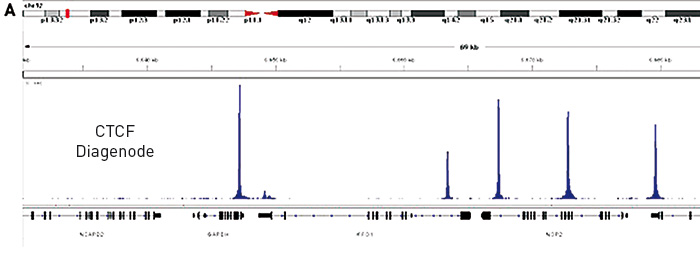
<p><strong>Figure 1.</strong> (A) Chromatin Immunoprecipitation has been performed using chromatin from HeLa cells, the iDeal ChIP-seq kit for Transcription Factors and the Diagenode ChIP-seq-grade CTCF antibody. The IP'd DNA was subsequently analysed on an Illumina<sup>®</sup> HiSeq. Library preparation, cluster generation and sequencing were performed according to the manufacturer's instructions. This figure shows the peak distribution in a region surrounding the GAPDH positive control gene.</p>
<p><img src="https://www.diagenode.com/img/product/kits/ideal-figure-b-total-diagendoe-peaks.png" alt="CTCF Diagenode" style="display: block; margin-left: auto; margin-right: auto;" /></p>
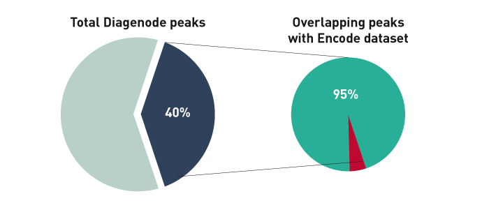
<p><strong>Figure 1B.</strong> The ChIP-seq dataset from this experiment has been compared with a reference dataset from the Broad Institute. We observed a perfect match between the top 40% of Diagenode peaks and the reference dataset. Based on the NIH Encode project criterion, ChIP-seq results are considered reproducible between an original and reproduced dataset if the top 40% of peaks have at least an 80% overlap ratio with the compared dataset.</p>
<p> </p>
<p><img src="https://www.diagenode.com/img/product/kits/ideal-TF-chip-seq-A.png" alt="ChIP-seq figure A" style="display: block; margin-left: auto; margin-right: auto;" /></p>
<p><img src="https://www.diagenode.com/img/product/kits/ideal-TF-chip-seq-B.png" alt="ChIP-seq figure B" style="display: block; margin-left: auto; margin-right: auto;" /></p>
<p><img src="https://www.diagenode.com/img/product/kits/ideal-TF-chip-seq-C.png" alt="ChIP-seq figure C" style="display: block; margin-left: auto; margin-right: auto;" /></p>
<p><strong>Figure 2.</strong> Chromatin Immunoprecipitation has been performed using chromatin from HeLa cells, the iDeal ChIP-seq kit for Transcription Factors and the Diagenode ChIP-seq-grade HDAC1 (A), LSD1 (B) and p53 antibody (C). The IP'd DNA was subsequently analysed on an Illumina<sup>®</sup> Genome Analyzer. Library preparation, cluster generation and sequencing were performed according to the manufacturer's instructions. This figure shows the peak distribution in regions of chromosome 3 (A), chromosome 12 (B) and chromosome 6 (C) respectively.</p>
<p> </p>
<h3>ChIP-seq on tissue</h3>
<p><img src="https://www.diagenode.com/img/product/kits/ideal-figure-3a.jpg" alt="ChIP-seq figure A" style="display: block; margin-left: auto; margin-right: auto;" /></p>
<p><strong>Figure 3A.</strong> Chromatin Immunoprecipitation has been performed using chromatin from mouse liver tissue, the iDeal ChIP-seq kit for Transcription Factors and the Diagenode ChIP-seq-grade CTCF antibody. The IP'd DNA was subsequently analysed on an Illumina® HiSeq. Library preparation, cluster generation and sequencing were performed according to the manufacturer's instructions. This figure shows the peak distribution in a region surrounding the Vwf positive control gene.</p>
<p><img src="https://www.diagenode.com/img/product/kits/match-of-the-top40-peaks.png" alt="Match of the Top40 peaks" style="display: block; margin-left: auto; margin-right: auto;" /></p>
<p><strong>Figure 3B.</strong> The ChIP-seq dataset from this experiment has been compared with a reference dataset from the Broad Institute. We observed a perfect match between the top 40% of Diagenode peaks and the reference dataset. Based on the NIH Encode project criterion, ChIP-seq results are considered reproducible between an original and reproduced dataset if the top 40% of peaks have at least an 80% overlap ratio with the compared dataset.</p>',
'label2' => 'Species, cell lines, tissues tested ',
'info2' => '<p>The iDeal ChIP-seq Kit for Transcription Factors is compatible with a broad variety of cell lines, tissues and species, as shown below. Other species / cell lines / tissues can be used with this kit.</p>
<p><span style="text-decoration: underline;">Cell lines:</span></p>
<p>Human: A549, A673, BT-549, CD4 T, HCC1806, HeLa, HepG2, HFF, HK-GFP-MR, ILC, K562, KYSE-180, LapC4, M14, MCF7, MDA-MB-231, MDA-MB-436, RDES, SKNO1, VCaP, U2-OS, ZR-75-1 </p>
<p>Mouse: ESC, NPCs, BZ, GT1-7, acinar cells, HSPCs, Th2 cells, keratinocytes</p>
<p>Cattle: pbMEC, <span>MAC-T</span></p>
<p><span style="text-decoration: underline;">Tissues:</span></p>
<p>Mouse: kidney, heart, brain, iris, liver, limbs from E10.5 embryos</p>
<p><span>Horse: l</span>iver, brain, heart, lung, skeletal muscle, lamina, ovary</p>
<p><span style="text-decoration: underline;">ChIP on yeast</span></p>
<p>The iDeal ChIP-seq kit for TF is compatible with yeast samples. Check out our <strong><a href="https://www.diagenode.com/files/products/kits/Application_Note-ChIP_on_Yeast.pdf">Application Note</a></strong> presenting an optimized detailed protocol for ChIP on yeast.</p>
<p></p>
<p>Did you use the iDeal ChIP-seq for Transcription Factors Kit on other cell line / tissue / species? <a href="mailto:agnieszka.zelisko@diagenode.com?subject=Species, cell lines, tissues tested with the iDeal ChIP-seq Kit for TF&body=Dear Customer,%0D%0A%0D%0APlease, leave below your feedback about the iDeal ChIP-seq for Transcription Factors (cell / tissue type, species, other information...).%0D%0A%0D%0AThank you for sharing with us your experience !%0D%0A%0D%0ABest regards,%0D%0A%0D%0AAgnieszka Zelisko-Schmidt, PhD">Let us know!</a></p>',
'label3' => 'Additional solutions compatible with iDeal ChIP-seq kit for Transcription Factors',
'info3' => '<p><span style="font-weight: 400;">The</span> <a href="https://www.diagenode.com/en/p/chromatin-shearing-optimization-kit-low-sds-for-tfs-25-rxns"><span style="font-weight: 400;">Chromatin shearing optimization kit – Low SDS (iDeal Kit for TFs)</span></a><span style="font-weight: 400;"> is the kit compatible with the iDeal ChIP-seq kit for TF, recommended for the optimization of chromatin shearing, a critical step for ChIP.</span></p>
<p><a href="https://www.diagenode.com/en/p/chip-cross-link-gold-600-ul"><span style="font-weight: 400;">ChIP Cross-link Gold</span></a> <span style="font-weight: 400;">should be used in combination with formaldehyde when working with higher order and/or dynamic interactions, for efficient protein-protein fixation.</span></p>
<p><span style="font-weight: 400;">For library preparation of immunoprecipitated samples we recommend to use the </span><b> </b><a href="https://www.diagenode.com/en/categories/library-preparation-for-ChIP-seq"><span style="font-weight: 400;">MicroPlex Library Preparation Kit</span></a><span style="font-weight: 400;"> - validated for library preparation from picogram inputs.</span></p>
<p><a href="https://www.diagenode.com/en/categories/chip-seq-grade-antibodies"><span style="font-weight: 400;">ChIP-seq grade antibodies</span></a><span style="font-weight: 400;"> provide high yields with excellent specificity and sensitivity.</span></p>
<p><span style="font-weight: 400;">Check the list of available </span><a href="https://www.diagenode.com/en/categories/primer-pairs"><span style="font-weight: 400;">Primer pairs</span></a><span style="font-weight: 400;"> designed for high specificity to specific genomic regions.</span></p>
<p><span style="font-weight: 400;">Plus, for our <a href="https://www.diagenode.com/en/categories/ip-star">IP-Star Automation</a> users for automated ChIP, check out our <a href="https://www.diagenode.com/en/p/auto-ideal-chip-seq-kit-for-transcription-factors-x24-24-rxns">automated version</a> of this kit.</span></p>',
'format' => '17 chrom. prep./100 IPs',
'catalog_number' => 'C01010170',
'old_catalog_number' => '',
'sf_code' => 'C01010170-',
'type' => 'RFR',
'search_order' => '',
'price_EUR' => '2230',
'price_USD' => '2340',
'price_GBP' => '2040',
'price_JPY' => '349330',
'price_CNY' => '',
'price_AUD' => '5850',
'country' => 'ALL',
'except_countries' => 'None',
'quote' => false,
'in_stock' => false,
'featured' => true,
'no_promo' => false,
'online' => true,
'master' => false,
'last_datasheet_update' => '0000-00-00',
'slug' => 'ideal-chip-seq-kit-for-transcription-factors-x100-100-rxns',
'meta_title' => 'iDeal ChIP-seq Kit for Transcription Factors x100 | Diagenode',
'meta_keywords' => 'iDeal ChIP-seq Kit for Transcription Factors,CHIP-seq ,Chromatin Immunoprecipitation',
'meta_description' => 'Diagenode’s iDeal ChIP-seq Kit for Transcription Factors is a highly specialized solution for robust Transcription Factor ChIP-seq results which utilizes a highly optimized protocol and is backed by validation with a broad number and range of transcriptio',
'modified' => '2023-04-20 16:01:37',
'created' => '2015-09-09 10:23:10',
'locale' => 'eng'
),
'Antibody' => array(
'host' => '*****',
'id' => null,
'name' => null,
'description' => null,
'clonality' => null,
'isotype' => null,
'lot' => null,
'concentration' => null,
'reactivity' => null,
'type' => null,
'purity' => null,
'classification' => null,
'application_table' => null,
'storage_conditions' => null,
'storage_buffer' => null,
'precautions' => null,
'uniprot_acc' => null,
'slug' => null,
'meta_keywords' => null,
'meta_description' => null,
'modified' => null,
'created' => null,
'select_label' => null
),
'Slave' => array(),
'Group' => array(
'Group' => array(
'id' => '10',
'name' => 'C01010055',
'product_id' => '1839',
'modified' => '2016-02-18 17:17:41',
'created' => '2016-02-18 17:17:41'
),
'Master' => array(
'id' => '1839',
'antibody_id' => null,
'name' => 'iDeal ChIP-seq kit for Transcription Factors',
'description' => '<p><a href="https://www.diagenode.com/files/products/kits/ideal-chipseq-transcription-factors-x10-manual.pdf"><img src="https://www.diagenode.com/img/buttons/bt-manual.png" /></a></p>
<div class="row">
<div class="small-12 medium-8 large-8 columns"><br />
<p><span style="font-weight: 400;">Diagenode’s <strong>iDeal ChIP-seq Kit for Transcription Factors</strong> is a highly validated solution for robust transcription factor and other non-histone proteins ChIP-seq results and contains everything you need for start-to-finish </span><b>ChIP </b><span style="font-weight: 400;">prior to </span><b>Next-Generation Sequencing</b><span style="font-weight: 400;">. This complete solution contains all buffers and reagents for cell lysis, chromatin shearing, immunoprecipitation, and DNA purification. In addition, unlike competing solutions, the kit contains positive and negative control antibodies (CTCF and IgG, respectively) as well as positive and negative control PCR primers pairs (H19 and Myoglobin exon 2, respectively) for your convenience and a guarantee of optimal results. <br /></span></p>
</div>
<div class="small-12 medium-4 large-4 columns"><center><br /><br />
<script>// <![CDATA[
var date = new Date(); var heure = date.getHours(); var jour = date.getDay(); var semaine = Math.floor(date.getDate() / 7) + 1; if (jour === 2 && ( (heure >= 9 && heure < 9.5) || (heure >= 18 && heure < 18.5) )) { document.write('<a href="https://us02web.zoom.us/j/85467619762"><img src="https://www.diagenode.com/img/epicafe-ON.gif"></a>'); } else { document.write('<a href="https://go.diagenode.com/l/928883/2023-04-26/3kq1v"><img src="https://www.diagenode.com/img/epicafe-OFF.png"></a>'); }
// ]]></script>
</center></div>
</div>
<p><span style="font-weight: 400;">The </span><b> iDeal ChIP-seq kit for Transcription Factors </b><span style="font-weight: 400;">is compatible for cells or tissues:</span></p>
<table style="width: 419px; margin-left: auto; margin-right: auto;">
<tbody>
<tr>
<td style="width: 144px;"></td>
<td style="width: 267px; text-align: center;"><span style="font-weight: 400;">Amount per IP</span></td>
</tr>
<tr>
<td style="width: 144px;">Cells</td>
<td style="width: 267px; text-align: center;"><strong>4,000,000</strong></td>
</tr>
<tr>
<td style="width: 144px;">Tissues</td>
<td style="width: 267px; text-align: center;"><strong>30 mg</strong></td>
</tr>
</tbody>
</table>
<p><span style="font-weight: 400;">The iDeal ChIP-seq kit is the only kit on the market validated for major sequencing systems. Our expertise in ChIP-seq tools allows reproducible and efficient results every time. </span></p>
<p></p>
<p></p>',
'label1' => 'Characteristics',
'info1' => '<ul>
<li><span style="font-weight: 400;"><strong>Highly optimized protocol</strong> for ChIP-seq from cells and tissues</span></li>
<li><span style="font-weight: 400;"><strong>Validated</strong> for <strong>ChIP-seq</strong> with multiple transcription factors and non-histone targets<br /></span></li>
<li><span style="font-weight: 400;"><strong>Most complete kit</strong> available (covers all steps, including the control antibodies and primers)<br /></span></li>
<li><span style="font-weight: 400;"><strong>Magnetic beads</strong> make ChIP <strong>easy</strong>, <strong>fast</strong> and more <strong>reproducible</strong></span></li>
<li><span style="font-weight: 400;">Combination with Diagenode ChIP-seq antibodies provides <strong>high yields</strong> with excellent <strong>specificity</strong> and <strong>sensitivity</strong><br /></span></li>
<li><span style="font-weight: 400;">Purified DNA suitable for any downstream application</span></li>
<li><span style="font-weight: 400;">Easy-to-follow protocol</span></li>
</ul>
<p><span style="font-weight: 400;"></span></p>
<p> </p>
<h3>ChIP-seq on cells</h3>
<p><img src="https://www.diagenode.com/img/product/kits/ideal-ctcf-diagenode.jpg" alt="CTCF Diagenode" style="display: block; margin-left: auto; margin-right: auto;" /></p>
<p><strong>Figure 1.</strong> (A) Chromatin Immunoprecipitation has been performed using chromatin from HeLa cells, the iDeal ChIP-seq kit for Transcription Factors and the Diagenode ChIP-seq-grade CTCF antibody. The IP'd DNA was subsequently analysed on an Illumina<sup>®</sup> HiSeq. Library preparation, cluster generation and sequencing were performed according to the manufacturer's instructions. This figure shows the peak distribution in a region surrounding the GAPDH positive control gene.</p>
<p><img src="https://www.diagenode.com/img/product/kits/ideal-figure-b-total-diagendoe-peaks.png" alt="CTCF Diagenode" style="display: block; margin-left: auto; margin-right: auto;" /></p>
<p><strong>Figure 1B.</strong> The ChIP-seq dataset from this experiment has been compared with a reference dataset from the Broad Institute. We observed a perfect match between the top 40% of Diagenode peaks and the reference dataset. Based on the NIH Encode project criterion, ChIP-seq results are considered reproducible between an original and reproduced dataset if the top 40% of peaks have at least an 80% overlap ratio with the compared dataset.</p>
<p> </p>
<p><img src="https://www.diagenode.com/img/product/kits/ideal-TF-chip-seq-A.png" alt="ChIP-seq figure A" style="display: block; margin-left: auto; margin-right: auto;" /></p>
<p><img src="https://www.diagenode.com/img/product/kits/ideal-TF-chip-seq-B.png" alt="ChIP-seq figure B" style="display: block; margin-left: auto; margin-right: auto;" /></p>
<p><img src="https://www.diagenode.com/img/product/kits/ideal-TF-chip-seq-C.png" alt="ChIP-seq figure C" style="display: block; margin-left: auto; margin-right: auto;" /></p>
<p><strong>Figure 2.</strong> Chromatin Immunoprecipitation has been performed using chromatin from HeLa cells, the iDeal ChIP-seq kit for Transcription Factors and the Diagenode ChIP-seq-grade HDAC1 (A), LSD1 (B) and p53 antibody (C). The IP'd DNA was subsequently analysed on an Illumina<sup>®</sup> Genome Analyzer. Library preparation, cluster generation and sequencing were performed according to the manufacturer's instructions. This figure shows the peak distribution in regions of chromosome 3 (A), chromosome 12 (B) and chromosome 6 (C) respectively.</p>
<p> </p>
<h3>ChIP-seq on tissue</h3>
<p><img src="https://www.diagenode.com/img/product/kits/ideal-figure-3a.jpg" alt="ChIP-seq figure A" style="display: block; margin-left: auto; margin-right: auto;" /></p>
<p><strong>Figure 3A.</strong> Chromatin Immunoprecipitation has been performed using chromatin from mouse liver tissue, the iDeal ChIP-seq kit for Transcription Factors and the Diagenode ChIP-seq-grade CTCF antibody. The IP'd DNA was subsequently analysed on an Illumina® HiSeq. Library preparation, cluster generation and sequencing were performed according to the manufacturer's instructions. This figure shows the peak distribution in a region surrounding the Vwf positive control gene.</p>
<p><img src="https://www.diagenode.com/img/product/kits/match-of-the-top40-peaks.png" alt="Match of the Top40 peaks" style="display: block; margin-left: auto; margin-right: auto;" /></p>
<p><strong>Figure 3B.</strong> The ChIP-seq dataset from this experiment has been compared with a reference dataset from the Broad Institute. We observed a perfect match between the top 40% of Diagenode peaks and the reference dataset. Based on the NIH Encode project criterion, ChIP-seq results are considered reproducible between an original and reproduced dataset if the top 40% of peaks have at least an 80% overlap ratio with the compared dataset.</p>',
'label2' => 'Species, cell lines, tissues tested',
'info2' => '<p>The iDeal ChIP-seq Kit for Transcription Factors is compatible with a broad variety of cell lines, tissues and species - some examples are shown below. Other species / cell lines / tissues can be used with this kit.</p>
<p><span style="text-decoration: underline;">Cell lines:</span></p>
<p>Human: A549, A673, BT-549, CD4 T, HCC1806, HeLa, HepG2, HFF, HK-GFP-MR, ILC, K562, KYSE-180, LapC4, M14, MCF7, MDA-MB-231, MDA-MB-436, RDES, SKNO1, VCaP, U2-OS, ZR-75-1 </p>
<p>Mouse: ESC, NPCs, BZ, GT1-7, acinar cells, HSPCs, Th2 cells, keratinocytes</p>
<p>Cattle: pbMEC, <span>MAC-T</span></p>
<p><span>Other cell lines / species: compatible, not tested</span></p>
<p><span style="text-decoration: underline;">Tissues:</span></p>
<p>Mouse: kidney, heart, brain, iris, liver, limbs from E10.5 embryos</p>
<p><span>Horse: l</span>iver, brain, heart, lung, skeletal muscle, lamina, ovary</p>
<p>Other tissues: compatible, not tested</p>
<p><span style="text-decoration: underline;">ChIP on yeast</span></p>
<p>The iDeal ChIP-seq kit for TF is compatible with yeast samples. Check out our <strong><a href="https://www.diagenode.com/files/products/kits/Application_Note-ChIP_on_Yeast.pdf">Application Note</a></strong> presenting an optimized detailed protocol for ChIP on yeast.</p>
<p></p>
<p>Did you use the iDeal ChIP-seq for Transcription Factors Kit on other cell line / tissue / species? <a href="mailto:agnieszka.zelisko@diagenode.com?subject=Species, cell lines, tissues tested with the iDeal ChIP-seq Kit for TF&body=Dear Customer,%0D%0A%0D%0APlease, leave below your feedback about the iDeal ChIP-seq for Transcription Factors (cell / tissue type, species, other information...).%0D%0A%0D%0AThank you for sharing with us your experience !%0D%0A%0D%0ABest regards,%0D%0A%0D%0AAgnieszka Zelisko-Schmidt, PhD">Let us know!</a></p>',
'label3' => 'Additional solutions compatible with iDeal ChIP-seq kit for Transcription Factors',
'info3' => '<p><span style="font-weight: 400;">The</span> <a href="https://www.diagenode.com/en/p/chromatin-shearing-optimization-kit-low-sds-for-tfs-25-rxns"><span style="font-weight: 400;">Chromatin EasyShear Kit – Low SDS </span></a><span style="font-weight: 400;">is the kit compatible with the iDeal ChIP-seq kit for TF, recommended for the optimization of chromatin shearing, a critical step for ChIP.</span></p>
<p><a href="https://www.diagenode.com/en/p/chip-cross-link-gold-600-ul"><span style="font-weight: 400;">ChIP Cross-link Gold</span></a> <span style="font-weight: 400;">should be used in combination with formaldehyde when working with higher order and/or dynamic interactions, for efficient protein-protein fixation.</span></p>
<p><span style="font-weight: 400;">For library preparation of immunoprecipitated samples we recommend to use the </span><b> </b><a href="https://www.diagenode.com/en/categories/library-preparation-for-ChIP-seq"><span style="font-weight: 400;">MicroPlex Library Preparation Kit</span></a><span style="font-weight: 400;"> - validated for library preparation from picogram inputs.</span></p>
<p><a href="https://www.diagenode.com/en/categories/chip-seq-grade-antibodies"><span style="font-weight: 400;">ChIP-seq grade antibodies</span></a><span style="font-weight: 400;"> provide high yields with excellent specificity and sensitivity.</span></p>
<p><span style="font-weight: 400;">Check the list of available </span><a href="https://www.diagenode.com/en/categories/primer-pairs"><span style="font-weight: 400;">Primer pairs</span></a><span style="font-weight: 400;"> designed for high specificity to specific genomic regions.</span></p>
<p><span style="font-weight: 400;">Plus, for our <a href="https://www.diagenode.com/en/categories/ip-star">IP-Star Automation</a> users for automated ChIP, check out our <a href="https://www.diagenode.com/en/p/auto-ideal-chip-seq-kit-for-transcription-factors-x24-24-rxns">automated version</a> of this kit.</span></p>',
'format' => '4 chrom. prep./24 IPs',
'catalog_number' => 'C01010055',
'old_catalog_number' => '',
'sf_code' => 'C01010055-',
'type' => 'RFR',
'search_order' => '04-undefined',
'price_EUR' => '915',
'price_USD' => '1130',
'price_GBP' => '840',
'price_JPY' => '143335',
'price_CNY' => '',
'price_AUD' => '2825',
'country' => 'ALL',
'except_countries' => 'None',
'quote' => false,
'in_stock' => false,
'featured' => true,
'no_promo' => false,
'online' => true,
'master' => true,
'last_datasheet_update' => '0000-00-00',
'slug' => 'ideal-chip-seq-kit-for-transcription-factors-x24-24-rxns',
'meta_title' => 'iDeal ChIP-seq kit for Transcription Factors x24',
'meta_keywords' => '',
'meta_description' => 'iDeal ChIP-seq kit for Transcription Factors x24',
'modified' => '2023-06-20 18:27:37',
'created' => '2015-06-29 14:08:20'
),
'Product' => array(
(int) 0 => array(
[maximum depth reached]
),
(int) 1 => array(
[maximum depth reached]
)
)
),
'Related' => array(
(int) 0 => array(
'id' => '1836',
'antibody_id' => null,
'name' => 'iDeal ChIP-seq kit for Histones',
'description' => '<p><a href="https://www.diagenode.com/files/products/kits/ideal-chipseq-for-histones-complete-manual.pdf"><img src="https://www.diagenode.com/img/buttons/bt-manual.png" /></a></p>
<p>Don’t risk wasting your precious sequencing samples. Diagenode’s validated <strong>iDeal ChIP-seq kit for Histones</strong> has everything you need for a successful start-to-finish <strong>ChIP of histones prior to Next-Generation Sequencing</strong>. The complete kit contains all buffers and reagents for cell lysis, chromatin shearing, immunoprecipitation and DNA purification. In addition, unlike competing solutions, the kit contains positive and negative control antibodies (H3K4me3 and IgG, respectively) as well as positive and negative control PCR primers pairs (GAPDH TSS and Myoglobin exon 2, respectively) for your convenience and a guarantee of optimal results. The kit has been validated on multiple histone marks.</p>
<p> The iDeal ChIP-seq kit for Histones<strong> </strong>is perfect for <strong>cells</strong> (<strong>100,000 cells</strong> to <strong>1,000,000 cells</strong> per IP) and has been validated for <strong>tissues</strong> (<strong>1.5 mg</strong> to <strong>5 mg</strong> of tissue per IP).</p>
<p> The iDeal ChIP-seq kit is the only kit on the market validated for the major sequencing systems. Our expertise in ChIP-seq tools allows reproducible and efficient results every time.</p>
<p></p>
<p> <strong></strong></p>
<p></p>',
'label1' => 'Characteristics',
'info1' => '<ul style="list-style-type: disc;">
<li>Highly <strong>optimized</strong> protocol for ChIP-seq from cells and tissues</li>
<li><strong>Validated</strong> for ChIP-seq with multiple histones marks</li>
<li>Most <strong>complete</strong> kit available (covers all steps, including the control antibodies and primers)</li>
<li>Optimized chromatin preparation in combination with the Bioruptor ensuring the best <strong>epitope integrity</strong></li>
<li>Magnetic beads make ChIP easy, fast and more <strong>reproducible</strong></li>
<li>Combination with Diagenode ChIP-seq antibodies provides high yields with excellent <strong>specificity</strong> and <strong>sensitivity</strong></li>
<li>Purified DNA suitable for any downstream application</li>
<li>Easy-to-follow protocol</li>
</ul>
<p>Note: to obtain optimal results, this kit should be used in combination with the DiaMag1.5 - magnetic rack.</p>
<h3>ChIP-seq on cells</h3>
<p><img src="https://www.diagenode.com/img/product/kits/iDeal-kit-C01010053-figure-1.jpg" alt="Figure 1A" style="display: block; margin-left: auto; margin-right: auto;" /></p>
<p><strong>Figure 1A. The high consistency of the iDeal ChIP-seq kit on the Ion Torrent™ PGM™ (Life Technologies) and GAIIx (Illumina<sup>®</sup>)</strong><br /> ChIP was performed on sheared chromatin from 1 million HelaS3 cells using the iDeal ChIP-seq kit and 1 µg of H3K4me3 positive control antibody. Two different biological samples have been analyzed using two different sequencers - GAIIx (Illumina<sup>®</sup>) and PGM™ (Ion Torrent™). The expected ChIP-seq profile for H3K4me3 on the GAPDH promoter region has been obtained.<br /> Image A shows a several hundred bp along chr12 with high similarity of read distribution despite the radically different sequencers. Image B is a close capture focusing on the GAPDH that shows that even the peak structure is similar.</p>
<p class="text-center"><strong>Perfect match between ChIP-seq data obtained with the iDeal ChIP-seq workflow and reference dataset</strong></p>
<p><img src="https://www.diagenode.com/img/product/kits/perfect-match-between-chipseq-data.png" alt="Figure 1B" style="display: block; margin-left: auto; margin-right: auto;" /></p>
<p><strong>Figure 1B.</strong> The ChIP-seq dataset from this experiment has been compared with a reference dataset from the Broad Institute. We observed a perfect match between the top 40% of Diagenode peaks and the reference dataset. Based on the NIH Encode project criterion, ChIP-seq results are considered reproducible between an original and reproduced dataset if the top 40% of peaks have at least an 80% overlap ratio with the compared dataset.</p>
<p><img src="https://www.diagenode.com/img/product/kits/iDeal-kit-C01010053-figure-2.jpg" alt="Figure 2" style="display: block; margin-left: auto; margin-right: auto;" /></p>
<p><strong>Figure 2. Efficient and easy chromatin shearing using the Bioruptor<sup>®</sup> and Shearing buffer iS1 from the iDeal ChIP-seq kit</strong><br /> Chromatin from 1 million of Hela cells was sheared using the Bioruptor<sup>®</sup> combined with the Bioruptor<sup>®</sup> Water cooler (Cat No. BioAcc-cool) during 3 rounds of 10 cycles of 30 seconds “ON” / 30 seconds “OFF” at HIGH power setting (position H). Diagenode 1.5 ml TPX tubes (Cat No. M-50001) were used for chromatin shearing. Samples were gently vortexed before and after performing each sonication round (rounds of 10 cycles), followed by a short centrifugation at 4°C to recover the sample volume at the bottom of the tube. The sheared chromatin was then decross-linked as described in the kit manual and analyzed by agarose gel electrophoresis.</p>
<p><img src="https://www.diagenode.com/img/product/kits/iDeal-kit-C01010053-figure-3.jpg" alt="Figure 3" style="display: block; margin-left: auto; margin-right: auto;" width="264" height="320" /></p>
<p><strong>Figure 3. Validation of ChIP by qPCR: reliable results using Diagenode’s ChIP-seq grade H3K4me3 antibody, isotype control and sets of validated primers</strong><br /> Specific enrichment on positive loci (GAPDH, EIF4A2, c-fos promoter regions) comparing to no enrichment on negative loci (TSH2B promoter region and Myoglobin exon 2) was detected by qPCR. Samples were prepared using the Diagenode iDeal ChIP-seq kit. Diagenode ChIP-seq grade antibody against H3K4me3 and the corresponding isotype control IgG were used for immunoprecipitation. qPCR amplification was performed with sets of validated primers.</p>
<h3>ChIP-seq on tissue</h3>
<p><img src="https://www.diagenode.com/img/product/kits/ideal-figure-h3k4me3.jpg" alt="Figure 4A" style="display: block; margin-left: auto; margin-right: auto;" /></p>
<p><strong>Figure 4A.</strong> Chromatin Immunoprecipitation has been performed using chromatin from mouse liver tissue, the iDeal ChIP-seq kit for Histones and the Diagenode ChIP-seq-grade H3K4me3 (Cat. No. C15410003) antibody. The IP'd DNA was subsequently analysed on an Illumina® HiSeq. Library preparation, cluster generation and sequencing were performed according to the manufacturer's instructions. This figure shows the peak distribution in a region surrounding the GAPDH positive control gene.</p>
<p><img src="https://www.diagenode.com/img/product/kits/match-of-the-top40-peaks-2.png" alt="Figure 4B" caption="false" style="display: block; margin-left: auto; margin-right: auto;" width="700" height="280" /></p>
<p><strong>Figure 4B.</strong> The ChIP-seq dataset from this experiment has been compared with a reference dataset from the Broad Institute. We observed a perfect match between the top 40% of Diagenode peaks and the reference dataset. Based on the NIH Encode project criterion, ChIP-seq results are considered reproducible between an original and reproduced dataset if the top 40% of peaks have at least an 80% overlap ratio with the compared dataset.</p>',
'label2' => 'Species, cell lines, tissues tested',
'info2' => '<p>The iDeal ChIP-seq Kit for Histones is compatible with a broad variety of cell lines, tissues and species - some examples are shown below. Other species / cell lines / tissues can be used with this kit.</p>
<p><u>Cell lines:</u></p>
<p>Human: A549, A673, CD8+ T, Blood vascular endothelial cells, Lymphatic endothelial cells, fibroblasts, K562, MDA-MB231</p>
<p>Pig: Alveolar macrophages</p>
<p>Mouse: C2C12, primary HSPC, synovial fibroblasts, HeLa-S3, FACS sorted cells from embryonic kidneys, macrophages, mesodermal cells, myoblasts, NPC, salivary glands, spermatids, spermatocytes, skeletal muscle stem cells, stem cells, Th2</p>
<p>Hamster: CHO</p>
<p>Other cell lines / species: compatible, not tested</p>
<p><u>Tissues</u></p>
<p>Bee – brain</p>
<p>Daphnia – whole animal</p>
<p>Horse – brain, heart, lamina, liver, lung, skeletal muscles, ovary</p>
<p>Human – Erwing sarcoma tumor samples</p>
<p>Other tissues: compatible, not tested</p>
<p>Did you use the iDeal ChIP-seq for Histones Kit on other cell line / tissue / species? <a href="mailto:agnieszka.zelisko@diagenode.com?subject=Species, cell lines, tissues tested with the iDeal ChIP-seq Kit for TF&body=Dear Customer,%0D%0A%0D%0APlease, leave below your feedback about the iDeal ChIP-seq for Transcription Factors (cell / tissue type, species, other information...).%0D%0A%0D%0AThank you for sharing with us your experience !%0D%0A%0D%0ABest regards,%0D%0A%0D%0AAgnieszka Zelisko-Schmidt, PhD">Let us know!</a></p>',
'label3' => ' Additional solutions compatible with iDeal ChIP-seq Kit for Histones',
'info3' => '<p><a href="../p/chromatin-shearing-optimization-kit-low-sds-100-million-cells">Chromatin EasyShear Kit - Ultra Low SDS </a>optimizes chromatin shearing, a critical step for ChIP.</p>
<p> The <a href="https://www.diagenode.com/en/categories/library-preparation-for-ChIP-seq">MicroPlex Library Preparation Kit </a>provides easy and optimal library preparation of ChIPed samples.</p>
<p><a href="../categories/chip-seq-grade-antibodies">ChIP-seq grade anti-histone antibodies</a> provide high yields with excellent specificity and sensitivity.</p>
<p> Plus, for our IP-Star Automation users for automated ChIP, check out our <a href="../p/auto-ideal-chip-seq-kit-for-histones-x24-24-rxns">automated</a> version of this kit.</p>',
'format' => '4 chrom. prep./24 IPs',
'catalog_number' => 'C01010051',
'old_catalog_number' => 'AB-001-0024',
'sf_code' => 'C01010051-',
'type' => 'RFR',
'search_order' => '04-undefined',
'price_EUR' => '915',
'price_USD' => '1130',
'price_GBP' => '840',
'price_JPY' => '143335',
'price_CNY' => '',
'price_AUD' => '2825',
'country' => 'ALL',
'except_countries' => 'None',
'quote' => false,
'in_stock' => false,
'featured' => true,
'no_promo' => false,
'online' => true,
'master' => true,
'last_datasheet_update' => '0000-00-00',
'slug' => 'ideal-chip-seq-kit-x24-24-rxns',
'meta_title' => 'iDeal ChIP-seq kit x24',
'meta_keywords' => '',
'meta_description' => 'iDeal ChIP-seq kit x24',
'modified' => '2023-04-20 16:00:20',
'created' => '2015-06-29 14:08:20',
'ProductsRelated' => array(
[maximum depth reached]
),
'Image' => array(
[maximum depth reached]
)
),
(int) 1 => array(
'id' => '2288',
'antibody_id' => '250',
'name' => 'CTCF Antibody ',
'description' => '<p>Alternative name: <strong>MRD21</strong></p>
<p>Polyclonal antibody raised in rabbit against human <strong>CTCF</strong> (<strong>CCCTC-Binding Factor</strong>), using 4 KLH coupled peptides.</p>
<p></p>',
'label1' => 'Validation Data',
'info1' => '<div class="row">
<div class="small-6 columns">
<p><img src="https://www.diagenode.com/img/product/antibodies/C15410210-chip.png" alt="CTCF Antibody ChIP Grade" /></p>
</div>
<div class="small-6 columns">
<p><small><strong> Figure 1. ChIP results obtained with the Diagenode antibody directed against CTCF</strong><br />ChIP was performed with the Diagenode antibody against CTCF (cat. No. C15410210) on sheared chromatin from 4,000,000 HeLa cells. A titration consisting of 1, 2, 5 and 10 µg of antibody per ChIP experiment was analyzed. IgG (2 µg/IP) was used as a negative IP control. Quantitative PCR was performed with optimized primers for the H19 imprinting control region, and a specific region in the GAPDH gene, used as positive controls, and for the Sat2 satellite repeat region, used as a negative control. Figure 1 shows the recovery, expressed as a % of input (the relative amount of immunoprecipitated DNA compared to input DNA after qPCR analysis). </small></p>
</div>
</div>
<div class="row">
<div class="small-12 columns">
<p>A.<img src="https://www.diagenode.com/img/product/antibodies/c15410210-chipseq-a.jpg" alt="CTCF Antibody ChIP-seq Grade" /></p>
<p>B.<img src="https://www.diagenode.com/img/product/antibodies/c15410210-chipseq-b.jpg" alt="CTCF Antibody for ChIP-seq " /></p>
<p>C.<img src="https://www.diagenode.com/img/product/antibodies/c15410210-chipseq-c.jpg" alt="CTCF Antibody for ChIP-seq assay" /></p>
<p>D.<img src="https://www.diagenode.com/img/product/antibodies/c15410210-chipseq-d.jpg" alt="CTCF Antibody validated in ChIP-seq" /></p>
</div>
</div>
<div class="row">
<div class="small-12 columns">
<p><small><strong> Figure 2. ChIP-seq results obtained with the Diagenode antibody directed against CTCF</strong><br /> ChIP was performed on sheared chromatin from 4,000,000 HeLa cells using 1 µg of the Diagenode antibody against CTCF (cat. No. C15410210) as described above. The IP'd DNA was subsequently analysed on an Illumina NovaSeq. Library preparation, cluster generation and sequencing were performed according to the manufacturer's instructions. The 50 bp tags were aligned to the human genome using the BWA algorithm. Figure 2 shows the peak distribution along the complete sequence and a 60 kb region of the human X-chromosome (figure 2A and B) and in two regions surrounding the GAPDH and H19 positive control genes, respectively (figure 2C and D).</small></p>
</div>
</div>
<div class="row">
<div class="small-12 columns">
<p>A.<img src="https://www.diagenode.com/img/product/antibodies/C15410210-cuttag-a.png" alt="CTCF Antibody CUT&Tag" /></p>
<p>B.<img src="https://www.diagenode.com/img/product/antibodies/C15410210-cuttag-b.png" alt="CTCF Antibody CUT&Tag " /></p>
</div>
</div>
<div class="row">
<div class="small-12 columns">
<p><small><strong> Figure 3. Cut&Tag results obtained with the Diagenode antibody directed against CTCF</strong><br /> CUT&TAG (Kaya-Okur, H.S., Nat Commun 10, 1930, 2019) was performed on 50,000 K562 cells using 1 µg of the Diagenode antibody against CTCF (cat. No. C15410210) and the Diagenode pA-Tn5 transposase (C01070001). The libraries were subsequently analysed on an Illumina NextSeq 500 sequencer (2x75 paired-end reads) according to the manufacturer's instructions. The tags were aligned to the human genome (hg19) using the BWA algorithm. Figure 3 shows the peak distribution in 2 genomic regions surrounding the h19 imprinting control gene on chromosome 11 and the AMER3 gene on chromosome 2 (figure 3A and B, respectively).</small></p>
</div>
</div>
<div class="row">
<div class="small-6 columns">
<p><img src="https://www.diagenode.com/img/product/antibodies/C15410210-elisa.png" alt="CTCF Antibody ELISA validation" /></p>
</div>
<div class="small-6 columns">
<p><small><strong> Figure 4. Determination of the antibody titer</strong><br />To determine the titer of the antibody, an ELISA was performed using a serial dilution of the Diagenode antibody against CTCF (cat. No. C15410210). The plates were coated with the peptides used for immunization of the rabbit. By plotting the absorbance against the antibody dilution (Figure 4), the titer of the antibody was estimated to be 1:90,000.</small></p>
</div>
</div>
<div class="row">
<div class="small-3 columns">
<p><img src="https://www.diagenode.com/img/product/antibodies/C15410210-wb.png" alt="CTCF Antibody for Western Blot" /></p>
</div>
<div class="small-9 columns">
<p><small><strong>Figure 5. Western blot analysis using the Diagenode antibody directed against CTCF</strong><br /> Whole cell extracts (40 µg) from HeLa cells transfected with CTCF siRNA (lane 2) and from an untransfected control (lane 1) were analysed by Western blot using the Diagenode antibody against CTCF (cat. No. C15410210) diluted 1:1,000 in TBS-Tween containing 5% skimmed milk. The position of the protein of interest is indicated on the right; the marker (in kDa) is shown on the left.</small></p>
</div>
</div>',
'label2' => 'Target Description',
'info2' => '<p>CTCF (UniProt/Swiss-Prot entry P49711) is a transcriptional regulator protein with 11 highly conserved zinc finger domains. By using different combinations of the zinc finger domains, CTCF can bind to different DNA sequences and proteins. As such it can act as both a transcriptional repressor and a transcriptional activator. By binding to transcriptional insulator elements, CTCF can also block communication between enhancers and upstream promoters, thereby regulating imprinted gene expression. CTCF also binds to the H19 imprinting control region and mediates maternally inherited higher-order chromatin conformation to restrict enhancer access to IGF2. Mutations in the CTCF gene have been associated with invasive breast cancers, prostate cancers, and Wilms’ tumor.</p>',
'label3' => '',
'info3' => '',
'format' => '50 μg',
'catalog_number' => 'C15410210',
'old_catalog_number' => '',
'sf_code' => 'C15410210-D001-000581',
'type' => 'FRE',
'search_order' => '03-Antibody',
'price_EUR' => '380',
'price_USD' => '380',
'price_GBP' => '340',
'price_JPY' => '59525',
'price_CNY' => '',
'price_AUD' => '950',
'country' => 'ALL',
'except_countries' => 'None',
'quote' => false,
'in_stock' => false,
'featured' => false,
'no_promo' => false,
'online' => true,
'master' => true,
'last_datasheet_update' => 'August 21, 2020',
'slug' => 'ctcf-polyclonal-antibody-classic-50-mg',
'meta_title' => 'CTCF Antibody - ChIP-seq grade (C15410210) | Diagenode',
'meta_keywords' => '',
'meta_description' => 'CTCF (CCCTC-Binding Factor) Polyclonal Antibody validated in ChIP-seq, ChIP-qPCR, WB, IF and ELISA. Specificity confirmed by siRNA assay. Batch-specific data available on the website. Other names: MRD21. Sample size available.',
'modified' => '2024-11-19 16:36:54',
'created' => '2015-06-29 14:08:20',
'ProductsRelated' => array(
[maximum depth reached]
),
'Image' => array(
[maximum depth reached]
)
),
(int) 2 => array(
'id' => '1951',
'antibody_id' => '194',
'name' => 'Pol II Antibody - replaced by the antibody C15200253 ',
'description' => '<p><strong>The antibody C15100055, format 100 µl has been discontinued. We recommend using the antibody <a href="https://www.diagenode.com/en/p/pol-ii-monoclonal-antibody-50-ul">C15200253</a></strong><span><strong>. </strong> </span></p>
<p>Alternative names: <strong>POLR2A</strong>, <strong>RPB1</strong>, <strong>POLR2</strong>, <strong>RPOL2</strong></p>
<p>Monoclonal antibody raised in mouse against the <strong>B1 subunit of RNA polymerase II</strong> (polymerase (RNA) II (DNA directed) polypeptide A) of wheat germ. Interacts with the highly conserved C-terminal domain of the protein containing the YSPTSPS repeat.</p>',
'label1' => 'Validation Data',
'info1' => '<div class="row">
<div class="small-4 columns">
<p><img src="https://www.diagenode.com/img/product/antibodies/C15100055_008_ChIP.png" alt="Pol II Antibody ChIP Grade " style="display: block; margin-left: auto; margin-right: auto;" /></p>
</div>
<div class="small-8 columns">
<p><small><strong> Figure 1. ChIP results obtained with the Diagenode monoclonal antibody directed against Pol II </strong><br />ChIP assays were performed using human HeLa cells, the Diagenode monoclonal antibody against Pol II (cat. No. C15100055) and optimized PCR primer pairs for qPCR. ChIP was performed with the “iDeal ChIP-seq” kit (cat. No. C01010051), using sheared chromatin from 1 million cells. A titration consisting of 1, 2, 5 and 10 μl of antibody per ChIP experiment was analyzed. IgG (2 μg/ IP) was used as a negative IP control. Quantitative PCR was performed with primers specific for the promoter of the GAPDH and EIF4A2 genes, used as positive controls, and for the MYOD1 gene and the Sat2 satellite repeat, used as negative controls. Figure 1 shows the recovery, expressed as a % of input (the relative amount of immunoprecipitated DNA compared to input DNA after qPCR analysis). </small></p>
</div>
</div>
<div class="row">
<div class="small-5 columns">
<p><img src="https://www.diagenode.com/img/product/antibodies/C15100055_ChIPSeq-A.png" alt="Pol II Antibody ChIP-seq Grade " style="display: block; margin-left: auto; margin-right: auto;" /></p>
<p><img src="https://www.diagenode.com/img/product/antibodies/C15100055_ChIPSeq-B.png" alt="Pol II Antibody for ChIP-seq" style="display: block; margin-left: auto; margin-right: auto;" /></p>
<p><img src="https://www.diagenode.com/img/product/antibodies/C15100055_ChIPSeq-C.png" alt="Pol II Antibody for ChIP-seq assay" style="display: block; margin-left: auto; margin-right: auto;" /></p>
<p><img src="https://www.diagenode.com/img/product/antibodies/C15100055_ChIPSeq-D.png" alt="Pol II Antibody validated in ChIP-seq" style="display: block; margin-left: auto; margin-right: auto;" /></p>
</div>
<div class="small-7 columns">
<p><small><strong> Figure 2. ChIP-seq results obtained with the Diagenode monoclonal antibody directed against Pol II</strong> <br />ChIP was performed on sheared chromatin from 1 million HeLaS3 cells using 2 μl of the Diagenode antibody against Pol II (cat. No. C15100055) as described above. The IP’d DNA was subsequently analysed on an Illumina HiSeq. Library preparation, cluster generation and sequencing were performed according to the manufacturer’s instructions. The 51 bp tags were aligned to the human genome using the BWA algorithm. Figure 2 shows the enrichment along the complete sequence and a 1 Mb region of the X-chromosome (fig 2A and B) and in genomic regions of chromosome 12 and 3, surrounding the GAPDH and EIF4A2 positive control genes (fig 2C and D). </small></p>
</div>
</div>
<div class="row">
<div class="small-4 columns">
<p><img src="https://www.diagenode.com/img/product/antibodies/C15100055_wb.png" alt="Pol II Antibody for Western Blot" style="display: block; margin-left: auto; margin-right: auto;" /></p>
</div>
<div class="small-8 columns">
<p><small><strong> Figure 3. Western blot analysis using the Diagenode monoclonal antibody directed against Pol II </strong><br />Whole cell extracts (40 μg) from HeLa cells transfected with Pol II siRNA (lane 2) and from an untransfected control (lane 1) were analysed by Western blot using the Diagenode antibody against Pol II (Cat. No. C15100055) diluted 1:500 in TBS-Tween containing 5% skimmed milk. The position of the protein of interest is indicated on the right; the marker (in kDa) is shown on the left.</small></p>
</div>
</div>',
'label2' => 'Target Description',
'info2' => '<p>RNA polymerase II (pol II) is a key enzyme in the regulation and control of gene transcription. It is able to unwind the DNA double helix, synthesize RNA, and proofread the result. Pol II is a complex enzyme, consisting of 12 subunits, of which the B1 subunit (UniProt/Swiss-Prot entry P24928) is the largest. Together with the second largest subunit, B1 forms the catalytic core of the RNA polymerase II transcription machinery</p>',
'label3' => '',
'info3' => '',
'format' => '100 µl',
'catalog_number' => 'C15100055-100',
'old_catalog_number' => 'AC-055-100',
'sf_code' => 'C15100055-D001-001161',
'type' => 'FRE',
'search_order' => '03-Antibody',
'price_EUR' => '380',
'price_USD' => '380',
'price_GBP' => '340',
'price_JPY' => '59525',
'price_CNY' => '',
'price_AUD' => '950',
'country' => 'ALL',
'except_countries' => 'None',
'quote' => false,
'in_stock' => true,
'featured' => false,
'no_promo' => false,
'online' => true,
'master' => true,
'last_datasheet_update' => '0000-00-00',
'slug' => 'pol-ii-monoclonal-antibody-classic-100-ul',
'meta_title' => 'Pol II Antibody - ChIP-seq Grade (C15100055) | Diagenode',
'meta_keywords' => '',
'meta_description' => 'Pol II (B1 subunit of RNA polymerase II) Monoclonal Antibody validated in ChIP-seq, ChIP-qPCR and WB. Specificity confirmed by siRNA assay. Batch-specific data available on the website. Alternative names: POLR2A, RPB1, POLR2, RPOL2. Sample size available.',
'modified' => '2024-12-03 15:02:42',
'created' => '2015-06-29 14:08:20',
'ProductsRelated' => array(
[maximum depth reached]
),
'Image' => array(
[maximum depth reached]
)
),
(int) 3 => array(
'id' => '2240',
'antibody_id' => '312',
'name' => 'p53 Antibody',
'description' => '<p><span>Alternative names: <strong>TP53</strong>, <strong>P53</strong>, <strong>TRP53</strong>, <strong>LSF1</strong></span></p>
<p><span>Polyclonal antibody raised in rabbit against human <strong>p53 (tumor protein p53)</strong>, using a KLH-conjugated synthetic peptide containing a sequence from the C-terminal part of the protein.</span></p>',
'label1' => 'Validation data',
'info1' => '<div class="row">
<div class="small-6 columns">
<p><img src="https://www.diagenode.com/img/product/antibodies/C15410083-chip.jpg" alt="p53 Antibody ChIP Grade" caption="false" width="400" height="304" /></p>
</div>
<div class="small-6 columns">
<p><small><strong>Figure 1. ChIP results obtained with the Diagenode antibody directed against p53</strong><br /> ChIP assays were performed using human U2OS cells, treated with camptothecin, the Diagenode antibody against p53 (Cat. No. C15410083) and optimized PCR primer sets for qPCR. ChIP was performed on sheared chromatin from 4 million cells. A titration of the antibody consisting of 1, 2, 5, and 10 µg per ChIP experiment was analysed. IgG (2 µg/IP) was used as negative IP control. qPCR was performed with primers for the p21 and GAS6 genes used as positive controls, and for GAPDH promoter and the Sat2 satellite repeat, used as negative controls. Figure 1 shows the recovery, expressed as a % of input (the relative amount of immunoprecipitated DNA compared to input DNA after qPCR analysis). </small></p>
</div>
</div>
<div class="row">
<div class="small-12 columns">
<p>A. <img src="https://www.diagenode.com/img/product/antibodies/C15410083_ChIPSeq-A.jpg" alt="p53 Antibody ChIP-seq Grade" style="display: block; margin-left: auto; margin-right: auto;" /></p>
<p>B. <img src="https://www.diagenode.com/img/product/antibodies/C15410083_ChIPSeq-B.jpg" alt="p53 Antibody for ChIP-seq" style="display: block; margin-left: auto; margin-right: auto;" /></p>
<p>C. <img src="https://www.diagenode.com/img/product/antibodies/C15410083_ChIPSeq-C.jpg" alt="p53 Antibody for ChIP-seq assay " style="display: block; margin-left: auto; margin-right: auto;" /></p>
<p>D. <img src="https://www.diagenode.com/img/product/antibodies/C15410083_ChIPSeq-D.jpg" alt="p53 Antibody validated in ChIP-seq" style="display: block; margin-left: auto; margin-right: auto;" /></p>
</div>
</div>
<div class="extra-spaced"></div>
<div class="row">
<div class="small-12 columns">
<p><small><strong>Figure 2. ChIP-seq results obtained with the Diagenode antibody directed against p53</strong><br /> ChIP was performed on sheared chromatin from 4 million U2OS cells using 1 µg of the Diagenode antibody against p53 (Cat. No. C15410083) as described above. The IP’d DNA was subsequently analysed on an Illumina HiSeq. Library preparation, cluster generation and sequencing were performed according to the manufacturer’s instructions. The 51 bp tags were aligned to the human genome using the BWA algorithm. Figure 2 shows the peak distribution along the X-chromosome (fig 2A) and in 3 genomic regions of chromosome 6, 13 and 12, surrounding p21 (CDKN1A), GAS6 and MDM2, 3 known targets genes of p53 (fig 2B, C and D, respectively). </small></p>
</div>
</div>
<div class="row">
<div class="small-6 columns">
<p><img src="https://www.diagenode.com/img/product/antibodies/C15410083_ELISA.jpg" alt="p53 Antibody ELISA validation " style="display: block; margin-left: auto; margin-right: auto;" /></p>
</div>
<div class="small-6 columns">
<p><small><strong> Figure 3. Determination of the antibody titer</strong><br /> To determine the titer of the antibody, an ELISA was performed using a serial dilution of Diagenode antibody directed against human p53 (Cat. No. C15410083), in antigen coated wells. By plotting the absorbance against the antibody dilution (Figure 3), the titer of the antibody was estimated to be 1:308,000. </small></p>
</div>
</div>
<div class="row">
<div class="small-3 columns">
<p><img src="https://www.diagenode.com/img/product/antibodies/C15410083_WB.jpg" alt="p53 Antibody validated in Western blot" style="display: block; margin-left: auto; margin-right: auto;" /></p>
</div>
<div class="small-9 columns">
<p><small><strong> Figure 4. Western blot analysis using the Diagenode antibody directed against p53</strong><br /> Nuclear extracts of HeLa cells (40 µg) were analysed by Western blot using the Diagenode antibody against p53 (Cat. No. C15410083) diluted 1:2,000 in TBS-Tween containing 5% skimmed milk. The position of the protein of interest is indicated on the right; the marker (in kDa) is shown on the left. </small></p>
</div>
</div>',
'label2' => 'Target Description',
'info2' => '<p>The transcription factor p53 (UniProt/Swiss-Prot entry P04637) is a tumour suppressor that regulates the cellular response to diverse cellular stresses. Upon activation, p53 induces several target genes which leads to cell cycle arrest and DNA repair, or alternatively, to apoptosis. In unstressed cells, p53 is kept inactive by the ubiquitin ligase MDM2 which inhibits the activity and promotes the degradation. Mutations in p53 are involved in a vast majority of human cancers.</p>',
'label3' => '',
'info3' => '',
'format' => '50 µg / 28 µl',
'catalog_number' => 'C15410083',
'old_catalog_number' => 'pAb-083-050',
'sf_code' => 'C15410083-D001-000581',
'type' => 'FRE',
'search_order' => '03-Antibody',
'price_EUR' => '380',
'price_USD' => '380',
'price_GBP' => '340',
'price_JPY' => '59525',
'price_CNY' => '',
'price_AUD' => '950',
'country' => 'ALL',
'except_countries' => 'None',
'quote' => false,
'in_stock' => false,
'featured' => false,
'no_promo' => false,
'online' => true,
'master' => true,
'last_datasheet_update' => '0000-00-00',
'slug' => 'p53-polyclonal-antibody-classic-50-ug-50-ul',
'meta_title' => 'p53 Antibody - ChIP-seq Grade (C15410083) | Diagenode',
'meta_keywords' => '',
'meta_description' => 'p53 (Tumor protein p53) Polyclonal Antibody validated in ChIP-seq, ChIP-qPCR, ELISA and WB. Batch-specific data available on the website. Alternative names: TP53, P53, TRP53, LSF1. Sample size available.',
'modified' => '2021-12-23 12:22:20',
'created' => '2015-06-29 14:08:20',
'ProductsRelated' => array(
[maximum depth reached]
),
'Image' => array(
[maximum depth reached]
)
),
(int) 4 => array(
'id' => '2021',
'antibody_id' => '408',
'name' => 'p300 Antibody',
'description' => '<p>Alternative names: <strong>EP300</strong>, <strong>KAT3B</strong>, <strong>RSTS2</strong></p>
<p>Monoclonal antibody raised in mouse against human <strong>p300</strong> (<strong>E1A Binding Protein P300</strong>) by DNA immunization in which the C-terminal part of the protein was cloned and expressed.</p>',
'label1' => 'Validation Data',
'info1' => '<div class="row">
<div class="small-6 columns"><center><img src="https://www.diagenode.com/img/product/antibodies/c15200211-chip.jpg" /></center></div>
<div class="small-6 columns">
<p><strong>Figure 1. ChIP results obtained with the Diagenode monoclonal antibody directed against p300</strong></p>
<p>ChIP was performed using HeLa cells, the Diagenode monoclonal antibody against p300 (cat. No. C15200211) and optimized PCR primer sets for qPCR. ChIP was performed with the “iDeal ChIP-seq” kit (cat. No. C01010055), using sheared chromatin from 4 million cells. A titration of the antibody consisting of 2, 5 and 10 µg per ChIP experiment was analysed. IgG (2 µg/IP) was used as negative IP control. Quantitative PCR was performed with primers for two genomic regions near the ANKRD32 and IRS2 genes, used as positive controls, and for the coding region of the inactive MYOD1 gene and an intergeic region on chromosome 11, used as negative controls. Figure 1 shows the recovery, expressed as a % of input (the relative amount of immunoprecipitated DNA compared to input DNA after qPCR analysis).</p>
</div>
</div>
<div class="row">
<div class="small-12 columns"><center>
<p style="text-align: center;">A.<img src="https://www.diagenode.com/img/product/antibodies/c15200211-chipseq-a.jpg" alt="p300 Antibody ChIP-seq Grade" caption="false" width="500" /></p>
<p style="text-align: center;">B.<img src="https://www.diagenode.com/img/product/antibodies/c15200211-chipseq-b.jpg" alt="p300 Antibody for ChIP-seq" caption="false" width="500" /></p>
<p style="text-align: center;">C.<img src="https://www.diagenode.com/img/product/antibodies/c15200211-chipseq-c.jpg" alt="p300 Antibody for ChIP-seq assay" caption="false" width="500" /></p>
<div class="extra-spaced"></div>
<p style="text-align: center;">D.<img src="https://www.diagenode.com/img/product/antibodies/c15200211-chipseq-d.jpg" alt="p300 Antibody validated in ChIP-seq" caption="false" width="500" /></p>
</center></div>
</div>
<div class="row">
<div class="small-12 columns">
<p><strong>Figure 2. ChIP-seq results obtained with the Diagenode monoclonal antibody directed against p300</strong></p>
<p>ChIP was performed with 5 µg of the Diagenode antibody against p300 (cat. No. C15200211) on sheared chromatin from 4 million HeLa cells as described above. The IP'd DNA was subsequently analysed on an Illumina NovaSeq. Library preparation, cluster generation and sequencing were performed according to the manufacturer's instructions. The 50 bp tags were aligned to the human genome using the BWA algorithm. Figure 2 shows the peak distribution along the complete sequence and a 3 mb region of chromosome 5 (figure 2A and B) and in two regions surrounding the IRS2 and ANKRD32 (SLF1) positive control genes (figure 2C and D). The position of the amplicon used for ChIP-qPCR is indicated by an arrow.</p>
</div>
</div>',
'label2' => 'Target Description',
'info2' => '<p>p300 (UniProt/Swiss-Prot entry Q09472) is a histone acetyltransferase that regulates transcription via chromatin remodelling. As such it is important for cell proliferation and differentiation. p300 is able to acetylate all four core histones in nucleosomes. Acetylation of histones is associated with transcriptional activation. p300 also acetylates non-histone proteins such as HDAC1 leading to its inactivation and modulation of transcription. It has also been identified as a co-activator of HIF1A (hypoxiainducible factor 1 alpha), and thus plays a role in the stimulation of hypoxia-induced genes such as VEGF. Defects in the p300 gene are a cause of Rubinstein-Taybi syndrome and may also play a role in epithelial cancer.</p>',
'label3' => '',
'info3' => '',
'format' => '50 μg',
'catalog_number' => 'C15200211',
'old_catalog_number' => '',
'sf_code' => 'C15200211-D001-000581',
'type' => 'FRE',
'search_order' => '03-Antibody',
'price_EUR' => '380',
'price_USD' => '380',
'price_GBP' => '340',
'price_JPY' => '59525',
'price_CNY' => '',
'price_AUD' => '950',
'country' => 'ALL',
'except_countries' => 'None',
'quote' => false,
'in_stock' => false,
'featured' => false,
'no_promo' => false,
'online' => true,
'master' => true,
'last_datasheet_update' => '0000-00-00',
'slug' => 'p300-monoclonal-antibody-classic-50-mg',
'meta_title' => 'p300 Antibody - ChIP-seq Grade (C15200211) | Diagenode',
'meta_keywords' => '',
'meta_description' => 'p300 (E1A Binding Protein P300) Monoclonal Antibody validated in ChIP-seq and ChIP-qPCR. Batch-specific data available on the website. Alternative names: EP300, KAT3B, RSTS2. Sample size available',
'modified' => '2024-01-28 12:15:17',
'created' => '2015-06-29 14:08:20',
'ProductsRelated' => array(
[maximum depth reached]
),
'Image' => array(
[maximum depth reached]
)
),
(int) 5 => array(
'id' => '1866',
'antibody_id' => null,
'name' => 'ChIP Cross-link Gold',
'description' => '<p style="text-align: justify;"><span>Cross-linking is typically achieved by using formaldehyde which forms reversible DNA-protein links. However, formaldehyde is usually not effective </span><span>in cross-linking</span><span> proteins that are not directly bound to the DNA.</span><span> </span><span>For example, inducible transcription factors or cofactors interact with DNA through protein-protein interactions, and these are not well preserved with formaldehyde. F</span><span>or such higher order and/or dynamic interactions such as this, other cross-linkers should be considered for efficient protein-protein stabilization. Diagenode's ChIP cross-link Gold which is</span><span> used in combination with formaldehyde is an excellent choice for such higher order protein interactions. </span></p>',
'label1' => '',
'info1' => '',
'label2' => '',
'info2' => '',
'label3' => '',
'info3' => '',
'format' => '600 µl',
'catalog_number' => 'C01019027',
'old_catalog_number' => '',
'sf_code' => 'C01019027-50620',
'type' => 'FRE',
'search_order' => '04-undefined',
'price_EUR' => '190',
'price_USD' => '160',
'price_GBP' => '170',
'price_JPY' => '29765',
'price_CNY' => '',
'price_AUD' => '400',
'country' => 'ALL',
'except_countries' => 'None',
'quote' => false,
'in_stock' => false,
'featured' => true,
'no_promo' => false,
'online' => true,
'master' => true,
'last_datasheet_update' => '0000-00-00',
'slug' => 'chip-cross-link-gold-600-ul',
'meta_title' => 'Chromatin immunoprecipitation(ChIP) Cross-linking Gold | Diagenode',
'meta_keywords' => 'ChIP Cross-link Gold,Chromatin immunoprecipitation(ChIP) Cross-linking Gold,DNA-protein,reagent,formaldehyde',
'meta_description' => 'Cross-linking is typically achieved by using formaldehyde which forms reversible DNA-protein links.For higher order and/or dynamic interactions, other cross-linkers should be considered for efficient protein-protein stabilization such as the Diagenode ChI',
'modified' => '2020-05-27 13:37:24',
'created' => '2015-06-29 14:08:20',
'ProductsRelated' => array(
[maximum depth reached]
),
'Image' => array([maximum depth reached])
),
(int) 6 => array(
'id' => '3046',
'antibody_id' => null,
'name' => 'Bioruptor<sup>®</sup> Pico sonication device',
'description' => '<p><a href="https://go.diagenode.com/bioruptor-upgrade"><img src="https://www.diagenode.com/img/banners/banner-br-trade.png" /></a></p>
<div class="row">
<div class="small-12 medium-8 large-8 columns"><br />
<p><span>The Bioruptor® Pico is the latest innovation in shearing and represents a new breakthrough as an all-in-one shearing system capable of shearing samples from 150 bp to 1 kb. </span>Since 2004, Diagenode has accumulated <strong>shearing expertise</strong> to design the Bioruptor® Pico and guarantee the best experience with the <strong>sample preparation</strong> for <strong>number of applications -- in various fields of studies</strong> including environmental research, toxicology, genomics and epigenomics, cancer research, stem cells and development, neuroscience, clinical applications, agriculture, and many more.</p>
</div>
<div class="small-12 medium-4 large-4 columns"><center>
<script>// <![CDATA[
var date = new Date(); var heure = date.getHours(); var jour = date.getDay(); var semaine = Math.floor(date.getDate() / 7) + 1; if (jour === 2 && ( (heure >= 9 && heure < 9.5) || (heure >= 18 && heure < 18.5) )) { document.write('<a href="https://us02web.zoom.us/j/85467619762"><img src="https://www.diagenode.com/img/epicafe-ON.gif"></a>'); } else { document.write('<a href="https://go.diagenode.com/l/928883/2023-04-26/3kq1v"><img src="https://www.diagenode.com/img/epicafe-OFF.png"></a>'); }
// ]]></script>
</center></div>
</div>
<p>The Bioruptor Pico shearing accessories and consumables have been developed to allow <strong>flexibility in sample volumes</strong> (20 µl - 2 ml) and a <strong>fast parallel processing of samples</strong> (up to 16 samples simultaneously). <span>The built-in cooling system (Bioruptor® Cooler) ensures high precision <strong>temperature control</strong>. The <strong>user-friendly interface</strong> has been designed for any researcher, providing an easy and advanced modes that give both beginners and experienced users the right level of control. </span></p>
<p>In addition, Diagenode provides fully-validated tubes that remain <strong>budget-friendly with low operating cost</strong> (< 1€/$/DNA sample) and shearing kits for best sample quality. <span></span></p>
<p><strong>Application versatility</strong>:</p>
<ul>
<li>DNA shearing for Next-Generation-Sequencing</li>
<li>Chromatin shearing</li>
<li>RNA shearing</li>
<li>Protein extraction from tissues and cells (also for mass spectrometry)</li>
<li>FFPE DNA extraction</li>
<li>Protein aggregation studies</li>
<li>CUT&RUN - shearing of input DNA for NGS</li>
</ul>
<div style="background-color: #f1f3f4; margin: 10px; padding: 50px;">
<p><strong>Bioruptor Pico: Recommended for CUT&RUN sequencing for input DNA</strong><br /><br /> By combining antibody-targeted controlled cleavage by MNase and NGS, <strong>CUT&RUN sequencing</strong> can be used to identify protein-DNA binding sites genome-wide. CUT&RUN works by using the DNA cleaving activity of a Protein A-fused MNase to isolate DNA that is bound by a protein of interest. This targeted digestion is controlled by the addition of calcium, which MNase requires for its nuclease activity. After MNase digestion, short DNA fragments are released and can then be purified for subsequent library preparation and high-throughput sequencing. While CUT&RUN does not require mechanical shearing chromatin given the enzymatic approach, sonication is highly recommended for the fragmentation of the input DNA (used to compare the enriched sample) in order to be compatible with downstream NGS. The Bioruptor Pico is the ideal instrument of choice for generating optimal DNA fragments with a tight distribution, assuring excellent library prep and excellent sequencing results for your CUT&RUN assay.<br /><br /> <strong>Explore the Bioruptor Pico now.</strong></p>
</div>
<div class="extra-spaced"><center><img alt="Bioruptor Sonication for Chromatin shearing" src="https://www.diagenode.com/img/product/shearing_technologies/pico-reproducibility-is-priority.jpg" /></center></div>
<div class="extra-spaced"><center><a href="https://www.diagenode.com/en/pages/form-demo"> <img alt="Bioruptor Sonication for RNA shearing" src="https://www.diagenode.com/img/product/shearing_technologies/pico-request-demo.jpg" /></a></center></div>
<div id="ConnectiveDocSignExtentionInstalled" data-extension-version="1.0.4"></div>',
'label1' => 'Specifications',
'info1' => '<center><img alt="Ultrasonic Sonicator" src="https://www.diagenode.com/img/product/shearing_technologies/pico-table.jpg" /></center>
<div id="ConnectiveDocSignExtentionInstalled" data-extension-version="1.0.4"></div>',
'label2' => 'View accessories & consumables for Bioruptor<sup>®</sup> Pico',
'info2' => '<h3>Shearing Accessories</h3>
<table style="width: 641px;">
<thead>
<tr style="background-color: #dddddd; height: 37px;">
<td style="width: 300px; height: 37px;"><strong>Name</strong></td>
<td style="width: 171px; text-align: center; height: 37px;">Catalog number</td>
<td style="width: 160px; text-align: center; height: 37px;">Throughput</td>
</tr>
</thead>
<tbody>
<tr style="height: 38px;">
<td style="width: 300px; height: 38px;"><a href="https://www.diagenode.com/en/p/0-2-ml-tube-holder-dock-for-bioruptor-pico">Tube holder for 0.2 ml tubes</a></td>
<td style="width: 171px; text-align: center; height: 38px;"><span style="font-weight: 400;">B01201144</span></td>
<td style="width: 160px; text-align: center; height: 38px;"><span style="font-weight: 400;">16 samples</span></td>
</tr>
<tr style="height: 38px;">
<td style="width: 300px; height: 38px;"><a href="https://www.diagenode.com/en/p/0-65-ml-tube-holder-dock-for-bioruptor-pico">Tube holder for 0.65 ml tubes</a></td>
<td style="width: 171px; text-align: center; height: 38px;"><span style="font-weight: 400;">B01201143</span></td>
<td style="width: 160px; text-align: center; height: 38px;"><span style="font-weight: 400;">12 samples<br /></span></td>
</tr>
<tr style="height: 38px;">
<td style="width: 300px; height: 38px;"><a href="https://www.diagenode.com/en/p/1-5-ml-tube-holder-dock-for-bioruptor-pico">Tube holder for 1.5 ml tubes</a></td>
<td style="width: 171px; text-align: center; height: 38px;"><span style="font-weight: 400;">B01201140</span></td>
<td style="width: 160px; text-align: center; height: 38px;"><span style="font-weight: 400;">6 samples<br /></span></td>
</tr>
<tr style="height: 37px;">
<td style="width: 300px; height: 37px;"><a href="https://www.diagenode.com/en/p/15-ml-sonication-accessories-for-bioruptor-standard-plus-pico-1-pack">15 ml sonication accessories</a></td>
<td style="width: 171px; text-align: center; height: 37px;"><span style="font-weight: 400;">B01200016</span></td>
<td style="width: 160px; text-align: center; height: 37px;"><span style="font-weight: 400;">6 samples<br /></span></td>
</tr>
</tbody>
</table>
<h3>Shearing Consumables</h3>
<table style="width: 646px;">
<thead>
<tr style="background-color: #dddddd; height: 37px;">
<td style="width: 286px; height: 37px;"><strong>Name</strong></td>
<td style="width: 76px; height: 37px; text-align: center;">Catalog Number</td>
</tr>
</thead>
<tbody>
<tr style="height: 37px;">
<td style="width: 286px; height: 37px;"><a href="https://www.diagenode.com/en/p/02ml-microtubes-for-bioruptor-pico">0.2 ml Pico Microtubes</a></td>
<td style="width: 76px; height: 37px; text-align: center;"><span style="font-weight: 400;">C30010020</span></td>
</tr>
<tr style="height: 37px;">
<td style="width: 286px; height: 37px;"><a href="https://www.diagenode.com/en/p/0-65-ml-bioruptor-microtubes-500-tubes">0.65 ml Pico Microtubes</a></td>
<td style="width: 76px; height: 37px; text-align: center;"><span style="font-weight: 400;">C30010011</span></td>
</tr>
<tr style="height: 37px;">
<td style="width: 286px; height: 37px;"><a href="https://www.diagenode.com/en/p/1-5-ml-bioruptor-microtubes-with-caps-300-tubes">1.5 ml Pico Microtubes</a></td>
<td style="width: 76px; height: 37px; text-align: center;"><span style="font-weight: 400;">C30010016</span></td>
</tr>
<tr style="height: 37px;">
<td style="width: 286px; height: 37px;"><a href="https://www.diagenode.com/en/p/15-ml-bioruptor-tubes-50-pc">15 ml Pico Tubes</a></td>
<td style="width: 76px; height: 37px; text-align: center;"><span style="font-weight: 400;">C30010017</span></td>
</tr>
<tr style="height: 37px;">
<td style="width: 286px; height: 37px;"><a href="https://www.diagenode.com/en/p/15-ml-bioruptor-tubes-sonication-beads-50-rxns">15 ml Pico Tubes & sonication beads</a></td>
<td style="width: 76px; height: 37px; text-align: center;"><span style="font-weight: 400;">C01020031</span></td>
</tr>
</tbody>
</table>
<p><a href="https://www.diagenode.com/files/products/shearing_technology/bioruptor_accessories/TDS-BioruptorTubes.pdf">Find datasheet for Diagenode tubes here</a></p>
<p><a href="../documents/bioruptor-organigram-tubes">Which tubes for which Bioruptor®?</a></p>',
'label3' => 'Available shearing Kits',
'info3' => '<p>Diagenode has optimized a range of solutions for <strong>successful chromatin preparation</strong>. Chromatin EasyShear Kits together with the Pico ultrasonicator combine the benefits of efficient cell lysis and chromatin shearing, while keeping epitopes accessible to the antibody, critical for efficient chromatin immunoprecipitation. Each Chromatin EasyShear Kit provides optimized reagents and a thoroughly validated protocol according to your specific experimental needs. SDS concentration is adapted to each workflow taking into account target-specific requirements.</p>
<p>For best results, choose your desired ChIP kit followed by the corresponding Chromatin EasyShear Kit (to optimize chromatin shearing ). The Chromatin EasyShear Kits can be used independently of Diagenode’s ChIP kits for chromatin preparation prior to any chromatin immunoprecipitation protocol. Choose an appropriate kit for your specific experimental needs.</p>
<h2>Kit choice guide</h2>
<table style="border: 0;" valign="center">
<tbody>
<tr style="background: #fff;">
<th class="text-center"></th>
<th class="text-center" style="font-size: 17px;">SAMPLE TYPE</th>
<th class="text-center" style="font-size: 17px;">SAMPLE INPUT</th>
<th class="text-center" style="font-size: 17px;">KIT</th>
<th class="text-center" style="font-size: 17px;">SDS<br /> CONCENTRATION</th>
<th class="text-center" style="font-size: 17px;">NUCLEI<br /> ISOLATION</th>
</tr>
<tr style="background: #fff;">
<td colspan="7"></td>
</tr>
<tr style="background: #fff;">
<td rowspan="5"><img src="https://www.diagenode.com/img/label-histones.png" /></td>
<td class="text-center" style="border-bottom: 1px solid #dedede;">
<div class="label alert" style="font-size: 17px;">CELLS</div>
</td>
<td class="text-center" style="font-size: 17px; border-bottom: 1px solid #dedede;">< 100,000</td>
<td class="text-center" style="font-size: 17px; border-bottom: 1px solid #dedede;"><a href="https://www.diagenode.com/en/p/chromatin-shearing-optimization-kit-high-sds-100-million-cells">Chromatin EasyShear Kit<br />High SDS</a></td>
<td class="text-center" style="font-size: 17px; border-bottom: 1px solid #dedede;">1%</td>
<td class="text-center" style="border-bottom: 1px solid #dedede;"><img src="https://www.diagenode.com/img/cross-unvalid-green.jpg" width="18" height="20" /></td>
</tr>
<tr style="background: #fff; border-bottom: 1px solid #dedede;">
<td class="text-center">
<div class="label alert" style="font-size: 17px;">CELLS</div>
</td>
<td class="text-center" style="font-size: 17px;">> 100,000</td>
<td class="text-center" style="font-size: 17px;"><a href="https://www.diagenode.com/en/p/chromatin-easyshear-kit-ultra-low-sds">Chromatin EasyShear Kit<br />Ultra Low SDS</a></td>
<td class="text-center" style="font-size: 17px;">< 0.1%</td>
<td class="text-center"><img src="https://www.diagenode.com/img/valid.png" width="20" height="16" /></td>
</tr>
<tr style="background: #fff; border-bottom: 1px solid #dedede;">
<td class="text-center">
<div class="label alert" style="font-size: 17px;">TISSUE</div>
</td>
<td class="text-center"></td>
<td class="text-center" style="font-size: 17px;"><a href="https://www.diagenode.com/en/p/chromatin-easyshear-kit-ultra-low-sds">Chromatin EasyShear Kit<br />Ultra Low SDS</a></td>
<td class="text-center" style="font-size: 17px;">< 0.1%</td>
<td class="text-center"><img src="https://www.diagenode.com/img/valid.png" width="20" height="16" /></td>
</tr>
<tr style="background: #fff; border-bottom: 1px solid #dedede;">
<td class="text-center">
<div class="label alert" style="font-size: 17px;">PLANT TISSUE</div>
</td>
<td class="text-center"></td>
<td class="text-center" style="font-size: 17px;"><a href="https://www.diagenode.com/en/p/chromatin-shearing-plant-chip-seq-kit">Chromatin EasyShear Kit<br />for Plant</a></td>
<td class="text-center" style="font-size: 17px;">0.5%</td>
<td class="text-center"><img src="https://www.diagenode.com/img/valid.png" width="20" height="16" /></td>
</tr>
<tr style="background: #fff;">
<td class="text-center">
<div class="label alert" style="font-size: 17px;">FFPE SAMPLES</div>
</td>
<td class="text-center"></td>
<td class="text-center" style="font-size: 17px;"><a href="https://www.diagenode.com/en/p/chromatin-easyshear-kit-low-sds">Chromatin EasyShear Kit<br />Low SDS</a></td>
<td class="text-center" style="font-size: 17px;">0.2%</td>
<td class="text-center"><img src="https://www.diagenode.com/img/valid.png" width="20" height="16" /></td>
</tr>
<tr style="background: #fff;">
<td colspan="7"></td>
</tr>
<tr style="background: #fff;">
<td rowspan="6"><img src="https://www.diagenode.com/img/label-tf.png" /></td>
<td colspan="6"></td>
</tr>
<tr style="background: #fff;">
<td colspan="6"></td>
</tr>
<tr style="background: #fff;">
<td class="text-center">
<div class="label alert" style="font-size: 17px;">CELLS</div>
</td>
<td class="text-center"></td>
<td rowspan="3" class="text-center" style="font-size: 17px;"><a href="https://www.diagenode.com/en/p/chromatin-easyshear-kit-low-sds">Chromatin EasyShear Kit<br />Low SDS</a></td>
<td rowspan="3" class="text-center" style="font-size: 17px;">0.2%</td>
<td rowspan="3" class="text-center"><img src="https://www.diagenode.com/img/valid.png" width="20" height="16" /></td>
</tr>
<tr style="background: #fff;">
<td class="text-center">
<div class="label alert" style="font-size: 17px;">TISSUE</div>
</td>
<td class="text-center"></td>
</tr>
<tr style="background: #fff;">
<td class="text-center">
<div class="label alert" style="font-size: 17px;">FFPE SAMPLES</div>
</td>
<td class="text-center"></td>
</tr>
<tr style="background: #fff;">
<td colspan="6"></td>
</tr>
</tbody>
</table>
<div class="extra-spaced">
<h3>Guide for optimal chromatin preparation using Chromatin EasyShear Kits <i class="fa fa-arrow-circle-right"></i> <a href="https://www.diagenode.com/pages/chromatin-prep-easyshear-kit-guide">Read more</a></h3>
</div>
<p>
<script src="chrome-extension://hhojmcideegachlhfgfdhailpfhgknjm/web_accessible_resources/index.js"></script>
</p>
<script src="chrome-extension://hhojmcideegachlhfgfdhailpfhgknjm/web_accessible_resources/index.js"></script>
<div id="ConnectiveDocSignExtentionInstalled" data-extension-version="1.0.4"></div>',
'format' => '1 unit',
'catalog_number' => 'B01080010',
'old_catalog_number' => '',
'sf_code' => 'B01080010-',
'type' => 'ACC',
'search_order' => '00-Machine',
'price_EUR' => '27000',
'price_USD' => '30000',
'price_GBP' => '23500',
'price_JPY' => '4229550',
'price_CNY' => '/',
'price_AUD' => '75000',
'country' => 'ALL',
'except_countries' => 'None',
'quote' => true,
'in_stock' => false,
'featured' => true,
'no_promo' => false,
'online' => true,
'master' => true,
'last_datasheet_update' => '',
'slug' => 'bioruptorpico2',
'meta_title' => 'Bioruptor® Pico sonication device',
'meta_keywords' => '',
'meta_description' => 'Bioruptor® Pico sonication device',
'modified' => '2024-12-19 15:08:43',
'created' => '2020-01-09 16:27:17',
'ProductsRelated' => array(
[maximum depth reached]
),
'Image' => array(
[maximum depth reached]
)
),
(int) 7 => array(
'id' => '3032',
'antibody_id' => null,
'name' => 'MicroPlex Library Preparation Kit v3 /48 rxns',
'description' => '<p><a href="https://www.diagenode.com/files/products/kits/Microplex-library-prep-v3.pdf"><img src="https://www.diagenode.com/img/buttons/bt-manual.png" /></a></p>
<p>Diagenode’s <strong>MicroPlex Library Preparation Kits v3</strong> have been extensively validated for ChIP-seq samples and are optimized to generate DNA libraries with high molecular complexity from the lowest input amounts – down to 50 pg. The kit MicroPlex v3 includes all buffers and enzymes necessary for the library preparation. For flexibility of the choice different formats of compatible primer indexes are available separately:</p>
<ul>
<li><a href="https://www.diagenode.com/en/p/24-dual-indexes-for-microplex-kit-v3-48-rxns">C05010003 - 24 Dual indexes for MicroPlex Kit v3 /48 rxns</a></li>
<li><a href="https://www.diagenode.com/en/p/96-dual-indexes-for-microplex-kit-v3-set-1">C05010004 - 96 Dual indexes for MicroPlex Kit v3 – Set I /96 rxns</a></li>
<li><a href="https://www.diagenode.com/en/p/96-dual-indexes-for-microplex-kit-v3-set-2">C05010005 - 96 Dual indexes for MicroPlex Kit v3 – Set II /96 rxns</a></li>
<li><a href="https://www.diagenode.com/en/p/96-dual-indexes-for-microplex-kit-v3-set-3">C05010006 - 96 Dual indexes for MicroPlex Kit v3 – Set III /96 rxns</a></li>
<li><a href="https://www.diagenode.com/en/p/96-dual-indexes-for-microplex-kit-v3-set-4">C05010007 - 96 Dual indexes for MicroPlex Kit v3 – Set IV /96 rxns</a></li>
</ul>
<p style="padding-left: 30px;">NEW! Unique dual indexes :</p>
<ul>
<li><a href="https://www.diagenode.com/en/p/24-unique-dual-indexes-for-microplex-kit-v3-set1">C05010008 - 24 UDI for MicroPlex Kit v3 - Set I</a></li>
<li><a href="https://www.diagenode.com/en/p/24-unique-dual-indexes-for-microplex-kit-v3-set2">C05010009 - 24 UDI for MicroPlex Kit v3 - Set II</a></li>
</ul>
<p>Read more about <a href="https://www.diagenode.com/en/categories/library-preparation-for-ChIP-seq">library preparation for ChIP-seq</a></p>
<script async="" src="https://edge.fullstory.com/s/fs.js" crossorigin="anonymous"></script>
<script src="chrome-extension://hhojmcideegachlhfgfdhailpfhgknjm/web_accessible_resources/index.js"></script>',
'label1' => 'Characteristics',
'info1' => '<ul>
<li><strong>1 tube</strong>, <strong>2 hours</strong>, <strong>3 steps</strong> protocol</li>
<li><strong>Input</strong>: 50 pg – 50 ng</li>
<li><strong>Reduce potential bias</strong> - few PCR amplification cycles needed</li>
<li><strong>High sensitivity ChIP-seq</strong> - low PCR duplication rate</li>
<li><strong>Great multiplexing flexibility</strong> with 24 dual indexes (8 nt)</li>
<li><strong>Validated with the IP-<a href="https://www.diagenode.com/en/p/sx-8g-ip-star-compact-automated-system-1-unit">Star<sup>®</sup></a></strong><a href="https://www.diagenode.com/en/p/sx-8g-ip-star-compact-automated-system-1-unit"> Automated Platform</a></li>
</ul>
<h3>How it works</h3>
<center><img alt="MicroPlex Library Preparation Kit v3 /48 rxns" src="https://www.diagenode.com/img/product/kits/microplex-3-method-overview.png" /></center>
<p style="margin-bottom: 0;"><small><strong>Microplex workflow - protocol with dual indexes</strong><br />An input of 50 pg to 50 ng of fragmented dsDNA is converted into sequencing-ready libraries for Illumina® NGS platforms using a fast and simple 3-step protocol</small></p>
<ul class="accordion" data-accordion="" id="readmore" style="margin-left: 0;">
<li class="accordion-navigation"><a href="#first" style="background: #ffffff; padding: 0rem; margin: 0rem; color: #13b2a2;"><small>Read more about MicroPlex workflow</small></a>
<div id="first" class="content">
<p><small><strong>Step 1. Template Preparation</strong> provides efficient repair of the fragmented double-stranded DNA input.</small></p>
<p><small>In this step, the DNA is repaired and yields molecules with blunt ends.</small></p>
<p><small><strong>Step 2. Library Synthesis.</strong> enables ligation of MicroPlex patented stem- loop adapters.</small></p>
<p><small>In the next step, stem-loop adaptors with blocked 5’ ends are ligated with high efficiency to the 5’ end of the genomic DNA, leaving a nick at the 3’ end. The adaptors cannot ligate to each other and do not have single- strand tails, both of which contribute to non-specific background found with many other NGS preparations.</small></p>
<p><small><strong>Step 3. Library Amplification</strong> enables extension of the template, cleavage of the stem-loop adaptors, and amplification of the library. Illumina- compatible indexes are also introduced using a high-fidelity, highly- processive, low-bias DNA polymerase.</small></p>
<p><small>In the final step, the 3’ ends of the genomic DNA are extended to complete library synthesis and Illumina-compatible indexes are added through a high-fidelity amplification. Any remaining free adaptors are destroyed. Hands-on time and the risk of contamination are minimized by using a single tube and eliminating intermediate purifications.</small></p>
<p><small>Obtained libraries are purified, quantified and sized. The libraries pooling can be performed as well before sequencing.</small></p>
</div>
</li>
</ul>
<script async="" src="https://edge.fullstory.com/s/fs.js" crossorigin="anonymous"></script>
<script src="chrome-extension://hhojmcideegachlhfgfdhailpfhgknjm/web_accessible_resources/index.js"></script>',
'label2' => '',
'info2' => '<p></p>
<script async="" src="https://edge.fullstory.com/s/fs.js" crossorigin="anonymous"></script>
<script src="chrome-extension://hhojmcideegachlhfgfdhailpfhgknjm/web_accessible_resources/index.js"></script>',
'label3' => '',
'info3' => '<p></p>
<script async="" src="https://edge.fullstory.com/s/fs.js" crossorigin="anonymous"></script>
<script src="chrome-extension://hhojmcideegachlhfgfdhailpfhgknjm/web_accessible_resources/index.js"></script>',
'format' => '48 rxns',
'catalog_number' => 'C05010001',
'old_catalog_number' => '',
'sf_code' => 'C05010001-',
'type' => 'FRE',
'search_order' => '04-undefined',
'price_EUR' => '1750',
'price_USD' => '1555',
'price_GBP' => '1555',
'price_JPY' => '274140',
'price_CNY' => '0',
'price_AUD' => '3888',
'country' => 'ALL',
'except_countries' => 'None',
'quote' => false,
'in_stock' => false,
'featured' => true,
'no_promo' => false,
'online' => true,
'master' => true,
'last_datasheet_update' => '',
'slug' => 'microplex-lib-prep-kit-v3-48-rxns',
'meta_title' => 'MicroPlex Library Preparation Kit v3 /48 rxns | Diagenode',
'meta_keywords' => 'MicroPlex Library Preparation Kit',
'meta_description' => 'MicroPlex Library Preparation Kits v3 have been extensively validated for ChIP-seq samples and are optimized to generate DNA libraries with high molecular complexity from the lowest input amounts – down to 50 pg.',
'modified' => '2023-04-06 12:08:35',
'created' => '2019-07-16 16:03:58',
'ProductsRelated' => array(
[maximum depth reached]
),
'Image' => array(
[maximum depth reached]
)
)
),
'Application' => array(
(int) 0 => array(
'id' => '9',
'position' => '10',
'parent_id' => '2',
'name' => 'ChIP-seq',
'description' => '<div class="row">
<div class="large-12 columns">Chromatin Immunoprecipitation (ChIP) coupled with high-throughput massively parallel sequencing as a detection method (ChIP-seq) has become one of the primary methods for epigenomics researchers, namely to investigate protein-DNA interaction on a genome-wide scale. This technique is now used in a variety of life science disciplines including cellular differentiation, tumor suppressor gene silencing, and the effect of histone modifications on gene expression.</div>
<div class="large-12 columns"></div>
<h5 class="large-12 columns"><strong></strong></h5>
<h5 class="large-12 columns"><strong>The ChIP-seq workflow</strong></h5>
<div class="small-12 medium-12 large-12 columns text-center"><br /><img src="https://www.diagenode.com/img/chip-seq-diagram.png" /></div>
<div class="large-12 columns"><br />
<ol>
<li class="large-12 columns"><strong>Chromatin preparation: </strong>Crosslink chromatin-bound proteins (histones or transcription factors) to DNA followed by cell lysis.</li>
<li class="large-12 columns"><strong>Chromatin shearing:</strong> Fragment chromatin by sonication to desired fragment size (100-500 bp)</li>
<li class="large-12 columns"><strong>Chromatin IP</strong>: Capture protein-DNA complexes with <strong><a href="../categories/chip-seq-grade-antibodies">specific ChIP-seq grade antibodies</a></strong> against the histone or transcription factor of interest</li>
<li class="large-12 columns"><strong>DNA purification</strong>: Reverse cross-links, elute, and purify </li>
<li class="large-12 columns"><strong>NGS Library Preparation</strong>: Ligate adapters and amplify IP'd material</li>
<li class="large-12 columns"><strong>Bioinformatic analysis</strong>: Perform r<span style="font-weight: 400;">ead filtering and trimming</span>, r<span style="font-weight: 400;">ead specific alignment, enrichment specific peak calling, QC metrics, multi-sample cross-comparison etc. </span></li>
</ol>
</div>
</div>
<div class="row" style="margin-top: 32px;">
<div class="small-12 medium-10 large-9 small-centered columns">
<div class="radius panel" style="background-color: #fff;">
<h3 class="text-center" style="color: #b21329;">Need guidance?</h3>
<p class="text-justify">Choose our full ChIP kits or simply choose what you need from antibodies, buffers, beads, chromatin shearing and purification reagents. With the ChIP Kit Customizer, you have complete flexibility on which components you want from our validated ChIP kits.</p>
<div class="row">
<div class="small-6 medium-6 large-6 columns"><a href="../pages/which-kit-to-choose"><img alt="" src="https://www.diagenode.com/img/banners/banner-decide.png" /></a></div>
<div class="small-6 medium-6 large-6 columns"><a href="../pages/chip-kit-customizer-1"><img alt="" src="https://www.diagenode.com/img/banners/banner-customizer.png" /></a></div>
</div>
</div>
</div>
</div>',
'in_footer' => false,
'in_menu' => true,
'online' => true,
'tabular' => true,
'slug' => 'chromatin-immunoprecipitation-sequencing',
'meta_keywords' => 'Chromatin Immunoprecipitation Sequencing,ChIP-Seq,ChIP-seq grade antibodies,DNA purification,qPCR,Shearing of chromatin',
'meta_description' => 'Diagenode offers wide range of kits and antibodies for Chromatin Immunoprecipitation Sequencing (ChIP-Seq) and also provides Bioruptor for chromatin shearing',
'meta_title' => 'Chromatin Immunoprecipitation - ChIP-seq Kits - Dna methylation | Diagenode',
'modified' => '2017-11-14 09:57:16',
'created' => '2015-04-12 18:08:46',
'ProductsApplication' => array(
[maximum depth reached]
)
)
),
'Category' => array(
(int) 0 => array(
'id' => '59',
'position' => '1',
'parent_id' => '13',
'name' => 'Chromatin immunoprecipitation',
'description' => '<div class="row">
<div class="small-12 medium-8 large-8 columns"><br />
<p><strong>ChIP</strong>, <b>C</b>hromatin <b>I</b>mmunoprecipitation, is a method used to determine the location of DNA binding sites on the genome for a particular protein of interest. This powerful method offers insight into the regulation of gene expression. This technique is now used in a variety of life science disciplines including cellular differentiation, tumor suppressor gene silencing, and the effect of histone modifications on gene expression.</p>
<p>Diagenode offers optimized, high performance solutions for chromatin immunoprecipitation that addresses specific research needs.</p>
</div>
<div class="small-12 medium-4 large-4 columns"><center>
<script>// <![CDATA[
var date = new Date(); var heure = date.getHours(); var jour = date.getDay(); var semaine = Math.floor(date.getDate() / 7) + 1; if (jour === 2 && ( (heure >= 9 && heure < 9.5) || (heure >= 18 && heure < 18.5) )) { document.write('<a href="https://us02web.zoom.us/j/85467619762"><img src="https://www.diagenode.com/img/epicafe-ON.gif"></a>'); } else { document.write('<a href="https://go.diagenode.com/l/928883/2023-04-26/3kq1v"><img src="https://www.diagenode.com/img/epicafe-OFF.png"></a>'); }
// ]]></script>
</center></div>
</div>
<ul>
<li><strong>Complete kits</strong> – including the reagents for all steps of the protocol</li>
<li><strong>Optimized robust protocols</strong></li>
<li>Magnetic beads for <strong>more reproducible results</strong></li>
<li><strong>Optimized DNA purification</strong> for maximum DNA recovery after immunoprecipitation</li>
<li><strong>Perfect match</strong> of sequencing results with reference dataset</li>
<li><strong>Many species tested</strong> (human, mouse, rat, horse, chicken, cow, pig, plants and more)</li>
</ul>
<p><em>Check out other products for: <a href="https://www.diagenode.com/en/categories/chromatin-function">Chromatin studies - </a> <a href="https://www.diagenode.com/en/categories/library-preparation-for-ChIP-seq">Library preparation after </a><a href="https://www.diagenode.com/en/categories/library-preparation-for-ChIP-seq">ChIP</a> - <a href="https://www.diagenode.com/en/categories/antibodies">Antibodies</a></em></p>
<div class="row" style="margin-top: 32px;">
<div class="small-12 medium-10 large-9 small-centered columns">
<div class="radius panel" style="background-color: #fff;">
<p class="text-justify">We offer <strong>complete ChIP kits</strong> or <strong>individual kit components</strong> from antibodies, buffers, beads, chromatin shearing, and purification reagents. With the ChIP Kit Customizer, you have complete flexibility on which components you want from our validated ChIP kits.</p>
<div class="center" style="text-align: center;"><a href="../pages/chip-kit-customizer-1"><img src="https://www.diagenode.com/img/banners/banner-customizer.png" alt="" /></a></div>
</div>
</div>
</div>',
'no_promo' => false,
'in_menu' => true,
'online' => true,
'tabular' => true,
'hide' => true,
'all_format' => false,
'is_antibody' => false,
'slug' => 'chromatin-immunoprecipitation',
'cookies_tag_id' => null,
'meta_keywords' => 'Chromatin immunoprecipitation,ChIP-seq kits,iDeal ChIP-seq kit,Plant ChIP-seq kit,True MicroChip Kit,Auto Transcription ChIP kit ',
'meta_description' => 'Chromatin Immunoprecipitation which has great Potential to Improve Knowledge about the Regulation of Gene Expression. This technique is now used in a variety of life science disciplines ',
'meta_title' => 'Chromatin immunoprecipitation - Chromatin function - ChIP assay | Diagenode',
'modified' => '2024-09-03 15:30:16',
'created' => '2015-07-08 10:17:56',
'ProductsCategory' => array(
[maximum depth reached]
),
'CookiesTag' => array([maximum depth reached])
),
(int) 1 => array(
'id' => '115',
'position' => '2',
'parent_id' => '59',
'name' => 'ChIP-seq kits',
'description' => '',
'no_promo' => false,
'in_menu' => false,
'online' => true,
'tabular' => true,
'hide' => false,
'all_format' => false,
'is_antibody' => false,
'slug' => 'chromatin-ip-chip-seq-kits',
'cookies_tag_id' => null,
'meta_keywords' => '',
'meta_description' => '',
'meta_title' => '',
'modified' => '2017-10-19 12:31:03',
'created' => '2016-07-19 16:52:57',
'ProductsCategory' => array(
[maximum depth reached]
),
'CookiesTag' => array([maximum depth reached])
)
),
'Document' => array(
(int) 0 => array(
'id' => '37',
'name' => 'Chromatin Brochure',
'description' => '<p>Whether you are experienced or new to the field of chromatin immunoprecipitation, Diagenode has everything you need to make ChIP easy and convenient while ensuring consistent data between samples and experiments. As an expert in the field of epigenetics, Diagenode is committed to providing complete solutions from chromatin shearing reagents, shearing instruments such as the Bioruptor® (the gold standard for chromatin shearing), ChIP kits, the largest number of validated and trusted antibodies on the market, and the SX-8G IP-Star® Compact Automated System to achieve unparalleled productivity and reproducibility.</p>',
'image_id' => null,
'type' => 'Brochure',
'url' => 'files/brochures/Chromatin_Immunoprecipitation_Brochure.pdf',
'slug' => 'chromatin-immunoprecipitation-brochure',
'meta_keywords' => '',
'meta_description' => '',
'modified' => '2022-03-24 12:34:11',
'created' => '2015-07-03 16:05:27',
'ProductsDocument' => array(
[maximum depth reached]
)
),
(int) 1 => array(
'id' => '97',
'name' => 'iDeal ChIP-seq Kit for Transcription Factors - Manual',
'description' => '<p>Diagenode’s <strong>iDeal ChIP-seq Kit for Transcription Factors</strong> is a highly specialized solution for robust Transcription Factor ChIP-seq results. Unlike competing solutions, our kit utilizes a highly optimized protocol and is backed by validation with a broad number and range of transcription factors. The kit provides high yields with excellent specificity and sensitivity</p>',
'image_id' => null,
'type' => 'Manual',
'url' => 'files/products/kits/ideal-chipseq-transcription-factors-x10-manual.pdf',
'slug' => 'ideal-chipseq-transcription-factors-manual',
'meta_keywords' => '',
'meta_description' => '',
'modified' => '2017-04-07 12:22:12',
'created' => '2015-07-07 11:47:43',
'ProductsDocument' => array(
[maximum depth reached]
)
),
(int) 2 => array(
'id' => '1005',
'name' => 'iDeal ChIP-seq Kit for Transcription Factors -マニュアル',
'description' => '<p>DiagenodeのiDeal ChIP-seq kit for transcription factorsは、強力な転写因子ChIP-seqの結果を得るための高度に特殊化されたソリューションです。 競合するソリューションとは異なり、このキットは高度に最適化されたプロトコルを使用し、幅広い数の転写因子による検証によってその性能が裏付けられています。 このキットは優れた特異性と感度で高い収量を実現します</p>',
'image_id' => null,
'type' => 'マニュアル',
'url' => 'files/products/kits/iDeal_ChIP-seq_Kit_for_Transcription_Factors-マニュアル.pdf',
'slug' => 'iDeal-ChIP-seq-Kit-for-Transcription-Factors-マニュアル',
'meta_keywords' => '',
'meta_description' => '',
'modified' => '2018-04-17 11:50:54',
'created' => '2018-04-17 11:50:54',
'ProductsDocument' => array(
[maximum depth reached]
)
),
(int) 3 => array(
'id' => '1007',
'name' => 'Optimize the selection of guide RNA by ChIP to keep CRISPR on-target',
'description' => '<p>The mechanisms of target recognition and target specificity of the Cas9 protein is still not completely understood. A major hurdle of this technology is the introduction of double-strand breaks (DSBs) at sites other than the intended on-target site (off-target effects). All CRISPR/Cas9 applications require the verification of the specific binding of the sgRNA at the locus of interest. Chromatin immunoprecipitation followed by real-time PCR (ChIP-qPCR) is a technique of choice for studying protein-DNA interactions. In this study, we show a successful ChIP-qPCR method to verify the binding efficiency of the dCas9/sgRNA complex in the targeted region; and ChIP-seq – to monitor off-target bindings of the dCas9/sgRNA complex in the genome.</p>',
'image_id' => '247',
'type' => 'Application note',
'url' => 'files/application_notes/AN-ChIP-Cas9-02_2018.pdf',
'slug' => 'an-chip-cas9-02-2018',
'meta_keywords' => '',
'meta_description' => '',
'modified' => '2018-05-18 15:41:41',
'created' => '2018-05-18 15:35:39',
'ProductsDocument' => array(
[maximum depth reached]
)
),
(int) 4 => array(
'id' => '1146',
'name' => 'Application note - ChIP on yeast',
'description' => '<p>The Application Note: "Use of the iDeal® chromatin immunoprecipitation (ChIP) kit for the identification of transcription factor binding sites in the pathogenic yeast Candida glabrata" presents a complete procedure to perform chromatin immunoprecipitation on yeast samples using Diagenode's kit iDeal ChIP-seq for Transcription Factors.</p>',
'image_id' => '247',
'type' => 'Application Note',
'url' => 'files/products/kits/Application_Note-ChIP_on_Yeast.pdf',
'slug' => 'app-note-chip-yeast',
'meta_keywords' => '',
'meta_description' => '',
'modified' => '2021-11-23 10:35:48',
'created' => '2021-11-23 10:16:24',
'ProductsDocument' => array(
[maximum depth reached]
)
)
),
'Feature' => array(),
'Image' => array(
(int) 0 => array(
'id' => '1775',
'name' => 'product/kits/chip-kit-icon.png',
'alt' => 'ChIP kit icon',
'modified' => '2018-04-17 11:52:29',
'created' => '2018-03-15 15:50:34',
'ProductsImage' => array(
[maximum depth reached]
)
)
),
'Promotion' => array(),
'Protocol' => array(),
'Publication' => array(
(int) 0 => array(
'id' => '5016',
'name' => 'The oncogenic lncRNA MIR503HG suppresses cellular senescence counteracting supraphysiological androgen treatment in prostate cancer',
'authors' => 'Julia Kallenbach et al.',
'description' => '<h3 class="c-article__sub-heading" data-test="abstract-sub-heading">Background</h3>
<p>The androgen receptor (AR), a ligand-dependent transcription factor, plays a key role in regulating prostate cancer (PCa) growth. The novel bipolar androgen therapy (BAT) uses supraphysiological androgen levels (SAL) that suppresses growth of PCa cells and induces cellular senescence functioning as a tumor suppressive mechanism. The role of long non-coding RNAs (lncRNAs) in the regulation of SAL-mediated senescence remains unclear. This study focuses on the SAL-repressed lncRNA<span> </span><i>MIR503HG</i>, examining its involvement in androgen-controlled cellular senescence in PCa.</p>
<h3 class="c-article__sub-heading" data-test="abstract-sub-heading">Methods</h3>
<p>Transcriptome and ChIP-Seq analyses of PCa cells treated with SAL were conducted to identify SAL-downregulated lncRNAs. Expression levels of<span> </span><i>MIR503HG</i><span> </span>were analyzed in 691 PCa patient tumor samples, mouse xenograft tumors and treated patient-derived xenografts. Knockdown and overexpression experiments were performed to assess the role of<span> </span><i>MIR503HG</i><span> </span>in cellular senescence and proliferation using senescence-associated β-Gal assays, qRT-PCRs, and Western blotting. The activity of<span> </span><i>MIR503HG</i><span> </span>was confirmed in PCa tumor spheroids.</p>
<h3 class="c-article__sub-heading" data-test="abstract-sub-heading">Results</h3>
<p>A large patient cohort analysis shows that<span> </span><i>MIR503HG</i><span> </span>is overexpressed in metastatic PCa and is associated with reduced patient survival, indicating its potential oncogenic role. Notably, SAL treatment suppresses<span> </span><i>MIR503HG</i><span> </span>expression across four different PCa cell lines and patient-derived xenografts but interestingly not in the senescence-resistant LNCaP Abl EnzaR cells. Functional assays reveal that<span> </span><i>MIR503HG</i><span> </span>promotes PCa cell proliferation and inhibits SAL-mediated cellular senescence, partly through miR-424-5p. Mechanistic analyses and rescue experiments indicate that<span> </span><i>MIR503HG</i><span> </span>regulates the AKT-p70S6K and the p15<sup>INK4b</sup>-pRb pathway. Reduced expression of<span> </span><i>MIR503HG</i><span> </span>by SAL or knockdown resulted in decreased<span> </span><i>BRCA2</i><span> </span>levels suggesting a role in DNA repair mechanisms and potential implications for PARP inhibitor sensitivity by SAL used in BAT clinical trial.</p>
<h3 class="c-article__sub-heading" data-test="abstract-sub-heading">Conclusions</h3>
<p>The lncRNA<span> </span><i>MIR503HG</i><span> </span>acts as an oncogenic regulator in PCa by repressing cellular senescence. SAL-induced suppression of<span> </span><i>MIR503HG</i><span> </span>enhances the tumor-suppressive effects of AR signaling, suggesting that<span> </span><i>MIR503HG</i><span> </span>could serve as a biomarker for BAT responsiveness and as a target for combination therapies with PARP inhibitors.</p>',
'date' => '2024-12-16',
'pmid' => 'https://jeccr.biomedcentral.com/articles/10.1186/s13046-024-03233-2',
'doi' => 'https://doi.org/10.1186/s13046-024-03233-2',
'modified' => '2024-12-19 14:54:26',
'created' => '2024-12-19 14:54:26',
'ProductsPublication' => array(
[maximum depth reached]
)
),
(int) 1 => array(
'id' => '5013',
'name' => 'EOMES establishes mesoderm and endoderm differentiation potential through SWI/SNF-mediated global enhancer remodeling',
'authors' => 'Chiara M. Schröder et al.',
'description' => '<section id="author-highlights-abstract" property="abstract" typeof="Text" role="doc-abstract">
<h2 property="name">Highlights</h2>
<div id="abspara0020" role="paragraph">
<div id="ulist0010" role="list">
<div id="u0010" role="listitem">
<div class="content">
<div id="p0010" role="paragraph">Enhancer chromatin is dynamically remodeled during mesoderm/endoderm (ME) differentiation</div>
</div>
</div>
<div id="u0015" role="listitem">
<div class="content">
<div id="p0015" role="paragraph">Global ME enhancer accessibility during pluripotency exit relies on the Tbx factor EOMES</div>
</div>
</div>
<div id="u0020" role="listitem">
<div class="content">
<div id="p0020" role="paragraph">EOMES and SWI/SNF cooperate to instruct chromatin accessibility at ME gene enhancers</div>
</div>
</div>
<div id="u0025" role="listitem">
<div class="content">
<div id="p0025" role="paragraph">ME enhancer accessibility enables competence for WNT and NODAL-induced ME gene expression</div>
</div>
</div>
</div>
</div>
</section>
<section id="author-abstract" property="abstract" typeof="Text" role="doc-abstract">
<h2 property="name">Summary</h2>
<div id="abspara0010" role="paragraph">Mammalian pluripotent cells first segregate into neuroectoderm (NE), or mesoderm and endoderm (ME), characterized by lineage-specific transcriptional programs and chromatin states. To date, the relationship between transcription factor activities and dynamic chromatin changes that guide cell specification remains ill-defined. In this study, we employ mouse embryonic stem cell differentiation toward ME lineages to reveal crucial roles of the Tbx factor<span> </span><i>Eomes</i><span> </span>to globally establish ME enhancer accessibility as the prerequisite for ME lineage competence and ME-specific gene expression. EOMES cooperates with the SWItch/sucrose non-fermentable (SWI/SNF) complex to drive chromatin rewiring that is essential to overcome default NE differentiation, which is favored by asymmetries in chromatin accessibility at pluripotent state. Following global ME enhancer remodeling, ME-specific gene transcription is controlled by additional signals such as Wnt and transforming growth factor β (TGF-β)/NODAL, as a second layer of gene expression regulation, which can be mechanistically separated from initial chromatin remodeling activities.</div>
</section>',
'date' => '2024-12-10',
'pmid' => 'https://www.cell.com/developmental-cell/fulltext/S1534-5807(24)00696-8',
'doi' => '10.1016/j.devcel.2024.11.014',
'modified' => '2024-12-13 14:40:48',
'created' => '2024-12-13 14:40:48',
'ProductsPublication' => array(
[maximum depth reached]
)
),
(int) 2 => array(
'id' => '5004',
'name' => 'The Novel Direct AR Target Gene Annexin A2 Mediates Androgen-Induced Cellular Senescence in Prostate Cancer Cells',
'authors' => 'Kimia Mirzakhani et al.',
'description' => '<p><span>Clinical trials for prostate cancer (PCa) patients have implemented the bipolar androgen therapy (BAT) that includes the treatment with supraphysiological androgen level (SAL). SAL treatment induces cellular senescence in tumor samples of PCa patients and in various PCa cell lines, including castration-resistant PCa (CRPC), and is associated with enhanced phospho-AKT levels. Using an AKT inhibitor (AKTi), the SAL-mediated cell senescence is inhibited. Here, we show by RNA-seq analyses of two human PCa cell lines, that annexin A2 (</span><i>ANXA2</i><span>) expression is induced by SAL and repressed by co-treatment with AKTi. Higher<span> </span></span><i>ANXA2</i><span><span> </span>expression is associated with better survival of PCa patients and suggests that ANXA2 is part of SAL-mediated tumor suppressive activity. ChIP-seq revealed that AR is recruited to the intronic regions of<span> </span></span><i>ANXA2</i><span><span> </span>gene suggesting that<span> </span></span><i>ANXA2</i><span><span> </span>is a novel direct AR target gene. Knockdown of ANXA2 shows that SAL-induced cellular senescence is mediated by ANXA2 and enhances the levels of phospho-AKT indicating an interaction between the AR, ANXA2 and AKT. Notably, we found that the level of heat shock protein HSP27, known to interact with ANXA2, is associated with cellular senescence. HSP27 level is induced by SAL but the induction is blunted by knockdown of ANXA2 suggesting a novel ANXA2-HSP27 pathway in PCa. This was confirmed using an HSP27 inhibitor that reduced the SAL-induced cellular senescence levels suggesting that ANXA2 upregulates HSP27 to mediate AR-signaling in SAL-induced cellular senescence. Thus, the data indicate ANXA2-HSP27 cross-talk as novel factors in the signaling by the AR-AKT pathway to mediate cellular senescence.</span></p>',
'date' => '2024-11-19',
'pmid' => 'https://link.springer.com/article/10.1007/s10528-024-10953-9',
'doi' => 'https://doi.org/10.1007/s10528-024-10953-9',
'modified' => '2024-11-29 11:58:56',
'created' => '2024-11-29 11:58:56',
'ProductsPublication' => array(
[maximum depth reached]
)
),
(int) 3 => array(
'id' => '4994',
'name' => 'Reciprocal inhibition of NOTCH and SOX2 shapes tumor cell plasticity and therapeutic escape in triple-negative breast cancer',
'authors' => 'Morgane Fournier et al.',
'description' => '<p><span>Cancer cell plasticity contributes significantly to the failure of chemo- and targeted therapies in triple-negative breast cancer (TNBC). Molecular mechanisms of therapy-induced tumor cell plasticity and associated resistance are largely unknown. Using a genome-wide CRISPR-Cas9 screen, we investigated escape mechanisms of NOTCH-driven TNBC treated with a gamma-secretase inhibitor (GSI) and identified SOX2 as a target of resistance to Notch inhibition. We describe a novel reciprocal inhibitory feedback mechanism between Notch signaling and SOX2. Specifically, Notch signaling inhibits SOX2 expression through its target genes of the HEY family, and SOX2 inhibits Notch signaling through direct interaction with RBPJ. This mechanism shapes divergent cell states with NOTCH positive TNBC being more epithelial-like, while SOX2 expression correlates with epithelial-mesenchymal transition, induces cancer stem cell features and GSI resistance. To counteract monotherapy-induced tumor relapse, we assessed GSI-paclitaxel and dasatinib-paclitaxel combination treatments in NOTCH inhibitor-sensitive and -resistant TNBC xenotransplants, respectively. These distinct preventive combinations and second-line treatment option dependent on NOTCH1 and SOX2 expression in TNBC are able to induce tumor growth control and reduce metastatic burden.</span></p>',
'date' => '2024-10-30',
'pmid' => 'https://pubmed.ncbi.nlm.nih.gov/39478150/',
'doi' => '10.1038/s44321-024-00161-8',
'modified' => '2024-11-04 10:28:17',
'created' => '2024-11-04 10:28:17',
'ProductsPublication' => array(
[maximum depth reached]
)
),
(int) 4 => array(
'id' => '4987',
'name' => 'Biochemical characterization of the feedforward loop between CDK1 and FOXM1 in epidermal stem cells',
'authors' => 'Maria Pia Polito et al.',
'description' => '<p>The complex network governing self-renewal in epidermal stem cells (EPSCs) is only partially defined. FOXM1 is one of the main players in this network, but the upstream signals regulating its activity remain to be elucidated. In this study, we identify cyclin-dependent kinase 1 (CDK1) as the principal kinase controlling FOXM1 activity in human primary keratinocytes. Mass spectrometry identified CDK1 as a key hub in a stem cell-associated protein network, showing its upregulation and interaction with essential self renewal-related markers. CDK1 phosphorylates FOXM1 at specific residues, stabilizing the protein and enhancing its nuclear localization and transcriptional activity, promoting self-renewal. Additionally, FOXM1 binds to the CDK1 promoter, inducing its expression.</p>
<p>We identify the CDK1-FOXM1 feedforward loop as a critical axis sustaining EPSCs during in vitro cultivation. Understanding the upstream regulators of FOXM1 activity offers new insights into the biochemical mechanisms underlying self-renewal and differentiation in human primary keratinocytes.</p>',
'date' => '2024-10-13',
'pmid' => 'https://biologydirect.biomedcentral.com/articles/10.1186/s13062-024-00540-8#MOESM3',
'doi' => 'https://doi.org/10.1186/s13062-024-00540-8',
'modified' => '2024-10-18 11:37:41',
'created' => '2024-10-18 11:37:41',
'ProductsPublication' => array(
[maximum depth reached]
)
),
(int) 5 => array(
'id' => '4975',
'name' => 'An ERRα-ZEB1 transcriptional signature predicts survival in triple-negative breast cancers',
'authors' => 'Shi J-R et al.',
'description' => '<h2>Background.</h2>
<p>Transcription factors (TFs) act together with co-regulators to modulate the expression of their target genes, which eventually dictates their pathophysiological effects. Depending on the co-regulator, TFs can exert different activities. The Estrogen Related Receptor α (ERRα) acts as a transcription factor that regulates several pathophysiological phenomena. In particular, interactions with PGC-1 co-activators are responsible for the metabolic activities of ERRα. In breast cancers, ERRα exerts several tumor-promoting, metabolism-unrelated activities that do not depend on PGC1, questioning the identity of the co-activators involved in these cancer-related effects.</p>
<h2>Methods.</h2>
<p>We used bio-computing methods to identify potential co-factors that could be responsible for the activities of ERRα in cancer progression. Experimental validations were conducted in different breast cancer cell lines, using determination of mRNA expression, ChIP-qPCR and proximity ligation assays.</p>
<h2>Results.</h2>
<p>ZEB1 is proposed as a major ERRα co-factor that could be responsible for the expression of direct ERRα targets in triple-negative breast cancers (TNBC). We establish that ERRα and ZEB1 interact together and are bound to the promoters of their target genes that they transcriptionally regulate. Our further analyses show that the ERRα-ZEB1 downstream signature can predict the survival of the TNBC patients.</p>
<h2>Conclusions.</h2>
<p>The ERRα-ZEB1 complex is a major actor in breast cancer progression and expression of its downstream transcriptional targets can predict the overall survival of triple-negative breast cancer patients.</p>',
'date' => '2024-09-15',
'pmid' => 'https://www.researchsquare.com/article/rs-4869822/v1',
'doi' => 'https://doi.org/10.21203/rs.3.rs-4869822/v1',
'modified' => '2024-09-23 10:17:19',
'created' => '2024-09-23 10:17:19',
'ProductsPublication' => array(
[maximum depth reached]
)
),
(int) 6 => array(
'id' => '4969',
'name' => 'Nuclear lamin A/C phosphorylation by loss of androgen receptor leads to cancer-associated fibroblast activation',
'authors' => 'Ghosh S. et al.',
'description' => '<p><span>Alterations in nuclear structure and function are hallmarks of cancer cells. Little is known about these changes in Cancer-Associated Fibroblasts (CAFs), crucial components of the tumor microenvironment. Loss of the androgen receptor (AR) in human dermal fibroblasts (HDFs), which triggers early steps of CAF activation, leads to nuclear membrane changes and micronuclei formation, independent of cellular senescence. Similar changes occur in established CAFs and are reversed by restoring AR activity. AR associates with nuclear lamin A/C, and its loss causes lamin A/C nucleoplasmic redistribution. AR serves as a bridge between lamin A/C and the protein phosphatase PPP1. Loss of AR decreases lamin-PPP1 association and increases lamin A/C phosphorylation at Ser 301, a characteristic of CAFs. Phosphorylated lamin A/C at Ser 301 binds to the regulatory region of CAF effector genes of the myofibroblast subtype. Expression of a lamin A/C Ser301 phosphomimetic mutant alone can transform normal fibroblasts into tumor-promoting CAFs.</span></p>',
'date' => '2024-09-12',
'pmid' => 'https://www.nature.com/articles/s41467-024-52344-z',
'doi' => 'https://doi.org/10.1038/s41467-024-52344-z',
'modified' => '2024-09-16 09:43:31',
'created' => '2024-09-16 09:43:31',
'ProductsPublication' => array(
[maximum depth reached]
)
),
(int) 7 => array(
'id' => '4955',
'name' => 'Biochemical role of FOXM1-dependent histone linker H1B in human epidermal stem cells',
'authors' => 'Piolito M. P. et al. ',
'description' => '<p><span>Epidermal stem cells orchestrate epidermal renewal and timely wound repair through a tight regulation of self-renewal, proliferation, and differentiation. In culture, human epidermal stem cells generate a clonal type referred to as holoclone, which give rise to transient amplifying progenitors (meroclone and paraclone-forming cells) eventually generating terminally differentiated cells. Leveraging single-cell transcriptomic data, we explored the FOXM1-dependent biochemical signals controlling self-renewal and differentiation in epidermal stem cells aimed at improving regenerative medicine applications. We report that the expression of H1 linker histone subtypes decrease during serial cultivation. At clonal level we observed that H1B is the most expressed isoform, particularly in epidermal stem cells, as compared to transient amplifying progenitors. Indeed, its expression decreases in primary epithelial culture where stem cells are exhausted due to FOXM1 downregulation. Conversely, H1B expression increases when the stem cells compartment is sustained by enforced FOXM1 expression, both in primary epithelial cultures derived from healthy donors and JEB patient. Moreover, we demonstrated that FOXM1 binds the promotorial region of H1B, hence regulates its expression. We also show that H1B is bound to the promotorial region of differentiation-related genes and negatively regulates their expression in epidermal stem cells. We propose a novel mechanism wherein the H1B acts downstream of FOXM1, contributing to the fine interplay between self-renewal and differentiation in human epidermal stem cells. These findings further define the networks that sustain self-renewal along the previously identified YAP-FOXM1 axis.</span></p>',
'date' => '2024-07-17',
'pmid' => 'https://www.nature.com/articles/s41419-024-06905-1',
'doi' => 'https://doi.org/10.1038/s41419-024-06905-1',
'modified' => '2024-07-29 11:36:04',
'created' => '2024-07-29 11:36:04',
'ProductsPublication' => array(
[maximum depth reached]
)
),
(int) 8 => array(
'id' => '4946',
'name' => 'The landscape of RNA-chromatin interaction reveals small non-coding RNAs as essential mediators of leukemia maintenance',
'authors' => 'Haiyang Yun et al.',
'description' => '<p><span>RNA constitutes a large fraction of chromatin. Spatial distribution and functional relevance of most of RNA-chromatin interactions remain unknown. We established a landscape analysis of RNA-chromatin interactions in human acute myeloid leukemia (AML). In total more than 50 million interactions were captured in an AML cell line. Protein-coding mRNAs and long non-coding RNAs exhibited a substantial number of interactions with chromatin in </span><i>cis</i><span><span> </span>suggesting transcriptional activity. In contrast, small nucleolar RNAs (snoRNAs) and small nuclear RNAs (snRNAs) associated with chromatin predominantly in<span> </span></span><i>trans</i><span><span> </span>suggesting chromatin specific functions. Of note, snoRNA-chromatin interaction was associated with chromatin modifications and occurred independently of the classical snoRNA-RNP complex. Two C/D box snoRNAs, namely<span> </span></span><i>SNORD118</i><span><span> </span>and<span> </span></span><i>SNORD3A</i><span>, displayed high frequency of<span> </span></span><i>trans</i><span>-association with chromatin. The transcription of<span> </span></span><i>SNORD118</i><span><span> </span>and<span> </span></span><i>SNORD3A</i><span><span> </span>was increased upon leukemia transformation and enriched in leukemia stem cells, but decreased during myeloid differentiation. Suppression of<span> </span></span><i>SNORD118</i><span><span> </span>and<span> </span></span><i>SNORD3A</i><span><span> </span>impaired leukemia cell proliferation and colony forming capacity in AML cell lines and primary patient samples. Notably, this effect was leukemia specific with less impact on healthy CD34+ hematopoietic stem and progenitor cells. These findings highlight the functional importance of chromatin-associated RNAs overall and in particular of<span> </span></span><i>SNORD118</i><span><span> </span>and<span> </span></span><i>SNORD3A</i><span><span> </span>in maintaining leukemia propagation.</span></p>',
'date' => '2024-06-28',
'pmid' => 'https://www.nature.com/articles/s41375-024-02322-7',
'doi' => 'https://doi.org/10.1038/s41375-024-02322-7',
'modified' => '2024-07-04 14:32:41',
'created' => '2024-07-04 14:32:41',
'ProductsPublication' => array(
[maximum depth reached]
)
),
(int) 9 => array(
'id' => '4920',
'name' => 'Focal cortical dysplasia type II-dependent maladaptive myelination in the human frontal lobe',
'authors' => 'Donkels C. et al.',
'description' => '<p><span>Focal cortical dysplasias (FCDs) are local malformations of the human neocortex and a leading cause of intractable epilepsy. FCDs are classified into different subtypes including FCD IIa and IIb, characterized by a blurred gray-white matter boundary or a transmantle sign indicating abnormal white matter myelination. Recently, we have shown that myelination is also compromised in the gray matter of FCD IIa of the temporal lobe. Since myelination is key for brain function, we investigated whether deficient myelination is a feature affecting also other FCD subtypes and brain areas. Here, we focused on the gray matter of FCD IIa and IIb from the frontal lobe. We applied </span><em>in situ</em><span><span> </span>hybridization, immunohistochemistry and electron microscopy to quantify oligodendrocytes, to visualize the myelination pattern and to determine ultrastructurally the axon diameter and the myelin sheath thickness. In addition, we analyzed the transcriptional regulation of myelin-associated transcripts by real-time RT-qPCR and chromatin immunoprecipitation (ChIP). We show that densities of myelinating oligodendrocytes and the extension of myelinated fibers up to layer II were unaltered in both FCD types but myelinated fibers appeared fractured mainly in FCD IIa. Interestingly, both FCD types presented with larger axon diameters when compared to controls. A significant correlation of axon diameter and myelin sheath thickness was found for FCD IIb and controls, whereas in FCD IIa large caliber axons were less myelinated. This was mirrored by a down-regulation of myelin-associated mRNAs and by reduced binding-capacities of the transcription factor MYRF to promoters of myelin-associated genes. FCD IIb, however, had significantly elevated transcript levels and MYRF-binding capacities reflecting the need for more myelin due to increased axon diameters. These data show that FCD IIa and IIb are characterized by divergent signs of maladaptive myelination which may contribute to the epileptic phenotype and underline the view of separate disease entities.</span></p>',
'date' => '2024-03-06',
'pmid' => 'https://www.biorxiv.org/content/10.1101/2024.03.02.582894v1',
'doi' => 'https://doi.org/10.1101/2024.03.02.582894',
'modified' => '2024-03-12 11:24:48',
'created' => '2024-03-12 11:24:48',
'ProductsPublication' => array(
[maximum depth reached]
)
),
(int) 10 => array(
'id' => '4901',
'name' => 'Cancer Cell Biomechanical Properties Accompany Tspan8-Dependent Cutaneous Melanoma Invasion',
'authors' => 'Runel G. et al.',
'description' => '<section class="html-abstract" id="html-abstract">
<section id="Abstract" type="">
<div class="html-p">The intrinsic biomechanical properties of cancer cells remain poorly understood. To decipher whether cell stiffness modulation could increase melanoma cells’ invasive capacity, we performed both in vitro and in vivo experiments exploring cell stiffness by atomic force microscopy (AFM). We correlated stiffness properties with cell morphology adaptation and the molecular mechanisms underlying epithelial-to-mesenchymal (EMT)-like phenotype switching. We found that melanoma cell stiffness reduction was systematically associated with the acquisition of invasive properties in cutaneous melanoma cell lines, human skin reconstructs, and Medaka fish developing spontaneous MAP-kinase-induced melanomas. We observed a systematic correlation of stiffness modulation with cell morphological changes towards mesenchymal characteristic gains. We accordingly found that inducing melanoma EMT switching by overexpressing the ZEB1 transcription factor, a major regulator of melanoma cell plasticity, was sufficient to decrease cell stiffness and transcriptionally induce tetraspanin-8-mediated dermal invasion. Moreover, ZEB1 expression correlated with Tspan8 expression in patient melanoma lesions. Our data suggest that intrinsic cell stiffness could be a highly relevant marker for human cutaneous melanoma development.</div>
</section>
</section>
<div id="html-keywords"></div>',
'date' => '2024-02-06',
'pmid' => 'https://www.mdpi.com/2072-6694/16/4/694',
'doi' => 'https://doi.org/10.3390/cancers16040694',
'modified' => '2024-02-12 12:30:10',
'created' => '2024-02-12 12:30:10',
'ProductsPublication' => array(
[maximum depth reached]
)
),
(int) 11 => array(
'id' => '4900',
'name' => 'ANKRD1 is a mesenchymal-specific driver of cancer-associated fibroblast activation bridging androgen receptor loss to AP-1 activation',
'authors' => 'Mazzeo L. et al.',
'description' => '<p><span>There are significant commonalities among several pathologies involving fibroblasts, ranging from auto-immune diseases to fibrosis and cancer. Early steps in cancer development and progression are closely linked to fibroblast senescence and transformation into tumor-promoting cancer-associated fibroblasts (CAFs), suppressed by the androgen receptor (AR). Here, we identify ANKRD1 as a mesenchymal-specific transcriptional coregulator under direct AR negative control in human dermal fibroblasts (HDFs) and a key driver of CAF conversion, independent of cellular senescence. ANKRD1 expression in CAFs is associated with poor survival in HNSCC, lung, and cervical SCC patients, and controls a specific gene expression program of myofibroblast CAFs (my-CAFs). ANKRD1 binds to the regulatory region of my-CAF effector genes in concert with AP-1 transcription factors, and promotes c-JUN and FOS association. Targeting ANKRD1 disrupts AP-1 complex formation, reverses CAF activation, and blocks the pro-tumorigenic properties of CAFs in an orthotopic skin cancer model. ANKRD1 thus represents a target for fibroblast-directed therapy in cancer and potentially beyond.</span></p>',
'date' => '2024-02-03',
'pmid' => 'https://www.nature.com/articles/s41467-024-45308-w',
'doi' => 'https://doi.org/10.1038/s41467-024-45308-w',
'modified' => '2024-02-06 11:22:55',
'created' => '2024-02-06 11:22:55',
'ProductsPublication' => array(
[maximum depth reached]
)
),
(int) 12 => array(
'id' => '4899',
'name' => 'Targeting the mSWI/SNF Complex in POU2F-POU2AF Transcription Factor-Driven Malignancies',
'authors' => 'Tongchen He et al.',
'description' => '<p><span>The POU2F3-POU2AF2/3 (OCA-T1/2) transcription factor complex is the master regulator of the tuft cell lineage and tuft cell-like small cell lung cancer (SCLC). Here, we found that the POU2F3 molecular subtype of SCLC (SCLC-P) exhibits an exquisite dependence on the activity of the mammalian switch/sucrose non-fermentable (mSWI/SNF) chromatin remodeling complex. SCLC-P cell lines were sensitive to nanomolar levels of a mSWI/SNF ATPase proteolysis targeting chimera (PROTAC) degrader when compared to other molecular subtypes of SCLC. POU2F3 and its cofactors were found to interact with components of the mSWI/SNF complex. The POU2F3 transcription factor complex was evicted from chromatin upon mSWI/SNF ATPase degradation, leading to attenuation of downstream oncogenic signaling in SCLC-P cells. A novel, orally bioavailable mSWI/SNF ATPase PROTAC degrader, AU-24118, demonstrated preferential efficacy in the SCLC-P relative to the SCLC-A subtype and significantly decreased tumor growth in preclinical models. AU-24118 did not alter normal tuft cell numbers in lung or colon, nor did it exhibit toxicity in mice. B cell malignancies which displayed a dependency on the POU2F1/2 cofactor, POU2AF1 (OCA-B), were also remarkably sensitive to mSWI/SNF ATPase degradation. Mechanistically, mSWI/SNF ATPase degrader treatment in multiple myeloma cells compacted chromatin, dislodged POU2AF1 and IRF4, and decreased IRF4 signaling. In a POU2AF1-dependent, disseminated murine model of multiple myeloma, AU-24118 enhanced survival compared to pomalidomide, an approved treatment for multiple myeloma. Taken together, our studies suggest that POU2F-POU2AF-driven malignancies have an intrinsic dependence on the mSWI/SNF complex, representing a therapeutic vulnerability.</span></p>',
'date' => '2024-01-25',
'pmid' => 'https://www.biorxiv.org/content/10.1101/2024.01.22.576669v1',
'doi' => 'https://doi.org/10.1101/2024.01.22.576669',
'modified' => '2024-01-30 08:34:18',
'created' => '2024-01-30 08:34:18',
'ProductsPublication' => array(
[maximum depth reached]
)
),
(int) 13 => array(
'id' => '4887',
'name' => 'In vitro production of cat-restricted Toxoplasma pre-sexual stages',
'authors' => 'Antunes, A.V. et al.',
'description' => '<p><span>Sexual reproduction of </span><i>Toxoplasma gondii</i><span>, confined to the felid gut, remains largely uncharted owing to ethical concerns regarding the use of cats as model organisms. Chromatin modifiers dictate the developmental fate of the parasite during its multistage life cycle, but their targeting to stage-specific cistromes is poorly described</span><sup><a data-track="click" data-track-action="reference anchor" data-track-label="link" data-test="citation-ref" aria-label="Reference 1" title="Farhat, D. C. et al. A MORC-driven transcriptional switch controls Toxoplasma developmental trajectories and sexual commitment. Nat. Microbiol. 5, 570–583 (2020)." href="https://www.nature.com/articles/s41586-023-06821-y#ref-CR1" id="ref-link-section-d277698175e527">1</a>,<a data-track="click" data-track-action="reference anchor" data-track-label="link" data-test="citation-ref" aria-label="Reference 2" title="Bougdour, A. et al. Drug inhibition of HDAC3 and epigenetic control of differentiation in Apicomplexa parasites. J. Exp. Med. 206, 953–966 (2009)." href="https://www.nature.com/articles/s41586-023-06821-y#ref-CR2" id="ref-link-section-d277698175e530">2</a></sup><span>. Here we found that the transcription factors AP2XII-1 and AP2XI-2 operate during the tachyzoite stage, a hallmark of acute toxoplasmosis, to silence genes necessary for merozoites, a developmental stage critical for subsequent sexual commitment and transmission to the next host, including humans. Their conditional and simultaneous depletion leads to a marked change in the transcriptional program, promoting a full transition from tachyzoites to merozoites. These in vitro-cultured pre-gametes have unique protein markers and undergo typical asexual endopolygenic division cycles. In tachyzoites, AP2XII-1 and AP2XI-2 bind DNA as heterodimers at merozoite promoters and recruit MORC and HDAC3 (ref. </span><sup><a data-track="click" data-track-action="reference anchor" data-track-label="link" data-test="citation-ref" aria-label="Reference 1" title="Farhat, D. C. et al. A MORC-driven transcriptional switch controls Toxoplasma developmental trajectories and sexual commitment. Nat. Microbiol. 5, 570–583 (2020)." href="https://www.nature.com/articles/s41586-023-06821-y#ref-CR1" id="ref-link-section-d277698175e534">1</a></sup><span>), thereby limiting chromatin accessibility and transcription. Consequently, the commitment to merogony stems from a profound epigenetic rewiring orchestrated by AP2XII-1 and AP2XI-2. Successful production of merozoites in vitro paves the way for future studies on<span> </span></span><i>Toxoplasma</i><span><span> </span>sexual development without the need for cat infections and holds promise for the development of therapies to prevent parasite transmission.</span></p>',
'date' => '2023-12-13',
'pmid' => 'https://www.nature.com/articles/s41586-023-06821-y',
'doi' => 'https://doi.org/10.1038/s41586-023-06821-y',
'modified' => '2023-12-18 10:40:50',
'created' => '2023-12-18 10:40:50',
'ProductsPublication' => array(
[maximum depth reached]
)
),
(int) 14 => array(
'id' => '4828',
'name' => 'ThPOK is a critical multifaceted regulator of myeloid lineagedevelopment.',
'authors' => 'Basu J. et al.',
'description' => '<p>The transcription factor ThPOK (encoded by Zbtb7b) is well known for its role as a master regulator of CD4 lineage commitment in the thymus. Here, we report an unexpected and critical role of ThPOK as a multifaceted regulator of myeloid lineage commitment, differentiation and maturation. Using reporter and knockout mouse models combined with single-cell RNA-sequencing, progenitor transfer and colony assays, we show that ThPOK controls monocyte-dendritic cell versus granulocyte lineage production during homeostatic differentiation, and serves as a brake for neutrophil maturation in granulocyte lineage-specified cells through transcriptional regulation of lineage-specific transcription factors and RNA via altered messenger RNA splicing to reprogram intron retention.</p>',
'date' => '2023-08-01',
'pmid' => 'https://www.ncbi.nlm.nih.gov/pubmed/37474652',
'doi' => '10.1038/s41590-023-01549-3',
'modified' => '2023-08-01 13:37:22',
'created' => '2023-08-01 15:59:38',
'ProductsPublication' => array(
[maximum depth reached]
)
),
(int) 15 => array(
'id' => '4826',
'name' => 'Mediator 1 ablation induces enamel-to-hair lineage conversion in micethrough enhancer dynamics.',
'authors' => 'Thaler R. et al.',
'description' => '<p>Postnatal cell fate is postulated to be primarily determined by the local tissue microenvironment. Here, we find that Mediator 1 (Med1) dependent epigenetic mechanisms dictate tissue-specific lineage commitment and progression of dental epithelia. Deletion of Med1, a key component of the Mediator complex linking enhancer activities to gene transcription, provokes a tissue extrinsic lineage shift, causing hair generation in incisors. Med1 deficiency gives rise to unusual hair growth via primitive cellular aggregates. Mechanistically, we find that MED1 establishes super-enhancers that control enamel lineage transcription factors in dental stem cells and their progenies. However, Med1 deficiency reshapes the enhancer landscape and causes a switch from the dental transcriptional program towards hair and epidermis on incisors in vivo, and in dental epithelial stem cells in vitro. Med1 loss also provokes an increase in the number and size of enhancers. Interestingly, control dental epithelia already exhibit enhancers for hair and epidermal key transcription factors; these transform into super-enhancers upon Med1 loss suggesting that these epigenetic mechanisms cause the shift towards epidermal and hair lineages. Thus, we propose a role for Med1 in safeguarding lineage specific enhancers, highlight the central role of enhancer accessibility in lineage reprogramming and provide insights into ectodermal regeneration.</p>',
'date' => '2023-07-01',
'pmid' => 'https://www.ncbi.nlm.nih.gov/pubmed/37479880',
'doi' => '10.1038/s42003-023-05105-5',
'modified' => '2023-08-01 13:33:45',
'created' => '2023-08-01 15:59:38',
'ProductsPublication' => array(
[maximum depth reached]
)
),
(int) 16 => array(
'id' => '4851',
'name' => 'Supraphysiological Androgens Promote the Tumor Suppressive Activity of the Androgen Receptor Through cMYC Repression and Recruitment of the DREAM Complex',
'authors' => 'Nyquist M. et al.',
'description' => '<p>The androgen receptor (AR) pathway regulates key cell survival programs in prostate epithelium. The AR represents a near-universal driver and therapeutic vulnerability in metastatic prostate cancer, and targeting AR has a remarkable therapeutic index. Though most approaches directed toward AR focus on inhibiting AR signaling, laboratory and now clinical data have shown that high dose, supraphysiological androgen treatment (SPA) results in growth repression and improved outcomes in subsets of prostate cancer patients. A better understanding of the mechanisms contributing to SPA response and resistance could help guide patient selection and combination therapies to improve efficacy. To characterize SPA signaling, we integrated metrics of gene expression changes induced by SPA together with cistrome data and protein-interactomes. These analyses indicated that the Dimerization partner, RB-like, E2F and Multi-vulval class B (DREAM) complex mediates growth repression and downregulation of E2F targets in response to SPA. Notably, prostate cancers with complete genomic loss of RB1 responded to SPA treatment whereas loss of DREAM complex components such as RBL1/2 promoted resistance. Overexpression of MYC resulted in complete resistance to SPA and attenuated the SPA/AR-mediated repression of E2F target genes. These findings support a model of SPA-mediated growth repression that relies on the negative regulation of MYC by AR leading to repression of E2F1 signaling via the DREAM complex. The integrity of MYC signaling and DREAM complex assembly may consequently serve as determinants of SPA responses and as pathways mediating SPA resistance.</p>',
'date' => '2023-06-01',
'pmid' => 'https://pubmed.ncbi.nlm.nih.gov/37352376/',
'doi' => '10.1158/0008-5472.CAN-22-2613',
'modified' => '2023-08-01 18:09:31',
'created' => '2023-08-01 15:59:38',
'ProductsPublication' => array(
[maximum depth reached]
)
),
(int) 17 => array(
'id' => '4852',
'name' => 'In skeletal muscle and neural crest cells, SMCHD1 regulates biologicalpathways relevant for Bosma syndrome and facioscapulohumeral dystrophyphenotype.',
'authors' => 'Laberthonnière C. et al.',
'description' => '<p>Many genetic syndromes are linked to mutations in genes encoding factors that guide chromatin organization. Among them, several distinct rare genetic diseases are linked to mutations in SMCHD1 that encodes the structural maintenance of chromosomes flexible hinge domain containing 1 chromatin-associated factor. In humans, its function as well as the impact of its mutations remains poorly defined. To fill this gap, we determined the episignature associated with heterozygous SMCHD1 variants in primary cells and cell lineages derived from induced pluripotent stem cells for Bosma arhinia and microphthalmia syndrome (BAMS) and type 2 facioscapulohumeral dystrophy (FSHD2). In human tissues, SMCHD1 regulates the distribution of methylated CpGs, H3K27 trimethylation and CTCF at repressed chromatin but also at euchromatin. Based on the exploration of tissues affected either in FSHD or in BAMS, i.e. skeletal muscle fibers and neural crest stem cells, respectively, our results emphasize multiple functions for SMCHD1, in chromatin compaction, chromatin insulation and gene regulation with variable targets or phenotypical outcomes. We concluded that in rare genetic diseases, SMCHD1 variants impact gene expression in two ways: (i) by changing the chromatin context at a number of euchromatin loci or (ii) by directly regulating some loci encoding master transcription factors required for cell fate determination and tissue differentiation.</p>',
'date' => '2023-06-01',
'pmid' => 'https://www.ncbi.nlm.nih.gov/pubmed/37334829',
'doi' => '10.1093/nar/gkad523',
'modified' => '2023-08-01 14:35:38',
'created' => '2023-08-01 15:59:38',
'ProductsPublication' => array(
[maximum depth reached]
)
),
(int) 18 => array(
'id' => '4855',
'name' => 'Vitamin D Receptor Cross-talk with p63 Signaling PromotesEpidermal Cell Fate.',
'authors' => 'Oda Y. et al.',
'description' => '<p>The vitamin D receptor with its ligand 1,25 dihydroxy vitamin D (1,25D) regulates epidermal stem cell fate, such that VDR removal from Krt14 expressing keratinocytes delays re-epithelialization of epidermis after wound injury in mice. In this study we deleted Vdr from Lrig1 expressing stem cells in the isthmus of the hair follicle then used lineage tracing to evaluate the impact on re-epithelialization following injury. We showed that Vdr deletion from these cells prevents their migration to and regeneration of the interfollicular epidermis without impairing their ability to repopulate the sebaceous gland. To pursue the molecular basis for these effects of VDR, we performed genome wide transcriptional analysis of keratinocytes from Vdr cKO and control littermate mice. Ingenuity Pathway analysis (IPA) pointed us to the TP53 family including p63 as a partner with VDR, a transcriptional factor that is essential for proliferation and differentiation of epidermal keratinocytes. Epigenetic studies on epidermal keratinocytes derived from interfollicular epidermis showed that VDR is colocalized with p63 within the specific regulatory region of MED1 containing super-enhancers of epidermal fate driven transcription factor genes such as Fos and Jun. Gene ontology analysis further implicated that Vdr and p63 associated genomic regions regulate genes involving stem cell fate and epidermal differentiation. To demonstrate the functional interaction between VDR and p63, we evaluated the response to 1,25(OH)D of keratinocytes lacking p63 and noted a reduction in epidermal cell fate determining transcription factors such as Fos, Jun. We conclude that VDR is required for the epidermal stem cell fate orientation towards interfollicular epidermis. We propose that this role of VDR involves cross-talk with the epidermal master regulator p63 through super-enhancer mediated epigenetic dynamics.</p>',
'date' => '2023-06-01',
'pmid' => 'https://www.ncbi.nlm.nih.gov/pubmed/37330071',
'doi' => '10.1016/j.jsbmb.2023.106352',
'modified' => '2023-08-01 14:41:49',
'created' => '2023-08-01 15:59:38',
'ProductsPublication' => array(
[maximum depth reached]
)
),
(int) 19 => array(
'id' => '4812',
'name' => 'SOX expression in prostate cancer drives resistance to nuclear hormonereceptor signaling inhibition through the WEE1/CDK1 signaling axis.',
'authors' => 'Williams A. et al.',
'description' => '<p><span>The development of androgen receptor signaling inhibitor (ARSI) drug resistance in prostate cancer (PC) remains therapeutically challenging. Our group has described the role of sex determining region Y-box 2 (SOX2) overexpression in ARSI-resistant PC. Continuing this work, we report that NR3C1, the gene encoding glucocorticoid receptor (GR), is a novel SOX2 target in PC, positively regulating its expression. Similar to ARSI treatment, SOX2-positive PC cells are insensitive to GR signaling inhibition using a GR modulating therapy. To understand SOX2-mediated nuclear hormone receptor signaling inhibitor (NHRSI) insensitivity, we performed RNA-seq in SOX2-positive and -negative PC cells following NHRSI treatment. RNA-seq prioritized differentially regulated genes mediating the cell cycle, including G2 checkpoint WEE1 Kinase (WEE1) and cyclin-dependent kinase 1 (CDK1). Additionally, WEE1 and CDK1 were differentially expressed in PC patient tumors dichotomized by high vs low SOX2 gene expression. Importantly, pharmacological targeting of WEE1 (WEE1i) in combination with an ARSI or GR modulator re-sensitizes SOX2-positive PC cells to nuclear hormone receptor signaling inhibition in vitro, and WEE1i combined with ARSI significantly slowed tumor growth in vivo. Collectively, our data suggest SOX2 predicts NHRSI resistance, and simultaneously indicates the addition of WEE1i to improve therapeutic efficacy of NHRSIs in SOX2-positive PC.</span></p>',
'date' => '2023-05-01',
'pmid' => 'https://www.ncbi.nlm.nih.gov/pubmed/37169162',
'doi' => '10.1016/j.canlet.2023.216209',
'modified' => '2023-06-15 08:58:59',
'created' => '2023-06-13 21:11:31',
'ProductsPublication' => array(
[maximum depth reached]
)
),
(int) 20 => array(
'id' => '4821',
'name' => 'Epigenetic silencing of selected hypothalamic neuropeptides in narcolepsywith cataplexy.',
'authors' => 'Seifinejad A. et al.',
'description' => '<p><span>Narcolepsy with cataplexy is a sleep disorder caused by deficiency in the hypothalamic neuropeptide hypocretin/orexin (HCRT), unanimously believed to result from autoimmune destruction of hypocretin-producing neurons. HCRT deficiency can also occur in secondary forms of narcolepsy and be only temporary, suggesting it can occur without irreversible neuronal loss. The recent discovery that narcolepsy patients also show loss of hypothalamic (corticotropin-releasing hormone) CRH-producing neurons suggests that other mechanisms than cell-specific autoimmune attack, are involved. Here, we identify the HCRT cell-colocalized neuropeptide QRFP as the best marker of HCRT neurons. We show that if HCRT neurons are ablated in mice, in addition to </span><i>Hcrt,</i><span><span> </span></span><i>Qrfp</i><span><span> </span>transcript is also lost in the lateral hypothalamus, while in mice where only the </span><i>Hcrt</i><span> gene is inactivated<span> </span></span><i>Qrfp</i><span><span> </span>is unchanged. Similarly, postmortem hypothalamic tissues of narcolepsy patients show preserved </span><i>QRFP</i><span> expression, suggesting the neurons are present but fail to actively produce HCRT. We show that the promoter of the </span><i>HCRT</i><span> gene of patients exhibits hypermethylation at a methylation-sensitive and evolutionary-conserved PAX5:ETS1 transcription factor-binding site, suggesting the gene is subject to transcriptional silencing. We show also that in addition to HCRT, </span><i>CRH</i><span> and Dynorphin (</span><i>PDYN</i><span>) gene promoters, exhibit hypermethylation in the hypothalamus of patients. Altogether, we propose that<span> </span></span><i>HCRT</i><span>, </span><i>PDYN</i><span>, and </span><i>CRH</i><span><span> </span>are epigenetically silenced by a hypothalamic assault (inflammation) in narcolepsy patients, without concurrent cell death. Since methylation is reversible, our findings open the prospect of reversing or curing narcolepsy.</span></p>',
'date' => '2023-05-01',
'pmid' => 'https://doi.org/10.1073%2Fpnas',
'doi' => '10.1073/pnas.2220911120',
'modified' => '2023-06-19 10:12:28',
'created' => '2023-06-13 21:11:31',
'ProductsPublication' => array(
[maximum depth reached]
)
),
(int) 21 => array(
'id' => '4720',
'name' => 'Activation of AKT induces EZH2-mediated β-catenin trimethylation incolorectal cancer.',
'authors' => 'Ghobashi A. H. et al.',
'description' => '<p>Colorectal cancer (CRC) develops in part through the deregulation of different signaling pathways, including activation of the WNT/β-catenin and PI3K/AKT pathways. Enhancer of zeste homolog 2 (EZH2) is a lysine methyltransferase that is involved in regulating stem cell development and differentiation and is overexpressed in CRC. However, depending on the study EZH2 has been found to be both positively and negatively correlated with the survival of CRC patients suggesting that EZH2's role in CRC may be context specific. In this study, we explored how PI3K/AKT activation alters EZH2's role in CRC. We found that activation of AKT by PTEN knockdown or by hydrogen peroxide treatment induced EZH2 phosphorylation at serine 21. Phosphorylation of EZH2 resulted in EZH2-mediated methylation of β-catenin and an associated increased interaction between β-catenin, TCF1, and RNA polymerase II. AKT activation increased β-catenin's enrichment across the genome and EZH2 inhibition reduced this enrichment by reducing the methylation of β-catenin. Furthermore, PTEN knockdown increased the expression of epithelial-mesenchymal transition (EMT)-related genes, and somewhat unexpectedly EZH2 inhibition further increased the expression of these genes. Consistent with these findings, EZH2 inhibition enhanced the migratory phenotype of PTEN knockdown cells. Overall, we demonstrated that EZH2 modulates AKT-induced changes in gene expression through the AKT/EZH2/ β-catenin axis in CRC with active PI3K/AKT signaling. Therefore, it is important to consider the use of EZH2 inhibitors in CRC with caution as these inhibitors will inhibit EZH2-mediated methylation of histone and non-histone targets such as β-catenin, which can have tumor-promoting effects.</p>',
'date' => '2023-02-01',
'pmid' => 'https://doi.org/10.1101%2F2023.01.31.526429',
'doi' => '10.1101/2023.01.31.526429',
'modified' => '2023-03-28 09:13:16',
'created' => '2023-02-28 12:19:11',
'ProductsPublication' => array(
[maximum depth reached]
)
),
(int) 22 => array(
'id' => '4613',
'name' => 'Low affinity CTCF binding drives transcriptional regulation whereashigh affinity binding encompasses architectural functions',
'authors' => 'Marina-Zárate E. et al. ',
'description' => '<p>CTCF is a DNA-binding protein which plays critical roles in chromatin structure organization and transcriptional regulation; however, little is known about the functional determinants of different CTCF-binding sites (CBS). Using a conditional mouse model, we have identified one set of CBSs that are lost upon CTCF depletion (lost CBSs) and another set that persists (retained CBSs). Retained CBSs are more similar to the consensus CTCF-binding sequence and usually span tandem CTCF peaks. Lost CBSs are enriched at enhancers and promoters and associate with active chromatin marks and higher transcriptional activity. In contrast, retained CBSs are enriched at TAD and loop boundaries. Integration of ChIP-seq and RNA-seq data has revealed that retained CBSs are located at the boundaries between distinct chromatin states, acting as chromatin barriers. Our results provide evidence that transient, lost CBSs are involved in transcriptional regulation, whereas retained CBSs are critical for establishing higher-order chromatin architecture.</p>',
'date' => '2023-02-01',
'pmid' => 'https://doi.org/10.1016%2Fj.isci.2023.106106',
'doi' => '10.1016/j.isci.2023.106106',
'modified' => '2023-04-04 08:38:51',
'created' => '2023-02-21 09:59:46',
'ProductsPublication' => array(
[maximum depth reached]
)
),
(int) 23 => array(
'id' => '4693',
'name' => 'ZEB1 controls a lineage-specific transcriptional program essential formelanoma cell state transitions',
'authors' => 'Tang Y. et al.',
'description' => '<p>Cell plasticity sustains intra-tumor heterogeneity and treatment resistance in melanoma. Deciphering the transcriptional mechanisms governing reversible phenotypic transitions between proliferative/differentiated and invasive/stem-like states is required in order to design novel therapeutic strategies. EMT-inducing transcription factors, extensively known for their role in metastasis in carcinoma, display cell-type specific functions in melanoma, with a decreased ZEB2/ZEB1 expression ratio fostering adaptive resistance to targeted therapies. While ZEB1 direct target genes have been well characterized in carcinoma models, they remain unknown in melanoma. Here, we performed a genome-wide characterization of ZEB1 transcriptional targets, by combining ChIP-sequencing and RNA-sequencing, upon phenotype switching in melanoma models. We identified and validated ZEB1 binding peaks in the promoter of key lineage-specific genes related to melanoma cell identity. Comparative analyses with breast carcinoma cells demonstrated melanoma-specific ZEB1 binding, further supporting lineage specificity. Gain- or loss-of-function of ZEB1, combined with functional analyses, further demonstrated that ZEB1 negatively regulates proliferative/melanocytic programs and positively regulates both invasive and stem-like programs. We then developed single-cell spatial multiplexed analyses to characterize melanoma cell states with respect to ZEB1/ZEB2 expression in human melanoma samples. We characterized the intra-tumoral heterogeneity of ZEB1 and ZEB2 and further validated ZEB1 increased expression in invasive cells, but also in stem-like cells, highlighting its relevance in vivo in both populations. Overall, our results define ZEB1 as a major transcriptional regulator of cell states transitions and provide a better understanding of lineage-specific transcriptional programs sustaining intra-tumor heterogeneity in melanoma.</p>',
'date' => '2023-02-01',
'pmid' => 'https://doi.org/10.1101%2F2023.02.10.526467',
'doi' => '10.1101/2023.02.10.526467',
'modified' => '2023-04-14 09:11:23',
'created' => '2023-02-28 12:19:11',
'ProductsPublication' => array(
[maximum depth reached]
)
),
(int) 24 => array(
'id' => '4672',
'name' => 'A dataset of definitive endoderm and hepatocyte differentiations fromhuman induced pluripotent stem cells.',
'authors' => 'Tanaka Y. et al.',
'description' => '<p>Hepatocytes are a major parenchymal cell type in the liver and play an essential role in liver function. Hepatocyte-like cells can be differentiated in vitro from induced pluripotent stem cells (iPSCs) via definitive endoderm (DE)-like cells and hepatoblast-like cells. Here, we explored the in vitro differentiation time-course of hepatocyte-like cells. We performed methylome and transcriptome analyses for hepatocyte-like cell differentiation. We also analyzed DE-like cell differentiation by methylome, transcriptome, chromatin accessibility, and GATA6 binding profiles, using finer time-course samples. In this manuscript, we provide a detailed description of the dataset and the technical validations. Our data may be valuable for the analysis of the molecular mechanisms underlying hepatocyte and DE differentiations.</p>',
'date' => '2023-02-01',
'pmid' => 'https://www.ncbi.nlm.nih.gov/pubmed/36788249',
'doi' => '10.1038/s41597-023-02001-9',
'modified' => '2023-04-14 09:41:29',
'created' => '2023-02-28 12:19:11',
'ProductsPublication' => array(
[maximum depth reached]
)
),
(int) 25 => array(
'id' => '4643',
'name' => 'The mineralocorticoid receptor modulates timing and location of genomicbinding by glucocorticoid receptor in response to synthetic glucocorticoidsin keratinocytes.',
'authors' => 'Carceller-Zazo E. et al.',
'description' => '<p>Glucocorticoids (GCs) exert potent antiproliferative and anti-inflammatory properties, explaining their therapeutic efficacy for skin diseases. GCs act by binding to the GC receptor (GR) and the mineralocorticoid receptor (MR), co-expressed in classical and non-classical targets including keratinocytes. Using knockout mice, we previously demonstrated that GR and MR exert essential nonoverlapping functions in skin homeostasis. These closely related receptors may homo- or heterodimerize to regulate transcription, and theoretically bind identical GC-response elements (GRE). We assessed the contribution of MR to GR genomic binding and the transcriptional response to the synthetic GC dexamethasone (Dex) using control (CO) and MR knockout (MR ) keratinocytes. GR chromatin immunoprecipitation (ChIP)-seq identified peaks common and unique to both genotypes upon Dex treatment (1 h). GREs, AP-1, TEAD, and p53 motifs were enriched in CO and MR peaks. However, GR genomic binding was 35\% reduced in MR , with significantly decreased GRE enrichment, and reduced nuclear GR. Surface plasmon resonance determined steady state affinity constants, suggesting preferred dimer formation as MR-MR > GR-MR ~ GR-GR; however, kinetic studies demonstrated that GR-containing dimers had the longest lifetimes. Despite GR-binding differences, RNA-seq identified largely similar subsets of differentially expressed genes in both genotypes upon Dex treatment (3 h). However, time-course experiments showed gene-dependent differences in the magnitude of expression, which correlated with earlier and more pronounced GR binding to GRE sites unique to CO including near Nr3c1. Our data show that endogenous MR has an impact on the kinetics and differential genomic binding of GR, affecting the time-course, specificity, and magnitude of GC transcriptional responses in keratinocytes.</p>',
'date' => '2023-01-01',
'pmid' => 'https://www.ncbi.nlm.nih.gov/pubmed/36527388',
'doi' => '10.1096/fj.202201199RR',
'modified' => '2023-03-28 08:55:08',
'created' => '2023-02-21 09:59:46',
'ProductsPublication' => array(
[maximum depth reached]
)
),
(int) 26 => array(
'id' => '4585',
'name' => 'A Systemic and Integrated Analysis of p63-Driven RegulatoryNetworks in Mouse Oral Squamous Cell Carcinoma.',
'authors' => 'Glathar A. R. et al.',
'description' => '<p>Oral squamous cell carcinoma (OSCC) is the most common malignancy of the oral cavity and is linked to tobacco exposure, alcohol consumption, and human papillomavirus infection. Despite therapeutic advances, a lack of molecular understanding of disease etiology, and delayed diagnoses continue to negatively affect survival. The identification of oncogenic drivers and prognostic biomarkers by leveraging bulk and single-cell RNA-sequencing datasets of OSCC can lead to more targeted therapies and improved patient outcomes. However, the generation, analysis, and continued utilization of additional genetic and genomic tools are warranted. Tobacco-induced OSCC can be modeled in mice via 4-nitroquinoline 1-oxide (4NQO), which generates a spectrum of neoplastic lesions mimicking human OSCC and upregulates the oncogenic master transcription factor p63. Here, we molecularly characterized established mouse 4NQO treatment-derived OSCC cell lines and utilized RNA and chromatin immunoprecipitation-sequencing to uncover the global p63 gene regulatory and signaling network. We integrated our p63 datasets with published bulk and single-cell RNA-sequencing of mouse 4NQO-treated tongue and esophageal tumors, respectively, to generate a p63-driven gene signature that sheds new light on the role of p63 in murine OSCC. Our analyses reveal known and novel players, such as COTL1, that are regulated by p63 and influence various oncogenic processes, including metastasis. The identification of new sets of potential biomarkers and pathways, some of which are functionally conserved in human OSCC and can prognosticate patient survival, offers new avenues for future mechanistic studies.</p>',
'date' => '2023-01-01',
'pmid' => 'https://pubmed.ncbi.nlm.nih.gov/36672394/',
'doi' => '10.3390/cancers15020446',
'modified' => '2023-04-11 10:09:52',
'created' => '2023-02-21 09:59:46',
'ProductsPublication' => array(
[maximum depth reached]
)
),
(int) 27 => array(
'id' => '4578',
'name' => 'The aryl hydrocarbon receptor cell intrinsically promotes resident memoryCD8 T cell differentiation and function.',
'authors' => 'Dean J. W. et al.',
'description' => '<p>The Aryl hydrocarbon receptor (Ahr) regulates the differentiation and function of CD4 T cells; however, its cell-intrinsic role in CD8 T cells remains elusive. Herein we show that Ahr acts as a promoter of resident memory CD8 T cell (T) differentiation and function. Genetic ablation of Ahr in mouse CD8 T cells leads to increased CD127KLRG1 short-lived effector cells and CD44CD62L T central memory cells but reduced granzyme-B-producing CD69CD103 T cells. Genome-wide analyses reveal that Ahr suppresses the circulating while promoting the resident memory core gene program. A tumor resident polyfunctional CD8 T cell population, revealed by single-cell RNA-seq, is diminished upon Ahr deletion, compromising anti-tumor immunity. Human intestinal intraepithelial CD8 T cells also highly express AHR that regulates in vitro T differentiation and granzyme B production. Collectively, these data suggest that Ahr is an important cell-intrinsic factor for CD8 T cell immunity.</p>',
'date' => '2023-01-01',
'pmid' => 'https://www.ncbi.nlm.nih.gov/pubmed/36640340',
'doi' => '10.1016/j.celrep.2022.111963',
'modified' => '2023-04-11 10:14:26',
'created' => '2023-02-21 09:59:46',
'ProductsPublication' => array(
[maximum depth reached]
)
),
(int) 28 => array(
'id' => '4577',
'name' => 'Impact of Fetal Exposure to Endocrine Disrupting ChemicalMixtures on FOXA3 Gene and Protein Expression in Adult RatTestes.',
'authors' => 'Walker C. et al.',
'description' => '<p>Perinatal exposure to endocrine disrupting chemicals (EDCs) has been shown to affect male reproductive functions. However, the effects on male reproduction of exposure to EDC mixtures at doses relevant to humans have not been fully characterized. In previous studies, we found that in utero exposure to mixtures of the plasticizer di(2-ethylhexyl) phthalate (DEHP) and the soy-based phytoestrogen genistein (Gen) induced abnormal testis development in rats. In the present study, we investigated the molecular basis of these effects in adult testes from the offspring of pregnant SD rats gavaged with corn oil or Gen + DEHP mixtures at 0.1 or 10 mg/kg/day. Testicular transcriptomes were determined by microarray and RNA-seq analyses. A protein analysis was performed on paraffin and frozen testis sections, mainly by immunofluorescence. The transcription factor forkhead box protein 3 (FOXA3), a key regulator of Leydig cell function, was identified as the most significantly downregulated gene in testes from rats exposed in utero to Gen + DEHP mixtures. FOXA3 protein levels were decreased in testicular interstitium at a dose previously found to reduce testosterone levels, suggesting a primary effect of fetal exposure to Gen + DEHP on adult Leydig cells, rather than on spermatids and Sertoli cells, also expressing FOXA3. Thus, FOXA3 downregulation in adult testes following fetal exposure to Gen + DEHP may contribute to adverse male reproductive outcomes.</p>',
'date' => '2023-01-01',
'pmid' => 'https://www.ncbi.nlm.nih.gov/pubmed/36674726',
'doi' => '10.3390/ijms24021211',
'modified' => '2023-04-11 10:18:58',
'created' => '2023-02-21 09:59:46',
'ProductsPublication' => array(
[maximum depth reached]
)
),
(int) 29 => array(
'id' => '4809',
'name' => 'Expression of RNA polymerase I catalytic core is influenced byRPA12.',
'authors' => 'Ford B. L. et al.',
'description' => '<p><span>RNA Polymerase I (Pol I) has recently been recognized as a cancer therapeutic target. The activity of this enzyme is essential for ribosome biogenesis and is universally activated in cancers. The enzymatic activity of this multi-subunit complex resides in its catalytic core composed of RPA194, RPA135, and RPA12, a subunit with functions in RNA cleavage, transcription initiation and elongation. Here we explore whether RPA12 influences the regulation of RPA194 in human cancer cells. We use a specific small-molecule Pol I inhibitor BMH-21 that inhibits transcription initiation, elongation and ultimately activates the degradation of Pol I catalytic subunit RPA194. We show that silencing RPA12 causes alterations in the expression and localization of Pol I subunits RPA194 and RPA135. Furthermore, we find that despite these alterations not only does the Pol I core complex between RPA194 and RPA135 remain intact upon RPA12 knockdown, but the transcription of Pol I and its engagement with chromatin remain unaffected. The BMH-21-mediated degradation of RPA194 was independent of RPA12 suggesting that RPA12 affects the basal expression, but not the drug-inducible turnover of RPA194. These studies add to knowledge defining regulatory factors for the expression of this Pol I catalytic subunit.</span></p>',
'date' => '2023-01-01',
'pmid' => 'https://www.ncbi.nlm.nih.gov/pubmed/37167337',
'doi' => '10.1371/journal.pone.0285660',
'modified' => '2023-06-15 08:51:52',
'created' => '2023-06-13 21:11:31',
'ProductsPublication' => array(
[maximum depth reached]
)
),
(int) 30 => array(
'id' => '4882',
'name' => 'ΔNp63α facilitates proliferation and migration, and modulates the chromatin landscape in intrahepatic cholangiocarcinoma cells',
'authors' => 'Anghui Peng et al.',
'description' => '<p><span>p63 plays a crucial role in epithelia-originating tumours; however, its role in intrahepatic cholangiocarcinoma (iCCA) has not been completely explored. Our study revealed the oncogenic properties of p63 in iCCA and identified the major expressed isoform as ΔNp63α. We collected iCCA clinical data from The Cancer Genome Atlas database and analyzed p63 expression in iCCA tissue samples. We further established genetically modified iCCA cell lines in which p63 was overexpressed or knocked down to study the protein function/function of p63 in iCCA. We found that cells overexpressing p63, but not p63 knockdown counterparts, displayed increased proliferation, migration, and invasion. Transcriptome analysis showed that p63 altered the iCCA transcriptome, particularly by affecting cell adhesion-related genes. Moreover, chromatin accessibility decreased at p63 target sites when p63 binding was lost and increased when p63 binding was gained. The majority of the p63 bound sites were located in the distal intergenic regions and showed strong enhancer marks; however, active histone modifications around the Transcription Start Site changed as p63 expression changed. We also detected an interaction between p63 and the chromatin structural protein YY1. Taken together, our results suggest an oncogenic role for p63 in iCCA.</span></p>',
'date' => '2022-11-27',
'pmid' => 'https://pubmed.ncbi.nlm.nih.gov/38012140/',
'doi' => '10.1038/s41419-023-06309-7',
'modified' => '2023-11-30 08:30:33',
'created' => '2023-11-30 08:30:33',
'ProductsPublication' => array(
[maximum depth reached]
)
),
(int) 31 => array(
'id' => '4544',
'name' => 'Identification of an E3 ligase that targets the catalytic subunit ofRNA polymerase I upon transcription stress.',
'authors' => 'Pitts Stephanie et al.',
'description' => '<p>RNA polymerase I (Pol I) synthesizes ribosomal RNA (rRNA), which is the first and rate-limiting step in ribosome biogenesis. Factors governing the stability of the polymerase complex are not known. Previous studies characterizing the Pol I inhibitor BMH-21 revealed a transcriptional stress-dependent pathway for degradation of the largest subunit of Pol I, RPA194. To identify the E3 ligase(s) involved, we conducted a cell-based RNAi screen for ubiquitin pathway genes. We establish Skp-Cullin-F-box protein complex (SCF complex) F-box protein FBXL14 as an E3 ligase for RPA194. We show that FBXL14 binds to RPA194 and mediates RPA194 ubiquitination and degradation in cancer cells treated with BMH-21. Mutation analysis in yeast identified lysines 1150, 1153 and 1156 on Rpa190 relevant for the protein degradation. These results reveal the regulated turnover of Pol I, showing that the stability of the catalytic subunit is controlled by the F-box protein FBXL14 in response to transcription stress.</p>',
'date' => '2022-11-01',
'pmid' => 'https://www.ncbi.nlm.nih.gov/pubmed/36372232',
'doi' => '10.1016/j.jbc.2022.102690',
'modified' => '2022-11-24 10:19:52',
'created' => '2022-11-24 08:49:52',
'ProductsPublication' => array(
[maximum depth reached]
)
),
(int) 32 => array(
'id' => '4545',
'name' => 'Histone Deacetylases 1 and 2 target gene regulatory networks of nephronprogenitors to control nephrogenesis.',
'authors' => 'Liu Hongbing et al.',
'description' => '<p>Our studies demonstrated the critical role of Histone deacetylases (HDACs) in the regulation of nephrogenesis. To better understand the key pathways regulated by HDAC1/2 in early nephrogenesis, we performed chromatin immunoprecipitation sequencing (ChIP-Seq) of Hdac1/2 on isolated nephron progenitor cells (NPCs) from mouse E16.5 kidneys. Our analysis revealed that 11802 (40.4\%) of Hdac1 peaks overlap with Hdac2 peaks, further demonstrates the redundant role of Hdac1 and Hdac2 during nephrogenesis. Common Hdac1/2 peaks are densely concentrated close to the transcriptional start site (TSS). GREAT Gene Ontology analysis of overlapping Hdac1/2 peaks reveals that Hdac1/2 are associated with metanephric nephron morphogenesis, chromatin assembly or disassembly, as well as other DNA checkpoints. Pathway analysis shows that negative regulation of Wnt signaling pathway is one of Hdac1/2's most significant function in NPCs. Known motif analysis indicated that Hdac1 is enriched in motifs for Six2, Hox family, and Tcf family members, which are essential for self-renewal and differentiation of nephron progenitors. Interestingly, we found the enrichment of HDAC1/2 at the enhancer and promoter regions of actively transcribed genes, especially those concerned with NPC self-renewal. HDAC1/2 simultaneously activate or repress the expression of different genes to maintain the cellular state of nephron progenitors. We used the Integrative Genomics Viewer to visualize these target genes associated with each function and found that Hdac1/2 co-bound to the enhancers or/and promoters of genes associated with nephron morphogenesis, differentiation, and cell cycle control. Taken together, our ChIP-Seq analysis demonstrates that Hdac1/2 directly regulate the molecular cascades essential for nephrogenesis.</p>',
'date' => '2022-11-01',
'pmid' => 'https://www.ncbi.nlm.nih.gov/pubmed/36356658',
'doi' => '10.1016/j.bcp.2022.115341',
'modified' => '2022-11-24 10:24:07',
'created' => '2022-11-24 08:49:52',
'ProductsPublication' => array(
[maximum depth reached]
)
),
(int) 33 => array(
'id' => '4535',
'name' => 'Identification of genomic binding sites and direct target genes for thetranscription factor DDIT3/CHOP.',
'authors' => 'Osman A. et al.',
'description' => '<p>DDIT3 is a tightly regulated basic leucine zipper (bZIP) transcription factor and key regulator in cellular stress responses. It is involved in a variety of pathological conditions and may cause cell cycle block and apoptosis. It is also implicated in differentiation of some specialized cell types and as an oncogene in several types of cancer. DDIT3 is believed to act as a dominant-negative inhibitor by forming heterodimers with other bZIP transcription factors, preventing their DNA binding and transactivating functions. DDIT3 has, however, been reported to bind DNA and regulate target genes. Here, we employed ChIP sequencing combined with microarray-based expression analysis to identify direct binding motifs and target genes of DDIT3. The results reveal DDIT3 binding to motifs similar to other bZIP transcription factors, known to form heterodimers with DDIT3. Binding to a class III satellite DNA repeat sequence was also detected. DDIT3 acted as a DNA-binding transcription factor and bound mainly to the promotor region of regulated genes. ChIP sequencing analysis of histone H3K27 methylation and acetylation showed a strong overlap between H3K27-acetylated marks and DDIT3 binding. These results support a role for DDIT3 as a transcriptional regulator of H3K27ac-marked genes in transcriptionally active chromatin.</p>',
'date' => '2022-11-01',
'pmid' => 'https://www.ncbi.nlm.nih.gov/pubmed/36402425',
'doi' => '10.1016/j.yexcr.2022.113418',
'modified' => '2022-11-25 08:47:49',
'created' => '2022-11-24 08:49:52',
'ProductsPublication' => array(
[maximum depth reached]
)
),
(int) 34 => array(
'id' => '4452',
'name' => 'Androgen-Induced MIG6 Regulates Phosphorylation ofRetinoblastoma Protein and AKT to Counteract Non-Genomic ARSignaling in Prostate Cancer Cells.',
'authors' => 'Schomann T. et al.',
'description' => '<p>The bipolar androgen therapy (BAT) includes the treatment of prostate cancer (PCa) patients with supraphysiological androgen level (SAL). Interestingly, SAL induces cell senescence in PCa cell lines as well as ex vivo in tumor samples of patients. The SAL-mediated cell senescence was shown to be androgen receptor (AR)-dependent and mediated in part by non-genomic AKT signaling. RNA-seq analyses compared with and without SAL treatment as well as by AKT inhibition (AKTi) revealed a specific transcriptome landscape. Comparing the top 100 genes similarly regulated by SAL in two human PCa cell lines that undergo cell senescence and being counteracted by AKTi revealed 33 commonly regulated genes. One gene, ERBB receptor feedback inhibitor 1 (), encodes the mitogen-inducible gene 6 (MIG6) that is potently upregulated by SAL, whereas the combinatory treatment of SAL with AKTi reverses the SAL-mediated upregulation. Functionally, knockdown of enhances the pro-survival AKT pathway by enhancing phosphorylation of AKT and the downstream AKT target S6, whereas the phospho-retinoblastoma (pRb) protein levels were decreased. Further, the expression of the cell cycle inhibitor p15 is enhanced by SAL and knockdown. In line with this, cell senescence is induced by knockdown and is enhanced slightly further by SAL. Treatment of SAL in the knockdown background enhances phosphorylation of both AKT and S6 whereas pRb becomes hypophosphorylated. Interestingly, the knockdown does not reduce AR protein levels or AR target gene expression, suggesting that MIG6 does not interfere with genomic signaling of AR but represses androgen-induced cell senescence and might therefore counteract SAL-induced signaling. The findings indicate that SAL treatment, used in BAT, upregulates MIG6, which inactivates both pRb and the pro-survival AKT signaling. This indicates a novel negative feedback loop integrating genomic and non-genomic AR signaling.</p>',
'date' => '2022-07-01',
'pmid' => 'https://doi.org/10.3390%2Fbiom12081048',
'doi' => '10.3390/biom12081048',
'modified' => '2022-10-21 09:33:25',
'created' => '2022-09-28 09:53:13',
'ProductsPublication' => array(
[maximum depth reached]
)
),
(int) 35 => array(
'id' => '4520',
'name' => 'Co-inhibition of ATM and ROCK synergistically improves cellproliferation in replicative senescence by activating FOXM1 and E2F1.',
'authors' => 'Yang Eun Jae et al.',
'description' => '<p>The multifaceted nature of senescent cell cycle arrest necessitates the targeting of multiple factors arresting or promoting the cell cycle. We report that co-inhibition of ATM and ROCK by KU-60019 and Y-27632, respectively, synergistically increases the proliferation of human diploid fibroblasts undergoing replicative senescence through activation of the transcription factors E2F1 and FOXM1. Time-course transcriptome analysis identified FOXM1 and E2F1 as crucial factors promoting proliferation. Co-inhibition of the kinases ATM and ROCK first promotes the G2/M transition via FOXM1 activation, leading to accumulation of cells undergoing the G1/S transition via E2F1 activation. The combination of both inhibitors increased this effect more significantly than either inhibitor alone, suggesting synergism. Our results demonstrate a FOXM1- and E2F1-mediated molecular pathway enhancing cell cycle progression in cells with proliferative potential under replicative senescence conditions, and treatment with the inhibitors can be tested for senomorphic effect in vivo.</p>',
'date' => '2022-07-01',
'pmid' => 'https://www.ncbi.nlm.nih.gov/pubmed/35835838',
'doi' => '10.1038/s42003-022-03658-5',
'modified' => '2022-11-24 10:15:30',
'created' => '2022-11-15 09:26:20',
'ProductsPublication' => array(
[maximum depth reached]
)
),
(int) 36 => array(
'id' => '4387',
'name' => 'Derailed peripheral circadian genes in polycystic ovary syndrome patientsalters peripheral conversion of androgens synthesis.',
'authors' => 'Johnson B.S. et al.',
'description' => '<p>STUDY QUESTION: Do circadian genes exhibit an altered profile in peripheral blood mononuclear cells (PBMCs) of polycystic ovary syndrome (PCOS) patients and do they have a potential role in androgen excess? SUMMARY ANSWER: Our findings revealed that an impaired circadian clock could hamper the regulation of peripheral steroid metabolism in PCOS women. WHAT IS KNOWN ALREADY: PCOS patients exhibit features of metabolic syndrome. Circadian rhythm disruption is involved in the development of metabolic diseases and subfertility. An association between shift work and the incidence of PCOS in females was recently reported. STUDY DESIGN, SIZE, DURATION: This is a retrospective case-referent study in which peripheral blood samples were obtained from 101 control and 101 PCOS subjects. PCOS diagnoses were based on Rotterdam Consensus criteria. PARTICIPANTS/MATERIALS, SETTING, METHODS: This study comprised 101 women with PCOS and 101 control volunteers, as well as Swiss albino mice treated with dehydroepiandrosterone (DHEA) to induce PCOS development. Gene expression analyses of circadian and steroidogenesis genes in human PBMC and mice ovaries and blood were executed by quantitative real-time PCR. MAIN RESULTS AND THE ROLE OF CHANCE: We observed aberrant expression of peripheral circadian clock genes in PCOS, with a significant reduction in the core clock genes, circadian locomotor output cycles kaput (CLOCK) (P ≤ 0.00001), brain and muscle ARNT-like 1 (BMAL1) (P ≤ 0.00001) and NPAS2 (P ≤ 0.001), and upregulation of their negative feedback loop genes, CRY1 (P ≤ 0.00003), CRY2 (P ≤ 0.00006), PER1 (P ≤ 0.003), PER2 (P ≤ 0.002), DEC1 (P ≤ 0.0001) and DEC2 (P ≤ 0.00005). Transcript levels of an additional feedback loop regulating BMAL1 showed varied expression, with reduced RORA (P ≤ 0.008) and increased NR1D1 (P ≤ 0.02) in PCOS patients in comparison with the control group. We also demonstrated the expression pattern of clock genes in PBMCs of PCOS women at three different time points. PCOS patients also exhibited increased mRNA levels of steroidogenic enzymes like StAR (P ≤ 0.0005), CYP17A1 (P ≤ 0.005), SRD5A1 (P ≤ 0.00006) and SRD5A2 (P ≤ 0.009). Knockdown of CLOCK/BMAL1 in PBMCs resulted in a significant reduction in estradiol production, by reducing CYP19A1 and a significant increase in dihydrotestosterone production, by upregulating SRD5A1 and SRD5A2 in PBMCs. Our data also showed that CYP17A1 as a direct CLOCK-BMAL1 target in PBMCs. Phenotypic classification of PCOS subgroups showed a higher variation in expression of clock genes and steroidogenesis genes with phenotype A of PCOS. In alignment with the above results, altered expression of ovarian core clock genes (Clock, Bmal1 and Per2) was found in DHEA-treated PCOS mice. The expression of peripheral blood core clock genes in DHEA-induced PCOS mice was less robust and showed a loss of periodicity in comparison with that of control mice. LARGE SCALE DATA: N/A. LIMITATIONS, REASONS FOR CAUTION: We could not evaluate the circadian oscillation of clock genes and clock-controlled genes over a 24-h period in the peripheral blood of control versus PCOS subjects. Additionally, circadian genes in the ovaries of PCOS women could not be evaluated due to limitations in sample availability, hence we employed the androgen excess mouse model of PCOS for ovarian circadian assessment. Clock genes were assessed in the whole ovary of the androgen excess mouse model of PCOS rather than in granulosa cells, which is another limitation of the present work. WIDER IMPLICATIONS OF THE FINDINGS: Our observations suggest that the biological clock is one of the contributing factors in androgen excess in PCOS, owing to its potential role in modulating peripheral androgen metabolism. Considering the increasing prevalence of PCOS and the rising frequency of delayed circadian rhythms and insufficient sleep among women, our study emphasizes the potential in modulating circadian rhythm as an important strategy in PCOS management, and further research on this aspect is highly warranted. STUDY FUNDING/COMPETING INTEREST(S): This work was supported by the RGCB-DBT Core Funds and a grant (#BT/PR29996/MED/97/472/2020) from the Department of Biotechnology (DBT), India, to M.L. B.S.J. was supported by a DST/INSPIRE Fellowship/2015/IF150361 and M.B.K. was supported by the Research Fellowship from Council of Scientific \& Industrial Research (CSIR) (10.2(5)/2007(ii).E.U.II). The authors declare no competing interests. TRIAL REGISTRATION NUMBER: N/A.</p>',
'date' => '2022-06-01',
'pmid' => 'https://www.ncbi.nlm.nih.gov/pubmed/35728080',
'doi' => '10.1093/humrep/deac139',
'modified' => '2022-08-11 14:09:30',
'created' => '2022-08-11 12:14:50',
'ProductsPublication' => array(
[maximum depth reached]
)
),
(int) 37 => array(
'id' => '4381',
'name' => 'GATA6 is predicted to regulate DNA methylation in an in vitro model ofhuman hepatocyte differentiation.',
'authors' => 'Suzuki T. et al.',
'description' => '<p>Hepatocytes are the dominant cell type in the human liver, with functions in metabolism, detoxification, and producing secreted proteins. Although gene regulation and master transcription factors involved in the hepatocyte differentiation have been extensively investigated, little is known about how the epigenome is regulated, particularly the dynamics of DNA methylation and the critical upstream factors. Here, by examining changes in the transcriptome and the methylome using an in vitro hepatocyte differentiation model, we show putative DNA methylation-regulating transcription factors, which are likely involved in DNA demethylation and maintenance of hypo-methylation in a differentiation stage-specific manner. Of these factors, we further reveal that GATA6 induces DNA demethylation together with chromatin activation in a binding-site-specific manner during endoderm differentiation. These results provide an insight into the spatiotemporal regulatory mechanisms exerted on the DNA methylation landscape by transcription factors and uncover an epigenetic role for transcription factors in early liver development.</p>',
'date' => '2022-05-01',
'pmid' => 'https://www.ncbi.nlm.nih.gov/pubmed/35508708',
'doi' => '10.1038/s42003-022-03365-1',
'modified' => '2022-08-04 16:07:43',
'created' => '2022-08-04 14:55:36',
'ProductsPublication' => array(
[maximum depth reached]
)
),
(int) 38 => array(
'id' => '4527',
'name' => 'A systematic comparison of FOSL1, FOSL2 and BATF-mediatedtranscriptional regulation during early human Th17 differentiation.',
'authors' => 'Shetty A. et al.',
'description' => '<p>Th17 cells are essential for protection against extracellular pathogens, but their aberrant activity can cause autoimmunity. Molecular mechanisms that dictate Th17 cell-differentiation have been extensively studied using mouse models. However, species-specific differences underscore the need to validate these findings in human. Here, we characterized the human-specific roles of three AP-1 transcription factors, FOSL1, FOSL2 and BATF, during early stages of Th17 differentiation. Our results demonstrate that FOSL1 and FOSL2 co-repress Th17 fate-specification, whereas BATF promotes the Th17 lineage. Strikingly, FOSL1 was found to play different roles in human and mouse. Genome-wide binding analysis indicated that FOSL1, FOSL2 and BATF share occupancy over regulatory regions of genes involved in Th17 lineage commitment. These AP-1 factors also share their protein interacting partners, which suggests mechanisms for their functional interplay. Our study further reveals that the genomic binding sites of FOSL1, FOSL2 and BATF harbour hundreds of autoimmune disease-linked SNPs. We show that many of these SNPs alter the ability of these transcription factors to bind DNA. Our findings thus provide critical insights into AP-1-mediated regulation of human Th17-fate and associated pathologies.</p>',
'date' => '2022-05-01',
'pmid' => 'https://www.ncbi.nlm.nih.gov/pubmed/35511484',
'doi' => '10.1093/nar/gkac256',
'modified' => '2022-11-24 09:22:06',
'created' => '2022-11-15 09:26:20',
'ProductsPublication' => array(
[maximum depth reached]
)
),
(int) 39 => array(
'id' => '4662',
'name' => 'An obesogenic feedforward loop involving PPARγ, acyl-CoA bindingprotein and GABA receptor.',
'authors' => 'Anagnostopoulos Gerasimos et al.',
'description' => '<p>Acyl-coenzyme-A-binding protein (ACBP), also known as a diazepam-binding inhibitor (DBI), is a potent stimulator of appetite and lipogenesis. Bioinformatic analyses combined with systematic screens revealed that peroxisome proliferator-activated receptor gamma (PPARγ) is the transcription factor that best explains the ACBP/DBI upregulation in metabolically active organs including the liver and adipose tissue. The PPARγ agonist rosiglitazone-induced ACBP/DBI upregulation, as well as weight gain, that could be prevented by knockout of Acbp/Dbi in mice. Moreover, liver-specific knockdown of Pparg prevented the high-fat diet (HFD)-induced upregulation of circulating ACBP/DBI levels and reduced body weight gain. Conversely, knockout of Acbp/Dbi prevented the HFD-induced upregulation of PPARγ. Notably, a single amino acid substitution (F77I) in the γ2 subunit of gamma-aminobutyric acid A receptor (GABAR), which abolishes ACBP/DBI binding to this receptor, prevented the HFD-induced weight gain, as well as the HFD-induced upregulation of ACBP/DBI, GABAR γ2, and PPARγ. Based on these results, we postulate the existence of an obesogenic feedforward loop relying on ACBP/DBI, GABAR, and PPARγ. Interruption of this vicious cycle, at any level, indistinguishably mitigates HFD-induced weight gain, hepatosteatosis, and hyperglycemia.</p>',
'date' => '2022-04-01',
'pmid' => 'https://www.ncbi.nlm.nih.gov/pubmed/35436993',
'doi' => '10.1038/s41419-022-04834-5',
'modified' => '2023-03-07 08:37:52',
'created' => '2023-02-21 09:59:46',
'ProductsPublication' => array(
[maximum depth reached]
)
),
(int) 40 => array(
'id' => '4407',
'name' => 'Transient regulation of focal adhesion via Tensin3 is required fornascent oligodendrocyte differentiation',
'authors' => 'Merour E. et al.',
'description' => '<p>The differentiation of oligodendroglia from oligodendrocyte precursor cells (OPCs) to complex and extensive myelinating oligodendrocytes (OLs) is a multistep process that involves largescale morphological changes with significant strain on the cytoskeleton. While key chromatin and transcriptional regulators of differentiation have been identified, their target genes responsible for the morphological changes occurring during OL myelination are still largely unknown. Here, we show that the regulator of focal adhesion, Tensin3 (Tns3), is a direct target gene of Olig2, Chd7, and Chd8, transcriptional regulators of OL differentiation. Tns3 is transiently upregulated and localized to cell processes of immature OLs, together with integrin-β1, a key mediator of survival at this transient stage. Constitutive Tns3 loss-of-function leads to reduced viability in mouse and humans, with surviving knockout mice still expressing Tns3 in oligodendroglia. Acute deletion of Tns3 in vivo, either in postnatal neural stem cells (NSCs) or in OPCs, leads to a two-fold reduction in OL numbers. We find that the transient upregulation of Tns3 is required to protect differentiating OPCs and immature OLs from cell death by preventing the upregulation of p53, a key regulator of apoptosis. Altogether, our findings reveal a specific time window during which transcriptional upregulation of Tns3 in immature OLs is required for OL differentiation likely by mediating integrin-β1 survival signaling to the actin cytoskeleton as OL undergo the large morphological changes required for their terminal differentiation.</p>',
'date' => '2022-02-01',
'pmid' => 'https://doi.org/10.1101%2F2022.02.25.481980',
'doi' => '10.1101/2022.02.25.481980',
'modified' => '2022-08-11 15:05:41',
'created' => '2022-08-11 12:14:50',
'ProductsPublication' => array(
[maximum depth reached]
)
),
(int) 41 => array(
'id' => '4295',
'name' => 'Characteristics of Immediate-Early 2 (IE2) and UL84 Proteins in UL84-Independent Strains of Human Cytomegalovirus (HCMV)',
'authors' => 'Salome Manska and Cyprian C Rossetto ',
'description' => '<p><span>Human cytomegalovirus (HCMV) immediate-early 2 (IE2) protein is the major transactivator for viral gene expression and is required for lytic replication. In addition to transcriptional activation, IE2 is known to mediate transcriptional repression of promoters, including the major immediate-early (MIE) promoter and a bidirectional promoter within the lytic origin of replication (</span><i>ori</i><span>Lyt). The activity of IE2 is modulated by another viral protein, UL84. UL84 is multifunctional and is proposed to act as the origin-binding protein (OBP) during lytic replication. UL84 specifically interacts with IE2 to relieve IE2-mediated repression at the MIE and<span> </span></span><i>ori</i><span>Lyt promoters. Originally, UL84 was thought to be indispensable for viral replication, but recent work demonstrated that some strains of HCMV (TB40E and TR) can replicate independently of UL84. This peculiarity is due to a single amino acid change of IE2 (UL122 H388D). Here, we identified that a UL84-dependent (AD169) Δ84 viral mutant had distinct IE2 localization and was unable to synthesize DNA. We also demonstrated that a TB40E Δ84 IE2 D388H mutant containing the reversed IE2 amino acid switch adopted the phenotype of AD169 Δ84. Further functional experiments, including chromatin-immunoprecipitation sequencing (ChIP-seq), suggest distinct protein interactions and transactivation function at<span> </span></span><i>ori</i><span>Lyt between strains. Together, these data further highlight the complexity of initiation of HCMV viral DNA replication.<span> </span></span><b>IMPORTANCE</b><span><span> </span>Human cytomegalovirus (HCMV) is a significant cause of morbidity and mortality in immunocompromised individuals and is also the leading viral cause of congenital birth defects. After initial infection, HCMV establishes a lifelong latent infection with periodic reactivation and lytic replication. During lytic DNA synthesis, IE2 and UL84 have been regarded as essential factors required for initiation of viral DNA replication. However, previous reports identified that some isolates of HCMV can replicate in a UL84-independent manner due to a single amino acid change in IE2 (H388D). These UL84-independent strains are an important consideration, as they may have implications for HCMV disease and research. This has prompted renewed interest into the functional roles of IE2 and UL84. The work presented here focuses on the described functions of UL84 and ascertains if those required functions are fulfilled by IE2 in UL84-independent strains.</span></p>',
'date' => '2021-10-21',
'pmid' => 'https://pubmed.ncbi.nlm.nih.gov/34550009/',
'doi' => '10.1128/Spectrum.00539-21',
'modified' => '2022-05-24 09:36:41',
'created' => '2022-05-19 10:41:50',
'ProductsPublication' => array(
[maximum depth reached]
)
),
(int) 42 => array(
'id' => '4351',
'name' => 'Essential role of a ThPOK autoregulatory loop in the maintenance ofmature CD4 T cell identity and function.',
'authors' => 'Basu Jayati et al.',
'description' => '<p>The transcription factor ThPOK (encoded by the Zbtb7b gene) controls homeostasis and differentiation of mature helper T cells, while opposing their differentiation to CD4 intraepithelial lymphocytes (IELs) in the intestinal mucosa. Thus CD4 IEL differentiation requires ThPOK transcriptional repression via reactivation of the ThPOK transcriptional silencer element (Sil). In the present study, we describe a new autoregulatory loop whereby ThPOK binds to the Sil to maintain its own long-term expression in CD4 T cells. Disruption of this loop in vivo prevents persistent ThPOK expression, leads to genome-wide changes in chromatin accessibility and derepresses the colonic regulatory T (T) cell gene expression signature. This promotes selective differentiation of naive CD4 T cells into GITRPD-1CD25 (Triple) T cells and conversion to CD4 IELs in the gut, thereby providing dominant protection from colitis. Hence, the ThPOK autoregulatory loop represents a key mechanism to physiologically control ThPOK expression and T cell differentiation in the gut, with potential therapeutic relevance.</p>',
'date' => '2021-08-01',
'pmid' => 'https://doi.org/10.1038%2Fs41590-021-00980-8',
'doi' => '10.1038/s41590-021-00980-8',
'modified' => '2022-06-22 12:32:59',
'created' => '2022-05-19 10:41:50',
'ProductsPublication' => array(
[maximum depth reached]
)
),
(int) 43 => array(
'id' => '4324',
'name' => 'Environmental enrichment preserves a young DNA methylation landscape inthe aged mouse hippocampus',
'authors' => 'Zocher S. et al. ',
'description' => '<p>The decline of brain function during aging is associated with epigenetic changes, including DNA methylation. Lifestyle interventions can improve brain function during aging, but their influence on age-related epigenetic changes is unknown. Using genome-wide DNA methylation sequencing, we here show that experiencing a stimulus-rich environment counteracts age-related DNA methylation changes in the hippocampal dentate gyrus of mice. Specifically, environmental enrichment prevented the aging-induced CpG hypomethylation at target sites of the methyl-CpG-binding protein Mecp2, which is critical to neuronal function. The genes at which environmental enrichment counteracted aging effects have described roles in neuronal plasticity, neuronal cell communication and adult hippocampal neurogenesis and are dysregulated with age-related cognitive decline in the human brain. Our results highlight the stimulating effects of environmental enrichment on hippocampal plasticity at the level of DNA methylation and give molecular insights into the specific aspects of brain aging that can be counteracted by lifestyle interventions.</p>',
'date' => '2021-06-01',
'pmid' => 'https://www.ncbi.nlm.nih.gov/pubmed/34162876',
'doi' => '10.1038/s41467-021-23993-1',
'modified' => '2022-08-03 15:56:05',
'created' => '2022-05-19 10:41:50',
'ProductsPublication' => array(
[maximum depth reached]
)
),
(int) 44 => array(
'id' => '4160',
'name' => 'Sarcomere function activates a p53-dependent DNA damage response that promotes polyploidization and limits in vivo cell engraftment.',
'authors' => 'Pettinato, Anthony M. et al. ',
'description' => '<p>Human cardiac regeneration is limited by low cardiomyocyte replicative rates and progressive polyploidization by unclear mechanisms. To study this process, we engineer a human cardiomyocyte model to track replication and polyploidization using fluorescently tagged cyclin B1 and cardiac troponin T. Using time-lapse imaging, in vitro cardiomyocyte replication patterns recapitulate the progressive mononuclear polyploidization and replicative arrest observed in vivo. Single-cell transcriptomics and chromatin state analyses reveal that polyploidization is preceded by sarcomere assembly, enhanced oxidative metabolism, a DNA damage response, and p53 activation. CRISPR knockout screening reveals p53 as a driver of cell-cycle arrest and polyploidization. Inhibiting sarcomere function, or scavenging ROS, inhibits cell-cycle arrest and polyploidization. Finally, we show that cardiomyocyte engraftment in infarcted rat hearts is enhanced 4-fold by the increased proliferation of troponin-knockout cardiomyocytes. Thus, the sarcomere inhibits cell division through a DNA damage response that can be targeted to improve cardiomyocyte replacement strategies.</p>',
'date' => '2021-05-01',
'pmid' => 'https://www.ncbi.nlm.nih.gov/pubmed/33951429',
'doi' => '10.1016/j.celrep.2021.109088',
'modified' => '2021-12-16 10:58:59',
'created' => '2021-12-06 15:53:19',
'ProductsPublication' => array(
[maximum depth reached]
)
),
(int) 45 => array(
'id' => '4109',
'name' => 'VPRBP functions downstream of the androgen receptor and OGT to restrict p53 activation in prostate cancer ',
'authors' => 'Poulose N. et al. ',
'description' => '<p>Androgen receptor (AR) is a major driver of prostate cancer (PCa) initiation and progression. O-GlcNAc transferase (OGT), the enzyme that catalyses the covalent addition of UDP-N-acetylglucosamine (UDP-GlcNAc) to serine and threonine residues of proteins, is often up-regulated in PCa with its expression correlated with high Gleason score. In this study we have identified an AR and OGT co-regulated factor, VPRBP/DCAF1. We show that VPRBP is regulated by the AR at the transcript level, and by OGT at the protein level. In human tissue samples, VPRBP protein expression correlated with AR amplification, OGT overexpression and poor prognosis. VPRBP knockdown in prostate cancer cells led to a significant decrease in cell proliferation, p53 stabilization, nucleolar fragmentation and increased p53 recruitment to the chromatin. In conclusion, we have shown that VPRBP/DCAF1 promotes prostate cancer cell proliferation by restraining p53 activation under the influence of the AR and OGT.</p>',
'date' => '2021-02-21',
'pmid' => 'https://www.biorxiv.org/content/10.1101/2021.02.28.433236v1',
'doi' => '',
'modified' => '2021-07-07 11:59:15',
'created' => '2021-07-07 11:59:15',
'ProductsPublication' => array(
[maximum depth reached]
)
),
(int) 46 => array(
'id' => '4108',
'name' => 'BAF complexes drive proliferation and block myogenic differentiation in fusion-positive rhabdomyosarcoma',
'authors' => 'Laubscher et. al.',
'description' => '<p><span>Rhabdomyosarcoma (RMS) is a pediatric malignancy of skeletal muscle lineage. The aggressive alveolar subtype is characterized by t(2;13) or t(1;13) translocations encoding for PAX3- or PAX7-FOXO1 chimeric transcription factors, respectively, and are referred to as fusion positive RMS (FP-RMS). The fusion gene alters the myogenic program and maintains the proliferative state wile blocking terminal differentiation. Here we investigated the contributions of chromatin regulatory complexes to FP-RMS tumor maintenance. We define, for the first time, the mSWI/SNF repertoire in FP-RMS. We find that </span><em>SMARCA4</em><span><span> </span>(encoding BRG1) is overexpressed in this malignancy compared to skeletal muscle and is essential for cell proliferation. Proteomic studies suggest proximity between PAX3-FOXO1 and BAF complexes, which is further supported by genome-wide binding profiles revealing enhancer colocalization of BAF with core regulatory transcription factors. Further, mSWI/SNF complexes act as sensors of chromatin state and are recruited to sites of<span> </span></span><em>de novo</em><span><span> </span>histone acetylation. Phenotypically, interference with mSWI/SNF complex function induces transcriptional activation of the skeletal muscle differentiation program associated with MYCN enhancer invasion at myogenic target genes which is reproduced by BRG1 targeting compounds. We conclude that inhibition of BRG1 overcomes the differentiation blockade of FP-RMS cells and may provide a therapeutic strategy for this lethal childhood tumor.</span></p>',
'date' => '2021-01-07',
'pmid' => 'https://www.researchsquare.com/article/rs-131009/v1',
'doi' => ' 10.21203/rs.3.rs-131009/v1',
'modified' => '2021-07-07 11:52:23',
'created' => '2021-07-07 06:38:34',
'ProductsPublication' => array(
[maximum depth reached]
)
),
(int) 47 => array(
'id' => '4201',
'name' => 'The epigenetic regulator RINF (CXXC5) maintains SMAD7 expression in human immature erythroid cells and sustains red blood cellsexpansion.',
'authors' => 'Astori A. et al.',
'description' => '<p>The gene CXXC5, encoding a Retinoid-Inducible Nuclear Factor (RINF), is located within a region at 5q31.2 commonly deleted in myelodysplastic syndrome (MDS) and adult acute myeloid leukemia (AML). RINF may act as an epigenetic regulator and has been proposed as a tumor suppressor in hematopoietic malignancies. However, functional studies in normal hematopoiesis are lacking, and its mechanism of action is unknow. Here, we evaluated the consequences of RINF silencing on cytokineinduced erythroid differentiation of human primary CD34+ progenitors. We found that RINF is expressed in immature erythroid cells and that RINF-knockdown accelerated erythropoietin-driven maturation, leading to a significant reduction (~45\%) in the number of red blood cells (RBCs), without affecting cell viability. The phenotype induced by RINF-silencing was TGFβ-dependent and mediated by SMAD7, a TGFβ- signaling inhibitor. RINF upregulates SMAD7 expression by direct binding to its promoter and we found a close correlation between RINF and SMAD7 mRNA levels both in CD34+ cells isolated from bone marrow of healthy donors and MDS patients with del(5q). Importantly, RINF knockdown attenuated SMAD7 expression in primary cells and ectopic SMAD7 expression was sufficient to prevent the RINF knockdowndependent erythroid phenotype. Finally, RINF silencing affects 5’-hydroxymethylation of human erythroblasts, in agreement with its recently described role as a Tet2- anchoring platform in mouse. Altogether, our data bring insight into how the epigenetic factor RINF, as a transcriptional regulator of SMAD7, may fine-tune cell sensitivity to TGFβ superfamily cytokines and thus play an important role in both normal and pathological erythropoiesis.</p>',
'date' => '2020-11-01',
'pmid' => 'https://www.ncbi.nlm.nih.gov/pubmed/33241676',
'doi' => '10.3324/haematol.2020.263558',
'modified' => '2022-01-06 14:46:32',
'created' => '2021-12-06 15:53:19',
'ProductsPublication' => array(
[maximum depth reached]
)
),
(int) 48 => array(
'id' => '4213',
'name' => 'ΔNp63 is a pioneer factor that binds inaccessible chromatin and elicitchromatin remodeling',
'authors' => 'Yu X. et al.',
'description' => '<p>Background: ΔNp63 is a master transcriptional regulator playing critical roles in epidermal development and other cellular processes. Recent studies suggest that ΔNp63 functions as a pioneer factor that can target its binding sites within inaccessible chromatin and induce chromatin remodeling. Methods: In order to examine if ΔNp63 can bind to inaccessible chromatin and to determine if specific histone modifications are required for binding we induced ΔNp63 expression in two p63 naive cell line. ΔNp63 binding was then examined by ChIP-seq and the chromatin at ΔNp63 targets sites was examined before and after binding. Further analysis with competitive nucleosome binding assays was used to determine how ΔNp63 directly interacts with nucleosomes. Results: Our results show that before ΔNp63 binding, targeted sites lack histone modifications, indicating ΔNp63’s capability to bind at unmodified chromatin. Moreover, the majority of the sites that are bound by ectopic ΔNp63 expression exist in an inaccessible state. Once bound ΔNp63 induces acetylation of the histone and the repositioning of nucleosomes at its binding sites. Further analysis with competitive nucleosome binding assays reveal that ΔNp63 can bind directly to nucleosome edges with significant binding inhibition occurring within 50 bp of the nucleosome dyad. Conclusion: Overall, our results demonstrate that ΔNp63 is a pioneer factor that binds nucleosome edges at inaccessible un-modified chromatin sites and induces histone acetylation and nucleosome repositioning.</p>',
'date' => '2020-11-01',
'pmid' => 'https://doi.org/10.21203%2Frs.3.rs-111164%2Fv1',
'doi' => '10.21203/rs.3.rs-111164/v1',
'modified' => '2022-01-13 15:14:55',
'created' => '2021-12-06 15:53:19',
'ProductsPublication' => array(
[maximum depth reached]
)
),
(int) 49 => array(
'id' => '4031',
'name' => 'Battle of the sex chromosomes: competition between X- and Y-chromosomeencoded proteins for partner interaction and chromatin occupancy drivesmulti-copy gene expression and evolution in muroid rodents.',
'authors' => 'Moretti, C and Blanco, M and Ialy-Radio, C and Serrentino, ME and Gobé,C and Friedman, R and Battail, C and Leduc, M and Ward, MA and Vaiman, Dand Tores, F and Cocquet, J',
'description' => '<p>Transmission distorters (TDs) are genetic elements that favor their own transmission to the detriments of others. Slx/Slxl1 (Sycp3-like-X-linked and Slx-like1) and Sly (Sycp3-like-Y-linked) are TDs which have been co-amplified on the X and Y chromosomes of Mus species. They are involved in an intragenomic conflict in which each favors its own transmission, resulting in sex ratio distortion of the progeny when Slx/Slxl1 vs. Sly copy number is unbalanced. They are specifically expressed in male postmeiotic gametes (spermatids) and have opposite effects on gene expression: Sly knockdown leads to the upregulation of hundreds of spermatid-expressed genes, while Slx/Slxl1-deficiency downregulates them. When both Slx/Slxl1 and Sly are knocked-down, sex ratio distortion and gene deregulation are corrected. Slx/Slxl1 and Sly are, therefore, in competition but the molecular mechanism remains unknown. By comparing their chromatin binding profiles and protein partners, we show that SLX/SLXL1 and SLY proteins compete for interaction with H3K4me3-reader SSTY1 (Spermiogenesis-specific-transcript-on-the-Y1) at the promoter of thousands of genes to drive their expression, and that the opposite effect they have on gene expression is mediated by different abilities to recruit SMRT/N-Cor transcriptional complex. Their target genes are predominantly spermatid-specific multicopy genes encoded by the sex chromosomes and the autosomal Speer/Takusan. Many of them have co-amplified with Slx/Slxl1/Sly but also Ssty during muroid rodent evolution. Overall, we identify Ssty as a key element of the X vs. Y intragenomic conflict, which may have influenced gene content and hybrid sterility beyond Mus lineage since Ssty amplification on the Y pre-dated that of Slx/Slxl1/Sly.</p>',
'date' => '2020-07-13',
'pmid' => 'http://www.pubmed.gov/32658962',
'doi' => '10.1093/molbev/msaa175/5870835',
'modified' => '2020-12-18 13:27:51',
'created' => '2020-10-12 14:54:59',
'ProductsPublication' => array(
[maximum depth reached]
)
),
(int) 50 => array(
'id' => '3971',
'name' => 'Dysregulation of BRD4 Function Underlies the Functional Abnormalities of MeCP2 Mutant Neurons.',
'authors' => 'Xiang Y, Tanaka Y, Patterson B, Hwang SM, Hysolli E, Cakir B, Kim KY, Wang W, Kang YJ, Clement EM, Zhong M, Lee SH, Cho YS, Patra P, Sullivan GJ, Weissman SM, Park IH',
'description' => '<p>Rett syndrome (RTT), mainly caused by mutations in methyl-CpG binding protein 2 (MeCP2), is one of the most prevalent intellectual disorders without effective therapies. Here, we used 2D and 3D human brain cultures to investigate MeCP2 function. We found that MeCP2 mutations cause severe abnormalities in human interneurons (INs). Surprisingly, treatment with a BET inhibitor, JQ1, rescued the molecular and functional phenotypes of MeCP2 mutant INs. We uncovered that abnormal increases in chromatin binding of BRD4 and enhancer-promoter interactions underlie the abnormal transcription in MeCP2 mutant INs, which were recovered to normal levels by JQ1. We revealed cell-type-specific transcriptome impairment in MeCP2 mutant region-specific human brain organoids that were rescued by JQ1. Finally, JQ1 ameliorated RTT-like phenotypes in mice. These data demonstrate that BRD4 dysregulation is a critical driver for RTT etiology and suggest that targeting BRD4 could be a potential therapeutic opportunity for RTT.</p>',
'date' => '2020-06-08',
'pmid' => 'http://www.pubmed.gov/32526163',
'doi' => '10.1016/j.molcel.2020.05.016',
'modified' => '2020-08-12 09:29:29',
'created' => '2020-08-10 12:12:25',
'ProductsPublication' => array(
[maximum depth reached]
)
),
(int) 51 => array(
'id' => '3946',
'name' => 'MYC transcription activation mediated by OCT4 as a mechanism of resistance to 13-cisRA-mediated differentiation in neuroblastoma.',
'authors' => 'Wei SJ, Nguyen TH, Yang IH, Mook DG, Makena MR, Verlekar D, Hindle A, Martinez GM, Yang S, Shimada H, Reynolds CP, Kang MH',
'description' => '<p>Despite the improvement in clinical outcome with 13-cis-retinoic acid (13-cisRA) + anti-GD2 antibody + cytokine immunotherapy given in first response ~40% of high-risk neuroblastoma patients die of recurrent disease. MYCN genomic amplification is a biomarker of aggressive tumors in the childhood cancer neuroblastoma. MYCN expression is downregulated by 13-cisRA, a differentiating agent that is a component of neuroblastoma therapy. Although MYC amplification is rare in neuroblastoma at diagnosis, we report transcriptional activation of MYC medicated by the transcription factor OCT4, functionally replacing MYCN in 13-cisRA-resistant progressive disease neuroblastoma in large panels of patient-derived cell lines and xenograft models. We identified novel OCT4-binding sites in the MYC promoter/enhancer region that regulated MYC expression via phosphorylation by MAPKAPK2 (MK2). OCT4 phosphorylation at the S111 residue by MK2 was upstream of MYC transcriptional activation. Expression of OCT4, MK2, and c-MYC was higher in progressive disease relative to pre-therapy neuroblastomas and was associated with inferior patient survival. OCT4 or MK2 knockdown decreased c-MYC expression and restored the sensitivity to 13-cisRA. In conclusion, we demonstrated that high c-MYC expression independent of genomic amplification is associated with disease progression in neuroblastoma. MK2-mediated OCT4 transcriptional activation is a novel mechanism for activating the MYC oncogene in progressive disease neuroblastoma that provides a therapeutic target.</p>',
'date' => '2020-05-14',
'pmid' => 'http://www.pubmed.gov/32409685',
'doi' => '10.1038/s41419-020-2563-4',
'modified' => '2020-08-17 10:11:18',
'created' => '2020-08-10 12:12:25',
'ProductsPublication' => array(
[maximum depth reached]
)
),
(int) 52 => array(
'id' => '3935',
'name' => 'CRISPR off-target detection with DISCOVER-seq.',
'authors' => 'Wienert B, Wyman SK, Yeh CD, Conklin BR, Corn JE',
'description' => '<p>DISCOVER-seq (discovery of in situ Cas off-targets and verification by sequencing) is a broadly applicable approach for unbiased CRISPR-Cas off-target identification in cells and tissues. It leverages the recruitment of DNA repair factors to double-strand breaks (DSBs) after genome editing with CRISPR nucleases. Here, we describe a detailed experimental protocol and analysis pipeline with which to perform DISCOVER-seq. The principle of this method is to track the precise recruitment of MRE11 to DSBs by chromatin immunoprecipitation followed by next-generation sequencing. A customized open-source bioinformatics pipeline, BLENDER (blunt end finder), then identifies off-target sequences genome wide. DISCOVER-seq is capable of finding and measuring off-targets in primary cells and in situ. The two main advantages of DISCOVER-seq are (i) low false-positive rates because DNA repair enzyme binding is required for genome edits to occur and (ii) its applicability to a wide variety of systems, including patient-derived cells and animal models. The whole protocol, including the analysis, can be completed within 2 weeks.</p>',
'date' => '2020-04-20',
'pmid' => 'http://www.pubmed.gov/32313254',
'doi' => '10.1038/s41596-020-0309-5',
'modified' => '2020-08-17 10:37:10',
'created' => '2020-08-10 12:12:25',
'ProductsPublication' => array(
[maximum depth reached]
)
),
(int) 53 => array(
'id' => '3876',
'name' => 'LncRNA np_5318 promotes renal ischemia‑reperfusion injury through the TGF‑β/Smad signaling pathway',
'authors' => 'Lu Jing , Miao Jiangang , Sun Jianhua ',
'description' => '<p>Long noncoding (Lnc)RNA np_5318 has been proved to be involved in renal injury, while its functionality in renal ischemia‑reperfusion (I/R) injury is unknown. Therefore, the present study aimed to investigate the role of lncRNA np_5318 in the development of renal I/R injury. Renal I/R injury model and I/R cell model were established in vitro. The expression of np_5318 in I/R cell was inhibited by small interfering (si)‑np_5318 and increased by pc‑np_5318. Renal function was detected and evaluated by automatic biochemical tests. Immunohistochemical staining was performed to detect the expression cluster of differentiation (CD)31, transforming growth factor (TGF)‑β1 and (mothers against decapentaplegic homolog 3) Smad3 in renal tissue. The interaction between np_5318 and Smad3 was verified by chromatin immunoprecipitation (ChIP). Western blotting was performed to detect the expression levels of TGF‑β1, Smad3 and phosphorylated (p)‑Smad3 in renal tissue and renal cells. Expression of np_5318 in renal tissue and renal cells was detected by reverse transcription‑quantitative PCR. Relative cell viability was confirmed by MTT assay. Renal function was impaired and pathological changes in renal tissue were observed in the renal I/R injury group, indicating the renal I/R injury model was successfully established. Compared with the sham group, the expression level of np_5318 significantly increased in the renal I/R injury group. ChIP data confirmed the interaction between np_5318 and Smad3. The expression of TGF‑β1, Smad3 and p‑Smad3 in renal tissue was also significantly increased in the renal I/R injury group. Furthermore, the I/R cell model in vitro was successfully constructed and np_5318 in I/R group was significantly increased compared with the control group. Cell growth was significantly suppressed in the I/R group compared with the control group. Additionally, transfection with pc‑np_5318 significantly inhibited cell growth of I/R cells at 48 and 72 h. While inhibition of np_5318 by si‑np_5318 significantly increased the cell growth of I/R cells at 48 and 72 h. Moreover, the level of TGF‑β1, p‑Smad3 and Smad3 was significantly increased in the I/R group compared with the control group, and transfection with pc‑np_5318 significantly increased the level of TGF‑β1, p‑Smad3 and Smad3. While inhibition of np_5318 by si‑np_5318 significantly suppressed the level of TGF‑β1, p‑Smad3 and Smad3.</p>',
'date' => '2020-02-18',
'pmid' => 'https://www.spandidos-publications.com/10.3892/etm.2020.8534',
'doi' => '10.3892/etm.2020.8534',
'modified' => '2020-03-20 17:37:19',
'created' => '2020-03-13 13:45:54',
'ProductsPublication' => array(
[maximum depth reached]
)
),
(int) 54 => array(
'id' => '3865',
'name' => 'Pro-death signaling of cytoprotective heat shock factor 1: upregulation of NOXA leading to apoptosis in heat-sensitive cells.',
'authors' => 'Janus P, Toma-Jonik A, Vydra N, Mrowiec K, Korfanty J, Chadalski M, Widłak P, Dudek K, Paszek A, Rusin M, Polańska J, Widłak W',
'description' => '<p>Heat shock can induce either cytoprotective mechanisms or cell death. We found that in certain human and mouse cells, including spermatocytes, activated heat shock factor 1 (HSF1) binds to sequences located in the intron(s) of the PMAIP1 (NOXA) gene and upregulates its expression which induces apoptosis. Such a mode of PMAIP1 activation is not dependent on p53. Therefore, HSF1 not only can activate the expression of genes encoding cytoprotective heat shock proteins, which prevents apoptosis, but it can also positively regulate the proapoptotic PMAIP1 gene, which facilitates cell death. This could be the primary cause of hyperthermia-induced elimination of heat-sensitive cells, yet other pro-death mechanisms might also be involved.</p>',
'date' => '2020-01-29',
'pmid' => 'http://www.pubmed.gov/31996779',
'doi' => '10.1038/s41418-020-0501-8',
'modified' => '2020-03-20 17:51:12',
'created' => '2020-03-13 13:45:54',
'ProductsPublication' => array(
[maximum depth reached]
)
),
(int) 55 => array(
'id' => '3799',
'name' => '17-Estradiol Activates HSF1 via MAPK Signaling in ER-Positive Breast Cancer Cells.',
'authors' => 'Vydra N, Janus P, Toma-Jonik A, Stokowy T, Mrowiec K, Korfanty J, Długajczyk A, Wojtaś B, Gielniewski B, Widłak W',
'description' => '<p>Heat Shock Factor 1 (HSF1) is a key regulator of gene expression during acute environmental stress that enables the cell survival, which is also involved in different cancer-related processes. A high level of HSF1 in estrogen receptor (ER)-positive breast cancer patients correlated with a worse prognosis. Here we demonstrated that 17-estradiol (E2), as well as xenoestrogen bisphenol A and ER agonist propyl pyrazole triol, led to HSF1 phosphorylation on S326 in ER positive but not in ER-negative mammary breast cancer cells. Furthermore, we showed that MAPK signaling (via MEK1/2) but not mTOR signaling was involved in E2/ER-dependent activation of HSF1. E2-activated HSF1 was transcriptionally potent and several genes essential for breast cancer cells growth and/or ER action, including , , , , and , were activated by E2 in a HSF1-dependent manner. Our findings suggest a hypothetical positive feedback loop between E2/ER and HSF1 signaling, which may support the growth of estrogen-dependent tumors.</p>',
'date' => '2019-10-11',
'pmid' => 'http://www.pubmed.gov/31614463',
'doi' => '10.3390/cancers11101533',
'modified' => '2019-12-05 11:30:54',
'created' => '2019-12-02 15:25:44',
'ProductsPublication' => array(
[maximum depth reached]
)
),
(int) 56 => array(
'id' => '3784',
'name' => 'Cooperation of cancer drivers with regulatory germline variants shapes clinical outcomes.',
'authors' => 'Musa J, Cidre-Aranaz F, Aynaud MM, Orth MF, Knott MML, Mirabeau O, Mazor G, Varon M, Hölting TLB, Grossetête S, Gartlgruber M, Surdez D, Gerke JS, Ohmura S, Marchetto A, Dallmayer M, Baldauf MC, Stein S, Sannino G, Li J, Romero-Pérez L, Westermann F, Hart',
'description' => '<p>Pediatric malignancies including Ewing sarcoma (EwS) feature a paucity of somatic alterations except for pathognomonic driver-mutations that cannot explain overt variations in clinical outcome. Here, we demonstrate in EwS how cooperation of dominant oncogenes and regulatory germline variants determine tumor growth, patient survival and drug response. Binding of the oncogenic EWSR1-FLI1 fusion transcription factor to a polymorphic enhancer-like DNA element controls expression of the transcription factor MYBL2 mediating these phenotypes. Whole-genome and RNA sequencing reveals that variability at this locus is inherited via the germline and is associated with variable inter-tumoral MYBL2 expression. High MYBL2 levels sensitize EwS cells for inhibition of its upstream activating kinase CDK2 in vitro and in vivo, suggesting MYBL2 as a putative biomarker for anti-CDK2-therapy. Collectively, we establish cooperation of somatic mutations and regulatory germline variants as a major determinant of tumor progression and highlight the importance of integrating the regulatory genome in precision medicine.</p>',
'date' => '2019-09-11',
'pmid' => 'http://www.pubmed.gov/31511524',
'doi' => '10.1038/s41467-019-12071-2',
'modified' => '2019-10-02 16:48:03',
'created' => '2019-10-02 16:16:55',
'ProductsPublication' => array(
[maximum depth reached]
)
),
(int) 57 => array(
'id' => '3718',
'name' => 'The Toxoplasma effector TEEGR promotes parasite persistence by modulating NF-κB signalling via EZH2.',
'authors' => 'Braun L, Brenier-Pinchart MP, Hammoudi PM, Cannella D, Kieffer-Jaquinod S, Vollaire J, Josserand V, Touquet B, Couté Y, Tardieux I, Bougdour A, Hakimi MA',
'description' => '<p>The protozoan parasite Toxoplasma gondii has co-evolved with its homeothermic hosts (humans included) strategies that drive its quasi-asymptomatic persistence in hosts, hence optimizing the chance of transmission to new hosts. Persistence, which starts with a small subset of parasites that escape host immune killing and colonize the so-called immune privileged tissues where they differentiate into a low replicating stage, is driven by the interleukin 12 (IL-12)-interferon-γ (IFN-γ) axis. Recent characterization of a family of Toxoplasma effectors that are delivered into the host cell, in which they rewire the host cell gene expression, has allowed the identification of regulators of the IL-12-IFN-γ axis, including repressors. We now report on the dense granule-resident effector, called TEEGR (Toxoplasma E2F4-associated EZH2-inducing gene regulator) that counteracts the nuclear factor-κB (NF-κB) signalling pathway. Once exported into the host cell, TEEGR ends up in the nucleus where it not only complexes with the E2F3 and E2F4 host transcription factors to induce gene expression, but also promotes shaping of a non-permissive chromatin through its capacity to switch on EZH2. Remarkably, EZH2 fosters the epigenetic silencing of a subset of NF-κB-regulated cytokines, thereby strongly contributing to the host immune equilibrium that influences the host immune response and promotes parasite persistence in mice.</p>',
'date' => '2019-07-01',
'pmid' => 'http://www.pubmed.gov/31036909',
'doi' => '10.1038/s41564-019-0431-8',
'modified' => '2019-07-04 18:09:37',
'created' => '2019-07-04 10:42:34',
'ProductsPublication' => array(
[maximum depth reached]
)
),
(int) 58 => array(
'id' => '3722',
'name' => 'Preformed chromatin topology assists transcriptional robustness of during limb development.',
'authors' => 'Paliou C, Guckelberger P, Schöpflin R, Heinrich V, Esposito A, Chiariello AM, Bianco S, Annunziatella C, Helmuth J, Haas S, Jerković I, Brieske N, Wittler L, Timmermann B, Nicodemi M, Vingron M, Mundlos S, Andrey G',
'description' => '<p>Long-range gene regulation involves physical proximity between enhancers and promoters to generate precise patterns of gene expression in space and time. However, in some cases, proximity coincides with gene activation, whereas, in others, preformed topologies already exist before activation. In this study, we investigate the preformed configuration underlying the regulation of the gene by its unique limb enhancer, the , in vivo during mouse development. Abrogating the constitutive transcription covering the region led to a shift within the contacts and a moderate reduction in transcription. Deletion of the CTCF binding sites around the resulted in the loss of the preformed interaction and a 50% decrease in expression but no phenotype, suggesting an additional, CTCF-independent mechanism of promoter-enhancer communication. This residual activity, however, was diminished by combining the loss of CTCF binding with a hypomorphic allele, resulting in severe loss of function and digit agenesis. Our results indicate that the preformed chromatin structure of the locus is sustained by multiple components and acts to reinforce enhancer-promoter communication for robust transcription.</p>',
'date' => '2019-05-30',
'pmid' => 'http://www.pubmed.gov/31147463',
'doi' => '10.1101/528877.',
'modified' => '2019-08-07 10:30:01',
'created' => '2019-07-31 13:35:50',
'ProductsPublication' => array(
[maximum depth reached]
)
),
(int) 59 => array(
'id' => '3752',
'name' => 'NRG1 is a critical regulator of differentiation in TP63-driven squamous cell carcinoma.',
'authors' => 'Hegde GV, de la Cruz C, Giltnane JM, Crocker L, Venkatanarayan A, Schaefer G, Dunlap D, Hoeck JD, Piskol R, Gnad F, Modrusan Z, de Sauvage FJ, Siebel CW, Jackson EL',
'description' => '<p>Squamous cell carcinomas (SCCs) account for the majority of cancer mortalities. Although TP63 is an established lineage-survival oncogene in SCCs, therapeutic strategies have not been developed to target TP63 or it's downstream effectors. In this study we demonstrate that TP63 directly regulates NRG1 expression in human SCC cell lines and that NRG1 is a critical component of the TP63 transcriptional program. Notably, we show that squamous tumors are dependent NRG1 signaling in vivo, in both genetically engineered mouse models and human xenograft models, and demonstrate that inhibition of NRG1 induces keratinization and terminal squamous differentiation of tumor cells, blocking proliferation and inhibiting tumor growth. Together, our findings identify a lineage-specific function of NRG1 in SCCs of diverse anatomic origin.</p>',
'date' => '2019-05-30',
'pmid' => 'http://www.pubmed.gov/31144617',
'doi' => '10.7554/eLife.46551',
'modified' => '2019-10-03 12:22:26',
'created' => '2019-10-02 16:16:55',
'ProductsPublication' => array(
[maximum depth reached]
)
),
(int) 60 => array(
'id' => '3631',
'name' => 'Guidelines for optimized gene knockout using CRISPR/Cas9',
'authors' => 'Campenhout CV et al.',
'description' => '<p>CRISPR/Cas9 technology has evolved as the most powerful approach to generate genetic models both for fundamental and preclinical research. Despite its apparent simplicity, the outcome of a genome-editing experiment can be substantially impacted by technical parameters and biological considerations. Here, we present guidelines and tools to optimize CRISPR/Cas9 genome-targeting efficiency and specificity. The nature of the target locus, the design of the single guide RNA and the choice of the delivery method should all be carefully considered prior to a genome-editing experiment. Different methods can also be used to detect off-target cleavages and decrease the risk of unwanted mutations. Together, these optimized tools and proper controls are essential to the assessment of CRISPR/Cas9 genome-editing experiments.</p>',
'date' => '2019-05-01',
'pmid' => 'https://www.ncbi.nlm.nih.gov/pubmed/31039627',
'doi' => '10.2144/btn-2018-0187',
'modified' => '2019-05-09 15:37:50',
'created' => '2019-05-09 15:37:50',
'ProductsPublication' => array(
[maximum depth reached]
)
),
(int) 61 => array(
'id' => '3699',
'name' => 'Maintenance of MYC expression promotes de novo resistance to BET bromodomain inhibition in castration-resistant prostate cancer.',
'authors' => 'Coleman DJ, Gao L, Schwartzman J, Korkola JE, Sampson D, Derrick DS, Urrutia J, Balter A, Burchard J, King CJ, Chiotti KE, Heiser LM, Alumkal JJ',
'description' => '<p>The BET bromodomain protein BRD4 is a chromatin reader that regulates transcription, including in cancer. In prostate cancer, specifically, the anti-tumor activity of BET bromodomain inhibition has been principally linked to suppression of androgen receptor (AR) function. MYC is a well-described BRD4 target gene in multiple cancer types, and prior work demonstrates that MYC plays an important role in promoting prostate cancer cell survival. Importantly, several BET bromodomain clinical trials are ongoing, including in prostate cancer. However, there is limited information about pharmacodynamic markers of response or mediators of de novo resistance. Using a panel of prostate cancer cell lines, we demonstrated that MYC suppression-rather than AR suppression-is a key determinant of BET bromodomain inhibitor sensitivity. Importantly, we determined that BRD4 was dispensable for MYC expression in the most resistant cell lines and that MYC RNAi + BET bromodomain inhibition led to additive anti-tumor activity in the most resistant cell lines. Our findings demonstrate that MYC suppression is an important pharmacodynamic marker of BET bromodomain inhibitor response and suggest that targeting MYC may be a promising therapeutic strategy to overcome de novo BET bromodomain inhibitor resistance in prostate cancer.</p>',
'date' => '2019-03-07',
'pmid' => 'http://www.pubmed.gov/30846826',
'doi' => '10.1038/s41598-019-40518-5',
'modified' => '2019-07-05 14:46:04',
'created' => '2019-07-04 10:42:34',
'ProductsPublication' => array(
[maximum depth reached]
)
),
(int) 62 => array(
'id' => '3608',
'name' => 'Crosstalk Between Glucocorticoid Receptor and Early-growth Response Protein 1 Accounts for Repression of Brain-derived Neurotrophic Factor Transcript 4 Expression.',
'authors' => 'Chen H, Amazit L, Lombès M, Le Menuet D',
'description' => '<p>The brain-derived neurotrophic factor (BDNF) is a key player in brain functions such as synaptic plasticity, stress, and behavior. Its gene structure in rodents contains 8 untranslated exons (I to VIII) whose expression is finely regulated and which spliced onto a common and unique translated exon IX. Altered Bdnf expression is associated with many pathologies such as depression, Alzheimer's disease and addiction. Through binding to glucocorticoid receptor (GR), glucocorticoids play a pivotal role for stress responses, mood and neuronal plasticity. We recently showed in neuronal primary culture and in the immortalized neuronal-like BZ cells that GR repressed Bdnf expression, notably the bdnf exon IV containing mRNA isoform (Bdnf4) via GR binding to a short 275-bp sequence of Bdnf promoter. Herein, we demonstrate by transient transfection experiments and mutagenesis in BZ cells that GR interacts with an early growth response protein 1 (EGR1) response element (EGR-RE) located in the transcription start site of Bdnf exon IV promoter. Using Chromatin Immunoprecipitation, we find that both GR and EGR1 bind to this promoter sequence in a glucocorticoid-dependent manner and demonstrate by co-immunoprecipitation that GR and EGR1 are interacting physically. Interestingly, EGR1 has been widely characterized as a regulator of brain plasticity. In conclusion, we deciphered a mechanism by which GR downregulates Bdnf expression, identifying a novel functional crosstalk between glucocorticoid pathways, immediate early growth response proteins and Bdnf. As all these factors are well-recognized germane for brain pathophysiology, these findings may have significant implications in neurosciences as well as in therapeutics.</p>',
'date' => '2019-02-10',
'pmid' => 'http://www.pubmed.gov/30578973',
'doi' => '10.1016/j.neuroscience.2018.12.012',
'modified' => '2019-04-17 14:49:25',
'created' => '2019-04-16 12:25:30',
'ProductsPublication' => array(
[maximum depth reached]
)
),
(int) 63 => array(
'id' => '3572',
'name' => 'Glucocorticoids stimulate hypothalamic dynorphin expression accounting for stress-induced impairment of GnRH secretion during preovulatory period.',
'authors' => 'Ayrout M, Le Billan F, Grange-Messent V, Mhaouty-Kodja S, Lombès M, Chauvin S',
'description' => '<p>Stress-induced reproductive dysfunction is frequently associated with increased glucocorticoid (GC) levels responsible for suppressed GnRH/LH secretion and impaired ovulation. Besides the major role of the hypothalamic kisspeptin system, other key regulators may be involved in such regulatory mechanisms. Herein, we identify dynorphin as a novel transcriptional target of GC. We demonstrate that only priming with high estrogen (E2) concentrations prevailing during the late prooestrus phase enables stress-like GC concentrations to specifically stimulate Pdyn (prodynorphin) expression both in vitro (GT1-7 mouse hypothalamic cell line) and ex vivo (ovariectomized E2-supplemented mouse brains). Our results indicate that stress-induced GC levels up-regulate dynorphin expression within a specific kisspeptin neuron-containing hypothalamic region (antero-ventral periventricular nucleus), thus lowering kisspeptin secretion and preventing preovulatory GnRH/LH surge at the end of the prooestrus phase. To further characterize the molecular mechanisms of E2 and GC crosstalk, chromatin immunoprecipitation experiments and luciferase reporter gene assays driven by the proximal promoter of Pdyn show that glucocorticoid receptors bind specific response elements located within the Pdyn promoter, exclusively in presence of E2. Altogether, our work provides novel understanding on how stress affects hypothalamic-pituitary-gonadal axis and underscores the role of dynorphin in mediating GC inhibitory actions on the preovulatory GnRH/LH surge to block ovulation.</p>',
'date' => '2019-01-01',
'pmid' => 'http://www.pubmed.gov/30176377',
'doi' => '10.1016/j.psyneuen.2018.08.034',
'modified' => '2019-03-21 17:19:13',
'created' => '2019-03-21 14:12:08',
'ProductsPublication' => array(
[maximum depth reached]
)
),
(int) 64 => array(
'id' => '3684',
'name' => 'Epigenetic Co-Deregulation of EZH2/TET1 is a Senescence-Countering, Actionable Vulnerability in Triple-Negative Breast Cancer.',
'authors' => 'Yu Y, Qi J, Xiong J, Jiang L, Cui D, He J, Chen P, Li L, Wu C, Ma T, Shao S, Wang J, Yu D, Zhou B, Huang D, Schmitt CA, Tao R',
'description' => '<p>Triple-negative breast cancer (TNBC) cells lack the expression of ER, PR and HER2. Thus, TNBC patients cannot benefit from hormone receptor-targeted therapy as non-TNBC patients, but can only receive chemotherapy as the systemic treatment and have a worse overall outcome. More effective therapeutic targets and combination therapy strategies are urgently needed to improve the treatment effectiveness. We analyzed the expression levels of EZH2 and TET1 in TCGA and our own breast cancer patient cohort, and tested their correlation with patient survival. We used TNBC and non-TNBC cell lines and mouse xenograft tumor model to unveil novel EZH2 targets and investigated the effect of EZH2 inhibition or TET1 overexpression in cell proliferation and viability of TNBC cells. In TNBC cells, EZH2 decreases TET1 expression by H3K27me3 epigenetic regulation and subsequently suppresses anti-tumor p53 signaling pathway. Patients with high EZH2 and low TET1 presented the poorest survival outcome. Experimentally, targeting EZH2 in TNBC cells with specific inhibitor GSK343 or shRNA genetic approach could induce cell cycle arrest and senescence by elevating TET1 expression and p53 pathway activation. Using mouse xenograft model, we have tested a novel therapy strategy to combine GSK343 and chemotherapy drug Adriamycin and could show drastic and robust inhibition of TNBC tumor growth by synergistic induction of senescence and apoptosis. We postulate that the well-controlled dynamic pathway EZH2-H3K27me3-TET1 is a novel epigenetic co-regulator module and provide evidence regarding how to exploit it as a novel therapeutic target via its pivotal role in senescence and apoptosis control. Of clinical and therapeutic significance, the present study opens a new avenue for TNBC treatment by targeting the EZH2-H3K27me3-TET1 pathway that can modulate the epigenetic landscape.</p>',
'date' => '2019-01-01',
'pmid' => 'http://www.pubmed.gov/30809307',
'doi' => '10.7150/thno.29520',
'modified' => '2019-06-28 13:59:53',
'created' => '2019-06-21 14:55:31',
'ProductsPublication' => array(
[maximum depth reached]
)
),
(int) 65 => array(
'id' => '3756',
'name' => 'The long noncoding RNA and nuclear paraspeckles are up-regulated by the transcription factor HSF1 in the heat shock response.',
'authors' => 'Lellahi SM, Rosenlund IA, Hedberg A, Kiær LT, Mikkola I, Knutsen E, Perander M',
'description' => '<p>The long noncoding RNA (lncRNA) (nuclear enriched abundant transcript 1) is the architectural component of nuclear paraspeckles, and it has recently gained considerable attention as it is abnormally expressed in pathological conditions such as cancer and neurodegenerative diseases. and paraspeckle formation are increased in cells upon exposure to a variety of environmental stressors and believed to play an important role in cell survival. The present study was undertaken to further investigate the role of in cellular stress response pathways. We show that is a novel target gene of heat shock transcription factor 1 (HSF1) and is up-regulated when the heat shock response pathway is activated by sulforaphane (SFN) or elevated temperature. HSF1 binds specifically to a newly identified conserved heat shock element in the promoter. In line with this, SFN induced the formation of -containing paraspeckles via an HSF1-dependent mechanism. HSF1 plays a key role in the cellular response to proteotoxic stress by promoting the expression of a series of genes, including those encoding molecular chaperones. We have found that the expression of HSP70, HSP90, and HSP27 is amplified and sustained during heat shock in -depleted cells compared with control cells, indicating that feeds back via an unknown mechanism to regulate HSF1 activity. This interrelationship is potentially significant in human diseases such as cancer and neurodegenerative disorders.</p>',
'date' => '2018-12-07',
'pmid' => 'http://www.pubmed.gov/30305397',
'doi' => '10.1074/jbc.RA118.004473',
'modified' => '2019-10-03 10:10:08',
'created' => '2019-10-02 16:16:55',
'ProductsPublication' => array(
[maximum depth reached]
)
),
(int) 66 => array(
'id' => '3548',
'name' => 'Aryl Hydrocarbon Receptor Signaling Cell Intrinsically Inhibits Intestinal Group 2 Innate Lymphoid Cell Function.',
'authors' => 'Li S, Bostick JW, Ye J, Qiu J, Zhang B, Urban JF, Avram D, Zhou L',
'description' => '<p>Innate lymphoid cells (ILCs) are important for mucosal immunity. The intestine harbors all ILC subsets, but how these cells are balanced to achieve immune homeostasis and mount appropriate responses during infection remains elusive. Here, we show that aryl hydrocarbon receptor (Ahr) expression in the gut regulates ILC balance. Among ILCs, Ahr is most highly expressed by gut ILC2s and controls chromatin accessibility at the Ahr locus via positive feedback. Ahr signaling suppresses Gfi1 transcription-factor-mediated expression of the interleukin-33 (IL-33) receptor ST2 in ILC2s and expression of ILC2 effector molecules IL-5, IL-13, and amphiregulin in a cell-intrinsic manner. Ablation of Ahr enhances anti-helminth immunity in the gut, whereas genetic or pharmacological activation of Ahr suppresses ILC2 function but enhances ILC3 maintenance to protect the host from Citrobacter rodentium infection. Thus, the host regulates the gut ILC2-ILC3 balance by engaging the Ahr pathway to mount appropriate immunity against various pathogens.</p>',
'date' => '2018-11-20',
'pmid' => 'http://www.pubmed.gov/30446384',
'doi' => '10.1016/j.immuni.2018.09.015',
'modified' => '2019-02-27 15:35:42',
'created' => '2019-02-27 12:54:44',
'ProductsPublication' => array(
[maximum depth reached]
)
),
(int) 67 => array(
'id' => '3643',
'name' => 'RRAD, IL4I1, CDKN1A, and SERPINE1 genes are potentially co-regulated by NF-κB and p53 transcription factors in cells exposed to high doses of ionizing radiation.',
'authors' => 'Szołtysek K, Janus P, Zając G, Stokowy T, Walaszczyk A, Widłak W, Wojtaś B, Gielniewski B, Cockell S, Perkins ND, Kimmel M, Widlak P',
'description' => '<p>BACKGROUND: The cellular response to ionizing radiation involves activation of p53-dependent pathways and activation of the atypical NF-κB pathway. The crosstalk between these two transcriptional networks include (co)regulation of common gene targets. Here we looked for novel genes potentially (co)regulated by p53 and NF-κB using integrative genomics screening in human osteosarcoma U2-OS cells irradiated with a high dose (4 and 10 Gy). Radiation-induced expression in cells with silenced TP53 or RELA (coding the p65 NF-κB subunit) genes was analyzed by RNA-Seq while radiation-enhanced binding of p53 and RelA in putative regulatory regions was analyzed by ChIP-Seq, then selected candidates were validated by qPCR. RESULTS: We identified a subset of radiation-modulated genes whose expression was affected by silencing of both TP53 and RELA, and a subset of radiation-upregulated genes where radiation stimulated binding of both p53 and RelA. For three genes, namely IL4I1, SERPINE1, and CDKN1A, an antagonistic effect of the TP53 and RELA silencing was consistent with radiation-enhanced binding of both p53 and RelA. This suggested the possibility of a direct antagonistic (co)regulation by both factors: activation by NF-κB and inhibition by p53 of IL4I1, and activation by p53 and inhibition by NF-κB of CDKN1A and SERPINE1. On the other hand, radiation-enhanced binding of both p53 and RelA was observed in a putative regulatory region of the RRAD gene whose expression was downregulated both by TP53 and RELA silencing, which suggested a possibility of direct (co)activation by both factors. CONCLUSIONS: Four new candidates for genes directly co-regulated by NF-κB and p53 were revealed.</p>',
'date' => '2018-11-12',
'pmid' => 'http://www.pubmed.gov/30419821',
'doi' => '10.1186/s12864-018-5211-y',
'modified' => '2019-06-07 10:18:29',
'created' => '2019-06-06 12:11:18',
'ProductsPublication' => array(
[maximum depth reached]
)
),
(int) 68 => array(
'id' => '3559',
'name' => 'H3K4me2 and WDR5 enriched chromatin interacting long non-coding RNAs maintain transcriptionally competent chromatin at divergent transcriptional units.',
'authors' => 'Subhash S, Mishra K, Akhade VS, Kanduri M, Mondal T, Kanduri C',
'description' => '<p>Recently lncRNAs have been implicated in the sub-compartmentalization of eukaryotic genome via genomic targeting of chromatin remodelers. To explore the function of lncRNAs in the maintenance of active chromatin, we characterized lncRNAs from the chromatin enriched with H3K4me2 and WDR5 using chromatin RNA immunoprecipitation (ChRIP). Significant portion of these enriched lncRNAs were arranged in antisense orientation with respect to their protein coding partners. Among these, 209 lncRNAs, commonly enriched in H3K4me2 and WDR5 chromatin fractions, were named as active chromatin associated lncRNAs (active lncCARs). Interestingly, 43% of these active lncCARs map to divergent transcription units. Divergent transcription (XH) units were overrepresented in the active lncCARs as compared to the inactive lncCARs. ChIP-seq analysis revealed that active XH transcription units are enriched with H3K4me2, H3K4me3 and WDR5. WDR5 depletion resulted in the loss of H3K4me3 but not H3K4me2 at the XH promoters. Active XH CARs interact with and recruit WDR5 to XH promoters, and their depletion leads to decrease in the expression of the corresponding protein coding genes and loss of H3K4me2, H3K4me3 and WDR5 at the active XH promoters. This study unravels a new facet of chromatin-based regulation at the divergent XH transcription units by this newly identified class of H3K4me2/WDR5 chromatin enriched lncRNAs.</p>',
'date' => '2018-10-12',
'pmid' => 'http://www.pubmed.gov/30010961',
'doi' => '10.1093/nar/gky635',
'modified' => '2019-03-25 11:01:49',
'created' => '2019-03-21 14:12:08',
'ProductsPublication' => array(
[maximum depth reached]
)
),
(int) 69 => array(
'id' => '3496',
'name' => 'The long non-coding RNA NEAT1 and nuclear paraspeckles are upregulated by the transcription factor HSF1 in the heat shock response.',
'authors' => 'Lellahi SM, Rosenlund IA, Hedberg A, Kiær LT, Mikkola I, Knutsen E, Perander M',
'description' => '<p>The long non-coding RNA (lncRNA) NEAT1 is the architectural component of nuclear paraspeckles, and has recently gained considerable attention as it is abnormally expressed in pathological conditions such as cancer and neurodegenerative diseases. NEAT1 and paraspeckle formation are increased in cells upon exposure to a variety of environmental stressors, and believed to play an important role in cell survival. The present study was undertaken to further investigate the role of NEAT1 in cellular stress response pathways. We show that NEAT1 is a novel target gene of heat shock transcription factor 1 (HSF1), and upregulated when the heat shock response pathway is activated by Sulforaphane (SFN) or elevated temperature. HSF1 binds specifically to a newly identified conserved heat shock element (HSE) in the NEAT1 promoter. In line with this, SFN induced the formation of NEAT1-containing paraspeckles via a HSF1-dependent mechanism. HSF1 plays a key role in the cellular response to proteotoxic stress by promoting the expression of a series of genes, including those encoding molecular chaperones. We have found that the expression of HSP70, HSP90, and HSP27 is amplified and sustained during heat shock in NEAT1-depleted cells compared to control cells, indicating that NEAT1 feeds back via an unknown mechanism to regulate HSF1 activity. This interrelationship is potentially significant in human diseases such as cancer and neurodegenerative disorders.</p>',
'date' => '2018-10-10',
'pmid' => 'http://www.pubmed.org/30305397',
'doi' => '10.1074/jbc.RA118.004473',
'modified' => '2019-02-27 16:22:28',
'created' => '2019-02-27 12:54:44',
'ProductsPublication' => array(
[maximum depth reached]
)
),
(int) 70 => array(
'id' => '3398',
'name' => 'ΔNp63-driven recruitment of myeloid-derived suppressor cells promotes metastasis in triple-negative breast cancer.',
'authors' => 'Kumar S, Wilkes DW, Samuel N, Blanco MA, Nayak A, Alicea-Torres K, Gluck C, Sinha S, Gabrilovich D, Chakrabarti R',
'description' => '<p>Triple-negative breast cancer (TNBC) is particularly aggressive, with enhanced incidence of tumor relapse, resistance to chemotherapy, and metastases. As the mechanistic basis for this aggressive phenotype is unclear, treatment options are limited. Here, we showed an increased population of myeloid-derived immunosuppressor cells (MDSCs) in TNBC patients compared with non-TNBC patients. We found that high levels of the transcription factor ΔNp63 correlate with an increased number of MDSCs in basal TNBC patients, and that ΔNp63 promotes tumor growth, progression, and metastasis in human and mouse TNBC cells. Furthermore, we showed that MDSC recruitment to the primary tumor and metastatic sites occurs via direct ΔNp63-dependent activation of the chemokines CXCL2 and CCL22. CXCR2/CCR4 inhibitors reduced MDSC recruitment, angiogenesis, and metastasis, highlighting a novel treatment option for this subset of TNBC patients. Finally, we found that MDSCs secrete prometastatic factors such as MMP9 and chitinase 3-like 1 to promote TNBC cancer stem cell function, thereby identifying a nonimmunologic role for MDSCs in promoting TNBC progression. These findings identify a unique crosstalk between ΔNp63+ TNBC cells and MDSCs that promotes tumor progression and metastasis, which could be exploited in future combined immunotherapy/chemotherapy strategies for TNBC patients.</p>',
'date' => '2018-10-08',
'pmid' => 'http://www.pubmed.gov/30295647',
'doi' => '10.1172/JCI99673.',
'modified' => '2018-11-09 11:50:54',
'created' => '2018-11-08 12:59:45',
'ProductsPublication' => array(
[maximum depth reached]
)
),
(int) 71 => array(
'id' => '3405',
'name' => 'FACT Sets a Barrier for Cell Fate Reprogramming in Caenorhabditis elegans and Human Cells.',
'authors' => 'Kolundzic E, Ofenbauer A, Bulut SI, Uyar B, Baytek G, Sommermeier A, Seelk S, He M, Hirsekorn A, Vucicevic D, Akalin A, Diecke S, Lacadie SA, Tursun B',
'description' => '<p>The chromatin regulator FACT (facilitates chromatin transcription) is essential for ensuring stable gene expression by promoting transcription. In a genetic screen using Caenorhabditis elegans, we identified that FACT maintains cell identities and acts as a barrier for transcription factor-mediated cell fate reprogramming. Strikingly, FACT's role as a barrier to cell fate conversion is conserved in humans as we show that FACT depletion enhances reprogramming of fibroblasts. Such activity is unexpected because FACT is known as a positive regulator of gene expression, and previously described reprogramming barriers typically repress gene expression. While FACT depletion in human fibroblasts results in decreased expression of many genes, a number of FACT-occupied genes, including reprogramming-promoting factors, show increased expression upon FACT depletion, suggesting a repressive function of FACT. Our findings identify FACT as a cellular reprogramming barrier in C. elegans and humans, revealing an evolutionarily conserved mechanism for cell fate protection.</p>',
'date' => '2018-09-10',
'pmid' => 'http://www.pubmed.gov/30078731',
'doi' => '10.1016/j.devcel.2018.07.006',
'modified' => '2018-11-09 11:22:55',
'created' => '2018-11-08 12:59:45',
'ProductsPublication' => array(
[maximum depth reached]
)
),
(int) 72 => array(
'id' => '3588',
'name' => 'The Alzheimer's disease-associated TREM2 gene is regulated by p53 tumor suppressor protein.',
'authors' => 'Zajkowicz A, Gdowicz-Kłosok A, Krześniak M, Janus P, Łasut B, Rusin M',
'description' => '<p>TREM2 mutations evoke neurodegenerative disorders, and recently genetic variants of this gene were correlated to increased risk of Alzheimer's disease. The signaling cascade originating from the TREM2 membrane receptor includes its binding partner TYROBP, BLNK adapter protein, and SYK kinase, which can be activated by p53. Moreover, in silico identification of a putative p53 response element (RE) at the TREM2 promoter led us to hypothesize that TREM2 and other pathway elements may be regulated in p53-dependent manner. To stimulate p53 in synergistic fashion, we exposed A549 lung cancer cells to actinomycin D and nutlin-3a (A + N). In these cells, exposure to A + N triggered expression of TREM2, TYROBP, SYK and BLNK in p53-dependent manner. TREM2 was also activated by A + N in U-2 OS osteosarcoma and A375 melanoma cell lines. Interestingly, nutlin-3a, a specific activator of p53, acting alone stimulated TREM2 in U-2 OS cells. Using in vitro mutagenesis, chromatin immunoprecipitation, and luciferase reporter assays, we confirmed the presence of the p53 RE in TREM2 promoter. Furthermore, activation of TREM2 and TYROBP by p53 was strongly inhibited by CHIR-98014, a potent and specific inhibitor of glycogen synthase kinase-3 (GSK-3). We conclude that TREM2 is a direct p53-target gene, and that activation of TREM2 by A + N or nutlin-3a may be critically dependent on GSK-3 function.</p>',
'date' => '2018-08-10',
'pmid' => 'http://www.pubmed.gov/29842899',
'doi' => '10.1016/j.neulet.2018.05.037',
'modified' => '2019-04-17 15:23:53',
'created' => '2019-04-16 12:25:30',
'ProductsPublication' => array(
[maximum depth reached]
)
),
(int) 73 => array(
'id' => '3582',
'name' => 'Genome-wide association study identifies multiple new loci associated with Ewing sarcoma susceptibility.',
'authors' => 'Machiela MJ, Grünewald TGP, Surdez D, Reynaud S, Mirabeau O, Karlins E, Rubio RA, Zaidi S, Grossetete-Lalami S, Ballet S, Lapouble E, Laurence V, Michon J, Pierron G, Kovar H, Gaspar N, Kontny U, González-Neira A, Picci P, Alonso J, Patino-Garcia A, Corra',
'description' => '<p>Ewing sarcoma (EWS) is a pediatric cancer characterized by the EWSR1-FLI1 fusion. We performed a genome-wide association study of 733 EWS cases and 1346 unaffected individuals of European ancestry. Our study replicates previously reported susceptibility loci at 1p36.22, 10q21.3 and 15q15.1, and identifies new loci at 6p25.1, 20p11.22 and 20p11.23. Effect estimates exhibit odds ratios in excess of 1.7, which is high for cancer GWAS, and striking in light of the rarity of EWS cases in familial cancer syndromes. Expression quantitative trait locus (eQTL) analyses identify candidate genes at 6p25.1 (RREB1) and 20p11.23 (KIZ). The 20p11.22 locus is near NKX2-2, a highly overexpressed gene in EWS. Interestingly, most loci reside near GGAA repeat sequences and may disrupt binding of the EWSR1-FLI1 fusion protein. The high locus to case discovery ratio from 733 EWS cases suggests a genetic architecture in which moderate risk SNPs constitute a significant fraction of risk.</p>',
'date' => '2018-08-09',
'pmid' => 'http://www.pubmed.gov/30093639',
'doi' => '10.1038/s41467-018-05537-2',
'modified' => '2019-04-17 15:51:49',
'created' => '2019-04-16 12:25:30',
'ProductsPublication' => array(
[maximum depth reached]
)
),
(int) 74 => array(
'id' => '3568',
'name' => 'Methyl-CpG-binding protein 2 mediates antifibrotic effects in scleroderma fibroblasts.',
'authors' => 'He Y, Tsou PS, Khanna D, Sawalha AH',
'description' => '<p>OBJECTIVE: Emerging evidence supports a role for epigenetic regulation in the pathogenesis of scleroderma (SSc). We aimed to assess the role of methyl-CpG-binding protein 2 (MeCP2), a key epigenetic regulator, in fibroblast activation and fibrosis in SSc. METHODS: Dermal fibroblasts were isolated from patients with diffuse cutaneous SSc (dcSSc) and from healthy controls. MeCP2 expression was measured by qPCR and western blot. Myofibroblast differentiation was evaluated by gel contraction assay in vitro. Fibroblast proliferation was analysed by ki67 immunofluorescence staining. A wound healing assay in vitro was used to determine fibroblast migration rates. RNA-seq was performed with and without MeCP2 knockdown in dcSSc to identify MeCP2-regulated genes. The expression of MeCP2 and its targets were modulated by siRNA or plasmid. Chromatin immunoprecipitation followed by sequencing (ChIP-seq) using anti-MeCP2 antibody was performed to assess MeCP2 binding sites within MeCP2-regulated genes. RESULTS: Elevated expression of MeCP2 was detected in dcSSc fibroblasts compared with normal fibroblasts. Overexpressing MeCP2 in normal fibroblasts suppressed myofibroblast differentiation, fibroblast proliferation and fibroblast migration. RNA-seq in MeCP2-deficient dcSSc fibroblasts identified MeCP2-regulated genes involved in fibrosis, including , and . Plasminogen activator urokinase (PLAU) overexpression in dcSSc fibroblasts reduced myofibroblast differentiation and fibroblast migration, while nidogen-2 (NID2) knockdown promoted myofibroblast differentiation and fibroblast migration. Adenosine deaminase (ADA) depletion in dcSSc fibroblasts inhibited cell migration rates. Taken together, antifibrotic effects of MeCP2 were mediated, at least partly, through modulating PLAU, NID2 and ADA. ChIP-seq further showed that MeCP2 directly binds regulatory sequences in and gene loci. CONCLUSIONS: This study demonstrates a novel role for MeCP2 in skin fibrosis and identifies MeCP2-regulated genes associated with fibroblast migration, myofibroblast differentiation and extracellular matrix degradation, which can be potentially targeted for therapy in SSc.</p>',
'date' => '2018-08-01',
'pmid' => 'http://www.pubmed.gov/29760157',
'doi' => '10.1136/annrheumdis-2018-213022',
'modified' => '2019-03-25 11:20:58',
'created' => '2019-03-21 14:12:08',
'ProductsPublication' => array(
[maximum depth reached]
)
),
(int) 75 => array(
'id' => '3597',
'name' => 'The BRG1/SOX9 axis is critical for acinar cell-derived pancreatic tumorigenesis.',
'authors' => 'Tsuda M, Fukuda A, Roy N, Hiramatsu Y, Leonhardt L, Kakiuchi N, Hoyer K, Ogawa S, Goto N, Ikuta K, Kimura Y, Matsumoto Y, Takada Y, Yoshioka T, Maruno T, Yamaga Y, Kim GE, Akiyama H, Ogawa S, Wright CV, Saur D, Takaori K, Uemoto S, Hebrok M, Chiba T, Seno',
'description' => '<p>Chromatin remodeler Brahma related gene 1 (BRG1) is silenced in approximately 10% of human pancreatic ductal adenocarcinomas (PDAs). We previously showed that BRG1 inhibits the formation of intraductal pancreatic mucinous neoplasm (IPMN) and that IPMN-derived PDA originated from ductal cells. However, the role of BRG1 in pancreatic intraepithelial neoplasia-derived (PanIN-derived) PDA that originated from acinar cells remains elusive. Here, we found that exclusive elimination of Brg1 in acinar cells of Ptf1a-CreER; KrasG12D; Brg1fl/fl mice impaired the formation of acinar-to-ductal metaplasia (ADM) and PanIN independently of p53 mutation, while PDA formation was inhibited in the presence of p53 mutation. BRG1 bound to regions of the Sox9 promoter to regulate its expression and was critical for recruitment of upstream regulators, including PDX1, to the Sox9 promoter and enhancer in acinar cells. SOX9 expression was downregulated in BRG1-depleted ADMs/PanINs. Notably, Sox9 overexpression canceled this PanIN-attenuated phenotype in KBC mice. Furthermore, Brg1 deletion in established PanIN by using a dual recombinase system resulted in regression of the lesions in mice. Finally, BRG1 expression correlated with SOX9 expression in human PDAs. In summary, BRG1 is critical for PanIN initiation and progression through positive regulation of SOX9. Thus, the BRG1/SOX9 axis is a potential target for PanIN-derived PDA.</p>',
'date' => '2018-08-01',
'pmid' => 'http://www.pubmed.gov/30010625',
'doi' => '10.1172/JCI94287.',
'modified' => '2019-04-17 15:09:09',
'created' => '2019-04-16 12:25:30',
'ProductsPublication' => array(
[maximum depth reached]
)
),
(int) 76 => array(
'id' => '3382',
'name' => 'Wnt receptor Frizzled 8 is a target of ERG in prostate cancer',
'authors' => 'Balabhadrapatruni V. S. K. Chakravarthi et al.',
'description' => '<p>Prostate cancer (PCa) is one of the most frequently diagnosed cancers among men. Many molecular changes have been detailed during PCa progression. The gene encoding the transcription factor ERG shows recurrent rearrangement, resulting in the overexpression of ERG in the majority of prostate cancers. Overexpression of ERG plays a critical role in prostate oncogenesis and development of metastatic disease. Among the downstream effectors of ERG, Frizzled family member FZD4 has been shown to be a target of ERG. Frizzled‐8 (FZD8) has been shown to be involved in PCa bone metastasis. In the present study, we show that the expression of FZD8 is directly correlated with ERG expression in PCa. Furthermore, we show that ERG directly targets and activates FZD8 by binding to its promoter. This activation is specific to ETS transcription factor ERG and not ETV1. We propose that ERG overexpression in PCa leads to induction of Frizzled family member FZD8, which is known to activate the Wnt pathway. Taken together, these findings uncover a novel mechanism for PCa metastasis, and indicate that FZD8 may represent a potential therapeutic target for PCa.</p>',
'date' => '2018-07-26',
'pmid' => 'https://onlinelibrary.wiley.com/doi/pdf/10.1002/pros.23704',
'doi' => '',
'modified' => '2018-07-31 10:12:27',
'created' => '2018-07-31 10:12:27',
'ProductsPublication' => array(
[maximum depth reached]
)
),
(int) 77 => array(
'id' => '3540',
'name' => 'Pro-inflammatory cytokine and high doses of ionizing radiation have similar effects on the expression of NF-kappaB-dependent genes.',
'authors' => 'Janus P, Szołtysek K, Zając G, Stokowy T, Walaszczyk A, Widłak W, Wojtaś B, Gielniewski B, Iwanaszko M, Braun R, Cockell S, Perkins ND, Kimmel M, Widlak P',
'description' => '<p>The NF-κB transcription factors are activated via diverse molecular mechanisms in response to various types of stimuli. A plethora of functions associated with specific sets of target genes could be regulated differentially by this factor, affecting cellular response to stress including an anticancer treatment. Here we aimed to compare subsets of NF-κB-dependent genes induced in cells stimulated with a pro-inflammatory cytokine and in cells damaged by a high dose of ionizing radiation (4 and 10 Gy). The RelA-containing NF-κB species were activated by the canonical TNFα-induced and the atypical radiation-induced pathways in human osteosarcoma cells. NF-κB-dependent genes were identified using the gene expression profiling (by RNA-Seq) in cells with downregulated RELA combined with the global profiling of RelA binding sites (by ChIP-Seq), with subsequent validation of selected candidates by quantitative PCR. There were 37 NF-κB-dependent protein-coding genes identified: in all cases RelA bound in their regulatory regions upon activation while downregulation of RELA suppressed their stimulus-induced upregulation, which apparently indicated the positive regulation mode. This set of genes included a few "novel" NF-κB-dependent species. Moreover, the evidence for possible negative regulation of ATF3 gene by NF-κB was collected. The kinetics of the NF-κB activation was slower in cells exposed to radiation than in cytokine-stimulated ones. However, subsets of NF-κB-dependent genes upregulated by both types of stimuli were essentially the same. Hence, one should expect that similar cellular processes resulting from activation of the NF-κB pathway could be induced in cells responding to pro-inflammatory cytokines and in cells where so-called "sterile inflammation" response was initiated by radiation-induced damage.</p>',
'date' => '2018-06-01',
'pmid' => 'http://www.pubmed.gov/29476964',
'doi' => '10.1016/j.cellsig.2018.02.011',
'modified' => '2019-02-28 10:39:26',
'created' => '2019-02-27 12:54:44',
'ProductsPublication' => array(
[maximum depth reached]
)
),
(int) 78 => array(
'id' => '3577',
'name' => 'UTX-mediated enhancer and chromatin remodeling suppresses myeloid leukemogenesis through noncatalytic inverse regulation of ETS and GATA programs.',
'authors' => 'Gozdecka M, Meduri E, Mazan M, Tzelepis K, Dudek M, Knights AJ, Pardo M, Yu L, Choudhary JS, Metzakopian E, Iyer V, Yun H, Park N, Varela I, Bautista R, Collord G, Dovey O, Garyfallos DA, De Braekeleer E, Kondo S, Cooper J, Göttgens B, Bullinger L, Northc',
'description' => '<p>The histone H3 Lys27-specific demethylase UTX (or KDM6A) is targeted by loss-of-function mutations in multiple cancers. Here, we demonstrate that UTX suppresses myeloid leukemogenesis through noncatalytic functions, a property shared with its catalytically inactive Y-chromosome paralog, UTY (or KDM6C). In keeping with this, we demonstrate concomitant loss/mutation of KDM6A (UTX) and UTY in multiple human cancers. Mechanistically, global genomic profiling showed only minor changes in H3K27me3 but significant and bidirectional alterations in H3K27ac and chromatin accessibility; a predominant loss of H3K4me1 modifications; alterations in ETS and GATA-factor binding; and altered gene expression after Utx loss. By integrating proteomic and genomic analyses, we link these changes to UTX regulation of ATP-dependent chromatin remodeling, coordination of the COMPASS complex and enhanced pioneering activity of ETS factors during evolution to AML. Collectively, our findings identify a dual role for UTX in suppressing acute myeloid leukemia via repression of oncogenic ETS and upregulation of tumor-suppressive GATA programs.</p>',
'date' => '2018-06-01',
'pmid' => 'http://www.pubmed.gov/29736013',
'doi' => '10.1038/s41588-018-0114-z',
'modified' => '2019-04-17 15:58:10',
'created' => '2019-04-16 12:25:30',
'ProductsPublication' => array(
[maximum depth reached]
)
),
(int) 79 => array(
'id' => '3392',
'name' => 'Corticosteroid receptors adopt distinct cyclical transcriptional signatures.',
'authors' => 'Le Billan F, Amazit L, Bleakley K, Xue QY, Pussard E, Lhadj C, Kolkhof P, Viengchareun S, Fagart J, Lombès M',
'description' => '<p>Mineralocorticoid receptors (MRs) and glucocorticoid receptors (GRs) are two closely related hormone-activated transcription factors that regulate major pathophysiologic functions. High homology between these receptors accounts for the crossbinding of their corresponding ligands, MR being activated by both aldosterone and cortisol and GR essentially activated by cortisol. Their coexpression and ability to bind similar DNA motifs highlight the need to investigate their respective contributions to overall corticosteroid signaling. Here, we decipher the transcriptional regulatory mechanisms that underlie selective effects of MRs and GRs on shared genomic targets in a human renal cellular model. Kinetic, serial, and sequential chromatin immunoprecipitation approaches were performed on the period circadian protein 1 ( PER1) target gene, providing evidence that both receptors dynamically and cyclically interact at the same target promoter in a specific and distinct transcriptional signature. During this process, both receptors regulate PER1 gene by binding as homo- or heterodimers to the same promoter region. Our results suggest a novel level of MR-GR target gene regulation, which should be considered for a better and integrated understanding of corticosteroid-related pathophysiology.-Le Billan, F., Amazit, L., Bleakley, K., Xue, Q.-Y., Pussard, E., Lhadj, C., Kolkhof, P., Viengchareun, S., Fagart, J., Lombès, M. Corticosteroid receptors adopt distinct cyclical transcriptional signatures.</p>',
'date' => '2018-05-07',
'pmid' => 'http://www.pubmed.gov/29733691',
'doi' => '10.1096/fj.201800391RR',
'modified' => '2018-12-31 11:50:41',
'created' => '2018-11-08 12:59:45',
'ProductsPublication' => array(
[maximum depth reached]
)
),
(int) 80 => array(
'id' => '3467',
'name' => 'Bcl11b, a novel GATA3-interacting protein, suppresses Th1 while limiting Th2 cell differentiation.',
'authors' => 'Fang D, Cui K, Hu G, Gurram RK, Zhong C, Oler AJ, Yagi R, Zhao M, Sharma S, Liu P, Sun B, Zhao K, Zhu J',
'description' => '<p>GATA-binding protein 3 (GATA3) acts as the master transcription factor for type 2 T helper (Th2) cell differentiation and function. However, it is still elusive how GATA3 function is precisely regulated in Th2 cells. Here, we show that the transcription factor B cell lymphoma 11b (Bcl11b), a previously unknown component of GATA3 transcriptional complex, is involved in GATA3-mediated gene regulation. Bcl11b binds to GATA3 through protein-protein interaction, and they colocalize at many important cis-regulatory elements in Th2 cells. The expression of type 2 cytokines, including IL-4, IL-5, and IL-13, is up-regulated in -deficient Th2 cells both in vitro and in vivo; such up-regulation is completely GATA3 dependent. Genome-wide analyses of Bcl11b- and GATA3-regulated genes (from RNA sequencing), cobinding patterns (from chromatin immunoprecipitation sequencing), and Bcl11b-modulated epigenetic modification and gene accessibility suggest that GATA3/Bcl11b complex is involved in limiting Th2 gene expression, as well as in inhibiting non-Th2 gene expression. Thus, Bcl11b controls both GATA3-mediated gene activation and repression in Th2 cells.</p>',
'date' => '2018-05-07',
'pmid' => 'http://www.pubmed.gov/29514917',
'doi' => '10.1084/jem.20171127',
'modified' => '2019-02-15 21:10:37',
'created' => '2019-02-14 15:01:22',
'ProductsPublication' => array(
[maximum depth reached]
)
),
(int) 81 => array(
'id' => '3331',
'name' => 'DNA methylation signatures follow preformed chromatin compartments in cardiac myocytes',
'authors' => 'Nothjunge S. et al.',
'description' => '<p>Storage of chromatin in restricted nuclear space requires dense packing while ensuring DNA accessibility. Thus, different layers of chromatin organization and epigenetic control mechanisms exist. Genome-wide chromatin interaction maps revealed large interaction domains (TADs) and higher order A and B compartments, reflecting active and inactive chromatin, respectively. The mutual dependencies between chromatin organization and patterns of epigenetic marks, including DNA methylation, remain poorly understood. Here, we demonstrate that establishment of A/B compartments precedes and defines DNA methylation signatures during differentiation and maturation of cardiac myocytes. Remarkably, dynamic CpG and non-CpG methylation in cardiac myocytes is confined to A compartments. Furthermore, genetic ablation or reduction of DNA methylation in embryonic stem cells or cardiac myocytes, respectively, does not alter genome-wide chromatin organization. Thus, DNA methylation appears to be established in preformed chromatin compartments and may be dispensable for the formation of higher order chromatin organization.</p>',
'date' => '2017-11-21',
'pmid' => 'https://www.nature.com/articles/s41467-017-01724-9',
'doi' => '',
'modified' => '2018-02-08 10:15:51',
'created' => '2018-02-08 10:15:51',
'ProductsPublication' => array(
[maximum depth reached]
)
),
(int) 82 => array(
'id' => '3301',
'name' => 'MYC drives overexpression of telomerase RNA (hTR/TERC) in prostate cancer',
'authors' => 'Baena-Del Valle JA et al.',
'description' => '<p>Telomerase consists of at least two essential elements, an RNA component hTR or TERC that contains the template for telomere DNA addition and a catalytic reverse transcriptase (TERT). While expression of TERT has been considered the key rate-limiting component for telomerase activity, increasing evidence suggests an important role for the regulation of TERC in telomere maintenance and perhaps other functions in human cancer. By using three orthogonal methods including RNAseq, RT-qPCR, and an analytically validated chromogenic RNA in situ hybridization assay, we report consistent overexpression of TERC in prostate cancer. This overexpression occurs at the precursor stage (e.g. high-grade prostatic intraepithelial neoplasia or PIN) and persists throughout all stages of disease progression. Levels of TERC correlate with levels of MYC (a known driver of prostate cancer) in clinical samples and we also show the following: forced reductions of MYC result in decreased TERC levels in eight cancer cell lines (prostate, lung, breast, and colorectal); forced overexpression of MYC in PCa cell lines, and in the mouse prostate, results in increased TERC levels; human TERC promoter activity is decreased after MYC silencing; and MYC occupies the TERC locus as assessed by chromatin immunoprecipitation (ChIP). Finally, we show that knockdown of TERC by siRNA results in reduced proliferation of prostate cancer cell lines. These studies indicate that TERC is consistently overexpressed in all stages of prostatic adenocarcinoma and that its expression is regulated by MYC. These findings nominate TERC as a novel prostate cancer biomarker and therapeutic target.</p>',
'date' => '2017-09-09',
'pmid' => 'https://www.ncbi.nlm.nih.gov/pubmed/28888037',
'doi' => '',
'modified' => '2017-12-05 10:17:33',
'created' => '2017-12-05 10:17:33',
'ProductsPublication' => array(
[maximum depth reached]
)
),
(int) 83 => array(
'id' => '3252',
'name' => 'The complex genetics of hypoplastic left heart syndrome',
'authors' => 'Liu X. et al.',
'description' => '<p>Congenital heart disease (CHD) affects up to 1% of live births. Although a genetic etiology is indicated by an increased recurrence risk, sporadic occurrence suggests that CHD genetics is complex. Here, we show that hypoplastic left heart syndrome (HLHS), a severe CHD, is multigenic and genetically heterogeneous. Using mouse forward genetics, we report what is, to our knowledge, the first isolation of HLHS mutant mice and identification of genes causing HLHS. Mutations from seven HLHS mouse lines showed multigenic enrichment in ten human chromosome regions linked to HLHS. Mutations in Sap130 and Pcdha9, genes not previously associated with CHD, were validated by CRISPR-Cas9 genome editing in mice as being digenic causes of HLHS. We also identified one subject with HLHS with SAP130 and PCDHA13 mutations. Mouse and zebrafish modeling showed that Sap130 mediates left ventricular hypoplasia, whereas Pcdha9 increases penetrance of aortic valve abnormalities, both signature HLHS defects. These findings show that HLHS can arise genetically in a combinatorial fashion, thus providing a new paradigm for the complex genetics of CHD.</p>',
'date' => '2017-07-01',
'pmid' => 'https://www.ncbi.nlm.nih.gov/pubmed/28530678',
'doi' => '',
'modified' => '2017-09-26 10:00:22',
'created' => '2017-09-26 10:00:22',
'ProductsPublication' => array(
[maximum depth reached]
)
),
(int) 84 => array(
'id' => '3241',
'name' => 'Evolutionary re-wiring of p63 and the epigenomic regulatory landscape in keratinocytes and its potential implications on species-specific gene expression and phenotypes',
'authors' => 'Sethi I. et al.',
'description' => '<p>Although epidermal keratinocyte development and differentiation proceeds in similar fashion between humans and mice, evolutionary pressures have also wrought significant species-specific physiological differences. These differences between species could arise in part, by the rewiring of regulatory network due to changes in the global targets of lineage-specific transcriptional master regulators such as p63. Here we have performed a systematic and comparative analysis of the p63 target gene network within the integrated framework of the transcriptomic and epigenomic landscape of mouse and human keratinocytes. We determined that there exists a core set of ∼1600 genomic regions distributed among enhancers and super-enhancers, which are conserved and occupied by p63 in keratinocytes from both species. Notably, these DNA segments are typified by consensus p63 binding motifs under purifying selection and are associated with genes involved in key keratinocyte and skin-centric biological processes. However, the majority of the p63-bound mouse target regions consist of either murine-specific DNA elements that are not alignable to the human genome or exhibit no p63 binding in the orthologous syntenic regions, typifying an occupancy lost subset. Our results suggest that these evolutionarily divergent regions have undergone significant turnover of p63 binding sites and are associated with an underlying inactive and inaccessible chromatin state, indicative of their selective functional activity in the transcriptional regulatory network in mouse but not human. Furthermore, we demonstrate that this selective targeting of genes by p63 correlates with subtle, but measurable transcriptional differences in mouse and human keratinocytes that converges on major metabolic processes, which often exhibit species-specific trends. Collectively our study offers possible molecular explanation for the observable phenotypic differences between the mouse and human skin and broadly informs on the prevailing principles that govern the tug-of-war between evolutionary forces of rigidity and plasticity over transcriptional regulatory programs.</p>',
'date' => '2017-05-13',
'pmid' => 'https://www.ncbi.nlm.nih.gov/pubmed/28505376',
'doi' => '',
'modified' => '2017-08-29 12:01:20',
'created' => '2017-08-29 12:01:20',
'ProductsPublication' => array(
[maximum depth reached]
)
),
(int) 85 => array(
'id' => '3176',
'name' => 'First landscape of binding to chromosomes for a domesticated mariner transposase in the human genome: diversity of genomic targets of SETMAR isoforms in two colorectal cell lines',
'authors' => 'Antoine-Lorquin A. et al.',
'description' => '<p>Setmar is a 3-exons gene coding a SET domain fused to a Hsmar1 transposase. Its different transcripts theoretically encode 8 isoforms with SET moieties differently spliced. In vitro, the largest isoform binds specifically to Hsmar1 DNA ends and with no specificity to DNA when it is associated with hPso4. In colon cell lines, we found they bind specifically to two chromosomal targets depending probably on the isoform, Hsmar1 ends and sites with no conserved motifs. We also discovered that the isoforms profile was different between cell lines and patient tissues, suggesting the isoforms encoded by this gene in healthy cells and their functions are currently not investigated.</p>',
'date' => '2017-03-09',
'pmid' => 'http://biorxiv.org/content/early/2017/03/09/115030',
'doi' => '',
'modified' => '2017-05-15 10:24:16',
'created' => '2017-05-15 10:24:16',
'ProductsPublication' => array(
[maximum depth reached]
)
),
(int) 86 => array(
'id' => '3130',
'name' => 'Suppression of RUNX1/ETO oncogenic activity by a small molecule inhibitor of tetramerization',
'authors' => 'Schanda J. et al.',
'description' => '<p>RUNX1/ETO, the product of the t(8;21) chromosomal translocation, is required for the onset and maintenance of one of the most common forms of acute myeloid leukemia (AML). RUNX1/ETO has a modular structure and, besides the DN A-binding domain (Runt), contains four evolutionary conserved functional domains named nervy homology regions 1-4 (NHR1 to N HR4). The NHR domains serve as docking sites for a variety of different proteins and in addition the N HR2 domain mediates tetramerization through hydrophobic and ionic /polar interactions . Tetramerization is essential for RUNX1/ETO oncogenic activity. Destabilization of the RUNX1/ETO high molecular weight complex abrogates RUNX1/ETO oncogenic activity. Using a structure-based virtual screening, we identified several small molecule inhibitors mimicking the tetramerization hot spot within the NHR2 domain of RUNX1/ETO. One of these compounds, 7.44, was of particular interest as it showed biological activity in vitro and in vivo.</p>',
'date' => '2017-02-02',
'pmid' => 'https://www.ncbi.nlm.nih.gov/pubmed/28154087',
'doi' => '',
'modified' => '2017-02-23 11:58:56',
'created' => '2017-02-23 11:50:26',
'ProductsPublication' => array(
[maximum depth reached]
)
),
(int) 87 => array(
'id' => '3066',
'name' => 'Foxo3 Transcription Factor Drives Pathogenic T Helper 1 Differentiation by Inducing the Expression of Eomes',
'authors' => 'Stienne C. et al.',
'description' => '<p>The transcription factor Foxo3 plays a crucial role in myeloid cell function but its role in lymphoid cells remains poorly defined. Here, we have shown that Foxo3 expression was increased after T cell receptor engagement and played a specific role in the polarization of CD4<sup>+</sup> T cells toward pathogenic T helper 1 (Th1) cells producing interferon-γ (IFN-γ) and granulocyte monocyte colony stimulating factor (GM-CSF). Consequently, Foxo3-deficient mice exhibited reduced susceptibility to experimental autoimmune encephalomyelitis. At the molecular level, we identified Eomes as a direct target gene for Foxo3 in CD4<sup>+</sup> T cells and we have shown that lentiviral-based overexpression of Eomes in Foxo3-deficient CD4<sup>+</sup> T cells restored both IFN-γ and GM-CSF production. Thus, the Foxo3-Eomes pathway is central to achieve the complete specialized gene program required for pathogenic Th1 cell differentiation and development of neuroinflammation.</p>',
'date' => '2016-10-18',
'pmid' => 'https://www.ncbi.nlm.nih.gov/pubmed/27742544',
'doi' => '',
'modified' => '2016-11-08 09:42:59',
'created' => '2016-11-08 09:42:59',
'ProductsPublication' => array(
[maximum depth reached]
)
),
(int) 88 => array(
'id' => '3016',
'name' => 'Loss of cohesin complex components STAG2 or STAG3 confers resistance to BRAF inhibition in melanoma',
'authors' => 'Shen CH et al.',
'description' => '<p>The protein kinase B-Raf proto-oncogene, serine/threonine kinase (BRAF) is an oncogenic driver and therapeutic target in melanoma. Inhibitors of BRAF (BRAFi) have shown high response rates and extended survival in patients with melanoma who bear tumors that express mutations encoding BRAF proteins mutant at Val600, but a vast majority of these patients develop drug resistance<sup><a href="http://www.nature.com/nm/journal/vaop/ncurrent/full/nm.4155.html#ref1" title="Ribas, A. & Flaherty, K.T. BRAF-targeted therapy changes the treatment paradigm in melanoma. Nat. Rev. Clin. Oncol. 8, 426-433 (2011)." id="ref-link-1">1</a>, <a href="http://www.nature.com/nm/journal/vaop/ncurrent/full/nm.4155.html#ref2" title="Holderfield, M., Deuker, M.M., McCormick, F. & McMahon, M. Targeting RAF kinases for cancer therapy: BRAF-mutated melanoma and beyond. Nat. Rev. Cancer 14, 455-467 (2014)." id="ref-link-2">2</a></sup>. Here we show that loss of stromal antigen 2 (STAG2) or STAG3, which encode subunits of the cohesin complex, in melanoma cells results in resistance to BRAFi. We identified loss-of-function mutations in <i>STAG2</i>, as well as decreased expression of STAG2 or STAG3 proteins in several tumor samples from patients with acquired resistance to BRAFi and in BRAFi-resistant melanoma cell lines. Knockdown of <i>STAG2</i> or <i>STAG3</i> expression decreased sensitivity of BRAF<sup>Val600Glu</sup>-mutant melanoma cells and xenograft tumors to BRAFi. Loss of STAG2 inhibited CCCTC-binding-factor-mediated expression of dual specificity phosphatase 6 (DUSP6), leading to reactivation of mitogen-activated protein kinase (MAPK) signaling (via the MAPKs ERK1 and ERK2; hereafter referred to as ERK). Our studies unveil a previously unknown genetic mechanism of BRAFi resistance and provide new insights into the tumor suppressor function of STAG2 and STAG3.</p>',
'date' => '2016-08-08',
'pmid' => 'http://www.nature.com/nm/journal/vaop/ncurrent/full/nm.4155.html',
'doi' => '',
'modified' => '2016-08-31 09:29:29',
'created' => '2016-08-31 09:29:29',
'ProductsPublication' => array(
[maximum depth reached]
)
),
(int) 89 => array(
'id' => '4549',
'name' => 'BET protein inhibition sensitizes glioblastoma cells to temozolomidetreatment by attenuating MGMT expression',
'authors' => 'Tancredi A. et al.',
'description' => '<p>Bromodomain and extra-terminal tail (BET) proteins have been identified as potential epigenetic targets in cancer, including glioblastoma. These epigenetic modifiers link the histone code to gene transcription that can be disrupted with small molecule BET inhibitors (BETi). With the aim of developing rational combination treatments for glioblastoma, we analyzed BETi-induced differential gene expression in glioblastoma derived-spheres, and identified 6 distinct response patterns. To uncover emerging actionable vulnerabilities that can be targeted with a second drug, we extracted the 169 significantly disturbed DNA Damage Response genes and inspected their response pattern. The most prominent candidate with consistent downregulation, was the O-6-methylguanine-DNA methyltransferase (MGMT) gene, a known resistance factor for alkylating agent therapy in glioblastoma. BETi not only reduced MGMT expression in GBM cells, but also inhibited its induction, typically observed upon temozolomide treatment. To determine the potential clinical relevance, we evaluated the specificity of the effect on MGMT expression and MGMT mediated treatment resistance to temozolomide. BETi-mediated attenuation of MGMT expression was associated with reduction of BRD4- and Pol II-binding at the MGMT promoter. On the functional level, we demonstrated that ectopic expression of MGMT under an unrelated promoter was not affected by BETi, while under the same conditions, pharmacologic inhibition of MGMT restored the sensitivity to temozolomide, reflected in an increased level of g-H2AX, a proxy for DNA double-strand breaks. Importantly, expression of MSH6 and MSH2, which are required for sensitivity to unrepaired O6-methylGuanin-lesions, was only briefly affected by BETi. Taken together, the addition of BET-inhibitors to the current standard of care, comprising temozolomide treatment, may sensitize the 50\% of patients whose glioblastoma exert an unmethylated MGMT promoter.</p>',
'date' => '0000-00-00',
'pmid' => 'https://www.researchsquare.com/article/rs-1832996/v1',
'doi' => '10.21203/rs.3.rs-1832996/v1',
'modified' => '2022-11-24 10:06:26',
'created' => '2022-11-24 08:49:52',
'ProductsPublication' => array(
[maximum depth reached]
)
)
),
'Testimonial' => array(
(int) 0 => array(
'id' => '79',
'name' => 'Researcher from University of Nice-Sophia Antipolis, Nice, France',
'description' => '<p>We were very happy with the method. It gave good results in the end, and required much smaller samples than we need to reliably perform conventional ChIP-seq. <br />In our view, the main advantages of the ChIPmentation kit compared to our conventional ChIP-seq protocol are (most important first):</p>
<ul>
<li>smaller sample requirement,</li>
<li>simpler workflow with less that can go wrong,</li>
<li>slightly higher resolution and signal: noise ratio.</li>
</ul>
<div class="small-12 columns"><center><img src="../../img/product/kits/chipmentation-sequencing-p65.png" /></center></div>
<div class="extra-spaced"></div>
<div class="row">
<div class="small-12 columns">
<p><small><strong>ChIPmentation sequencing profiles for p65. </strong>Chromatin preparation and immunoprecipitation have been performed on stimulated NIH3T3 cells using the <a href="../../p/ideal-chip-seq-kit-for-transcription-factors-x24-24-rxns">iDeal ChIP-seq kit for TFs</a> (Cat. No. C01010055). Chromatin from 4,000,000 cells was used for the immunoprecipitation in combination with either anti-p65 antibody or IgG. The library preparation was performed with the <a href="../../p/tag-kit-for-chipmentation-24">TAG Kit for ChIPmentation</a> (Cat. No. C01011030) and <a href="../../p/24-si-for-chipmentation">24 SI for ChIPmentation</a> (Cat. No. C01011031). </small></p>
</div>
</div>',
'author' => 'Researcher from University of Nice-Sophia Antipolis, Nice, France',
'featured' => false,
'slug' => 'testimonial-chipmentation-sequencing',
'meta_keywords' => '',
'meta_description' => '',
'modified' => '2020-09-28 12:13:39',
'created' => '2020-09-28 11:59:38',
'ProductsTestimonial' => array(
[maximum depth reached]
)
),
(int) 1 => array(
'id' => '78',
'name' => 'From Dr Takahiro Suzuki about iDeal ChIP-seq kit for Transcription Factors, TAG Kit for ChIPmentation, 24 SI for ChIPmentation',
'description' => '<p>One of our issues was that we could obtain only a limited number of cells, which is not enough for canonical ChIP-seq protocols. To solve this issue, we used the Diagenode ChIPmentation solution composed of iDeal ChIP-seq Kit for Transcription Factor, TAG Kit for ChIPmentation, and 24 SI for ChIPmentation. We performed ChIPmentation with IP-Star automated system for GATA6 in 2 million GATA6-overxpressing iPS cells. The result showed clear signal/noise ratio and was highly reproducible. This solution also worked in vitro differentiated definitive endoderm cells (data not shown).</p>
<div class="extra-spaced"></div>
<div class="row">
<div class="small-12 columns">
<p><small><strong>Region 1</strong></small></p>
<center>
<p><img src="../../img/product/kits/chipmentation-gata6-region1.png" /></p>
</center></div>
<div class="small-12 columns">
<p><small><strong>Region 2</strong></small></p>
<center><img src="../../img/product/kits/chipmentation-gata6-region2.png" /></center></div>
</div>
<div class="extra-spaced"></div>
<div class="row">
<div class="small-12 columns">
<p><small><strong>Figure 1. ChIPmentation sequencing profiles for Gata6</strong><br />Chromatin preparation and immunoprecipitation have been performed on hiPSCs (human induced Pluripotent Stem Cells) overexpressing Gata6 using the <a href="../../p/ideal-chip-seq-kit-for-transcription-factors-x24-24-rxns">iDeal ChIP-seq kit for TFs</a> (Cat. No. C01010055). Chromatin from 2,000,000 cells was used for the immunoprecipitation in combination with either anti-GATA6 antibody. The library preparation was performed with the <a href="../../p/tag-kit-for-chipmentation-24">TAG Kit for ChIPmentation</a> (Cat. No. C01011030) and <a href="../../p/24-si-for-chipmentation">24 SI for ChIPmentation</a> (Cat. No. C01011031).</small></p>
</div>
</div>',
'author' => 'Takahiro Suzuki, Ph.D., Senior Research Scientist, Cellular Function Conversion Technology Team, RIKEN Center for Integrative Medical Sciences, Yokohama, Japan',
'featured' => true,
'slug' => 'chipseq-tf-tag-kits-chipmentation',
'meta_keywords' => '',
'meta_description' => '',
'modified' => '2020-09-28 12:15:41',
'created' => '2020-09-10 13:08:18',
'ProductsTestimonial' => array(
[maximum depth reached]
)
),
(int) 2 => array(
'id' => '63',
'name' => 'iDeal + Abs F. Martinez Real',
'description' => '<p>I have been using Diagenode products to perform ChIP-seq during the last three years and I am very satisfied, with the Bioruptor, the kits and the <a href="../categories/antibodies">antibodies</a>. I have used the<span> </span><a href="../p/ideal-chip-seq-kit-x24-24-rxns">iDeal ChIP-seq kit for Histones</a><span> </span>and the<span> </span><a href="../p/ideal-chip-seq-kit-for-transcription-factors-x24-24-rxns">iDeal ChIP-seq kit for Transcription Factors</a><span> </span>with very successful and reproducible results. Once I tried to ChIP histones with a home-made protocol and it worked much worse in comparison with Diagenode kits. In other occasion, I tried a non-Diagenode antibody for a transcription factor and I also got much poor results, however with the Diagenode antibody I always got very nice results. I strongly recommend the use of Diagenode products.</p>',
'author' => 'Dr. Francisca Martinez Real - Development and Disease Research Group - Max Planck Institute for Molecular Genetics, Berlin, Germany',
'featured' => false,
'slug' => '',
'meta_keywords' => '',
'meta_description' => '',
'modified' => '2018-01-16 09:51:58',
'created' => '2017-03-21 12:56:54',
'ProductsTestimonial' => array(
[maximum depth reached]
)
),
(int) 3 => array(
'id' => '45',
'name' => 'Imperial College London - iDeal ChIP-seq kit for TF + MicroPlex v2',
'description' => '<p>There are so many ChIP-related products on the market, but I feel so lucky that I have been using the ones from Diagenode since I started my CHIP-seq project. I have used their <a href="../p/ideal-chip-seq-kit-for-transcription-factors-x100-100-rxns">iDeal CHIP-seq Kit for Transcription Factors</a> and <a href="../p/microplex-library-preparation-kit-v2-x48-12-indices-48-rxns">MicroPlex Library Prep Kit v2</a>. Both of them are fantastic and very reproducible. With the very-well written protocols, you will just be home and dry. Particularly, I want to thank the technical support, who is very patient, knowledgeable and extremely helpful. I would definitely recommend my colleagues to use the CHIP products from Diagenode.</p>',
'author' => 'Dr Kaiyu Lei, Faculty of Medicine, Department of Surgery & Cancer, Imperial College London',
'featured' => false,
'slug' => 'testimonial-kaiyu',
'meta_keywords' => '',
'meta_description' => '',
'modified' => '2016-03-09 16:00:31',
'created' => '2015-12-18 15:40:02',
'ProductsTestimonial' => array(
[maximum depth reached]
)
)
),
'Area' => array(),
'SafetySheet' => array(
(int) 0 => array(
'id' => '32',
'name' => 'iDeal ChIP-seq Kit for Transcription Factors SDS US en',
'language' => 'en',
'url' => 'files/SDS/IDeal-ChIP-seq-Kit-TF/SDS-C01010054_C01010055_C01010170-iDeal_ChIP-seq_Kit_for_Transcription_Factors_x10_x24_x100-US-en-1_0.pdf',
'countries' => 'US',
'modified' => '2020-02-12 10:55:18',
'created' => '2020-02-12 10:55:18',
'ProductsSafetySheet' => array(
[maximum depth reached]
)
),
(int) 1 => array(
'id' => '30',
'name' => 'iDeal ChIP-seq Kit for Transcription Factors SDS GB en',
'language' => 'en',
'url' => 'files/SDS/IDeal-ChIP-seq-Kit-TF/SDS-C01010054_C01010055_C01010170-iDeal_ChIP-seq_Kit_for_Transcription_Factors_x10_x24_x100-GB-en-1_2.pdf',
'countries' => 'GB',
'modified' => '2020-02-12 10:53:32',
'created' => '2020-02-12 10:53:32',
'ProductsSafetySheet' => array(
[maximum depth reached]
)
),
(int) 2 => array(
'id' => '25',
'name' => 'iDeal ChIP-seq Kit for Transcription Factors SDS BE fr',
'language' => 'fr',
'url' => 'files/SDS/IDeal-ChIP-seq-Kit-TF/SDS-C01010054_C01010055_C01010170-iDeal_ChIP-seq_Kit_for_Transcription_Factors_x10_x24_x100-BE-fr-1_0-1.pdf',
'countries' => 'BE',
'modified' => '2020-02-12 10:51:39',
'created' => '2020-02-12 10:47:45',
'ProductsSafetySheet' => array(
[maximum depth reached]
)
),
(int) 3 => array(
'id' => '29',
'name' => 'iDeal ChIP-seq Kit for Transcription Factors SDS FR fr',
'language' => 'fr',
'url' => 'files/SDS/IDeal-ChIP-seq-Kit-TF/SDS-C01010054_C01010055_C01010170-iDeal_ChIP-seq_Kit_for_Transcription_Factors_x10_x24_x100-FR-fr-1_0.pdf',
'countries' => 'FR',
'modified' => '2020-02-12 10:52:42',
'created' => '2020-02-12 10:52:42',
'ProductsSafetySheet' => array(
[maximum depth reached]
)
),
(int) 4 => array(
'id' => '28',
'name' => 'iDeal ChIP-seq Kit for Transcription Factors SDS ES es',
'language' => 'es',
'url' => 'files/SDS/IDeal-ChIP-seq-Kit-TF/SDS-C01010054_C01010055_C01010170-iDeal_ChIP-seq_Kit_for_Transcription_Factors_x10_x24_x100-ES-es-1_0.pdf',
'countries' => 'ES',
'modified' => '2020-02-12 10:51:15',
'created' => '2020-02-12 10:51:15',
'ProductsSafetySheet' => array(
[maximum depth reached]
)
),
(int) 5 => array(
'id' => '27',
'name' => 'iDeal ChIP-seq Kit for Transcription Factors SDS DE de',
'language' => 'de',
'url' => 'files/SDS/IDeal-ChIP-seq-Kit-TF/SDS-C01010054_C01010055_C01010170-iDeal_ChIP-seq_Kit_for_Transcription_Factors_x10_x24_x100-DE-de-1_0.pdf',
'countries' => 'DE',
'modified' => '2020-02-12 10:50:27',
'created' => '2020-02-12 10:50:27',
'ProductsSafetySheet' => array(
[maximum depth reached]
)
),
(int) 6 => array(
'id' => '31',
'name' => 'iDeal ChIP-seq Kit for Transcription Factors SDS JP ja',
'language' => 'ja',
'url' => 'files/SDS/IDeal-ChIP-seq-Kit-TF/SDS-C01010054_C01010055_C01010170-iDeal_ChIP-seq_Kit_for_Transcription_Factors_x10_x24_x100-JP-ja-1_1.pdf',
'countries' => 'JP',
'modified' => '2020-02-12 10:54:17',
'created' => '2020-02-12 10:54:17',
'ProductsSafetySheet' => array(
[maximum depth reached]
)
),
(int) 7 => array(
'id' => '26',
'name' => 'iDeal ChIP-seq Kit for Transcription Factors SDS BE nl',
'language' => 'nl',
'url' => 'files/SDS/IDeal-ChIP-seq-Kit-TF/SDS-C01010054_C01010055_C01010170-iDeal_ChIP-seq_Kit_for_Transcription_Factors_x10_x24_x100-BE-nl-1_0-1.pdf',
'countries' => 'BE',
'modified' => '2020-02-12 10:49:35',
'created' => '2020-02-12 10:49:35',
'ProductsSafetySheet' => array(
[maximum depth reached]
)
)
)
)
$meta_canonical = 'https://www.diagenode.com/en/p/ideal-chip-seq-kit-for-transcription-factors-x24-24-rxns'
$country = 'US'
$countries_allowed = array(
(int) 0 => 'CA',
(int) 1 => 'US',
(int) 2 => 'IE',
(int) 3 => 'GB',
(int) 4 => 'DK',
(int) 5 => 'NO',
(int) 6 => 'SE',
(int) 7 => 'FI',
(int) 8 => 'NL',
(int) 9 => 'BE',
(int) 10 => 'LU',
(int) 11 => 'FR',
(int) 12 => 'DE',
(int) 13 => 'CH',
(int) 14 => 'AT',
(int) 15 => 'ES',
(int) 16 => 'IT',
(int) 17 => 'PT'
)
$outsource = true
$other_formats = array(
(int) 0 => array(
'id' => '1839',
'antibody_id' => null,
'name' => 'iDeal ChIP-seq kit for Transcription Factors',
'description' => '<p><a href="https://www.diagenode.com/files/products/kits/ideal-chipseq-transcription-factors-x10-manual.pdf"><img src="https://www.diagenode.com/img/buttons/bt-manual.png" /></a></p>
<div class="row">
<div class="small-12 medium-8 large-8 columns"><br />
<p><span style="font-weight: 400;">Diagenode’s <strong>iDeal ChIP-seq Kit for Transcription Factors</strong> is a highly validated solution for robust transcription factor and other non-histone proteins ChIP-seq results and contains everything you need for start-to-finish </span><b>ChIP </b><span style="font-weight: 400;">prior to </span><b>Next-Generation Sequencing</b><span style="font-weight: 400;">. This complete solution contains all buffers and reagents for cell lysis, chromatin shearing, immunoprecipitation, and DNA purification. In addition, unlike competing solutions, the kit contains positive and negative control antibodies (CTCF and IgG, respectively) as well as positive and negative control PCR primers pairs (H19 and Myoglobin exon 2, respectively) for your convenience and a guarantee of optimal results. <br /></span></p>
</div>
<div class="small-12 medium-4 large-4 columns"><center><br /><br />
<script>// <![CDATA[
var date = new Date(); var heure = date.getHours(); var jour = date.getDay(); var semaine = Math.floor(date.getDate() / 7) + 1; if (jour === 2 && ( (heure >= 9 && heure < 9.5) || (heure >= 18 && heure < 18.5) )) { document.write('<a href="https://us02web.zoom.us/j/85467619762"><img src="https://www.diagenode.com/img/epicafe-ON.gif"></a>'); } else { document.write('<a href="https://go.diagenode.com/l/928883/2023-04-26/3kq1v"><img src="https://www.diagenode.com/img/epicafe-OFF.png"></a>'); }
// ]]></script>
</center></div>
</div>
<p><span style="font-weight: 400;">The </span><b> iDeal ChIP-seq kit for Transcription Factors </b><span style="font-weight: 400;">is compatible for cells or tissues:</span></p>
<table style="width: 419px; margin-left: auto; margin-right: auto;">
<tbody>
<tr>
<td style="width: 144px;"></td>
<td style="width: 267px; text-align: center;"><span style="font-weight: 400;">Amount per IP</span></td>
</tr>
<tr>
<td style="width: 144px;">Cells</td>
<td style="width: 267px; text-align: center;"><strong>4,000,000</strong></td>
</tr>
<tr>
<td style="width: 144px;">Tissues</td>
<td style="width: 267px; text-align: center;"><strong>30 mg</strong></td>
</tr>
</tbody>
</table>
<p><span style="font-weight: 400;">The iDeal ChIP-seq kit is the only kit on the market validated for major sequencing systems. Our expertise in ChIP-seq tools allows reproducible and efficient results every time. </span></p>
<p></p>
<p></p>',
'label1' => 'Characteristics',
'info1' => '<ul>
<li><span style="font-weight: 400;"><strong>Highly optimized protocol</strong> for ChIP-seq from cells and tissues</span></li>
<li><span style="font-weight: 400;"><strong>Validated</strong> for <strong>ChIP-seq</strong> with multiple transcription factors and non-histone targets<br /></span></li>
<li><span style="font-weight: 400;"><strong>Most complete kit</strong> available (covers all steps, including the control antibodies and primers)<br /></span></li>
<li><span style="font-weight: 400;"><strong>Magnetic beads</strong> make ChIP <strong>easy</strong>, <strong>fast</strong> and more <strong>reproducible</strong></span></li>
<li><span style="font-weight: 400;">Combination with Diagenode ChIP-seq antibodies provides <strong>high yields</strong> with excellent <strong>specificity</strong> and <strong>sensitivity</strong><br /></span></li>
<li><span style="font-weight: 400;">Purified DNA suitable for any downstream application</span></li>
<li><span style="font-weight: 400;">Easy-to-follow protocol</span></li>
</ul>
<p><span style="font-weight: 400;"></span></p>
<p> </p>
<h3>ChIP-seq on cells</h3>
<p><img src="https://www.diagenode.com/img/product/kits/ideal-ctcf-diagenode.jpg" alt="CTCF Diagenode" style="display: block; margin-left: auto; margin-right: auto;" /></p>
<p><strong>Figure 1.</strong> (A) Chromatin Immunoprecipitation has been performed using chromatin from HeLa cells, the iDeal ChIP-seq kit for Transcription Factors and the Diagenode ChIP-seq-grade CTCF antibody. The IP'd DNA was subsequently analysed on an Illumina<sup>®</sup> HiSeq. Library preparation, cluster generation and sequencing were performed according to the manufacturer's instructions. This figure shows the peak distribution in a region surrounding the GAPDH positive control gene.</p>
<p><img src="https://www.diagenode.com/img/product/kits/ideal-figure-b-total-diagendoe-peaks.png" alt="CTCF Diagenode" style="display: block; margin-left: auto; margin-right: auto;" /></p>
<p><strong>Figure 1B.</strong> The ChIP-seq dataset from this experiment has been compared with a reference dataset from the Broad Institute. We observed a perfect match between the top 40% of Diagenode peaks and the reference dataset. Based on the NIH Encode project criterion, ChIP-seq results are considered reproducible between an original and reproduced dataset if the top 40% of peaks have at least an 80% overlap ratio with the compared dataset.</p>
<p> </p>
<p><img src="https://www.diagenode.com/img/product/kits/ideal-TF-chip-seq-A.png" alt="ChIP-seq figure A" style="display: block; margin-left: auto; margin-right: auto;" /></p>
<p><img src="https://www.diagenode.com/img/product/kits/ideal-TF-chip-seq-B.png" alt="ChIP-seq figure B" style="display: block; margin-left: auto; margin-right: auto;" /></p>
<p><img src="https://www.diagenode.com/img/product/kits/ideal-TF-chip-seq-C.png" alt="ChIP-seq figure C" style="display: block; margin-left: auto; margin-right: auto;" /></p>
<p><strong>Figure 2.</strong> Chromatin Immunoprecipitation has been performed using chromatin from HeLa cells, the iDeal ChIP-seq kit for Transcription Factors and the Diagenode ChIP-seq-grade HDAC1 (A), LSD1 (B) and p53 antibody (C). The IP'd DNA was subsequently analysed on an Illumina<sup>®</sup> Genome Analyzer. Library preparation, cluster generation and sequencing were performed according to the manufacturer's instructions. This figure shows the peak distribution in regions of chromosome 3 (A), chromosome 12 (B) and chromosome 6 (C) respectively.</p>
<p> </p>
<h3>ChIP-seq on tissue</h3>
<p><img src="https://www.diagenode.com/img/product/kits/ideal-figure-3a.jpg" alt="ChIP-seq figure A" style="display: block; margin-left: auto; margin-right: auto;" /></p>
<p><strong>Figure 3A.</strong> Chromatin Immunoprecipitation has been performed using chromatin from mouse liver tissue, the iDeal ChIP-seq kit for Transcription Factors and the Diagenode ChIP-seq-grade CTCF antibody. The IP'd DNA was subsequently analysed on an Illumina® HiSeq. Library preparation, cluster generation and sequencing were performed according to the manufacturer's instructions. This figure shows the peak distribution in a region surrounding the Vwf positive control gene.</p>
<p><img src="https://www.diagenode.com/img/product/kits/match-of-the-top40-peaks.png" alt="Match of the Top40 peaks" style="display: block; margin-left: auto; margin-right: auto;" /></p>
<p><strong>Figure 3B.</strong> The ChIP-seq dataset from this experiment has been compared with a reference dataset from the Broad Institute. We observed a perfect match between the top 40% of Diagenode peaks and the reference dataset. Based on the NIH Encode project criterion, ChIP-seq results are considered reproducible between an original and reproduced dataset if the top 40% of peaks have at least an 80% overlap ratio with the compared dataset.</p>',
'label2' => 'Species, cell lines, tissues tested',
'info2' => '<p>The iDeal ChIP-seq Kit for Transcription Factors is compatible with a broad variety of cell lines, tissues and species - some examples are shown below. Other species / cell lines / tissues can be used with this kit.</p>
<p><span style="text-decoration: underline;">Cell lines:</span></p>
<p>Human: A549, A673, BT-549, CD4 T, HCC1806, HeLa, HepG2, HFF, HK-GFP-MR, ILC, K562, KYSE-180, LapC4, M14, MCF7, MDA-MB-231, MDA-MB-436, RDES, SKNO1, VCaP, U2-OS, ZR-75-1 </p>
<p>Mouse: ESC, NPCs, BZ, GT1-7, acinar cells, HSPCs, Th2 cells, keratinocytes</p>
<p>Cattle: pbMEC, <span>MAC-T</span></p>
<p><span>Other cell lines / species: compatible, not tested</span></p>
<p><span style="text-decoration: underline;">Tissues:</span></p>
<p>Mouse: kidney, heart, brain, iris, liver, limbs from E10.5 embryos</p>
<p><span>Horse: l</span>iver, brain, heart, lung, skeletal muscle, lamina, ovary</p>
<p>Other tissues: compatible, not tested</p>
<p><span style="text-decoration: underline;">ChIP on yeast</span></p>
<p>The iDeal ChIP-seq kit for TF is compatible with yeast samples. Check out our <strong><a href="https://www.diagenode.com/files/products/kits/Application_Note-ChIP_on_Yeast.pdf">Application Note</a></strong> presenting an optimized detailed protocol for ChIP on yeast.</p>
<p></p>
<p>Did you use the iDeal ChIP-seq for Transcription Factors Kit on other cell line / tissue / species? <a href="mailto:agnieszka.zelisko@diagenode.com?subject=Species, cell lines, tissues tested with the iDeal ChIP-seq Kit for TF&body=Dear Customer,%0D%0A%0D%0APlease, leave below your feedback about the iDeal ChIP-seq for Transcription Factors (cell / tissue type, species, other information...).%0D%0A%0D%0AThank you for sharing with us your experience !%0D%0A%0D%0ABest regards,%0D%0A%0D%0AAgnieszka Zelisko-Schmidt, PhD">Let us know!</a></p>',
'label3' => 'Additional solutions compatible with iDeal ChIP-seq kit for Transcription Factors',
'info3' => '<p><span style="font-weight: 400;">The</span> <a href="https://www.diagenode.com/en/p/chromatin-shearing-optimization-kit-low-sds-for-tfs-25-rxns"><span style="font-weight: 400;">Chromatin EasyShear Kit – Low SDS </span></a><span style="font-weight: 400;">is the kit compatible with the iDeal ChIP-seq kit for TF, recommended for the optimization of chromatin shearing, a critical step for ChIP.</span></p>
<p><a href="https://www.diagenode.com/en/p/chip-cross-link-gold-600-ul"><span style="font-weight: 400;">ChIP Cross-link Gold</span></a> <span style="font-weight: 400;">should be used in combination with formaldehyde when working with higher order and/or dynamic interactions, for efficient protein-protein fixation.</span></p>
<p><span style="font-weight: 400;">For library preparation of immunoprecipitated samples we recommend to use the </span><b> </b><a href="https://www.diagenode.com/en/categories/library-preparation-for-ChIP-seq"><span style="font-weight: 400;">MicroPlex Library Preparation Kit</span></a><span style="font-weight: 400;"> - validated for library preparation from picogram inputs.</span></p>
<p><a href="https://www.diagenode.com/en/categories/chip-seq-grade-antibodies"><span style="font-weight: 400;">ChIP-seq grade antibodies</span></a><span style="font-weight: 400;"> provide high yields with excellent specificity and sensitivity.</span></p>
<p><span style="font-weight: 400;">Check the list of available </span><a href="https://www.diagenode.com/en/categories/primer-pairs"><span style="font-weight: 400;">Primer pairs</span></a><span style="font-weight: 400;"> designed for high specificity to specific genomic regions.</span></p>
<p><span style="font-weight: 400;">Plus, for our <a href="https://www.diagenode.com/en/categories/ip-star">IP-Star Automation</a> users for automated ChIP, check out our <a href="https://www.diagenode.com/en/p/auto-ideal-chip-seq-kit-for-transcription-factors-x24-24-rxns">automated version</a> of this kit.</span></p>',
'format' => '4 chrom. prep./24 IPs',
'catalog_number' => 'C01010055',
'old_catalog_number' => '',
'sf_code' => 'C01010055-',
'type' => 'RFR',
'search_order' => '04-undefined',
'price_EUR' => '915',
'price_USD' => '1130',
'price_GBP' => '840',
'price_JPY' => '143335',
'price_CNY' => '',
'price_AUD' => '2825',
'country' => 'ALL',
'except_countries' => 'None',
'quote' => false,
'in_stock' => false,
'featured' => true,
'no_promo' => false,
'online' => true,
'master' => true,
'last_datasheet_update' => '0000-00-00',
'slug' => 'ideal-chip-seq-kit-for-transcription-factors-x24-24-rxns',
'meta_title' => 'iDeal ChIP-seq kit for Transcription Factors x24',
'meta_keywords' => '',
'meta_description' => 'iDeal ChIP-seq kit for Transcription Factors x24',
'modified' => '2023-06-20 18:27:37',
'created' => '2015-06-29 14:08:20'
),
(int) 1 => array(
'id' => '1838',
'antibody_id' => null,
'name' => 'iDeal ChIP-seq kit for Transcription Factors',
'description' => '<p><a href="https://www.diagenode.com/files/products/kits/ideal-chipseq-transcription-factors-x10-manual.pdf"><img src="https://www.diagenode.com/img/buttons/bt-manual.png" /></a></p>
<p><span style="font-weight: 400;">Diagenode’s <strong>iDeal ChIP-seq Kit for Transcription Factors</strong> is a highly validated solution for robust transcription factor and other non-histone proteins ChIP-seq results and contains everything you need for start-to-finish </span><b>ChIP </b><span style="font-weight: 400;">prior to </span><b>Next-Generation Sequencing</b><span style="font-weight: 400;">. This complete solution contains all buffers and reagents for cell lysis, chromatin shearing, immunoprecipitation, and DNA purification. In addition, unlike competing solutions, the kit contains positive and negative control antibodies (CTCF and IgG, respectively) as well as positive and negative control PCR primers pairs (H19 and Myoglobin exon 2, respectively) for your convenience and a guarantee of optimal results. <br /></span></p>
<p><span style="font-weight: 400;">The </span><b> iDeal ChIP-seq kit for Transcription Factors </b><span style="font-weight: 400;">is compatible for cells or tissues:</span></p>
<table style="width: 419px; margin-left: auto; margin-right: auto;">
<tbody>
<tr>
<td style="width: 144px;"></td>
<td style="width: 267px; text-align: center;"><span style="font-weight: 400;">Amount per IP</span></td>
</tr>
<tr>
<td style="width: 144px;">Cells</td>
<td style="width: 267px; text-align: center;"><strong>4,000,000</strong></td>
</tr>
<tr>
<td style="width: 144px;">Tissues</td>
<td style="width: 267px; text-align: center;"><strong>30 mg</strong></td>
</tr>
</tbody>
</table>
<p><span style="font-weight: 400;">The iDeal ChIP-seq kit is the only kit on the market validated for major sequencing systems. Our expertise in ChIP-seq tools allows reproducible and efficient results every time. </span></p>
<p></p>
<p></p>',
'label1' => 'Characteristics',
'info1' => '',
'label2' => 'Species, cell lines, tissues tested ',
'info2' => '<p>The iDeal ChIP-seq Kit for Transcription Factors is compatible with a broad variety of cell lines, tissues and species, as shown below. Other species / cell lines / tissues can be used with this kit.</p>
<p><span style="text-decoration: underline;">Cell lines:</span></p>
<p>Human: A549, A673, BT-549, CD4 T, HCC1806, HeLa, HepG2, HFF, HK-GFP-MR, ILC, K562, KYSE-180, LapC4, M14, MCF7, MDA-MB-231, MDA-MB-436, RDES, SKNO1, VCaP, U2-OS, ZR-75-1 </p>
<p>Mouse: ESC, NPCs, BZ, GT1-7, acinar cells, HSPCs, Th2 cells, keratinocytes</p>
<p>Cattle: pbMEC, <span>MAC-T</span></p>
<p><span style="text-decoration: underline;">Tissues:</span></p>
<p>Mouse: kidney, heart, brain, iris, liver, limbs from E10.5 embryos</p>
<p><span>Horse: l</span>iver, brain, heart, lung, skeletal muscle, lamina, ovary</p>
<p><span style="text-decoration: underline;">ChIP on yeast</span></p>
<p>The iDeal ChIP-seq kit for TF is compatible with yeast samples. Check out our <strong><a href="https://www.diagenode.com/files/products/kits/Application_Note-ChIP_on_Yeast.pdf">Application Note</a></strong> presenting an optimized detailed protocol for ChIP on yeast.</p>
<p></p>
<p>Did you use the iDeal ChIP-seq for Transcription Factors Kit on other cell line / tissue / species? <a href="mailto:agnieszka.zelisko@diagenode.com?subject=Species, cell lines, tissues tested with the iDeal ChIP-seq Kit for TF&body=Dear Customer,%0D%0A%0D%0APlease, leave below your feedback about the iDeal ChIP-seq for Transcription Factors (cell / tissue type, species, other information...).%0D%0A%0D%0AThank you for sharing with us your experience !%0D%0A%0D%0ABest regards,%0D%0A%0D%0AAgnieszka Zelisko-Schmidt, PhD">Let us know!</a></p>',
'label3' => 'Additional solutions compatible with iDeal ChIP-seq kit for Transcription Factors',
'info3' => '<p><span style="font-weight: 400;">The</span> <a href="https://www.diagenode.com/en/p/chromatin-shearing-optimization-kit-low-sds-for-tfs-25-rxns"><span style="font-weight: 400;">Chromatin shearing optimization kit – Low SDS (iDeal Kit for TFs)</span></a><span style="font-weight: 400;"> is the kit compatible with the iDeal ChIP-seq kit for TF, recommended for the optimization of chromatin shearing, a critical step for ChIP.</span></p>
<p><a href="https://www.diagenode.com/en/p/chip-cross-link-gold-600-ul"><span style="font-weight: 400;">ChIP Cross-link Gold</span></a> <span style="font-weight: 400;">should be used in combination with formaldehyde when working with higher order and/or dynamic interactions, for efficient protein-protein fixation.</span></p>
<p><span style="font-weight: 400;">For library preparation of immunoprecipitated samples we recommend to use the </span><b> </b><a href="https://www.diagenode.com/en/categories/library-preparation-for-ChIP-seq"><span style="font-weight: 400;">MicroPlex Library Preparation Kit</span></a><span style="font-weight: 400;"> - validated for library preparation from picogram inputs.</span></p>
<p><a href="https://www.diagenode.com/en/categories/chip-seq-grade-antibodies"><span style="font-weight: 400;">ChIP-seq grade antibodies</span></a><span style="font-weight: 400;"> provide high yields with excellent specificity and sensitivity.</span></p>
<p><span style="font-weight: 400;">Check the list of available </span><a href="https://www.diagenode.com/en/categories/primer-pairs"><span style="font-weight: 400;">Primer pairs</span></a><span style="font-weight: 400;"> designed for high specificity to specific genomic regions.</span></p>
<p><span style="font-weight: 400;">Plus, for our <a href="https://www.diagenode.com/en/categories/ip-star">IP-Star Automation</a> users for automated ChIP, check out our <a href="https://www.diagenode.com/en/p/auto-ideal-chip-seq-kit-for-transcription-factors-x24-24-rxns">automated version</a> of this kit.</span></p>',
'format' => '2 chrom. prep./10 IPs',
'catalog_number' => 'C01010054',
'old_catalog_number' => '',
'sf_code' => 'C01010054-',
'type' => 'RFR',
'search_order' => '04-undefined',
'price_EUR' => '455',
'price_USD' => '600',
'price_GBP' => '415',
'price_JPY' => '71275',
'price_CNY' => '',
'price_AUD' => '1500',
'country' => 'ALL',
'except_countries' => 'None',
'quote' => false,
'in_stock' => false,
'featured' => false,
'no_promo' => false,
'online' => true,
'master' => false,
'last_datasheet_update' => '0000-00-00',
'slug' => 'ideal-chip-seq-kit-for-transcription-factors-x10-10-rxns',
'meta_title' => 'iDeal ChIP-seq kit for Transcription Factors x10 meta title',
'meta_keywords' => 'iDeal ChIP-seq kit for Transcription Factors x10 meta keywords',
'meta_description' => 'iDeal ChIP-seq kit for Transcription Factors x10 meta description',
'modified' => '2023-04-20 16:02:11',
'created' => '2015-06-29 14:08:20',
'ProductsGroup' => array(
'id' => '15',
'product_id' => '1838',
'group_id' => '10'
)
)
)
$pro = array(
'id' => '1838',
'antibody_id' => null,
'name' => 'iDeal ChIP-seq kit for Transcription Factors',
'description' => '<p><a href="https://www.diagenode.com/files/products/kits/ideal-chipseq-transcription-factors-x10-manual.pdf"><img src="https://www.diagenode.com/img/buttons/bt-manual.png" /></a></p>
<p><span style="font-weight: 400;">Diagenode’s <strong>iDeal ChIP-seq Kit for Transcription Factors</strong> is a highly validated solution for robust transcription factor and other non-histone proteins ChIP-seq results and contains everything you need for start-to-finish </span><b>ChIP </b><span style="font-weight: 400;">prior to </span><b>Next-Generation Sequencing</b><span style="font-weight: 400;">. This complete solution contains all buffers and reagents for cell lysis, chromatin shearing, immunoprecipitation, and DNA purification. In addition, unlike competing solutions, the kit contains positive and negative control antibodies (CTCF and IgG, respectively) as well as positive and negative control PCR primers pairs (H19 and Myoglobin exon 2, respectively) for your convenience and a guarantee of optimal results. <br /></span></p>
<p><span style="font-weight: 400;">The </span><b> iDeal ChIP-seq kit for Transcription Factors </b><span style="font-weight: 400;">is compatible for cells or tissues:</span></p>
<table style="width: 419px; margin-left: auto; margin-right: auto;">
<tbody>
<tr>
<td style="width: 144px;"></td>
<td style="width: 267px; text-align: center;"><span style="font-weight: 400;">Amount per IP</span></td>
</tr>
<tr>
<td style="width: 144px;">Cells</td>
<td style="width: 267px; text-align: center;"><strong>4,000,000</strong></td>
</tr>
<tr>
<td style="width: 144px;">Tissues</td>
<td style="width: 267px; text-align: center;"><strong>30 mg</strong></td>
</tr>
</tbody>
</table>
<p><span style="font-weight: 400;">The iDeal ChIP-seq kit is the only kit on the market validated for major sequencing systems. Our expertise in ChIP-seq tools allows reproducible and efficient results every time. </span></p>
<p></p>
<p></p>',
'label1' => 'Characteristics',
'info1' => '',
'label2' => 'Species, cell lines, tissues tested ',
'info2' => '<p>The iDeal ChIP-seq Kit for Transcription Factors is compatible with a broad variety of cell lines, tissues and species, as shown below. Other species / cell lines / tissues can be used with this kit.</p>
<p><span style="text-decoration: underline;">Cell lines:</span></p>
<p>Human: A549, A673, BT-549, CD4 T, HCC1806, HeLa, HepG2, HFF, HK-GFP-MR, ILC, K562, KYSE-180, LapC4, M14, MCF7, MDA-MB-231, MDA-MB-436, RDES, SKNO1, VCaP, U2-OS, ZR-75-1 </p>
<p>Mouse: ESC, NPCs, BZ, GT1-7, acinar cells, HSPCs, Th2 cells, keratinocytes</p>
<p>Cattle: pbMEC, <span>MAC-T</span></p>
<p><span style="text-decoration: underline;">Tissues:</span></p>
<p>Mouse: kidney, heart, brain, iris, liver, limbs from E10.5 embryos</p>
<p><span>Horse: l</span>iver, brain, heart, lung, skeletal muscle, lamina, ovary</p>
<p><span style="text-decoration: underline;">ChIP on yeast</span></p>
<p>The iDeal ChIP-seq kit for TF is compatible with yeast samples. Check out our <strong><a href="https://www.diagenode.com/files/products/kits/Application_Note-ChIP_on_Yeast.pdf">Application Note</a></strong> presenting an optimized detailed protocol for ChIP on yeast.</p>
<p></p>
<p>Did you use the iDeal ChIP-seq for Transcription Factors Kit on other cell line / tissue / species? <a href="mailto:agnieszka.zelisko@diagenode.com?subject=Species, cell lines, tissues tested with the iDeal ChIP-seq Kit for TF&body=Dear Customer,%0D%0A%0D%0APlease, leave below your feedback about the iDeal ChIP-seq for Transcription Factors (cell / tissue type, species, other information...).%0D%0A%0D%0AThank you for sharing with us your experience !%0D%0A%0D%0ABest regards,%0D%0A%0D%0AAgnieszka Zelisko-Schmidt, PhD">Let us know!</a></p>',
'label3' => 'Additional solutions compatible with iDeal ChIP-seq kit for Transcription Factors',
'info3' => '<p><span style="font-weight: 400;">The</span> <a href="https://www.diagenode.com/en/p/chromatin-shearing-optimization-kit-low-sds-for-tfs-25-rxns"><span style="font-weight: 400;">Chromatin shearing optimization kit – Low SDS (iDeal Kit for TFs)</span></a><span style="font-weight: 400;"> is the kit compatible with the iDeal ChIP-seq kit for TF, recommended for the optimization of chromatin shearing, a critical step for ChIP.</span></p>
<p><a href="https://www.diagenode.com/en/p/chip-cross-link-gold-600-ul"><span style="font-weight: 400;">ChIP Cross-link Gold</span></a> <span style="font-weight: 400;">should be used in combination with formaldehyde when working with higher order and/or dynamic interactions, for efficient protein-protein fixation.</span></p>
<p><span style="font-weight: 400;">For library preparation of immunoprecipitated samples we recommend to use the </span><b> </b><a href="https://www.diagenode.com/en/categories/library-preparation-for-ChIP-seq"><span style="font-weight: 400;">MicroPlex Library Preparation Kit</span></a><span style="font-weight: 400;"> - validated for library preparation from picogram inputs.</span></p>
<p><a href="https://www.diagenode.com/en/categories/chip-seq-grade-antibodies"><span style="font-weight: 400;">ChIP-seq grade antibodies</span></a><span style="font-weight: 400;"> provide high yields with excellent specificity and sensitivity.</span></p>
<p><span style="font-weight: 400;">Check the list of available </span><a href="https://www.diagenode.com/en/categories/primer-pairs"><span style="font-weight: 400;">Primer pairs</span></a><span style="font-weight: 400;"> designed for high specificity to specific genomic regions.</span></p>
<p><span style="font-weight: 400;">Plus, for our <a href="https://www.diagenode.com/en/categories/ip-star">IP-Star Automation</a> users for automated ChIP, check out our <a href="https://www.diagenode.com/en/p/auto-ideal-chip-seq-kit-for-transcription-factors-x24-24-rxns">automated version</a> of this kit.</span></p>',
'format' => '2 chrom. prep./10 IPs',
'catalog_number' => 'C01010054',
'old_catalog_number' => '',
'sf_code' => 'C01010054-',
'type' => 'RFR',
'search_order' => '04-undefined',
'price_EUR' => '455',
'price_USD' => '600',
'price_GBP' => '415',
'price_JPY' => '71275',
'price_CNY' => '',
'price_AUD' => '1500',
'country' => 'ALL',
'except_countries' => 'None',
'quote' => false,
'in_stock' => false,
'featured' => false,
'no_promo' => false,
'online' => true,
'master' => false,
'last_datasheet_update' => '0000-00-00',
'slug' => 'ideal-chip-seq-kit-for-transcription-factors-x10-10-rxns',
'meta_title' => 'iDeal ChIP-seq kit for Transcription Factors x10 meta title',
'meta_keywords' => 'iDeal ChIP-seq kit for Transcription Factors x10 meta keywords',
'meta_description' => 'iDeal ChIP-seq kit for Transcription Factors x10 meta description',
'modified' => '2023-04-20 16:02:11',
'created' => '2015-06-29 14:08:20',
'ProductsGroup' => array(
'id' => '15',
'product_id' => '1838',
'group_id' => '10'
)
)
$edit = ''
$testimonials = '<blockquote><p>We were very happy with the method. It gave good results in the end, and required much smaller samples than we need to reliably perform conventional ChIP-seq. <br />In our view, the main advantages of the ChIPmentation kit compared to our conventional ChIP-seq protocol are (most important first):</p>
<ul>
<li>smaller sample requirement,</li>
<li>simpler workflow with less that can go wrong,</li>
<li>slightly higher resolution and signal: noise ratio.</li>
</ul>
<div class="small-12 columns"><center><img src="../../img/product/kits/chipmentation-sequencing-p65.png" /></center></div>
<div class="extra-spaced"></div>
<div class="row">
<div class="small-12 columns">
<p><small><strong>ChIPmentation sequencing profiles for p65. </strong>Chromatin preparation and immunoprecipitation have been performed on stimulated NIH3T3 cells using the <a href="../../p/ideal-chip-seq-kit-for-transcription-factors-x24-24-rxns">iDeal ChIP-seq kit for TFs</a> (Cat. No. C01010055). Chromatin from 4,000,000 cells was used for the immunoprecipitation in combination with either anti-p65 antibody or IgG. The library preparation was performed with the <a href="../../p/tag-kit-for-chipmentation-24">TAG Kit for ChIPmentation</a> (Cat. No. C01011030) and <a href="../../p/24-si-for-chipmentation">24 SI for ChIPmentation</a> (Cat. No. C01011031). </small></p>
</div>
</div><cite>Researcher from University of Nice-Sophia Antipolis, Nice, France</cite></blockquote>
<blockquote><p>I have been using Diagenode products to perform ChIP-seq during the last three years and I am very satisfied, with the Bioruptor, the kits and the <a href="../categories/antibodies">antibodies</a>. I have used the<span> </span><a href="../p/ideal-chip-seq-kit-x24-24-rxns">iDeal ChIP-seq kit for Histones</a><span> </span>and the<span> </span><a href="../p/ideal-chip-seq-kit-for-transcription-factors-x24-24-rxns">iDeal ChIP-seq kit for Transcription Factors</a><span> </span>with very successful and reproducible results. Once I tried to ChIP histones with a home-made protocol and it worked much worse in comparison with Diagenode kits. In other occasion, I tried a non-Diagenode antibody for a transcription factor and I also got much poor results, however with the Diagenode antibody I always got very nice results. I strongly recommend the use of Diagenode products.</p><cite>Dr. Francisca Martinez Real - Development and Disease Research Group - Max Planck Institute for Molecular Genetics, Berlin, Germany</cite></blockquote>
<blockquote><p>There are so many ChIP-related products on the market, but I feel so lucky that I have been using the ones from Diagenode since I started my CHIP-seq project. I have used their <a href="../p/ideal-chip-seq-kit-for-transcription-factors-x100-100-rxns">iDeal CHIP-seq Kit for Transcription Factors</a> and <a href="../p/microplex-library-preparation-kit-v2-x48-12-indices-48-rxns">MicroPlex Library Prep Kit v2</a>. Both of them are fantastic and very reproducible. With the very-well written protocols, you will just be home and dry. Particularly, I want to thank the technical support, who is very patient, knowledgeable and extremely helpful. I would definitely recommend my colleagues to use the CHIP products from Diagenode.</p><cite>Dr Kaiyu Lei, Faculty of Medicine, Department of Surgery & Cancer, Imperial College London</cite></blockquote>
'
$featured_testimonials = '<blockquote><span class="label-green" style="margin-bottom:16px;margin-left:-22px">TESTIMONIAL</span><p>One of our issues was that we could obtain only a limited number of cells, which is not enough for canonical ChIP-seq protocols. To solve this issue, we used the Diagenode ChIPmentation solution composed of iDeal ChIP-seq Kit for Transcription Factor, TAG Kit for ChIPmentation, and 24 SI for ChIPmentation. We performed ChIPmentation with IP-Star automated system for GATA6 in 2 million GATA6-overxpressing iPS cells. The result showed clear signal/noise ratio and was highly reproducible. This solution also worked in vitro differentiated definitive endoderm cells (data not shown).</p>
<div class="extra-spaced"></div>
<div class="row">
<div class="small-12 columns">
<p><small><strong>Region 1</strong></small></p>
<center>
<p><img src="../../img/product/kits/chipmentation-gata6-region1.png" /></p>
</center></div>
<div class="small-12 columns">
<p><small><strong>Region 2</strong></small></p>
<center><img src="../../img/product/kits/chipmentation-gata6-region2.png" /></center></div>
</div>
<div class="extra-spaced"></div>
<div class="row">
<div class="small-12 columns">
<p><small><strong>Figure 1. ChIPmentation sequencing profiles for Gata6</strong><br />Chromatin preparation and immunoprecipitation have been performed on hiPSCs (human induced Pluripotent Stem Cells) overexpressing Gata6 using the <a href="../../p/ideal-chip-seq-kit-for-transcription-factors-x24-24-rxns">iDeal ChIP-seq kit for TFs</a> (Cat. No. C01010055). Chromatin from 2,000,000 cells was used for the immunoprecipitation in combination with either anti-GATA6 antibody. The library preparation was performed with the <a href="../../p/tag-kit-for-chipmentation-24">TAG Kit for ChIPmentation</a> (Cat. No. C01011030) and <a href="../../p/24-si-for-chipmentation">24 SI for ChIPmentation</a> (Cat. No. C01011031).</small></p>
</div>
</div><cite>Takahiro Suzuki, Ph.D., Senior Research Scientist, Cellular Function Conversion Technology Team, RIKEN Center for Integrative Medical Sciences, Yokohama, Japan</cite></blockquote>
'
$testimonial = array(
'id' => '45',
'name' => 'Imperial College London - iDeal ChIP-seq kit for TF + MicroPlex v2',
'description' => '<p>There are so many ChIP-related products on the market, but I feel so lucky that I have been using the ones from Diagenode since I started my CHIP-seq project. I have used their <a href="../p/ideal-chip-seq-kit-for-transcription-factors-x100-100-rxns">iDeal CHIP-seq Kit for Transcription Factors</a> and <a href="../p/microplex-library-preparation-kit-v2-x48-12-indices-48-rxns">MicroPlex Library Prep Kit v2</a>. Both of them are fantastic and very reproducible. With the very-well written protocols, you will just be home and dry. Particularly, I want to thank the technical support, who is very patient, knowledgeable and extremely helpful. I would definitely recommend my colleagues to use the CHIP products from Diagenode.</p>',
'author' => 'Dr Kaiyu Lei, Faculty of Medicine, Department of Surgery & Cancer, Imperial College London',
'featured' => false,
'slug' => 'testimonial-kaiyu',
'meta_keywords' => '',
'meta_description' => '',
'modified' => '2016-03-09 16:00:31',
'created' => '2015-12-18 15:40:02',
'ProductsTestimonial' => array(
'id' => '82',
'product_id' => '2684',
'testimonial_id' => '45'
)
)
$related_products = '<li>
<div class="row">
<div class="small-12 columns">
<a href="/en/p/ideal-chip-seq-kit-x24-24-rxns"><img src="/img/product/kits/chip-kit-icon.png" alt="ChIP kit icon" class="th"/></a> </div>
<div class="small-12 columns">
<div class="small-6 columns" style="padding-left:0px;padding-right:0px;margin-top:-6px;margin-left:-1px">
<span class="success label" style="">C01010051</span>
</div>
<div class="small-6 columns text-right" style="padding-left:0px;padding-right:0px;margin-top:-6px">
<!--a href="#" style="color:#B21329"><i class="fa fa-info-circle"></i></a-->
<!-- BEGIN: ADD TO CART MODAL --><div id="cartModal-1836" class="reveal-modal small" data-reveal aria-labelledby="modalTitle" aria-hidden="true" role="dialog">
<form action="/en/carts/add/1836" id="CartAdd/1836Form" method="post" accept-charset="utf-8"><div style="display:none;"><input type="hidden" name="_method" value="POST"/></div><input type="hidden" name="data[Cart][product_id]" value="1836" id="CartProductId"/>
<div class="row">
<div class="small-12 medium-12 large-12 columns">
<p>Add <input name="data[Cart][quantity]" placeholder="1" value="1" min="1" style="width:60px;display:inline" type="number" id="CartQuantity" required="required"/> <strong> iDeal ChIP-seq kit for Histones</strong> to my shopping cart.</p>
<div class="row">
<div class="small-6 medium-6 large-6 columns">
<button class="alert small button expand" onclick="$(this).addToCart('iDeal ChIP-seq kit for Histones',
'C01010051',
'1130',
$('#CartQuantity').val());" name="checkout" id="checkout" value="checkout" type="submit">Checkout</button> </div>
<div class="small-6 medium-6 large-6 columns">
<button class="alert small button expand" onclick="$(this).addToCart('iDeal ChIP-seq kit for Histones',
'C01010051',
'1130',
$('#CartQuantity').val());" name="keepshop" id="keepshop" type="submit">Keep shopping</button> </div>
</div>
</div>
</div>
</form><a class="close-reveal-modal" aria-label="Close">×</a></div><!-- END: ADD TO CART MODAL --><a href="#" id="ideal-chip-seq-kit-x24-24-rxns" data-reveal-id="cartModal-1836" class="" style="color:#B21329"><i class="fa fa-cart-plus"></i></a>
</div>
</div>
<div class="small-12 columns" >
<h6 style="height:60px">iDeal ChIP-seq kit for Histones</h6>
</div>
</div>
</li>
<li>
<div class="row">
<div class="small-12 columns">
<a href="/en/p/ctcf-polyclonal-antibody-classic-50-mg"><img src="/img/product/antibodies/ab-cuttag-icon.png" alt="cut and tag antibody icon" class="th"/></a> </div>
<div class="small-12 columns">
<div class="small-6 columns" style="padding-left:0px;padding-right:0px;margin-top:-6px;margin-left:-1px">
<span class="success label" style="">C15410210</span>
</div>
<div class="small-6 columns text-right" style="padding-left:0px;padding-right:0px;margin-top:-6px">
<!--a href="#" style="color:#B21329"><i class="fa fa-info-circle"></i></a-->
<!-- BEGIN: ADD TO CART MODAL --><div id="cartModal-2288" class="reveal-modal small" data-reveal aria-labelledby="modalTitle" aria-hidden="true" role="dialog">
<form action="/en/carts/add/2288" id="CartAdd/2288Form" method="post" accept-charset="utf-8"><div style="display:none;"><input type="hidden" name="_method" value="POST"/></div><input type="hidden" name="data[Cart][product_id]" value="2288" id="CartProductId"/>
<div class="row">
<div class="small-12 medium-12 large-12 columns">
<p>Add <input name="data[Cart][quantity]" placeholder="1" value="1" min="1" style="width:60px;display:inline" type="number" id="CartQuantity" required="required"/> <strong> CTCF Antibody </strong> to my shopping cart.</p>
<div class="row">
<div class="small-6 medium-6 large-6 columns">
<button class="alert small button expand" onclick="$(this).addToCart('CTCF Antibody ',
'C15410210',
'380',
$('#CartQuantity').val());" name="checkout" id="checkout" value="checkout" type="submit">Checkout</button> </div>
<div class="small-6 medium-6 large-6 columns">
<button class="alert small button expand" onclick="$(this).addToCart('CTCF Antibody ',
'C15410210',
'380',
$('#CartQuantity').val());" name="keepshop" id="keepshop" type="submit">Keep shopping</button> </div>
</div>
</div>
</div>
</form><a class="close-reveal-modal" aria-label="Close">×</a></div><!-- END: ADD TO CART MODAL --><a href="#" id="ctcf-polyclonal-antibody-classic-50-mg" data-reveal-id="cartModal-2288" class="" style="color:#B21329"><i class="fa fa-cart-plus"></i></a>
</div>
</div>
<div class="small-12 columns" >
<h6 style="height:60px">CTCF Antibody </h6>
</div>
</div>
</li>
<li>
<div class="row">
<div class="small-12 columns">
<a href="/en/p/pol-ii-monoclonal-antibody-classic-100-ul"><img src="/img/product/antibodies/chipseq-grade-ab-icon.png" alt="ChIP-seq Grade" class="th"/></a> </div>
<div class="small-12 columns">
<div class="small-6 columns" style="padding-left:0px;padding-right:0px;margin-top:-6px;margin-left:-1px">
<span class="success label" style="">C15100055-100</span>
</div>
<div class="small-6 columns text-right" style="padding-left:0px;padding-right:0px;margin-top:-6px">
<!--a href="#" style="color:#B21329"><i class="fa fa-info-circle"></i></a-->
</div>
</div>
<div class="small-12 columns" >
<h6 style="height:60px">Pol II Antibody - replaced by the antibody C152...</h6>
</div>
</div>
</li>
<li>
<div class="row">
<div class="small-12 columns">
<a href="/en/p/p53-polyclonal-antibody-classic-50-ug-50-ul"><img src="/img/product/antibodies/chipseq-grade-ab-icon.png" alt="ChIP-seq Grade" class="th"/></a> </div>
<div class="small-12 columns">
<div class="small-6 columns" style="padding-left:0px;padding-right:0px;margin-top:-6px;margin-left:-1px">
<span class="success label" style="">C15410083</span>
</div>
<div class="small-6 columns text-right" style="padding-left:0px;padding-right:0px;margin-top:-6px">
<!--a href="#" style="color:#B21329"><i class="fa fa-info-circle"></i></a-->
<!-- BEGIN: ADD TO CART MODAL --><div id="cartModal-2240" class="reveal-modal small" data-reveal aria-labelledby="modalTitle" aria-hidden="true" role="dialog">
<form action="/en/carts/add/2240" id="CartAdd/2240Form" method="post" accept-charset="utf-8"><div style="display:none;"><input type="hidden" name="_method" value="POST"/></div><input type="hidden" name="data[Cart][product_id]" value="2240" id="CartProductId"/>
<div class="row">
<div class="small-12 medium-12 large-12 columns">
<p>Add <input name="data[Cart][quantity]" placeholder="1" value="1" min="1" style="width:60px;display:inline" type="number" id="CartQuantity" required="required"/> <strong> p53 Antibody</strong> to my shopping cart.</p>
<div class="row">
<div class="small-6 medium-6 large-6 columns">
<button class="alert small button expand" onclick="$(this).addToCart('p53 Antibody',
'C15410083',
'380',
$('#CartQuantity').val());" name="checkout" id="checkout" value="checkout" type="submit">Checkout</button> </div>
<div class="small-6 medium-6 large-6 columns">
<button class="alert small button expand" onclick="$(this).addToCart('p53 Antibody',
'C15410083',
'380',
$('#CartQuantity').val());" name="keepshop" id="keepshop" type="submit">Keep shopping</button> </div>
</div>
</div>
</div>
</form><a class="close-reveal-modal" aria-label="Close">×</a></div><!-- END: ADD TO CART MODAL --><a href="#" id="p53-polyclonal-antibody-classic-50-ug-50-ul" data-reveal-id="cartModal-2240" class="" style="color:#B21329"><i class="fa fa-cart-plus"></i></a>
</div>
</div>
<div class="small-12 columns" >
<h6 style="height:60px">p53 Antibody</h6>
</div>
</div>
</li>
<li>
<div class="row">
<div class="small-12 columns">
<a href="/en/p/p300-monoclonal-antibody-classic-50-mg"><img src="/img/product/antibodies/chipseq-grade-ab-icon.png" alt="ChIP-seq Grade" class="th"/></a> </div>
<div class="small-12 columns">
<div class="small-6 columns" style="padding-left:0px;padding-right:0px;margin-top:-6px;margin-left:-1px">
<span class="success label" style="">C15200211</span>
</div>
<div class="small-6 columns text-right" style="padding-left:0px;padding-right:0px;margin-top:-6px">
<!--a href="#" style="color:#B21329"><i class="fa fa-info-circle"></i></a-->
<!-- BEGIN: ADD TO CART MODAL --><div id="cartModal-2021" class="reveal-modal small" data-reveal aria-labelledby="modalTitle" aria-hidden="true" role="dialog">
<form action="/en/carts/add/2021" id="CartAdd/2021Form" method="post" accept-charset="utf-8"><div style="display:none;"><input type="hidden" name="_method" value="POST"/></div><input type="hidden" name="data[Cart][product_id]" value="2021" id="CartProductId"/>
<div class="row">
<div class="small-12 medium-12 large-12 columns">
<p>Add <input name="data[Cart][quantity]" placeholder="1" value="1" min="1" style="width:60px;display:inline" type="number" id="CartQuantity" required="required"/> <strong> p300 Antibody</strong> to my shopping cart.</p>
<div class="row">
<div class="small-6 medium-6 large-6 columns">
<button class="alert small button expand" onclick="$(this).addToCart('p300 Antibody',
'C15200211',
'380',
$('#CartQuantity').val());" name="checkout" id="checkout" value="checkout" type="submit">Checkout</button> </div>
<div class="small-6 medium-6 large-6 columns">
<button class="alert small button expand" onclick="$(this).addToCart('p300 Antibody',
'C15200211',
'380',
$('#CartQuantity').val());" name="keepshop" id="keepshop" type="submit">Keep shopping</button> </div>
</div>
</div>
</div>
</form><a class="close-reveal-modal" aria-label="Close">×</a></div><!-- END: ADD TO CART MODAL --><a href="#" id="p300-monoclonal-antibody-classic-50-mg" data-reveal-id="cartModal-2021" class="" style="color:#B21329"><i class="fa fa-cart-plus"></i></a>
</div>
</div>
<div class="small-12 columns" >
<h6 style="height:60px">p300 Antibody</h6>
</div>
</div>
</li>
<li>
<div class="row">
<div class="small-12 columns">
<a href="/en/p/chip-cross-link-gold-600-ul"><img src="/img/grey-logo.jpg" alt="default alt" class="th"/></a> </div>
<div class="small-12 columns">
<div class="small-6 columns" style="padding-left:0px;padding-right:0px;margin-top:-6px;margin-left:-1px">
<span class="success label" style="">C01019027</span>
</div>
<div class="small-6 columns text-right" style="padding-left:0px;padding-right:0px;margin-top:-6px">
<!--a href="#" style="color:#B21329"><i class="fa fa-info-circle"></i></a-->
<!-- BEGIN: ADD TO CART MODAL --><div id="cartModal-1866" class="reveal-modal small" data-reveal aria-labelledby="modalTitle" aria-hidden="true" role="dialog">
<form action="/en/carts/add/1866" id="CartAdd/1866Form" method="post" accept-charset="utf-8"><div style="display:none;"><input type="hidden" name="_method" value="POST"/></div><input type="hidden" name="data[Cart][product_id]" value="1866" id="CartProductId"/>
<div class="row">
<div class="small-12 medium-12 large-12 columns">
<p>Add <input name="data[Cart][quantity]" placeholder="1" value="1" min="1" style="width:60px;display:inline" type="number" id="CartQuantity" required="required"/> <strong> ChIP Cross-link Gold</strong> to my shopping cart.</p>
<div class="row">
<div class="small-6 medium-6 large-6 columns">
<button class="alert small button expand" onclick="$(this).addToCart('ChIP Cross-link Gold',
'C01019027',
'160',
$('#CartQuantity').val());" name="checkout" id="checkout" value="checkout" type="submit">Checkout</button> </div>
<div class="small-6 medium-6 large-6 columns">
<button class="alert small button expand" onclick="$(this).addToCart('ChIP Cross-link Gold',
'C01019027',
'160',
$('#CartQuantity').val());" name="keepshop" id="keepshop" type="submit">Keep shopping</button> </div>
</div>
</div>
</div>
</form><a class="close-reveal-modal" aria-label="Close">×</a></div><!-- END: ADD TO CART MODAL --><a href="#" id="chip-cross-link-gold-600-ul" data-reveal-id="cartModal-1866" class="" style="color:#B21329"><i class="fa fa-cart-plus"></i></a>
</div>
</div>
<div class="small-12 columns" >
<h6 style="height:60px">ChIP Cross-link Gold</h6>
</div>
</div>
</li>
<li>
<div class="row">
<div class="small-12 columns">
<a href="/en/p/bioruptorpico2"><img src="/img/product/shearing_technologies/B01080000-1.jpg" alt="Bioruptor Pico" class="th"/></a> </div>
<div class="small-12 columns">
<div class="small-6 columns" style="padding-left:0px;padding-right:0px;margin-top:-6px;margin-left:-1px">
<span class="success label" style="">B01080010</span>
</div>
<div class="small-6 columns text-right" style="padding-left:0px;padding-right:0px;margin-top:-6px">
<!--a href="#" style="color:#B21329"><i class="fa fa-info-circle"></i></a-->
<!-- BEGIN: QUOTE MODAL --><div id="quoteModal-3046" class="reveal-modal small" data-reveal aria-labelledby="modalTitle" aria-hidden="true" role="dialog">
<div class="row">
<div class="small-12 medium-12 large-12 columns">
<h3>Get a quote</h3><p class="lead">You are about to request a quote for <strong>Bioruptor<sup>®</sup> Pico sonication device</strong>. Fill out the form below and we will be in touch with you very soon.</p><p><small>All <span style="font-size:16px;color:red;">*</span> fields are mandatory</small></p>
</div>
</div>
<form action="/en/quotes/quote?id=3046" id="Quote-3046" class="quote" method="post" accept-charset="utf-8"><div style="display:none;"><input type="hidden" name="_method" value="POST"/></div><input type="hidden" name="data[Quote][product_id]" value="3046" id="QuoteProductId"/><input type="hidden" name="data[Quote][formLoaded6tY4bPYk]" value="WndkQnJ3M3dYSGV3WDJYYmhYMTlTQT09" id="QuoteFormLoaded6tY4bPYk"/><input type="hidden" name="data[Quote][product_rfq_tag]" value="bioruptorpico2" id="QuoteProductRfqTag"/><input type="hidden" name="data[Quote][source_quote]" value="modal quote" id="QuoteSourceQuote"/>
<div class="row collapse">
<h2>Contact Information</h2>
<div class="small-3 large-2 columns">
<span class="prefix">First name <sup style="font-size:16px;color:red;">*</sup></span>
</div>
<div class="small-9 large-10 columns">
<input name="data[Quote][first_name]" placeholder="john" maxlength="255" type="text" id="QuoteFirstName" required="required"/> </div>
</div>
<div class="row collapse">
<div class="small-3 large-2 columns">
<span class="prefix">Last name <sup style="font-size:16px;color:red;">*</sup></span>
</div>
<div class="small-9 large-10 columns">
<input name="data[Quote][last_name]" placeholder="doe" maxlength="255" type="text" id="QuoteLastName" required="required"/> </div>
</div>
<div class="row collapse">
<div class="small-3 large-2 columns">
<span class="prefix">Company <sup style="font-size:16px;color:red;">*</sup></span>
</div>
<div class="small-9 large-10 columns">
<input name="data[Quote][company]" placeholder="Organisation / Institute" maxlength="255" type="text" id="QuoteCompany" required="required"/> </div>
</div>
<div class="row collapse">
<div class="small-3 large-2 columns">
<span class="prefix">Phone number</span>
</div>
<div class="small-9 large-10 columns">
<input name="data[Quote][phone_number]" placeholder="+1 862 209-4680" maxlength="255" type="text" id="QuotePhoneNumber"/> </div>
</div>
<div class="row collapse">
<div class="small-3 large-2 columns">
<span class="prefix">City</span>
</div>
<div class="small-9 large-10 columns">
<input name="data[Quote][city]" placeholder="Denville" maxlength="255" type="text" id="QuoteCity"/> </div>
</div>
<div class="row collapse">
<div class="small-3 large-2 columns">
<span class="prefix">Country <sup style="font-size:16px;color:red;">*</sup></span>
</div>
<div class="small-9 large-10 columns">
<select name="data[Quote][country]" required="required" class="triggers" id="country_selector_quote-3046">
<option value="">-- select a country --</option>
<option value="AF">Afghanistan</option>
<option value="AX">Åland Islands</option>
<option value="AL">Albania</option>
<option value="DZ">Algeria</option>
<option value="AS">American Samoa</option>
<option value="AD">Andorra</option>
<option value="AO">Angola</option>
<option value="AI">Anguilla</option>
<option value="AQ">Antarctica</option>
<option value="AG">Antigua and Barbuda</option>
<option value="AR">Argentina</option>
<option value="AM">Armenia</option>
<option value="AW">Aruba</option>
<option value="AU">Australia</option>
<option value="AT">Austria</option>
<option value="AZ">Azerbaijan</option>
<option value="BS">Bahamas</option>
<option value="BH">Bahrain</option>
<option value="BD">Bangladesh</option>
<option value="BB">Barbados</option>
<option value="BY">Belarus</option>
<option value="BE">Belgium</option>
<option value="BZ">Belize</option>
<option value="BJ">Benin</option>
<option value="BM">Bermuda</option>
<option value="BT">Bhutan</option>
<option value="BO">Bolivia</option>
<option value="BQ">Bonaire, Sint Eustatius and Saba</option>
<option value="BA">Bosnia and Herzegovina</option>
<option value="BW">Botswana</option>
<option value="BV">Bouvet Island</option>
<option value="BR">Brazil</option>
<option value="IO">British Indian Ocean Territory</option>
<option value="BN">Brunei Darussalam</option>
<option value="BG">Bulgaria</option>
<option value="BF">Burkina Faso</option>
<option value="BI">Burundi</option>
<option value="KH">Cambodia</option>
<option value="CM">Cameroon</option>
<option value="CA">Canada</option>
<option value="CV">Cape Verde</option>
<option value="KY">Cayman Islands</option>
<option value="CF">Central African Republic</option>
<option value="TD">Chad</option>
<option value="CL">Chile</option>
<option value="CN">China</option>
<option value="CX">Christmas Island</option>
<option value="CC">Cocos (Keeling) Islands</option>
<option value="CO">Colombia</option>
<option value="KM">Comoros</option>
<option value="CG">Congo</option>
<option value="CD">Congo, The Democratic Republic of the</option>
<option value="CK">Cook Islands</option>
<option value="CR">Costa Rica</option>
<option value="CI">Côte d'Ivoire</option>
<option value="HR">Croatia</option>
<option value="CU">Cuba</option>
<option value="CW">Curaçao</option>
<option value="CY">Cyprus</option>
<option value="CZ">Czech Republic</option>
<option value="DK">Denmark</option>
<option value="DJ">Djibouti</option>
<option value="DM">Dominica</option>
<option value="DO">Dominican Republic</option>
<option value="EC">Ecuador</option>
<option value="EG">Egypt</option>
<option value="SV">El Salvador</option>
<option value="GQ">Equatorial Guinea</option>
<option value="ER">Eritrea</option>
<option value="EE">Estonia</option>
<option value="ET">Ethiopia</option>
<option value="FK">Falkland Islands (Malvinas)</option>
<option value="FO">Faroe Islands</option>
<option value="FJ">Fiji</option>
<option value="FI">Finland</option>
<option value="FR">France</option>
<option value="GF">French Guiana</option>
<option value="PF">French Polynesia</option>
<option value="TF">French Southern Territories</option>
<option value="GA">Gabon</option>
<option value="GM">Gambia</option>
<option value="GE">Georgia</option>
<option value="DE">Germany</option>
<option value="GH">Ghana</option>
<option value="GI">Gibraltar</option>
<option value="GR">Greece</option>
<option value="GL">Greenland</option>
<option value="GD">Grenada</option>
<option value="GP">Guadeloupe</option>
<option value="GU">Guam</option>
<option value="GT">Guatemala</option>
<option value="GG">Guernsey</option>
<option value="GN">Guinea</option>
<option value="GW">Guinea-Bissau</option>
<option value="GY">Guyana</option>
<option value="HT">Haiti</option>
<option value="HM">Heard Island and McDonald Islands</option>
<option value="VA">Holy See (Vatican City State)</option>
<option value="HN">Honduras</option>
<option value="HK">Hong Kong</option>
<option value="HU">Hungary</option>
<option value="IS">Iceland</option>
<option value="IN">India</option>
<option value="ID">Indonesia</option>
<option value="IR">Iran, Islamic Republic of</option>
<option value="IQ">Iraq</option>
<option value="IE">Ireland</option>
<option value="IM">Isle of Man</option>
<option value="IL">Israel</option>
<option value="IT">Italy</option>
<option value="JM">Jamaica</option>
<option value="JP">Japan</option>
<option value="JE">Jersey</option>
<option value="JO">Jordan</option>
<option value="KZ">Kazakhstan</option>
<option value="KE">Kenya</option>
<option value="KI">Kiribati</option>
<option value="KP">Korea, Democratic People's Republic of</option>
<option value="KR">Korea, Republic of</option>
<option value="KW">Kuwait</option>
<option value="KG">Kyrgyzstan</option>
<option value="LA">Lao People's Democratic Republic</option>
<option value="LV">Latvia</option>
<option value="LB">Lebanon</option>
<option value="LS">Lesotho</option>
<option value="LR">Liberia</option>
<option value="LY">Libya</option>
<option value="LI">Liechtenstein</option>
<option value="LT">Lithuania</option>
<option value="LU">Luxembourg</option>
<option value="MO">Macao</option>
<option value="MK">Macedonia, Republic of</option>
<option value="MG">Madagascar</option>
<option value="MW">Malawi</option>
<option value="MY">Malaysia</option>
<option value="MV">Maldives</option>
<option value="ML">Mali</option>
<option value="MT">Malta</option>
<option value="MH">Marshall Islands</option>
<option value="MQ">Martinique</option>
<option value="MR">Mauritania</option>
<option value="MU">Mauritius</option>
<option value="YT">Mayotte</option>
<option value="MX">Mexico</option>
<option value="FM">Micronesia, Federated States of</option>
<option value="MD">Moldova</option>
<option value="MC">Monaco</option>
<option value="MN">Mongolia</option>
<option value="ME">Montenegro</option>
<option value="MS">Montserrat</option>
<option value="MA">Morocco</option>
<option value="MZ">Mozambique</option>
<option value="MM">Myanmar</option>
<option value="NA">Namibia</option>
<option value="NR">Nauru</option>
<option value="NP">Nepal</option>
<option value="NL">Netherlands</option>
<option value="NC">New Caledonia</option>
<option value="NZ">New Zealand</option>
<option value="NI">Nicaragua</option>
<option value="NE">Niger</option>
<option value="NG">Nigeria</option>
<option value="NU">Niue</option>
<option value="NF">Norfolk Island</option>
<option value="MP">Northern Mariana Islands</option>
<option value="NO">Norway</option>
<option value="OM">Oman</option>
<option value="PK">Pakistan</option>
<option value="PW">Palau</option>
<option value="PS">Palestine, State of</option>
<option value="PA">Panama</option>
<option value="PG">Papua New Guinea</option>
<option value="PY">Paraguay</option>
<option value="PE">Peru</option>
<option value="PH">Philippines</option>
<option value="PN">Pitcairn</option>
<option value="PL">Poland</option>
<option value="PT">Portugal</option>
<option value="PR">Puerto Rico</option>
<option value="QA">Qatar</option>
<option value="RE">Réunion</option>
<option value="RO">Romania</option>
<option value="RU">Russian Federation</option>
<option value="RW">Rwanda</option>
<option value="BL">Saint Barthélemy</option>
<option value="SH">Saint Helena, Ascension and Tristan da Cunha</option>
<option value="KN">Saint Kitts and Nevis</option>
<option value="LC">Saint Lucia</option>
<option value="MF">Saint Martin (French part)</option>
<option value="PM">Saint Pierre and Miquelon</option>
<option value="VC">Saint Vincent and the Grenadines</option>
<option value="WS">Samoa</option>
<option value="SM">San Marino</option>
<option value="ST">Sao Tome and Principe</option>
<option value="SA">Saudi Arabia</option>
<option value="SN">Senegal</option>
<option value="RS">Serbia</option>
<option value="SC">Seychelles</option>
<option value="SL">Sierra Leone</option>
<option value="SG">Singapore</option>
<option value="SX">Sint Maarten (Dutch part)</option>
<option value="SK">Slovakia</option>
<option value="SI">Slovenia</option>
<option value="SB">Solomon Islands</option>
<option value="SO">Somalia</option>
<option value="ZA">South Africa</option>
<option value="GS">South Georgia and the South Sandwich Islands</option>
<option value="ES">Spain</option>
<option value="LK">Sri Lanka</option>
<option value="SD">Sudan</option>
<option value="SR">Suriname</option>
<option value="SS">South Sudan</option>
<option value="SJ">Svalbard and Jan Mayen</option>
<option value="SZ">Swaziland</option>
<option value="SE">Sweden</option>
<option value="CH">Switzerland</option>
<option value="SY">Syrian Arab Republic</option>
<option value="TW">Taiwan</option>
<option value="TJ">Tajikistan</option>
<option value="TZ">Tanzania</option>
<option value="TH">Thailand</option>
<option value="TL">Timor-Leste</option>
<option value="TG">Togo</option>
<option value="TK">Tokelau</option>
<option value="TO">Tonga</option>
<option value="TT">Trinidad and Tobago</option>
<option value="TN">Tunisia</option>
<option value="TR">Turkey</option>
<option value="TM">Turkmenistan</option>
<option value="TC">Turks and Caicos Islands</option>
<option value="TV">Tuvalu</option>
<option value="UG">Uganda</option>
<option value="UA">Ukraine</option>
<option value="AE">United Arab Emirates</option>
<option value="GB">United Kingdom</option>
<option value="US" selected="selected">United States</option>
<option value="UM">United States Minor Outlying Islands</option>
<option value="UY">Uruguay</option>
<option value="UZ">Uzbekistan</option>
<option value="VU">Vanuatu</option>
<option value="VE">Venezuela</option>
<option value="VN">Viet Nam</option>
<option value="VG">Virgin Islands, British</option>
<option value="VI">Virgin Islands, U.S.</option>
<option value="WF">Wallis and Futuna</option>
<option value="EH">Western Sahara</option>
<option value="YE">Yemen</option>
<option value="ZM">Zambia</option>
<option value="ZW">Zimbabwe</option>
</select><script>
$('#country_selector_quote-3046').selectize();
</script><br />
</div>
</div>
<div class="row collapse">
<div class="small-3 large-2 columns">
<span class="prefix">State</span>
</div>
<div class="small-9 large-10 columns">
<input name="data[Quote][state]" id="state-3046" maxlength="3" type="text"/><br />
</div>
</div>
<div class="row collapse">
<div class="small-3 large-2 columns">
<span class="prefix">Email <sup style="font-size:16px;color:red;">*</sup></span>
</div>
<div class="small-9 large-10 columns">
<input name="data[Quote][email]" placeholder="email@address.com" maxlength="255" type="email" id="QuoteEmail" required="required"/> </div>
</div>
<div class="row collapse" id="email_v">
<div class="small-3 large-2 columns">
<span class="prefix">Email verification<sup style="font-size:16px;color:red;">*</sup></span>
</div>
<div class="small-9 large-10 columns">
<input name="data[Quote][email_v]" autocomplete="nope" type="text" id="QuoteEmailV"/> </div>
</div>
<div class="row collapse">
<div class="small-3 large-2 columns">
<span class="prefix">Comment</span>
</div>
<div class="small-9 large-10 columns">
<textarea name="data[Quote][comment]" placeholder="Additional comments" cols="30" rows="6" id="QuoteComment"></textarea> </div>
</div>
<!------------SERVICES PARTICULAR FORM START---------------->
<!------------DATA TO POPULATE REGARDING SPECIFIC SERVICES----->
<div class="row collapse">
<div class="small-3 large-2 columns">
</div>
<div class="small-9 large-10 columns">
<div class="recaptcha"><div id="recaptcha676daf27e4bde"></div></div> </div>
</div>
<br />
<div class="row collapse">
<div class="small-3 large-2 columns">
</div>
<div class="small-9 large-10 columns">
<button id="submit_btn-3046" class="alert button expand" form="Quote-3046" type="submit">Contact me</button> </div>
</div>
</form><script>
var pardotFormHandlerURL = 'https://go.diagenode.com/l/928883/2022-10-10/36b1c';
function postToPardot(formAction, id) {
$('#pardot-form-handler').load(function(){
$('#Quote-' + id).attr('action', formAction);
$('#Quote-' + id).submit();
});
$('#pardot-form-handler').attr('src', pardotFormHandlerURL + '?' + $('#Quote-' + id).serialize());
}
$(document).ready(function() {
$('body').append('<iframe id="pardot-form-handler" height="0" width="0" style="position:absolute; left:-100000px; top:-100000px" src="javascript:false;"></iframe>');
$('#Quote-3046').attr('action','javascript:postToPardot(\'' + $('#Quote-3046').attr('action') + '\', 3046)');
});
$("#Quote-3046 #submit_btn-3046").click(function (e) {
if($(this).closest('form')[0].checkValidity()){
e.preventDefault();
//disable the submit button
$("#Quote-3046 #submit_btn-3046").attr("disabled", true);
$("#Quote-3046 #submit_btn-3046").html("Processing...");
//submit the form
$("#Quote-3046").submit();
}
})
</script>
<script>
if ($("#Quote-3046 #country_selector_quote-3046.selectized").val() == 'US') {
var val = $("#state-3046").val();
$("#Quote-3046 #state-3046").replaceWith('<select name="data[Quote][state]" id="state-3046" required><option disabled selected value> -- select a state -- </option><option value="AL">Alabama (AL)</option><option value="AK">Alaska (AK)</option><option value="AZ">Arizona (AZ)</option><option value="AR">Arkansas (AR)</option><option value="CA">California (CA)</option><option value="CO">Colorado (CO)</option><option value="CT">Connecticut (CT)</option><option value="DE">Delaware (DE)</option><option value="FL">Florida (FL)</option><option value="GA">Georgia (GA)</option><option value="HI">Hawaii (HI)</option><option value="ID">Idaho (ID)</option><option value="IL">Illinois (IL)</option><option value="IN">Indiana (IN)</option><option value="IA">Iowa (IA)</option><option value="KS">Kansas (KS)</option><option value="KY">Kentucky (KY)</option><option value="LA">Louisiana (LA)</option><option value="ME">Maine (ME)</option><option value="MD">Maryland (MD)</option><option value="MA">Massachusetts (MA)</option><option value="MI">Michigan (MI)</option><option value="MN">Minnesota (MN)</option><option value="MS">Mississippi (MS)</option><option value="MO">Missouri (MO)</option><option value="MT">Montana (MT)</option><option value="NE">Nebraska (NE)</option><option value="NV">Nevada (NV)</option><option value="NH">New Hampshire (NH)</option><option value="NJ">New Jersey (NJ)</option><option value="NM">New Mexico (NM)</option><option value="NY">New York (NY)</option><option value="NC">North Carolina (NC)</option><option value="ND">North Dakota (ND)</option><option value="OH">Ohio (OH)</option><option value="OK">Oklahoma (OK)</option><option value="OR">Oregon (OR)</option><option value="PA">Pennsylvania (PA)</option><option value="PR">Puerto Rico (PR)</option><option value="RI">Rhode Island (RI)</option><option value="SC">South Carolina (SC)</option><option value="SD">South Dakota (SD)</option><option value="TN">Tennessee (TN)</option><option value="TX">Texas (TX)</option><option value="UT">Utah (UT)</option><option value="VT">Vermont (VT)</option><option value="VA">Virginia (VA)</option><option value="WA">Washington (WA)</option><option value="WV">West Virginia (WV)</option><option value="WI">Wisconsin (WI)</option><option value="WY">Wyoming (WY)</option><option value="DC">District of Columbia (DC)</option><option value="AS">American Samoa (AS)</option><option value="GU">Guam (GU)</option><option value="MP">Northern Mariana Islands (MP)</option><option value="PR">Puerto Rico (PR)</option><option value="UM">United States Minor Outlying Islands (UM)</option><option value="VI">Virgin Islands (VI)</option></select>');
if (val.length == 2) {
$("#state-3046").val(val);
}
$("#state-3046").parent().parent().show();
} else if ($("#Quote-3046 #country_selector_quote-3046.selectized").val() == 'CA') {
var val = $("#state-3046").val();
$("#Quote-3046 #state-3046").replaceWith('<select name="data[Quote][state]" id="state-3046" required><option disabled selected value> -- select a state -- </option><option value="AB">Alberta (AB)</option><option value="BC">British Columbia (BC)</option><option value="MB">Manitoba (MB)</option><option value="NB">New Brunswick (NB)</option><option value="NL">Newfoundland and Labrador (NL)</option><option value="NS">Nova Scotia (NS)</option><option value="ON">Ontario (ON)</option><option value="PE">Prince Edward Island (PE)</option><option value="QC">Quebec (QC)</option><option value="SK">Saskatchewan (SK)</option></select>');
if (val.length == 2) {
$("#state-3046").val(val);
}
$("#state-3046").parent().parent().show();
} else if ($("#Quote-3046 #country_selector_quote-3046.selectized").val() == 'DE') {
var val = $("#state-3046").val();
$("#Quote-3046 #state-3046").replaceWith('<select name="data[Quote][state]" id="state-3046" required><option disabled selected value> -- select a state -- </option><option value="BW">Baden-Württemberg (BW)</option><option value="BY">Bayern (BY)</option><option value="BE">Berlin (BE)</option><option value="BB">Brandeburg (BB)</option><option value="HB">Bremen (HB)</option><option value="HH">Hamburg (HH)</option><option value="HE">Hessen (HE)</option><option value="MV">Mecklenburg-Vorpommern (MV)</option><option value="NI">Niedersachsen (NI)</option><option value="NW">Nordrhein-Westfalen (NW)</option><option value="RP">Rheinland-Pfalz (RP)</option><option value="SL">Saarland (SL)</option><option value="SN">Sachsen (SN)</option><option value ="SA">Sachsen-Anhalt (SA)</option><option value="SH">Schleswig-Holstein (SH)</option><option value="TH">Thüringen</option></select>');
if (val.length == 2) {
$("#state-3046").val(val);
}
$("#state-3046").parent().parent().show();
} else {
$("#Quote-3046 #state-3046").parent().parent().hide();
$("#Quote-3046 #state-3046").replaceWith('<input name="data[Quote][state]" maxlength="255" type="text" id="state-3046" value="">');
}
$("#Quote-3046 #country_selector_quote-3046.selectized").change(function() {
if (this.value == 'US') {
$("#Quote-3046 #state-3046").replaceWith('<select name="data[Quote][state]" id="state-3046" required><option disabled selected value> -- select a state -- </option><option value="AL">Alabama (AL)</option><option value="AK">Alaska (AK)</option><option value="AZ">Arizona (AZ)</option><option value="AR">Arkansas (AR)</option><option value="CA">California (CA)</option><option value="CO">Colorado (CO)</option><option value="CT">Connecticut (CT)</option><option value="DE">Delaware (DE)</option><option value="FL">Florida (FL)</option><option value="GA">Georgia (GA)</option><option value="HI">Hawaii (HI)</option><option value="ID">Idaho (ID)</option><option value="IL">Illinois (IL)</option><option value="IN">Indiana (IN)</option><option value="IA">Iowa (IA)</option><option value="KS">Kansas (KS)</option><option value="KY">Kentucky (KY)</option><option value="LA">Louisiana (LA)</option><option value="ME">Maine (ME)</option><option value="MD">Maryland (MD)</option><option value="MA">Massachusetts (MA)</option><option value="MI">Michigan (MI)</option><option value="MN">Minnesota (MN)</option><option value="MS">Mississippi (MS)</option><option value="MO">Missouri (MO)</option><option value="MT">Montana (MT)</option><option value="NE">Nebraska (NE)</option><option value="NV">Nevada (NV)</option><option value="NH">New Hampshire (NH)</option><option value="NJ">New Jersey (NJ)</option><option value="NM">New Mexico (NM)</option><option value="NY">New York (NY)</option><option value="NC">North Carolina (NC)</option><option value="ND">North Dakota (ND)</option><option value="OH">Ohio (OH)</option><option value="OK">Oklahoma (OK)</option><option value="OR">Oregon (OR)</option><option value="PA">Pennsylvania (PA)</option><option value="PR">Puerto Rico (PR)</option><option value="RI">Rhode Island (RI)</option><option value="SC">South Carolina (SC)</option><option value="SD">South Dakota (SD)</option><option value="TN">Tennessee (TN)</option><option value="TX">Texas (TX)</option><option value="UT">Utah (UT)</option><option value="VT">Vermont (VT)</option><option value="VA">Virginia (VA)</option><option value="WA">Washington (WA)</option><option value="WV">West Virginia (WV)</option><option value="WI">Wisconsin (WI)</option><option value="WY">Wyoming (WY)</option><option value="DC">District of Columbia (DC)</option><option value="AS">American Samoa (AS)</option><option value="GU">Guam (GU)</option><option value="MP">Northern Mariana Islands (MP)</option><option value="PR">Puerto Rico (PR)</option><option value="UM">United States Minor Outlying Islands (UM)</option><option value="VI">Virgin Islands (VI)</option></select>');
$("#Quote-3046 #state-3046").parent().parent().show();
} else if (this.value == 'CA') {
$("#Quote-3046 #state-3046").replaceWith('<select name="data[Quote][state]" id="state-3046" required><option disabled selected value> -- select a state -- </option><option value="AB">Alberta (AB)</option><option value="BC">British Columbia (BC)</option><option value="MB">Manitoba (MB)</option><option value="NB">New Brunswick (NB)</option><option value="NL">Newfoundland and Labrador (NL)</option><option value="NS">Nova Scotia (NS)</option><option value="ON">Ontario (ON)</option><option value="PE">Prince Edward Island (PE)</option><option value="QC">Quebec (QC)</option><option value="SK">Saskatchewan (SK)</option></select>');
$("#Quote-3046 #state-3046").parent().parent().show();
} else if (this.value == 'DE') {
$("#Quote-3046 #state-3046").replaceWith('<select name="data[Quote][state]" id="state-3046" required><option disabled selected value> -- select a state -- </option><option value="BW">Baden-Württemberg (BW)</option><option value="BY">Bayern (BY)</option><option value="BE">Berlin (BE)</option><option value="BB">Brandeburg (BB)</option><option value="HB">Bremen (HB)</option><option value="HH">Hamburg (HH)</option><option value="HE">Hessen (HE)</option><option value="MV">Mecklenburg-Vorpommern (MV)</option><option value="NI">Niedersachsen (NI)</option><option value="NW">Nordrhein-Westfalen (NW)</option><option value="RP">Rheinland-Pfalz (RP)</option><option value="SL">Saarland (SL)</option><option value="SN">Sachsen (SN)</option><option value ="SA">Sachsen-Anhalt (SA)</option><option value="SH">Schleswig-Holstein (SH)</option><option value="TH">Thüringen</option></select>');
$("#Quote-3046 #state-3046").parent().parent().show();
} else {
$("#Quote-3046 #state-3046").parent().parent().hide();
$("#Quote-3046 #state-3046").replaceWith('<input name="data[Quote][state]" maxlength="255" type="text" id="state-3046" value="">');
}
});
</script>
<a class="close-reveal-modal" aria-label="Close">×</a></div><!-- END: QUOTE MODAL --><a href="#" id="bioruptorpico2" data-reveal-id="quoteModal-3046" class="quote_btn" style="color:#B21329"><i class="fa fa-info-circle"></i></a>
</div>
</div>
<div class="small-12 columns" >
<h6 style="height:60px">Bioruptor® Pico sonication device</h6>
</div>
</div>
</li>
<li>
<div class="row">
<div class="small-12 columns">
<a href="/en/p/microplex-lib-prep-kit-v3-48-rxns"><img src="/img/product/kits/chip-kit-icon.png" alt="chip kit icon" class="th"/></a> </div>
<div class="small-12 columns">
<div class="small-6 columns" style="padding-left:0px;padding-right:0px;margin-top:-6px;margin-left:-1px">
<span class="success label" style="">C05010001</span>
</div>
<div class="small-6 columns text-right" style="padding-left:0px;padding-right:0px;margin-top:-6px">
<!--a href="#" style="color:#B21329"><i class="fa fa-info-circle"></i></a-->
<!-- BEGIN: ADD TO CART MODAL --><div id="cartModal-3032" class="reveal-modal small" data-reveal aria-labelledby="modalTitle" aria-hidden="true" role="dialog">
<form action="/en/carts/add/3032" id="CartAdd/3032Form" method="post" accept-charset="utf-8"><div style="display:none;"><input type="hidden" name="_method" value="POST"/></div><input type="hidden" name="data[Cart][product_id]" value="3032" id="CartProductId"/>
<div class="row">
<div class="small-12 medium-12 large-12 columns">
<p>Add <input name="data[Cart][quantity]" placeholder="1" value="1" min="1" style="width:60px;display:inline" type="number" id="CartQuantity" required="required"/> <strong> MicroPlex Library Preparation Kit v3 /48 rxns</strong> to my shopping cart.</p>
<div class="row">
<div class="small-6 medium-6 large-6 columns">
<button class="alert small button expand" onclick="$(this).addToCart('MicroPlex Library Preparation Kit v3 /48 rxns',
'C05010001',
'1555',
$('#CartQuantity').val());" name="checkout" id="checkout" value="checkout" type="submit">Checkout</button> </div>
<div class="small-6 medium-6 large-6 columns">
<button class="alert small button expand" onclick="$(this).addToCart('MicroPlex Library Preparation Kit v3 /48 rxns',
'C05010001',
'1555',
$('#CartQuantity').val());" name="keepshop" id="keepshop" type="submit">Keep shopping</button> </div>
</div>
</div>
</div>
</form><a class="close-reveal-modal" aria-label="Close">×</a></div><!-- END: ADD TO CART MODAL --><a href="#" id="microplex-lib-prep-kit-v3-48-rxns" data-reveal-id="cartModal-3032" class="" style="color:#B21329"><i class="fa fa-cart-plus"></i></a>
</div>
</div>
<div class="small-12 columns" >
<h6 style="height:60px">MicroPlex Library Preparation Kit v3 /48 rxns</h6>
</div>
</div>
</li>
'
$related = array(
'id' => '3032',
'antibody_id' => null,
'name' => 'MicroPlex Library Preparation Kit v3 /48 rxns',
'description' => '<p><a href="https://www.diagenode.com/files/products/kits/Microplex-library-prep-v3.pdf"><img src="https://www.diagenode.com/img/buttons/bt-manual.png" /></a></p>
<p>Diagenode’s <strong>MicroPlex Library Preparation Kits v3</strong> have been extensively validated for ChIP-seq samples and are optimized to generate DNA libraries with high molecular complexity from the lowest input amounts – down to 50 pg. The kit MicroPlex v3 includes all buffers and enzymes necessary for the library preparation. For flexibility of the choice different formats of compatible primer indexes are available separately:</p>
<ul>
<li><a href="https://www.diagenode.com/en/p/24-dual-indexes-for-microplex-kit-v3-48-rxns">C05010003 - 24 Dual indexes for MicroPlex Kit v3 /48 rxns</a></li>
<li><a href="https://www.diagenode.com/en/p/96-dual-indexes-for-microplex-kit-v3-set-1">C05010004 - 96 Dual indexes for MicroPlex Kit v3 – Set I /96 rxns</a></li>
<li><a href="https://www.diagenode.com/en/p/96-dual-indexes-for-microplex-kit-v3-set-2">C05010005 - 96 Dual indexes for MicroPlex Kit v3 – Set II /96 rxns</a></li>
<li><a href="https://www.diagenode.com/en/p/96-dual-indexes-for-microplex-kit-v3-set-3">C05010006 - 96 Dual indexes for MicroPlex Kit v3 – Set III /96 rxns</a></li>
<li><a href="https://www.diagenode.com/en/p/96-dual-indexes-for-microplex-kit-v3-set-4">C05010007 - 96 Dual indexes for MicroPlex Kit v3 – Set IV /96 rxns</a></li>
</ul>
<p style="padding-left: 30px;">NEW! Unique dual indexes :</p>
<ul>
<li><a href="https://www.diagenode.com/en/p/24-unique-dual-indexes-for-microplex-kit-v3-set1">C05010008 - 24 UDI for MicroPlex Kit v3 - Set I</a></li>
<li><a href="https://www.diagenode.com/en/p/24-unique-dual-indexes-for-microplex-kit-v3-set2">C05010009 - 24 UDI for MicroPlex Kit v3 - Set II</a></li>
</ul>
<p>Read more about <a href="https://www.diagenode.com/en/categories/library-preparation-for-ChIP-seq">library preparation for ChIP-seq</a></p>
<script async="" src="https://edge.fullstory.com/s/fs.js" crossorigin="anonymous"></script>
<script src="chrome-extension://hhojmcideegachlhfgfdhailpfhgknjm/web_accessible_resources/index.js"></script>',
'label1' => 'Characteristics',
'info1' => '<ul>
<li><strong>1 tube</strong>, <strong>2 hours</strong>, <strong>3 steps</strong> protocol</li>
<li><strong>Input</strong>: 50 pg – 50 ng</li>
<li><strong>Reduce potential bias</strong> - few PCR amplification cycles needed</li>
<li><strong>High sensitivity ChIP-seq</strong> - low PCR duplication rate</li>
<li><strong>Great multiplexing flexibility</strong> with 24 dual indexes (8 nt)</li>
<li><strong>Validated with the IP-<a href="https://www.diagenode.com/en/p/sx-8g-ip-star-compact-automated-system-1-unit">Star<sup>®</sup></a></strong><a href="https://www.diagenode.com/en/p/sx-8g-ip-star-compact-automated-system-1-unit"> Automated Platform</a></li>
</ul>
<h3>How it works</h3>
<center><img alt="MicroPlex Library Preparation Kit v3 /48 rxns" src="https://www.diagenode.com/img/product/kits/microplex-3-method-overview.png" /></center>
<p style="margin-bottom: 0;"><small><strong>Microplex workflow - protocol with dual indexes</strong><br />An input of 50 pg to 50 ng of fragmented dsDNA is converted into sequencing-ready libraries for Illumina® NGS platforms using a fast and simple 3-step protocol</small></p>
<ul class="accordion" data-accordion="" id="readmore" style="margin-left: 0;">
<li class="accordion-navigation"><a href="#first" style="background: #ffffff; padding: 0rem; margin: 0rem; color: #13b2a2;"><small>Read more about MicroPlex workflow</small></a>
<div id="first" class="content">
<p><small><strong>Step 1. Template Preparation</strong> provides efficient repair of the fragmented double-stranded DNA input.</small></p>
<p><small>In this step, the DNA is repaired and yields molecules with blunt ends.</small></p>
<p><small><strong>Step 2. Library Synthesis.</strong> enables ligation of MicroPlex patented stem- loop adapters.</small></p>
<p><small>In the next step, stem-loop adaptors with blocked 5’ ends are ligated with high efficiency to the 5’ end of the genomic DNA, leaving a nick at the 3’ end. The adaptors cannot ligate to each other and do not have single- strand tails, both of which contribute to non-specific background found with many other NGS preparations.</small></p>
<p><small><strong>Step 3. Library Amplification</strong> enables extension of the template, cleavage of the stem-loop adaptors, and amplification of the library. Illumina- compatible indexes are also introduced using a high-fidelity, highly- processive, low-bias DNA polymerase.</small></p>
<p><small>In the final step, the 3’ ends of the genomic DNA are extended to complete library synthesis and Illumina-compatible indexes are added through a high-fidelity amplification. Any remaining free adaptors are destroyed. Hands-on time and the risk of contamination are minimized by using a single tube and eliminating intermediate purifications.</small></p>
<p><small>Obtained libraries are purified, quantified and sized. The libraries pooling can be performed as well before sequencing.</small></p>
</div>
</li>
</ul>
<script async="" src="https://edge.fullstory.com/s/fs.js" crossorigin="anonymous"></script>
<script src="chrome-extension://hhojmcideegachlhfgfdhailpfhgknjm/web_accessible_resources/index.js"></script>',
'label2' => '',
'info2' => '<p></p>
<script async="" src="https://edge.fullstory.com/s/fs.js" crossorigin="anonymous"></script>
<script src="chrome-extension://hhojmcideegachlhfgfdhailpfhgknjm/web_accessible_resources/index.js"></script>',
'label3' => '',
'info3' => '<p></p>
<script async="" src="https://edge.fullstory.com/s/fs.js" crossorigin="anonymous"></script>
<script src="chrome-extension://hhojmcideegachlhfgfdhailpfhgknjm/web_accessible_resources/index.js"></script>',
'format' => '48 rxns',
'catalog_number' => 'C05010001',
'old_catalog_number' => '',
'sf_code' => 'C05010001-',
'type' => 'FRE',
'search_order' => '04-undefined',
'price_EUR' => '1750',
'price_USD' => '1555',
'price_GBP' => '1555',
'price_JPY' => '274140',
'price_CNY' => '0',
'price_AUD' => '3888',
'country' => 'ALL',
'except_countries' => 'None',
'quote' => false,
'in_stock' => false,
'featured' => true,
'no_promo' => false,
'online' => true,
'master' => true,
'last_datasheet_update' => '',
'slug' => 'microplex-lib-prep-kit-v3-48-rxns',
'meta_title' => 'MicroPlex Library Preparation Kit v3 /48 rxns | Diagenode',
'meta_keywords' => 'MicroPlex Library Preparation Kit',
'meta_description' => 'MicroPlex Library Preparation Kits v3 have been extensively validated for ChIP-seq samples and are optimized to generate DNA libraries with high molecular complexity from the lowest input amounts – down to 50 pg.',
'modified' => '2023-04-06 12:08:35',
'created' => '2019-07-16 16:03:58',
'ProductsRelated' => array(
'id' => '4805',
'product_id' => '2684',
'related_id' => '3032'
),
'Image' => array(
(int) 0 => array(
'id' => '1759',
'name' => 'product/kits/chip-kit-icon.png',
'alt' => 'chip kit icon',
'modified' => '2018-01-18 15:26:13',
'created' => '2018-01-18 15:08:38',
'ProductsImage' => array(
[maximum depth reached]
)
)
)
)
$rrbs_service = array(
(int) 0 => (int) 1894,
(int) 1 => (int) 1895
)
$chipseq_service = array(
(int) 0 => (int) 2683,
(int) 1 => (int) 1835,
(int) 2 => (int) 1836,
(int) 3 => (int) 2684,
(int) 4 => (int) 1838,
(int) 5 => (int) 1839,
(int) 6 => (int) 1856
)
$label = '<img src="/img/banners/banner-customizer-back.png" alt=""/>'
$labelize = object(Closure) {
}
$old_catalog_number = ''
$country_code = 'US'
$other_format = array(
'id' => '1838',
'antibody_id' => null,
'name' => 'iDeal ChIP-seq kit for Transcription Factors',
'description' => '<p><a href="https://www.diagenode.com/files/products/kits/ideal-chipseq-transcription-factors-x10-manual.pdf"><img src="https://www.diagenode.com/img/buttons/bt-manual.png" /></a></p>
<p><span style="font-weight: 400;">Diagenode’s <strong>iDeal ChIP-seq Kit for Transcription Factors</strong> is a highly validated solution for robust transcription factor and other non-histone proteins ChIP-seq results and contains everything you need for start-to-finish </span><b>ChIP </b><span style="font-weight: 400;">prior to </span><b>Next-Generation Sequencing</b><span style="font-weight: 400;">. This complete solution contains all buffers and reagents for cell lysis, chromatin shearing, immunoprecipitation, and DNA purification. In addition, unlike competing solutions, the kit contains positive and negative control antibodies (CTCF and IgG, respectively) as well as positive and negative control PCR primers pairs (H19 and Myoglobin exon 2, respectively) for your convenience and a guarantee of optimal results. <br /></span></p>
<p><span style="font-weight: 400;">The </span><b> iDeal ChIP-seq kit for Transcription Factors </b><span style="font-weight: 400;">is compatible for cells or tissues:</span></p>
<table style="width: 419px; margin-left: auto; margin-right: auto;">
<tbody>
<tr>
<td style="width: 144px;"></td>
<td style="width: 267px; text-align: center;"><span style="font-weight: 400;">Amount per IP</span></td>
</tr>
<tr>
<td style="width: 144px;">Cells</td>
<td style="width: 267px; text-align: center;"><strong>4,000,000</strong></td>
</tr>
<tr>
<td style="width: 144px;">Tissues</td>
<td style="width: 267px; text-align: center;"><strong>30 mg</strong></td>
</tr>
</tbody>
</table>
<p><span style="font-weight: 400;">The iDeal ChIP-seq kit is the only kit on the market validated for major sequencing systems. Our expertise in ChIP-seq tools allows reproducible and efficient results every time. </span></p>
<p></p>
<p></p>',
'label1' => 'Characteristics',
'info1' => '',
'label2' => 'Species, cell lines, tissues tested ',
'info2' => '<p>The iDeal ChIP-seq Kit for Transcription Factors is compatible with a broad variety of cell lines, tissues and species, as shown below. Other species / cell lines / tissues can be used with this kit.</p>
<p><span style="text-decoration: underline;">Cell lines:</span></p>
<p>Human: A549, A673, BT-549, CD4 T, HCC1806, HeLa, HepG2, HFF, HK-GFP-MR, ILC, K562, KYSE-180, LapC4, M14, MCF7, MDA-MB-231, MDA-MB-436, RDES, SKNO1, VCaP, U2-OS, ZR-75-1 </p>
<p>Mouse: ESC, NPCs, BZ, GT1-7, acinar cells, HSPCs, Th2 cells, keratinocytes</p>
<p>Cattle: pbMEC, <span>MAC-T</span></p>
<p><span style="text-decoration: underline;">Tissues:</span></p>
<p>Mouse: kidney, heart, brain, iris, liver, limbs from E10.5 embryos</p>
<p><span>Horse: l</span>iver, brain, heart, lung, skeletal muscle, lamina, ovary</p>
<p><span style="text-decoration: underline;">ChIP on yeast</span></p>
<p>The iDeal ChIP-seq kit for TF is compatible with yeast samples. Check out our <strong><a href="https://www.diagenode.com/files/products/kits/Application_Note-ChIP_on_Yeast.pdf">Application Note</a></strong> presenting an optimized detailed protocol for ChIP on yeast.</p>
<p></p>
<p>Did you use the iDeal ChIP-seq for Transcription Factors Kit on other cell line / tissue / species? <a href="mailto:agnieszka.zelisko@diagenode.com?subject=Species, cell lines, tissues tested with the iDeal ChIP-seq Kit for TF&body=Dear Customer,%0D%0A%0D%0APlease, leave below your feedback about the iDeal ChIP-seq for Transcription Factors (cell / tissue type, species, other information...).%0D%0A%0D%0AThank you for sharing with us your experience !%0D%0A%0D%0ABest regards,%0D%0A%0D%0AAgnieszka Zelisko-Schmidt, PhD">Let us know!</a></p>',
'label3' => 'Additional solutions compatible with iDeal ChIP-seq kit for Transcription Factors',
'info3' => '<p><span style="font-weight: 400;">The</span> <a href="https://www.diagenode.com/en/p/chromatin-shearing-optimization-kit-low-sds-for-tfs-25-rxns"><span style="font-weight: 400;">Chromatin shearing optimization kit – Low SDS (iDeal Kit for TFs)</span></a><span style="font-weight: 400;"> is the kit compatible with the iDeal ChIP-seq kit for TF, recommended for the optimization of chromatin shearing, a critical step for ChIP.</span></p>
<p><a href="https://www.diagenode.com/en/p/chip-cross-link-gold-600-ul"><span style="font-weight: 400;">ChIP Cross-link Gold</span></a> <span style="font-weight: 400;">should be used in combination with formaldehyde when working with higher order and/or dynamic interactions, for efficient protein-protein fixation.</span></p>
<p><span style="font-weight: 400;">For library preparation of immunoprecipitated samples we recommend to use the </span><b> </b><a href="https://www.diagenode.com/en/categories/library-preparation-for-ChIP-seq"><span style="font-weight: 400;">MicroPlex Library Preparation Kit</span></a><span style="font-weight: 400;"> - validated for library preparation from picogram inputs.</span></p>
<p><a href="https://www.diagenode.com/en/categories/chip-seq-grade-antibodies"><span style="font-weight: 400;">ChIP-seq grade antibodies</span></a><span style="font-weight: 400;"> provide high yields with excellent specificity and sensitivity.</span></p>
<p><span style="font-weight: 400;">Check the list of available </span><a href="https://www.diagenode.com/en/categories/primer-pairs"><span style="font-weight: 400;">Primer pairs</span></a><span style="font-weight: 400;"> designed for high specificity to specific genomic regions.</span></p>
<p><span style="font-weight: 400;">Plus, for our <a href="https://www.diagenode.com/en/categories/ip-star">IP-Star Automation</a> users for automated ChIP, check out our <a href="https://www.diagenode.com/en/p/auto-ideal-chip-seq-kit-for-transcription-factors-x24-24-rxns">automated version</a> of this kit.</span></p>',
'format' => '2 chrom. prep./10 IPs',
'catalog_number' => 'C01010054',
'old_catalog_number' => '',
'sf_code' => 'C01010054-',
'type' => 'RFR',
'search_order' => '04-undefined',
'price_EUR' => '455',
'price_USD' => '600',
'price_GBP' => '415',
'price_JPY' => '71275',
'price_CNY' => '',
'price_AUD' => '1500',
'country' => 'ALL',
'except_countries' => 'None',
'quote' => false,
'in_stock' => false,
'featured' => false,
'no_promo' => false,
'online' => true,
'master' => false,
'last_datasheet_update' => '0000-00-00',
'slug' => 'ideal-chip-seq-kit-for-transcription-factors-x10-10-rxns',
'meta_title' => 'iDeal ChIP-seq kit for Transcription Factors x10 meta title',
'meta_keywords' => 'iDeal ChIP-seq kit for Transcription Factors x10 meta keywords',
'meta_description' => 'iDeal ChIP-seq kit for Transcription Factors x10 meta description',
'modified' => '2023-04-20 16:02:11',
'created' => '2015-06-29 14:08:20',
'ProductsGroup' => array(
'id' => '15',
'product_id' => '1838',
'group_id' => '10'
)
)
$document = array(
'id' => '1146',
'name' => 'Application note - ChIP on yeast',
'description' => '<p>The Application Note: "Use of the iDeal® chromatin immunoprecipitation (ChIP) kit for the identification of transcription factor binding sites in the pathogenic yeast Candida glabrata" presents a complete procedure to perform chromatin immunoprecipitation on yeast samples using Diagenode's kit iDeal ChIP-seq for Transcription Factors.</p>',
'image_id' => '247',
'type' => 'Application Note',
'url' => 'files/products/kits/Application_Note-ChIP_on_Yeast.pdf',
'slug' => 'app-note-chip-yeast',
'meta_keywords' => '',
'meta_description' => '',
'modified' => '2021-11-23 10:35:48',
'created' => '2021-11-23 10:16:24',
'ProductsDocument' => array(
'id' => '3169',
'product_id' => '2684',
'document_id' => '1146'
)
)
$sds = array(
'id' => '26',
'name' => 'iDeal ChIP-seq Kit for Transcription Factors SDS BE nl',
'language' => 'nl',
'url' => 'files/SDS/IDeal-ChIP-seq-Kit-TF/SDS-C01010054_C01010055_C01010170-iDeal_ChIP-seq_Kit_for_Transcription_Factors_x10_x24_x100-BE-nl-1_0-1.pdf',
'countries' => 'BE',
'modified' => '2020-02-12 10:49:35',
'created' => '2020-02-12 10:49:35',
'ProductsSafetySheet' => array(
'id' => '54',
'product_id' => '2684',
'safety_sheet_id' => '26'
)
)
$publication = array(
'id' => '4549',
'name' => 'BET protein inhibition sensitizes glioblastoma cells to temozolomidetreatment by attenuating MGMT expression',
'authors' => 'Tancredi A. et al.',
'description' => '<p>Bromodomain and extra-terminal tail (BET) proteins have been identified as potential epigenetic targets in cancer, including glioblastoma. These epigenetic modifiers link the histone code to gene transcription that can be disrupted with small molecule BET inhibitors (BETi). With the aim of developing rational combination treatments for glioblastoma, we analyzed BETi-induced differential gene expression in glioblastoma derived-spheres, and identified 6 distinct response patterns. To uncover emerging actionable vulnerabilities that can be targeted with a second drug, we extracted the 169 significantly disturbed DNA Damage Response genes and inspected their response pattern. The most prominent candidate with consistent downregulation, was the O-6-methylguanine-DNA methyltransferase (MGMT) gene, a known resistance factor for alkylating agent therapy in glioblastoma. BETi not only reduced MGMT expression in GBM cells, but also inhibited its induction, typically observed upon temozolomide treatment. To determine the potential clinical relevance, we evaluated the specificity of the effect on MGMT expression and MGMT mediated treatment resistance to temozolomide. BETi-mediated attenuation of MGMT expression was associated with reduction of BRD4- and Pol II-binding at the MGMT promoter. On the functional level, we demonstrated that ectopic expression of MGMT under an unrelated promoter was not affected by BETi, while under the same conditions, pharmacologic inhibition of MGMT restored the sensitivity to temozolomide, reflected in an increased level of g-H2AX, a proxy for DNA double-strand breaks. Importantly, expression of MSH6 and MSH2, which are required for sensitivity to unrepaired O6-methylGuanin-lesions, was only briefly affected by BETi. Taken together, the addition of BET-inhibitors to the current standard of care, comprising temozolomide treatment, may sensitize the 50\% of patients whose glioblastoma exert an unmethylated MGMT promoter.</p>',
'date' => '0000-00-00',
'pmid' => 'https://www.researchsquare.com/article/rs-1832996/v1',
'doi' => '10.21203/rs.3.rs-1832996/v1',
'modified' => '2022-11-24 10:06:26',
'created' => '2022-11-24 08:49:52',
'ProductsPublication' => array(
'id' => '6419',
'product_id' => '2684',
'publication_id' => '4549'
)
)
$externalLink = ' <a href="https://www.researchsquare.com/article/rs-1832996/v1" target="_blank"><i class="fa fa-external-link"></i></a>'
include - APP/View/Products/view.ctp, line 755
View::_evaluate() - CORE/Cake/View/View.php, line 971
View::_render() - CORE/Cake/View/View.php, line 933
View::render() - CORE/Cake/View/View.php, line 473
Controller::render() - CORE/Cake/Controller/Controller.php, line 963
ProductsController::slug() - APP/Controller/ProductsController.php, line 1052
ReflectionMethod::invokeArgs() - [internal], line ??
Controller::invokeAction() - CORE/Cake/Controller/Controller.php, line 491
Dispatcher::_invoke() - CORE/Cake/Routing/Dispatcher.php, line 193
Dispatcher::dispatch() - CORE/Cake/Routing/Dispatcher.php, line 167
[main] - APP/webroot/index.php, line 118
Notice (8): Undefined variable: header [APP/View/Products/view.ctp, line 755]Code Context<!-- BEGIN: REQUEST_FORM MODAL -->
<div id="request_formModal" class="reveal-modal medium" data-reveal aria-labelledby="modalTitle" aria-hidden="true" role="dialog">
<?= $this->element('Forms/simple_form', array('solution_of_interest' => $solution_of_interest, 'header' => $header, 'message' => $message, 'campaign_id' => $campaign_id)) ?>
$viewFile = '/home/website-server/www/app/View/Products/view.ctp'
$dataForView = array(
'language' => 'en',
'meta_keywords' => 'iDeal ChIP-seq Kit for Transcription Factors,CHIP-seq ,Chromatin Immunoprecipitation',
'meta_description' => 'Diagenode’s iDeal ChIP-seq Kit for Transcription Factors is a highly specialized solution for robust Transcription Factor ChIP-seq results which utilizes a highly optimized protocol and is backed by validation with a broad number and range of transcriptio',
'meta_title' => 'iDeal ChIP-seq Kit for Transcription Factors x100 | Diagenode',
'product' => array(
'Product' => array(
'id' => '2684',
'antibody_id' => null,
'name' => 'iDeal ChIP-seq Kit for Transcription Factors',
'description' => '<p><a href="https://www.diagenode.com/files/products/kits/ideal-chipseq-transcription-factors-x10-manual.pdf"><img src="https://www.diagenode.com/img/buttons/bt-manual.png" /></a></p>
<p><span style="font-weight: 400;">Diagenode’s <strong>iDeal ChIP-seq Kit for Transcription Factors</strong> is a highly validated solution for robust transcription factor and other non-histone proteins ChIP-seq results and contains everything you need for start-to-finish </span><b>ChIP </b><span style="font-weight: 400;">prior to </span><b>Next-Generation Sequencing</b><span style="font-weight: 400;">. This complete solution contains all buffers and reagents for cell lysis, chromatin shearing, immunoprecipitation, and DNA purification. In addition, unlike competing solutions, the kit contains positive and negative control antibodies (CTCF and IgG, respectively) as well as positive and negative control PCR primers pairs (H19 and Myoglobin exon 2, respectively) for your convenience and a guarantee of optimal results. <br /></span></p>
<p><span style="font-weight: 400;">The </span><b> iDeal ChIP-seq kit for Transcription Factors </b><span style="font-weight: 400;">is compatible for cells or tissues:</span></p>
<table style="width: 419px; margin-left: auto; margin-right: auto;">
<tbody>
<tr>
<td style="width: 144px;"></td>
<td style="width: 267px; text-align: center;"><span style="font-weight: 400;">Amount per IP</span></td>
</tr>
<tr>
<td style="width: 144px;">Cells</td>
<td style="width: 267px; text-align: center;"><strong>4,000,000</strong></td>
</tr>
<tr>
<td style="width: 144px;">Tissues</td>
<td style="width: 267px; text-align: center;"><strong>30 mg</strong></td>
</tr>
</tbody>
</table>
<p><span style="font-weight: 400;">The iDeal ChIP-seq kit is the only kit on the market validated for major sequencing systems. Our expertise in ChIP-seq tools allows reproducible and efficient results every time. </span></p>
<p></p>',
'label1' => 'Characteristics',
'info1' => '',
'label2' => 'Species, cell lines, tissues tested ',
'info2' => '<p>The iDeal ChIP-seq Kit for Transcription Factors is compatible with a broad variety of cell lines, tissues and species, as shown below. Other species / cell lines / tissues can be used with this kit.</p>
<p><span style="text-decoration: underline;">Cell lines:</span></p>
<p>Human: A549, A673, BT-549, CD4 T, HCC1806, HeLa, HepG2, HFF, HK-GFP-MR, ILC, K562, KYSE-180, LapC4, M14, MCF7, MDA-MB-231, MDA-MB-436, RDES, SKNO1, VCaP, U2-OS, ZR-75-1 </p>
<p>Mouse: ESC, NPCs, BZ, GT1-7, acinar cells, HSPCs, Th2 cells, keratinocytes</p>
<p>Cattle: pbMEC, <span>MAC-T</span></p>
<p><span style="text-decoration: underline;">Tissues:</span></p>
<p>Mouse: kidney, heart, brain, iris, liver, limbs from E10.5 embryos</p>
<p><span>Horse: l</span>iver, brain, heart, lung, skeletal muscle, lamina, ovary</p>
<p><span style="text-decoration: underline;">ChIP on yeast</span></p>
<p>The iDeal ChIP-seq kit for TF is compatible with yeast samples. Check out our <strong><a href="https://www.diagenode.com/files/products/kits/Application_Note-ChIP_on_Yeast.pdf">Application Note</a></strong> presenting an optimized detailed protocol for ChIP on yeast.</p>
<p></p>
<p>Did you use the iDeal ChIP-seq for Transcription Factors Kit on other cell line / tissue / species? <a href="mailto:agnieszka.zelisko@diagenode.com?subject=Species, cell lines, tissues tested with the iDeal ChIP-seq Kit for TF&body=Dear Customer,%0D%0A%0D%0APlease, leave below your feedback about the iDeal ChIP-seq for Transcription Factors (cell / tissue type, species, other information...).%0D%0A%0D%0AThank you for sharing with us your experience !%0D%0A%0D%0ABest regards,%0D%0A%0D%0AAgnieszka Zelisko-Schmidt, PhD">Let us know!</a></p>',
'label3' => 'Additional solutions compatible with iDeal ChIP-seq kit for Transcription Factors',
'info3' => '<p><span style="font-weight: 400;">The</span> <a href="https://www.diagenode.com/en/p/chromatin-shearing-optimization-kit-low-sds-for-tfs-25-rxns"><span style="font-weight: 400;">Chromatin shearing optimization kit – Low SDS (iDeal Kit for TFs)</span></a><span style="font-weight: 400;"> is the kit compatible with the iDeal ChIP-seq kit for TF, recommended for the optimization of chromatin shearing, a critical step for ChIP.</span></p>
<p><a href="https://www.diagenode.com/en/p/chip-cross-link-gold-600-ul"><span style="font-weight: 400;">ChIP Cross-link Gold</span></a> <span style="font-weight: 400;">should be used in combination with formaldehyde when working with higher order and/or dynamic interactions, for efficient protein-protein fixation.</span></p>
<p><span style="font-weight: 400;">For library preparation of immunoprecipitated samples we recommend to use the </span><b> </b><a href="https://www.diagenode.com/en/categories/library-preparation-for-ChIP-seq"><span style="font-weight: 400;">MicroPlex Library Preparation Kit</span></a><span style="font-weight: 400;"> - validated for library preparation from picogram inputs.</span></p>
<p><a href="https://www.diagenode.com/en/categories/chip-seq-grade-antibodies"><span style="font-weight: 400;">ChIP-seq grade antibodies</span></a><span style="font-weight: 400;"> provide high yields with excellent specificity and sensitivity.</span></p>
<p><span style="font-weight: 400;">Check the list of available </span><a href="https://www.diagenode.com/en/categories/primer-pairs"><span style="font-weight: 400;">Primer pairs</span></a><span style="font-weight: 400;"> designed for high specificity to specific genomic regions.</span></p>
<p><span style="font-weight: 400;">Plus, for our <a href="https://www.diagenode.com/en/categories/ip-star">IP-Star Automation</a> users for automated ChIP, check out our <a href="https://www.diagenode.com/en/p/auto-ideal-chip-seq-kit-for-transcription-factors-x24-24-rxns">automated version</a> of this kit.</span></p>',
'format' => '17 chrom. prep./100 IPs',
'catalog_number' => 'C01010170',
'old_catalog_number' => '',
'sf_code' => 'C01010170-',
'type' => 'RFR',
'search_order' => '',
'price_EUR' => '2230',
'price_USD' => '2340',
'price_GBP' => '2040',
'price_JPY' => '349330',
'price_CNY' => '',
'price_AUD' => '5850',
'country' => 'ALL',
'except_countries' => 'None',
'quote' => false,
'in_stock' => false,
'featured' => true,
'no_promo' => false,
'online' => true,
'master' => false,
'last_datasheet_update' => '0000-00-00',
'slug' => 'ideal-chip-seq-kit-for-transcription-factors-x100-100-rxns',
'meta_title' => 'iDeal ChIP-seq Kit for Transcription Factors x100 | Diagenode',
'meta_keywords' => 'iDeal ChIP-seq Kit for Transcription Factors,CHIP-seq ,Chromatin Immunoprecipitation',
'meta_description' => 'Diagenode’s iDeal ChIP-seq Kit for Transcription Factors is a highly specialized solution for robust Transcription Factor ChIP-seq results which utilizes a highly optimized protocol and is backed by validation with a broad number and range of transcriptio',
'modified' => '2023-04-20 16:01:37',
'created' => '2015-09-09 10:23:10',
'locale' => 'eng'
),
'Antibody' => array(
'host' => '*****',
'id' => null,
'name' => null,
'description' => null,
'clonality' => null,
'isotype' => null,
'lot' => null,
'concentration' => null,
'reactivity' => null,
'type' => null,
'purity' => null,
'classification' => null,
'application_table' => null,
'storage_conditions' => null,
'storage_buffer' => null,
'precautions' => null,
'uniprot_acc' => null,
'slug' => null,
'meta_keywords' => null,
'meta_description' => null,
'modified' => null,
'created' => null,
'select_label' => null
),
'Slave' => array(),
'Group' => array(
'Group' => array(
[maximum depth reached]
),
'Master' => array(
[maximum depth reached]
),
'Product' => array(
[maximum depth reached]
)
),
'Related' => array(
(int) 0 => array(
[maximum depth reached]
),
(int) 1 => array(
[maximum depth reached]
),
(int) 2 => array(
[maximum depth reached]
),
(int) 3 => array(
[maximum depth reached]
),
(int) 4 => array(
[maximum depth reached]
),
(int) 5 => array(
[maximum depth reached]
),
(int) 6 => array(
[maximum depth reached]
),
(int) 7 => array(
[maximum depth reached]
)
),
'Application' => array(
(int) 0 => array(
[maximum depth reached]
)
),
'Category' => array(
(int) 0 => array(
[maximum depth reached]
),
(int) 1 => array(
[maximum depth reached]
)
),
'Document' => array(
(int) 0 => array(
[maximum depth reached]
),
(int) 1 => array(
[maximum depth reached]
),
(int) 2 => array(
[maximum depth reached]
),
(int) 3 => array(
[maximum depth reached]
),
(int) 4 => array(
[maximum depth reached]
)
),
'Feature' => array(),
'Image' => array(
(int) 0 => array(
[maximum depth reached]
)
),
'Promotion' => array(),
'Protocol' => array(),
'Publication' => array(
(int) 0 => array(
[maximum depth reached]
),
(int) 1 => array(
[maximum depth reached]
),
(int) 2 => array(
[maximum depth reached]
),
(int) 3 => array(
[maximum depth reached]
),
(int) 4 => array(
[maximum depth reached]
),
(int) 5 => array(
[maximum depth reached]
),
(int) 6 => array(
[maximum depth reached]
),
(int) 7 => array(
[maximum depth reached]
),
(int) 8 => array(
[maximum depth reached]
),
(int) 9 => array(
[maximum depth reached]
),
(int) 10 => array(
[maximum depth reached]
),
(int) 11 => array(
[maximum depth reached]
),
(int) 12 => array(
[maximum depth reached]
),
(int) 13 => array(
[maximum depth reached]
),
(int) 14 => array(
[maximum depth reached]
),
(int) 15 => array(
[maximum depth reached]
),
(int) 16 => array(
[maximum depth reached]
),
(int) 17 => array(
[maximum depth reached]
),
(int) 18 => array(
[maximum depth reached]
),
(int) 19 => array(
[maximum depth reached]
),
(int) 20 => array(
[maximum depth reached]
),
(int) 21 => array(
[maximum depth reached]
),
(int) 22 => array(
[maximum depth reached]
),
(int) 23 => array(
[maximum depth reached]
),
(int) 24 => array(
[maximum depth reached]
),
(int) 25 => array(
[maximum depth reached]
),
(int) 26 => array(
[maximum depth reached]
),
(int) 27 => array(
[maximum depth reached]
),
(int) 28 => array(
[maximum depth reached]
),
(int) 29 => array(
[maximum depth reached]
),
(int) 30 => array(
[maximum depth reached]
),
(int) 31 => array(
[maximum depth reached]
),
(int) 32 => array(
[maximum depth reached]
),
(int) 33 => array(
[maximum depth reached]
),
(int) 34 => array(
[maximum depth reached]
),
(int) 35 => array(
[maximum depth reached]
),
(int) 36 => array(
[maximum depth reached]
),
(int) 37 => array(
[maximum depth reached]
),
(int) 38 => array(
[maximum depth reached]
),
(int) 39 => array(
[maximum depth reached]
),
(int) 40 => array(
[maximum depth reached]
),
(int) 41 => array(
[maximum depth reached]
),
(int) 42 => array(
[maximum depth reached]
),
(int) 43 => array(
[maximum depth reached]
),
(int) 44 => array(
[maximum depth reached]
),
(int) 45 => array(
[maximum depth reached]
),
(int) 46 => array(
[maximum depth reached]
),
(int) 47 => array(
[maximum depth reached]
),
(int) 48 => array(
[maximum depth reached]
),
(int) 49 => array(
[maximum depth reached]
),
(int) 50 => array(
[maximum depth reached]
),
(int) 51 => array(
[maximum depth reached]
),
(int) 52 => array(
[maximum depth reached]
),
(int) 53 => array(
[maximum depth reached]
),
(int) 54 => array(
[maximum depth reached]
),
(int) 55 => array(
[maximum depth reached]
),
(int) 56 => array(
[maximum depth reached]
),
(int) 57 => array(
[maximum depth reached]
),
(int) 58 => array(
[maximum depth reached]
),
(int) 59 => array(
[maximum depth reached]
),
(int) 60 => array(
[maximum depth reached]
),
(int) 61 => array(
[maximum depth reached]
),
(int) 62 => array(
[maximum depth reached]
),
(int) 63 => array(
[maximum depth reached]
),
(int) 64 => array(
[maximum depth reached]
),
(int) 65 => array(
[maximum depth reached]
),
(int) 66 => array(
[maximum depth reached]
),
(int) 67 => array(
[maximum depth reached]
),
(int) 68 => array(
[maximum depth reached]
),
(int) 69 => array(
[maximum depth reached]
),
(int) 70 => array(
[maximum depth reached]
),
(int) 71 => array(
[maximum depth reached]
),
(int) 72 => array(
[maximum depth reached]
),
(int) 73 => array(
[maximum depth reached]
),
(int) 74 => array(
[maximum depth reached]
),
(int) 75 => array(
[maximum depth reached]
),
(int) 76 => array(
[maximum depth reached]
),
(int) 77 => array(
[maximum depth reached]
),
(int) 78 => array(
[maximum depth reached]
),
(int) 79 => array(
[maximum depth reached]
),
(int) 80 => array(
[maximum depth reached]
),
(int) 81 => array(
[maximum depth reached]
),
(int) 82 => array(
[maximum depth reached]
),
(int) 83 => array(
[maximum depth reached]
),
(int) 84 => array(
[maximum depth reached]
),
(int) 85 => array(
[maximum depth reached]
),
(int) 86 => array(
[maximum depth reached]
),
(int) 87 => array(
[maximum depth reached]
),
(int) 88 => array(
[maximum depth reached]
),
(int) 89 => array(
[maximum depth reached]
)
),
'Testimonial' => array(
(int) 0 => array(
[maximum depth reached]
),
(int) 1 => array(
[maximum depth reached]
),
(int) 2 => array(
[maximum depth reached]
),
(int) 3 => array(
[maximum depth reached]
)
),
'Area' => array(),
'SafetySheet' => array(
(int) 0 => array(
[maximum depth reached]
),
(int) 1 => array(
[maximum depth reached]
),
(int) 2 => array(
[maximum depth reached]
),
(int) 3 => array(
[maximum depth reached]
),
(int) 4 => array(
[maximum depth reached]
),
(int) 5 => array(
[maximum depth reached]
),
(int) 6 => array(
[maximum depth reached]
),
(int) 7 => array(
[maximum depth reached]
)
)
),
'meta_canonical' => 'https://www.diagenode.com/en/p/ideal-chip-seq-kit-for-transcription-factors-x24-24-rxns'
)
$language = 'en'
$meta_keywords = 'iDeal ChIP-seq Kit for Transcription Factors,CHIP-seq ,Chromatin Immunoprecipitation'
$meta_description = 'Diagenode’s iDeal ChIP-seq Kit for Transcription Factors is a highly specialized solution for robust Transcription Factor ChIP-seq results which utilizes a highly optimized protocol and is backed by validation with a broad number and range of transcriptio'
$meta_title = 'iDeal ChIP-seq Kit for Transcription Factors x100 | Diagenode'
$product = array(
'Product' => array(
'id' => '2684',
'antibody_id' => null,
'name' => 'iDeal ChIP-seq Kit for Transcription Factors',
'description' => '<p><a href="https://www.diagenode.com/files/products/kits/ideal-chipseq-transcription-factors-x10-manual.pdf"><img src="https://www.diagenode.com/img/buttons/bt-manual.png" /></a></p>
<p><span style="font-weight: 400;">Diagenode’s <strong>iDeal ChIP-seq Kit for Transcription Factors</strong> is a highly validated solution for robust transcription factor and other non-histone proteins ChIP-seq results and contains everything you need for start-to-finish </span><b>ChIP </b><span style="font-weight: 400;">prior to </span><b>Next-Generation Sequencing</b><span style="font-weight: 400;">. This complete solution contains all buffers and reagents for cell lysis, chromatin shearing, immunoprecipitation, and DNA purification. In addition, unlike competing solutions, the kit contains positive and negative control antibodies (CTCF and IgG, respectively) as well as positive and negative control PCR primers pairs (H19 and Myoglobin exon 2, respectively) for your convenience and a guarantee of optimal results. <br /></span></p>
<p><span style="font-weight: 400;">The </span><b> iDeal ChIP-seq kit for Transcription Factors </b><span style="font-weight: 400;">is compatible for cells or tissues:</span></p>
<table style="width: 419px; margin-left: auto; margin-right: auto;">
<tbody>
<tr>
<td style="width: 144px;"></td>
<td style="width: 267px; text-align: center;"><span style="font-weight: 400;">Amount per IP</span></td>
</tr>
<tr>
<td style="width: 144px;">Cells</td>
<td style="width: 267px; text-align: center;"><strong>4,000,000</strong></td>
</tr>
<tr>
<td style="width: 144px;">Tissues</td>
<td style="width: 267px; text-align: center;"><strong>30 mg</strong></td>
</tr>
</tbody>
</table>
<p><span style="font-weight: 400;">The iDeal ChIP-seq kit is the only kit on the market validated for major sequencing systems. Our expertise in ChIP-seq tools allows reproducible and efficient results every time. </span></p>
<p></p>',
'label1' => 'Characteristics',
'info1' => '<ul>
<li><span style="font-weight: 400;"><strong>Highly optimized protocol</strong> for ChIP-seq from cells and tissues</span></li>
<li><span style="font-weight: 400;"><strong>Validated</strong> for <strong>ChIP-seq</strong> with multiple transcription factors and non-histone targets<br /></span></li>
<li><span style="font-weight: 400;"><strong>Most complete kit</strong> available (covers all steps, including the control antibodies and primers)<br /></span></li>
<li><span style="font-weight: 400;"><strong>Magnetic beads</strong> make ChIP <strong>easy</strong>, <strong>fast</strong> and more <strong>reproducible</strong></span></li>
<li><span style="font-weight: 400;">Combination with Diagenode ChIP-seq antibodies provides <strong>high yields</strong> with excellent <strong>specificity</strong> and <strong>sensitivity</strong><br /></span></li>
<li><span style="font-weight: 400;">Purified DNA suitable for any downstream application</span></li>
<li><span style="font-weight: 400;">Easy-to-follow protocol</span></li>
</ul>
<p><span style="font-weight: 400;"></span></p>
<p> </p>
<h3>ChIP-seq on cells</h3>
<p><img src="https://www.diagenode.com/img/product/kits/ideal-ctcf-diagenode.jpg" alt="CTCF Diagenode" style="display: block; margin-left: auto; margin-right: auto;" /></p>
<p><strong>Figure 1.</strong> (A) Chromatin Immunoprecipitation has been performed using chromatin from HeLa cells, the iDeal ChIP-seq kit for Transcription Factors and the Diagenode ChIP-seq-grade CTCF antibody. The IP'd DNA was subsequently analysed on an Illumina<sup>®</sup> HiSeq. Library preparation, cluster generation and sequencing were performed according to the manufacturer's instructions. This figure shows the peak distribution in a region surrounding the GAPDH positive control gene.</p>
<p><img src="https://www.diagenode.com/img/product/kits/ideal-figure-b-total-diagendoe-peaks.png" alt="CTCF Diagenode" style="display: block; margin-left: auto; margin-right: auto;" /></p>
<p><strong>Figure 1B.</strong> The ChIP-seq dataset from this experiment has been compared with a reference dataset from the Broad Institute. We observed a perfect match between the top 40% of Diagenode peaks and the reference dataset. Based on the NIH Encode project criterion, ChIP-seq results are considered reproducible between an original and reproduced dataset if the top 40% of peaks have at least an 80% overlap ratio with the compared dataset.</p>
<p> </p>
<p><img src="https://www.diagenode.com/img/product/kits/ideal-TF-chip-seq-A.png" alt="ChIP-seq figure A" style="display: block; margin-left: auto; margin-right: auto;" /></p>
<p><img src="https://www.diagenode.com/img/product/kits/ideal-TF-chip-seq-B.png" alt="ChIP-seq figure B" style="display: block; margin-left: auto; margin-right: auto;" /></p>
<p><img src="https://www.diagenode.com/img/product/kits/ideal-TF-chip-seq-C.png" alt="ChIP-seq figure C" style="display: block; margin-left: auto; margin-right: auto;" /></p>
<p><strong>Figure 2.</strong> Chromatin Immunoprecipitation has been performed using chromatin from HeLa cells, the iDeal ChIP-seq kit for Transcription Factors and the Diagenode ChIP-seq-grade HDAC1 (A), LSD1 (B) and p53 antibody (C). The IP'd DNA was subsequently analysed on an Illumina<sup>®</sup> Genome Analyzer. Library preparation, cluster generation and sequencing were performed according to the manufacturer's instructions. This figure shows the peak distribution in regions of chromosome 3 (A), chromosome 12 (B) and chromosome 6 (C) respectively.</p>
<p> </p>
<h3>ChIP-seq on tissue</h3>
<p><img src="https://www.diagenode.com/img/product/kits/ideal-figure-3a.jpg" alt="ChIP-seq figure A" style="display: block; margin-left: auto; margin-right: auto;" /></p>
<p><strong>Figure 3A.</strong> Chromatin Immunoprecipitation has been performed using chromatin from mouse liver tissue, the iDeal ChIP-seq kit for Transcription Factors and the Diagenode ChIP-seq-grade CTCF antibody. The IP'd DNA was subsequently analysed on an Illumina® HiSeq. Library preparation, cluster generation and sequencing were performed according to the manufacturer's instructions. This figure shows the peak distribution in a region surrounding the Vwf positive control gene.</p>
<p><img src="https://www.diagenode.com/img/product/kits/match-of-the-top40-peaks.png" alt="Match of the Top40 peaks" style="display: block; margin-left: auto; margin-right: auto;" /></p>
<p><strong>Figure 3B.</strong> The ChIP-seq dataset from this experiment has been compared with a reference dataset from the Broad Institute. We observed a perfect match between the top 40% of Diagenode peaks and the reference dataset. Based on the NIH Encode project criterion, ChIP-seq results are considered reproducible between an original and reproduced dataset if the top 40% of peaks have at least an 80% overlap ratio with the compared dataset.</p>',
'label2' => 'Species, cell lines, tissues tested ',
'info2' => '<p>The iDeal ChIP-seq Kit for Transcription Factors is compatible with a broad variety of cell lines, tissues and species, as shown below. Other species / cell lines / tissues can be used with this kit.</p>
<p><span style="text-decoration: underline;">Cell lines:</span></p>
<p>Human: A549, A673, BT-549, CD4 T, HCC1806, HeLa, HepG2, HFF, HK-GFP-MR, ILC, K562, KYSE-180, LapC4, M14, MCF7, MDA-MB-231, MDA-MB-436, RDES, SKNO1, VCaP, U2-OS, ZR-75-1 </p>
<p>Mouse: ESC, NPCs, BZ, GT1-7, acinar cells, HSPCs, Th2 cells, keratinocytes</p>
<p>Cattle: pbMEC, <span>MAC-T</span></p>
<p><span style="text-decoration: underline;">Tissues:</span></p>
<p>Mouse: kidney, heart, brain, iris, liver, limbs from E10.5 embryos</p>
<p><span>Horse: l</span>iver, brain, heart, lung, skeletal muscle, lamina, ovary</p>
<p><span style="text-decoration: underline;">ChIP on yeast</span></p>
<p>The iDeal ChIP-seq kit for TF is compatible with yeast samples. Check out our <strong><a href="https://www.diagenode.com/files/products/kits/Application_Note-ChIP_on_Yeast.pdf">Application Note</a></strong> presenting an optimized detailed protocol for ChIP on yeast.</p>
<p></p>
<p>Did you use the iDeal ChIP-seq for Transcription Factors Kit on other cell line / tissue / species? <a href="mailto:agnieszka.zelisko@diagenode.com?subject=Species, cell lines, tissues tested with the iDeal ChIP-seq Kit for TF&body=Dear Customer,%0D%0A%0D%0APlease, leave below your feedback about the iDeal ChIP-seq for Transcription Factors (cell / tissue type, species, other information...).%0D%0A%0D%0AThank you for sharing with us your experience !%0D%0A%0D%0ABest regards,%0D%0A%0D%0AAgnieszka Zelisko-Schmidt, PhD">Let us know!</a></p>',
'label3' => 'Additional solutions compatible with iDeal ChIP-seq kit for Transcription Factors',
'info3' => '<p><span style="font-weight: 400;">The</span> <a href="https://www.diagenode.com/en/p/chromatin-shearing-optimization-kit-low-sds-for-tfs-25-rxns"><span style="font-weight: 400;">Chromatin shearing optimization kit – Low SDS (iDeal Kit for TFs)</span></a><span style="font-weight: 400;"> is the kit compatible with the iDeal ChIP-seq kit for TF, recommended for the optimization of chromatin shearing, a critical step for ChIP.</span></p>
<p><a href="https://www.diagenode.com/en/p/chip-cross-link-gold-600-ul"><span style="font-weight: 400;">ChIP Cross-link Gold</span></a> <span style="font-weight: 400;">should be used in combination with formaldehyde when working with higher order and/or dynamic interactions, for efficient protein-protein fixation.</span></p>
<p><span style="font-weight: 400;">For library preparation of immunoprecipitated samples we recommend to use the </span><b> </b><a href="https://www.diagenode.com/en/categories/library-preparation-for-ChIP-seq"><span style="font-weight: 400;">MicroPlex Library Preparation Kit</span></a><span style="font-weight: 400;"> - validated for library preparation from picogram inputs.</span></p>
<p><a href="https://www.diagenode.com/en/categories/chip-seq-grade-antibodies"><span style="font-weight: 400;">ChIP-seq grade antibodies</span></a><span style="font-weight: 400;"> provide high yields with excellent specificity and sensitivity.</span></p>
<p><span style="font-weight: 400;">Check the list of available </span><a href="https://www.diagenode.com/en/categories/primer-pairs"><span style="font-weight: 400;">Primer pairs</span></a><span style="font-weight: 400;"> designed for high specificity to specific genomic regions.</span></p>
<p><span style="font-weight: 400;">Plus, for our <a href="https://www.diagenode.com/en/categories/ip-star">IP-Star Automation</a> users for automated ChIP, check out our <a href="https://www.diagenode.com/en/p/auto-ideal-chip-seq-kit-for-transcription-factors-x24-24-rxns">automated version</a> of this kit.</span></p>',
'format' => '17 chrom. prep./100 IPs',
'catalog_number' => 'C01010170',
'old_catalog_number' => '',
'sf_code' => 'C01010170-',
'type' => 'RFR',
'search_order' => '',
'price_EUR' => '2230',
'price_USD' => '2340',
'price_GBP' => '2040',
'price_JPY' => '349330',
'price_CNY' => '',
'price_AUD' => '5850',
'country' => 'ALL',
'except_countries' => 'None',
'quote' => false,
'in_stock' => false,
'featured' => true,
'no_promo' => false,
'online' => true,
'master' => false,
'last_datasheet_update' => '0000-00-00',
'slug' => 'ideal-chip-seq-kit-for-transcription-factors-x100-100-rxns',
'meta_title' => 'iDeal ChIP-seq Kit for Transcription Factors x100 | Diagenode',
'meta_keywords' => 'iDeal ChIP-seq Kit for Transcription Factors,CHIP-seq ,Chromatin Immunoprecipitation',
'meta_description' => 'Diagenode’s iDeal ChIP-seq Kit for Transcription Factors is a highly specialized solution for robust Transcription Factor ChIP-seq results which utilizes a highly optimized protocol and is backed by validation with a broad number and range of transcriptio',
'modified' => '2023-04-20 16:01:37',
'created' => '2015-09-09 10:23:10',
'locale' => 'eng'
),
'Antibody' => array(
'host' => '*****',
'id' => null,
'name' => null,
'description' => null,
'clonality' => null,
'isotype' => null,
'lot' => null,
'concentration' => null,
'reactivity' => null,
'type' => null,
'purity' => null,
'classification' => null,
'application_table' => null,
'storage_conditions' => null,
'storage_buffer' => null,
'precautions' => null,
'uniprot_acc' => null,
'slug' => null,
'meta_keywords' => null,
'meta_description' => null,
'modified' => null,
'created' => null,
'select_label' => null
),
'Slave' => array(),
'Group' => array(
'Group' => array(
'id' => '10',
'name' => 'C01010055',
'product_id' => '1839',
'modified' => '2016-02-18 17:17:41',
'created' => '2016-02-18 17:17:41'
),
'Master' => array(
'id' => '1839',
'antibody_id' => null,
'name' => 'iDeal ChIP-seq kit for Transcription Factors',
'description' => '<p><a href="https://www.diagenode.com/files/products/kits/ideal-chipseq-transcription-factors-x10-manual.pdf"><img src="https://www.diagenode.com/img/buttons/bt-manual.png" /></a></p>
<div class="row">
<div class="small-12 medium-8 large-8 columns"><br />
<p><span style="font-weight: 400;">Diagenode’s <strong>iDeal ChIP-seq Kit for Transcription Factors</strong> is a highly validated solution for robust transcription factor and other non-histone proteins ChIP-seq results and contains everything you need for start-to-finish </span><b>ChIP </b><span style="font-weight: 400;">prior to </span><b>Next-Generation Sequencing</b><span style="font-weight: 400;">. This complete solution contains all buffers and reagents for cell lysis, chromatin shearing, immunoprecipitation, and DNA purification. In addition, unlike competing solutions, the kit contains positive and negative control antibodies (CTCF and IgG, respectively) as well as positive and negative control PCR primers pairs (H19 and Myoglobin exon 2, respectively) for your convenience and a guarantee of optimal results. <br /></span></p>
</div>
<div class="small-12 medium-4 large-4 columns"><center><br /><br />
<script>// <![CDATA[
var date = new Date(); var heure = date.getHours(); var jour = date.getDay(); var semaine = Math.floor(date.getDate() / 7) + 1; if (jour === 2 && ( (heure >= 9 && heure < 9.5) || (heure >= 18 && heure < 18.5) )) { document.write('<a href="https://us02web.zoom.us/j/85467619762"><img src="https://www.diagenode.com/img/epicafe-ON.gif"></a>'); } else { document.write('<a href="https://go.diagenode.com/l/928883/2023-04-26/3kq1v"><img src="https://www.diagenode.com/img/epicafe-OFF.png"></a>'); }
// ]]></script>
</center></div>
</div>
<p><span style="font-weight: 400;">The </span><b> iDeal ChIP-seq kit for Transcription Factors </b><span style="font-weight: 400;">is compatible for cells or tissues:</span></p>
<table style="width: 419px; margin-left: auto; margin-right: auto;">
<tbody>
<tr>
<td style="width: 144px;"></td>
<td style="width: 267px; text-align: center;"><span style="font-weight: 400;">Amount per IP</span></td>
</tr>
<tr>
<td style="width: 144px;">Cells</td>
<td style="width: 267px; text-align: center;"><strong>4,000,000</strong></td>
</tr>
<tr>
<td style="width: 144px;">Tissues</td>
<td style="width: 267px; text-align: center;"><strong>30 mg</strong></td>
</tr>
</tbody>
</table>
<p><span style="font-weight: 400;">The iDeal ChIP-seq kit is the only kit on the market validated for major sequencing systems. Our expertise in ChIP-seq tools allows reproducible and efficient results every time. </span></p>
<p></p>
<p></p>',
'label1' => 'Characteristics',
'info1' => '<ul>
<li><span style="font-weight: 400;"><strong>Highly optimized protocol</strong> for ChIP-seq from cells and tissues</span></li>
<li><span style="font-weight: 400;"><strong>Validated</strong> for <strong>ChIP-seq</strong> with multiple transcription factors and non-histone targets<br /></span></li>
<li><span style="font-weight: 400;"><strong>Most complete kit</strong> available (covers all steps, including the control antibodies and primers)<br /></span></li>
<li><span style="font-weight: 400;"><strong>Magnetic beads</strong> make ChIP <strong>easy</strong>, <strong>fast</strong> and more <strong>reproducible</strong></span></li>
<li><span style="font-weight: 400;">Combination with Diagenode ChIP-seq antibodies provides <strong>high yields</strong> with excellent <strong>specificity</strong> and <strong>sensitivity</strong><br /></span></li>
<li><span style="font-weight: 400;">Purified DNA suitable for any downstream application</span></li>
<li><span style="font-weight: 400;">Easy-to-follow protocol</span></li>
</ul>
<p><span style="font-weight: 400;"></span></p>
<p> </p>
<h3>ChIP-seq on cells</h3>
<p><img src="https://www.diagenode.com/img/product/kits/ideal-ctcf-diagenode.jpg" alt="CTCF Diagenode" style="display: block; margin-left: auto; margin-right: auto;" /></p>
<p><strong>Figure 1.</strong> (A) Chromatin Immunoprecipitation has been performed using chromatin from HeLa cells, the iDeal ChIP-seq kit for Transcription Factors and the Diagenode ChIP-seq-grade CTCF antibody. The IP'd DNA was subsequently analysed on an Illumina<sup>®</sup> HiSeq. Library preparation, cluster generation and sequencing were performed according to the manufacturer's instructions. This figure shows the peak distribution in a region surrounding the GAPDH positive control gene.</p>
<p><img src="https://www.diagenode.com/img/product/kits/ideal-figure-b-total-diagendoe-peaks.png" alt="CTCF Diagenode" style="display: block; margin-left: auto; margin-right: auto;" /></p>
<p><strong>Figure 1B.</strong> The ChIP-seq dataset from this experiment has been compared with a reference dataset from the Broad Institute. We observed a perfect match between the top 40% of Diagenode peaks and the reference dataset. Based on the NIH Encode project criterion, ChIP-seq results are considered reproducible between an original and reproduced dataset if the top 40% of peaks have at least an 80% overlap ratio with the compared dataset.</p>
<p> </p>
<p><img src="https://www.diagenode.com/img/product/kits/ideal-TF-chip-seq-A.png" alt="ChIP-seq figure A" style="display: block; margin-left: auto; margin-right: auto;" /></p>
<p><img src="https://www.diagenode.com/img/product/kits/ideal-TF-chip-seq-B.png" alt="ChIP-seq figure B" style="display: block; margin-left: auto; margin-right: auto;" /></p>
<p><img src="https://www.diagenode.com/img/product/kits/ideal-TF-chip-seq-C.png" alt="ChIP-seq figure C" style="display: block; margin-left: auto; margin-right: auto;" /></p>
<p><strong>Figure 2.</strong> Chromatin Immunoprecipitation has been performed using chromatin from HeLa cells, the iDeal ChIP-seq kit for Transcription Factors and the Diagenode ChIP-seq-grade HDAC1 (A), LSD1 (B) and p53 antibody (C). The IP'd DNA was subsequently analysed on an Illumina<sup>®</sup> Genome Analyzer. Library preparation, cluster generation and sequencing were performed according to the manufacturer's instructions. This figure shows the peak distribution in regions of chromosome 3 (A), chromosome 12 (B) and chromosome 6 (C) respectively.</p>
<p> </p>
<h3>ChIP-seq on tissue</h3>
<p><img src="https://www.diagenode.com/img/product/kits/ideal-figure-3a.jpg" alt="ChIP-seq figure A" style="display: block; margin-left: auto; margin-right: auto;" /></p>
<p><strong>Figure 3A.</strong> Chromatin Immunoprecipitation has been performed using chromatin from mouse liver tissue, the iDeal ChIP-seq kit for Transcription Factors and the Diagenode ChIP-seq-grade CTCF antibody. The IP'd DNA was subsequently analysed on an Illumina® HiSeq. Library preparation, cluster generation and sequencing were performed according to the manufacturer's instructions. This figure shows the peak distribution in a region surrounding the Vwf positive control gene.</p>
<p><img src="https://www.diagenode.com/img/product/kits/match-of-the-top40-peaks.png" alt="Match of the Top40 peaks" style="display: block; margin-left: auto; margin-right: auto;" /></p>
<p><strong>Figure 3B.</strong> The ChIP-seq dataset from this experiment has been compared with a reference dataset from the Broad Institute. We observed a perfect match between the top 40% of Diagenode peaks and the reference dataset. Based on the NIH Encode project criterion, ChIP-seq results are considered reproducible between an original and reproduced dataset if the top 40% of peaks have at least an 80% overlap ratio with the compared dataset.</p>',
'label2' => 'Species, cell lines, tissues tested',
'info2' => '<p>The iDeal ChIP-seq Kit for Transcription Factors is compatible with a broad variety of cell lines, tissues and species - some examples are shown below. Other species / cell lines / tissues can be used with this kit.</p>
<p><span style="text-decoration: underline;">Cell lines:</span></p>
<p>Human: A549, A673, BT-549, CD4 T, HCC1806, HeLa, HepG2, HFF, HK-GFP-MR, ILC, K562, KYSE-180, LapC4, M14, MCF7, MDA-MB-231, MDA-MB-436, RDES, SKNO1, VCaP, U2-OS, ZR-75-1 </p>
<p>Mouse: ESC, NPCs, BZ, GT1-7, acinar cells, HSPCs, Th2 cells, keratinocytes</p>
<p>Cattle: pbMEC, <span>MAC-T</span></p>
<p><span>Other cell lines / species: compatible, not tested</span></p>
<p><span style="text-decoration: underline;">Tissues:</span></p>
<p>Mouse: kidney, heart, brain, iris, liver, limbs from E10.5 embryos</p>
<p><span>Horse: l</span>iver, brain, heart, lung, skeletal muscle, lamina, ovary</p>
<p>Other tissues: compatible, not tested</p>
<p><span style="text-decoration: underline;">ChIP on yeast</span></p>
<p>The iDeal ChIP-seq kit for TF is compatible with yeast samples. Check out our <strong><a href="https://www.diagenode.com/files/products/kits/Application_Note-ChIP_on_Yeast.pdf">Application Note</a></strong> presenting an optimized detailed protocol for ChIP on yeast.</p>
<p></p>
<p>Did you use the iDeal ChIP-seq for Transcription Factors Kit on other cell line / tissue / species? <a href="mailto:agnieszka.zelisko@diagenode.com?subject=Species, cell lines, tissues tested with the iDeal ChIP-seq Kit for TF&body=Dear Customer,%0D%0A%0D%0APlease, leave below your feedback about the iDeal ChIP-seq for Transcription Factors (cell / tissue type, species, other information...).%0D%0A%0D%0AThank you for sharing with us your experience !%0D%0A%0D%0ABest regards,%0D%0A%0D%0AAgnieszka Zelisko-Schmidt, PhD">Let us know!</a></p>',
'label3' => 'Additional solutions compatible with iDeal ChIP-seq kit for Transcription Factors',
'info3' => '<p><span style="font-weight: 400;">The</span> <a href="https://www.diagenode.com/en/p/chromatin-shearing-optimization-kit-low-sds-for-tfs-25-rxns"><span style="font-weight: 400;">Chromatin EasyShear Kit – Low SDS </span></a><span style="font-weight: 400;">is the kit compatible with the iDeal ChIP-seq kit for TF, recommended for the optimization of chromatin shearing, a critical step for ChIP.</span></p>
<p><a href="https://www.diagenode.com/en/p/chip-cross-link-gold-600-ul"><span style="font-weight: 400;">ChIP Cross-link Gold</span></a> <span style="font-weight: 400;">should be used in combination with formaldehyde when working with higher order and/or dynamic interactions, for efficient protein-protein fixation.</span></p>
<p><span style="font-weight: 400;">For library preparation of immunoprecipitated samples we recommend to use the </span><b> </b><a href="https://www.diagenode.com/en/categories/library-preparation-for-ChIP-seq"><span style="font-weight: 400;">MicroPlex Library Preparation Kit</span></a><span style="font-weight: 400;"> - validated for library preparation from picogram inputs.</span></p>
<p><a href="https://www.diagenode.com/en/categories/chip-seq-grade-antibodies"><span style="font-weight: 400;">ChIP-seq grade antibodies</span></a><span style="font-weight: 400;"> provide high yields with excellent specificity and sensitivity.</span></p>
<p><span style="font-weight: 400;">Check the list of available </span><a href="https://www.diagenode.com/en/categories/primer-pairs"><span style="font-weight: 400;">Primer pairs</span></a><span style="font-weight: 400;"> designed for high specificity to specific genomic regions.</span></p>
<p><span style="font-weight: 400;">Plus, for our <a href="https://www.diagenode.com/en/categories/ip-star">IP-Star Automation</a> users for automated ChIP, check out our <a href="https://www.diagenode.com/en/p/auto-ideal-chip-seq-kit-for-transcription-factors-x24-24-rxns">automated version</a> of this kit.</span></p>',
'format' => '4 chrom. prep./24 IPs',
'catalog_number' => 'C01010055',
'old_catalog_number' => '',
'sf_code' => 'C01010055-',
'type' => 'RFR',
'search_order' => '04-undefined',
'price_EUR' => '915',
'price_USD' => '1130',
'price_GBP' => '840',
'price_JPY' => '143335',
'price_CNY' => '',
'price_AUD' => '2825',
'country' => 'ALL',
'except_countries' => 'None',
'quote' => false,
'in_stock' => false,
'featured' => true,
'no_promo' => false,
'online' => true,
'master' => true,
'last_datasheet_update' => '0000-00-00',
'slug' => 'ideal-chip-seq-kit-for-transcription-factors-x24-24-rxns',
'meta_title' => 'iDeal ChIP-seq kit for Transcription Factors x24',
'meta_keywords' => '',
'meta_description' => 'iDeal ChIP-seq kit for Transcription Factors x24',
'modified' => '2023-06-20 18:27:37',
'created' => '2015-06-29 14:08:20'
),
'Product' => array(
(int) 0 => array(
[maximum depth reached]
),
(int) 1 => array(
[maximum depth reached]
)
)
),
'Related' => array(
(int) 0 => array(
'id' => '1836',
'antibody_id' => null,
'name' => 'iDeal ChIP-seq kit for Histones',
'description' => '<p><a href="https://www.diagenode.com/files/products/kits/ideal-chipseq-for-histones-complete-manual.pdf"><img src="https://www.diagenode.com/img/buttons/bt-manual.png" /></a></p>
<p>Don’t risk wasting your precious sequencing samples. Diagenode’s validated <strong>iDeal ChIP-seq kit for Histones</strong> has everything you need for a successful start-to-finish <strong>ChIP of histones prior to Next-Generation Sequencing</strong>. The complete kit contains all buffers and reagents for cell lysis, chromatin shearing, immunoprecipitation and DNA purification. In addition, unlike competing solutions, the kit contains positive and negative control antibodies (H3K4me3 and IgG, respectively) as well as positive and negative control PCR primers pairs (GAPDH TSS and Myoglobin exon 2, respectively) for your convenience and a guarantee of optimal results. The kit has been validated on multiple histone marks.</p>
<p> The iDeal ChIP-seq kit for Histones<strong> </strong>is perfect for <strong>cells</strong> (<strong>100,000 cells</strong> to <strong>1,000,000 cells</strong> per IP) and has been validated for <strong>tissues</strong> (<strong>1.5 mg</strong> to <strong>5 mg</strong> of tissue per IP).</p>
<p> The iDeal ChIP-seq kit is the only kit on the market validated for the major sequencing systems. Our expertise in ChIP-seq tools allows reproducible and efficient results every time.</p>
<p></p>
<p> <strong></strong></p>
<p></p>',
'label1' => 'Characteristics',
'info1' => '<ul style="list-style-type: disc;">
<li>Highly <strong>optimized</strong> protocol for ChIP-seq from cells and tissues</li>
<li><strong>Validated</strong> for ChIP-seq with multiple histones marks</li>
<li>Most <strong>complete</strong> kit available (covers all steps, including the control antibodies and primers)</li>
<li>Optimized chromatin preparation in combination with the Bioruptor ensuring the best <strong>epitope integrity</strong></li>
<li>Magnetic beads make ChIP easy, fast and more <strong>reproducible</strong></li>
<li>Combination with Diagenode ChIP-seq antibodies provides high yields with excellent <strong>specificity</strong> and <strong>sensitivity</strong></li>
<li>Purified DNA suitable for any downstream application</li>
<li>Easy-to-follow protocol</li>
</ul>
<p>Note: to obtain optimal results, this kit should be used in combination with the DiaMag1.5 - magnetic rack.</p>
<h3>ChIP-seq on cells</h3>
<p><img src="https://www.diagenode.com/img/product/kits/iDeal-kit-C01010053-figure-1.jpg" alt="Figure 1A" style="display: block; margin-left: auto; margin-right: auto;" /></p>
<p><strong>Figure 1A. The high consistency of the iDeal ChIP-seq kit on the Ion Torrent™ PGM™ (Life Technologies) and GAIIx (Illumina<sup>®</sup>)</strong><br /> ChIP was performed on sheared chromatin from 1 million HelaS3 cells using the iDeal ChIP-seq kit and 1 µg of H3K4me3 positive control antibody. Two different biological samples have been analyzed using two different sequencers - GAIIx (Illumina<sup>®</sup>) and PGM™ (Ion Torrent™). The expected ChIP-seq profile for H3K4me3 on the GAPDH promoter region has been obtained.<br /> Image A shows a several hundred bp along chr12 with high similarity of read distribution despite the radically different sequencers. Image B is a close capture focusing on the GAPDH that shows that even the peak structure is similar.</p>
<p class="text-center"><strong>Perfect match between ChIP-seq data obtained with the iDeal ChIP-seq workflow and reference dataset</strong></p>
<p><img src="https://www.diagenode.com/img/product/kits/perfect-match-between-chipseq-data.png" alt="Figure 1B" style="display: block; margin-left: auto; margin-right: auto;" /></p>
<p><strong>Figure 1B.</strong> The ChIP-seq dataset from this experiment has been compared with a reference dataset from the Broad Institute. We observed a perfect match between the top 40% of Diagenode peaks and the reference dataset. Based on the NIH Encode project criterion, ChIP-seq results are considered reproducible between an original and reproduced dataset if the top 40% of peaks have at least an 80% overlap ratio with the compared dataset.</p>
<p><img src="https://www.diagenode.com/img/product/kits/iDeal-kit-C01010053-figure-2.jpg" alt="Figure 2" style="display: block; margin-left: auto; margin-right: auto;" /></p>
<p><strong>Figure 2. Efficient and easy chromatin shearing using the Bioruptor<sup>®</sup> and Shearing buffer iS1 from the iDeal ChIP-seq kit</strong><br /> Chromatin from 1 million of Hela cells was sheared using the Bioruptor<sup>®</sup> combined with the Bioruptor<sup>®</sup> Water cooler (Cat No. BioAcc-cool) during 3 rounds of 10 cycles of 30 seconds “ON” / 30 seconds “OFF” at HIGH power setting (position H). Diagenode 1.5 ml TPX tubes (Cat No. M-50001) were used for chromatin shearing. Samples were gently vortexed before and after performing each sonication round (rounds of 10 cycles), followed by a short centrifugation at 4°C to recover the sample volume at the bottom of the tube. The sheared chromatin was then decross-linked as described in the kit manual and analyzed by agarose gel electrophoresis.</p>
<p><img src="https://www.diagenode.com/img/product/kits/iDeal-kit-C01010053-figure-3.jpg" alt="Figure 3" style="display: block; margin-left: auto; margin-right: auto;" width="264" height="320" /></p>
<p><strong>Figure 3. Validation of ChIP by qPCR: reliable results using Diagenode’s ChIP-seq grade H3K4me3 antibody, isotype control and sets of validated primers</strong><br /> Specific enrichment on positive loci (GAPDH, EIF4A2, c-fos promoter regions) comparing to no enrichment on negative loci (TSH2B promoter region and Myoglobin exon 2) was detected by qPCR. Samples were prepared using the Diagenode iDeal ChIP-seq kit. Diagenode ChIP-seq grade antibody against H3K4me3 and the corresponding isotype control IgG were used for immunoprecipitation. qPCR amplification was performed with sets of validated primers.</p>
<h3>ChIP-seq on tissue</h3>
<p><img src="https://www.diagenode.com/img/product/kits/ideal-figure-h3k4me3.jpg" alt="Figure 4A" style="display: block; margin-left: auto; margin-right: auto;" /></p>
<p><strong>Figure 4A.</strong> Chromatin Immunoprecipitation has been performed using chromatin from mouse liver tissue, the iDeal ChIP-seq kit for Histones and the Diagenode ChIP-seq-grade H3K4me3 (Cat. No. C15410003) antibody. The IP'd DNA was subsequently analysed on an Illumina® HiSeq. Library preparation, cluster generation and sequencing were performed according to the manufacturer's instructions. This figure shows the peak distribution in a region surrounding the GAPDH positive control gene.</p>
<p><img src="https://www.diagenode.com/img/product/kits/match-of-the-top40-peaks-2.png" alt="Figure 4B" caption="false" style="display: block; margin-left: auto; margin-right: auto;" width="700" height="280" /></p>
<p><strong>Figure 4B.</strong> The ChIP-seq dataset from this experiment has been compared with a reference dataset from the Broad Institute. We observed a perfect match between the top 40% of Diagenode peaks and the reference dataset. Based on the NIH Encode project criterion, ChIP-seq results are considered reproducible between an original and reproduced dataset if the top 40% of peaks have at least an 80% overlap ratio with the compared dataset.</p>',
'label2' => 'Species, cell lines, tissues tested',
'info2' => '<p>The iDeal ChIP-seq Kit for Histones is compatible with a broad variety of cell lines, tissues and species - some examples are shown below. Other species / cell lines / tissues can be used with this kit.</p>
<p><u>Cell lines:</u></p>
<p>Human: A549, A673, CD8+ T, Blood vascular endothelial cells, Lymphatic endothelial cells, fibroblasts, K562, MDA-MB231</p>
<p>Pig: Alveolar macrophages</p>
<p>Mouse: C2C12, primary HSPC, synovial fibroblasts, HeLa-S3, FACS sorted cells from embryonic kidneys, macrophages, mesodermal cells, myoblasts, NPC, salivary glands, spermatids, spermatocytes, skeletal muscle stem cells, stem cells, Th2</p>
<p>Hamster: CHO</p>
<p>Other cell lines / species: compatible, not tested</p>
<p><u>Tissues</u></p>
<p>Bee – brain</p>
<p>Daphnia – whole animal</p>
<p>Horse – brain, heart, lamina, liver, lung, skeletal muscles, ovary</p>
<p>Human – Erwing sarcoma tumor samples</p>
<p>Other tissues: compatible, not tested</p>
<p>Did you use the iDeal ChIP-seq for Histones Kit on other cell line / tissue / species? <a href="mailto:agnieszka.zelisko@diagenode.com?subject=Species, cell lines, tissues tested with the iDeal ChIP-seq Kit for TF&body=Dear Customer,%0D%0A%0D%0APlease, leave below your feedback about the iDeal ChIP-seq for Transcription Factors (cell / tissue type, species, other information...).%0D%0A%0D%0AThank you for sharing with us your experience !%0D%0A%0D%0ABest regards,%0D%0A%0D%0AAgnieszka Zelisko-Schmidt, PhD">Let us know!</a></p>',
'label3' => ' Additional solutions compatible with iDeal ChIP-seq Kit for Histones',
'info3' => '<p><a href="../p/chromatin-shearing-optimization-kit-low-sds-100-million-cells">Chromatin EasyShear Kit - Ultra Low SDS </a>optimizes chromatin shearing, a critical step for ChIP.</p>
<p> The <a href="https://www.diagenode.com/en/categories/library-preparation-for-ChIP-seq">MicroPlex Library Preparation Kit </a>provides easy and optimal library preparation of ChIPed samples.</p>
<p><a href="../categories/chip-seq-grade-antibodies">ChIP-seq grade anti-histone antibodies</a> provide high yields with excellent specificity and sensitivity.</p>
<p> Plus, for our IP-Star Automation users for automated ChIP, check out our <a href="../p/auto-ideal-chip-seq-kit-for-histones-x24-24-rxns">automated</a> version of this kit.</p>',
'format' => '4 chrom. prep./24 IPs',
'catalog_number' => 'C01010051',
'old_catalog_number' => 'AB-001-0024',
'sf_code' => 'C01010051-',
'type' => 'RFR',
'search_order' => '04-undefined',
'price_EUR' => '915',
'price_USD' => '1130',
'price_GBP' => '840',
'price_JPY' => '143335',
'price_CNY' => '',
'price_AUD' => '2825',
'country' => 'ALL',
'except_countries' => 'None',
'quote' => false,
'in_stock' => false,
'featured' => true,
'no_promo' => false,
'online' => true,
'master' => true,
'last_datasheet_update' => '0000-00-00',
'slug' => 'ideal-chip-seq-kit-x24-24-rxns',
'meta_title' => 'iDeal ChIP-seq kit x24',
'meta_keywords' => '',
'meta_description' => 'iDeal ChIP-seq kit x24',
'modified' => '2023-04-20 16:00:20',
'created' => '2015-06-29 14:08:20',
'ProductsRelated' => array(
[maximum depth reached]
),
'Image' => array(
[maximum depth reached]
)
),
(int) 1 => array(
'id' => '2288',
'antibody_id' => '250',
'name' => 'CTCF Antibody ',
'description' => '<p>Alternative name: <strong>MRD21</strong></p>
<p>Polyclonal antibody raised in rabbit against human <strong>CTCF</strong> (<strong>CCCTC-Binding Factor</strong>), using 4 KLH coupled peptides.</p>
<p></p>',
'label1' => 'Validation Data',
'info1' => '<div class="row">
<div class="small-6 columns">
<p><img src="https://www.diagenode.com/img/product/antibodies/C15410210-chip.png" alt="CTCF Antibody ChIP Grade" /></p>
</div>
<div class="small-6 columns">
<p><small><strong> Figure 1. ChIP results obtained with the Diagenode antibody directed against CTCF</strong><br />ChIP was performed with the Diagenode antibody against CTCF (cat. No. C15410210) on sheared chromatin from 4,000,000 HeLa cells. A titration consisting of 1, 2, 5 and 10 µg of antibody per ChIP experiment was analyzed. IgG (2 µg/IP) was used as a negative IP control. Quantitative PCR was performed with optimized primers for the H19 imprinting control region, and a specific region in the GAPDH gene, used as positive controls, and for the Sat2 satellite repeat region, used as a negative control. Figure 1 shows the recovery, expressed as a % of input (the relative amount of immunoprecipitated DNA compared to input DNA after qPCR analysis). </small></p>
</div>
</div>
<div class="row">
<div class="small-12 columns">
<p>A.<img src="https://www.diagenode.com/img/product/antibodies/c15410210-chipseq-a.jpg" alt="CTCF Antibody ChIP-seq Grade" /></p>
<p>B.<img src="https://www.diagenode.com/img/product/antibodies/c15410210-chipseq-b.jpg" alt="CTCF Antibody for ChIP-seq " /></p>
<p>C.<img src="https://www.diagenode.com/img/product/antibodies/c15410210-chipseq-c.jpg" alt="CTCF Antibody for ChIP-seq assay" /></p>
<p>D.<img src="https://www.diagenode.com/img/product/antibodies/c15410210-chipseq-d.jpg" alt="CTCF Antibody validated in ChIP-seq" /></p>
</div>
</div>
<div class="row">
<div class="small-12 columns">
<p><small><strong> Figure 2. ChIP-seq results obtained with the Diagenode antibody directed against CTCF</strong><br /> ChIP was performed on sheared chromatin from 4,000,000 HeLa cells using 1 µg of the Diagenode antibody against CTCF (cat. No. C15410210) as described above. The IP'd DNA was subsequently analysed on an Illumina NovaSeq. Library preparation, cluster generation and sequencing were performed according to the manufacturer's instructions. The 50 bp tags were aligned to the human genome using the BWA algorithm. Figure 2 shows the peak distribution along the complete sequence and a 60 kb region of the human X-chromosome (figure 2A and B) and in two regions surrounding the GAPDH and H19 positive control genes, respectively (figure 2C and D).</small></p>
</div>
</div>
<div class="row">
<div class="small-12 columns">
<p>A.<img src="https://www.diagenode.com/img/product/antibodies/C15410210-cuttag-a.png" alt="CTCF Antibody CUT&Tag" /></p>
<p>B.<img src="https://www.diagenode.com/img/product/antibodies/C15410210-cuttag-b.png" alt="CTCF Antibody CUT&Tag " /></p>
</div>
</div>
<div class="row">
<div class="small-12 columns">
<p><small><strong> Figure 3. Cut&Tag results obtained with the Diagenode antibody directed against CTCF</strong><br /> CUT&TAG (Kaya-Okur, H.S., Nat Commun 10, 1930, 2019) was performed on 50,000 K562 cells using 1 µg of the Diagenode antibody against CTCF (cat. No. C15410210) and the Diagenode pA-Tn5 transposase (C01070001). The libraries were subsequently analysed on an Illumina NextSeq 500 sequencer (2x75 paired-end reads) according to the manufacturer's instructions. The tags were aligned to the human genome (hg19) using the BWA algorithm. Figure 3 shows the peak distribution in 2 genomic regions surrounding the h19 imprinting control gene on chromosome 11 and the AMER3 gene on chromosome 2 (figure 3A and B, respectively).</small></p>
</div>
</div>
<div class="row">
<div class="small-6 columns">
<p><img src="https://www.diagenode.com/img/product/antibodies/C15410210-elisa.png" alt="CTCF Antibody ELISA validation" /></p>
</div>
<div class="small-6 columns">
<p><small><strong> Figure 4. Determination of the antibody titer</strong><br />To determine the titer of the antibody, an ELISA was performed using a serial dilution of the Diagenode antibody against CTCF (cat. No. C15410210). The plates were coated with the peptides used for immunization of the rabbit. By plotting the absorbance against the antibody dilution (Figure 4), the titer of the antibody was estimated to be 1:90,000.</small></p>
</div>
</div>
<div class="row">
<div class="small-3 columns">
<p><img src="https://www.diagenode.com/img/product/antibodies/C15410210-wb.png" alt="CTCF Antibody for Western Blot" /></p>
</div>
<div class="small-9 columns">
<p><small><strong>Figure 5. Western blot analysis using the Diagenode antibody directed against CTCF</strong><br /> Whole cell extracts (40 µg) from HeLa cells transfected with CTCF siRNA (lane 2) and from an untransfected control (lane 1) were analysed by Western blot using the Diagenode antibody against CTCF (cat. No. C15410210) diluted 1:1,000 in TBS-Tween containing 5% skimmed milk. The position of the protein of interest is indicated on the right; the marker (in kDa) is shown on the left.</small></p>
</div>
</div>',
'label2' => 'Target Description',
'info2' => '<p>CTCF (UniProt/Swiss-Prot entry P49711) is a transcriptional regulator protein with 11 highly conserved zinc finger domains. By using different combinations of the zinc finger domains, CTCF can bind to different DNA sequences and proteins. As such it can act as both a transcriptional repressor and a transcriptional activator. By binding to transcriptional insulator elements, CTCF can also block communication between enhancers and upstream promoters, thereby regulating imprinted gene expression. CTCF also binds to the H19 imprinting control region and mediates maternally inherited higher-order chromatin conformation to restrict enhancer access to IGF2. Mutations in the CTCF gene have been associated with invasive breast cancers, prostate cancers, and Wilms’ tumor.</p>',
'label3' => '',
'info3' => '',
'format' => '50 μg',
'catalog_number' => 'C15410210',
'old_catalog_number' => '',
'sf_code' => 'C15410210-D001-000581',
'type' => 'FRE',
'search_order' => '03-Antibody',
'price_EUR' => '380',
'price_USD' => '380',
'price_GBP' => '340',
'price_JPY' => '59525',
'price_CNY' => '',
'price_AUD' => '950',
'country' => 'ALL',
'except_countries' => 'None',
'quote' => false,
'in_stock' => false,
'featured' => false,
'no_promo' => false,
'online' => true,
'master' => true,
'last_datasheet_update' => 'August 21, 2020',
'slug' => 'ctcf-polyclonal-antibody-classic-50-mg',
'meta_title' => 'CTCF Antibody - ChIP-seq grade (C15410210) | Diagenode',
'meta_keywords' => '',
'meta_description' => 'CTCF (CCCTC-Binding Factor) Polyclonal Antibody validated in ChIP-seq, ChIP-qPCR, WB, IF and ELISA. Specificity confirmed by siRNA assay. Batch-specific data available on the website. Other names: MRD21. Sample size available.',
'modified' => '2024-11-19 16:36:54',
'created' => '2015-06-29 14:08:20',
'ProductsRelated' => array(
[maximum depth reached]
),
'Image' => array(
[maximum depth reached]
)
),
(int) 2 => array(
'id' => '1951',
'antibody_id' => '194',
'name' => 'Pol II Antibody - replaced by the antibody C15200253 ',
'description' => '<p><strong>The antibody C15100055, format 100 µl has been discontinued. We recommend using the antibody <a href="https://www.diagenode.com/en/p/pol-ii-monoclonal-antibody-50-ul">C15200253</a></strong><span><strong>. </strong> </span></p>
<p>Alternative names: <strong>POLR2A</strong>, <strong>RPB1</strong>, <strong>POLR2</strong>, <strong>RPOL2</strong></p>
<p>Monoclonal antibody raised in mouse against the <strong>B1 subunit of RNA polymerase II</strong> (polymerase (RNA) II (DNA directed) polypeptide A) of wheat germ. Interacts with the highly conserved C-terminal domain of the protein containing the YSPTSPS repeat.</p>',
'label1' => 'Validation Data',
'info1' => '<div class="row">
<div class="small-4 columns">
<p><img src="https://www.diagenode.com/img/product/antibodies/C15100055_008_ChIP.png" alt="Pol II Antibody ChIP Grade " style="display: block; margin-left: auto; margin-right: auto;" /></p>
</div>
<div class="small-8 columns">
<p><small><strong> Figure 1. ChIP results obtained with the Diagenode monoclonal antibody directed against Pol II </strong><br />ChIP assays were performed using human HeLa cells, the Diagenode monoclonal antibody against Pol II (cat. No. C15100055) and optimized PCR primer pairs for qPCR. ChIP was performed with the “iDeal ChIP-seq” kit (cat. No. C01010051), using sheared chromatin from 1 million cells. A titration consisting of 1, 2, 5 and 10 μl of antibody per ChIP experiment was analyzed. IgG (2 μg/ IP) was used as a negative IP control. Quantitative PCR was performed with primers specific for the promoter of the GAPDH and EIF4A2 genes, used as positive controls, and for the MYOD1 gene and the Sat2 satellite repeat, used as negative controls. Figure 1 shows the recovery, expressed as a % of input (the relative amount of immunoprecipitated DNA compared to input DNA after qPCR analysis). </small></p>
</div>
</div>
<div class="row">
<div class="small-5 columns">
<p><img src="https://www.diagenode.com/img/product/antibodies/C15100055_ChIPSeq-A.png" alt="Pol II Antibody ChIP-seq Grade " style="display: block; margin-left: auto; margin-right: auto;" /></p>
<p><img src="https://www.diagenode.com/img/product/antibodies/C15100055_ChIPSeq-B.png" alt="Pol II Antibody for ChIP-seq" style="display: block; margin-left: auto; margin-right: auto;" /></p>
<p><img src="https://www.diagenode.com/img/product/antibodies/C15100055_ChIPSeq-C.png" alt="Pol II Antibody for ChIP-seq assay" style="display: block; margin-left: auto; margin-right: auto;" /></p>
<p><img src="https://www.diagenode.com/img/product/antibodies/C15100055_ChIPSeq-D.png" alt="Pol II Antibody validated in ChIP-seq" style="display: block; margin-left: auto; margin-right: auto;" /></p>
</div>
<div class="small-7 columns">
<p><small><strong> Figure 2. ChIP-seq results obtained with the Diagenode monoclonal antibody directed against Pol II</strong> <br />ChIP was performed on sheared chromatin from 1 million HeLaS3 cells using 2 μl of the Diagenode antibody against Pol II (cat. No. C15100055) as described above. The IP’d DNA was subsequently analysed on an Illumina HiSeq. Library preparation, cluster generation and sequencing were performed according to the manufacturer’s instructions. The 51 bp tags were aligned to the human genome using the BWA algorithm. Figure 2 shows the enrichment along the complete sequence and a 1 Mb region of the X-chromosome (fig 2A and B) and in genomic regions of chromosome 12 and 3, surrounding the GAPDH and EIF4A2 positive control genes (fig 2C and D). </small></p>
</div>
</div>
<div class="row">
<div class="small-4 columns">
<p><img src="https://www.diagenode.com/img/product/antibodies/C15100055_wb.png" alt="Pol II Antibody for Western Blot" style="display: block; margin-left: auto; margin-right: auto;" /></p>
</div>
<div class="small-8 columns">
<p><small><strong> Figure 3. Western blot analysis using the Diagenode monoclonal antibody directed against Pol II </strong><br />Whole cell extracts (40 μg) from HeLa cells transfected with Pol II siRNA (lane 2) and from an untransfected control (lane 1) were analysed by Western blot using the Diagenode antibody against Pol II (Cat. No. C15100055) diluted 1:500 in TBS-Tween containing 5% skimmed milk. The position of the protein of interest is indicated on the right; the marker (in kDa) is shown on the left.</small></p>
</div>
</div>',
'label2' => 'Target Description',
'info2' => '<p>RNA polymerase II (pol II) is a key enzyme in the regulation and control of gene transcription. It is able to unwind the DNA double helix, synthesize RNA, and proofread the result. Pol II is a complex enzyme, consisting of 12 subunits, of which the B1 subunit (UniProt/Swiss-Prot entry P24928) is the largest. Together with the second largest subunit, B1 forms the catalytic core of the RNA polymerase II transcription machinery</p>',
'label3' => '',
'info3' => '',
'format' => '100 µl',
'catalog_number' => 'C15100055-100',
'old_catalog_number' => 'AC-055-100',
'sf_code' => 'C15100055-D001-001161',
'type' => 'FRE',
'search_order' => '03-Antibody',
'price_EUR' => '380',
'price_USD' => '380',
'price_GBP' => '340',
'price_JPY' => '59525',
'price_CNY' => '',
'price_AUD' => '950',
'country' => 'ALL',
'except_countries' => 'None',
'quote' => false,
'in_stock' => true,
'featured' => false,
'no_promo' => false,
'online' => true,
'master' => true,
'last_datasheet_update' => '0000-00-00',
'slug' => 'pol-ii-monoclonal-antibody-classic-100-ul',
'meta_title' => 'Pol II Antibody - ChIP-seq Grade (C15100055) | Diagenode',
'meta_keywords' => '',
'meta_description' => 'Pol II (B1 subunit of RNA polymerase II) Monoclonal Antibody validated in ChIP-seq, ChIP-qPCR and WB. Specificity confirmed by siRNA assay. Batch-specific data available on the website. Alternative names: POLR2A, RPB1, POLR2, RPOL2. Sample size available.',
'modified' => '2024-12-03 15:02:42',
'created' => '2015-06-29 14:08:20',
'ProductsRelated' => array(
[maximum depth reached]
),
'Image' => array(
[maximum depth reached]
)
),
(int) 3 => array(
'id' => '2240',
'antibody_id' => '312',
'name' => 'p53 Antibody',
'description' => '<p><span>Alternative names: <strong>TP53</strong>, <strong>P53</strong>, <strong>TRP53</strong>, <strong>LSF1</strong></span></p>
<p><span>Polyclonal antibody raised in rabbit against human <strong>p53 (tumor protein p53)</strong>, using a KLH-conjugated synthetic peptide containing a sequence from the C-terminal part of the protein.</span></p>',
'label1' => 'Validation data',
'info1' => '<div class="row">
<div class="small-6 columns">
<p><img src="https://www.diagenode.com/img/product/antibodies/C15410083-chip.jpg" alt="p53 Antibody ChIP Grade" caption="false" width="400" height="304" /></p>
</div>
<div class="small-6 columns">
<p><small><strong>Figure 1. ChIP results obtained with the Diagenode antibody directed against p53</strong><br /> ChIP assays were performed using human U2OS cells, treated with camptothecin, the Diagenode antibody against p53 (Cat. No. C15410083) and optimized PCR primer sets for qPCR. ChIP was performed on sheared chromatin from 4 million cells. A titration of the antibody consisting of 1, 2, 5, and 10 µg per ChIP experiment was analysed. IgG (2 µg/IP) was used as negative IP control. qPCR was performed with primers for the p21 and GAS6 genes used as positive controls, and for GAPDH promoter and the Sat2 satellite repeat, used as negative controls. Figure 1 shows the recovery, expressed as a % of input (the relative amount of immunoprecipitated DNA compared to input DNA after qPCR analysis). </small></p>
</div>
</div>
<div class="row">
<div class="small-12 columns">
<p>A. <img src="https://www.diagenode.com/img/product/antibodies/C15410083_ChIPSeq-A.jpg" alt="p53 Antibody ChIP-seq Grade" style="display: block; margin-left: auto; margin-right: auto;" /></p>
<p>B. <img src="https://www.diagenode.com/img/product/antibodies/C15410083_ChIPSeq-B.jpg" alt="p53 Antibody for ChIP-seq" style="display: block; margin-left: auto; margin-right: auto;" /></p>
<p>C. <img src="https://www.diagenode.com/img/product/antibodies/C15410083_ChIPSeq-C.jpg" alt="p53 Antibody for ChIP-seq assay " style="display: block; margin-left: auto; margin-right: auto;" /></p>
<p>D. <img src="https://www.diagenode.com/img/product/antibodies/C15410083_ChIPSeq-D.jpg" alt="p53 Antibody validated in ChIP-seq" style="display: block; margin-left: auto; margin-right: auto;" /></p>
</div>
</div>
<div class="extra-spaced"></div>
<div class="row">
<div class="small-12 columns">
<p><small><strong>Figure 2. ChIP-seq results obtained with the Diagenode antibody directed against p53</strong><br /> ChIP was performed on sheared chromatin from 4 million U2OS cells using 1 µg of the Diagenode antibody against p53 (Cat. No. C15410083) as described above. The IP’d DNA was subsequently analysed on an Illumina HiSeq. Library preparation, cluster generation and sequencing were performed according to the manufacturer’s instructions. The 51 bp tags were aligned to the human genome using the BWA algorithm. Figure 2 shows the peak distribution along the X-chromosome (fig 2A) and in 3 genomic regions of chromosome 6, 13 and 12, surrounding p21 (CDKN1A), GAS6 and MDM2, 3 known targets genes of p53 (fig 2B, C and D, respectively). </small></p>
</div>
</div>
<div class="row">
<div class="small-6 columns">
<p><img src="https://www.diagenode.com/img/product/antibodies/C15410083_ELISA.jpg" alt="p53 Antibody ELISA validation " style="display: block; margin-left: auto; margin-right: auto;" /></p>
</div>
<div class="small-6 columns">
<p><small><strong> Figure 3. Determination of the antibody titer</strong><br /> To determine the titer of the antibody, an ELISA was performed using a serial dilution of Diagenode antibody directed against human p53 (Cat. No. C15410083), in antigen coated wells. By plotting the absorbance against the antibody dilution (Figure 3), the titer of the antibody was estimated to be 1:308,000. </small></p>
</div>
</div>
<div class="row">
<div class="small-3 columns">
<p><img src="https://www.diagenode.com/img/product/antibodies/C15410083_WB.jpg" alt="p53 Antibody validated in Western blot" style="display: block; margin-left: auto; margin-right: auto;" /></p>
</div>
<div class="small-9 columns">
<p><small><strong> Figure 4. Western blot analysis using the Diagenode antibody directed against p53</strong><br /> Nuclear extracts of HeLa cells (40 µg) were analysed by Western blot using the Diagenode antibody against p53 (Cat. No. C15410083) diluted 1:2,000 in TBS-Tween containing 5% skimmed milk. The position of the protein of interest is indicated on the right; the marker (in kDa) is shown on the left. </small></p>
</div>
</div>',
'label2' => 'Target Description',
'info2' => '<p>The transcription factor p53 (UniProt/Swiss-Prot entry P04637) is a tumour suppressor that regulates the cellular response to diverse cellular stresses. Upon activation, p53 induces several target genes which leads to cell cycle arrest and DNA repair, or alternatively, to apoptosis. In unstressed cells, p53 is kept inactive by the ubiquitin ligase MDM2 which inhibits the activity and promotes the degradation. Mutations in p53 are involved in a vast majority of human cancers.</p>',
'label3' => '',
'info3' => '',
'format' => '50 µg / 28 µl',
'catalog_number' => 'C15410083',
'old_catalog_number' => 'pAb-083-050',
'sf_code' => 'C15410083-D001-000581',
'type' => 'FRE',
'search_order' => '03-Antibody',
'price_EUR' => '380',
'price_USD' => '380',
'price_GBP' => '340',
'price_JPY' => '59525',
'price_CNY' => '',
'price_AUD' => '950',
'country' => 'ALL',
'except_countries' => 'None',
'quote' => false,
'in_stock' => false,
'featured' => false,
'no_promo' => false,
'online' => true,
'master' => true,
'last_datasheet_update' => '0000-00-00',
'slug' => 'p53-polyclonal-antibody-classic-50-ug-50-ul',
'meta_title' => 'p53 Antibody - ChIP-seq Grade (C15410083) | Diagenode',
'meta_keywords' => '',
'meta_description' => 'p53 (Tumor protein p53) Polyclonal Antibody validated in ChIP-seq, ChIP-qPCR, ELISA and WB. Batch-specific data available on the website. Alternative names: TP53, P53, TRP53, LSF1. Sample size available.',
'modified' => '2021-12-23 12:22:20',
'created' => '2015-06-29 14:08:20',
'ProductsRelated' => array(
[maximum depth reached]
),
'Image' => array(
[maximum depth reached]
)
),
(int) 4 => array(
'id' => '2021',
'antibody_id' => '408',
'name' => 'p300 Antibody',
'description' => '<p>Alternative names: <strong>EP300</strong>, <strong>KAT3B</strong>, <strong>RSTS2</strong></p>
<p>Monoclonal antibody raised in mouse against human <strong>p300</strong> (<strong>E1A Binding Protein P300</strong>) by DNA immunization in which the C-terminal part of the protein was cloned and expressed.</p>',
'label1' => 'Validation Data',
'info1' => '<div class="row">
<div class="small-6 columns"><center><img src="https://www.diagenode.com/img/product/antibodies/c15200211-chip.jpg" /></center></div>
<div class="small-6 columns">
<p><strong>Figure 1. ChIP results obtained with the Diagenode monoclonal antibody directed against p300</strong></p>
<p>ChIP was performed using HeLa cells, the Diagenode monoclonal antibody against p300 (cat. No. C15200211) and optimized PCR primer sets for qPCR. ChIP was performed with the “iDeal ChIP-seq” kit (cat. No. C01010055), using sheared chromatin from 4 million cells. A titration of the antibody consisting of 2, 5 and 10 µg per ChIP experiment was analysed. IgG (2 µg/IP) was used as negative IP control. Quantitative PCR was performed with primers for two genomic regions near the ANKRD32 and IRS2 genes, used as positive controls, and for the coding region of the inactive MYOD1 gene and an intergeic region on chromosome 11, used as negative controls. Figure 1 shows the recovery, expressed as a % of input (the relative amount of immunoprecipitated DNA compared to input DNA after qPCR analysis).</p>
</div>
</div>
<div class="row">
<div class="small-12 columns"><center>
<p style="text-align: center;">A.<img src="https://www.diagenode.com/img/product/antibodies/c15200211-chipseq-a.jpg" alt="p300 Antibody ChIP-seq Grade" caption="false" width="500" /></p>
<p style="text-align: center;">B.<img src="https://www.diagenode.com/img/product/antibodies/c15200211-chipseq-b.jpg" alt="p300 Antibody for ChIP-seq" caption="false" width="500" /></p>
<p style="text-align: center;">C.<img src="https://www.diagenode.com/img/product/antibodies/c15200211-chipseq-c.jpg" alt="p300 Antibody for ChIP-seq assay" caption="false" width="500" /></p>
<div class="extra-spaced"></div>
<p style="text-align: center;">D.<img src="https://www.diagenode.com/img/product/antibodies/c15200211-chipseq-d.jpg" alt="p300 Antibody validated in ChIP-seq" caption="false" width="500" /></p>
</center></div>
</div>
<div class="row">
<div class="small-12 columns">
<p><strong>Figure 2. ChIP-seq results obtained with the Diagenode monoclonal antibody directed against p300</strong></p>
<p>ChIP was performed with 5 µg of the Diagenode antibody against p300 (cat. No. C15200211) on sheared chromatin from 4 million HeLa cells as described above. The IP'd DNA was subsequently analysed on an Illumina NovaSeq. Library preparation, cluster generation and sequencing were performed according to the manufacturer's instructions. The 50 bp tags were aligned to the human genome using the BWA algorithm. Figure 2 shows the peak distribution along the complete sequence and a 3 mb region of chromosome 5 (figure 2A and B) and in two regions surrounding the IRS2 and ANKRD32 (SLF1) positive control genes (figure 2C and D). The position of the amplicon used for ChIP-qPCR is indicated by an arrow.</p>
</div>
</div>',
'label2' => 'Target Description',
'info2' => '<p>p300 (UniProt/Swiss-Prot entry Q09472) is a histone acetyltransferase that regulates transcription via chromatin remodelling. As such it is important for cell proliferation and differentiation. p300 is able to acetylate all four core histones in nucleosomes. Acetylation of histones is associated with transcriptional activation. p300 also acetylates non-histone proteins such as HDAC1 leading to its inactivation and modulation of transcription. It has also been identified as a co-activator of HIF1A (hypoxiainducible factor 1 alpha), and thus plays a role in the stimulation of hypoxia-induced genes such as VEGF. Defects in the p300 gene are a cause of Rubinstein-Taybi syndrome and may also play a role in epithelial cancer.</p>',
'label3' => '',
'info3' => '',
'format' => '50 μg',
'catalog_number' => 'C15200211',
'old_catalog_number' => '',
'sf_code' => 'C15200211-D001-000581',
'type' => 'FRE',
'search_order' => '03-Antibody',
'price_EUR' => '380',
'price_USD' => '380',
'price_GBP' => '340',
'price_JPY' => '59525',
'price_CNY' => '',
'price_AUD' => '950',
'country' => 'ALL',
'except_countries' => 'None',
'quote' => false,
'in_stock' => false,
'featured' => false,
'no_promo' => false,
'online' => true,
'master' => true,
'last_datasheet_update' => '0000-00-00',
'slug' => 'p300-monoclonal-antibody-classic-50-mg',
'meta_title' => 'p300 Antibody - ChIP-seq Grade (C15200211) | Diagenode',
'meta_keywords' => '',
'meta_description' => 'p300 (E1A Binding Protein P300) Monoclonal Antibody validated in ChIP-seq and ChIP-qPCR. Batch-specific data available on the website. Alternative names: EP300, KAT3B, RSTS2. Sample size available',
'modified' => '2024-01-28 12:15:17',
'created' => '2015-06-29 14:08:20',
'ProductsRelated' => array(
[maximum depth reached]
),
'Image' => array(
[maximum depth reached]
)
),
(int) 5 => array(
'id' => '1866',
'antibody_id' => null,
'name' => 'ChIP Cross-link Gold',
'description' => '<p style="text-align: justify;"><span>Cross-linking is typically achieved by using formaldehyde which forms reversible DNA-protein links. However, formaldehyde is usually not effective </span><span>in cross-linking</span><span> proteins that are not directly bound to the DNA.</span><span> </span><span>For example, inducible transcription factors or cofactors interact with DNA through protein-protein interactions, and these are not well preserved with formaldehyde. F</span><span>or such higher order and/or dynamic interactions such as this, other cross-linkers should be considered for efficient protein-protein stabilization. Diagenode's ChIP cross-link Gold which is</span><span> used in combination with formaldehyde is an excellent choice for such higher order protein interactions. </span></p>',
'label1' => '',
'info1' => '',
'label2' => '',
'info2' => '',
'label3' => '',
'info3' => '',
'format' => '600 µl',
'catalog_number' => 'C01019027',
'old_catalog_number' => '',
'sf_code' => 'C01019027-50620',
'type' => 'FRE',
'search_order' => '04-undefined',
'price_EUR' => '190',
'price_USD' => '160',
'price_GBP' => '170',
'price_JPY' => '29765',
'price_CNY' => '',
'price_AUD' => '400',
'country' => 'ALL',
'except_countries' => 'None',
'quote' => false,
'in_stock' => false,
'featured' => true,
'no_promo' => false,
'online' => true,
'master' => true,
'last_datasheet_update' => '0000-00-00',
'slug' => 'chip-cross-link-gold-600-ul',
'meta_title' => 'Chromatin immunoprecipitation(ChIP) Cross-linking Gold | Diagenode',
'meta_keywords' => 'ChIP Cross-link Gold,Chromatin immunoprecipitation(ChIP) Cross-linking Gold,DNA-protein,reagent,formaldehyde',
'meta_description' => 'Cross-linking is typically achieved by using formaldehyde which forms reversible DNA-protein links.For higher order and/or dynamic interactions, other cross-linkers should be considered for efficient protein-protein stabilization such as the Diagenode ChI',
'modified' => '2020-05-27 13:37:24',
'created' => '2015-06-29 14:08:20',
'ProductsRelated' => array(
[maximum depth reached]
),
'Image' => array([maximum depth reached])
),
(int) 6 => array(
'id' => '3046',
'antibody_id' => null,
'name' => 'Bioruptor<sup>®</sup> Pico sonication device',
'description' => '<p><a href="https://go.diagenode.com/bioruptor-upgrade"><img src="https://www.diagenode.com/img/banners/banner-br-trade.png" /></a></p>
<div class="row">
<div class="small-12 medium-8 large-8 columns"><br />
<p><span>The Bioruptor® Pico is the latest innovation in shearing and represents a new breakthrough as an all-in-one shearing system capable of shearing samples from 150 bp to 1 kb. </span>Since 2004, Diagenode has accumulated <strong>shearing expertise</strong> to design the Bioruptor® Pico and guarantee the best experience with the <strong>sample preparation</strong> for <strong>number of applications -- in various fields of studies</strong> including environmental research, toxicology, genomics and epigenomics, cancer research, stem cells and development, neuroscience, clinical applications, agriculture, and many more.</p>
</div>
<div class="small-12 medium-4 large-4 columns"><center>
<script>// <![CDATA[
var date = new Date(); var heure = date.getHours(); var jour = date.getDay(); var semaine = Math.floor(date.getDate() / 7) + 1; if (jour === 2 && ( (heure >= 9 && heure < 9.5) || (heure >= 18 && heure < 18.5) )) { document.write('<a href="https://us02web.zoom.us/j/85467619762"><img src="https://www.diagenode.com/img/epicafe-ON.gif"></a>'); } else { document.write('<a href="https://go.diagenode.com/l/928883/2023-04-26/3kq1v"><img src="https://www.diagenode.com/img/epicafe-OFF.png"></a>'); }
// ]]></script>
</center></div>
</div>
<p>The Bioruptor Pico shearing accessories and consumables have been developed to allow <strong>flexibility in sample volumes</strong> (20 µl - 2 ml) and a <strong>fast parallel processing of samples</strong> (up to 16 samples simultaneously). <span>The built-in cooling system (Bioruptor® Cooler) ensures high precision <strong>temperature control</strong>. The <strong>user-friendly interface</strong> has been designed for any researcher, providing an easy and advanced modes that give both beginners and experienced users the right level of control. </span></p>
<p>In addition, Diagenode provides fully-validated tubes that remain <strong>budget-friendly with low operating cost</strong> (< 1€/$/DNA sample) and shearing kits for best sample quality. <span></span></p>
<p><strong>Application versatility</strong>:</p>
<ul>
<li>DNA shearing for Next-Generation-Sequencing</li>
<li>Chromatin shearing</li>
<li>RNA shearing</li>
<li>Protein extraction from tissues and cells (also for mass spectrometry)</li>
<li>FFPE DNA extraction</li>
<li>Protein aggregation studies</li>
<li>CUT&RUN - shearing of input DNA for NGS</li>
</ul>
<div style="background-color: #f1f3f4; margin: 10px; padding: 50px;">
<p><strong>Bioruptor Pico: Recommended for CUT&RUN sequencing for input DNA</strong><br /><br /> By combining antibody-targeted controlled cleavage by MNase and NGS, <strong>CUT&RUN sequencing</strong> can be used to identify protein-DNA binding sites genome-wide. CUT&RUN works by using the DNA cleaving activity of a Protein A-fused MNase to isolate DNA that is bound by a protein of interest. This targeted digestion is controlled by the addition of calcium, which MNase requires for its nuclease activity. After MNase digestion, short DNA fragments are released and can then be purified for subsequent library preparation and high-throughput sequencing. While CUT&RUN does not require mechanical shearing chromatin given the enzymatic approach, sonication is highly recommended for the fragmentation of the input DNA (used to compare the enriched sample) in order to be compatible with downstream NGS. The Bioruptor Pico is the ideal instrument of choice for generating optimal DNA fragments with a tight distribution, assuring excellent library prep and excellent sequencing results for your CUT&RUN assay.<br /><br /> <strong>Explore the Bioruptor Pico now.</strong></p>
</div>
<div class="extra-spaced"><center><img alt="Bioruptor Sonication for Chromatin shearing" src="https://www.diagenode.com/img/product/shearing_technologies/pico-reproducibility-is-priority.jpg" /></center></div>
<div class="extra-spaced"><center><a href="https://www.diagenode.com/en/pages/form-demo"> <img alt="Bioruptor Sonication for RNA shearing" src="https://www.diagenode.com/img/product/shearing_technologies/pico-request-demo.jpg" /></a></center></div>
<div id="ConnectiveDocSignExtentionInstalled" data-extension-version="1.0.4"></div>',
'label1' => 'Specifications',
'info1' => '<center><img alt="Ultrasonic Sonicator" src="https://www.diagenode.com/img/product/shearing_technologies/pico-table.jpg" /></center>
<div id="ConnectiveDocSignExtentionInstalled" data-extension-version="1.0.4"></div>',
'label2' => 'View accessories & consumables for Bioruptor<sup>®</sup> Pico',
'info2' => '<h3>Shearing Accessories</h3>
<table style="width: 641px;">
<thead>
<tr style="background-color: #dddddd; height: 37px;">
<td style="width: 300px; height: 37px;"><strong>Name</strong></td>
<td style="width: 171px; text-align: center; height: 37px;">Catalog number</td>
<td style="width: 160px; text-align: center; height: 37px;">Throughput</td>
</tr>
</thead>
<tbody>
<tr style="height: 38px;">
<td style="width: 300px; height: 38px;"><a href="https://www.diagenode.com/en/p/0-2-ml-tube-holder-dock-for-bioruptor-pico">Tube holder for 0.2 ml tubes</a></td>
<td style="width: 171px; text-align: center; height: 38px;"><span style="font-weight: 400;">B01201144</span></td>
<td style="width: 160px; text-align: center; height: 38px;"><span style="font-weight: 400;">16 samples</span></td>
</tr>
<tr style="height: 38px;">
<td style="width: 300px; height: 38px;"><a href="https://www.diagenode.com/en/p/0-65-ml-tube-holder-dock-for-bioruptor-pico">Tube holder for 0.65 ml tubes</a></td>
<td style="width: 171px; text-align: center; height: 38px;"><span style="font-weight: 400;">B01201143</span></td>
<td style="width: 160px; text-align: center; height: 38px;"><span style="font-weight: 400;">12 samples<br /></span></td>
</tr>
<tr style="height: 38px;">
<td style="width: 300px; height: 38px;"><a href="https://www.diagenode.com/en/p/1-5-ml-tube-holder-dock-for-bioruptor-pico">Tube holder for 1.5 ml tubes</a></td>
<td style="width: 171px; text-align: center; height: 38px;"><span style="font-weight: 400;">B01201140</span></td>
<td style="width: 160px; text-align: center; height: 38px;"><span style="font-weight: 400;">6 samples<br /></span></td>
</tr>
<tr style="height: 37px;">
<td style="width: 300px; height: 37px;"><a href="https://www.diagenode.com/en/p/15-ml-sonication-accessories-for-bioruptor-standard-plus-pico-1-pack">15 ml sonication accessories</a></td>
<td style="width: 171px; text-align: center; height: 37px;"><span style="font-weight: 400;">B01200016</span></td>
<td style="width: 160px; text-align: center; height: 37px;"><span style="font-weight: 400;">6 samples<br /></span></td>
</tr>
</tbody>
</table>
<h3>Shearing Consumables</h3>
<table style="width: 646px;">
<thead>
<tr style="background-color: #dddddd; height: 37px;">
<td style="width: 286px; height: 37px;"><strong>Name</strong></td>
<td style="width: 76px; height: 37px; text-align: center;">Catalog Number</td>
</tr>
</thead>
<tbody>
<tr style="height: 37px;">
<td style="width: 286px; height: 37px;"><a href="https://www.diagenode.com/en/p/02ml-microtubes-for-bioruptor-pico">0.2 ml Pico Microtubes</a></td>
<td style="width: 76px; height: 37px; text-align: center;"><span style="font-weight: 400;">C30010020</span></td>
</tr>
<tr style="height: 37px;">
<td style="width: 286px; height: 37px;"><a href="https://www.diagenode.com/en/p/0-65-ml-bioruptor-microtubes-500-tubes">0.65 ml Pico Microtubes</a></td>
<td style="width: 76px; height: 37px; text-align: center;"><span style="font-weight: 400;">C30010011</span></td>
</tr>
<tr style="height: 37px;">
<td style="width: 286px; height: 37px;"><a href="https://www.diagenode.com/en/p/1-5-ml-bioruptor-microtubes-with-caps-300-tubes">1.5 ml Pico Microtubes</a></td>
<td style="width: 76px; height: 37px; text-align: center;"><span style="font-weight: 400;">C30010016</span></td>
</tr>
<tr style="height: 37px;">
<td style="width: 286px; height: 37px;"><a href="https://www.diagenode.com/en/p/15-ml-bioruptor-tubes-50-pc">15 ml Pico Tubes</a></td>
<td style="width: 76px; height: 37px; text-align: center;"><span style="font-weight: 400;">C30010017</span></td>
</tr>
<tr style="height: 37px;">
<td style="width: 286px; height: 37px;"><a href="https://www.diagenode.com/en/p/15-ml-bioruptor-tubes-sonication-beads-50-rxns">15 ml Pico Tubes & sonication beads</a></td>
<td style="width: 76px; height: 37px; text-align: center;"><span style="font-weight: 400;">C01020031</span></td>
</tr>
</tbody>
</table>
<p><a href="https://www.diagenode.com/files/products/shearing_technology/bioruptor_accessories/TDS-BioruptorTubes.pdf">Find datasheet for Diagenode tubes here</a></p>
<p><a href="../documents/bioruptor-organigram-tubes">Which tubes for which Bioruptor®?</a></p>',
'label3' => 'Available shearing Kits',
'info3' => '<p>Diagenode has optimized a range of solutions for <strong>successful chromatin preparation</strong>. Chromatin EasyShear Kits together with the Pico ultrasonicator combine the benefits of efficient cell lysis and chromatin shearing, while keeping epitopes accessible to the antibody, critical for efficient chromatin immunoprecipitation. Each Chromatin EasyShear Kit provides optimized reagents and a thoroughly validated protocol according to your specific experimental needs. SDS concentration is adapted to each workflow taking into account target-specific requirements.</p>
<p>For best results, choose your desired ChIP kit followed by the corresponding Chromatin EasyShear Kit (to optimize chromatin shearing ). The Chromatin EasyShear Kits can be used independently of Diagenode’s ChIP kits for chromatin preparation prior to any chromatin immunoprecipitation protocol. Choose an appropriate kit for your specific experimental needs.</p>
<h2>Kit choice guide</h2>
<table style="border: 0;" valign="center">
<tbody>
<tr style="background: #fff;">
<th class="text-center"></th>
<th class="text-center" style="font-size: 17px;">SAMPLE TYPE</th>
<th class="text-center" style="font-size: 17px;">SAMPLE INPUT</th>
<th class="text-center" style="font-size: 17px;">KIT</th>
<th class="text-center" style="font-size: 17px;">SDS<br /> CONCENTRATION</th>
<th class="text-center" style="font-size: 17px;">NUCLEI<br /> ISOLATION</th>
</tr>
<tr style="background: #fff;">
<td colspan="7"></td>
</tr>
<tr style="background: #fff;">
<td rowspan="5"><img src="https://www.diagenode.com/img/label-histones.png" /></td>
<td class="text-center" style="border-bottom: 1px solid #dedede;">
<div class="label alert" style="font-size: 17px;">CELLS</div>
</td>
<td class="text-center" style="font-size: 17px; border-bottom: 1px solid #dedede;">< 100,000</td>
<td class="text-center" style="font-size: 17px; border-bottom: 1px solid #dedede;"><a href="https://www.diagenode.com/en/p/chromatin-shearing-optimization-kit-high-sds-100-million-cells">Chromatin EasyShear Kit<br />High SDS</a></td>
<td class="text-center" style="font-size: 17px; border-bottom: 1px solid #dedede;">1%</td>
<td class="text-center" style="border-bottom: 1px solid #dedede;"><img src="https://www.diagenode.com/img/cross-unvalid-green.jpg" width="18" height="20" /></td>
</tr>
<tr style="background: #fff; border-bottom: 1px solid #dedede;">
<td class="text-center">
<div class="label alert" style="font-size: 17px;">CELLS</div>
</td>
<td class="text-center" style="font-size: 17px;">> 100,000</td>
<td class="text-center" style="font-size: 17px;"><a href="https://www.diagenode.com/en/p/chromatin-easyshear-kit-ultra-low-sds">Chromatin EasyShear Kit<br />Ultra Low SDS</a></td>
<td class="text-center" style="font-size: 17px;">< 0.1%</td>
<td class="text-center"><img src="https://www.diagenode.com/img/valid.png" width="20" height="16" /></td>
</tr>
<tr style="background: #fff; border-bottom: 1px solid #dedede;">
<td class="text-center">
<div class="label alert" style="font-size: 17px;">TISSUE</div>
</td>
<td class="text-center"></td>
<td class="text-center" style="font-size: 17px;"><a href="https://www.diagenode.com/en/p/chromatin-easyshear-kit-ultra-low-sds">Chromatin EasyShear Kit<br />Ultra Low SDS</a></td>
<td class="text-center" style="font-size: 17px;">< 0.1%</td>
<td class="text-center"><img src="https://www.diagenode.com/img/valid.png" width="20" height="16" /></td>
</tr>
<tr style="background: #fff; border-bottom: 1px solid #dedede;">
<td class="text-center">
<div class="label alert" style="font-size: 17px;">PLANT TISSUE</div>
</td>
<td class="text-center"></td>
<td class="text-center" style="font-size: 17px;"><a href="https://www.diagenode.com/en/p/chromatin-shearing-plant-chip-seq-kit">Chromatin EasyShear Kit<br />for Plant</a></td>
<td class="text-center" style="font-size: 17px;">0.5%</td>
<td class="text-center"><img src="https://www.diagenode.com/img/valid.png" width="20" height="16" /></td>
</tr>
<tr style="background: #fff;">
<td class="text-center">
<div class="label alert" style="font-size: 17px;">FFPE SAMPLES</div>
</td>
<td class="text-center"></td>
<td class="text-center" style="font-size: 17px;"><a href="https://www.diagenode.com/en/p/chromatin-easyshear-kit-low-sds">Chromatin EasyShear Kit<br />Low SDS</a></td>
<td class="text-center" style="font-size: 17px;">0.2%</td>
<td class="text-center"><img src="https://www.diagenode.com/img/valid.png" width="20" height="16" /></td>
</tr>
<tr style="background: #fff;">
<td colspan="7"></td>
</tr>
<tr style="background: #fff;">
<td rowspan="6"><img src="https://www.diagenode.com/img/label-tf.png" /></td>
<td colspan="6"></td>
</tr>
<tr style="background: #fff;">
<td colspan="6"></td>
</tr>
<tr style="background: #fff;">
<td class="text-center">
<div class="label alert" style="font-size: 17px;">CELLS</div>
</td>
<td class="text-center"></td>
<td rowspan="3" class="text-center" style="font-size: 17px;"><a href="https://www.diagenode.com/en/p/chromatin-easyshear-kit-low-sds">Chromatin EasyShear Kit<br />Low SDS</a></td>
<td rowspan="3" class="text-center" style="font-size: 17px;">0.2%</td>
<td rowspan="3" class="text-center"><img src="https://www.diagenode.com/img/valid.png" width="20" height="16" /></td>
</tr>
<tr style="background: #fff;">
<td class="text-center">
<div class="label alert" style="font-size: 17px;">TISSUE</div>
</td>
<td class="text-center"></td>
</tr>
<tr style="background: #fff;">
<td class="text-center">
<div class="label alert" style="font-size: 17px;">FFPE SAMPLES</div>
</td>
<td class="text-center"></td>
</tr>
<tr style="background: #fff;">
<td colspan="6"></td>
</tr>
</tbody>
</table>
<div class="extra-spaced">
<h3>Guide for optimal chromatin preparation using Chromatin EasyShear Kits <i class="fa fa-arrow-circle-right"></i> <a href="https://www.diagenode.com/pages/chromatin-prep-easyshear-kit-guide">Read more</a></h3>
</div>
<p>
<script src="chrome-extension://hhojmcideegachlhfgfdhailpfhgknjm/web_accessible_resources/index.js"></script>
</p>
<script src="chrome-extension://hhojmcideegachlhfgfdhailpfhgknjm/web_accessible_resources/index.js"></script>
<div id="ConnectiveDocSignExtentionInstalled" data-extension-version="1.0.4"></div>',
'format' => '1 unit',
'catalog_number' => 'B01080010',
'old_catalog_number' => '',
'sf_code' => 'B01080010-',
'type' => 'ACC',
'search_order' => '00-Machine',
'price_EUR' => '27000',
'price_USD' => '30000',
'price_GBP' => '23500',
'price_JPY' => '4229550',
'price_CNY' => '/',
'price_AUD' => '75000',
'country' => 'ALL',
'except_countries' => 'None',
'quote' => true,
'in_stock' => false,
'featured' => true,
'no_promo' => false,
'online' => true,
'master' => true,
'last_datasheet_update' => '',
'slug' => 'bioruptorpico2',
'meta_title' => 'Bioruptor® Pico sonication device',
'meta_keywords' => '',
'meta_description' => 'Bioruptor® Pico sonication device',
'modified' => '2024-12-19 15:08:43',
'created' => '2020-01-09 16:27:17',
'ProductsRelated' => array(
[maximum depth reached]
),
'Image' => array(
[maximum depth reached]
)
),
(int) 7 => array(
'id' => '3032',
'antibody_id' => null,
'name' => 'MicroPlex Library Preparation Kit v3 /48 rxns',
'description' => '<p><a href="https://www.diagenode.com/files/products/kits/Microplex-library-prep-v3.pdf"><img src="https://www.diagenode.com/img/buttons/bt-manual.png" /></a></p>
<p>Diagenode’s <strong>MicroPlex Library Preparation Kits v3</strong> have been extensively validated for ChIP-seq samples and are optimized to generate DNA libraries with high molecular complexity from the lowest input amounts – down to 50 pg. The kit MicroPlex v3 includes all buffers and enzymes necessary for the library preparation. For flexibility of the choice different formats of compatible primer indexes are available separately:</p>
<ul>
<li><a href="https://www.diagenode.com/en/p/24-dual-indexes-for-microplex-kit-v3-48-rxns">C05010003 - 24 Dual indexes for MicroPlex Kit v3 /48 rxns</a></li>
<li><a href="https://www.diagenode.com/en/p/96-dual-indexes-for-microplex-kit-v3-set-1">C05010004 - 96 Dual indexes for MicroPlex Kit v3 – Set I /96 rxns</a></li>
<li><a href="https://www.diagenode.com/en/p/96-dual-indexes-for-microplex-kit-v3-set-2">C05010005 - 96 Dual indexes for MicroPlex Kit v3 – Set II /96 rxns</a></li>
<li><a href="https://www.diagenode.com/en/p/96-dual-indexes-for-microplex-kit-v3-set-3">C05010006 - 96 Dual indexes for MicroPlex Kit v3 – Set III /96 rxns</a></li>
<li><a href="https://www.diagenode.com/en/p/96-dual-indexes-for-microplex-kit-v3-set-4">C05010007 - 96 Dual indexes for MicroPlex Kit v3 – Set IV /96 rxns</a></li>
</ul>
<p style="padding-left: 30px;">NEW! Unique dual indexes :</p>
<ul>
<li><a href="https://www.diagenode.com/en/p/24-unique-dual-indexes-for-microplex-kit-v3-set1">C05010008 - 24 UDI for MicroPlex Kit v3 - Set I</a></li>
<li><a href="https://www.diagenode.com/en/p/24-unique-dual-indexes-for-microplex-kit-v3-set2">C05010009 - 24 UDI for MicroPlex Kit v3 - Set II</a></li>
</ul>
<p>Read more about <a href="https://www.diagenode.com/en/categories/library-preparation-for-ChIP-seq">library preparation for ChIP-seq</a></p>
<script async="" src="https://edge.fullstory.com/s/fs.js" crossorigin="anonymous"></script>
<script src="chrome-extension://hhojmcideegachlhfgfdhailpfhgknjm/web_accessible_resources/index.js"></script>',
'label1' => 'Characteristics',
'info1' => '<ul>
<li><strong>1 tube</strong>, <strong>2 hours</strong>, <strong>3 steps</strong> protocol</li>
<li><strong>Input</strong>: 50 pg – 50 ng</li>
<li><strong>Reduce potential bias</strong> - few PCR amplification cycles needed</li>
<li><strong>High sensitivity ChIP-seq</strong> - low PCR duplication rate</li>
<li><strong>Great multiplexing flexibility</strong> with 24 dual indexes (8 nt)</li>
<li><strong>Validated with the IP-<a href="https://www.diagenode.com/en/p/sx-8g-ip-star-compact-automated-system-1-unit">Star<sup>®</sup></a></strong><a href="https://www.diagenode.com/en/p/sx-8g-ip-star-compact-automated-system-1-unit"> Automated Platform</a></li>
</ul>
<h3>How it works</h3>
<center><img alt="MicroPlex Library Preparation Kit v3 /48 rxns" src="https://www.diagenode.com/img/product/kits/microplex-3-method-overview.png" /></center>
<p style="margin-bottom: 0;"><small><strong>Microplex workflow - protocol with dual indexes</strong><br />An input of 50 pg to 50 ng of fragmented dsDNA is converted into sequencing-ready libraries for Illumina® NGS platforms using a fast and simple 3-step protocol</small></p>
<ul class="accordion" data-accordion="" id="readmore" style="margin-left: 0;">
<li class="accordion-navigation"><a href="#first" style="background: #ffffff; padding: 0rem; margin: 0rem; color: #13b2a2;"><small>Read more about MicroPlex workflow</small></a>
<div id="first" class="content">
<p><small><strong>Step 1. Template Preparation</strong> provides efficient repair of the fragmented double-stranded DNA input.</small></p>
<p><small>In this step, the DNA is repaired and yields molecules with blunt ends.</small></p>
<p><small><strong>Step 2. Library Synthesis.</strong> enables ligation of MicroPlex patented stem- loop adapters.</small></p>
<p><small>In the next step, stem-loop adaptors with blocked 5’ ends are ligated with high efficiency to the 5’ end of the genomic DNA, leaving a nick at the 3’ end. The adaptors cannot ligate to each other and do not have single- strand tails, both of which contribute to non-specific background found with many other NGS preparations.</small></p>
<p><small><strong>Step 3. Library Amplification</strong> enables extension of the template, cleavage of the stem-loop adaptors, and amplification of the library. Illumina- compatible indexes are also introduced using a high-fidelity, highly- processive, low-bias DNA polymerase.</small></p>
<p><small>In the final step, the 3’ ends of the genomic DNA are extended to complete library synthesis and Illumina-compatible indexes are added through a high-fidelity amplification. Any remaining free adaptors are destroyed. Hands-on time and the risk of contamination are minimized by using a single tube and eliminating intermediate purifications.</small></p>
<p><small>Obtained libraries are purified, quantified and sized. The libraries pooling can be performed as well before sequencing.</small></p>
</div>
</li>
</ul>
<script async="" src="https://edge.fullstory.com/s/fs.js" crossorigin="anonymous"></script>
<script src="chrome-extension://hhojmcideegachlhfgfdhailpfhgknjm/web_accessible_resources/index.js"></script>',
'label2' => '',
'info2' => '<p></p>
<script async="" src="https://edge.fullstory.com/s/fs.js" crossorigin="anonymous"></script>
<script src="chrome-extension://hhojmcideegachlhfgfdhailpfhgknjm/web_accessible_resources/index.js"></script>',
'label3' => '',
'info3' => '<p></p>
<script async="" src="https://edge.fullstory.com/s/fs.js" crossorigin="anonymous"></script>
<script src="chrome-extension://hhojmcideegachlhfgfdhailpfhgknjm/web_accessible_resources/index.js"></script>',
'format' => '48 rxns',
'catalog_number' => 'C05010001',
'old_catalog_number' => '',
'sf_code' => 'C05010001-',
'type' => 'FRE',
'search_order' => '04-undefined',
'price_EUR' => '1750',
'price_USD' => '1555',
'price_GBP' => '1555',
'price_JPY' => '274140',
'price_CNY' => '0',
'price_AUD' => '3888',
'country' => 'ALL',
'except_countries' => 'None',
'quote' => false,
'in_stock' => false,
'featured' => true,
'no_promo' => false,
'online' => true,
'master' => true,
'last_datasheet_update' => '',
'slug' => 'microplex-lib-prep-kit-v3-48-rxns',
'meta_title' => 'MicroPlex Library Preparation Kit v3 /48 rxns | Diagenode',
'meta_keywords' => 'MicroPlex Library Preparation Kit',
'meta_description' => 'MicroPlex Library Preparation Kits v3 have been extensively validated for ChIP-seq samples and are optimized to generate DNA libraries with high molecular complexity from the lowest input amounts – down to 50 pg.',
'modified' => '2023-04-06 12:08:35',
'created' => '2019-07-16 16:03:58',
'ProductsRelated' => array(
[maximum depth reached]
),
'Image' => array(
[maximum depth reached]
)
)
),
'Application' => array(
(int) 0 => array(
'id' => '9',
'position' => '10',
'parent_id' => '2',
'name' => 'ChIP-seq',
'description' => '<div class="row">
<div class="large-12 columns">Chromatin Immunoprecipitation (ChIP) coupled with high-throughput massively parallel sequencing as a detection method (ChIP-seq) has become one of the primary methods for epigenomics researchers, namely to investigate protein-DNA interaction on a genome-wide scale. This technique is now used in a variety of life science disciplines including cellular differentiation, tumor suppressor gene silencing, and the effect of histone modifications on gene expression.</div>
<div class="large-12 columns"></div>
<h5 class="large-12 columns"><strong></strong></h5>
<h5 class="large-12 columns"><strong>The ChIP-seq workflow</strong></h5>
<div class="small-12 medium-12 large-12 columns text-center"><br /><img src="https://www.diagenode.com/img/chip-seq-diagram.png" /></div>
<div class="large-12 columns"><br />
<ol>
<li class="large-12 columns"><strong>Chromatin preparation: </strong>Crosslink chromatin-bound proteins (histones or transcription factors) to DNA followed by cell lysis.</li>
<li class="large-12 columns"><strong>Chromatin shearing:</strong> Fragment chromatin by sonication to desired fragment size (100-500 bp)</li>
<li class="large-12 columns"><strong>Chromatin IP</strong>: Capture protein-DNA complexes with <strong><a href="../categories/chip-seq-grade-antibodies">specific ChIP-seq grade antibodies</a></strong> against the histone or transcription factor of interest</li>
<li class="large-12 columns"><strong>DNA purification</strong>: Reverse cross-links, elute, and purify </li>
<li class="large-12 columns"><strong>NGS Library Preparation</strong>: Ligate adapters and amplify IP'd material</li>
<li class="large-12 columns"><strong>Bioinformatic analysis</strong>: Perform r<span style="font-weight: 400;">ead filtering and trimming</span>, r<span style="font-weight: 400;">ead specific alignment, enrichment specific peak calling, QC metrics, multi-sample cross-comparison etc. </span></li>
</ol>
</div>
</div>
<div class="row" style="margin-top: 32px;">
<div class="small-12 medium-10 large-9 small-centered columns">
<div class="radius panel" style="background-color: #fff;">
<h3 class="text-center" style="color: #b21329;">Need guidance?</h3>
<p class="text-justify">Choose our full ChIP kits or simply choose what you need from antibodies, buffers, beads, chromatin shearing and purification reagents. With the ChIP Kit Customizer, you have complete flexibility on which components you want from our validated ChIP kits.</p>
<div class="row">
<div class="small-6 medium-6 large-6 columns"><a href="../pages/which-kit-to-choose"><img alt="" src="https://www.diagenode.com/img/banners/banner-decide.png" /></a></div>
<div class="small-6 medium-6 large-6 columns"><a href="../pages/chip-kit-customizer-1"><img alt="" src="https://www.diagenode.com/img/banners/banner-customizer.png" /></a></div>
</div>
</div>
</div>
</div>',
'in_footer' => false,
'in_menu' => true,
'online' => true,
'tabular' => true,
'slug' => 'chromatin-immunoprecipitation-sequencing',
'meta_keywords' => 'Chromatin Immunoprecipitation Sequencing,ChIP-Seq,ChIP-seq grade antibodies,DNA purification,qPCR,Shearing of chromatin',
'meta_description' => 'Diagenode offers wide range of kits and antibodies for Chromatin Immunoprecipitation Sequencing (ChIP-Seq) and also provides Bioruptor for chromatin shearing',
'meta_title' => 'Chromatin Immunoprecipitation - ChIP-seq Kits - Dna methylation | Diagenode',
'modified' => '2017-11-14 09:57:16',
'created' => '2015-04-12 18:08:46',
'ProductsApplication' => array(
[maximum depth reached]
)
)
),
'Category' => array(
(int) 0 => array(
'id' => '59',
'position' => '1',
'parent_id' => '13',
'name' => 'Chromatin immunoprecipitation',
'description' => '<div class="row">
<div class="small-12 medium-8 large-8 columns"><br />
<p><strong>ChIP</strong>, <b>C</b>hromatin <b>I</b>mmunoprecipitation, is a method used to determine the location of DNA binding sites on the genome for a particular protein of interest. This powerful method offers insight into the regulation of gene expression. This technique is now used in a variety of life science disciplines including cellular differentiation, tumor suppressor gene silencing, and the effect of histone modifications on gene expression.</p>
<p>Diagenode offers optimized, high performance solutions for chromatin immunoprecipitation that addresses specific research needs.</p>
</div>
<div class="small-12 medium-4 large-4 columns"><center>
<script>// <![CDATA[
var date = new Date(); var heure = date.getHours(); var jour = date.getDay(); var semaine = Math.floor(date.getDate() / 7) + 1; if (jour === 2 && ( (heure >= 9 && heure < 9.5) || (heure >= 18 && heure < 18.5) )) { document.write('<a href="https://us02web.zoom.us/j/85467619762"><img src="https://www.diagenode.com/img/epicafe-ON.gif"></a>'); } else { document.write('<a href="https://go.diagenode.com/l/928883/2023-04-26/3kq1v"><img src="https://www.diagenode.com/img/epicafe-OFF.png"></a>'); }
// ]]></script>
</center></div>
</div>
<ul>
<li><strong>Complete kits</strong> – including the reagents for all steps of the protocol</li>
<li><strong>Optimized robust protocols</strong></li>
<li>Magnetic beads for <strong>more reproducible results</strong></li>
<li><strong>Optimized DNA purification</strong> for maximum DNA recovery after immunoprecipitation</li>
<li><strong>Perfect match</strong> of sequencing results with reference dataset</li>
<li><strong>Many species tested</strong> (human, mouse, rat, horse, chicken, cow, pig, plants and more)</li>
</ul>
<p><em>Check out other products for: <a href="https://www.diagenode.com/en/categories/chromatin-function">Chromatin studies - </a> <a href="https://www.diagenode.com/en/categories/library-preparation-for-ChIP-seq">Library preparation after </a><a href="https://www.diagenode.com/en/categories/library-preparation-for-ChIP-seq">ChIP</a> - <a href="https://www.diagenode.com/en/categories/antibodies">Antibodies</a></em></p>
<div class="row" style="margin-top: 32px;">
<div class="small-12 medium-10 large-9 small-centered columns">
<div class="radius panel" style="background-color: #fff;">
<p class="text-justify">We offer <strong>complete ChIP kits</strong> or <strong>individual kit components</strong> from antibodies, buffers, beads, chromatin shearing, and purification reagents. With the ChIP Kit Customizer, you have complete flexibility on which components you want from our validated ChIP kits.</p>
<div class="center" style="text-align: center;"><a href="../pages/chip-kit-customizer-1"><img src="https://www.diagenode.com/img/banners/banner-customizer.png" alt="" /></a></div>
</div>
</div>
</div>',
'no_promo' => false,
'in_menu' => true,
'online' => true,
'tabular' => true,
'hide' => true,
'all_format' => false,
'is_antibody' => false,
'slug' => 'chromatin-immunoprecipitation',
'cookies_tag_id' => null,
'meta_keywords' => 'Chromatin immunoprecipitation,ChIP-seq kits,iDeal ChIP-seq kit,Plant ChIP-seq kit,True MicroChip Kit,Auto Transcription ChIP kit ',
'meta_description' => 'Chromatin Immunoprecipitation which has great Potential to Improve Knowledge about the Regulation of Gene Expression. This technique is now used in a variety of life science disciplines ',
'meta_title' => 'Chromatin immunoprecipitation - Chromatin function - ChIP assay | Diagenode',
'modified' => '2024-09-03 15:30:16',
'created' => '2015-07-08 10:17:56',
'ProductsCategory' => array(
[maximum depth reached]
),
'CookiesTag' => array([maximum depth reached])
),
(int) 1 => array(
'id' => '115',
'position' => '2',
'parent_id' => '59',
'name' => 'ChIP-seq kits',
'description' => '',
'no_promo' => false,
'in_menu' => false,
'online' => true,
'tabular' => true,
'hide' => false,
'all_format' => false,
'is_antibody' => false,
'slug' => 'chromatin-ip-chip-seq-kits',
'cookies_tag_id' => null,
'meta_keywords' => '',
'meta_description' => '',
'meta_title' => '',
'modified' => '2017-10-19 12:31:03',
'created' => '2016-07-19 16:52:57',
'ProductsCategory' => array(
[maximum depth reached]
),
'CookiesTag' => array([maximum depth reached])
)
),
'Document' => array(
(int) 0 => array(
'id' => '37',
'name' => 'Chromatin Brochure',
'description' => '<p>Whether you are experienced or new to the field of chromatin immunoprecipitation, Diagenode has everything you need to make ChIP easy and convenient while ensuring consistent data between samples and experiments. As an expert in the field of epigenetics, Diagenode is committed to providing complete solutions from chromatin shearing reagents, shearing instruments such as the Bioruptor® (the gold standard for chromatin shearing), ChIP kits, the largest number of validated and trusted antibodies on the market, and the SX-8G IP-Star® Compact Automated System to achieve unparalleled productivity and reproducibility.</p>',
'image_id' => null,
'type' => 'Brochure',
'url' => 'files/brochures/Chromatin_Immunoprecipitation_Brochure.pdf',
'slug' => 'chromatin-immunoprecipitation-brochure',
'meta_keywords' => '',
'meta_description' => '',
'modified' => '2022-03-24 12:34:11',
'created' => '2015-07-03 16:05:27',
'ProductsDocument' => array(
[maximum depth reached]
)
),
(int) 1 => array(
'id' => '97',
'name' => 'iDeal ChIP-seq Kit for Transcription Factors - Manual',
'description' => '<p>Diagenode’s <strong>iDeal ChIP-seq Kit for Transcription Factors</strong> is a highly specialized solution for robust Transcription Factor ChIP-seq results. Unlike competing solutions, our kit utilizes a highly optimized protocol and is backed by validation with a broad number and range of transcription factors. The kit provides high yields with excellent specificity and sensitivity</p>',
'image_id' => null,
'type' => 'Manual',
'url' => 'files/products/kits/ideal-chipseq-transcription-factors-x10-manual.pdf',
'slug' => 'ideal-chipseq-transcription-factors-manual',
'meta_keywords' => '',
'meta_description' => '',
'modified' => '2017-04-07 12:22:12',
'created' => '2015-07-07 11:47:43',
'ProductsDocument' => array(
[maximum depth reached]
)
),
(int) 2 => array(
'id' => '1005',
'name' => 'iDeal ChIP-seq Kit for Transcription Factors -マニュアル',
'description' => '<p>DiagenodeのiDeal ChIP-seq kit for transcription factorsは、強力な転写因子ChIP-seqの結果を得るための高度に特殊化されたソリューションです。 競合するソリューションとは異なり、このキットは高度に最適化されたプロトコルを使用し、幅広い数の転写因子による検証によってその性能が裏付けられています。 このキットは優れた特異性と感度で高い収量を実現します</p>',
'image_id' => null,
'type' => 'マニュアル',
'url' => 'files/products/kits/iDeal_ChIP-seq_Kit_for_Transcription_Factors-マニュアル.pdf',
'slug' => 'iDeal-ChIP-seq-Kit-for-Transcription-Factors-マニュアル',
'meta_keywords' => '',
'meta_description' => '',
'modified' => '2018-04-17 11:50:54',
'created' => '2018-04-17 11:50:54',
'ProductsDocument' => array(
[maximum depth reached]
)
),
(int) 3 => array(
'id' => '1007',
'name' => 'Optimize the selection of guide RNA by ChIP to keep CRISPR on-target',
'description' => '<p>The mechanisms of target recognition and target specificity of the Cas9 protein is still not completely understood. A major hurdle of this technology is the introduction of double-strand breaks (DSBs) at sites other than the intended on-target site (off-target effects). All CRISPR/Cas9 applications require the verification of the specific binding of the sgRNA at the locus of interest. Chromatin immunoprecipitation followed by real-time PCR (ChIP-qPCR) is a technique of choice for studying protein-DNA interactions. In this study, we show a successful ChIP-qPCR method to verify the binding efficiency of the dCas9/sgRNA complex in the targeted region; and ChIP-seq – to monitor off-target bindings of the dCas9/sgRNA complex in the genome.</p>',
'image_id' => '247',
'type' => 'Application note',
'url' => 'files/application_notes/AN-ChIP-Cas9-02_2018.pdf',
'slug' => 'an-chip-cas9-02-2018',
'meta_keywords' => '',
'meta_description' => '',
'modified' => '2018-05-18 15:41:41',
'created' => '2018-05-18 15:35:39',
'ProductsDocument' => array(
[maximum depth reached]
)
),
(int) 4 => array(
'id' => '1146',
'name' => 'Application note - ChIP on yeast',
'description' => '<p>The Application Note: "Use of the iDeal® chromatin immunoprecipitation (ChIP) kit for the identification of transcription factor binding sites in the pathogenic yeast Candida glabrata" presents a complete procedure to perform chromatin immunoprecipitation on yeast samples using Diagenode's kit iDeal ChIP-seq for Transcription Factors.</p>',
'image_id' => '247',
'type' => 'Application Note',
'url' => 'files/products/kits/Application_Note-ChIP_on_Yeast.pdf',
'slug' => 'app-note-chip-yeast',
'meta_keywords' => '',
'meta_description' => '',
'modified' => '2021-11-23 10:35:48',
'created' => '2021-11-23 10:16:24',
'ProductsDocument' => array(
[maximum depth reached]
)
)
),
'Feature' => array(),
'Image' => array(
(int) 0 => array(
'id' => '1775',
'name' => 'product/kits/chip-kit-icon.png',
'alt' => 'ChIP kit icon',
'modified' => '2018-04-17 11:52:29',
'created' => '2018-03-15 15:50:34',
'ProductsImage' => array(
[maximum depth reached]
)
)
),
'Promotion' => array(),
'Protocol' => array(),
'Publication' => array(
(int) 0 => array(
'id' => '5016',
'name' => 'The oncogenic lncRNA MIR503HG suppresses cellular senescence counteracting supraphysiological androgen treatment in prostate cancer',
'authors' => 'Julia Kallenbach et al.',
'description' => '<h3 class="c-article__sub-heading" data-test="abstract-sub-heading">Background</h3>
<p>The androgen receptor (AR), a ligand-dependent transcription factor, plays a key role in regulating prostate cancer (PCa) growth. The novel bipolar androgen therapy (BAT) uses supraphysiological androgen levels (SAL) that suppresses growth of PCa cells and induces cellular senescence functioning as a tumor suppressive mechanism. The role of long non-coding RNAs (lncRNAs) in the regulation of SAL-mediated senescence remains unclear. This study focuses on the SAL-repressed lncRNA<span> </span><i>MIR503HG</i>, examining its involvement in androgen-controlled cellular senescence in PCa.</p>
<h3 class="c-article__sub-heading" data-test="abstract-sub-heading">Methods</h3>
<p>Transcriptome and ChIP-Seq analyses of PCa cells treated with SAL were conducted to identify SAL-downregulated lncRNAs. Expression levels of<span> </span><i>MIR503HG</i><span> </span>were analyzed in 691 PCa patient tumor samples, mouse xenograft tumors and treated patient-derived xenografts. Knockdown and overexpression experiments were performed to assess the role of<span> </span><i>MIR503HG</i><span> </span>in cellular senescence and proliferation using senescence-associated β-Gal assays, qRT-PCRs, and Western blotting. The activity of<span> </span><i>MIR503HG</i><span> </span>was confirmed in PCa tumor spheroids.</p>
<h3 class="c-article__sub-heading" data-test="abstract-sub-heading">Results</h3>
<p>A large patient cohort analysis shows that<span> </span><i>MIR503HG</i><span> </span>is overexpressed in metastatic PCa and is associated with reduced patient survival, indicating its potential oncogenic role. Notably, SAL treatment suppresses<span> </span><i>MIR503HG</i><span> </span>expression across four different PCa cell lines and patient-derived xenografts but interestingly not in the senescence-resistant LNCaP Abl EnzaR cells. Functional assays reveal that<span> </span><i>MIR503HG</i><span> </span>promotes PCa cell proliferation and inhibits SAL-mediated cellular senescence, partly through miR-424-5p. Mechanistic analyses and rescue experiments indicate that<span> </span><i>MIR503HG</i><span> </span>regulates the AKT-p70S6K and the p15<sup>INK4b</sup>-pRb pathway. Reduced expression of<span> </span><i>MIR503HG</i><span> </span>by SAL or knockdown resulted in decreased<span> </span><i>BRCA2</i><span> </span>levels suggesting a role in DNA repair mechanisms and potential implications for PARP inhibitor sensitivity by SAL used in BAT clinical trial.</p>
<h3 class="c-article__sub-heading" data-test="abstract-sub-heading">Conclusions</h3>
<p>The lncRNA<span> </span><i>MIR503HG</i><span> </span>acts as an oncogenic regulator in PCa by repressing cellular senescence. SAL-induced suppression of<span> </span><i>MIR503HG</i><span> </span>enhances the tumor-suppressive effects of AR signaling, suggesting that<span> </span><i>MIR503HG</i><span> </span>could serve as a biomarker for BAT responsiveness and as a target for combination therapies with PARP inhibitors.</p>',
'date' => '2024-12-16',
'pmid' => 'https://jeccr.biomedcentral.com/articles/10.1186/s13046-024-03233-2',
'doi' => 'https://doi.org/10.1186/s13046-024-03233-2',
'modified' => '2024-12-19 14:54:26',
'created' => '2024-12-19 14:54:26',
'ProductsPublication' => array(
[maximum depth reached]
)
),
(int) 1 => array(
'id' => '5013',
'name' => 'EOMES establishes mesoderm and endoderm differentiation potential through SWI/SNF-mediated global enhancer remodeling',
'authors' => 'Chiara M. Schröder et al.',
'description' => '<section id="author-highlights-abstract" property="abstract" typeof="Text" role="doc-abstract">
<h2 property="name">Highlights</h2>
<div id="abspara0020" role="paragraph">
<div id="ulist0010" role="list">
<div id="u0010" role="listitem">
<div class="content">
<div id="p0010" role="paragraph">Enhancer chromatin is dynamically remodeled during mesoderm/endoderm (ME) differentiation</div>
</div>
</div>
<div id="u0015" role="listitem">
<div class="content">
<div id="p0015" role="paragraph">Global ME enhancer accessibility during pluripotency exit relies on the Tbx factor EOMES</div>
</div>
</div>
<div id="u0020" role="listitem">
<div class="content">
<div id="p0020" role="paragraph">EOMES and SWI/SNF cooperate to instruct chromatin accessibility at ME gene enhancers</div>
</div>
</div>
<div id="u0025" role="listitem">
<div class="content">
<div id="p0025" role="paragraph">ME enhancer accessibility enables competence for WNT and NODAL-induced ME gene expression</div>
</div>
</div>
</div>
</div>
</section>
<section id="author-abstract" property="abstract" typeof="Text" role="doc-abstract">
<h2 property="name">Summary</h2>
<div id="abspara0010" role="paragraph">Mammalian pluripotent cells first segregate into neuroectoderm (NE), or mesoderm and endoderm (ME), characterized by lineage-specific transcriptional programs and chromatin states. To date, the relationship between transcription factor activities and dynamic chromatin changes that guide cell specification remains ill-defined. In this study, we employ mouse embryonic stem cell differentiation toward ME lineages to reveal crucial roles of the Tbx factor<span> </span><i>Eomes</i><span> </span>to globally establish ME enhancer accessibility as the prerequisite for ME lineage competence and ME-specific gene expression. EOMES cooperates with the SWItch/sucrose non-fermentable (SWI/SNF) complex to drive chromatin rewiring that is essential to overcome default NE differentiation, which is favored by asymmetries in chromatin accessibility at pluripotent state. Following global ME enhancer remodeling, ME-specific gene transcription is controlled by additional signals such as Wnt and transforming growth factor β (TGF-β)/NODAL, as a second layer of gene expression regulation, which can be mechanistically separated from initial chromatin remodeling activities.</div>
</section>',
'date' => '2024-12-10',
'pmid' => 'https://www.cell.com/developmental-cell/fulltext/S1534-5807(24)00696-8',
'doi' => '10.1016/j.devcel.2024.11.014',
'modified' => '2024-12-13 14:40:48',
'created' => '2024-12-13 14:40:48',
'ProductsPublication' => array(
[maximum depth reached]
)
),
(int) 2 => array(
'id' => '5004',
'name' => 'The Novel Direct AR Target Gene Annexin A2 Mediates Androgen-Induced Cellular Senescence in Prostate Cancer Cells',
'authors' => 'Kimia Mirzakhani et al.',
'description' => '<p><span>Clinical trials for prostate cancer (PCa) patients have implemented the bipolar androgen therapy (BAT) that includes the treatment with supraphysiological androgen level (SAL). SAL treatment induces cellular senescence in tumor samples of PCa patients and in various PCa cell lines, including castration-resistant PCa (CRPC), and is associated with enhanced phospho-AKT levels. Using an AKT inhibitor (AKTi), the SAL-mediated cell senescence is inhibited. Here, we show by RNA-seq analyses of two human PCa cell lines, that annexin A2 (</span><i>ANXA2</i><span>) expression is induced by SAL and repressed by co-treatment with AKTi. Higher<span> </span></span><i>ANXA2</i><span><span> </span>expression is associated with better survival of PCa patients and suggests that ANXA2 is part of SAL-mediated tumor suppressive activity. ChIP-seq revealed that AR is recruited to the intronic regions of<span> </span></span><i>ANXA2</i><span><span> </span>gene suggesting that<span> </span></span><i>ANXA2</i><span><span> </span>is a novel direct AR target gene. Knockdown of ANXA2 shows that SAL-induced cellular senescence is mediated by ANXA2 and enhances the levels of phospho-AKT indicating an interaction between the AR, ANXA2 and AKT. Notably, we found that the level of heat shock protein HSP27, known to interact with ANXA2, is associated with cellular senescence. HSP27 level is induced by SAL but the induction is blunted by knockdown of ANXA2 suggesting a novel ANXA2-HSP27 pathway in PCa. This was confirmed using an HSP27 inhibitor that reduced the SAL-induced cellular senescence levels suggesting that ANXA2 upregulates HSP27 to mediate AR-signaling in SAL-induced cellular senescence. Thus, the data indicate ANXA2-HSP27 cross-talk as novel factors in the signaling by the AR-AKT pathway to mediate cellular senescence.</span></p>',
'date' => '2024-11-19',
'pmid' => 'https://link.springer.com/article/10.1007/s10528-024-10953-9',
'doi' => 'https://doi.org/10.1007/s10528-024-10953-9',
'modified' => '2024-11-29 11:58:56',
'created' => '2024-11-29 11:58:56',
'ProductsPublication' => array(
[maximum depth reached]
)
),
(int) 3 => array(
'id' => '4994',
'name' => 'Reciprocal inhibition of NOTCH and SOX2 shapes tumor cell plasticity and therapeutic escape in triple-negative breast cancer',
'authors' => 'Morgane Fournier et al.',
'description' => '<p><span>Cancer cell plasticity contributes significantly to the failure of chemo- and targeted therapies in triple-negative breast cancer (TNBC). Molecular mechanisms of therapy-induced tumor cell plasticity and associated resistance are largely unknown. Using a genome-wide CRISPR-Cas9 screen, we investigated escape mechanisms of NOTCH-driven TNBC treated with a gamma-secretase inhibitor (GSI) and identified SOX2 as a target of resistance to Notch inhibition. We describe a novel reciprocal inhibitory feedback mechanism between Notch signaling and SOX2. Specifically, Notch signaling inhibits SOX2 expression through its target genes of the HEY family, and SOX2 inhibits Notch signaling through direct interaction with RBPJ. This mechanism shapes divergent cell states with NOTCH positive TNBC being more epithelial-like, while SOX2 expression correlates with epithelial-mesenchymal transition, induces cancer stem cell features and GSI resistance. To counteract monotherapy-induced tumor relapse, we assessed GSI-paclitaxel and dasatinib-paclitaxel combination treatments in NOTCH inhibitor-sensitive and -resistant TNBC xenotransplants, respectively. These distinct preventive combinations and second-line treatment option dependent on NOTCH1 and SOX2 expression in TNBC are able to induce tumor growth control and reduce metastatic burden.</span></p>',
'date' => '2024-10-30',
'pmid' => 'https://pubmed.ncbi.nlm.nih.gov/39478150/',
'doi' => '10.1038/s44321-024-00161-8',
'modified' => '2024-11-04 10:28:17',
'created' => '2024-11-04 10:28:17',
'ProductsPublication' => array(
[maximum depth reached]
)
),
(int) 4 => array(
'id' => '4987',
'name' => 'Biochemical characterization of the feedforward loop between CDK1 and FOXM1 in epidermal stem cells',
'authors' => 'Maria Pia Polito et al.',
'description' => '<p>The complex network governing self-renewal in epidermal stem cells (EPSCs) is only partially defined. FOXM1 is one of the main players in this network, but the upstream signals regulating its activity remain to be elucidated. In this study, we identify cyclin-dependent kinase 1 (CDK1) as the principal kinase controlling FOXM1 activity in human primary keratinocytes. Mass spectrometry identified CDK1 as a key hub in a stem cell-associated protein network, showing its upregulation and interaction with essential self renewal-related markers. CDK1 phosphorylates FOXM1 at specific residues, stabilizing the protein and enhancing its nuclear localization and transcriptional activity, promoting self-renewal. Additionally, FOXM1 binds to the CDK1 promoter, inducing its expression.</p>
<p>We identify the CDK1-FOXM1 feedforward loop as a critical axis sustaining EPSCs during in vitro cultivation. Understanding the upstream regulators of FOXM1 activity offers new insights into the biochemical mechanisms underlying self-renewal and differentiation in human primary keratinocytes.</p>',
'date' => '2024-10-13',
'pmid' => 'https://biologydirect.biomedcentral.com/articles/10.1186/s13062-024-00540-8#MOESM3',
'doi' => 'https://doi.org/10.1186/s13062-024-00540-8',
'modified' => '2024-10-18 11:37:41',
'created' => '2024-10-18 11:37:41',
'ProductsPublication' => array(
[maximum depth reached]
)
),
(int) 5 => array(
'id' => '4975',
'name' => 'An ERRα-ZEB1 transcriptional signature predicts survival in triple-negative breast cancers',
'authors' => 'Shi J-R et al.',
'description' => '<h2>Background.</h2>
<p>Transcription factors (TFs) act together with co-regulators to modulate the expression of their target genes, which eventually dictates their pathophysiological effects. Depending on the co-regulator, TFs can exert different activities. The Estrogen Related Receptor α (ERRα) acts as a transcription factor that regulates several pathophysiological phenomena. In particular, interactions with PGC-1 co-activators are responsible for the metabolic activities of ERRα. In breast cancers, ERRα exerts several tumor-promoting, metabolism-unrelated activities that do not depend on PGC1, questioning the identity of the co-activators involved in these cancer-related effects.</p>
<h2>Methods.</h2>
<p>We used bio-computing methods to identify potential co-factors that could be responsible for the activities of ERRα in cancer progression. Experimental validations were conducted in different breast cancer cell lines, using determination of mRNA expression, ChIP-qPCR and proximity ligation assays.</p>
<h2>Results.</h2>
<p>ZEB1 is proposed as a major ERRα co-factor that could be responsible for the expression of direct ERRα targets in triple-negative breast cancers (TNBC). We establish that ERRα and ZEB1 interact together and are bound to the promoters of their target genes that they transcriptionally regulate. Our further analyses show that the ERRα-ZEB1 downstream signature can predict the survival of the TNBC patients.</p>
<h2>Conclusions.</h2>
<p>The ERRα-ZEB1 complex is a major actor in breast cancer progression and expression of its downstream transcriptional targets can predict the overall survival of triple-negative breast cancer patients.</p>',
'date' => '2024-09-15',
'pmid' => 'https://www.researchsquare.com/article/rs-4869822/v1',
'doi' => 'https://doi.org/10.21203/rs.3.rs-4869822/v1',
'modified' => '2024-09-23 10:17:19',
'created' => '2024-09-23 10:17:19',
'ProductsPublication' => array(
[maximum depth reached]
)
),
(int) 6 => array(
'id' => '4969',
'name' => 'Nuclear lamin A/C phosphorylation by loss of androgen receptor leads to cancer-associated fibroblast activation',
'authors' => 'Ghosh S. et al.',
'description' => '<p><span>Alterations in nuclear structure and function are hallmarks of cancer cells. Little is known about these changes in Cancer-Associated Fibroblasts (CAFs), crucial components of the tumor microenvironment. Loss of the androgen receptor (AR) in human dermal fibroblasts (HDFs), which triggers early steps of CAF activation, leads to nuclear membrane changes and micronuclei formation, independent of cellular senescence. Similar changes occur in established CAFs and are reversed by restoring AR activity. AR associates with nuclear lamin A/C, and its loss causes lamin A/C nucleoplasmic redistribution. AR serves as a bridge between lamin A/C and the protein phosphatase PPP1. Loss of AR decreases lamin-PPP1 association and increases lamin A/C phosphorylation at Ser 301, a characteristic of CAFs. Phosphorylated lamin A/C at Ser 301 binds to the regulatory region of CAF effector genes of the myofibroblast subtype. Expression of a lamin A/C Ser301 phosphomimetic mutant alone can transform normal fibroblasts into tumor-promoting CAFs.</span></p>',
'date' => '2024-09-12',
'pmid' => 'https://www.nature.com/articles/s41467-024-52344-z',
'doi' => 'https://doi.org/10.1038/s41467-024-52344-z',
'modified' => '2024-09-16 09:43:31',
'created' => '2024-09-16 09:43:31',
'ProductsPublication' => array(
[maximum depth reached]
)
),
(int) 7 => array(
'id' => '4955',
'name' => 'Biochemical role of FOXM1-dependent histone linker H1B in human epidermal stem cells',
'authors' => 'Piolito M. P. et al. ',
'description' => '<p><span>Epidermal stem cells orchestrate epidermal renewal and timely wound repair through a tight regulation of self-renewal, proliferation, and differentiation. In culture, human epidermal stem cells generate a clonal type referred to as holoclone, which give rise to transient amplifying progenitors (meroclone and paraclone-forming cells) eventually generating terminally differentiated cells. Leveraging single-cell transcriptomic data, we explored the FOXM1-dependent biochemical signals controlling self-renewal and differentiation in epidermal stem cells aimed at improving regenerative medicine applications. We report that the expression of H1 linker histone subtypes decrease during serial cultivation. At clonal level we observed that H1B is the most expressed isoform, particularly in epidermal stem cells, as compared to transient amplifying progenitors. Indeed, its expression decreases in primary epithelial culture where stem cells are exhausted due to FOXM1 downregulation. Conversely, H1B expression increases when the stem cells compartment is sustained by enforced FOXM1 expression, both in primary epithelial cultures derived from healthy donors and JEB patient. Moreover, we demonstrated that FOXM1 binds the promotorial region of H1B, hence regulates its expression. We also show that H1B is bound to the promotorial region of differentiation-related genes and negatively regulates their expression in epidermal stem cells. We propose a novel mechanism wherein the H1B acts downstream of FOXM1, contributing to the fine interplay between self-renewal and differentiation in human epidermal stem cells. These findings further define the networks that sustain self-renewal along the previously identified YAP-FOXM1 axis.</span></p>',
'date' => '2024-07-17',
'pmid' => 'https://www.nature.com/articles/s41419-024-06905-1',
'doi' => 'https://doi.org/10.1038/s41419-024-06905-1',
'modified' => '2024-07-29 11:36:04',
'created' => '2024-07-29 11:36:04',
'ProductsPublication' => array(
[maximum depth reached]
)
),
(int) 8 => array(
'id' => '4946',
'name' => 'The landscape of RNA-chromatin interaction reveals small non-coding RNAs as essential mediators of leukemia maintenance',
'authors' => 'Haiyang Yun et al.',
'description' => '<p><span>RNA constitutes a large fraction of chromatin. Spatial distribution and functional relevance of most of RNA-chromatin interactions remain unknown. We established a landscape analysis of RNA-chromatin interactions in human acute myeloid leukemia (AML). In total more than 50 million interactions were captured in an AML cell line. Protein-coding mRNAs and long non-coding RNAs exhibited a substantial number of interactions with chromatin in </span><i>cis</i><span><span> </span>suggesting transcriptional activity. In contrast, small nucleolar RNAs (snoRNAs) and small nuclear RNAs (snRNAs) associated with chromatin predominantly in<span> </span></span><i>trans</i><span><span> </span>suggesting chromatin specific functions. Of note, snoRNA-chromatin interaction was associated with chromatin modifications and occurred independently of the classical snoRNA-RNP complex. Two C/D box snoRNAs, namely<span> </span></span><i>SNORD118</i><span><span> </span>and<span> </span></span><i>SNORD3A</i><span>, displayed high frequency of<span> </span></span><i>trans</i><span>-association with chromatin. The transcription of<span> </span></span><i>SNORD118</i><span><span> </span>and<span> </span></span><i>SNORD3A</i><span><span> </span>was increased upon leukemia transformation and enriched in leukemia stem cells, but decreased during myeloid differentiation. Suppression of<span> </span></span><i>SNORD118</i><span><span> </span>and<span> </span></span><i>SNORD3A</i><span><span> </span>impaired leukemia cell proliferation and colony forming capacity in AML cell lines and primary patient samples. Notably, this effect was leukemia specific with less impact on healthy CD34+ hematopoietic stem and progenitor cells. These findings highlight the functional importance of chromatin-associated RNAs overall and in particular of<span> </span></span><i>SNORD118</i><span><span> </span>and<span> </span></span><i>SNORD3A</i><span><span> </span>in maintaining leukemia propagation.</span></p>',
'date' => '2024-06-28',
'pmid' => 'https://www.nature.com/articles/s41375-024-02322-7',
'doi' => 'https://doi.org/10.1038/s41375-024-02322-7',
'modified' => '2024-07-04 14:32:41',
'created' => '2024-07-04 14:32:41',
'ProductsPublication' => array(
[maximum depth reached]
)
),
(int) 9 => array(
'id' => '4920',
'name' => 'Focal cortical dysplasia type II-dependent maladaptive myelination in the human frontal lobe',
'authors' => 'Donkels C. et al.',
'description' => '<p><span>Focal cortical dysplasias (FCDs) are local malformations of the human neocortex and a leading cause of intractable epilepsy. FCDs are classified into different subtypes including FCD IIa and IIb, characterized by a blurred gray-white matter boundary or a transmantle sign indicating abnormal white matter myelination. Recently, we have shown that myelination is also compromised in the gray matter of FCD IIa of the temporal lobe. Since myelination is key for brain function, we investigated whether deficient myelination is a feature affecting also other FCD subtypes and brain areas. Here, we focused on the gray matter of FCD IIa and IIb from the frontal lobe. We applied </span><em>in situ</em><span><span> </span>hybridization, immunohistochemistry and electron microscopy to quantify oligodendrocytes, to visualize the myelination pattern and to determine ultrastructurally the axon diameter and the myelin sheath thickness. In addition, we analyzed the transcriptional regulation of myelin-associated transcripts by real-time RT-qPCR and chromatin immunoprecipitation (ChIP). We show that densities of myelinating oligodendrocytes and the extension of myelinated fibers up to layer II were unaltered in both FCD types but myelinated fibers appeared fractured mainly in FCD IIa. Interestingly, both FCD types presented with larger axon diameters when compared to controls. A significant correlation of axon diameter and myelin sheath thickness was found for FCD IIb and controls, whereas in FCD IIa large caliber axons were less myelinated. This was mirrored by a down-regulation of myelin-associated mRNAs and by reduced binding-capacities of the transcription factor MYRF to promoters of myelin-associated genes. FCD IIb, however, had significantly elevated transcript levels and MYRF-binding capacities reflecting the need for more myelin due to increased axon diameters. These data show that FCD IIa and IIb are characterized by divergent signs of maladaptive myelination which may contribute to the epileptic phenotype and underline the view of separate disease entities.</span></p>',
'date' => '2024-03-06',
'pmid' => 'https://www.biorxiv.org/content/10.1101/2024.03.02.582894v1',
'doi' => 'https://doi.org/10.1101/2024.03.02.582894',
'modified' => '2024-03-12 11:24:48',
'created' => '2024-03-12 11:24:48',
'ProductsPublication' => array(
[maximum depth reached]
)
),
(int) 10 => array(
'id' => '4901',
'name' => 'Cancer Cell Biomechanical Properties Accompany Tspan8-Dependent Cutaneous Melanoma Invasion',
'authors' => 'Runel G. et al.',
'description' => '<section class="html-abstract" id="html-abstract">
<section id="Abstract" type="">
<div class="html-p">The intrinsic biomechanical properties of cancer cells remain poorly understood. To decipher whether cell stiffness modulation could increase melanoma cells’ invasive capacity, we performed both in vitro and in vivo experiments exploring cell stiffness by atomic force microscopy (AFM). We correlated stiffness properties with cell morphology adaptation and the molecular mechanisms underlying epithelial-to-mesenchymal (EMT)-like phenotype switching. We found that melanoma cell stiffness reduction was systematically associated with the acquisition of invasive properties in cutaneous melanoma cell lines, human skin reconstructs, and Medaka fish developing spontaneous MAP-kinase-induced melanomas. We observed a systematic correlation of stiffness modulation with cell morphological changes towards mesenchymal characteristic gains. We accordingly found that inducing melanoma EMT switching by overexpressing the ZEB1 transcription factor, a major regulator of melanoma cell plasticity, was sufficient to decrease cell stiffness and transcriptionally induce tetraspanin-8-mediated dermal invasion. Moreover, ZEB1 expression correlated with Tspan8 expression in patient melanoma lesions. Our data suggest that intrinsic cell stiffness could be a highly relevant marker for human cutaneous melanoma development.</div>
</section>
</section>
<div id="html-keywords"></div>',
'date' => '2024-02-06',
'pmid' => 'https://www.mdpi.com/2072-6694/16/4/694',
'doi' => 'https://doi.org/10.3390/cancers16040694',
'modified' => '2024-02-12 12:30:10',
'created' => '2024-02-12 12:30:10',
'ProductsPublication' => array(
[maximum depth reached]
)
),
(int) 11 => array(
'id' => '4900',
'name' => 'ANKRD1 is a mesenchymal-specific driver of cancer-associated fibroblast activation bridging androgen receptor loss to AP-1 activation',
'authors' => 'Mazzeo L. et al.',
'description' => '<p><span>There are significant commonalities among several pathologies involving fibroblasts, ranging from auto-immune diseases to fibrosis and cancer. Early steps in cancer development and progression are closely linked to fibroblast senescence and transformation into tumor-promoting cancer-associated fibroblasts (CAFs), suppressed by the androgen receptor (AR). Here, we identify ANKRD1 as a mesenchymal-specific transcriptional coregulator under direct AR negative control in human dermal fibroblasts (HDFs) and a key driver of CAF conversion, independent of cellular senescence. ANKRD1 expression in CAFs is associated with poor survival in HNSCC, lung, and cervical SCC patients, and controls a specific gene expression program of myofibroblast CAFs (my-CAFs). ANKRD1 binds to the regulatory region of my-CAF effector genes in concert with AP-1 transcription factors, and promotes c-JUN and FOS association. Targeting ANKRD1 disrupts AP-1 complex formation, reverses CAF activation, and blocks the pro-tumorigenic properties of CAFs in an orthotopic skin cancer model. ANKRD1 thus represents a target for fibroblast-directed therapy in cancer and potentially beyond.</span></p>',
'date' => '2024-02-03',
'pmid' => 'https://www.nature.com/articles/s41467-024-45308-w',
'doi' => 'https://doi.org/10.1038/s41467-024-45308-w',
'modified' => '2024-02-06 11:22:55',
'created' => '2024-02-06 11:22:55',
'ProductsPublication' => array(
[maximum depth reached]
)
),
(int) 12 => array(
'id' => '4899',
'name' => 'Targeting the mSWI/SNF Complex in POU2F-POU2AF Transcription Factor-Driven Malignancies',
'authors' => 'Tongchen He et al.',
'description' => '<p><span>The POU2F3-POU2AF2/3 (OCA-T1/2) transcription factor complex is the master regulator of the tuft cell lineage and tuft cell-like small cell lung cancer (SCLC). Here, we found that the POU2F3 molecular subtype of SCLC (SCLC-P) exhibits an exquisite dependence on the activity of the mammalian switch/sucrose non-fermentable (mSWI/SNF) chromatin remodeling complex. SCLC-P cell lines were sensitive to nanomolar levels of a mSWI/SNF ATPase proteolysis targeting chimera (PROTAC) degrader when compared to other molecular subtypes of SCLC. POU2F3 and its cofactors were found to interact with components of the mSWI/SNF complex. The POU2F3 transcription factor complex was evicted from chromatin upon mSWI/SNF ATPase degradation, leading to attenuation of downstream oncogenic signaling in SCLC-P cells. A novel, orally bioavailable mSWI/SNF ATPase PROTAC degrader, AU-24118, demonstrated preferential efficacy in the SCLC-P relative to the SCLC-A subtype and significantly decreased tumor growth in preclinical models. AU-24118 did not alter normal tuft cell numbers in lung or colon, nor did it exhibit toxicity in mice. B cell malignancies which displayed a dependency on the POU2F1/2 cofactor, POU2AF1 (OCA-B), were also remarkably sensitive to mSWI/SNF ATPase degradation. Mechanistically, mSWI/SNF ATPase degrader treatment in multiple myeloma cells compacted chromatin, dislodged POU2AF1 and IRF4, and decreased IRF4 signaling. In a POU2AF1-dependent, disseminated murine model of multiple myeloma, AU-24118 enhanced survival compared to pomalidomide, an approved treatment for multiple myeloma. Taken together, our studies suggest that POU2F-POU2AF-driven malignancies have an intrinsic dependence on the mSWI/SNF complex, representing a therapeutic vulnerability.</span></p>',
'date' => '2024-01-25',
'pmid' => 'https://www.biorxiv.org/content/10.1101/2024.01.22.576669v1',
'doi' => 'https://doi.org/10.1101/2024.01.22.576669',
'modified' => '2024-01-30 08:34:18',
'created' => '2024-01-30 08:34:18',
'ProductsPublication' => array(
[maximum depth reached]
)
),
(int) 13 => array(
'id' => '4887',
'name' => 'In vitro production of cat-restricted Toxoplasma pre-sexual stages',
'authors' => 'Antunes, A.V. et al.',
'description' => '<p><span>Sexual reproduction of </span><i>Toxoplasma gondii</i><span>, confined to the felid gut, remains largely uncharted owing to ethical concerns regarding the use of cats as model organisms. Chromatin modifiers dictate the developmental fate of the parasite during its multistage life cycle, but their targeting to stage-specific cistromes is poorly described</span><sup><a data-track="click" data-track-action="reference anchor" data-track-label="link" data-test="citation-ref" aria-label="Reference 1" title="Farhat, D. C. et al. A MORC-driven transcriptional switch controls Toxoplasma developmental trajectories and sexual commitment. Nat. Microbiol. 5, 570–583 (2020)." href="https://www.nature.com/articles/s41586-023-06821-y#ref-CR1" id="ref-link-section-d277698175e527">1</a>,<a data-track="click" data-track-action="reference anchor" data-track-label="link" data-test="citation-ref" aria-label="Reference 2" title="Bougdour, A. et al. Drug inhibition of HDAC3 and epigenetic control of differentiation in Apicomplexa parasites. J. Exp. Med. 206, 953–966 (2009)." href="https://www.nature.com/articles/s41586-023-06821-y#ref-CR2" id="ref-link-section-d277698175e530">2</a></sup><span>. Here we found that the transcription factors AP2XII-1 and AP2XI-2 operate during the tachyzoite stage, a hallmark of acute toxoplasmosis, to silence genes necessary for merozoites, a developmental stage critical for subsequent sexual commitment and transmission to the next host, including humans. Their conditional and simultaneous depletion leads to a marked change in the transcriptional program, promoting a full transition from tachyzoites to merozoites. These in vitro-cultured pre-gametes have unique protein markers and undergo typical asexual endopolygenic division cycles. In tachyzoites, AP2XII-1 and AP2XI-2 bind DNA as heterodimers at merozoite promoters and recruit MORC and HDAC3 (ref. </span><sup><a data-track="click" data-track-action="reference anchor" data-track-label="link" data-test="citation-ref" aria-label="Reference 1" title="Farhat, D. C. et al. A MORC-driven transcriptional switch controls Toxoplasma developmental trajectories and sexual commitment. Nat. Microbiol. 5, 570–583 (2020)." href="https://www.nature.com/articles/s41586-023-06821-y#ref-CR1" id="ref-link-section-d277698175e534">1</a></sup><span>), thereby limiting chromatin accessibility and transcription. Consequently, the commitment to merogony stems from a profound epigenetic rewiring orchestrated by AP2XII-1 and AP2XI-2. Successful production of merozoites in vitro paves the way for future studies on<span> </span></span><i>Toxoplasma</i><span><span> </span>sexual development without the need for cat infections and holds promise for the development of therapies to prevent parasite transmission.</span></p>',
'date' => '2023-12-13',
'pmid' => 'https://www.nature.com/articles/s41586-023-06821-y',
'doi' => 'https://doi.org/10.1038/s41586-023-06821-y',
'modified' => '2023-12-18 10:40:50',
'created' => '2023-12-18 10:40:50',
'ProductsPublication' => array(
[maximum depth reached]
)
),
(int) 14 => array(
'id' => '4828',
'name' => 'ThPOK is a critical multifaceted regulator of myeloid lineagedevelopment.',
'authors' => 'Basu J. et al.',
'description' => '<p>The transcription factor ThPOK (encoded by Zbtb7b) is well known for its role as a master regulator of CD4 lineage commitment in the thymus. Here, we report an unexpected and critical role of ThPOK as a multifaceted regulator of myeloid lineage commitment, differentiation and maturation. Using reporter and knockout mouse models combined with single-cell RNA-sequencing, progenitor transfer and colony assays, we show that ThPOK controls monocyte-dendritic cell versus granulocyte lineage production during homeostatic differentiation, and serves as a brake for neutrophil maturation in granulocyte lineage-specified cells through transcriptional regulation of lineage-specific transcription factors and RNA via altered messenger RNA splicing to reprogram intron retention.</p>',
'date' => '2023-08-01',
'pmid' => 'https://www.ncbi.nlm.nih.gov/pubmed/37474652',
'doi' => '10.1038/s41590-023-01549-3',
'modified' => '2023-08-01 13:37:22',
'created' => '2023-08-01 15:59:38',
'ProductsPublication' => array(
[maximum depth reached]
)
),
(int) 15 => array(
'id' => '4826',
'name' => 'Mediator 1 ablation induces enamel-to-hair lineage conversion in micethrough enhancer dynamics.',
'authors' => 'Thaler R. et al.',
'description' => '<p>Postnatal cell fate is postulated to be primarily determined by the local tissue microenvironment. Here, we find that Mediator 1 (Med1) dependent epigenetic mechanisms dictate tissue-specific lineage commitment and progression of dental epithelia. Deletion of Med1, a key component of the Mediator complex linking enhancer activities to gene transcription, provokes a tissue extrinsic lineage shift, causing hair generation in incisors. Med1 deficiency gives rise to unusual hair growth via primitive cellular aggregates. Mechanistically, we find that MED1 establishes super-enhancers that control enamel lineage transcription factors in dental stem cells and their progenies. However, Med1 deficiency reshapes the enhancer landscape and causes a switch from the dental transcriptional program towards hair and epidermis on incisors in vivo, and in dental epithelial stem cells in vitro. Med1 loss also provokes an increase in the number and size of enhancers. Interestingly, control dental epithelia already exhibit enhancers for hair and epidermal key transcription factors; these transform into super-enhancers upon Med1 loss suggesting that these epigenetic mechanisms cause the shift towards epidermal and hair lineages. Thus, we propose a role for Med1 in safeguarding lineage specific enhancers, highlight the central role of enhancer accessibility in lineage reprogramming and provide insights into ectodermal regeneration.</p>',
'date' => '2023-07-01',
'pmid' => 'https://www.ncbi.nlm.nih.gov/pubmed/37479880',
'doi' => '10.1038/s42003-023-05105-5',
'modified' => '2023-08-01 13:33:45',
'created' => '2023-08-01 15:59:38',
'ProductsPublication' => array(
[maximum depth reached]
)
),
(int) 16 => array(
'id' => '4851',
'name' => 'Supraphysiological Androgens Promote the Tumor Suppressive Activity of the Androgen Receptor Through cMYC Repression and Recruitment of the DREAM Complex',
'authors' => 'Nyquist M. et al.',
'description' => '<p>The androgen receptor (AR) pathway regulates key cell survival programs in prostate epithelium. The AR represents a near-universal driver and therapeutic vulnerability in metastatic prostate cancer, and targeting AR has a remarkable therapeutic index. Though most approaches directed toward AR focus on inhibiting AR signaling, laboratory and now clinical data have shown that high dose, supraphysiological androgen treatment (SPA) results in growth repression and improved outcomes in subsets of prostate cancer patients. A better understanding of the mechanisms contributing to SPA response and resistance could help guide patient selection and combination therapies to improve efficacy. To characterize SPA signaling, we integrated metrics of gene expression changes induced by SPA together with cistrome data and protein-interactomes. These analyses indicated that the Dimerization partner, RB-like, E2F and Multi-vulval class B (DREAM) complex mediates growth repression and downregulation of E2F targets in response to SPA. Notably, prostate cancers with complete genomic loss of RB1 responded to SPA treatment whereas loss of DREAM complex components such as RBL1/2 promoted resistance. Overexpression of MYC resulted in complete resistance to SPA and attenuated the SPA/AR-mediated repression of E2F target genes. These findings support a model of SPA-mediated growth repression that relies on the negative regulation of MYC by AR leading to repression of E2F1 signaling via the DREAM complex. The integrity of MYC signaling and DREAM complex assembly may consequently serve as determinants of SPA responses and as pathways mediating SPA resistance.</p>',
'date' => '2023-06-01',
'pmid' => 'https://pubmed.ncbi.nlm.nih.gov/37352376/',
'doi' => '10.1158/0008-5472.CAN-22-2613',
'modified' => '2023-08-01 18:09:31',
'created' => '2023-08-01 15:59:38',
'ProductsPublication' => array(
[maximum depth reached]
)
),
(int) 17 => array(
'id' => '4852',
'name' => 'In skeletal muscle and neural crest cells, SMCHD1 regulates biologicalpathways relevant for Bosma syndrome and facioscapulohumeral dystrophyphenotype.',
'authors' => 'Laberthonnière C. et al.',
'description' => '<p>Many genetic syndromes are linked to mutations in genes encoding factors that guide chromatin organization. Among them, several distinct rare genetic diseases are linked to mutations in SMCHD1 that encodes the structural maintenance of chromosomes flexible hinge domain containing 1 chromatin-associated factor. In humans, its function as well as the impact of its mutations remains poorly defined. To fill this gap, we determined the episignature associated with heterozygous SMCHD1 variants in primary cells and cell lineages derived from induced pluripotent stem cells for Bosma arhinia and microphthalmia syndrome (BAMS) and type 2 facioscapulohumeral dystrophy (FSHD2). In human tissues, SMCHD1 regulates the distribution of methylated CpGs, H3K27 trimethylation and CTCF at repressed chromatin but also at euchromatin. Based on the exploration of tissues affected either in FSHD or in BAMS, i.e. skeletal muscle fibers and neural crest stem cells, respectively, our results emphasize multiple functions for SMCHD1, in chromatin compaction, chromatin insulation and gene regulation with variable targets or phenotypical outcomes. We concluded that in rare genetic diseases, SMCHD1 variants impact gene expression in two ways: (i) by changing the chromatin context at a number of euchromatin loci or (ii) by directly regulating some loci encoding master transcription factors required for cell fate determination and tissue differentiation.</p>',
'date' => '2023-06-01',
'pmid' => 'https://www.ncbi.nlm.nih.gov/pubmed/37334829',
'doi' => '10.1093/nar/gkad523',
'modified' => '2023-08-01 14:35:38',
'created' => '2023-08-01 15:59:38',
'ProductsPublication' => array(
[maximum depth reached]
)
),
(int) 18 => array(
'id' => '4855',
'name' => 'Vitamin D Receptor Cross-talk with p63 Signaling PromotesEpidermal Cell Fate.',
'authors' => 'Oda Y. et al.',
'description' => '<p>The vitamin D receptor with its ligand 1,25 dihydroxy vitamin D (1,25D) regulates epidermal stem cell fate, such that VDR removal from Krt14 expressing keratinocytes delays re-epithelialization of epidermis after wound injury in mice. In this study we deleted Vdr from Lrig1 expressing stem cells in the isthmus of the hair follicle then used lineage tracing to evaluate the impact on re-epithelialization following injury. We showed that Vdr deletion from these cells prevents their migration to and regeneration of the interfollicular epidermis without impairing their ability to repopulate the sebaceous gland. To pursue the molecular basis for these effects of VDR, we performed genome wide transcriptional analysis of keratinocytes from Vdr cKO and control littermate mice. Ingenuity Pathway analysis (IPA) pointed us to the TP53 family including p63 as a partner with VDR, a transcriptional factor that is essential for proliferation and differentiation of epidermal keratinocytes. Epigenetic studies on epidermal keratinocytes derived from interfollicular epidermis showed that VDR is colocalized with p63 within the specific regulatory region of MED1 containing super-enhancers of epidermal fate driven transcription factor genes such as Fos and Jun. Gene ontology analysis further implicated that Vdr and p63 associated genomic regions regulate genes involving stem cell fate and epidermal differentiation. To demonstrate the functional interaction between VDR and p63, we evaluated the response to 1,25(OH)D of keratinocytes lacking p63 and noted a reduction in epidermal cell fate determining transcription factors such as Fos, Jun. We conclude that VDR is required for the epidermal stem cell fate orientation towards interfollicular epidermis. We propose that this role of VDR involves cross-talk with the epidermal master regulator p63 through super-enhancer mediated epigenetic dynamics.</p>',
'date' => '2023-06-01',
'pmid' => 'https://www.ncbi.nlm.nih.gov/pubmed/37330071',
'doi' => '10.1016/j.jsbmb.2023.106352',
'modified' => '2023-08-01 14:41:49',
'created' => '2023-08-01 15:59:38',
'ProductsPublication' => array(
[maximum depth reached]
)
),
(int) 19 => array(
'id' => '4812',
'name' => 'SOX expression in prostate cancer drives resistance to nuclear hormonereceptor signaling inhibition through the WEE1/CDK1 signaling axis.',
'authors' => 'Williams A. et al.',
'description' => '<p><span>The development of androgen receptor signaling inhibitor (ARSI) drug resistance in prostate cancer (PC) remains therapeutically challenging. Our group has described the role of sex determining region Y-box 2 (SOX2) overexpression in ARSI-resistant PC. Continuing this work, we report that NR3C1, the gene encoding glucocorticoid receptor (GR), is a novel SOX2 target in PC, positively regulating its expression. Similar to ARSI treatment, SOX2-positive PC cells are insensitive to GR signaling inhibition using a GR modulating therapy. To understand SOX2-mediated nuclear hormone receptor signaling inhibitor (NHRSI) insensitivity, we performed RNA-seq in SOX2-positive and -negative PC cells following NHRSI treatment. RNA-seq prioritized differentially regulated genes mediating the cell cycle, including G2 checkpoint WEE1 Kinase (WEE1) and cyclin-dependent kinase 1 (CDK1). Additionally, WEE1 and CDK1 were differentially expressed in PC patient tumors dichotomized by high vs low SOX2 gene expression. Importantly, pharmacological targeting of WEE1 (WEE1i) in combination with an ARSI or GR modulator re-sensitizes SOX2-positive PC cells to nuclear hormone receptor signaling inhibition in vitro, and WEE1i combined with ARSI significantly slowed tumor growth in vivo. Collectively, our data suggest SOX2 predicts NHRSI resistance, and simultaneously indicates the addition of WEE1i to improve therapeutic efficacy of NHRSIs in SOX2-positive PC.</span></p>',
'date' => '2023-05-01',
'pmid' => 'https://www.ncbi.nlm.nih.gov/pubmed/37169162',
'doi' => '10.1016/j.canlet.2023.216209',
'modified' => '2023-06-15 08:58:59',
'created' => '2023-06-13 21:11:31',
'ProductsPublication' => array(
[maximum depth reached]
)
),
(int) 20 => array(
'id' => '4821',
'name' => 'Epigenetic silencing of selected hypothalamic neuropeptides in narcolepsywith cataplexy.',
'authors' => 'Seifinejad A. et al.',
'description' => '<p><span>Narcolepsy with cataplexy is a sleep disorder caused by deficiency in the hypothalamic neuropeptide hypocretin/orexin (HCRT), unanimously believed to result from autoimmune destruction of hypocretin-producing neurons. HCRT deficiency can also occur in secondary forms of narcolepsy and be only temporary, suggesting it can occur without irreversible neuronal loss. The recent discovery that narcolepsy patients also show loss of hypothalamic (corticotropin-releasing hormone) CRH-producing neurons suggests that other mechanisms than cell-specific autoimmune attack, are involved. Here, we identify the HCRT cell-colocalized neuropeptide QRFP as the best marker of HCRT neurons. We show that if HCRT neurons are ablated in mice, in addition to </span><i>Hcrt,</i><span><span> </span></span><i>Qrfp</i><span><span> </span>transcript is also lost in the lateral hypothalamus, while in mice where only the </span><i>Hcrt</i><span> gene is inactivated<span> </span></span><i>Qrfp</i><span><span> </span>is unchanged. Similarly, postmortem hypothalamic tissues of narcolepsy patients show preserved </span><i>QRFP</i><span> expression, suggesting the neurons are present but fail to actively produce HCRT. We show that the promoter of the </span><i>HCRT</i><span> gene of patients exhibits hypermethylation at a methylation-sensitive and evolutionary-conserved PAX5:ETS1 transcription factor-binding site, suggesting the gene is subject to transcriptional silencing. We show also that in addition to HCRT, </span><i>CRH</i><span> and Dynorphin (</span><i>PDYN</i><span>) gene promoters, exhibit hypermethylation in the hypothalamus of patients. Altogether, we propose that<span> </span></span><i>HCRT</i><span>, </span><i>PDYN</i><span>, and </span><i>CRH</i><span><span> </span>are epigenetically silenced by a hypothalamic assault (inflammation) in narcolepsy patients, without concurrent cell death. Since methylation is reversible, our findings open the prospect of reversing or curing narcolepsy.</span></p>',
'date' => '2023-05-01',
'pmid' => 'https://doi.org/10.1073%2Fpnas',
'doi' => '10.1073/pnas.2220911120',
'modified' => '2023-06-19 10:12:28',
'created' => '2023-06-13 21:11:31',
'ProductsPublication' => array(
[maximum depth reached]
)
),
(int) 21 => array(
'id' => '4720',
'name' => 'Activation of AKT induces EZH2-mediated β-catenin trimethylation incolorectal cancer.',
'authors' => 'Ghobashi A. H. et al.',
'description' => '<p>Colorectal cancer (CRC) develops in part through the deregulation of different signaling pathways, including activation of the WNT/β-catenin and PI3K/AKT pathways. Enhancer of zeste homolog 2 (EZH2) is a lysine methyltransferase that is involved in regulating stem cell development and differentiation and is overexpressed in CRC. However, depending on the study EZH2 has been found to be both positively and negatively correlated with the survival of CRC patients suggesting that EZH2's role in CRC may be context specific. In this study, we explored how PI3K/AKT activation alters EZH2's role in CRC. We found that activation of AKT by PTEN knockdown or by hydrogen peroxide treatment induced EZH2 phosphorylation at serine 21. Phosphorylation of EZH2 resulted in EZH2-mediated methylation of β-catenin and an associated increased interaction between β-catenin, TCF1, and RNA polymerase II. AKT activation increased β-catenin's enrichment across the genome and EZH2 inhibition reduced this enrichment by reducing the methylation of β-catenin. Furthermore, PTEN knockdown increased the expression of epithelial-mesenchymal transition (EMT)-related genes, and somewhat unexpectedly EZH2 inhibition further increased the expression of these genes. Consistent with these findings, EZH2 inhibition enhanced the migratory phenotype of PTEN knockdown cells. Overall, we demonstrated that EZH2 modulates AKT-induced changes in gene expression through the AKT/EZH2/ β-catenin axis in CRC with active PI3K/AKT signaling. Therefore, it is important to consider the use of EZH2 inhibitors in CRC with caution as these inhibitors will inhibit EZH2-mediated methylation of histone and non-histone targets such as β-catenin, which can have tumor-promoting effects.</p>',
'date' => '2023-02-01',
'pmid' => 'https://doi.org/10.1101%2F2023.01.31.526429',
'doi' => '10.1101/2023.01.31.526429',
'modified' => '2023-03-28 09:13:16',
'created' => '2023-02-28 12:19:11',
'ProductsPublication' => array(
[maximum depth reached]
)
),
(int) 22 => array(
'id' => '4613',
'name' => 'Low affinity CTCF binding drives transcriptional regulation whereashigh affinity binding encompasses architectural functions',
'authors' => 'Marina-Zárate E. et al. ',
'description' => '<p>CTCF is a DNA-binding protein which plays critical roles in chromatin structure organization and transcriptional regulation; however, little is known about the functional determinants of different CTCF-binding sites (CBS). Using a conditional mouse model, we have identified one set of CBSs that are lost upon CTCF depletion (lost CBSs) and another set that persists (retained CBSs). Retained CBSs are more similar to the consensus CTCF-binding sequence and usually span tandem CTCF peaks. Lost CBSs are enriched at enhancers and promoters and associate with active chromatin marks and higher transcriptional activity. In contrast, retained CBSs are enriched at TAD and loop boundaries. Integration of ChIP-seq and RNA-seq data has revealed that retained CBSs are located at the boundaries between distinct chromatin states, acting as chromatin barriers. Our results provide evidence that transient, lost CBSs are involved in transcriptional regulation, whereas retained CBSs are critical for establishing higher-order chromatin architecture.</p>',
'date' => '2023-02-01',
'pmid' => 'https://doi.org/10.1016%2Fj.isci.2023.106106',
'doi' => '10.1016/j.isci.2023.106106',
'modified' => '2023-04-04 08:38:51',
'created' => '2023-02-21 09:59:46',
'ProductsPublication' => array(
[maximum depth reached]
)
),
(int) 23 => array(
'id' => '4693',
'name' => 'ZEB1 controls a lineage-specific transcriptional program essential formelanoma cell state transitions',
'authors' => 'Tang Y. et al.',
'description' => '<p>Cell plasticity sustains intra-tumor heterogeneity and treatment resistance in melanoma. Deciphering the transcriptional mechanisms governing reversible phenotypic transitions between proliferative/differentiated and invasive/stem-like states is required in order to design novel therapeutic strategies. EMT-inducing transcription factors, extensively known for their role in metastasis in carcinoma, display cell-type specific functions in melanoma, with a decreased ZEB2/ZEB1 expression ratio fostering adaptive resistance to targeted therapies. While ZEB1 direct target genes have been well characterized in carcinoma models, they remain unknown in melanoma. Here, we performed a genome-wide characterization of ZEB1 transcriptional targets, by combining ChIP-sequencing and RNA-sequencing, upon phenotype switching in melanoma models. We identified and validated ZEB1 binding peaks in the promoter of key lineage-specific genes related to melanoma cell identity. Comparative analyses with breast carcinoma cells demonstrated melanoma-specific ZEB1 binding, further supporting lineage specificity. Gain- or loss-of-function of ZEB1, combined with functional analyses, further demonstrated that ZEB1 negatively regulates proliferative/melanocytic programs and positively regulates both invasive and stem-like programs. We then developed single-cell spatial multiplexed analyses to characterize melanoma cell states with respect to ZEB1/ZEB2 expression in human melanoma samples. We characterized the intra-tumoral heterogeneity of ZEB1 and ZEB2 and further validated ZEB1 increased expression in invasive cells, but also in stem-like cells, highlighting its relevance in vivo in both populations. Overall, our results define ZEB1 as a major transcriptional regulator of cell states transitions and provide a better understanding of lineage-specific transcriptional programs sustaining intra-tumor heterogeneity in melanoma.</p>',
'date' => '2023-02-01',
'pmid' => 'https://doi.org/10.1101%2F2023.02.10.526467',
'doi' => '10.1101/2023.02.10.526467',
'modified' => '2023-04-14 09:11:23',
'created' => '2023-02-28 12:19:11',
'ProductsPublication' => array(
[maximum depth reached]
)
),
(int) 24 => array(
'id' => '4672',
'name' => 'A dataset of definitive endoderm and hepatocyte differentiations fromhuman induced pluripotent stem cells.',
'authors' => 'Tanaka Y. et al.',
'description' => '<p>Hepatocytes are a major parenchymal cell type in the liver and play an essential role in liver function. Hepatocyte-like cells can be differentiated in vitro from induced pluripotent stem cells (iPSCs) via definitive endoderm (DE)-like cells and hepatoblast-like cells. Here, we explored the in vitro differentiation time-course of hepatocyte-like cells. We performed methylome and transcriptome analyses for hepatocyte-like cell differentiation. We also analyzed DE-like cell differentiation by methylome, transcriptome, chromatin accessibility, and GATA6 binding profiles, using finer time-course samples. In this manuscript, we provide a detailed description of the dataset and the technical validations. Our data may be valuable for the analysis of the molecular mechanisms underlying hepatocyte and DE differentiations.</p>',
'date' => '2023-02-01',
'pmid' => 'https://www.ncbi.nlm.nih.gov/pubmed/36788249',
'doi' => '10.1038/s41597-023-02001-9',
'modified' => '2023-04-14 09:41:29',
'created' => '2023-02-28 12:19:11',
'ProductsPublication' => array(
[maximum depth reached]
)
),
(int) 25 => array(
'id' => '4643',
'name' => 'The mineralocorticoid receptor modulates timing and location of genomicbinding by glucocorticoid receptor in response to synthetic glucocorticoidsin keratinocytes.',
'authors' => 'Carceller-Zazo E. et al.',
'description' => '<p>Glucocorticoids (GCs) exert potent antiproliferative and anti-inflammatory properties, explaining their therapeutic efficacy for skin diseases. GCs act by binding to the GC receptor (GR) and the mineralocorticoid receptor (MR), co-expressed in classical and non-classical targets including keratinocytes. Using knockout mice, we previously demonstrated that GR and MR exert essential nonoverlapping functions in skin homeostasis. These closely related receptors may homo- or heterodimerize to regulate transcription, and theoretically bind identical GC-response elements (GRE). We assessed the contribution of MR to GR genomic binding and the transcriptional response to the synthetic GC dexamethasone (Dex) using control (CO) and MR knockout (MR ) keratinocytes. GR chromatin immunoprecipitation (ChIP)-seq identified peaks common and unique to both genotypes upon Dex treatment (1 h). GREs, AP-1, TEAD, and p53 motifs were enriched in CO and MR peaks. However, GR genomic binding was 35\% reduced in MR , with significantly decreased GRE enrichment, and reduced nuclear GR. Surface plasmon resonance determined steady state affinity constants, suggesting preferred dimer formation as MR-MR > GR-MR ~ GR-GR; however, kinetic studies demonstrated that GR-containing dimers had the longest lifetimes. Despite GR-binding differences, RNA-seq identified largely similar subsets of differentially expressed genes in both genotypes upon Dex treatment (3 h). However, time-course experiments showed gene-dependent differences in the magnitude of expression, which correlated with earlier and more pronounced GR binding to GRE sites unique to CO including near Nr3c1. Our data show that endogenous MR has an impact on the kinetics and differential genomic binding of GR, affecting the time-course, specificity, and magnitude of GC transcriptional responses in keratinocytes.</p>',
'date' => '2023-01-01',
'pmid' => 'https://www.ncbi.nlm.nih.gov/pubmed/36527388',
'doi' => '10.1096/fj.202201199RR',
'modified' => '2023-03-28 08:55:08',
'created' => '2023-02-21 09:59:46',
'ProductsPublication' => array(
[maximum depth reached]
)
),
(int) 26 => array(
'id' => '4585',
'name' => 'A Systemic and Integrated Analysis of p63-Driven RegulatoryNetworks in Mouse Oral Squamous Cell Carcinoma.',
'authors' => 'Glathar A. R. et al.',
'description' => '<p>Oral squamous cell carcinoma (OSCC) is the most common malignancy of the oral cavity and is linked to tobacco exposure, alcohol consumption, and human papillomavirus infection. Despite therapeutic advances, a lack of molecular understanding of disease etiology, and delayed diagnoses continue to negatively affect survival. The identification of oncogenic drivers and prognostic biomarkers by leveraging bulk and single-cell RNA-sequencing datasets of OSCC can lead to more targeted therapies and improved patient outcomes. However, the generation, analysis, and continued utilization of additional genetic and genomic tools are warranted. Tobacco-induced OSCC can be modeled in mice via 4-nitroquinoline 1-oxide (4NQO), which generates a spectrum of neoplastic lesions mimicking human OSCC and upregulates the oncogenic master transcription factor p63. Here, we molecularly characterized established mouse 4NQO treatment-derived OSCC cell lines and utilized RNA and chromatin immunoprecipitation-sequencing to uncover the global p63 gene regulatory and signaling network. We integrated our p63 datasets with published bulk and single-cell RNA-sequencing of mouse 4NQO-treated tongue and esophageal tumors, respectively, to generate a p63-driven gene signature that sheds new light on the role of p63 in murine OSCC. Our analyses reveal known and novel players, such as COTL1, that are regulated by p63 and influence various oncogenic processes, including metastasis. The identification of new sets of potential biomarkers and pathways, some of which are functionally conserved in human OSCC and can prognosticate patient survival, offers new avenues for future mechanistic studies.</p>',
'date' => '2023-01-01',
'pmid' => 'https://pubmed.ncbi.nlm.nih.gov/36672394/',
'doi' => '10.3390/cancers15020446',
'modified' => '2023-04-11 10:09:52',
'created' => '2023-02-21 09:59:46',
'ProductsPublication' => array(
[maximum depth reached]
)
),
(int) 27 => array(
'id' => '4578',
'name' => 'The aryl hydrocarbon receptor cell intrinsically promotes resident memoryCD8 T cell differentiation and function.',
'authors' => 'Dean J. W. et al.',
'description' => '<p>The Aryl hydrocarbon receptor (Ahr) regulates the differentiation and function of CD4 T cells; however, its cell-intrinsic role in CD8 T cells remains elusive. Herein we show that Ahr acts as a promoter of resident memory CD8 T cell (T) differentiation and function. Genetic ablation of Ahr in mouse CD8 T cells leads to increased CD127KLRG1 short-lived effector cells and CD44CD62L T central memory cells but reduced granzyme-B-producing CD69CD103 T cells. Genome-wide analyses reveal that Ahr suppresses the circulating while promoting the resident memory core gene program. A tumor resident polyfunctional CD8 T cell population, revealed by single-cell RNA-seq, is diminished upon Ahr deletion, compromising anti-tumor immunity. Human intestinal intraepithelial CD8 T cells also highly express AHR that regulates in vitro T differentiation and granzyme B production. Collectively, these data suggest that Ahr is an important cell-intrinsic factor for CD8 T cell immunity.</p>',
'date' => '2023-01-01',
'pmid' => 'https://www.ncbi.nlm.nih.gov/pubmed/36640340',
'doi' => '10.1016/j.celrep.2022.111963',
'modified' => '2023-04-11 10:14:26',
'created' => '2023-02-21 09:59:46',
'ProductsPublication' => array(
[maximum depth reached]
)
),
(int) 28 => array(
'id' => '4577',
'name' => 'Impact of Fetal Exposure to Endocrine Disrupting ChemicalMixtures on FOXA3 Gene and Protein Expression in Adult RatTestes.',
'authors' => 'Walker C. et al.',
'description' => '<p>Perinatal exposure to endocrine disrupting chemicals (EDCs) has been shown to affect male reproductive functions. However, the effects on male reproduction of exposure to EDC mixtures at doses relevant to humans have not been fully characterized. In previous studies, we found that in utero exposure to mixtures of the plasticizer di(2-ethylhexyl) phthalate (DEHP) and the soy-based phytoestrogen genistein (Gen) induced abnormal testis development in rats. In the present study, we investigated the molecular basis of these effects in adult testes from the offspring of pregnant SD rats gavaged with corn oil or Gen + DEHP mixtures at 0.1 or 10 mg/kg/day. Testicular transcriptomes were determined by microarray and RNA-seq analyses. A protein analysis was performed on paraffin and frozen testis sections, mainly by immunofluorescence. The transcription factor forkhead box protein 3 (FOXA3), a key regulator of Leydig cell function, was identified as the most significantly downregulated gene in testes from rats exposed in utero to Gen + DEHP mixtures. FOXA3 protein levels were decreased in testicular interstitium at a dose previously found to reduce testosterone levels, suggesting a primary effect of fetal exposure to Gen + DEHP on adult Leydig cells, rather than on spermatids and Sertoli cells, also expressing FOXA3. Thus, FOXA3 downregulation in adult testes following fetal exposure to Gen + DEHP may contribute to adverse male reproductive outcomes.</p>',
'date' => '2023-01-01',
'pmid' => 'https://www.ncbi.nlm.nih.gov/pubmed/36674726',
'doi' => '10.3390/ijms24021211',
'modified' => '2023-04-11 10:18:58',
'created' => '2023-02-21 09:59:46',
'ProductsPublication' => array(
[maximum depth reached]
)
),
(int) 29 => array(
'id' => '4809',
'name' => 'Expression of RNA polymerase I catalytic core is influenced byRPA12.',
'authors' => 'Ford B. L. et al.',
'description' => '<p><span>RNA Polymerase I (Pol I) has recently been recognized as a cancer therapeutic target. The activity of this enzyme is essential for ribosome biogenesis and is universally activated in cancers. The enzymatic activity of this multi-subunit complex resides in its catalytic core composed of RPA194, RPA135, and RPA12, a subunit with functions in RNA cleavage, transcription initiation and elongation. Here we explore whether RPA12 influences the regulation of RPA194 in human cancer cells. We use a specific small-molecule Pol I inhibitor BMH-21 that inhibits transcription initiation, elongation and ultimately activates the degradation of Pol I catalytic subunit RPA194. We show that silencing RPA12 causes alterations in the expression and localization of Pol I subunits RPA194 and RPA135. Furthermore, we find that despite these alterations not only does the Pol I core complex between RPA194 and RPA135 remain intact upon RPA12 knockdown, but the transcription of Pol I and its engagement with chromatin remain unaffected. The BMH-21-mediated degradation of RPA194 was independent of RPA12 suggesting that RPA12 affects the basal expression, but not the drug-inducible turnover of RPA194. These studies add to knowledge defining regulatory factors for the expression of this Pol I catalytic subunit.</span></p>',
'date' => '2023-01-01',
'pmid' => 'https://www.ncbi.nlm.nih.gov/pubmed/37167337',
'doi' => '10.1371/journal.pone.0285660',
'modified' => '2023-06-15 08:51:52',
'created' => '2023-06-13 21:11:31',
'ProductsPublication' => array(
[maximum depth reached]
)
),
(int) 30 => array(
'id' => '4882',
'name' => 'ΔNp63α facilitates proliferation and migration, and modulates the chromatin landscape in intrahepatic cholangiocarcinoma cells',
'authors' => 'Anghui Peng et al.',
'description' => '<p><span>p63 plays a crucial role in epithelia-originating tumours; however, its role in intrahepatic cholangiocarcinoma (iCCA) has not been completely explored. Our study revealed the oncogenic properties of p63 in iCCA and identified the major expressed isoform as ΔNp63α. We collected iCCA clinical data from The Cancer Genome Atlas database and analyzed p63 expression in iCCA tissue samples. We further established genetically modified iCCA cell lines in which p63 was overexpressed or knocked down to study the protein function/function of p63 in iCCA. We found that cells overexpressing p63, but not p63 knockdown counterparts, displayed increased proliferation, migration, and invasion. Transcriptome analysis showed that p63 altered the iCCA transcriptome, particularly by affecting cell adhesion-related genes. Moreover, chromatin accessibility decreased at p63 target sites when p63 binding was lost and increased when p63 binding was gained. The majority of the p63 bound sites were located in the distal intergenic regions and showed strong enhancer marks; however, active histone modifications around the Transcription Start Site changed as p63 expression changed. We also detected an interaction between p63 and the chromatin structural protein YY1. Taken together, our results suggest an oncogenic role for p63 in iCCA.</span></p>',
'date' => '2022-11-27',
'pmid' => 'https://pubmed.ncbi.nlm.nih.gov/38012140/',
'doi' => '10.1038/s41419-023-06309-7',
'modified' => '2023-11-30 08:30:33',
'created' => '2023-11-30 08:30:33',
'ProductsPublication' => array(
[maximum depth reached]
)
),
(int) 31 => array(
'id' => '4544',
'name' => 'Identification of an E3 ligase that targets the catalytic subunit ofRNA polymerase I upon transcription stress.',
'authors' => 'Pitts Stephanie et al.',
'description' => '<p>RNA polymerase I (Pol I) synthesizes ribosomal RNA (rRNA), which is the first and rate-limiting step in ribosome biogenesis. Factors governing the stability of the polymerase complex are not known. Previous studies characterizing the Pol I inhibitor BMH-21 revealed a transcriptional stress-dependent pathway for degradation of the largest subunit of Pol I, RPA194. To identify the E3 ligase(s) involved, we conducted a cell-based RNAi screen for ubiquitin pathway genes. We establish Skp-Cullin-F-box protein complex (SCF complex) F-box protein FBXL14 as an E3 ligase for RPA194. We show that FBXL14 binds to RPA194 and mediates RPA194 ubiquitination and degradation in cancer cells treated with BMH-21. Mutation analysis in yeast identified lysines 1150, 1153 and 1156 on Rpa190 relevant for the protein degradation. These results reveal the regulated turnover of Pol I, showing that the stability of the catalytic subunit is controlled by the F-box protein FBXL14 in response to transcription stress.</p>',
'date' => '2022-11-01',
'pmid' => 'https://www.ncbi.nlm.nih.gov/pubmed/36372232',
'doi' => '10.1016/j.jbc.2022.102690',
'modified' => '2022-11-24 10:19:52',
'created' => '2022-11-24 08:49:52',
'ProductsPublication' => array(
[maximum depth reached]
)
),
(int) 32 => array(
'id' => '4545',
'name' => 'Histone Deacetylases 1 and 2 target gene regulatory networks of nephronprogenitors to control nephrogenesis.',
'authors' => 'Liu Hongbing et al.',
'description' => '<p>Our studies demonstrated the critical role of Histone deacetylases (HDACs) in the regulation of nephrogenesis. To better understand the key pathways regulated by HDAC1/2 in early nephrogenesis, we performed chromatin immunoprecipitation sequencing (ChIP-Seq) of Hdac1/2 on isolated nephron progenitor cells (NPCs) from mouse E16.5 kidneys. Our analysis revealed that 11802 (40.4\%) of Hdac1 peaks overlap with Hdac2 peaks, further demonstrates the redundant role of Hdac1 and Hdac2 during nephrogenesis. Common Hdac1/2 peaks are densely concentrated close to the transcriptional start site (TSS). GREAT Gene Ontology analysis of overlapping Hdac1/2 peaks reveals that Hdac1/2 are associated with metanephric nephron morphogenesis, chromatin assembly or disassembly, as well as other DNA checkpoints. Pathway analysis shows that negative regulation of Wnt signaling pathway is one of Hdac1/2's most significant function in NPCs. Known motif analysis indicated that Hdac1 is enriched in motifs for Six2, Hox family, and Tcf family members, which are essential for self-renewal and differentiation of nephron progenitors. Interestingly, we found the enrichment of HDAC1/2 at the enhancer and promoter regions of actively transcribed genes, especially those concerned with NPC self-renewal. HDAC1/2 simultaneously activate or repress the expression of different genes to maintain the cellular state of nephron progenitors. We used the Integrative Genomics Viewer to visualize these target genes associated with each function and found that Hdac1/2 co-bound to the enhancers or/and promoters of genes associated with nephron morphogenesis, differentiation, and cell cycle control. Taken together, our ChIP-Seq analysis demonstrates that Hdac1/2 directly regulate the molecular cascades essential for nephrogenesis.</p>',
'date' => '2022-11-01',
'pmid' => 'https://www.ncbi.nlm.nih.gov/pubmed/36356658',
'doi' => '10.1016/j.bcp.2022.115341',
'modified' => '2022-11-24 10:24:07',
'created' => '2022-11-24 08:49:52',
'ProductsPublication' => array(
[maximum depth reached]
)
),
(int) 33 => array(
'id' => '4535',
'name' => 'Identification of genomic binding sites and direct target genes for thetranscription factor DDIT3/CHOP.',
'authors' => 'Osman A. et al.',
'description' => '<p>DDIT3 is a tightly regulated basic leucine zipper (bZIP) transcription factor and key regulator in cellular stress responses. It is involved in a variety of pathological conditions and may cause cell cycle block and apoptosis. It is also implicated in differentiation of some specialized cell types and as an oncogene in several types of cancer. DDIT3 is believed to act as a dominant-negative inhibitor by forming heterodimers with other bZIP transcription factors, preventing their DNA binding and transactivating functions. DDIT3 has, however, been reported to bind DNA and regulate target genes. Here, we employed ChIP sequencing combined with microarray-based expression analysis to identify direct binding motifs and target genes of DDIT3. The results reveal DDIT3 binding to motifs similar to other bZIP transcription factors, known to form heterodimers with DDIT3. Binding to a class III satellite DNA repeat sequence was also detected. DDIT3 acted as a DNA-binding transcription factor and bound mainly to the promotor region of regulated genes. ChIP sequencing analysis of histone H3K27 methylation and acetylation showed a strong overlap between H3K27-acetylated marks and DDIT3 binding. These results support a role for DDIT3 as a transcriptional regulator of H3K27ac-marked genes in transcriptionally active chromatin.</p>',
'date' => '2022-11-01',
'pmid' => 'https://www.ncbi.nlm.nih.gov/pubmed/36402425',
'doi' => '10.1016/j.yexcr.2022.113418',
'modified' => '2022-11-25 08:47:49',
'created' => '2022-11-24 08:49:52',
'ProductsPublication' => array(
[maximum depth reached]
)
),
(int) 34 => array(
'id' => '4452',
'name' => 'Androgen-Induced MIG6 Regulates Phosphorylation ofRetinoblastoma Protein and AKT to Counteract Non-Genomic ARSignaling in Prostate Cancer Cells.',
'authors' => 'Schomann T. et al.',
'description' => '<p>The bipolar androgen therapy (BAT) includes the treatment of prostate cancer (PCa) patients with supraphysiological androgen level (SAL). Interestingly, SAL induces cell senescence in PCa cell lines as well as ex vivo in tumor samples of patients. The SAL-mediated cell senescence was shown to be androgen receptor (AR)-dependent and mediated in part by non-genomic AKT signaling. RNA-seq analyses compared with and without SAL treatment as well as by AKT inhibition (AKTi) revealed a specific transcriptome landscape. Comparing the top 100 genes similarly regulated by SAL in two human PCa cell lines that undergo cell senescence and being counteracted by AKTi revealed 33 commonly regulated genes. One gene, ERBB receptor feedback inhibitor 1 (), encodes the mitogen-inducible gene 6 (MIG6) that is potently upregulated by SAL, whereas the combinatory treatment of SAL with AKTi reverses the SAL-mediated upregulation. Functionally, knockdown of enhances the pro-survival AKT pathway by enhancing phosphorylation of AKT and the downstream AKT target S6, whereas the phospho-retinoblastoma (pRb) protein levels were decreased. Further, the expression of the cell cycle inhibitor p15 is enhanced by SAL and knockdown. In line with this, cell senescence is induced by knockdown and is enhanced slightly further by SAL. Treatment of SAL in the knockdown background enhances phosphorylation of both AKT and S6 whereas pRb becomes hypophosphorylated. Interestingly, the knockdown does not reduce AR protein levels or AR target gene expression, suggesting that MIG6 does not interfere with genomic signaling of AR but represses androgen-induced cell senescence and might therefore counteract SAL-induced signaling. The findings indicate that SAL treatment, used in BAT, upregulates MIG6, which inactivates both pRb and the pro-survival AKT signaling. This indicates a novel negative feedback loop integrating genomic and non-genomic AR signaling.</p>',
'date' => '2022-07-01',
'pmid' => 'https://doi.org/10.3390%2Fbiom12081048',
'doi' => '10.3390/biom12081048',
'modified' => '2022-10-21 09:33:25',
'created' => '2022-09-28 09:53:13',
'ProductsPublication' => array(
[maximum depth reached]
)
),
(int) 35 => array(
'id' => '4520',
'name' => 'Co-inhibition of ATM and ROCK synergistically improves cellproliferation in replicative senescence by activating FOXM1 and E2F1.',
'authors' => 'Yang Eun Jae et al.',
'description' => '<p>The multifaceted nature of senescent cell cycle arrest necessitates the targeting of multiple factors arresting or promoting the cell cycle. We report that co-inhibition of ATM and ROCK by KU-60019 and Y-27632, respectively, synergistically increases the proliferation of human diploid fibroblasts undergoing replicative senescence through activation of the transcription factors E2F1 and FOXM1. Time-course transcriptome analysis identified FOXM1 and E2F1 as crucial factors promoting proliferation. Co-inhibition of the kinases ATM and ROCK first promotes the G2/M transition via FOXM1 activation, leading to accumulation of cells undergoing the G1/S transition via E2F1 activation. The combination of both inhibitors increased this effect more significantly than either inhibitor alone, suggesting synergism. Our results demonstrate a FOXM1- and E2F1-mediated molecular pathway enhancing cell cycle progression in cells with proliferative potential under replicative senescence conditions, and treatment with the inhibitors can be tested for senomorphic effect in vivo.</p>',
'date' => '2022-07-01',
'pmid' => 'https://www.ncbi.nlm.nih.gov/pubmed/35835838',
'doi' => '10.1038/s42003-022-03658-5',
'modified' => '2022-11-24 10:15:30',
'created' => '2022-11-15 09:26:20',
'ProductsPublication' => array(
[maximum depth reached]
)
),
(int) 36 => array(
'id' => '4387',
'name' => 'Derailed peripheral circadian genes in polycystic ovary syndrome patientsalters peripheral conversion of androgens synthesis.',
'authors' => 'Johnson B.S. et al.',
'description' => '<p>STUDY QUESTION: Do circadian genes exhibit an altered profile in peripheral blood mononuclear cells (PBMCs) of polycystic ovary syndrome (PCOS) patients and do they have a potential role in androgen excess? SUMMARY ANSWER: Our findings revealed that an impaired circadian clock could hamper the regulation of peripheral steroid metabolism in PCOS women. WHAT IS KNOWN ALREADY: PCOS patients exhibit features of metabolic syndrome. Circadian rhythm disruption is involved in the development of metabolic diseases and subfertility. An association between shift work and the incidence of PCOS in females was recently reported. STUDY DESIGN, SIZE, DURATION: This is a retrospective case-referent study in which peripheral blood samples were obtained from 101 control and 101 PCOS subjects. PCOS diagnoses were based on Rotterdam Consensus criteria. PARTICIPANTS/MATERIALS, SETTING, METHODS: This study comprised 101 women with PCOS and 101 control volunteers, as well as Swiss albino mice treated with dehydroepiandrosterone (DHEA) to induce PCOS development. Gene expression analyses of circadian and steroidogenesis genes in human PBMC and mice ovaries and blood were executed by quantitative real-time PCR. MAIN RESULTS AND THE ROLE OF CHANCE: We observed aberrant expression of peripheral circadian clock genes in PCOS, with a significant reduction in the core clock genes, circadian locomotor output cycles kaput (CLOCK) (P ≤ 0.00001), brain and muscle ARNT-like 1 (BMAL1) (P ≤ 0.00001) and NPAS2 (P ≤ 0.001), and upregulation of their negative feedback loop genes, CRY1 (P ≤ 0.00003), CRY2 (P ≤ 0.00006), PER1 (P ≤ 0.003), PER2 (P ≤ 0.002), DEC1 (P ≤ 0.0001) and DEC2 (P ≤ 0.00005). Transcript levels of an additional feedback loop regulating BMAL1 showed varied expression, with reduced RORA (P ≤ 0.008) and increased NR1D1 (P ≤ 0.02) in PCOS patients in comparison with the control group. We also demonstrated the expression pattern of clock genes in PBMCs of PCOS women at three different time points. PCOS patients also exhibited increased mRNA levels of steroidogenic enzymes like StAR (P ≤ 0.0005), CYP17A1 (P ≤ 0.005), SRD5A1 (P ≤ 0.00006) and SRD5A2 (P ≤ 0.009). Knockdown of CLOCK/BMAL1 in PBMCs resulted in a significant reduction in estradiol production, by reducing CYP19A1 and a significant increase in dihydrotestosterone production, by upregulating SRD5A1 and SRD5A2 in PBMCs. Our data also showed that CYP17A1 as a direct CLOCK-BMAL1 target in PBMCs. Phenotypic classification of PCOS subgroups showed a higher variation in expression of clock genes and steroidogenesis genes with phenotype A of PCOS. In alignment with the above results, altered expression of ovarian core clock genes (Clock, Bmal1 and Per2) was found in DHEA-treated PCOS mice. The expression of peripheral blood core clock genes in DHEA-induced PCOS mice was less robust and showed a loss of periodicity in comparison with that of control mice. LARGE SCALE DATA: N/A. LIMITATIONS, REASONS FOR CAUTION: We could not evaluate the circadian oscillation of clock genes and clock-controlled genes over a 24-h period in the peripheral blood of control versus PCOS subjects. Additionally, circadian genes in the ovaries of PCOS women could not be evaluated due to limitations in sample availability, hence we employed the androgen excess mouse model of PCOS for ovarian circadian assessment. Clock genes were assessed in the whole ovary of the androgen excess mouse model of PCOS rather than in granulosa cells, which is another limitation of the present work. WIDER IMPLICATIONS OF THE FINDINGS: Our observations suggest that the biological clock is one of the contributing factors in androgen excess in PCOS, owing to its potential role in modulating peripheral androgen metabolism. Considering the increasing prevalence of PCOS and the rising frequency of delayed circadian rhythms and insufficient sleep among women, our study emphasizes the potential in modulating circadian rhythm as an important strategy in PCOS management, and further research on this aspect is highly warranted. STUDY FUNDING/COMPETING INTEREST(S): This work was supported by the RGCB-DBT Core Funds and a grant (#BT/PR29996/MED/97/472/2020) from the Department of Biotechnology (DBT), India, to M.L. B.S.J. was supported by a DST/INSPIRE Fellowship/2015/IF150361 and M.B.K. was supported by the Research Fellowship from Council of Scientific \& Industrial Research (CSIR) (10.2(5)/2007(ii).E.U.II). The authors declare no competing interests. TRIAL REGISTRATION NUMBER: N/A.</p>',
'date' => '2022-06-01',
'pmid' => 'https://www.ncbi.nlm.nih.gov/pubmed/35728080',
'doi' => '10.1093/humrep/deac139',
'modified' => '2022-08-11 14:09:30',
'created' => '2022-08-11 12:14:50',
'ProductsPublication' => array(
[maximum depth reached]
)
),
(int) 37 => array(
'id' => '4381',
'name' => 'GATA6 is predicted to regulate DNA methylation in an in vitro model ofhuman hepatocyte differentiation.',
'authors' => 'Suzuki T. et al.',
'description' => '<p>Hepatocytes are the dominant cell type in the human liver, with functions in metabolism, detoxification, and producing secreted proteins. Although gene regulation and master transcription factors involved in the hepatocyte differentiation have been extensively investigated, little is known about how the epigenome is regulated, particularly the dynamics of DNA methylation and the critical upstream factors. Here, by examining changes in the transcriptome and the methylome using an in vitro hepatocyte differentiation model, we show putative DNA methylation-regulating transcription factors, which are likely involved in DNA demethylation and maintenance of hypo-methylation in a differentiation stage-specific manner. Of these factors, we further reveal that GATA6 induces DNA demethylation together with chromatin activation in a binding-site-specific manner during endoderm differentiation. These results provide an insight into the spatiotemporal regulatory mechanisms exerted on the DNA methylation landscape by transcription factors and uncover an epigenetic role for transcription factors in early liver development.</p>',
'date' => '2022-05-01',
'pmid' => 'https://www.ncbi.nlm.nih.gov/pubmed/35508708',
'doi' => '10.1038/s42003-022-03365-1',
'modified' => '2022-08-04 16:07:43',
'created' => '2022-08-04 14:55:36',
'ProductsPublication' => array(
[maximum depth reached]
)
),
(int) 38 => array(
'id' => '4527',
'name' => 'A systematic comparison of FOSL1, FOSL2 and BATF-mediatedtranscriptional regulation during early human Th17 differentiation.',
'authors' => 'Shetty A. et al.',
'description' => '<p>Th17 cells are essential for protection against extracellular pathogens, but their aberrant activity can cause autoimmunity. Molecular mechanisms that dictate Th17 cell-differentiation have been extensively studied using mouse models. However, species-specific differences underscore the need to validate these findings in human. Here, we characterized the human-specific roles of three AP-1 transcription factors, FOSL1, FOSL2 and BATF, during early stages of Th17 differentiation. Our results demonstrate that FOSL1 and FOSL2 co-repress Th17 fate-specification, whereas BATF promotes the Th17 lineage. Strikingly, FOSL1 was found to play different roles in human and mouse. Genome-wide binding analysis indicated that FOSL1, FOSL2 and BATF share occupancy over regulatory regions of genes involved in Th17 lineage commitment. These AP-1 factors also share their protein interacting partners, which suggests mechanisms for their functional interplay. Our study further reveals that the genomic binding sites of FOSL1, FOSL2 and BATF harbour hundreds of autoimmune disease-linked SNPs. We show that many of these SNPs alter the ability of these transcription factors to bind DNA. Our findings thus provide critical insights into AP-1-mediated regulation of human Th17-fate and associated pathologies.</p>',
'date' => '2022-05-01',
'pmid' => 'https://www.ncbi.nlm.nih.gov/pubmed/35511484',
'doi' => '10.1093/nar/gkac256',
'modified' => '2022-11-24 09:22:06',
'created' => '2022-11-15 09:26:20',
'ProductsPublication' => array(
[maximum depth reached]
)
),
(int) 39 => array(
'id' => '4662',
'name' => 'An obesogenic feedforward loop involving PPARγ, acyl-CoA bindingprotein and GABA receptor.',
'authors' => 'Anagnostopoulos Gerasimos et al.',
'description' => '<p>Acyl-coenzyme-A-binding protein (ACBP), also known as a diazepam-binding inhibitor (DBI), is a potent stimulator of appetite and lipogenesis. Bioinformatic analyses combined with systematic screens revealed that peroxisome proliferator-activated receptor gamma (PPARγ) is the transcription factor that best explains the ACBP/DBI upregulation in metabolically active organs including the liver and adipose tissue. The PPARγ agonist rosiglitazone-induced ACBP/DBI upregulation, as well as weight gain, that could be prevented by knockout of Acbp/Dbi in mice. Moreover, liver-specific knockdown of Pparg prevented the high-fat diet (HFD)-induced upregulation of circulating ACBP/DBI levels and reduced body weight gain. Conversely, knockout of Acbp/Dbi prevented the HFD-induced upregulation of PPARγ. Notably, a single amino acid substitution (F77I) in the γ2 subunit of gamma-aminobutyric acid A receptor (GABAR), which abolishes ACBP/DBI binding to this receptor, prevented the HFD-induced weight gain, as well as the HFD-induced upregulation of ACBP/DBI, GABAR γ2, and PPARγ. Based on these results, we postulate the existence of an obesogenic feedforward loop relying on ACBP/DBI, GABAR, and PPARγ. Interruption of this vicious cycle, at any level, indistinguishably mitigates HFD-induced weight gain, hepatosteatosis, and hyperglycemia.</p>',
'date' => '2022-04-01',
'pmid' => 'https://www.ncbi.nlm.nih.gov/pubmed/35436993',
'doi' => '10.1038/s41419-022-04834-5',
'modified' => '2023-03-07 08:37:52',
'created' => '2023-02-21 09:59:46',
'ProductsPublication' => array(
[maximum depth reached]
)
),
(int) 40 => array(
'id' => '4407',
'name' => 'Transient regulation of focal adhesion via Tensin3 is required fornascent oligodendrocyte differentiation',
'authors' => 'Merour E. et al.',
'description' => '<p>The differentiation of oligodendroglia from oligodendrocyte precursor cells (OPCs) to complex and extensive myelinating oligodendrocytes (OLs) is a multistep process that involves largescale morphological changes with significant strain on the cytoskeleton. While key chromatin and transcriptional regulators of differentiation have been identified, their target genes responsible for the morphological changes occurring during OL myelination are still largely unknown. Here, we show that the regulator of focal adhesion, Tensin3 (Tns3), is a direct target gene of Olig2, Chd7, and Chd8, transcriptional regulators of OL differentiation. Tns3 is transiently upregulated and localized to cell processes of immature OLs, together with integrin-β1, a key mediator of survival at this transient stage. Constitutive Tns3 loss-of-function leads to reduced viability in mouse and humans, with surviving knockout mice still expressing Tns3 in oligodendroglia. Acute deletion of Tns3 in vivo, either in postnatal neural stem cells (NSCs) or in OPCs, leads to a two-fold reduction in OL numbers. We find that the transient upregulation of Tns3 is required to protect differentiating OPCs and immature OLs from cell death by preventing the upregulation of p53, a key regulator of apoptosis. Altogether, our findings reveal a specific time window during which transcriptional upregulation of Tns3 in immature OLs is required for OL differentiation likely by mediating integrin-β1 survival signaling to the actin cytoskeleton as OL undergo the large morphological changes required for their terminal differentiation.</p>',
'date' => '2022-02-01',
'pmid' => 'https://doi.org/10.1101%2F2022.02.25.481980',
'doi' => '10.1101/2022.02.25.481980',
'modified' => '2022-08-11 15:05:41',
'created' => '2022-08-11 12:14:50',
'ProductsPublication' => array(
[maximum depth reached]
)
),
(int) 41 => array(
'id' => '4295',
'name' => 'Characteristics of Immediate-Early 2 (IE2) and UL84 Proteins in UL84-Independent Strains of Human Cytomegalovirus (HCMV)',
'authors' => 'Salome Manska and Cyprian C Rossetto ',
'description' => '<p><span>Human cytomegalovirus (HCMV) immediate-early 2 (IE2) protein is the major transactivator for viral gene expression and is required for lytic replication. In addition to transcriptional activation, IE2 is known to mediate transcriptional repression of promoters, including the major immediate-early (MIE) promoter and a bidirectional promoter within the lytic origin of replication (</span><i>ori</i><span>Lyt). The activity of IE2 is modulated by another viral protein, UL84. UL84 is multifunctional and is proposed to act as the origin-binding protein (OBP) during lytic replication. UL84 specifically interacts with IE2 to relieve IE2-mediated repression at the MIE and<span> </span></span><i>ori</i><span>Lyt promoters. Originally, UL84 was thought to be indispensable for viral replication, but recent work demonstrated that some strains of HCMV (TB40E and TR) can replicate independently of UL84. This peculiarity is due to a single amino acid change of IE2 (UL122 H388D). Here, we identified that a UL84-dependent (AD169) Δ84 viral mutant had distinct IE2 localization and was unable to synthesize DNA. We also demonstrated that a TB40E Δ84 IE2 D388H mutant containing the reversed IE2 amino acid switch adopted the phenotype of AD169 Δ84. Further functional experiments, including chromatin-immunoprecipitation sequencing (ChIP-seq), suggest distinct protein interactions and transactivation function at<span> </span></span><i>ori</i><span>Lyt between strains. Together, these data further highlight the complexity of initiation of HCMV viral DNA replication.<span> </span></span><b>IMPORTANCE</b><span><span> </span>Human cytomegalovirus (HCMV) is a significant cause of morbidity and mortality in immunocompromised individuals and is also the leading viral cause of congenital birth defects. After initial infection, HCMV establishes a lifelong latent infection with periodic reactivation and lytic replication. During lytic DNA synthesis, IE2 and UL84 have been regarded as essential factors required for initiation of viral DNA replication. However, previous reports identified that some isolates of HCMV can replicate in a UL84-independent manner due to a single amino acid change in IE2 (H388D). These UL84-independent strains are an important consideration, as they may have implications for HCMV disease and research. This has prompted renewed interest into the functional roles of IE2 and UL84. The work presented here focuses on the described functions of UL84 and ascertains if those required functions are fulfilled by IE2 in UL84-independent strains.</span></p>',
'date' => '2021-10-21',
'pmid' => 'https://pubmed.ncbi.nlm.nih.gov/34550009/',
'doi' => '10.1128/Spectrum.00539-21',
'modified' => '2022-05-24 09:36:41',
'created' => '2022-05-19 10:41:50',
'ProductsPublication' => array(
[maximum depth reached]
)
),
(int) 42 => array(
'id' => '4351',
'name' => 'Essential role of a ThPOK autoregulatory loop in the maintenance ofmature CD4 T cell identity and function.',
'authors' => 'Basu Jayati et al.',
'description' => '<p>The transcription factor ThPOK (encoded by the Zbtb7b gene) controls homeostasis and differentiation of mature helper T cells, while opposing their differentiation to CD4 intraepithelial lymphocytes (IELs) in the intestinal mucosa. Thus CD4 IEL differentiation requires ThPOK transcriptional repression via reactivation of the ThPOK transcriptional silencer element (Sil). In the present study, we describe a new autoregulatory loop whereby ThPOK binds to the Sil to maintain its own long-term expression in CD4 T cells. Disruption of this loop in vivo prevents persistent ThPOK expression, leads to genome-wide changes in chromatin accessibility and derepresses the colonic regulatory T (T) cell gene expression signature. This promotes selective differentiation of naive CD4 T cells into GITRPD-1CD25 (Triple) T cells and conversion to CD4 IELs in the gut, thereby providing dominant protection from colitis. Hence, the ThPOK autoregulatory loop represents a key mechanism to physiologically control ThPOK expression and T cell differentiation in the gut, with potential therapeutic relevance.</p>',
'date' => '2021-08-01',
'pmid' => 'https://doi.org/10.1038%2Fs41590-021-00980-8',
'doi' => '10.1038/s41590-021-00980-8',
'modified' => '2022-06-22 12:32:59',
'created' => '2022-05-19 10:41:50',
'ProductsPublication' => array(
[maximum depth reached]
)
),
(int) 43 => array(
'id' => '4324',
'name' => 'Environmental enrichment preserves a young DNA methylation landscape inthe aged mouse hippocampus',
'authors' => 'Zocher S. et al. ',
'description' => '<p>The decline of brain function during aging is associated with epigenetic changes, including DNA methylation. Lifestyle interventions can improve brain function during aging, but their influence on age-related epigenetic changes is unknown. Using genome-wide DNA methylation sequencing, we here show that experiencing a stimulus-rich environment counteracts age-related DNA methylation changes in the hippocampal dentate gyrus of mice. Specifically, environmental enrichment prevented the aging-induced CpG hypomethylation at target sites of the methyl-CpG-binding protein Mecp2, which is critical to neuronal function. The genes at which environmental enrichment counteracted aging effects have described roles in neuronal plasticity, neuronal cell communication and adult hippocampal neurogenesis and are dysregulated with age-related cognitive decline in the human brain. Our results highlight the stimulating effects of environmental enrichment on hippocampal plasticity at the level of DNA methylation and give molecular insights into the specific aspects of brain aging that can be counteracted by lifestyle interventions.</p>',
'date' => '2021-06-01',
'pmid' => 'https://www.ncbi.nlm.nih.gov/pubmed/34162876',
'doi' => '10.1038/s41467-021-23993-1',
'modified' => '2022-08-03 15:56:05',
'created' => '2022-05-19 10:41:50',
'ProductsPublication' => array(
[maximum depth reached]
)
),
(int) 44 => array(
'id' => '4160',
'name' => 'Sarcomere function activates a p53-dependent DNA damage response that promotes polyploidization and limits in vivo cell engraftment.',
'authors' => 'Pettinato, Anthony M. et al. ',
'description' => '<p>Human cardiac regeneration is limited by low cardiomyocyte replicative rates and progressive polyploidization by unclear mechanisms. To study this process, we engineer a human cardiomyocyte model to track replication and polyploidization using fluorescently tagged cyclin B1 and cardiac troponin T. Using time-lapse imaging, in vitro cardiomyocyte replication patterns recapitulate the progressive mononuclear polyploidization and replicative arrest observed in vivo. Single-cell transcriptomics and chromatin state analyses reveal that polyploidization is preceded by sarcomere assembly, enhanced oxidative metabolism, a DNA damage response, and p53 activation. CRISPR knockout screening reveals p53 as a driver of cell-cycle arrest and polyploidization. Inhibiting sarcomere function, or scavenging ROS, inhibits cell-cycle arrest and polyploidization. Finally, we show that cardiomyocyte engraftment in infarcted rat hearts is enhanced 4-fold by the increased proliferation of troponin-knockout cardiomyocytes. Thus, the sarcomere inhibits cell division through a DNA damage response that can be targeted to improve cardiomyocyte replacement strategies.</p>',
'date' => '2021-05-01',
'pmid' => 'https://www.ncbi.nlm.nih.gov/pubmed/33951429',
'doi' => '10.1016/j.celrep.2021.109088',
'modified' => '2021-12-16 10:58:59',
'created' => '2021-12-06 15:53:19',
'ProductsPublication' => array(
[maximum depth reached]
)
),
(int) 45 => array(
'id' => '4109',
'name' => 'VPRBP functions downstream of the androgen receptor and OGT to restrict p53 activation in prostate cancer ',
'authors' => 'Poulose N. et al. ',
'description' => '<p>Androgen receptor (AR) is a major driver of prostate cancer (PCa) initiation and progression. O-GlcNAc transferase (OGT), the enzyme that catalyses the covalent addition of UDP-N-acetylglucosamine (UDP-GlcNAc) to serine and threonine residues of proteins, is often up-regulated in PCa with its expression correlated with high Gleason score. In this study we have identified an AR and OGT co-regulated factor, VPRBP/DCAF1. We show that VPRBP is regulated by the AR at the transcript level, and by OGT at the protein level. In human tissue samples, VPRBP protein expression correlated with AR amplification, OGT overexpression and poor prognosis. VPRBP knockdown in prostate cancer cells led to a significant decrease in cell proliferation, p53 stabilization, nucleolar fragmentation and increased p53 recruitment to the chromatin. In conclusion, we have shown that VPRBP/DCAF1 promotes prostate cancer cell proliferation by restraining p53 activation under the influence of the AR and OGT.</p>',
'date' => '2021-02-21',
'pmid' => 'https://www.biorxiv.org/content/10.1101/2021.02.28.433236v1',
'doi' => '',
'modified' => '2021-07-07 11:59:15',
'created' => '2021-07-07 11:59:15',
'ProductsPublication' => array(
[maximum depth reached]
)
),
(int) 46 => array(
'id' => '4108',
'name' => 'BAF complexes drive proliferation and block myogenic differentiation in fusion-positive rhabdomyosarcoma',
'authors' => 'Laubscher et. al.',
'description' => '<p><span>Rhabdomyosarcoma (RMS) is a pediatric malignancy of skeletal muscle lineage. The aggressive alveolar subtype is characterized by t(2;13) or t(1;13) translocations encoding for PAX3- or PAX7-FOXO1 chimeric transcription factors, respectively, and are referred to as fusion positive RMS (FP-RMS). The fusion gene alters the myogenic program and maintains the proliferative state wile blocking terminal differentiation. Here we investigated the contributions of chromatin regulatory complexes to FP-RMS tumor maintenance. We define, for the first time, the mSWI/SNF repertoire in FP-RMS. We find that </span><em>SMARCA4</em><span><span> </span>(encoding BRG1) is overexpressed in this malignancy compared to skeletal muscle and is essential for cell proliferation. Proteomic studies suggest proximity between PAX3-FOXO1 and BAF complexes, which is further supported by genome-wide binding profiles revealing enhancer colocalization of BAF with core regulatory transcription factors. Further, mSWI/SNF complexes act as sensors of chromatin state and are recruited to sites of<span> </span></span><em>de novo</em><span><span> </span>histone acetylation. Phenotypically, interference with mSWI/SNF complex function induces transcriptional activation of the skeletal muscle differentiation program associated with MYCN enhancer invasion at myogenic target genes which is reproduced by BRG1 targeting compounds. We conclude that inhibition of BRG1 overcomes the differentiation blockade of FP-RMS cells and may provide a therapeutic strategy for this lethal childhood tumor.</span></p>',
'date' => '2021-01-07',
'pmid' => 'https://www.researchsquare.com/article/rs-131009/v1',
'doi' => ' 10.21203/rs.3.rs-131009/v1',
'modified' => '2021-07-07 11:52:23',
'created' => '2021-07-07 06:38:34',
'ProductsPublication' => array(
[maximum depth reached]
)
),
(int) 47 => array(
'id' => '4201',
'name' => 'The epigenetic regulator RINF (CXXC5) maintains SMAD7 expression in human immature erythroid cells and sustains red blood cellsexpansion.',
'authors' => 'Astori A. et al.',
'description' => '<p>The gene CXXC5, encoding a Retinoid-Inducible Nuclear Factor (RINF), is located within a region at 5q31.2 commonly deleted in myelodysplastic syndrome (MDS) and adult acute myeloid leukemia (AML). RINF may act as an epigenetic regulator and has been proposed as a tumor suppressor in hematopoietic malignancies. However, functional studies in normal hematopoiesis are lacking, and its mechanism of action is unknow. Here, we evaluated the consequences of RINF silencing on cytokineinduced erythroid differentiation of human primary CD34+ progenitors. We found that RINF is expressed in immature erythroid cells and that RINF-knockdown accelerated erythropoietin-driven maturation, leading to a significant reduction (~45\%) in the number of red blood cells (RBCs), without affecting cell viability. The phenotype induced by RINF-silencing was TGFβ-dependent and mediated by SMAD7, a TGFβ- signaling inhibitor. RINF upregulates SMAD7 expression by direct binding to its promoter and we found a close correlation between RINF and SMAD7 mRNA levels both in CD34+ cells isolated from bone marrow of healthy donors and MDS patients with del(5q). Importantly, RINF knockdown attenuated SMAD7 expression in primary cells and ectopic SMAD7 expression was sufficient to prevent the RINF knockdowndependent erythroid phenotype. Finally, RINF silencing affects 5’-hydroxymethylation of human erythroblasts, in agreement with its recently described role as a Tet2- anchoring platform in mouse. Altogether, our data bring insight into how the epigenetic factor RINF, as a transcriptional regulator of SMAD7, may fine-tune cell sensitivity to TGFβ superfamily cytokines and thus play an important role in both normal and pathological erythropoiesis.</p>',
'date' => '2020-11-01',
'pmid' => 'https://www.ncbi.nlm.nih.gov/pubmed/33241676',
'doi' => '10.3324/haematol.2020.263558',
'modified' => '2022-01-06 14:46:32',
'created' => '2021-12-06 15:53:19',
'ProductsPublication' => array(
[maximum depth reached]
)
),
(int) 48 => array(
'id' => '4213',
'name' => 'ΔNp63 is a pioneer factor that binds inaccessible chromatin and elicitchromatin remodeling',
'authors' => 'Yu X. et al.',
'description' => '<p>Background: ΔNp63 is a master transcriptional regulator playing critical roles in epidermal development and other cellular processes. Recent studies suggest that ΔNp63 functions as a pioneer factor that can target its binding sites within inaccessible chromatin and induce chromatin remodeling. Methods: In order to examine if ΔNp63 can bind to inaccessible chromatin and to determine if specific histone modifications are required for binding we induced ΔNp63 expression in two p63 naive cell line. ΔNp63 binding was then examined by ChIP-seq and the chromatin at ΔNp63 targets sites was examined before and after binding. Further analysis with competitive nucleosome binding assays was used to determine how ΔNp63 directly interacts with nucleosomes. Results: Our results show that before ΔNp63 binding, targeted sites lack histone modifications, indicating ΔNp63’s capability to bind at unmodified chromatin. Moreover, the majority of the sites that are bound by ectopic ΔNp63 expression exist in an inaccessible state. Once bound ΔNp63 induces acetylation of the histone and the repositioning of nucleosomes at its binding sites. Further analysis with competitive nucleosome binding assays reveal that ΔNp63 can bind directly to nucleosome edges with significant binding inhibition occurring within 50 bp of the nucleosome dyad. Conclusion: Overall, our results demonstrate that ΔNp63 is a pioneer factor that binds nucleosome edges at inaccessible un-modified chromatin sites and induces histone acetylation and nucleosome repositioning.</p>',
'date' => '2020-11-01',
'pmid' => 'https://doi.org/10.21203%2Frs.3.rs-111164%2Fv1',
'doi' => '10.21203/rs.3.rs-111164/v1',
'modified' => '2022-01-13 15:14:55',
'created' => '2021-12-06 15:53:19',
'ProductsPublication' => array(
[maximum depth reached]
)
),
(int) 49 => array(
'id' => '4031',
'name' => 'Battle of the sex chromosomes: competition between X- and Y-chromosomeencoded proteins for partner interaction and chromatin occupancy drivesmulti-copy gene expression and evolution in muroid rodents.',
'authors' => 'Moretti, C and Blanco, M and Ialy-Radio, C and Serrentino, ME and Gobé,C and Friedman, R and Battail, C and Leduc, M and Ward, MA and Vaiman, Dand Tores, F and Cocquet, J',
'description' => '<p>Transmission distorters (TDs) are genetic elements that favor their own transmission to the detriments of others. Slx/Slxl1 (Sycp3-like-X-linked and Slx-like1) and Sly (Sycp3-like-Y-linked) are TDs which have been co-amplified on the X and Y chromosomes of Mus species. They are involved in an intragenomic conflict in which each favors its own transmission, resulting in sex ratio distortion of the progeny when Slx/Slxl1 vs. Sly copy number is unbalanced. They are specifically expressed in male postmeiotic gametes (spermatids) and have opposite effects on gene expression: Sly knockdown leads to the upregulation of hundreds of spermatid-expressed genes, while Slx/Slxl1-deficiency downregulates them. When both Slx/Slxl1 and Sly are knocked-down, sex ratio distortion and gene deregulation are corrected. Slx/Slxl1 and Sly are, therefore, in competition but the molecular mechanism remains unknown. By comparing their chromatin binding profiles and protein partners, we show that SLX/SLXL1 and SLY proteins compete for interaction with H3K4me3-reader SSTY1 (Spermiogenesis-specific-transcript-on-the-Y1) at the promoter of thousands of genes to drive their expression, and that the opposite effect they have on gene expression is mediated by different abilities to recruit SMRT/N-Cor transcriptional complex. Their target genes are predominantly spermatid-specific multicopy genes encoded by the sex chromosomes and the autosomal Speer/Takusan. Many of them have co-amplified with Slx/Slxl1/Sly but also Ssty during muroid rodent evolution. Overall, we identify Ssty as a key element of the X vs. Y intragenomic conflict, which may have influenced gene content and hybrid sterility beyond Mus lineage since Ssty amplification on the Y pre-dated that of Slx/Slxl1/Sly.</p>',
'date' => '2020-07-13',
'pmid' => 'http://www.pubmed.gov/32658962',
'doi' => '10.1093/molbev/msaa175/5870835',
'modified' => '2020-12-18 13:27:51',
'created' => '2020-10-12 14:54:59',
'ProductsPublication' => array(
[maximum depth reached]
)
),
(int) 50 => array(
'id' => '3971',
'name' => 'Dysregulation of BRD4 Function Underlies the Functional Abnormalities of MeCP2 Mutant Neurons.',
'authors' => 'Xiang Y, Tanaka Y, Patterson B, Hwang SM, Hysolli E, Cakir B, Kim KY, Wang W, Kang YJ, Clement EM, Zhong M, Lee SH, Cho YS, Patra P, Sullivan GJ, Weissman SM, Park IH',
'description' => '<p>Rett syndrome (RTT), mainly caused by mutations in methyl-CpG binding protein 2 (MeCP2), is one of the most prevalent intellectual disorders without effective therapies. Here, we used 2D and 3D human brain cultures to investigate MeCP2 function. We found that MeCP2 mutations cause severe abnormalities in human interneurons (INs). Surprisingly, treatment with a BET inhibitor, JQ1, rescued the molecular and functional phenotypes of MeCP2 mutant INs. We uncovered that abnormal increases in chromatin binding of BRD4 and enhancer-promoter interactions underlie the abnormal transcription in MeCP2 mutant INs, which were recovered to normal levels by JQ1. We revealed cell-type-specific transcriptome impairment in MeCP2 mutant region-specific human brain organoids that were rescued by JQ1. Finally, JQ1 ameliorated RTT-like phenotypes in mice. These data demonstrate that BRD4 dysregulation is a critical driver for RTT etiology and suggest that targeting BRD4 could be a potential therapeutic opportunity for RTT.</p>',
'date' => '2020-06-08',
'pmid' => 'http://www.pubmed.gov/32526163',
'doi' => '10.1016/j.molcel.2020.05.016',
'modified' => '2020-08-12 09:29:29',
'created' => '2020-08-10 12:12:25',
'ProductsPublication' => array(
[maximum depth reached]
)
),
(int) 51 => array(
'id' => '3946',
'name' => 'MYC transcription activation mediated by OCT4 as a mechanism of resistance to 13-cisRA-mediated differentiation in neuroblastoma.',
'authors' => 'Wei SJ, Nguyen TH, Yang IH, Mook DG, Makena MR, Verlekar D, Hindle A, Martinez GM, Yang S, Shimada H, Reynolds CP, Kang MH',
'description' => '<p>Despite the improvement in clinical outcome with 13-cis-retinoic acid (13-cisRA) + anti-GD2 antibody + cytokine immunotherapy given in first response ~40% of high-risk neuroblastoma patients die of recurrent disease. MYCN genomic amplification is a biomarker of aggressive tumors in the childhood cancer neuroblastoma. MYCN expression is downregulated by 13-cisRA, a differentiating agent that is a component of neuroblastoma therapy. Although MYC amplification is rare in neuroblastoma at diagnosis, we report transcriptional activation of MYC medicated by the transcription factor OCT4, functionally replacing MYCN in 13-cisRA-resistant progressive disease neuroblastoma in large panels of patient-derived cell lines and xenograft models. We identified novel OCT4-binding sites in the MYC promoter/enhancer region that regulated MYC expression via phosphorylation by MAPKAPK2 (MK2). OCT4 phosphorylation at the S111 residue by MK2 was upstream of MYC transcriptional activation. Expression of OCT4, MK2, and c-MYC was higher in progressive disease relative to pre-therapy neuroblastomas and was associated with inferior patient survival. OCT4 or MK2 knockdown decreased c-MYC expression and restored the sensitivity to 13-cisRA. In conclusion, we demonstrated that high c-MYC expression independent of genomic amplification is associated with disease progression in neuroblastoma. MK2-mediated OCT4 transcriptional activation is a novel mechanism for activating the MYC oncogene in progressive disease neuroblastoma that provides a therapeutic target.</p>',
'date' => '2020-05-14',
'pmid' => 'http://www.pubmed.gov/32409685',
'doi' => '10.1038/s41419-020-2563-4',
'modified' => '2020-08-17 10:11:18',
'created' => '2020-08-10 12:12:25',
'ProductsPublication' => array(
[maximum depth reached]
)
),
(int) 52 => array(
'id' => '3935',
'name' => 'CRISPR off-target detection with DISCOVER-seq.',
'authors' => 'Wienert B, Wyman SK, Yeh CD, Conklin BR, Corn JE',
'description' => '<p>DISCOVER-seq (discovery of in situ Cas off-targets and verification by sequencing) is a broadly applicable approach for unbiased CRISPR-Cas off-target identification in cells and tissues. It leverages the recruitment of DNA repair factors to double-strand breaks (DSBs) after genome editing with CRISPR nucleases. Here, we describe a detailed experimental protocol and analysis pipeline with which to perform DISCOVER-seq. The principle of this method is to track the precise recruitment of MRE11 to DSBs by chromatin immunoprecipitation followed by next-generation sequencing. A customized open-source bioinformatics pipeline, BLENDER (blunt end finder), then identifies off-target sequences genome wide. DISCOVER-seq is capable of finding and measuring off-targets in primary cells and in situ. The two main advantages of DISCOVER-seq are (i) low false-positive rates because DNA repair enzyme binding is required for genome edits to occur and (ii) its applicability to a wide variety of systems, including patient-derived cells and animal models. The whole protocol, including the analysis, can be completed within 2 weeks.</p>',
'date' => '2020-04-20',
'pmid' => 'http://www.pubmed.gov/32313254',
'doi' => '10.1038/s41596-020-0309-5',
'modified' => '2020-08-17 10:37:10',
'created' => '2020-08-10 12:12:25',
'ProductsPublication' => array(
[maximum depth reached]
)
),
(int) 53 => array(
'id' => '3876',
'name' => 'LncRNA np_5318 promotes renal ischemia‑reperfusion injury through the TGF‑β/Smad signaling pathway',
'authors' => 'Lu Jing , Miao Jiangang , Sun Jianhua ',
'description' => '<p>Long noncoding (Lnc)RNA np_5318 has been proved to be involved in renal injury, while its functionality in renal ischemia‑reperfusion (I/R) injury is unknown. Therefore, the present study aimed to investigate the role of lncRNA np_5318 in the development of renal I/R injury. Renal I/R injury model and I/R cell model were established in vitro. The expression of np_5318 in I/R cell was inhibited by small interfering (si)‑np_5318 and increased by pc‑np_5318. Renal function was detected and evaluated by automatic biochemical tests. Immunohistochemical staining was performed to detect the expression cluster of differentiation (CD)31, transforming growth factor (TGF)‑β1 and (mothers against decapentaplegic homolog 3) Smad3 in renal tissue. The interaction between np_5318 and Smad3 was verified by chromatin immunoprecipitation (ChIP). Western blotting was performed to detect the expression levels of TGF‑β1, Smad3 and phosphorylated (p)‑Smad3 in renal tissue and renal cells. Expression of np_5318 in renal tissue and renal cells was detected by reverse transcription‑quantitative PCR. Relative cell viability was confirmed by MTT assay. Renal function was impaired and pathological changes in renal tissue were observed in the renal I/R injury group, indicating the renal I/R injury model was successfully established. Compared with the sham group, the expression level of np_5318 significantly increased in the renal I/R injury group. ChIP data confirmed the interaction between np_5318 and Smad3. The expression of TGF‑β1, Smad3 and p‑Smad3 in renal tissue was also significantly increased in the renal I/R injury group. Furthermore, the I/R cell model in vitro was successfully constructed and np_5318 in I/R group was significantly increased compared with the control group. Cell growth was significantly suppressed in the I/R group compared with the control group. Additionally, transfection with pc‑np_5318 significantly inhibited cell growth of I/R cells at 48 and 72 h. While inhibition of np_5318 by si‑np_5318 significantly increased the cell growth of I/R cells at 48 and 72 h. Moreover, the level of TGF‑β1, p‑Smad3 and Smad3 was significantly increased in the I/R group compared with the control group, and transfection with pc‑np_5318 significantly increased the level of TGF‑β1, p‑Smad3 and Smad3. While inhibition of np_5318 by si‑np_5318 significantly suppressed the level of TGF‑β1, p‑Smad3 and Smad3.</p>',
'date' => '2020-02-18',
'pmid' => 'https://www.spandidos-publications.com/10.3892/etm.2020.8534',
'doi' => '10.3892/etm.2020.8534',
'modified' => '2020-03-20 17:37:19',
'created' => '2020-03-13 13:45:54',
'ProductsPublication' => array(
[maximum depth reached]
)
),
(int) 54 => array(
'id' => '3865',
'name' => 'Pro-death signaling of cytoprotective heat shock factor 1: upregulation of NOXA leading to apoptosis in heat-sensitive cells.',
'authors' => 'Janus P, Toma-Jonik A, Vydra N, Mrowiec K, Korfanty J, Chadalski M, Widłak P, Dudek K, Paszek A, Rusin M, Polańska J, Widłak W',
'description' => '<p>Heat shock can induce either cytoprotective mechanisms or cell death. We found that in certain human and mouse cells, including spermatocytes, activated heat shock factor 1 (HSF1) binds to sequences located in the intron(s) of the PMAIP1 (NOXA) gene and upregulates its expression which induces apoptosis. Such a mode of PMAIP1 activation is not dependent on p53. Therefore, HSF1 not only can activate the expression of genes encoding cytoprotective heat shock proteins, which prevents apoptosis, but it can also positively regulate the proapoptotic PMAIP1 gene, which facilitates cell death. This could be the primary cause of hyperthermia-induced elimination of heat-sensitive cells, yet other pro-death mechanisms might also be involved.</p>',
'date' => '2020-01-29',
'pmid' => 'http://www.pubmed.gov/31996779',
'doi' => '10.1038/s41418-020-0501-8',
'modified' => '2020-03-20 17:51:12',
'created' => '2020-03-13 13:45:54',
'ProductsPublication' => array(
[maximum depth reached]
)
),
(int) 55 => array(
'id' => '3799',
'name' => '17-Estradiol Activates HSF1 via MAPK Signaling in ER-Positive Breast Cancer Cells.',
'authors' => 'Vydra N, Janus P, Toma-Jonik A, Stokowy T, Mrowiec K, Korfanty J, Długajczyk A, Wojtaś B, Gielniewski B, Widłak W',
'description' => '<p>Heat Shock Factor 1 (HSF1) is a key regulator of gene expression during acute environmental stress that enables the cell survival, which is also involved in different cancer-related processes. A high level of HSF1 in estrogen receptor (ER)-positive breast cancer patients correlated with a worse prognosis. Here we demonstrated that 17-estradiol (E2), as well as xenoestrogen bisphenol A and ER agonist propyl pyrazole triol, led to HSF1 phosphorylation on S326 in ER positive but not in ER-negative mammary breast cancer cells. Furthermore, we showed that MAPK signaling (via MEK1/2) but not mTOR signaling was involved in E2/ER-dependent activation of HSF1. E2-activated HSF1 was transcriptionally potent and several genes essential for breast cancer cells growth and/or ER action, including , , , , and , were activated by E2 in a HSF1-dependent manner. Our findings suggest a hypothetical positive feedback loop between E2/ER and HSF1 signaling, which may support the growth of estrogen-dependent tumors.</p>',
'date' => '2019-10-11',
'pmid' => 'http://www.pubmed.gov/31614463',
'doi' => '10.3390/cancers11101533',
'modified' => '2019-12-05 11:30:54',
'created' => '2019-12-02 15:25:44',
'ProductsPublication' => array(
[maximum depth reached]
)
),
(int) 56 => array(
'id' => '3784',
'name' => 'Cooperation of cancer drivers with regulatory germline variants shapes clinical outcomes.',
'authors' => 'Musa J, Cidre-Aranaz F, Aynaud MM, Orth MF, Knott MML, Mirabeau O, Mazor G, Varon M, Hölting TLB, Grossetête S, Gartlgruber M, Surdez D, Gerke JS, Ohmura S, Marchetto A, Dallmayer M, Baldauf MC, Stein S, Sannino G, Li J, Romero-Pérez L, Westermann F, Hart',
'description' => '<p>Pediatric malignancies including Ewing sarcoma (EwS) feature a paucity of somatic alterations except for pathognomonic driver-mutations that cannot explain overt variations in clinical outcome. Here, we demonstrate in EwS how cooperation of dominant oncogenes and regulatory germline variants determine tumor growth, patient survival and drug response. Binding of the oncogenic EWSR1-FLI1 fusion transcription factor to a polymorphic enhancer-like DNA element controls expression of the transcription factor MYBL2 mediating these phenotypes. Whole-genome and RNA sequencing reveals that variability at this locus is inherited via the germline and is associated with variable inter-tumoral MYBL2 expression. High MYBL2 levels sensitize EwS cells for inhibition of its upstream activating kinase CDK2 in vitro and in vivo, suggesting MYBL2 as a putative biomarker for anti-CDK2-therapy. Collectively, we establish cooperation of somatic mutations and regulatory germline variants as a major determinant of tumor progression and highlight the importance of integrating the regulatory genome in precision medicine.</p>',
'date' => '2019-09-11',
'pmid' => 'http://www.pubmed.gov/31511524',
'doi' => '10.1038/s41467-019-12071-2',
'modified' => '2019-10-02 16:48:03',
'created' => '2019-10-02 16:16:55',
'ProductsPublication' => array(
[maximum depth reached]
)
),
(int) 57 => array(
'id' => '3718',
'name' => 'The Toxoplasma effector TEEGR promotes parasite persistence by modulating NF-κB signalling via EZH2.',
'authors' => 'Braun L, Brenier-Pinchart MP, Hammoudi PM, Cannella D, Kieffer-Jaquinod S, Vollaire J, Josserand V, Touquet B, Couté Y, Tardieux I, Bougdour A, Hakimi MA',
'description' => '<p>The protozoan parasite Toxoplasma gondii has co-evolved with its homeothermic hosts (humans included) strategies that drive its quasi-asymptomatic persistence in hosts, hence optimizing the chance of transmission to new hosts. Persistence, which starts with a small subset of parasites that escape host immune killing and colonize the so-called immune privileged tissues where they differentiate into a low replicating stage, is driven by the interleukin 12 (IL-12)-interferon-γ (IFN-γ) axis. Recent characterization of a family of Toxoplasma effectors that are delivered into the host cell, in which they rewire the host cell gene expression, has allowed the identification of regulators of the IL-12-IFN-γ axis, including repressors. We now report on the dense granule-resident effector, called TEEGR (Toxoplasma E2F4-associated EZH2-inducing gene regulator) that counteracts the nuclear factor-κB (NF-κB) signalling pathway. Once exported into the host cell, TEEGR ends up in the nucleus where it not only complexes with the E2F3 and E2F4 host transcription factors to induce gene expression, but also promotes shaping of a non-permissive chromatin through its capacity to switch on EZH2. Remarkably, EZH2 fosters the epigenetic silencing of a subset of NF-κB-regulated cytokines, thereby strongly contributing to the host immune equilibrium that influences the host immune response and promotes parasite persistence in mice.</p>',
'date' => '2019-07-01',
'pmid' => 'http://www.pubmed.gov/31036909',
'doi' => '10.1038/s41564-019-0431-8',
'modified' => '2019-07-04 18:09:37',
'created' => '2019-07-04 10:42:34',
'ProductsPublication' => array(
[maximum depth reached]
)
),
(int) 58 => array(
'id' => '3722',
'name' => 'Preformed chromatin topology assists transcriptional robustness of during limb development.',
'authors' => 'Paliou C, Guckelberger P, Schöpflin R, Heinrich V, Esposito A, Chiariello AM, Bianco S, Annunziatella C, Helmuth J, Haas S, Jerković I, Brieske N, Wittler L, Timmermann B, Nicodemi M, Vingron M, Mundlos S, Andrey G',
'description' => '<p>Long-range gene regulation involves physical proximity between enhancers and promoters to generate precise patterns of gene expression in space and time. However, in some cases, proximity coincides with gene activation, whereas, in others, preformed topologies already exist before activation. In this study, we investigate the preformed configuration underlying the regulation of the gene by its unique limb enhancer, the , in vivo during mouse development. Abrogating the constitutive transcription covering the region led to a shift within the contacts and a moderate reduction in transcription. Deletion of the CTCF binding sites around the resulted in the loss of the preformed interaction and a 50% decrease in expression but no phenotype, suggesting an additional, CTCF-independent mechanism of promoter-enhancer communication. This residual activity, however, was diminished by combining the loss of CTCF binding with a hypomorphic allele, resulting in severe loss of function and digit agenesis. Our results indicate that the preformed chromatin structure of the locus is sustained by multiple components and acts to reinforce enhancer-promoter communication for robust transcription.</p>',
'date' => '2019-05-30',
'pmid' => 'http://www.pubmed.gov/31147463',
'doi' => '10.1101/528877.',
'modified' => '2019-08-07 10:30:01',
'created' => '2019-07-31 13:35:50',
'ProductsPublication' => array(
[maximum depth reached]
)
),
(int) 59 => array(
'id' => '3752',
'name' => 'NRG1 is a critical regulator of differentiation in TP63-driven squamous cell carcinoma.',
'authors' => 'Hegde GV, de la Cruz C, Giltnane JM, Crocker L, Venkatanarayan A, Schaefer G, Dunlap D, Hoeck JD, Piskol R, Gnad F, Modrusan Z, de Sauvage FJ, Siebel CW, Jackson EL',
'description' => '<p>Squamous cell carcinomas (SCCs) account for the majority of cancer mortalities. Although TP63 is an established lineage-survival oncogene in SCCs, therapeutic strategies have not been developed to target TP63 or it's downstream effectors. In this study we demonstrate that TP63 directly regulates NRG1 expression in human SCC cell lines and that NRG1 is a critical component of the TP63 transcriptional program. Notably, we show that squamous tumors are dependent NRG1 signaling in vivo, in both genetically engineered mouse models and human xenograft models, and demonstrate that inhibition of NRG1 induces keratinization and terminal squamous differentiation of tumor cells, blocking proliferation and inhibiting tumor growth. Together, our findings identify a lineage-specific function of NRG1 in SCCs of diverse anatomic origin.</p>',
'date' => '2019-05-30',
'pmid' => 'http://www.pubmed.gov/31144617',
'doi' => '10.7554/eLife.46551',
'modified' => '2019-10-03 12:22:26',
'created' => '2019-10-02 16:16:55',
'ProductsPublication' => array(
[maximum depth reached]
)
),
(int) 60 => array(
'id' => '3631',
'name' => 'Guidelines for optimized gene knockout using CRISPR/Cas9',
'authors' => 'Campenhout CV et al.',
'description' => '<p>CRISPR/Cas9 technology has evolved as the most powerful approach to generate genetic models both for fundamental and preclinical research. Despite its apparent simplicity, the outcome of a genome-editing experiment can be substantially impacted by technical parameters and biological considerations. Here, we present guidelines and tools to optimize CRISPR/Cas9 genome-targeting efficiency and specificity. The nature of the target locus, the design of the single guide RNA and the choice of the delivery method should all be carefully considered prior to a genome-editing experiment. Different methods can also be used to detect off-target cleavages and decrease the risk of unwanted mutations. Together, these optimized tools and proper controls are essential to the assessment of CRISPR/Cas9 genome-editing experiments.</p>',
'date' => '2019-05-01',
'pmid' => 'https://www.ncbi.nlm.nih.gov/pubmed/31039627',
'doi' => '10.2144/btn-2018-0187',
'modified' => '2019-05-09 15:37:50',
'created' => '2019-05-09 15:37:50',
'ProductsPublication' => array(
[maximum depth reached]
)
),
(int) 61 => array(
'id' => '3699',
'name' => 'Maintenance of MYC expression promotes de novo resistance to BET bromodomain inhibition in castration-resistant prostate cancer.',
'authors' => 'Coleman DJ, Gao L, Schwartzman J, Korkola JE, Sampson D, Derrick DS, Urrutia J, Balter A, Burchard J, King CJ, Chiotti KE, Heiser LM, Alumkal JJ',
'description' => '<p>The BET bromodomain protein BRD4 is a chromatin reader that regulates transcription, including in cancer. In prostate cancer, specifically, the anti-tumor activity of BET bromodomain inhibition has been principally linked to suppression of androgen receptor (AR) function. MYC is a well-described BRD4 target gene in multiple cancer types, and prior work demonstrates that MYC plays an important role in promoting prostate cancer cell survival. Importantly, several BET bromodomain clinical trials are ongoing, including in prostate cancer. However, there is limited information about pharmacodynamic markers of response or mediators of de novo resistance. Using a panel of prostate cancer cell lines, we demonstrated that MYC suppression-rather than AR suppression-is a key determinant of BET bromodomain inhibitor sensitivity. Importantly, we determined that BRD4 was dispensable for MYC expression in the most resistant cell lines and that MYC RNAi + BET bromodomain inhibition led to additive anti-tumor activity in the most resistant cell lines. Our findings demonstrate that MYC suppression is an important pharmacodynamic marker of BET bromodomain inhibitor response and suggest that targeting MYC may be a promising therapeutic strategy to overcome de novo BET bromodomain inhibitor resistance in prostate cancer.</p>',
'date' => '2019-03-07',
'pmid' => 'http://www.pubmed.gov/30846826',
'doi' => '10.1038/s41598-019-40518-5',
'modified' => '2019-07-05 14:46:04',
'created' => '2019-07-04 10:42:34',
'ProductsPublication' => array(
[maximum depth reached]
)
),
(int) 62 => array(
'id' => '3608',
'name' => 'Crosstalk Between Glucocorticoid Receptor and Early-growth Response Protein 1 Accounts for Repression of Brain-derived Neurotrophic Factor Transcript 4 Expression.',
'authors' => 'Chen H, Amazit L, Lombès M, Le Menuet D',
'description' => '<p>The brain-derived neurotrophic factor (BDNF) is a key player in brain functions such as synaptic plasticity, stress, and behavior. Its gene structure in rodents contains 8 untranslated exons (I to VIII) whose expression is finely regulated and which spliced onto a common and unique translated exon IX. Altered Bdnf expression is associated with many pathologies such as depression, Alzheimer's disease and addiction. Through binding to glucocorticoid receptor (GR), glucocorticoids play a pivotal role for stress responses, mood and neuronal plasticity. We recently showed in neuronal primary culture and in the immortalized neuronal-like BZ cells that GR repressed Bdnf expression, notably the bdnf exon IV containing mRNA isoform (Bdnf4) via GR binding to a short 275-bp sequence of Bdnf promoter. Herein, we demonstrate by transient transfection experiments and mutagenesis in BZ cells that GR interacts with an early growth response protein 1 (EGR1) response element (EGR-RE) located in the transcription start site of Bdnf exon IV promoter. Using Chromatin Immunoprecipitation, we find that both GR and EGR1 bind to this promoter sequence in a glucocorticoid-dependent manner and demonstrate by co-immunoprecipitation that GR and EGR1 are interacting physically. Interestingly, EGR1 has been widely characterized as a regulator of brain plasticity. In conclusion, we deciphered a mechanism by which GR downregulates Bdnf expression, identifying a novel functional crosstalk between glucocorticoid pathways, immediate early growth response proteins and Bdnf. As all these factors are well-recognized germane for brain pathophysiology, these findings may have significant implications in neurosciences as well as in therapeutics.</p>',
'date' => '2019-02-10',
'pmid' => 'http://www.pubmed.gov/30578973',
'doi' => '10.1016/j.neuroscience.2018.12.012',
'modified' => '2019-04-17 14:49:25',
'created' => '2019-04-16 12:25:30',
'ProductsPublication' => array(
[maximum depth reached]
)
),
(int) 63 => array(
'id' => '3572',
'name' => 'Glucocorticoids stimulate hypothalamic dynorphin expression accounting for stress-induced impairment of GnRH secretion during preovulatory period.',
'authors' => 'Ayrout M, Le Billan F, Grange-Messent V, Mhaouty-Kodja S, Lombès M, Chauvin S',
'description' => '<p>Stress-induced reproductive dysfunction is frequently associated with increased glucocorticoid (GC) levels responsible for suppressed GnRH/LH secretion and impaired ovulation. Besides the major role of the hypothalamic kisspeptin system, other key regulators may be involved in such regulatory mechanisms. Herein, we identify dynorphin as a novel transcriptional target of GC. We demonstrate that only priming with high estrogen (E2) concentrations prevailing during the late prooestrus phase enables stress-like GC concentrations to specifically stimulate Pdyn (prodynorphin) expression both in vitro (GT1-7 mouse hypothalamic cell line) and ex vivo (ovariectomized E2-supplemented mouse brains). Our results indicate that stress-induced GC levels up-regulate dynorphin expression within a specific kisspeptin neuron-containing hypothalamic region (antero-ventral periventricular nucleus), thus lowering kisspeptin secretion and preventing preovulatory GnRH/LH surge at the end of the prooestrus phase. To further characterize the molecular mechanisms of E2 and GC crosstalk, chromatin immunoprecipitation experiments and luciferase reporter gene assays driven by the proximal promoter of Pdyn show that glucocorticoid receptors bind specific response elements located within the Pdyn promoter, exclusively in presence of E2. Altogether, our work provides novel understanding on how stress affects hypothalamic-pituitary-gonadal axis and underscores the role of dynorphin in mediating GC inhibitory actions on the preovulatory GnRH/LH surge to block ovulation.</p>',
'date' => '2019-01-01',
'pmid' => 'http://www.pubmed.gov/30176377',
'doi' => '10.1016/j.psyneuen.2018.08.034',
'modified' => '2019-03-21 17:19:13',
'created' => '2019-03-21 14:12:08',
'ProductsPublication' => array(
[maximum depth reached]
)
),
(int) 64 => array(
'id' => '3684',
'name' => 'Epigenetic Co-Deregulation of EZH2/TET1 is a Senescence-Countering, Actionable Vulnerability in Triple-Negative Breast Cancer.',
'authors' => 'Yu Y, Qi J, Xiong J, Jiang L, Cui D, He J, Chen P, Li L, Wu C, Ma T, Shao S, Wang J, Yu D, Zhou B, Huang D, Schmitt CA, Tao R',
'description' => '<p>Triple-negative breast cancer (TNBC) cells lack the expression of ER, PR and HER2. Thus, TNBC patients cannot benefit from hormone receptor-targeted therapy as non-TNBC patients, but can only receive chemotherapy as the systemic treatment and have a worse overall outcome. More effective therapeutic targets and combination therapy strategies are urgently needed to improve the treatment effectiveness. We analyzed the expression levels of EZH2 and TET1 in TCGA and our own breast cancer patient cohort, and tested their correlation with patient survival. We used TNBC and non-TNBC cell lines and mouse xenograft tumor model to unveil novel EZH2 targets and investigated the effect of EZH2 inhibition or TET1 overexpression in cell proliferation and viability of TNBC cells. In TNBC cells, EZH2 decreases TET1 expression by H3K27me3 epigenetic regulation and subsequently suppresses anti-tumor p53 signaling pathway. Patients with high EZH2 and low TET1 presented the poorest survival outcome. Experimentally, targeting EZH2 in TNBC cells with specific inhibitor GSK343 or shRNA genetic approach could induce cell cycle arrest and senescence by elevating TET1 expression and p53 pathway activation. Using mouse xenograft model, we have tested a novel therapy strategy to combine GSK343 and chemotherapy drug Adriamycin and could show drastic and robust inhibition of TNBC tumor growth by synergistic induction of senescence and apoptosis. We postulate that the well-controlled dynamic pathway EZH2-H3K27me3-TET1 is a novel epigenetic co-regulator module and provide evidence regarding how to exploit it as a novel therapeutic target via its pivotal role in senescence and apoptosis control. Of clinical and therapeutic significance, the present study opens a new avenue for TNBC treatment by targeting the EZH2-H3K27me3-TET1 pathway that can modulate the epigenetic landscape.</p>',
'date' => '2019-01-01',
'pmid' => 'http://www.pubmed.gov/30809307',
'doi' => '10.7150/thno.29520',
'modified' => '2019-06-28 13:59:53',
'created' => '2019-06-21 14:55:31',
'ProductsPublication' => array(
[maximum depth reached]
)
),
(int) 65 => array(
'id' => '3756',
'name' => 'The long noncoding RNA and nuclear paraspeckles are up-regulated by the transcription factor HSF1 in the heat shock response.',
'authors' => 'Lellahi SM, Rosenlund IA, Hedberg A, Kiær LT, Mikkola I, Knutsen E, Perander M',
'description' => '<p>The long noncoding RNA (lncRNA) (nuclear enriched abundant transcript 1) is the architectural component of nuclear paraspeckles, and it has recently gained considerable attention as it is abnormally expressed in pathological conditions such as cancer and neurodegenerative diseases. and paraspeckle formation are increased in cells upon exposure to a variety of environmental stressors and believed to play an important role in cell survival. The present study was undertaken to further investigate the role of in cellular stress response pathways. We show that is a novel target gene of heat shock transcription factor 1 (HSF1) and is up-regulated when the heat shock response pathway is activated by sulforaphane (SFN) or elevated temperature. HSF1 binds specifically to a newly identified conserved heat shock element in the promoter. In line with this, SFN induced the formation of -containing paraspeckles via an HSF1-dependent mechanism. HSF1 plays a key role in the cellular response to proteotoxic stress by promoting the expression of a series of genes, including those encoding molecular chaperones. We have found that the expression of HSP70, HSP90, and HSP27 is amplified and sustained during heat shock in -depleted cells compared with control cells, indicating that feeds back via an unknown mechanism to regulate HSF1 activity. This interrelationship is potentially significant in human diseases such as cancer and neurodegenerative disorders.</p>',
'date' => '2018-12-07',
'pmid' => 'http://www.pubmed.gov/30305397',
'doi' => '10.1074/jbc.RA118.004473',
'modified' => '2019-10-03 10:10:08',
'created' => '2019-10-02 16:16:55',
'ProductsPublication' => array(
[maximum depth reached]
)
),
(int) 66 => array(
'id' => '3548',
'name' => 'Aryl Hydrocarbon Receptor Signaling Cell Intrinsically Inhibits Intestinal Group 2 Innate Lymphoid Cell Function.',
'authors' => 'Li S, Bostick JW, Ye J, Qiu J, Zhang B, Urban JF, Avram D, Zhou L',
'description' => '<p>Innate lymphoid cells (ILCs) are important for mucosal immunity. The intestine harbors all ILC subsets, but how these cells are balanced to achieve immune homeostasis and mount appropriate responses during infection remains elusive. Here, we show that aryl hydrocarbon receptor (Ahr) expression in the gut regulates ILC balance. Among ILCs, Ahr is most highly expressed by gut ILC2s and controls chromatin accessibility at the Ahr locus via positive feedback. Ahr signaling suppresses Gfi1 transcription-factor-mediated expression of the interleukin-33 (IL-33) receptor ST2 in ILC2s and expression of ILC2 effector molecules IL-5, IL-13, and amphiregulin in a cell-intrinsic manner. Ablation of Ahr enhances anti-helminth immunity in the gut, whereas genetic or pharmacological activation of Ahr suppresses ILC2 function but enhances ILC3 maintenance to protect the host from Citrobacter rodentium infection. Thus, the host regulates the gut ILC2-ILC3 balance by engaging the Ahr pathway to mount appropriate immunity against various pathogens.</p>',
'date' => '2018-11-20',
'pmid' => 'http://www.pubmed.gov/30446384',
'doi' => '10.1016/j.immuni.2018.09.015',
'modified' => '2019-02-27 15:35:42',
'created' => '2019-02-27 12:54:44',
'ProductsPublication' => array(
[maximum depth reached]
)
),
(int) 67 => array(
'id' => '3643',
'name' => 'RRAD, IL4I1, CDKN1A, and SERPINE1 genes are potentially co-regulated by NF-κB and p53 transcription factors in cells exposed to high doses of ionizing radiation.',
'authors' => 'Szołtysek K, Janus P, Zając G, Stokowy T, Walaszczyk A, Widłak W, Wojtaś B, Gielniewski B, Cockell S, Perkins ND, Kimmel M, Widlak P',
'description' => '<p>BACKGROUND: The cellular response to ionizing radiation involves activation of p53-dependent pathways and activation of the atypical NF-κB pathway. The crosstalk between these two transcriptional networks include (co)regulation of common gene targets. Here we looked for novel genes potentially (co)regulated by p53 and NF-κB using integrative genomics screening in human osteosarcoma U2-OS cells irradiated with a high dose (4 and 10 Gy). Radiation-induced expression in cells with silenced TP53 or RELA (coding the p65 NF-κB subunit) genes was analyzed by RNA-Seq while radiation-enhanced binding of p53 and RelA in putative regulatory regions was analyzed by ChIP-Seq, then selected candidates were validated by qPCR. RESULTS: We identified a subset of radiation-modulated genes whose expression was affected by silencing of both TP53 and RELA, and a subset of radiation-upregulated genes where radiation stimulated binding of both p53 and RelA. For three genes, namely IL4I1, SERPINE1, and CDKN1A, an antagonistic effect of the TP53 and RELA silencing was consistent with radiation-enhanced binding of both p53 and RelA. This suggested the possibility of a direct antagonistic (co)regulation by both factors: activation by NF-κB and inhibition by p53 of IL4I1, and activation by p53 and inhibition by NF-κB of CDKN1A and SERPINE1. On the other hand, radiation-enhanced binding of both p53 and RelA was observed in a putative regulatory region of the RRAD gene whose expression was downregulated both by TP53 and RELA silencing, which suggested a possibility of direct (co)activation by both factors. CONCLUSIONS: Four new candidates for genes directly co-regulated by NF-κB and p53 were revealed.</p>',
'date' => '2018-11-12',
'pmid' => 'http://www.pubmed.gov/30419821',
'doi' => '10.1186/s12864-018-5211-y',
'modified' => '2019-06-07 10:18:29',
'created' => '2019-06-06 12:11:18',
'ProductsPublication' => array(
[maximum depth reached]
)
),
(int) 68 => array(
'id' => '3559',
'name' => 'H3K4me2 and WDR5 enriched chromatin interacting long non-coding RNAs maintain transcriptionally competent chromatin at divergent transcriptional units.',
'authors' => 'Subhash S, Mishra K, Akhade VS, Kanduri M, Mondal T, Kanduri C',
'description' => '<p>Recently lncRNAs have been implicated in the sub-compartmentalization of eukaryotic genome via genomic targeting of chromatin remodelers. To explore the function of lncRNAs in the maintenance of active chromatin, we characterized lncRNAs from the chromatin enriched with H3K4me2 and WDR5 using chromatin RNA immunoprecipitation (ChRIP). Significant portion of these enriched lncRNAs were arranged in antisense orientation with respect to their protein coding partners. Among these, 209 lncRNAs, commonly enriched in H3K4me2 and WDR5 chromatin fractions, were named as active chromatin associated lncRNAs (active lncCARs). Interestingly, 43% of these active lncCARs map to divergent transcription units. Divergent transcription (XH) units were overrepresented in the active lncCARs as compared to the inactive lncCARs. ChIP-seq analysis revealed that active XH transcription units are enriched with H3K4me2, H3K4me3 and WDR5. WDR5 depletion resulted in the loss of H3K4me3 but not H3K4me2 at the XH promoters. Active XH CARs interact with and recruit WDR5 to XH promoters, and their depletion leads to decrease in the expression of the corresponding protein coding genes and loss of H3K4me2, H3K4me3 and WDR5 at the active XH promoters. This study unravels a new facet of chromatin-based regulation at the divergent XH transcription units by this newly identified class of H3K4me2/WDR5 chromatin enriched lncRNAs.</p>',
'date' => '2018-10-12',
'pmid' => 'http://www.pubmed.gov/30010961',
'doi' => '10.1093/nar/gky635',
'modified' => '2019-03-25 11:01:49',
'created' => '2019-03-21 14:12:08',
'ProductsPublication' => array(
[maximum depth reached]
)
),
(int) 69 => array(
'id' => '3496',
'name' => 'The long non-coding RNA NEAT1 and nuclear paraspeckles are upregulated by the transcription factor HSF1 in the heat shock response.',
'authors' => 'Lellahi SM, Rosenlund IA, Hedberg A, Kiær LT, Mikkola I, Knutsen E, Perander M',
'description' => '<p>The long non-coding RNA (lncRNA) NEAT1 is the architectural component of nuclear paraspeckles, and has recently gained considerable attention as it is abnormally expressed in pathological conditions such as cancer and neurodegenerative diseases. NEAT1 and paraspeckle formation are increased in cells upon exposure to a variety of environmental stressors, and believed to play an important role in cell survival. The present study was undertaken to further investigate the role of NEAT1 in cellular stress response pathways. We show that NEAT1 is a novel target gene of heat shock transcription factor 1 (HSF1), and upregulated when the heat shock response pathway is activated by Sulforaphane (SFN) or elevated temperature. HSF1 binds specifically to a newly identified conserved heat shock element (HSE) in the NEAT1 promoter. In line with this, SFN induced the formation of NEAT1-containing paraspeckles via a HSF1-dependent mechanism. HSF1 plays a key role in the cellular response to proteotoxic stress by promoting the expression of a series of genes, including those encoding molecular chaperones. We have found that the expression of HSP70, HSP90, and HSP27 is amplified and sustained during heat shock in NEAT1-depleted cells compared to control cells, indicating that NEAT1 feeds back via an unknown mechanism to regulate HSF1 activity. This interrelationship is potentially significant in human diseases such as cancer and neurodegenerative disorders.</p>',
'date' => '2018-10-10',
'pmid' => 'http://www.pubmed.org/30305397',
'doi' => '10.1074/jbc.RA118.004473',
'modified' => '2019-02-27 16:22:28',
'created' => '2019-02-27 12:54:44',
'ProductsPublication' => array(
[maximum depth reached]
)
),
(int) 70 => array(
'id' => '3398',
'name' => 'ΔNp63-driven recruitment of myeloid-derived suppressor cells promotes metastasis in triple-negative breast cancer.',
'authors' => 'Kumar S, Wilkes DW, Samuel N, Blanco MA, Nayak A, Alicea-Torres K, Gluck C, Sinha S, Gabrilovich D, Chakrabarti R',
'description' => '<p>Triple-negative breast cancer (TNBC) is particularly aggressive, with enhanced incidence of tumor relapse, resistance to chemotherapy, and metastases. As the mechanistic basis for this aggressive phenotype is unclear, treatment options are limited. Here, we showed an increased population of myeloid-derived immunosuppressor cells (MDSCs) in TNBC patients compared with non-TNBC patients. We found that high levels of the transcription factor ΔNp63 correlate with an increased number of MDSCs in basal TNBC patients, and that ΔNp63 promotes tumor growth, progression, and metastasis in human and mouse TNBC cells. Furthermore, we showed that MDSC recruitment to the primary tumor and metastatic sites occurs via direct ΔNp63-dependent activation of the chemokines CXCL2 and CCL22. CXCR2/CCR4 inhibitors reduced MDSC recruitment, angiogenesis, and metastasis, highlighting a novel treatment option for this subset of TNBC patients. Finally, we found that MDSCs secrete prometastatic factors such as MMP9 and chitinase 3-like 1 to promote TNBC cancer stem cell function, thereby identifying a nonimmunologic role for MDSCs in promoting TNBC progression. These findings identify a unique crosstalk between ΔNp63+ TNBC cells and MDSCs that promotes tumor progression and metastasis, which could be exploited in future combined immunotherapy/chemotherapy strategies for TNBC patients.</p>',
'date' => '2018-10-08',
'pmid' => 'http://www.pubmed.gov/30295647',
'doi' => '10.1172/JCI99673.',
'modified' => '2018-11-09 11:50:54',
'created' => '2018-11-08 12:59:45',
'ProductsPublication' => array(
[maximum depth reached]
)
),
(int) 71 => array(
'id' => '3405',
'name' => 'FACT Sets a Barrier for Cell Fate Reprogramming in Caenorhabditis elegans and Human Cells.',
'authors' => 'Kolundzic E, Ofenbauer A, Bulut SI, Uyar B, Baytek G, Sommermeier A, Seelk S, He M, Hirsekorn A, Vucicevic D, Akalin A, Diecke S, Lacadie SA, Tursun B',
'description' => '<p>The chromatin regulator FACT (facilitates chromatin transcription) is essential for ensuring stable gene expression by promoting transcription. In a genetic screen using Caenorhabditis elegans, we identified that FACT maintains cell identities and acts as a barrier for transcription factor-mediated cell fate reprogramming. Strikingly, FACT's role as a barrier to cell fate conversion is conserved in humans as we show that FACT depletion enhances reprogramming of fibroblasts. Such activity is unexpected because FACT is known as a positive regulator of gene expression, and previously described reprogramming barriers typically repress gene expression. While FACT depletion in human fibroblasts results in decreased expression of many genes, a number of FACT-occupied genes, including reprogramming-promoting factors, show increased expression upon FACT depletion, suggesting a repressive function of FACT. Our findings identify FACT as a cellular reprogramming barrier in C. elegans and humans, revealing an evolutionarily conserved mechanism for cell fate protection.</p>',
'date' => '2018-09-10',
'pmid' => 'http://www.pubmed.gov/30078731',
'doi' => '10.1016/j.devcel.2018.07.006',
'modified' => '2018-11-09 11:22:55',
'created' => '2018-11-08 12:59:45',
'ProductsPublication' => array(
[maximum depth reached]
)
),
(int) 72 => array(
'id' => '3588',
'name' => 'The Alzheimer's disease-associated TREM2 gene is regulated by p53 tumor suppressor protein.',
'authors' => 'Zajkowicz A, Gdowicz-Kłosok A, Krześniak M, Janus P, Łasut B, Rusin M',
'description' => '<p>TREM2 mutations evoke neurodegenerative disorders, and recently genetic variants of this gene were correlated to increased risk of Alzheimer's disease. The signaling cascade originating from the TREM2 membrane receptor includes its binding partner TYROBP, BLNK adapter protein, and SYK kinase, which can be activated by p53. Moreover, in silico identification of a putative p53 response element (RE) at the TREM2 promoter led us to hypothesize that TREM2 and other pathway elements may be regulated in p53-dependent manner. To stimulate p53 in synergistic fashion, we exposed A549 lung cancer cells to actinomycin D and nutlin-3a (A + N). In these cells, exposure to A + N triggered expression of TREM2, TYROBP, SYK and BLNK in p53-dependent manner. TREM2 was also activated by A + N in U-2 OS osteosarcoma and A375 melanoma cell lines. Interestingly, nutlin-3a, a specific activator of p53, acting alone stimulated TREM2 in U-2 OS cells. Using in vitro mutagenesis, chromatin immunoprecipitation, and luciferase reporter assays, we confirmed the presence of the p53 RE in TREM2 promoter. Furthermore, activation of TREM2 and TYROBP by p53 was strongly inhibited by CHIR-98014, a potent and specific inhibitor of glycogen synthase kinase-3 (GSK-3). We conclude that TREM2 is a direct p53-target gene, and that activation of TREM2 by A + N or nutlin-3a may be critically dependent on GSK-3 function.</p>',
'date' => '2018-08-10',
'pmid' => 'http://www.pubmed.gov/29842899',
'doi' => '10.1016/j.neulet.2018.05.037',
'modified' => '2019-04-17 15:23:53',
'created' => '2019-04-16 12:25:30',
'ProductsPublication' => array(
[maximum depth reached]
)
),
(int) 73 => array(
'id' => '3582',
'name' => 'Genome-wide association study identifies multiple new loci associated with Ewing sarcoma susceptibility.',
'authors' => 'Machiela MJ, Grünewald TGP, Surdez D, Reynaud S, Mirabeau O, Karlins E, Rubio RA, Zaidi S, Grossetete-Lalami S, Ballet S, Lapouble E, Laurence V, Michon J, Pierron G, Kovar H, Gaspar N, Kontny U, González-Neira A, Picci P, Alonso J, Patino-Garcia A, Corra',
'description' => '<p>Ewing sarcoma (EWS) is a pediatric cancer characterized by the EWSR1-FLI1 fusion. We performed a genome-wide association study of 733 EWS cases and 1346 unaffected individuals of European ancestry. Our study replicates previously reported susceptibility loci at 1p36.22, 10q21.3 and 15q15.1, and identifies new loci at 6p25.1, 20p11.22 and 20p11.23. Effect estimates exhibit odds ratios in excess of 1.7, which is high for cancer GWAS, and striking in light of the rarity of EWS cases in familial cancer syndromes. Expression quantitative trait locus (eQTL) analyses identify candidate genes at 6p25.1 (RREB1) and 20p11.23 (KIZ). The 20p11.22 locus is near NKX2-2, a highly overexpressed gene in EWS. Interestingly, most loci reside near GGAA repeat sequences and may disrupt binding of the EWSR1-FLI1 fusion protein. The high locus to case discovery ratio from 733 EWS cases suggests a genetic architecture in which moderate risk SNPs constitute a significant fraction of risk.</p>',
'date' => '2018-08-09',
'pmid' => 'http://www.pubmed.gov/30093639',
'doi' => '10.1038/s41467-018-05537-2',
'modified' => '2019-04-17 15:51:49',
'created' => '2019-04-16 12:25:30',
'ProductsPublication' => array(
[maximum depth reached]
)
),
(int) 74 => array(
'id' => '3568',
'name' => 'Methyl-CpG-binding protein 2 mediates antifibrotic effects in scleroderma fibroblasts.',
'authors' => 'He Y, Tsou PS, Khanna D, Sawalha AH',
'description' => '<p>OBJECTIVE: Emerging evidence supports a role for epigenetic regulation in the pathogenesis of scleroderma (SSc). We aimed to assess the role of methyl-CpG-binding protein 2 (MeCP2), a key epigenetic regulator, in fibroblast activation and fibrosis in SSc. METHODS: Dermal fibroblasts were isolated from patients with diffuse cutaneous SSc (dcSSc) and from healthy controls. MeCP2 expression was measured by qPCR and western blot. Myofibroblast differentiation was evaluated by gel contraction assay in vitro. Fibroblast proliferation was analysed by ki67 immunofluorescence staining. A wound healing assay in vitro was used to determine fibroblast migration rates. RNA-seq was performed with and without MeCP2 knockdown in dcSSc to identify MeCP2-regulated genes. The expression of MeCP2 and its targets were modulated by siRNA or plasmid. Chromatin immunoprecipitation followed by sequencing (ChIP-seq) using anti-MeCP2 antibody was performed to assess MeCP2 binding sites within MeCP2-regulated genes. RESULTS: Elevated expression of MeCP2 was detected in dcSSc fibroblasts compared with normal fibroblasts. Overexpressing MeCP2 in normal fibroblasts suppressed myofibroblast differentiation, fibroblast proliferation and fibroblast migration. RNA-seq in MeCP2-deficient dcSSc fibroblasts identified MeCP2-regulated genes involved in fibrosis, including , and . Plasminogen activator urokinase (PLAU) overexpression in dcSSc fibroblasts reduced myofibroblast differentiation and fibroblast migration, while nidogen-2 (NID2) knockdown promoted myofibroblast differentiation and fibroblast migration. Adenosine deaminase (ADA) depletion in dcSSc fibroblasts inhibited cell migration rates. Taken together, antifibrotic effects of MeCP2 were mediated, at least partly, through modulating PLAU, NID2 and ADA. ChIP-seq further showed that MeCP2 directly binds regulatory sequences in and gene loci. CONCLUSIONS: This study demonstrates a novel role for MeCP2 in skin fibrosis and identifies MeCP2-regulated genes associated with fibroblast migration, myofibroblast differentiation and extracellular matrix degradation, which can be potentially targeted for therapy in SSc.</p>',
'date' => '2018-08-01',
'pmid' => 'http://www.pubmed.gov/29760157',
'doi' => '10.1136/annrheumdis-2018-213022',
'modified' => '2019-03-25 11:20:58',
'created' => '2019-03-21 14:12:08',
'ProductsPublication' => array(
[maximum depth reached]
)
),
(int) 75 => array(
'id' => '3597',
'name' => 'The BRG1/SOX9 axis is critical for acinar cell-derived pancreatic tumorigenesis.',
'authors' => 'Tsuda M, Fukuda A, Roy N, Hiramatsu Y, Leonhardt L, Kakiuchi N, Hoyer K, Ogawa S, Goto N, Ikuta K, Kimura Y, Matsumoto Y, Takada Y, Yoshioka T, Maruno T, Yamaga Y, Kim GE, Akiyama H, Ogawa S, Wright CV, Saur D, Takaori K, Uemoto S, Hebrok M, Chiba T, Seno',
'description' => '<p>Chromatin remodeler Brahma related gene 1 (BRG1) is silenced in approximately 10% of human pancreatic ductal adenocarcinomas (PDAs). We previously showed that BRG1 inhibits the formation of intraductal pancreatic mucinous neoplasm (IPMN) and that IPMN-derived PDA originated from ductal cells. However, the role of BRG1 in pancreatic intraepithelial neoplasia-derived (PanIN-derived) PDA that originated from acinar cells remains elusive. Here, we found that exclusive elimination of Brg1 in acinar cells of Ptf1a-CreER; KrasG12D; Brg1fl/fl mice impaired the formation of acinar-to-ductal metaplasia (ADM) and PanIN independently of p53 mutation, while PDA formation was inhibited in the presence of p53 mutation. BRG1 bound to regions of the Sox9 promoter to regulate its expression and was critical for recruitment of upstream regulators, including PDX1, to the Sox9 promoter and enhancer in acinar cells. SOX9 expression was downregulated in BRG1-depleted ADMs/PanINs. Notably, Sox9 overexpression canceled this PanIN-attenuated phenotype in KBC mice. Furthermore, Brg1 deletion in established PanIN by using a dual recombinase system resulted in regression of the lesions in mice. Finally, BRG1 expression correlated with SOX9 expression in human PDAs. In summary, BRG1 is critical for PanIN initiation and progression through positive regulation of SOX9. Thus, the BRG1/SOX9 axis is a potential target for PanIN-derived PDA.</p>',
'date' => '2018-08-01',
'pmid' => 'http://www.pubmed.gov/30010625',
'doi' => '10.1172/JCI94287.',
'modified' => '2019-04-17 15:09:09',
'created' => '2019-04-16 12:25:30',
'ProductsPublication' => array(
[maximum depth reached]
)
),
(int) 76 => array(
'id' => '3382',
'name' => 'Wnt receptor Frizzled 8 is a target of ERG in prostate cancer',
'authors' => 'Balabhadrapatruni V. S. K. Chakravarthi et al.',
'description' => '<p>Prostate cancer (PCa) is one of the most frequently diagnosed cancers among men. Many molecular changes have been detailed during PCa progression. The gene encoding the transcription factor ERG shows recurrent rearrangement, resulting in the overexpression of ERG in the majority of prostate cancers. Overexpression of ERG plays a critical role in prostate oncogenesis and development of metastatic disease. Among the downstream effectors of ERG, Frizzled family member FZD4 has been shown to be a target of ERG. Frizzled‐8 (FZD8) has been shown to be involved in PCa bone metastasis. In the present study, we show that the expression of FZD8 is directly correlated with ERG expression in PCa. Furthermore, we show that ERG directly targets and activates FZD8 by binding to its promoter. This activation is specific to ETS transcription factor ERG and not ETV1. We propose that ERG overexpression in PCa leads to induction of Frizzled family member FZD8, which is known to activate the Wnt pathway. Taken together, these findings uncover a novel mechanism for PCa metastasis, and indicate that FZD8 may represent a potential therapeutic target for PCa.</p>',
'date' => '2018-07-26',
'pmid' => 'https://onlinelibrary.wiley.com/doi/pdf/10.1002/pros.23704',
'doi' => '',
'modified' => '2018-07-31 10:12:27',
'created' => '2018-07-31 10:12:27',
'ProductsPublication' => array(
[maximum depth reached]
)
),
(int) 77 => array(
'id' => '3540',
'name' => 'Pro-inflammatory cytokine and high doses of ionizing radiation have similar effects on the expression of NF-kappaB-dependent genes.',
'authors' => 'Janus P, Szołtysek K, Zając G, Stokowy T, Walaszczyk A, Widłak W, Wojtaś B, Gielniewski B, Iwanaszko M, Braun R, Cockell S, Perkins ND, Kimmel M, Widlak P',
'description' => '<p>The NF-κB transcription factors are activated via diverse molecular mechanisms in response to various types of stimuli. A plethora of functions associated with specific sets of target genes could be regulated differentially by this factor, affecting cellular response to stress including an anticancer treatment. Here we aimed to compare subsets of NF-κB-dependent genes induced in cells stimulated with a pro-inflammatory cytokine and in cells damaged by a high dose of ionizing radiation (4 and 10 Gy). The RelA-containing NF-κB species were activated by the canonical TNFα-induced and the atypical radiation-induced pathways in human osteosarcoma cells. NF-κB-dependent genes were identified using the gene expression profiling (by RNA-Seq) in cells with downregulated RELA combined with the global profiling of RelA binding sites (by ChIP-Seq), with subsequent validation of selected candidates by quantitative PCR. There were 37 NF-κB-dependent protein-coding genes identified: in all cases RelA bound in their regulatory regions upon activation while downregulation of RELA suppressed their stimulus-induced upregulation, which apparently indicated the positive regulation mode. This set of genes included a few "novel" NF-κB-dependent species. Moreover, the evidence for possible negative regulation of ATF3 gene by NF-κB was collected. The kinetics of the NF-κB activation was slower in cells exposed to radiation than in cytokine-stimulated ones. However, subsets of NF-κB-dependent genes upregulated by both types of stimuli were essentially the same. Hence, one should expect that similar cellular processes resulting from activation of the NF-κB pathway could be induced in cells responding to pro-inflammatory cytokines and in cells where so-called "sterile inflammation" response was initiated by radiation-induced damage.</p>',
'date' => '2018-06-01',
'pmid' => 'http://www.pubmed.gov/29476964',
'doi' => '10.1016/j.cellsig.2018.02.011',
'modified' => '2019-02-28 10:39:26',
'created' => '2019-02-27 12:54:44',
'ProductsPublication' => array(
[maximum depth reached]
)
),
(int) 78 => array(
'id' => '3577',
'name' => 'UTX-mediated enhancer and chromatin remodeling suppresses myeloid leukemogenesis through noncatalytic inverse regulation of ETS and GATA programs.',
'authors' => 'Gozdecka M, Meduri E, Mazan M, Tzelepis K, Dudek M, Knights AJ, Pardo M, Yu L, Choudhary JS, Metzakopian E, Iyer V, Yun H, Park N, Varela I, Bautista R, Collord G, Dovey O, Garyfallos DA, De Braekeleer E, Kondo S, Cooper J, Göttgens B, Bullinger L, Northc',
'description' => '<p>The histone H3 Lys27-specific demethylase UTX (or KDM6A) is targeted by loss-of-function mutations in multiple cancers. Here, we demonstrate that UTX suppresses myeloid leukemogenesis through noncatalytic functions, a property shared with its catalytically inactive Y-chromosome paralog, UTY (or KDM6C). In keeping with this, we demonstrate concomitant loss/mutation of KDM6A (UTX) and UTY in multiple human cancers. Mechanistically, global genomic profiling showed only minor changes in H3K27me3 but significant and bidirectional alterations in H3K27ac and chromatin accessibility; a predominant loss of H3K4me1 modifications; alterations in ETS and GATA-factor binding; and altered gene expression after Utx loss. By integrating proteomic and genomic analyses, we link these changes to UTX regulation of ATP-dependent chromatin remodeling, coordination of the COMPASS complex and enhanced pioneering activity of ETS factors during evolution to AML. Collectively, our findings identify a dual role for UTX in suppressing acute myeloid leukemia via repression of oncogenic ETS and upregulation of tumor-suppressive GATA programs.</p>',
'date' => '2018-06-01',
'pmid' => 'http://www.pubmed.gov/29736013',
'doi' => '10.1038/s41588-018-0114-z',
'modified' => '2019-04-17 15:58:10',
'created' => '2019-04-16 12:25:30',
'ProductsPublication' => array(
[maximum depth reached]
)
),
(int) 79 => array(
'id' => '3392',
'name' => 'Corticosteroid receptors adopt distinct cyclical transcriptional signatures.',
'authors' => 'Le Billan F, Amazit L, Bleakley K, Xue QY, Pussard E, Lhadj C, Kolkhof P, Viengchareun S, Fagart J, Lombès M',
'description' => '<p>Mineralocorticoid receptors (MRs) and glucocorticoid receptors (GRs) are two closely related hormone-activated transcription factors that regulate major pathophysiologic functions. High homology between these receptors accounts for the crossbinding of their corresponding ligands, MR being activated by both aldosterone and cortisol and GR essentially activated by cortisol. Their coexpression and ability to bind similar DNA motifs highlight the need to investigate their respective contributions to overall corticosteroid signaling. Here, we decipher the transcriptional regulatory mechanisms that underlie selective effects of MRs and GRs on shared genomic targets in a human renal cellular model. Kinetic, serial, and sequential chromatin immunoprecipitation approaches were performed on the period circadian protein 1 ( PER1) target gene, providing evidence that both receptors dynamically and cyclically interact at the same target promoter in a specific and distinct transcriptional signature. During this process, both receptors regulate PER1 gene by binding as homo- or heterodimers to the same promoter region. Our results suggest a novel level of MR-GR target gene regulation, which should be considered for a better and integrated understanding of corticosteroid-related pathophysiology.-Le Billan, F., Amazit, L., Bleakley, K., Xue, Q.-Y., Pussard, E., Lhadj, C., Kolkhof, P., Viengchareun, S., Fagart, J., Lombès, M. Corticosteroid receptors adopt distinct cyclical transcriptional signatures.</p>',
'date' => '2018-05-07',
'pmid' => 'http://www.pubmed.gov/29733691',
'doi' => '10.1096/fj.201800391RR',
'modified' => '2018-12-31 11:50:41',
'created' => '2018-11-08 12:59:45',
'ProductsPublication' => array(
[maximum depth reached]
)
),
(int) 80 => array(
'id' => '3467',
'name' => 'Bcl11b, a novel GATA3-interacting protein, suppresses Th1 while limiting Th2 cell differentiation.',
'authors' => 'Fang D, Cui K, Hu G, Gurram RK, Zhong C, Oler AJ, Yagi R, Zhao M, Sharma S, Liu P, Sun B, Zhao K, Zhu J',
'description' => '<p>GATA-binding protein 3 (GATA3) acts as the master transcription factor for type 2 T helper (Th2) cell differentiation and function. However, it is still elusive how GATA3 function is precisely regulated in Th2 cells. Here, we show that the transcription factor B cell lymphoma 11b (Bcl11b), a previously unknown component of GATA3 transcriptional complex, is involved in GATA3-mediated gene regulation. Bcl11b binds to GATA3 through protein-protein interaction, and they colocalize at many important cis-regulatory elements in Th2 cells. The expression of type 2 cytokines, including IL-4, IL-5, and IL-13, is up-regulated in -deficient Th2 cells both in vitro and in vivo; such up-regulation is completely GATA3 dependent. Genome-wide analyses of Bcl11b- and GATA3-regulated genes (from RNA sequencing), cobinding patterns (from chromatin immunoprecipitation sequencing), and Bcl11b-modulated epigenetic modification and gene accessibility suggest that GATA3/Bcl11b complex is involved in limiting Th2 gene expression, as well as in inhibiting non-Th2 gene expression. Thus, Bcl11b controls both GATA3-mediated gene activation and repression in Th2 cells.</p>',
'date' => '2018-05-07',
'pmid' => 'http://www.pubmed.gov/29514917',
'doi' => '10.1084/jem.20171127',
'modified' => '2019-02-15 21:10:37',
'created' => '2019-02-14 15:01:22',
'ProductsPublication' => array(
[maximum depth reached]
)
),
(int) 81 => array(
'id' => '3331',
'name' => 'DNA methylation signatures follow preformed chromatin compartments in cardiac myocytes',
'authors' => 'Nothjunge S. et al.',
'description' => '<p>Storage of chromatin in restricted nuclear space requires dense packing while ensuring DNA accessibility. Thus, different layers of chromatin organization and epigenetic control mechanisms exist. Genome-wide chromatin interaction maps revealed large interaction domains (TADs) and higher order A and B compartments, reflecting active and inactive chromatin, respectively. The mutual dependencies between chromatin organization and patterns of epigenetic marks, including DNA methylation, remain poorly understood. Here, we demonstrate that establishment of A/B compartments precedes and defines DNA methylation signatures during differentiation and maturation of cardiac myocytes. Remarkably, dynamic CpG and non-CpG methylation in cardiac myocytes is confined to A compartments. Furthermore, genetic ablation or reduction of DNA methylation in embryonic stem cells or cardiac myocytes, respectively, does not alter genome-wide chromatin organization. Thus, DNA methylation appears to be established in preformed chromatin compartments and may be dispensable for the formation of higher order chromatin organization.</p>',
'date' => '2017-11-21',
'pmid' => 'https://www.nature.com/articles/s41467-017-01724-9',
'doi' => '',
'modified' => '2018-02-08 10:15:51',
'created' => '2018-02-08 10:15:51',
'ProductsPublication' => array(
[maximum depth reached]
)
),
(int) 82 => array(
'id' => '3301',
'name' => 'MYC drives overexpression of telomerase RNA (hTR/TERC) in prostate cancer',
'authors' => 'Baena-Del Valle JA et al.',
'description' => '<p>Telomerase consists of at least two essential elements, an RNA component hTR or TERC that contains the template for telomere DNA addition and a catalytic reverse transcriptase (TERT). While expression of TERT has been considered the key rate-limiting component for telomerase activity, increasing evidence suggests an important role for the regulation of TERC in telomere maintenance and perhaps other functions in human cancer. By using three orthogonal methods including RNAseq, RT-qPCR, and an analytically validated chromogenic RNA in situ hybridization assay, we report consistent overexpression of TERC in prostate cancer. This overexpression occurs at the precursor stage (e.g. high-grade prostatic intraepithelial neoplasia or PIN) and persists throughout all stages of disease progression. Levels of TERC correlate with levels of MYC (a known driver of prostate cancer) in clinical samples and we also show the following: forced reductions of MYC result in decreased TERC levels in eight cancer cell lines (prostate, lung, breast, and colorectal); forced overexpression of MYC in PCa cell lines, and in the mouse prostate, results in increased TERC levels; human TERC promoter activity is decreased after MYC silencing; and MYC occupies the TERC locus as assessed by chromatin immunoprecipitation (ChIP). Finally, we show that knockdown of TERC by siRNA results in reduced proliferation of prostate cancer cell lines. These studies indicate that TERC is consistently overexpressed in all stages of prostatic adenocarcinoma and that its expression is regulated by MYC. These findings nominate TERC as a novel prostate cancer biomarker and therapeutic target.</p>',
'date' => '2017-09-09',
'pmid' => 'https://www.ncbi.nlm.nih.gov/pubmed/28888037',
'doi' => '',
'modified' => '2017-12-05 10:17:33',
'created' => '2017-12-05 10:17:33',
'ProductsPublication' => array(
[maximum depth reached]
)
),
(int) 83 => array(
'id' => '3252',
'name' => 'The complex genetics of hypoplastic left heart syndrome',
'authors' => 'Liu X. et al.',
'description' => '<p>Congenital heart disease (CHD) affects up to 1% of live births. Although a genetic etiology is indicated by an increased recurrence risk, sporadic occurrence suggests that CHD genetics is complex. Here, we show that hypoplastic left heart syndrome (HLHS), a severe CHD, is multigenic and genetically heterogeneous. Using mouse forward genetics, we report what is, to our knowledge, the first isolation of HLHS mutant mice and identification of genes causing HLHS. Mutations from seven HLHS mouse lines showed multigenic enrichment in ten human chromosome regions linked to HLHS. Mutations in Sap130 and Pcdha9, genes not previously associated with CHD, were validated by CRISPR-Cas9 genome editing in mice as being digenic causes of HLHS. We also identified one subject with HLHS with SAP130 and PCDHA13 mutations. Mouse and zebrafish modeling showed that Sap130 mediates left ventricular hypoplasia, whereas Pcdha9 increases penetrance of aortic valve abnormalities, both signature HLHS defects. These findings show that HLHS can arise genetically in a combinatorial fashion, thus providing a new paradigm for the complex genetics of CHD.</p>',
'date' => '2017-07-01',
'pmid' => 'https://www.ncbi.nlm.nih.gov/pubmed/28530678',
'doi' => '',
'modified' => '2017-09-26 10:00:22',
'created' => '2017-09-26 10:00:22',
'ProductsPublication' => array(
[maximum depth reached]
)
),
(int) 84 => array(
'id' => '3241',
'name' => 'Evolutionary re-wiring of p63 and the epigenomic regulatory landscape in keratinocytes and its potential implications on species-specific gene expression and phenotypes',
'authors' => 'Sethi I. et al.',
'description' => '<p>Although epidermal keratinocyte development and differentiation proceeds in similar fashion between humans and mice, evolutionary pressures have also wrought significant species-specific physiological differences. These differences between species could arise in part, by the rewiring of regulatory network due to changes in the global targets of lineage-specific transcriptional master regulators such as p63. Here we have performed a systematic and comparative analysis of the p63 target gene network within the integrated framework of the transcriptomic and epigenomic landscape of mouse and human keratinocytes. We determined that there exists a core set of ∼1600 genomic regions distributed among enhancers and super-enhancers, which are conserved and occupied by p63 in keratinocytes from both species. Notably, these DNA segments are typified by consensus p63 binding motifs under purifying selection and are associated with genes involved in key keratinocyte and skin-centric biological processes. However, the majority of the p63-bound mouse target regions consist of either murine-specific DNA elements that are not alignable to the human genome or exhibit no p63 binding in the orthologous syntenic regions, typifying an occupancy lost subset. Our results suggest that these evolutionarily divergent regions have undergone significant turnover of p63 binding sites and are associated with an underlying inactive and inaccessible chromatin state, indicative of their selective functional activity in the transcriptional regulatory network in mouse but not human. Furthermore, we demonstrate that this selective targeting of genes by p63 correlates with subtle, but measurable transcriptional differences in mouse and human keratinocytes that converges on major metabolic processes, which often exhibit species-specific trends. Collectively our study offers possible molecular explanation for the observable phenotypic differences between the mouse and human skin and broadly informs on the prevailing principles that govern the tug-of-war between evolutionary forces of rigidity and plasticity over transcriptional regulatory programs.</p>',
'date' => '2017-05-13',
'pmid' => 'https://www.ncbi.nlm.nih.gov/pubmed/28505376',
'doi' => '',
'modified' => '2017-08-29 12:01:20',
'created' => '2017-08-29 12:01:20',
'ProductsPublication' => array(
[maximum depth reached]
)
),
(int) 85 => array(
'id' => '3176',
'name' => 'First landscape of binding to chromosomes for a domesticated mariner transposase in the human genome: diversity of genomic targets of SETMAR isoforms in two colorectal cell lines',
'authors' => 'Antoine-Lorquin A. et al.',
'description' => '<p>Setmar is a 3-exons gene coding a SET domain fused to a Hsmar1 transposase. Its different transcripts theoretically encode 8 isoforms with SET moieties differently spliced. In vitro, the largest isoform binds specifically to Hsmar1 DNA ends and with no specificity to DNA when it is associated with hPso4. In colon cell lines, we found they bind specifically to two chromosomal targets depending probably on the isoform, Hsmar1 ends and sites with no conserved motifs. We also discovered that the isoforms profile was different between cell lines and patient tissues, suggesting the isoforms encoded by this gene in healthy cells and their functions are currently not investigated.</p>',
'date' => '2017-03-09',
'pmid' => 'http://biorxiv.org/content/early/2017/03/09/115030',
'doi' => '',
'modified' => '2017-05-15 10:24:16',
'created' => '2017-05-15 10:24:16',
'ProductsPublication' => array(
[maximum depth reached]
)
),
(int) 86 => array(
'id' => '3130',
'name' => 'Suppression of RUNX1/ETO oncogenic activity by a small molecule inhibitor of tetramerization',
'authors' => 'Schanda J. et al.',
'description' => '<p>RUNX1/ETO, the product of the t(8;21) chromosomal translocation, is required for the onset and maintenance of one of the most common forms of acute myeloid leukemia (AML). RUNX1/ETO has a modular structure and, besides the DN A-binding domain (Runt), contains four evolutionary conserved functional domains named nervy homology regions 1-4 (NHR1 to N HR4). The NHR domains serve as docking sites for a variety of different proteins and in addition the N HR2 domain mediates tetramerization through hydrophobic and ionic /polar interactions . Tetramerization is essential for RUNX1/ETO oncogenic activity. Destabilization of the RUNX1/ETO high molecular weight complex abrogates RUNX1/ETO oncogenic activity. Using a structure-based virtual screening, we identified several small molecule inhibitors mimicking the tetramerization hot spot within the NHR2 domain of RUNX1/ETO. One of these compounds, 7.44, was of particular interest as it showed biological activity in vitro and in vivo.</p>',
'date' => '2017-02-02',
'pmid' => 'https://www.ncbi.nlm.nih.gov/pubmed/28154087',
'doi' => '',
'modified' => '2017-02-23 11:58:56',
'created' => '2017-02-23 11:50:26',
'ProductsPublication' => array(
[maximum depth reached]
)
),
(int) 87 => array(
'id' => '3066',
'name' => 'Foxo3 Transcription Factor Drives Pathogenic T Helper 1 Differentiation by Inducing the Expression of Eomes',
'authors' => 'Stienne C. et al.',
'description' => '<p>The transcription factor Foxo3 plays a crucial role in myeloid cell function but its role in lymphoid cells remains poorly defined. Here, we have shown that Foxo3 expression was increased after T cell receptor engagement and played a specific role in the polarization of CD4<sup>+</sup> T cells toward pathogenic T helper 1 (Th1) cells producing interferon-γ (IFN-γ) and granulocyte monocyte colony stimulating factor (GM-CSF). Consequently, Foxo3-deficient mice exhibited reduced susceptibility to experimental autoimmune encephalomyelitis. At the molecular level, we identified Eomes as a direct target gene for Foxo3 in CD4<sup>+</sup> T cells and we have shown that lentiviral-based overexpression of Eomes in Foxo3-deficient CD4<sup>+</sup> T cells restored both IFN-γ and GM-CSF production. Thus, the Foxo3-Eomes pathway is central to achieve the complete specialized gene program required for pathogenic Th1 cell differentiation and development of neuroinflammation.</p>',
'date' => '2016-10-18',
'pmid' => 'https://www.ncbi.nlm.nih.gov/pubmed/27742544',
'doi' => '',
'modified' => '2016-11-08 09:42:59',
'created' => '2016-11-08 09:42:59',
'ProductsPublication' => array(
[maximum depth reached]
)
),
(int) 88 => array(
'id' => '3016',
'name' => 'Loss of cohesin complex components STAG2 or STAG3 confers resistance to BRAF inhibition in melanoma',
'authors' => 'Shen CH et al.',
'description' => '<p>The protein kinase B-Raf proto-oncogene, serine/threonine kinase (BRAF) is an oncogenic driver and therapeutic target in melanoma. Inhibitors of BRAF (BRAFi) have shown high response rates and extended survival in patients with melanoma who bear tumors that express mutations encoding BRAF proteins mutant at Val600, but a vast majority of these patients develop drug resistance<sup><a href="http://www.nature.com/nm/journal/vaop/ncurrent/full/nm.4155.html#ref1" title="Ribas, A. & Flaherty, K.T. BRAF-targeted therapy changes the treatment paradigm in melanoma. Nat. Rev. Clin. Oncol. 8, 426-433 (2011)." id="ref-link-1">1</a>, <a href="http://www.nature.com/nm/journal/vaop/ncurrent/full/nm.4155.html#ref2" title="Holderfield, M., Deuker, M.M., McCormick, F. & McMahon, M. Targeting RAF kinases for cancer therapy: BRAF-mutated melanoma and beyond. Nat. Rev. Cancer 14, 455-467 (2014)." id="ref-link-2">2</a></sup>. Here we show that loss of stromal antigen 2 (STAG2) or STAG3, which encode subunits of the cohesin complex, in melanoma cells results in resistance to BRAFi. We identified loss-of-function mutations in <i>STAG2</i>, as well as decreased expression of STAG2 or STAG3 proteins in several tumor samples from patients with acquired resistance to BRAFi and in BRAFi-resistant melanoma cell lines. Knockdown of <i>STAG2</i> or <i>STAG3</i> expression decreased sensitivity of BRAF<sup>Val600Glu</sup>-mutant melanoma cells and xenograft tumors to BRAFi. Loss of STAG2 inhibited CCCTC-binding-factor-mediated expression of dual specificity phosphatase 6 (DUSP6), leading to reactivation of mitogen-activated protein kinase (MAPK) signaling (via the MAPKs ERK1 and ERK2; hereafter referred to as ERK). Our studies unveil a previously unknown genetic mechanism of BRAFi resistance and provide new insights into the tumor suppressor function of STAG2 and STAG3.</p>',
'date' => '2016-08-08',
'pmid' => 'http://www.nature.com/nm/journal/vaop/ncurrent/full/nm.4155.html',
'doi' => '',
'modified' => '2016-08-31 09:29:29',
'created' => '2016-08-31 09:29:29',
'ProductsPublication' => array(
[maximum depth reached]
)
),
(int) 89 => array(
'id' => '4549',
'name' => 'BET protein inhibition sensitizes glioblastoma cells to temozolomidetreatment by attenuating MGMT expression',
'authors' => 'Tancredi A. et al.',
'description' => '<p>Bromodomain and extra-terminal tail (BET) proteins have been identified as potential epigenetic targets in cancer, including glioblastoma. These epigenetic modifiers link the histone code to gene transcription that can be disrupted with small molecule BET inhibitors (BETi). With the aim of developing rational combination treatments for glioblastoma, we analyzed BETi-induced differential gene expression in glioblastoma derived-spheres, and identified 6 distinct response patterns. To uncover emerging actionable vulnerabilities that can be targeted with a second drug, we extracted the 169 significantly disturbed DNA Damage Response genes and inspected their response pattern. The most prominent candidate with consistent downregulation, was the O-6-methylguanine-DNA methyltransferase (MGMT) gene, a known resistance factor for alkylating agent therapy in glioblastoma. BETi not only reduced MGMT expression in GBM cells, but also inhibited its induction, typically observed upon temozolomide treatment. To determine the potential clinical relevance, we evaluated the specificity of the effect on MGMT expression and MGMT mediated treatment resistance to temozolomide. BETi-mediated attenuation of MGMT expression was associated with reduction of BRD4- and Pol II-binding at the MGMT promoter. On the functional level, we demonstrated that ectopic expression of MGMT under an unrelated promoter was not affected by BETi, while under the same conditions, pharmacologic inhibition of MGMT restored the sensitivity to temozolomide, reflected in an increased level of g-H2AX, a proxy for DNA double-strand breaks. Importantly, expression of MSH6 and MSH2, which are required for sensitivity to unrepaired O6-methylGuanin-lesions, was only briefly affected by BETi. Taken together, the addition of BET-inhibitors to the current standard of care, comprising temozolomide treatment, may sensitize the 50\% of patients whose glioblastoma exert an unmethylated MGMT promoter.</p>',
'date' => '0000-00-00',
'pmid' => 'https://www.researchsquare.com/article/rs-1832996/v1',
'doi' => '10.21203/rs.3.rs-1832996/v1',
'modified' => '2022-11-24 10:06:26',
'created' => '2022-11-24 08:49:52',
'ProductsPublication' => array(
[maximum depth reached]
)
)
),
'Testimonial' => array(
(int) 0 => array(
'id' => '79',
'name' => 'Researcher from University of Nice-Sophia Antipolis, Nice, France',
'description' => '<p>We were very happy with the method. It gave good results in the end, and required much smaller samples than we need to reliably perform conventional ChIP-seq. <br />In our view, the main advantages of the ChIPmentation kit compared to our conventional ChIP-seq protocol are (most important first):</p>
<ul>
<li>smaller sample requirement,</li>
<li>simpler workflow with less that can go wrong,</li>
<li>slightly higher resolution and signal: noise ratio.</li>
</ul>
<div class="small-12 columns"><center><img src="../../img/product/kits/chipmentation-sequencing-p65.png" /></center></div>
<div class="extra-spaced"></div>
<div class="row">
<div class="small-12 columns">
<p><small><strong>ChIPmentation sequencing profiles for p65. </strong>Chromatin preparation and immunoprecipitation have been performed on stimulated NIH3T3 cells using the <a href="../../p/ideal-chip-seq-kit-for-transcription-factors-x24-24-rxns">iDeal ChIP-seq kit for TFs</a> (Cat. No. C01010055). Chromatin from 4,000,000 cells was used for the immunoprecipitation in combination with either anti-p65 antibody or IgG. The library preparation was performed with the <a href="../../p/tag-kit-for-chipmentation-24">TAG Kit for ChIPmentation</a> (Cat. No. C01011030) and <a href="../../p/24-si-for-chipmentation">24 SI for ChIPmentation</a> (Cat. No. C01011031). </small></p>
</div>
</div>',
'author' => 'Researcher from University of Nice-Sophia Antipolis, Nice, France',
'featured' => false,
'slug' => 'testimonial-chipmentation-sequencing',
'meta_keywords' => '',
'meta_description' => '',
'modified' => '2020-09-28 12:13:39',
'created' => '2020-09-28 11:59:38',
'ProductsTestimonial' => array(
[maximum depth reached]
)
),
(int) 1 => array(
'id' => '78',
'name' => 'From Dr Takahiro Suzuki about iDeal ChIP-seq kit for Transcription Factors, TAG Kit for ChIPmentation, 24 SI for ChIPmentation',
'description' => '<p>One of our issues was that we could obtain only a limited number of cells, which is not enough for canonical ChIP-seq protocols. To solve this issue, we used the Diagenode ChIPmentation solution composed of iDeal ChIP-seq Kit for Transcription Factor, TAG Kit for ChIPmentation, and 24 SI for ChIPmentation. We performed ChIPmentation with IP-Star automated system for GATA6 in 2 million GATA6-overxpressing iPS cells. The result showed clear signal/noise ratio and was highly reproducible. This solution also worked in vitro differentiated definitive endoderm cells (data not shown).</p>
<div class="extra-spaced"></div>
<div class="row">
<div class="small-12 columns">
<p><small><strong>Region 1</strong></small></p>
<center>
<p><img src="../../img/product/kits/chipmentation-gata6-region1.png" /></p>
</center></div>
<div class="small-12 columns">
<p><small><strong>Region 2</strong></small></p>
<center><img src="../../img/product/kits/chipmentation-gata6-region2.png" /></center></div>
</div>
<div class="extra-spaced"></div>
<div class="row">
<div class="small-12 columns">
<p><small><strong>Figure 1. ChIPmentation sequencing profiles for Gata6</strong><br />Chromatin preparation and immunoprecipitation have been performed on hiPSCs (human induced Pluripotent Stem Cells) overexpressing Gata6 using the <a href="../../p/ideal-chip-seq-kit-for-transcription-factors-x24-24-rxns">iDeal ChIP-seq kit for TFs</a> (Cat. No. C01010055). Chromatin from 2,000,000 cells was used for the immunoprecipitation in combination with either anti-GATA6 antibody. The library preparation was performed with the <a href="../../p/tag-kit-for-chipmentation-24">TAG Kit for ChIPmentation</a> (Cat. No. C01011030) and <a href="../../p/24-si-for-chipmentation">24 SI for ChIPmentation</a> (Cat. No. C01011031).</small></p>
</div>
</div>',
'author' => 'Takahiro Suzuki, Ph.D., Senior Research Scientist, Cellular Function Conversion Technology Team, RIKEN Center for Integrative Medical Sciences, Yokohama, Japan',
'featured' => true,
'slug' => 'chipseq-tf-tag-kits-chipmentation',
'meta_keywords' => '',
'meta_description' => '',
'modified' => '2020-09-28 12:15:41',
'created' => '2020-09-10 13:08:18',
'ProductsTestimonial' => array(
[maximum depth reached]
)
),
(int) 2 => array(
'id' => '63',
'name' => 'iDeal + Abs F. Martinez Real',
'description' => '<p>I have been using Diagenode products to perform ChIP-seq during the last three years and I am very satisfied, with the Bioruptor, the kits and the <a href="../categories/antibodies">antibodies</a>. I have used the<span> </span><a href="../p/ideal-chip-seq-kit-x24-24-rxns">iDeal ChIP-seq kit for Histones</a><span> </span>and the<span> </span><a href="../p/ideal-chip-seq-kit-for-transcription-factors-x24-24-rxns">iDeal ChIP-seq kit for Transcription Factors</a><span> </span>with very successful and reproducible results. Once I tried to ChIP histones with a home-made protocol and it worked much worse in comparison with Diagenode kits. In other occasion, I tried a non-Diagenode antibody for a transcription factor and I also got much poor results, however with the Diagenode antibody I always got very nice results. I strongly recommend the use of Diagenode products.</p>',
'author' => 'Dr. Francisca Martinez Real - Development and Disease Research Group - Max Planck Institute for Molecular Genetics, Berlin, Germany',
'featured' => false,
'slug' => '',
'meta_keywords' => '',
'meta_description' => '',
'modified' => '2018-01-16 09:51:58',
'created' => '2017-03-21 12:56:54',
'ProductsTestimonial' => array(
[maximum depth reached]
)
),
(int) 3 => array(
'id' => '45',
'name' => 'Imperial College London - iDeal ChIP-seq kit for TF + MicroPlex v2',
'description' => '<p>There are so many ChIP-related products on the market, but I feel so lucky that I have been using the ones from Diagenode since I started my CHIP-seq project. I have used their <a href="../p/ideal-chip-seq-kit-for-transcription-factors-x100-100-rxns">iDeal CHIP-seq Kit for Transcription Factors</a> and <a href="../p/microplex-library-preparation-kit-v2-x48-12-indices-48-rxns">MicroPlex Library Prep Kit v2</a>. Both of them are fantastic and very reproducible. With the very-well written protocols, you will just be home and dry. Particularly, I want to thank the technical support, who is very patient, knowledgeable and extremely helpful. I would definitely recommend my colleagues to use the CHIP products from Diagenode.</p>',
'author' => 'Dr Kaiyu Lei, Faculty of Medicine, Department of Surgery & Cancer, Imperial College London',
'featured' => false,
'slug' => 'testimonial-kaiyu',
'meta_keywords' => '',
'meta_description' => '',
'modified' => '2016-03-09 16:00:31',
'created' => '2015-12-18 15:40:02',
'ProductsTestimonial' => array(
[maximum depth reached]
)
)
),
'Area' => array(),
'SafetySheet' => array(
(int) 0 => array(
'id' => '32',
'name' => 'iDeal ChIP-seq Kit for Transcription Factors SDS US en',
'language' => 'en',
'url' => 'files/SDS/IDeal-ChIP-seq-Kit-TF/SDS-C01010054_C01010055_C01010170-iDeal_ChIP-seq_Kit_for_Transcription_Factors_x10_x24_x100-US-en-1_0.pdf',
'countries' => 'US',
'modified' => '2020-02-12 10:55:18',
'created' => '2020-02-12 10:55:18',
'ProductsSafetySheet' => array(
[maximum depth reached]
)
),
(int) 1 => array(
'id' => '30',
'name' => 'iDeal ChIP-seq Kit for Transcription Factors SDS GB en',
'language' => 'en',
'url' => 'files/SDS/IDeal-ChIP-seq-Kit-TF/SDS-C01010054_C01010055_C01010170-iDeal_ChIP-seq_Kit_for_Transcription_Factors_x10_x24_x100-GB-en-1_2.pdf',
'countries' => 'GB',
'modified' => '2020-02-12 10:53:32',
'created' => '2020-02-12 10:53:32',
'ProductsSafetySheet' => array(
[maximum depth reached]
)
),
(int) 2 => array(
'id' => '25',
'name' => 'iDeal ChIP-seq Kit for Transcription Factors SDS BE fr',
'language' => 'fr',
'url' => 'files/SDS/IDeal-ChIP-seq-Kit-TF/SDS-C01010054_C01010055_C01010170-iDeal_ChIP-seq_Kit_for_Transcription_Factors_x10_x24_x100-BE-fr-1_0-1.pdf',
'countries' => 'BE',
'modified' => '2020-02-12 10:51:39',
'created' => '2020-02-12 10:47:45',
'ProductsSafetySheet' => array(
[maximum depth reached]
)
),
(int) 3 => array(
'id' => '29',
'name' => 'iDeal ChIP-seq Kit for Transcription Factors SDS FR fr',
'language' => 'fr',
'url' => 'files/SDS/IDeal-ChIP-seq-Kit-TF/SDS-C01010054_C01010055_C01010170-iDeal_ChIP-seq_Kit_for_Transcription_Factors_x10_x24_x100-FR-fr-1_0.pdf',
'countries' => 'FR',
'modified' => '2020-02-12 10:52:42',
'created' => '2020-02-12 10:52:42',
'ProductsSafetySheet' => array(
[maximum depth reached]
)
),
(int) 4 => array(
'id' => '28',
'name' => 'iDeal ChIP-seq Kit for Transcription Factors SDS ES es',
'language' => 'es',
'url' => 'files/SDS/IDeal-ChIP-seq-Kit-TF/SDS-C01010054_C01010055_C01010170-iDeal_ChIP-seq_Kit_for_Transcription_Factors_x10_x24_x100-ES-es-1_0.pdf',
'countries' => 'ES',
'modified' => '2020-02-12 10:51:15',
'created' => '2020-02-12 10:51:15',
'ProductsSafetySheet' => array(
[maximum depth reached]
)
),
(int) 5 => array(
'id' => '27',
'name' => 'iDeal ChIP-seq Kit for Transcription Factors SDS DE de',
'language' => 'de',
'url' => 'files/SDS/IDeal-ChIP-seq-Kit-TF/SDS-C01010054_C01010055_C01010170-iDeal_ChIP-seq_Kit_for_Transcription_Factors_x10_x24_x100-DE-de-1_0.pdf',
'countries' => 'DE',
'modified' => '2020-02-12 10:50:27',
'created' => '2020-02-12 10:50:27',
'ProductsSafetySheet' => array(
[maximum depth reached]
)
),
(int) 6 => array(
'id' => '31',
'name' => 'iDeal ChIP-seq Kit for Transcription Factors SDS JP ja',
'language' => 'ja',
'url' => 'files/SDS/IDeal-ChIP-seq-Kit-TF/SDS-C01010054_C01010055_C01010170-iDeal_ChIP-seq_Kit_for_Transcription_Factors_x10_x24_x100-JP-ja-1_1.pdf',
'countries' => 'JP',
'modified' => '2020-02-12 10:54:17',
'created' => '2020-02-12 10:54:17',
'ProductsSafetySheet' => array(
[maximum depth reached]
)
),
(int) 7 => array(
'id' => '26',
'name' => 'iDeal ChIP-seq Kit for Transcription Factors SDS BE nl',
'language' => 'nl',
'url' => 'files/SDS/IDeal-ChIP-seq-Kit-TF/SDS-C01010054_C01010055_C01010170-iDeal_ChIP-seq_Kit_for_Transcription_Factors_x10_x24_x100-BE-nl-1_0-1.pdf',
'countries' => 'BE',
'modified' => '2020-02-12 10:49:35',
'created' => '2020-02-12 10:49:35',
'ProductsSafetySheet' => array(
[maximum depth reached]
)
)
)
)
$meta_canonical = 'https://www.diagenode.com/en/p/ideal-chip-seq-kit-for-transcription-factors-x24-24-rxns'
$country = 'US'
$countries_allowed = array(
(int) 0 => 'CA',
(int) 1 => 'US',
(int) 2 => 'IE',
(int) 3 => 'GB',
(int) 4 => 'DK',
(int) 5 => 'NO',
(int) 6 => 'SE',
(int) 7 => 'FI',
(int) 8 => 'NL',
(int) 9 => 'BE',
(int) 10 => 'LU',
(int) 11 => 'FR',
(int) 12 => 'DE',
(int) 13 => 'CH',
(int) 14 => 'AT',
(int) 15 => 'ES',
(int) 16 => 'IT',
(int) 17 => 'PT'
)
$outsource = true
$other_formats = array(
(int) 0 => array(
'id' => '1839',
'antibody_id' => null,
'name' => 'iDeal ChIP-seq kit for Transcription Factors',
'description' => '<p><a href="https://www.diagenode.com/files/products/kits/ideal-chipseq-transcription-factors-x10-manual.pdf"><img src="https://www.diagenode.com/img/buttons/bt-manual.png" /></a></p>
<div class="row">
<div class="small-12 medium-8 large-8 columns"><br />
<p><span style="font-weight: 400;">Diagenode’s <strong>iDeal ChIP-seq Kit for Transcription Factors</strong> is a highly validated solution for robust transcription factor and other non-histone proteins ChIP-seq results and contains everything you need for start-to-finish </span><b>ChIP </b><span style="font-weight: 400;">prior to </span><b>Next-Generation Sequencing</b><span style="font-weight: 400;">. This complete solution contains all buffers and reagents for cell lysis, chromatin shearing, immunoprecipitation, and DNA purification. In addition, unlike competing solutions, the kit contains positive and negative control antibodies (CTCF and IgG, respectively) as well as positive and negative control PCR primers pairs (H19 and Myoglobin exon 2, respectively) for your convenience and a guarantee of optimal results. <br /></span></p>
</div>
<div class="small-12 medium-4 large-4 columns"><center><br /><br />
<script>// <![CDATA[
var date = new Date(); var heure = date.getHours(); var jour = date.getDay(); var semaine = Math.floor(date.getDate() / 7) + 1; if (jour === 2 && ( (heure >= 9 && heure < 9.5) || (heure >= 18 && heure < 18.5) )) { document.write('<a href="https://us02web.zoom.us/j/85467619762"><img src="https://www.diagenode.com/img/epicafe-ON.gif"></a>'); } else { document.write('<a href="https://go.diagenode.com/l/928883/2023-04-26/3kq1v"><img src="https://www.diagenode.com/img/epicafe-OFF.png"></a>'); }
// ]]></script>
</center></div>
</div>
<p><span style="font-weight: 400;">The </span><b> iDeal ChIP-seq kit for Transcription Factors </b><span style="font-weight: 400;">is compatible for cells or tissues:</span></p>
<table style="width: 419px; margin-left: auto; margin-right: auto;">
<tbody>
<tr>
<td style="width: 144px;"></td>
<td style="width: 267px; text-align: center;"><span style="font-weight: 400;">Amount per IP</span></td>
</tr>
<tr>
<td style="width: 144px;">Cells</td>
<td style="width: 267px; text-align: center;"><strong>4,000,000</strong></td>
</tr>
<tr>
<td style="width: 144px;">Tissues</td>
<td style="width: 267px; text-align: center;"><strong>30 mg</strong></td>
</tr>
</tbody>
</table>
<p><span style="font-weight: 400;">The iDeal ChIP-seq kit is the only kit on the market validated for major sequencing systems. Our expertise in ChIP-seq tools allows reproducible and efficient results every time. </span></p>
<p></p>
<p></p>',
'label1' => 'Characteristics',
'info1' => '<ul>
<li><span style="font-weight: 400;"><strong>Highly optimized protocol</strong> for ChIP-seq from cells and tissues</span></li>
<li><span style="font-weight: 400;"><strong>Validated</strong> for <strong>ChIP-seq</strong> with multiple transcription factors and non-histone targets<br /></span></li>
<li><span style="font-weight: 400;"><strong>Most complete kit</strong> available (covers all steps, including the control antibodies and primers)<br /></span></li>
<li><span style="font-weight: 400;"><strong>Magnetic beads</strong> make ChIP <strong>easy</strong>, <strong>fast</strong> and more <strong>reproducible</strong></span></li>
<li><span style="font-weight: 400;">Combination with Diagenode ChIP-seq antibodies provides <strong>high yields</strong> with excellent <strong>specificity</strong> and <strong>sensitivity</strong><br /></span></li>
<li><span style="font-weight: 400;">Purified DNA suitable for any downstream application</span></li>
<li><span style="font-weight: 400;">Easy-to-follow protocol</span></li>
</ul>
<p><span style="font-weight: 400;"></span></p>
<p> </p>
<h3>ChIP-seq on cells</h3>
<p><img src="https://www.diagenode.com/img/product/kits/ideal-ctcf-diagenode.jpg" alt="CTCF Diagenode" style="display: block; margin-left: auto; margin-right: auto;" /></p>
<p><strong>Figure 1.</strong> (A) Chromatin Immunoprecipitation has been performed using chromatin from HeLa cells, the iDeal ChIP-seq kit for Transcription Factors and the Diagenode ChIP-seq-grade CTCF antibody. The IP'd DNA was subsequently analysed on an Illumina<sup>®</sup> HiSeq. Library preparation, cluster generation and sequencing were performed according to the manufacturer's instructions. This figure shows the peak distribution in a region surrounding the GAPDH positive control gene.</p>
<p><img src="https://www.diagenode.com/img/product/kits/ideal-figure-b-total-diagendoe-peaks.png" alt="CTCF Diagenode" style="display: block; margin-left: auto; margin-right: auto;" /></p>
<p><strong>Figure 1B.</strong> The ChIP-seq dataset from this experiment has been compared with a reference dataset from the Broad Institute. We observed a perfect match between the top 40% of Diagenode peaks and the reference dataset. Based on the NIH Encode project criterion, ChIP-seq results are considered reproducible between an original and reproduced dataset if the top 40% of peaks have at least an 80% overlap ratio with the compared dataset.</p>
<p> </p>
<p><img src="https://www.diagenode.com/img/product/kits/ideal-TF-chip-seq-A.png" alt="ChIP-seq figure A" style="display: block; margin-left: auto; margin-right: auto;" /></p>
<p><img src="https://www.diagenode.com/img/product/kits/ideal-TF-chip-seq-B.png" alt="ChIP-seq figure B" style="display: block; margin-left: auto; margin-right: auto;" /></p>
<p><img src="https://www.diagenode.com/img/product/kits/ideal-TF-chip-seq-C.png" alt="ChIP-seq figure C" style="display: block; margin-left: auto; margin-right: auto;" /></p>
<p><strong>Figure 2.</strong> Chromatin Immunoprecipitation has been performed using chromatin from HeLa cells, the iDeal ChIP-seq kit for Transcription Factors and the Diagenode ChIP-seq-grade HDAC1 (A), LSD1 (B) and p53 antibody (C). The IP'd DNA was subsequently analysed on an Illumina<sup>®</sup> Genome Analyzer. Library preparation, cluster generation and sequencing were performed according to the manufacturer's instructions. This figure shows the peak distribution in regions of chromosome 3 (A), chromosome 12 (B) and chromosome 6 (C) respectively.</p>
<p> </p>
<h3>ChIP-seq on tissue</h3>
<p><img src="https://www.diagenode.com/img/product/kits/ideal-figure-3a.jpg" alt="ChIP-seq figure A" style="display: block; margin-left: auto; margin-right: auto;" /></p>
<p><strong>Figure 3A.</strong> Chromatin Immunoprecipitation has been performed using chromatin from mouse liver tissue, the iDeal ChIP-seq kit for Transcription Factors and the Diagenode ChIP-seq-grade CTCF antibody. The IP'd DNA was subsequently analysed on an Illumina® HiSeq. Library preparation, cluster generation and sequencing were performed according to the manufacturer's instructions. This figure shows the peak distribution in a region surrounding the Vwf positive control gene.</p>
<p><img src="https://www.diagenode.com/img/product/kits/match-of-the-top40-peaks.png" alt="Match of the Top40 peaks" style="display: block; margin-left: auto; margin-right: auto;" /></p>
<p><strong>Figure 3B.</strong> The ChIP-seq dataset from this experiment has been compared with a reference dataset from the Broad Institute. We observed a perfect match between the top 40% of Diagenode peaks and the reference dataset. Based on the NIH Encode project criterion, ChIP-seq results are considered reproducible between an original and reproduced dataset if the top 40% of peaks have at least an 80% overlap ratio with the compared dataset.</p>',
'label2' => 'Species, cell lines, tissues tested',
'info2' => '<p>The iDeal ChIP-seq Kit for Transcription Factors is compatible with a broad variety of cell lines, tissues and species - some examples are shown below. Other species / cell lines / tissues can be used with this kit.</p>
<p><span style="text-decoration: underline;">Cell lines:</span></p>
<p>Human: A549, A673, BT-549, CD4 T, HCC1806, HeLa, HepG2, HFF, HK-GFP-MR, ILC, K562, KYSE-180, LapC4, M14, MCF7, MDA-MB-231, MDA-MB-436, RDES, SKNO1, VCaP, U2-OS, ZR-75-1 </p>
<p>Mouse: ESC, NPCs, BZ, GT1-7, acinar cells, HSPCs, Th2 cells, keratinocytes</p>
<p>Cattle: pbMEC, <span>MAC-T</span></p>
<p><span>Other cell lines / species: compatible, not tested</span></p>
<p><span style="text-decoration: underline;">Tissues:</span></p>
<p>Mouse: kidney, heart, brain, iris, liver, limbs from E10.5 embryos</p>
<p><span>Horse: l</span>iver, brain, heart, lung, skeletal muscle, lamina, ovary</p>
<p>Other tissues: compatible, not tested</p>
<p><span style="text-decoration: underline;">ChIP on yeast</span></p>
<p>The iDeal ChIP-seq kit for TF is compatible with yeast samples. Check out our <strong><a href="https://www.diagenode.com/files/products/kits/Application_Note-ChIP_on_Yeast.pdf">Application Note</a></strong> presenting an optimized detailed protocol for ChIP on yeast.</p>
<p></p>
<p>Did you use the iDeal ChIP-seq for Transcription Factors Kit on other cell line / tissue / species? <a href="mailto:agnieszka.zelisko@diagenode.com?subject=Species, cell lines, tissues tested with the iDeal ChIP-seq Kit for TF&body=Dear Customer,%0D%0A%0D%0APlease, leave below your feedback about the iDeal ChIP-seq for Transcription Factors (cell / tissue type, species, other information...).%0D%0A%0D%0AThank you for sharing with us your experience !%0D%0A%0D%0ABest regards,%0D%0A%0D%0AAgnieszka Zelisko-Schmidt, PhD">Let us know!</a></p>',
'label3' => 'Additional solutions compatible with iDeal ChIP-seq kit for Transcription Factors',
'info3' => '<p><span style="font-weight: 400;">The</span> <a href="https://www.diagenode.com/en/p/chromatin-shearing-optimization-kit-low-sds-for-tfs-25-rxns"><span style="font-weight: 400;">Chromatin EasyShear Kit – Low SDS </span></a><span style="font-weight: 400;">is the kit compatible with the iDeal ChIP-seq kit for TF, recommended for the optimization of chromatin shearing, a critical step for ChIP.</span></p>
<p><a href="https://www.diagenode.com/en/p/chip-cross-link-gold-600-ul"><span style="font-weight: 400;">ChIP Cross-link Gold</span></a> <span style="font-weight: 400;">should be used in combination with formaldehyde when working with higher order and/or dynamic interactions, for efficient protein-protein fixation.</span></p>
<p><span style="font-weight: 400;">For library preparation of immunoprecipitated samples we recommend to use the </span><b> </b><a href="https://www.diagenode.com/en/categories/library-preparation-for-ChIP-seq"><span style="font-weight: 400;">MicroPlex Library Preparation Kit</span></a><span style="font-weight: 400;"> - validated for library preparation from picogram inputs.</span></p>
<p><a href="https://www.diagenode.com/en/categories/chip-seq-grade-antibodies"><span style="font-weight: 400;">ChIP-seq grade antibodies</span></a><span style="font-weight: 400;"> provide high yields with excellent specificity and sensitivity.</span></p>
<p><span style="font-weight: 400;">Check the list of available </span><a href="https://www.diagenode.com/en/categories/primer-pairs"><span style="font-weight: 400;">Primer pairs</span></a><span style="font-weight: 400;"> designed for high specificity to specific genomic regions.</span></p>
<p><span style="font-weight: 400;">Plus, for our <a href="https://www.diagenode.com/en/categories/ip-star">IP-Star Automation</a> users for automated ChIP, check out our <a href="https://www.diagenode.com/en/p/auto-ideal-chip-seq-kit-for-transcription-factors-x24-24-rxns">automated version</a> of this kit.</span></p>',
'format' => '4 chrom. prep./24 IPs',
'catalog_number' => 'C01010055',
'old_catalog_number' => '',
'sf_code' => 'C01010055-',
'type' => 'RFR',
'search_order' => '04-undefined',
'price_EUR' => '915',
'price_USD' => '1130',
'price_GBP' => '840',
'price_JPY' => '143335',
'price_CNY' => '',
'price_AUD' => '2825',
'country' => 'ALL',
'except_countries' => 'None',
'quote' => false,
'in_stock' => false,
'featured' => true,
'no_promo' => false,
'online' => true,
'master' => true,
'last_datasheet_update' => '0000-00-00',
'slug' => 'ideal-chip-seq-kit-for-transcription-factors-x24-24-rxns',
'meta_title' => 'iDeal ChIP-seq kit for Transcription Factors x24',
'meta_keywords' => '',
'meta_description' => 'iDeal ChIP-seq kit for Transcription Factors x24',
'modified' => '2023-06-20 18:27:37',
'created' => '2015-06-29 14:08:20'
),
(int) 1 => array(
'id' => '1838',
'antibody_id' => null,
'name' => 'iDeal ChIP-seq kit for Transcription Factors',
'description' => '<p><a href="https://www.diagenode.com/files/products/kits/ideal-chipseq-transcription-factors-x10-manual.pdf"><img src="https://www.diagenode.com/img/buttons/bt-manual.png" /></a></p>
<p><span style="font-weight: 400;">Diagenode’s <strong>iDeal ChIP-seq Kit for Transcription Factors</strong> is a highly validated solution for robust transcription factor and other non-histone proteins ChIP-seq results and contains everything you need for start-to-finish </span><b>ChIP </b><span style="font-weight: 400;">prior to </span><b>Next-Generation Sequencing</b><span style="font-weight: 400;">. This complete solution contains all buffers and reagents for cell lysis, chromatin shearing, immunoprecipitation, and DNA purification. In addition, unlike competing solutions, the kit contains positive and negative control antibodies (CTCF and IgG, respectively) as well as positive and negative control PCR primers pairs (H19 and Myoglobin exon 2, respectively) for your convenience and a guarantee of optimal results. <br /></span></p>
<p><span style="font-weight: 400;">The </span><b> iDeal ChIP-seq kit for Transcription Factors </b><span style="font-weight: 400;">is compatible for cells or tissues:</span></p>
<table style="width: 419px; margin-left: auto; margin-right: auto;">
<tbody>
<tr>
<td style="width: 144px;"></td>
<td style="width: 267px; text-align: center;"><span style="font-weight: 400;">Amount per IP</span></td>
</tr>
<tr>
<td style="width: 144px;">Cells</td>
<td style="width: 267px; text-align: center;"><strong>4,000,000</strong></td>
</tr>
<tr>
<td style="width: 144px;">Tissues</td>
<td style="width: 267px; text-align: center;"><strong>30 mg</strong></td>
</tr>
</tbody>
</table>
<p><span style="font-weight: 400;">The iDeal ChIP-seq kit is the only kit on the market validated for major sequencing systems. Our expertise in ChIP-seq tools allows reproducible and efficient results every time. </span></p>
<p></p>
<p></p>',
'label1' => 'Characteristics',
'info1' => '',
'label2' => 'Species, cell lines, tissues tested ',
'info2' => '<p>The iDeal ChIP-seq Kit for Transcription Factors is compatible with a broad variety of cell lines, tissues and species, as shown below. Other species / cell lines / tissues can be used with this kit.</p>
<p><span style="text-decoration: underline;">Cell lines:</span></p>
<p>Human: A549, A673, BT-549, CD4 T, HCC1806, HeLa, HepG2, HFF, HK-GFP-MR, ILC, K562, KYSE-180, LapC4, M14, MCF7, MDA-MB-231, MDA-MB-436, RDES, SKNO1, VCaP, U2-OS, ZR-75-1 </p>
<p>Mouse: ESC, NPCs, BZ, GT1-7, acinar cells, HSPCs, Th2 cells, keratinocytes</p>
<p>Cattle: pbMEC, <span>MAC-T</span></p>
<p><span style="text-decoration: underline;">Tissues:</span></p>
<p>Mouse: kidney, heart, brain, iris, liver, limbs from E10.5 embryos</p>
<p><span>Horse: l</span>iver, brain, heart, lung, skeletal muscle, lamina, ovary</p>
<p><span style="text-decoration: underline;">ChIP on yeast</span></p>
<p>The iDeal ChIP-seq kit for TF is compatible with yeast samples. Check out our <strong><a href="https://www.diagenode.com/files/products/kits/Application_Note-ChIP_on_Yeast.pdf">Application Note</a></strong> presenting an optimized detailed protocol for ChIP on yeast.</p>
<p></p>
<p>Did you use the iDeal ChIP-seq for Transcription Factors Kit on other cell line / tissue / species? <a href="mailto:agnieszka.zelisko@diagenode.com?subject=Species, cell lines, tissues tested with the iDeal ChIP-seq Kit for TF&body=Dear Customer,%0D%0A%0D%0APlease, leave below your feedback about the iDeal ChIP-seq for Transcription Factors (cell / tissue type, species, other information...).%0D%0A%0D%0AThank you for sharing with us your experience !%0D%0A%0D%0ABest regards,%0D%0A%0D%0AAgnieszka Zelisko-Schmidt, PhD">Let us know!</a></p>',
'label3' => 'Additional solutions compatible with iDeal ChIP-seq kit for Transcription Factors',
'info3' => '<p><span style="font-weight: 400;">The</span> <a href="https://www.diagenode.com/en/p/chromatin-shearing-optimization-kit-low-sds-for-tfs-25-rxns"><span style="font-weight: 400;">Chromatin shearing optimization kit – Low SDS (iDeal Kit for TFs)</span></a><span style="font-weight: 400;"> is the kit compatible with the iDeal ChIP-seq kit for TF, recommended for the optimization of chromatin shearing, a critical step for ChIP.</span></p>
<p><a href="https://www.diagenode.com/en/p/chip-cross-link-gold-600-ul"><span style="font-weight: 400;">ChIP Cross-link Gold</span></a> <span style="font-weight: 400;">should be used in combination with formaldehyde when working with higher order and/or dynamic interactions, for efficient protein-protein fixation.</span></p>
<p><span style="font-weight: 400;">For library preparation of immunoprecipitated samples we recommend to use the </span><b> </b><a href="https://www.diagenode.com/en/categories/library-preparation-for-ChIP-seq"><span style="font-weight: 400;">MicroPlex Library Preparation Kit</span></a><span style="font-weight: 400;"> - validated for library preparation from picogram inputs.</span></p>
<p><a href="https://www.diagenode.com/en/categories/chip-seq-grade-antibodies"><span style="font-weight: 400;">ChIP-seq grade antibodies</span></a><span style="font-weight: 400;"> provide high yields with excellent specificity and sensitivity.</span></p>
<p><span style="font-weight: 400;">Check the list of available </span><a href="https://www.diagenode.com/en/categories/primer-pairs"><span style="font-weight: 400;">Primer pairs</span></a><span style="font-weight: 400;"> designed for high specificity to specific genomic regions.</span></p>
<p><span style="font-weight: 400;">Plus, for our <a href="https://www.diagenode.com/en/categories/ip-star">IP-Star Automation</a> users for automated ChIP, check out our <a href="https://www.diagenode.com/en/p/auto-ideal-chip-seq-kit-for-transcription-factors-x24-24-rxns">automated version</a> of this kit.</span></p>',
'format' => '2 chrom. prep./10 IPs',
'catalog_number' => 'C01010054',
'old_catalog_number' => '',
'sf_code' => 'C01010054-',
'type' => 'RFR',
'search_order' => '04-undefined',
'price_EUR' => '455',
'price_USD' => '600',
'price_GBP' => '415',
'price_JPY' => '71275',
'price_CNY' => '',
'price_AUD' => '1500',
'country' => 'ALL',
'except_countries' => 'None',
'quote' => false,
'in_stock' => false,
'featured' => false,
'no_promo' => false,
'online' => true,
'master' => false,
'last_datasheet_update' => '0000-00-00',
'slug' => 'ideal-chip-seq-kit-for-transcription-factors-x10-10-rxns',
'meta_title' => 'iDeal ChIP-seq kit for Transcription Factors x10 meta title',
'meta_keywords' => 'iDeal ChIP-seq kit for Transcription Factors x10 meta keywords',
'meta_description' => 'iDeal ChIP-seq kit for Transcription Factors x10 meta description',
'modified' => '2023-04-20 16:02:11',
'created' => '2015-06-29 14:08:20',
'ProductsGroup' => array(
'id' => '15',
'product_id' => '1838',
'group_id' => '10'
)
)
)
$pro = array(
'id' => '1838',
'antibody_id' => null,
'name' => 'iDeal ChIP-seq kit for Transcription Factors',
'description' => '<p><a href="https://www.diagenode.com/files/products/kits/ideal-chipseq-transcription-factors-x10-manual.pdf"><img src="https://www.diagenode.com/img/buttons/bt-manual.png" /></a></p>
<p><span style="font-weight: 400;">Diagenode’s <strong>iDeal ChIP-seq Kit for Transcription Factors</strong> is a highly validated solution for robust transcription factor and other non-histone proteins ChIP-seq results and contains everything you need for start-to-finish </span><b>ChIP </b><span style="font-weight: 400;">prior to </span><b>Next-Generation Sequencing</b><span style="font-weight: 400;">. This complete solution contains all buffers and reagents for cell lysis, chromatin shearing, immunoprecipitation, and DNA purification. In addition, unlike competing solutions, the kit contains positive and negative control antibodies (CTCF and IgG, respectively) as well as positive and negative control PCR primers pairs (H19 and Myoglobin exon 2, respectively) for your convenience and a guarantee of optimal results. <br /></span></p>
<p><span style="font-weight: 400;">The </span><b> iDeal ChIP-seq kit for Transcription Factors </b><span style="font-weight: 400;">is compatible for cells or tissues:</span></p>
<table style="width: 419px; margin-left: auto; margin-right: auto;">
<tbody>
<tr>
<td style="width: 144px;"></td>
<td style="width: 267px; text-align: center;"><span style="font-weight: 400;">Amount per IP</span></td>
</tr>
<tr>
<td style="width: 144px;">Cells</td>
<td style="width: 267px; text-align: center;"><strong>4,000,000</strong></td>
</tr>
<tr>
<td style="width: 144px;">Tissues</td>
<td style="width: 267px; text-align: center;"><strong>30 mg</strong></td>
</tr>
</tbody>
</table>
<p><span style="font-weight: 400;">The iDeal ChIP-seq kit is the only kit on the market validated for major sequencing systems. Our expertise in ChIP-seq tools allows reproducible and efficient results every time. </span></p>
<p></p>
<p></p>',
'label1' => 'Characteristics',
'info1' => '',
'label2' => 'Species, cell lines, tissues tested ',
'info2' => '<p>The iDeal ChIP-seq Kit for Transcription Factors is compatible with a broad variety of cell lines, tissues and species, as shown below. Other species / cell lines / tissues can be used with this kit.</p>
<p><span style="text-decoration: underline;">Cell lines:</span></p>
<p>Human: A549, A673, BT-549, CD4 T, HCC1806, HeLa, HepG2, HFF, HK-GFP-MR, ILC, K562, KYSE-180, LapC4, M14, MCF7, MDA-MB-231, MDA-MB-436, RDES, SKNO1, VCaP, U2-OS, ZR-75-1 </p>
<p>Mouse: ESC, NPCs, BZ, GT1-7, acinar cells, HSPCs, Th2 cells, keratinocytes</p>
<p>Cattle: pbMEC, <span>MAC-T</span></p>
<p><span style="text-decoration: underline;">Tissues:</span></p>
<p>Mouse: kidney, heart, brain, iris, liver, limbs from E10.5 embryos</p>
<p><span>Horse: l</span>iver, brain, heart, lung, skeletal muscle, lamina, ovary</p>
<p><span style="text-decoration: underline;">ChIP on yeast</span></p>
<p>The iDeal ChIP-seq kit for TF is compatible with yeast samples. Check out our <strong><a href="https://www.diagenode.com/files/products/kits/Application_Note-ChIP_on_Yeast.pdf">Application Note</a></strong> presenting an optimized detailed protocol for ChIP on yeast.</p>
<p></p>
<p>Did you use the iDeal ChIP-seq for Transcription Factors Kit on other cell line / tissue / species? <a href="mailto:agnieszka.zelisko@diagenode.com?subject=Species, cell lines, tissues tested with the iDeal ChIP-seq Kit for TF&body=Dear Customer,%0D%0A%0D%0APlease, leave below your feedback about the iDeal ChIP-seq for Transcription Factors (cell / tissue type, species, other information...).%0D%0A%0D%0AThank you for sharing with us your experience !%0D%0A%0D%0ABest regards,%0D%0A%0D%0AAgnieszka Zelisko-Schmidt, PhD">Let us know!</a></p>',
'label3' => 'Additional solutions compatible with iDeal ChIP-seq kit for Transcription Factors',
'info3' => '<p><span style="font-weight: 400;">The</span> <a href="https://www.diagenode.com/en/p/chromatin-shearing-optimization-kit-low-sds-for-tfs-25-rxns"><span style="font-weight: 400;">Chromatin shearing optimization kit – Low SDS (iDeal Kit for TFs)</span></a><span style="font-weight: 400;"> is the kit compatible with the iDeal ChIP-seq kit for TF, recommended for the optimization of chromatin shearing, a critical step for ChIP.</span></p>
<p><a href="https://www.diagenode.com/en/p/chip-cross-link-gold-600-ul"><span style="font-weight: 400;">ChIP Cross-link Gold</span></a> <span style="font-weight: 400;">should be used in combination with formaldehyde when working with higher order and/or dynamic interactions, for efficient protein-protein fixation.</span></p>
<p><span style="font-weight: 400;">For library preparation of immunoprecipitated samples we recommend to use the </span><b> </b><a href="https://www.diagenode.com/en/categories/library-preparation-for-ChIP-seq"><span style="font-weight: 400;">MicroPlex Library Preparation Kit</span></a><span style="font-weight: 400;"> - validated for library preparation from picogram inputs.</span></p>
<p><a href="https://www.diagenode.com/en/categories/chip-seq-grade-antibodies"><span style="font-weight: 400;">ChIP-seq grade antibodies</span></a><span style="font-weight: 400;"> provide high yields with excellent specificity and sensitivity.</span></p>
<p><span style="font-weight: 400;">Check the list of available </span><a href="https://www.diagenode.com/en/categories/primer-pairs"><span style="font-weight: 400;">Primer pairs</span></a><span style="font-weight: 400;"> designed for high specificity to specific genomic regions.</span></p>
<p><span style="font-weight: 400;">Plus, for our <a href="https://www.diagenode.com/en/categories/ip-star">IP-Star Automation</a> users for automated ChIP, check out our <a href="https://www.diagenode.com/en/p/auto-ideal-chip-seq-kit-for-transcription-factors-x24-24-rxns">automated version</a> of this kit.</span></p>',
'format' => '2 chrom. prep./10 IPs',
'catalog_number' => 'C01010054',
'old_catalog_number' => '',
'sf_code' => 'C01010054-',
'type' => 'RFR',
'search_order' => '04-undefined',
'price_EUR' => '455',
'price_USD' => '600',
'price_GBP' => '415',
'price_JPY' => '71275',
'price_CNY' => '',
'price_AUD' => '1500',
'country' => 'ALL',
'except_countries' => 'None',
'quote' => false,
'in_stock' => false,
'featured' => false,
'no_promo' => false,
'online' => true,
'master' => false,
'last_datasheet_update' => '0000-00-00',
'slug' => 'ideal-chip-seq-kit-for-transcription-factors-x10-10-rxns',
'meta_title' => 'iDeal ChIP-seq kit for Transcription Factors x10 meta title',
'meta_keywords' => 'iDeal ChIP-seq kit for Transcription Factors x10 meta keywords',
'meta_description' => 'iDeal ChIP-seq kit for Transcription Factors x10 meta description',
'modified' => '2023-04-20 16:02:11',
'created' => '2015-06-29 14:08:20',
'ProductsGroup' => array(
'id' => '15',
'product_id' => '1838',
'group_id' => '10'
)
)
$edit = ''
$testimonials = '<blockquote><p>We were very happy with the method. It gave good results in the end, and required much smaller samples than we need to reliably perform conventional ChIP-seq. <br />In our view, the main advantages of the ChIPmentation kit compared to our conventional ChIP-seq protocol are (most important first):</p>
<ul>
<li>smaller sample requirement,</li>
<li>simpler workflow with less that can go wrong,</li>
<li>slightly higher resolution and signal: noise ratio.</li>
</ul>
<div class="small-12 columns"><center><img src="../../img/product/kits/chipmentation-sequencing-p65.png" /></center></div>
<div class="extra-spaced"></div>
<div class="row">
<div class="small-12 columns">
<p><small><strong>ChIPmentation sequencing profiles for p65. </strong>Chromatin preparation and immunoprecipitation have been performed on stimulated NIH3T3 cells using the <a href="../../p/ideal-chip-seq-kit-for-transcription-factors-x24-24-rxns">iDeal ChIP-seq kit for TFs</a> (Cat. No. C01010055). Chromatin from 4,000,000 cells was used for the immunoprecipitation in combination with either anti-p65 antibody or IgG. The library preparation was performed with the <a href="../../p/tag-kit-for-chipmentation-24">TAG Kit for ChIPmentation</a> (Cat. No. C01011030) and <a href="../../p/24-si-for-chipmentation">24 SI for ChIPmentation</a> (Cat. No. C01011031). </small></p>
</div>
</div><cite>Researcher from University of Nice-Sophia Antipolis, Nice, France</cite></blockquote>
<blockquote><p>I have been using Diagenode products to perform ChIP-seq during the last three years and I am very satisfied, with the Bioruptor, the kits and the <a href="../categories/antibodies">antibodies</a>. I have used the<span> </span><a href="../p/ideal-chip-seq-kit-x24-24-rxns">iDeal ChIP-seq kit for Histones</a><span> </span>and the<span> </span><a href="../p/ideal-chip-seq-kit-for-transcription-factors-x24-24-rxns">iDeal ChIP-seq kit for Transcription Factors</a><span> </span>with very successful and reproducible results. Once I tried to ChIP histones with a home-made protocol and it worked much worse in comparison with Diagenode kits. In other occasion, I tried a non-Diagenode antibody for a transcription factor and I also got much poor results, however with the Diagenode antibody I always got very nice results. I strongly recommend the use of Diagenode products.</p><cite>Dr. Francisca Martinez Real - Development and Disease Research Group - Max Planck Institute for Molecular Genetics, Berlin, Germany</cite></blockquote>
<blockquote><p>There are so many ChIP-related products on the market, but I feel so lucky that I have been using the ones from Diagenode since I started my CHIP-seq project. I have used their <a href="../p/ideal-chip-seq-kit-for-transcription-factors-x100-100-rxns">iDeal CHIP-seq Kit for Transcription Factors</a> and <a href="../p/microplex-library-preparation-kit-v2-x48-12-indices-48-rxns">MicroPlex Library Prep Kit v2</a>. Both of them are fantastic and very reproducible. With the very-well written protocols, you will just be home and dry. Particularly, I want to thank the technical support, who is very patient, knowledgeable and extremely helpful. I would definitely recommend my colleagues to use the CHIP products from Diagenode.</p><cite>Dr Kaiyu Lei, Faculty of Medicine, Department of Surgery & Cancer, Imperial College London</cite></blockquote>
'
$featured_testimonials = '<blockquote><span class="label-green" style="margin-bottom:16px;margin-left:-22px">TESTIMONIAL</span><p>One of our issues was that we could obtain only a limited number of cells, which is not enough for canonical ChIP-seq protocols. To solve this issue, we used the Diagenode ChIPmentation solution composed of iDeal ChIP-seq Kit for Transcription Factor, TAG Kit for ChIPmentation, and 24 SI for ChIPmentation. We performed ChIPmentation with IP-Star automated system for GATA6 in 2 million GATA6-overxpressing iPS cells. The result showed clear signal/noise ratio and was highly reproducible. This solution also worked in vitro differentiated definitive endoderm cells (data not shown).</p>
<div class="extra-spaced"></div>
<div class="row">
<div class="small-12 columns">
<p><small><strong>Region 1</strong></small></p>
<center>
<p><img src="../../img/product/kits/chipmentation-gata6-region1.png" /></p>
</center></div>
<div class="small-12 columns">
<p><small><strong>Region 2</strong></small></p>
<center><img src="../../img/product/kits/chipmentation-gata6-region2.png" /></center></div>
</div>
<div class="extra-spaced"></div>
<div class="row">
<div class="small-12 columns">
<p><small><strong>Figure 1. ChIPmentation sequencing profiles for Gata6</strong><br />Chromatin preparation and immunoprecipitation have been performed on hiPSCs (human induced Pluripotent Stem Cells) overexpressing Gata6 using the <a href="../../p/ideal-chip-seq-kit-for-transcription-factors-x24-24-rxns">iDeal ChIP-seq kit for TFs</a> (Cat. No. C01010055). Chromatin from 2,000,000 cells was used for the immunoprecipitation in combination with either anti-GATA6 antibody. The library preparation was performed with the <a href="../../p/tag-kit-for-chipmentation-24">TAG Kit for ChIPmentation</a> (Cat. No. C01011030) and <a href="../../p/24-si-for-chipmentation">24 SI for ChIPmentation</a> (Cat. No. C01011031).</small></p>
</div>
</div><cite>Takahiro Suzuki, Ph.D., Senior Research Scientist, Cellular Function Conversion Technology Team, RIKEN Center for Integrative Medical Sciences, Yokohama, Japan</cite></blockquote>
'
$testimonial = array(
'id' => '45',
'name' => 'Imperial College London - iDeal ChIP-seq kit for TF + MicroPlex v2',
'description' => '<p>There are so many ChIP-related products on the market, but I feel so lucky that I have been using the ones from Diagenode since I started my CHIP-seq project. I have used their <a href="../p/ideal-chip-seq-kit-for-transcription-factors-x100-100-rxns">iDeal CHIP-seq Kit for Transcription Factors</a> and <a href="../p/microplex-library-preparation-kit-v2-x48-12-indices-48-rxns">MicroPlex Library Prep Kit v2</a>. Both of them are fantastic and very reproducible. With the very-well written protocols, you will just be home and dry. Particularly, I want to thank the technical support, who is very patient, knowledgeable and extremely helpful. I would definitely recommend my colleagues to use the CHIP products from Diagenode.</p>',
'author' => 'Dr Kaiyu Lei, Faculty of Medicine, Department of Surgery & Cancer, Imperial College London',
'featured' => false,
'slug' => 'testimonial-kaiyu',
'meta_keywords' => '',
'meta_description' => '',
'modified' => '2016-03-09 16:00:31',
'created' => '2015-12-18 15:40:02',
'ProductsTestimonial' => array(
'id' => '82',
'product_id' => '2684',
'testimonial_id' => '45'
)
)
$related_products = '<li>
<div class="row">
<div class="small-12 columns">
<a href="/en/p/ideal-chip-seq-kit-x24-24-rxns"><img src="/img/product/kits/chip-kit-icon.png" alt="ChIP kit icon" class="th"/></a> </div>
<div class="small-12 columns">
<div class="small-6 columns" style="padding-left:0px;padding-right:0px;margin-top:-6px;margin-left:-1px">
<span class="success label" style="">C01010051</span>
</div>
<div class="small-6 columns text-right" style="padding-left:0px;padding-right:0px;margin-top:-6px">
<!--a href="#" style="color:#B21329"><i class="fa fa-info-circle"></i></a-->
<!-- BEGIN: ADD TO CART MODAL --><div id="cartModal-1836" class="reveal-modal small" data-reveal aria-labelledby="modalTitle" aria-hidden="true" role="dialog">
<form action="/en/carts/add/1836" id="CartAdd/1836Form" method="post" accept-charset="utf-8"><div style="display:none;"><input type="hidden" name="_method" value="POST"/></div><input type="hidden" name="data[Cart][product_id]" value="1836" id="CartProductId"/>
<div class="row">
<div class="small-12 medium-12 large-12 columns">
<p>Add <input name="data[Cart][quantity]" placeholder="1" value="1" min="1" style="width:60px;display:inline" type="number" id="CartQuantity" required="required"/> <strong> iDeal ChIP-seq kit for Histones</strong> to my shopping cart.</p>
<div class="row">
<div class="small-6 medium-6 large-6 columns">
<button class="alert small button expand" onclick="$(this).addToCart('iDeal ChIP-seq kit for Histones',
'C01010051',
'1130',
$('#CartQuantity').val());" name="checkout" id="checkout" value="checkout" type="submit">Checkout</button> </div>
<div class="small-6 medium-6 large-6 columns">
<button class="alert small button expand" onclick="$(this).addToCart('iDeal ChIP-seq kit for Histones',
'C01010051',
'1130',
$('#CartQuantity').val());" name="keepshop" id="keepshop" type="submit">Keep shopping</button> </div>
</div>
</div>
</div>
</form><a class="close-reveal-modal" aria-label="Close">×</a></div><!-- END: ADD TO CART MODAL --><a href="#" id="ideal-chip-seq-kit-x24-24-rxns" data-reveal-id="cartModal-1836" class="" style="color:#B21329"><i class="fa fa-cart-plus"></i></a>
</div>
</div>
<div class="small-12 columns" >
<h6 style="height:60px">iDeal ChIP-seq kit for Histones</h6>
</div>
</div>
</li>
<li>
<div class="row">
<div class="small-12 columns">
<a href="/en/p/ctcf-polyclonal-antibody-classic-50-mg"><img src="/img/product/antibodies/ab-cuttag-icon.png" alt="cut and tag antibody icon" class="th"/></a> </div>
<div class="small-12 columns">
<div class="small-6 columns" style="padding-left:0px;padding-right:0px;margin-top:-6px;margin-left:-1px">
<span class="success label" style="">C15410210</span>
</div>
<div class="small-6 columns text-right" style="padding-left:0px;padding-right:0px;margin-top:-6px">
<!--a href="#" style="color:#B21329"><i class="fa fa-info-circle"></i></a-->
<!-- BEGIN: ADD TO CART MODAL --><div id="cartModal-2288" class="reveal-modal small" data-reveal aria-labelledby="modalTitle" aria-hidden="true" role="dialog">
<form action="/en/carts/add/2288" id="CartAdd/2288Form" method="post" accept-charset="utf-8"><div style="display:none;"><input type="hidden" name="_method" value="POST"/></div><input type="hidden" name="data[Cart][product_id]" value="2288" id="CartProductId"/>
<div class="row">
<div class="small-12 medium-12 large-12 columns">
<p>Add <input name="data[Cart][quantity]" placeholder="1" value="1" min="1" style="width:60px;display:inline" type="number" id="CartQuantity" required="required"/> <strong> CTCF Antibody </strong> to my shopping cart.</p>
<div class="row">
<div class="small-6 medium-6 large-6 columns">
<button class="alert small button expand" onclick="$(this).addToCart('CTCF Antibody ',
'C15410210',
'380',
$('#CartQuantity').val());" name="checkout" id="checkout" value="checkout" type="submit">Checkout</button> </div>
<div class="small-6 medium-6 large-6 columns">
<button class="alert small button expand" onclick="$(this).addToCart('CTCF Antibody ',
'C15410210',
'380',
$('#CartQuantity').val());" name="keepshop" id="keepshop" type="submit">Keep shopping</button> </div>
</div>
</div>
</div>
</form><a class="close-reveal-modal" aria-label="Close">×</a></div><!-- END: ADD TO CART MODAL --><a href="#" id="ctcf-polyclonal-antibody-classic-50-mg" data-reveal-id="cartModal-2288" class="" style="color:#B21329"><i class="fa fa-cart-plus"></i></a>
</div>
</div>
<div class="small-12 columns" >
<h6 style="height:60px">CTCF Antibody </h6>
</div>
</div>
</li>
<li>
<div class="row">
<div class="small-12 columns">
<a href="/en/p/pol-ii-monoclonal-antibody-classic-100-ul"><img src="/img/product/antibodies/chipseq-grade-ab-icon.png" alt="ChIP-seq Grade" class="th"/></a> </div>
<div class="small-12 columns">
<div class="small-6 columns" style="padding-left:0px;padding-right:0px;margin-top:-6px;margin-left:-1px">
<span class="success label" style="">C15100055-100</span>
</div>
<div class="small-6 columns text-right" style="padding-left:0px;padding-right:0px;margin-top:-6px">
<!--a href="#" style="color:#B21329"><i class="fa fa-info-circle"></i></a-->
</div>
</div>
<div class="small-12 columns" >
<h6 style="height:60px">Pol II Antibody - replaced by the antibody C152...</h6>
</div>
</div>
</li>
<li>
<div class="row">
<div class="small-12 columns">
<a href="/en/p/p53-polyclonal-antibody-classic-50-ug-50-ul"><img src="/img/product/antibodies/chipseq-grade-ab-icon.png" alt="ChIP-seq Grade" class="th"/></a> </div>
<div class="small-12 columns">
<div class="small-6 columns" style="padding-left:0px;padding-right:0px;margin-top:-6px;margin-left:-1px">
<span class="success label" style="">C15410083</span>
</div>
<div class="small-6 columns text-right" style="padding-left:0px;padding-right:0px;margin-top:-6px">
<!--a href="#" style="color:#B21329"><i class="fa fa-info-circle"></i></a-->
<!-- BEGIN: ADD TO CART MODAL --><div id="cartModal-2240" class="reveal-modal small" data-reveal aria-labelledby="modalTitle" aria-hidden="true" role="dialog">
<form action="/en/carts/add/2240" id="CartAdd/2240Form" method="post" accept-charset="utf-8"><div style="display:none;"><input type="hidden" name="_method" value="POST"/></div><input type="hidden" name="data[Cart][product_id]" value="2240" id="CartProductId"/>
<div class="row">
<div class="small-12 medium-12 large-12 columns">
<p>Add <input name="data[Cart][quantity]" placeholder="1" value="1" min="1" style="width:60px;display:inline" type="number" id="CartQuantity" required="required"/> <strong> p53 Antibody</strong> to my shopping cart.</p>
<div class="row">
<div class="small-6 medium-6 large-6 columns">
<button class="alert small button expand" onclick="$(this).addToCart('p53 Antibody',
'C15410083',
'380',
$('#CartQuantity').val());" name="checkout" id="checkout" value="checkout" type="submit">Checkout</button> </div>
<div class="small-6 medium-6 large-6 columns">
<button class="alert small button expand" onclick="$(this).addToCart('p53 Antibody',
'C15410083',
'380',
$('#CartQuantity').val());" name="keepshop" id="keepshop" type="submit">Keep shopping</button> </div>
</div>
</div>
</div>
</form><a class="close-reveal-modal" aria-label="Close">×</a></div><!-- END: ADD TO CART MODAL --><a href="#" id="p53-polyclonal-antibody-classic-50-ug-50-ul" data-reveal-id="cartModal-2240" class="" style="color:#B21329"><i class="fa fa-cart-plus"></i></a>
</div>
</div>
<div class="small-12 columns" >
<h6 style="height:60px">p53 Antibody</h6>
</div>
</div>
</li>
<li>
<div class="row">
<div class="small-12 columns">
<a href="/en/p/p300-monoclonal-antibody-classic-50-mg"><img src="/img/product/antibodies/chipseq-grade-ab-icon.png" alt="ChIP-seq Grade" class="th"/></a> </div>
<div class="small-12 columns">
<div class="small-6 columns" style="padding-left:0px;padding-right:0px;margin-top:-6px;margin-left:-1px">
<span class="success label" style="">C15200211</span>
</div>
<div class="small-6 columns text-right" style="padding-left:0px;padding-right:0px;margin-top:-6px">
<!--a href="#" style="color:#B21329"><i class="fa fa-info-circle"></i></a-->
<!-- BEGIN: ADD TO CART MODAL --><div id="cartModal-2021" class="reveal-modal small" data-reveal aria-labelledby="modalTitle" aria-hidden="true" role="dialog">
<form action="/en/carts/add/2021" id="CartAdd/2021Form" method="post" accept-charset="utf-8"><div style="display:none;"><input type="hidden" name="_method" value="POST"/></div><input type="hidden" name="data[Cart][product_id]" value="2021" id="CartProductId"/>
<div class="row">
<div class="small-12 medium-12 large-12 columns">
<p>Add <input name="data[Cart][quantity]" placeholder="1" value="1" min="1" style="width:60px;display:inline" type="number" id="CartQuantity" required="required"/> <strong> p300 Antibody</strong> to my shopping cart.</p>
<div class="row">
<div class="small-6 medium-6 large-6 columns">
<button class="alert small button expand" onclick="$(this).addToCart('p300 Antibody',
'C15200211',
'380',
$('#CartQuantity').val());" name="checkout" id="checkout" value="checkout" type="submit">Checkout</button> </div>
<div class="small-6 medium-6 large-6 columns">
<button class="alert small button expand" onclick="$(this).addToCart('p300 Antibody',
'C15200211',
'380',
$('#CartQuantity').val());" name="keepshop" id="keepshop" type="submit">Keep shopping</button> </div>
</div>
</div>
</div>
</form><a class="close-reveal-modal" aria-label="Close">×</a></div><!-- END: ADD TO CART MODAL --><a href="#" id="p300-monoclonal-antibody-classic-50-mg" data-reveal-id="cartModal-2021" class="" style="color:#B21329"><i class="fa fa-cart-plus"></i></a>
</div>
</div>
<div class="small-12 columns" >
<h6 style="height:60px">p300 Antibody</h6>
</div>
</div>
</li>
<li>
<div class="row">
<div class="small-12 columns">
<a href="/en/p/chip-cross-link-gold-600-ul"><img src="/img/grey-logo.jpg" alt="default alt" class="th"/></a> </div>
<div class="small-12 columns">
<div class="small-6 columns" style="padding-left:0px;padding-right:0px;margin-top:-6px;margin-left:-1px">
<span class="success label" style="">C01019027</span>
</div>
<div class="small-6 columns text-right" style="padding-left:0px;padding-right:0px;margin-top:-6px">
<!--a href="#" style="color:#B21329"><i class="fa fa-info-circle"></i></a-->
<!-- BEGIN: ADD TO CART MODAL --><div id="cartModal-1866" class="reveal-modal small" data-reveal aria-labelledby="modalTitle" aria-hidden="true" role="dialog">
<form action="/en/carts/add/1866" id="CartAdd/1866Form" method="post" accept-charset="utf-8"><div style="display:none;"><input type="hidden" name="_method" value="POST"/></div><input type="hidden" name="data[Cart][product_id]" value="1866" id="CartProductId"/>
<div class="row">
<div class="small-12 medium-12 large-12 columns">
<p>Add <input name="data[Cart][quantity]" placeholder="1" value="1" min="1" style="width:60px;display:inline" type="number" id="CartQuantity" required="required"/> <strong> ChIP Cross-link Gold</strong> to my shopping cart.</p>
<div class="row">
<div class="small-6 medium-6 large-6 columns">
<button class="alert small button expand" onclick="$(this).addToCart('ChIP Cross-link Gold',
'C01019027',
'160',
$('#CartQuantity').val());" name="checkout" id="checkout" value="checkout" type="submit">Checkout</button> </div>
<div class="small-6 medium-6 large-6 columns">
<button class="alert small button expand" onclick="$(this).addToCart('ChIP Cross-link Gold',
'C01019027',
'160',
$('#CartQuantity').val());" name="keepshop" id="keepshop" type="submit">Keep shopping</button> </div>
</div>
</div>
</div>
</form><a class="close-reveal-modal" aria-label="Close">×</a></div><!-- END: ADD TO CART MODAL --><a href="#" id="chip-cross-link-gold-600-ul" data-reveal-id="cartModal-1866" class="" style="color:#B21329"><i class="fa fa-cart-plus"></i></a>
</div>
</div>
<div class="small-12 columns" >
<h6 style="height:60px">ChIP Cross-link Gold</h6>
</div>
</div>
</li>
<li>
<div class="row">
<div class="small-12 columns">
<a href="/en/p/bioruptorpico2"><img src="/img/product/shearing_technologies/B01080000-1.jpg" alt="Bioruptor Pico" class="th"/></a> </div>
<div class="small-12 columns">
<div class="small-6 columns" style="padding-left:0px;padding-right:0px;margin-top:-6px;margin-left:-1px">
<span class="success label" style="">B01080010</span>
</div>
<div class="small-6 columns text-right" style="padding-left:0px;padding-right:0px;margin-top:-6px">
<!--a href="#" style="color:#B21329"><i class="fa fa-info-circle"></i></a-->
<!-- BEGIN: QUOTE MODAL --><div id="quoteModal-3046" class="reveal-modal small" data-reveal aria-labelledby="modalTitle" aria-hidden="true" role="dialog">
<div class="row">
<div class="small-12 medium-12 large-12 columns">
<h3>Get a quote</h3><p class="lead">You are about to request a quote for <strong>Bioruptor<sup>®</sup> Pico sonication device</strong>. Fill out the form below and we will be in touch with you very soon.</p><p><small>All <span style="font-size:16px;color:red;">*</span> fields are mandatory</small></p>
</div>
</div>
<form action="/en/quotes/quote?id=3046" id="Quote-3046" class="quote" method="post" accept-charset="utf-8"><div style="display:none;"><input type="hidden" name="_method" value="POST"/></div><input type="hidden" name="data[Quote][product_id]" value="3046" id="QuoteProductId"/><input type="hidden" name="data[Quote][formLoaded6tY4bPYk]" value="WndkQnJ3M3dYSGV3WDJYYmhYMTlTQT09" id="QuoteFormLoaded6tY4bPYk"/><input type="hidden" name="data[Quote][product_rfq_tag]" value="bioruptorpico2" id="QuoteProductRfqTag"/><input type="hidden" name="data[Quote][source_quote]" value="modal quote" id="QuoteSourceQuote"/>
<div class="row collapse">
<h2>Contact Information</h2>
<div class="small-3 large-2 columns">
<span class="prefix">First name <sup style="font-size:16px;color:red;">*</sup></span>
</div>
<div class="small-9 large-10 columns">
<input name="data[Quote][first_name]" placeholder="john" maxlength="255" type="text" id="QuoteFirstName" required="required"/> </div>
</div>
<div class="row collapse">
<div class="small-3 large-2 columns">
<span class="prefix">Last name <sup style="font-size:16px;color:red;">*</sup></span>
</div>
<div class="small-9 large-10 columns">
<input name="data[Quote][last_name]" placeholder="doe" maxlength="255" type="text" id="QuoteLastName" required="required"/> </div>
</div>
<div class="row collapse">
<div class="small-3 large-2 columns">
<span class="prefix">Company <sup style="font-size:16px;color:red;">*</sup></span>
</div>
<div class="small-9 large-10 columns">
<input name="data[Quote][company]" placeholder="Organisation / Institute" maxlength="255" type="text" id="QuoteCompany" required="required"/> </div>
</div>
<div class="row collapse">
<div class="small-3 large-2 columns">
<span class="prefix">Phone number</span>
</div>
<div class="small-9 large-10 columns">
<input name="data[Quote][phone_number]" placeholder="+1 862 209-4680" maxlength="255" type="text" id="QuotePhoneNumber"/> </div>
</div>
<div class="row collapse">
<div class="small-3 large-2 columns">
<span class="prefix">City</span>
</div>
<div class="small-9 large-10 columns">
<input name="data[Quote][city]" placeholder="Denville" maxlength="255" type="text" id="QuoteCity"/> </div>
</div>
<div class="row collapse">
<div class="small-3 large-2 columns">
<span class="prefix">Country <sup style="font-size:16px;color:red;">*</sup></span>
</div>
<div class="small-9 large-10 columns">
<select name="data[Quote][country]" required="required" class="triggers" id="country_selector_quote-3046">
<option value="">-- select a country --</option>
<option value="AF">Afghanistan</option>
<option value="AX">Åland Islands</option>
<option value="AL">Albania</option>
<option value="DZ">Algeria</option>
<option value="AS">American Samoa</option>
<option value="AD">Andorra</option>
<option value="AO">Angola</option>
<option value="AI">Anguilla</option>
<option value="AQ">Antarctica</option>
<option value="AG">Antigua and Barbuda</option>
<option value="AR">Argentina</option>
<option value="AM">Armenia</option>
<option value="AW">Aruba</option>
<option value="AU">Australia</option>
<option value="AT">Austria</option>
<option value="AZ">Azerbaijan</option>
<option value="BS">Bahamas</option>
<option value="BH">Bahrain</option>
<option value="BD">Bangladesh</option>
<option value="BB">Barbados</option>
<option value="BY">Belarus</option>
<option value="BE">Belgium</option>
<option value="BZ">Belize</option>
<option value="BJ">Benin</option>
<option value="BM">Bermuda</option>
<option value="BT">Bhutan</option>
<option value="BO">Bolivia</option>
<option value="BQ">Bonaire, Sint Eustatius and Saba</option>
<option value="BA">Bosnia and Herzegovina</option>
<option value="BW">Botswana</option>
<option value="BV">Bouvet Island</option>
<option value="BR">Brazil</option>
<option value="IO">British Indian Ocean Territory</option>
<option value="BN">Brunei Darussalam</option>
<option value="BG">Bulgaria</option>
<option value="BF">Burkina Faso</option>
<option value="BI">Burundi</option>
<option value="KH">Cambodia</option>
<option value="CM">Cameroon</option>
<option value="CA">Canada</option>
<option value="CV">Cape Verde</option>
<option value="KY">Cayman Islands</option>
<option value="CF">Central African Republic</option>
<option value="TD">Chad</option>
<option value="CL">Chile</option>
<option value="CN">China</option>
<option value="CX">Christmas Island</option>
<option value="CC">Cocos (Keeling) Islands</option>
<option value="CO">Colombia</option>
<option value="KM">Comoros</option>
<option value="CG">Congo</option>
<option value="CD">Congo, The Democratic Republic of the</option>
<option value="CK">Cook Islands</option>
<option value="CR">Costa Rica</option>
<option value="CI">Côte d'Ivoire</option>
<option value="HR">Croatia</option>
<option value="CU">Cuba</option>
<option value="CW">Curaçao</option>
<option value="CY">Cyprus</option>
<option value="CZ">Czech Republic</option>
<option value="DK">Denmark</option>
<option value="DJ">Djibouti</option>
<option value="DM">Dominica</option>
<option value="DO">Dominican Republic</option>
<option value="EC">Ecuador</option>
<option value="EG">Egypt</option>
<option value="SV">El Salvador</option>
<option value="GQ">Equatorial Guinea</option>
<option value="ER">Eritrea</option>
<option value="EE">Estonia</option>
<option value="ET">Ethiopia</option>
<option value="FK">Falkland Islands (Malvinas)</option>
<option value="FO">Faroe Islands</option>
<option value="FJ">Fiji</option>
<option value="FI">Finland</option>
<option value="FR">France</option>
<option value="GF">French Guiana</option>
<option value="PF">French Polynesia</option>
<option value="TF">French Southern Territories</option>
<option value="GA">Gabon</option>
<option value="GM">Gambia</option>
<option value="GE">Georgia</option>
<option value="DE">Germany</option>
<option value="GH">Ghana</option>
<option value="GI">Gibraltar</option>
<option value="GR">Greece</option>
<option value="GL">Greenland</option>
<option value="GD">Grenada</option>
<option value="GP">Guadeloupe</option>
<option value="GU">Guam</option>
<option value="GT">Guatemala</option>
<option value="GG">Guernsey</option>
<option value="GN">Guinea</option>
<option value="GW">Guinea-Bissau</option>
<option value="GY">Guyana</option>
<option value="HT">Haiti</option>
<option value="HM">Heard Island and McDonald Islands</option>
<option value="VA">Holy See (Vatican City State)</option>
<option value="HN">Honduras</option>
<option value="HK">Hong Kong</option>
<option value="HU">Hungary</option>
<option value="IS">Iceland</option>
<option value="IN">India</option>
<option value="ID">Indonesia</option>
<option value="IR">Iran, Islamic Republic of</option>
<option value="IQ">Iraq</option>
<option value="IE">Ireland</option>
<option value="IM">Isle of Man</option>
<option value="IL">Israel</option>
<option value="IT">Italy</option>
<option value="JM">Jamaica</option>
<option value="JP">Japan</option>
<option value="JE">Jersey</option>
<option value="JO">Jordan</option>
<option value="KZ">Kazakhstan</option>
<option value="KE">Kenya</option>
<option value="KI">Kiribati</option>
<option value="KP">Korea, Democratic People's Republic of</option>
<option value="KR">Korea, Republic of</option>
<option value="KW">Kuwait</option>
<option value="KG">Kyrgyzstan</option>
<option value="LA">Lao People's Democratic Republic</option>
<option value="LV">Latvia</option>
<option value="LB">Lebanon</option>
<option value="LS">Lesotho</option>
<option value="LR">Liberia</option>
<option value="LY">Libya</option>
<option value="LI">Liechtenstein</option>
<option value="LT">Lithuania</option>
<option value="LU">Luxembourg</option>
<option value="MO">Macao</option>
<option value="MK">Macedonia, Republic of</option>
<option value="MG">Madagascar</option>
<option value="MW">Malawi</option>
<option value="MY">Malaysia</option>
<option value="MV">Maldives</option>
<option value="ML">Mali</option>
<option value="MT">Malta</option>
<option value="MH">Marshall Islands</option>
<option value="MQ">Martinique</option>
<option value="MR">Mauritania</option>
<option value="MU">Mauritius</option>
<option value="YT">Mayotte</option>
<option value="MX">Mexico</option>
<option value="FM">Micronesia, Federated States of</option>
<option value="MD">Moldova</option>
<option value="MC">Monaco</option>
<option value="MN">Mongolia</option>
<option value="ME">Montenegro</option>
<option value="MS">Montserrat</option>
<option value="MA">Morocco</option>
<option value="MZ">Mozambique</option>
<option value="MM">Myanmar</option>
<option value="NA">Namibia</option>
<option value="NR">Nauru</option>
<option value="NP">Nepal</option>
<option value="NL">Netherlands</option>
<option value="NC">New Caledonia</option>
<option value="NZ">New Zealand</option>
<option value="NI">Nicaragua</option>
<option value="NE">Niger</option>
<option value="NG">Nigeria</option>
<option value="NU">Niue</option>
<option value="NF">Norfolk Island</option>
<option value="MP">Northern Mariana Islands</option>
<option value="NO">Norway</option>
<option value="OM">Oman</option>
<option value="PK">Pakistan</option>
<option value="PW">Palau</option>
<option value="PS">Palestine, State of</option>
<option value="PA">Panama</option>
<option value="PG">Papua New Guinea</option>
<option value="PY">Paraguay</option>
<option value="PE">Peru</option>
<option value="PH">Philippines</option>
<option value="PN">Pitcairn</option>
<option value="PL">Poland</option>
<option value="PT">Portugal</option>
<option value="PR">Puerto Rico</option>
<option value="QA">Qatar</option>
<option value="RE">Réunion</option>
<option value="RO">Romania</option>
<option value="RU">Russian Federation</option>
<option value="RW">Rwanda</option>
<option value="BL">Saint Barthélemy</option>
<option value="SH">Saint Helena, Ascension and Tristan da Cunha</option>
<option value="KN">Saint Kitts and Nevis</option>
<option value="LC">Saint Lucia</option>
<option value="MF">Saint Martin (French part)</option>
<option value="PM">Saint Pierre and Miquelon</option>
<option value="VC">Saint Vincent and the Grenadines</option>
<option value="WS">Samoa</option>
<option value="SM">San Marino</option>
<option value="ST">Sao Tome and Principe</option>
<option value="SA">Saudi Arabia</option>
<option value="SN">Senegal</option>
<option value="RS">Serbia</option>
<option value="SC">Seychelles</option>
<option value="SL">Sierra Leone</option>
<option value="SG">Singapore</option>
<option value="SX">Sint Maarten (Dutch part)</option>
<option value="SK">Slovakia</option>
<option value="SI">Slovenia</option>
<option value="SB">Solomon Islands</option>
<option value="SO">Somalia</option>
<option value="ZA">South Africa</option>
<option value="GS">South Georgia and the South Sandwich Islands</option>
<option value="ES">Spain</option>
<option value="LK">Sri Lanka</option>
<option value="SD">Sudan</option>
<option value="SR">Suriname</option>
<option value="SS">South Sudan</option>
<option value="SJ">Svalbard and Jan Mayen</option>
<option value="SZ">Swaziland</option>
<option value="SE">Sweden</option>
<option value="CH">Switzerland</option>
<option value="SY">Syrian Arab Republic</option>
<option value="TW">Taiwan</option>
<option value="TJ">Tajikistan</option>
<option value="TZ">Tanzania</option>
<option value="TH">Thailand</option>
<option value="TL">Timor-Leste</option>
<option value="TG">Togo</option>
<option value="TK">Tokelau</option>
<option value="TO">Tonga</option>
<option value="TT">Trinidad and Tobago</option>
<option value="TN">Tunisia</option>
<option value="TR">Turkey</option>
<option value="TM">Turkmenistan</option>
<option value="TC">Turks and Caicos Islands</option>
<option value="TV">Tuvalu</option>
<option value="UG">Uganda</option>
<option value="UA">Ukraine</option>
<option value="AE">United Arab Emirates</option>
<option value="GB">United Kingdom</option>
<option value="US" selected="selected">United States</option>
<option value="UM">United States Minor Outlying Islands</option>
<option value="UY">Uruguay</option>
<option value="UZ">Uzbekistan</option>
<option value="VU">Vanuatu</option>
<option value="VE">Venezuela</option>
<option value="VN">Viet Nam</option>
<option value="VG">Virgin Islands, British</option>
<option value="VI">Virgin Islands, U.S.</option>
<option value="WF">Wallis and Futuna</option>
<option value="EH">Western Sahara</option>
<option value="YE">Yemen</option>
<option value="ZM">Zambia</option>
<option value="ZW">Zimbabwe</option>
</select><script>
$('#country_selector_quote-3046').selectize();
</script><br />
</div>
</div>
<div class="row collapse">
<div class="small-3 large-2 columns">
<span class="prefix">State</span>
</div>
<div class="small-9 large-10 columns">
<input name="data[Quote][state]" id="state-3046" maxlength="3" type="text"/><br />
</div>
</div>
<div class="row collapse">
<div class="small-3 large-2 columns">
<span class="prefix">Email <sup style="font-size:16px;color:red;">*</sup></span>
</div>
<div class="small-9 large-10 columns">
<input name="data[Quote][email]" placeholder="email@address.com" maxlength="255" type="email" id="QuoteEmail" required="required"/> </div>
</div>
<div class="row collapse" id="email_v">
<div class="small-3 large-2 columns">
<span class="prefix">Email verification<sup style="font-size:16px;color:red;">*</sup></span>
</div>
<div class="small-9 large-10 columns">
<input name="data[Quote][email_v]" autocomplete="nope" type="text" id="QuoteEmailV"/> </div>
</div>
<div class="row collapse">
<div class="small-3 large-2 columns">
<span class="prefix">Comment</span>
</div>
<div class="small-9 large-10 columns">
<textarea name="data[Quote][comment]" placeholder="Additional comments" cols="30" rows="6" id="QuoteComment"></textarea> </div>
</div>
<!------------SERVICES PARTICULAR FORM START---------------->
<!------------DATA TO POPULATE REGARDING SPECIFIC SERVICES----->
<div class="row collapse">
<div class="small-3 large-2 columns">
</div>
<div class="small-9 large-10 columns">
<div class="recaptcha"><div id="recaptcha676daf27e4bde"></div></div> </div>
</div>
<br />
<div class="row collapse">
<div class="small-3 large-2 columns">
</div>
<div class="small-9 large-10 columns">
<button id="submit_btn-3046" class="alert button expand" form="Quote-3046" type="submit">Contact me</button> </div>
</div>
</form><script>
var pardotFormHandlerURL = 'https://go.diagenode.com/l/928883/2022-10-10/36b1c';
function postToPardot(formAction, id) {
$('#pardot-form-handler').load(function(){
$('#Quote-' + id).attr('action', formAction);
$('#Quote-' + id).submit();
});
$('#pardot-form-handler').attr('src', pardotFormHandlerURL + '?' + $('#Quote-' + id).serialize());
}
$(document).ready(function() {
$('body').append('<iframe id="pardot-form-handler" height="0" width="0" style="position:absolute; left:-100000px; top:-100000px" src="javascript:false;"></iframe>');
$('#Quote-3046').attr('action','javascript:postToPardot(\'' + $('#Quote-3046').attr('action') + '\', 3046)');
});
$("#Quote-3046 #submit_btn-3046").click(function (e) {
if($(this).closest('form')[0].checkValidity()){
e.preventDefault();
//disable the submit button
$("#Quote-3046 #submit_btn-3046").attr("disabled", true);
$("#Quote-3046 #submit_btn-3046").html("Processing...");
//submit the form
$("#Quote-3046").submit();
}
})
</script>
<script>
if ($("#Quote-3046 #country_selector_quote-3046.selectized").val() == 'US') {
var val = $("#state-3046").val();
$("#Quote-3046 #state-3046").replaceWith('<select name="data[Quote][state]" id="state-3046" required><option disabled selected value> -- select a state -- </option><option value="AL">Alabama (AL)</option><option value="AK">Alaska (AK)</option><option value="AZ">Arizona (AZ)</option><option value="AR">Arkansas (AR)</option><option value="CA">California (CA)</option><option value="CO">Colorado (CO)</option><option value="CT">Connecticut (CT)</option><option value="DE">Delaware (DE)</option><option value="FL">Florida (FL)</option><option value="GA">Georgia (GA)</option><option value="HI">Hawaii (HI)</option><option value="ID">Idaho (ID)</option><option value="IL">Illinois (IL)</option><option value="IN">Indiana (IN)</option><option value="IA">Iowa (IA)</option><option value="KS">Kansas (KS)</option><option value="KY">Kentucky (KY)</option><option value="LA">Louisiana (LA)</option><option value="ME">Maine (ME)</option><option value="MD">Maryland (MD)</option><option value="MA">Massachusetts (MA)</option><option value="MI">Michigan (MI)</option><option value="MN">Minnesota (MN)</option><option value="MS">Mississippi (MS)</option><option value="MO">Missouri (MO)</option><option value="MT">Montana (MT)</option><option value="NE">Nebraska (NE)</option><option value="NV">Nevada (NV)</option><option value="NH">New Hampshire (NH)</option><option value="NJ">New Jersey (NJ)</option><option value="NM">New Mexico (NM)</option><option value="NY">New York (NY)</option><option value="NC">North Carolina (NC)</option><option value="ND">North Dakota (ND)</option><option value="OH">Ohio (OH)</option><option value="OK">Oklahoma (OK)</option><option value="OR">Oregon (OR)</option><option value="PA">Pennsylvania (PA)</option><option value="PR">Puerto Rico (PR)</option><option value="RI">Rhode Island (RI)</option><option value="SC">South Carolina (SC)</option><option value="SD">South Dakota (SD)</option><option value="TN">Tennessee (TN)</option><option value="TX">Texas (TX)</option><option value="UT">Utah (UT)</option><option value="VT">Vermont (VT)</option><option value="VA">Virginia (VA)</option><option value="WA">Washington (WA)</option><option value="WV">West Virginia (WV)</option><option value="WI">Wisconsin (WI)</option><option value="WY">Wyoming (WY)</option><option value="DC">District of Columbia (DC)</option><option value="AS">American Samoa (AS)</option><option value="GU">Guam (GU)</option><option value="MP">Northern Mariana Islands (MP)</option><option value="PR">Puerto Rico (PR)</option><option value="UM">United States Minor Outlying Islands (UM)</option><option value="VI">Virgin Islands (VI)</option></select>');
if (val.length == 2) {
$("#state-3046").val(val);
}
$("#state-3046").parent().parent().show();
} else if ($("#Quote-3046 #country_selector_quote-3046.selectized").val() == 'CA') {
var val = $("#state-3046").val();
$("#Quote-3046 #state-3046").replaceWith('<select name="data[Quote][state]" id="state-3046" required><option disabled selected value> -- select a state -- </option><option value="AB">Alberta (AB)</option><option value="BC">British Columbia (BC)</option><option value="MB">Manitoba (MB)</option><option value="NB">New Brunswick (NB)</option><option value="NL">Newfoundland and Labrador (NL)</option><option value="NS">Nova Scotia (NS)</option><option value="ON">Ontario (ON)</option><option value="PE">Prince Edward Island (PE)</option><option value="QC">Quebec (QC)</option><option value="SK">Saskatchewan (SK)</option></select>');
if (val.length == 2) {
$("#state-3046").val(val);
}
$("#state-3046").parent().parent().show();
} else if ($("#Quote-3046 #country_selector_quote-3046.selectized").val() == 'DE') {
var val = $("#state-3046").val();
$("#Quote-3046 #state-3046").replaceWith('<select name="data[Quote][state]" id="state-3046" required><option disabled selected value> -- select a state -- </option><option value="BW">Baden-Württemberg (BW)</option><option value="BY">Bayern (BY)</option><option value="BE">Berlin (BE)</option><option value="BB">Brandeburg (BB)</option><option value="HB">Bremen (HB)</option><option value="HH">Hamburg (HH)</option><option value="HE">Hessen (HE)</option><option value="MV">Mecklenburg-Vorpommern (MV)</option><option value="NI">Niedersachsen (NI)</option><option value="NW">Nordrhein-Westfalen (NW)</option><option value="RP">Rheinland-Pfalz (RP)</option><option value="SL">Saarland (SL)</option><option value="SN">Sachsen (SN)</option><option value ="SA">Sachsen-Anhalt (SA)</option><option value="SH">Schleswig-Holstein (SH)</option><option value="TH">Thüringen</option></select>');
if (val.length == 2) {
$("#state-3046").val(val);
}
$("#state-3046").parent().parent().show();
} else {
$("#Quote-3046 #state-3046").parent().parent().hide();
$("#Quote-3046 #state-3046").replaceWith('<input name="data[Quote][state]" maxlength="255" type="text" id="state-3046" value="">');
}
$("#Quote-3046 #country_selector_quote-3046.selectized").change(function() {
if (this.value == 'US') {
$("#Quote-3046 #state-3046").replaceWith('<select name="data[Quote][state]" id="state-3046" required><option disabled selected value> -- select a state -- </option><option value="AL">Alabama (AL)</option><option value="AK">Alaska (AK)</option><option value="AZ">Arizona (AZ)</option><option value="AR">Arkansas (AR)</option><option value="CA">California (CA)</option><option value="CO">Colorado (CO)</option><option value="CT">Connecticut (CT)</option><option value="DE">Delaware (DE)</option><option value="FL">Florida (FL)</option><option value="GA">Georgia (GA)</option><option value="HI">Hawaii (HI)</option><option value="ID">Idaho (ID)</option><option value="IL">Illinois (IL)</option><option value="IN">Indiana (IN)</option><option value="IA">Iowa (IA)</option><option value="KS">Kansas (KS)</option><option value="KY">Kentucky (KY)</option><option value="LA">Louisiana (LA)</option><option value="ME">Maine (ME)</option><option value="MD">Maryland (MD)</option><option value="MA">Massachusetts (MA)</option><option value="MI">Michigan (MI)</option><option value="MN">Minnesota (MN)</option><option value="MS">Mississippi (MS)</option><option value="MO">Missouri (MO)</option><option value="MT">Montana (MT)</option><option value="NE">Nebraska (NE)</option><option value="NV">Nevada (NV)</option><option value="NH">New Hampshire (NH)</option><option value="NJ">New Jersey (NJ)</option><option value="NM">New Mexico (NM)</option><option value="NY">New York (NY)</option><option value="NC">North Carolina (NC)</option><option value="ND">North Dakota (ND)</option><option value="OH">Ohio (OH)</option><option value="OK">Oklahoma (OK)</option><option value="OR">Oregon (OR)</option><option value="PA">Pennsylvania (PA)</option><option value="PR">Puerto Rico (PR)</option><option value="RI">Rhode Island (RI)</option><option value="SC">South Carolina (SC)</option><option value="SD">South Dakota (SD)</option><option value="TN">Tennessee (TN)</option><option value="TX">Texas (TX)</option><option value="UT">Utah (UT)</option><option value="VT">Vermont (VT)</option><option value="VA">Virginia (VA)</option><option value="WA">Washington (WA)</option><option value="WV">West Virginia (WV)</option><option value="WI">Wisconsin (WI)</option><option value="WY">Wyoming (WY)</option><option value="DC">District of Columbia (DC)</option><option value="AS">American Samoa (AS)</option><option value="GU">Guam (GU)</option><option value="MP">Northern Mariana Islands (MP)</option><option value="PR">Puerto Rico (PR)</option><option value="UM">United States Minor Outlying Islands (UM)</option><option value="VI">Virgin Islands (VI)</option></select>');
$("#Quote-3046 #state-3046").parent().parent().show();
} else if (this.value == 'CA') {
$("#Quote-3046 #state-3046").replaceWith('<select name="data[Quote][state]" id="state-3046" required><option disabled selected value> -- select a state -- </option><option value="AB">Alberta (AB)</option><option value="BC">British Columbia (BC)</option><option value="MB">Manitoba (MB)</option><option value="NB">New Brunswick (NB)</option><option value="NL">Newfoundland and Labrador (NL)</option><option value="NS">Nova Scotia (NS)</option><option value="ON">Ontario (ON)</option><option value="PE">Prince Edward Island (PE)</option><option value="QC">Quebec (QC)</option><option value="SK">Saskatchewan (SK)</option></select>');
$("#Quote-3046 #state-3046").parent().parent().show();
} else if (this.value == 'DE') {
$("#Quote-3046 #state-3046").replaceWith('<select name="data[Quote][state]" id="state-3046" required><option disabled selected value> -- select a state -- </option><option value="BW">Baden-Württemberg (BW)</option><option value="BY">Bayern (BY)</option><option value="BE">Berlin (BE)</option><option value="BB">Brandeburg (BB)</option><option value="HB">Bremen (HB)</option><option value="HH">Hamburg (HH)</option><option value="HE">Hessen (HE)</option><option value="MV">Mecklenburg-Vorpommern (MV)</option><option value="NI">Niedersachsen (NI)</option><option value="NW">Nordrhein-Westfalen (NW)</option><option value="RP">Rheinland-Pfalz (RP)</option><option value="SL">Saarland (SL)</option><option value="SN">Sachsen (SN)</option><option value ="SA">Sachsen-Anhalt (SA)</option><option value="SH">Schleswig-Holstein (SH)</option><option value="TH">Thüringen</option></select>');
$("#Quote-3046 #state-3046").parent().parent().show();
} else {
$("#Quote-3046 #state-3046").parent().parent().hide();
$("#Quote-3046 #state-3046").replaceWith('<input name="data[Quote][state]" maxlength="255" type="text" id="state-3046" value="">');
}
});
</script>
<a class="close-reveal-modal" aria-label="Close">×</a></div><!-- END: QUOTE MODAL --><a href="#" id="bioruptorpico2" data-reveal-id="quoteModal-3046" class="quote_btn" style="color:#B21329"><i class="fa fa-info-circle"></i></a>
</div>
</div>
<div class="small-12 columns" >
<h6 style="height:60px">Bioruptor® Pico sonication device</h6>
</div>
</div>
</li>
<li>
<div class="row">
<div class="small-12 columns">
<a href="/en/p/microplex-lib-prep-kit-v3-48-rxns"><img src="/img/product/kits/chip-kit-icon.png" alt="chip kit icon" class="th"/></a> </div>
<div class="small-12 columns">
<div class="small-6 columns" style="padding-left:0px;padding-right:0px;margin-top:-6px;margin-left:-1px">
<span class="success label" style="">C05010001</span>
</div>
<div class="small-6 columns text-right" style="padding-left:0px;padding-right:0px;margin-top:-6px">
<!--a href="#" style="color:#B21329"><i class="fa fa-info-circle"></i></a-->
<!-- BEGIN: ADD TO CART MODAL --><div id="cartModal-3032" class="reveal-modal small" data-reveal aria-labelledby="modalTitle" aria-hidden="true" role="dialog">
<form action="/en/carts/add/3032" id="CartAdd/3032Form" method="post" accept-charset="utf-8"><div style="display:none;"><input type="hidden" name="_method" value="POST"/></div><input type="hidden" name="data[Cart][product_id]" value="3032" id="CartProductId"/>
<div class="row">
<div class="small-12 medium-12 large-12 columns">
<p>Add <input name="data[Cart][quantity]" placeholder="1" value="1" min="1" style="width:60px;display:inline" type="number" id="CartQuantity" required="required"/> <strong> MicroPlex Library Preparation Kit v3 /48 rxns</strong> to my shopping cart.</p>
<div class="row">
<div class="small-6 medium-6 large-6 columns">
<button class="alert small button expand" onclick="$(this).addToCart('MicroPlex Library Preparation Kit v3 /48 rxns',
'C05010001',
'1555',
$('#CartQuantity').val());" name="checkout" id="checkout" value="checkout" type="submit">Checkout</button> </div>
<div class="small-6 medium-6 large-6 columns">
<button class="alert small button expand" onclick="$(this).addToCart('MicroPlex Library Preparation Kit v3 /48 rxns',
'C05010001',
'1555',
$('#CartQuantity').val());" name="keepshop" id="keepshop" type="submit">Keep shopping</button> </div>
</div>
</div>
</div>
</form><a class="close-reveal-modal" aria-label="Close">×</a></div><!-- END: ADD TO CART MODAL --><a href="#" id="microplex-lib-prep-kit-v3-48-rxns" data-reveal-id="cartModal-3032" class="" style="color:#B21329"><i class="fa fa-cart-plus"></i></a>
</div>
</div>
<div class="small-12 columns" >
<h6 style="height:60px">MicroPlex Library Preparation Kit v3 /48 rxns</h6>
</div>
</div>
</li>
'
$related = array(
'id' => '3032',
'antibody_id' => null,
'name' => 'MicroPlex Library Preparation Kit v3 /48 rxns',
'description' => '<p><a href="https://www.diagenode.com/files/products/kits/Microplex-library-prep-v3.pdf"><img src="https://www.diagenode.com/img/buttons/bt-manual.png" /></a></p>
<p>Diagenode’s <strong>MicroPlex Library Preparation Kits v3</strong> have been extensively validated for ChIP-seq samples and are optimized to generate DNA libraries with high molecular complexity from the lowest input amounts – down to 50 pg. The kit MicroPlex v3 includes all buffers and enzymes necessary for the library preparation. For flexibility of the choice different formats of compatible primer indexes are available separately:</p>
<ul>
<li><a href="https://www.diagenode.com/en/p/24-dual-indexes-for-microplex-kit-v3-48-rxns">C05010003 - 24 Dual indexes for MicroPlex Kit v3 /48 rxns</a></li>
<li><a href="https://www.diagenode.com/en/p/96-dual-indexes-for-microplex-kit-v3-set-1">C05010004 - 96 Dual indexes for MicroPlex Kit v3 – Set I /96 rxns</a></li>
<li><a href="https://www.diagenode.com/en/p/96-dual-indexes-for-microplex-kit-v3-set-2">C05010005 - 96 Dual indexes for MicroPlex Kit v3 – Set II /96 rxns</a></li>
<li><a href="https://www.diagenode.com/en/p/96-dual-indexes-for-microplex-kit-v3-set-3">C05010006 - 96 Dual indexes for MicroPlex Kit v3 – Set III /96 rxns</a></li>
<li><a href="https://www.diagenode.com/en/p/96-dual-indexes-for-microplex-kit-v3-set-4">C05010007 - 96 Dual indexes for MicroPlex Kit v3 – Set IV /96 rxns</a></li>
</ul>
<p style="padding-left: 30px;">NEW! Unique dual indexes :</p>
<ul>
<li><a href="https://www.diagenode.com/en/p/24-unique-dual-indexes-for-microplex-kit-v3-set1">C05010008 - 24 UDI for MicroPlex Kit v3 - Set I</a></li>
<li><a href="https://www.diagenode.com/en/p/24-unique-dual-indexes-for-microplex-kit-v3-set2">C05010009 - 24 UDI for MicroPlex Kit v3 - Set II</a></li>
</ul>
<p>Read more about <a href="https://www.diagenode.com/en/categories/library-preparation-for-ChIP-seq">library preparation for ChIP-seq</a></p>
<script async="" src="https://edge.fullstory.com/s/fs.js" crossorigin="anonymous"></script>
<script src="chrome-extension://hhojmcideegachlhfgfdhailpfhgknjm/web_accessible_resources/index.js"></script>',
'label1' => 'Characteristics',
'info1' => '<ul>
<li><strong>1 tube</strong>, <strong>2 hours</strong>, <strong>3 steps</strong> protocol</li>
<li><strong>Input</strong>: 50 pg – 50 ng</li>
<li><strong>Reduce potential bias</strong> - few PCR amplification cycles needed</li>
<li><strong>High sensitivity ChIP-seq</strong> - low PCR duplication rate</li>
<li><strong>Great multiplexing flexibility</strong> with 24 dual indexes (8 nt)</li>
<li><strong>Validated with the IP-<a href="https://www.diagenode.com/en/p/sx-8g-ip-star-compact-automated-system-1-unit">Star<sup>®</sup></a></strong><a href="https://www.diagenode.com/en/p/sx-8g-ip-star-compact-automated-system-1-unit"> Automated Platform</a></li>
</ul>
<h3>How it works</h3>
<center><img alt="MicroPlex Library Preparation Kit v3 /48 rxns" src="https://www.diagenode.com/img/product/kits/microplex-3-method-overview.png" /></center>
<p style="margin-bottom: 0;"><small><strong>Microplex workflow - protocol with dual indexes</strong><br />An input of 50 pg to 50 ng of fragmented dsDNA is converted into sequencing-ready libraries for Illumina® NGS platforms using a fast and simple 3-step protocol</small></p>
<ul class="accordion" data-accordion="" id="readmore" style="margin-left: 0;">
<li class="accordion-navigation"><a href="#first" style="background: #ffffff; padding: 0rem; margin: 0rem; color: #13b2a2;"><small>Read more about MicroPlex workflow</small></a>
<div id="first" class="content">
<p><small><strong>Step 1. Template Preparation</strong> provides efficient repair of the fragmented double-stranded DNA input.</small></p>
<p><small>In this step, the DNA is repaired and yields molecules with blunt ends.</small></p>
<p><small><strong>Step 2. Library Synthesis.</strong> enables ligation of MicroPlex patented stem- loop adapters.</small></p>
<p><small>In the next step, stem-loop adaptors with blocked 5’ ends are ligated with high efficiency to the 5’ end of the genomic DNA, leaving a nick at the 3’ end. The adaptors cannot ligate to each other and do not have single- strand tails, both of which contribute to non-specific background found with many other NGS preparations.</small></p>
<p><small><strong>Step 3. Library Amplification</strong> enables extension of the template, cleavage of the stem-loop adaptors, and amplification of the library. Illumina- compatible indexes are also introduced using a high-fidelity, highly- processive, low-bias DNA polymerase.</small></p>
<p><small>In the final step, the 3’ ends of the genomic DNA are extended to complete library synthesis and Illumina-compatible indexes are added through a high-fidelity amplification. Any remaining free adaptors are destroyed. Hands-on time and the risk of contamination are minimized by using a single tube and eliminating intermediate purifications.</small></p>
<p><small>Obtained libraries are purified, quantified and sized. The libraries pooling can be performed as well before sequencing.</small></p>
</div>
</li>
</ul>
<script async="" src="https://edge.fullstory.com/s/fs.js" crossorigin="anonymous"></script>
<script src="chrome-extension://hhojmcideegachlhfgfdhailpfhgknjm/web_accessible_resources/index.js"></script>',
'label2' => '',
'info2' => '<p></p>
<script async="" src="https://edge.fullstory.com/s/fs.js" crossorigin="anonymous"></script>
<script src="chrome-extension://hhojmcideegachlhfgfdhailpfhgknjm/web_accessible_resources/index.js"></script>',
'label3' => '',
'info3' => '<p></p>
<script async="" src="https://edge.fullstory.com/s/fs.js" crossorigin="anonymous"></script>
<script src="chrome-extension://hhojmcideegachlhfgfdhailpfhgknjm/web_accessible_resources/index.js"></script>',
'format' => '48 rxns',
'catalog_number' => 'C05010001',
'old_catalog_number' => '',
'sf_code' => 'C05010001-',
'type' => 'FRE',
'search_order' => '04-undefined',
'price_EUR' => '1750',
'price_USD' => '1555',
'price_GBP' => '1555',
'price_JPY' => '274140',
'price_CNY' => '0',
'price_AUD' => '3888',
'country' => 'ALL',
'except_countries' => 'None',
'quote' => false,
'in_stock' => false,
'featured' => true,
'no_promo' => false,
'online' => true,
'master' => true,
'last_datasheet_update' => '',
'slug' => 'microplex-lib-prep-kit-v3-48-rxns',
'meta_title' => 'MicroPlex Library Preparation Kit v3 /48 rxns | Diagenode',
'meta_keywords' => 'MicroPlex Library Preparation Kit',
'meta_description' => 'MicroPlex Library Preparation Kits v3 have been extensively validated for ChIP-seq samples and are optimized to generate DNA libraries with high molecular complexity from the lowest input amounts – down to 50 pg.',
'modified' => '2023-04-06 12:08:35',
'created' => '2019-07-16 16:03:58',
'ProductsRelated' => array(
'id' => '4805',
'product_id' => '2684',
'related_id' => '3032'
),
'Image' => array(
(int) 0 => array(
'id' => '1759',
'name' => 'product/kits/chip-kit-icon.png',
'alt' => 'chip kit icon',
'modified' => '2018-01-18 15:26:13',
'created' => '2018-01-18 15:08:38',
'ProductsImage' => array(
[maximum depth reached]
)
)
)
)
$rrbs_service = array(
(int) 0 => (int) 1894,
(int) 1 => (int) 1895
)
$chipseq_service = array(
(int) 0 => (int) 2683,
(int) 1 => (int) 1835,
(int) 2 => (int) 1836,
(int) 3 => (int) 2684,
(int) 4 => (int) 1838,
(int) 5 => (int) 1839,
(int) 6 => (int) 1856
)
$label = '<img src="/img/banners/banner-customizer-back.png" alt=""/>'
$labelize = object(Closure) {
}
$old_catalog_number = ''
$country_code = 'US'
$other_format = array(
'id' => '1838',
'antibody_id' => null,
'name' => 'iDeal ChIP-seq kit for Transcription Factors',
'description' => '<p><a href="https://www.diagenode.com/files/products/kits/ideal-chipseq-transcription-factors-x10-manual.pdf"><img src="https://www.diagenode.com/img/buttons/bt-manual.png" /></a></p>
<p><span style="font-weight: 400;">Diagenode’s <strong>iDeal ChIP-seq Kit for Transcription Factors</strong> is a highly validated solution for robust transcription factor and other non-histone proteins ChIP-seq results and contains everything you need for start-to-finish </span><b>ChIP </b><span style="font-weight: 400;">prior to </span><b>Next-Generation Sequencing</b><span style="font-weight: 400;">. This complete solution contains all buffers and reagents for cell lysis, chromatin shearing, immunoprecipitation, and DNA purification. In addition, unlike competing solutions, the kit contains positive and negative control antibodies (CTCF and IgG, respectively) as well as positive and negative control PCR primers pairs (H19 and Myoglobin exon 2, respectively) for your convenience and a guarantee of optimal results. <br /></span></p>
<p><span style="font-weight: 400;">The </span><b> iDeal ChIP-seq kit for Transcription Factors </b><span style="font-weight: 400;">is compatible for cells or tissues:</span></p>
<table style="width: 419px; margin-left: auto; margin-right: auto;">
<tbody>
<tr>
<td style="width: 144px;"></td>
<td style="width: 267px; text-align: center;"><span style="font-weight: 400;">Amount per IP</span></td>
</tr>
<tr>
<td style="width: 144px;">Cells</td>
<td style="width: 267px; text-align: center;"><strong>4,000,000</strong></td>
</tr>
<tr>
<td style="width: 144px;">Tissues</td>
<td style="width: 267px; text-align: center;"><strong>30 mg</strong></td>
</tr>
</tbody>
</table>
<p><span style="font-weight: 400;">The iDeal ChIP-seq kit is the only kit on the market validated for major sequencing systems. Our expertise in ChIP-seq tools allows reproducible and efficient results every time. </span></p>
<p></p>
<p></p>',
'label1' => 'Characteristics',
'info1' => '',
'label2' => 'Species, cell lines, tissues tested ',
'info2' => '<p>The iDeal ChIP-seq Kit for Transcription Factors is compatible with a broad variety of cell lines, tissues and species, as shown below. Other species / cell lines / tissues can be used with this kit.</p>
<p><span style="text-decoration: underline;">Cell lines:</span></p>
<p>Human: A549, A673, BT-549, CD4 T, HCC1806, HeLa, HepG2, HFF, HK-GFP-MR, ILC, K562, KYSE-180, LapC4, M14, MCF7, MDA-MB-231, MDA-MB-436, RDES, SKNO1, VCaP, U2-OS, ZR-75-1 </p>
<p>Mouse: ESC, NPCs, BZ, GT1-7, acinar cells, HSPCs, Th2 cells, keratinocytes</p>
<p>Cattle: pbMEC, <span>MAC-T</span></p>
<p><span style="text-decoration: underline;">Tissues:</span></p>
<p>Mouse: kidney, heart, brain, iris, liver, limbs from E10.5 embryos</p>
<p><span>Horse: l</span>iver, brain, heart, lung, skeletal muscle, lamina, ovary</p>
<p><span style="text-decoration: underline;">ChIP on yeast</span></p>
<p>The iDeal ChIP-seq kit for TF is compatible with yeast samples. Check out our <strong><a href="https://www.diagenode.com/files/products/kits/Application_Note-ChIP_on_Yeast.pdf">Application Note</a></strong> presenting an optimized detailed protocol for ChIP on yeast.</p>
<p></p>
<p>Did you use the iDeal ChIP-seq for Transcription Factors Kit on other cell line / tissue / species? <a href="mailto:agnieszka.zelisko@diagenode.com?subject=Species, cell lines, tissues tested with the iDeal ChIP-seq Kit for TF&body=Dear Customer,%0D%0A%0D%0APlease, leave below your feedback about the iDeal ChIP-seq for Transcription Factors (cell / tissue type, species, other information...).%0D%0A%0D%0AThank you for sharing with us your experience !%0D%0A%0D%0ABest regards,%0D%0A%0D%0AAgnieszka Zelisko-Schmidt, PhD">Let us know!</a></p>',
'label3' => 'Additional solutions compatible with iDeal ChIP-seq kit for Transcription Factors',
'info3' => '<p><span style="font-weight: 400;">The</span> <a href="https://www.diagenode.com/en/p/chromatin-shearing-optimization-kit-low-sds-for-tfs-25-rxns"><span style="font-weight: 400;">Chromatin shearing optimization kit – Low SDS (iDeal Kit for TFs)</span></a><span style="font-weight: 400;"> is the kit compatible with the iDeal ChIP-seq kit for TF, recommended for the optimization of chromatin shearing, a critical step for ChIP.</span></p>
<p><a href="https://www.diagenode.com/en/p/chip-cross-link-gold-600-ul"><span style="font-weight: 400;">ChIP Cross-link Gold</span></a> <span style="font-weight: 400;">should be used in combination with formaldehyde when working with higher order and/or dynamic interactions, for efficient protein-protein fixation.</span></p>
<p><span style="font-weight: 400;">For library preparation of immunoprecipitated samples we recommend to use the </span><b> </b><a href="https://www.diagenode.com/en/categories/library-preparation-for-ChIP-seq"><span style="font-weight: 400;">MicroPlex Library Preparation Kit</span></a><span style="font-weight: 400;"> - validated for library preparation from picogram inputs.</span></p>
<p><a href="https://www.diagenode.com/en/categories/chip-seq-grade-antibodies"><span style="font-weight: 400;">ChIP-seq grade antibodies</span></a><span style="font-weight: 400;"> provide high yields with excellent specificity and sensitivity.</span></p>
<p><span style="font-weight: 400;">Check the list of available </span><a href="https://www.diagenode.com/en/categories/primer-pairs"><span style="font-weight: 400;">Primer pairs</span></a><span style="font-weight: 400;"> designed for high specificity to specific genomic regions.</span></p>
<p><span style="font-weight: 400;">Plus, for our <a href="https://www.diagenode.com/en/categories/ip-star">IP-Star Automation</a> users for automated ChIP, check out our <a href="https://www.diagenode.com/en/p/auto-ideal-chip-seq-kit-for-transcription-factors-x24-24-rxns">automated version</a> of this kit.</span></p>',
'format' => '2 chrom. prep./10 IPs',
'catalog_number' => 'C01010054',
'old_catalog_number' => '',
'sf_code' => 'C01010054-',
'type' => 'RFR',
'search_order' => '04-undefined',
'price_EUR' => '455',
'price_USD' => '600',
'price_GBP' => '415',
'price_JPY' => '71275',
'price_CNY' => '',
'price_AUD' => '1500',
'country' => 'ALL',
'except_countries' => 'None',
'quote' => false,
'in_stock' => false,
'featured' => false,
'no_promo' => false,
'online' => true,
'master' => false,
'last_datasheet_update' => '0000-00-00',
'slug' => 'ideal-chip-seq-kit-for-transcription-factors-x10-10-rxns',
'meta_title' => 'iDeal ChIP-seq kit for Transcription Factors x10 meta title',
'meta_keywords' => 'iDeal ChIP-seq kit for Transcription Factors x10 meta keywords',
'meta_description' => 'iDeal ChIP-seq kit for Transcription Factors x10 meta description',
'modified' => '2023-04-20 16:02:11',
'created' => '2015-06-29 14:08:20',
'ProductsGroup' => array(
'id' => '15',
'product_id' => '1838',
'group_id' => '10'
)
)
$document = array(
'id' => '1146',
'name' => 'Application note - ChIP on yeast',
'description' => '<p>The Application Note: "Use of the iDeal® chromatin immunoprecipitation (ChIP) kit for the identification of transcription factor binding sites in the pathogenic yeast Candida glabrata" presents a complete procedure to perform chromatin immunoprecipitation on yeast samples using Diagenode's kit iDeal ChIP-seq for Transcription Factors.</p>',
'image_id' => '247',
'type' => 'Application Note',
'url' => 'files/products/kits/Application_Note-ChIP_on_Yeast.pdf',
'slug' => 'app-note-chip-yeast',
'meta_keywords' => '',
'meta_description' => '',
'modified' => '2021-11-23 10:35:48',
'created' => '2021-11-23 10:16:24',
'ProductsDocument' => array(
'id' => '3169',
'product_id' => '2684',
'document_id' => '1146'
)
)
$sds = array(
'id' => '26',
'name' => 'iDeal ChIP-seq Kit for Transcription Factors SDS BE nl',
'language' => 'nl',
'url' => 'files/SDS/IDeal-ChIP-seq-Kit-TF/SDS-C01010054_C01010055_C01010170-iDeal_ChIP-seq_Kit_for_Transcription_Factors_x10_x24_x100-BE-nl-1_0-1.pdf',
'countries' => 'BE',
'modified' => '2020-02-12 10:49:35',
'created' => '2020-02-12 10:49:35',
'ProductsSafetySheet' => array(
'id' => '54',
'product_id' => '2684',
'safety_sheet_id' => '26'
)
)
$publication = array(
'id' => '4549',
'name' => 'BET protein inhibition sensitizes glioblastoma cells to temozolomidetreatment by attenuating MGMT expression',
'authors' => 'Tancredi A. et al.',
'description' => '<p>Bromodomain and extra-terminal tail (BET) proteins have been identified as potential epigenetic targets in cancer, including glioblastoma. These epigenetic modifiers link the histone code to gene transcription that can be disrupted with small molecule BET inhibitors (BETi). With the aim of developing rational combination treatments for glioblastoma, we analyzed BETi-induced differential gene expression in glioblastoma derived-spheres, and identified 6 distinct response patterns. To uncover emerging actionable vulnerabilities that can be targeted with a second drug, we extracted the 169 significantly disturbed DNA Damage Response genes and inspected their response pattern. The most prominent candidate with consistent downregulation, was the O-6-methylguanine-DNA methyltransferase (MGMT) gene, a known resistance factor for alkylating agent therapy in glioblastoma. BETi not only reduced MGMT expression in GBM cells, but also inhibited its induction, typically observed upon temozolomide treatment. To determine the potential clinical relevance, we evaluated the specificity of the effect on MGMT expression and MGMT mediated treatment resistance to temozolomide. BETi-mediated attenuation of MGMT expression was associated with reduction of BRD4- and Pol II-binding at the MGMT promoter. On the functional level, we demonstrated that ectopic expression of MGMT under an unrelated promoter was not affected by BETi, while under the same conditions, pharmacologic inhibition of MGMT restored the sensitivity to temozolomide, reflected in an increased level of g-H2AX, a proxy for DNA double-strand breaks. Importantly, expression of MSH6 and MSH2, which are required for sensitivity to unrepaired O6-methylGuanin-lesions, was only briefly affected by BETi. Taken together, the addition of BET-inhibitors to the current standard of care, comprising temozolomide treatment, may sensitize the 50\% of patients whose glioblastoma exert an unmethylated MGMT promoter.</p>',
'date' => '0000-00-00',
'pmid' => 'https://www.researchsquare.com/article/rs-1832996/v1',
'doi' => '10.21203/rs.3.rs-1832996/v1',
'modified' => '2022-11-24 10:06:26',
'created' => '2022-11-24 08:49:52',
'ProductsPublication' => array(
'id' => '6419',
'product_id' => '2684',
'publication_id' => '4549'
)
)
$externalLink = ' <a href="https://www.researchsquare.com/article/rs-1832996/v1" target="_blank"><i class="fa fa-external-link"></i></a>'
include - APP/View/Products/view.ctp, line 755
View::_evaluate() - CORE/Cake/View/View.php, line 971
View::_render() - CORE/Cake/View/View.php, line 933
View::render() - CORE/Cake/View/View.php, line 473
Controller::render() - CORE/Cake/Controller/Controller.php, line 963
ProductsController::slug() - APP/Controller/ProductsController.php, line 1052
ReflectionMethod::invokeArgs() - [internal], line ??
Controller::invokeAction() - CORE/Cake/Controller/Controller.php, line 491
Dispatcher::_invoke() - CORE/Cake/Routing/Dispatcher.php, line 193
Dispatcher::dispatch() - CORE/Cake/Routing/Dispatcher.php, line 167
[main] - APP/webroot/index.php, line 118
Notice (8): Undefined variable: message [APP/View/Products/view.ctp, line 755]Code Context<!-- BEGIN: REQUEST_FORM MODAL -->
<div id="request_formModal" class="reveal-modal medium" data-reveal aria-labelledby="modalTitle" aria-hidden="true" role="dialog">
<?= $this->element('Forms/simple_form', array('solution_of_interest' => $solution_of_interest, 'header' => $header, 'message' => $message, 'campaign_id' => $campaign_id)) ?>
$viewFile = '/home/website-server/www/app/View/Products/view.ctp'
$dataForView = array(
'language' => 'en',
'meta_keywords' => 'iDeal ChIP-seq Kit for Transcription Factors,CHIP-seq ,Chromatin Immunoprecipitation',
'meta_description' => 'Diagenode’s iDeal ChIP-seq Kit for Transcription Factors is a highly specialized solution for robust Transcription Factor ChIP-seq results which utilizes a highly optimized protocol and is backed by validation with a broad number and range of transcriptio',
'meta_title' => 'iDeal ChIP-seq Kit for Transcription Factors x100 | Diagenode',
'product' => array(
'Product' => array(
'id' => '2684',
'antibody_id' => null,
'name' => 'iDeal ChIP-seq Kit for Transcription Factors',
'description' => '<p><a href="https://www.diagenode.com/files/products/kits/ideal-chipseq-transcription-factors-x10-manual.pdf"><img src="https://www.diagenode.com/img/buttons/bt-manual.png" /></a></p>
<p><span style="font-weight: 400;">Diagenode’s <strong>iDeal ChIP-seq Kit for Transcription Factors</strong> is a highly validated solution for robust transcription factor and other non-histone proteins ChIP-seq results and contains everything you need for start-to-finish </span><b>ChIP </b><span style="font-weight: 400;">prior to </span><b>Next-Generation Sequencing</b><span style="font-weight: 400;">. This complete solution contains all buffers and reagents for cell lysis, chromatin shearing, immunoprecipitation, and DNA purification. In addition, unlike competing solutions, the kit contains positive and negative control antibodies (CTCF and IgG, respectively) as well as positive and negative control PCR primers pairs (H19 and Myoglobin exon 2, respectively) for your convenience and a guarantee of optimal results. <br /></span></p>
<p><span style="font-weight: 400;">The </span><b> iDeal ChIP-seq kit for Transcription Factors </b><span style="font-weight: 400;">is compatible for cells or tissues:</span></p>
<table style="width: 419px; margin-left: auto; margin-right: auto;">
<tbody>
<tr>
<td style="width: 144px;"></td>
<td style="width: 267px; text-align: center;"><span style="font-weight: 400;">Amount per IP</span></td>
</tr>
<tr>
<td style="width: 144px;">Cells</td>
<td style="width: 267px; text-align: center;"><strong>4,000,000</strong></td>
</tr>
<tr>
<td style="width: 144px;">Tissues</td>
<td style="width: 267px; text-align: center;"><strong>30 mg</strong></td>
</tr>
</tbody>
</table>
<p><span style="font-weight: 400;">The iDeal ChIP-seq kit is the only kit on the market validated for major sequencing systems. Our expertise in ChIP-seq tools allows reproducible and efficient results every time. </span></p>
<p></p>',
'label1' => 'Characteristics',
'info1' => '',
'label2' => 'Species, cell lines, tissues tested ',
'info2' => '<p>The iDeal ChIP-seq Kit for Transcription Factors is compatible with a broad variety of cell lines, tissues and species, as shown below. Other species / cell lines / tissues can be used with this kit.</p>
<p><span style="text-decoration: underline;">Cell lines:</span></p>
<p>Human: A549, A673, BT-549, CD4 T, HCC1806, HeLa, HepG2, HFF, HK-GFP-MR, ILC, K562, KYSE-180, LapC4, M14, MCF7, MDA-MB-231, MDA-MB-436, RDES, SKNO1, VCaP, U2-OS, ZR-75-1 </p>
<p>Mouse: ESC, NPCs, BZ, GT1-7, acinar cells, HSPCs, Th2 cells, keratinocytes</p>
<p>Cattle: pbMEC, <span>MAC-T</span></p>
<p><span style="text-decoration: underline;">Tissues:</span></p>
<p>Mouse: kidney, heart, brain, iris, liver, limbs from E10.5 embryos</p>
<p><span>Horse: l</span>iver, brain, heart, lung, skeletal muscle, lamina, ovary</p>
<p><span style="text-decoration: underline;">ChIP on yeast</span></p>
<p>The iDeal ChIP-seq kit for TF is compatible with yeast samples. Check out our <strong><a href="https://www.diagenode.com/files/products/kits/Application_Note-ChIP_on_Yeast.pdf">Application Note</a></strong> presenting an optimized detailed protocol for ChIP on yeast.</p>
<p></p>
<p>Did you use the iDeal ChIP-seq for Transcription Factors Kit on other cell line / tissue / species? <a href="mailto:agnieszka.zelisko@diagenode.com?subject=Species, cell lines, tissues tested with the iDeal ChIP-seq Kit for TF&body=Dear Customer,%0D%0A%0D%0APlease, leave below your feedback about the iDeal ChIP-seq for Transcription Factors (cell / tissue type, species, other information...).%0D%0A%0D%0AThank you for sharing with us your experience !%0D%0A%0D%0ABest regards,%0D%0A%0D%0AAgnieszka Zelisko-Schmidt, PhD">Let us know!</a></p>',
'label3' => 'Additional solutions compatible with iDeal ChIP-seq kit for Transcription Factors',
'info3' => '<p><span style="font-weight: 400;">The</span> <a href="https://www.diagenode.com/en/p/chromatin-shearing-optimization-kit-low-sds-for-tfs-25-rxns"><span style="font-weight: 400;">Chromatin shearing optimization kit – Low SDS (iDeal Kit for TFs)</span></a><span style="font-weight: 400;"> is the kit compatible with the iDeal ChIP-seq kit for TF, recommended for the optimization of chromatin shearing, a critical step for ChIP.</span></p>
<p><a href="https://www.diagenode.com/en/p/chip-cross-link-gold-600-ul"><span style="font-weight: 400;">ChIP Cross-link Gold</span></a> <span style="font-weight: 400;">should be used in combination with formaldehyde when working with higher order and/or dynamic interactions, for efficient protein-protein fixation.</span></p>
<p><span style="font-weight: 400;">For library preparation of immunoprecipitated samples we recommend to use the </span><b> </b><a href="https://www.diagenode.com/en/categories/library-preparation-for-ChIP-seq"><span style="font-weight: 400;">MicroPlex Library Preparation Kit</span></a><span style="font-weight: 400;"> - validated for library preparation from picogram inputs.</span></p>
<p><a href="https://www.diagenode.com/en/categories/chip-seq-grade-antibodies"><span style="font-weight: 400;">ChIP-seq grade antibodies</span></a><span style="font-weight: 400;"> provide high yields with excellent specificity and sensitivity.</span></p>
<p><span style="font-weight: 400;">Check the list of available </span><a href="https://www.diagenode.com/en/categories/primer-pairs"><span style="font-weight: 400;">Primer pairs</span></a><span style="font-weight: 400;"> designed for high specificity to specific genomic regions.</span></p>
<p><span style="font-weight: 400;">Plus, for our <a href="https://www.diagenode.com/en/categories/ip-star">IP-Star Automation</a> users for automated ChIP, check out our <a href="https://www.diagenode.com/en/p/auto-ideal-chip-seq-kit-for-transcription-factors-x24-24-rxns">automated version</a> of this kit.</span></p>',
'format' => '17 chrom. prep./100 IPs',
'catalog_number' => 'C01010170',
'old_catalog_number' => '',
'sf_code' => 'C01010170-',
'type' => 'RFR',
'search_order' => '',
'price_EUR' => '2230',
'price_USD' => '2340',
'price_GBP' => '2040',
'price_JPY' => '349330',
'price_CNY' => '',
'price_AUD' => '5850',
'country' => 'ALL',
'except_countries' => 'None',
'quote' => false,
'in_stock' => false,
'featured' => true,
'no_promo' => false,
'online' => true,
'master' => false,
'last_datasheet_update' => '0000-00-00',
'slug' => 'ideal-chip-seq-kit-for-transcription-factors-x100-100-rxns',
'meta_title' => 'iDeal ChIP-seq Kit for Transcription Factors x100 | Diagenode',
'meta_keywords' => 'iDeal ChIP-seq Kit for Transcription Factors,CHIP-seq ,Chromatin Immunoprecipitation',
'meta_description' => 'Diagenode’s iDeal ChIP-seq Kit for Transcription Factors is a highly specialized solution for robust Transcription Factor ChIP-seq results which utilizes a highly optimized protocol and is backed by validation with a broad number and range of transcriptio',
'modified' => '2023-04-20 16:01:37',
'created' => '2015-09-09 10:23:10',
'locale' => 'eng'
),
'Antibody' => array(
'host' => '*****',
'id' => null,
'name' => null,
'description' => null,
'clonality' => null,
'isotype' => null,
'lot' => null,
'concentration' => null,
'reactivity' => null,
'type' => null,
'purity' => null,
'classification' => null,
'application_table' => null,
'storage_conditions' => null,
'storage_buffer' => null,
'precautions' => null,
'uniprot_acc' => null,
'slug' => null,
'meta_keywords' => null,
'meta_description' => null,
'modified' => null,
'created' => null,
'select_label' => null
),
'Slave' => array(),
'Group' => array(
'Group' => array(
[maximum depth reached]
),
'Master' => array(
[maximum depth reached]
),
'Product' => array(
[maximum depth reached]
)
),
'Related' => array(
(int) 0 => array(
[maximum depth reached]
),
(int) 1 => array(
[maximum depth reached]
),
(int) 2 => array(
[maximum depth reached]
),
(int) 3 => array(
[maximum depth reached]
),
(int) 4 => array(
[maximum depth reached]
),
(int) 5 => array(
[maximum depth reached]
),
(int) 6 => array(
[maximum depth reached]
),
(int) 7 => array(
[maximum depth reached]
)
),
'Application' => array(
(int) 0 => array(
[maximum depth reached]
)
),
'Category' => array(
(int) 0 => array(
[maximum depth reached]
),
(int) 1 => array(
[maximum depth reached]
)
),
'Document' => array(
(int) 0 => array(
[maximum depth reached]
),
(int) 1 => array(
[maximum depth reached]
),
(int) 2 => array(
[maximum depth reached]
),
(int) 3 => array(
[maximum depth reached]
),
(int) 4 => array(
[maximum depth reached]
)
),
'Feature' => array(),
'Image' => array(
(int) 0 => array(
[maximum depth reached]
)
),
'Promotion' => array(),
'Protocol' => array(),
'Publication' => array(
(int) 0 => array(
[maximum depth reached]
),
(int) 1 => array(
[maximum depth reached]
),
(int) 2 => array(
[maximum depth reached]
),
(int) 3 => array(
[maximum depth reached]
),
(int) 4 => array(
[maximum depth reached]
),
(int) 5 => array(
[maximum depth reached]
),
(int) 6 => array(
[maximum depth reached]
),
(int) 7 => array(
[maximum depth reached]
),
(int) 8 => array(
[maximum depth reached]
),
(int) 9 => array(
[maximum depth reached]
),
(int) 10 => array(
[maximum depth reached]
),
(int) 11 => array(
[maximum depth reached]
),
(int) 12 => array(
[maximum depth reached]
),
(int) 13 => array(
[maximum depth reached]
),
(int) 14 => array(
[maximum depth reached]
),
(int) 15 => array(
[maximum depth reached]
),
(int) 16 => array(
[maximum depth reached]
),
(int) 17 => array(
[maximum depth reached]
),
(int) 18 => array(
[maximum depth reached]
),
(int) 19 => array(
[maximum depth reached]
),
(int) 20 => array(
[maximum depth reached]
),
(int) 21 => array(
[maximum depth reached]
),
(int) 22 => array(
[maximum depth reached]
),
(int) 23 => array(
[maximum depth reached]
),
(int) 24 => array(
[maximum depth reached]
),
(int) 25 => array(
[maximum depth reached]
),
(int) 26 => array(
[maximum depth reached]
),
(int) 27 => array(
[maximum depth reached]
),
(int) 28 => array(
[maximum depth reached]
),
(int) 29 => array(
[maximum depth reached]
),
(int) 30 => array(
[maximum depth reached]
),
(int) 31 => array(
[maximum depth reached]
),
(int) 32 => array(
[maximum depth reached]
),
(int) 33 => array(
[maximum depth reached]
),
(int) 34 => array(
[maximum depth reached]
),
(int) 35 => array(
[maximum depth reached]
),
(int) 36 => array(
[maximum depth reached]
),
(int) 37 => array(
[maximum depth reached]
),
(int) 38 => array(
[maximum depth reached]
),
(int) 39 => array(
[maximum depth reached]
),
(int) 40 => array(
[maximum depth reached]
),
(int) 41 => array(
[maximum depth reached]
),
(int) 42 => array(
[maximum depth reached]
),
(int) 43 => array(
[maximum depth reached]
),
(int) 44 => array(
[maximum depth reached]
),
(int) 45 => array(
[maximum depth reached]
),
(int) 46 => array(
[maximum depth reached]
),
(int) 47 => array(
[maximum depth reached]
),
(int) 48 => array(
[maximum depth reached]
),
(int) 49 => array(
[maximum depth reached]
),
(int) 50 => array(
[maximum depth reached]
),
(int) 51 => array(
[maximum depth reached]
),
(int) 52 => array(
[maximum depth reached]
),
(int) 53 => array(
[maximum depth reached]
),
(int) 54 => array(
[maximum depth reached]
),
(int) 55 => array(
[maximum depth reached]
),
(int) 56 => array(
[maximum depth reached]
),
(int) 57 => array(
[maximum depth reached]
),
(int) 58 => array(
[maximum depth reached]
),
(int) 59 => array(
[maximum depth reached]
),
(int) 60 => array(
[maximum depth reached]
),
(int) 61 => array(
[maximum depth reached]
),
(int) 62 => array(
[maximum depth reached]
),
(int) 63 => array(
[maximum depth reached]
),
(int) 64 => array(
[maximum depth reached]
),
(int) 65 => array(
[maximum depth reached]
),
(int) 66 => array(
[maximum depth reached]
),
(int) 67 => array(
[maximum depth reached]
),
(int) 68 => array(
[maximum depth reached]
),
(int) 69 => array(
[maximum depth reached]
),
(int) 70 => array(
[maximum depth reached]
),
(int) 71 => array(
[maximum depth reached]
),
(int) 72 => array(
[maximum depth reached]
),
(int) 73 => array(
[maximum depth reached]
),
(int) 74 => array(
[maximum depth reached]
),
(int) 75 => array(
[maximum depth reached]
),
(int) 76 => array(
[maximum depth reached]
),
(int) 77 => array(
[maximum depth reached]
),
(int) 78 => array(
[maximum depth reached]
),
(int) 79 => array(
[maximum depth reached]
),
(int) 80 => array(
[maximum depth reached]
),
(int) 81 => array(
[maximum depth reached]
),
(int) 82 => array(
[maximum depth reached]
),
(int) 83 => array(
[maximum depth reached]
),
(int) 84 => array(
[maximum depth reached]
),
(int) 85 => array(
[maximum depth reached]
),
(int) 86 => array(
[maximum depth reached]
),
(int) 87 => array(
[maximum depth reached]
),
(int) 88 => array(
[maximum depth reached]
),
(int) 89 => array(
[maximum depth reached]
)
),
'Testimonial' => array(
(int) 0 => array(
[maximum depth reached]
),
(int) 1 => array(
[maximum depth reached]
),
(int) 2 => array(
[maximum depth reached]
),
(int) 3 => array(
[maximum depth reached]
)
),
'Area' => array(),
'SafetySheet' => array(
(int) 0 => array(
[maximum depth reached]
),
(int) 1 => array(
[maximum depth reached]
),
(int) 2 => array(
[maximum depth reached]
),
(int) 3 => array(
[maximum depth reached]
),
(int) 4 => array(
[maximum depth reached]
),
(int) 5 => array(
[maximum depth reached]
),
(int) 6 => array(
[maximum depth reached]
),
(int) 7 => array(
[maximum depth reached]
)
)
),
'meta_canonical' => 'https://www.diagenode.com/en/p/ideal-chip-seq-kit-for-transcription-factors-x24-24-rxns'
)
$language = 'en'
$meta_keywords = 'iDeal ChIP-seq Kit for Transcription Factors,CHIP-seq ,Chromatin Immunoprecipitation'
$meta_description = 'Diagenode’s iDeal ChIP-seq Kit for Transcription Factors is a highly specialized solution for robust Transcription Factor ChIP-seq results which utilizes a highly optimized protocol and is backed by validation with a broad number and range of transcriptio'
$meta_title = 'iDeal ChIP-seq Kit for Transcription Factors x100 | Diagenode'
$product = array(
'Product' => array(
'id' => '2684',
'antibody_id' => null,
'name' => 'iDeal ChIP-seq Kit for Transcription Factors',
'description' => '<p><a href="https://www.diagenode.com/files/products/kits/ideal-chipseq-transcription-factors-x10-manual.pdf"><img src="https://www.diagenode.com/img/buttons/bt-manual.png" /></a></p>
<p><span style="font-weight: 400;">Diagenode’s <strong>iDeal ChIP-seq Kit for Transcription Factors</strong> is a highly validated solution for robust transcription factor and other non-histone proteins ChIP-seq results and contains everything you need for start-to-finish </span><b>ChIP </b><span style="font-weight: 400;">prior to </span><b>Next-Generation Sequencing</b><span style="font-weight: 400;">. This complete solution contains all buffers and reagents for cell lysis, chromatin shearing, immunoprecipitation, and DNA purification. In addition, unlike competing solutions, the kit contains positive and negative control antibodies (CTCF and IgG, respectively) as well as positive and negative control PCR primers pairs (H19 and Myoglobin exon 2, respectively) for your convenience and a guarantee of optimal results. <br /></span></p>
<p><span style="font-weight: 400;">The </span><b> iDeal ChIP-seq kit for Transcription Factors </b><span style="font-weight: 400;">is compatible for cells or tissues:</span></p>
<table style="width: 419px; margin-left: auto; margin-right: auto;">
<tbody>
<tr>
<td style="width: 144px;"></td>
<td style="width: 267px; text-align: center;"><span style="font-weight: 400;">Amount per IP</span></td>
</tr>
<tr>
<td style="width: 144px;">Cells</td>
<td style="width: 267px; text-align: center;"><strong>4,000,000</strong></td>
</tr>
<tr>
<td style="width: 144px;">Tissues</td>
<td style="width: 267px; text-align: center;"><strong>30 mg</strong></td>
</tr>
</tbody>
</table>
<p><span style="font-weight: 400;">The iDeal ChIP-seq kit is the only kit on the market validated for major sequencing systems. Our expertise in ChIP-seq tools allows reproducible and efficient results every time. </span></p>
<p></p>',
'label1' => 'Characteristics',
'info1' => '<ul>
<li><span style="font-weight: 400;"><strong>Highly optimized protocol</strong> for ChIP-seq from cells and tissues</span></li>
<li><span style="font-weight: 400;"><strong>Validated</strong> for <strong>ChIP-seq</strong> with multiple transcription factors and non-histone targets<br /></span></li>
<li><span style="font-weight: 400;"><strong>Most complete kit</strong> available (covers all steps, including the control antibodies and primers)<br /></span></li>
<li><span style="font-weight: 400;"><strong>Magnetic beads</strong> make ChIP <strong>easy</strong>, <strong>fast</strong> and more <strong>reproducible</strong></span></li>
<li><span style="font-weight: 400;">Combination with Diagenode ChIP-seq antibodies provides <strong>high yields</strong> with excellent <strong>specificity</strong> and <strong>sensitivity</strong><br /></span></li>
<li><span style="font-weight: 400;">Purified DNA suitable for any downstream application</span></li>
<li><span style="font-weight: 400;">Easy-to-follow protocol</span></li>
</ul>
<p><span style="font-weight: 400;"></span></p>
<p> </p>
<h3>ChIP-seq on cells</h3>
<p><img src="https://www.diagenode.com/img/product/kits/ideal-ctcf-diagenode.jpg" alt="CTCF Diagenode" style="display: block; margin-left: auto; margin-right: auto;" /></p>
<p><strong>Figure 1.</strong> (A) Chromatin Immunoprecipitation has been performed using chromatin from HeLa cells, the iDeal ChIP-seq kit for Transcription Factors and the Diagenode ChIP-seq-grade CTCF antibody. The IP'd DNA was subsequently analysed on an Illumina<sup>®</sup> HiSeq. Library preparation, cluster generation and sequencing were performed according to the manufacturer's instructions. This figure shows the peak distribution in a region surrounding the GAPDH positive control gene.</p>
<p><img src="https://www.diagenode.com/img/product/kits/ideal-figure-b-total-diagendoe-peaks.png" alt="CTCF Diagenode" style="display: block; margin-left: auto; margin-right: auto;" /></p>
<p><strong>Figure 1B.</strong> The ChIP-seq dataset from this experiment has been compared with a reference dataset from the Broad Institute. We observed a perfect match between the top 40% of Diagenode peaks and the reference dataset. Based on the NIH Encode project criterion, ChIP-seq results are considered reproducible between an original and reproduced dataset if the top 40% of peaks have at least an 80% overlap ratio with the compared dataset.</p>
<p> </p>
<p><img src="https://www.diagenode.com/img/product/kits/ideal-TF-chip-seq-A.png" alt="ChIP-seq figure A" style="display: block; margin-left: auto; margin-right: auto;" /></p>
<p><img src="https://www.diagenode.com/img/product/kits/ideal-TF-chip-seq-B.png" alt="ChIP-seq figure B" style="display: block; margin-left: auto; margin-right: auto;" /></p>
<p><img src="https://www.diagenode.com/img/product/kits/ideal-TF-chip-seq-C.png" alt="ChIP-seq figure C" style="display: block; margin-left: auto; margin-right: auto;" /></p>
<p><strong>Figure 2.</strong> Chromatin Immunoprecipitation has been performed using chromatin from HeLa cells, the iDeal ChIP-seq kit for Transcription Factors and the Diagenode ChIP-seq-grade HDAC1 (A), LSD1 (B) and p53 antibody (C). The IP'd DNA was subsequently analysed on an Illumina<sup>®</sup> Genome Analyzer. Library preparation, cluster generation and sequencing were performed according to the manufacturer's instructions. This figure shows the peak distribution in regions of chromosome 3 (A), chromosome 12 (B) and chromosome 6 (C) respectively.</p>
<p> </p>
<h3>ChIP-seq on tissue</h3>
<p><img src="https://www.diagenode.com/img/product/kits/ideal-figure-3a.jpg" alt="ChIP-seq figure A" style="display: block; margin-left: auto; margin-right: auto;" /></p>
<p><strong>Figure 3A.</strong> Chromatin Immunoprecipitation has been performed using chromatin from mouse liver tissue, the iDeal ChIP-seq kit for Transcription Factors and the Diagenode ChIP-seq-grade CTCF antibody. The IP'd DNA was subsequently analysed on an Illumina® HiSeq. Library preparation, cluster generation and sequencing were performed according to the manufacturer's instructions. This figure shows the peak distribution in a region surrounding the Vwf positive control gene.</p>
<p><img src="https://www.diagenode.com/img/product/kits/match-of-the-top40-peaks.png" alt="Match of the Top40 peaks" style="display: block; margin-left: auto; margin-right: auto;" /></p>
<p><strong>Figure 3B.</strong> The ChIP-seq dataset from this experiment has been compared with a reference dataset from the Broad Institute. We observed a perfect match between the top 40% of Diagenode peaks and the reference dataset. Based on the NIH Encode project criterion, ChIP-seq results are considered reproducible between an original and reproduced dataset if the top 40% of peaks have at least an 80% overlap ratio with the compared dataset.</p>',
'label2' => 'Species, cell lines, tissues tested ',
'info2' => '<p>The iDeal ChIP-seq Kit for Transcription Factors is compatible with a broad variety of cell lines, tissues and species, as shown below. Other species / cell lines / tissues can be used with this kit.</p>
<p><span style="text-decoration: underline;">Cell lines:</span></p>
<p>Human: A549, A673, BT-549, CD4 T, HCC1806, HeLa, HepG2, HFF, HK-GFP-MR, ILC, K562, KYSE-180, LapC4, M14, MCF7, MDA-MB-231, MDA-MB-436, RDES, SKNO1, VCaP, U2-OS, ZR-75-1 </p>
<p>Mouse: ESC, NPCs, BZ, GT1-7, acinar cells, HSPCs, Th2 cells, keratinocytes</p>
<p>Cattle: pbMEC, <span>MAC-T</span></p>
<p><span style="text-decoration: underline;">Tissues:</span></p>
<p>Mouse: kidney, heart, brain, iris, liver, limbs from E10.5 embryos</p>
<p><span>Horse: l</span>iver, brain, heart, lung, skeletal muscle, lamina, ovary</p>
<p><span style="text-decoration: underline;">ChIP on yeast</span></p>
<p>The iDeal ChIP-seq kit for TF is compatible with yeast samples. Check out our <strong><a href="https://www.diagenode.com/files/products/kits/Application_Note-ChIP_on_Yeast.pdf">Application Note</a></strong> presenting an optimized detailed protocol for ChIP on yeast.</p>
<p></p>
<p>Did you use the iDeal ChIP-seq for Transcription Factors Kit on other cell line / tissue / species? <a href="mailto:agnieszka.zelisko@diagenode.com?subject=Species, cell lines, tissues tested with the iDeal ChIP-seq Kit for TF&body=Dear Customer,%0D%0A%0D%0APlease, leave below your feedback about the iDeal ChIP-seq for Transcription Factors (cell / tissue type, species, other information...).%0D%0A%0D%0AThank you for sharing with us your experience !%0D%0A%0D%0ABest regards,%0D%0A%0D%0AAgnieszka Zelisko-Schmidt, PhD">Let us know!</a></p>',
'label3' => 'Additional solutions compatible with iDeal ChIP-seq kit for Transcription Factors',
'info3' => '<p><span style="font-weight: 400;">The</span> <a href="https://www.diagenode.com/en/p/chromatin-shearing-optimization-kit-low-sds-for-tfs-25-rxns"><span style="font-weight: 400;">Chromatin shearing optimization kit – Low SDS (iDeal Kit for TFs)</span></a><span style="font-weight: 400;"> is the kit compatible with the iDeal ChIP-seq kit for TF, recommended for the optimization of chromatin shearing, a critical step for ChIP.</span></p>
<p><a href="https://www.diagenode.com/en/p/chip-cross-link-gold-600-ul"><span style="font-weight: 400;">ChIP Cross-link Gold</span></a> <span style="font-weight: 400;">should be used in combination with formaldehyde when working with higher order and/or dynamic interactions, for efficient protein-protein fixation.</span></p>
<p><span style="font-weight: 400;">For library preparation of immunoprecipitated samples we recommend to use the </span><b> </b><a href="https://www.diagenode.com/en/categories/library-preparation-for-ChIP-seq"><span style="font-weight: 400;">MicroPlex Library Preparation Kit</span></a><span style="font-weight: 400;"> - validated for library preparation from picogram inputs.</span></p>
<p><a href="https://www.diagenode.com/en/categories/chip-seq-grade-antibodies"><span style="font-weight: 400;">ChIP-seq grade antibodies</span></a><span style="font-weight: 400;"> provide high yields with excellent specificity and sensitivity.</span></p>
<p><span style="font-weight: 400;">Check the list of available </span><a href="https://www.diagenode.com/en/categories/primer-pairs"><span style="font-weight: 400;">Primer pairs</span></a><span style="font-weight: 400;"> designed for high specificity to specific genomic regions.</span></p>
<p><span style="font-weight: 400;">Plus, for our <a href="https://www.diagenode.com/en/categories/ip-star">IP-Star Automation</a> users for automated ChIP, check out our <a href="https://www.diagenode.com/en/p/auto-ideal-chip-seq-kit-for-transcription-factors-x24-24-rxns">automated version</a> of this kit.</span></p>',
'format' => '17 chrom. prep./100 IPs',
'catalog_number' => 'C01010170',
'old_catalog_number' => '',
'sf_code' => 'C01010170-',
'type' => 'RFR',
'search_order' => '',
'price_EUR' => '2230',
'price_USD' => '2340',
'price_GBP' => '2040',
'price_JPY' => '349330',
'price_CNY' => '',
'price_AUD' => '5850',
'country' => 'ALL',
'except_countries' => 'None',
'quote' => false,
'in_stock' => false,
'featured' => true,
'no_promo' => false,
'online' => true,
'master' => false,
'last_datasheet_update' => '0000-00-00',
'slug' => 'ideal-chip-seq-kit-for-transcription-factors-x100-100-rxns',
'meta_title' => 'iDeal ChIP-seq Kit for Transcription Factors x100 | Diagenode',
'meta_keywords' => 'iDeal ChIP-seq Kit for Transcription Factors,CHIP-seq ,Chromatin Immunoprecipitation',
'meta_description' => 'Diagenode’s iDeal ChIP-seq Kit for Transcription Factors is a highly specialized solution for robust Transcription Factor ChIP-seq results which utilizes a highly optimized protocol and is backed by validation with a broad number and range of transcriptio',
'modified' => '2023-04-20 16:01:37',
'created' => '2015-09-09 10:23:10',
'locale' => 'eng'
),
'Antibody' => array(
'host' => '*****',
'id' => null,
'name' => null,
'description' => null,
'clonality' => null,
'isotype' => null,
'lot' => null,
'concentration' => null,
'reactivity' => null,
'type' => null,
'purity' => null,
'classification' => null,
'application_table' => null,
'storage_conditions' => null,
'storage_buffer' => null,
'precautions' => null,
'uniprot_acc' => null,
'slug' => null,
'meta_keywords' => null,
'meta_description' => null,
'modified' => null,
'created' => null,
'select_label' => null
),
'Slave' => array(),
'Group' => array(
'Group' => array(
'id' => '10',
'name' => 'C01010055',
'product_id' => '1839',
'modified' => '2016-02-18 17:17:41',
'created' => '2016-02-18 17:17:41'
),
'Master' => array(
'id' => '1839',
'antibody_id' => null,
'name' => 'iDeal ChIP-seq kit for Transcription Factors',
'description' => '<p><a href="https://www.diagenode.com/files/products/kits/ideal-chipseq-transcription-factors-x10-manual.pdf"><img src="https://www.diagenode.com/img/buttons/bt-manual.png" /></a></p>
<div class="row">
<div class="small-12 medium-8 large-8 columns"><br />
<p><span style="font-weight: 400;">Diagenode’s <strong>iDeal ChIP-seq Kit for Transcription Factors</strong> is a highly validated solution for robust transcription factor and other non-histone proteins ChIP-seq results and contains everything you need for start-to-finish </span><b>ChIP </b><span style="font-weight: 400;">prior to </span><b>Next-Generation Sequencing</b><span style="font-weight: 400;">. This complete solution contains all buffers and reagents for cell lysis, chromatin shearing, immunoprecipitation, and DNA purification. In addition, unlike competing solutions, the kit contains positive and negative control antibodies (CTCF and IgG, respectively) as well as positive and negative control PCR primers pairs (H19 and Myoglobin exon 2, respectively) for your convenience and a guarantee of optimal results. <br /></span></p>
</div>
<div class="small-12 medium-4 large-4 columns"><center><br /><br />
<script>// <![CDATA[
var date = new Date(); var heure = date.getHours(); var jour = date.getDay(); var semaine = Math.floor(date.getDate() / 7) + 1; if (jour === 2 && ( (heure >= 9 && heure < 9.5) || (heure >= 18 && heure < 18.5) )) { document.write('<a href="https://us02web.zoom.us/j/85467619762"><img src="https://www.diagenode.com/img/epicafe-ON.gif"></a>'); } else { document.write('<a href="https://go.diagenode.com/l/928883/2023-04-26/3kq1v"><img src="https://www.diagenode.com/img/epicafe-OFF.png"></a>'); }
// ]]></script>
</center></div>
</div>
<p><span style="font-weight: 400;">The </span><b> iDeal ChIP-seq kit for Transcription Factors </b><span style="font-weight: 400;">is compatible for cells or tissues:</span></p>
<table style="width: 419px; margin-left: auto; margin-right: auto;">
<tbody>
<tr>
<td style="width: 144px;"></td>
<td style="width: 267px; text-align: center;"><span style="font-weight: 400;">Amount per IP</span></td>
</tr>
<tr>
<td style="width: 144px;">Cells</td>
<td style="width: 267px; text-align: center;"><strong>4,000,000</strong></td>
</tr>
<tr>
<td style="width: 144px;">Tissues</td>
<td style="width: 267px; text-align: center;"><strong>30 mg</strong></td>
</tr>
</tbody>
</table>
<p><span style="font-weight: 400;">The iDeal ChIP-seq kit is the only kit on the market validated for major sequencing systems. Our expertise in ChIP-seq tools allows reproducible and efficient results every time. </span></p>
<p></p>
<p></p>',
'label1' => 'Characteristics',
'info1' => '<ul>
<li><span style="font-weight: 400;"><strong>Highly optimized protocol</strong> for ChIP-seq from cells and tissues</span></li>
<li><span style="font-weight: 400;"><strong>Validated</strong> for <strong>ChIP-seq</strong> with multiple transcription factors and non-histone targets<br /></span></li>
<li><span style="font-weight: 400;"><strong>Most complete kit</strong> available (covers all steps, including the control antibodies and primers)<br /></span></li>
<li><span style="font-weight: 400;"><strong>Magnetic beads</strong> make ChIP <strong>easy</strong>, <strong>fast</strong> and more <strong>reproducible</strong></span></li>
<li><span style="font-weight: 400;">Combination with Diagenode ChIP-seq antibodies provides <strong>high yields</strong> with excellent <strong>specificity</strong> and <strong>sensitivity</strong><br /></span></li>
<li><span style="font-weight: 400;">Purified DNA suitable for any downstream application</span></li>
<li><span style="font-weight: 400;">Easy-to-follow protocol</span></li>
</ul>
<p><span style="font-weight: 400;"></span></p>
<p> </p>
<h3>ChIP-seq on cells</h3>
<p><img src="https://www.diagenode.com/img/product/kits/ideal-ctcf-diagenode.jpg" alt="CTCF Diagenode" style="display: block; margin-left: auto; margin-right: auto;" /></p>
<p><strong>Figure 1.</strong> (A) Chromatin Immunoprecipitation has been performed using chromatin from HeLa cells, the iDeal ChIP-seq kit for Transcription Factors and the Diagenode ChIP-seq-grade CTCF antibody. The IP'd DNA was subsequently analysed on an Illumina<sup>®</sup> HiSeq. Library preparation, cluster generation and sequencing were performed according to the manufacturer's instructions. This figure shows the peak distribution in a region surrounding the GAPDH positive control gene.</p>
<p><img src="https://www.diagenode.com/img/product/kits/ideal-figure-b-total-diagendoe-peaks.png" alt="CTCF Diagenode" style="display: block; margin-left: auto; margin-right: auto;" /></p>
<p><strong>Figure 1B.</strong> The ChIP-seq dataset from this experiment has been compared with a reference dataset from the Broad Institute. We observed a perfect match between the top 40% of Diagenode peaks and the reference dataset. Based on the NIH Encode project criterion, ChIP-seq results are considered reproducible between an original and reproduced dataset if the top 40% of peaks have at least an 80% overlap ratio with the compared dataset.</p>
<p> </p>
<p><img src="https://www.diagenode.com/img/product/kits/ideal-TF-chip-seq-A.png" alt="ChIP-seq figure A" style="display: block; margin-left: auto; margin-right: auto;" /></p>
<p><img src="https://www.diagenode.com/img/product/kits/ideal-TF-chip-seq-B.png" alt="ChIP-seq figure B" style="display: block; margin-left: auto; margin-right: auto;" /></p>
<p><img src="https://www.diagenode.com/img/product/kits/ideal-TF-chip-seq-C.png" alt="ChIP-seq figure C" style="display: block; margin-left: auto; margin-right: auto;" /></p>
<p><strong>Figure 2.</strong> Chromatin Immunoprecipitation has been performed using chromatin from HeLa cells, the iDeal ChIP-seq kit for Transcription Factors and the Diagenode ChIP-seq-grade HDAC1 (A), LSD1 (B) and p53 antibody (C). The IP'd DNA was subsequently analysed on an Illumina<sup>®</sup> Genome Analyzer. Library preparation, cluster generation and sequencing were performed according to the manufacturer's instructions. This figure shows the peak distribution in regions of chromosome 3 (A), chromosome 12 (B) and chromosome 6 (C) respectively.</p>
<p> </p>
<h3>ChIP-seq on tissue</h3>
<p><img src="https://www.diagenode.com/img/product/kits/ideal-figure-3a.jpg" alt="ChIP-seq figure A" style="display: block; margin-left: auto; margin-right: auto;" /></p>
<p><strong>Figure 3A.</strong> Chromatin Immunoprecipitation has been performed using chromatin from mouse liver tissue, the iDeal ChIP-seq kit for Transcription Factors and the Diagenode ChIP-seq-grade CTCF antibody. The IP'd DNA was subsequently analysed on an Illumina® HiSeq. Library preparation, cluster generation and sequencing were performed according to the manufacturer's instructions. This figure shows the peak distribution in a region surrounding the Vwf positive control gene.</p>
<p><img src="https://www.diagenode.com/img/product/kits/match-of-the-top40-peaks.png" alt="Match of the Top40 peaks" style="display: block; margin-left: auto; margin-right: auto;" /></p>
<p><strong>Figure 3B.</strong> The ChIP-seq dataset from this experiment has been compared with a reference dataset from the Broad Institute. We observed a perfect match between the top 40% of Diagenode peaks and the reference dataset. Based on the NIH Encode project criterion, ChIP-seq results are considered reproducible between an original and reproduced dataset if the top 40% of peaks have at least an 80% overlap ratio with the compared dataset.</p>',
'label2' => 'Species, cell lines, tissues tested',
'info2' => '<p>The iDeal ChIP-seq Kit for Transcription Factors is compatible with a broad variety of cell lines, tissues and species - some examples are shown below. Other species / cell lines / tissues can be used with this kit.</p>
<p><span style="text-decoration: underline;">Cell lines:</span></p>
<p>Human: A549, A673, BT-549, CD4 T, HCC1806, HeLa, HepG2, HFF, HK-GFP-MR, ILC, K562, KYSE-180, LapC4, M14, MCF7, MDA-MB-231, MDA-MB-436, RDES, SKNO1, VCaP, U2-OS, ZR-75-1 </p>
<p>Mouse: ESC, NPCs, BZ, GT1-7, acinar cells, HSPCs, Th2 cells, keratinocytes</p>
<p>Cattle: pbMEC, <span>MAC-T</span></p>
<p><span>Other cell lines / species: compatible, not tested</span></p>
<p><span style="text-decoration: underline;">Tissues:</span></p>
<p>Mouse: kidney, heart, brain, iris, liver, limbs from E10.5 embryos</p>
<p><span>Horse: l</span>iver, brain, heart, lung, skeletal muscle, lamina, ovary</p>
<p>Other tissues: compatible, not tested</p>
<p><span style="text-decoration: underline;">ChIP on yeast</span></p>
<p>The iDeal ChIP-seq kit for TF is compatible with yeast samples. Check out our <strong><a href="https://www.diagenode.com/files/products/kits/Application_Note-ChIP_on_Yeast.pdf">Application Note</a></strong> presenting an optimized detailed protocol for ChIP on yeast.</p>
<p></p>
<p>Did you use the iDeal ChIP-seq for Transcription Factors Kit on other cell line / tissue / species? <a href="mailto:agnieszka.zelisko@diagenode.com?subject=Species, cell lines, tissues tested with the iDeal ChIP-seq Kit for TF&body=Dear Customer,%0D%0A%0D%0APlease, leave below your feedback about the iDeal ChIP-seq for Transcription Factors (cell / tissue type, species, other information...).%0D%0A%0D%0AThank you for sharing with us your experience !%0D%0A%0D%0ABest regards,%0D%0A%0D%0AAgnieszka Zelisko-Schmidt, PhD">Let us know!</a></p>',
'label3' => 'Additional solutions compatible with iDeal ChIP-seq kit for Transcription Factors',
'info3' => '<p><span style="font-weight: 400;">The</span> <a href="https://www.diagenode.com/en/p/chromatin-shearing-optimization-kit-low-sds-for-tfs-25-rxns"><span style="font-weight: 400;">Chromatin EasyShear Kit – Low SDS </span></a><span style="font-weight: 400;">is the kit compatible with the iDeal ChIP-seq kit for TF, recommended for the optimization of chromatin shearing, a critical step for ChIP.</span></p>
<p><a href="https://www.diagenode.com/en/p/chip-cross-link-gold-600-ul"><span style="font-weight: 400;">ChIP Cross-link Gold</span></a> <span style="font-weight: 400;">should be used in combination with formaldehyde when working with higher order and/or dynamic interactions, for efficient protein-protein fixation.</span></p>
<p><span style="font-weight: 400;">For library preparation of immunoprecipitated samples we recommend to use the </span><b> </b><a href="https://www.diagenode.com/en/categories/library-preparation-for-ChIP-seq"><span style="font-weight: 400;">MicroPlex Library Preparation Kit</span></a><span style="font-weight: 400;"> - validated for library preparation from picogram inputs.</span></p>
<p><a href="https://www.diagenode.com/en/categories/chip-seq-grade-antibodies"><span style="font-weight: 400;">ChIP-seq grade antibodies</span></a><span style="font-weight: 400;"> provide high yields with excellent specificity and sensitivity.</span></p>
<p><span style="font-weight: 400;">Check the list of available </span><a href="https://www.diagenode.com/en/categories/primer-pairs"><span style="font-weight: 400;">Primer pairs</span></a><span style="font-weight: 400;"> designed for high specificity to specific genomic regions.</span></p>
<p><span style="font-weight: 400;">Plus, for our <a href="https://www.diagenode.com/en/categories/ip-star">IP-Star Automation</a> users for automated ChIP, check out our <a href="https://www.diagenode.com/en/p/auto-ideal-chip-seq-kit-for-transcription-factors-x24-24-rxns">automated version</a> of this kit.</span></p>',
'format' => '4 chrom. prep./24 IPs',
'catalog_number' => 'C01010055',
'old_catalog_number' => '',
'sf_code' => 'C01010055-',
'type' => 'RFR',
'search_order' => '04-undefined',
'price_EUR' => '915',
'price_USD' => '1130',
'price_GBP' => '840',
'price_JPY' => '143335',
'price_CNY' => '',
'price_AUD' => '2825',
'country' => 'ALL',
'except_countries' => 'None',
'quote' => false,
'in_stock' => false,
'featured' => true,
'no_promo' => false,
'online' => true,
'master' => true,
'last_datasheet_update' => '0000-00-00',
'slug' => 'ideal-chip-seq-kit-for-transcription-factors-x24-24-rxns',
'meta_title' => 'iDeal ChIP-seq kit for Transcription Factors x24',
'meta_keywords' => '',
'meta_description' => 'iDeal ChIP-seq kit for Transcription Factors x24',
'modified' => '2023-06-20 18:27:37',
'created' => '2015-06-29 14:08:20'
),
'Product' => array(
(int) 0 => array(
[maximum depth reached]
),
(int) 1 => array(
[maximum depth reached]
)
)
),
'Related' => array(
(int) 0 => array(
'id' => '1836',
'antibody_id' => null,
'name' => 'iDeal ChIP-seq kit for Histones',
'description' => '<p><a href="https://www.diagenode.com/files/products/kits/ideal-chipseq-for-histones-complete-manual.pdf"><img src="https://www.diagenode.com/img/buttons/bt-manual.png" /></a></p>
<p>Don’t risk wasting your precious sequencing samples. Diagenode’s validated <strong>iDeal ChIP-seq kit for Histones</strong> has everything you need for a successful start-to-finish <strong>ChIP of histones prior to Next-Generation Sequencing</strong>. The complete kit contains all buffers and reagents for cell lysis, chromatin shearing, immunoprecipitation and DNA purification. In addition, unlike competing solutions, the kit contains positive and negative control antibodies (H3K4me3 and IgG, respectively) as well as positive and negative control PCR primers pairs (GAPDH TSS and Myoglobin exon 2, respectively) for your convenience and a guarantee of optimal results. The kit has been validated on multiple histone marks.</p>
<p> The iDeal ChIP-seq kit for Histones<strong> </strong>is perfect for <strong>cells</strong> (<strong>100,000 cells</strong> to <strong>1,000,000 cells</strong> per IP) and has been validated for <strong>tissues</strong> (<strong>1.5 mg</strong> to <strong>5 mg</strong> of tissue per IP).</p>
<p> The iDeal ChIP-seq kit is the only kit on the market validated for the major sequencing systems. Our expertise in ChIP-seq tools allows reproducible and efficient results every time.</p>
<p></p>
<p> <strong></strong></p>
<p></p>',
'label1' => 'Characteristics',
'info1' => '<ul style="list-style-type: disc;">
<li>Highly <strong>optimized</strong> protocol for ChIP-seq from cells and tissues</li>
<li><strong>Validated</strong> for ChIP-seq with multiple histones marks</li>
<li>Most <strong>complete</strong> kit available (covers all steps, including the control antibodies and primers)</li>
<li>Optimized chromatin preparation in combination with the Bioruptor ensuring the best <strong>epitope integrity</strong></li>
<li>Magnetic beads make ChIP easy, fast and more <strong>reproducible</strong></li>
<li>Combination with Diagenode ChIP-seq antibodies provides high yields with excellent <strong>specificity</strong> and <strong>sensitivity</strong></li>
<li>Purified DNA suitable for any downstream application</li>
<li>Easy-to-follow protocol</li>
</ul>
<p>Note: to obtain optimal results, this kit should be used in combination with the DiaMag1.5 - magnetic rack.</p>
<h3>ChIP-seq on cells</h3>
<p><img src="https://www.diagenode.com/img/product/kits/iDeal-kit-C01010053-figure-1.jpg" alt="Figure 1A" style="display: block; margin-left: auto; margin-right: auto;" /></p>
<p><strong>Figure 1A. The high consistency of the iDeal ChIP-seq kit on the Ion Torrent™ PGM™ (Life Technologies) and GAIIx (Illumina<sup>®</sup>)</strong><br /> ChIP was performed on sheared chromatin from 1 million HelaS3 cells using the iDeal ChIP-seq kit and 1 µg of H3K4me3 positive control antibody. Two different biological samples have been analyzed using two different sequencers - GAIIx (Illumina<sup>®</sup>) and PGM™ (Ion Torrent™). The expected ChIP-seq profile for H3K4me3 on the GAPDH promoter region has been obtained.<br /> Image A shows a several hundred bp along chr12 with high similarity of read distribution despite the radically different sequencers. Image B is a close capture focusing on the GAPDH that shows that even the peak structure is similar.</p>
<p class="text-center"><strong>Perfect match between ChIP-seq data obtained with the iDeal ChIP-seq workflow and reference dataset</strong></p>
<p><img src="https://www.diagenode.com/img/product/kits/perfect-match-between-chipseq-data.png" alt="Figure 1B" style="display: block; margin-left: auto; margin-right: auto;" /></p>
<p><strong>Figure 1B.</strong> The ChIP-seq dataset from this experiment has been compared with a reference dataset from the Broad Institute. We observed a perfect match between the top 40% of Diagenode peaks and the reference dataset. Based on the NIH Encode project criterion, ChIP-seq results are considered reproducible between an original and reproduced dataset if the top 40% of peaks have at least an 80% overlap ratio with the compared dataset.</p>
<p><img src="https://www.diagenode.com/img/product/kits/iDeal-kit-C01010053-figure-2.jpg" alt="Figure 2" style="display: block; margin-left: auto; margin-right: auto;" /></p>
<p><strong>Figure 2. Efficient and easy chromatin shearing using the Bioruptor<sup>®</sup> and Shearing buffer iS1 from the iDeal ChIP-seq kit</strong><br /> Chromatin from 1 million of Hela cells was sheared using the Bioruptor<sup>®</sup> combined with the Bioruptor<sup>®</sup> Water cooler (Cat No. BioAcc-cool) during 3 rounds of 10 cycles of 30 seconds “ON” / 30 seconds “OFF” at HIGH power setting (position H). Diagenode 1.5 ml TPX tubes (Cat No. M-50001) were used for chromatin shearing. Samples were gently vortexed before and after performing each sonication round (rounds of 10 cycles), followed by a short centrifugation at 4°C to recover the sample volume at the bottom of the tube. The sheared chromatin was then decross-linked as described in the kit manual and analyzed by agarose gel electrophoresis.</p>
<p><img src="https://www.diagenode.com/img/product/kits/iDeal-kit-C01010053-figure-3.jpg" alt="Figure 3" style="display: block; margin-left: auto; margin-right: auto;" width="264" height="320" /></p>
<p><strong>Figure 3. Validation of ChIP by qPCR: reliable results using Diagenode’s ChIP-seq grade H3K4me3 antibody, isotype control and sets of validated primers</strong><br /> Specific enrichment on positive loci (GAPDH, EIF4A2, c-fos promoter regions) comparing to no enrichment on negative loci (TSH2B promoter region and Myoglobin exon 2) was detected by qPCR. Samples were prepared using the Diagenode iDeal ChIP-seq kit. Diagenode ChIP-seq grade antibody against H3K4me3 and the corresponding isotype control IgG were used for immunoprecipitation. qPCR amplification was performed with sets of validated primers.</p>
<h3>ChIP-seq on tissue</h3>
<p><img src="https://www.diagenode.com/img/product/kits/ideal-figure-h3k4me3.jpg" alt="Figure 4A" style="display: block; margin-left: auto; margin-right: auto;" /></p>
<p><strong>Figure 4A.</strong> Chromatin Immunoprecipitation has been performed using chromatin from mouse liver tissue, the iDeal ChIP-seq kit for Histones and the Diagenode ChIP-seq-grade H3K4me3 (Cat. No. C15410003) antibody. The IP'd DNA was subsequently analysed on an Illumina® HiSeq. Library preparation, cluster generation and sequencing were performed according to the manufacturer's instructions. This figure shows the peak distribution in a region surrounding the GAPDH positive control gene.</p>
<p><img src="https://www.diagenode.com/img/product/kits/match-of-the-top40-peaks-2.png" alt="Figure 4B" caption="false" style="display: block; margin-left: auto; margin-right: auto;" width="700" height="280" /></p>
<p><strong>Figure 4B.</strong> The ChIP-seq dataset from this experiment has been compared with a reference dataset from the Broad Institute. We observed a perfect match between the top 40% of Diagenode peaks and the reference dataset. Based on the NIH Encode project criterion, ChIP-seq results are considered reproducible between an original and reproduced dataset if the top 40% of peaks have at least an 80% overlap ratio with the compared dataset.</p>',
'label2' => 'Species, cell lines, tissues tested',
'info2' => '<p>The iDeal ChIP-seq Kit for Histones is compatible with a broad variety of cell lines, tissues and species - some examples are shown below. Other species / cell lines / tissues can be used with this kit.</p>
<p><u>Cell lines:</u></p>
<p>Human: A549, A673, CD8+ T, Blood vascular endothelial cells, Lymphatic endothelial cells, fibroblasts, K562, MDA-MB231</p>
<p>Pig: Alveolar macrophages</p>
<p>Mouse: C2C12, primary HSPC, synovial fibroblasts, HeLa-S3, FACS sorted cells from embryonic kidneys, macrophages, mesodermal cells, myoblasts, NPC, salivary glands, spermatids, spermatocytes, skeletal muscle stem cells, stem cells, Th2</p>
<p>Hamster: CHO</p>
<p>Other cell lines / species: compatible, not tested</p>
<p><u>Tissues</u></p>
<p>Bee – brain</p>
<p>Daphnia – whole animal</p>
<p>Horse – brain, heart, lamina, liver, lung, skeletal muscles, ovary</p>
<p>Human – Erwing sarcoma tumor samples</p>
<p>Other tissues: compatible, not tested</p>
<p>Did you use the iDeal ChIP-seq for Histones Kit on other cell line / tissue / species? <a href="mailto:agnieszka.zelisko@diagenode.com?subject=Species, cell lines, tissues tested with the iDeal ChIP-seq Kit for TF&body=Dear Customer,%0D%0A%0D%0APlease, leave below your feedback about the iDeal ChIP-seq for Transcription Factors (cell / tissue type, species, other information...).%0D%0A%0D%0AThank you for sharing with us your experience !%0D%0A%0D%0ABest regards,%0D%0A%0D%0AAgnieszka Zelisko-Schmidt, PhD">Let us know!</a></p>',
'label3' => ' Additional solutions compatible with iDeal ChIP-seq Kit for Histones',
'info3' => '<p><a href="../p/chromatin-shearing-optimization-kit-low-sds-100-million-cells">Chromatin EasyShear Kit - Ultra Low SDS </a>optimizes chromatin shearing, a critical step for ChIP.</p>
<p> The <a href="https://www.diagenode.com/en/categories/library-preparation-for-ChIP-seq">MicroPlex Library Preparation Kit </a>provides easy and optimal library preparation of ChIPed samples.</p>
<p><a href="../categories/chip-seq-grade-antibodies">ChIP-seq grade anti-histone antibodies</a> provide high yields with excellent specificity and sensitivity.</p>
<p> Plus, for our IP-Star Automation users for automated ChIP, check out our <a href="../p/auto-ideal-chip-seq-kit-for-histones-x24-24-rxns">automated</a> version of this kit.</p>',
'format' => '4 chrom. prep./24 IPs',
'catalog_number' => 'C01010051',
'old_catalog_number' => 'AB-001-0024',
'sf_code' => 'C01010051-',
'type' => 'RFR',
'search_order' => '04-undefined',
'price_EUR' => '915',
'price_USD' => '1130',
'price_GBP' => '840',
'price_JPY' => '143335',
'price_CNY' => '',
'price_AUD' => '2825',
'country' => 'ALL',
'except_countries' => 'None',
'quote' => false,
'in_stock' => false,
'featured' => true,
'no_promo' => false,
'online' => true,
'master' => true,
'last_datasheet_update' => '0000-00-00',
'slug' => 'ideal-chip-seq-kit-x24-24-rxns',
'meta_title' => 'iDeal ChIP-seq kit x24',
'meta_keywords' => '',
'meta_description' => 'iDeal ChIP-seq kit x24',
'modified' => '2023-04-20 16:00:20',
'created' => '2015-06-29 14:08:20',
'ProductsRelated' => array(
[maximum depth reached]
),
'Image' => array(
[maximum depth reached]
)
),
(int) 1 => array(
'id' => '2288',
'antibody_id' => '250',
'name' => 'CTCF Antibody ',
'description' => '<p>Alternative name: <strong>MRD21</strong></p>
<p>Polyclonal antibody raised in rabbit against human <strong>CTCF</strong> (<strong>CCCTC-Binding Factor</strong>), using 4 KLH coupled peptides.</p>
<p></p>',
'label1' => 'Validation Data',
'info1' => '<div class="row">
<div class="small-6 columns">
<p><img src="https://www.diagenode.com/img/product/antibodies/C15410210-chip.png" alt="CTCF Antibody ChIP Grade" /></p>
</div>
<div class="small-6 columns">
<p><small><strong> Figure 1. ChIP results obtained with the Diagenode antibody directed against CTCF</strong><br />ChIP was performed with the Diagenode antibody against CTCF (cat. No. C15410210) on sheared chromatin from 4,000,000 HeLa cells. A titration consisting of 1, 2, 5 and 10 µg of antibody per ChIP experiment was analyzed. IgG (2 µg/IP) was used as a negative IP control. Quantitative PCR was performed with optimized primers for the H19 imprinting control region, and a specific region in the GAPDH gene, used as positive controls, and for the Sat2 satellite repeat region, used as a negative control. Figure 1 shows the recovery, expressed as a % of input (the relative amount of immunoprecipitated DNA compared to input DNA after qPCR analysis). </small></p>
</div>
</div>
<div class="row">
<div class="small-12 columns">
<p>A.<img src="https://www.diagenode.com/img/product/antibodies/c15410210-chipseq-a.jpg" alt="CTCF Antibody ChIP-seq Grade" /></p>
<p>B.<img src="https://www.diagenode.com/img/product/antibodies/c15410210-chipseq-b.jpg" alt="CTCF Antibody for ChIP-seq " /></p>
<p>C.<img src="https://www.diagenode.com/img/product/antibodies/c15410210-chipseq-c.jpg" alt="CTCF Antibody for ChIP-seq assay" /></p>
<p>D.<img src="https://www.diagenode.com/img/product/antibodies/c15410210-chipseq-d.jpg" alt="CTCF Antibody validated in ChIP-seq" /></p>
</div>
</div>
<div class="row">
<div class="small-12 columns">
<p><small><strong> Figure 2. ChIP-seq results obtained with the Diagenode antibody directed against CTCF</strong><br /> ChIP was performed on sheared chromatin from 4,000,000 HeLa cells using 1 µg of the Diagenode antibody against CTCF (cat. No. C15410210) as described above. The IP'd DNA was subsequently analysed on an Illumina NovaSeq. Library preparation, cluster generation and sequencing were performed according to the manufacturer's instructions. The 50 bp tags were aligned to the human genome using the BWA algorithm. Figure 2 shows the peak distribution along the complete sequence and a 60 kb region of the human X-chromosome (figure 2A and B) and in two regions surrounding the GAPDH and H19 positive control genes, respectively (figure 2C and D).</small></p>
</div>
</div>
<div class="row">
<div class="small-12 columns">
<p>A.<img src="https://www.diagenode.com/img/product/antibodies/C15410210-cuttag-a.png" alt="CTCF Antibody CUT&Tag" /></p>
<p>B.<img src="https://www.diagenode.com/img/product/antibodies/C15410210-cuttag-b.png" alt="CTCF Antibody CUT&Tag " /></p>
</div>
</div>
<div class="row">
<div class="small-12 columns">
<p><small><strong> Figure 3. Cut&Tag results obtained with the Diagenode antibody directed against CTCF</strong><br /> CUT&TAG (Kaya-Okur, H.S., Nat Commun 10, 1930, 2019) was performed on 50,000 K562 cells using 1 µg of the Diagenode antibody against CTCF (cat. No. C15410210) and the Diagenode pA-Tn5 transposase (C01070001). The libraries were subsequently analysed on an Illumina NextSeq 500 sequencer (2x75 paired-end reads) according to the manufacturer's instructions. The tags were aligned to the human genome (hg19) using the BWA algorithm. Figure 3 shows the peak distribution in 2 genomic regions surrounding the h19 imprinting control gene on chromosome 11 and the AMER3 gene on chromosome 2 (figure 3A and B, respectively).</small></p>
</div>
</div>
<div class="row">
<div class="small-6 columns">
<p><img src="https://www.diagenode.com/img/product/antibodies/C15410210-elisa.png" alt="CTCF Antibody ELISA validation" /></p>
</div>
<div class="small-6 columns">
<p><small><strong> Figure 4. Determination of the antibody titer</strong><br />To determine the titer of the antibody, an ELISA was performed using a serial dilution of the Diagenode antibody against CTCF (cat. No. C15410210). The plates were coated with the peptides used for immunization of the rabbit. By plotting the absorbance against the antibody dilution (Figure 4), the titer of the antibody was estimated to be 1:90,000.</small></p>
</div>
</div>
<div class="row">
<div class="small-3 columns">
<p><img src="https://www.diagenode.com/img/product/antibodies/C15410210-wb.png" alt="CTCF Antibody for Western Blot" /></p>
</div>
<div class="small-9 columns">
<p><small><strong>Figure 5. Western blot analysis using the Diagenode antibody directed against CTCF</strong><br /> Whole cell extracts (40 µg) from HeLa cells transfected with CTCF siRNA (lane 2) and from an untransfected control (lane 1) were analysed by Western blot using the Diagenode antibody against CTCF (cat. No. C15410210) diluted 1:1,000 in TBS-Tween containing 5% skimmed milk. The position of the protein of interest is indicated on the right; the marker (in kDa) is shown on the left.</small></p>
</div>
</div>',
'label2' => 'Target Description',
'info2' => '<p>CTCF (UniProt/Swiss-Prot entry P49711) is a transcriptional regulator protein with 11 highly conserved zinc finger domains. By using different combinations of the zinc finger domains, CTCF can bind to different DNA sequences and proteins. As such it can act as both a transcriptional repressor and a transcriptional activator. By binding to transcriptional insulator elements, CTCF can also block communication between enhancers and upstream promoters, thereby regulating imprinted gene expression. CTCF also binds to the H19 imprinting control region and mediates maternally inherited higher-order chromatin conformation to restrict enhancer access to IGF2. Mutations in the CTCF gene have been associated with invasive breast cancers, prostate cancers, and Wilms’ tumor.</p>',
'label3' => '',
'info3' => '',
'format' => '50 μg',
'catalog_number' => 'C15410210',
'old_catalog_number' => '',
'sf_code' => 'C15410210-D001-000581',
'type' => 'FRE',
'search_order' => '03-Antibody',
'price_EUR' => '380',
'price_USD' => '380',
'price_GBP' => '340',
'price_JPY' => '59525',
'price_CNY' => '',
'price_AUD' => '950',
'country' => 'ALL',
'except_countries' => 'None',
'quote' => false,
'in_stock' => false,
'featured' => false,
'no_promo' => false,
'online' => true,
'master' => true,
'last_datasheet_update' => 'August 21, 2020',
'slug' => 'ctcf-polyclonal-antibody-classic-50-mg',
'meta_title' => 'CTCF Antibody - ChIP-seq grade (C15410210) | Diagenode',
'meta_keywords' => '',
'meta_description' => 'CTCF (CCCTC-Binding Factor) Polyclonal Antibody validated in ChIP-seq, ChIP-qPCR, WB, IF and ELISA. Specificity confirmed by siRNA assay. Batch-specific data available on the website. Other names: MRD21. Sample size available.',
'modified' => '2024-11-19 16:36:54',
'created' => '2015-06-29 14:08:20',
'ProductsRelated' => array(
[maximum depth reached]
),
'Image' => array(
[maximum depth reached]
)
),
(int) 2 => array(
'id' => '1951',
'antibody_id' => '194',
'name' => 'Pol II Antibody - replaced by the antibody C15200253 ',
'description' => '<p><strong>The antibody C15100055, format 100 µl has been discontinued. We recommend using the antibody <a href="https://www.diagenode.com/en/p/pol-ii-monoclonal-antibody-50-ul">C15200253</a></strong><span><strong>. </strong> </span></p>
<p>Alternative names: <strong>POLR2A</strong>, <strong>RPB1</strong>, <strong>POLR2</strong>, <strong>RPOL2</strong></p>
<p>Monoclonal antibody raised in mouse against the <strong>B1 subunit of RNA polymerase II</strong> (polymerase (RNA) II (DNA directed) polypeptide A) of wheat germ. Interacts with the highly conserved C-terminal domain of the protein containing the YSPTSPS repeat.</p>',
'label1' => 'Validation Data',
'info1' => '<div class="row">
<div class="small-4 columns">
<p><img src="https://www.diagenode.com/img/product/antibodies/C15100055_008_ChIP.png" alt="Pol II Antibody ChIP Grade " style="display: block; margin-left: auto; margin-right: auto;" /></p>
</div>
<div class="small-8 columns">
<p><small><strong> Figure 1. ChIP results obtained with the Diagenode monoclonal antibody directed against Pol II </strong><br />ChIP assays were performed using human HeLa cells, the Diagenode monoclonal antibody against Pol II (cat. No. C15100055) and optimized PCR primer pairs for qPCR. ChIP was performed with the “iDeal ChIP-seq” kit (cat. No. C01010051), using sheared chromatin from 1 million cells. A titration consisting of 1, 2, 5 and 10 μl of antibody per ChIP experiment was analyzed. IgG (2 μg/ IP) was used as a negative IP control. Quantitative PCR was performed with primers specific for the promoter of the GAPDH and EIF4A2 genes, used as positive controls, and for the MYOD1 gene and the Sat2 satellite repeat, used as negative controls. Figure 1 shows the recovery, expressed as a % of input (the relative amount of immunoprecipitated DNA compared to input DNA after qPCR analysis). </small></p>
</div>
</div>
<div class="row">
<div class="small-5 columns">
<p><img src="https://www.diagenode.com/img/product/antibodies/C15100055_ChIPSeq-A.png" alt="Pol II Antibody ChIP-seq Grade " style="display: block; margin-left: auto; margin-right: auto;" /></p>
<p><img src="https://www.diagenode.com/img/product/antibodies/C15100055_ChIPSeq-B.png" alt="Pol II Antibody for ChIP-seq" style="display: block; margin-left: auto; margin-right: auto;" /></p>
<p><img src="https://www.diagenode.com/img/product/antibodies/C15100055_ChIPSeq-C.png" alt="Pol II Antibody for ChIP-seq assay" style="display: block; margin-left: auto; margin-right: auto;" /></p>
<p><img src="https://www.diagenode.com/img/product/antibodies/C15100055_ChIPSeq-D.png" alt="Pol II Antibody validated in ChIP-seq" style="display: block; margin-left: auto; margin-right: auto;" /></p>
</div>
<div class="small-7 columns">
<p><small><strong> Figure 2. ChIP-seq results obtained with the Diagenode monoclonal antibody directed against Pol II</strong> <br />ChIP was performed on sheared chromatin from 1 million HeLaS3 cells using 2 μl of the Diagenode antibody against Pol II (cat. No. C15100055) as described above. The IP’d DNA was subsequently analysed on an Illumina HiSeq. Library preparation, cluster generation and sequencing were performed according to the manufacturer’s instructions. The 51 bp tags were aligned to the human genome using the BWA algorithm. Figure 2 shows the enrichment along the complete sequence and a 1 Mb region of the X-chromosome (fig 2A and B) and in genomic regions of chromosome 12 and 3, surrounding the GAPDH and EIF4A2 positive control genes (fig 2C and D). </small></p>
</div>
</div>
<div class="row">
<div class="small-4 columns">
<p><img src="https://www.diagenode.com/img/product/antibodies/C15100055_wb.png" alt="Pol II Antibody for Western Blot" style="display: block; margin-left: auto; margin-right: auto;" /></p>
</div>
<div class="small-8 columns">
<p><small><strong> Figure 3. Western blot analysis using the Diagenode monoclonal antibody directed against Pol II </strong><br />Whole cell extracts (40 μg) from HeLa cells transfected with Pol II siRNA (lane 2) and from an untransfected control (lane 1) were analysed by Western blot using the Diagenode antibody against Pol II (Cat. No. C15100055) diluted 1:500 in TBS-Tween containing 5% skimmed milk. The position of the protein of interest is indicated on the right; the marker (in kDa) is shown on the left.</small></p>
</div>
</div>',
'label2' => 'Target Description',
'info2' => '<p>RNA polymerase II (pol II) is a key enzyme in the regulation and control of gene transcription. It is able to unwind the DNA double helix, synthesize RNA, and proofread the result. Pol II is a complex enzyme, consisting of 12 subunits, of which the B1 subunit (UniProt/Swiss-Prot entry P24928) is the largest. Together with the second largest subunit, B1 forms the catalytic core of the RNA polymerase II transcription machinery</p>',
'label3' => '',
'info3' => '',
'format' => '100 µl',
'catalog_number' => 'C15100055-100',
'old_catalog_number' => 'AC-055-100',
'sf_code' => 'C15100055-D001-001161',
'type' => 'FRE',
'search_order' => '03-Antibody',
'price_EUR' => '380',
'price_USD' => '380',
'price_GBP' => '340',
'price_JPY' => '59525',
'price_CNY' => '',
'price_AUD' => '950',
'country' => 'ALL',
'except_countries' => 'None',
'quote' => false,
'in_stock' => true,
'featured' => false,
'no_promo' => false,
'online' => true,
'master' => true,
'last_datasheet_update' => '0000-00-00',
'slug' => 'pol-ii-monoclonal-antibody-classic-100-ul',
'meta_title' => 'Pol II Antibody - ChIP-seq Grade (C15100055) | Diagenode',
'meta_keywords' => '',
'meta_description' => 'Pol II (B1 subunit of RNA polymerase II) Monoclonal Antibody validated in ChIP-seq, ChIP-qPCR and WB. Specificity confirmed by siRNA assay. Batch-specific data available on the website. Alternative names: POLR2A, RPB1, POLR2, RPOL2. Sample size available.',
'modified' => '2024-12-03 15:02:42',
'created' => '2015-06-29 14:08:20',
'ProductsRelated' => array(
[maximum depth reached]
),
'Image' => array(
[maximum depth reached]
)
),
(int) 3 => array(
'id' => '2240',
'antibody_id' => '312',
'name' => 'p53 Antibody',
'description' => '<p><span>Alternative names: <strong>TP53</strong>, <strong>P53</strong>, <strong>TRP53</strong>, <strong>LSF1</strong></span></p>
<p><span>Polyclonal antibody raised in rabbit against human <strong>p53 (tumor protein p53)</strong>, using a KLH-conjugated synthetic peptide containing a sequence from the C-terminal part of the protein.</span></p>',
'label1' => 'Validation data',
'info1' => '<div class="row">
<div class="small-6 columns">
<p><img src="https://www.diagenode.com/img/product/antibodies/C15410083-chip.jpg" alt="p53 Antibody ChIP Grade" caption="false" width="400" height="304" /></p>
</div>
<div class="small-6 columns">
<p><small><strong>Figure 1. ChIP results obtained with the Diagenode antibody directed against p53</strong><br /> ChIP assays were performed using human U2OS cells, treated with camptothecin, the Diagenode antibody against p53 (Cat. No. C15410083) and optimized PCR primer sets for qPCR. ChIP was performed on sheared chromatin from 4 million cells. A titration of the antibody consisting of 1, 2, 5, and 10 µg per ChIP experiment was analysed. IgG (2 µg/IP) was used as negative IP control. qPCR was performed with primers for the p21 and GAS6 genes used as positive controls, and for GAPDH promoter and the Sat2 satellite repeat, used as negative controls. Figure 1 shows the recovery, expressed as a % of input (the relative amount of immunoprecipitated DNA compared to input DNA after qPCR analysis). </small></p>
</div>
</div>
<div class="row">
<div class="small-12 columns">
<p>A. <img src="https://www.diagenode.com/img/product/antibodies/C15410083_ChIPSeq-A.jpg" alt="p53 Antibody ChIP-seq Grade" style="display: block; margin-left: auto; margin-right: auto;" /></p>
<p>B. <img src="https://www.diagenode.com/img/product/antibodies/C15410083_ChIPSeq-B.jpg" alt="p53 Antibody for ChIP-seq" style="display: block; margin-left: auto; margin-right: auto;" /></p>
<p>C. <img src="https://www.diagenode.com/img/product/antibodies/C15410083_ChIPSeq-C.jpg" alt="p53 Antibody for ChIP-seq assay " style="display: block; margin-left: auto; margin-right: auto;" /></p>
<p>D. <img src="https://www.diagenode.com/img/product/antibodies/C15410083_ChIPSeq-D.jpg" alt="p53 Antibody validated in ChIP-seq" style="display: block; margin-left: auto; margin-right: auto;" /></p>
</div>
</div>
<div class="extra-spaced"></div>
<div class="row">
<div class="small-12 columns">
<p><small><strong>Figure 2. ChIP-seq results obtained with the Diagenode antibody directed against p53</strong><br /> ChIP was performed on sheared chromatin from 4 million U2OS cells using 1 µg of the Diagenode antibody against p53 (Cat. No. C15410083) as described above. The IP’d DNA was subsequently analysed on an Illumina HiSeq. Library preparation, cluster generation and sequencing were performed according to the manufacturer’s instructions. The 51 bp tags were aligned to the human genome using the BWA algorithm. Figure 2 shows the peak distribution along the X-chromosome (fig 2A) and in 3 genomic regions of chromosome 6, 13 and 12, surrounding p21 (CDKN1A), GAS6 and MDM2, 3 known targets genes of p53 (fig 2B, C and D, respectively). </small></p>
</div>
</div>
<div class="row">
<div class="small-6 columns">
<p><img src="https://www.diagenode.com/img/product/antibodies/C15410083_ELISA.jpg" alt="p53 Antibody ELISA validation " style="display: block; margin-left: auto; margin-right: auto;" /></p>
</div>
<div class="small-6 columns">
<p><small><strong> Figure 3. Determination of the antibody titer</strong><br /> To determine the titer of the antibody, an ELISA was performed using a serial dilution of Diagenode antibody directed against human p53 (Cat. No. C15410083), in antigen coated wells. By plotting the absorbance against the antibody dilution (Figure 3), the titer of the antibody was estimated to be 1:308,000. </small></p>
</div>
</div>
<div class="row">
<div class="small-3 columns">
<p><img src="https://www.diagenode.com/img/product/antibodies/C15410083_WB.jpg" alt="p53 Antibody validated in Western blot" style="display: block; margin-left: auto; margin-right: auto;" /></p>
</div>
<div class="small-9 columns">
<p><small><strong> Figure 4. Western blot analysis using the Diagenode antibody directed against p53</strong><br /> Nuclear extracts of HeLa cells (40 µg) were analysed by Western blot using the Diagenode antibody against p53 (Cat. No. C15410083) diluted 1:2,000 in TBS-Tween containing 5% skimmed milk. The position of the protein of interest is indicated on the right; the marker (in kDa) is shown on the left. </small></p>
</div>
</div>',
'label2' => 'Target Description',
'info2' => '<p>The transcription factor p53 (UniProt/Swiss-Prot entry P04637) is a tumour suppressor that regulates the cellular response to diverse cellular stresses. Upon activation, p53 induces several target genes which leads to cell cycle arrest and DNA repair, or alternatively, to apoptosis. In unstressed cells, p53 is kept inactive by the ubiquitin ligase MDM2 which inhibits the activity and promotes the degradation. Mutations in p53 are involved in a vast majority of human cancers.</p>',
'label3' => '',
'info3' => '',
'format' => '50 µg / 28 µl',
'catalog_number' => 'C15410083',
'old_catalog_number' => 'pAb-083-050',
'sf_code' => 'C15410083-D001-000581',
'type' => 'FRE',
'search_order' => '03-Antibody',
'price_EUR' => '380',
'price_USD' => '380',
'price_GBP' => '340',
'price_JPY' => '59525',
'price_CNY' => '',
'price_AUD' => '950',
'country' => 'ALL',
'except_countries' => 'None',
'quote' => false,
'in_stock' => false,
'featured' => false,
'no_promo' => false,
'online' => true,
'master' => true,
'last_datasheet_update' => '0000-00-00',
'slug' => 'p53-polyclonal-antibody-classic-50-ug-50-ul',
'meta_title' => 'p53 Antibody - ChIP-seq Grade (C15410083) | Diagenode',
'meta_keywords' => '',
'meta_description' => 'p53 (Tumor protein p53) Polyclonal Antibody validated in ChIP-seq, ChIP-qPCR, ELISA and WB. Batch-specific data available on the website. Alternative names: TP53, P53, TRP53, LSF1. Sample size available.',
'modified' => '2021-12-23 12:22:20',
'created' => '2015-06-29 14:08:20',
'ProductsRelated' => array(
[maximum depth reached]
),
'Image' => array(
[maximum depth reached]
)
),
(int) 4 => array(
'id' => '2021',
'antibody_id' => '408',
'name' => 'p300 Antibody',
'description' => '<p>Alternative names: <strong>EP300</strong>, <strong>KAT3B</strong>, <strong>RSTS2</strong></p>
<p>Monoclonal antibody raised in mouse against human <strong>p300</strong> (<strong>E1A Binding Protein P300</strong>) by DNA immunization in which the C-terminal part of the protein was cloned and expressed.</p>',
'label1' => 'Validation Data',
'info1' => '<div class="row">
<div class="small-6 columns"><center><img src="https://www.diagenode.com/img/product/antibodies/c15200211-chip.jpg" /></center></div>
<div class="small-6 columns">
<p><strong>Figure 1. ChIP results obtained with the Diagenode monoclonal antibody directed against p300</strong></p>
<p>ChIP was performed using HeLa cells, the Diagenode monoclonal antibody against p300 (cat. No. C15200211) and optimized PCR primer sets for qPCR. ChIP was performed with the “iDeal ChIP-seq” kit (cat. No. C01010055), using sheared chromatin from 4 million cells. A titration of the antibody consisting of 2, 5 and 10 µg per ChIP experiment was analysed. IgG (2 µg/IP) was used as negative IP control. Quantitative PCR was performed with primers for two genomic regions near the ANKRD32 and IRS2 genes, used as positive controls, and for the coding region of the inactive MYOD1 gene and an intergeic region on chromosome 11, used as negative controls. Figure 1 shows the recovery, expressed as a % of input (the relative amount of immunoprecipitated DNA compared to input DNA after qPCR analysis).</p>
</div>
</div>
<div class="row">
<div class="small-12 columns"><center>
<p style="text-align: center;">A.<img src="https://www.diagenode.com/img/product/antibodies/c15200211-chipseq-a.jpg" alt="p300 Antibody ChIP-seq Grade" caption="false" width="500" /></p>
<p style="text-align: center;">B.<img src="https://www.diagenode.com/img/product/antibodies/c15200211-chipseq-b.jpg" alt="p300 Antibody for ChIP-seq" caption="false" width="500" /></p>
<p style="text-align: center;">C.<img src="https://www.diagenode.com/img/product/antibodies/c15200211-chipseq-c.jpg" alt="p300 Antibody for ChIP-seq assay" caption="false" width="500" /></p>
<div class="extra-spaced"></div>
<p style="text-align: center;">D.<img src="https://www.diagenode.com/img/product/antibodies/c15200211-chipseq-d.jpg" alt="p300 Antibody validated in ChIP-seq" caption="false" width="500" /></p>
</center></div>
</div>
<div class="row">
<div class="small-12 columns">
<p><strong>Figure 2. ChIP-seq results obtained with the Diagenode monoclonal antibody directed against p300</strong></p>
<p>ChIP was performed with 5 µg of the Diagenode antibody against p300 (cat. No. C15200211) on sheared chromatin from 4 million HeLa cells as described above. The IP'd DNA was subsequently analysed on an Illumina NovaSeq. Library preparation, cluster generation and sequencing were performed according to the manufacturer's instructions. The 50 bp tags were aligned to the human genome using the BWA algorithm. Figure 2 shows the peak distribution along the complete sequence and a 3 mb region of chromosome 5 (figure 2A and B) and in two regions surrounding the IRS2 and ANKRD32 (SLF1) positive control genes (figure 2C and D). The position of the amplicon used for ChIP-qPCR is indicated by an arrow.</p>
</div>
</div>',
'label2' => 'Target Description',
'info2' => '<p>p300 (UniProt/Swiss-Prot entry Q09472) is a histone acetyltransferase that regulates transcription via chromatin remodelling. As such it is important for cell proliferation and differentiation. p300 is able to acetylate all four core histones in nucleosomes. Acetylation of histones is associated with transcriptional activation. p300 also acetylates non-histone proteins such as HDAC1 leading to its inactivation and modulation of transcription. It has also been identified as a co-activator of HIF1A (hypoxiainducible factor 1 alpha), and thus plays a role in the stimulation of hypoxia-induced genes such as VEGF. Defects in the p300 gene are a cause of Rubinstein-Taybi syndrome and may also play a role in epithelial cancer.</p>',
'label3' => '',
'info3' => '',
'format' => '50 μg',
'catalog_number' => 'C15200211',
'old_catalog_number' => '',
'sf_code' => 'C15200211-D001-000581',
'type' => 'FRE',
'search_order' => '03-Antibody',
'price_EUR' => '380',
'price_USD' => '380',
'price_GBP' => '340',
'price_JPY' => '59525',
'price_CNY' => '',
'price_AUD' => '950',
'country' => 'ALL',
'except_countries' => 'None',
'quote' => false,
'in_stock' => false,
'featured' => false,
'no_promo' => false,
'online' => true,
'master' => true,
'last_datasheet_update' => '0000-00-00',
'slug' => 'p300-monoclonal-antibody-classic-50-mg',
'meta_title' => 'p300 Antibody - ChIP-seq Grade (C15200211) | Diagenode',
'meta_keywords' => '',
'meta_description' => 'p300 (E1A Binding Protein P300) Monoclonal Antibody validated in ChIP-seq and ChIP-qPCR. Batch-specific data available on the website. Alternative names: EP300, KAT3B, RSTS2. Sample size available',
'modified' => '2024-01-28 12:15:17',
'created' => '2015-06-29 14:08:20',
'ProductsRelated' => array(
[maximum depth reached]
),
'Image' => array(
[maximum depth reached]
)
),
(int) 5 => array(
'id' => '1866',
'antibody_id' => null,
'name' => 'ChIP Cross-link Gold',
'description' => '<p style="text-align: justify;"><span>Cross-linking is typically achieved by using formaldehyde which forms reversible DNA-protein links. However, formaldehyde is usually not effective </span><span>in cross-linking</span><span> proteins that are not directly bound to the DNA.</span><span> </span><span>For example, inducible transcription factors or cofactors interact with DNA through protein-protein interactions, and these are not well preserved with formaldehyde. F</span><span>or such higher order and/or dynamic interactions such as this, other cross-linkers should be considered for efficient protein-protein stabilization. Diagenode's ChIP cross-link Gold which is</span><span> used in combination with formaldehyde is an excellent choice for such higher order protein interactions. </span></p>',
'label1' => '',
'info1' => '',
'label2' => '',
'info2' => '',
'label3' => '',
'info3' => '',
'format' => '600 µl',
'catalog_number' => 'C01019027',
'old_catalog_number' => '',
'sf_code' => 'C01019027-50620',
'type' => 'FRE',
'search_order' => '04-undefined',
'price_EUR' => '190',
'price_USD' => '160',
'price_GBP' => '170',
'price_JPY' => '29765',
'price_CNY' => '',
'price_AUD' => '400',
'country' => 'ALL',
'except_countries' => 'None',
'quote' => false,
'in_stock' => false,
'featured' => true,
'no_promo' => false,
'online' => true,
'master' => true,
'last_datasheet_update' => '0000-00-00',
'slug' => 'chip-cross-link-gold-600-ul',
'meta_title' => 'Chromatin immunoprecipitation(ChIP) Cross-linking Gold | Diagenode',
'meta_keywords' => 'ChIP Cross-link Gold,Chromatin immunoprecipitation(ChIP) Cross-linking Gold,DNA-protein,reagent,formaldehyde',
'meta_description' => 'Cross-linking is typically achieved by using formaldehyde which forms reversible DNA-protein links.For higher order and/or dynamic interactions, other cross-linkers should be considered for efficient protein-protein stabilization such as the Diagenode ChI',
'modified' => '2020-05-27 13:37:24',
'created' => '2015-06-29 14:08:20',
'ProductsRelated' => array(
[maximum depth reached]
),
'Image' => array([maximum depth reached])
),
(int) 6 => array(
'id' => '3046',
'antibody_id' => null,
'name' => 'Bioruptor<sup>®</sup> Pico sonication device',
'description' => '<p><a href="https://go.diagenode.com/bioruptor-upgrade"><img src="https://www.diagenode.com/img/banners/banner-br-trade.png" /></a></p>
<div class="row">
<div class="small-12 medium-8 large-8 columns"><br />
<p><span>The Bioruptor® Pico is the latest innovation in shearing and represents a new breakthrough as an all-in-one shearing system capable of shearing samples from 150 bp to 1 kb. </span>Since 2004, Diagenode has accumulated <strong>shearing expertise</strong> to design the Bioruptor® Pico and guarantee the best experience with the <strong>sample preparation</strong> for <strong>number of applications -- in various fields of studies</strong> including environmental research, toxicology, genomics and epigenomics, cancer research, stem cells and development, neuroscience, clinical applications, agriculture, and many more.</p>
</div>
<div class="small-12 medium-4 large-4 columns"><center>
<script>// <![CDATA[
var date = new Date(); var heure = date.getHours(); var jour = date.getDay(); var semaine = Math.floor(date.getDate() / 7) + 1; if (jour === 2 && ( (heure >= 9 && heure < 9.5) || (heure >= 18 && heure < 18.5) )) { document.write('<a href="https://us02web.zoom.us/j/85467619762"><img src="https://www.diagenode.com/img/epicafe-ON.gif"></a>'); } else { document.write('<a href="https://go.diagenode.com/l/928883/2023-04-26/3kq1v"><img src="https://www.diagenode.com/img/epicafe-OFF.png"></a>'); }
// ]]></script>
</center></div>
</div>
<p>The Bioruptor Pico shearing accessories and consumables have been developed to allow <strong>flexibility in sample volumes</strong> (20 µl - 2 ml) and a <strong>fast parallel processing of samples</strong> (up to 16 samples simultaneously). <span>The built-in cooling system (Bioruptor® Cooler) ensures high precision <strong>temperature control</strong>. The <strong>user-friendly interface</strong> has been designed for any researcher, providing an easy and advanced modes that give both beginners and experienced users the right level of control. </span></p>
<p>In addition, Diagenode provides fully-validated tubes that remain <strong>budget-friendly with low operating cost</strong> (< 1€/$/DNA sample) and shearing kits for best sample quality. <span></span></p>
<p><strong>Application versatility</strong>:</p>
<ul>
<li>DNA shearing for Next-Generation-Sequencing</li>
<li>Chromatin shearing</li>
<li>RNA shearing</li>
<li>Protein extraction from tissues and cells (also for mass spectrometry)</li>
<li>FFPE DNA extraction</li>
<li>Protein aggregation studies</li>
<li>CUT&RUN - shearing of input DNA for NGS</li>
</ul>
<div style="background-color: #f1f3f4; margin: 10px; padding: 50px;">
<p><strong>Bioruptor Pico: Recommended for CUT&RUN sequencing for input DNA</strong><br /><br /> By combining antibody-targeted controlled cleavage by MNase and NGS, <strong>CUT&RUN sequencing</strong> can be used to identify protein-DNA binding sites genome-wide. CUT&RUN works by using the DNA cleaving activity of a Protein A-fused MNase to isolate DNA that is bound by a protein of interest. This targeted digestion is controlled by the addition of calcium, which MNase requires for its nuclease activity. After MNase digestion, short DNA fragments are released and can then be purified for subsequent library preparation and high-throughput sequencing. While CUT&RUN does not require mechanical shearing chromatin given the enzymatic approach, sonication is highly recommended for the fragmentation of the input DNA (used to compare the enriched sample) in order to be compatible with downstream NGS. The Bioruptor Pico is the ideal instrument of choice for generating optimal DNA fragments with a tight distribution, assuring excellent library prep and excellent sequencing results for your CUT&RUN assay.<br /><br /> <strong>Explore the Bioruptor Pico now.</strong></p>
</div>
<div class="extra-spaced"><center><img alt="Bioruptor Sonication for Chromatin shearing" src="https://www.diagenode.com/img/product/shearing_technologies/pico-reproducibility-is-priority.jpg" /></center></div>
<div class="extra-spaced"><center><a href="https://www.diagenode.com/en/pages/form-demo"> <img alt="Bioruptor Sonication for RNA shearing" src="https://www.diagenode.com/img/product/shearing_technologies/pico-request-demo.jpg" /></a></center></div>
<div id="ConnectiveDocSignExtentionInstalled" data-extension-version="1.0.4"></div>',
'label1' => 'Specifications',
'info1' => '<center><img alt="Ultrasonic Sonicator" src="https://www.diagenode.com/img/product/shearing_technologies/pico-table.jpg" /></center>
<div id="ConnectiveDocSignExtentionInstalled" data-extension-version="1.0.4"></div>',
'label2' => 'View accessories & consumables for Bioruptor<sup>®</sup> Pico',
'info2' => '<h3>Shearing Accessories</h3>
<table style="width: 641px;">
<thead>
<tr style="background-color: #dddddd; height: 37px;">
<td style="width: 300px; height: 37px;"><strong>Name</strong></td>
<td style="width: 171px; text-align: center; height: 37px;">Catalog number</td>
<td style="width: 160px; text-align: center; height: 37px;">Throughput</td>
</tr>
</thead>
<tbody>
<tr style="height: 38px;">
<td style="width: 300px; height: 38px;"><a href="https://www.diagenode.com/en/p/0-2-ml-tube-holder-dock-for-bioruptor-pico">Tube holder for 0.2 ml tubes</a></td>
<td style="width: 171px; text-align: center; height: 38px;"><span style="font-weight: 400;">B01201144</span></td>
<td style="width: 160px; text-align: center; height: 38px;"><span style="font-weight: 400;">16 samples</span></td>
</tr>
<tr style="height: 38px;">
<td style="width: 300px; height: 38px;"><a href="https://www.diagenode.com/en/p/0-65-ml-tube-holder-dock-for-bioruptor-pico">Tube holder for 0.65 ml tubes</a></td>
<td style="width: 171px; text-align: center; height: 38px;"><span style="font-weight: 400;">B01201143</span></td>
<td style="width: 160px; text-align: center; height: 38px;"><span style="font-weight: 400;">12 samples<br /></span></td>
</tr>
<tr style="height: 38px;">
<td style="width: 300px; height: 38px;"><a href="https://www.diagenode.com/en/p/1-5-ml-tube-holder-dock-for-bioruptor-pico">Tube holder for 1.5 ml tubes</a></td>
<td style="width: 171px; text-align: center; height: 38px;"><span style="font-weight: 400;">B01201140</span></td>
<td style="width: 160px; text-align: center; height: 38px;"><span style="font-weight: 400;">6 samples<br /></span></td>
</tr>
<tr style="height: 37px;">
<td style="width: 300px; height: 37px;"><a href="https://www.diagenode.com/en/p/15-ml-sonication-accessories-for-bioruptor-standard-plus-pico-1-pack">15 ml sonication accessories</a></td>
<td style="width: 171px; text-align: center; height: 37px;"><span style="font-weight: 400;">B01200016</span></td>
<td style="width: 160px; text-align: center; height: 37px;"><span style="font-weight: 400;">6 samples<br /></span></td>
</tr>
</tbody>
</table>
<h3>Shearing Consumables</h3>
<table style="width: 646px;">
<thead>
<tr style="background-color: #dddddd; height: 37px;">
<td style="width: 286px; height: 37px;"><strong>Name</strong></td>
<td style="width: 76px; height: 37px; text-align: center;">Catalog Number</td>
</tr>
</thead>
<tbody>
<tr style="height: 37px;">
<td style="width: 286px; height: 37px;"><a href="https://www.diagenode.com/en/p/02ml-microtubes-for-bioruptor-pico">0.2 ml Pico Microtubes</a></td>
<td style="width: 76px; height: 37px; text-align: center;"><span style="font-weight: 400;">C30010020</span></td>
</tr>
<tr style="height: 37px;">
<td style="width: 286px; height: 37px;"><a href="https://www.diagenode.com/en/p/0-65-ml-bioruptor-microtubes-500-tubes">0.65 ml Pico Microtubes</a></td>
<td style="width: 76px; height: 37px; text-align: center;"><span style="font-weight: 400;">C30010011</span></td>
</tr>
<tr style="height: 37px;">
<td style="width: 286px; height: 37px;"><a href="https://www.diagenode.com/en/p/1-5-ml-bioruptor-microtubes-with-caps-300-tubes">1.5 ml Pico Microtubes</a></td>
<td style="width: 76px; height: 37px; text-align: center;"><span style="font-weight: 400;">C30010016</span></td>
</tr>
<tr style="height: 37px;">
<td style="width: 286px; height: 37px;"><a href="https://www.diagenode.com/en/p/15-ml-bioruptor-tubes-50-pc">15 ml Pico Tubes</a></td>
<td style="width: 76px; height: 37px; text-align: center;"><span style="font-weight: 400;">C30010017</span></td>
</tr>
<tr style="height: 37px;">
<td style="width: 286px; height: 37px;"><a href="https://www.diagenode.com/en/p/15-ml-bioruptor-tubes-sonication-beads-50-rxns">15 ml Pico Tubes & sonication beads</a></td>
<td style="width: 76px; height: 37px; text-align: center;"><span style="font-weight: 400;">C01020031</span></td>
</tr>
</tbody>
</table>
<p><a href="https://www.diagenode.com/files/products/shearing_technology/bioruptor_accessories/TDS-BioruptorTubes.pdf">Find datasheet for Diagenode tubes here</a></p>
<p><a href="../documents/bioruptor-organigram-tubes">Which tubes for which Bioruptor®?</a></p>',
'label3' => 'Available shearing Kits',
'info3' => '<p>Diagenode has optimized a range of solutions for <strong>successful chromatin preparation</strong>. Chromatin EasyShear Kits together with the Pico ultrasonicator combine the benefits of efficient cell lysis and chromatin shearing, while keeping epitopes accessible to the antibody, critical for efficient chromatin immunoprecipitation. Each Chromatin EasyShear Kit provides optimized reagents and a thoroughly validated protocol according to your specific experimental needs. SDS concentration is adapted to each workflow taking into account target-specific requirements.</p>
<p>For best results, choose your desired ChIP kit followed by the corresponding Chromatin EasyShear Kit (to optimize chromatin shearing ). The Chromatin EasyShear Kits can be used independently of Diagenode’s ChIP kits for chromatin preparation prior to any chromatin immunoprecipitation protocol. Choose an appropriate kit for your specific experimental needs.</p>
<h2>Kit choice guide</h2>
<table style="border: 0;" valign="center">
<tbody>
<tr style="background: #fff;">
<th class="text-center"></th>
<th class="text-center" style="font-size: 17px;">SAMPLE TYPE</th>
<th class="text-center" style="font-size: 17px;">SAMPLE INPUT</th>
<th class="text-center" style="font-size: 17px;">KIT</th>
<th class="text-center" style="font-size: 17px;">SDS<br /> CONCENTRATION</th>
<th class="text-center" style="font-size: 17px;">NUCLEI<br /> ISOLATION</th>
</tr>
<tr style="background: #fff;">
<td colspan="7"></td>
</tr>
<tr style="background: #fff;">
<td rowspan="5"><img src="https://www.diagenode.com/img/label-histones.png" /></td>
<td class="text-center" style="border-bottom: 1px solid #dedede;">
<div class="label alert" style="font-size: 17px;">CELLS</div>
</td>
<td class="text-center" style="font-size: 17px; border-bottom: 1px solid #dedede;">< 100,000</td>
<td class="text-center" style="font-size: 17px; border-bottom: 1px solid #dedede;"><a href="https://www.diagenode.com/en/p/chromatin-shearing-optimization-kit-high-sds-100-million-cells">Chromatin EasyShear Kit<br />High SDS</a></td>
<td class="text-center" style="font-size: 17px; border-bottom: 1px solid #dedede;">1%</td>
<td class="text-center" style="border-bottom: 1px solid #dedede;"><img src="https://www.diagenode.com/img/cross-unvalid-green.jpg" width="18" height="20" /></td>
</tr>
<tr style="background: #fff; border-bottom: 1px solid #dedede;">
<td class="text-center">
<div class="label alert" style="font-size: 17px;">CELLS</div>
</td>
<td class="text-center" style="font-size: 17px;">> 100,000</td>
<td class="text-center" style="font-size: 17px;"><a href="https://www.diagenode.com/en/p/chromatin-easyshear-kit-ultra-low-sds">Chromatin EasyShear Kit<br />Ultra Low SDS</a></td>
<td class="text-center" style="font-size: 17px;">< 0.1%</td>
<td class="text-center"><img src="https://www.diagenode.com/img/valid.png" width="20" height="16" /></td>
</tr>
<tr style="background: #fff; border-bottom: 1px solid #dedede;">
<td class="text-center">
<div class="label alert" style="font-size: 17px;">TISSUE</div>
</td>
<td class="text-center"></td>
<td class="text-center" style="font-size: 17px;"><a href="https://www.diagenode.com/en/p/chromatin-easyshear-kit-ultra-low-sds">Chromatin EasyShear Kit<br />Ultra Low SDS</a></td>
<td class="text-center" style="font-size: 17px;">< 0.1%</td>
<td class="text-center"><img src="https://www.diagenode.com/img/valid.png" width="20" height="16" /></td>
</tr>
<tr style="background: #fff; border-bottom: 1px solid #dedede;">
<td class="text-center">
<div class="label alert" style="font-size: 17px;">PLANT TISSUE</div>
</td>
<td class="text-center"></td>
<td class="text-center" style="font-size: 17px;"><a href="https://www.diagenode.com/en/p/chromatin-shearing-plant-chip-seq-kit">Chromatin EasyShear Kit<br />for Plant</a></td>
<td class="text-center" style="font-size: 17px;">0.5%</td>
<td class="text-center"><img src="https://www.diagenode.com/img/valid.png" width="20" height="16" /></td>
</tr>
<tr style="background: #fff;">
<td class="text-center">
<div class="label alert" style="font-size: 17px;">FFPE SAMPLES</div>
</td>
<td class="text-center"></td>
<td class="text-center" style="font-size: 17px;"><a href="https://www.diagenode.com/en/p/chromatin-easyshear-kit-low-sds">Chromatin EasyShear Kit<br />Low SDS</a></td>
<td class="text-center" style="font-size: 17px;">0.2%</td>
<td class="text-center"><img src="https://www.diagenode.com/img/valid.png" width="20" height="16" /></td>
</tr>
<tr style="background: #fff;">
<td colspan="7"></td>
</tr>
<tr style="background: #fff;">
<td rowspan="6"><img src="https://www.diagenode.com/img/label-tf.png" /></td>
<td colspan="6"></td>
</tr>
<tr style="background: #fff;">
<td colspan="6"></td>
</tr>
<tr style="background: #fff;">
<td class="text-center">
<div class="label alert" style="font-size: 17px;">CELLS</div>
</td>
<td class="text-center"></td>
<td rowspan="3" class="text-center" style="font-size: 17px;"><a href="https://www.diagenode.com/en/p/chromatin-easyshear-kit-low-sds">Chromatin EasyShear Kit<br />Low SDS</a></td>
<td rowspan="3" class="text-center" style="font-size: 17px;">0.2%</td>
<td rowspan="3" class="text-center"><img src="https://www.diagenode.com/img/valid.png" width="20" height="16" /></td>
</tr>
<tr style="background: #fff;">
<td class="text-center">
<div class="label alert" style="font-size: 17px;">TISSUE</div>
</td>
<td class="text-center"></td>
</tr>
<tr style="background: #fff;">
<td class="text-center">
<div class="label alert" style="font-size: 17px;">FFPE SAMPLES</div>
</td>
<td class="text-center"></td>
</tr>
<tr style="background: #fff;">
<td colspan="6"></td>
</tr>
</tbody>
</table>
<div class="extra-spaced">
<h3>Guide for optimal chromatin preparation using Chromatin EasyShear Kits <i class="fa fa-arrow-circle-right"></i> <a href="https://www.diagenode.com/pages/chromatin-prep-easyshear-kit-guide">Read more</a></h3>
</div>
<p>
<script src="chrome-extension://hhojmcideegachlhfgfdhailpfhgknjm/web_accessible_resources/index.js"></script>
</p>
<script src="chrome-extension://hhojmcideegachlhfgfdhailpfhgknjm/web_accessible_resources/index.js"></script>
<div id="ConnectiveDocSignExtentionInstalled" data-extension-version="1.0.4"></div>',
'format' => '1 unit',
'catalog_number' => 'B01080010',
'old_catalog_number' => '',
'sf_code' => 'B01080010-',
'type' => 'ACC',
'search_order' => '00-Machine',
'price_EUR' => '27000',
'price_USD' => '30000',
'price_GBP' => '23500',
'price_JPY' => '4229550',
'price_CNY' => '/',
'price_AUD' => '75000',
'country' => 'ALL',
'except_countries' => 'None',
'quote' => true,
'in_stock' => false,
'featured' => true,
'no_promo' => false,
'online' => true,
'master' => true,
'last_datasheet_update' => '',
'slug' => 'bioruptorpico2',
'meta_title' => 'Bioruptor® Pico sonication device',
'meta_keywords' => '',
'meta_description' => 'Bioruptor® Pico sonication device',
'modified' => '2024-12-19 15:08:43',
'created' => '2020-01-09 16:27:17',
'ProductsRelated' => array(
[maximum depth reached]
),
'Image' => array(
[maximum depth reached]
)
),
(int) 7 => array(
'id' => '3032',
'antibody_id' => null,
'name' => 'MicroPlex Library Preparation Kit v3 /48 rxns',
'description' => '<p><a href="https://www.diagenode.com/files/products/kits/Microplex-library-prep-v3.pdf"><img src="https://www.diagenode.com/img/buttons/bt-manual.png" /></a></p>
<p>Diagenode’s <strong>MicroPlex Library Preparation Kits v3</strong> have been extensively validated for ChIP-seq samples and are optimized to generate DNA libraries with high molecular complexity from the lowest input amounts – down to 50 pg. The kit MicroPlex v3 includes all buffers and enzymes necessary for the library preparation. For flexibility of the choice different formats of compatible primer indexes are available separately:</p>
<ul>
<li><a href="https://www.diagenode.com/en/p/24-dual-indexes-for-microplex-kit-v3-48-rxns">C05010003 - 24 Dual indexes for MicroPlex Kit v3 /48 rxns</a></li>
<li><a href="https://www.diagenode.com/en/p/96-dual-indexes-for-microplex-kit-v3-set-1">C05010004 - 96 Dual indexes for MicroPlex Kit v3 – Set I /96 rxns</a></li>
<li><a href="https://www.diagenode.com/en/p/96-dual-indexes-for-microplex-kit-v3-set-2">C05010005 - 96 Dual indexes for MicroPlex Kit v3 – Set II /96 rxns</a></li>
<li><a href="https://www.diagenode.com/en/p/96-dual-indexes-for-microplex-kit-v3-set-3">C05010006 - 96 Dual indexes for MicroPlex Kit v3 – Set III /96 rxns</a></li>
<li><a href="https://www.diagenode.com/en/p/96-dual-indexes-for-microplex-kit-v3-set-4">C05010007 - 96 Dual indexes for MicroPlex Kit v3 – Set IV /96 rxns</a></li>
</ul>
<p style="padding-left: 30px;">NEW! Unique dual indexes :</p>
<ul>
<li><a href="https://www.diagenode.com/en/p/24-unique-dual-indexes-for-microplex-kit-v3-set1">C05010008 - 24 UDI for MicroPlex Kit v3 - Set I</a></li>
<li><a href="https://www.diagenode.com/en/p/24-unique-dual-indexes-for-microplex-kit-v3-set2">C05010009 - 24 UDI for MicroPlex Kit v3 - Set II</a></li>
</ul>
<p>Read more about <a href="https://www.diagenode.com/en/categories/library-preparation-for-ChIP-seq">library preparation for ChIP-seq</a></p>
<script async="" src="https://edge.fullstory.com/s/fs.js" crossorigin="anonymous"></script>
<script src="chrome-extension://hhojmcideegachlhfgfdhailpfhgknjm/web_accessible_resources/index.js"></script>',
'label1' => 'Characteristics',
'info1' => '<ul>
<li><strong>1 tube</strong>, <strong>2 hours</strong>, <strong>3 steps</strong> protocol</li>
<li><strong>Input</strong>: 50 pg – 50 ng</li>
<li><strong>Reduce potential bias</strong> - few PCR amplification cycles needed</li>
<li><strong>High sensitivity ChIP-seq</strong> - low PCR duplication rate</li>
<li><strong>Great multiplexing flexibility</strong> with 24 dual indexes (8 nt)</li>
<li><strong>Validated with the IP-<a href="https://www.diagenode.com/en/p/sx-8g-ip-star-compact-automated-system-1-unit">Star<sup>®</sup></a></strong><a href="https://www.diagenode.com/en/p/sx-8g-ip-star-compact-automated-system-1-unit"> Automated Platform</a></li>
</ul>
<h3>How it works</h3>
<center><img alt="MicroPlex Library Preparation Kit v3 /48 rxns" src="https://www.diagenode.com/img/product/kits/microplex-3-method-overview.png" /></center>
<p style="margin-bottom: 0;"><small><strong>Microplex workflow - protocol with dual indexes</strong><br />An input of 50 pg to 50 ng of fragmented dsDNA is converted into sequencing-ready libraries for Illumina® NGS platforms using a fast and simple 3-step protocol</small></p>
<ul class="accordion" data-accordion="" id="readmore" style="margin-left: 0;">
<li class="accordion-navigation"><a href="#first" style="background: #ffffff; padding: 0rem; margin: 0rem; color: #13b2a2;"><small>Read more about MicroPlex workflow</small></a>
<div id="first" class="content">
<p><small><strong>Step 1. Template Preparation</strong> provides efficient repair of the fragmented double-stranded DNA input.</small></p>
<p><small>In this step, the DNA is repaired and yields molecules with blunt ends.</small></p>
<p><small><strong>Step 2. Library Synthesis.</strong> enables ligation of MicroPlex patented stem- loop adapters.</small></p>
<p><small>In the next step, stem-loop adaptors with blocked 5’ ends are ligated with high efficiency to the 5’ end of the genomic DNA, leaving a nick at the 3’ end. The adaptors cannot ligate to each other and do not have single- strand tails, both of which contribute to non-specific background found with many other NGS preparations.</small></p>
<p><small><strong>Step 3. Library Amplification</strong> enables extension of the template, cleavage of the stem-loop adaptors, and amplification of the library. Illumina- compatible indexes are also introduced using a high-fidelity, highly- processive, low-bias DNA polymerase.</small></p>
<p><small>In the final step, the 3’ ends of the genomic DNA are extended to complete library synthesis and Illumina-compatible indexes are added through a high-fidelity amplification. Any remaining free adaptors are destroyed. Hands-on time and the risk of contamination are minimized by using a single tube and eliminating intermediate purifications.</small></p>
<p><small>Obtained libraries are purified, quantified and sized. The libraries pooling can be performed as well before sequencing.</small></p>
</div>
</li>
</ul>
<script async="" src="https://edge.fullstory.com/s/fs.js" crossorigin="anonymous"></script>
<script src="chrome-extension://hhojmcideegachlhfgfdhailpfhgknjm/web_accessible_resources/index.js"></script>',
'label2' => '',
'info2' => '<p></p>
<script async="" src="https://edge.fullstory.com/s/fs.js" crossorigin="anonymous"></script>
<script src="chrome-extension://hhojmcideegachlhfgfdhailpfhgknjm/web_accessible_resources/index.js"></script>',
'label3' => '',
'info3' => '<p></p>
<script async="" src="https://edge.fullstory.com/s/fs.js" crossorigin="anonymous"></script>
<script src="chrome-extension://hhojmcideegachlhfgfdhailpfhgknjm/web_accessible_resources/index.js"></script>',
'format' => '48 rxns',
'catalog_number' => 'C05010001',
'old_catalog_number' => '',
'sf_code' => 'C05010001-',
'type' => 'FRE',
'search_order' => '04-undefined',
'price_EUR' => '1750',
'price_USD' => '1555',
'price_GBP' => '1555',
'price_JPY' => '274140',
'price_CNY' => '0',
'price_AUD' => '3888',
'country' => 'ALL',
'except_countries' => 'None',
'quote' => false,
'in_stock' => false,
'featured' => true,
'no_promo' => false,
'online' => true,
'master' => true,
'last_datasheet_update' => '',
'slug' => 'microplex-lib-prep-kit-v3-48-rxns',
'meta_title' => 'MicroPlex Library Preparation Kit v3 /48 rxns | Diagenode',
'meta_keywords' => 'MicroPlex Library Preparation Kit',
'meta_description' => 'MicroPlex Library Preparation Kits v3 have been extensively validated for ChIP-seq samples and are optimized to generate DNA libraries with high molecular complexity from the lowest input amounts – down to 50 pg.',
'modified' => '2023-04-06 12:08:35',
'created' => '2019-07-16 16:03:58',
'ProductsRelated' => array(
[maximum depth reached]
),
'Image' => array(
[maximum depth reached]
)
)
),
'Application' => array(
(int) 0 => array(
'id' => '9',
'position' => '10',
'parent_id' => '2',
'name' => 'ChIP-seq',
'description' => '<div class="row">
<div class="large-12 columns">Chromatin Immunoprecipitation (ChIP) coupled with high-throughput massively parallel sequencing as a detection method (ChIP-seq) has become one of the primary methods for epigenomics researchers, namely to investigate protein-DNA interaction on a genome-wide scale. This technique is now used in a variety of life science disciplines including cellular differentiation, tumor suppressor gene silencing, and the effect of histone modifications on gene expression.</div>
<div class="large-12 columns"></div>
<h5 class="large-12 columns"><strong></strong></h5>
<h5 class="large-12 columns"><strong>The ChIP-seq workflow</strong></h5>
<div class="small-12 medium-12 large-12 columns text-center"><br /><img src="https://www.diagenode.com/img/chip-seq-diagram.png" /></div>
<div class="large-12 columns"><br />
<ol>
<li class="large-12 columns"><strong>Chromatin preparation: </strong>Crosslink chromatin-bound proteins (histones or transcription factors) to DNA followed by cell lysis.</li>
<li class="large-12 columns"><strong>Chromatin shearing:</strong> Fragment chromatin by sonication to desired fragment size (100-500 bp)</li>
<li class="large-12 columns"><strong>Chromatin IP</strong>: Capture protein-DNA complexes with <strong><a href="../categories/chip-seq-grade-antibodies">specific ChIP-seq grade antibodies</a></strong> against the histone or transcription factor of interest</li>
<li class="large-12 columns"><strong>DNA purification</strong>: Reverse cross-links, elute, and purify </li>
<li class="large-12 columns"><strong>NGS Library Preparation</strong>: Ligate adapters and amplify IP'd material</li>
<li class="large-12 columns"><strong>Bioinformatic analysis</strong>: Perform r<span style="font-weight: 400;">ead filtering and trimming</span>, r<span style="font-weight: 400;">ead specific alignment, enrichment specific peak calling, QC metrics, multi-sample cross-comparison etc. </span></li>
</ol>
</div>
</div>
<div class="row" style="margin-top: 32px;">
<div class="small-12 medium-10 large-9 small-centered columns">
<div class="radius panel" style="background-color: #fff;">
<h3 class="text-center" style="color: #b21329;">Need guidance?</h3>
<p class="text-justify">Choose our full ChIP kits or simply choose what you need from antibodies, buffers, beads, chromatin shearing and purification reagents. With the ChIP Kit Customizer, you have complete flexibility on which components you want from our validated ChIP kits.</p>
<div class="row">
<div class="small-6 medium-6 large-6 columns"><a href="../pages/which-kit-to-choose"><img alt="" src="https://www.diagenode.com/img/banners/banner-decide.png" /></a></div>
<div class="small-6 medium-6 large-6 columns"><a href="../pages/chip-kit-customizer-1"><img alt="" src="https://www.diagenode.com/img/banners/banner-customizer.png" /></a></div>
</div>
</div>
</div>
</div>',
'in_footer' => false,
'in_menu' => true,
'online' => true,
'tabular' => true,
'slug' => 'chromatin-immunoprecipitation-sequencing',
'meta_keywords' => 'Chromatin Immunoprecipitation Sequencing,ChIP-Seq,ChIP-seq grade antibodies,DNA purification,qPCR,Shearing of chromatin',
'meta_description' => 'Diagenode offers wide range of kits and antibodies for Chromatin Immunoprecipitation Sequencing (ChIP-Seq) and also provides Bioruptor for chromatin shearing',
'meta_title' => 'Chromatin Immunoprecipitation - ChIP-seq Kits - Dna methylation | Diagenode',
'modified' => '2017-11-14 09:57:16',
'created' => '2015-04-12 18:08:46',
'ProductsApplication' => array(
[maximum depth reached]
)
)
),
'Category' => array(
(int) 0 => array(
'id' => '59',
'position' => '1',
'parent_id' => '13',
'name' => 'Chromatin immunoprecipitation',
'description' => '<div class="row">
<div class="small-12 medium-8 large-8 columns"><br />
<p><strong>ChIP</strong>, <b>C</b>hromatin <b>I</b>mmunoprecipitation, is a method used to determine the location of DNA binding sites on the genome for a particular protein of interest. This powerful method offers insight into the regulation of gene expression. This technique is now used in a variety of life science disciplines including cellular differentiation, tumor suppressor gene silencing, and the effect of histone modifications on gene expression.</p>
<p>Diagenode offers optimized, high performance solutions for chromatin immunoprecipitation that addresses specific research needs.</p>
</div>
<div class="small-12 medium-4 large-4 columns"><center>
<script>// <![CDATA[
var date = new Date(); var heure = date.getHours(); var jour = date.getDay(); var semaine = Math.floor(date.getDate() / 7) + 1; if (jour === 2 && ( (heure >= 9 && heure < 9.5) || (heure >= 18 && heure < 18.5) )) { document.write('<a href="https://us02web.zoom.us/j/85467619762"><img src="https://www.diagenode.com/img/epicafe-ON.gif"></a>'); } else { document.write('<a href="https://go.diagenode.com/l/928883/2023-04-26/3kq1v"><img src="https://www.diagenode.com/img/epicafe-OFF.png"></a>'); }
// ]]></script>
</center></div>
</div>
<ul>
<li><strong>Complete kits</strong> – including the reagents for all steps of the protocol</li>
<li><strong>Optimized robust protocols</strong></li>
<li>Magnetic beads for <strong>more reproducible results</strong></li>
<li><strong>Optimized DNA purification</strong> for maximum DNA recovery after immunoprecipitation</li>
<li><strong>Perfect match</strong> of sequencing results with reference dataset</li>
<li><strong>Many species tested</strong> (human, mouse, rat, horse, chicken, cow, pig, plants and more)</li>
</ul>
<p><em>Check out other products for: <a href="https://www.diagenode.com/en/categories/chromatin-function">Chromatin studies - </a> <a href="https://www.diagenode.com/en/categories/library-preparation-for-ChIP-seq">Library preparation after </a><a href="https://www.diagenode.com/en/categories/library-preparation-for-ChIP-seq">ChIP</a> - <a href="https://www.diagenode.com/en/categories/antibodies">Antibodies</a></em></p>
<div class="row" style="margin-top: 32px;">
<div class="small-12 medium-10 large-9 small-centered columns">
<div class="radius panel" style="background-color: #fff;">
<p class="text-justify">We offer <strong>complete ChIP kits</strong> or <strong>individual kit components</strong> from antibodies, buffers, beads, chromatin shearing, and purification reagents. With the ChIP Kit Customizer, you have complete flexibility on which components you want from our validated ChIP kits.</p>
<div class="center" style="text-align: center;"><a href="../pages/chip-kit-customizer-1"><img src="https://www.diagenode.com/img/banners/banner-customizer.png" alt="" /></a></div>
</div>
</div>
</div>',
'no_promo' => false,
'in_menu' => true,
'online' => true,
'tabular' => true,
'hide' => true,
'all_format' => false,
'is_antibody' => false,
'slug' => 'chromatin-immunoprecipitation',
'cookies_tag_id' => null,
'meta_keywords' => 'Chromatin immunoprecipitation,ChIP-seq kits,iDeal ChIP-seq kit,Plant ChIP-seq kit,True MicroChip Kit,Auto Transcription ChIP kit ',
'meta_description' => 'Chromatin Immunoprecipitation which has great Potential to Improve Knowledge about the Regulation of Gene Expression. This technique is now used in a variety of life science disciplines ',
'meta_title' => 'Chromatin immunoprecipitation - Chromatin function - ChIP assay | Diagenode',
'modified' => '2024-09-03 15:30:16',
'created' => '2015-07-08 10:17:56',
'ProductsCategory' => array(
[maximum depth reached]
),
'CookiesTag' => array([maximum depth reached])
),
(int) 1 => array(
'id' => '115',
'position' => '2',
'parent_id' => '59',
'name' => 'ChIP-seq kits',
'description' => '',
'no_promo' => false,
'in_menu' => false,
'online' => true,
'tabular' => true,
'hide' => false,
'all_format' => false,
'is_antibody' => false,
'slug' => 'chromatin-ip-chip-seq-kits',
'cookies_tag_id' => null,
'meta_keywords' => '',
'meta_description' => '',
'meta_title' => '',
'modified' => '2017-10-19 12:31:03',
'created' => '2016-07-19 16:52:57',
'ProductsCategory' => array(
[maximum depth reached]
),
'CookiesTag' => array([maximum depth reached])
)
),
'Document' => array(
(int) 0 => array(
'id' => '37',
'name' => 'Chromatin Brochure',
'description' => '<p>Whether you are experienced or new to the field of chromatin immunoprecipitation, Diagenode has everything you need to make ChIP easy and convenient while ensuring consistent data between samples and experiments. As an expert in the field of epigenetics, Diagenode is committed to providing complete solutions from chromatin shearing reagents, shearing instruments such as the Bioruptor® (the gold standard for chromatin shearing), ChIP kits, the largest number of validated and trusted antibodies on the market, and the SX-8G IP-Star® Compact Automated System to achieve unparalleled productivity and reproducibility.</p>',
'image_id' => null,
'type' => 'Brochure',
'url' => 'files/brochures/Chromatin_Immunoprecipitation_Brochure.pdf',
'slug' => 'chromatin-immunoprecipitation-brochure',
'meta_keywords' => '',
'meta_description' => '',
'modified' => '2022-03-24 12:34:11',
'created' => '2015-07-03 16:05:27',
'ProductsDocument' => array(
[maximum depth reached]
)
),
(int) 1 => array(
'id' => '97',
'name' => 'iDeal ChIP-seq Kit for Transcription Factors - Manual',
'description' => '<p>Diagenode’s <strong>iDeal ChIP-seq Kit for Transcription Factors</strong> is a highly specialized solution for robust Transcription Factor ChIP-seq results. Unlike competing solutions, our kit utilizes a highly optimized protocol and is backed by validation with a broad number and range of transcription factors. The kit provides high yields with excellent specificity and sensitivity</p>',
'image_id' => null,
'type' => 'Manual',
'url' => 'files/products/kits/ideal-chipseq-transcription-factors-x10-manual.pdf',
'slug' => 'ideal-chipseq-transcription-factors-manual',
'meta_keywords' => '',
'meta_description' => '',
'modified' => '2017-04-07 12:22:12',
'created' => '2015-07-07 11:47:43',
'ProductsDocument' => array(
[maximum depth reached]
)
),
(int) 2 => array(
'id' => '1005',
'name' => 'iDeal ChIP-seq Kit for Transcription Factors -マニュアル',
'description' => '<p>DiagenodeのiDeal ChIP-seq kit for transcription factorsは、強力な転写因子ChIP-seqの結果を得るための高度に特殊化されたソリューションです。 競合するソリューションとは異なり、このキットは高度に最適化されたプロトコルを使用し、幅広い数の転写因子による検証によってその性能が裏付けられています。 このキットは優れた特異性と感度で高い収量を実現します</p>',
'image_id' => null,
'type' => 'マニュアル',
'url' => 'files/products/kits/iDeal_ChIP-seq_Kit_for_Transcription_Factors-マニュアル.pdf',
'slug' => 'iDeal-ChIP-seq-Kit-for-Transcription-Factors-マニュアル',
'meta_keywords' => '',
'meta_description' => '',
'modified' => '2018-04-17 11:50:54',
'created' => '2018-04-17 11:50:54',
'ProductsDocument' => array(
[maximum depth reached]
)
),
(int) 3 => array(
'id' => '1007',
'name' => 'Optimize the selection of guide RNA by ChIP to keep CRISPR on-target',
'description' => '<p>The mechanisms of target recognition and target specificity of the Cas9 protein is still not completely understood. A major hurdle of this technology is the introduction of double-strand breaks (DSBs) at sites other than the intended on-target site (off-target effects). All CRISPR/Cas9 applications require the verification of the specific binding of the sgRNA at the locus of interest. Chromatin immunoprecipitation followed by real-time PCR (ChIP-qPCR) is a technique of choice for studying protein-DNA interactions. In this study, we show a successful ChIP-qPCR method to verify the binding efficiency of the dCas9/sgRNA complex in the targeted region; and ChIP-seq – to monitor off-target bindings of the dCas9/sgRNA complex in the genome.</p>',
'image_id' => '247',
'type' => 'Application note',
'url' => 'files/application_notes/AN-ChIP-Cas9-02_2018.pdf',
'slug' => 'an-chip-cas9-02-2018',
'meta_keywords' => '',
'meta_description' => '',
'modified' => '2018-05-18 15:41:41',
'created' => '2018-05-18 15:35:39',
'ProductsDocument' => array(
[maximum depth reached]
)
),
(int) 4 => array(
'id' => '1146',
'name' => 'Application note - ChIP on yeast',
'description' => '<p>The Application Note: "Use of the iDeal® chromatin immunoprecipitation (ChIP) kit for the identification of transcription factor binding sites in the pathogenic yeast Candida glabrata" presents a complete procedure to perform chromatin immunoprecipitation on yeast samples using Diagenode's kit iDeal ChIP-seq for Transcription Factors.</p>',
'image_id' => '247',
'type' => 'Application Note',
'url' => 'files/products/kits/Application_Note-ChIP_on_Yeast.pdf',
'slug' => 'app-note-chip-yeast',
'meta_keywords' => '',
'meta_description' => '',
'modified' => '2021-11-23 10:35:48',
'created' => '2021-11-23 10:16:24',
'ProductsDocument' => array(
[maximum depth reached]
)
)
),
'Feature' => array(),
'Image' => array(
(int) 0 => array(
'id' => '1775',
'name' => 'product/kits/chip-kit-icon.png',
'alt' => 'ChIP kit icon',
'modified' => '2018-04-17 11:52:29',
'created' => '2018-03-15 15:50:34',
'ProductsImage' => array(
[maximum depth reached]
)
)
),
'Promotion' => array(),
'Protocol' => array(),
'Publication' => array(
(int) 0 => array(
'id' => '5016',
'name' => 'The oncogenic lncRNA MIR503HG suppresses cellular senescence counteracting supraphysiological androgen treatment in prostate cancer',
'authors' => 'Julia Kallenbach et al.',
'description' => '<h3 class="c-article__sub-heading" data-test="abstract-sub-heading">Background</h3>
<p>The androgen receptor (AR), a ligand-dependent transcription factor, plays a key role in regulating prostate cancer (PCa) growth. The novel bipolar androgen therapy (BAT) uses supraphysiological androgen levels (SAL) that suppresses growth of PCa cells and induces cellular senescence functioning as a tumor suppressive mechanism. The role of long non-coding RNAs (lncRNAs) in the regulation of SAL-mediated senescence remains unclear. This study focuses on the SAL-repressed lncRNA<span> </span><i>MIR503HG</i>, examining its involvement in androgen-controlled cellular senescence in PCa.</p>
<h3 class="c-article__sub-heading" data-test="abstract-sub-heading">Methods</h3>
<p>Transcriptome and ChIP-Seq analyses of PCa cells treated with SAL were conducted to identify SAL-downregulated lncRNAs. Expression levels of<span> </span><i>MIR503HG</i><span> </span>were analyzed in 691 PCa patient tumor samples, mouse xenograft tumors and treated patient-derived xenografts. Knockdown and overexpression experiments were performed to assess the role of<span> </span><i>MIR503HG</i><span> </span>in cellular senescence and proliferation using senescence-associated β-Gal assays, qRT-PCRs, and Western blotting. The activity of<span> </span><i>MIR503HG</i><span> </span>was confirmed in PCa tumor spheroids.</p>
<h3 class="c-article__sub-heading" data-test="abstract-sub-heading">Results</h3>
<p>A large patient cohort analysis shows that<span> </span><i>MIR503HG</i><span> </span>is overexpressed in metastatic PCa and is associated with reduced patient survival, indicating its potential oncogenic role. Notably, SAL treatment suppresses<span> </span><i>MIR503HG</i><span> </span>expression across four different PCa cell lines and patient-derived xenografts but interestingly not in the senescence-resistant LNCaP Abl EnzaR cells. Functional assays reveal that<span> </span><i>MIR503HG</i><span> </span>promotes PCa cell proliferation and inhibits SAL-mediated cellular senescence, partly through miR-424-5p. Mechanistic analyses and rescue experiments indicate that<span> </span><i>MIR503HG</i><span> </span>regulates the AKT-p70S6K and the p15<sup>INK4b</sup>-pRb pathway. Reduced expression of<span> </span><i>MIR503HG</i><span> </span>by SAL or knockdown resulted in decreased<span> </span><i>BRCA2</i><span> </span>levels suggesting a role in DNA repair mechanisms and potential implications for PARP inhibitor sensitivity by SAL used in BAT clinical trial.</p>
<h3 class="c-article__sub-heading" data-test="abstract-sub-heading">Conclusions</h3>
<p>The lncRNA<span> </span><i>MIR503HG</i><span> </span>acts as an oncogenic regulator in PCa by repressing cellular senescence. SAL-induced suppression of<span> </span><i>MIR503HG</i><span> </span>enhances the tumor-suppressive effects of AR signaling, suggesting that<span> </span><i>MIR503HG</i><span> </span>could serve as a biomarker for BAT responsiveness and as a target for combination therapies with PARP inhibitors.</p>',
'date' => '2024-12-16',
'pmid' => 'https://jeccr.biomedcentral.com/articles/10.1186/s13046-024-03233-2',
'doi' => 'https://doi.org/10.1186/s13046-024-03233-2',
'modified' => '2024-12-19 14:54:26',
'created' => '2024-12-19 14:54:26',
'ProductsPublication' => array(
[maximum depth reached]
)
),
(int) 1 => array(
'id' => '5013',
'name' => 'EOMES establishes mesoderm and endoderm differentiation potential through SWI/SNF-mediated global enhancer remodeling',
'authors' => 'Chiara M. Schröder et al.',
'description' => '<section id="author-highlights-abstract" property="abstract" typeof="Text" role="doc-abstract">
<h2 property="name">Highlights</h2>
<div id="abspara0020" role="paragraph">
<div id="ulist0010" role="list">
<div id="u0010" role="listitem">
<div class="content">
<div id="p0010" role="paragraph">Enhancer chromatin is dynamically remodeled during mesoderm/endoderm (ME) differentiation</div>
</div>
</div>
<div id="u0015" role="listitem">
<div class="content">
<div id="p0015" role="paragraph">Global ME enhancer accessibility during pluripotency exit relies on the Tbx factor EOMES</div>
</div>
</div>
<div id="u0020" role="listitem">
<div class="content">
<div id="p0020" role="paragraph">EOMES and SWI/SNF cooperate to instruct chromatin accessibility at ME gene enhancers</div>
</div>
</div>
<div id="u0025" role="listitem">
<div class="content">
<div id="p0025" role="paragraph">ME enhancer accessibility enables competence for WNT and NODAL-induced ME gene expression</div>
</div>
</div>
</div>
</div>
</section>
<section id="author-abstract" property="abstract" typeof="Text" role="doc-abstract">
<h2 property="name">Summary</h2>
<div id="abspara0010" role="paragraph">Mammalian pluripotent cells first segregate into neuroectoderm (NE), or mesoderm and endoderm (ME), characterized by lineage-specific transcriptional programs and chromatin states. To date, the relationship between transcription factor activities and dynamic chromatin changes that guide cell specification remains ill-defined. In this study, we employ mouse embryonic stem cell differentiation toward ME lineages to reveal crucial roles of the Tbx factor<span> </span><i>Eomes</i><span> </span>to globally establish ME enhancer accessibility as the prerequisite for ME lineage competence and ME-specific gene expression. EOMES cooperates with the SWItch/sucrose non-fermentable (SWI/SNF) complex to drive chromatin rewiring that is essential to overcome default NE differentiation, which is favored by asymmetries in chromatin accessibility at pluripotent state. Following global ME enhancer remodeling, ME-specific gene transcription is controlled by additional signals such as Wnt and transforming growth factor β (TGF-β)/NODAL, as a second layer of gene expression regulation, which can be mechanistically separated from initial chromatin remodeling activities.</div>
</section>',
'date' => '2024-12-10',
'pmid' => 'https://www.cell.com/developmental-cell/fulltext/S1534-5807(24)00696-8',
'doi' => '10.1016/j.devcel.2024.11.014',
'modified' => '2024-12-13 14:40:48',
'created' => '2024-12-13 14:40:48',
'ProductsPublication' => array(
[maximum depth reached]
)
),
(int) 2 => array(
'id' => '5004',
'name' => 'The Novel Direct AR Target Gene Annexin A2 Mediates Androgen-Induced Cellular Senescence in Prostate Cancer Cells',
'authors' => 'Kimia Mirzakhani et al.',
'description' => '<p><span>Clinical trials for prostate cancer (PCa) patients have implemented the bipolar androgen therapy (BAT) that includes the treatment with supraphysiological androgen level (SAL). SAL treatment induces cellular senescence in tumor samples of PCa patients and in various PCa cell lines, including castration-resistant PCa (CRPC), and is associated with enhanced phospho-AKT levels. Using an AKT inhibitor (AKTi), the SAL-mediated cell senescence is inhibited. Here, we show by RNA-seq analyses of two human PCa cell lines, that annexin A2 (</span><i>ANXA2</i><span>) expression is induced by SAL and repressed by co-treatment with AKTi. Higher<span> </span></span><i>ANXA2</i><span><span> </span>expression is associated with better survival of PCa patients and suggests that ANXA2 is part of SAL-mediated tumor suppressive activity. ChIP-seq revealed that AR is recruited to the intronic regions of<span> </span></span><i>ANXA2</i><span><span> </span>gene suggesting that<span> </span></span><i>ANXA2</i><span><span> </span>is a novel direct AR target gene. Knockdown of ANXA2 shows that SAL-induced cellular senescence is mediated by ANXA2 and enhances the levels of phospho-AKT indicating an interaction between the AR, ANXA2 and AKT. Notably, we found that the level of heat shock protein HSP27, known to interact with ANXA2, is associated with cellular senescence. HSP27 level is induced by SAL but the induction is blunted by knockdown of ANXA2 suggesting a novel ANXA2-HSP27 pathway in PCa. This was confirmed using an HSP27 inhibitor that reduced the SAL-induced cellular senescence levels suggesting that ANXA2 upregulates HSP27 to mediate AR-signaling in SAL-induced cellular senescence. Thus, the data indicate ANXA2-HSP27 cross-talk as novel factors in the signaling by the AR-AKT pathway to mediate cellular senescence.</span></p>',
'date' => '2024-11-19',
'pmid' => 'https://link.springer.com/article/10.1007/s10528-024-10953-9',
'doi' => 'https://doi.org/10.1007/s10528-024-10953-9',
'modified' => '2024-11-29 11:58:56',
'created' => '2024-11-29 11:58:56',
'ProductsPublication' => array(
[maximum depth reached]
)
),
(int) 3 => array(
'id' => '4994',
'name' => 'Reciprocal inhibition of NOTCH and SOX2 shapes tumor cell plasticity and therapeutic escape in triple-negative breast cancer',
'authors' => 'Morgane Fournier et al.',
'description' => '<p><span>Cancer cell plasticity contributes significantly to the failure of chemo- and targeted therapies in triple-negative breast cancer (TNBC). Molecular mechanisms of therapy-induced tumor cell plasticity and associated resistance are largely unknown. Using a genome-wide CRISPR-Cas9 screen, we investigated escape mechanisms of NOTCH-driven TNBC treated with a gamma-secretase inhibitor (GSI) and identified SOX2 as a target of resistance to Notch inhibition. We describe a novel reciprocal inhibitory feedback mechanism between Notch signaling and SOX2. Specifically, Notch signaling inhibits SOX2 expression through its target genes of the HEY family, and SOX2 inhibits Notch signaling through direct interaction with RBPJ. This mechanism shapes divergent cell states with NOTCH positive TNBC being more epithelial-like, while SOX2 expression correlates with epithelial-mesenchymal transition, induces cancer stem cell features and GSI resistance. To counteract monotherapy-induced tumor relapse, we assessed GSI-paclitaxel and dasatinib-paclitaxel combination treatments in NOTCH inhibitor-sensitive and -resistant TNBC xenotransplants, respectively. These distinct preventive combinations and second-line treatment option dependent on NOTCH1 and SOX2 expression in TNBC are able to induce tumor growth control and reduce metastatic burden.</span></p>',
'date' => '2024-10-30',
'pmid' => 'https://pubmed.ncbi.nlm.nih.gov/39478150/',
'doi' => '10.1038/s44321-024-00161-8',
'modified' => '2024-11-04 10:28:17',
'created' => '2024-11-04 10:28:17',
'ProductsPublication' => array(
[maximum depth reached]
)
),
(int) 4 => array(
'id' => '4987',
'name' => 'Biochemical characterization of the feedforward loop between CDK1 and FOXM1 in epidermal stem cells',
'authors' => 'Maria Pia Polito et al.',
'description' => '<p>The complex network governing self-renewal in epidermal stem cells (EPSCs) is only partially defined. FOXM1 is one of the main players in this network, but the upstream signals regulating its activity remain to be elucidated. In this study, we identify cyclin-dependent kinase 1 (CDK1) as the principal kinase controlling FOXM1 activity in human primary keratinocytes. Mass spectrometry identified CDK1 as a key hub in a stem cell-associated protein network, showing its upregulation and interaction with essential self renewal-related markers. CDK1 phosphorylates FOXM1 at specific residues, stabilizing the protein and enhancing its nuclear localization and transcriptional activity, promoting self-renewal. Additionally, FOXM1 binds to the CDK1 promoter, inducing its expression.</p>
<p>We identify the CDK1-FOXM1 feedforward loop as a critical axis sustaining EPSCs during in vitro cultivation. Understanding the upstream regulators of FOXM1 activity offers new insights into the biochemical mechanisms underlying self-renewal and differentiation in human primary keratinocytes.</p>',
'date' => '2024-10-13',
'pmid' => 'https://biologydirect.biomedcentral.com/articles/10.1186/s13062-024-00540-8#MOESM3',
'doi' => 'https://doi.org/10.1186/s13062-024-00540-8',
'modified' => '2024-10-18 11:37:41',
'created' => '2024-10-18 11:37:41',
'ProductsPublication' => array(
[maximum depth reached]
)
),
(int) 5 => array(
'id' => '4975',
'name' => 'An ERRα-ZEB1 transcriptional signature predicts survival in triple-negative breast cancers',
'authors' => 'Shi J-R et al.',
'description' => '<h2>Background.</h2>
<p>Transcription factors (TFs) act together with co-regulators to modulate the expression of their target genes, which eventually dictates their pathophysiological effects. Depending on the co-regulator, TFs can exert different activities. The Estrogen Related Receptor α (ERRα) acts as a transcription factor that regulates several pathophysiological phenomena. In particular, interactions with PGC-1 co-activators are responsible for the metabolic activities of ERRα. In breast cancers, ERRα exerts several tumor-promoting, metabolism-unrelated activities that do not depend on PGC1, questioning the identity of the co-activators involved in these cancer-related effects.</p>
<h2>Methods.</h2>
<p>We used bio-computing methods to identify potential co-factors that could be responsible for the activities of ERRα in cancer progression. Experimental validations were conducted in different breast cancer cell lines, using determination of mRNA expression, ChIP-qPCR and proximity ligation assays.</p>
<h2>Results.</h2>
<p>ZEB1 is proposed as a major ERRα co-factor that could be responsible for the expression of direct ERRα targets in triple-negative breast cancers (TNBC). We establish that ERRα and ZEB1 interact together and are bound to the promoters of their target genes that they transcriptionally regulate. Our further analyses show that the ERRα-ZEB1 downstream signature can predict the survival of the TNBC patients.</p>
<h2>Conclusions.</h2>
<p>The ERRα-ZEB1 complex is a major actor in breast cancer progression and expression of its downstream transcriptional targets can predict the overall survival of triple-negative breast cancer patients.</p>',
'date' => '2024-09-15',
'pmid' => 'https://www.researchsquare.com/article/rs-4869822/v1',
'doi' => 'https://doi.org/10.21203/rs.3.rs-4869822/v1',
'modified' => '2024-09-23 10:17:19',
'created' => '2024-09-23 10:17:19',
'ProductsPublication' => array(
[maximum depth reached]
)
),
(int) 6 => array(
'id' => '4969',
'name' => 'Nuclear lamin A/C phosphorylation by loss of androgen receptor leads to cancer-associated fibroblast activation',
'authors' => 'Ghosh S. et al.',
'description' => '<p><span>Alterations in nuclear structure and function are hallmarks of cancer cells. Little is known about these changes in Cancer-Associated Fibroblasts (CAFs), crucial components of the tumor microenvironment. Loss of the androgen receptor (AR) in human dermal fibroblasts (HDFs), which triggers early steps of CAF activation, leads to nuclear membrane changes and micronuclei formation, independent of cellular senescence. Similar changes occur in established CAFs and are reversed by restoring AR activity. AR associates with nuclear lamin A/C, and its loss causes lamin A/C nucleoplasmic redistribution. AR serves as a bridge between lamin A/C and the protein phosphatase PPP1. Loss of AR decreases lamin-PPP1 association and increases lamin A/C phosphorylation at Ser 301, a characteristic of CAFs. Phosphorylated lamin A/C at Ser 301 binds to the regulatory region of CAF effector genes of the myofibroblast subtype. Expression of a lamin A/C Ser301 phosphomimetic mutant alone can transform normal fibroblasts into tumor-promoting CAFs.</span></p>',
'date' => '2024-09-12',
'pmid' => 'https://www.nature.com/articles/s41467-024-52344-z',
'doi' => 'https://doi.org/10.1038/s41467-024-52344-z',
'modified' => '2024-09-16 09:43:31',
'created' => '2024-09-16 09:43:31',
'ProductsPublication' => array(
[maximum depth reached]
)
),
(int) 7 => array(
'id' => '4955',
'name' => 'Biochemical role of FOXM1-dependent histone linker H1B in human epidermal stem cells',
'authors' => 'Piolito M. P. et al. ',
'description' => '<p><span>Epidermal stem cells orchestrate epidermal renewal and timely wound repair through a tight regulation of self-renewal, proliferation, and differentiation. In culture, human epidermal stem cells generate a clonal type referred to as holoclone, which give rise to transient amplifying progenitors (meroclone and paraclone-forming cells) eventually generating terminally differentiated cells. Leveraging single-cell transcriptomic data, we explored the FOXM1-dependent biochemical signals controlling self-renewal and differentiation in epidermal stem cells aimed at improving regenerative medicine applications. We report that the expression of H1 linker histone subtypes decrease during serial cultivation. At clonal level we observed that H1B is the most expressed isoform, particularly in epidermal stem cells, as compared to transient amplifying progenitors. Indeed, its expression decreases in primary epithelial culture where stem cells are exhausted due to FOXM1 downregulation. Conversely, H1B expression increases when the stem cells compartment is sustained by enforced FOXM1 expression, both in primary epithelial cultures derived from healthy donors and JEB patient. Moreover, we demonstrated that FOXM1 binds the promotorial region of H1B, hence regulates its expression. We also show that H1B is bound to the promotorial region of differentiation-related genes and negatively regulates their expression in epidermal stem cells. We propose a novel mechanism wherein the H1B acts downstream of FOXM1, contributing to the fine interplay between self-renewal and differentiation in human epidermal stem cells. These findings further define the networks that sustain self-renewal along the previously identified YAP-FOXM1 axis.</span></p>',
'date' => '2024-07-17',
'pmid' => 'https://www.nature.com/articles/s41419-024-06905-1',
'doi' => 'https://doi.org/10.1038/s41419-024-06905-1',
'modified' => '2024-07-29 11:36:04',
'created' => '2024-07-29 11:36:04',
'ProductsPublication' => array(
[maximum depth reached]
)
),
(int) 8 => array(
'id' => '4946',
'name' => 'The landscape of RNA-chromatin interaction reveals small non-coding RNAs as essential mediators of leukemia maintenance',
'authors' => 'Haiyang Yun et al.',
'description' => '<p><span>RNA constitutes a large fraction of chromatin. Spatial distribution and functional relevance of most of RNA-chromatin interactions remain unknown. We established a landscape analysis of RNA-chromatin interactions in human acute myeloid leukemia (AML). In total more than 50 million interactions were captured in an AML cell line. Protein-coding mRNAs and long non-coding RNAs exhibited a substantial number of interactions with chromatin in </span><i>cis</i><span><span> </span>suggesting transcriptional activity. In contrast, small nucleolar RNAs (snoRNAs) and small nuclear RNAs (snRNAs) associated with chromatin predominantly in<span> </span></span><i>trans</i><span><span> </span>suggesting chromatin specific functions. Of note, snoRNA-chromatin interaction was associated with chromatin modifications and occurred independently of the classical snoRNA-RNP complex. Two C/D box snoRNAs, namely<span> </span></span><i>SNORD118</i><span><span> </span>and<span> </span></span><i>SNORD3A</i><span>, displayed high frequency of<span> </span></span><i>trans</i><span>-association with chromatin. The transcription of<span> </span></span><i>SNORD118</i><span><span> </span>and<span> </span></span><i>SNORD3A</i><span><span> </span>was increased upon leukemia transformation and enriched in leukemia stem cells, but decreased during myeloid differentiation. Suppression of<span> </span></span><i>SNORD118</i><span><span> </span>and<span> </span></span><i>SNORD3A</i><span><span> </span>impaired leukemia cell proliferation and colony forming capacity in AML cell lines and primary patient samples. Notably, this effect was leukemia specific with less impact on healthy CD34+ hematopoietic stem and progenitor cells. These findings highlight the functional importance of chromatin-associated RNAs overall and in particular of<span> </span></span><i>SNORD118</i><span><span> </span>and<span> </span></span><i>SNORD3A</i><span><span> </span>in maintaining leukemia propagation.</span></p>',
'date' => '2024-06-28',
'pmid' => 'https://www.nature.com/articles/s41375-024-02322-7',
'doi' => 'https://doi.org/10.1038/s41375-024-02322-7',
'modified' => '2024-07-04 14:32:41',
'created' => '2024-07-04 14:32:41',
'ProductsPublication' => array(
[maximum depth reached]
)
),
(int) 9 => array(
'id' => '4920',
'name' => 'Focal cortical dysplasia type II-dependent maladaptive myelination in the human frontal lobe',
'authors' => 'Donkels C. et al.',
'description' => '<p><span>Focal cortical dysplasias (FCDs) are local malformations of the human neocortex and a leading cause of intractable epilepsy. FCDs are classified into different subtypes including FCD IIa and IIb, characterized by a blurred gray-white matter boundary or a transmantle sign indicating abnormal white matter myelination. Recently, we have shown that myelination is also compromised in the gray matter of FCD IIa of the temporal lobe. Since myelination is key for brain function, we investigated whether deficient myelination is a feature affecting also other FCD subtypes and brain areas. Here, we focused on the gray matter of FCD IIa and IIb from the frontal lobe. We applied </span><em>in situ</em><span><span> </span>hybridization, immunohistochemistry and electron microscopy to quantify oligodendrocytes, to visualize the myelination pattern and to determine ultrastructurally the axon diameter and the myelin sheath thickness. In addition, we analyzed the transcriptional regulation of myelin-associated transcripts by real-time RT-qPCR and chromatin immunoprecipitation (ChIP). We show that densities of myelinating oligodendrocytes and the extension of myelinated fibers up to layer II were unaltered in both FCD types but myelinated fibers appeared fractured mainly in FCD IIa. Interestingly, both FCD types presented with larger axon diameters when compared to controls. A significant correlation of axon diameter and myelin sheath thickness was found for FCD IIb and controls, whereas in FCD IIa large caliber axons were less myelinated. This was mirrored by a down-regulation of myelin-associated mRNAs and by reduced binding-capacities of the transcription factor MYRF to promoters of myelin-associated genes. FCD IIb, however, had significantly elevated transcript levels and MYRF-binding capacities reflecting the need for more myelin due to increased axon diameters. These data show that FCD IIa and IIb are characterized by divergent signs of maladaptive myelination which may contribute to the epileptic phenotype and underline the view of separate disease entities.</span></p>',
'date' => '2024-03-06',
'pmid' => 'https://www.biorxiv.org/content/10.1101/2024.03.02.582894v1',
'doi' => 'https://doi.org/10.1101/2024.03.02.582894',
'modified' => '2024-03-12 11:24:48',
'created' => '2024-03-12 11:24:48',
'ProductsPublication' => array(
[maximum depth reached]
)
),
(int) 10 => array(
'id' => '4901',
'name' => 'Cancer Cell Biomechanical Properties Accompany Tspan8-Dependent Cutaneous Melanoma Invasion',
'authors' => 'Runel G. et al.',
'description' => '<section class="html-abstract" id="html-abstract">
<section id="Abstract" type="">
<div class="html-p">The intrinsic biomechanical properties of cancer cells remain poorly understood. To decipher whether cell stiffness modulation could increase melanoma cells’ invasive capacity, we performed both in vitro and in vivo experiments exploring cell stiffness by atomic force microscopy (AFM). We correlated stiffness properties with cell morphology adaptation and the molecular mechanisms underlying epithelial-to-mesenchymal (EMT)-like phenotype switching. We found that melanoma cell stiffness reduction was systematically associated with the acquisition of invasive properties in cutaneous melanoma cell lines, human skin reconstructs, and Medaka fish developing spontaneous MAP-kinase-induced melanomas. We observed a systematic correlation of stiffness modulation with cell morphological changes towards mesenchymal characteristic gains. We accordingly found that inducing melanoma EMT switching by overexpressing the ZEB1 transcription factor, a major regulator of melanoma cell plasticity, was sufficient to decrease cell stiffness and transcriptionally induce tetraspanin-8-mediated dermal invasion. Moreover, ZEB1 expression correlated with Tspan8 expression in patient melanoma lesions. Our data suggest that intrinsic cell stiffness could be a highly relevant marker for human cutaneous melanoma development.</div>
</section>
</section>
<div id="html-keywords"></div>',
'date' => '2024-02-06',
'pmid' => 'https://www.mdpi.com/2072-6694/16/4/694',
'doi' => 'https://doi.org/10.3390/cancers16040694',
'modified' => '2024-02-12 12:30:10',
'created' => '2024-02-12 12:30:10',
'ProductsPublication' => array(
[maximum depth reached]
)
),
(int) 11 => array(
'id' => '4900',
'name' => 'ANKRD1 is a mesenchymal-specific driver of cancer-associated fibroblast activation bridging androgen receptor loss to AP-1 activation',
'authors' => 'Mazzeo L. et al.',
'description' => '<p><span>There are significant commonalities among several pathologies involving fibroblasts, ranging from auto-immune diseases to fibrosis and cancer. Early steps in cancer development and progression are closely linked to fibroblast senescence and transformation into tumor-promoting cancer-associated fibroblasts (CAFs), suppressed by the androgen receptor (AR). Here, we identify ANKRD1 as a mesenchymal-specific transcriptional coregulator under direct AR negative control in human dermal fibroblasts (HDFs) and a key driver of CAF conversion, independent of cellular senescence. ANKRD1 expression in CAFs is associated with poor survival in HNSCC, lung, and cervical SCC patients, and controls a specific gene expression program of myofibroblast CAFs (my-CAFs). ANKRD1 binds to the regulatory region of my-CAF effector genes in concert with AP-1 transcription factors, and promotes c-JUN and FOS association. Targeting ANKRD1 disrupts AP-1 complex formation, reverses CAF activation, and blocks the pro-tumorigenic properties of CAFs in an orthotopic skin cancer model. ANKRD1 thus represents a target for fibroblast-directed therapy in cancer and potentially beyond.</span></p>',
'date' => '2024-02-03',
'pmid' => 'https://www.nature.com/articles/s41467-024-45308-w',
'doi' => 'https://doi.org/10.1038/s41467-024-45308-w',
'modified' => '2024-02-06 11:22:55',
'created' => '2024-02-06 11:22:55',
'ProductsPublication' => array(
[maximum depth reached]
)
),
(int) 12 => array(
'id' => '4899',
'name' => 'Targeting the mSWI/SNF Complex in POU2F-POU2AF Transcription Factor-Driven Malignancies',
'authors' => 'Tongchen He et al.',
'description' => '<p><span>The POU2F3-POU2AF2/3 (OCA-T1/2) transcription factor complex is the master regulator of the tuft cell lineage and tuft cell-like small cell lung cancer (SCLC). Here, we found that the POU2F3 molecular subtype of SCLC (SCLC-P) exhibits an exquisite dependence on the activity of the mammalian switch/sucrose non-fermentable (mSWI/SNF) chromatin remodeling complex. SCLC-P cell lines were sensitive to nanomolar levels of a mSWI/SNF ATPase proteolysis targeting chimera (PROTAC) degrader when compared to other molecular subtypes of SCLC. POU2F3 and its cofactors were found to interact with components of the mSWI/SNF complex. The POU2F3 transcription factor complex was evicted from chromatin upon mSWI/SNF ATPase degradation, leading to attenuation of downstream oncogenic signaling in SCLC-P cells. A novel, orally bioavailable mSWI/SNF ATPase PROTAC degrader, AU-24118, demonstrated preferential efficacy in the SCLC-P relative to the SCLC-A subtype and significantly decreased tumor growth in preclinical models. AU-24118 did not alter normal tuft cell numbers in lung or colon, nor did it exhibit toxicity in mice. B cell malignancies which displayed a dependency on the POU2F1/2 cofactor, POU2AF1 (OCA-B), were also remarkably sensitive to mSWI/SNF ATPase degradation. Mechanistically, mSWI/SNF ATPase degrader treatment in multiple myeloma cells compacted chromatin, dislodged POU2AF1 and IRF4, and decreased IRF4 signaling. In a POU2AF1-dependent, disseminated murine model of multiple myeloma, AU-24118 enhanced survival compared to pomalidomide, an approved treatment for multiple myeloma. Taken together, our studies suggest that POU2F-POU2AF-driven malignancies have an intrinsic dependence on the mSWI/SNF complex, representing a therapeutic vulnerability.</span></p>',
'date' => '2024-01-25',
'pmid' => 'https://www.biorxiv.org/content/10.1101/2024.01.22.576669v1',
'doi' => 'https://doi.org/10.1101/2024.01.22.576669',
'modified' => '2024-01-30 08:34:18',
'created' => '2024-01-30 08:34:18',
'ProductsPublication' => array(
[maximum depth reached]
)
),
(int) 13 => array(
'id' => '4887',
'name' => 'In vitro production of cat-restricted Toxoplasma pre-sexual stages',
'authors' => 'Antunes, A.V. et al.',
'description' => '<p><span>Sexual reproduction of </span><i>Toxoplasma gondii</i><span>, confined to the felid gut, remains largely uncharted owing to ethical concerns regarding the use of cats as model organisms. Chromatin modifiers dictate the developmental fate of the parasite during its multistage life cycle, but their targeting to stage-specific cistromes is poorly described</span><sup><a data-track="click" data-track-action="reference anchor" data-track-label="link" data-test="citation-ref" aria-label="Reference 1" title="Farhat, D. C. et al. A MORC-driven transcriptional switch controls Toxoplasma developmental trajectories and sexual commitment. Nat. Microbiol. 5, 570–583 (2020)." href="https://www.nature.com/articles/s41586-023-06821-y#ref-CR1" id="ref-link-section-d277698175e527">1</a>,<a data-track="click" data-track-action="reference anchor" data-track-label="link" data-test="citation-ref" aria-label="Reference 2" title="Bougdour, A. et al. Drug inhibition of HDAC3 and epigenetic control of differentiation in Apicomplexa parasites. J. Exp. Med. 206, 953–966 (2009)." href="https://www.nature.com/articles/s41586-023-06821-y#ref-CR2" id="ref-link-section-d277698175e530">2</a></sup><span>. Here we found that the transcription factors AP2XII-1 and AP2XI-2 operate during the tachyzoite stage, a hallmark of acute toxoplasmosis, to silence genes necessary for merozoites, a developmental stage critical for subsequent sexual commitment and transmission to the next host, including humans. Their conditional and simultaneous depletion leads to a marked change in the transcriptional program, promoting a full transition from tachyzoites to merozoites. These in vitro-cultured pre-gametes have unique protein markers and undergo typical asexual endopolygenic division cycles. In tachyzoites, AP2XII-1 and AP2XI-2 bind DNA as heterodimers at merozoite promoters and recruit MORC and HDAC3 (ref. </span><sup><a data-track="click" data-track-action="reference anchor" data-track-label="link" data-test="citation-ref" aria-label="Reference 1" title="Farhat, D. C. et al. A MORC-driven transcriptional switch controls Toxoplasma developmental trajectories and sexual commitment. Nat. Microbiol. 5, 570–583 (2020)." href="https://www.nature.com/articles/s41586-023-06821-y#ref-CR1" id="ref-link-section-d277698175e534">1</a></sup><span>), thereby limiting chromatin accessibility and transcription. Consequently, the commitment to merogony stems from a profound epigenetic rewiring orchestrated by AP2XII-1 and AP2XI-2. Successful production of merozoites in vitro paves the way for future studies on<span> </span></span><i>Toxoplasma</i><span><span> </span>sexual development without the need for cat infections and holds promise for the development of therapies to prevent parasite transmission.</span></p>',
'date' => '2023-12-13',
'pmid' => 'https://www.nature.com/articles/s41586-023-06821-y',
'doi' => 'https://doi.org/10.1038/s41586-023-06821-y',
'modified' => '2023-12-18 10:40:50',
'created' => '2023-12-18 10:40:50',
'ProductsPublication' => array(
[maximum depth reached]
)
),
(int) 14 => array(
'id' => '4828',
'name' => 'ThPOK is a critical multifaceted regulator of myeloid lineagedevelopment.',
'authors' => 'Basu J. et al.',
'description' => '<p>The transcription factor ThPOK (encoded by Zbtb7b) is well known for its role as a master regulator of CD4 lineage commitment in the thymus. Here, we report an unexpected and critical role of ThPOK as a multifaceted regulator of myeloid lineage commitment, differentiation and maturation. Using reporter and knockout mouse models combined with single-cell RNA-sequencing, progenitor transfer and colony assays, we show that ThPOK controls monocyte-dendritic cell versus granulocyte lineage production during homeostatic differentiation, and serves as a brake for neutrophil maturation in granulocyte lineage-specified cells through transcriptional regulation of lineage-specific transcription factors and RNA via altered messenger RNA splicing to reprogram intron retention.</p>',
'date' => '2023-08-01',
'pmid' => 'https://www.ncbi.nlm.nih.gov/pubmed/37474652',
'doi' => '10.1038/s41590-023-01549-3',
'modified' => '2023-08-01 13:37:22',
'created' => '2023-08-01 15:59:38',
'ProductsPublication' => array(
[maximum depth reached]
)
),
(int) 15 => array(
'id' => '4826',
'name' => 'Mediator 1 ablation induces enamel-to-hair lineage conversion in micethrough enhancer dynamics.',
'authors' => 'Thaler R. et al.',
'description' => '<p>Postnatal cell fate is postulated to be primarily determined by the local tissue microenvironment. Here, we find that Mediator 1 (Med1) dependent epigenetic mechanisms dictate tissue-specific lineage commitment and progression of dental epithelia. Deletion of Med1, a key component of the Mediator complex linking enhancer activities to gene transcription, provokes a tissue extrinsic lineage shift, causing hair generation in incisors. Med1 deficiency gives rise to unusual hair growth via primitive cellular aggregates. Mechanistically, we find that MED1 establishes super-enhancers that control enamel lineage transcription factors in dental stem cells and their progenies. However, Med1 deficiency reshapes the enhancer landscape and causes a switch from the dental transcriptional program towards hair and epidermis on incisors in vivo, and in dental epithelial stem cells in vitro. Med1 loss also provokes an increase in the number and size of enhancers. Interestingly, control dental epithelia already exhibit enhancers for hair and epidermal key transcription factors; these transform into super-enhancers upon Med1 loss suggesting that these epigenetic mechanisms cause the shift towards epidermal and hair lineages. Thus, we propose a role for Med1 in safeguarding lineage specific enhancers, highlight the central role of enhancer accessibility in lineage reprogramming and provide insights into ectodermal regeneration.</p>',
'date' => '2023-07-01',
'pmid' => 'https://www.ncbi.nlm.nih.gov/pubmed/37479880',
'doi' => '10.1038/s42003-023-05105-5',
'modified' => '2023-08-01 13:33:45',
'created' => '2023-08-01 15:59:38',
'ProductsPublication' => array(
[maximum depth reached]
)
),
(int) 16 => array(
'id' => '4851',
'name' => 'Supraphysiological Androgens Promote the Tumor Suppressive Activity of the Androgen Receptor Through cMYC Repression and Recruitment of the DREAM Complex',
'authors' => 'Nyquist M. et al.',
'description' => '<p>The androgen receptor (AR) pathway regulates key cell survival programs in prostate epithelium. The AR represents a near-universal driver and therapeutic vulnerability in metastatic prostate cancer, and targeting AR has a remarkable therapeutic index. Though most approaches directed toward AR focus on inhibiting AR signaling, laboratory and now clinical data have shown that high dose, supraphysiological androgen treatment (SPA) results in growth repression and improved outcomes in subsets of prostate cancer patients. A better understanding of the mechanisms contributing to SPA response and resistance could help guide patient selection and combination therapies to improve efficacy. To characterize SPA signaling, we integrated metrics of gene expression changes induced by SPA together with cistrome data and protein-interactomes. These analyses indicated that the Dimerization partner, RB-like, E2F and Multi-vulval class B (DREAM) complex mediates growth repression and downregulation of E2F targets in response to SPA. Notably, prostate cancers with complete genomic loss of RB1 responded to SPA treatment whereas loss of DREAM complex components such as RBL1/2 promoted resistance. Overexpression of MYC resulted in complete resistance to SPA and attenuated the SPA/AR-mediated repression of E2F target genes. These findings support a model of SPA-mediated growth repression that relies on the negative regulation of MYC by AR leading to repression of E2F1 signaling via the DREAM complex. The integrity of MYC signaling and DREAM complex assembly may consequently serve as determinants of SPA responses and as pathways mediating SPA resistance.</p>',
'date' => '2023-06-01',
'pmid' => 'https://pubmed.ncbi.nlm.nih.gov/37352376/',
'doi' => '10.1158/0008-5472.CAN-22-2613',
'modified' => '2023-08-01 18:09:31',
'created' => '2023-08-01 15:59:38',
'ProductsPublication' => array(
[maximum depth reached]
)
),
(int) 17 => array(
'id' => '4852',
'name' => 'In skeletal muscle and neural crest cells, SMCHD1 regulates biologicalpathways relevant for Bosma syndrome and facioscapulohumeral dystrophyphenotype.',
'authors' => 'Laberthonnière C. et al.',
'description' => '<p>Many genetic syndromes are linked to mutations in genes encoding factors that guide chromatin organization. Among them, several distinct rare genetic diseases are linked to mutations in SMCHD1 that encodes the structural maintenance of chromosomes flexible hinge domain containing 1 chromatin-associated factor. In humans, its function as well as the impact of its mutations remains poorly defined. To fill this gap, we determined the episignature associated with heterozygous SMCHD1 variants in primary cells and cell lineages derived from induced pluripotent stem cells for Bosma arhinia and microphthalmia syndrome (BAMS) and type 2 facioscapulohumeral dystrophy (FSHD2). In human tissues, SMCHD1 regulates the distribution of methylated CpGs, H3K27 trimethylation and CTCF at repressed chromatin but also at euchromatin. Based on the exploration of tissues affected either in FSHD or in BAMS, i.e. skeletal muscle fibers and neural crest stem cells, respectively, our results emphasize multiple functions for SMCHD1, in chromatin compaction, chromatin insulation and gene regulation with variable targets or phenotypical outcomes. We concluded that in rare genetic diseases, SMCHD1 variants impact gene expression in two ways: (i) by changing the chromatin context at a number of euchromatin loci or (ii) by directly regulating some loci encoding master transcription factors required for cell fate determination and tissue differentiation.</p>',
'date' => '2023-06-01',
'pmid' => 'https://www.ncbi.nlm.nih.gov/pubmed/37334829',
'doi' => '10.1093/nar/gkad523',
'modified' => '2023-08-01 14:35:38',
'created' => '2023-08-01 15:59:38',
'ProductsPublication' => array(
[maximum depth reached]
)
),
(int) 18 => array(
'id' => '4855',
'name' => 'Vitamin D Receptor Cross-talk with p63 Signaling PromotesEpidermal Cell Fate.',
'authors' => 'Oda Y. et al.',
'description' => '<p>The vitamin D receptor with its ligand 1,25 dihydroxy vitamin D (1,25D) regulates epidermal stem cell fate, such that VDR removal from Krt14 expressing keratinocytes delays re-epithelialization of epidermis after wound injury in mice. In this study we deleted Vdr from Lrig1 expressing stem cells in the isthmus of the hair follicle then used lineage tracing to evaluate the impact on re-epithelialization following injury. We showed that Vdr deletion from these cells prevents their migration to and regeneration of the interfollicular epidermis without impairing their ability to repopulate the sebaceous gland. To pursue the molecular basis for these effects of VDR, we performed genome wide transcriptional analysis of keratinocytes from Vdr cKO and control littermate mice. Ingenuity Pathway analysis (IPA) pointed us to the TP53 family including p63 as a partner with VDR, a transcriptional factor that is essential for proliferation and differentiation of epidermal keratinocytes. Epigenetic studies on epidermal keratinocytes derived from interfollicular epidermis showed that VDR is colocalized with p63 within the specific regulatory region of MED1 containing super-enhancers of epidermal fate driven transcription factor genes such as Fos and Jun. Gene ontology analysis further implicated that Vdr and p63 associated genomic regions regulate genes involving stem cell fate and epidermal differentiation. To demonstrate the functional interaction between VDR and p63, we evaluated the response to 1,25(OH)D of keratinocytes lacking p63 and noted a reduction in epidermal cell fate determining transcription factors such as Fos, Jun. We conclude that VDR is required for the epidermal stem cell fate orientation towards interfollicular epidermis. We propose that this role of VDR involves cross-talk with the epidermal master regulator p63 through super-enhancer mediated epigenetic dynamics.</p>',
'date' => '2023-06-01',
'pmid' => 'https://www.ncbi.nlm.nih.gov/pubmed/37330071',
'doi' => '10.1016/j.jsbmb.2023.106352',
'modified' => '2023-08-01 14:41:49',
'created' => '2023-08-01 15:59:38',
'ProductsPublication' => array(
[maximum depth reached]
)
),
(int) 19 => array(
'id' => '4812',
'name' => 'SOX expression in prostate cancer drives resistance to nuclear hormonereceptor signaling inhibition through the WEE1/CDK1 signaling axis.',
'authors' => 'Williams A. et al.',
'description' => '<p><span>The development of androgen receptor signaling inhibitor (ARSI) drug resistance in prostate cancer (PC) remains therapeutically challenging. Our group has described the role of sex determining region Y-box 2 (SOX2) overexpression in ARSI-resistant PC. Continuing this work, we report that NR3C1, the gene encoding glucocorticoid receptor (GR), is a novel SOX2 target in PC, positively regulating its expression. Similar to ARSI treatment, SOX2-positive PC cells are insensitive to GR signaling inhibition using a GR modulating therapy. To understand SOX2-mediated nuclear hormone receptor signaling inhibitor (NHRSI) insensitivity, we performed RNA-seq in SOX2-positive and -negative PC cells following NHRSI treatment. RNA-seq prioritized differentially regulated genes mediating the cell cycle, including G2 checkpoint WEE1 Kinase (WEE1) and cyclin-dependent kinase 1 (CDK1). Additionally, WEE1 and CDK1 were differentially expressed in PC patient tumors dichotomized by high vs low SOX2 gene expression. Importantly, pharmacological targeting of WEE1 (WEE1i) in combination with an ARSI or GR modulator re-sensitizes SOX2-positive PC cells to nuclear hormone receptor signaling inhibition in vitro, and WEE1i combined with ARSI significantly slowed tumor growth in vivo. Collectively, our data suggest SOX2 predicts NHRSI resistance, and simultaneously indicates the addition of WEE1i to improve therapeutic efficacy of NHRSIs in SOX2-positive PC.</span></p>',
'date' => '2023-05-01',
'pmid' => 'https://www.ncbi.nlm.nih.gov/pubmed/37169162',
'doi' => '10.1016/j.canlet.2023.216209',
'modified' => '2023-06-15 08:58:59',
'created' => '2023-06-13 21:11:31',
'ProductsPublication' => array(
[maximum depth reached]
)
),
(int) 20 => array(
'id' => '4821',
'name' => 'Epigenetic silencing of selected hypothalamic neuropeptides in narcolepsywith cataplexy.',
'authors' => 'Seifinejad A. et al.',
'description' => '<p><span>Narcolepsy with cataplexy is a sleep disorder caused by deficiency in the hypothalamic neuropeptide hypocretin/orexin (HCRT), unanimously believed to result from autoimmune destruction of hypocretin-producing neurons. HCRT deficiency can also occur in secondary forms of narcolepsy and be only temporary, suggesting it can occur without irreversible neuronal loss. The recent discovery that narcolepsy patients also show loss of hypothalamic (corticotropin-releasing hormone) CRH-producing neurons suggests that other mechanisms than cell-specific autoimmune attack, are involved. Here, we identify the HCRT cell-colocalized neuropeptide QRFP as the best marker of HCRT neurons. We show that if HCRT neurons are ablated in mice, in addition to </span><i>Hcrt,</i><span><span> </span></span><i>Qrfp</i><span><span> </span>transcript is also lost in the lateral hypothalamus, while in mice where only the </span><i>Hcrt</i><span> gene is inactivated<span> </span></span><i>Qrfp</i><span><span> </span>is unchanged. Similarly, postmortem hypothalamic tissues of narcolepsy patients show preserved </span><i>QRFP</i><span> expression, suggesting the neurons are present but fail to actively produce HCRT. We show that the promoter of the </span><i>HCRT</i><span> gene of patients exhibits hypermethylation at a methylation-sensitive and evolutionary-conserved PAX5:ETS1 transcription factor-binding site, suggesting the gene is subject to transcriptional silencing. We show also that in addition to HCRT, </span><i>CRH</i><span> and Dynorphin (</span><i>PDYN</i><span>) gene promoters, exhibit hypermethylation in the hypothalamus of patients. Altogether, we propose that<span> </span></span><i>HCRT</i><span>, </span><i>PDYN</i><span>, and </span><i>CRH</i><span><span> </span>are epigenetically silenced by a hypothalamic assault (inflammation) in narcolepsy patients, without concurrent cell death. Since methylation is reversible, our findings open the prospect of reversing or curing narcolepsy.</span></p>',
'date' => '2023-05-01',
'pmid' => 'https://doi.org/10.1073%2Fpnas',
'doi' => '10.1073/pnas.2220911120',
'modified' => '2023-06-19 10:12:28',
'created' => '2023-06-13 21:11:31',
'ProductsPublication' => array(
[maximum depth reached]
)
),
(int) 21 => array(
'id' => '4720',
'name' => 'Activation of AKT induces EZH2-mediated β-catenin trimethylation incolorectal cancer.',
'authors' => 'Ghobashi A. H. et al.',
'description' => '<p>Colorectal cancer (CRC) develops in part through the deregulation of different signaling pathways, including activation of the WNT/β-catenin and PI3K/AKT pathways. Enhancer of zeste homolog 2 (EZH2) is a lysine methyltransferase that is involved in regulating stem cell development and differentiation and is overexpressed in CRC. However, depending on the study EZH2 has been found to be both positively and negatively correlated with the survival of CRC patients suggesting that EZH2's role in CRC may be context specific. In this study, we explored how PI3K/AKT activation alters EZH2's role in CRC. We found that activation of AKT by PTEN knockdown or by hydrogen peroxide treatment induced EZH2 phosphorylation at serine 21. Phosphorylation of EZH2 resulted in EZH2-mediated methylation of β-catenin and an associated increased interaction between β-catenin, TCF1, and RNA polymerase II. AKT activation increased β-catenin's enrichment across the genome and EZH2 inhibition reduced this enrichment by reducing the methylation of β-catenin. Furthermore, PTEN knockdown increased the expression of epithelial-mesenchymal transition (EMT)-related genes, and somewhat unexpectedly EZH2 inhibition further increased the expression of these genes. Consistent with these findings, EZH2 inhibition enhanced the migratory phenotype of PTEN knockdown cells. Overall, we demonstrated that EZH2 modulates AKT-induced changes in gene expression through the AKT/EZH2/ β-catenin axis in CRC with active PI3K/AKT signaling. Therefore, it is important to consider the use of EZH2 inhibitors in CRC with caution as these inhibitors will inhibit EZH2-mediated methylation of histone and non-histone targets such as β-catenin, which can have tumor-promoting effects.</p>',
'date' => '2023-02-01',
'pmid' => 'https://doi.org/10.1101%2F2023.01.31.526429',
'doi' => '10.1101/2023.01.31.526429',
'modified' => '2023-03-28 09:13:16',
'created' => '2023-02-28 12:19:11',
'ProductsPublication' => array(
[maximum depth reached]
)
),
(int) 22 => array(
'id' => '4613',
'name' => 'Low affinity CTCF binding drives transcriptional regulation whereashigh affinity binding encompasses architectural functions',
'authors' => 'Marina-Zárate E. et al. ',
'description' => '<p>CTCF is a DNA-binding protein which plays critical roles in chromatin structure organization and transcriptional regulation; however, little is known about the functional determinants of different CTCF-binding sites (CBS). Using a conditional mouse model, we have identified one set of CBSs that are lost upon CTCF depletion (lost CBSs) and another set that persists (retained CBSs). Retained CBSs are more similar to the consensus CTCF-binding sequence and usually span tandem CTCF peaks. Lost CBSs are enriched at enhancers and promoters and associate with active chromatin marks and higher transcriptional activity. In contrast, retained CBSs are enriched at TAD and loop boundaries. Integration of ChIP-seq and RNA-seq data has revealed that retained CBSs are located at the boundaries between distinct chromatin states, acting as chromatin barriers. Our results provide evidence that transient, lost CBSs are involved in transcriptional regulation, whereas retained CBSs are critical for establishing higher-order chromatin architecture.</p>',
'date' => '2023-02-01',
'pmid' => 'https://doi.org/10.1016%2Fj.isci.2023.106106',
'doi' => '10.1016/j.isci.2023.106106',
'modified' => '2023-04-04 08:38:51',
'created' => '2023-02-21 09:59:46',
'ProductsPublication' => array(
[maximum depth reached]
)
),
(int) 23 => array(
'id' => '4693',
'name' => 'ZEB1 controls a lineage-specific transcriptional program essential formelanoma cell state transitions',
'authors' => 'Tang Y. et al.',
'description' => '<p>Cell plasticity sustains intra-tumor heterogeneity and treatment resistance in melanoma. Deciphering the transcriptional mechanisms governing reversible phenotypic transitions between proliferative/differentiated and invasive/stem-like states is required in order to design novel therapeutic strategies. EMT-inducing transcription factors, extensively known for their role in metastasis in carcinoma, display cell-type specific functions in melanoma, with a decreased ZEB2/ZEB1 expression ratio fostering adaptive resistance to targeted therapies. While ZEB1 direct target genes have been well characterized in carcinoma models, they remain unknown in melanoma. Here, we performed a genome-wide characterization of ZEB1 transcriptional targets, by combining ChIP-sequencing and RNA-sequencing, upon phenotype switching in melanoma models. We identified and validated ZEB1 binding peaks in the promoter of key lineage-specific genes related to melanoma cell identity. Comparative analyses with breast carcinoma cells demonstrated melanoma-specific ZEB1 binding, further supporting lineage specificity. Gain- or loss-of-function of ZEB1, combined with functional analyses, further demonstrated that ZEB1 negatively regulates proliferative/melanocytic programs and positively regulates both invasive and stem-like programs. We then developed single-cell spatial multiplexed analyses to characterize melanoma cell states with respect to ZEB1/ZEB2 expression in human melanoma samples. We characterized the intra-tumoral heterogeneity of ZEB1 and ZEB2 and further validated ZEB1 increased expression in invasive cells, but also in stem-like cells, highlighting its relevance in vivo in both populations. Overall, our results define ZEB1 as a major transcriptional regulator of cell states transitions and provide a better understanding of lineage-specific transcriptional programs sustaining intra-tumor heterogeneity in melanoma.</p>',
'date' => '2023-02-01',
'pmid' => 'https://doi.org/10.1101%2F2023.02.10.526467',
'doi' => '10.1101/2023.02.10.526467',
'modified' => '2023-04-14 09:11:23',
'created' => '2023-02-28 12:19:11',
'ProductsPublication' => array(
[maximum depth reached]
)
),
(int) 24 => array(
'id' => '4672',
'name' => 'A dataset of definitive endoderm and hepatocyte differentiations fromhuman induced pluripotent stem cells.',
'authors' => 'Tanaka Y. et al.',
'description' => '<p>Hepatocytes are a major parenchymal cell type in the liver and play an essential role in liver function. Hepatocyte-like cells can be differentiated in vitro from induced pluripotent stem cells (iPSCs) via definitive endoderm (DE)-like cells and hepatoblast-like cells. Here, we explored the in vitro differentiation time-course of hepatocyte-like cells. We performed methylome and transcriptome analyses for hepatocyte-like cell differentiation. We also analyzed DE-like cell differentiation by methylome, transcriptome, chromatin accessibility, and GATA6 binding profiles, using finer time-course samples. In this manuscript, we provide a detailed description of the dataset and the technical validations. Our data may be valuable for the analysis of the molecular mechanisms underlying hepatocyte and DE differentiations.</p>',
'date' => '2023-02-01',
'pmid' => 'https://www.ncbi.nlm.nih.gov/pubmed/36788249',
'doi' => '10.1038/s41597-023-02001-9',
'modified' => '2023-04-14 09:41:29',
'created' => '2023-02-28 12:19:11',
'ProductsPublication' => array(
[maximum depth reached]
)
),
(int) 25 => array(
'id' => '4643',
'name' => 'The mineralocorticoid receptor modulates timing and location of genomicbinding by glucocorticoid receptor in response to synthetic glucocorticoidsin keratinocytes.',
'authors' => 'Carceller-Zazo E. et al.',
'description' => '<p>Glucocorticoids (GCs) exert potent antiproliferative and anti-inflammatory properties, explaining their therapeutic efficacy for skin diseases. GCs act by binding to the GC receptor (GR) and the mineralocorticoid receptor (MR), co-expressed in classical and non-classical targets including keratinocytes. Using knockout mice, we previously demonstrated that GR and MR exert essential nonoverlapping functions in skin homeostasis. These closely related receptors may homo- or heterodimerize to regulate transcription, and theoretically bind identical GC-response elements (GRE). We assessed the contribution of MR to GR genomic binding and the transcriptional response to the synthetic GC dexamethasone (Dex) using control (CO) and MR knockout (MR ) keratinocytes. GR chromatin immunoprecipitation (ChIP)-seq identified peaks common and unique to both genotypes upon Dex treatment (1 h). GREs, AP-1, TEAD, and p53 motifs were enriched in CO and MR peaks. However, GR genomic binding was 35\% reduced in MR , with significantly decreased GRE enrichment, and reduced nuclear GR. Surface plasmon resonance determined steady state affinity constants, suggesting preferred dimer formation as MR-MR > GR-MR ~ GR-GR; however, kinetic studies demonstrated that GR-containing dimers had the longest lifetimes. Despite GR-binding differences, RNA-seq identified largely similar subsets of differentially expressed genes in both genotypes upon Dex treatment (3 h). However, time-course experiments showed gene-dependent differences in the magnitude of expression, which correlated with earlier and more pronounced GR binding to GRE sites unique to CO including near Nr3c1. Our data show that endogenous MR has an impact on the kinetics and differential genomic binding of GR, affecting the time-course, specificity, and magnitude of GC transcriptional responses in keratinocytes.</p>',
'date' => '2023-01-01',
'pmid' => 'https://www.ncbi.nlm.nih.gov/pubmed/36527388',
'doi' => '10.1096/fj.202201199RR',
'modified' => '2023-03-28 08:55:08',
'created' => '2023-02-21 09:59:46',
'ProductsPublication' => array(
[maximum depth reached]
)
),
(int) 26 => array(
'id' => '4585',
'name' => 'A Systemic and Integrated Analysis of p63-Driven RegulatoryNetworks in Mouse Oral Squamous Cell Carcinoma.',
'authors' => 'Glathar A. R. et al.',
'description' => '<p>Oral squamous cell carcinoma (OSCC) is the most common malignancy of the oral cavity and is linked to tobacco exposure, alcohol consumption, and human papillomavirus infection. Despite therapeutic advances, a lack of molecular understanding of disease etiology, and delayed diagnoses continue to negatively affect survival. The identification of oncogenic drivers and prognostic biomarkers by leveraging bulk and single-cell RNA-sequencing datasets of OSCC can lead to more targeted therapies and improved patient outcomes. However, the generation, analysis, and continued utilization of additional genetic and genomic tools are warranted. Tobacco-induced OSCC can be modeled in mice via 4-nitroquinoline 1-oxide (4NQO), which generates a spectrum of neoplastic lesions mimicking human OSCC and upregulates the oncogenic master transcription factor p63. Here, we molecularly characterized established mouse 4NQO treatment-derived OSCC cell lines and utilized RNA and chromatin immunoprecipitation-sequencing to uncover the global p63 gene regulatory and signaling network. We integrated our p63 datasets with published bulk and single-cell RNA-sequencing of mouse 4NQO-treated tongue and esophageal tumors, respectively, to generate a p63-driven gene signature that sheds new light on the role of p63 in murine OSCC. Our analyses reveal known and novel players, such as COTL1, that are regulated by p63 and influence various oncogenic processes, including metastasis. The identification of new sets of potential biomarkers and pathways, some of which are functionally conserved in human OSCC and can prognosticate patient survival, offers new avenues for future mechanistic studies.</p>',
'date' => '2023-01-01',
'pmid' => 'https://pubmed.ncbi.nlm.nih.gov/36672394/',
'doi' => '10.3390/cancers15020446',
'modified' => '2023-04-11 10:09:52',
'created' => '2023-02-21 09:59:46',
'ProductsPublication' => array(
[maximum depth reached]
)
),
(int) 27 => array(
'id' => '4578',
'name' => 'The aryl hydrocarbon receptor cell intrinsically promotes resident memoryCD8 T cell differentiation and function.',
'authors' => 'Dean J. W. et al.',
'description' => '<p>The Aryl hydrocarbon receptor (Ahr) regulates the differentiation and function of CD4 T cells; however, its cell-intrinsic role in CD8 T cells remains elusive. Herein we show that Ahr acts as a promoter of resident memory CD8 T cell (T) differentiation and function. Genetic ablation of Ahr in mouse CD8 T cells leads to increased CD127KLRG1 short-lived effector cells and CD44CD62L T central memory cells but reduced granzyme-B-producing CD69CD103 T cells. Genome-wide analyses reveal that Ahr suppresses the circulating while promoting the resident memory core gene program. A tumor resident polyfunctional CD8 T cell population, revealed by single-cell RNA-seq, is diminished upon Ahr deletion, compromising anti-tumor immunity. Human intestinal intraepithelial CD8 T cells also highly express AHR that regulates in vitro T differentiation and granzyme B production. Collectively, these data suggest that Ahr is an important cell-intrinsic factor for CD8 T cell immunity.</p>',
'date' => '2023-01-01',
'pmid' => 'https://www.ncbi.nlm.nih.gov/pubmed/36640340',
'doi' => '10.1016/j.celrep.2022.111963',
'modified' => '2023-04-11 10:14:26',
'created' => '2023-02-21 09:59:46',
'ProductsPublication' => array(
[maximum depth reached]
)
),
(int) 28 => array(
'id' => '4577',
'name' => 'Impact of Fetal Exposure to Endocrine Disrupting ChemicalMixtures on FOXA3 Gene and Protein Expression in Adult RatTestes.',
'authors' => 'Walker C. et al.',
'description' => '<p>Perinatal exposure to endocrine disrupting chemicals (EDCs) has been shown to affect male reproductive functions. However, the effects on male reproduction of exposure to EDC mixtures at doses relevant to humans have not been fully characterized. In previous studies, we found that in utero exposure to mixtures of the plasticizer di(2-ethylhexyl) phthalate (DEHP) and the soy-based phytoestrogen genistein (Gen) induced abnormal testis development in rats. In the present study, we investigated the molecular basis of these effects in adult testes from the offspring of pregnant SD rats gavaged with corn oil or Gen + DEHP mixtures at 0.1 or 10 mg/kg/day. Testicular transcriptomes were determined by microarray and RNA-seq analyses. A protein analysis was performed on paraffin and frozen testis sections, mainly by immunofluorescence. The transcription factor forkhead box protein 3 (FOXA3), a key regulator of Leydig cell function, was identified as the most significantly downregulated gene in testes from rats exposed in utero to Gen + DEHP mixtures. FOXA3 protein levels were decreased in testicular interstitium at a dose previously found to reduce testosterone levels, suggesting a primary effect of fetal exposure to Gen + DEHP on adult Leydig cells, rather than on spermatids and Sertoli cells, also expressing FOXA3. Thus, FOXA3 downregulation in adult testes following fetal exposure to Gen + DEHP may contribute to adverse male reproductive outcomes.</p>',
'date' => '2023-01-01',
'pmid' => 'https://www.ncbi.nlm.nih.gov/pubmed/36674726',
'doi' => '10.3390/ijms24021211',
'modified' => '2023-04-11 10:18:58',
'created' => '2023-02-21 09:59:46',
'ProductsPublication' => array(
[maximum depth reached]
)
),
(int) 29 => array(
'id' => '4809',
'name' => 'Expression of RNA polymerase I catalytic core is influenced byRPA12.',
'authors' => 'Ford B. L. et al.',
'description' => '<p><span>RNA Polymerase I (Pol I) has recently been recognized as a cancer therapeutic target. The activity of this enzyme is essential for ribosome biogenesis and is universally activated in cancers. The enzymatic activity of this multi-subunit complex resides in its catalytic core composed of RPA194, RPA135, and RPA12, a subunit with functions in RNA cleavage, transcription initiation and elongation. Here we explore whether RPA12 influences the regulation of RPA194 in human cancer cells. We use a specific small-molecule Pol I inhibitor BMH-21 that inhibits transcription initiation, elongation and ultimately activates the degradation of Pol I catalytic subunit RPA194. We show that silencing RPA12 causes alterations in the expression and localization of Pol I subunits RPA194 and RPA135. Furthermore, we find that despite these alterations not only does the Pol I core complex between RPA194 and RPA135 remain intact upon RPA12 knockdown, but the transcription of Pol I and its engagement with chromatin remain unaffected. The BMH-21-mediated degradation of RPA194 was independent of RPA12 suggesting that RPA12 affects the basal expression, but not the drug-inducible turnover of RPA194. These studies add to knowledge defining regulatory factors for the expression of this Pol I catalytic subunit.</span></p>',
'date' => '2023-01-01',
'pmid' => 'https://www.ncbi.nlm.nih.gov/pubmed/37167337',
'doi' => '10.1371/journal.pone.0285660',
'modified' => '2023-06-15 08:51:52',
'created' => '2023-06-13 21:11:31',
'ProductsPublication' => array(
[maximum depth reached]
)
),
(int) 30 => array(
'id' => '4882',
'name' => 'ΔNp63α facilitates proliferation and migration, and modulates the chromatin landscape in intrahepatic cholangiocarcinoma cells',
'authors' => 'Anghui Peng et al.',
'description' => '<p><span>p63 plays a crucial role in epithelia-originating tumours; however, its role in intrahepatic cholangiocarcinoma (iCCA) has not been completely explored. Our study revealed the oncogenic properties of p63 in iCCA and identified the major expressed isoform as ΔNp63α. We collected iCCA clinical data from The Cancer Genome Atlas database and analyzed p63 expression in iCCA tissue samples. We further established genetically modified iCCA cell lines in which p63 was overexpressed or knocked down to study the protein function/function of p63 in iCCA. We found that cells overexpressing p63, but not p63 knockdown counterparts, displayed increased proliferation, migration, and invasion. Transcriptome analysis showed that p63 altered the iCCA transcriptome, particularly by affecting cell adhesion-related genes. Moreover, chromatin accessibility decreased at p63 target sites when p63 binding was lost and increased when p63 binding was gained. The majority of the p63 bound sites were located in the distal intergenic regions and showed strong enhancer marks; however, active histone modifications around the Transcription Start Site changed as p63 expression changed. We also detected an interaction between p63 and the chromatin structural protein YY1. Taken together, our results suggest an oncogenic role for p63 in iCCA.</span></p>',
'date' => '2022-11-27',
'pmid' => 'https://pubmed.ncbi.nlm.nih.gov/38012140/',
'doi' => '10.1038/s41419-023-06309-7',
'modified' => '2023-11-30 08:30:33',
'created' => '2023-11-30 08:30:33',
'ProductsPublication' => array(
[maximum depth reached]
)
),
(int) 31 => array(
'id' => '4544',
'name' => 'Identification of an E3 ligase that targets the catalytic subunit ofRNA polymerase I upon transcription stress.',
'authors' => 'Pitts Stephanie et al.',
'description' => '<p>RNA polymerase I (Pol I) synthesizes ribosomal RNA (rRNA), which is the first and rate-limiting step in ribosome biogenesis. Factors governing the stability of the polymerase complex are not known. Previous studies characterizing the Pol I inhibitor BMH-21 revealed a transcriptional stress-dependent pathway for degradation of the largest subunit of Pol I, RPA194. To identify the E3 ligase(s) involved, we conducted a cell-based RNAi screen for ubiquitin pathway genes. We establish Skp-Cullin-F-box protein complex (SCF complex) F-box protein FBXL14 as an E3 ligase for RPA194. We show that FBXL14 binds to RPA194 and mediates RPA194 ubiquitination and degradation in cancer cells treated with BMH-21. Mutation analysis in yeast identified lysines 1150, 1153 and 1156 on Rpa190 relevant for the protein degradation. These results reveal the regulated turnover of Pol I, showing that the stability of the catalytic subunit is controlled by the F-box protein FBXL14 in response to transcription stress.</p>',
'date' => '2022-11-01',
'pmid' => 'https://www.ncbi.nlm.nih.gov/pubmed/36372232',
'doi' => '10.1016/j.jbc.2022.102690',
'modified' => '2022-11-24 10:19:52',
'created' => '2022-11-24 08:49:52',
'ProductsPublication' => array(
[maximum depth reached]
)
),
(int) 32 => array(
'id' => '4545',
'name' => 'Histone Deacetylases 1 and 2 target gene regulatory networks of nephronprogenitors to control nephrogenesis.',
'authors' => 'Liu Hongbing et al.',
'description' => '<p>Our studies demonstrated the critical role of Histone deacetylases (HDACs) in the regulation of nephrogenesis. To better understand the key pathways regulated by HDAC1/2 in early nephrogenesis, we performed chromatin immunoprecipitation sequencing (ChIP-Seq) of Hdac1/2 on isolated nephron progenitor cells (NPCs) from mouse E16.5 kidneys. Our analysis revealed that 11802 (40.4\%) of Hdac1 peaks overlap with Hdac2 peaks, further demonstrates the redundant role of Hdac1 and Hdac2 during nephrogenesis. Common Hdac1/2 peaks are densely concentrated close to the transcriptional start site (TSS). GREAT Gene Ontology analysis of overlapping Hdac1/2 peaks reveals that Hdac1/2 are associated with metanephric nephron morphogenesis, chromatin assembly or disassembly, as well as other DNA checkpoints. Pathway analysis shows that negative regulation of Wnt signaling pathway is one of Hdac1/2's most significant function in NPCs. Known motif analysis indicated that Hdac1 is enriched in motifs for Six2, Hox family, and Tcf family members, which are essential for self-renewal and differentiation of nephron progenitors. Interestingly, we found the enrichment of HDAC1/2 at the enhancer and promoter regions of actively transcribed genes, especially those concerned with NPC self-renewal. HDAC1/2 simultaneously activate or repress the expression of different genes to maintain the cellular state of nephron progenitors. We used the Integrative Genomics Viewer to visualize these target genes associated with each function and found that Hdac1/2 co-bound to the enhancers or/and promoters of genes associated with nephron morphogenesis, differentiation, and cell cycle control. Taken together, our ChIP-Seq analysis demonstrates that Hdac1/2 directly regulate the molecular cascades essential for nephrogenesis.</p>',
'date' => '2022-11-01',
'pmid' => 'https://www.ncbi.nlm.nih.gov/pubmed/36356658',
'doi' => '10.1016/j.bcp.2022.115341',
'modified' => '2022-11-24 10:24:07',
'created' => '2022-11-24 08:49:52',
'ProductsPublication' => array(
[maximum depth reached]
)
),
(int) 33 => array(
'id' => '4535',
'name' => 'Identification of genomic binding sites and direct target genes for thetranscription factor DDIT3/CHOP.',
'authors' => 'Osman A. et al.',
'description' => '<p>DDIT3 is a tightly regulated basic leucine zipper (bZIP) transcription factor and key regulator in cellular stress responses. It is involved in a variety of pathological conditions and may cause cell cycle block and apoptosis. It is also implicated in differentiation of some specialized cell types and as an oncogene in several types of cancer. DDIT3 is believed to act as a dominant-negative inhibitor by forming heterodimers with other bZIP transcription factors, preventing their DNA binding and transactivating functions. DDIT3 has, however, been reported to bind DNA and regulate target genes. Here, we employed ChIP sequencing combined with microarray-based expression analysis to identify direct binding motifs and target genes of DDIT3. The results reveal DDIT3 binding to motifs similar to other bZIP transcription factors, known to form heterodimers with DDIT3. Binding to a class III satellite DNA repeat sequence was also detected. DDIT3 acted as a DNA-binding transcription factor and bound mainly to the promotor region of regulated genes. ChIP sequencing analysis of histone H3K27 methylation and acetylation showed a strong overlap between H3K27-acetylated marks and DDIT3 binding. These results support a role for DDIT3 as a transcriptional regulator of H3K27ac-marked genes in transcriptionally active chromatin.</p>',
'date' => '2022-11-01',
'pmid' => 'https://www.ncbi.nlm.nih.gov/pubmed/36402425',
'doi' => '10.1016/j.yexcr.2022.113418',
'modified' => '2022-11-25 08:47:49',
'created' => '2022-11-24 08:49:52',
'ProductsPublication' => array(
[maximum depth reached]
)
),
(int) 34 => array(
'id' => '4452',
'name' => 'Androgen-Induced MIG6 Regulates Phosphorylation ofRetinoblastoma Protein and AKT to Counteract Non-Genomic ARSignaling in Prostate Cancer Cells.',
'authors' => 'Schomann T. et al.',
'description' => '<p>The bipolar androgen therapy (BAT) includes the treatment of prostate cancer (PCa) patients with supraphysiological androgen level (SAL). Interestingly, SAL induces cell senescence in PCa cell lines as well as ex vivo in tumor samples of patients. The SAL-mediated cell senescence was shown to be androgen receptor (AR)-dependent and mediated in part by non-genomic AKT signaling. RNA-seq analyses compared with and without SAL treatment as well as by AKT inhibition (AKTi) revealed a specific transcriptome landscape. Comparing the top 100 genes similarly regulated by SAL in two human PCa cell lines that undergo cell senescence and being counteracted by AKTi revealed 33 commonly regulated genes. One gene, ERBB receptor feedback inhibitor 1 (), encodes the mitogen-inducible gene 6 (MIG6) that is potently upregulated by SAL, whereas the combinatory treatment of SAL with AKTi reverses the SAL-mediated upregulation. Functionally, knockdown of enhances the pro-survival AKT pathway by enhancing phosphorylation of AKT and the downstream AKT target S6, whereas the phospho-retinoblastoma (pRb) protein levels were decreased. Further, the expression of the cell cycle inhibitor p15 is enhanced by SAL and knockdown. In line with this, cell senescence is induced by knockdown and is enhanced slightly further by SAL. Treatment of SAL in the knockdown background enhances phosphorylation of both AKT and S6 whereas pRb becomes hypophosphorylated. Interestingly, the knockdown does not reduce AR protein levels or AR target gene expression, suggesting that MIG6 does not interfere with genomic signaling of AR but represses androgen-induced cell senescence and might therefore counteract SAL-induced signaling. The findings indicate that SAL treatment, used in BAT, upregulates MIG6, which inactivates both pRb and the pro-survival AKT signaling. This indicates a novel negative feedback loop integrating genomic and non-genomic AR signaling.</p>',
'date' => '2022-07-01',
'pmid' => 'https://doi.org/10.3390%2Fbiom12081048',
'doi' => '10.3390/biom12081048',
'modified' => '2022-10-21 09:33:25',
'created' => '2022-09-28 09:53:13',
'ProductsPublication' => array(
[maximum depth reached]
)
),
(int) 35 => array(
'id' => '4520',
'name' => 'Co-inhibition of ATM and ROCK synergistically improves cellproliferation in replicative senescence by activating FOXM1 and E2F1.',
'authors' => 'Yang Eun Jae et al.',
'description' => '<p>The multifaceted nature of senescent cell cycle arrest necessitates the targeting of multiple factors arresting or promoting the cell cycle. We report that co-inhibition of ATM and ROCK by KU-60019 and Y-27632, respectively, synergistically increases the proliferation of human diploid fibroblasts undergoing replicative senescence through activation of the transcription factors E2F1 and FOXM1. Time-course transcriptome analysis identified FOXM1 and E2F1 as crucial factors promoting proliferation. Co-inhibition of the kinases ATM and ROCK first promotes the G2/M transition via FOXM1 activation, leading to accumulation of cells undergoing the G1/S transition via E2F1 activation. The combination of both inhibitors increased this effect more significantly than either inhibitor alone, suggesting synergism. Our results demonstrate a FOXM1- and E2F1-mediated molecular pathway enhancing cell cycle progression in cells with proliferative potential under replicative senescence conditions, and treatment with the inhibitors can be tested for senomorphic effect in vivo.</p>',
'date' => '2022-07-01',
'pmid' => 'https://www.ncbi.nlm.nih.gov/pubmed/35835838',
'doi' => '10.1038/s42003-022-03658-5',
'modified' => '2022-11-24 10:15:30',
'created' => '2022-11-15 09:26:20',
'ProductsPublication' => array(
[maximum depth reached]
)
),
(int) 36 => array(
'id' => '4387',
'name' => 'Derailed peripheral circadian genes in polycystic ovary syndrome patientsalters peripheral conversion of androgens synthesis.',
'authors' => 'Johnson B.S. et al.',
'description' => '<p>STUDY QUESTION: Do circadian genes exhibit an altered profile in peripheral blood mononuclear cells (PBMCs) of polycystic ovary syndrome (PCOS) patients and do they have a potential role in androgen excess? SUMMARY ANSWER: Our findings revealed that an impaired circadian clock could hamper the regulation of peripheral steroid metabolism in PCOS women. WHAT IS KNOWN ALREADY: PCOS patients exhibit features of metabolic syndrome. Circadian rhythm disruption is involved in the development of metabolic diseases and subfertility. An association between shift work and the incidence of PCOS in females was recently reported. STUDY DESIGN, SIZE, DURATION: This is a retrospective case-referent study in which peripheral blood samples were obtained from 101 control and 101 PCOS subjects. PCOS diagnoses were based on Rotterdam Consensus criteria. PARTICIPANTS/MATERIALS, SETTING, METHODS: This study comprised 101 women with PCOS and 101 control volunteers, as well as Swiss albino mice treated with dehydroepiandrosterone (DHEA) to induce PCOS development. Gene expression analyses of circadian and steroidogenesis genes in human PBMC and mice ovaries and blood were executed by quantitative real-time PCR. MAIN RESULTS AND THE ROLE OF CHANCE: We observed aberrant expression of peripheral circadian clock genes in PCOS, with a significant reduction in the core clock genes, circadian locomotor output cycles kaput (CLOCK) (P ≤ 0.00001), brain and muscle ARNT-like 1 (BMAL1) (P ≤ 0.00001) and NPAS2 (P ≤ 0.001), and upregulation of their negative feedback loop genes, CRY1 (P ≤ 0.00003), CRY2 (P ≤ 0.00006), PER1 (P ≤ 0.003), PER2 (P ≤ 0.002), DEC1 (P ≤ 0.0001) and DEC2 (P ≤ 0.00005). Transcript levels of an additional feedback loop regulating BMAL1 showed varied expression, with reduced RORA (P ≤ 0.008) and increased NR1D1 (P ≤ 0.02) in PCOS patients in comparison with the control group. We also demonstrated the expression pattern of clock genes in PBMCs of PCOS women at three different time points. PCOS patients also exhibited increased mRNA levels of steroidogenic enzymes like StAR (P ≤ 0.0005), CYP17A1 (P ≤ 0.005), SRD5A1 (P ≤ 0.00006) and SRD5A2 (P ≤ 0.009). Knockdown of CLOCK/BMAL1 in PBMCs resulted in a significant reduction in estradiol production, by reducing CYP19A1 and a significant increase in dihydrotestosterone production, by upregulating SRD5A1 and SRD5A2 in PBMCs. Our data also showed that CYP17A1 as a direct CLOCK-BMAL1 target in PBMCs. Phenotypic classification of PCOS subgroups showed a higher variation in expression of clock genes and steroidogenesis genes with phenotype A of PCOS. In alignment with the above results, altered expression of ovarian core clock genes (Clock, Bmal1 and Per2) was found in DHEA-treated PCOS mice. The expression of peripheral blood core clock genes in DHEA-induced PCOS mice was less robust and showed a loss of periodicity in comparison with that of control mice. LARGE SCALE DATA: N/A. LIMITATIONS, REASONS FOR CAUTION: We could not evaluate the circadian oscillation of clock genes and clock-controlled genes over a 24-h period in the peripheral blood of control versus PCOS subjects. Additionally, circadian genes in the ovaries of PCOS women could not be evaluated due to limitations in sample availability, hence we employed the androgen excess mouse model of PCOS for ovarian circadian assessment. Clock genes were assessed in the whole ovary of the androgen excess mouse model of PCOS rather than in granulosa cells, which is another limitation of the present work. WIDER IMPLICATIONS OF THE FINDINGS: Our observations suggest that the biological clock is one of the contributing factors in androgen excess in PCOS, owing to its potential role in modulating peripheral androgen metabolism. Considering the increasing prevalence of PCOS and the rising frequency of delayed circadian rhythms and insufficient sleep among women, our study emphasizes the potential in modulating circadian rhythm as an important strategy in PCOS management, and further research on this aspect is highly warranted. STUDY FUNDING/COMPETING INTEREST(S): This work was supported by the RGCB-DBT Core Funds and a grant (#BT/PR29996/MED/97/472/2020) from the Department of Biotechnology (DBT), India, to M.L. B.S.J. was supported by a DST/INSPIRE Fellowship/2015/IF150361 and M.B.K. was supported by the Research Fellowship from Council of Scientific \& Industrial Research (CSIR) (10.2(5)/2007(ii).E.U.II). The authors declare no competing interests. TRIAL REGISTRATION NUMBER: N/A.</p>',
'date' => '2022-06-01',
'pmid' => 'https://www.ncbi.nlm.nih.gov/pubmed/35728080',
'doi' => '10.1093/humrep/deac139',
'modified' => '2022-08-11 14:09:30',
'created' => '2022-08-11 12:14:50',
'ProductsPublication' => array(
[maximum depth reached]
)
),
(int) 37 => array(
'id' => '4381',
'name' => 'GATA6 is predicted to regulate DNA methylation in an in vitro model ofhuman hepatocyte differentiation.',
'authors' => 'Suzuki T. et al.',
'description' => '<p>Hepatocytes are the dominant cell type in the human liver, with functions in metabolism, detoxification, and producing secreted proteins. Although gene regulation and master transcription factors involved in the hepatocyte differentiation have been extensively investigated, little is known about how the epigenome is regulated, particularly the dynamics of DNA methylation and the critical upstream factors. Here, by examining changes in the transcriptome and the methylome using an in vitro hepatocyte differentiation model, we show putative DNA methylation-regulating transcription factors, which are likely involved in DNA demethylation and maintenance of hypo-methylation in a differentiation stage-specific manner. Of these factors, we further reveal that GATA6 induces DNA demethylation together with chromatin activation in a binding-site-specific manner during endoderm differentiation. These results provide an insight into the spatiotemporal regulatory mechanisms exerted on the DNA methylation landscape by transcription factors and uncover an epigenetic role for transcription factors in early liver development.</p>',
'date' => '2022-05-01',
'pmid' => 'https://www.ncbi.nlm.nih.gov/pubmed/35508708',
'doi' => '10.1038/s42003-022-03365-1',
'modified' => '2022-08-04 16:07:43',
'created' => '2022-08-04 14:55:36',
'ProductsPublication' => array(
[maximum depth reached]
)
),
(int) 38 => array(
'id' => '4527',
'name' => 'A systematic comparison of FOSL1, FOSL2 and BATF-mediatedtranscriptional regulation during early human Th17 differentiation.',
'authors' => 'Shetty A. et al.',
'description' => '<p>Th17 cells are essential for protection against extracellular pathogens, but their aberrant activity can cause autoimmunity. Molecular mechanisms that dictate Th17 cell-differentiation have been extensively studied using mouse models. However, species-specific differences underscore the need to validate these findings in human. Here, we characterized the human-specific roles of three AP-1 transcription factors, FOSL1, FOSL2 and BATF, during early stages of Th17 differentiation. Our results demonstrate that FOSL1 and FOSL2 co-repress Th17 fate-specification, whereas BATF promotes the Th17 lineage. Strikingly, FOSL1 was found to play different roles in human and mouse. Genome-wide binding analysis indicated that FOSL1, FOSL2 and BATF share occupancy over regulatory regions of genes involved in Th17 lineage commitment. These AP-1 factors also share their protein interacting partners, which suggests mechanisms for their functional interplay. Our study further reveals that the genomic binding sites of FOSL1, FOSL2 and BATF harbour hundreds of autoimmune disease-linked SNPs. We show that many of these SNPs alter the ability of these transcription factors to bind DNA. Our findings thus provide critical insights into AP-1-mediated regulation of human Th17-fate and associated pathologies.</p>',
'date' => '2022-05-01',
'pmid' => 'https://www.ncbi.nlm.nih.gov/pubmed/35511484',
'doi' => '10.1093/nar/gkac256',
'modified' => '2022-11-24 09:22:06',
'created' => '2022-11-15 09:26:20',
'ProductsPublication' => array(
[maximum depth reached]
)
),
(int) 39 => array(
'id' => '4662',
'name' => 'An obesogenic feedforward loop involving PPARγ, acyl-CoA bindingprotein and GABA receptor.',
'authors' => 'Anagnostopoulos Gerasimos et al.',
'description' => '<p>Acyl-coenzyme-A-binding protein (ACBP), also known as a diazepam-binding inhibitor (DBI), is a potent stimulator of appetite and lipogenesis. Bioinformatic analyses combined with systematic screens revealed that peroxisome proliferator-activated receptor gamma (PPARγ) is the transcription factor that best explains the ACBP/DBI upregulation in metabolically active organs including the liver and adipose tissue. The PPARγ agonist rosiglitazone-induced ACBP/DBI upregulation, as well as weight gain, that could be prevented by knockout of Acbp/Dbi in mice. Moreover, liver-specific knockdown of Pparg prevented the high-fat diet (HFD)-induced upregulation of circulating ACBP/DBI levels and reduced body weight gain. Conversely, knockout of Acbp/Dbi prevented the HFD-induced upregulation of PPARγ. Notably, a single amino acid substitution (F77I) in the γ2 subunit of gamma-aminobutyric acid A receptor (GABAR), which abolishes ACBP/DBI binding to this receptor, prevented the HFD-induced weight gain, as well as the HFD-induced upregulation of ACBP/DBI, GABAR γ2, and PPARγ. Based on these results, we postulate the existence of an obesogenic feedforward loop relying on ACBP/DBI, GABAR, and PPARγ. Interruption of this vicious cycle, at any level, indistinguishably mitigates HFD-induced weight gain, hepatosteatosis, and hyperglycemia.</p>',
'date' => '2022-04-01',
'pmid' => 'https://www.ncbi.nlm.nih.gov/pubmed/35436993',
'doi' => '10.1038/s41419-022-04834-5',
'modified' => '2023-03-07 08:37:52',
'created' => '2023-02-21 09:59:46',
'ProductsPublication' => array(
[maximum depth reached]
)
),
(int) 40 => array(
'id' => '4407',
'name' => 'Transient regulation of focal adhesion via Tensin3 is required fornascent oligodendrocyte differentiation',
'authors' => 'Merour E. et al.',
'description' => '<p>The differentiation of oligodendroglia from oligodendrocyte precursor cells (OPCs) to complex and extensive myelinating oligodendrocytes (OLs) is a multistep process that involves largescale morphological changes with significant strain on the cytoskeleton. While key chromatin and transcriptional regulators of differentiation have been identified, their target genes responsible for the morphological changes occurring during OL myelination are still largely unknown. Here, we show that the regulator of focal adhesion, Tensin3 (Tns3), is a direct target gene of Olig2, Chd7, and Chd8, transcriptional regulators of OL differentiation. Tns3 is transiently upregulated and localized to cell processes of immature OLs, together with integrin-β1, a key mediator of survival at this transient stage. Constitutive Tns3 loss-of-function leads to reduced viability in mouse and humans, with surviving knockout mice still expressing Tns3 in oligodendroglia. Acute deletion of Tns3 in vivo, either in postnatal neural stem cells (NSCs) or in OPCs, leads to a two-fold reduction in OL numbers. We find that the transient upregulation of Tns3 is required to protect differentiating OPCs and immature OLs from cell death by preventing the upregulation of p53, a key regulator of apoptosis. Altogether, our findings reveal a specific time window during which transcriptional upregulation of Tns3 in immature OLs is required for OL differentiation likely by mediating integrin-β1 survival signaling to the actin cytoskeleton as OL undergo the large morphological changes required for their terminal differentiation.</p>',
'date' => '2022-02-01',
'pmid' => 'https://doi.org/10.1101%2F2022.02.25.481980',
'doi' => '10.1101/2022.02.25.481980',
'modified' => '2022-08-11 15:05:41',
'created' => '2022-08-11 12:14:50',
'ProductsPublication' => array(
[maximum depth reached]
)
),
(int) 41 => array(
'id' => '4295',
'name' => 'Characteristics of Immediate-Early 2 (IE2) and UL84 Proteins in UL84-Independent Strains of Human Cytomegalovirus (HCMV)',
'authors' => 'Salome Manska and Cyprian C Rossetto ',
'description' => '<p><span>Human cytomegalovirus (HCMV) immediate-early 2 (IE2) protein is the major transactivator for viral gene expression and is required for lytic replication. In addition to transcriptional activation, IE2 is known to mediate transcriptional repression of promoters, including the major immediate-early (MIE) promoter and a bidirectional promoter within the lytic origin of replication (</span><i>ori</i><span>Lyt). The activity of IE2 is modulated by another viral protein, UL84. UL84 is multifunctional and is proposed to act as the origin-binding protein (OBP) during lytic replication. UL84 specifically interacts with IE2 to relieve IE2-mediated repression at the MIE and<span> </span></span><i>ori</i><span>Lyt promoters. Originally, UL84 was thought to be indispensable for viral replication, but recent work demonstrated that some strains of HCMV (TB40E and TR) can replicate independently of UL84. This peculiarity is due to a single amino acid change of IE2 (UL122 H388D). Here, we identified that a UL84-dependent (AD169) Δ84 viral mutant had distinct IE2 localization and was unable to synthesize DNA. We also demonstrated that a TB40E Δ84 IE2 D388H mutant containing the reversed IE2 amino acid switch adopted the phenotype of AD169 Δ84. Further functional experiments, including chromatin-immunoprecipitation sequencing (ChIP-seq), suggest distinct protein interactions and transactivation function at<span> </span></span><i>ori</i><span>Lyt between strains. Together, these data further highlight the complexity of initiation of HCMV viral DNA replication.<span> </span></span><b>IMPORTANCE</b><span><span> </span>Human cytomegalovirus (HCMV) is a significant cause of morbidity and mortality in immunocompromised individuals and is also the leading viral cause of congenital birth defects. After initial infection, HCMV establishes a lifelong latent infection with periodic reactivation and lytic replication. During lytic DNA synthesis, IE2 and UL84 have been regarded as essential factors required for initiation of viral DNA replication. However, previous reports identified that some isolates of HCMV can replicate in a UL84-independent manner due to a single amino acid change in IE2 (H388D). These UL84-independent strains are an important consideration, as they may have implications for HCMV disease and research. This has prompted renewed interest into the functional roles of IE2 and UL84. The work presented here focuses on the described functions of UL84 and ascertains if those required functions are fulfilled by IE2 in UL84-independent strains.</span></p>',
'date' => '2021-10-21',
'pmid' => 'https://pubmed.ncbi.nlm.nih.gov/34550009/',
'doi' => '10.1128/Spectrum.00539-21',
'modified' => '2022-05-24 09:36:41',
'created' => '2022-05-19 10:41:50',
'ProductsPublication' => array(
[maximum depth reached]
)
),
(int) 42 => array(
'id' => '4351',
'name' => 'Essential role of a ThPOK autoregulatory loop in the maintenance ofmature CD4 T cell identity and function.',
'authors' => 'Basu Jayati et al.',
'description' => '<p>The transcription factor ThPOK (encoded by the Zbtb7b gene) controls homeostasis and differentiation of mature helper T cells, while opposing their differentiation to CD4 intraepithelial lymphocytes (IELs) in the intestinal mucosa. Thus CD4 IEL differentiation requires ThPOK transcriptional repression via reactivation of the ThPOK transcriptional silencer element (Sil). In the present study, we describe a new autoregulatory loop whereby ThPOK binds to the Sil to maintain its own long-term expression in CD4 T cells. Disruption of this loop in vivo prevents persistent ThPOK expression, leads to genome-wide changes in chromatin accessibility and derepresses the colonic regulatory T (T) cell gene expression signature. This promotes selective differentiation of naive CD4 T cells into GITRPD-1CD25 (Triple) T cells and conversion to CD4 IELs in the gut, thereby providing dominant protection from colitis. Hence, the ThPOK autoregulatory loop represents a key mechanism to physiologically control ThPOK expression and T cell differentiation in the gut, with potential therapeutic relevance.</p>',
'date' => '2021-08-01',
'pmid' => 'https://doi.org/10.1038%2Fs41590-021-00980-8',
'doi' => '10.1038/s41590-021-00980-8',
'modified' => '2022-06-22 12:32:59',
'created' => '2022-05-19 10:41:50',
'ProductsPublication' => array(
[maximum depth reached]
)
),
(int) 43 => array(
'id' => '4324',
'name' => 'Environmental enrichment preserves a young DNA methylation landscape inthe aged mouse hippocampus',
'authors' => 'Zocher S. et al. ',
'description' => '<p>The decline of brain function during aging is associated with epigenetic changes, including DNA methylation. Lifestyle interventions can improve brain function during aging, but their influence on age-related epigenetic changes is unknown. Using genome-wide DNA methylation sequencing, we here show that experiencing a stimulus-rich environment counteracts age-related DNA methylation changes in the hippocampal dentate gyrus of mice. Specifically, environmental enrichment prevented the aging-induced CpG hypomethylation at target sites of the methyl-CpG-binding protein Mecp2, which is critical to neuronal function. The genes at which environmental enrichment counteracted aging effects have described roles in neuronal plasticity, neuronal cell communication and adult hippocampal neurogenesis and are dysregulated with age-related cognitive decline in the human brain. Our results highlight the stimulating effects of environmental enrichment on hippocampal plasticity at the level of DNA methylation and give molecular insights into the specific aspects of brain aging that can be counteracted by lifestyle interventions.</p>',
'date' => '2021-06-01',
'pmid' => 'https://www.ncbi.nlm.nih.gov/pubmed/34162876',
'doi' => '10.1038/s41467-021-23993-1',
'modified' => '2022-08-03 15:56:05',
'created' => '2022-05-19 10:41:50',
'ProductsPublication' => array(
[maximum depth reached]
)
),
(int) 44 => array(
'id' => '4160',
'name' => 'Sarcomere function activates a p53-dependent DNA damage response that promotes polyploidization and limits in vivo cell engraftment.',
'authors' => 'Pettinato, Anthony M. et al. ',
'description' => '<p>Human cardiac regeneration is limited by low cardiomyocyte replicative rates and progressive polyploidization by unclear mechanisms. To study this process, we engineer a human cardiomyocyte model to track replication and polyploidization using fluorescently tagged cyclin B1 and cardiac troponin T. Using time-lapse imaging, in vitro cardiomyocyte replication patterns recapitulate the progressive mononuclear polyploidization and replicative arrest observed in vivo. Single-cell transcriptomics and chromatin state analyses reveal that polyploidization is preceded by sarcomere assembly, enhanced oxidative metabolism, a DNA damage response, and p53 activation. CRISPR knockout screening reveals p53 as a driver of cell-cycle arrest and polyploidization. Inhibiting sarcomere function, or scavenging ROS, inhibits cell-cycle arrest and polyploidization. Finally, we show that cardiomyocyte engraftment in infarcted rat hearts is enhanced 4-fold by the increased proliferation of troponin-knockout cardiomyocytes. Thus, the sarcomere inhibits cell division through a DNA damage response that can be targeted to improve cardiomyocyte replacement strategies.</p>',
'date' => '2021-05-01',
'pmid' => 'https://www.ncbi.nlm.nih.gov/pubmed/33951429',
'doi' => '10.1016/j.celrep.2021.109088',
'modified' => '2021-12-16 10:58:59',
'created' => '2021-12-06 15:53:19',
'ProductsPublication' => array(
[maximum depth reached]
)
),
(int) 45 => array(
'id' => '4109',
'name' => 'VPRBP functions downstream of the androgen receptor and OGT to restrict p53 activation in prostate cancer ',
'authors' => 'Poulose N. et al. ',
'description' => '<p>Androgen receptor (AR) is a major driver of prostate cancer (PCa) initiation and progression. O-GlcNAc transferase (OGT), the enzyme that catalyses the covalent addition of UDP-N-acetylglucosamine (UDP-GlcNAc) to serine and threonine residues of proteins, is often up-regulated in PCa with its expression correlated with high Gleason score. In this study we have identified an AR and OGT co-regulated factor, VPRBP/DCAF1. We show that VPRBP is regulated by the AR at the transcript level, and by OGT at the protein level. In human tissue samples, VPRBP protein expression correlated with AR amplification, OGT overexpression and poor prognosis. VPRBP knockdown in prostate cancer cells led to a significant decrease in cell proliferation, p53 stabilization, nucleolar fragmentation and increased p53 recruitment to the chromatin. In conclusion, we have shown that VPRBP/DCAF1 promotes prostate cancer cell proliferation by restraining p53 activation under the influence of the AR and OGT.</p>',
'date' => '2021-02-21',
'pmid' => 'https://www.biorxiv.org/content/10.1101/2021.02.28.433236v1',
'doi' => '',
'modified' => '2021-07-07 11:59:15',
'created' => '2021-07-07 11:59:15',
'ProductsPublication' => array(
[maximum depth reached]
)
),
(int) 46 => array(
'id' => '4108',
'name' => 'BAF complexes drive proliferation and block myogenic differentiation in fusion-positive rhabdomyosarcoma',
'authors' => 'Laubscher et. al.',
'description' => '<p><span>Rhabdomyosarcoma (RMS) is a pediatric malignancy of skeletal muscle lineage. The aggressive alveolar subtype is characterized by t(2;13) or t(1;13) translocations encoding for PAX3- or PAX7-FOXO1 chimeric transcription factors, respectively, and are referred to as fusion positive RMS (FP-RMS). The fusion gene alters the myogenic program and maintains the proliferative state wile blocking terminal differentiation. Here we investigated the contributions of chromatin regulatory complexes to FP-RMS tumor maintenance. We define, for the first time, the mSWI/SNF repertoire in FP-RMS. We find that </span><em>SMARCA4</em><span><span> </span>(encoding BRG1) is overexpressed in this malignancy compared to skeletal muscle and is essential for cell proliferation. Proteomic studies suggest proximity between PAX3-FOXO1 and BAF complexes, which is further supported by genome-wide binding profiles revealing enhancer colocalization of BAF with core regulatory transcription factors. Further, mSWI/SNF complexes act as sensors of chromatin state and are recruited to sites of<span> </span></span><em>de novo</em><span><span> </span>histone acetylation. Phenotypically, interference with mSWI/SNF complex function induces transcriptional activation of the skeletal muscle differentiation program associated with MYCN enhancer invasion at myogenic target genes which is reproduced by BRG1 targeting compounds. We conclude that inhibition of BRG1 overcomes the differentiation blockade of FP-RMS cells and may provide a therapeutic strategy for this lethal childhood tumor.</span></p>',
'date' => '2021-01-07',
'pmid' => 'https://www.researchsquare.com/article/rs-131009/v1',
'doi' => ' 10.21203/rs.3.rs-131009/v1',
'modified' => '2021-07-07 11:52:23',
'created' => '2021-07-07 06:38:34',
'ProductsPublication' => array(
[maximum depth reached]
)
),
(int) 47 => array(
'id' => '4201',
'name' => 'The epigenetic regulator RINF (CXXC5) maintains SMAD7 expression in human immature erythroid cells and sustains red blood cellsexpansion.',
'authors' => 'Astori A. et al.',
'description' => '<p>The gene CXXC5, encoding a Retinoid-Inducible Nuclear Factor (RINF), is located within a region at 5q31.2 commonly deleted in myelodysplastic syndrome (MDS) and adult acute myeloid leukemia (AML). RINF may act as an epigenetic regulator and has been proposed as a tumor suppressor in hematopoietic malignancies. However, functional studies in normal hematopoiesis are lacking, and its mechanism of action is unknow. Here, we evaluated the consequences of RINF silencing on cytokineinduced erythroid differentiation of human primary CD34+ progenitors. We found that RINF is expressed in immature erythroid cells and that RINF-knockdown accelerated erythropoietin-driven maturation, leading to a significant reduction (~45\%) in the number of red blood cells (RBCs), without affecting cell viability. The phenotype induced by RINF-silencing was TGFβ-dependent and mediated by SMAD7, a TGFβ- signaling inhibitor. RINF upregulates SMAD7 expression by direct binding to its promoter and we found a close correlation between RINF and SMAD7 mRNA levels both in CD34+ cells isolated from bone marrow of healthy donors and MDS patients with del(5q). Importantly, RINF knockdown attenuated SMAD7 expression in primary cells and ectopic SMAD7 expression was sufficient to prevent the RINF knockdowndependent erythroid phenotype. Finally, RINF silencing affects 5’-hydroxymethylation of human erythroblasts, in agreement with its recently described role as a Tet2- anchoring platform in mouse. Altogether, our data bring insight into how the epigenetic factor RINF, as a transcriptional regulator of SMAD7, may fine-tune cell sensitivity to TGFβ superfamily cytokines and thus play an important role in both normal and pathological erythropoiesis.</p>',
'date' => '2020-11-01',
'pmid' => 'https://www.ncbi.nlm.nih.gov/pubmed/33241676',
'doi' => '10.3324/haematol.2020.263558',
'modified' => '2022-01-06 14:46:32',
'created' => '2021-12-06 15:53:19',
'ProductsPublication' => array(
[maximum depth reached]
)
),
(int) 48 => array(
'id' => '4213',
'name' => 'ΔNp63 is a pioneer factor that binds inaccessible chromatin and elicitchromatin remodeling',
'authors' => 'Yu X. et al.',
'description' => '<p>Background: ΔNp63 is a master transcriptional regulator playing critical roles in epidermal development and other cellular processes. Recent studies suggest that ΔNp63 functions as a pioneer factor that can target its binding sites within inaccessible chromatin and induce chromatin remodeling. Methods: In order to examine if ΔNp63 can bind to inaccessible chromatin and to determine if specific histone modifications are required for binding we induced ΔNp63 expression in two p63 naive cell line. ΔNp63 binding was then examined by ChIP-seq and the chromatin at ΔNp63 targets sites was examined before and after binding. Further analysis with competitive nucleosome binding assays was used to determine how ΔNp63 directly interacts with nucleosomes. Results: Our results show that before ΔNp63 binding, targeted sites lack histone modifications, indicating ΔNp63’s capability to bind at unmodified chromatin. Moreover, the majority of the sites that are bound by ectopic ΔNp63 expression exist in an inaccessible state. Once bound ΔNp63 induces acetylation of the histone and the repositioning of nucleosomes at its binding sites. Further analysis with competitive nucleosome binding assays reveal that ΔNp63 can bind directly to nucleosome edges with significant binding inhibition occurring within 50 bp of the nucleosome dyad. Conclusion: Overall, our results demonstrate that ΔNp63 is a pioneer factor that binds nucleosome edges at inaccessible un-modified chromatin sites and induces histone acetylation and nucleosome repositioning.</p>',
'date' => '2020-11-01',
'pmid' => 'https://doi.org/10.21203%2Frs.3.rs-111164%2Fv1',
'doi' => '10.21203/rs.3.rs-111164/v1',
'modified' => '2022-01-13 15:14:55',
'created' => '2021-12-06 15:53:19',
'ProductsPublication' => array(
[maximum depth reached]
)
),
(int) 49 => array(
'id' => '4031',
'name' => 'Battle of the sex chromosomes: competition between X- and Y-chromosomeencoded proteins for partner interaction and chromatin occupancy drivesmulti-copy gene expression and evolution in muroid rodents.',
'authors' => 'Moretti, C and Blanco, M and Ialy-Radio, C and Serrentino, ME and Gobé,C and Friedman, R and Battail, C and Leduc, M and Ward, MA and Vaiman, Dand Tores, F and Cocquet, J',
'description' => '<p>Transmission distorters (TDs) are genetic elements that favor their own transmission to the detriments of others. Slx/Slxl1 (Sycp3-like-X-linked and Slx-like1) and Sly (Sycp3-like-Y-linked) are TDs which have been co-amplified on the X and Y chromosomes of Mus species. They are involved in an intragenomic conflict in which each favors its own transmission, resulting in sex ratio distortion of the progeny when Slx/Slxl1 vs. Sly copy number is unbalanced. They are specifically expressed in male postmeiotic gametes (spermatids) and have opposite effects on gene expression: Sly knockdown leads to the upregulation of hundreds of spermatid-expressed genes, while Slx/Slxl1-deficiency downregulates them. When both Slx/Slxl1 and Sly are knocked-down, sex ratio distortion and gene deregulation are corrected. Slx/Slxl1 and Sly are, therefore, in competition but the molecular mechanism remains unknown. By comparing their chromatin binding profiles and protein partners, we show that SLX/SLXL1 and SLY proteins compete for interaction with H3K4me3-reader SSTY1 (Spermiogenesis-specific-transcript-on-the-Y1) at the promoter of thousands of genes to drive their expression, and that the opposite effect they have on gene expression is mediated by different abilities to recruit SMRT/N-Cor transcriptional complex. Their target genes are predominantly spermatid-specific multicopy genes encoded by the sex chromosomes and the autosomal Speer/Takusan. Many of them have co-amplified with Slx/Slxl1/Sly but also Ssty during muroid rodent evolution. Overall, we identify Ssty as a key element of the X vs. Y intragenomic conflict, which may have influenced gene content and hybrid sterility beyond Mus lineage since Ssty amplification on the Y pre-dated that of Slx/Slxl1/Sly.</p>',
'date' => '2020-07-13',
'pmid' => 'http://www.pubmed.gov/32658962',
'doi' => '10.1093/molbev/msaa175/5870835',
'modified' => '2020-12-18 13:27:51',
'created' => '2020-10-12 14:54:59',
'ProductsPublication' => array(
[maximum depth reached]
)
),
(int) 50 => array(
'id' => '3971',
'name' => 'Dysregulation of BRD4 Function Underlies the Functional Abnormalities of MeCP2 Mutant Neurons.',
'authors' => 'Xiang Y, Tanaka Y, Patterson B, Hwang SM, Hysolli E, Cakir B, Kim KY, Wang W, Kang YJ, Clement EM, Zhong M, Lee SH, Cho YS, Patra P, Sullivan GJ, Weissman SM, Park IH',
'description' => '<p>Rett syndrome (RTT), mainly caused by mutations in methyl-CpG binding protein 2 (MeCP2), is one of the most prevalent intellectual disorders without effective therapies. Here, we used 2D and 3D human brain cultures to investigate MeCP2 function. We found that MeCP2 mutations cause severe abnormalities in human interneurons (INs). Surprisingly, treatment with a BET inhibitor, JQ1, rescued the molecular and functional phenotypes of MeCP2 mutant INs. We uncovered that abnormal increases in chromatin binding of BRD4 and enhancer-promoter interactions underlie the abnormal transcription in MeCP2 mutant INs, which were recovered to normal levels by JQ1. We revealed cell-type-specific transcriptome impairment in MeCP2 mutant region-specific human brain organoids that were rescued by JQ1. Finally, JQ1 ameliorated RTT-like phenotypes in mice. These data demonstrate that BRD4 dysregulation is a critical driver for RTT etiology and suggest that targeting BRD4 could be a potential therapeutic opportunity for RTT.</p>',
'date' => '2020-06-08',
'pmid' => 'http://www.pubmed.gov/32526163',
'doi' => '10.1016/j.molcel.2020.05.016',
'modified' => '2020-08-12 09:29:29',
'created' => '2020-08-10 12:12:25',
'ProductsPublication' => array(
[maximum depth reached]
)
),
(int) 51 => array(
'id' => '3946',
'name' => 'MYC transcription activation mediated by OCT4 as a mechanism of resistance to 13-cisRA-mediated differentiation in neuroblastoma.',
'authors' => 'Wei SJ, Nguyen TH, Yang IH, Mook DG, Makena MR, Verlekar D, Hindle A, Martinez GM, Yang S, Shimada H, Reynolds CP, Kang MH',
'description' => '<p>Despite the improvement in clinical outcome with 13-cis-retinoic acid (13-cisRA) + anti-GD2 antibody + cytokine immunotherapy given in first response ~40% of high-risk neuroblastoma patients die of recurrent disease. MYCN genomic amplification is a biomarker of aggressive tumors in the childhood cancer neuroblastoma. MYCN expression is downregulated by 13-cisRA, a differentiating agent that is a component of neuroblastoma therapy. Although MYC amplification is rare in neuroblastoma at diagnosis, we report transcriptional activation of MYC medicated by the transcription factor OCT4, functionally replacing MYCN in 13-cisRA-resistant progressive disease neuroblastoma in large panels of patient-derived cell lines and xenograft models. We identified novel OCT4-binding sites in the MYC promoter/enhancer region that regulated MYC expression via phosphorylation by MAPKAPK2 (MK2). OCT4 phosphorylation at the S111 residue by MK2 was upstream of MYC transcriptional activation. Expression of OCT4, MK2, and c-MYC was higher in progressive disease relative to pre-therapy neuroblastomas and was associated with inferior patient survival. OCT4 or MK2 knockdown decreased c-MYC expression and restored the sensitivity to 13-cisRA. In conclusion, we demonstrated that high c-MYC expression independent of genomic amplification is associated with disease progression in neuroblastoma. MK2-mediated OCT4 transcriptional activation is a novel mechanism for activating the MYC oncogene in progressive disease neuroblastoma that provides a therapeutic target.</p>',
'date' => '2020-05-14',
'pmid' => 'http://www.pubmed.gov/32409685',
'doi' => '10.1038/s41419-020-2563-4',
'modified' => '2020-08-17 10:11:18',
'created' => '2020-08-10 12:12:25',
'ProductsPublication' => array(
[maximum depth reached]
)
),
(int) 52 => array(
'id' => '3935',
'name' => 'CRISPR off-target detection with DISCOVER-seq.',
'authors' => 'Wienert B, Wyman SK, Yeh CD, Conklin BR, Corn JE',
'description' => '<p>DISCOVER-seq (discovery of in situ Cas off-targets and verification by sequencing) is a broadly applicable approach for unbiased CRISPR-Cas off-target identification in cells and tissues. It leverages the recruitment of DNA repair factors to double-strand breaks (DSBs) after genome editing with CRISPR nucleases. Here, we describe a detailed experimental protocol and analysis pipeline with which to perform DISCOVER-seq. The principle of this method is to track the precise recruitment of MRE11 to DSBs by chromatin immunoprecipitation followed by next-generation sequencing. A customized open-source bioinformatics pipeline, BLENDER (blunt end finder), then identifies off-target sequences genome wide. DISCOVER-seq is capable of finding and measuring off-targets in primary cells and in situ. The two main advantages of DISCOVER-seq are (i) low false-positive rates because DNA repair enzyme binding is required for genome edits to occur and (ii) its applicability to a wide variety of systems, including patient-derived cells and animal models. The whole protocol, including the analysis, can be completed within 2 weeks.</p>',
'date' => '2020-04-20',
'pmid' => 'http://www.pubmed.gov/32313254',
'doi' => '10.1038/s41596-020-0309-5',
'modified' => '2020-08-17 10:37:10',
'created' => '2020-08-10 12:12:25',
'ProductsPublication' => array(
[maximum depth reached]
)
),
(int) 53 => array(
'id' => '3876',
'name' => 'LncRNA np_5318 promotes renal ischemia‑reperfusion injury through the TGF‑β/Smad signaling pathway',
'authors' => 'Lu Jing , Miao Jiangang , Sun Jianhua ',
'description' => '<p>Long noncoding (Lnc)RNA np_5318 has been proved to be involved in renal injury, while its functionality in renal ischemia‑reperfusion (I/R) injury is unknown. Therefore, the present study aimed to investigate the role of lncRNA np_5318 in the development of renal I/R injury. Renal I/R injury model and I/R cell model were established in vitro. The expression of np_5318 in I/R cell was inhibited by small interfering (si)‑np_5318 and increased by pc‑np_5318. Renal function was detected and evaluated by automatic biochemical tests. Immunohistochemical staining was performed to detect the expression cluster of differentiation (CD)31, transforming growth factor (TGF)‑β1 and (mothers against decapentaplegic homolog 3) Smad3 in renal tissue. The interaction between np_5318 and Smad3 was verified by chromatin immunoprecipitation (ChIP). Western blotting was performed to detect the expression levels of TGF‑β1, Smad3 and phosphorylated (p)‑Smad3 in renal tissue and renal cells. Expression of np_5318 in renal tissue and renal cells was detected by reverse transcription‑quantitative PCR. Relative cell viability was confirmed by MTT assay. Renal function was impaired and pathological changes in renal tissue were observed in the renal I/R injury group, indicating the renal I/R injury model was successfully established. Compared with the sham group, the expression level of np_5318 significantly increased in the renal I/R injury group. ChIP data confirmed the interaction between np_5318 and Smad3. The expression of TGF‑β1, Smad3 and p‑Smad3 in renal tissue was also significantly increased in the renal I/R injury group. Furthermore, the I/R cell model in vitro was successfully constructed and np_5318 in I/R group was significantly increased compared with the control group. Cell growth was significantly suppressed in the I/R group compared with the control group. Additionally, transfection with pc‑np_5318 significantly inhibited cell growth of I/R cells at 48 and 72 h. While inhibition of np_5318 by si‑np_5318 significantly increased the cell growth of I/R cells at 48 and 72 h. Moreover, the level of TGF‑β1, p‑Smad3 and Smad3 was significantly increased in the I/R group compared with the control group, and transfection with pc‑np_5318 significantly increased the level of TGF‑β1, p‑Smad3 and Smad3. While inhibition of np_5318 by si‑np_5318 significantly suppressed the level of TGF‑β1, p‑Smad3 and Smad3.</p>',
'date' => '2020-02-18',
'pmid' => 'https://www.spandidos-publications.com/10.3892/etm.2020.8534',
'doi' => '10.3892/etm.2020.8534',
'modified' => '2020-03-20 17:37:19',
'created' => '2020-03-13 13:45:54',
'ProductsPublication' => array(
[maximum depth reached]
)
),
(int) 54 => array(
'id' => '3865',
'name' => 'Pro-death signaling of cytoprotective heat shock factor 1: upregulation of NOXA leading to apoptosis in heat-sensitive cells.',
'authors' => 'Janus P, Toma-Jonik A, Vydra N, Mrowiec K, Korfanty J, Chadalski M, Widłak P, Dudek K, Paszek A, Rusin M, Polańska J, Widłak W',
'description' => '<p>Heat shock can induce either cytoprotective mechanisms or cell death. We found that in certain human and mouse cells, including spermatocytes, activated heat shock factor 1 (HSF1) binds to sequences located in the intron(s) of the PMAIP1 (NOXA) gene and upregulates its expression which induces apoptosis. Such a mode of PMAIP1 activation is not dependent on p53. Therefore, HSF1 not only can activate the expression of genes encoding cytoprotective heat shock proteins, which prevents apoptosis, but it can also positively regulate the proapoptotic PMAIP1 gene, which facilitates cell death. This could be the primary cause of hyperthermia-induced elimination of heat-sensitive cells, yet other pro-death mechanisms might also be involved.</p>',
'date' => '2020-01-29',
'pmid' => 'http://www.pubmed.gov/31996779',
'doi' => '10.1038/s41418-020-0501-8',
'modified' => '2020-03-20 17:51:12',
'created' => '2020-03-13 13:45:54',
'ProductsPublication' => array(
[maximum depth reached]
)
),
(int) 55 => array(
'id' => '3799',
'name' => '17-Estradiol Activates HSF1 via MAPK Signaling in ER-Positive Breast Cancer Cells.',
'authors' => 'Vydra N, Janus P, Toma-Jonik A, Stokowy T, Mrowiec K, Korfanty J, Długajczyk A, Wojtaś B, Gielniewski B, Widłak W',
'description' => '<p>Heat Shock Factor 1 (HSF1) is a key regulator of gene expression during acute environmental stress that enables the cell survival, which is also involved in different cancer-related processes. A high level of HSF1 in estrogen receptor (ER)-positive breast cancer patients correlated with a worse prognosis. Here we demonstrated that 17-estradiol (E2), as well as xenoestrogen bisphenol A and ER agonist propyl pyrazole triol, led to HSF1 phosphorylation on S326 in ER positive but not in ER-negative mammary breast cancer cells. Furthermore, we showed that MAPK signaling (via MEK1/2) but not mTOR signaling was involved in E2/ER-dependent activation of HSF1. E2-activated HSF1 was transcriptionally potent and several genes essential for breast cancer cells growth and/or ER action, including , , , , and , were activated by E2 in a HSF1-dependent manner. Our findings suggest a hypothetical positive feedback loop between E2/ER and HSF1 signaling, which may support the growth of estrogen-dependent tumors.</p>',
'date' => '2019-10-11',
'pmid' => 'http://www.pubmed.gov/31614463',
'doi' => '10.3390/cancers11101533',
'modified' => '2019-12-05 11:30:54',
'created' => '2019-12-02 15:25:44',
'ProductsPublication' => array(
[maximum depth reached]
)
),
(int) 56 => array(
'id' => '3784',
'name' => 'Cooperation of cancer drivers with regulatory germline variants shapes clinical outcomes.',
'authors' => 'Musa J, Cidre-Aranaz F, Aynaud MM, Orth MF, Knott MML, Mirabeau O, Mazor G, Varon M, Hölting TLB, Grossetête S, Gartlgruber M, Surdez D, Gerke JS, Ohmura S, Marchetto A, Dallmayer M, Baldauf MC, Stein S, Sannino G, Li J, Romero-Pérez L, Westermann F, Hart',
'description' => '<p>Pediatric malignancies including Ewing sarcoma (EwS) feature a paucity of somatic alterations except for pathognomonic driver-mutations that cannot explain overt variations in clinical outcome. Here, we demonstrate in EwS how cooperation of dominant oncogenes and regulatory germline variants determine tumor growth, patient survival and drug response. Binding of the oncogenic EWSR1-FLI1 fusion transcription factor to a polymorphic enhancer-like DNA element controls expression of the transcription factor MYBL2 mediating these phenotypes. Whole-genome and RNA sequencing reveals that variability at this locus is inherited via the germline and is associated with variable inter-tumoral MYBL2 expression. High MYBL2 levels sensitize EwS cells for inhibition of its upstream activating kinase CDK2 in vitro and in vivo, suggesting MYBL2 as a putative biomarker for anti-CDK2-therapy. Collectively, we establish cooperation of somatic mutations and regulatory germline variants as a major determinant of tumor progression and highlight the importance of integrating the regulatory genome in precision medicine.</p>',
'date' => '2019-09-11',
'pmid' => 'http://www.pubmed.gov/31511524',
'doi' => '10.1038/s41467-019-12071-2',
'modified' => '2019-10-02 16:48:03',
'created' => '2019-10-02 16:16:55',
'ProductsPublication' => array(
[maximum depth reached]
)
),
(int) 57 => array(
'id' => '3718',
'name' => 'The Toxoplasma effector TEEGR promotes parasite persistence by modulating NF-κB signalling via EZH2.',
'authors' => 'Braun L, Brenier-Pinchart MP, Hammoudi PM, Cannella D, Kieffer-Jaquinod S, Vollaire J, Josserand V, Touquet B, Couté Y, Tardieux I, Bougdour A, Hakimi MA',
'description' => '<p>The protozoan parasite Toxoplasma gondii has co-evolved with its homeothermic hosts (humans included) strategies that drive its quasi-asymptomatic persistence in hosts, hence optimizing the chance of transmission to new hosts. Persistence, which starts with a small subset of parasites that escape host immune killing and colonize the so-called immune privileged tissues where they differentiate into a low replicating stage, is driven by the interleukin 12 (IL-12)-interferon-γ (IFN-γ) axis. Recent characterization of a family of Toxoplasma effectors that are delivered into the host cell, in which they rewire the host cell gene expression, has allowed the identification of regulators of the IL-12-IFN-γ axis, including repressors. We now report on the dense granule-resident effector, called TEEGR (Toxoplasma E2F4-associated EZH2-inducing gene regulator) that counteracts the nuclear factor-κB (NF-κB) signalling pathway. Once exported into the host cell, TEEGR ends up in the nucleus where it not only complexes with the E2F3 and E2F4 host transcription factors to induce gene expression, but also promotes shaping of a non-permissive chromatin through its capacity to switch on EZH2. Remarkably, EZH2 fosters the epigenetic silencing of a subset of NF-κB-regulated cytokines, thereby strongly contributing to the host immune equilibrium that influences the host immune response and promotes parasite persistence in mice.</p>',
'date' => '2019-07-01',
'pmid' => 'http://www.pubmed.gov/31036909',
'doi' => '10.1038/s41564-019-0431-8',
'modified' => '2019-07-04 18:09:37',
'created' => '2019-07-04 10:42:34',
'ProductsPublication' => array(
[maximum depth reached]
)
),
(int) 58 => array(
'id' => '3722',
'name' => 'Preformed chromatin topology assists transcriptional robustness of during limb development.',
'authors' => 'Paliou C, Guckelberger P, Schöpflin R, Heinrich V, Esposito A, Chiariello AM, Bianco S, Annunziatella C, Helmuth J, Haas S, Jerković I, Brieske N, Wittler L, Timmermann B, Nicodemi M, Vingron M, Mundlos S, Andrey G',
'description' => '<p>Long-range gene regulation involves physical proximity between enhancers and promoters to generate precise patterns of gene expression in space and time. However, in some cases, proximity coincides with gene activation, whereas, in others, preformed topologies already exist before activation. In this study, we investigate the preformed configuration underlying the regulation of the gene by its unique limb enhancer, the , in vivo during mouse development. Abrogating the constitutive transcription covering the region led to a shift within the contacts and a moderate reduction in transcription. Deletion of the CTCF binding sites around the resulted in the loss of the preformed interaction and a 50% decrease in expression but no phenotype, suggesting an additional, CTCF-independent mechanism of promoter-enhancer communication. This residual activity, however, was diminished by combining the loss of CTCF binding with a hypomorphic allele, resulting in severe loss of function and digit agenesis. Our results indicate that the preformed chromatin structure of the locus is sustained by multiple components and acts to reinforce enhancer-promoter communication for robust transcription.</p>',
'date' => '2019-05-30',
'pmid' => 'http://www.pubmed.gov/31147463',
'doi' => '10.1101/528877.',
'modified' => '2019-08-07 10:30:01',
'created' => '2019-07-31 13:35:50',
'ProductsPublication' => array(
[maximum depth reached]
)
),
(int) 59 => array(
'id' => '3752',
'name' => 'NRG1 is a critical regulator of differentiation in TP63-driven squamous cell carcinoma.',
'authors' => 'Hegde GV, de la Cruz C, Giltnane JM, Crocker L, Venkatanarayan A, Schaefer G, Dunlap D, Hoeck JD, Piskol R, Gnad F, Modrusan Z, de Sauvage FJ, Siebel CW, Jackson EL',
'description' => '<p>Squamous cell carcinomas (SCCs) account for the majority of cancer mortalities. Although TP63 is an established lineage-survival oncogene in SCCs, therapeutic strategies have not been developed to target TP63 or it's downstream effectors. In this study we demonstrate that TP63 directly regulates NRG1 expression in human SCC cell lines and that NRG1 is a critical component of the TP63 transcriptional program. Notably, we show that squamous tumors are dependent NRG1 signaling in vivo, in both genetically engineered mouse models and human xenograft models, and demonstrate that inhibition of NRG1 induces keratinization and terminal squamous differentiation of tumor cells, blocking proliferation and inhibiting tumor growth. Together, our findings identify a lineage-specific function of NRG1 in SCCs of diverse anatomic origin.</p>',
'date' => '2019-05-30',
'pmid' => 'http://www.pubmed.gov/31144617',
'doi' => '10.7554/eLife.46551',
'modified' => '2019-10-03 12:22:26',
'created' => '2019-10-02 16:16:55',
'ProductsPublication' => array(
[maximum depth reached]
)
),
(int) 60 => array(
'id' => '3631',
'name' => 'Guidelines for optimized gene knockout using CRISPR/Cas9',
'authors' => 'Campenhout CV et al.',
'description' => '<p>CRISPR/Cas9 technology has evolved as the most powerful approach to generate genetic models both for fundamental and preclinical research. Despite its apparent simplicity, the outcome of a genome-editing experiment can be substantially impacted by technical parameters and biological considerations. Here, we present guidelines and tools to optimize CRISPR/Cas9 genome-targeting efficiency and specificity. The nature of the target locus, the design of the single guide RNA and the choice of the delivery method should all be carefully considered prior to a genome-editing experiment. Different methods can also be used to detect off-target cleavages and decrease the risk of unwanted mutations. Together, these optimized tools and proper controls are essential to the assessment of CRISPR/Cas9 genome-editing experiments.</p>',
'date' => '2019-05-01',
'pmid' => 'https://www.ncbi.nlm.nih.gov/pubmed/31039627',
'doi' => '10.2144/btn-2018-0187',
'modified' => '2019-05-09 15:37:50',
'created' => '2019-05-09 15:37:50',
'ProductsPublication' => array(
[maximum depth reached]
)
),
(int) 61 => array(
'id' => '3699',
'name' => 'Maintenance of MYC expression promotes de novo resistance to BET bromodomain inhibition in castration-resistant prostate cancer.',
'authors' => 'Coleman DJ, Gao L, Schwartzman J, Korkola JE, Sampson D, Derrick DS, Urrutia J, Balter A, Burchard J, King CJ, Chiotti KE, Heiser LM, Alumkal JJ',
'description' => '<p>The BET bromodomain protein BRD4 is a chromatin reader that regulates transcription, including in cancer. In prostate cancer, specifically, the anti-tumor activity of BET bromodomain inhibition has been principally linked to suppression of androgen receptor (AR) function. MYC is a well-described BRD4 target gene in multiple cancer types, and prior work demonstrates that MYC plays an important role in promoting prostate cancer cell survival. Importantly, several BET bromodomain clinical trials are ongoing, including in prostate cancer. However, there is limited information about pharmacodynamic markers of response or mediators of de novo resistance. Using a panel of prostate cancer cell lines, we demonstrated that MYC suppression-rather than AR suppression-is a key determinant of BET bromodomain inhibitor sensitivity. Importantly, we determined that BRD4 was dispensable for MYC expression in the most resistant cell lines and that MYC RNAi + BET bromodomain inhibition led to additive anti-tumor activity in the most resistant cell lines. Our findings demonstrate that MYC suppression is an important pharmacodynamic marker of BET bromodomain inhibitor response and suggest that targeting MYC may be a promising therapeutic strategy to overcome de novo BET bromodomain inhibitor resistance in prostate cancer.</p>',
'date' => '2019-03-07',
'pmid' => 'http://www.pubmed.gov/30846826',
'doi' => '10.1038/s41598-019-40518-5',
'modified' => '2019-07-05 14:46:04',
'created' => '2019-07-04 10:42:34',
'ProductsPublication' => array(
[maximum depth reached]
)
),
(int) 62 => array(
'id' => '3608',
'name' => 'Crosstalk Between Glucocorticoid Receptor and Early-growth Response Protein 1 Accounts for Repression of Brain-derived Neurotrophic Factor Transcript 4 Expression.',
'authors' => 'Chen H, Amazit L, Lombès M, Le Menuet D',
'description' => '<p>The brain-derived neurotrophic factor (BDNF) is a key player in brain functions such as synaptic plasticity, stress, and behavior. Its gene structure in rodents contains 8 untranslated exons (I to VIII) whose expression is finely regulated and which spliced onto a common and unique translated exon IX. Altered Bdnf expression is associated with many pathologies such as depression, Alzheimer's disease and addiction. Through binding to glucocorticoid receptor (GR), glucocorticoids play a pivotal role for stress responses, mood and neuronal plasticity. We recently showed in neuronal primary culture and in the immortalized neuronal-like BZ cells that GR repressed Bdnf expression, notably the bdnf exon IV containing mRNA isoform (Bdnf4) via GR binding to a short 275-bp sequence of Bdnf promoter. Herein, we demonstrate by transient transfection experiments and mutagenesis in BZ cells that GR interacts with an early growth response protein 1 (EGR1) response element (EGR-RE) located in the transcription start site of Bdnf exon IV promoter. Using Chromatin Immunoprecipitation, we find that both GR and EGR1 bind to this promoter sequence in a glucocorticoid-dependent manner and demonstrate by co-immunoprecipitation that GR and EGR1 are interacting physically. Interestingly, EGR1 has been widely characterized as a regulator of brain plasticity. In conclusion, we deciphered a mechanism by which GR downregulates Bdnf expression, identifying a novel functional crosstalk between glucocorticoid pathways, immediate early growth response proteins and Bdnf. As all these factors are well-recognized germane for brain pathophysiology, these findings may have significant implications in neurosciences as well as in therapeutics.</p>',
'date' => '2019-02-10',
'pmid' => 'http://www.pubmed.gov/30578973',
'doi' => '10.1016/j.neuroscience.2018.12.012',
'modified' => '2019-04-17 14:49:25',
'created' => '2019-04-16 12:25:30',
'ProductsPublication' => array(
[maximum depth reached]
)
),
(int) 63 => array(
'id' => '3572',
'name' => 'Glucocorticoids stimulate hypothalamic dynorphin expression accounting for stress-induced impairment of GnRH secretion during preovulatory period.',
'authors' => 'Ayrout M, Le Billan F, Grange-Messent V, Mhaouty-Kodja S, Lombès M, Chauvin S',
'description' => '<p>Stress-induced reproductive dysfunction is frequently associated with increased glucocorticoid (GC) levels responsible for suppressed GnRH/LH secretion and impaired ovulation. Besides the major role of the hypothalamic kisspeptin system, other key regulators may be involved in such regulatory mechanisms. Herein, we identify dynorphin as a novel transcriptional target of GC. We demonstrate that only priming with high estrogen (E2) concentrations prevailing during the late prooestrus phase enables stress-like GC concentrations to specifically stimulate Pdyn (prodynorphin) expression both in vitro (GT1-7 mouse hypothalamic cell line) and ex vivo (ovariectomized E2-supplemented mouse brains). Our results indicate that stress-induced GC levels up-regulate dynorphin expression within a specific kisspeptin neuron-containing hypothalamic region (antero-ventral periventricular nucleus), thus lowering kisspeptin secretion and preventing preovulatory GnRH/LH surge at the end of the prooestrus phase. To further characterize the molecular mechanisms of E2 and GC crosstalk, chromatin immunoprecipitation experiments and luciferase reporter gene assays driven by the proximal promoter of Pdyn show that glucocorticoid receptors bind specific response elements located within the Pdyn promoter, exclusively in presence of E2. Altogether, our work provides novel understanding on how stress affects hypothalamic-pituitary-gonadal axis and underscores the role of dynorphin in mediating GC inhibitory actions on the preovulatory GnRH/LH surge to block ovulation.</p>',
'date' => '2019-01-01',
'pmid' => 'http://www.pubmed.gov/30176377',
'doi' => '10.1016/j.psyneuen.2018.08.034',
'modified' => '2019-03-21 17:19:13',
'created' => '2019-03-21 14:12:08',
'ProductsPublication' => array(
[maximum depth reached]
)
),
(int) 64 => array(
'id' => '3684',
'name' => 'Epigenetic Co-Deregulation of EZH2/TET1 is a Senescence-Countering, Actionable Vulnerability in Triple-Negative Breast Cancer.',
'authors' => 'Yu Y, Qi J, Xiong J, Jiang L, Cui D, He J, Chen P, Li L, Wu C, Ma T, Shao S, Wang J, Yu D, Zhou B, Huang D, Schmitt CA, Tao R',
'description' => '<p>Triple-negative breast cancer (TNBC) cells lack the expression of ER, PR and HER2. Thus, TNBC patients cannot benefit from hormone receptor-targeted therapy as non-TNBC patients, but can only receive chemotherapy as the systemic treatment and have a worse overall outcome. More effective therapeutic targets and combination therapy strategies are urgently needed to improve the treatment effectiveness. We analyzed the expression levels of EZH2 and TET1 in TCGA and our own breast cancer patient cohort, and tested their correlation with patient survival. We used TNBC and non-TNBC cell lines and mouse xenograft tumor model to unveil novel EZH2 targets and investigated the effect of EZH2 inhibition or TET1 overexpression in cell proliferation and viability of TNBC cells. In TNBC cells, EZH2 decreases TET1 expression by H3K27me3 epigenetic regulation and subsequently suppresses anti-tumor p53 signaling pathway. Patients with high EZH2 and low TET1 presented the poorest survival outcome. Experimentally, targeting EZH2 in TNBC cells with specific inhibitor GSK343 or shRNA genetic approach could induce cell cycle arrest and senescence by elevating TET1 expression and p53 pathway activation. Using mouse xenograft model, we have tested a novel therapy strategy to combine GSK343 and chemotherapy drug Adriamycin and could show drastic and robust inhibition of TNBC tumor growth by synergistic induction of senescence and apoptosis. We postulate that the well-controlled dynamic pathway EZH2-H3K27me3-TET1 is a novel epigenetic co-regulator module and provide evidence regarding how to exploit it as a novel therapeutic target via its pivotal role in senescence and apoptosis control. Of clinical and therapeutic significance, the present study opens a new avenue for TNBC treatment by targeting the EZH2-H3K27me3-TET1 pathway that can modulate the epigenetic landscape.</p>',
'date' => '2019-01-01',
'pmid' => 'http://www.pubmed.gov/30809307',
'doi' => '10.7150/thno.29520',
'modified' => '2019-06-28 13:59:53',
'created' => '2019-06-21 14:55:31',
'ProductsPublication' => array(
[maximum depth reached]
)
),
(int) 65 => array(
'id' => '3756',
'name' => 'The long noncoding RNA and nuclear paraspeckles are up-regulated by the transcription factor HSF1 in the heat shock response.',
'authors' => 'Lellahi SM, Rosenlund IA, Hedberg A, Kiær LT, Mikkola I, Knutsen E, Perander M',
'description' => '<p>The long noncoding RNA (lncRNA) (nuclear enriched abundant transcript 1) is the architectural component of nuclear paraspeckles, and it has recently gained considerable attention as it is abnormally expressed in pathological conditions such as cancer and neurodegenerative diseases. and paraspeckle formation are increased in cells upon exposure to a variety of environmental stressors and believed to play an important role in cell survival. The present study was undertaken to further investigate the role of in cellular stress response pathways. We show that is a novel target gene of heat shock transcription factor 1 (HSF1) and is up-regulated when the heat shock response pathway is activated by sulforaphane (SFN) or elevated temperature. HSF1 binds specifically to a newly identified conserved heat shock element in the promoter. In line with this, SFN induced the formation of -containing paraspeckles via an HSF1-dependent mechanism. HSF1 plays a key role in the cellular response to proteotoxic stress by promoting the expression of a series of genes, including those encoding molecular chaperones. We have found that the expression of HSP70, HSP90, and HSP27 is amplified and sustained during heat shock in -depleted cells compared with control cells, indicating that feeds back via an unknown mechanism to regulate HSF1 activity. This interrelationship is potentially significant in human diseases such as cancer and neurodegenerative disorders.</p>',
'date' => '2018-12-07',
'pmid' => 'http://www.pubmed.gov/30305397',
'doi' => '10.1074/jbc.RA118.004473',
'modified' => '2019-10-03 10:10:08',
'created' => '2019-10-02 16:16:55',
'ProductsPublication' => array(
[maximum depth reached]
)
),
(int) 66 => array(
'id' => '3548',
'name' => 'Aryl Hydrocarbon Receptor Signaling Cell Intrinsically Inhibits Intestinal Group 2 Innate Lymphoid Cell Function.',
'authors' => 'Li S, Bostick JW, Ye J, Qiu J, Zhang B, Urban JF, Avram D, Zhou L',
'description' => '<p>Innate lymphoid cells (ILCs) are important for mucosal immunity. The intestine harbors all ILC subsets, but how these cells are balanced to achieve immune homeostasis and mount appropriate responses during infection remains elusive. Here, we show that aryl hydrocarbon receptor (Ahr) expression in the gut regulates ILC balance. Among ILCs, Ahr is most highly expressed by gut ILC2s and controls chromatin accessibility at the Ahr locus via positive feedback. Ahr signaling suppresses Gfi1 transcription-factor-mediated expression of the interleukin-33 (IL-33) receptor ST2 in ILC2s and expression of ILC2 effector molecules IL-5, IL-13, and amphiregulin in a cell-intrinsic manner. Ablation of Ahr enhances anti-helminth immunity in the gut, whereas genetic or pharmacological activation of Ahr suppresses ILC2 function but enhances ILC3 maintenance to protect the host from Citrobacter rodentium infection. Thus, the host regulates the gut ILC2-ILC3 balance by engaging the Ahr pathway to mount appropriate immunity against various pathogens.</p>',
'date' => '2018-11-20',
'pmid' => 'http://www.pubmed.gov/30446384',
'doi' => '10.1016/j.immuni.2018.09.015',
'modified' => '2019-02-27 15:35:42',
'created' => '2019-02-27 12:54:44',
'ProductsPublication' => array(
[maximum depth reached]
)
),
(int) 67 => array(
'id' => '3643',
'name' => 'RRAD, IL4I1, CDKN1A, and SERPINE1 genes are potentially co-regulated by NF-κB and p53 transcription factors in cells exposed to high doses of ionizing radiation.',
'authors' => 'Szołtysek K, Janus P, Zając G, Stokowy T, Walaszczyk A, Widłak W, Wojtaś B, Gielniewski B, Cockell S, Perkins ND, Kimmel M, Widlak P',
'description' => '<p>BACKGROUND: The cellular response to ionizing radiation involves activation of p53-dependent pathways and activation of the atypical NF-κB pathway. The crosstalk between these two transcriptional networks include (co)regulation of common gene targets. Here we looked for novel genes potentially (co)regulated by p53 and NF-κB using integrative genomics screening in human osteosarcoma U2-OS cells irradiated with a high dose (4 and 10 Gy). Radiation-induced expression in cells with silenced TP53 or RELA (coding the p65 NF-κB subunit) genes was analyzed by RNA-Seq while radiation-enhanced binding of p53 and RelA in putative regulatory regions was analyzed by ChIP-Seq, then selected candidates were validated by qPCR. RESULTS: We identified a subset of radiation-modulated genes whose expression was affected by silencing of both TP53 and RELA, and a subset of radiation-upregulated genes where radiation stimulated binding of both p53 and RelA. For three genes, namely IL4I1, SERPINE1, and CDKN1A, an antagonistic effect of the TP53 and RELA silencing was consistent with radiation-enhanced binding of both p53 and RelA. This suggested the possibility of a direct antagonistic (co)regulation by both factors: activation by NF-κB and inhibition by p53 of IL4I1, and activation by p53 and inhibition by NF-κB of CDKN1A and SERPINE1. On the other hand, radiation-enhanced binding of both p53 and RelA was observed in a putative regulatory region of the RRAD gene whose expression was downregulated both by TP53 and RELA silencing, which suggested a possibility of direct (co)activation by both factors. CONCLUSIONS: Four new candidates for genes directly co-regulated by NF-κB and p53 were revealed.</p>',
'date' => '2018-11-12',
'pmid' => 'http://www.pubmed.gov/30419821',
'doi' => '10.1186/s12864-018-5211-y',
'modified' => '2019-06-07 10:18:29',
'created' => '2019-06-06 12:11:18',
'ProductsPublication' => array(
[maximum depth reached]
)
),
(int) 68 => array(
'id' => '3559',
'name' => 'H3K4me2 and WDR5 enriched chromatin interacting long non-coding RNAs maintain transcriptionally competent chromatin at divergent transcriptional units.',
'authors' => 'Subhash S, Mishra K, Akhade VS, Kanduri M, Mondal T, Kanduri C',
'description' => '<p>Recently lncRNAs have been implicated in the sub-compartmentalization of eukaryotic genome via genomic targeting of chromatin remodelers. To explore the function of lncRNAs in the maintenance of active chromatin, we characterized lncRNAs from the chromatin enriched with H3K4me2 and WDR5 using chromatin RNA immunoprecipitation (ChRIP). Significant portion of these enriched lncRNAs were arranged in antisense orientation with respect to their protein coding partners. Among these, 209 lncRNAs, commonly enriched in H3K4me2 and WDR5 chromatin fractions, were named as active chromatin associated lncRNAs (active lncCARs). Interestingly, 43% of these active lncCARs map to divergent transcription units. Divergent transcription (XH) units were overrepresented in the active lncCARs as compared to the inactive lncCARs. ChIP-seq analysis revealed that active XH transcription units are enriched with H3K4me2, H3K4me3 and WDR5. WDR5 depletion resulted in the loss of H3K4me3 but not H3K4me2 at the XH promoters. Active XH CARs interact with and recruit WDR5 to XH promoters, and their depletion leads to decrease in the expression of the corresponding protein coding genes and loss of H3K4me2, H3K4me3 and WDR5 at the active XH promoters. This study unravels a new facet of chromatin-based regulation at the divergent XH transcription units by this newly identified class of H3K4me2/WDR5 chromatin enriched lncRNAs.</p>',
'date' => '2018-10-12',
'pmid' => 'http://www.pubmed.gov/30010961',
'doi' => '10.1093/nar/gky635',
'modified' => '2019-03-25 11:01:49',
'created' => '2019-03-21 14:12:08',
'ProductsPublication' => array(
[maximum depth reached]
)
),
(int) 69 => array(
'id' => '3496',
'name' => 'The long non-coding RNA NEAT1 and nuclear paraspeckles are upregulated by the transcription factor HSF1 in the heat shock response.',
'authors' => 'Lellahi SM, Rosenlund IA, Hedberg A, Kiær LT, Mikkola I, Knutsen E, Perander M',
'description' => '<p>The long non-coding RNA (lncRNA) NEAT1 is the architectural component of nuclear paraspeckles, and has recently gained considerable attention as it is abnormally expressed in pathological conditions such as cancer and neurodegenerative diseases. NEAT1 and paraspeckle formation are increased in cells upon exposure to a variety of environmental stressors, and believed to play an important role in cell survival. The present study was undertaken to further investigate the role of NEAT1 in cellular stress response pathways. We show that NEAT1 is a novel target gene of heat shock transcription factor 1 (HSF1), and upregulated when the heat shock response pathway is activated by Sulforaphane (SFN) or elevated temperature. HSF1 binds specifically to a newly identified conserved heat shock element (HSE) in the NEAT1 promoter. In line with this, SFN induced the formation of NEAT1-containing paraspeckles via a HSF1-dependent mechanism. HSF1 plays a key role in the cellular response to proteotoxic stress by promoting the expression of a series of genes, including those encoding molecular chaperones. We have found that the expression of HSP70, HSP90, and HSP27 is amplified and sustained during heat shock in NEAT1-depleted cells compared to control cells, indicating that NEAT1 feeds back via an unknown mechanism to regulate HSF1 activity. This interrelationship is potentially significant in human diseases such as cancer and neurodegenerative disorders.</p>',
'date' => '2018-10-10',
'pmid' => 'http://www.pubmed.org/30305397',
'doi' => '10.1074/jbc.RA118.004473',
'modified' => '2019-02-27 16:22:28',
'created' => '2019-02-27 12:54:44',
'ProductsPublication' => array(
[maximum depth reached]
)
),
(int) 70 => array(
'id' => '3398',
'name' => 'ΔNp63-driven recruitment of myeloid-derived suppressor cells promotes metastasis in triple-negative breast cancer.',
'authors' => 'Kumar S, Wilkes DW, Samuel N, Blanco MA, Nayak A, Alicea-Torres K, Gluck C, Sinha S, Gabrilovich D, Chakrabarti R',
'description' => '<p>Triple-negative breast cancer (TNBC) is particularly aggressive, with enhanced incidence of tumor relapse, resistance to chemotherapy, and metastases. As the mechanistic basis for this aggressive phenotype is unclear, treatment options are limited. Here, we showed an increased population of myeloid-derived immunosuppressor cells (MDSCs) in TNBC patients compared with non-TNBC patients. We found that high levels of the transcription factor ΔNp63 correlate with an increased number of MDSCs in basal TNBC patients, and that ΔNp63 promotes tumor growth, progression, and metastasis in human and mouse TNBC cells. Furthermore, we showed that MDSC recruitment to the primary tumor and metastatic sites occurs via direct ΔNp63-dependent activation of the chemokines CXCL2 and CCL22. CXCR2/CCR4 inhibitors reduced MDSC recruitment, angiogenesis, and metastasis, highlighting a novel treatment option for this subset of TNBC patients. Finally, we found that MDSCs secrete prometastatic factors such as MMP9 and chitinase 3-like 1 to promote TNBC cancer stem cell function, thereby identifying a nonimmunologic role for MDSCs in promoting TNBC progression. These findings identify a unique crosstalk between ΔNp63+ TNBC cells and MDSCs that promotes tumor progression and metastasis, which could be exploited in future combined immunotherapy/chemotherapy strategies for TNBC patients.</p>',
'date' => '2018-10-08',
'pmid' => 'http://www.pubmed.gov/30295647',
'doi' => '10.1172/JCI99673.',
'modified' => '2018-11-09 11:50:54',
'created' => '2018-11-08 12:59:45',
'ProductsPublication' => array(
[maximum depth reached]
)
),
(int) 71 => array(
'id' => '3405',
'name' => 'FACT Sets a Barrier for Cell Fate Reprogramming in Caenorhabditis elegans and Human Cells.',
'authors' => 'Kolundzic E, Ofenbauer A, Bulut SI, Uyar B, Baytek G, Sommermeier A, Seelk S, He M, Hirsekorn A, Vucicevic D, Akalin A, Diecke S, Lacadie SA, Tursun B',
'description' => '<p>The chromatin regulator FACT (facilitates chromatin transcription) is essential for ensuring stable gene expression by promoting transcription. In a genetic screen using Caenorhabditis elegans, we identified that FACT maintains cell identities and acts as a barrier for transcription factor-mediated cell fate reprogramming. Strikingly, FACT's role as a barrier to cell fate conversion is conserved in humans as we show that FACT depletion enhances reprogramming of fibroblasts. Such activity is unexpected because FACT is known as a positive regulator of gene expression, and previously described reprogramming barriers typically repress gene expression. While FACT depletion in human fibroblasts results in decreased expression of many genes, a number of FACT-occupied genes, including reprogramming-promoting factors, show increased expression upon FACT depletion, suggesting a repressive function of FACT. Our findings identify FACT as a cellular reprogramming barrier in C. elegans and humans, revealing an evolutionarily conserved mechanism for cell fate protection.</p>',
'date' => '2018-09-10',
'pmid' => 'http://www.pubmed.gov/30078731',
'doi' => '10.1016/j.devcel.2018.07.006',
'modified' => '2018-11-09 11:22:55',
'created' => '2018-11-08 12:59:45',
'ProductsPublication' => array(
[maximum depth reached]
)
),
(int) 72 => array(
'id' => '3588',
'name' => 'The Alzheimer's disease-associated TREM2 gene is regulated by p53 tumor suppressor protein.',
'authors' => 'Zajkowicz A, Gdowicz-Kłosok A, Krześniak M, Janus P, Łasut B, Rusin M',
'description' => '<p>TREM2 mutations evoke neurodegenerative disorders, and recently genetic variants of this gene were correlated to increased risk of Alzheimer's disease. The signaling cascade originating from the TREM2 membrane receptor includes its binding partner TYROBP, BLNK adapter protein, and SYK kinase, which can be activated by p53. Moreover, in silico identification of a putative p53 response element (RE) at the TREM2 promoter led us to hypothesize that TREM2 and other pathway elements may be regulated in p53-dependent manner. To stimulate p53 in synergistic fashion, we exposed A549 lung cancer cells to actinomycin D and nutlin-3a (A + N). In these cells, exposure to A + N triggered expression of TREM2, TYROBP, SYK and BLNK in p53-dependent manner. TREM2 was also activated by A + N in U-2 OS osteosarcoma and A375 melanoma cell lines. Interestingly, nutlin-3a, a specific activator of p53, acting alone stimulated TREM2 in U-2 OS cells. Using in vitro mutagenesis, chromatin immunoprecipitation, and luciferase reporter assays, we confirmed the presence of the p53 RE in TREM2 promoter. Furthermore, activation of TREM2 and TYROBP by p53 was strongly inhibited by CHIR-98014, a potent and specific inhibitor of glycogen synthase kinase-3 (GSK-3). We conclude that TREM2 is a direct p53-target gene, and that activation of TREM2 by A + N or nutlin-3a may be critically dependent on GSK-3 function.</p>',
'date' => '2018-08-10',
'pmid' => 'http://www.pubmed.gov/29842899',
'doi' => '10.1016/j.neulet.2018.05.037',
'modified' => '2019-04-17 15:23:53',
'created' => '2019-04-16 12:25:30',
'ProductsPublication' => array(
[maximum depth reached]
)
),
(int) 73 => array(
'id' => '3582',
'name' => 'Genome-wide association study identifies multiple new loci associated with Ewing sarcoma susceptibility.',
'authors' => 'Machiela MJ, Grünewald TGP, Surdez D, Reynaud S, Mirabeau O, Karlins E, Rubio RA, Zaidi S, Grossetete-Lalami S, Ballet S, Lapouble E, Laurence V, Michon J, Pierron G, Kovar H, Gaspar N, Kontny U, González-Neira A, Picci P, Alonso J, Patino-Garcia A, Corra',
'description' => '<p>Ewing sarcoma (EWS) is a pediatric cancer characterized by the EWSR1-FLI1 fusion. We performed a genome-wide association study of 733 EWS cases and 1346 unaffected individuals of European ancestry. Our study replicates previously reported susceptibility loci at 1p36.22, 10q21.3 and 15q15.1, and identifies new loci at 6p25.1, 20p11.22 and 20p11.23. Effect estimates exhibit odds ratios in excess of 1.7, which is high for cancer GWAS, and striking in light of the rarity of EWS cases in familial cancer syndromes. Expression quantitative trait locus (eQTL) analyses identify candidate genes at 6p25.1 (RREB1) and 20p11.23 (KIZ). The 20p11.22 locus is near NKX2-2, a highly overexpressed gene in EWS. Interestingly, most loci reside near GGAA repeat sequences and may disrupt binding of the EWSR1-FLI1 fusion protein. The high locus to case discovery ratio from 733 EWS cases suggests a genetic architecture in which moderate risk SNPs constitute a significant fraction of risk.</p>',
'date' => '2018-08-09',
'pmid' => 'http://www.pubmed.gov/30093639',
'doi' => '10.1038/s41467-018-05537-2',
'modified' => '2019-04-17 15:51:49',
'created' => '2019-04-16 12:25:30',
'ProductsPublication' => array(
[maximum depth reached]
)
),
(int) 74 => array(
'id' => '3568',
'name' => 'Methyl-CpG-binding protein 2 mediates antifibrotic effects in scleroderma fibroblasts.',
'authors' => 'He Y, Tsou PS, Khanna D, Sawalha AH',
'description' => '<p>OBJECTIVE: Emerging evidence supports a role for epigenetic regulation in the pathogenesis of scleroderma (SSc). We aimed to assess the role of methyl-CpG-binding protein 2 (MeCP2), a key epigenetic regulator, in fibroblast activation and fibrosis in SSc. METHODS: Dermal fibroblasts were isolated from patients with diffuse cutaneous SSc (dcSSc) and from healthy controls. MeCP2 expression was measured by qPCR and western blot. Myofibroblast differentiation was evaluated by gel contraction assay in vitro. Fibroblast proliferation was analysed by ki67 immunofluorescence staining. A wound healing assay in vitro was used to determine fibroblast migration rates. RNA-seq was performed with and without MeCP2 knockdown in dcSSc to identify MeCP2-regulated genes. The expression of MeCP2 and its targets were modulated by siRNA or plasmid. Chromatin immunoprecipitation followed by sequencing (ChIP-seq) using anti-MeCP2 antibody was performed to assess MeCP2 binding sites within MeCP2-regulated genes. RESULTS: Elevated expression of MeCP2 was detected in dcSSc fibroblasts compared with normal fibroblasts. Overexpressing MeCP2 in normal fibroblasts suppressed myofibroblast differentiation, fibroblast proliferation and fibroblast migration. RNA-seq in MeCP2-deficient dcSSc fibroblasts identified MeCP2-regulated genes involved in fibrosis, including , and . Plasminogen activator urokinase (PLAU) overexpression in dcSSc fibroblasts reduced myofibroblast differentiation and fibroblast migration, while nidogen-2 (NID2) knockdown promoted myofibroblast differentiation and fibroblast migration. Adenosine deaminase (ADA) depletion in dcSSc fibroblasts inhibited cell migration rates. Taken together, antifibrotic effects of MeCP2 were mediated, at least partly, through modulating PLAU, NID2 and ADA. ChIP-seq further showed that MeCP2 directly binds regulatory sequences in and gene loci. CONCLUSIONS: This study demonstrates a novel role for MeCP2 in skin fibrosis and identifies MeCP2-regulated genes associated with fibroblast migration, myofibroblast differentiation and extracellular matrix degradation, which can be potentially targeted for therapy in SSc.</p>',
'date' => '2018-08-01',
'pmid' => 'http://www.pubmed.gov/29760157',
'doi' => '10.1136/annrheumdis-2018-213022',
'modified' => '2019-03-25 11:20:58',
'created' => '2019-03-21 14:12:08',
'ProductsPublication' => array(
[maximum depth reached]
)
),
(int) 75 => array(
'id' => '3597',
'name' => 'The BRG1/SOX9 axis is critical for acinar cell-derived pancreatic tumorigenesis.',
'authors' => 'Tsuda M, Fukuda A, Roy N, Hiramatsu Y, Leonhardt L, Kakiuchi N, Hoyer K, Ogawa S, Goto N, Ikuta K, Kimura Y, Matsumoto Y, Takada Y, Yoshioka T, Maruno T, Yamaga Y, Kim GE, Akiyama H, Ogawa S, Wright CV, Saur D, Takaori K, Uemoto S, Hebrok M, Chiba T, Seno',
'description' => '<p>Chromatin remodeler Brahma related gene 1 (BRG1) is silenced in approximately 10% of human pancreatic ductal adenocarcinomas (PDAs). We previously showed that BRG1 inhibits the formation of intraductal pancreatic mucinous neoplasm (IPMN) and that IPMN-derived PDA originated from ductal cells. However, the role of BRG1 in pancreatic intraepithelial neoplasia-derived (PanIN-derived) PDA that originated from acinar cells remains elusive. Here, we found that exclusive elimination of Brg1 in acinar cells of Ptf1a-CreER; KrasG12D; Brg1fl/fl mice impaired the formation of acinar-to-ductal metaplasia (ADM) and PanIN independently of p53 mutation, while PDA formation was inhibited in the presence of p53 mutation. BRG1 bound to regions of the Sox9 promoter to regulate its expression and was critical for recruitment of upstream regulators, including PDX1, to the Sox9 promoter and enhancer in acinar cells. SOX9 expression was downregulated in BRG1-depleted ADMs/PanINs. Notably, Sox9 overexpression canceled this PanIN-attenuated phenotype in KBC mice. Furthermore, Brg1 deletion in established PanIN by using a dual recombinase system resulted in regression of the lesions in mice. Finally, BRG1 expression correlated with SOX9 expression in human PDAs. In summary, BRG1 is critical for PanIN initiation and progression through positive regulation of SOX9. Thus, the BRG1/SOX9 axis is a potential target for PanIN-derived PDA.</p>',
'date' => '2018-08-01',
'pmid' => 'http://www.pubmed.gov/30010625',
'doi' => '10.1172/JCI94287.',
'modified' => '2019-04-17 15:09:09',
'created' => '2019-04-16 12:25:30',
'ProductsPublication' => array(
[maximum depth reached]
)
),
(int) 76 => array(
'id' => '3382',
'name' => 'Wnt receptor Frizzled 8 is a target of ERG in prostate cancer',
'authors' => 'Balabhadrapatruni V. S. K. Chakravarthi et al.',
'description' => '<p>Prostate cancer (PCa) is one of the most frequently diagnosed cancers among men. Many molecular changes have been detailed during PCa progression. The gene encoding the transcription factor ERG shows recurrent rearrangement, resulting in the overexpression of ERG in the majority of prostate cancers. Overexpression of ERG plays a critical role in prostate oncogenesis and development of metastatic disease. Among the downstream effectors of ERG, Frizzled family member FZD4 has been shown to be a target of ERG. Frizzled‐8 (FZD8) has been shown to be involved in PCa bone metastasis. In the present study, we show that the expression of FZD8 is directly correlated with ERG expression in PCa. Furthermore, we show that ERG directly targets and activates FZD8 by binding to its promoter. This activation is specific to ETS transcription factor ERG and not ETV1. We propose that ERG overexpression in PCa leads to induction of Frizzled family member FZD8, which is known to activate the Wnt pathway. Taken together, these findings uncover a novel mechanism for PCa metastasis, and indicate that FZD8 may represent a potential therapeutic target for PCa.</p>',
'date' => '2018-07-26',
'pmid' => 'https://onlinelibrary.wiley.com/doi/pdf/10.1002/pros.23704',
'doi' => '',
'modified' => '2018-07-31 10:12:27',
'created' => '2018-07-31 10:12:27',
'ProductsPublication' => array(
[maximum depth reached]
)
),
(int) 77 => array(
'id' => '3540',
'name' => 'Pro-inflammatory cytokine and high doses of ionizing radiation have similar effects on the expression of NF-kappaB-dependent genes.',
'authors' => 'Janus P, Szołtysek K, Zając G, Stokowy T, Walaszczyk A, Widłak W, Wojtaś B, Gielniewski B, Iwanaszko M, Braun R, Cockell S, Perkins ND, Kimmel M, Widlak P',
'description' => '<p>The NF-κB transcription factors are activated via diverse molecular mechanisms in response to various types of stimuli. A plethora of functions associated with specific sets of target genes could be regulated differentially by this factor, affecting cellular response to stress including an anticancer treatment. Here we aimed to compare subsets of NF-κB-dependent genes induced in cells stimulated with a pro-inflammatory cytokine and in cells damaged by a high dose of ionizing radiation (4 and 10 Gy). The RelA-containing NF-κB species were activated by the canonical TNFα-induced and the atypical radiation-induced pathways in human osteosarcoma cells. NF-κB-dependent genes were identified using the gene expression profiling (by RNA-Seq) in cells with downregulated RELA combined with the global profiling of RelA binding sites (by ChIP-Seq), with subsequent validation of selected candidates by quantitative PCR. There were 37 NF-κB-dependent protein-coding genes identified: in all cases RelA bound in their regulatory regions upon activation while downregulation of RELA suppressed their stimulus-induced upregulation, which apparently indicated the positive regulation mode. This set of genes included a few "novel" NF-κB-dependent species. Moreover, the evidence for possible negative regulation of ATF3 gene by NF-κB was collected. The kinetics of the NF-κB activation was slower in cells exposed to radiation than in cytokine-stimulated ones. However, subsets of NF-κB-dependent genes upregulated by both types of stimuli were essentially the same. Hence, one should expect that similar cellular processes resulting from activation of the NF-κB pathway could be induced in cells responding to pro-inflammatory cytokines and in cells where so-called "sterile inflammation" response was initiated by radiation-induced damage.</p>',
'date' => '2018-06-01',
'pmid' => 'http://www.pubmed.gov/29476964',
'doi' => '10.1016/j.cellsig.2018.02.011',
'modified' => '2019-02-28 10:39:26',
'created' => '2019-02-27 12:54:44',
'ProductsPublication' => array(
[maximum depth reached]
)
),
(int) 78 => array(
'id' => '3577',
'name' => 'UTX-mediated enhancer and chromatin remodeling suppresses myeloid leukemogenesis through noncatalytic inverse regulation of ETS and GATA programs.',
'authors' => 'Gozdecka M, Meduri E, Mazan M, Tzelepis K, Dudek M, Knights AJ, Pardo M, Yu L, Choudhary JS, Metzakopian E, Iyer V, Yun H, Park N, Varela I, Bautista R, Collord G, Dovey O, Garyfallos DA, De Braekeleer E, Kondo S, Cooper J, Göttgens B, Bullinger L, Northc',
'description' => '<p>The histone H3 Lys27-specific demethylase UTX (or KDM6A) is targeted by loss-of-function mutations in multiple cancers. Here, we demonstrate that UTX suppresses myeloid leukemogenesis through noncatalytic functions, a property shared with its catalytically inactive Y-chromosome paralog, UTY (or KDM6C). In keeping with this, we demonstrate concomitant loss/mutation of KDM6A (UTX) and UTY in multiple human cancers. Mechanistically, global genomic profiling showed only minor changes in H3K27me3 but significant and bidirectional alterations in H3K27ac and chromatin accessibility; a predominant loss of H3K4me1 modifications; alterations in ETS and GATA-factor binding; and altered gene expression after Utx loss. By integrating proteomic and genomic analyses, we link these changes to UTX regulation of ATP-dependent chromatin remodeling, coordination of the COMPASS complex and enhanced pioneering activity of ETS factors during evolution to AML. Collectively, our findings identify a dual role for UTX in suppressing acute myeloid leukemia via repression of oncogenic ETS and upregulation of tumor-suppressive GATA programs.</p>',
'date' => '2018-06-01',
'pmid' => 'http://www.pubmed.gov/29736013',
'doi' => '10.1038/s41588-018-0114-z',
'modified' => '2019-04-17 15:58:10',
'created' => '2019-04-16 12:25:30',
'ProductsPublication' => array(
[maximum depth reached]
)
),
(int) 79 => array(
'id' => '3392',
'name' => 'Corticosteroid receptors adopt distinct cyclical transcriptional signatures.',
'authors' => 'Le Billan F, Amazit L, Bleakley K, Xue QY, Pussard E, Lhadj C, Kolkhof P, Viengchareun S, Fagart J, Lombès M',
'description' => '<p>Mineralocorticoid receptors (MRs) and glucocorticoid receptors (GRs) are two closely related hormone-activated transcription factors that regulate major pathophysiologic functions. High homology between these receptors accounts for the crossbinding of their corresponding ligands, MR being activated by both aldosterone and cortisol and GR essentially activated by cortisol. Their coexpression and ability to bind similar DNA motifs highlight the need to investigate their respective contributions to overall corticosteroid signaling. Here, we decipher the transcriptional regulatory mechanisms that underlie selective effects of MRs and GRs on shared genomic targets in a human renal cellular model. Kinetic, serial, and sequential chromatin immunoprecipitation approaches were performed on the period circadian protein 1 ( PER1) target gene, providing evidence that both receptors dynamically and cyclically interact at the same target promoter in a specific and distinct transcriptional signature. During this process, both receptors regulate PER1 gene by binding as homo- or heterodimers to the same promoter region. Our results suggest a novel level of MR-GR target gene regulation, which should be considered for a better and integrated understanding of corticosteroid-related pathophysiology.-Le Billan, F., Amazit, L., Bleakley, K., Xue, Q.-Y., Pussard, E., Lhadj, C., Kolkhof, P., Viengchareun, S., Fagart, J., Lombès, M. Corticosteroid receptors adopt distinct cyclical transcriptional signatures.</p>',
'date' => '2018-05-07',
'pmid' => 'http://www.pubmed.gov/29733691',
'doi' => '10.1096/fj.201800391RR',
'modified' => '2018-12-31 11:50:41',
'created' => '2018-11-08 12:59:45',
'ProductsPublication' => array(
[maximum depth reached]
)
),
(int) 80 => array(
'id' => '3467',
'name' => 'Bcl11b, a novel GATA3-interacting protein, suppresses Th1 while limiting Th2 cell differentiation.',
'authors' => 'Fang D, Cui K, Hu G, Gurram RK, Zhong C, Oler AJ, Yagi R, Zhao M, Sharma S, Liu P, Sun B, Zhao K, Zhu J',
'description' => '<p>GATA-binding protein 3 (GATA3) acts as the master transcription factor for type 2 T helper (Th2) cell differentiation and function. However, it is still elusive how GATA3 function is precisely regulated in Th2 cells. Here, we show that the transcription factor B cell lymphoma 11b (Bcl11b), a previously unknown component of GATA3 transcriptional complex, is involved in GATA3-mediated gene regulation. Bcl11b binds to GATA3 through protein-protein interaction, and they colocalize at many important cis-regulatory elements in Th2 cells. The expression of type 2 cytokines, including IL-4, IL-5, and IL-13, is up-regulated in -deficient Th2 cells both in vitro and in vivo; such up-regulation is completely GATA3 dependent. Genome-wide analyses of Bcl11b- and GATA3-regulated genes (from RNA sequencing), cobinding patterns (from chromatin immunoprecipitation sequencing), and Bcl11b-modulated epigenetic modification and gene accessibility suggest that GATA3/Bcl11b complex is involved in limiting Th2 gene expression, as well as in inhibiting non-Th2 gene expression. Thus, Bcl11b controls both GATA3-mediated gene activation and repression in Th2 cells.</p>',
'date' => '2018-05-07',
'pmid' => 'http://www.pubmed.gov/29514917',
'doi' => '10.1084/jem.20171127',
'modified' => '2019-02-15 21:10:37',
'created' => '2019-02-14 15:01:22',
'ProductsPublication' => array(
[maximum depth reached]
)
),
(int) 81 => array(
'id' => '3331',
'name' => 'DNA methylation signatures follow preformed chromatin compartments in cardiac myocytes',
'authors' => 'Nothjunge S. et al.',
'description' => '<p>Storage of chromatin in restricted nuclear space requires dense packing while ensuring DNA accessibility. Thus, different layers of chromatin organization and epigenetic control mechanisms exist. Genome-wide chromatin interaction maps revealed large interaction domains (TADs) and higher order A and B compartments, reflecting active and inactive chromatin, respectively. The mutual dependencies between chromatin organization and patterns of epigenetic marks, including DNA methylation, remain poorly understood. Here, we demonstrate that establishment of A/B compartments precedes and defines DNA methylation signatures during differentiation and maturation of cardiac myocytes. Remarkably, dynamic CpG and non-CpG methylation in cardiac myocytes is confined to A compartments. Furthermore, genetic ablation or reduction of DNA methylation in embryonic stem cells or cardiac myocytes, respectively, does not alter genome-wide chromatin organization. Thus, DNA methylation appears to be established in preformed chromatin compartments and may be dispensable for the formation of higher order chromatin organization.</p>',
'date' => '2017-11-21',
'pmid' => 'https://www.nature.com/articles/s41467-017-01724-9',
'doi' => '',
'modified' => '2018-02-08 10:15:51',
'created' => '2018-02-08 10:15:51',
'ProductsPublication' => array(
[maximum depth reached]
)
),
(int) 82 => array(
'id' => '3301',
'name' => 'MYC drives overexpression of telomerase RNA (hTR/TERC) in prostate cancer',
'authors' => 'Baena-Del Valle JA et al.',
'description' => '<p>Telomerase consists of at least two essential elements, an RNA component hTR or TERC that contains the template for telomere DNA addition and a catalytic reverse transcriptase (TERT). While expression of TERT has been considered the key rate-limiting component for telomerase activity, increasing evidence suggests an important role for the regulation of TERC in telomere maintenance and perhaps other functions in human cancer. By using three orthogonal methods including RNAseq, RT-qPCR, and an analytically validated chromogenic RNA in situ hybridization assay, we report consistent overexpression of TERC in prostate cancer. This overexpression occurs at the precursor stage (e.g. high-grade prostatic intraepithelial neoplasia or PIN) and persists throughout all stages of disease progression. Levels of TERC correlate with levels of MYC (a known driver of prostate cancer) in clinical samples and we also show the following: forced reductions of MYC result in decreased TERC levels in eight cancer cell lines (prostate, lung, breast, and colorectal); forced overexpression of MYC in PCa cell lines, and in the mouse prostate, results in increased TERC levels; human TERC promoter activity is decreased after MYC silencing; and MYC occupies the TERC locus as assessed by chromatin immunoprecipitation (ChIP). Finally, we show that knockdown of TERC by siRNA results in reduced proliferation of prostate cancer cell lines. These studies indicate that TERC is consistently overexpressed in all stages of prostatic adenocarcinoma and that its expression is regulated by MYC. These findings nominate TERC as a novel prostate cancer biomarker and therapeutic target.</p>',
'date' => '2017-09-09',
'pmid' => 'https://www.ncbi.nlm.nih.gov/pubmed/28888037',
'doi' => '',
'modified' => '2017-12-05 10:17:33',
'created' => '2017-12-05 10:17:33',
'ProductsPublication' => array(
[maximum depth reached]
)
),
(int) 83 => array(
'id' => '3252',
'name' => 'The complex genetics of hypoplastic left heart syndrome',
'authors' => 'Liu X. et al.',
'description' => '<p>Congenital heart disease (CHD) affects up to 1% of live births. Although a genetic etiology is indicated by an increased recurrence risk, sporadic occurrence suggests that CHD genetics is complex. Here, we show that hypoplastic left heart syndrome (HLHS), a severe CHD, is multigenic and genetically heterogeneous. Using mouse forward genetics, we report what is, to our knowledge, the first isolation of HLHS mutant mice and identification of genes causing HLHS. Mutations from seven HLHS mouse lines showed multigenic enrichment in ten human chromosome regions linked to HLHS. Mutations in Sap130 and Pcdha9, genes not previously associated with CHD, were validated by CRISPR-Cas9 genome editing in mice as being digenic causes of HLHS. We also identified one subject with HLHS with SAP130 and PCDHA13 mutations. Mouse and zebrafish modeling showed that Sap130 mediates left ventricular hypoplasia, whereas Pcdha9 increases penetrance of aortic valve abnormalities, both signature HLHS defects. These findings show that HLHS can arise genetically in a combinatorial fashion, thus providing a new paradigm for the complex genetics of CHD.</p>',
'date' => '2017-07-01',
'pmid' => 'https://www.ncbi.nlm.nih.gov/pubmed/28530678',
'doi' => '',
'modified' => '2017-09-26 10:00:22',
'created' => '2017-09-26 10:00:22',
'ProductsPublication' => array(
[maximum depth reached]
)
),
(int) 84 => array(
'id' => '3241',
'name' => 'Evolutionary re-wiring of p63 and the epigenomic regulatory landscape in keratinocytes and its potential implications on species-specific gene expression and phenotypes',
'authors' => 'Sethi I. et al.',
'description' => '<p>Although epidermal keratinocyte development and differentiation proceeds in similar fashion between humans and mice, evolutionary pressures have also wrought significant species-specific physiological differences. These differences between species could arise in part, by the rewiring of regulatory network due to changes in the global targets of lineage-specific transcriptional master regulators such as p63. Here we have performed a systematic and comparative analysis of the p63 target gene network within the integrated framework of the transcriptomic and epigenomic landscape of mouse and human keratinocytes. We determined that there exists a core set of ∼1600 genomic regions distributed among enhancers and super-enhancers, which are conserved and occupied by p63 in keratinocytes from both species. Notably, these DNA segments are typified by consensus p63 binding motifs under purifying selection and are associated with genes involved in key keratinocyte and skin-centric biological processes. However, the majority of the p63-bound mouse target regions consist of either murine-specific DNA elements that are not alignable to the human genome or exhibit no p63 binding in the orthologous syntenic regions, typifying an occupancy lost subset. Our results suggest that these evolutionarily divergent regions have undergone significant turnover of p63 binding sites and are associated with an underlying inactive and inaccessible chromatin state, indicative of their selective functional activity in the transcriptional regulatory network in mouse but not human. Furthermore, we demonstrate that this selective targeting of genes by p63 correlates with subtle, but measurable transcriptional differences in mouse and human keratinocytes that converges on major metabolic processes, which often exhibit species-specific trends. Collectively our study offers possible molecular explanation for the observable phenotypic differences between the mouse and human skin and broadly informs on the prevailing principles that govern the tug-of-war between evolutionary forces of rigidity and plasticity over transcriptional regulatory programs.</p>',
'date' => '2017-05-13',
'pmid' => 'https://www.ncbi.nlm.nih.gov/pubmed/28505376',
'doi' => '',
'modified' => '2017-08-29 12:01:20',
'created' => '2017-08-29 12:01:20',
'ProductsPublication' => array(
[maximum depth reached]
)
),
(int) 85 => array(
'id' => '3176',
'name' => 'First landscape of binding to chromosomes for a domesticated mariner transposase in the human genome: diversity of genomic targets of SETMAR isoforms in two colorectal cell lines',
'authors' => 'Antoine-Lorquin A. et al.',
'description' => '<p>Setmar is a 3-exons gene coding a SET domain fused to a Hsmar1 transposase. Its different transcripts theoretically encode 8 isoforms with SET moieties differently spliced. In vitro, the largest isoform binds specifically to Hsmar1 DNA ends and with no specificity to DNA when it is associated with hPso4. In colon cell lines, we found they bind specifically to two chromosomal targets depending probably on the isoform, Hsmar1 ends and sites with no conserved motifs. We also discovered that the isoforms profile was different between cell lines and patient tissues, suggesting the isoforms encoded by this gene in healthy cells and their functions are currently not investigated.</p>',
'date' => '2017-03-09',
'pmid' => 'http://biorxiv.org/content/early/2017/03/09/115030',
'doi' => '',
'modified' => '2017-05-15 10:24:16',
'created' => '2017-05-15 10:24:16',
'ProductsPublication' => array(
[maximum depth reached]
)
),
(int) 86 => array(
'id' => '3130',
'name' => 'Suppression of RUNX1/ETO oncogenic activity by a small molecule inhibitor of tetramerization',
'authors' => 'Schanda J. et al.',
'description' => '<p>RUNX1/ETO, the product of the t(8;21) chromosomal translocation, is required for the onset and maintenance of one of the most common forms of acute myeloid leukemia (AML). RUNX1/ETO has a modular structure and, besides the DN A-binding domain (Runt), contains four evolutionary conserved functional domains named nervy homology regions 1-4 (NHR1 to N HR4). The NHR domains serve as docking sites for a variety of different proteins and in addition the N HR2 domain mediates tetramerization through hydrophobic and ionic /polar interactions . Tetramerization is essential for RUNX1/ETO oncogenic activity. Destabilization of the RUNX1/ETO high molecular weight complex abrogates RUNX1/ETO oncogenic activity. Using a structure-based virtual screening, we identified several small molecule inhibitors mimicking the tetramerization hot spot within the NHR2 domain of RUNX1/ETO. One of these compounds, 7.44, was of particular interest as it showed biological activity in vitro and in vivo.</p>',
'date' => '2017-02-02',
'pmid' => 'https://www.ncbi.nlm.nih.gov/pubmed/28154087',
'doi' => '',
'modified' => '2017-02-23 11:58:56',
'created' => '2017-02-23 11:50:26',
'ProductsPublication' => array(
[maximum depth reached]
)
),
(int) 87 => array(
'id' => '3066',
'name' => 'Foxo3 Transcription Factor Drives Pathogenic T Helper 1 Differentiation by Inducing the Expression of Eomes',
'authors' => 'Stienne C. et al.',
'description' => '<p>The transcription factor Foxo3 plays a crucial role in myeloid cell function but its role in lymphoid cells remains poorly defined. Here, we have shown that Foxo3 expression was increased after T cell receptor engagement and played a specific role in the polarization of CD4<sup>+</sup> T cells toward pathogenic T helper 1 (Th1) cells producing interferon-γ (IFN-γ) and granulocyte monocyte colony stimulating factor (GM-CSF). Consequently, Foxo3-deficient mice exhibited reduced susceptibility to experimental autoimmune encephalomyelitis. At the molecular level, we identified Eomes as a direct target gene for Foxo3 in CD4<sup>+</sup> T cells and we have shown that lentiviral-based overexpression of Eomes in Foxo3-deficient CD4<sup>+</sup> T cells restored both IFN-γ and GM-CSF production. Thus, the Foxo3-Eomes pathway is central to achieve the complete specialized gene program required for pathogenic Th1 cell differentiation and development of neuroinflammation.</p>',
'date' => '2016-10-18',
'pmid' => 'https://www.ncbi.nlm.nih.gov/pubmed/27742544',
'doi' => '',
'modified' => '2016-11-08 09:42:59',
'created' => '2016-11-08 09:42:59',
'ProductsPublication' => array(
[maximum depth reached]
)
),
(int) 88 => array(
'id' => '3016',
'name' => 'Loss of cohesin complex components STAG2 or STAG3 confers resistance to BRAF inhibition in melanoma',
'authors' => 'Shen CH et al.',
'description' => '<p>The protein kinase B-Raf proto-oncogene, serine/threonine kinase (BRAF) is an oncogenic driver and therapeutic target in melanoma. Inhibitors of BRAF (BRAFi) have shown high response rates and extended survival in patients with melanoma who bear tumors that express mutations encoding BRAF proteins mutant at Val600, but a vast majority of these patients develop drug resistance<sup><a href="http://www.nature.com/nm/journal/vaop/ncurrent/full/nm.4155.html#ref1" title="Ribas, A. & Flaherty, K.T. BRAF-targeted therapy changes the treatment paradigm in melanoma. Nat. Rev. Clin. Oncol. 8, 426-433 (2011)." id="ref-link-1">1</a>, <a href="http://www.nature.com/nm/journal/vaop/ncurrent/full/nm.4155.html#ref2" title="Holderfield, M., Deuker, M.M., McCormick, F. & McMahon, M. Targeting RAF kinases for cancer therapy: BRAF-mutated melanoma and beyond. Nat. Rev. Cancer 14, 455-467 (2014)." id="ref-link-2">2</a></sup>. Here we show that loss of stromal antigen 2 (STAG2) or STAG3, which encode subunits of the cohesin complex, in melanoma cells results in resistance to BRAFi. We identified loss-of-function mutations in <i>STAG2</i>, as well as decreased expression of STAG2 or STAG3 proteins in several tumor samples from patients with acquired resistance to BRAFi and in BRAFi-resistant melanoma cell lines. Knockdown of <i>STAG2</i> or <i>STAG3</i> expression decreased sensitivity of BRAF<sup>Val600Glu</sup>-mutant melanoma cells and xenograft tumors to BRAFi. Loss of STAG2 inhibited CCCTC-binding-factor-mediated expression of dual specificity phosphatase 6 (DUSP6), leading to reactivation of mitogen-activated protein kinase (MAPK) signaling (via the MAPKs ERK1 and ERK2; hereafter referred to as ERK). Our studies unveil a previously unknown genetic mechanism of BRAFi resistance and provide new insights into the tumor suppressor function of STAG2 and STAG3.</p>',
'date' => '2016-08-08',
'pmid' => 'http://www.nature.com/nm/journal/vaop/ncurrent/full/nm.4155.html',
'doi' => '',
'modified' => '2016-08-31 09:29:29',
'created' => '2016-08-31 09:29:29',
'ProductsPublication' => array(
[maximum depth reached]
)
),
(int) 89 => array(
'id' => '4549',
'name' => 'BET protein inhibition sensitizes glioblastoma cells to temozolomidetreatment by attenuating MGMT expression',
'authors' => 'Tancredi A. et al.',
'description' => '<p>Bromodomain and extra-terminal tail (BET) proteins have been identified as potential epigenetic targets in cancer, including glioblastoma. These epigenetic modifiers link the histone code to gene transcription that can be disrupted with small molecule BET inhibitors (BETi). With the aim of developing rational combination treatments for glioblastoma, we analyzed BETi-induced differential gene expression in glioblastoma derived-spheres, and identified 6 distinct response patterns. To uncover emerging actionable vulnerabilities that can be targeted with a second drug, we extracted the 169 significantly disturbed DNA Damage Response genes and inspected their response pattern. The most prominent candidate with consistent downregulation, was the O-6-methylguanine-DNA methyltransferase (MGMT) gene, a known resistance factor for alkylating agent therapy in glioblastoma. BETi not only reduced MGMT expression in GBM cells, but also inhibited its induction, typically observed upon temozolomide treatment. To determine the potential clinical relevance, we evaluated the specificity of the effect on MGMT expression and MGMT mediated treatment resistance to temozolomide. BETi-mediated attenuation of MGMT expression was associated with reduction of BRD4- and Pol II-binding at the MGMT promoter. On the functional level, we demonstrated that ectopic expression of MGMT under an unrelated promoter was not affected by BETi, while under the same conditions, pharmacologic inhibition of MGMT restored the sensitivity to temozolomide, reflected in an increased level of g-H2AX, a proxy for DNA double-strand breaks. Importantly, expression of MSH6 and MSH2, which are required for sensitivity to unrepaired O6-methylGuanin-lesions, was only briefly affected by BETi. Taken together, the addition of BET-inhibitors to the current standard of care, comprising temozolomide treatment, may sensitize the 50\% of patients whose glioblastoma exert an unmethylated MGMT promoter.</p>',
'date' => '0000-00-00',
'pmid' => 'https://www.researchsquare.com/article/rs-1832996/v1',
'doi' => '10.21203/rs.3.rs-1832996/v1',
'modified' => '2022-11-24 10:06:26',
'created' => '2022-11-24 08:49:52',
'ProductsPublication' => array(
[maximum depth reached]
)
)
),
'Testimonial' => array(
(int) 0 => array(
'id' => '79',
'name' => 'Researcher from University of Nice-Sophia Antipolis, Nice, France',
'description' => '<p>We were very happy with the method. It gave good results in the end, and required much smaller samples than we need to reliably perform conventional ChIP-seq. <br />In our view, the main advantages of the ChIPmentation kit compared to our conventional ChIP-seq protocol are (most important first):</p>
<ul>
<li>smaller sample requirement,</li>
<li>simpler workflow with less that can go wrong,</li>
<li>slightly higher resolution and signal: noise ratio.</li>
</ul>
<div class="small-12 columns"><center><img src="../../img/product/kits/chipmentation-sequencing-p65.png" /></center></div>
<div class="extra-spaced"></div>
<div class="row">
<div class="small-12 columns">
<p><small><strong>ChIPmentation sequencing profiles for p65. </strong>Chromatin preparation and immunoprecipitation have been performed on stimulated NIH3T3 cells using the <a href="../../p/ideal-chip-seq-kit-for-transcription-factors-x24-24-rxns">iDeal ChIP-seq kit for TFs</a> (Cat. No. C01010055). Chromatin from 4,000,000 cells was used for the immunoprecipitation in combination with either anti-p65 antibody or IgG. The library preparation was performed with the <a href="../../p/tag-kit-for-chipmentation-24">TAG Kit for ChIPmentation</a> (Cat. No. C01011030) and <a href="../../p/24-si-for-chipmentation">24 SI for ChIPmentation</a> (Cat. No. C01011031). </small></p>
</div>
</div>',
'author' => 'Researcher from University of Nice-Sophia Antipolis, Nice, France',
'featured' => false,
'slug' => 'testimonial-chipmentation-sequencing',
'meta_keywords' => '',
'meta_description' => '',
'modified' => '2020-09-28 12:13:39',
'created' => '2020-09-28 11:59:38',
'ProductsTestimonial' => array(
[maximum depth reached]
)
),
(int) 1 => array(
'id' => '78',
'name' => 'From Dr Takahiro Suzuki about iDeal ChIP-seq kit for Transcription Factors, TAG Kit for ChIPmentation, 24 SI for ChIPmentation',
'description' => '<p>One of our issues was that we could obtain only a limited number of cells, which is not enough for canonical ChIP-seq protocols. To solve this issue, we used the Diagenode ChIPmentation solution composed of iDeal ChIP-seq Kit for Transcription Factor, TAG Kit for ChIPmentation, and 24 SI for ChIPmentation. We performed ChIPmentation with IP-Star automated system for GATA6 in 2 million GATA6-overxpressing iPS cells. The result showed clear signal/noise ratio and was highly reproducible. This solution also worked in vitro differentiated definitive endoderm cells (data not shown).</p>
<div class="extra-spaced"></div>
<div class="row">
<div class="small-12 columns">
<p><small><strong>Region 1</strong></small></p>
<center>
<p><img src="../../img/product/kits/chipmentation-gata6-region1.png" /></p>
</center></div>
<div class="small-12 columns">
<p><small><strong>Region 2</strong></small></p>
<center><img src="../../img/product/kits/chipmentation-gata6-region2.png" /></center></div>
</div>
<div class="extra-spaced"></div>
<div class="row">
<div class="small-12 columns">
<p><small><strong>Figure 1. ChIPmentation sequencing profiles for Gata6</strong><br />Chromatin preparation and immunoprecipitation have been performed on hiPSCs (human induced Pluripotent Stem Cells) overexpressing Gata6 using the <a href="../../p/ideal-chip-seq-kit-for-transcription-factors-x24-24-rxns">iDeal ChIP-seq kit for TFs</a> (Cat. No. C01010055). Chromatin from 2,000,000 cells was used for the immunoprecipitation in combination with either anti-GATA6 antibody. The library preparation was performed with the <a href="../../p/tag-kit-for-chipmentation-24">TAG Kit for ChIPmentation</a> (Cat. No. C01011030) and <a href="../../p/24-si-for-chipmentation">24 SI for ChIPmentation</a> (Cat. No. C01011031).</small></p>
</div>
</div>',
'author' => 'Takahiro Suzuki, Ph.D., Senior Research Scientist, Cellular Function Conversion Technology Team, RIKEN Center for Integrative Medical Sciences, Yokohama, Japan',
'featured' => true,
'slug' => 'chipseq-tf-tag-kits-chipmentation',
'meta_keywords' => '',
'meta_description' => '',
'modified' => '2020-09-28 12:15:41',
'created' => '2020-09-10 13:08:18',
'ProductsTestimonial' => array(
[maximum depth reached]
)
),
(int) 2 => array(
'id' => '63',
'name' => 'iDeal + Abs F. Martinez Real',
'description' => '<p>I have been using Diagenode products to perform ChIP-seq during the last three years and I am very satisfied, with the Bioruptor, the kits and the <a href="../categories/antibodies">antibodies</a>. I have used the<span> </span><a href="../p/ideal-chip-seq-kit-x24-24-rxns">iDeal ChIP-seq kit for Histones</a><span> </span>and the<span> </span><a href="../p/ideal-chip-seq-kit-for-transcription-factors-x24-24-rxns">iDeal ChIP-seq kit for Transcription Factors</a><span> </span>with very successful and reproducible results. Once I tried to ChIP histones with a home-made protocol and it worked much worse in comparison with Diagenode kits. In other occasion, I tried a non-Diagenode antibody for a transcription factor and I also got much poor results, however with the Diagenode antibody I always got very nice results. I strongly recommend the use of Diagenode products.</p>',
'author' => 'Dr. Francisca Martinez Real - Development and Disease Research Group - Max Planck Institute for Molecular Genetics, Berlin, Germany',
'featured' => false,
'slug' => '',
'meta_keywords' => '',
'meta_description' => '',
'modified' => '2018-01-16 09:51:58',
'created' => '2017-03-21 12:56:54',
'ProductsTestimonial' => array(
[maximum depth reached]
)
),
(int) 3 => array(
'id' => '45',
'name' => 'Imperial College London - iDeal ChIP-seq kit for TF + MicroPlex v2',
'description' => '<p>There are so many ChIP-related products on the market, but I feel so lucky that I have been using the ones from Diagenode since I started my CHIP-seq project. I have used their <a href="../p/ideal-chip-seq-kit-for-transcription-factors-x100-100-rxns">iDeal CHIP-seq Kit for Transcription Factors</a> and <a href="../p/microplex-library-preparation-kit-v2-x48-12-indices-48-rxns">MicroPlex Library Prep Kit v2</a>. Both of them are fantastic and very reproducible. With the very-well written protocols, you will just be home and dry. Particularly, I want to thank the technical support, who is very patient, knowledgeable and extremely helpful. I would definitely recommend my colleagues to use the CHIP products from Diagenode.</p>',
'author' => 'Dr Kaiyu Lei, Faculty of Medicine, Department of Surgery & Cancer, Imperial College London',
'featured' => false,
'slug' => 'testimonial-kaiyu',
'meta_keywords' => '',
'meta_description' => '',
'modified' => '2016-03-09 16:00:31',
'created' => '2015-12-18 15:40:02',
'ProductsTestimonial' => array(
[maximum depth reached]
)
)
),
'Area' => array(),
'SafetySheet' => array(
(int) 0 => array(
'id' => '32',
'name' => 'iDeal ChIP-seq Kit for Transcription Factors SDS US en',
'language' => 'en',
'url' => 'files/SDS/IDeal-ChIP-seq-Kit-TF/SDS-C01010054_C01010055_C01010170-iDeal_ChIP-seq_Kit_for_Transcription_Factors_x10_x24_x100-US-en-1_0.pdf',
'countries' => 'US',
'modified' => '2020-02-12 10:55:18',
'created' => '2020-02-12 10:55:18',
'ProductsSafetySheet' => array(
[maximum depth reached]
)
),
(int) 1 => array(
'id' => '30',
'name' => 'iDeal ChIP-seq Kit for Transcription Factors SDS GB en',
'language' => 'en',
'url' => 'files/SDS/IDeal-ChIP-seq-Kit-TF/SDS-C01010054_C01010055_C01010170-iDeal_ChIP-seq_Kit_for_Transcription_Factors_x10_x24_x100-GB-en-1_2.pdf',
'countries' => 'GB',
'modified' => '2020-02-12 10:53:32',
'created' => '2020-02-12 10:53:32',
'ProductsSafetySheet' => array(
[maximum depth reached]
)
),
(int) 2 => array(
'id' => '25',
'name' => 'iDeal ChIP-seq Kit for Transcription Factors SDS BE fr',
'language' => 'fr',
'url' => 'files/SDS/IDeal-ChIP-seq-Kit-TF/SDS-C01010054_C01010055_C01010170-iDeal_ChIP-seq_Kit_for_Transcription_Factors_x10_x24_x100-BE-fr-1_0-1.pdf',
'countries' => 'BE',
'modified' => '2020-02-12 10:51:39',
'created' => '2020-02-12 10:47:45',
'ProductsSafetySheet' => array(
[maximum depth reached]
)
),
(int) 3 => array(
'id' => '29',
'name' => 'iDeal ChIP-seq Kit for Transcription Factors SDS FR fr',
'language' => 'fr',
'url' => 'files/SDS/IDeal-ChIP-seq-Kit-TF/SDS-C01010054_C01010055_C01010170-iDeal_ChIP-seq_Kit_for_Transcription_Factors_x10_x24_x100-FR-fr-1_0.pdf',
'countries' => 'FR',
'modified' => '2020-02-12 10:52:42',
'created' => '2020-02-12 10:52:42',
'ProductsSafetySheet' => array(
[maximum depth reached]
)
),
(int) 4 => array(
'id' => '28',
'name' => 'iDeal ChIP-seq Kit for Transcription Factors SDS ES es',
'language' => 'es',
'url' => 'files/SDS/IDeal-ChIP-seq-Kit-TF/SDS-C01010054_C01010055_C01010170-iDeal_ChIP-seq_Kit_for_Transcription_Factors_x10_x24_x100-ES-es-1_0.pdf',
'countries' => 'ES',
'modified' => '2020-02-12 10:51:15',
'created' => '2020-02-12 10:51:15',
'ProductsSafetySheet' => array(
[maximum depth reached]
)
),
(int) 5 => array(
'id' => '27',
'name' => 'iDeal ChIP-seq Kit for Transcription Factors SDS DE de',
'language' => 'de',
'url' => 'files/SDS/IDeal-ChIP-seq-Kit-TF/SDS-C01010054_C01010055_C01010170-iDeal_ChIP-seq_Kit_for_Transcription_Factors_x10_x24_x100-DE-de-1_0.pdf',
'countries' => 'DE',
'modified' => '2020-02-12 10:50:27',
'created' => '2020-02-12 10:50:27',
'ProductsSafetySheet' => array(
[maximum depth reached]
)
),
(int) 6 => array(
'id' => '31',
'name' => 'iDeal ChIP-seq Kit for Transcription Factors SDS JP ja',
'language' => 'ja',
'url' => 'files/SDS/IDeal-ChIP-seq-Kit-TF/SDS-C01010054_C01010055_C01010170-iDeal_ChIP-seq_Kit_for_Transcription_Factors_x10_x24_x100-JP-ja-1_1.pdf',
'countries' => 'JP',
'modified' => '2020-02-12 10:54:17',
'created' => '2020-02-12 10:54:17',
'ProductsSafetySheet' => array(
[maximum depth reached]
)
),
(int) 7 => array(
'id' => '26',
'name' => 'iDeal ChIP-seq Kit for Transcription Factors SDS BE nl',
'language' => 'nl',
'url' => 'files/SDS/IDeal-ChIP-seq-Kit-TF/SDS-C01010054_C01010055_C01010170-iDeal_ChIP-seq_Kit_for_Transcription_Factors_x10_x24_x100-BE-nl-1_0-1.pdf',
'countries' => 'BE',
'modified' => '2020-02-12 10:49:35',
'created' => '2020-02-12 10:49:35',
'ProductsSafetySheet' => array(
[maximum depth reached]
)
)
)
)
$meta_canonical = 'https://www.diagenode.com/en/p/ideal-chip-seq-kit-for-transcription-factors-x24-24-rxns'
$country = 'US'
$countries_allowed = array(
(int) 0 => 'CA',
(int) 1 => 'US',
(int) 2 => 'IE',
(int) 3 => 'GB',
(int) 4 => 'DK',
(int) 5 => 'NO',
(int) 6 => 'SE',
(int) 7 => 'FI',
(int) 8 => 'NL',
(int) 9 => 'BE',
(int) 10 => 'LU',
(int) 11 => 'FR',
(int) 12 => 'DE',
(int) 13 => 'CH',
(int) 14 => 'AT',
(int) 15 => 'ES',
(int) 16 => 'IT',
(int) 17 => 'PT'
)
$outsource = true
$other_formats = array(
(int) 0 => array(
'id' => '1839',
'antibody_id' => null,
'name' => 'iDeal ChIP-seq kit for Transcription Factors',
'description' => '<p><a href="https://www.diagenode.com/files/products/kits/ideal-chipseq-transcription-factors-x10-manual.pdf"><img src="https://www.diagenode.com/img/buttons/bt-manual.png" /></a></p>
<div class="row">
<div class="small-12 medium-8 large-8 columns"><br />
<p><span style="font-weight: 400;">Diagenode’s <strong>iDeal ChIP-seq Kit for Transcription Factors</strong> is a highly validated solution for robust transcription factor and other non-histone proteins ChIP-seq results and contains everything you need for start-to-finish </span><b>ChIP </b><span style="font-weight: 400;">prior to </span><b>Next-Generation Sequencing</b><span style="font-weight: 400;">. This complete solution contains all buffers and reagents for cell lysis, chromatin shearing, immunoprecipitation, and DNA purification. In addition, unlike competing solutions, the kit contains positive and negative control antibodies (CTCF and IgG, respectively) as well as positive and negative control PCR primers pairs (H19 and Myoglobin exon 2, respectively) for your convenience and a guarantee of optimal results. <br /></span></p>
</div>
<div class="small-12 medium-4 large-4 columns"><center><br /><br />
<script>// <![CDATA[
var date = new Date(); var heure = date.getHours(); var jour = date.getDay(); var semaine = Math.floor(date.getDate() / 7) + 1; if (jour === 2 && ( (heure >= 9 && heure < 9.5) || (heure >= 18 && heure < 18.5) )) { document.write('<a href="https://us02web.zoom.us/j/85467619762"><img src="https://www.diagenode.com/img/epicafe-ON.gif"></a>'); } else { document.write('<a href="https://go.diagenode.com/l/928883/2023-04-26/3kq1v"><img src="https://www.diagenode.com/img/epicafe-OFF.png"></a>'); }
// ]]></script>
</center></div>
</div>
<p><span style="font-weight: 400;">The </span><b> iDeal ChIP-seq kit for Transcription Factors </b><span style="font-weight: 400;">is compatible for cells or tissues:</span></p>
<table style="width: 419px; margin-left: auto; margin-right: auto;">
<tbody>
<tr>
<td style="width: 144px;"></td>
<td style="width: 267px; text-align: center;"><span style="font-weight: 400;">Amount per IP</span></td>
</tr>
<tr>
<td style="width: 144px;">Cells</td>
<td style="width: 267px; text-align: center;"><strong>4,000,000</strong></td>
</tr>
<tr>
<td style="width: 144px;">Tissues</td>
<td style="width: 267px; text-align: center;"><strong>30 mg</strong></td>
</tr>
</tbody>
</table>
<p><span style="font-weight: 400;">The iDeal ChIP-seq kit is the only kit on the market validated for major sequencing systems. Our expertise in ChIP-seq tools allows reproducible and efficient results every time. </span></p>
<p></p>
<p></p>',
'label1' => 'Characteristics',
'info1' => '<ul>
<li><span style="font-weight: 400;"><strong>Highly optimized protocol</strong> for ChIP-seq from cells and tissues</span></li>
<li><span style="font-weight: 400;"><strong>Validated</strong> for <strong>ChIP-seq</strong> with multiple transcription factors and non-histone targets<br /></span></li>
<li><span style="font-weight: 400;"><strong>Most complete kit</strong> available (covers all steps, including the control antibodies and primers)<br /></span></li>
<li><span style="font-weight: 400;"><strong>Magnetic beads</strong> make ChIP <strong>easy</strong>, <strong>fast</strong> and more <strong>reproducible</strong></span></li>
<li><span style="font-weight: 400;">Combination with Diagenode ChIP-seq antibodies provides <strong>high yields</strong> with excellent <strong>specificity</strong> and <strong>sensitivity</strong><br /></span></li>
<li><span style="font-weight: 400;">Purified DNA suitable for any downstream application</span></li>
<li><span style="font-weight: 400;">Easy-to-follow protocol</span></li>
</ul>
<p><span style="font-weight: 400;"></span></p>
<p> </p>
<h3>ChIP-seq on cells</h3>
<p><img src="https://www.diagenode.com/img/product/kits/ideal-ctcf-diagenode.jpg" alt="CTCF Diagenode" style="display: block; margin-left: auto; margin-right: auto;" /></p>
<p><strong>Figure 1.</strong> (A) Chromatin Immunoprecipitation has been performed using chromatin from HeLa cells, the iDeal ChIP-seq kit for Transcription Factors and the Diagenode ChIP-seq-grade CTCF antibody. The IP'd DNA was subsequently analysed on an Illumina<sup>®</sup> HiSeq. Library preparation, cluster generation and sequencing were performed according to the manufacturer's instructions. This figure shows the peak distribution in a region surrounding the GAPDH positive control gene.</p>
<p><img src="https://www.diagenode.com/img/product/kits/ideal-figure-b-total-diagendoe-peaks.png" alt="CTCF Diagenode" style="display: block; margin-left: auto; margin-right: auto;" /></p>
<p><strong>Figure 1B.</strong> The ChIP-seq dataset from this experiment has been compared with a reference dataset from the Broad Institute. We observed a perfect match between the top 40% of Diagenode peaks and the reference dataset. Based on the NIH Encode project criterion, ChIP-seq results are considered reproducible between an original and reproduced dataset if the top 40% of peaks have at least an 80% overlap ratio with the compared dataset.</p>
<p> </p>
<p><img src="https://www.diagenode.com/img/product/kits/ideal-TF-chip-seq-A.png" alt="ChIP-seq figure A" style="display: block; margin-left: auto; margin-right: auto;" /></p>
<p><img src="https://www.diagenode.com/img/product/kits/ideal-TF-chip-seq-B.png" alt="ChIP-seq figure B" style="display: block; margin-left: auto; margin-right: auto;" /></p>
<p><img src="https://www.diagenode.com/img/product/kits/ideal-TF-chip-seq-C.png" alt="ChIP-seq figure C" style="display: block; margin-left: auto; margin-right: auto;" /></p>
<p><strong>Figure 2.</strong> Chromatin Immunoprecipitation has been performed using chromatin from HeLa cells, the iDeal ChIP-seq kit for Transcription Factors and the Diagenode ChIP-seq-grade HDAC1 (A), LSD1 (B) and p53 antibody (C). The IP'd DNA was subsequently analysed on an Illumina<sup>®</sup> Genome Analyzer. Library preparation, cluster generation and sequencing were performed according to the manufacturer's instructions. This figure shows the peak distribution in regions of chromosome 3 (A), chromosome 12 (B) and chromosome 6 (C) respectively.</p>
<p> </p>
<h3>ChIP-seq on tissue</h3>
<p><img src="https://www.diagenode.com/img/product/kits/ideal-figure-3a.jpg" alt="ChIP-seq figure A" style="display: block; margin-left: auto; margin-right: auto;" /></p>
<p><strong>Figure 3A.</strong> Chromatin Immunoprecipitation has been performed using chromatin from mouse liver tissue, the iDeal ChIP-seq kit for Transcription Factors and the Diagenode ChIP-seq-grade CTCF antibody. The IP'd DNA was subsequently analysed on an Illumina® HiSeq. Library preparation, cluster generation and sequencing were performed according to the manufacturer's instructions. This figure shows the peak distribution in a region surrounding the Vwf positive control gene.</p>
<p><img src="https://www.diagenode.com/img/product/kits/match-of-the-top40-peaks.png" alt="Match of the Top40 peaks" style="display: block; margin-left: auto; margin-right: auto;" /></p>
<p><strong>Figure 3B.</strong> The ChIP-seq dataset from this experiment has been compared with a reference dataset from the Broad Institute. We observed a perfect match between the top 40% of Diagenode peaks and the reference dataset. Based on the NIH Encode project criterion, ChIP-seq results are considered reproducible between an original and reproduced dataset if the top 40% of peaks have at least an 80% overlap ratio with the compared dataset.</p>',
'label2' => 'Species, cell lines, tissues tested',
'info2' => '<p>The iDeal ChIP-seq Kit for Transcription Factors is compatible with a broad variety of cell lines, tissues and species - some examples are shown below. Other species / cell lines / tissues can be used with this kit.</p>
<p><span style="text-decoration: underline;">Cell lines:</span></p>
<p>Human: A549, A673, BT-549, CD4 T, HCC1806, HeLa, HepG2, HFF, HK-GFP-MR, ILC, K562, KYSE-180, LapC4, M14, MCF7, MDA-MB-231, MDA-MB-436, RDES, SKNO1, VCaP, U2-OS, ZR-75-1 </p>
<p>Mouse: ESC, NPCs, BZ, GT1-7, acinar cells, HSPCs, Th2 cells, keratinocytes</p>
<p>Cattle: pbMEC, <span>MAC-T</span></p>
<p><span>Other cell lines / species: compatible, not tested</span></p>
<p><span style="text-decoration: underline;">Tissues:</span></p>
<p>Mouse: kidney, heart, brain, iris, liver, limbs from E10.5 embryos</p>
<p><span>Horse: l</span>iver, brain, heart, lung, skeletal muscle, lamina, ovary</p>
<p>Other tissues: compatible, not tested</p>
<p><span style="text-decoration: underline;">ChIP on yeast</span></p>
<p>The iDeal ChIP-seq kit for TF is compatible with yeast samples. Check out our <strong><a href="https://www.diagenode.com/files/products/kits/Application_Note-ChIP_on_Yeast.pdf">Application Note</a></strong> presenting an optimized detailed protocol for ChIP on yeast.</p>
<p></p>
<p>Did you use the iDeal ChIP-seq for Transcription Factors Kit on other cell line / tissue / species? <a href="mailto:agnieszka.zelisko@diagenode.com?subject=Species, cell lines, tissues tested with the iDeal ChIP-seq Kit for TF&body=Dear Customer,%0D%0A%0D%0APlease, leave below your feedback about the iDeal ChIP-seq for Transcription Factors (cell / tissue type, species, other information...).%0D%0A%0D%0AThank you for sharing with us your experience !%0D%0A%0D%0ABest regards,%0D%0A%0D%0AAgnieszka Zelisko-Schmidt, PhD">Let us know!</a></p>',
'label3' => 'Additional solutions compatible with iDeal ChIP-seq kit for Transcription Factors',
'info3' => '<p><span style="font-weight: 400;">The</span> <a href="https://www.diagenode.com/en/p/chromatin-shearing-optimization-kit-low-sds-for-tfs-25-rxns"><span style="font-weight: 400;">Chromatin EasyShear Kit – Low SDS </span></a><span style="font-weight: 400;">is the kit compatible with the iDeal ChIP-seq kit for TF, recommended for the optimization of chromatin shearing, a critical step for ChIP.</span></p>
<p><a href="https://www.diagenode.com/en/p/chip-cross-link-gold-600-ul"><span style="font-weight: 400;">ChIP Cross-link Gold</span></a> <span style="font-weight: 400;">should be used in combination with formaldehyde when working with higher order and/or dynamic interactions, for efficient protein-protein fixation.</span></p>
<p><span style="font-weight: 400;">For library preparation of immunoprecipitated samples we recommend to use the </span><b> </b><a href="https://www.diagenode.com/en/categories/library-preparation-for-ChIP-seq"><span style="font-weight: 400;">MicroPlex Library Preparation Kit</span></a><span style="font-weight: 400;"> - validated for library preparation from picogram inputs.</span></p>
<p><a href="https://www.diagenode.com/en/categories/chip-seq-grade-antibodies"><span style="font-weight: 400;">ChIP-seq grade antibodies</span></a><span style="font-weight: 400;"> provide high yields with excellent specificity and sensitivity.</span></p>
<p><span style="font-weight: 400;">Check the list of available </span><a href="https://www.diagenode.com/en/categories/primer-pairs"><span style="font-weight: 400;">Primer pairs</span></a><span style="font-weight: 400;"> designed for high specificity to specific genomic regions.</span></p>
<p><span style="font-weight: 400;">Plus, for our <a href="https://www.diagenode.com/en/categories/ip-star">IP-Star Automation</a> users for automated ChIP, check out our <a href="https://www.diagenode.com/en/p/auto-ideal-chip-seq-kit-for-transcription-factors-x24-24-rxns">automated version</a> of this kit.</span></p>',
'format' => '4 chrom. prep./24 IPs',
'catalog_number' => 'C01010055',
'old_catalog_number' => '',
'sf_code' => 'C01010055-',
'type' => 'RFR',
'search_order' => '04-undefined',
'price_EUR' => '915',
'price_USD' => '1130',
'price_GBP' => '840',
'price_JPY' => '143335',
'price_CNY' => '',
'price_AUD' => '2825',
'country' => 'ALL',
'except_countries' => 'None',
'quote' => false,
'in_stock' => false,
'featured' => true,
'no_promo' => false,
'online' => true,
'master' => true,
'last_datasheet_update' => '0000-00-00',
'slug' => 'ideal-chip-seq-kit-for-transcription-factors-x24-24-rxns',
'meta_title' => 'iDeal ChIP-seq kit for Transcription Factors x24',
'meta_keywords' => '',
'meta_description' => 'iDeal ChIP-seq kit for Transcription Factors x24',
'modified' => '2023-06-20 18:27:37',
'created' => '2015-06-29 14:08:20'
),
(int) 1 => array(
'id' => '1838',
'antibody_id' => null,
'name' => 'iDeal ChIP-seq kit for Transcription Factors',
'description' => '<p><a href="https://www.diagenode.com/files/products/kits/ideal-chipseq-transcription-factors-x10-manual.pdf"><img src="https://www.diagenode.com/img/buttons/bt-manual.png" /></a></p>
<p><span style="font-weight: 400;">Diagenode’s <strong>iDeal ChIP-seq Kit for Transcription Factors</strong> is a highly validated solution for robust transcription factor and other non-histone proteins ChIP-seq results and contains everything you need for start-to-finish </span><b>ChIP </b><span style="font-weight: 400;">prior to </span><b>Next-Generation Sequencing</b><span style="font-weight: 400;">. This complete solution contains all buffers and reagents for cell lysis, chromatin shearing, immunoprecipitation, and DNA purification. In addition, unlike competing solutions, the kit contains positive and negative control antibodies (CTCF and IgG, respectively) as well as positive and negative control PCR primers pairs (H19 and Myoglobin exon 2, respectively) for your convenience and a guarantee of optimal results. <br /></span></p>
<p><span style="font-weight: 400;">The </span><b> iDeal ChIP-seq kit for Transcription Factors </b><span style="font-weight: 400;">is compatible for cells or tissues:</span></p>
<table style="width: 419px; margin-left: auto; margin-right: auto;">
<tbody>
<tr>
<td style="width: 144px;"></td>
<td style="width: 267px; text-align: center;"><span style="font-weight: 400;">Amount per IP</span></td>
</tr>
<tr>
<td style="width: 144px;">Cells</td>
<td style="width: 267px; text-align: center;"><strong>4,000,000</strong></td>
</tr>
<tr>
<td style="width: 144px;">Tissues</td>
<td style="width: 267px; text-align: center;"><strong>30 mg</strong></td>
</tr>
</tbody>
</table>
<p><span style="font-weight: 400;">The iDeal ChIP-seq kit is the only kit on the market validated for major sequencing systems. Our expertise in ChIP-seq tools allows reproducible and efficient results every time. </span></p>
<p></p>
<p></p>',
'label1' => 'Characteristics',
'info1' => '',
'label2' => 'Species, cell lines, tissues tested ',
'info2' => '<p>The iDeal ChIP-seq Kit for Transcription Factors is compatible with a broad variety of cell lines, tissues and species, as shown below. Other species / cell lines / tissues can be used with this kit.</p>
<p><span style="text-decoration: underline;">Cell lines:</span></p>
<p>Human: A549, A673, BT-549, CD4 T, HCC1806, HeLa, HepG2, HFF, HK-GFP-MR, ILC, K562, KYSE-180, LapC4, M14, MCF7, MDA-MB-231, MDA-MB-436, RDES, SKNO1, VCaP, U2-OS, ZR-75-1 </p>
<p>Mouse: ESC, NPCs, BZ, GT1-7, acinar cells, HSPCs, Th2 cells, keratinocytes</p>
<p>Cattle: pbMEC, <span>MAC-T</span></p>
<p><span style="text-decoration: underline;">Tissues:</span></p>
<p>Mouse: kidney, heart, brain, iris, liver, limbs from E10.5 embryos</p>
<p><span>Horse: l</span>iver, brain, heart, lung, skeletal muscle, lamina, ovary</p>
<p><span style="text-decoration: underline;">ChIP on yeast</span></p>
<p>The iDeal ChIP-seq kit for TF is compatible with yeast samples. Check out our <strong><a href="https://www.diagenode.com/files/products/kits/Application_Note-ChIP_on_Yeast.pdf">Application Note</a></strong> presenting an optimized detailed protocol for ChIP on yeast.</p>
<p></p>
<p>Did you use the iDeal ChIP-seq for Transcription Factors Kit on other cell line / tissue / species? <a href="mailto:agnieszka.zelisko@diagenode.com?subject=Species, cell lines, tissues tested with the iDeal ChIP-seq Kit for TF&body=Dear Customer,%0D%0A%0D%0APlease, leave below your feedback about the iDeal ChIP-seq for Transcription Factors (cell / tissue type, species, other information...).%0D%0A%0D%0AThank you for sharing with us your experience !%0D%0A%0D%0ABest regards,%0D%0A%0D%0AAgnieszka Zelisko-Schmidt, PhD">Let us know!</a></p>',
'label3' => 'Additional solutions compatible with iDeal ChIP-seq kit for Transcription Factors',
'info3' => '<p><span style="font-weight: 400;">The</span> <a href="https://www.diagenode.com/en/p/chromatin-shearing-optimization-kit-low-sds-for-tfs-25-rxns"><span style="font-weight: 400;">Chromatin shearing optimization kit – Low SDS (iDeal Kit for TFs)</span></a><span style="font-weight: 400;"> is the kit compatible with the iDeal ChIP-seq kit for TF, recommended for the optimization of chromatin shearing, a critical step for ChIP.</span></p>
<p><a href="https://www.diagenode.com/en/p/chip-cross-link-gold-600-ul"><span style="font-weight: 400;">ChIP Cross-link Gold</span></a> <span style="font-weight: 400;">should be used in combination with formaldehyde when working with higher order and/or dynamic interactions, for efficient protein-protein fixation.</span></p>
<p><span style="font-weight: 400;">For library preparation of immunoprecipitated samples we recommend to use the </span><b> </b><a href="https://www.diagenode.com/en/categories/library-preparation-for-ChIP-seq"><span style="font-weight: 400;">MicroPlex Library Preparation Kit</span></a><span style="font-weight: 400;"> - validated for library preparation from picogram inputs.</span></p>
<p><a href="https://www.diagenode.com/en/categories/chip-seq-grade-antibodies"><span style="font-weight: 400;">ChIP-seq grade antibodies</span></a><span style="font-weight: 400;"> provide high yields with excellent specificity and sensitivity.</span></p>
<p><span style="font-weight: 400;">Check the list of available </span><a href="https://www.diagenode.com/en/categories/primer-pairs"><span style="font-weight: 400;">Primer pairs</span></a><span style="font-weight: 400;"> designed for high specificity to specific genomic regions.</span></p>
<p><span style="font-weight: 400;">Plus, for our <a href="https://www.diagenode.com/en/categories/ip-star">IP-Star Automation</a> users for automated ChIP, check out our <a href="https://www.diagenode.com/en/p/auto-ideal-chip-seq-kit-for-transcription-factors-x24-24-rxns">automated version</a> of this kit.</span></p>',
'format' => '2 chrom. prep./10 IPs',
'catalog_number' => 'C01010054',
'old_catalog_number' => '',
'sf_code' => 'C01010054-',
'type' => 'RFR',
'search_order' => '04-undefined',
'price_EUR' => '455',
'price_USD' => '600',
'price_GBP' => '415',
'price_JPY' => '71275',
'price_CNY' => '',
'price_AUD' => '1500',
'country' => 'ALL',
'except_countries' => 'None',
'quote' => false,
'in_stock' => false,
'featured' => false,
'no_promo' => false,
'online' => true,
'master' => false,
'last_datasheet_update' => '0000-00-00',
'slug' => 'ideal-chip-seq-kit-for-transcription-factors-x10-10-rxns',
'meta_title' => 'iDeal ChIP-seq kit for Transcription Factors x10 meta title',
'meta_keywords' => 'iDeal ChIP-seq kit for Transcription Factors x10 meta keywords',
'meta_description' => 'iDeal ChIP-seq kit for Transcription Factors x10 meta description',
'modified' => '2023-04-20 16:02:11',
'created' => '2015-06-29 14:08:20',
'ProductsGroup' => array(
'id' => '15',
'product_id' => '1838',
'group_id' => '10'
)
)
)
$pro = array(
'id' => '1838',
'antibody_id' => null,
'name' => 'iDeal ChIP-seq kit for Transcription Factors',
'description' => '<p><a href="https://www.diagenode.com/files/products/kits/ideal-chipseq-transcription-factors-x10-manual.pdf"><img src="https://www.diagenode.com/img/buttons/bt-manual.png" /></a></p>
<p><span style="font-weight: 400;">Diagenode’s <strong>iDeal ChIP-seq Kit for Transcription Factors</strong> is a highly validated solution for robust transcription factor and other non-histone proteins ChIP-seq results and contains everything you need for start-to-finish </span><b>ChIP </b><span style="font-weight: 400;">prior to </span><b>Next-Generation Sequencing</b><span style="font-weight: 400;">. This complete solution contains all buffers and reagents for cell lysis, chromatin shearing, immunoprecipitation, and DNA purification. In addition, unlike competing solutions, the kit contains positive and negative control antibodies (CTCF and IgG, respectively) as well as positive and negative control PCR primers pairs (H19 and Myoglobin exon 2, respectively) for your convenience and a guarantee of optimal results. <br /></span></p>
<p><span style="font-weight: 400;">The </span><b> iDeal ChIP-seq kit for Transcription Factors </b><span style="font-weight: 400;">is compatible for cells or tissues:</span></p>
<table style="width: 419px; margin-left: auto; margin-right: auto;">
<tbody>
<tr>
<td style="width: 144px;"></td>
<td style="width: 267px; text-align: center;"><span style="font-weight: 400;">Amount per IP</span></td>
</tr>
<tr>
<td style="width: 144px;">Cells</td>
<td style="width: 267px; text-align: center;"><strong>4,000,000</strong></td>
</tr>
<tr>
<td style="width: 144px;">Tissues</td>
<td style="width: 267px; text-align: center;"><strong>30 mg</strong></td>
</tr>
</tbody>
</table>
<p><span style="font-weight: 400;">The iDeal ChIP-seq kit is the only kit on the market validated for major sequencing systems. Our expertise in ChIP-seq tools allows reproducible and efficient results every time. </span></p>
<p></p>
<p></p>',
'label1' => 'Characteristics',
'info1' => '',
'label2' => 'Species, cell lines, tissues tested ',
'info2' => '<p>The iDeal ChIP-seq Kit for Transcription Factors is compatible with a broad variety of cell lines, tissues and species, as shown below. Other species / cell lines / tissues can be used with this kit.</p>
<p><span style="text-decoration: underline;">Cell lines:</span></p>
<p>Human: A549, A673, BT-549, CD4 T, HCC1806, HeLa, HepG2, HFF, HK-GFP-MR, ILC, K562, KYSE-180, LapC4, M14, MCF7, MDA-MB-231, MDA-MB-436, RDES, SKNO1, VCaP, U2-OS, ZR-75-1 </p>
<p>Mouse: ESC, NPCs, BZ, GT1-7, acinar cells, HSPCs, Th2 cells, keratinocytes</p>
<p>Cattle: pbMEC, <span>MAC-T</span></p>
<p><span style="text-decoration: underline;">Tissues:</span></p>
<p>Mouse: kidney, heart, brain, iris, liver, limbs from E10.5 embryos</p>
<p><span>Horse: l</span>iver, brain, heart, lung, skeletal muscle, lamina, ovary</p>
<p><span style="text-decoration: underline;">ChIP on yeast</span></p>
<p>The iDeal ChIP-seq kit for TF is compatible with yeast samples. Check out our <strong><a href="https://www.diagenode.com/files/products/kits/Application_Note-ChIP_on_Yeast.pdf">Application Note</a></strong> presenting an optimized detailed protocol for ChIP on yeast.</p>
<p></p>
<p>Did you use the iDeal ChIP-seq for Transcription Factors Kit on other cell line / tissue / species? <a href="mailto:agnieszka.zelisko@diagenode.com?subject=Species, cell lines, tissues tested with the iDeal ChIP-seq Kit for TF&body=Dear Customer,%0D%0A%0D%0APlease, leave below your feedback about the iDeal ChIP-seq for Transcription Factors (cell / tissue type, species, other information...).%0D%0A%0D%0AThank you for sharing with us your experience !%0D%0A%0D%0ABest regards,%0D%0A%0D%0AAgnieszka Zelisko-Schmidt, PhD">Let us know!</a></p>',
'label3' => 'Additional solutions compatible with iDeal ChIP-seq kit for Transcription Factors',
'info3' => '<p><span style="font-weight: 400;">The</span> <a href="https://www.diagenode.com/en/p/chromatin-shearing-optimization-kit-low-sds-for-tfs-25-rxns"><span style="font-weight: 400;">Chromatin shearing optimization kit – Low SDS (iDeal Kit for TFs)</span></a><span style="font-weight: 400;"> is the kit compatible with the iDeal ChIP-seq kit for TF, recommended for the optimization of chromatin shearing, a critical step for ChIP.</span></p>
<p><a href="https://www.diagenode.com/en/p/chip-cross-link-gold-600-ul"><span style="font-weight: 400;">ChIP Cross-link Gold</span></a> <span style="font-weight: 400;">should be used in combination with formaldehyde when working with higher order and/or dynamic interactions, for efficient protein-protein fixation.</span></p>
<p><span style="font-weight: 400;">For library preparation of immunoprecipitated samples we recommend to use the </span><b> </b><a href="https://www.diagenode.com/en/categories/library-preparation-for-ChIP-seq"><span style="font-weight: 400;">MicroPlex Library Preparation Kit</span></a><span style="font-weight: 400;"> - validated for library preparation from picogram inputs.</span></p>
<p><a href="https://www.diagenode.com/en/categories/chip-seq-grade-antibodies"><span style="font-weight: 400;">ChIP-seq grade antibodies</span></a><span style="font-weight: 400;"> provide high yields with excellent specificity and sensitivity.</span></p>
<p><span style="font-weight: 400;">Check the list of available </span><a href="https://www.diagenode.com/en/categories/primer-pairs"><span style="font-weight: 400;">Primer pairs</span></a><span style="font-weight: 400;"> designed for high specificity to specific genomic regions.</span></p>
<p><span style="font-weight: 400;">Plus, for our <a href="https://www.diagenode.com/en/categories/ip-star">IP-Star Automation</a> users for automated ChIP, check out our <a href="https://www.diagenode.com/en/p/auto-ideal-chip-seq-kit-for-transcription-factors-x24-24-rxns">automated version</a> of this kit.</span></p>',
'format' => '2 chrom. prep./10 IPs',
'catalog_number' => 'C01010054',
'old_catalog_number' => '',
'sf_code' => 'C01010054-',
'type' => 'RFR',
'search_order' => '04-undefined',
'price_EUR' => '455',
'price_USD' => '600',
'price_GBP' => '415',
'price_JPY' => '71275',
'price_CNY' => '',
'price_AUD' => '1500',
'country' => 'ALL',
'except_countries' => 'None',
'quote' => false,
'in_stock' => false,
'featured' => false,
'no_promo' => false,
'online' => true,
'master' => false,
'last_datasheet_update' => '0000-00-00',
'slug' => 'ideal-chip-seq-kit-for-transcription-factors-x10-10-rxns',
'meta_title' => 'iDeal ChIP-seq kit for Transcription Factors x10 meta title',
'meta_keywords' => 'iDeal ChIP-seq kit for Transcription Factors x10 meta keywords',
'meta_description' => 'iDeal ChIP-seq kit for Transcription Factors x10 meta description',
'modified' => '2023-04-20 16:02:11',
'created' => '2015-06-29 14:08:20',
'ProductsGroup' => array(
'id' => '15',
'product_id' => '1838',
'group_id' => '10'
)
)
$edit = ''
$testimonials = '<blockquote><p>We were very happy with the method. It gave good results in the end, and required much smaller samples than we need to reliably perform conventional ChIP-seq. <br />In our view, the main advantages of the ChIPmentation kit compared to our conventional ChIP-seq protocol are (most important first):</p>
<ul>
<li>smaller sample requirement,</li>
<li>simpler workflow with less that can go wrong,</li>
<li>slightly higher resolution and signal: noise ratio.</li>
</ul>
<div class="small-12 columns"><center><img src="../../img/product/kits/chipmentation-sequencing-p65.png" /></center></div>
<div class="extra-spaced"></div>
<div class="row">
<div class="small-12 columns">
<p><small><strong>ChIPmentation sequencing profiles for p65. </strong>Chromatin preparation and immunoprecipitation have been performed on stimulated NIH3T3 cells using the <a href="../../p/ideal-chip-seq-kit-for-transcription-factors-x24-24-rxns">iDeal ChIP-seq kit for TFs</a> (Cat. No. C01010055). Chromatin from 4,000,000 cells was used for the immunoprecipitation in combination with either anti-p65 antibody or IgG. The library preparation was performed with the <a href="../../p/tag-kit-for-chipmentation-24">TAG Kit for ChIPmentation</a> (Cat. No. C01011030) and <a href="../../p/24-si-for-chipmentation">24 SI for ChIPmentation</a> (Cat. No. C01011031). </small></p>
</div>
</div><cite>Researcher from University of Nice-Sophia Antipolis, Nice, France</cite></blockquote>
<blockquote><p>I have been using Diagenode products to perform ChIP-seq during the last three years and I am very satisfied, with the Bioruptor, the kits and the <a href="../categories/antibodies">antibodies</a>. I have used the<span> </span><a href="../p/ideal-chip-seq-kit-x24-24-rxns">iDeal ChIP-seq kit for Histones</a><span> </span>and the<span> </span><a href="../p/ideal-chip-seq-kit-for-transcription-factors-x24-24-rxns">iDeal ChIP-seq kit for Transcription Factors</a><span> </span>with very successful and reproducible results. Once I tried to ChIP histones with a home-made protocol and it worked much worse in comparison with Diagenode kits. In other occasion, I tried a non-Diagenode antibody for a transcription factor and I also got much poor results, however with the Diagenode antibody I always got very nice results. I strongly recommend the use of Diagenode products.</p><cite>Dr. Francisca Martinez Real - Development and Disease Research Group - Max Planck Institute for Molecular Genetics, Berlin, Germany</cite></blockquote>
<blockquote><p>There are so many ChIP-related products on the market, but I feel so lucky that I have been using the ones from Diagenode since I started my CHIP-seq project. I have used their <a href="../p/ideal-chip-seq-kit-for-transcription-factors-x100-100-rxns">iDeal CHIP-seq Kit for Transcription Factors</a> and <a href="../p/microplex-library-preparation-kit-v2-x48-12-indices-48-rxns">MicroPlex Library Prep Kit v2</a>. Both of them are fantastic and very reproducible. With the very-well written protocols, you will just be home and dry. Particularly, I want to thank the technical support, who is very patient, knowledgeable and extremely helpful. I would definitely recommend my colleagues to use the CHIP products from Diagenode.</p><cite>Dr Kaiyu Lei, Faculty of Medicine, Department of Surgery & Cancer, Imperial College London</cite></blockquote>
'
$featured_testimonials = '<blockquote><span class="label-green" style="margin-bottom:16px;margin-left:-22px">TESTIMONIAL</span><p>One of our issues was that we could obtain only a limited number of cells, which is not enough for canonical ChIP-seq protocols. To solve this issue, we used the Diagenode ChIPmentation solution composed of iDeal ChIP-seq Kit for Transcription Factor, TAG Kit for ChIPmentation, and 24 SI for ChIPmentation. We performed ChIPmentation with IP-Star automated system for GATA6 in 2 million GATA6-overxpressing iPS cells. The result showed clear signal/noise ratio and was highly reproducible. This solution also worked in vitro differentiated definitive endoderm cells (data not shown).</p>
<div class="extra-spaced"></div>
<div class="row">
<div class="small-12 columns">
<p><small><strong>Region 1</strong></small></p>
<center>
<p><img src="../../img/product/kits/chipmentation-gata6-region1.png" /></p>
</center></div>
<div class="small-12 columns">
<p><small><strong>Region 2</strong></small></p>
<center><img src="../../img/product/kits/chipmentation-gata6-region2.png" /></center></div>
</div>
<div class="extra-spaced"></div>
<div class="row">
<div class="small-12 columns">
<p><small><strong>Figure 1. ChIPmentation sequencing profiles for Gata6</strong><br />Chromatin preparation and immunoprecipitation have been performed on hiPSCs (human induced Pluripotent Stem Cells) overexpressing Gata6 using the <a href="../../p/ideal-chip-seq-kit-for-transcription-factors-x24-24-rxns">iDeal ChIP-seq kit for TFs</a> (Cat. No. C01010055). Chromatin from 2,000,000 cells was used for the immunoprecipitation in combination with either anti-GATA6 antibody. The library preparation was performed with the <a href="../../p/tag-kit-for-chipmentation-24">TAG Kit for ChIPmentation</a> (Cat. No. C01011030) and <a href="../../p/24-si-for-chipmentation">24 SI for ChIPmentation</a> (Cat. No. C01011031).</small></p>
</div>
</div><cite>Takahiro Suzuki, Ph.D., Senior Research Scientist, Cellular Function Conversion Technology Team, RIKEN Center for Integrative Medical Sciences, Yokohama, Japan</cite></blockquote>
'
$testimonial = array(
'id' => '45',
'name' => 'Imperial College London - iDeal ChIP-seq kit for TF + MicroPlex v2',
'description' => '<p>There are so many ChIP-related products on the market, but I feel so lucky that I have been using the ones from Diagenode since I started my CHIP-seq project. I have used their <a href="../p/ideal-chip-seq-kit-for-transcription-factors-x100-100-rxns">iDeal CHIP-seq Kit for Transcription Factors</a> and <a href="../p/microplex-library-preparation-kit-v2-x48-12-indices-48-rxns">MicroPlex Library Prep Kit v2</a>. Both of them are fantastic and very reproducible. With the very-well written protocols, you will just be home and dry. Particularly, I want to thank the technical support, who is very patient, knowledgeable and extremely helpful. I would definitely recommend my colleagues to use the CHIP products from Diagenode.</p>',
'author' => 'Dr Kaiyu Lei, Faculty of Medicine, Department of Surgery & Cancer, Imperial College London',
'featured' => false,
'slug' => 'testimonial-kaiyu',
'meta_keywords' => '',
'meta_description' => '',
'modified' => '2016-03-09 16:00:31',
'created' => '2015-12-18 15:40:02',
'ProductsTestimonial' => array(
'id' => '82',
'product_id' => '2684',
'testimonial_id' => '45'
)
)
$related_products = '<li>
<div class="row">
<div class="small-12 columns">
<a href="/en/p/ideal-chip-seq-kit-x24-24-rxns"><img src="/img/product/kits/chip-kit-icon.png" alt="ChIP kit icon" class="th"/></a> </div>
<div class="small-12 columns">
<div class="small-6 columns" style="padding-left:0px;padding-right:0px;margin-top:-6px;margin-left:-1px">
<span class="success label" style="">C01010051</span>
</div>
<div class="small-6 columns text-right" style="padding-left:0px;padding-right:0px;margin-top:-6px">
<!--a href="#" style="color:#B21329"><i class="fa fa-info-circle"></i></a-->
<!-- BEGIN: ADD TO CART MODAL --><div id="cartModal-1836" class="reveal-modal small" data-reveal aria-labelledby="modalTitle" aria-hidden="true" role="dialog">
<form action="/en/carts/add/1836" id="CartAdd/1836Form" method="post" accept-charset="utf-8"><div style="display:none;"><input type="hidden" name="_method" value="POST"/></div><input type="hidden" name="data[Cart][product_id]" value="1836" id="CartProductId"/>
<div class="row">
<div class="small-12 medium-12 large-12 columns">
<p>Add <input name="data[Cart][quantity]" placeholder="1" value="1" min="1" style="width:60px;display:inline" type="number" id="CartQuantity" required="required"/> <strong> iDeal ChIP-seq kit for Histones</strong> to my shopping cart.</p>
<div class="row">
<div class="small-6 medium-6 large-6 columns">
<button class="alert small button expand" onclick="$(this).addToCart('iDeal ChIP-seq kit for Histones',
'C01010051',
'1130',
$('#CartQuantity').val());" name="checkout" id="checkout" value="checkout" type="submit">Checkout</button> </div>
<div class="small-6 medium-6 large-6 columns">
<button class="alert small button expand" onclick="$(this).addToCart('iDeal ChIP-seq kit for Histones',
'C01010051',
'1130',
$('#CartQuantity').val());" name="keepshop" id="keepshop" type="submit">Keep shopping</button> </div>
</div>
</div>
</div>
</form><a class="close-reveal-modal" aria-label="Close">×</a></div><!-- END: ADD TO CART MODAL --><a href="#" id="ideal-chip-seq-kit-x24-24-rxns" data-reveal-id="cartModal-1836" class="" style="color:#B21329"><i class="fa fa-cart-plus"></i></a>
</div>
</div>
<div class="small-12 columns" >
<h6 style="height:60px">iDeal ChIP-seq kit for Histones</h6>
</div>
</div>
</li>
<li>
<div class="row">
<div class="small-12 columns">
<a href="/en/p/ctcf-polyclonal-antibody-classic-50-mg"><img src="/img/product/antibodies/ab-cuttag-icon.png" alt="cut and tag antibody icon" class="th"/></a> </div>
<div class="small-12 columns">
<div class="small-6 columns" style="padding-left:0px;padding-right:0px;margin-top:-6px;margin-left:-1px">
<span class="success label" style="">C15410210</span>
</div>
<div class="small-6 columns text-right" style="padding-left:0px;padding-right:0px;margin-top:-6px">
<!--a href="#" style="color:#B21329"><i class="fa fa-info-circle"></i></a-->
<!-- BEGIN: ADD TO CART MODAL --><div id="cartModal-2288" class="reveal-modal small" data-reveal aria-labelledby="modalTitle" aria-hidden="true" role="dialog">
<form action="/en/carts/add/2288" id="CartAdd/2288Form" method="post" accept-charset="utf-8"><div style="display:none;"><input type="hidden" name="_method" value="POST"/></div><input type="hidden" name="data[Cart][product_id]" value="2288" id="CartProductId"/>
<div class="row">
<div class="small-12 medium-12 large-12 columns">
<p>Add <input name="data[Cart][quantity]" placeholder="1" value="1" min="1" style="width:60px;display:inline" type="number" id="CartQuantity" required="required"/> <strong> CTCF Antibody </strong> to my shopping cart.</p>
<div class="row">
<div class="small-6 medium-6 large-6 columns">
<button class="alert small button expand" onclick="$(this).addToCart('CTCF Antibody ',
'C15410210',
'380',
$('#CartQuantity').val());" name="checkout" id="checkout" value="checkout" type="submit">Checkout</button> </div>
<div class="small-6 medium-6 large-6 columns">
<button class="alert small button expand" onclick="$(this).addToCart('CTCF Antibody ',
'C15410210',
'380',
$('#CartQuantity').val());" name="keepshop" id="keepshop" type="submit">Keep shopping</button> </div>
</div>
</div>
</div>
</form><a class="close-reveal-modal" aria-label="Close">×</a></div><!-- END: ADD TO CART MODAL --><a href="#" id="ctcf-polyclonal-antibody-classic-50-mg" data-reveal-id="cartModal-2288" class="" style="color:#B21329"><i class="fa fa-cart-plus"></i></a>
</div>
</div>
<div class="small-12 columns" >
<h6 style="height:60px">CTCF Antibody </h6>
</div>
</div>
</li>
<li>
<div class="row">
<div class="small-12 columns">
<a href="/en/p/pol-ii-monoclonal-antibody-classic-100-ul"><img src="/img/product/antibodies/chipseq-grade-ab-icon.png" alt="ChIP-seq Grade" class="th"/></a> </div>
<div class="small-12 columns">
<div class="small-6 columns" style="padding-left:0px;padding-right:0px;margin-top:-6px;margin-left:-1px">
<span class="success label" style="">C15100055-100</span>
</div>
<div class="small-6 columns text-right" style="padding-left:0px;padding-right:0px;margin-top:-6px">
<!--a href="#" style="color:#B21329"><i class="fa fa-info-circle"></i></a-->
</div>
</div>
<div class="small-12 columns" >
<h6 style="height:60px">Pol II Antibody - replaced by the antibody C152...</h6>
</div>
</div>
</li>
<li>
<div class="row">
<div class="small-12 columns">
<a href="/en/p/p53-polyclonal-antibody-classic-50-ug-50-ul"><img src="/img/product/antibodies/chipseq-grade-ab-icon.png" alt="ChIP-seq Grade" class="th"/></a> </div>
<div class="small-12 columns">
<div class="small-6 columns" style="padding-left:0px;padding-right:0px;margin-top:-6px;margin-left:-1px">
<span class="success label" style="">C15410083</span>
</div>
<div class="small-6 columns text-right" style="padding-left:0px;padding-right:0px;margin-top:-6px">
<!--a href="#" style="color:#B21329"><i class="fa fa-info-circle"></i></a-->
<!-- BEGIN: ADD TO CART MODAL --><div id="cartModal-2240" class="reveal-modal small" data-reveal aria-labelledby="modalTitle" aria-hidden="true" role="dialog">
<form action="/en/carts/add/2240" id="CartAdd/2240Form" method="post" accept-charset="utf-8"><div style="display:none;"><input type="hidden" name="_method" value="POST"/></div><input type="hidden" name="data[Cart][product_id]" value="2240" id="CartProductId"/>
<div class="row">
<div class="small-12 medium-12 large-12 columns">
<p>Add <input name="data[Cart][quantity]" placeholder="1" value="1" min="1" style="width:60px;display:inline" type="number" id="CartQuantity" required="required"/> <strong> p53 Antibody</strong> to my shopping cart.</p>
<div class="row">
<div class="small-6 medium-6 large-6 columns">
<button class="alert small button expand" onclick="$(this).addToCart('p53 Antibody',
'C15410083',
'380',
$('#CartQuantity').val());" name="checkout" id="checkout" value="checkout" type="submit">Checkout</button> </div>
<div class="small-6 medium-6 large-6 columns">
<button class="alert small button expand" onclick="$(this).addToCart('p53 Antibody',
'C15410083',
'380',
$('#CartQuantity').val());" name="keepshop" id="keepshop" type="submit">Keep shopping</button> </div>
</div>
</div>
</div>
</form><a class="close-reveal-modal" aria-label="Close">×</a></div><!-- END: ADD TO CART MODAL --><a href="#" id="p53-polyclonal-antibody-classic-50-ug-50-ul" data-reveal-id="cartModal-2240" class="" style="color:#B21329"><i class="fa fa-cart-plus"></i></a>
</div>
</div>
<div class="small-12 columns" >
<h6 style="height:60px">p53 Antibody</h6>
</div>
</div>
</li>
<li>
<div class="row">
<div class="small-12 columns">
<a href="/en/p/p300-monoclonal-antibody-classic-50-mg"><img src="/img/product/antibodies/chipseq-grade-ab-icon.png" alt="ChIP-seq Grade" class="th"/></a> </div>
<div class="small-12 columns">
<div class="small-6 columns" style="padding-left:0px;padding-right:0px;margin-top:-6px;margin-left:-1px">
<span class="success label" style="">C15200211</span>
</div>
<div class="small-6 columns text-right" style="padding-left:0px;padding-right:0px;margin-top:-6px">
<!--a href="#" style="color:#B21329"><i class="fa fa-info-circle"></i></a-->
<!-- BEGIN: ADD TO CART MODAL --><div id="cartModal-2021" class="reveal-modal small" data-reveal aria-labelledby="modalTitle" aria-hidden="true" role="dialog">
<form action="/en/carts/add/2021" id="CartAdd/2021Form" method="post" accept-charset="utf-8"><div style="display:none;"><input type="hidden" name="_method" value="POST"/></div><input type="hidden" name="data[Cart][product_id]" value="2021" id="CartProductId"/>
<div class="row">
<div class="small-12 medium-12 large-12 columns">
<p>Add <input name="data[Cart][quantity]" placeholder="1" value="1" min="1" style="width:60px;display:inline" type="number" id="CartQuantity" required="required"/> <strong> p300 Antibody</strong> to my shopping cart.</p>
<div class="row">
<div class="small-6 medium-6 large-6 columns">
<button class="alert small button expand" onclick="$(this).addToCart('p300 Antibody',
'C15200211',
'380',
$('#CartQuantity').val());" name="checkout" id="checkout" value="checkout" type="submit">Checkout</button> </div>
<div class="small-6 medium-6 large-6 columns">
<button class="alert small button expand" onclick="$(this).addToCart('p300 Antibody',
'C15200211',
'380',
$('#CartQuantity').val());" name="keepshop" id="keepshop" type="submit">Keep shopping</button> </div>
</div>
</div>
</div>
</form><a class="close-reveal-modal" aria-label="Close">×</a></div><!-- END: ADD TO CART MODAL --><a href="#" id="p300-monoclonal-antibody-classic-50-mg" data-reveal-id="cartModal-2021" class="" style="color:#B21329"><i class="fa fa-cart-plus"></i></a>
</div>
</div>
<div class="small-12 columns" >
<h6 style="height:60px">p300 Antibody</h6>
</div>
</div>
</li>
<li>
<div class="row">
<div class="small-12 columns">
<a href="/en/p/chip-cross-link-gold-600-ul"><img src="/img/grey-logo.jpg" alt="default alt" class="th"/></a> </div>
<div class="small-12 columns">
<div class="small-6 columns" style="padding-left:0px;padding-right:0px;margin-top:-6px;margin-left:-1px">
<span class="success label" style="">C01019027</span>
</div>
<div class="small-6 columns text-right" style="padding-left:0px;padding-right:0px;margin-top:-6px">
<!--a href="#" style="color:#B21329"><i class="fa fa-info-circle"></i></a-->
<!-- BEGIN: ADD TO CART MODAL --><div id="cartModal-1866" class="reveal-modal small" data-reveal aria-labelledby="modalTitle" aria-hidden="true" role="dialog">
<form action="/en/carts/add/1866" id="CartAdd/1866Form" method="post" accept-charset="utf-8"><div style="display:none;"><input type="hidden" name="_method" value="POST"/></div><input type="hidden" name="data[Cart][product_id]" value="1866" id="CartProductId"/>
<div class="row">
<div class="small-12 medium-12 large-12 columns">
<p>Add <input name="data[Cart][quantity]" placeholder="1" value="1" min="1" style="width:60px;display:inline" type="number" id="CartQuantity" required="required"/> <strong> ChIP Cross-link Gold</strong> to my shopping cart.</p>
<div class="row">
<div class="small-6 medium-6 large-6 columns">
<button class="alert small button expand" onclick="$(this).addToCart('ChIP Cross-link Gold',
'C01019027',
'160',
$('#CartQuantity').val());" name="checkout" id="checkout" value="checkout" type="submit">Checkout</button> </div>
<div class="small-6 medium-6 large-6 columns">
<button class="alert small button expand" onclick="$(this).addToCart('ChIP Cross-link Gold',
'C01019027',
'160',
$('#CartQuantity').val());" name="keepshop" id="keepshop" type="submit">Keep shopping</button> </div>
</div>
</div>
</div>
</form><a class="close-reveal-modal" aria-label="Close">×</a></div><!-- END: ADD TO CART MODAL --><a href="#" id="chip-cross-link-gold-600-ul" data-reveal-id="cartModal-1866" class="" style="color:#B21329"><i class="fa fa-cart-plus"></i></a>
</div>
</div>
<div class="small-12 columns" >
<h6 style="height:60px">ChIP Cross-link Gold</h6>
</div>
</div>
</li>
<li>
<div class="row">
<div class="small-12 columns">
<a href="/en/p/bioruptorpico2"><img src="/img/product/shearing_technologies/B01080000-1.jpg" alt="Bioruptor Pico" class="th"/></a> </div>
<div class="small-12 columns">
<div class="small-6 columns" style="padding-left:0px;padding-right:0px;margin-top:-6px;margin-left:-1px">
<span class="success label" style="">B01080010</span>
</div>
<div class="small-6 columns text-right" style="padding-left:0px;padding-right:0px;margin-top:-6px">
<!--a href="#" style="color:#B21329"><i class="fa fa-info-circle"></i></a-->
<!-- BEGIN: QUOTE MODAL --><div id="quoteModal-3046" class="reveal-modal small" data-reveal aria-labelledby="modalTitle" aria-hidden="true" role="dialog">
<div class="row">
<div class="small-12 medium-12 large-12 columns">
<h3>Get a quote</h3><p class="lead">You are about to request a quote for <strong>Bioruptor<sup>®</sup> Pico sonication device</strong>. Fill out the form below and we will be in touch with you very soon.</p><p><small>All <span style="font-size:16px;color:red;">*</span> fields are mandatory</small></p>
</div>
</div>
<form action="/en/quotes/quote?id=3046" id="Quote-3046" class="quote" method="post" accept-charset="utf-8"><div style="display:none;"><input type="hidden" name="_method" value="POST"/></div><input type="hidden" name="data[Quote][product_id]" value="3046" id="QuoteProductId"/><input type="hidden" name="data[Quote][formLoaded6tY4bPYk]" value="WndkQnJ3M3dYSGV3WDJYYmhYMTlTQT09" id="QuoteFormLoaded6tY4bPYk"/><input type="hidden" name="data[Quote][product_rfq_tag]" value="bioruptorpico2" id="QuoteProductRfqTag"/><input type="hidden" name="data[Quote][source_quote]" value="modal quote" id="QuoteSourceQuote"/>
<div class="row collapse">
<h2>Contact Information</h2>
<div class="small-3 large-2 columns">
<span class="prefix">First name <sup style="font-size:16px;color:red;">*</sup></span>
</div>
<div class="small-9 large-10 columns">
<input name="data[Quote][first_name]" placeholder="john" maxlength="255" type="text" id="QuoteFirstName" required="required"/> </div>
</div>
<div class="row collapse">
<div class="small-3 large-2 columns">
<span class="prefix">Last name <sup style="font-size:16px;color:red;">*</sup></span>
</div>
<div class="small-9 large-10 columns">
<input name="data[Quote][last_name]" placeholder="doe" maxlength="255" type="text" id="QuoteLastName" required="required"/> </div>
</div>
<div class="row collapse">
<div class="small-3 large-2 columns">
<span class="prefix">Company <sup style="font-size:16px;color:red;">*</sup></span>
</div>
<div class="small-9 large-10 columns">
<input name="data[Quote][company]" placeholder="Organisation / Institute" maxlength="255" type="text" id="QuoteCompany" required="required"/> </div>
</div>
<div class="row collapse">
<div class="small-3 large-2 columns">
<span class="prefix">Phone number</span>
</div>
<div class="small-9 large-10 columns">
<input name="data[Quote][phone_number]" placeholder="+1 862 209-4680" maxlength="255" type="text" id="QuotePhoneNumber"/> </div>
</div>
<div class="row collapse">
<div class="small-3 large-2 columns">
<span class="prefix">City</span>
</div>
<div class="small-9 large-10 columns">
<input name="data[Quote][city]" placeholder="Denville" maxlength="255" type="text" id="QuoteCity"/> </div>
</div>
<div class="row collapse">
<div class="small-3 large-2 columns">
<span class="prefix">Country <sup style="font-size:16px;color:red;">*</sup></span>
</div>
<div class="small-9 large-10 columns">
<select name="data[Quote][country]" required="required" class="triggers" id="country_selector_quote-3046">
<option value="">-- select a country --</option>
<option value="AF">Afghanistan</option>
<option value="AX">Åland Islands</option>
<option value="AL">Albania</option>
<option value="DZ">Algeria</option>
<option value="AS">American Samoa</option>
<option value="AD">Andorra</option>
<option value="AO">Angola</option>
<option value="AI">Anguilla</option>
<option value="AQ">Antarctica</option>
<option value="AG">Antigua and Barbuda</option>
<option value="AR">Argentina</option>
<option value="AM">Armenia</option>
<option value="AW">Aruba</option>
<option value="AU">Australia</option>
<option value="AT">Austria</option>
<option value="AZ">Azerbaijan</option>
<option value="BS">Bahamas</option>
<option value="BH">Bahrain</option>
<option value="BD">Bangladesh</option>
<option value="BB">Barbados</option>
<option value="BY">Belarus</option>
<option value="BE">Belgium</option>
<option value="BZ">Belize</option>
<option value="BJ">Benin</option>
<option value="BM">Bermuda</option>
<option value="BT">Bhutan</option>
<option value="BO">Bolivia</option>
<option value="BQ">Bonaire, Sint Eustatius and Saba</option>
<option value="BA">Bosnia and Herzegovina</option>
<option value="BW">Botswana</option>
<option value="BV">Bouvet Island</option>
<option value="BR">Brazil</option>
<option value="IO">British Indian Ocean Territory</option>
<option value="BN">Brunei Darussalam</option>
<option value="BG">Bulgaria</option>
<option value="BF">Burkina Faso</option>
<option value="BI">Burundi</option>
<option value="KH">Cambodia</option>
<option value="CM">Cameroon</option>
<option value="CA">Canada</option>
<option value="CV">Cape Verde</option>
<option value="KY">Cayman Islands</option>
<option value="CF">Central African Republic</option>
<option value="TD">Chad</option>
<option value="CL">Chile</option>
<option value="CN">China</option>
<option value="CX">Christmas Island</option>
<option value="CC">Cocos (Keeling) Islands</option>
<option value="CO">Colombia</option>
<option value="KM">Comoros</option>
<option value="CG">Congo</option>
<option value="CD">Congo, The Democratic Republic of the</option>
<option value="CK">Cook Islands</option>
<option value="CR">Costa Rica</option>
<option value="CI">Côte d'Ivoire</option>
<option value="HR">Croatia</option>
<option value="CU">Cuba</option>
<option value="CW">Curaçao</option>
<option value="CY">Cyprus</option>
<option value="CZ">Czech Republic</option>
<option value="DK">Denmark</option>
<option value="DJ">Djibouti</option>
<option value="DM">Dominica</option>
<option value="DO">Dominican Republic</option>
<option value="EC">Ecuador</option>
<option value="EG">Egypt</option>
<option value="SV">El Salvador</option>
<option value="GQ">Equatorial Guinea</option>
<option value="ER">Eritrea</option>
<option value="EE">Estonia</option>
<option value="ET">Ethiopia</option>
<option value="FK">Falkland Islands (Malvinas)</option>
<option value="FO">Faroe Islands</option>
<option value="FJ">Fiji</option>
<option value="FI">Finland</option>
<option value="FR">France</option>
<option value="GF">French Guiana</option>
<option value="PF">French Polynesia</option>
<option value="TF">French Southern Territories</option>
<option value="GA">Gabon</option>
<option value="GM">Gambia</option>
<option value="GE">Georgia</option>
<option value="DE">Germany</option>
<option value="GH">Ghana</option>
<option value="GI">Gibraltar</option>
<option value="GR">Greece</option>
<option value="GL">Greenland</option>
<option value="GD">Grenada</option>
<option value="GP">Guadeloupe</option>
<option value="GU">Guam</option>
<option value="GT">Guatemala</option>
<option value="GG">Guernsey</option>
<option value="GN">Guinea</option>
<option value="GW">Guinea-Bissau</option>
<option value="GY">Guyana</option>
<option value="HT">Haiti</option>
<option value="HM">Heard Island and McDonald Islands</option>
<option value="VA">Holy See (Vatican City State)</option>
<option value="HN">Honduras</option>
<option value="HK">Hong Kong</option>
<option value="HU">Hungary</option>
<option value="IS">Iceland</option>
<option value="IN">India</option>
<option value="ID">Indonesia</option>
<option value="IR">Iran, Islamic Republic of</option>
<option value="IQ">Iraq</option>
<option value="IE">Ireland</option>
<option value="IM">Isle of Man</option>
<option value="IL">Israel</option>
<option value="IT">Italy</option>
<option value="JM">Jamaica</option>
<option value="JP">Japan</option>
<option value="JE">Jersey</option>
<option value="JO">Jordan</option>
<option value="KZ">Kazakhstan</option>
<option value="KE">Kenya</option>
<option value="KI">Kiribati</option>
<option value="KP">Korea, Democratic People's Republic of</option>
<option value="KR">Korea, Republic of</option>
<option value="KW">Kuwait</option>
<option value="KG">Kyrgyzstan</option>
<option value="LA">Lao People's Democratic Republic</option>
<option value="LV">Latvia</option>
<option value="LB">Lebanon</option>
<option value="LS">Lesotho</option>
<option value="LR">Liberia</option>
<option value="LY">Libya</option>
<option value="LI">Liechtenstein</option>
<option value="LT">Lithuania</option>
<option value="LU">Luxembourg</option>
<option value="MO">Macao</option>
<option value="MK">Macedonia, Republic of</option>
<option value="MG">Madagascar</option>
<option value="MW">Malawi</option>
<option value="MY">Malaysia</option>
<option value="MV">Maldives</option>
<option value="ML">Mali</option>
<option value="MT">Malta</option>
<option value="MH">Marshall Islands</option>
<option value="MQ">Martinique</option>
<option value="MR">Mauritania</option>
<option value="MU">Mauritius</option>
<option value="YT">Mayotte</option>
<option value="MX">Mexico</option>
<option value="FM">Micronesia, Federated States of</option>
<option value="MD">Moldova</option>
<option value="MC">Monaco</option>
<option value="MN">Mongolia</option>
<option value="ME">Montenegro</option>
<option value="MS">Montserrat</option>
<option value="MA">Morocco</option>
<option value="MZ">Mozambique</option>
<option value="MM">Myanmar</option>
<option value="NA">Namibia</option>
<option value="NR">Nauru</option>
<option value="NP">Nepal</option>
<option value="NL">Netherlands</option>
<option value="NC">New Caledonia</option>
<option value="NZ">New Zealand</option>
<option value="NI">Nicaragua</option>
<option value="NE">Niger</option>
<option value="NG">Nigeria</option>
<option value="NU">Niue</option>
<option value="NF">Norfolk Island</option>
<option value="MP">Northern Mariana Islands</option>
<option value="NO">Norway</option>
<option value="OM">Oman</option>
<option value="PK">Pakistan</option>
<option value="PW">Palau</option>
<option value="PS">Palestine, State of</option>
<option value="PA">Panama</option>
<option value="PG">Papua New Guinea</option>
<option value="PY">Paraguay</option>
<option value="PE">Peru</option>
<option value="PH">Philippines</option>
<option value="PN">Pitcairn</option>
<option value="PL">Poland</option>
<option value="PT">Portugal</option>
<option value="PR">Puerto Rico</option>
<option value="QA">Qatar</option>
<option value="RE">Réunion</option>
<option value="RO">Romania</option>
<option value="RU">Russian Federation</option>
<option value="RW">Rwanda</option>
<option value="BL">Saint Barthélemy</option>
<option value="SH">Saint Helena, Ascension and Tristan da Cunha</option>
<option value="KN">Saint Kitts and Nevis</option>
<option value="LC">Saint Lucia</option>
<option value="MF">Saint Martin (French part)</option>
<option value="PM">Saint Pierre and Miquelon</option>
<option value="VC">Saint Vincent and the Grenadines</option>
<option value="WS">Samoa</option>
<option value="SM">San Marino</option>
<option value="ST">Sao Tome and Principe</option>
<option value="SA">Saudi Arabia</option>
<option value="SN">Senegal</option>
<option value="RS">Serbia</option>
<option value="SC">Seychelles</option>
<option value="SL">Sierra Leone</option>
<option value="SG">Singapore</option>
<option value="SX">Sint Maarten (Dutch part)</option>
<option value="SK">Slovakia</option>
<option value="SI">Slovenia</option>
<option value="SB">Solomon Islands</option>
<option value="SO">Somalia</option>
<option value="ZA">South Africa</option>
<option value="GS">South Georgia and the South Sandwich Islands</option>
<option value="ES">Spain</option>
<option value="LK">Sri Lanka</option>
<option value="SD">Sudan</option>
<option value="SR">Suriname</option>
<option value="SS">South Sudan</option>
<option value="SJ">Svalbard and Jan Mayen</option>
<option value="SZ">Swaziland</option>
<option value="SE">Sweden</option>
<option value="CH">Switzerland</option>
<option value="SY">Syrian Arab Republic</option>
<option value="TW">Taiwan</option>
<option value="TJ">Tajikistan</option>
<option value="TZ">Tanzania</option>
<option value="TH">Thailand</option>
<option value="TL">Timor-Leste</option>
<option value="TG">Togo</option>
<option value="TK">Tokelau</option>
<option value="TO">Tonga</option>
<option value="TT">Trinidad and Tobago</option>
<option value="TN">Tunisia</option>
<option value="TR">Turkey</option>
<option value="TM">Turkmenistan</option>
<option value="TC">Turks and Caicos Islands</option>
<option value="TV">Tuvalu</option>
<option value="UG">Uganda</option>
<option value="UA">Ukraine</option>
<option value="AE">United Arab Emirates</option>
<option value="GB">United Kingdom</option>
<option value="US" selected="selected">United States</option>
<option value="UM">United States Minor Outlying Islands</option>
<option value="UY">Uruguay</option>
<option value="UZ">Uzbekistan</option>
<option value="VU">Vanuatu</option>
<option value="VE">Venezuela</option>
<option value="VN">Viet Nam</option>
<option value="VG">Virgin Islands, British</option>
<option value="VI">Virgin Islands, U.S.</option>
<option value="WF">Wallis and Futuna</option>
<option value="EH">Western Sahara</option>
<option value="YE">Yemen</option>
<option value="ZM">Zambia</option>
<option value="ZW">Zimbabwe</option>
</select><script>
$('#country_selector_quote-3046').selectize();
</script><br />
</div>
</div>
<div class="row collapse">
<div class="small-3 large-2 columns">
<span class="prefix">State</span>
</div>
<div class="small-9 large-10 columns">
<input name="data[Quote][state]" id="state-3046" maxlength="3" type="text"/><br />
</div>
</div>
<div class="row collapse">
<div class="small-3 large-2 columns">
<span class="prefix">Email <sup style="font-size:16px;color:red;">*</sup></span>
</div>
<div class="small-9 large-10 columns">
<input name="data[Quote][email]" placeholder="email@address.com" maxlength="255" type="email" id="QuoteEmail" required="required"/> </div>
</div>
<div class="row collapse" id="email_v">
<div class="small-3 large-2 columns">
<span class="prefix">Email verification<sup style="font-size:16px;color:red;">*</sup></span>
</div>
<div class="small-9 large-10 columns">
<input name="data[Quote][email_v]" autocomplete="nope" type="text" id="QuoteEmailV"/> </div>
</div>
<div class="row collapse">
<div class="small-3 large-2 columns">
<span class="prefix">Comment</span>
</div>
<div class="small-9 large-10 columns">
<textarea name="data[Quote][comment]" placeholder="Additional comments" cols="30" rows="6" id="QuoteComment"></textarea> </div>
</div>
<!------------SERVICES PARTICULAR FORM START---------------->
<!------------DATA TO POPULATE REGARDING SPECIFIC SERVICES----->
<div class="row collapse">
<div class="small-3 large-2 columns">
</div>
<div class="small-9 large-10 columns">
<div class="recaptcha"><div id="recaptcha676daf27e4bde"></div></div> </div>
</div>
<br />
<div class="row collapse">
<div class="small-3 large-2 columns">
</div>
<div class="small-9 large-10 columns">
<button id="submit_btn-3046" class="alert button expand" form="Quote-3046" type="submit">Contact me</button> </div>
</div>
</form><script>
var pardotFormHandlerURL = 'https://go.diagenode.com/l/928883/2022-10-10/36b1c';
function postToPardot(formAction, id) {
$('#pardot-form-handler').load(function(){
$('#Quote-' + id).attr('action', formAction);
$('#Quote-' + id).submit();
});
$('#pardot-form-handler').attr('src', pardotFormHandlerURL + '?' + $('#Quote-' + id).serialize());
}
$(document).ready(function() {
$('body').append('<iframe id="pardot-form-handler" height="0" width="0" style="position:absolute; left:-100000px; top:-100000px" src="javascript:false;"></iframe>');
$('#Quote-3046').attr('action','javascript:postToPardot(\'' + $('#Quote-3046').attr('action') + '\', 3046)');
});
$("#Quote-3046 #submit_btn-3046").click(function (e) {
if($(this).closest('form')[0].checkValidity()){
e.preventDefault();
//disable the submit button
$("#Quote-3046 #submit_btn-3046").attr("disabled", true);
$("#Quote-3046 #submit_btn-3046").html("Processing...");
//submit the form
$("#Quote-3046").submit();
}
})
</script>
<script>
if ($("#Quote-3046 #country_selector_quote-3046.selectized").val() == 'US') {
var val = $("#state-3046").val();
$("#Quote-3046 #state-3046").replaceWith('<select name="data[Quote][state]" id="state-3046" required><option disabled selected value> -- select a state -- </option><option value="AL">Alabama (AL)</option><option value="AK">Alaska (AK)</option><option value="AZ">Arizona (AZ)</option><option value="AR">Arkansas (AR)</option><option value="CA">California (CA)</option><option value="CO">Colorado (CO)</option><option value="CT">Connecticut (CT)</option><option value="DE">Delaware (DE)</option><option value="FL">Florida (FL)</option><option value="GA">Georgia (GA)</option><option value="HI">Hawaii (HI)</option><option value="ID">Idaho (ID)</option><option value="IL">Illinois (IL)</option><option value="IN">Indiana (IN)</option><option value="IA">Iowa (IA)</option><option value="KS">Kansas (KS)</option><option value="KY">Kentucky (KY)</option><option value="LA">Louisiana (LA)</option><option value="ME">Maine (ME)</option><option value="MD">Maryland (MD)</option><option value="MA">Massachusetts (MA)</option><option value="MI">Michigan (MI)</option><option value="MN">Minnesota (MN)</option><option value="MS">Mississippi (MS)</option><option value="MO">Missouri (MO)</option><option value="MT">Montana (MT)</option><option value="NE">Nebraska (NE)</option><option value="NV">Nevada (NV)</option><option value="NH">New Hampshire (NH)</option><option value="NJ">New Jersey (NJ)</option><option value="NM">New Mexico (NM)</option><option value="NY">New York (NY)</option><option value="NC">North Carolina (NC)</option><option value="ND">North Dakota (ND)</option><option value="OH">Ohio (OH)</option><option value="OK">Oklahoma (OK)</option><option value="OR">Oregon (OR)</option><option value="PA">Pennsylvania (PA)</option><option value="PR">Puerto Rico (PR)</option><option value="RI">Rhode Island (RI)</option><option value="SC">South Carolina (SC)</option><option value="SD">South Dakota (SD)</option><option value="TN">Tennessee (TN)</option><option value="TX">Texas (TX)</option><option value="UT">Utah (UT)</option><option value="VT">Vermont (VT)</option><option value="VA">Virginia (VA)</option><option value="WA">Washington (WA)</option><option value="WV">West Virginia (WV)</option><option value="WI">Wisconsin (WI)</option><option value="WY">Wyoming (WY)</option><option value="DC">District of Columbia (DC)</option><option value="AS">American Samoa (AS)</option><option value="GU">Guam (GU)</option><option value="MP">Northern Mariana Islands (MP)</option><option value="PR">Puerto Rico (PR)</option><option value="UM">United States Minor Outlying Islands (UM)</option><option value="VI">Virgin Islands (VI)</option></select>');
if (val.length == 2) {
$("#state-3046").val(val);
}
$("#state-3046").parent().parent().show();
} else if ($("#Quote-3046 #country_selector_quote-3046.selectized").val() == 'CA') {
var val = $("#state-3046").val();
$("#Quote-3046 #state-3046").replaceWith('<select name="data[Quote][state]" id="state-3046" required><option disabled selected value> -- select a state -- </option><option value="AB">Alberta (AB)</option><option value="BC">British Columbia (BC)</option><option value="MB">Manitoba (MB)</option><option value="NB">New Brunswick (NB)</option><option value="NL">Newfoundland and Labrador (NL)</option><option value="NS">Nova Scotia (NS)</option><option value="ON">Ontario (ON)</option><option value="PE">Prince Edward Island (PE)</option><option value="QC">Quebec (QC)</option><option value="SK">Saskatchewan (SK)</option></select>');
if (val.length == 2) {
$("#state-3046").val(val);
}
$("#state-3046").parent().parent().show();
} else if ($("#Quote-3046 #country_selector_quote-3046.selectized").val() == 'DE') {
var val = $("#state-3046").val();
$("#Quote-3046 #state-3046").replaceWith('<select name="data[Quote][state]" id="state-3046" required><option disabled selected value> -- select a state -- </option><option value="BW">Baden-Württemberg (BW)</option><option value="BY">Bayern (BY)</option><option value="BE">Berlin (BE)</option><option value="BB">Brandeburg (BB)</option><option value="HB">Bremen (HB)</option><option value="HH">Hamburg (HH)</option><option value="HE">Hessen (HE)</option><option value="MV">Mecklenburg-Vorpommern (MV)</option><option value="NI">Niedersachsen (NI)</option><option value="NW">Nordrhein-Westfalen (NW)</option><option value="RP">Rheinland-Pfalz (RP)</option><option value="SL">Saarland (SL)</option><option value="SN">Sachsen (SN)</option><option value ="SA">Sachsen-Anhalt (SA)</option><option value="SH">Schleswig-Holstein (SH)</option><option value="TH">Thüringen</option></select>');
if (val.length == 2) {
$("#state-3046").val(val);
}
$("#state-3046").parent().parent().show();
} else {
$("#Quote-3046 #state-3046").parent().parent().hide();
$("#Quote-3046 #state-3046").replaceWith('<input name="data[Quote][state]" maxlength="255" type="text" id="state-3046" value="">');
}
$("#Quote-3046 #country_selector_quote-3046.selectized").change(function() {
if (this.value == 'US') {
$("#Quote-3046 #state-3046").replaceWith('<select name="data[Quote][state]" id="state-3046" required><option disabled selected value> -- select a state -- </option><option value="AL">Alabama (AL)</option><option value="AK">Alaska (AK)</option><option value="AZ">Arizona (AZ)</option><option value="AR">Arkansas (AR)</option><option value="CA">California (CA)</option><option value="CO">Colorado (CO)</option><option value="CT">Connecticut (CT)</option><option value="DE">Delaware (DE)</option><option value="FL">Florida (FL)</option><option value="GA">Georgia (GA)</option><option value="HI">Hawaii (HI)</option><option value="ID">Idaho (ID)</option><option value="IL">Illinois (IL)</option><option value="IN">Indiana (IN)</option><option value="IA">Iowa (IA)</option><option value="KS">Kansas (KS)</option><option value="KY">Kentucky (KY)</option><option value="LA">Louisiana (LA)</option><option value="ME">Maine (ME)</option><option value="MD">Maryland (MD)</option><option value="MA">Massachusetts (MA)</option><option value="MI">Michigan (MI)</option><option value="MN">Minnesota (MN)</option><option value="MS">Mississippi (MS)</option><option value="MO">Missouri (MO)</option><option value="MT">Montana (MT)</option><option value="NE">Nebraska (NE)</option><option value="NV">Nevada (NV)</option><option value="NH">New Hampshire (NH)</option><option value="NJ">New Jersey (NJ)</option><option value="NM">New Mexico (NM)</option><option value="NY">New York (NY)</option><option value="NC">North Carolina (NC)</option><option value="ND">North Dakota (ND)</option><option value="OH">Ohio (OH)</option><option value="OK">Oklahoma (OK)</option><option value="OR">Oregon (OR)</option><option value="PA">Pennsylvania (PA)</option><option value="PR">Puerto Rico (PR)</option><option value="RI">Rhode Island (RI)</option><option value="SC">South Carolina (SC)</option><option value="SD">South Dakota (SD)</option><option value="TN">Tennessee (TN)</option><option value="TX">Texas (TX)</option><option value="UT">Utah (UT)</option><option value="VT">Vermont (VT)</option><option value="VA">Virginia (VA)</option><option value="WA">Washington (WA)</option><option value="WV">West Virginia (WV)</option><option value="WI">Wisconsin (WI)</option><option value="WY">Wyoming (WY)</option><option value="DC">District of Columbia (DC)</option><option value="AS">American Samoa (AS)</option><option value="GU">Guam (GU)</option><option value="MP">Northern Mariana Islands (MP)</option><option value="PR">Puerto Rico (PR)</option><option value="UM">United States Minor Outlying Islands (UM)</option><option value="VI">Virgin Islands (VI)</option></select>');
$("#Quote-3046 #state-3046").parent().parent().show();
} else if (this.value == 'CA') {
$("#Quote-3046 #state-3046").replaceWith('<select name="data[Quote][state]" id="state-3046" required><option disabled selected value> -- select a state -- </option><option value="AB">Alberta (AB)</option><option value="BC">British Columbia (BC)</option><option value="MB">Manitoba (MB)</option><option value="NB">New Brunswick (NB)</option><option value="NL">Newfoundland and Labrador (NL)</option><option value="NS">Nova Scotia (NS)</option><option value="ON">Ontario (ON)</option><option value="PE">Prince Edward Island (PE)</option><option value="QC">Quebec (QC)</option><option value="SK">Saskatchewan (SK)</option></select>');
$("#Quote-3046 #state-3046").parent().parent().show();
} else if (this.value == 'DE') {
$("#Quote-3046 #state-3046").replaceWith('<select name="data[Quote][state]" id="state-3046" required><option disabled selected value> -- select a state -- </option><option value="BW">Baden-Württemberg (BW)</option><option value="BY">Bayern (BY)</option><option value="BE">Berlin (BE)</option><option value="BB">Brandeburg (BB)</option><option value="HB">Bremen (HB)</option><option value="HH">Hamburg (HH)</option><option value="HE">Hessen (HE)</option><option value="MV">Mecklenburg-Vorpommern (MV)</option><option value="NI">Niedersachsen (NI)</option><option value="NW">Nordrhein-Westfalen (NW)</option><option value="RP">Rheinland-Pfalz (RP)</option><option value="SL">Saarland (SL)</option><option value="SN">Sachsen (SN)</option><option value ="SA">Sachsen-Anhalt (SA)</option><option value="SH">Schleswig-Holstein (SH)</option><option value="TH">Thüringen</option></select>');
$("#Quote-3046 #state-3046").parent().parent().show();
} else {
$("#Quote-3046 #state-3046").parent().parent().hide();
$("#Quote-3046 #state-3046").replaceWith('<input name="data[Quote][state]" maxlength="255" type="text" id="state-3046" value="">');
}
});
</script>
<a class="close-reveal-modal" aria-label="Close">×</a></div><!-- END: QUOTE MODAL --><a href="#" id="bioruptorpico2" data-reveal-id="quoteModal-3046" class="quote_btn" style="color:#B21329"><i class="fa fa-info-circle"></i></a>
</div>
</div>
<div class="small-12 columns" >
<h6 style="height:60px">Bioruptor® Pico sonication device</h6>
</div>
</div>
</li>
<li>
<div class="row">
<div class="small-12 columns">
<a href="/en/p/microplex-lib-prep-kit-v3-48-rxns"><img src="/img/product/kits/chip-kit-icon.png" alt="chip kit icon" class="th"/></a> </div>
<div class="small-12 columns">
<div class="small-6 columns" style="padding-left:0px;padding-right:0px;margin-top:-6px;margin-left:-1px">
<span class="success label" style="">C05010001</span>
</div>
<div class="small-6 columns text-right" style="padding-left:0px;padding-right:0px;margin-top:-6px">
<!--a href="#" style="color:#B21329"><i class="fa fa-info-circle"></i></a-->
<!-- BEGIN: ADD TO CART MODAL --><div id="cartModal-3032" class="reveal-modal small" data-reveal aria-labelledby="modalTitle" aria-hidden="true" role="dialog">
<form action="/en/carts/add/3032" id="CartAdd/3032Form" method="post" accept-charset="utf-8"><div style="display:none;"><input type="hidden" name="_method" value="POST"/></div><input type="hidden" name="data[Cart][product_id]" value="3032" id="CartProductId"/>
<div class="row">
<div class="small-12 medium-12 large-12 columns">
<p>Add <input name="data[Cart][quantity]" placeholder="1" value="1" min="1" style="width:60px;display:inline" type="number" id="CartQuantity" required="required"/> <strong> MicroPlex Library Preparation Kit v3 /48 rxns</strong> to my shopping cart.</p>
<div class="row">
<div class="small-6 medium-6 large-6 columns">
<button class="alert small button expand" onclick="$(this).addToCart('MicroPlex Library Preparation Kit v3 /48 rxns',
'C05010001',
'1555',
$('#CartQuantity').val());" name="checkout" id="checkout" value="checkout" type="submit">Checkout</button> </div>
<div class="small-6 medium-6 large-6 columns">
<button class="alert small button expand" onclick="$(this).addToCart('MicroPlex Library Preparation Kit v3 /48 rxns',
'C05010001',
'1555',
$('#CartQuantity').val());" name="keepshop" id="keepshop" type="submit">Keep shopping</button> </div>
</div>
</div>
</div>
</form><a class="close-reveal-modal" aria-label="Close">×</a></div><!-- END: ADD TO CART MODAL --><a href="#" id="microplex-lib-prep-kit-v3-48-rxns" data-reveal-id="cartModal-3032" class="" style="color:#B21329"><i class="fa fa-cart-plus"></i></a>
</div>
</div>
<div class="small-12 columns" >
<h6 style="height:60px">MicroPlex Library Preparation Kit v3 /48 rxns</h6>
</div>
</div>
</li>
'
$related = array(
'id' => '3032',
'antibody_id' => null,
'name' => 'MicroPlex Library Preparation Kit v3 /48 rxns',
'description' => '<p><a href="https://www.diagenode.com/files/products/kits/Microplex-library-prep-v3.pdf"><img src="https://www.diagenode.com/img/buttons/bt-manual.png" /></a></p>
<p>Diagenode’s <strong>MicroPlex Library Preparation Kits v3</strong> have been extensively validated for ChIP-seq samples and are optimized to generate DNA libraries with high molecular complexity from the lowest input amounts – down to 50 pg. The kit MicroPlex v3 includes all buffers and enzymes necessary for the library preparation. For flexibility of the choice different formats of compatible primer indexes are available separately:</p>
<ul>
<li><a href="https://www.diagenode.com/en/p/24-dual-indexes-for-microplex-kit-v3-48-rxns">C05010003 - 24 Dual indexes for MicroPlex Kit v3 /48 rxns</a></li>
<li><a href="https://www.diagenode.com/en/p/96-dual-indexes-for-microplex-kit-v3-set-1">C05010004 - 96 Dual indexes for MicroPlex Kit v3 – Set I /96 rxns</a></li>
<li><a href="https://www.diagenode.com/en/p/96-dual-indexes-for-microplex-kit-v3-set-2">C05010005 - 96 Dual indexes for MicroPlex Kit v3 – Set II /96 rxns</a></li>
<li><a href="https://www.diagenode.com/en/p/96-dual-indexes-for-microplex-kit-v3-set-3">C05010006 - 96 Dual indexes for MicroPlex Kit v3 – Set III /96 rxns</a></li>
<li><a href="https://www.diagenode.com/en/p/96-dual-indexes-for-microplex-kit-v3-set-4">C05010007 - 96 Dual indexes for MicroPlex Kit v3 – Set IV /96 rxns</a></li>
</ul>
<p style="padding-left: 30px;">NEW! Unique dual indexes :</p>
<ul>
<li><a href="https://www.diagenode.com/en/p/24-unique-dual-indexes-for-microplex-kit-v3-set1">C05010008 - 24 UDI for MicroPlex Kit v3 - Set I</a></li>
<li><a href="https://www.diagenode.com/en/p/24-unique-dual-indexes-for-microplex-kit-v3-set2">C05010009 - 24 UDI for MicroPlex Kit v3 - Set II</a></li>
</ul>
<p>Read more about <a href="https://www.diagenode.com/en/categories/library-preparation-for-ChIP-seq">library preparation for ChIP-seq</a></p>
<script async="" src="https://edge.fullstory.com/s/fs.js" crossorigin="anonymous"></script>
<script src="chrome-extension://hhojmcideegachlhfgfdhailpfhgknjm/web_accessible_resources/index.js"></script>',
'label1' => 'Characteristics',
'info1' => '<ul>
<li><strong>1 tube</strong>, <strong>2 hours</strong>, <strong>3 steps</strong> protocol</li>
<li><strong>Input</strong>: 50 pg – 50 ng</li>
<li><strong>Reduce potential bias</strong> - few PCR amplification cycles needed</li>
<li><strong>High sensitivity ChIP-seq</strong> - low PCR duplication rate</li>
<li><strong>Great multiplexing flexibility</strong> with 24 dual indexes (8 nt)</li>
<li><strong>Validated with the IP-<a href="https://www.diagenode.com/en/p/sx-8g-ip-star-compact-automated-system-1-unit">Star<sup>®</sup></a></strong><a href="https://www.diagenode.com/en/p/sx-8g-ip-star-compact-automated-system-1-unit"> Automated Platform</a></li>
</ul>
<h3>How it works</h3>
<center><img alt="MicroPlex Library Preparation Kit v3 /48 rxns" src="https://www.diagenode.com/img/product/kits/microplex-3-method-overview.png" /></center>
<p style="margin-bottom: 0;"><small><strong>Microplex workflow - protocol with dual indexes</strong><br />An input of 50 pg to 50 ng of fragmented dsDNA is converted into sequencing-ready libraries for Illumina® NGS platforms using a fast and simple 3-step protocol</small></p>
<ul class="accordion" data-accordion="" id="readmore" style="margin-left: 0;">
<li class="accordion-navigation"><a href="#first" style="background: #ffffff; padding: 0rem; margin: 0rem; color: #13b2a2;"><small>Read more about MicroPlex workflow</small></a>
<div id="first" class="content">
<p><small><strong>Step 1. Template Preparation</strong> provides efficient repair of the fragmented double-stranded DNA input.</small></p>
<p><small>In this step, the DNA is repaired and yields molecules with blunt ends.</small></p>
<p><small><strong>Step 2. Library Synthesis.</strong> enables ligation of MicroPlex patented stem- loop adapters.</small></p>
<p><small>In the next step, stem-loop adaptors with blocked 5’ ends are ligated with high efficiency to the 5’ end of the genomic DNA, leaving a nick at the 3’ end. The adaptors cannot ligate to each other and do not have single- strand tails, both of which contribute to non-specific background found with many other NGS preparations.</small></p>
<p><small><strong>Step 3. Library Amplification</strong> enables extension of the template, cleavage of the stem-loop adaptors, and amplification of the library. Illumina- compatible indexes are also introduced using a high-fidelity, highly- processive, low-bias DNA polymerase.</small></p>
<p><small>In the final step, the 3’ ends of the genomic DNA are extended to complete library synthesis and Illumina-compatible indexes are added through a high-fidelity amplification. Any remaining free adaptors are destroyed. Hands-on time and the risk of contamination are minimized by using a single tube and eliminating intermediate purifications.</small></p>
<p><small>Obtained libraries are purified, quantified and sized. The libraries pooling can be performed as well before sequencing.</small></p>
</div>
</li>
</ul>
<script async="" src="https://edge.fullstory.com/s/fs.js" crossorigin="anonymous"></script>
<script src="chrome-extension://hhojmcideegachlhfgfdhailpfhgknjm/web_accessible_resources/index.js"></script>',
'label2' => '',
'info2' => '<p></p>
<script async="" src="https://edge.fullstory.com/s/fs.js" crossorigin="anonymous"></script>
<script src="chrome-extension://hhojmcideegachlhfgfdhailpfhgknjm/web_accessible_resources/index.js"></script>',
'label3' => '',
'info3' => '<p></p>
<script async="" src="https://edge.fullstory.com/s/fs.js" crossorigin="anonymous"></script>
<script src="chrome-extension://hhojmcideegachlhfgfdhailpfhgknjm/web_accessible_resources/index.js"></script>',
'format' => '48 rxns',
'catalog_number' => 'C05010001',
'old_catalog_number' => '',
'sf_code' => 'C05010001-',
'type' => 'FRE',
'search_order' => '04-undefined',
'price_EUR' => '1750',
'price_USD' => '1555',
'price_GBP' => '1555',
'price_JPY' => '274140',
'price_CNY' => '0',
'price_AUD' => '3888',
'country' => 'ALL',
'except_countries' => 'None',
'quote' => false,
'in_stock' => false,
'featured' => true,
'no_promo' => false,
'online' => true,
'master' => true,
'last_datasheet_update' => '',
'slug' => 'microplex-lib-prep-kit-v3-48-rxns',
'meta_title' => 'MicroPlex Library Preparation Kit v3 /48 rxns | Diagenode',
'meta_keywords' => 'MicroPlex Library Preparation Kit',
'meta_description' => 'MicroPlex Library Preparation Kits v3 have been extensively validated for ChIP-seq samples and are optimized to generate DNA libraries with high molecular complexity from the lowest input amounts – down to 50 pg.',
'modified' => '2023-04-06 12:08:35',
'created' => '2019-07-16 16:03:58',
'ProductsRelated' => array(
'id' => '4805',
'product_id' => '2684',
'related_id' => '3032'
),
'Image' => array(
(int) 0 => array(
'id' => '1759',
'name' => 'product/kits/chip-kit-icon.png',
'alt' => 'chip kit icon',
'modified' => '2018-01-18 15:26:13',
'created' => '2018-01-18 15:08:38',
'ProductsImage' => array(
[maximum depth reached]
)
)
)
)
$rrbs_service = array(
(int) 0 => (int) 1894,
(int) 1 => (int) 1895
)
$chipseq_service = array(
(int) 0 => (int) 2683,
(int) 1 => (int) 1835,
(int) 2 => (int) 1836,
(int) 3 => (int) 2684,
(int) 4 => (int) 1838,
(int) 5 => (int) 1839,
(int) 6 => (int) 1856
)
$label = '<img src="/img/banners/banner-customizer-back.png" alt=""/>'
$labelize = object(Closure) {
}
$old_catalog_number = ''
$country_code = 'US'
$other_format = array(
'id' => '1838',
'antibody_id' => null,
'name' => 'iDeal ChIP-seq kit for Transcription Factors',
'description' => '<p><a href="https://www.diagenode.com/files/products/kits/ideal-chipseq-transcription-factors-x10-manual.pdf"><img src="https://www.diagenode.com/img/buttons/bt-manual.png" /></a></p>
<p><span style="font-weight: 400;">Diagenode’s <strong>iDeal ChIP-seq Kit for Transcription Factors</strong> is a highly validated solution for robust transcription factor and other non-histone proteins ChIP-seq results and contains everything you need for start-to-finish </span><b>ChIP </b><span style="font-weight: 400;">prior to </span><b>Next-Generation Sequencing</b><span style="font-weight: 400;">. This complete solution contains all buffers and reagents for cell lysis, chromatin shearing, immunoprecipitation, and DNA purification. In addition, unlike competing solutions, the kit contains positive and negative control antibodies (CTCF and IgG, respectively) as well as positive and negative control PCR primers pairs (H19 and Myoglobin exon 2, respectively) for your convenience and a guarantee of optimal results. <br /></span></p>
<p><span style="font-weight: 400;">The </span><b> iDeal ChIP-seq kit for Transcription Factors </b><span style="font-weight: 400;">is compatible for cells or tissues:</span></p>
<table style="width: 419px; margin-left: auto; margin-right: auto;">
<tbody>
<tr>
<td style="width: 144px;"></td>
<td style="width: 267px; text-align: center;"><span style="font-weight: 400;">Amount per IP</span></td>
</tr>
<tr>
<td style="width: 144px;">Cells</td>
<td style="width: 267px; text-align: center;"><strong>4,000,000</strong></td>
</tr>
<tr>
<td style="width: 144px;">Tissues</td>
<td style="width: 267px; text-align: center;"><strong>30 mg</strong></td>
</tr>
</tbody>
</table>
<p><span style="font-weight: 400;">The iDeal ChIP-seq kit is the only kit on the market validated for major sequencing systems. Our expertise in ChIP-seq tools allows reproducible and efficient results every time. </span></p>
<p></p>
<p></p>',
'label1' => 'Characteristics',
'info1' => '',
'label2' => 'Species, cell lines, tissues tested ',
'info2' => '<p>The iDeal ChIP-seq Kit for Transcription Factors is compatible with a broad variety of cell lines, tissues and species, as shown below. Other species / cell lines / tissues can be used with this kit.</p>
<p><span style="text-decoration: underline;">Cell lines:</span></p>
<p>Human: A549, A673, BT-549, CD4 T, HCC1806, HeLa, HepG2, HFF, HK-GFP-MR, ILC, K562, KYSE-180, LapC4, M14, MCF7, MDA-MB-231, MDA-MB-436, RDES, SKNO1, VCaP, U2-OS, ZR-75-1 </p>
<p>Mouse: ESC, NPCs, BZ, GT1-7, acinar cells, HSPCs, Th2 cells, keratinocytes</p>
<p>Cattle: pbMEC, <span>MAC-T</span></p>
<p><span style="text-decoration: underline;">Tissues:</span></p>
<p>Mouse: kidney, heart, brain, iris, liver, limbs from E10.5 embryos</p>
<p><span>Horse: l</span>iver, brain, heart, lung, skeletal muscle, lamina, ovary</p>
<p><span style="text-decoration: underline;">ChIP on yeast</span></p>
<p>The iDeal ChIP-seq kit for TF is compatible with yeast samples. Check out our <strong><a href="https://www.diagenode.com/files/products/kits/Application_Note-ChIP_on_Yeast.pdf">Application Note</a></strong> presenting an optimized detailed protocol for ChIP on yeast.</p>
<p></p>
<p>Did you use the iDeal ChIP-seq for Transcription Factors Kit on other cell line / tissue / species? <a href="mailto:agnieszka.zelisko@diagenode.com?subject=Species, cell lines, tissues tested with the iDeal ChIP-seq Kit for TF&body=Dear Customer,%0D%0A%0D%0APlease, leave below your feedback about the iDeal ChIP-seq for Transcription Factors (cell / tissue type, species, other information...).%0D%0A%0D%0AThank you for sharing with us your experience !%0D%0A%0D%0ABest regards,%0D%0A%0D%0AAgnieszka Zelisko-Schmidt, PhD">Let us know!</a></p>',
'label3' => 'Additional solutions compatible with iDeal ChIP-seq kit for Transcription Factors',
'info3' => '<p><span style="font-weight: 400;">The</span> <a href="https://www.diagenode.com/en/p/chromatin-shearing-optimization-kit-low-sds-for-tfs-25-rxns"><span style="font-weight: 400;">Chromatin shearing optimization kit – Low SDS (iDeal Kit for TFs)</span></a><span style="font-weight: 400;"> is the kit compatible with the iDeal ChIP-seq kit for TF, recommended for the optimization of chromatin shearing, a critical step for ChIP.</span></p>
<p><a href="https://www.diagenode.com/en/p/chip-cross-link-gold-600-ul"><span style="font-weight: 400;">ChIP Cross-link Gold</span></a> <span style="font-weight: 400;">should be used in combination with formaldehyde when working with higher order and/or dynamic interactions, for efficient protein-protein fixation.</span></p>
<p><span style="font-weight: 400;">For library preparation of immunoprecipitated samples we recommend to use the </span><b> </b><a href="https://www.diagenode.com/en/categories/library-preparation-for-ChIP-seq"><span style="font-weight: 400;">MicroPlex Library Preparation Kit</span></a><span style="font-weight: 400;"> - validated for library preparation from picogram inputs.</span></p>
<p><a href="https://www.diagenode.com/en/categories/chip-seq-grade-antibodies"><span style="font-weight: 400;">ChIP-seq grade antibodies</span></a><span style="font-weight: 400;"> provide high yields with excellent specificity and sensitivity.</span></p>
<p><span style="font-weight: 400;">Check the list of available </span><a href="https://www.diagenode.com/en/categories/primer-pairs"><span style="font-weight: 400;">Primer pairs</span></a><span style="font-weight: 400;"> designed for high specificity to specific genomic regions.</span></p>
<p><span style="font-weight: 400;">Plus, for our <a href="https://www.diagenode.com/en/categories/ip-star">IP-Star Automation</a> users for automated ChIP, check out our <a href="https://www.diagenode.com/en/p/auto-ideal-chip-seq-kit-for-transcription-factors-x24-24-rxns">automated version</a> of this kit.</span></p>',
'format' => '2 chrom. prep./10 IPs',
'catalog_number' => 'C01010054',
'old_catalog_number' => '',
'sf_code' => 'C01010054-',
'type' => 'RFR',
'search_order' => '04-undefined',
'price_EUR' => '455',
'price_USD' => '600',
'price_GBP' => '415',
'price_JPY' => '71275',
'price_CNY' => '',
'price_AUD' => '1500',
'country' => 'ALL',
'except_countries' => 'None',
'quote' => false,
'in_stock' => false,
'featured' => false,
'no_promo' => false,
'online' => true,
'master' => false,
'last_datasheet_update' => '0000-00-00',
'slug' => 'ideal-chip-seq-kit-for-transcription-factors-x10-10-rxns',
'meta_title' => 'iDeal ChIP-seq kit for Transcription Factors x10 meta title',
'meta_keywords' => 'iDeal ChIP-seq kit for Transcription Factors x10 meta keywords',
'meta_description' => 'iDeal ChIP-seq kit for Transcription Factors x10 meta description',
'modified' => '2023-04-20 16:02:11',
'created' => '2015-06-29 14:08:20',
'ProductsGroup' => array(
'id' => '15',
'product_id' => '1838',
'group_id' => '10'
)
)
$document = array(
'id' => '1146',
'name' => 'Application note - ChIP on yeast',
'description' => '<p>The Application Note: "Use of the iDeal® chromatin immunoprecipitation (ChIP) kit for the identification of transcription factor binding sites in the pathogenic yeast Candida glabrata" presents a complete procedure to perform chromatin immunoprecipitation on yeast samples using Diagenode's kit iDeal ChIP-seq for Transcription Factors.</p>',
'image_id' => '247',
'type' => 'Application Note',
'url' => 'files/products/kits/Application_Note-ChIP_on_Yeast.pdf',
'slug' => 'app-note-chip-yeast',
'meta_keywords' => '',
'meta_description' => '',
'modified' => '2021-11-23 10:35:48',
'created' => '2021-11-23 10:16:24',
'ProductsDocument' => array(
'id' => '3169',
'product_id' => '2684',
'document_id' => '1146'
)
)
$sds = array(
'id' => '26',
'name' => 'iDeal ChIP-seq Kit for Transcription Factors SDS BE nl',
'language' => 'nl',
'url' => 'files/SDS/IDeal-ChIP-seq-Kit-TF/SDS-C01010054_C01010055_C01010170-iDeal_ChIP-seq_Kit_for_Transcription_Factors_x10_x24_x100-BE-nl-1_0-1.pdf',
'countries' => 'BE',
'modified' => '2020-02-12 10:49:35',
'created' => '2020-02-12 10:49:35',
'ProductsSafetySheet' => array(
'id' => '54',
'product_id' => '2684',
'safety_sheet_id' => '26'
)
)
$publication = array(
'id' => '4549',
'name' => 'BET protein inhibition sensitizes glioblastoma cells to temozolomidetreatment by attenuating MGMT expression',
'authors' => 'Tancredi A. et al.',
'description' => '<p>Bromodomain and extra-terminal tail (BET) proteins have been identified as potential epigenetic targets in cancer, including glioblastoma. These epigenetic modifiers link the histone code to gene transcription that can be disrupted with small molecule BET inhibitors (BETi). With the aim of developing rational combination treatments for glioblastoma, we analyzed BETi-induced differential gene expression in glioblastoma derived-spheres, and identified 6 distinct response patterns. To uncover emerging actionable vulnerabilities that can be targeted with a second drug, we extracted the 169 significantly disturbed DNA Damage Response genes and inspected their response pattern. The most prominent candidate with consistent downregulation, was the O-6-methylguanine-DNA methyltransferase (MGMT) gene, a known resistance factor for alkylating agent therapy in glioblastoma. BETi not only reduced MGMT expression in GBM cells, but also inhibited its induction, typically observed upon temozolomide treatment. To determine the potential clinical relevance, we evaluated the specificity of the effect on MGMT expression and MGMT mediated treatment resistance to temozolomide. BETi-mediated attenuation of MGMT expression was associated with reduction of BRD4- and Pol II-binding at the MGMT promoter. On the functional level, we demonstrated that ectopic expression of MGMT under an unrelated promoter was not affected by BETi, while under the same conditions, pharmacologic inhibition of MGMT restored the sensitivity to temozolomide, reflected in an increased level of g-H2AX, a proxy for DNA double-strand breaks. Importantly, expression of MSH6 and MSH2, which are required for sensitivity to unrepaired O6-methylGuanin-lesions, was only briefly affected by BETi. Taken together, the addition of BET-inhibitors to the current standard of care, comprising temozolomide treatment, may sensitize the 50\% of patients whose glioblastoma exert an unmethylated MGMT promoter.</p>',
'date' => '0000-00-00',
'pmid' => 'https://www.researchsquare.com/article/rs-1832996/v1',
'doi' => '10.21203/rs.3.rs-1832996/v1',
'modified' => '2022-11-24 10:06:26',
'created' => '2022-11-24 08:49:52',
'ProductsPublication' => array(
'id' => '6419',
'product_id' => '2684',
'publication_id' => '4549'
)
)
$externalLink = ' <a href="https://www.researchsquare.com/article/rs-1832996/v1" target="_blank"><i class="fa fa-external-link"></i></a>'
include - APP/View/Products/view.ctp, line 755
View::_evaluate() - CORE/Cake/View/View.php, line 971
View::_render() - CORE/Cake/View/View.php, line 933
View::render() - CORE/Cake/View/View.php, line 473
Controller::render() - CORE/Cake/Controller/Controller.php, line 963
ProductsController::slug() - APP/Controller/ProductsController.php, line 1052
ReflectionMethod::invokeArgs() - [internal], line ??
Controller::invokeAction() - CORE/Cake/Controller/Controller.php, line 491
Dispatcher::_invoke() - CORE/Cake/Routing/Dispatcher.php, line 193
Dispatcher::dispatch() - CORE/Cake/Routing/Dispatcher.php, line 167
[main] - APP/webroot/index.php, line 118
Notice (8): Undefined variable: campaign_id [APP/View/Products/view.ctp, line 755]Code Context<!-- BEGIN: REQUEST_FORM MODAL -->
<div id="request_formModal" class="reveal-modal medium" data-reveal aria-labelledby="modalTitle" aria-hidden="true" role="dialog">
<?= $this->element('Forms/simple_form', array('solution_of_interest' => $solution_of_interest, 'header' => $header, 'message' => $message, 'campaign_id' => $campaign_id)) ?>
$viewFile = '/home/website-server/www/app/View/Products/view.ctp'
$dataForView = array(
'language' => 'en',
'meta_keywords' => 'iDeal ChIP-seq Kit for Transcription Factors,CHIP-seq ,Chromatin Immunoprecipitation',
'meta_description' => 'Diagenode’s iDeal ChIP-seq Kit for Transcription Factors is a highly specialized solution for robust Transcription Factor ChIP-seq results which utilizes a highly optimized protocol and is backed by validation with a broad number and range of transcriptio',
'meta_title' => 'iDeal ChIP-seq Kit for Transcription Factors x100 | Diagenode',
'product' => array(
'Product' => array(
'id' => '2684',
'antibody_id' => null,
'name' => 'iDeal ChIP-seq Kit for Transcription Factors',
'description' => '<p><a href="https://www.diagenode.com/files/products/kits/ideal-chipseq-transcription-factors-x10-manual.pdf"><img src="https://www.diagenode.com/img/buttons/bt-manual.png" /></a></p>
<p><span style="font-weight: 400;">Diagenode’s <strong>iDeal ChIP-seq Kit for Transcription Factors</strong> is a highly validated solution for robust transcription factor and other non-histone proteins ChIP-seq results and contains everything you need for start-to-finish </span><b>ChIP </b><span style="font-weight: 400;">prior to </span><b>Next-Generation Sequencing</b><span style="font-weight: 400;">. This complete solution contains all buffers and reagents for cell lysis, chromatin shearing, immunoprecipitation, and DNA purification. In addition, unlike competing solutions, the kit contains positive and negative control antibodies (CTCF and IgG, respectively) as well as positive and negative control PCR primers pairs (H19 and Myoglobin exon 2, respectively) for your convenience and a guarantee of optimal results. <br /></span></p>
<p><span style="font-weight: 400;">The </span><b> iDeal ChIP-seq kit for Transcription Factors </b><span style="font-weight: 400;">is compatible for cells or tissues:</span></p>
<table style="width: 419px; margin-left: auto; margin-right: auto;">
<tbody>
<tr>
<td style="width: 144px;"></td>
<td style="width: 267px; text-align: center;"><span style="font-weight: 400;">Amount per IP</span></td>
</tr>
<tr>
<td style="width: 144px;">Cells</td>
<td style="width: 267px; text-align: center;"><strong>4,000,000</strong></td>
</tr>
<tr>
<td style="width: 144px;">Tissues</td>
<td style="width: 267px; text-align: center;"><strong>30 mg</strong></td>
</tr>
</tbody>
</table>
<p><span style="font-weight: 400;">The iDeal ChIP-seq kit is the only kit on the market validated for major sequencing systems. Our expertise in ChIP-seq tools allows reproducible and efficient results every time. </span></p>
<p></p>',
'label1' => 'Characteristics',
'info1' => '',
'label2' => 'Species, cell lines, tissues tested ',
'info2' => '<p>The iDeal ChIP-seq Kit for Transcription Factors is compatible with a broad variety of cell lines, tissues and species, as shown below. Other species / cell lines / tissues can be used with this kit.</p>
<p><span style="text-decoration: underline;">Cell lines:</span></p>
<p>Human: A549, A673, BT-549, CD4 T, HCC1806, HeLa, HepG2, HFF, HK-GFP-MR, ILC, K562, KYSE-180, LapC4, M14, MCF7, MDA-MB-231, MDA-MB-436, RDES, SKNO1, VCaP, U2-OS, ZR-75-1 </p>
<p>Mouse: ESC, NPCs, BZ, GT1-7, acinar cells, HSPCs, Th2 cells, keratinocytes</p>
<p>Cattle: pbMEC, <span>MAC-T</span></p>
<p><span style="text-decoration: underline;">Tissues:</span></p>
<p>Mouse: kidney, heart, brain, iris, liver, limbs from E10.5 embryos</p>
<p><span>Horse: l</span>iver, brain, heart, lung, skeletal muscle, lamina, ovary</p>
<p><span style="text-decoration: underline;">ChIP on yeast</span></p>
<p>The iDeal ChIP-seq kit for TF is compatible with yeast samples. Check out our <strong><a href="https://www.diagenode.com/files/products/kits/Application_Note-ChIP_on_Yeast.pdf">Application Note</a></strong> presenting an optimized detailed protocol for ChIP on yeast.</p>
<p></p>
<p>Did you use the iDeal ChIP-seq for Transcription Factors Kit on other cell line / tissue / species? <a href="mailto:agnieszka.zelisko@diagenode.com?subject=Species, cell lines, tissues tested with the iDeal ChIP-seq Kit for TF&body=Dear Customer,%0D%0A%0D%0APlease, leave below your feedback about the iDeal ChIP-seq for Transcription Factors (cell / tissue type, species, other information...).%0D%0A%0D%0AThank you for sharing with us your experience !%0D%0A%0D%0ABest regards,%0D%0A%0D%0AAgnieszka Zelisko-Schmidt, PhD">Let us know!</a></p>',
'label3' => 'Additional solutions compatible with iDeal ChIP-seq kit for Transcription Factors',
'info3' => '<p><span style="font-weight: 400;">The</span> <a href="https://www.diagenode.com/en/p/chromatin-shearing-optimization-kit-low-sds-for-tfs-25-rxns"><span style="font-weight: 400;">Chromatin shearing optimization kit – Low SDS (iDeal Kit for TFs)</span></a><span style="font-weight: 400;"> is the kit compatible with the iDeal ChIP-seq kit for TF, recommended for the optimization of chromatin shearing, a critical step for ChIP.</span></p>
<p><a href="https://www.diagenode.com/en/p/chip-cross-link-gold-600-ul"><span style="font-weight: 400;">ChIP Cross-link Gold</span></a> <span style="font-weight: 400;">should be used in combination with formaldehyde when working with higher order and/or dynamic interactions, for efficient protein-protein fixation.</span></p>
<p><span style="font-weight: 400;">For library preparation of immunoprecipitated samples we recommend to use the </span><b> </b><a href="https://www.diagenode.com/en/categories/library-preparation-for-ChIP-seq"><span style="font-weight: 400;">MicroPlex Library Preparation Kit</span></a><span style="font-weight: 400;"> - validated for library preparation from picogram inputs.</span></p>
<p><a href="https://www.diagenode.com/en/categories/chip-seq-grade-antibodies"><span style="font-weight: 400;">ChIP-seq grade antibodies</span></a><span style="font-weight: 400;"> provide high yields with excellent specificity and sensitivity.</span></p>
<p><span style="font-weight: 400;">Check the list of available </span><a href="https://www.diagenode.com/en/categories/primer-pairs"><span style="font-weight: 400;">Primer pairs</span></a><span style="font-weight: 400;"> designed for high specificity to specific genomic regions.</span></p>
<p><span style="font-weight: 400;">Plus, for our <a href="https://www.diagenode.com/en/categories/ip-star">IP-Star Automation</a> users for automated ChIP, check out our <a href="https://www.diagenode.com/en/p/auto-ideal-chip-seq-kit-for-transcription-factors-x24-24-rxns">automated version</a> of this kit.</span></p>',
'format' => '17 chrom. prep./100 IPs',
'catalog_number' => 'C01010170',
'old_catalog_number' => '',
'sf_code' => 'C01010170-',
'type' => 'RFR',
'search_order' => '',
'price_EUR' => '2230',
'price_USD' => '2340',
'price_GBP' => '2040',
'price_JPY' => '349330',
'price_CNY' => '',
'price_AUD' => '5850',
'country' => 'ALL',
'except_countries' => 'None',
'quote' => false,
'in_stock' => false,
'featured' => true,
'no_promo' => false,
'online' => true,
'master' => false,
'last_datasheet_update' => '0000-00-00',
'slug' => 'ideal-chip-seq-kit-for-transcription-factors-x100-100-rxns',
'meta_title' => 'iDeal ChIP-seq Kit for Transcription Factors x100 | Diagenode',
'meta_keywords' => 'iDeal ChIP-seq Kit for Transcription Factors,CHIP-seq ,Chromatin Immunoprecipitation',
'meta_description' => 'Diagenode’s iDeal ChIP-seq Kit for Transcription Factors is a highly specialized solution for robust Transcription Factor ChIP-seq results which utilizes a highly optimized protocol and is backed by validation with a broad number and range of transcriptio',
'modified' => '2023-04-20 16:01:37',
'created' => '2015-09-09 10:23:10',
'locale' => 'eng'
),
'Antibody' => array(
'host' => '*****',
'id' => null,
'name' => null,
'description' => null,
'clonality' => null,
'isotype' => null,
'lot' => null,
'concentration' => null,
'reactivity' => null,
'type' => null,
'purity' => null,
'classification' => null,
'application_table' => null,
'storage_conditions' => null,
'storage_buffer' => null,
'precautions' => null,
'uniprot_acc' => null,
'slug' => null,
'meta_keywords' => null,
'meta_description' => null,
'modified' => null,
'created' => null,
'select_label' => null
),
'Slave' => array(),
'Group' => array(
'Group' => array(
[maximum depth reached]
),
'Master' => array(
[maximum depth reached]
),
'Product' => array(
[maximum depth reached]
)
),
'Related' => array(
(int) 0 => array(
[maximum depth reached]
),
(int) 1 => array(
[maximum depth reached]
),
(int) 2 => array(
[maximum depth reached]
),
(int) 3 => array(
[maximum depth reached]
),
(int) 4 => array(
[maximum depth reached]
),
(int) 5 => array(
[maximum depth reached]
),
(int) 6 => array(
[maximum depth reached]
),
(int) 7 => array(
[maximum depth reached]
)
),
'Application' => array(
(int) 0 => array(
[maximum depth reached]
)
),
'Category' => array(
(int) 0 => array(
[maximum depth reached]
),
(int) 1 => array(
[maximum depth reached]
)
),
'Document' => array(
(int) 0 => array(
[maximum depth reached]
),
(int) 1 => array(
[maximum depth reached]
),
(int) 2 => array(
[maximum depth reached]
),
(int) 3 => array(
[maximum depth reached]
),
(int) 4 => array(
[maximum depth reached]
)
),
'Feature' => array(),
'Image' => array(
(int) 0 => array(
[maximum depth reached]
)
),
'Promotion' => array(),
'Protocol' => array(),
'Publication' => array(
(int) 0 => array(
[maximum depth reached]
),
(int) 1 => array(
[maximum depth reached]
),
(int) 2 => array(
[maximum depth reached]
),
(int) 3 => array(
[maximum depth reached]
),
(int) 4 => array(
[maximum depth reached]
),
(int) 5 => array(
[maximum depth reached]
),
(int) 6 => array(
[maximum depth reached]
),
(int) 7 => array(
[maximum depth reached]
),
(int) 8 => array(
[maximum depth reached]
),
(int) 9 => array(
[maximum depth reached]
),
(int) 10 => array(
[maximum depth reached]
),
(int) 11 => array(
[maximum depth reached]
),
(int) 12 => array(
[maximum depth reached]
),
(int) 13 => array(
[maximum depth reached]
),
(int) 14 => array(
[maximum depth reached]
),
(int) 15 => array(
[maximum depth reached]
),
(int) 16 => array(
[maximum depth reached]
),
(int) 17 => array(
[maximum depth reached]
),
(int) 18 => array(
[maximum depth reached]
),
(int) 19 => array(
[maximum depth reached]
),
(int) 20 => array(
[maximum depth reached]
),
(int) 21 => array(
[maximum depth reached]
),
(int) 22 => array(
[maximum depth reached]
),
(int) 23 => array(
[maximum depth reached]
),
(int) 24 => array(
[maximum depth reached]
),
(int) 25 => array(
[maximum depth reached]
),
(int) 26 => array(
[maximum depth reached]
),
(int) 27 => array(
[maximum depth reached]
),
(int) 28 => array(
[maximum depth reached]
),
(int) 29 => array(
[maximum depth reached]
),
(int) 30 => array(
[maximum depth reached]
),
(int) 31 => array(
[maximum depth reached]
),
(int) 32 => array(
[maximum depth reached]
),
(int) 33 => array(
[maximum depth reached]
),
(int) 34 => array(
[maximum depth reached]
),
(int) 35 => array(
[maximum depth reached]
),
(int) 36 => array(
[maximum depth reached]
),
(int) 37 => array(
[maximum depth reached]
),
(int) 38 => array(
[maximum depth reached]
),
(int) 39 => array(
[maximum depth reached]
),
(int) 40 => array(
[maximum depth reached]
),
(int) 41 => array(
[maximum depth reached]
),
(int) 42 => array(
[maximum depth reached]
),
(int) 43 => array(
[maximum depth reached]
),
(int) 44 => array(
[maximum depth reached]
),
(int) 45 => array(
[maximum depth reached]
),
(int) 46 => array(
[maximum depth reached]
),
(int) 47 => array(
[maximum depth reached]
),
(int) 48 => array(
[maximum depth reached]
),
(int) 49 => array(
[maximum depth reached]
),
(int) 50 => array(
[maximum depth reached]
),
(int) 51 => array(
[maximum depth reached]
),
(int) 52 => array(
[maximum depth reached]
),
(int) 53 => array(
[maximum depth reached]
),
(int) 54 => array(
[maximum depth reached]
),
(int) 55 => array(
[maximum depth reached]
),
(int) 56 => array(
[maximum depth reached]
),
(int) 57 => array(
[maximum depth reached]
),
(int) 58 => array(
[maximum depth reached]
),
(int) 59 => array(
[maximum depth reached]
),
(int) 60 => array(
[maximum depth reached]
),
(int) 61 => array(
[maximum depth reached]
),
(int) 62 => array(
[maximum depth reached]
),
(int) 63 => array(
[maximum depth reached]
),
(int) 64 => array(
[maximum depth reached]
),
(int) 65 => array(
[maximum depth reached]
),
(int) 66 => array(
[maximum depth reached]
),
(int) 67 => array(
[maximum depth reached]
),
(int) 68 => array(
[maximum depth reached]
),
(int) 69 => array(
[maximum depth reached]
),
(int) 70 => array(
[maximum depth reached]
),
(int) 71 => array(
[maximum depth reached]
),
(int) 72 => array(
[maximum depth reached]
),
(int) 73 => array(
[maximum depth reached]
),
(int) 74 => array(
[maximum depth reached]
),
(int) 75 => array(
[maximum depth reached]
),
(int) 76 => array(
[maximum depth reached]
),
(int) 77 => array(
[maximum depth reached]
),
(int) 78 => array(
[maximum depth reached]
),
(int) 79 => array(
[maximum depth reached]
),
(int) 80 => array(
[maximum depth reached]
),
(int) 81 => array(
[maximum depth reached]
),
(int) 82 => array(
[maximum depth reached]
),
(int) 83 => array(
[maximum depth reached]
),
(int) 84 => array(
[maximum depth reached]
),
(int) 85 => array(
[maximum depth reached]
),
(int) 86 => array(
[maximum depth reached]
),
(int) 87 => array(
[maximum depth reached]
),
(int) 88 => array(
[maximum depth reached]
),
(int) 89 => array(
[maximum depth reached]
)
),
'Testimonial' => array(
(int) 0 => array(
[maximum depth reached]
),
(int) 1 => array(
[maximum depth reached]
),
(int) 2 => array(
[maximum depth reached]
),
(int) 3 => array(
[maximum depth reached]
)
),
'Area' => array(),
'SafetySheet' => array(
(int) 0 => array(
[maximum depth reached]
),
(int) 1 => array(
[maximum depth reached]
),
(int) 2 => array(
[maximum depth reached]
),
(int) 3 => array(
[maximum depth reached]
),
(int) 4 => array(
[maximum depth reached]
),
(int) 5 => array(
[maximum depth reached]
),
(int) 6 => array(
[maximum depth reached]
),
(int) 7 => array(
[maximum depth reached]
)
)
),
'meta_canonical' => 'https://www.diagenode.com/en/p/ideal-chip-seq-kit-for-transcription-factors-x24-24-rxns'
)
$language = 'en'
$meta_keywords = 'iDeal ChIP-seq Kit for Transcription Factors,CHIP-seq ,Chromatin Immunoprecipitation'
$meta_description = 'Diagenode’s iDeal ChIP-seq Kit for Transcription Factors is a highly specialized solution for robust Transcription Factor ChIP-seq results which utilizes a highly optimized protocol and is backed by validation with a broad number and range of transcriptio'
$meta_title = 'iDeal ChIP-seq Kit for Transcription Factors x100 | Diagenode'
$product = array(
'Product' => array(
'id' => '2684',
'antibody_id' => null,
'name' => 'iDeal ChIP-seq Kit for Transcription Factors',
'description' => '<p><a href="https://www.diagenode.com/files/products/kits/ideal-chipseq-transcription-factors-x10-manual.pdf"><img src="https://www.diagenode.com/img/buttons/bt-manual.png" /></a></p>
<p><span style="font-weight: 400;">Diagenode’s <strong>iDeal ChIP-seq Kit for Transcription Factors</strong> is a highly validated solution for robust transcription factor and other non-histone proteins ChIP-seq results and contains everything you need for start-to-finish </span><b>ChIP </b><span style="font-weight: 400;">prior to </span><b>Next-Generation Sequencing</b><span style="font-weight: 400;">. This complete solution contains all buffers and reagents for cell lysis, chromatin shearing, immunoprecipitation, and DNA purification. In addition, unlike competing solutions, the kit contains positive and negative control antibodies (CTCF and IgG, respectively) as well as positive and negative control PCR primers pairs (H19 and Myoglobin exon 2, respectively) for your convenience and a guarantee of optimal results. <br /></span></p>
<p><span style="font-weight: 400;">The </span><b> iDeal ChIP-seq kit for Transcription Factors </b><span style="font-weight: 400;">is compatible for cells or tissues:</span></p>
<table style="width: 419px; margin-left: auto; margin-right: auto;">
<tbody>
<tr>
<td style="width: 144px;"></td>
<td style="width: 267px; text-align: center;"><span style="font-weight: 400;">Amount per IP</span></td>
</tr>
<tr>
<td style="width: 144px;">Cells</td>
<td style="width: 267px; text-align: center;"><strong>4,000,000</strong></td>
</tr>
<tr>
<td style="width: 144px;">Tissues</td>
<td style="width: 267px; text-align: center;"><strong>30 mg</strong></td>
</tr>
</tbody>
</table>
<p><span style="font-weight: 400;">The iDeal ChIP-seq kit is the only kit on the market validated for major sequencing systems. Our expertise in ChIP-seq tools allows reproducible and efficient results every time. </span></p>
<p></p>',
'label1' => 'Characteristics',
'info1' => '<ul>
<li><span style="font-weight: 400;"><strong>Highly optimized protocol</strong> for ChIP-seq from cells and tissues</span></li>
<li><span style="font-weight: 400;"><strong>Validated</strong> for <strong>ChIP-seq</strong> with multiple transcription factors and non-histone targets<br /></span></li>
<li><span style="font-weight: 400;"><strong>Most complete kit</strong> available (covers all steps, including the control antibodies and primers)<br /></span></li>
<li><span style="font-weight: 400;"><strong>Magnetic beads</strong> make ChIP <strong>easy</strong>, <strong>fast</strong> and more <strong>reproducible</strong></span></li>
<li><span style="font-weight: 400;">Combination with Diagenode ChIP-seq antibodies provides <strong>high yields</strong> with excellent <strong>specificity</strong> and <strong>sensitivity</strong><br /></span></li>
<li><span style="font-weight: 400;">Purified DNA suitable for any downstream application</span></li>
<li><span style="font-weight: 400;">Easy-to-follow protocol</span></li>
</ul>
<p><span style="font-weight: 400;"></span></p>
<p> </p>
<h3>ChIP-seq on cells</h3>
<p><img src="https://www.diagenode.com/img/product/kits/ideal-ctcf-diagenode.jpg" alt="CTCF Diagenode" style="display: block; margin-left: auto; margin-right: auto;" /></p>
<p><strong>Figure 1.</strong> (A) Chromatin Immunoprecipitation has been performed using chromatin from HeLa cells, the iDeal ChIP-seq kit for Transcription Factors and the Diagenode ChIP-seq-grade CTCF antibody. The IP'd DNA was subsequently analysed on an Illumina<sup>®</sup> HiSeq. Library preparation, cluster generation and sequencing were performed according to the manufacturer's instructions. This figure shows the peak distribution in a region surrounding the GAPDH positive control gene.</p>
<p><img src="https://www.diagenode.com/img/product/kits/ideal-figure-b-total-diagendoe-peaks.png" alt="CTCF Diagenode" style="display: block; margin-left: auto; margin-right: auto;" /></p>
<p><strong>Figure 1B.</strong> The ChIP-seq dataset from this experiment has been compared with a reference dataset from the Broad Institute. We observed a perfect match between the top 40% of Diagenode peaks and the reference dataset. Based on the NIH Encode project criterion, ChIP-seq results are considered reproducible between an original and reproduced dataset if the top 40% of peaks have at least an 80% overlap ratio with the compared dataset.</p>
<p> </p>
<p><img src="https://www.diagenode.com/img/product/kits/ideal-TF-chip-seq-A.png" alt="ChIP-seq figure A" style="display: block; margin-left: auto; margin-right: auto;" /></p>
<p><img src="https://www.diagenode.com/img/product/kits/ideal-TF-chip-seq-B.png" alt="ChIP-seq figure B" style="display: block; margin-left: auto; margin-right: auto;" /></p>
<p><img src="https://www.diagenode.com/img/product/kits/ideal-TF-chip-seq-C.png" alt="ChIP-seq figure C" style="display: block; margin-left: auto; margin-right: auto;" /></p>
<p><strong>Figure 2.</strong> Chromatin Immunoprecipitation has been performed using chromatin from HeLa cells, the iDeal ChIP-seq kit for Transcription Factors and the Diagenode ChIP-seq-grade HDAC1 (A), LSD1 (B) and p53 antibody (C). The IP'd DNA was subsequently analysed on an Illumina<sup>®</sup> Genome Analyzer. Library preparation, cluster generation and sequencing were performed according to the manufacturer's instructions. This figure shows the peak distribution in regions of chromosome 3 (A), chromosome 12 (B) and chromosome 6 (C) respectively.</p>
<p> </p>
<h3>ChIP-seq on tissue</h3>
<p><img src="https://www.diagenode.com/img/product/kits/ideal-figure-3a.jpg" alt="ChIP-seq figure A" style="display: block; margin-left: auto; margin-right: auto;" /></p>
<p><strong>Figure 3A.</strong> Chromatin Immunoprecipitation has been performed using chromatin from mouse liver tissue, the iDeal ChIP-seq kit for Transcription Factors and the Diagenode ChIP-seq-grade CTCF antibody. The IP'd DNA was subsequently analysed on an Illumina® HiSeq. Library preparation, cluster generation and sequencing were performed according to the manufacturer's instructions. This figure shows the peak distribution in a region surrounding the Vwf positive control gene.</p>
<p><img src="https://www.diagenode.com/img/product/kits/match-of-the-top40-peaks.png" alt="Match of the Top40 peaks" style="display: block; margin-left: auto; margin-right: auto;" /></p>
<p><strong>Figure 3B.</strong> The ChIP-seq dataset from this experiment has been compared with a reference dataset from the Broad Institute. We observed a perfect match between the top 40% of Diagenode peaks and the reference dataset. Based on the NIH Encode project criterion, ChIP-seq results are considered reproducible between an original and reproduced dataset if the top 40% of peaks have at least an 80% overlap ratio with the compared dataset.</p>',
'label2' => 'Species, cell lines, tissues tested ',
'info2' => '<p>The iDeal ChIP-seq Kit for Transcription Factors is compatible with a broad variety of cell lines, tissues and species, as shown below. Other species / cell lines / tissues can be used with this kit.</p>
<p><span style="text-decoration: underline;">Cell lines:</span></p>
<p>Human: A549, A673, BT-549, CD4 T, HCC1806, HeLa, HepG2, HFF, HK-GFP-MR, ILC, K562, KYSE-180, LapC4, M14, MCF7, MDA-MB-231, MDA-MB-436, RDES, SKNO1, VCaP, U2-OS, ZR-75-1 </p>
<p>Mouse: ESC, NPCs, BZ, GT1-7, acinar cells, HSPCs, Th2 cells, keratinocytes</p>
<p>Cattle: pbMEC, <span>MAC-T</span></p>
<p><span style="text-decoration: underline;">Tissues:</span></p>
<p>Mouse: kidney, heart, brain, iris, liver, limbs from E10.5 embryos</p>
<p><span>Horse: l</span>iver, brain, heart, lung, skeletal muscle, lamina, ovary</p>
<p><span style="text-decoration: underline;">ChIP on yeast</span></p>
<p>The iDeal ChIP-seq kit for TF is compatible with yeast samples. Check out our <strong><a href="https://www.diagenode.com/files/products/kits/Application_Note-ChIP_on_Yeast.pdf">Application Note</a></strong> presenting an optimized detailed protocol for ChIP on yeast.</p>
<p></p>
<p>Did you use the iDeal ChIP-seq for Transcription Factors Kit on other cell line / tissue / species? <a href="mailto:agnieszka.zelisko@diagenode.com?subject=Species, cell lines, tissues tested with the iDeal ChIP-seq Kit for TF&body=Dear Customer,%0D%0A%0D%0APlease, leave below your feedback about the iDeal ChIP-seq for Transcription Factors (cell / tissue type, species, other information...).%0D%0A%0D%0AThank you for sharing with us your experience !%0D%0A%0D%0ABest regards,%0D%0A%0D%0AAgnieszka Zelisko-Schmidt, PhD">Let us know!</a></p>',
'label3' => 'Additional solutions compatible with iDeal ChIP-seq kit for Transcription Factors',
'info3' => '<p><span style="font-weight: 400;">The</span> <a href="https://www.diagenode.com/en/p/chromatin-shearing-optimization-kit-low-sds-for-tfs-25-rxns"><span style="font-weight: 400;">Chromatin shearing optimization kit – Low SDS (iDeal Kit for TFs)</span></a><span style="font-weight: 400;"> is the kit compatible with the iDeal ChIP-seq kit for TF, recommended for the optimization of chromatin shearing, a critical step for ChIP.</span></p>
<p><a href="https://www.diagenode.com/en/p/chip-cross-link-gold-600-ul"><span style="font-weight: 400;">ChIP Cross-link Gold</span></a> <span style="font-weight: 400;">should be used in combination with formaldehyde when working with higher order and/or dynamic interactions, for efficient protein-protein fixation.</span></p>
<p><span style="font-weight: 400;">For library preparation of immunoprecipitated samples we recommend to use the </span><b> </b><a href="https://www.diagenode.com/en/categories/library-preparation-for-ChIP-seq"><span style="font-weight: 400;">MicroPlex Library Preparation Kit</span></a><span style="font-weight: 400;"> - validated for library preparation from picogram inputs.</span></p>
<p><a href="https://www.diagenode.com/en/categories/chip-seq-grade-antibodies"><span style="font-weight: 400;">ChIP-seq grade antibodies</span></a><span style="font-weight: 400;"> provide high yields with excellent specificity and sensitivity.</span></p>
<p><span style="font-weight: 400;">Check the list of available </span><a href="https://www.diagenode.com/en/categories/primer-pairs"><span style="font-weight: 400;">Primer pairs</span></a><span style="font-weight: 400;"> designed for high specificity to specific genomic regions.</span></p>
<p><span style="font-weight: 400;">Plus, for our <a href="https://www.diagenode.com/en/categories/ip-star">IP-Star Automation</a> users for automated ChIP, check out our <a href="https://www.diagenode.com/en/p/auto-ideal-chip-seq-kit-for-transcription-factors-x24-24-rxns">automated version</a> of this kit.</span></p>',
'format' => '17 chrom. prep./100 IPs',
'catalog_number' => 'C01010170',
'old_catalog_number' => '',
'sf_code' => 'C01010170-',
'type' => 'RFR',
'search_order' => '',
'price_EUR' => '2230',
'price_USD' => '2340',
'price_GBP' => '2040',
'price_JPY' => '349330',
'price_CNY' => '',
'price_AUD' => '5850',
'country' => 'ALL',
'except_countries' => 'None',
'quote' => false,
'in_stock' => false,
'featured' => true,
'no_promo' => false,
'online' => true,
'master' => false,
'last_datasheet_update' => '0000-00-00',
'slug' => 'ideal-chip-seq-kit-for-transcription-factors-x100-100-rxns',
'meta_title' => 'iDeal ChIP-seq Kit for Transcription Factors x100 | Diagenode',
'meta_keywords' => 'iDeal ChIP-seq Kit for Transcription Factors,CHIP-seq ,Chromatin Immunoprecipitation',
'meta_description' => 'Diagenode’s iDeal ChIP-seq Kit for Transcription Factors is a highly specialized solution for robust Transcription Factor ChIP-seq results which utilizes a highly optimized protocol and is backed by validation with a broad number and range of transcriptio',
'modified' => '2023-04-20 16:01:37',
'created' => '2015-09-09 10:23:10',
'locale' => 'eng'
),
'Antibody' => array(
'host' => '*****',
'id' => null,
'name' => null,
'description' => null,
'clonality' => null,
'isotype' => null,
'lot' => null,
'concentration' => null,
'reactivity' => null,
'type' => null,
'purity' => null,
'classification' => null,
'application_table' => null,
'storage_conditions' => null,
'storage_buffer' => null,
'precautions' => null,
'uniprot_acc' => null,
'slug' => null,
'meta_keywords' => null,
'meta_description' => null,
'modified' => null,
'created' => null,
'select_label' => null
),
'Slave' => array(),
'Group' => array(
'Group' => array(
'id' => '10',
'name' => 'C01010055',
'product_id' => '1839',
'modified' => '2016-02-18 17:17:41',
'created' => '2016-02-18 17:17:41'
),
'Master' => array(
'id' => '1839',
'antibody_id' => null,
'name' => 'iDeal ChIP-seq kit for Transcription Factors',
'description' => '<p><a href="https://www.diagenode.com/files/products/kits/ideal-chipseq-transcription-factors-x10-manual.pdf"><img src="https://www.diagenode.com/img/buttons/bt-manual.png" /></a></p>
<div class="row">
<div class="small-12 medium-8 large-8 columns"><br />
<p><span style="font-weight: 400;">Diagenode’s <strong>iDeal ChIP-seq Kit for Transcription Factors</strong> is a highly validated solution for robust transcription factor and other non-histone proteins ChIP-seq results and contains everything you need for start-to-finish </span><b>ChIP </b><span style="font-weight: 400;">prior to </span><b>Next-Generation Sequencing</b><span style="font-weight: 400;">. This complete solution contains all buffers and reagents for cell lysis, chromatin shearing, immunoprecipitation, and DNA purification. In addition, unlike competing solutions, the kit contains positive and negative control antibodies (CTCF and IgG, respectively) as well as positive and negative control PCR primers pairs (H19 and Myoglobin exon 2, respectively) for your convenience and a guarantee of optimal results. <br /></span></p>
</div>
<div class="small-12 medium-4 large-4 columns"><center><br /><br />
<script>// <![CDATA[
var date = new Date(); var heure = date.getHours(); var jour = date.getDay(); var semaine = Math.floor(date.getDate() / 7) + 1; if (jour === 2 && ( (heure >= 9 && heure < 9.5) || (heure >= 18 && heure < 18.5) )) { document.write('<a href="https://us02web.zoom.us/j/85467619762"><img src="https://www.diagenode.com/img/epicafe-ON.gif"></a>'); } else { document.write('<a href="https://go.diagenode.com/l/928883/2023-04-26/3kq1v"><img src="https://www.diagenode.com/img/epicafe-OFF.png"></a>'); }
// ]]></script>
</center></div>
</div>
<p><span style="font-weight: 400;">The </span><b> iDeal ChIP-seq kit for Transcription Factors </b><span style="font-weight: 400;">is compatible for cells or tissues:</span></p>
<table style="width: 419px; margin-left: auto; margin-right: auto;">
<tbody>
<tr>
<td style="width: 144px;"></td>
<td style="width: 267px; text-align: center;"><span style="font-weight: 400;">Amount per IP</span></td>
</tr>
<tr>
<td style="width: 144px;">Cells</td>
<td style="width: 267px; text-align: center;"><strong>4,000,000</strong></td>
</tr>
<tr>
<td style="width: 144px;">Tissues</td>
<td style="width: 267px; text-align: center;"><strong>30 mg</strong></td>
</tr>
</tbody>
</table>
<p><span style="font-weight: 400;">The iDeal ChIP-seq kit is the only kit on the market validated for major sequencing systems. Our expertise in ChIP-seq tools allows reproducible and efficient results every time. </span></p>
<p></p>
<p></p>',
'label1' => 'Characteristics',
'info1' => '<ul>
<li><span style="font-weight: 400;"><strong>Highly optimized protocol</strong> for ChIP-seq from cells and tissues</span></li>
<li><span style="font-weight: 400;"><strong>Validated</strong> for <strong>ChIP-seq</strong> with multiple transcription factors and non-histone targets<br /></span></li>
<li><span style="font-weight: 400;"><strong>Most complete kit</strong> available (covers all steps, including the control antibodies and primers)<br /></span></li>
<li><span style="font-weight: 400;"><strong>Magnetic beads</strong> make ChIP <strong>easy</strong>, <strong>fast</strong> and more <strong>reproducible</strong></span></li>
<li><span style="font-weight: 400;">Combination with Diagenode ChIP-seq antibodies provides <strong>high yields</strong> with excellent <strong>specificity</strong> and <strong>sensitivity</strong><br /></span></li>
<li><span style="font-weight: 400;">Purified DNA suitable for any downstream application</span></li>
<li><span style="font-weight: 400;">Easy-to-follow protocol</span></li>
</ul>
<p><span style="font-weight: 400;"></span></p>
<p> </p>
<h3>ChIP-seq on cells</h3>
<p><img src="https://www.diagenode.com/img/product/kits/ideal-ctcf-diagenode.jpg" alt="CTCF Diagenode" style="display: block; margin-left: auto; margin-right: auto;" /></p>
<p><strong>Figure 1.</strong> (A) Chromatin Immunoprecipitation has been performed using chromatin from HeLa cells, the iDeal ChIP-seq kit for Transcription Factors and the Diagenode ChIP-seq-grade CTCF antibody. The IP'd DNA was subsequently analysed on an Illumina<sup>®</sup> HiSeq. Library preparation, cluster generation and sequencing were performed according to the manufacturer's instructions. This figure shows the peak distribution in a region surrounding the GAPDH positive control gene.</p>
<p><img src="https://www.diagenode.com/img/product/kits/ideal-figure-b-total-diagendoe-peaks.png" alt="CTCF Diagenode" style="display: block; margin-left: auto; margin-right: auto;" /></p>
<p><strong>Figure 1B.</strong> The ChIP-seq dataset from this experiment has been compared with a reference dataset from the Broad Institute. We observed a perfect match between the top 40% of Diagenode peaks and the reference dataset. Based on the NIH Encode project criterion, ChIP-seq results are considered reproducible between an original and reproduced dataset if the top 40% of peaks have at least an 80% overlap ratio with the compared dataset.</p>
<p> </p>
<p><img src="https://www.diagenode.com/img/product/kits/ideal-TF-chip-seq-A.png" alt="ChIP-seq figure A" style="display: block; margin-left: auto; margin-right: auto;" /></p>
<p><img src="https://www.diagenode.com/img/product/kits/ideal-TF-chip-seq-B.png" alt="ChIP-seq figure B" style="display: block; margin-left: auto; margin-right: auto;" /></p>
<p><img src="https://www.diagenode.com/img/product/kits/ideal-TF-chip-seq-C.png" alt="ChIP-seq figure C" style="display: block; margin-left: auto; margin-right: auto;" /></p>
<p><strong>Figure 2.</strong> Chromatin Immunoprecipitation has been performed using chromatin from HeLa cells, the iDeal ChIP-seq kit for Transcription Factors and the Diagenode ChIP-seq-grade HDAC1 (A), LSD1 (B) and p53 antibody (C). The IP'd DNA was subsequently analysed on an Illumina<sup>®</sup> Genome Analyzer. Library preparation, cluster generation and sequencing were performed according to the manufacturer's instructions. This figure shows the peak distribution in regions of chromosome 3 (A), chromosome 12 (B) and chromosome 6 (C) respectively.</p>
<p> </p>
<h3>ChIP-seq on tissue</h3>
<p><img src="https://www.diagenode.com/img/product/kits/ideal-figure-3a.jpg" alt="ChIP-seq figure A" style="display: block; margin-left: auto; margin-right: auto;" /></p>
<p><strong>Figure 3A.</strong> Chromatin Immunoprecipitation has been performed using chromatin from mouse liver tissue, the iDeal ChIP-seq kit for Transcription Factors and the Diagenode ChIP-seq-grade CTCF antibody. The IP'd DNA was subsequently analysed on an Illumina® HiSeq. Library preparation, cluster generation and sequencing were performed according to the manufacturer's instructions. This figure shows the peak distribution in a region surrounding the Vwf positive control gene.</p>
<p><img src="https://www.diagenode.com/img/product/kits/match-of-the-top40-peaks.png" alt="Match of the Top40 peaks" style="display: block; margin-left: auto; margin-right: auto;" /></p>
<p><strong>Figure 3B.</strong> The ChIP-seq dataset from this experiment has been compared with a reference dataset from the Broad Institute. We observed a perfect match between the top 40% of Diagenode peaks and the reference dataset. Based on the NIH Encode project criterion, ChIP-seq results are considered reproducible between an original and reproduced dataset if the top 40% of peaks have at least an 80% overlap ratio with the compared dataset.</p>',
'label2' => 'Species, cell lines, tissues tested',
'info2' => '<p>The iDeal ChIP-seq Kit for Transcription Factors is compatible with a broad variety of cell lines, tissues and species - some examples are shown below. Other species / cell lines / tissues can be used with this kit.</p>
<p><span style="text-decoration: underline;">Cell lines:</span></p>
<p>Human: A549, A673, BT-549, CD4 T, HCC1806, HeLa, HepG2, HFF, HK-GFP-MR, ILC, K562, KYSE-180, LapC4, M14, MCF7, MDA-MB-231, MDA-MB-436, RDES, SKNO1, VCaP, U2-OS, ZR-75-1 </p>
<p>Mouse: ESC, NPCs, BZ, GT1-7, acinar cells, HSPCs, Th2 cells, keratinocytes</p>
<p>Cattle: pbMEC, <span>MAC-T</span></p>
<p><span>Other cell lines / species: compatible, not tested</span></p>
<p><span style="text-decoration: underline;">Tissues:</span></p>
<p>Mouse: kidney, heart, brain, iris, liver, limbs from E10.5 embryos</p>
<p><span>Horse: l</span>iver, brain, heart, lung, skeletal muscle, lamina, ovary</p>
<p>Other tissues: compatible, not tested</p>
<p><span style="text-decoration: underline;">ChIP on yeast</span></p>
<p>The iDeal ChIP-seq kit for TF is compatible with yeast samples. Check out our <strong><a href="https://www.diagenode.com/files/products/kits/Application_Note-ChIP_on_Yeast.pdf">Application Note</a></strong> presenting an optimized detailed protocol for ChIP on yeast.</p>
<p></p>
<p>Did you use the iDeal ChIP-seq for Transcription Factors Kit on other cell line / tissue / species? <a href="mailto:agnieszka.zelisko@diagenode.com?subject=Species, cell lines, tissues tested with the iDeal ChIP-seq Kit for TF&body=Dear Customer,%0D%0A%0D%0APlease, leave below your feedback about the iDeal ChIP-seq for Transcription Factors (cell / tissue type, species, other information...).%0D%0A%0D%0AThank you for sharing with us your experience !%0D%0A%0D%0ABest regards,%0D%0A%0D%0AAgnieszka Zelisko-Schmidt, PhD">Let us know!</a></p>',
'label3' => 'Additional solutions compatible with iDeal ChIP-seq kit for Transcription Factors',
'info3' => '<p><span style="font-weight: 400;">The</span> <a href="https://www.diagenode.com/en/p/chromatin-shearing-optimization-kit-low-sds-for-tfs-25-rxns"><span style="font-weight: 400;">Chromatin EasyShear Kit – Low SDS </span></a><span style="font-weight: 400;">is the kit compatible with the iDeal ChIP-seq kit for TF, recommended for the optimization of chromatin shearing, a critical step for ChIP.</span></p>
<p><a href="https://www.diagenode.com/en/p/chip-cross-link-gold-600-ul"><span style="font-weight: 400;">ChIP Cross-link Gold</span></a> <span style="font-weight: 400;">should be used in combination with formaldehyde when working with higher order and/or dynamic interactions, for efficient protein-protein fixation.</span></p>
<p><span style="font-weight: 400;">For library preparation of immunoprecipitated samples we recommend to use the </span><b> </b><a href="https://www.diagenode.com/en/categories/library-preparation-for-ChIP-seq"><span style="font-weight: 400;">MicroPlex Library Preparation Kit</span></a><span style="font-weight: 400;"> - validated for library preparation from picogram inputs.</span></p>
<p><a href="https://www.diagenode.com/en/categories/chip-seq-grade-antibodies"><span style="font-weight: 400;">ChIP-seq grade antibodies</span></a><span style="font-weight: 400;"> provide high yields with excellent specificity and sensitivity.</span></p>
<p><span style="font-weight: 400;">Check the list of available </span><a href="https://www.diagenode.com/en/categories/primer-pairs"><span style="font-weight: 400;">Primer pairs</span></a><span style="font-weight: 400;"> designed for high specificity to specific genomic regions.</span></p>
<p><span style="font-weight: 400;">Plus, for our <a href="https://www.diagenode.com/en/categories/ip-star">IP-Star Automation</a> users for automated ChIP, check out our <a href="https://www.diagenode.com/en/p/auto-ideal-chip-seq-kit-for-transcription-factors-x24-24-rxns">automated version</a> of this kit.</span></p>',
'format' => '4 chrom. prep./24 IPs',
'catalog_number' => 'C01010055',
'old_catalog_number' => '',
'sf_code' => 'C01010055-',
'type' => 'RFR',
'search_order' => '04-undefined',
'price_EUR' => '915',
'price_USD' => '1130',
'price_GBP' => '840',
'price_JPY' => '143335',
'price_CNY' => '',
'price_AUD' => '2825',
'country' => 'ALL',
'except_countries' => 'None',
'quote' => false,
'in_stock' => false,
'featured' => true,
'no_promo' => false,
'online' => true,
'master' => true,
'last_datasheet_update' => '0000-00-00',
'slug' => 'ideal-chip-seq-kit-for-transcription-factors-x24-24-rxns',
'meta_title' => 'iDeal ChIP-seq kit for Transcription Factors x24',
'meta_keywords' => '',
'meta_description' => 'iDeal ChIP-seq kit for Transcription Factors x24',
'modified' => '2023-06-20 18:27:37',
'created' => '2015-06-29 14:08:20'
),
'Product' => array(
(int) 0 => array(
[maximum depth reached]
),
(int) 1 => array(
[maximum depth reached]
)
)
),
'Related' => array(
(int) 0 => array(
'id' => '1836',
'antibody_id' => null,
'name' => 'iDeal ChIP-seq kit for Histones',
'description' => '<p><a href="https://www.diagenode.com/files/products/kits/ideal-chipseq-for-histones-complete-manual.pdf"><img src="https://www.diagenode.com/img/buttons/bt-manual.png" /></a></p>
<p>Don’t risk wasting your precious sequencing samples. Diagenode’s validated <strong>iDeal ChIP-seq kit for Histones</strong> has everything you need for a successful start-to-finish <strong>ChIP of histones prior to Next-Generation Sequencing</strong>. The complete kit contains all buffers and reagents for cell lysis, chromatin shearing, immunoprecipitation and DNA purification. In addition, unlike competing solutions, the kit contains positive and negative control antibodies (H3K4me3 and IgG, respectively) as well as positive and negative control PCR primers pairs (GAPDH TSS and Myoglobin exon 2, respectively) for your convenience and a guarantee of optimal results. The kit has been validated on multiple histone marks.</p>
<p> The iDeal ChIP-seq kit for Histones<strong> </strong>is perfect for <strong>cells</strong> (<strong>100,000 cells</strong> to <strong>1,000,000 cells</strong> per IP) and has been validated for <strong>tissues</strong> (<strong>1.5 mg</strong> to <strong>5 mg</strong> of tissue per IP).</p>
<p> The iDeal ChIP-seq kit is the only kit on the market validated for the major sequencing systems. Our expertise in ChIP-seq tools allows reproducible and efficient results every time.</p>
<p></p>
<p> <strong></strong></p>
<p></p>',
'label1' => 'Characteristics',
'info1' => '<ul style="list-style-type: disc;">
<li>Highly <strong>optimized</strong> protocol for ChIP-seq from cells and tissues</li>
<li><strong>Validated</strong> for ChIP-seq with multiple histones marks</li>
<li>Most <strong>complete</strong> kit available (covers all steps, including the control antibodies and primers)</li>
<li>Optimized chromatin preparation in combination with the Bioruptor ensuring the best <strong>epitope integrity</strong></li>
<li>Magnetic beads make ChIP easy, fast and more <strong>reproducible</strong></li>
<li>Combination with Diagenode ChIP-seq antibodies provides high yields with excellent <strong>specificity</strong> and <strong>sensitivity</strong></li>
<li>Purified DNA suitable for any downstream application</li>
<li>Easy-to-follow protocol</li>
</ul>
<p>Note: to obtain optimal results, this kit should be used in combination with the DiaMag1.5 - magnetic rack.</p>
<h3>ChIP-seq on cells</h3>
<p><img src="https://www.diagenode.com/img/product/kits/iDeal-kit-C01010053-figure-1.jpg" alt="Figure 1A" style="display: block; margin-left: auto; margin-right: auto;" /></p>
<p><strong>Figure 1A. The high consistency of the iDeal ChIP-seq kit on the Ion Torrent™ PGM™ (Life Technologies) and GAIIx (Illumina<sup>®</sup>)</strong><br /> ChIP was performed on sheared chromatin from 1 million HelaS3 cells using the iDeal ChIP-seq kit and 1 µg of H3K4me3 positive control antibody. Two different biological samples have been analyzed using two different sequencers - GAIIx (Illumina<sup>®</sup>) and PGM™ (Ion Torrent™). The expected ChIP-seq profile for H3K4me3 on the GAPDH promoter region has been obtained.<br /> Image A shows a several hundred bp along chr12 with high similarity of read distribution despite the radically different sequencers. Image B is a close capture focusing on the GAPDH that shows that even the peak structure is similar.</p>
<p class="text-center"><strong>Perfect match between ChIP-seq data obtained with the iDeal ChIP-seq workflow and reference dataset</strong></p>
<p><img src="https://www.diagenode.com/img/product/kits/perfect-match-between-chipseq-data.png" alt="Figure 1B" style="display: block; margin-left: auto; margin-right: auto;" /></p>
<p><strong>Figure 1B.</strong> The ChIP-seq dataset from this experiment has been compared with a reference dataset from the Broad Institute. We observed a perfect match between the top 40% of Diagenode peaks and the reference dataset. Based on the NIH Encode project criterion, ChIP-seq results are considered reproducible between an original and reproduced dataset if the top 40% of peaks have at least an 80% overlap ratio with the compared dataset.</p>
<p><img src="https://www.diagenode.com/img/product/kits/iDeal-kit-C01010053-figure-2.jpg" alt="Figure 2" style="display: block; margin-left: auto; margin-right: auto;" /></p>
<p><strong>Figure 2. Efficient and easy chromatin shearing using the Bioruptor<sup>®</sup> and Shearing buffer iS1 from the iDeal ChIP-seq kit</strong><br /> Chromatin from 1 million of Hela cells was sheared using the Bioruptor<sup>®</sup> combined with the Bioruptor<sup>®</sup> Water cooler (Cat No. BioAcc-cool) during 3 rounds of 10 cycles of 30 seconds “ON” / 30 seconds “OFF” at HIGH power setting (position H). Diagenode 1.5 ml TPX tubes (Cat No. M-50001) were used for chromatin shearing. Samples were gently vortexed before and after performing each sonication round (rounds of 10 cycles), followed by a short centrifugation at 4°C to recover the sample volume at the bottom of the tube. The sheared chromatin was then decross-linked as described in the kit manual and analyzed by agarose gel electrophoresis.</p>
<p><img src="https://www.diagenode.com/img/product/kits/iDeal-kit-C01010053-figure-3.jpg" alt="Figure 3" style="display: block; margin-left: auto; margin-right: auto;" width="264" height="320" /></p>
<p><strong>Figure 3. Validation of ChIP by qPCR: reliable results using Diagenode’s ChIP-seq grade H3K4me3 antibody, isotype control and sets of validated primers</strong><br /> Specific enrichment on positive loci (GAPDH, EIF4A2, c-fos promoter regions) comparing to no enrichment on negative loci (TSH2B promoter region and Myoglobin exon 2) was detected by qPCR. Samples were prepared using the Diagenode iDeal ChIP-seq kit. Diagenode ChIP-seq grade antibody against H3K4me3 and the corresponding isotype control IgG were used for immunoprecipitation. qPCR amplification was performed with sets of validated primers.</p>
<h3>ChIP-seq on tissue</h3>
<p><img src="https://www.diagenode.com/img/product/kits/ideal-figure-h3k4me3.jpg" alt="Figure 4A" style="display: block; margin-left: auto; margin-right: auto;" /></p>
<p><strong>Figure 4A.</strong> Chromatin Immunoprecipitation has been performed using chromatin from mouse liver tissue, the iDeal ChIP-seq kit for Histones and the Diagenode ChIP-seq-grade H3K4me3 (Cat. No. C15410003) antibody. The IP'd DNA was subsequently analysed on an Illumina® HiSeq. Library preparation, cluster generation and sequencing were performed according to the manufacturer's instructions. This figure shows the peak distribution in a region surrounding the GAPDH positive control gene.</p>
<p><img src="https://www.diagenode.com/img/product/kits/match-of-the-top40-peaks-2.png" alt="Figure 4B" caption="false" style="display: block; margin-left: auto; margin-right: auto;" width="700" height="280" /></p>
<p><strong>Figure 4B.</strong> The ChIP-seq dataset from this experiment has been compared with a reference dataset from the Broad Institute. We observed a perfect match between the top 40% of Diagenode peaks and the reference dataset. Based on the NIH Encode project criterion, ChIP-seq results are considered reproducible between an original and reproduced dataset if the top 40% of peaks have at least an 80% overlap ratio with the compared dataset.</p>',
'label2' => 'Species, cell lines, tissues tested',
'info2' => '<p>The iDeal ChIP-seq Kit for Histones is compatible with a broad variety of cell lines, tissues and species - some examples are shown below. Other species / cell lines / tissues can be used with this kit.</p>
<p><u>Cell lines:</u></p>
<p>Human: A549, A673, CD8+ T, Blood vascular endothelial cells, Lymphatic endothelial cells, fibroblasts, K562, MDA-MB231</p>
<p>Pig: Alveolar macrophages</p>
<p>Mouse: C2C12, primary HSPC, synovial fibroblasts, HeLa-S3, FACS sorted cells from embryonic kidneys, macrophages, mesodermal cells, myoblasts, NPC, salivary glands, spermatids, spermatocytes, skeletal muscle stem cells, stem cells, Th2</p>
<p>Hamster: CHO</p>
<p>Other cell lines / species: compatible, not tested</p>
<p><u>Tissues</u></p>
<p>Bee – brain</p>
<p>Daphnia – whole animal</p>
<p>Horse – brain, heart, lamina, liver, lung, skeletal muscles, ovary</p>
<p>Human – Erwing sarcoma tumor samples</p>
<p>Other tissues: compatible, not tested</p>
<p>Did you use the iDeal ChIP-seq for Histones Kit on other cell line / tissue / species? <a href="mailto:agnieszka.zelisko@diagenode.com?subject=Species, cell lines, tissues tested with the iDeal ChIP-seq Kit for TF&body=Dear Customer,%0D%0A%0D%0APlease, leave below your feedback about the iDeal ChIP-seq for Transcription Factors (cell / tissue type, species, other information...).%0D%0A%0D%0AThank you for sharing with us your experience !%0D%0A%0D%0ABest regards,%0D%0A%0D%0AAgnieszka Zelisko-Schmidt, PhD">Let us know!</a></p>',
'label3' => ' Additional solutions compatible with iDeal ChIP-seq Kit for Histones',
'info3' => '<p><a href="../p/chromatin-shearing-optimization-kit-low-sds-100-million-cells">Chromatin EasyShear Kit - Ultra Low SDS </a>optimizes chromatin shearing, a critical step for ChIP.</p>
<p> The <a href="https://www.diagenode.com/en/categories/library-preparation-for-ChIP-seq">MicroPlex Library Preparation Kit </a>provides easy and optimal library preparation of ChIPed samples.</p>
<p><a href="../categories/chip-seq-grade-antibodies">ChIP-seq grade anti-histone antibodies</a> provide high yields with excellent specificity and sensitivity.</p>
<p> Plus, for our IP-Star Automation users for automated ChIP, check out our <a href="../p/auto-ideal-chip-seq-kit-for-histones-x24-24-rxns">automated</a> version of this kit.</p>',
'format' => '4 chrom. prep./24 IPs',
'catalog_number' => 'C01010051',
'old_catalog_number' => 'AB-001-0024',
'sf_code' => 'C01010051-',
'type' => 'RFR',
'search_order' => '04-undefined',
'price_EUR' => '915',
'price_USD' => '1130',
'price_GBP' => '840',
'price_JPY' => '143335',
'price_CNY' => '',
'price_AUD' => '2825',
'country' => 'ALL',
'except_countries' => 'None',
'quote' => false,
'in_stock' => false,
'featured' => true,
'no_promo' => false,
'online' => true,
'master' => true,
'last_datasheet_update' => '0000-00-00',
'slug' => 'ideal-chip-seq-kit-x24-24-rxns',
'meta_title' => 'iDeal ChIP-seq kit x24',
'meta_keywords' => '',
'meta_description' => 'iDeal ChIP-seq kit x24',
'modified' => '2023-04-20 16:00:20',
'created' => '2015-06-29 14:08:20',
'ProductsRelated' => array(
[maximum depth reached]
),
'Image' => array(
[maximum depth reached]
)
),
(int) 1 => array(
'id' => '2288',
'antibody_id' => '250',
'name' => 'CTCF Antibody ',
'description' => '<p>Alternative name: <strong>MRD21</strong></p>
<p>Polyclonal antibody raised in rabbit against human <strong>CTCF</strong> (<strong>CCCTC-Binding Factor</strong>), using 4 KLH coupled peptides.</p>
<p></p>',
'label1' => 'Validation Data',
'info1' => '<div class="row">
<div class="small-6 columns">
<p><img src="https://www.diagenode.com/img/product/antibodies/C15410210-chip.png" alt="CTCF Antibody ChIP Grade" /></p>
</div>
<div class="small-6 columns">
<p><small><strong> Figure 1. ChIP results obtained with the Diagenode antibody directed against CTCF</strong><br />ChIP was performed with the Diagenode antibody against CTCF (cat. No. C15410210) on sheared chromatin from 4,000,000 HeLa cells. A titration consisting of 1, 2, 5 and 10 µg of antibody per ChIP experiment was analyzed. IgG (2 µg/IP) was used as a negative IP control. Quantitative PCR was performed with optimized primers for the H19 imprinting control region, and a specific region in the GAPDH gene, used as positive controls, and for the Sat2 satellite repeat region, used as a negative control. Figure 1 shows the recovery, expressed as a % of input (the relative amount of immunoprecipitated DNA compared to input DNA after qPCR analysis). </small></p>
</div>
</div>
<div class="row">
<div class="small-12 columns">
<p>A.<img src="https://www.diagenode.com/img/product/antibodies/c15410210-chipseq-a.jpg" alt="CTCF Antibody ChIP-seq Grade" /></p>
<p>B.<img src="https://www.diagenode.com/img/product/antibodies/c15410210-chipseq-b.jpg" alt="CTCF Antibody for ChIP-seq " /></p>
<p>C.<img src="https://www.diagenode.com/img/product/antibodies/c15410210-chipseq-c.jpg" alt="CTCF Antibody for ChIP-seq assay" /></p>
<p>D.<img src="https://www.diagenode.com/img/product/antibodies/c15410210-chipseq-d.jpg" alt="CTCF Antibody validated in ChIP-seq" /></p>
</div>
</div>
<div class="row">
<div class="small-12 columns">
<p><small><strong> Figure 2. ChIP-seq results obtained with the Diagenode antibody directed against CTCF</strong><br /> ChIP was performed on sheared chromatin from 4,000,000 HeLa cells using 1 µg of the Diagenode antibody against CTCF (cat. No. C15410210) as described above. The IP'd DNA was subsequently analysed on an Illumina NovaSeq. Library preparation, cluster generation and sequencing were performed according to the manufacturer's instructions. The 50 bp tags were aligned to the human genome using the BWA algorithm. Figure 2 shows the peak distribution along the complete sequence and a 60 kb region of the human X-chromosome (figure 2A and B) and in two regions surrounding the GAPDH and H19 positive control genes, respectively (figure 2C and D).</small></p>
</div>
</div>
<div class="row">
<div class="small-12 columns">
<p>A.<img src="https://www.diagenode.com/img/product/antibodies/C15410210-cuttag-a.png" alt="CTCF Antibody CUT&Tag" /></p>
<p>B.<img src="https://www.diagenode.com/img/product/antibodies/C15410210-cuttag-b.png" alt="CTCF Antibody CUT&Tag " /></p>
</div>
</div>
<div class="row">
<div class="small-12 columns">
<p><small><strong> Figure 3. Cut&Tag results obtained with the Diagenode antibody directed against CTCF</strong><br /> CUT&TAG (Kaya-Okur, H.S., Nat Commun 10, 1930, 2019) was performed on 50,000 K562 cells using 1 µg of the Diagenode antibody against CTCF (cat. No. C15410210) and the Diagenode pA-Tn5 transposase (C01070001). The libraries were subsequently analysed on an Illumina NextSeq 500 sequencer (2x75 paired-end reads) according to the manufacturer's instructions. The tags were aligned to the human genome (hg19) using the BWA algorithm. Figure 3 shows the peak distribution in 2 genomic regions surrounding the h19 imprinting control gene on chromosome 11 and the AMER3 gene on chromosome 2 (figure 3A and B, respectively).</small></p>
</div>
</div>
<div class="row">
<div class="small-6 columns">
<p><img src="https://www.diagenode.com/img/product/antibodies/C15410210-elisa.png" alt="CTCF Antibody ELISA validation" /></p>
</div>
<div class="small-6 columns">
<p><small><strong> Figure 4. Determination of the antibody titer</strong><br />To determine the titer of the antibody, an ELISA was performed using a serial dilution of the Diagenode antibody against CTCF (cat. No. C15410210). The plates were coated with the peptides used for immunization of the rabbit. By plotting the absorbance against the antibody dilution (Figure 4), the titer of the antibody was estimated to be 1:90,000.</small></p>
</div>
</div>
<div class="row">
<div class="small-3 columns">
<p><img src="https://www.diagenode.com/img/product/antibodies/C15410210-wb.png" alt="CTCF Antibody for Western Blot" /></p>
</div>
<div class="small-9 columns">
<p><small><strong>Figure 5. Western blot analysis using the Diagenode antibody directed against CTCF</strong><br /> Whole cell extracts (40 µg) from HeLa cells transfected with CTCF siRNA (lane 2) and from an untransfected control (lane 1) were analysed by Western blot using the Diagenode antibody against CTCF (cat. No. C15410210) diluted 1:1,000 in TBS-Tween containing 5% skimmed milk. The position of the protein of interest is indicated on the right; the marker (in kDa) is shown on the left.</small></p>
</div>
</div>',
'label2' => 'Target Description',
'info2' => '<p>CTCF (UniProt/Swiss-Prot entry P49711) is a transcriptional regulator protein with 11 highly conserved zinc finger domains. By using different combinations of the zinc finger domains, CTCF can bind to different DNA sequences and proteins. As such it can act as both a transcriptional repressor and a transcriptional activator. By binding to transcriptional insulator elements, CTCF can also block communication between enhancers and upstream promoters, thereby regulating imprinted gene expression. CTCF also binds to the H19 imprinting control region and mediates maternally inherited higher-order chromatin conformation to restrict enhancer access to IGF2. Mutations in the CTCF gene have been associated with invasive breast cancers, prostate cancers, and Wilms’ tumor.</p>',
'label3' => '',
'info3' => '',
'format' => '50 μg',
'catalog_number' => 'C15410210',
'old_catalog_number' => '',
'sf_code' => 'C15410210-D001-000581',
'type' => 'FRE',
'search_order' => '03-Antibody',
'price_EUR' => '380',
'price_USD' => '380',
'price_GBP' => '340',
'price_JPY' => '59525',
'price_CNY' => '',
'price_AUD' => '950',
'country' => 'ALL',
'except_countries' => 'None',
'quote' => false,
'in_stock' => false,
'featured' => false,
'no_promo' => false,
'online' => true,
'master' => true,
'last_datasheet_update' => 'August 21, 2020',
'slug' => 'ctcf-polyclonal-antibody-classic-50-mg',
'meta_title' => 'CTCF Antibody - ChIP-seq grade (C15410210) | Diagenode',
'meta_keywords' => '',
'meta_description' => 'CTCF (CCCTC-Binding Factor) Polyclonal Antibody validated in ChIP-seq, ChIP-qPCR, WB, IF and ELISA. Specificity confirmed by siRNA assay. Batch-specific data available on the website. Other names: MRD21. Sample size available.',
'modified' => '2024-11-19 16:36:54',
'created' => '2015-06-29 14:08:20',
'ProductsRelated' => array(
[maximum depth reached]
),
'Image' => array(
[maximum depth reached]
)
),
(int) 2 => array(
'id' => '1951',
'antibody_id' => '194',
'name' => 'Pol II Antibody - replaced by the antibody C15200253 ',
'description' => '<p><strong>The antibody C15100055, format 100 µl has been discontinued. We recommend using the antibody <a href="https://www.diagenode.com/en/p/pol-ii-monoclonal-antibody-50-ul">C15200253</a></strong><span><strong>. </strong> </span></p>
<p>Alternative names: <strong>POLR2A</strong>, <strong>RPB1</strong>, <strong>POLR2</strong>, <strong>RPOL2</strong></p>
<p>Monoclonal antibody raised in mouse against the <strong>B1 subunit of RNA polymerase II</strong> (polymerase (RNA) II (DNA directed) polypeptide A) of wheat germ. Interacts with the highly conserved C-terminal domain of the protein containing the YSPTSPS repeat.</p>',
'label1' => 'Validation Data',
'info1' => '<div class="row">
<div class="small-4 columns">
<p><img src="https://www.diagenode.com/img/product/antibodies/C15100055_008_ChIP.png" alt="Pol II Antibody ChIP Grade " style="display: block; margin-left: auto; margin-right: auto;" /></p>
</div>
<div class="small-8 columns">
<p><small><strong> Figure 1. ChIP results obtained with the Diagenode monoclonal antibody directed against Pol II </strong><br />ChIP assays were performed using human HeLa cells, the Diagenode monoclonal antibody against Pol II (cat. No. C15100055) and optimized PCR primer pairs for qPCR. ChIP was performed with the “iDeal ChIP-seq” kit (cat. No. C01010051), using sheared chromatin from 1 million cells. A titration consisting of 1, 2, 5 and 10 μl of antibody per ChIP experiment was analyzed. IgG (2 μg/ IP) was used as a negative IP control. Quantitative PCR was performed with primers specific for the promoter of the GAPDH and EIF4A2 genes, used as positive controls, and for the MYOD1 gene and the Sat2 satellite repeat, used as negative controls. Figure 1 shows the recovery, expressed as a % of input (the relative amount of immunoprecipitated DNA compared to input DNA after qPCR analysis). </small></p>
</div>
</div>
<div class="row">
<div class="small-5 columns">
<p><img src="https://www.diagenode.com/img/product/antibodies/C15100055_ChIPSeq-A.png" alt="Pol II Antibody ChIP-seq Grade " style="display: block; margin-left: auto; margin-right: auto;" /></p>
<p><img src="https://www.diagenode.com/img/product/antibodies/C15100055_ChIPSeq-B.png" alt="Pol II Antibody for ChIP-seq" style="display: block; margin-left: auto; margin-right: auto;" /></p>
<p><img src="https://www.diagenode.com/img/product/antibodies/C15100055_ChIPSeq-C.png" alt="Pol II Antibody for ChIP-seq assay" style="display: block; margin-left: auto; margin-right: auto;" /></p>
<p><img src="https://www.diagenode.com/img/product/antibodies/C15100055_ChIPSeq-D.png" alt="Pol II Antibody validated in ChIP-seq" style="display: block; margin-left: auto; margin-right: auto;" /></p>
</div>
<div class="small-7 columns">
<p><small><strong> Figure 2. ChIP-seq results obtained with the Diagenode monoclonal antibody directed against Pol II</strong> <br />ChIP was performed on sheared chromatin from 1 million HeLaS3 cells using 2 μl of the Diagenode antibody against Pol II (cat. No. C15100055) as described above. The IP’d DNA was subsequently analysed on an Illumina HiSeq. Library preparation, cluster generation and sequencing were performed according to the manufacturer’s instructions. The 51 bp tags were aligned to the human genome using the BWA algorithm. Figure 2 shows the enrichment along the complete sequence and a 1 Mb region of the X-chromosome (fig 2A and B) and in genomic regions of chromosome 12 and 3, surrounding the GAPDH and EIF4A2 positive control genes (fig 2C and D). </small></p>
</div>
</div>
<div class="row">
<div class="small-4 columns">
<p><img src="https://www.diagenode.com/img/product/antibodies/C15100055_wb.png" alt="Pol II Antibody for Western Blot" style="display: block; margin-left: auto; margin-right: auto;" /></p>
</div>
<div class="small-8 columns">
<p><small><strong> Figure 3. Western blot analysis using the Diagenode monoclonal antibody directed against Pol II </strong><br />Whole cell extracts (40 μg) from HeLa cells transfected with Pol II siRNA (lane 2) and from an untransfected control (lane 1) were analysed by Western blot using the Diagenode antibody against Pol II (Cat. No. C15100055) diluted 1:500 in TBS-Tween containing 5% skimmed milk. The position of the protein of interest is indicated on the right; the marker (in kDa) is shown on the left.</small></p>
</div>
</div>',
'label2' => 'Target Description',
'info2' => '<p>RNA polymerase II (pol II) is a key enzyme in the regulation and control of gene transcription. It is able to unwind the DNA double helix, synthesize RNA, and proofread the result. Pol II is a complex enzyme, consisting of 12 subunits, of which the B1 subunit (UniProt/Swiss-Prot entry P24928) is the largest. Together with the second largest subunit, B1 forms the catalytic core of the RNA polymerase II transcription machinery</p>',
'label3' => '',
'info3' => '',
'format' => '100 µl',
'catalog_number' => 'C15100055-100',
'old_catalog_number' => 'AC-055-100',
'sf_code' => 'C15100055-D001-001161',
'type' => 'FRE',
'search_order' => '03-Antibody',
'price_EUR' => '380',
'price_USD' => '380',
'price_GBP' => '340',
'price_JPY' => '59525',
'price_CNY' => '',
'price_AUD' => '950',
'country' => 'ALL',
'except_countries' => 'None',
'quote' => false,
'in_stock' => true,
'featured' => false,
'no_promo' => false,
'online' => true,
'master' => true,
'last_datasheet_update' => '0000-00-00',
'slug' => 'pol-ii-monoclonal-antibody-classic-100-ul',
'meta_title' => 'Pol II Antibody - ChIP-seq Grade (C15100055) | Diagenode',
'meta_keywords' => '',
'meta_description' => 'Pol II (B1 subunit of RNA polymerase II) Monoclonal Antibody validated in ChIP-seq, ChIP-qPCR and WB. Specificity confirmed by siRNA assay. Batch-specific data available on the website. Alternative names: POLR2A, RPB1, POLR2, RPOL2. Sample size available.',
'modified' => '2024-12-03 15:02:42',
'created' => '2015-06-29 14:08:20',
'ProductsRelated' => array(
[maximum depth reached]
),
'Image' => array(
[maximum depth reached]
)
),
(int) 3 => array(
'id' => '2240',
'antibody_id' => '312',
'name' => 'p53 Antibody',
'description' => '<p><span>Alternative names: <strong>TP53</strong>, <strong>P53</strong>, <strong>TRP53</strong>, <strong>LSF1</strong></span></p>
<p><span>Polyclonal antibody raised in rabbit against human <strong>p53 (tumor protein p53)</strong>, using a KLH-conjugated synthetic peptide containing a sequence from the C-terminal part of the protein.</span></p>',
'label1' => 'Validation data',
'info1' => '<div class="row">
<div class="small-6 columns">
<p><img src="https://www.diagenode.com/img/product/antibodies/C15410083-chip.jpg" alt="p53 Antibody ChIP Grade" caption="false" width="400" height="304" /></p>
</div>
<div class="small-6 columns">
<p><small><strong>Figure 1. ChIP results obtained with the Diagenode antibody directed against p53</strong><br /> ChIP assays were performed using human U2OS cells, treated with camptothecin, the Diagenode antibody against p53 (Cat. No. C15410083) and optimized PCR primer sets for qPCR. ChIP was performed on sheared chromatin from 4 million cells. A titration of the antibody consisting of 1, 2, 5, and 10 µg per ChIP experiment was analysed. IgG (2 µg/IP) was used as negative IP control. qPCR was performed with primers for the p21 and GAS6 genes used as positive controls, and for GAPDH promoter and the Sat2 satellite repeat, used as negative controls. Figure 1 shows the recovery, expressed as a % of input (the relative amount of immunoprecipitated DNA compared to input DNA after qPCR analysis). </small></p>
</div>
</div>
<div class="row">
<div class="small-12 columns">
<p>A. <img src="https://www.diagenode.com/img/product/antibodies/C15410083_ChIPSeq-A.jpg" alt="p53 Antibody ChIP-seq Grade" style="display: block; margin-left: auto; margin-right: auto;" /></p>
<p>B. <img src="https://www.diagenode.com/img/product/antibodies/C15410083_ChIPSeq-B.jpg" alt="p53 Antibody for ChIP-seq" style="display: block; margin-left: auto; margin-right: auto;" /></p>
<p>C. <img src="https://www.diagenode.com/img/product/antibodies/C15410083_ChIPSeq-C.jpg" alt="p53 Antibody for ChIP-seq assay " style="display: block; margin-left: auto; margin-right: auto;" /></p>
<p>D. <img src="https://www.diagenode.com/img/product/antibodies/C15410083_ChIPSeq-D.jpg" alt="p53 Antibody validated in ChIP-seq" style="display: block; margin-left: auto; margin-right: auto;" /></p>
</div>
</div>
<div class="extra-spaced"></div>
<div class="row">
<div class="small-12 columns">
<p><small><strong>Figure 2. ChIP-seq results obtained with the Diagenode antibody directed against p53</strong><br /> ChIP was performed on sheared chromatin from 4 million U2OS cells using 1 µg of the Diagenode antibody against p53 (Cat. No. C15410083) as described above. The IP’d DNA was subsequently analysed on an Illumina HiSeq. Library preparation, cluster generation and sequencing were performed according to the manufacturer’s instructions. The 51 bp tags were aligned to the human genome using the BWA algorithm. Figure 2 shows the peak distribution along the X-chromosome (fig 2A) and in 3 genomic regions of chromosome 6, 13 and 12, surrounding p21 (CDKN1A), GAS6 and MDM2, 3 known targets genes of p53 (fig 2B, C and D, respectively). </small></p>
</div>
</div>
<div class="row">
<div class="small-6 columns">
<p><img src="https://www.diagenode.com/img/product/antibodies/C15410083_ELISA.jpg" alt="p53 Antibody ELISA validation " style="display: block; margin-left: auto; margin-right: auto;" /></p>
</div>
<div class="small-6 columns">
<p><small><strong> Figure 3. Determination of the antibody titer</strong><br /> To determine the titer of the antibody, an ELISA was performed using a serial dilution of Diagenode antibody directed against human p53 (Cat. No. C15410083), in antigen coated wells. By plotting the absorbance against the antibody dilution (Figure 3), the titer of the antibody was estimated to be 1:308,000. </small></p>
</div>
</div>
<div class="row">
<div class="small-3 columns">
<p><img src="https://www.diagenode.com/img/product/antibodies/C15410083_WB.jpg" alt="p53 Antibody validated in Western blot" style="display: block; margin-left: auto; margin-right: auto;" /></p>
</div>
<div class="small-9 columns">
<p><small><strong> Figure 4. Western blot analysis using the Diagenode antibody directed against p53</strong><br /> Nuclear extracts of HeLa cells (40 µg) were analysed by Western blot using the Diagenode antibody against p53 (Cat. No. C15410083) diluted 1:2,000 in TBS-Tween containing 5% skimmed milk. The position of the protein of interest is indicated on the right; the marker (in kDa) is shown on the left. </small></p>
</div>
</div>',
'label2' => 'Target Description',
'info2' => '<p>The transcription factor p53 (UniProt/Swiss-Prot entry P04637) is a tumour suppressor that regulates the cellular response to diverse cellular stresses. Upon activation, p53 induces several target genes which leads to cell cycle arrest and DNA repair, or alternatively, to apoptosis. In unstressed cells, p53 is kept inactive by the ubiquitin ligase MDM2 which inhibits the activity and promotes the degradation. Mutations in p53 are involved in a vast majority of human cancers.</p>',
'label3' => '',
'info3' => '',
'format' => '50 µg / 28 µl',
'catalog_number' => 'C15410083',
'old_catalog_number' => 'pAb-083-050',
'sf_code' => 'C15410083-D001-000581',
'type' => 'FRE',
'search_order' => '03-Antibody',
'price_EUR' => '380',
'price_USD' => '380',
'price_GBP' => '340',
'price_JPY' => '59525',
'price_CNY' => '',
'price_AUD' => '950',
'country' => 'ALL',
'except_countries' => 'None',
'quote' => false,
'in_stock' => false,
'featured' => false,
'no_promo' => false,
'online' => true,
'master' => true,
'last_datasheet_update' => '0000-00-00',
'slug' => 'p53-polyclonal-antibody-classic-50-ug-50-ul',
'meta_title' => 'p53 Antibody - ChIP-seq Grade (C15410083) | Diagenode',
'meta_keywords' => '',
'meta_description' => 'p53 (Tumor protein p53) Polyclonal Antibody validated in ChIP-seq, ChIP-qPCR, ELISA and WB. Batch-specific data available on the website. Alternative names: TP53, P53, TRP53, LSF1. Sample size available.',
'modified' => '2021-12-23 12:22:20',
'created' => '2015-06-29 14:08:20',
'ProductsRelated' => array(
[maximum depth reached]
),
'Image' => array(
[maximum depth reached]
)
),
(int) 4 => array(
'id' => '2021',
'antibody_id' => '408',
'name' => 'p300 Antibody',
'description' => '<p>Alternative names: <strong>EP300</strong>, <strong>KAT3B</strong>, <strong>RSTS2</strong></p>
<p>Monoclonal antibody raised in mouse against human <strong>p300</strong> (<strong>E1A Binding Protein P300</strong>) by DNA immunization in which the C-terminal part of the protein was cloned and expressed.</p>',
'label1' => 'Validation Data',
'info1' => '<div class="row">
<div class="small-6 columns"><center><img src="https://www.diagenode.com/img/product/antibodies/c15200211-chip.jpg" /></center></div>
<div class="small-6 columns">
<p><strong>Figure 1. ChIP results obtained with the Diagenode monoclonal antibody directed against p300</strong></p>
<p>ChIP was performed using HeLa cells, the Diagenode monoclonal antibody against p300 (cat. No. C15200211) and optimized PCR primer sets for qPCR. ChIP was performed with the “iDeal ChIP-seq” kit (cat. No. C01010055), using sheared chromatin from 4 million cells. A titration of the antibody consisting of 2, 5 and 10 µg per ChIP experiment was analysed. IgG (2 µg/IP) was used as negative IP control. Quantitative PCR was performed with primers for two genomic regions near the ANKRD32 and IRS2 genes, used as positive controls, and for the coding region of the inactive MYOD1 gene and an intergeic region on chromosome 11, used as negative controls. Figure 1 shows the recovery, expressed as a % of input (the relative amount of immunoprecipitated DNA compared to input DNA after qPCR analysis).</p>
</div>
</div>
<div class="row">
<div class="small-12 columns"><center>
<p style="text-align: center;">A.<img src="https://www.diagenode.com/img/product/antibodies/c15200211-chipseq-a.jpg" alt="p300 Antibody ChIP-seq Grade" caption="false" width="500" /></p>
<p style="text-align: center;">B.<img src="https://www.diagenode.com/img/product/antibodies/c15200211-chipseq-b.jpg" alt="p300 Antibody for ChIP-seq" caption="false" width="500" /></p>
<p style="text-align: center;">C.<img src="https://www.diagenode.com/img/product/antibodies/c15200211-chipseq-c.jpg" alt="p300 Antibody for ChIP-seq assay" caption="false" width="500" /></p>
<div class="extra-spaced"></div>
<p style="text-align: center;">D.<img src="https://www.diagenode.com/img/product/antibodies/c15200211-chipseq-d.jpg" alt="p300 Antibody validated in ChIP-seq" caption="false" width="500" /></p>
</center></div>
</div>
<div class="row">
<div class="small-12 columns">
<p><strong>Figure 2. ChIP-seq results obtained with the Diagenode monoclonal antibody directed against p300</strong></p>
<p>ChIP was performed with 5 µg of the Diagenode antibody against p300 (cat. No. C15200211) on sheared chromatin from 4 million HeLa cells as described above. The IP'd DNA was subsequently analysed on an Illumina NovaSeq. Library preparation, cluster generation and sequencing were performed according to the manufacturer's instructions. The 50 bp tags were aligned to the human genome using the BWA algorithm. Figure 2 shows the peak distribution along the complete sequence and a 3 mb region of chromosome 5 (figure 2A and B) and in two regions surrounding the IRS2 and ANKRD32 (SLF1) positive control genes (figure 2C and D). The position of the amplicon used for ChIP-qPCR is indicated by an arrow.</p>
</div>
</div>',
'label2' => 'Target Description',
'info2' => '<p>p300 (UniProt/Swiss-Prot entry Q09472) is a histone acetyltransferase that regulates transcription via chromatin remodelling. As such it is important for cell proliferation and differentiation. p300 is able to acetylate all four core histones in nucleosomes. Acetylation of histones is associated with transcriptional activation. p300 also acetylates non-histone proteins such as HDAC1 leading to its inactivation and modulation of transcription. It has also been identified as a co-activator of HIF1A (hypoxiainducible factor 1 alpha), and thus plays a role in the stimulation of hypoxia-induced genes such as VEGF. Defects in the p300 gene are a cause of Rubinstein-Taybi syndrome and may also play a role in epithelial cancer.</p>',
'label3' => '',
'info3' => '',
'format' => '50 μg',
'catalog_number' => 'C15200211',
'old_catalog_number' => '',
'sf_code' => 'C15200211-D001-000581',
'type' => 'FRE',
'search_order' => '03-Antibody',
'price_EUR' => '380',
'price_USD' => '380',
'price_GBP' => '340',
'price_JPY' => '59525',
'price_CNY' => '',
'price_AUD' => '950',
'country' => 'ALL',
'except_countries' => 'None',
'quote' => false,
'in_stock' => false,
'featured' => false,
'no_promo' => false,
'online' => true,
'master' => true,
'last_datasheet_update' => '0000-00-00',
'slug' => 'p300-monoclonal-antibody-classic-50-mg',
'meta_title' => 'p300 Antibody - ChIP-seq Grade (C15200211) | Diagenode',
'meta_keywords' => '',
'meta_description' => 'p300 (E1A Binding Protein P300) Monoclonal Antibody validated in ChIP-seq and ChIP-qPCR. Batch-specific data available on the website. Alternative names: EP300, KAT3B, RSTS2. Sample size available',
'modified' => '2024-01-28 12:15:17',
'created' => '2015-06-29 14:08:20',
'ProductsRelated' => array(
[maximum depth reached]
),
'Image' => array(
[maximum depth reached]
)
),
(int) 5 => array(
'id' => '1866',
'antibody_id' => null,
'name' => 'ChIP Cross-link Gold',
'description' => '<p style="text-align: justify;"><span>Cross-linking is typically achieved by using formaldehyde which forms reversible DNA-protein links. However, formaldehyde is usually not effective </span><span>in cross-linking</span><span> proteins that are not directly bound to the DNA.</span><span> </span><span>For example, inducible transcription factors or cofactors interact with DNA through protein-protein interactions, and these are not well preserved with formaldehyde. F</span><span>or such higher order and/or dynamic interactions such as this, other cross-linkers should be considered for efficient protein-protein stabilization. Diagenode's ChIP cross-link Gold which is</span><span> used in combination with formaldehyde is an excellent choice for such higher order protein interactions. </span></p>',
'label1' => '',
'info1' => '',
'label2' => '',
'info2' => '',
'label3' => '',
'info3' => '',
'format' => '600 µl',
'catalog_number' => 'C01019027',
'old_catalog_number' => '',
'sf_code' => 'C01019027-50620',
'type' => 'FRE',
'search_order' => '04-undefined',
'price_EUR' => '190',
'price_USD' => '160',
'price_GBP' => '170',
'price_JPY' => '29765',
'price_CNY' => '',
'price_AUD' => '400',
'country' => 'ALL',
'except_countries' => 'None',
'quote' => false,
'in_stock' => false,
'featured' => true,
'no_promo' => false,
'online' => true,
'master' => true,
'last_datasheet_update' => '0000-00-00',
'slug' => 'chip-cross-link-gold-600-ul',
'meta_title' => 'Chromatin immunoprecipitation(ChIP) Cross-linking Gold | Diagenode',
'meta_keywords' => 'ChIP Cross-link Gold,Chromatin immunoprecipitation(ChIP) Cross-linking Gold,DNA-protein,reagent,formaldehyde',
'meta_description' => 'Cross-linking is typically achieved by using formaldehyde which forms reversible DNA-protein links.For higher order and/or dynamic interactions, other cross-linkers should be considered for efficient protein-protein stabilization such as the Diagenode ChI',
'modified' => '2020-05-27 13:37:24',
'created' => '2015-06-29 14:08:20',
'ProductsRelated' => array(
[maximum depth reached]
),
'Image' => array([maximum depth reached])
),
(int) 6 => array(
'id' => '3046',
'antibody_id' => null,
'name' => 'Bioruptor<sup>®</sup> Pico sonication device',
'description' => '<p><a href="https://go.diagenode.com/bioruptor-upgrade"><img src="https://www.diagenode.com/img/banners/banner-br-trade.png" /></a></p>
<div class="row">
<div class="small-12 medium-8 large-8 columns"><br />
<p><span>The Bioruptor® Pico is the latest innovation in shearing and represents a new breakthrough as an all-in-one shearing system capable of shearing samples from 150 bp to 1 kb. </span>Since 2004, Diagenode has accumulated <strong>shearing expertise</strong> to design the Bioruptor® Pico and guarantee the best experience with the <strong>sample preparation</strong> for <strong>number of applications -- in various fields of studies</strong> including environmental research, toxicology, genomics and epigenomics, cancer research, stem cells and development, neuroscience, clinical applications, agriculture, and many more.</p>
</div>
<div class="small-12 medium-4 large-4 columns"><center>
<script>// <![CDATA[
var date = new Date(); var heure = date.getHours(); var jour = date.getDay(); var semaine = Math.floor(date.getDate() / 7) + 1; if (jour === 2 && ( (heure >= 9 && heure < 9.5) || (heure >= 18 && heure < 18.5) )) { document.write('<a href="https://us02web.zoom.us/j/85467619762"><img src="https://www.diagenode.com/img/epicafe-ON.gif"></a>'); } else { document.write('<a href="https://go.diagenode.com/l/928883/2023-04-26/3kq1v"><img src="https://www.diagenode.com/img/epicafe-OFF.png"></a>'); }
// ]]></script>
</center></div>
</div>
<p>The Bioruptor Pico shearing accessories and consumables have been developed to allow <strong>flexibility in sample volumes</strong> (20 µl - 2 ml) and a <strong>fast parallel processing of samples</strong> (up to 16 samples simultaneously). <span>The built-in cooling system (Bioruptor® Cooler) ensures high precision <strong>temperature control</strong>. The <strong>user-friendly interface</strong> has been designed for any researcher, providing an easy and advanced modes that give both beginners and experienced users the right level of control. </span></p>
<p>In addition, Diagenode provides fully-validated tubes that remain <strong>budget-friendly with low operating cost</strong> (< 1€/$/DNA sample) and shearing kits for best sample quality. <span></span></p>
<p><strong>Application versatility</strong>:</p>
<ul>
<li>DNA shearing for Next-Generation-Sequencing</li>
<li>Chromatin shearing</li>
<li>RNA shearing</li>
<li>Protein extraction from tissues and cells (also for mass spectrometry)</li>
<li>FFPE DNA extraction</li>
<li>Protein aggregation studies</li>
<li>CUT&RUN - shearing of input DNA for NGS</li>
</ul>
<div style="background-color: #f1f3f4; margin: 10px; padding: 50px;">
<p><strong>Bioruptor Pico: Recommended for CUT&RUN sequencing for input DNA</strong><br /><br /> By combining antibody-targeted controlled cleavage by MNase and NGS, <strong>CUT&RUN sequencing</strong> can be used to identify protein-DNA binding sites genome-wide. CUT&RUN works by using the DNA cleaving activity of a Protein A-fused MNase to isolate DNA that is bound by a protein of interest. This targeted digestion is controlled by the addition of calcium, which MNase requires for its nuclease activity. After MNase digestion, short DNA fragments are released and can then be purified for subsequent library preparation and high-throughput sequencing. While CUT&RUN does not require mechanical shearing chromatin given the enzymatic approach, sonication is highly recommended for the fragmentation of the input DNA (used to compare the enriched sample) in order to be compatible with downstream NGS. The Bioruptor Pico is the ideal instrument of choice for generating optimal DNA fragments with a tight distribution, assuring excellent library prep and excellent sequencing results for your CUT&RUN assay.<br /><br /> <strong>Explore the Bioruptor Pico now.</strong></p>
</div>
<div class="extra-spaced"><center><img alt="Bioruptor Sonication for Chromatin shearing" src="https://www.diagenode.com/img/product/shearing_technologies/pico-reproducibility-is-priority.jpg" /></center></div>
<div class="extra-spaced"><center><a href="https://www.diagenode.com/en/pages/form-demo"> <img alt="Bioruptor Sonication for RNA shearing" src="https://www.diagenode.com/img/product/shearing_technologies/pico-request-demo.jpg" /></a></center></div>
<div id="ConnectiveDocSignExtentionInstalled" data-extension-version="1.0.4"></div>',
'label1' => 'Specifications',
'info1' => '<center><img alt="Ultrasonic Sonicator" src="https://www.diagenode.com/img/product/shearing_technologies/pico-table.jpg" /></center>
<div id="ConnectiveDocSignExtentionInstalled" data-extension-version="1.0.4"></div>',
'label2' => 'View accessories & consumables for Bioruptor<sup>®</sup> Pico',
'info2' => '<h3>Shearing Accessories</h3>
<table style="width: 641px;">
<thead>
<tr style="background-color: #dddddd; height: 37px;">
<td style="width: 300px; height: 37px;"><strong>Name</strong></td>
<td style="width: 171px; text-align: center; height: 37px;">Catalog number</td>
<td style="width: 160px; text-align: center; height: 37px;">Throughput</td>
</tr>
</thead>
<tbody>
<tr style="height: 38px;">
<td style="width: 300px; height: 38px;"><a href="https://www.diagenode.com/en/p/0-2-ml-tube-holder-dock-for-bioruptor-pico">Tube holder for 0.2 ml tubes</a></td>
<td style="width: 171px; text-align: center; height: 38px;"><span style="font-weight: 400;">B01201144</span></td>
<td style="width: 160px; text-align: center; height: 38px;"><span style="font-weight: 400;">16 samples</span></td>
</tr>
<tr style="height: 38px;">
<td style="width: 300px; height: 38px;"><a href="https://www.diagenode.com/en/p/0-65-ml-tube-holder-dock-for-bioruptor-pico">Tube holder for 0.65 ml tubes</a></td>
<td style="width: 171px; text-align: center; height: 38px;"><span style="font-weight: 400;">B01201143</span></td>
<td style="width: 160px; text-align: center; height: 38px;"><span style="font-weight: 400;">12 samples<br /></span></td>
</tr>
<tr style="height: 38px;">
<td style="width: 300px; height: 38px;"><a href="https://www.diagenode.com/en/p/1-5-ml-tube-holder-dock-for-bioruptor-pico">Tube holder for 1.5 ml tubes</a></td>
<td style="width: 171px; text-align: center; height: 38px;"><span style="font-weight: 400;">B01201140</span></td>
<td style="width: 160px; text-align: center; height: 38px;"><span style="font-weight: 400;">6 samples<br /></span></td>
</tr>
<tr style="height: 37px;">
<td style="width: 300px; height: 37px;"><a href="https://www.diagenode.com/en/p/15-ml-sonication-accessories-for-bioruptor-standard-plus-pico-1-pack">15 ml sonication accessories</a></td>
<td style="width: 171px; text-align: center; height: 37px;"><span style="font-weight: 400;">B01200016</span></td>
<td style="width: 160px; text-align: center; height: 37px;"><span style="font-weight: 400;">6 samples<br /></span></td>
</tr>
</tbody>
</table>
<h3>Shearing Consumables</h3>
<table style="width: 646px;">
<thead>
<tr style="background-color: #dddddd; height: 37px;">
<td style="width: 286px; height: 37px;"><strong>Name</strong></td>
<td style="width: 76px; height: 37px; text-align: center;">Catalog Number</td>
</tr>
</thead>
<tbody>
<tr style="height: 37px;">
<td style="width: 286px; height: 37px;"><a href="https://www.diagenode.com/en/p/02ml-microtubes-for-bioruptor-pico">0.2 ml Pico Microtubes</a></td>
<td style="width: 76px; height: 37px; text-align: center;"><span style="font-weight: 400;">C30010020</span></td>
</tr>
<tr style="height: 37px;">
<td style="width: 286px; height: 37px;"><a href="https://www.diagenode.com/en/p/0-65-ml-bioruptor-microtubes-500-tubes">0.65 ml Pico Microtubes</a></td>
<td style="width: 76px; height: 37px; text-align: center;"><span style="font-weight: 400;">C30010011</span></td>
</tr>
<tr style="height: 37px;">
<td style="width: 286px; height: 37px;"><a href="https://www.diagenode.com/en/p/1-5-ml-bioruptor-microtubes-with-caps-300-tubes">1.5 ml Pico Microtubes</a></td>
<td style="width: 76px; height: 37px; text-align: center;"><span style="font-weight: 400;">C30010016</span></td>
</tr>
<tr style="height: 37px;">
<td style="width: 286px; height: 37px;"><a href="https://www.diagenode.com/en/p/15-ml-bioruptor-tubes-50-pc">15 ml Pico Tubes</a></td>
<td style="width: 76px; height: 37px; text-align: center;"><span style="font-weight: 400;">C30010017</span></td>
</tr>
<tr style="height: 37px;">
<td style="width: 286px; height: 37px;"><a href="https://www.diagenode.com/en/p/15-ml-bioruptor-tubes-sonication-beads-50-rxns">15 ml Pico Tubes & sonication beads</a></td>
<td style="width: 76px; height: 37px; text-align: center;"><span style="font-weight: 400;">C01020031</span></td>
</tr>
</tbody>
</table>
<p><a href="https://www.diagenode.com/files/products/shearing_technology/bioruptor_accessories/TDS-BioruptorTubes.pdf">Find datasheet for Diagenode tubes here</a></p>
<p><a href="../documents/bioruptor-organigram-tubes">Which tubes for which Bioruptor®?</a></p>',
'label3' => 'Available shearing Kits',
'info3' => '<p>Diagenode has optimized a range of solutions for <strong>successful chromatin preparation</strong>. Chromatin EasyShear Kits together with the Pico ultrasonicator combine the benefits of efficient cell lysis and chromatin shearing, while keeping epitopes accessible to the antibody, critical for efficient chromatin immunoprecipitation. Each Chromatin EasyShear Kit provides optimized reagents and a thoroughly validated protocol according to your specific experimental needs. SDS concentration is adapted to each workflow taking into account target-specific requirements.</p>
<p>For best results, choose your desired ChIP kit followed by the corresponding Chromatin EasyShear Kit (to optimize chromatin shearing ). The Chromatin EasyShear Kits can be used independently of Diagenode’s ChIP kits for chromatin preparation prior to any chromatin immunoprecipitation protocol. Choose an appropriate kit for your specific experimental needs.</p>
<h2>Kit choice guide</h2>
<table style="border: 0;" valign="center">
<tbody>
<tr style="background: #fff;">
<th class="text-center"></th>
<th class="text-center" style="font-size: 17px;">SAMPLE TYPE</th>
<th class="text-center" style="font-size: 17px;">SAMPLE INPUT</th>
<th class="text-center" style="font-size: 17px;">KIT</th>
<th class="text-center" style="font-size: 17px;">SDS<br /> CONCENTRATION</th>
<th class="text-center" style="font-size: 17px;">NUCLEI<br /> ISOLATION</th>
</tr>
<tr style="background: #fff;">
<td colspan="7"></td>
</tr>
<tr style="background: #fff;">
<td rowspan="5"><img src="https://www.diagenode.com/img/label-histones.png" /></td>
<td class="text-center" style="border-bottom: 1px solid #dedede;">
<div class="label alert" style="font-size: 17px;">CELLS</div>
</td>
<td class="text-center" style="font-size: 17px; border-bottom: 1px solid #dedede;">< 100,000</td>
<td class="text-center" style="font-size: 17px; border-bottom: 1px solid #dedede;"><a href="https://www.diagenode.com/en/p/chromatin-shearing-optimization-kit-high-sds-100-million-cells">Chromatin EasyShear Kit<br />High SDS</a></td>
<td class="text-center" style="font-size: 17px; border-bottom: 1px solid #dedede;">1%</td>
<td class="text-center" style="border-bottom: 1px solid #dedede;"><img src="https://www.diagenode.com/img/cross-unvalid-green.jpg" width="18" height="20" /></td>
</tr>
<tr style="background: #fff; border-bottom: 1px solid #dedede;">
<td class="text-center">
<div class="label alert" style="font-size: 17px;">CELLS</div>
</td>
<td class="text-center" style="font-size: 17px;">> 100,000</td>
<td class="text-center" style="font-size: 17px;"><a href="https://www.diagenode.com/en/p/chromatin-easyshear-kit-ultra-low-sds">Chromatin EasyShear Kit<br />Ultra Low SDS</a></td>
<td class="text-center" style="font-size: 17px;">< 0.1%</td>
<td class="text-center"><img src="https://www.diagenode.com/img/valid.png" width="20" height="16" /></td>
</tr>
<tr style="background: #fff; border-bottom: 1px solid #dedede;">
<td class="text-center">
<div class="label alert" style="font-size: 17px;">TISSUE</div>
</td>
<td class="text-center"></td>
<td class="text-center" style="font-size: 17px;"><a href="https://www.diagenode.com/en/p/chromatin-easyshear-kit-ultra-low-sds">Chromatin EasyShear Kit<br />Ultra Low SDS</a></td>
<td class="text-center" style="font-size: 17px;">< 0.1%</td>
<td class="text-center"><img src="https://www.diagenode.com/img/valid.png" width="20" height="16" /></td>
</tr>
<tr style="background: #fff; border-bottom: 1px solid #dedede;">
<td class="text-center">
<div class="label alert" style="font-size: 17px;">PLANT TISSUE</div>
</td>
<td class="text-center"></td>
<td class="text-center" style="font-size: 17px;"><a href="https://www.diagenode.com/en/p/chromatin-shearing-plant-chip-seq-kit">Chromatin EasyShear Kit<br />for Plant</a></td>
<td class="text-center" style="font-size: 17px;">0.5%</td>
<td class="text-center"><img src="https://www.diagenode.com/img/valid.png" width="20" height="16" /></td>
</tr>
<tr style="background: #fff;">
<td class="text-center">
<div class="label alert" style="font-size: 17px;">FFPE SAMPLES</div>
</td>
<td class="text-center"></td>
<td class="text-center" style="font-size: 17px;"><a href="https://www.diagenode.com/en/p/chromatin-easyshear-kit-low-sds">Chromatin EasyShear Kit<br />Low SDS</a></td>
<td class="text-center" style="font-size: 17px;">0.2%</td>
<td class="text-center"><img src="https://www.diagenode.com/img/valid.png" width="20" height="16" /></td>
</tr>
<tr style="background: #fff;">
<td colspan="7"></td>
</tr>
<tr style="background: #fff;">
<td rowspan="6"><img src="https://www.diagenode.com/img/label-tf.png" /></td>
<td colspan="6"></td>
</tr>
<tr style="background: #fff;">
<td colspan="6"></td>
</tr>
<tr style="background: #fff;">
<td class="text-center">
<div class="label alert" style="font-size: 17px;">CELLS</div>
</td>
<td class="text-center"></td>
<td rowspan="3" class="text-center" style="font-size: 17px;"><a href="https://www.diagenode.com/en/p/chromatin-easyshear-kit-low-sds">Chromatin EasyShear Kit<br />Low SDS</a></td>
<td rowspan="3" class="text-center" style="font-size: 17px;">0.2%</td>
<td rowspan="3" class="text-center"><img src="https://www.diagenode.com/img/valid.png" width="20" height="16" /></td>
</tr>
<tr style="background: #fff;">
<td class="text-center">
<div class="label alert" style="font-size: 17px;">TISSUE</div>
</td>
<td class="text-center"></td>
</tr>
<tr style="background: #fff;">
<td class="text-center">
<div class="label alert" style="font-size: 17px;">FFPE SAMPLES</div>
</td>
<td class="text-center"></td>
</tr>
<tr style="background: #fff;">
<td colspan="6"></td>
</tr>
</tbody>
</table>
<div class="extra-spaced">
<h3>Guide for optimal chromatin preparation using Chromatin EasyShear Kits <i class="fa fa-arrow-circle-right"></i> <a href="https://www.diagenode.com/pages/chromatin-prep-easyshear-kit-guide">Read more</a></h3>
</div>
<p>
<script src="chrome-extension://hhojmcideegachlhfgfdhailpfhgknjm/web_accessible_resources/index.js"></script>
</p>
<script src="chrome-extension://hhojmcideegachlhfgfdhailpfhgknjm/web_accessible_resources/index.js"></script>
<div id="ConnectiveDocSignExtentionInstalled" data-extension-version="1.0.4"></div>',
'format' => '1 unit',
'catalog_number' => 'B01080010',
'old_catalog_number' => '',
'sf_code' => 'B01080010-',
'type' => 'ACC',
'search_order' => '00-Machine',
'price_EUR' => '27000',
'price_USD' => '30000',
'price_GBP' => '23500',
'price_JPY' => '4229550',
'price_CNY' => '/',
'price_AUD' => '75000',
'country' => 'ALL',
'except_countries' => 'None',
'quote' => true,
'in_stock' => false,
'featured' => true,
'no_promo' => false,
'online' => true,
'master' => true,
'last_datasheet_update' => '',
'slug' => 'bioruptorpico2',
'meta_title' => 'Bioruptor® Pico sonication device',
'meta_keywords' => '',
'meta_description' => 'Bioruptor® Pico sonication device',
'modified' => '2024-12-19 15:08:43',
'created' => '2020-01-09 16:27:17',
'ProductsRelated' => array(
[maximum depth reached]
),
'Image' => array(
[maximum depth reached]
)
),
(int) 7 => array(
'id' => '3032',
'antibody_id' => null,
'name' => 'MicroPlex Library Preparation Kit v3 /48 rxns',
'description' => '<p><a href="https://www.diagenode.com/files/products/kits/Microplex-library-prep-v3.pdf"><img src="https://www.diagenode.com/img/buttons/bt-manual.png" /></a></p>
<p>Diagenode’s <strong>MicroPlex Library Preparation Kits v3</strong> have been extensively validated for ChIP-seq samples and are optimized to generate DNA libraries with high molecular complexity from the lowest input amounts – down to 50 pg. The kit MicroPlex v3 includes all buffers and enzymes necessary for the library preparation. For flexibility of the choice different formats of compatible primer indexes are available separately:</p>
<ul>
<li><a href="https://www.diagenode.com/en/p/24-dual-indexes-for-microplex-kit-v3-48-rxns">C05010003 - 24 Dual indexes for MicroPlex Kit v3 /48 rxns</a></li>
<li><a href="https://www.diagenode.com/en/p/96-dual-indexes-for-microplex-kit-v3-set-1">C05010004 - 96 Dual indexes for MicroPlex Kit v3 – Set I /96 rxns</a></li>
<li><a href="https://www.diagenode.com/en/p/96-dual-indexes-for-microplex-kit-v3-set-2">C05010005 - 96 Dual indexes for MicroPlex Kit v3 – Set II /96 rxns</a></li>
<li><a href="https://www.diagenode.com/en/p/96-dual-indexes-for-microplex-kit-v3-set-3">C05010006 - 96 Dual indexes for MicroPlex Kit v3 – Set III /96 rxns</a></li>
<li><a href="https://www.diagenode.com/en/p/96-dual-indexes-for-microplex-kit-v3-set-4">C05010007 - 96 Dual indexes for MicroPlex Kit v3 – Set IV /96 rxns</a></li>
</ul>
<p style="padding-left: 30px;">NEW! Unique dual indexes :</p>
<ul>
<li><a href="https://www.diagenode.com/en/p/24-unique-dual-indexes-for-microplex-kit-v3-set1">C05010008 - 24 UDI for MicroPlex Kit v3 - Set I</a></li>
<li><a href="https://www.diagenode.com/en/p/24-unique-dual-indexes-for-microplex-kit-v3-set2">C05010009 - 24 UDI for MicroPlex Kit v3 - Set II</a></li>
</ul>
<p>Read more about <a href="https://www.diagenode.com/en/categories/library-preparation-for-ChIP-seq">library preparation for ChIP-seq</a></p>
<script async="" src="https://edge.fullstory.com/s/fs.js" crossorigin="anonymous"></script>
<script src="chrome-extension://hhojmcideegachlhfgfdhailpfhgknjm/web_accessible_resources/index.js"></script>',
'label1' => 'Characteristics',
'info1' => '<ul>
<li><strong>1 tube</strong>, <strong>2 hours</strong>, <strong>3 steps</strong> protocol</li>
<li><strong>Input</strong>: 50 pg – 50 ng</li>
<li><strong>Reduce potential bias</strong> - few PCR amplification cycles needed</li>
<li><strong>High sensitivity ChIP-seq</strong> - low PCR duplication rate</li>
<li><strong>Great multiplexing flexibility</strong> with 24 dual indexes (8 nt)</li>
<li><strong>Validated with the IP-<a href="https://www.diagenode.com/en/p/sx-8g-ip-star-compact-automated-system-1-unit">Star<sup>®</sup></a></strong><a href="https://www.diagenode.com/en/p/sx-8g-ip-star-compact-automated-system-1-unit"> Automated Platform</a></li>
</ul>
<h3>How it works</h3>
<center><img alt="MicroPlex Library Preparation Kit v3 /48 rxns" src="https://www.diagenode.com/img/product/kits/microplex-3-method-overview.png" /></center>
<p style="margin-bottom: 0;"><small><strong>Microplex workflow - protocol with dual indexes</strong><br />An input of 50 pg to 50 ng of fragmented dsDNA is converted into sequencing-ready libraries for Illumina® NGS platforms using a fast and simple 3-step protocol</small></p>
<ul class="accordion" data-accordion="" id="readmore" style="margin-left: 0;">
<li class="accordion-navigation"><a href="#first" style="background: #ffffff; padding: 0rem; margin: 0rem; color: #13b2a2;"><small>Read more about MicroPlex workflow</small></a>
<div id="first" class="content">
<p><small><strong>Step 1. Template Preparation</strong> provides efficient repair of the fragmented double-stranded DNA input.</small></p>
<p><small>In this step, the DNA is repaired and yields molecules with blunt ends.</small></p>
<p><small><strong>Step 2. Library Synthesis.</strong> enables ligation of MicroPlex patented stem- loop adapters.</small></p>
<p><small>In the next step, stem-loop adaptors with blocked 5’ ends are ligated with high efficiency to the 5’ end of the genomic DNA, leaving a nick at the 3’ end. The adaptors cannot ligate to each other and do not have single- strand tails, both of which contribute to non-specific background found with many other NGS preparations.</small></p>
<p><small><strong>Step 3. Library Amplification</strong> enables extension of the template, cleavage of the stem-loop adaptors, and amplification of the library. Illumina- compatible indexes are also introduced using a high-fidelity, highly- processive, low-bias DNA polymerase.</small></p>
<p><small>In the final step, the 3’ ends of the genomic DNA are extended to complete library synthesis and Illumina-compatible indexes are added through a high-fidelity amplification. Any remaining free adaptors are destroyed. Hands-on time and the risk of contamination are minimized by using a single tube and eliminating intermediate purifications.</small></p>
<p><small>Obtained libraries are purified, quantified and sized. The libraries pooling can be performed as well before sequencing.</small></p>
</div>
</li>
</ul>
<script async="" src="https://edge.fullstory.com/s/fs.js" crossorigin="anonymous"></script>
<script src="chrome-extension://hhojmcideegachlhfgfdhailpfhgknjm/web_accessible_resources/index.js"></script>',
'label2' => '',
'info2' => '<p></p>
<script async="" src="https://edge.fullstory.com/s/fs.js" crossorigin="anonymous"></script>
<script src="chrome-extension://hhojmcideegachlhfgfdhailpfhgknjm/web_accessible_resources/index.js"></script>',
'label3' => '',
'info3' => '<p></p>
<script async="" src="https://edge.fullstory.com/s/fs.js" crossorigin="anonymous"></script>
<script src="chrome-extension://hhojmcideegachlhfgfdhailpfhgknjm/web_accessible_resources/index.js"></script>',
'format' => '48 rxns',
'catalog_number' => 'C05010001',
'old_catalog_number' => '',
'sf_code' => 'C05010001-',
'type' => 'FRE',
'search_order' => '04-undefined',
'price_EUR' => '1750',
'price_USD' => '1555',
'price_GBP' => '1555',
'price_JPY' => '274140',
'price_CNY' => '0',
'price_AUD' => '3888',
'country' => 'ALL',
'except_countries' => 'None',
'quote' => false,
'in_stock' => false,
'featured' => true,
'no_promo' => false,
'online' => true,
'master' => true,
'last_datasheet_update' => '',
'slug' => 'microplex-lib-prep-kit-v3-48-rxns',
'meta_title' => 'MicroPlex Library Preparation Kit v3 /48 rxns | Diagenode',
'meta_keywords' => 'MicroPlex Library Preparation Kit',
'meta_description' => 'MicroPlex Library Preparation Kits v3 have been extensively validated for ChIP-seq samples and are optimized to generate DNA libraries with high molecular complexity from the lowest input amounts – down to 50 pg.',
'modified' => '2023-04-06 12:08:35',
'created' => '2019-07-16 16:03:58',
'ProductsRelated' => array(
[maximum depth reached]
),
'Image' => array(
[maximum depth reached]
)
)
),
'Application' => array(
(int) 0 => array(
'id' => '9',
'position' => '10',
'parent_id' => '2',
'name' => 'ChIP-seq',
'description' => '<div class="row">
<div class="large-12 columns">Chromatin Immunoprecipitation (ChIP) coupled with high-throughput massively parallel sequencing as a detection method (ChIP-seq) has become one of the primary methods for epigenomics researchers, namely to investigate protein-DNA interaction on a genome-wide scale. This technique is now used in a variety of life science disciplines including cellular differentiation, tumor suppressor gene silencing, and the effect of histone modifications on gene expression.</div>
<div class="large-12 columns"></div>
<h5 class="large-12 columns"><strong></strong></h5>
<h5 class="large-12 columns"><strong>The ChIP-seq workflow</strong></h5>
<div class="small-12 medium-12 large-12 columns text-center"><br /><img src="https://www.diagenode.com/img/chip-seq-diagram.png" /></div>
<div class="large-12 columns"><br />
<ol>
<li class="large-12 columns"><strong>Chromatin preparation: </strong>Crosslink chromatin-bound proteins (histones or transcription factors) to DNA followed by cell lysis.</li>
<li class="large-12 columns"><strong>Chromatin shearing:</strong> Fragment chromatin by sonication to desired fragment size (100-500 bp)</li>
<li class="large-12 columns"><strong>Chromatin IP</strong>: Capture protein-DNA complexes with <strong><a href="../categories/chip-seq-grade-antibodies">specific ChIP-seq grade antibodies</a></strong> against the histone or transcription factor of interest</li>
<li class="large-12 columns"><strong>DNA purification</strong>: Reverse cross-links, elute, and purify </li>
<li class="large-12 columns"><strong>NGS Library Preparation</strong>: Ligate adapters and amplify IP'd material</li>
<li class="large-12 columns"><strong>Bioinformatic analysis</strong>: Perform r<span style="font-weight: 400;">ead filtering and trimming</span>, r<span style="font-weight: 400;">ead specific alignment, enrichment specific peak calling, QC metrics, multi-sample cross-comparison etc. </span></li>
</ol>
</div>
</div>
<div class="row" style="margin-top: 32px;">
<div class="small-12 medium-10 large-9 small-centered columns">
<div class="radius panel" style="background-color: #fff;">
<h3 class="text-center" style="color: #b21329;">Need guidance?</h3>
<p class="text-justify">Choose our full ChIP kits or simply choose what you need from antibodies, buffers, beads, chromatin shearing and purification reagents. With the ChIP Kit Customizer, you have complete flexibility on which components you want from our validated ChIP kits.</p>
<div class="row">
<div class="small-6 medium-6 large-6 columns"><a href="../pages/which-kit-to-choose"><img alt="" src="https://www.diagenode.com/img/banners/banner-decide.png" /></a></div>
<div class="small-6 medium-6 large-6 columns"><a href="../pages/chip-kit-customizer-1"><img alt="" src="https://www.diagenode.com/img/banners/banner-customizer.png" /></a></div>
</div>
</div>
</div>
</div>',
'in_footer' => false,
'in_menu' => true,
'online' => true,
'tabular' => true,
'slug' => 'chromatin-immunoprecipitation-sequencing',
'meta_keywords' => 'Chromatin Immunoprecipitation Sequencing,ChIP-Seq,ChIP-seq grade antibodies,DNA purification,qPCR,Shearing of chromatin',
'meta_description' => 'Diagenode offers wide range of kits and antibodies for Chromatin Immunoprecipitation Sequencing (ChIP-Seq) and also provides Bioruptor for chromatin shearing',
'meta_title' => 'Chromatin Immunoprecipitation - ChIP-seq Kits - Dna methylation | Diagenode',
'modified' => '2017-11-14 09:57:16',
'created' => '2015-04-12 18:08:46',
'ProductsApplication' => array(
[maximum depth reached]
)
)
),
'Category' => array(
(int) 0 => array(
'id' => '59',
'position' => '1',
'parent_id' => '13',
'name' => 'Chromatin immunoprecipitation',
'description' => '<div class="row">
<div class="small-12 medium-8 large-8 columns"><br />
<p><strong>ChIP</strong>, <b>C</b>hromatin <b>I</b>mmunoprecipitation, is a method used to determine the location of DNA binding sites on the genome for a particular protein of interest. This powerful method offers insight into the regulation of gene expression. This technique is now used in a variety of life science disciplines including cellular differentiation, tumor suppressor gene silencing, and the effect of histone modifications on gene expression.</p>
<p>Diagenode offers optimized, high performance solutions for chromatin immunoprecipitation that addresses specific research needs.</p>
</div>
<div class="small-12 medium-4 large-4 columns"><center>
<script>// <![CDATA[
var date = new Date(); var heure = date.getHours(); var jour = date.getDay(); var semaine = Math.floor(date.getDate() / 7) + 1; if (jour === 2 && ( (heure >= 9 && heure < 9.5) || (heure >= 18 && heure < 18.5) )) { document.write('<a href="https://us02web.zoom.us/j/85467619762"><img src="https://www.diagenode.com/img/epicafe-ON.gif"></a>'); } else { document.write('<a href="https://go.diagenode.com/l/928883/2023-04-26/3kq1v"><img src="https://www.diagenode.com/img/epicafe-OFF.png"></a>'); }
// ]]></script>
</center></div>
</div>
<ul>
<li><strong>Complete kits</strong> – including the reagents for all steps of the protocol</li>
<li><strong>Optimized robust protocols</strong></li>
<li>Magnetic beads for <strong>more reproducible results</strong></li>
<li><strong>Optimized DNA purification</strong> for maximum DNA recovery after immunoprecipitation</li>
<li><strong>Perfect match</strong> of sequencing results with reference dataset</li>
<li><strong>Many species tested</strong> (human, mouse, rat, horse, chicken, cow, pig, plants and more)</li>
</ul>
<p><em>Check out other products for: <a href="https://www.diagenode.com/en/categories/chromatin-function">Chromatin studies - </a> <a href="https://www.diagenode.com/en/categories/library-preparation-for-ChIP-seq">Library preparation after </a><a href="https://www.diagenode.com/en/categories/library-preparation-for-ChIP-seq">ChIP</a> - <a href="https://www.diagenode.com/en/categories/antibodies">Antibodies</a></em></p>
<div class="row" style="margin-top: 32px;">
<div class="small-12 medium-10 large-9 small-centered columns">
<div class="radius panel" style="background-color: #fff;">
<p class="text-justify">We offer <strong>complete ChIP kits</strong> or <strong>individual kit components</strong> from antibodies, buffers, beads, chromatin shearing, and purification reagents. With the ChIP Kit Customizer, you have complete flexibility on which components you want from our validated ChIP kits.</p>
<div class="center" style="text-align: center;"><a href="../pages/chip-kit-customizer-1"><img src="https://www.diagenode.com/img/banners/banner-customizer.png" alt="" /></a></div>
</div>
</div>
</div>',
'no_promo' => false,
'in_menu' => true,
'online' => true,
'tabular' => true,
'hide' => true,
'all_format' => false,
'is_antibody' => false,
'slug' => 'chromatin-immunoprecipitation',
'cookies_tag_id' => null,
'meta_keywords' => 'Chromatin immunoprecipitation,ChIP-seq kits,iDeal ChIP-seq kit,Plant ChIP-seq kit,True MicroChip Kit,Auto Transcription ChIP kit ',
'meta_description' => 'Chromatin Immunoprecipitation which has great Potential to Improve Knowledge about the Regulation of Gene Expression. This technique is now used in a variety of life science disciplines ',
'meta_title' => 'Chromatin immunoprecipitation - Chromatin function - ChIP assay | Diagenode',
'modified' => '2024-09-03 15:30:16',
'created' => '2015-07-08 10:17:56',
'ProductsCategory' => array(
[maximum depth reached]
),
'CookiesTag' => array([maximum depth reached])
),
(int) 1 => array(
'id' => '115',
'position' => '2',
'parent_id' => '59',
'name' => 'ChIP-seq kits',
'description' => '',
'no_promo' => false,
'in_menu' => false,
'online' => true,
'tabular' => true,
'hide' => false,
'all_format' => false,
'is_antibody' => false,
'slug' => 'chromatin-ip-chip-seq-kits',
'cookies_tag_id' => null,
'meta_keywords' => '',
'meta_description' => '',
'meta_title' => '',
'modified' => '2017-10-19 12:31:03',
'created' => '2016-07-19 16:52:57',
'ProductsCategory' => array(
[maximum depth reached]
),
'CookiesTag' => array([maximum depth reached])
)
),
'Document' => array(
(int) 0 => array(
'id' => '37',
'name' => 'Chromatin Brochure',
'description' => '<p>Whether you are experienced or new to the field of chromatin immunoprecipitation, Diagenode has everything you need to make ChIP easy and convenient while ensuring consistent data between samples and experiments. As an expert in the field of epigenetics, Diagenode is committed to providing complete solutions from chromatin shearing reagents, shearing instruments such as the Bioruptor® (the gold standard for chromatin shearing), ChIP kits, the largest number of validated and trusted antibodies on the market, and the SX-8G IP-Star® Compact Automated System to achieve unparalleled productivity and reproducibility.</p>',
'image_id' => null,
'type' => 'Brochure',
'url' => 'files/brochures/Chromatin_Immunoprecipitation_Brochure.pdf',
'slug' => 'chromatin-immunoprecipitation-brochure',
'meta_keywords' => '',
'meta_description' => '',
'modified' => '2022-03-24 12:34:11',
'created' => '2015-07-03 16:05:27',
'ProductsDocument' => array(
[maximum depth reached]
)
),
(int) 1 => array(
'id' => '97',
'name' => 'iDeal ChIP-seq Kit for Transcription Factors - Manual',
'description' => '<p>Diagenode’s <strong>iDeal ChIP-seq Kit for Transcription Factors</strong> is a highly specialized solution for robust Transcription Factor ChIP-seq results. Unlike competing solutions, our kit utilizes a highly optimized protocol and is backed by validation with a broad number and range of transcription factors. The kit provides high yields with excellent specificity and sensitivity</p>',
'image_id' => null,
'type' => 'Manual',
'url' => 'files/products/kits/ideal-chipseq-transcription-factors-x10-manual.pdf',
'slug' => 'ideal-chipseq-transcription-factors-manual',
'meta_keywords' => '',
'meta_description' => '',
'modified' => '2017-04-07 12:22:12',
'created' => '2015-07-07 11:47:43',
'ProductsDocument' => array(
[maximum depth reached]
)
),
(int) 2 => array(
'id' => '1005',
'name' => 'iDeal ChIP-seq Kit for Transcription Factors -マニュアル',
'description' => '<p>DiagenodeのiDeal ChIP-seq kit for transcription factorsは、強力な転写因子ChIP-seqの結果を得るための高度に特殊化されたソリューションです。 競合するソリューションとは異なり、このキットは高度に最適化されたプロトコルを使用し、幅広い数の転写因子による検証によってその性能が裏付けられています。 このキットは優れた特異性と感度で高い収量を実現します</p>',
'image_id' => null,
'type' => 'マニュアル',
'url' => 'files/products/kits/iDeal_ChIP-seq_Kit_for_Transcription_Factors-マニュアル.pdf',
'slug' => 'iDeal-ChIP-seq-Kit-for-Transcription-Factors-マニュアル',
'meta_keywords' => '',
'meta_description' => '',
'modified' => '2018-04-17 11:50:54',
'created' => '2018-04-17 11:50:54',
'ProductsDocument' => array(
[maximum depth reached]
)
),
(int) 3 => array(
'id' => '1007',
'name' => 'Optimize the selection of guide RNA by ChIP to keep CRISPR on-target',
'description' => '<p>The mechanisms of target recognition and target specificity of the Cas9 protein is still not completely understood. A major hurdle of this technology is the introduction of double-strand breaks (DSBs) at sites other than the intended on-target site (off-target effects). All CRISPR/Cas9 applications require the verification of the specific binding of the sgRNA at the locus of interest. Chromatin immunoprecipitation followed by real-time PCR (ChIP-qPCR) is a technique of choice for studying protein-DNA interactions. In this study, we show a successful ChIP-qPCR method to verify the binding efficiency of the dCas9/sgRNA complex in the targeted region; and ChIP-seq – to monitor off-target bindings of the dCas9/sgRNA complex in the genome.</p>',
'image_id' => '247',
'type' => 'Application note',
'url' => 'files/application_notes/AN-ChIP-Cas9-02_2018.pdf',
'slug' => 'an-chip-cas9-02-2018',
'meta_keywords' => '',
'meta_description' => '',
'modified' => '2018-05-18 15:41:41',
'created' => '2018-05-18 15:35:39',
'ProductsDocument' => array(
[maximum depth reached]
)
),
(int) 4 => array(
'id' => '1146',
'name' => 'Application note - ChIP on yeast',
'description' => '<p>The Application Note: "Use of the iDeal® chromatin immunoprecipitation (ChIP) kit for the identification of transcription factor binding sites in the pathogenic yeast Candida glabrata" presents a complete procedure to perform chromatin immunoprecipitation on yeast samples using Diagenode's kit iDeal ChIP-seq for Transcription Factors.</p>',
'image_id' => '247',
'type' => 'Application Note',
'url' => 'files/products/kits/Application_Note-ChIP_on_Yeast.pdf',
'slug' => 'app-note-chip-yeast',
'meta_keywords' => '',
'meta_description' => '',
'modified' => '2021-11-23 10:35:48',
'created' => '2021-11-23 10:16:24',
'ProductsDocument' => array(
[maximum depth reached]
)
)
),
'Feature' => array(),
'Image' => array(
(int) 0 => array(
'id' => '1775',
'name' => 'product/kits/chip-kit-icon.png',
'alt' => 'ChIP kit icon',
'modified' => '2018-04-17 11:52:29',
'created' => '2018-03-15 15:50:34',
'ProductsImage' => array(
[maximum depth reached]
)
)
),
'Promotion' => array(),
'Protocol' => array(),
'Publication' => array(
(int) 0 => array(
'id' => '5016',
'name' => 'The oncogenic lncRNA MIR503HG suppresses cellular senescence counteracting supraphysiological androgen treatment in prostate cancer',
'authors' => 'Julia Kallenbach et al.',
'description' => '<h3 class="c-article__sub-heading" data-test="abstract-sub-heading">Background</h3>
<p>The androgen receptor (AR), a ligand-dependent transcription factor, plays a key role in regulating prostate cancer (PCa) growth. The novel bipolar androgen therapy (BAT) uses supraphysiological androgen levels (SAL) that suppresses growth of PCa cells and induces cellular senescence functioning as a tumor suppressive mechanism. The role of long non-coding RNAs (lncRNAs) in the regulation of SAL-mediated senescence remains unclear. This study focuses on the SAL-repressed lncRNA<span> </span><i>MIR503HG</i>, examining its involvement in androgen-controlled cellular senescence in PCa.</p>
<h3 class="c-article__sub-heading" data-test="abstract-sub-heading">Methods</h3>
<p>Transcriptome and ChIP-Seq analyses of PCa cells treated with SAL were conducted to identify SAL-downregulated lncRNAs. Expression levels of<span> </span><i>MIR503HG</i><span> </span>were analyzed in 691 PCa patient tumor samples, mouse xenograft tumors and treated patient-derived xenografts. Knockdown and overexpression experiments were performed to assess the role of<span> </span><i>MIR503HG</i><span> </span>in cellular senescence and proliferation using senescence-associated β-Gal assays, qRT-PCRs, and Western blotting. The activity of<span> </span><i>MIR503HG</i><span> </span>was confirmed in PCa tumor spheroids.</p>
<h3 class="c-article__sub-heading" data-test="abstract-sub-heading">Results</h3>
<p>A large patient cohort analysis shows that<span> </span><i>MIR503HG</i><span> </span>is overexpressed in metastatic PCa and is associated with reduced patient survival, indicating its potential oncogenic role. Notably, SAL treatment suppresses<span> </span><i>MIR503HG</i><span> </span>expression across four different PCa cell lines and patient-derived xenografts but interestingly not in the senescence-resistant LNCaP Abl EnzaR cells. Functional assays reveal that<span> </span><i>MIR503HG</i><span> </span>promotes PCa cell proliferation and inhibits SAL-mediated cellular senescence, partly through miR-424-5p. Mechanistic analyses and rescue experiments indicate that<span> </span><i>MIR503HG</i><span> </span>regulates the AKT-p70S6K and the p15<sup>INK4b</sup>-pRb pathway. Reduced expression of<span> </span><i>MIR503HG</i><span> </span>by SAL or knockdown resulted in decreased<span> </span><i>BRCA2</i><span> </span>levels suggesting a role in DNA repair mechanisms and potential implications for PARP inhibitor sensitivity by SAL used in BAT clinical trial.</p>
<h3 class="c-article__sub-heading" data-test="abstract-sub-heading">Conclusions</h3>
<p>The lncRNA<span> </span><i>MIR503HG</i><span> </span>acts as an oncogenic regulator in PCa by repressing cellular senescence. SAL-induced suppression of<span> </span><i>MIR503HG</i><span> </span>enhances the tumor-suppressive effects of AR signaling, suggesting that<span> </span><i>MIR503HG</i><span> </span>could serve as a biomarker for BAT responsiveness and as a target for combination therapies with PARP inhibitors.</p>',
'date' => '2024-12-16',
'pmid' => 'https://jeccr.biomedcentral.com/articles/10.1186/s13046-024-03233-2',
'doi' => 'https://doi.org/10.1186/s13046-024-03233-2',
'modified' => '2024-12-19 14:54:26',
'created' => '2024-12-19 14:54:26',
'ProductsPublication' => array(
[maximum depth reached]
)
),
(int) 1 => array(
'id' => '5013',
'name' => 'EOMES establishes mesoderm and endoderm differentiation potential through SWI/SNF-mediated global enhancer remodeling',
'authors' => 'Chiara M. Schröder et al.',
'description' => '<section id="author-highlights-abstract" property="abstract" typeof="Text" role="doc-abstract">
<h2 property="name">Highlights</h2>
<div id="abspara0020" role="paragraph">
<div id="ulist0010" role="list">
<div id="u0010" role="listitem">
<div class="content">
<div id="p0010" role="paragraph">Enhancer chromatin is dynamically remodeled during mesoderm/endoderm (ME) differentiation</div>
</div>
</div>
<div id="u0015" role="listitem">
<div class="content">
<div id="p0015" role="paragraph">Global ME enhancer accessibility during pluripotency exit relies on the Tbx factor EOMES</div>
</div>
</div>
<div id="u0020" role="listitem">
<div class="content">
<div id="p0020" role="paragraph">EOMES and SWI/SNF cooperate to instruct chromatin accessibility at ME gene enhancers</div>
</div>
</div>
<div id="u0025" role="listitem">
<div class="content">
<div id="p0025" role="paragraph">ME enhancer accessibility enables competence for WNT and NODAL-induced ME gene expression</div>
</div>
</div>
</div>
</div>
</section>
<section id="author-abstract" property="abstract" typeof="Text" role="doc-abstract">
<h2 property="name">Summary</h2>
<div id="abspara0010" role="paragraph">Mammalian pluripotent cells first segregate into neuroectoderm (NE), or mesoderm and endoderm (ME), characterized by lineage-specific transcriptional programs and chromatin states. To date, the relationship between transcription factor activities and dynamic chromatin changes that guide cell specification remains ill-defined. In this study, we employ mouse embryonic stem cell differentiation toward ME lineages to reveal crucial roles of the Tbx factor<span> </span><i>Eomes</i><span> </span>to globally establish ME enhancer accessibility as the prerequisite for ME lineage competence and ME-specific gene expression. EOMES cooperates with the SWItch/sucrose non-fermentable (SWI/SNF) complex to drive chromatin rewiring that is essential to overcome default NE differentiation, which is favored by asymmetries in chromatin accessibility at pluripotent state. Following global ME enhancer remodeling, ME-specific gene transcription is controlled by additional signals such as Wnt and transforming growth factor β (TGF-β)/NODAL, as a second layer of gene expression regulation, which can be mechanistically separated from initial chromatin remodeling activities.</div>
</section>',
'date' => '2024-12-10',
'pmid' => 'https://www.cell.com/developmental-cell/fulltext/S1534-5807(24)00696-8',
'doi' => '10.1016/j.devcel.2024.11.014',
'modified' => '2024-12-13 14:40:48',
'created' => '2024-12-13 14:40:48',
'ProductsPublication' => array(
[maximum depth reached]
)
),
(int) 2 => array(
'id' => '5004',
'name' => 'The Novel Direct AR Target Gene Annexin A2 Mediates Androgen-Induced Cellular Senescence in Prostate Cancer Cells',
'authors' => 'Kimia Mirzakhani et al.',
'description' => '<p><span>Clinical trials for prostate cancer (PCa) patients have implemented the bipolar androgen therapy (BAT) that includes the treatment with supraphysiological androgen level (SAL). SAL treatment induces cellular senescence in tumor samples of PCa patients and in various PCa cell lines, including castration-resistant PCa (CRPC), and is associated with enhanced phospho-AKT levels. Using an AKT inhibitor (AKTi), the SAL-mediated cell senescence is inhibited. Here, we show by RNA-seq analyses of two human PCa cell lines, that annexin A2 (</span><i>ANXA2</i><span>) expression is induced by SAL and repressed by co-treatment with AKTi. Higher<span> </span></span><i>ANXA2</i><span><span> </span>expression is associated with better survival of PCa patients and suggests that ANXA2 is part of SAL-mediated tumor suppressive activity. ChIP-seq revealed that AR is recruited to the intronic regions of<span> </span></span><i>ANXA2</i><span><span> </span>gene suggesting that<span> </span></span><i>ANXA2</i><span><span> </span>is a novel direct AR target gene. Knockdown of ANXA2 shows that SAL-induced cellular senescence is mediated by ANXA2 and enhances the levels of phospho-AKT indicating an interaction between the AR, ANXA2 and AKT. Notably, we found that the level of heat shock protein HSP27, known to interact with ANXA2, is associated with cellular senescence. HSP27 level is induced by SAL but the induction is blunted by knockdown of ANXA2 suggesting a novel ANXA2-HSP27 pathway in PCa. This was confirmed using an HSP27 inhibitor that reduced the SAL-induced cellular senescence levels suggesting that ANXA2 upregulates HSP27 to mediate AR-signaling in SAL-induced cellular senescence. Thus, the data indicate ANXA2-HSP27 cross-talk as novel factors in the signaling by the AR-AKT pathway to mediate cellular senescence.</span></p>',
'date' => '2024-11-19',
'pmid' => 'https://link.springer.com/article/10.1007/s10528-024-10953-9',
'doi' => 'https://doi.org/10.1007/s10528-024-10953-9',
'modified' => '2024-11-29 11:58:56',
'created' => '2024-11-29 11:58:56',
'ProductsPublication' => array(
[maximum depth reached]
)
),
(int) 3 => array(
'id' => '4994',
'name' => 'Reciprocal inhibition of NOTCH and SOX2 shapes tumor cell plasticity and therapeutic escape in triple-negative breast cancer',
'authors' => 'Morgane Fournier et al.',
'description' => '<p><span>Cancer cell plasticity contributes significantly to the failure of chemo- and targeted therapies in triple-negative breast cancer (TNBC). Molecular mechanisms of therapy-induced tumor cell plasticity and associated resistance are largely unknown. Using a genome-wide CRISPR-Cas9 screen, we investigated escape mechanisms of NOTCH-driven TNBC treated with a gamma-secretase inhibitor (GSI) and identified SOX2 as a target of resistance to Notch inhibition. We describe a novel reciprocal inhibitory feedback mechanism between Notch signaling and SOX2. Specifically, Notch signaling inhibits SOX2 expression through its target genes of the HEY family, and SOX2 inhibits Notch signaling through direct interaction with RBPJ. This mechanism shapes divergent cell states with NOTCH positive TNBC being more epithelial-like, while SOX2 expression correlates with epithelial-mesenchymal transition, induces cancer stem cell features and GSI resistance. To counteract monotherapy-induced tumor relapse, we assessed GSI-paclitaxel and dasatinib-paclitaxel combination treatments in NOTCH inhibitor-sensitive and -resistant TNBC xenotransplants, respectively. These distinct preventive combinations and second-line treatment option dependent on NOTCH1 and SOX2 expression in TNBC are able to induce tumor growth control and reduce metastatic burden.</span></p>',
'date' => '2024-10-30',
'pmid' => 'https://pubmed.ncbi.nlm.nih.gov/39478150/',
'doi' => '10.1038/s44321-024-00161-8',
'modified' => '2024-11-04 10:28:17',
'created' => '2024-11-04 10:28:17',
'ProductsPublication' => array(
[maximum depth reached]
)
),
(int) 4 => array(
'id' => '4987',
'name' => 'Biochemical characterization of the feedforward loop between CDK1 and FOXM1 in epidermal stem cells',
'authors' => 'Maria Pia Polito et al.',
'description' => '<p>The complex network governing self-renewal in epidermal stem cells (EPSCs) is only partially defined. FOXM1 is one of the main players in this network, but the upstream signals regulating its activity remain to be elucidated. In this study, we identify cyclin-dependent kinase 1 (CDK1) as the principal kinase controlling FOXM1 activity in human primary keratinocytes. Mass spectrometry identified CDK1 as a key hub in a stem cell-associated protein network, showing its upregulation and interaction with essential self renewal-related markers. CDK1 phosphorylates FOXM1 at specific residues, stabilizing the protein and enhancing its nuclear localization and transcriptional activity, promoting self-renewal. Additionally, FOXM1 binds to the CDK1 promoter, inducing its expression.</p>
<p>We identify the CDK1-FOXM1 feedforward loop as a critical axis sustaining EPSCs during in vitro cultivation. Understanding the upstream regulators of FOXM1 activity offers new insights into the biochemical mechanisms underlying self-renewal and differentiation in human primary keratinocytes.</p>',
'date' => '2024-10-13',
'pmid' => 'https://biologydirect.biomedcentral.com/articles/10.1186/s13062-024-00540-8#MOESM3',
'doi' => 'https://doi.org/10.1186/s13062-024-00540-8',
'modified' => '2024-10-18 11:37:41',
'created' => '2024-10-18 11:37:41',
'ProductsPublication' => array(
[maximum depth reached]
)
),
(int) 5 => array(
'id' => '4975',
'name' => 'An ERRα-ZEB1 transcriptional signature predicts survival in triple-negative breast cancers',
'authors' => 'Shi J-R et al.',
'description' => '<h2>Background.</h2>
<p>Transcription factors (TFs) act together with co-regulators to modulate the expression of their target genes, which eventually dictates their pathophysiological effects. Depending on the co-regulator, TFs can exert different activities. The Estrogen Related Receptor α (ERRα) acts as a transcription factor that regulates several pathophysiological phenomena. In particular, interactions with PGC-1 co-activators are responsible for the metabolic activities of ERRα. In breast cancers, ERRα exerts several tumor-promoting, metabolism-unrelated activities that do not depend on PGC1, questioning the identity of the co-activators involved in these cancer-related effects.</p>
<h2>Methods.</h2>
<p>We used bio-computing methods to identify potential co-factors that could be responsible for the activities of ERRα in cancer progression. Experimental validations were conducted in different breast cancer cell lines, using determination of mRNA expression, ChIP-qPCR and proximity ligation assays.</p>
<h2>Results.</h2>
<p>ZEB1 is proposed as a major ERRα co-factor that could be responsible for the expression of direct ERRα targets in triple-negative breast cancers (TNBC). We establish that ERRα and ZEB1 interact together and are bound to the promoters of their target genes that they transcriptionally regulate. Our further analyses show that the ERRα-ZEB1 downstream signature can predict the survival of the TNBC patients.</p>
<h2>Conclusions.</h2>
<p>The ERRα-ZEB1 complex is a major actor in breast cancer progression and expression of its downstream transcriptional targets can predict the overall survival of triple-negative breast cancer patients.</p>',
'date' => '2024-09-15',
'pmid' => 'https://www.researchsquare.com/article/rs-4869822/v1',
'doi' => 'https://doi.org/10.21203/rs.3.rs-4869822/v1',
'modified' => '2024-09-23 10:17:19',
'created' => '2024-09-23 10:17:19',
'ProductsPublication' => array(
[maximum depth reached]
)
),
(int) 6 => array(
'id' => '4969',
'name' => 'Nuclear lamin A/C phosphorylation by loss of androgen receptor leads to cancer-associated fibroblast activation',
'authors' => 'Ghosh S. et al.',
'description' => '<p><span>Alterations in nuclear structure and function are hallmarks of cancer cells. Little is known about these changes in Cancer-Associated Fibroblasts (CAFs), crucial components of the tumor microenvironment. Loss of the androgen receptor (AR) in human dermal fibroblasts (HDFs), which triggers early steps of CAF activation, leads to nuclear membrane changes and micronuclei formation, independent of cellular senescence. Similar changes occur in established CAFs and are reversed by restoring AR activity. AR associates with nuclear lamin A/C, and its loss causes lamin A/C nucleoplasmic redistribution. AR serves as a bridge between lamin A/C and the protein phosphatase PPP1. Loss of AR decreases lamin-PPP1 association and increases lamin A/C phosphorylation at Ser 301, a characteristic of CAFs. Phosphorylated lamin A/C at Ser 301 binds to the regulatory region of CAF effector genes of the myofibroblast subtype. Expression of a lamin A/C Ser301 phosphomimetic mutant alone can transform normal fibroblasts into tumor-promoting CAFs.</span></p>',
'date' => '2024-09-12',
'pmid' => 'https://www.nature.com/articles/s41467-024-52344-z',
'doi' => 'https://doi.org/10.1038/s41467-024-52344-z',
'modified' => '2024-09-16 09:43:31',
'created' => '2024-09-16 09:43:31',
'ProductsPublication' => array(
[maximum depth reached]
)
),
(int) 7 => array(
'id' => '4955',
'name' => 'Biochemical role of FOXM1-dependent histone linker H1B in human epidermal stem cells',
'authors' => 'Piolito M. P. et al. ',
'description' => '<p><span>Epidermal stem cells orchestrate epidermal renewal and timely wound repair through a tight regulation of self-renewal, proliferation, and differentiation. In culture, human epidermal stem cells generate a clonal type referred to as holoclone, which give rise to transient amplifying progenitors (meroclone and paraclone-forming cells) eventually generating terminally differentiated cells. Leveraging single-cell transcriptomic data, we explored the FOXM1-dependent biochemical signals controlling self-renewal and differentiation in epidermal stem cells aimed at improving regenerative medicine applications. We report that the expression of H1 linker histone subtypes decrease during serial cultivation. At clonal level we observed that H1B is the most expressed isoform, particularly in epidermal stem cells, as compared to transient amplifying progenitors. Indeed, its expression decreases in primary epithelial culture where stem cells are exhausted due to FOXM1 downregulation. Conversely, H1B expression increases when the stem cells compartment is sustained by enforced FOXM1 expression, both in primary epithelial cultures derived from healthy donors and JEB patient. Moreover, we demonstrated that FOXM1 binds the promotorial region of H1B, hence regulates its expression. We also show that H1B is bound to the promotorial region of differentiation-related genes and negatively regulates their expression in epidermal stem cells. We propose a novel mechanism wherein the H1B acts downstream of FOXM1, contributing to the fine interplay between self-renewal and differentiation in human epidermal stem cells. These findings further define the networks that sustain self-renewal along the previously identified YAP-FOXM1 axis.</span></p>',
'date' => '2024-07-17',
'pmid' => 'https://www.nature.com/articles/s41419-024-06905-1',
'doi' => 'https://doi.org/10.1038/s41419-024-06905-1',
'modified' => '2024-07-29 11:36:04',
'created' => '2024-07-29 11:36:04',
'ProductsPublication' => array(
[maximum depth reached]
)
),
(int) 8 => array(
'id' => '4946',
'name' => 'The landscape of RNA-chromatin interaction reveals small non-coding RNAs as essential mediators of leukemia maintenance',
'authors' => 'Haiyang Yun et al.',
'description' => '<p><span>RNA constitutes a large fraction of chromatin. Spatial distribution and functional relevance of most of RNA-chromatin interactions remain unknown. We established a landscape analysis of RNA-chromatin interactions in human acute myeloid leukemia (AML). In total more than 50 million interactions were captured in an AML cell line. Protein-coding mRNAs and long non-coding RNAs exhibited a substantial number of interactions with chromatin in </span><i>cis</i><span><span> </span>suggesting transcriptional activity. In contrast, small nucleolar RNAs (snoRNAs) and small nuclear RNAs (snRNAs) associated with chromatin predominantly in<span> </span></span><i>trans</i><span><span> </span>suggesting chromatin specific functions. Of note, snoRNA-chromatin interaction was associated with chromatin modifications and occurred independently of the classical snoRNA-RNP complex. Two C/D box snoRNAs, namely<span> </span></span><i>SNORD118</i><span><span> </span>and<span> </span></span><i>SNORD3A</i><span>, displayed high frequency of<span> </span></span><i>trans</i><span>-association with chromatin. The transcription of<span> </span></span><i>SNORD118</i><span><span> </span>and<span> </span></span><i>SNORD3A</i><span><span> </span>was increased upon leukemia transformation and enriched in leukemia stem cells, but decreased during myeloid differentiation. Suppression of<span> </span></span><i>SNORD118</i><span><span> </span>and<span> </span></span><i>SNORD3A</i><span><span> </span>impaired leukemia cell proliferation and colony forming capacity in AML cell lines and primary patient samples. Notably, this effect was leukemia specific with less impact on healthy CD34+ hematopoietic stem and progenitor cells. These findings highlight the functional importance of chromatin-associated RNAs overall and in particular of<span> </span></span><i>SNORD118</i><span><span> </span>and<span> </span></span><i>SNORD3A</i><span><span> </span>in maintaining leukemia propagation.</span></p>',
'date' => '2024-06-28',
'pmid' => 'https://www.nature.com/articles/s41375-024-02322-7',
'doi' => 'https://doi.org/10.1038/s41375-024-02322-7',
'modified' => '2024-07-04 14:32:41',
'created' => '2024-07-04 14:32:41',
'ProductsPublication' => array(
[maximum depth reached]
)
),
(int) 9 => array(
'id' => '4920',
'name' => 'Focal cortical dysplasia type II-dependent maladaptive myelination in the human frontal lobe',
'authors' => 'Donkels C. et al.',
'description' => '<p><span>Focal cortical dysplasias (FCDs) are local malformations of the human neocortex and a leading cause of intractable epilepsy. FCDs are classified into different subtypes including FCD IIa and IIb, characterized by a blurred gray-white matter boundary or a transmantle sign indicating abnormal white matter myelination. Recently, we have shown that myelination is also compromised in the gray matter of FCD IIa of the temporal lobe. Since myelination is key for brain function, we investigated whether deficient myelination is a feature affecting also other FCD subtypes and brain areas. Here, we focused on the gray matter of FCD IIa and IIb from the frontal lobe. We applied </span><em>in situ</em><span><span> </span>hybridization, immunohistochemistry and electron microscopy to quantify oligodendrocytes, to visualize the myelination pattern and to determine ultrastructurally the axon diameter and the myelin sheath thickness. In addition, we analyzed the transcriptional regulation of myelin-associated transcripts by real-time RT-qPCR and chromatin immunoprecipitation (ChIP). We show that densities of myelinating oligodendrocytes and the extension of myelinated fibers up to layer II were unaltered in both FCD types but myelinated fibers appeared fractured mainly in FCD IIa. Interestingly, both FCD types presented with larger axon diameters when compared to controls. A significant correlation of axon diameter and myelin sheath thickness was found for FCD IIb and controls, whereas in FCD IIa large caliber axons were less myelinated. This was mirrored by a down-regulation of myelin-associated mRNAs and by reduced binding-capacities of the transcription factor MYRF to promoters of myelin-associated genes. FCD IIb, however, had significantly elevated transcript levels and MYRF-binding capacities reflecting the need for more myelin due to increased axon diameters. These data show that FCD IIa and IIb are characterized by divergent signs of maladaptive myelination which may contribute to the epileptic phenotype and underline the view of separate disease entities.</span></p>',
'date' => '2024-03-06',
'pmid' => 'https://www.biorxiv.org/content/10.1101/2024.03.02.582894v1',
'doi' => 'https://doi.org/10.1101/2024.03.02.582894',
'modified' => '2024-03-12 11:24:48',
'created' => '2024-03-12 11:24:48',
'ProductsPublication' => array(
[maximum depth reached]
)
),
(int) 10 => array(
'id' => '4901',
'name' => 'Cancer Cell Biomechanical Properties Accompany Tspan8-Dependent Cutaneous Melanoma Invasion',
'authors' => 'Runel G. et al.',
'description' => '<section class="html-abstract" id="html-abstract">
<section id="Abstract" type="">
<div class="html-p">The intrinsic biomechanical properties of cancer cells remain poorly understood. To decipher whether cell stiffness modulation could increase melanoma cells’ invasive capacity, we performed both in vitro and in vivo experiments exploring cell stiffness by atomic force microscopy (AFM). We correlated stiffness properties with cell morphology adaptation and the molecular mechanisms underlying epithelial-to-mesenchymal (EMT)-like phenotype switching. We found that melanoma cell stiffness reduction was systematically associated with the acquisition of invasive properties in cutaneous melanoma cell lines, human skin reconstructs, and Medaka fish developing spontaneous MAP-kinase-induced melanomas. We observed a systematic correlation of stiffness modulation with cell morphological changes towards mesenchymal characteristic gains. We accordingly found that inducing melanoma EMT switching by overexpressing the ZEB1 transcription factor, a major regulator of melanoma cell plasticity, was sufficient to decrease cell stiffness and transcriptionally induce tetraspanin-8-mediated dermal invasion. Moreover, ZEB1 expression correlated with Tspan8 expression in patient melanoma lesions. Our data suggest that intrinsic cell stiffness could be a highly relevant marker for human cutaneous melanoma development.</div>
</section>
</section>
<div id="html-keywords"></div>',
'date' => '2024-02-06',
'pmid' => 'https://www.mdpi.com/2072-6694/16/4/694',
'doi' => 'https://doi.org/10.3390/cancers16040694',
'modified' => '2024-02-12 12:30:10',
'created' => '2024-02-12 12:30:10',
'ProductsPublication' => array(
[maximum depth reached]
)
),
(int) 11 => array(
'id' => '4900',
'name' => 'ANKRD1 is a mesenchymal-specific driver of cancer-associated fibroblast activation bridging androgen receptor loss to AP-1 activation',
'authors' => 'Mazzeo L. et al.',
'description' => '<p><span>There are significant commonalities among several pathologies involving fibroblasts, ranging from auto-immune diseases to fibrosis and cancer. Early steps in cancer development and progression are closely linked to fibroblast senescence and transformation into tumor-promoting cancer-associated fibroblasts (CAFs), suppressed by the androgen receptor (AR). Here, we identify ANKRD1 as a mesenchymal-specific transcriptional coregulator under direct AR negative control in human dermal fibroblasts (HDFs) and a key driver of CAF conversion, independent of cellular senescence. ANKRD1 expression in CAFs is associated with poor survival in HNSCC, lung, and cervical SCC patients, and controls a specific gene expression program of myofibroblast CAFs (my-CAFs). ANKRD1 binds to the regulatory region of my-CAF effector genes in concert with AP-1 transcription factors, and promotes c-JUN and FOS association. Targeting ANKRD1 disrupts AP-1 complex formation, reverses CAF activation, and blocks the pro-tumorigenic properties of CAFs in an orthotopic skin cancer model. ANKRD1 thus represents a target for fibroblast-directed therapy in cancer and potentially beyond.</span></p>',
'date' => '2024-02-03',
'pmid' => 'https://www.nature.com/articles/s41467-024-45308-w',
'doi' => 'https://doi.org/10.1038/s41467-024-45308-w',
'modified' => '2024-02-06 11:22:55',
'created' => '2024-02-06 11:22:55',
'ProductsPublication' => array(
[maximum depth reached]
)
),
(int) 12 => array(
'id' => '4899',
'name' => 'Targeting the mSWI/SNF Complex in POU2F-POU2AF Transcription Factor-Driven Malignancies',
'authors' => 'Tongchen He et al.',
'description' => '<p><span>The POU2F3-POU2AF2/3 (OCA-T1/2) transcription factor complex is the master regulator of the tuft cell lineage and tuft cell-like small cell lung cancer (SCLC). Here, we found that the POU2F3 molecular subtype of SCLC (SCLC-P) exhibits an exquisite dependence on the activity of the mammalian switch/sucrose non-fermentable (mSWI/SNF) chromatin remodeling complex. SCLC-P cell lines were sensitive to nanomolar levels of a mSWI/SNF ATPase proteolysis targeting chimera (PROTAC) degrader when compared to other molecular subtypes of SCLC. POU2F3 and its cofactors were found to interact with components of the mSWI/SNF complex. The POU2F3 transcription factor complex was evicted from chromatin upon mSWI/SNF ATPase degradation, leading to attenuation of downstream oncogenic signaling in SCLC-P cells. A novel, orally bioavailable mSWI/SNF ATPase PROTAC degrader, AU-24118, demonstrated preferential efficacy in the SCLC-P relative to the SCLC-A subtype and significantly decreased tumor growth in preclinical models. AU-24118 did not alter normal tuft cell numbers in lung or colon, nor did it exhibit toxicity in mice. B cell malignancies which displayed a dependency on the POU2F1/2 cofactor, POU2AF1 (OCA-B), were also remarkably sensitive to mSWI/SNF ATPase degradation. Mechanistically, mSWI/SNF ATPase degrader treatment in multiple myeloma cells compacted chromatin, dislodged POU2AF1 and IRF4, and decreased IRF4 signaling. In a POU2AF1-dependent, disseminated murine model of multiple myeloma, AU-24118 enhanced survival compared to pomalidomide, an approved treatment for multiple myeloma. Taken together, our studies suggest that POU2F-POU2AF-driven malignancies have an intrinsic dependence on the mSWI/SNF complex, representing a therapeutic vulnerability.</span></p>',
'date' => '2024-01-25',
'pmid' => 'https://www.biorxiv.org/content/10.1101/2024.01.22.576669v1',
'doi' => 'https://doi.org/10.1101/2024.01.22.576669',
'modified' => '2024-01-30 08:34:18',
'created' => '2024-01-30 08:34:18',
'ProductsPublication' => array(
[maximum depth reached]
)
),
(int) 13 => array(
'id' => '4887',
'name' => 'In vitro production of cat-restricted Toxoplasma pre-sexual stages',
'authors' => 'Antunes, A.V. et al.',
'description' => '<p><span>Sexual reproduction of </span><i>Toxoplasma gondii</i><span>, confined to the felid gut, remains largely uncharted owing to ethical concerns regarding the use of cats as model organisms. Chromatin modifiers dictate the developmental fate of the parasite during its multistage life cycle, but their targeting to stage-specific cistromes is poorly described</span><sup><a data-track="click" data-track-action="reference anchor" data-track-label="link" data-test="citation-ref" aria-label="Reference 1" title="Farhat, D. C. et al. A MORC-driven transcriptional switch controls Toxoplasma developmental trajectories and sexual commitment. Nat. Microbiol. 5, 570–583 (2020)." href="https://www.nature.com/articles/s41586-023-06821-y#ref-CR1" id="ref-link-section-d277698175e527">1</a>,<a data-track="click" data-track-action="reference anchor" data-track-label="link" data-test="citation-ref" aria-label="Reference 2" title="Bougdour, A. et al. Drug inhibition of HDAC3 and epigenetic control of differentiation in Apicomplexa parasites. J. Exp. Med. 206, 953–966 (2009)." href="https://www.nature.com/articles/s41586-023-06821-y#ref-CR2" id="ref-link-section-d277698175e530">2</a></sup><span>. Here we found that the transcription factors AP2XII-1 and AP2XI-2 operate during the tachyzoite stage, a hallmark of acute toxoplasmosis, to silence genes necessary for merozoites, a developmental stage critical for subsequent sexual commitment and transmission to the next host, including humans. Their conditional and simultaneous depletion leads to a marked change in the transcriptional program, promoting a full transition from tachyzoites to merozoites. These in vitro-cultured pre-gametes have unique protein markers and undergo typical asexual endopolygenic division cycles. In tachyzoites, AP2XII-1 and AP2XI-2 bind DNA as heterodimers at merozoite promoters and recruit MORC and HDAC3 (ref. </span><sup><a data-track="click" data-track-action="reference anchor" data-track-label="link" data-test="citation-ref" aria-label="Reference 1" title="Farhat, D. C. et al. A MORC-driven transcriptional switch controls Toxoplasma developmental trajectories and sexual commitment. Nat. Microbiol. 5, 570–583 (2020)." href="https://www.nature.com/articles/s41586-023-06821-y#ref-CR1" id="ref-link-section-d277698175e534">1</a></sup><span>), thereby limiting chromatin accessibility and transcription. Consequently, the commitment to merogony stems from a profound epigenetic rewiring orchestrated by AP2XII-1 and AP2XI-2. Successful production of merozoites in vitro paves the way for future studies on<span> </span></span><i>Toxoplasma</i><span><span> </span>sexual development without the need for cat infections and holds promise for the development of therapies to prevent parasite transmission.</span></p>',
'date' => '2023-12-13',
'pmid' => 'https://www.nature.com/articles/s41586-023-06821-y',
'doi' => 'https://doi.org/10.1038/s41586-023-06821-y',
'modified' => '2023-12-18 10:40:50',
'created' => '2023-12-18 10:40:50',
'ProductsPublication' => array(
[maximum depth reached]
)
),
(int) 14 => array(
'id' => '4828',
'name' => 'ThPOK is a critical multifaceted regulator of myeloid lineagedevelopment.',
'authors' => 'Basu J. et al.',
'description' => '<p>The transcription factor ThPOK (encoded by Zbtb7b) is well known for its role as a master regulator of CD4 lineage commitment in the thymus. Here, we report an unexpected and critical role of ThPOK as a multifaceted regulator of myeloid lineage commitment, differentiation and maturation. Using reporter and knockout mouse models combined with single-cell RNA-sequencing, progenitor transfer and colony assays, we show that ThPOK controls monocyte-dendritic cell versus granulocyte lineage production during homeostatic differentiation, and serves as a brake for neutrophil maturation in granulocyte lineage-specified cells through transcriptional regulation of lineage-specific transcription factors and RNA via altered messenger RNA splicing to reprogram intron retention.</p>',
'date' => '2023-08-01',
'pmid' => 'https://www.ncbi.nlm.nih.gov/pubmed/37474652',
'doi' => '10.1038/s41590-023-01549-3',
'modified' => '2023-08-01 13:37:22',
'created' => '2023-08-01 15:59:38',
'ProductsPublication' => array(
[maximum depth reached]
)
),
(int) 15 => array(
'id' => '4826',
'name' => 'Mediator 1 ablation induces enamel-to-hair lineage conversion in micethrough enhancer dynamics.',
'authors' => 'Thaler R. et al.',
'description' => '<p>Postnatal cell fate is postulated to be primarily determined by the local tissue microenvironment. Here, we find that Mediator 1 (Med1) dependent epigenetic mechanisms dictate tissue-specific lineage commitment and progression of dental epithelia. Deletion of Med1, a key component of the Mediator complex linking enhancer activities to gene transcription, provokes a tissue extrinsic lineage shift, causing hair generation in incisors. Med1 deficiency gives rise to unusual hair growth via primitive cellular aggregates. Mechanistically, we find that MED1 establishes super-enhancers that control enamel lineage transcription factors in dental stem cells and their progenies. However, Med1 deficiency reshapes the enhancer landscape and causes a switch from the dental transcriptional program towards hair and epidermis on incisors in vivo, and in dental epithelial stem cells in vitro. Med1 loss also provokes an increase in the number and size of enhancers. Interestingly, control dental epithelia already exhibit enhancers for hair and epidermal key transcription factors; these transform into super-enhancers upon Med1 loss suggesting that these epigenetic mechanisms cause the shift towards epidermal and hair lineages. Thus, we propose a role for Med1 in safeguarding lineage specific enhancers, highlight the central role of enhancer accessibility in lineage reprogramming and provide insights into ectodermal regeneration.</p>',
'date' => '2023-07-01',
'pmid' => 'https://www.ncbi.nlm.nih.gov/pubmed/37479880',
'doi' => '10.1038/s42003-023-05105-5',
'modified' => '2023-08-01 13:33:45',
'created' => '2023-08-01 15:59:38',
'ProductsPublication' => array(
[maximum depth reached]
)
),
(int) 16 => array(
'id' => '4851',
'name' => 'Supraphysiological Androgens Promote the Tumor Suppressive Activity of the Androgen Receptor Through cMYC Repression and Recruitment of the DREAM Complex',
'authors' => 'Nyquist M. et al.',
'description' => '<p>The androgen receptor (AR) pathway regulates key cell survival programs in prostate epithelium. The AR represents a near-universal driver and therapeutic vulnerability in metastatic prostate cancer, and targeting AR has a remarkable therapeutic index. Though most approaches directed toward AR focus on inhibiting AR signaling, laboratory and now clinical data have shown that high dose, supraphysiological androgen treatment (SPA) results in growth repression and improved outcomes in subsets of prostate cancer patients. A better understanding of the mechanisms contributing to SPA response and resistance could help guide patient selection and combination therapies to improve efficacy. To characterize SPA signaling, we integrated metrics of gene expression changes induced by SPA together with cistrome data and protein-interactomes. These analyses indicated that the Dimerization partner, RB-like, E2F and Multi-vulval class B (DREAM) complex mediates growth repression and downregulation of E2F targets in response to SPA. Notably, prostate cancers with complete genomic loss of RB1 responded to SPA treatment whereas loss of DREAM complex components such as RBL1/2 promoted resistance. Overexpression of MYC resulted in complete resistance to SPA and attenuated the SPA/AR-mediated repression of E2F target genes. These findings support a model of SPA-mediated growth repression that relies on the negative regulation of MYC by AR leading to repression of E2F1 signaling via the DREAM complex. The integrity of MYC signaling and DREAM complex assembly may consequently serve as determinants of SPA responses and as pathways mediating SPA resistance.</p>',
'date' => '2023-06-01',
'pmid' => 'https://pubmed.ncbi.nlm.nih.gov/37352376/',
'doi' => '10.1158/0008-5472.CAN-22-2613',
'modified' => '2023-08-01 18:09:31',
'created' => '2023-08-01 15:59:38',
'ProductsPublication' => array(
[maximum depth reached]
)
),
(int) 17 => array(
'id' => '4852',
'name' => 'In skeletal muscle and neural crest cells, SMCHD1 regulates biologicalpathways relevant for Bosma syndrome and facioscapulohumeral dystrophyphenotype.',
'authors' => 'Laberthonnière C. et al.',
'description' => '<p>Many genetic syndromes are linked to mutations in genes encoding factors that guide chromatin organization. Among them, several distinct rare genetic diseases are linked to mutations in SMCHD1 that encodes the structural maintenance of chromosomes flexible hinge domain containing 1 chromatin-associated factor. In humans, its function as well as the impact of its mutations remains poorly defined. To fill this gap, we determined the episignature associated with heterozygous SMCHD1 variants in primary cells and cell lineages derived from induced pluripotent stem cells for Bosma arhinia and microphthalmia syndrome (BAMS) and type 2 facioscapulohumeral dystrophy (FSHD2). In human tissues, SMCHD1 regulates the distribution of methylated CpGs, H3K27 trimethylation and CTCF at repressed chromatin but also at euchromatin. Based on the exploration of tissues affected either in FSHD or in BAMS, i.e. skeletal muscle fibers and neural crest stem cells, respectively, our results emphasize multiple functions for SMCHD1, in chromatin compaction, chromatin insulation and gene regulation with variable targets or phenotypical outcomes. We concluded that in rare genetic diseases, SMCHD1 variants impact gene expression in two ways: (i) by changing the chromatin context at a number of euchromatin loci or (ii) by directly regulating some loci encoding master transcription factors required for cell fate determination and tissue differentiation.</p>',
'date' => '2023-06-01',
'pmid' => 'https://www.ncbi.nlm.nih.gov/pubmed/37334829',
'doi' => '10.1093/nar/gkad523',
'modified' => '2023-08-01 14:35:38',
'created' => '2023-08-01 15:59:38',
'ProductsPublication' => array(
[maximum depth reached]
)
),
(int) 18 => array(
'id' => '4855',
'name' => 'Vitamin D Receptor Cross-talk with p63 Signaling PromotesEpidermal Cell Fate.',
'authors' => 'Oda Y. et al.',
'description' => '<p>The vitamin D receptor with its ligand 1,25 dihydroxy vitamin D (1,25D) regulates epidermal stem cell fate, such that VDR removal from Krt14 expressing keratinocytes delays re-epithelialization of epidermis after wound injury in mice. In this study we deleted Vdr from Lrig1 expressing stem cells in the isthmus of the hair follicle then used lineage tracing to evaluate the impact on re-epithelialization following injury. We showed that Vdr deletion from these cells prevents their migration to and regeneration of the interfollicular epidermis without impairing their ability to repopulate the sebaceous gland. To pursue the molecular basis for these effects of VDR, we performed genome wide transcriptional analysis of keratinocytes from Vdr cKO and control littermate mice. Ingenuity Pathway analysis (IPA) pointed us to the TP53 family including p63 as a partner with VDR, a transcriptional factor that is essential for proliferation and differentiation of epidermal keratinocytes. Epigenetic studies on epidermal keratinocytes derived from interfollicular epidermis showed that VDR is colocalized with p63 within the specific regulatory region of MED1 containing super-enhancers of epidermal fate driven transcription factor genes such as Fos and Jun. Gene ontology analysis further implicated that Vdr and p63 associated genomic regions regulate genes involving stem cell fate and epidermal differentiation. To demonstrate the functional interaction between VDR and p63, we evaluated the response to 1,25(OH)D of keratinocytes lacking p63 and noted a reduction in epidermal cell fate determining transcription factors such as Fos, Jun. We conclude that VDR is required for the epidermal stem cell fate orientation towards interfollicular epidermis. We propose that this role of VDR involves cross-talk with the epidermal master regulator p63 through super-enhancer mediated epigenetic dynamics.</p>',
'date' => '2023-06-01',
'pmid' => 'https://www.ncbi.nlm.nih.gov/pubmed/37330071',
'doi' => '10.1016/j.jsbmb.2023.106352',
'modified' => '2023-08-01 14:41:49',
'created' => '2023-08-01 15:59:38',
'ProductsPublication' => array(
[maximum depth reached]
)
),
(int) 19 => array(
'id' => '4812',
'name' => 'SOX expression in prostate cancer drives resistance to nuclear hormonereceptor signaling inhibition through the WEE1/CDK1 signaling axis.',
'authors' => 'Williams A. et al.',
'description' => '<p><span>The development of androgen receptor signaling inhibitor (ARSI) drug resistance in prostate cancer (PC) remains therapeutically challenging. Our group has described the role of sex determining region Y-box 2 (SOX2) overexpression in ARSI-resistant PC. Continuing this work, we report that NR3C1, the gene encoding glucocorticoid receptor (GR), is a novel SOX2 target in PC, positively regulating its expression. Similar to ARSI treatment, SOX2-positive PC cells are insensitive to GR signaling inhibition using a GR modulating therapy. To understand SOX2-mediated nuclear hormone receptor signaling inhibitor (NHRSI) insensitivity, we performed RNA-seq in SOX2-positive and -negative PC cells following NHRSI treatment. RNA-seq prioritized differentially regulated genes mediating the cell cycle, including G2 checkpoint WEE1 Kinase (WEE1) and cyclin-dependent kinase 1 (CDK1). Additionally, WEE1 and CDK1 were differentially expressed in PC patient tumors dichotomized by high vs low SOX2 gene expression. Importantly, pharmacological targeting of WEE1 (WEE1i) in combination with an ARSI or GR modulator re-sensitizes SOX2-positive PC cells to nuclear hormone receptor signaling inhibition in vitro, and WEE1i combined with ARSI significantly slowed tumor growth in vivo. Collectively, our data suggest SOX2 predicts NHRSI resistance, and simultaneously indicates the addition of WEE1i to improve therapeutic efficacy of NHRSIs in SOX2-positive PC.</span></p>',
'date' => '2023-05-01',
'pmid' => 'https://www.ncbi.nlm.nih.gov/pubmed/37169162',
'doi' => '10.1016/j.canlet.2023.216209',
'modified' => '2023-06-15 08:58:59',
'created' => '2023-06-13 21:11:31',
'ProductsPublication' => array(
[maximum depth reached]
)
),
(int) 20 => array(
'id' => '4821',
'name' => 'Epigenetic silencing of selected hypothalamic neuropeptides in narcolepsywith cataplexy.',
'authors' => 'Seifinejad A. et al.',
'description' => '<p><span>Narcolepsy with cataplexy is a sleep disorder caused by deficiency in the hypothalamic neuropeptide hypocretin/orexin (HCRT), unanimously believed to result from autoimmune destruction of hypocretin-producing neurons. HCRT deficiency can also occur in secondary forms of narcolepsy and be only temporary, suggesting it can occur without irreversible neuronal loss. The recent discovery that narcolepsy patients also show loss of hypothalamic (corticotropin-releasing hormone) CRH-producing neurons suggests that other mechanisms than cell-specific autoimmune attack, are involved. Here, we identify the HCRT cell-colocalized neuropeptide QRFP as the best marker of HCRT neurons. We show that if HCRT neurons are ablated in mice, in addition to </span><i>Hcrt,</i><span><span> </span></span><i>Qrfp</i><span><span> </span>transcript is also lost in the lateral hypothalamus, while in mice where only the </span><i>Hcrt</i><span> gene is inactivated<span> </span></span><i>Qrfp</i><span><span> </span>is unchanged. Similarly, postmortem hypothalamic tissues of narcolepsy patients show preserved </span><i>QRFP</i><span> expression, suggesting the neurons are present but fail to actively produce HCRT. We show that the promoter of the </span><i>HCRT</i><span> gene of patients exhibits hypermethylation at a methylation-sensitive and evolutionary-conserved PAX5:ETS1 transcription factor-binding site, suggesting the gene is subject to transcriptional silencing. We show also that in addition to HCRT, </span><i>CRH</i><span> and Dynorphin (</span><i>PDYN</i><span>) gene promoters, exhibit hypermethylation in the hypothalamus of patients. Altogether, we propose that<span> </span></span><i>HCRT</i><span>, </span><i>PDYN</i><span>, and </span><i>CRH</i><span><span> </span>are epigenetically silenced by a hypothalamic assault (inflammation) in narcolepsy patients, without concurrent cell death. Since methylation is reversible, our findings open the prospect of reversing or curing narcolepsy.</span></p>',
'date' => '2023-05-01',
'pmid' => 'https://doi.org/10.1073%2Fpnas',
'doi' => '10.1073/pnas.2220911120',
'modified' => '2023-06-19 10:12:28',
'created' => '2023-06-13 21:11:31',
'ProductsPublication' => array(
[maximum depth reached]
)
),
(int) 21 => array(
'id' => '4720',
'name' => 'Activation of AKT induces EZH2-mediated β-catenin trimethylation incolorectal cancer.',
'authors' => 'Ghobashi A. H. et al.',
'description' => '<p>Colorectal cancer (CRC) develops in part through the deregulation of different signaling pathways, including activation of the WNT/β-catenin and PI3K/AKT pathways. Enhancer of zeste homolog 2 (EZH2) is a lysine methyltransferase that is involved in regulating stem cell development and differentiation and is overexpressed in CRC. However, depending on the study EZH2 has been found to be both positively and negatively correlated with the survival of CRC patients suggesting that EZH2's role in CRC may be context specific. In this study, we explored how PI3K/AKT activation alters EZH2's role in CRC. We found that activation of AKT by PTEN knockdown or by hydrogen peroxide treatment induced EZH2 phosphorylation at serine 21. Phosphorylation of EZH2 resulted in EZH2-mediated methylation of β-catenin and an associated increased interaction between β-catenin, TCF1, and RNA polymerase II. AKT activation increased β-catenin's enrichment across the genome and EZH2 inhibition reduced this enrichment by reducing the methylation of β-catenin. Furthermore, PTEN knockdown increased the expression of epithelial-mesenchymal transition (EMT)-related genes, and somewhat unexpectedly EZH2 inhibition further increased the expression of these genes. Consistent with these findings, EZH2 inhibition enhanced the migratory phenotype of PTEN knockdown cells. Overall, we demonstrated that EZH2 modulates AKT-induced changes in gene expression through the AKT/EZH2/ β-catenin axis in CRC with active PI3K/AKT signaling. Therefore, it is important to consider the use of EZH2 inhibitors in CRC with caution as these inhibitors will inhibit EZH2-mediated methylation of histone and non-histone targets such as β-catenin, which can have tumor-promoting effects.</p>',
'date' => '2023-02-01',
'pmid' => 'https://doi.org/10.1101%2F2023.01.31.526429',
'doi' => '10.1101/2023.01.31.526429',
'modified' => '2023-03-28 09:13:16',
'created' => '2023-02-28 12:19:11',
'ProductsPublication' => array(
[maximum depth reached]
)
),
(int) 22 => array(
'id' => '4613',
'name' => 'Low affinity CTCF binding drives transcriptional regulation whereashigh affinity binding encompasses architectural functions',
'authors' => 'Marina-Zárate E. et al. ',
'description' => '<p>CTCF is a DNA-binding protein which plays critical roles in chromatin structure organization and transcriptional regulation; however, little is known about the functional determinants of different CTCF-binding sites (CBS). Using a conditional mouse model, we have identified one set of CBSs that are lost upon CTCF depletion (lost CBSs) and another set that persists (retained CBSs). Retained CBSs are more similar to the consensus CTCF-binding sequence and usually span tandem CTCF peaks. Lost CBSs are enriched at enhancers and promoters and associate with active chromatin marks and higher transcriptional activity. In contrast, retained CBSs are enriched at TAD and loop boundaries. Integration of ChIP-seq and RNA-seq data has revealed that retained CBSs are located at the boundaries between distinct chromatin states, acting as chromatin barriers. Our results provide evidence that transient, lost CBSs are involved in transcriptional regulation, whereas retained CBSs are critical for establishing higher-order chromatin architecture.</p>',
'date' => '2023-02-01',
'pmid' => 'https://doi.org/10.1016%2Fj.isci.2023.106106',
'doi' => '10.1016/j.isci.2023.106106',
'modified' => '2023-04-04 08:38:51',
'created' => '2023-02-21 09:59:46',
'ProductsPublication' => array(
[maximum depth reached]
)
),
(int) 23 => array(
'id' => '4693',
'name' => 'ZEB1 controls a lineage-specific transcriptional program essential formelanoma cell state transitions',
'authors' => 'Tang Y. et al.',
'description' => '<p>Cell plasticity sustains intra-tumor heterogeneity and treatment resistance in melanoma. Deciphering the transcriptional mechanisms governing reversible phenotypic transitions between proliferative/differentiated and invasive/stem-like states is required in order to design novel therapeutic strategies. EMT-inducing transcription factors, extensively known for their role in metastasis in carcinoma, display cell-type specific functions in melanoma, with a decreased ZEB2/ZEB1 expression ratio fostering adaptive resistance to targeted therapies. While ZEB1 direct target genes have been well characterized in carcinoma models, they remain unknown in melanoma. Here, we performed a genome-wide characterization of ZEB1 transcriptional targets, by combining ChIP-sequencing and RNA-sequencing, upon phenotype switching in melanoma models. We identified and validated ZEB1 binding peaks in the promoter of key lineage-specific genes related to melanoma cell identity. Comparative analyses with breast carcinoma cells demonstrated melanoma-specific ZEB1 binding, further supporting lineage specificity. Gain- or loss-of-function of ZEB1, combined with functional analyses, further demonstrated that ZEB1 negatively regulates proliferative/melanocytic programs and positively regulates both invasive and stem-like programs. We then developed single-cell spatial multiplexed analyses to characterize melanoma cell states with respect to ZEB1/ZEB2 expression in human melanoma samples. We characterized the intra-tumoral heterogeneity of ZEB1 and ZEB2 and further validated ZEB1 increased expression in invasive cells, but also in stem-like cells, highlighting its relevance in vivo in both populations. Overall, our results define ZEB1 as a major transcriptional regulator of cell states transitions and provide a better understanding of lineage-specific transcriptional programs sustaining intra-tumor heterogeneity in melanoma.</p>',
'date' => '2023-02-01',
'pmid' => 'https://doi.org/10.1101%2F2023.02.10.526467',
'doi' => '10.1101/2023.02.10.526467',
'modified' => '2023-04-14 09:11:23',
'created' => '2023-02-28 12:19:11',
'ProductsPublication' => array(
[maximum depth reached]
)
),
(int) 24 => array(
'id' => '4672',
'name' => 'A dataset of definitive endoderm and hepatocyte differentiations fromhuman induced pluripotent stem cells.',
'authors' => 'Tanaka Y. et al.',
'description' => '<p>Hepatocytes are a major parenchymal cell type in the liver and play an essential role in liver function. Hepatocyte-like cells can be differentiated in vitro from induced pluripotent stem cells (iPSCs) via definitive endoderm (DE)-like cells and hepatoblast-like cells. Here, we explored the in vitro differentiation time-course of hepatocyte-like cells. We performed methylome and transcriptome analyses for hepatocyte-like cell differentiation. We also analyzed DE-like cell differentiation by methylome, transcriptome, chromatin accessibility, and GATA6 binding profiles, using finer time-course samples. In this manuscript, we provide a detailed description of the dataset and the technical validations. Our data may be valuable for the analysis of the molecular mechanisms underlying hepatocyte and DE differentiations.</p>',
'date' => '2023-02-01',
'pmid' => 'https://www.ncbi.nlm.nih.gov/pubmed/36788249',
'doi' => '10.1038/s41597-023-02001-9',
'modified' => '2023-04-14 09:41:29',
'created' => '2023-02-28 12:19:11',
'ProductsPublication' => array(
[maximum depth reached]
)
),
(int) 25 => array(
'id' => '4643',
'name' => 'The mineralocorticoid receptor modulates timing and location of genomicbinding by glucocorticoid receptor in response to synthetic glucocorticoidsin keratinocytes.',
'authors' => 'Carceller-Zazo E. et al.',
'description' => '<p>Glucocorticoids (GCs) exert potent antiproliferative and anti-inflammatory properties, explaining their therapeutic efficacy for skin diseases. GCs act by binding to the GC receptor (GR) and the mineralocorticoid receptor (MR), co-expressed in classical and non-classical targets including keratinocytes. Using knockout mice, we previously demonstrated that GR and MR exert essential nonoverlapping functions in skin homeostasis. These closely related receptors may homo- or heterodimerize to regulate transcription, and theoretically bind identical GC-response elements (GRE). We assessed the contribution of MR to GR genomic binding and the transcriptional response to the synthetic GC dexamethasone (Dex) using control (CO) and MR knockout (MR ) keratinocytes. GR chromatin immunoprecipitation (ChIP)-seq identified peaks common and unique to both genotypes upon Dex treatment (1 h). GREs, AP-1, TEAD, and p53 motifs were enriched in CO and MR peaks. However, GR genomic binding was 35\% reduced in MR , with significantly decreased GRE enrichment, and reduced nuclear GR. Surface plasmon resonance determined steady state affinity constants, suggesting preferred dimer formation as MR-MR > GR-MR ~ GR-GR; however, kinetic studies demonstrated that GR-containing dimers had the longest lifetimes. Despite GR-binding differences, RNA-seq identified largely similar subsets of differentially expressed genes in both genotypes upon Dex treatment (3 h). However, time-course experiments showed gene-dependent differences in the magnitude of expression, which correlated with earlier and more pronounced GR binding to GRE sites unique to CO including near Nr3c1. Our data show that endogenous MR has an impact on the kinetics and differential genomic binding of GR, affecting the time-course, specificity, and magnitude of GC transcriptional responses in keratinocytes.</p>',
'date' => '2023-01-01',
'pmid' => 'https://www.ncbi.nlm.nih.gov/pubmed/36527388',
'doi' => '10.1096/fj.202201199RR',
'modified' => '2023-03-28 08:55:08',
'created' => '2023-02-21 09:59:46',
'ProductsPublication' => array(
[maximum depth reached]
)
),
(int) 26 => array(
'id' => '4585',
'name' => 'A Systemic and Integrated Analysis of p63-Driven RegulatoryNetworks in Mouse Oral Squamous Cell Carcinoma.',
'authors' => 'Glathar A. R. et al.',
'description' => '<p>Oral squamous cell carcinoma (OSCC) is the most common malignancy of the oral cavity and is linked to tobacco exposure, alcohol consumption, and human papillomavirus infection. Despite therapeutic advances, a lack of molecular understanding of disease etiology, and delayed diagnoses continue to negatively affect survival. The identification of oncogenic drivers and prognostic biomarkers by leveraging bulk and single-cell RNA-sequencing datasets of OSCC can lead to more targeted therapies and improved patient outcomes. However, the generation, analysis, and continued utilization of additional genetic and genomic tools are warranted. Tobacco-induced OSCC can be modeled in mice via 4-nitroquinoline 1-oxide (4NQO), which generates a spectrum of neoplastic lesions mimicking human OSCC and upregulates the oncogenic master transcription factor p63. Here, we molecularly characterized established mouse 4NQO treatment-derived OSCC cell lines and utilized RNA and chromatin immunoprecipitation-sequencing to uncover the global p63 gene regulatory and signaling network. We integrated our p63 datasets with published bulk and single-cell RNA-sequencing of mouse 4NQO-treated tongue and esophageal tumors, respectively, to generate a p63-driven gene signature that sheds new light on the role of p63 in murine OSCC. Our analyses reveal known and novel players, such as COTL1, that are regulated by p63 and influence various oncogenic processes, including metastasis. The identification of new sets of potential biomarkers and pathways, some of which are functionally conserved in human OSCC and can prognosticate patient survival, offers new avenues for future mechanistic studies.</p>',
'date' => '2023-01-01',
'pmid' => 'https://pubmed.ncbi.nlm.nih.gov/36672394/',
'doi' => '10.3390/cancers15020446',
'modified' => '2023-04-11 10:09:52',
'created' => '2023-02-21 09:59:46',
'ProductsPublication' => array(
[maximum depth reached]
)
),
(int) 27 => array(
'id' => '4578',
'name' => 'The aryl hydrocarbon receptor cell intrinsically promotes resident memoryCD8 T cell differentiation and function.',
'authors' => 'Dean J. W. et al.',
'description' => '<p>The Aryl hydrocarbon receptor (Ahr) regulates the differentiation and function of CD4 T cells; however, its cell-intrinsic role in CD8 T cells remains elusive. Herein we show that Ahr acts as a promoter of resident memory CD8 T cell (T) differentiation and function. Genetic ablation of Ahr in mouse CD8 T cells leads to increased CD127KLRG1 short-lived effector cells and CD44CD62L T central memory cells but reduced granzyme-B-producing CD69CD103 T cells. Genome-wide analyses reveal that Ahr suppresses the circulating while promoting the resident memory core gene program. A tumor resident polyfunctional CD8 T cell population, revealed by single-cell RNA-seq, is diminished upon Ahr deletion, compromising anti-tumor immunity. Human intestinal intraepithelial CD8 T cells also highly express AHR that regulates in vitro T differentiation and granzyme B production. Collectively, these data suggest that Ahr is an important cell-intrinsic factor for CD8 T cell immunity.</p>',
'date' => '2023-01-01',
'pmid' => 'https://www.ncbi.nlm.nih.gov/pubmed/36640340',
'doi' => '10.1016/j.celrep.2022.111963',
'modified' => '2023-04-11 10:14:26',
'created' => '2023-02-21 09:59:46',
'ProductsPublication' => array(
[maximum depth reached]
)
),
(int) 28 => array(
'id' => '4577',
'name' => 'Impact of Fetal Exposure to Endocrine Disrupting ChemicalMixtures on FOXA3 Gene and Protein Expression in Adult RatTestes.',
'authors' => 'Walker C. et al.',
'description' => '<p>Perinatal exposure to endocrine disrupting chemicals (EDCs) has been shown to affect male reproductive functions. However, the effects on male reproduction of exposure to EDC mixtures at doses relevant to humans have not been fully characterized. In previous studies, we found that in utero exposure to mixtures of the plasticizer di(2-ethylhexyl) phthalate (DEHP) and the soy-based phytoestrogen genistein (Gen) induced abnormal testis development in rats. In the present study, we investigated the molecular basis of these effects in adult testes from the offspring of pregnant SD rats gavaged with corn oil or Gen + DEHP mixtures at 0.1 or 10 mg/kg/day. Testicular transcriptomes were determined by microarray and RNA-seq analyses. A protein analysis was performed on paraffin and frozen testis sections, mainly by immunofluorescence. The transcription factor forkhead box protein 3 (FOXA3), a key regulator of Leydig cell function, was identified as the most significantly downregulated gene in testes from rats exposed in utero to Gen + DEHP mixtures. FOXA3 protein levels were decreased in testicular interstitium at a dose previously found to reduce testosterone levels, suggesting a primary effect of fetal exposure to Gen + DEHP on adult Leydig cells, rather than on spermatids and Sertoli cells, also expressing FOXA3. Thus, FOXA3 downregulation in adult testes following fetal exposure to Gen + DEHP may contribute to adverse male reproductive outcomes.</p>',
'date' => '2023-01-01',
'pmid' => 'https://www.ncbi.nlm.nih.gov/pubmed/36674726',
'doi' => '10.3390/ijms24021211',
'modified' => '2023-04-11 10:18:58',
'created' => '2023-02-21 09:59:46',
'ProductsPublication' => array(
[maximum depth reached]
)
),
(int) 29 => array(
'id' => '4809',
'name' => 'Expression of RNA polymerase I catalytic core is influenced byRPA12.',
'authors' => 'Ford B. L. et al.',
'description' => '<p><span>RNA Polymerase I (Pol I) has recently been recognized as a cancer therapeutic target. The activity of this enzyme is essential for ribosome biogenesis and is universally activated in cancers. The enzymatic activity of this multi-subunit complex resides in its catalytic core composed of RPA194, RPA135, and RPA12, a subunit with functions in RNA cleavage, transcription initiation and elongation. Here we explore whether RPA12 influences the regulation of RPA194 in human cancer cells. We use a specific small-molecule Pol I inhibitor BMH-21 that inhibits transcription initiation, elongation and ultimately activates the degradation of Pol I catalytic subunit RPA194. We show that silencing RPA12 causes alterations in the expression and localization of Pol I subunits RPA194 and RPA135. Furthermore, we find that despite these alterations not only does the Pol I core complex between RPA194 and RPA135 remain intact upon RPA12 knockdown, but the transcription of Pol I and its engagement with chromatin remain unaffected. The BMH-21-mediated degradation of RPA194 was independent of RPA12 suggesting that RPA12 affects the basal expression, but not the drug-inducible turnover of RPA194. These studies add to knowledge defining regulatory factors for the expression of this Pol I catalytic subunit.</span></p>',
'date' => '2023-01-01',
'pmid' => 'https://www.ncbi.nlm.nih.gov/pubmed/37167337',
'doi' => '10.1371/journal.pone.0285660',
'modified' => '2023-06-15 08:51:52',
'created' => '2023-06-13 21:11:31',
'ProductsPublication' => array(
[maximum depth reached]
)
),
(int) 30 => array(
'id' => '4882',
'name' => 'ΔNp63α facilitates proliferation and migration, and modulates the chromatin landscape in intrahepatic cholangiocarcinoma cells',
'authors' => 'Anghui Peng et al.',
'description' => '<p><span>p63 plays a crucial role in epithelia-originating tumours; however, its role in intrahepatic cholangiocarcinoma (iCCA) has not been completely explored. Our study revealed the oncogenic properties of p63 in iCCA and identified the major expressed isoform as ΔNp63α. We collected iCCA clinical data from The Cancer Genome Atlas database and analyzed p63 expression in iCCA tissue samples. We further established genetically modified iCCA cell lines in which p63 was overexpressed or knocked down to study the protein function/function of p63 in iCCA. We found that cells overexpressing p63, but not p63 knockdown counterparts, displayed increased proliferation, migration, and invasion. Transcriptome analysis showed that p63 altered the iCCA transcriptome, particularly by affecting cell adhesion-related genes. Moreover, chromatin accessibility decreased at p63 target sites when p63 binding was lost and increased when p63 binding was gained. The majority of the p63 bound sites were located in the distal intergenic regions and showed strong enhancer marks; however, active histone modifications around the Transcription Start Site changed as p63 expression changed. We also detected an interaction between p63 and the chromatin structural protein YY1. Taken together, our results suggest an oncogenic role for p63 in iCCA.</span></p>',
'date' => '2022-11-27',
'pmid' => 'https://pubmed.ncbi.nlm.nih.gov/38012140/',
'doi' => '10.1038/s41419-023-06309-7',
'modified' => '2023-11-30 08:30:33',
'created' => '2023-11-30 08:30:33',
'ProductsPublication' => array(
[maximum depth reached]
)
),
(int) 31 => array(
'id' => '4544',
'name' => 'Identification of an E3 ligase that targets the catalytic subunit ofRNA polymerase I upon transcription stress.',
'authors' => 'Pitts Stephanie et al.',
'description' => '<p>RNA polymerase I (Pol I) synthesizes ribosomal RNA (rRNA), which is the first and rate-limiting step in ribosome biogenesis. Factors governing the stability of the polymerase complex are not known. Previous studies characterizing the Pol I inhibitor BMH-21 revealed a transcriptional stress-dependent pathway for degradation of the largest subunit of Pol I, RPA194. To identify the E3 ligase(s) involved, we conducted a cell-based RNAi screen for ubiquitin pathway genes. We establish Skp-Cullin-F-box protein complex (SCF complex) F-box protein FBXL14 as an E3 ligase for RPA194. We show that FBXL14 binds to RPA194 and mediates RPA194 ubiquitination and degradation in cancer cells treated with BMH-21. Mutation analysis in yeast identified lysines 1150, 1153 and 1156 on Rpa190 relevant for the protein degradation. These results reveal the regulated turnover of Pol I, showing that the stability of the catalytic subunit is controlled by the F-box protein FBXL14 in response to transcription stress.</p>',
'date' => '2022-11-01',
'pmid' => 'https://www.ncbi.nlm.nih.gov/pubmed/36372232',
'doi' => '10.1016/j.jbc.2022.102690',
'modified' => '2022-11-24 10:19:52',
'created' => '2022-11-24 08:49:52',
'ProductsPublication' => array(
[maximum depth reached]
)
),
(int) 32 => array(
'id' => '4545',
'name' => 'Histone Deacetylases 1 and 2 target gene regulatory networks of nephronprogenitors to control nephrogenesis.',
'authors' => 'Liu Hongbing et al.',
'description' => '<p>Our studies demonstrated the critical role of Histone deacetylases (HDACs) in the regulation of nephrogenesis. To better understand the key pathways regulated by HDAC1/2 in early nephrogenesis, we performed chromatin immunoprecipitation sequencing (ChIP-Seq) of Hdac1/2 on isolated nephron progenitor cells (NPCs) from mouse E16.5 kidneys. Our analysis revealed that 11802 (40.4\%) of Hdac1 peaks overlap with Hdac2 peaks, further demonstrates the redundant role of Hdac1 and Hdac2 during nephrogenesis. Common Hdac1/2 peaks are densely concentrated close to the transcriptional start site (TSS). GREAT Gene Ontology analysis of overlapping Hdac1/2 peaks reveals that Hdac1/2 are associated with metanephric nephron morphogenesis, chromatin assembly or disassembly, as well as other DNA checkpoints. Pathway analysis shows that negative regulation of Wnt signaling pathway is one of Hdac1/2's most significant function in NPCs. Known motif analysis indicated that Hdac1 is enriched in motifs for Six2, Hox family, and Tcf family members, which are essential for self-renewal and differentiation of nephron progenitors. Interestingly, we found the enrichment of HDAC1/2 at the enhancer and promoter regions of actively transcribed genes, especially those concerned with NPC self-renewal. HDAC1/2 simultaneously activate or repress the expression of different genes to maintain the cellular state of nephron progenitors. We used the Integrative Genomics Viewer to visualize these target genes associated with each function and found that Hdac1/2 co-bound to the enhancers or/and promoters of genes associated with nephron morphogenesis, differentiation, and cell cycle control. Taken together, our ChIP-Seq analysis demonstrates that Hdac1/2 directly regulate the molecular cascades essential for nephrogenesis.</p>',
'date' => '2022-11-01',
'pmid' => 'https://www.ncbi.nlm.nih.gov/pubmed/36356658',
'doi' => '10.1016/j.bcp.2022.115341',
'modified' => '2022-11-24 10:24:07',
'created' => '2022-11-24 08:49:52',
'ProductsPublication' => array(
[maximum depth reached]
)
),
(int) 33 => array(
'id' => '4535',
'name' => 'Identification of genomic binding sites and direct target genes for thetranscription factor DDIT3/CHOP.',
'authors' => 'Osman A. et al.',
'description' => '<p>DDIT3 is a tightly regulated basic leucine zipper (bZIP) transcription factor and key regulator in cellular stress responses. It is involved in a variety of pathological conditions and may cause cell cycle block and apoptosis. It is also implicated in differentiation of some specialized cell types and as an oncogene in several types of cancer. DDIT3 is believed to act as a dominant-negative inhibitor by forming heterodimers with other bZIP transcription factors, preventing their DNA binding and transactivating functions. DDIT3 has, however, been reported to bind DNA and regulate target genes. Here, we employed ChIP sequencing combined with microarray-based expression analysis to identify direct binding motifs and target genes of DDIT3. The results reveal DDIT3 binding to motifs similar to other bZIP transcription factors, known to form heterodimers with DDIT3. Binding to a class III satellite DNA repeat sequence was also detected. DDIT3 acted as a DNA-binding transcription factor and bound mainly to the promotor region of regulated genes. ChIP sequencing analysis of histone H3K27 methylation and acetylation showed a strong overlap between H3K27-acetylated marks and DDIT3 binding. These results support a role for DDIT3 as a transcriptional regulator of H3K27ac-marked genes in transcriptionally active chromatin.</p>',
'date' => '2022-11-01',
'pmid' => 'https://www.ncbi.nlm.nih.gov/pubmed/36402425',
'doi' => '10.1016/j.yexcr.2022.113418',
'modified' => '2022-11-25 08:47:49',
'created' => '2022-11-24 08:49:52',
'ProductsPublication' => array(
[maximum depth reached]
)
),
(int) 34 => array(
'id' => '4452',
'name' => 'Androgen-Induced MIG6 Regulates Phosphorylation ofRetinoblastoma Protein and AKT to Counteract Non-Genomic ARSignaling in Prostate Cancer Cells.',
'authors' => 'Schomann T. et al.',
'description' => '<p>The bipolar androgen therapy (BAT) includes the treatment of prostate cancer (PCa) patients with supraphysiological androgen level (SAL). Interestingly, SAL induces cell senescence in PCa cell lines as well as ex vivo in tumor samples of patients. The SAL-mediated cell senescence was shown to be androgen receptor (AR)-dependent and mediated in part by non-genomic AKT signaling. RNA-seq analyses compared with and without SAL treatment as well as by AKT inhibition (AKTi) revealed a specific transcriptome landscape. Comparing the top 100 genes similarly regulated by SAL in two human PCa cell lines that undergo cell senescence and being counteracted by AKTi revealed 33 commonly regulated genes. One gene, ERBB receptor feedback inhibitor 1 (), encodes the mitogen-inducible gene 6 (MIG6) that is potently upregulated by SAL, whereas the combinatory treatment of SAL with AKTi reverses the SAL-mediated upregulation. Functionally, knockdown of enhances the pro-survival AKT pathway by enhancing phosphorylation of AKT and the downstream AKT target S6, whereas the phospho-retinoblastoma (pRb) protein levels were decreased. Further, the expression of the cell cycle inhibitor p15 is enhanced by SAL and knockdown. In line with this, cell senescence is induced by knockdown and is enhanced slightly further by SAL. Treatment of SAL in the knockdown background enhances phosphorylation of both AKT and S6 whereas pRb becomes hypophosphorylated. Interestingly, the knockdown does not reduce AR protein levels or AR target gene expression, suggesting that MIG6 does not interfere with genomic signaling of AR but represses androgen-induced cell senescence and might therefore counteract SAL-induced signaling. The findings indicate that SAL treatment, used in BAT, upregulates MIG6, which inactivates both pRb and the pro-survival AKT signaling. This indicates a novel negative feedback loop integrating genomic and non-genomic AR signaling.</p>',
'date' => '2022-07-01',
'pmid' => 'https://doi.org/10.3390%2Fbiom12081048',
'doi' => '10.3390/biom12081048',
'modified' => '2022-10-21 09:33:25',
'created' => '2022-09-28 09:53:13',
'ProductsPublication' => array(
[maximum depth reached]
)
),
(int) 35 => array(
'id' => '4520',
'name' => 'Co-inhibition of ATM and ROCK synergistically improves cellproliferation in replicative senescence by activating FOXM1 and E2F1.',
'authors' => 'Yang Eun Jae et al.',
'description' => '<p>The multifaceted nature of senescent cell cycle arrest necessitates the targeting of multiple factors arresting or promoting the cell cycle. We report that co-inhibition of ATM and ROCK by KU-60019 and Y-27632, respectively, synergistically increases the proliferation of human diploid fibroblasts undergoing replicative senescence through activation of the transcription factors E2F1 and FOXM1. Time-course transcriptome analysis identified FOXM1 and E2F1 as crucial factors promoting proliferation. Co-inhibition of the kinases ATM and ROCK first promotes the G2/M transition via FOXM1 activation, leading to accumulation of cells undergoing the G1/S transition via E2F1 activation. The combination of both inhibitors increased this effect more significantly than either inhibitor alone, suggesting synergism. Our results demonstrate a FOXM1- and E2F1-mediated molecular pathway enhancing cell cycle progression in cells with proliferative potential under replicative senescence conditions, and treatment with the inhibitors can be tested for senomorphic effect in vivo.</p>',
'date' => '2022-07-01',
'pmid' => 'https://www.ncbi.nlm.nih.gov/pubmed/35835838',
'doi' => '10.1038/s42003-022-03658-5',
'modified' => '2022-11-24 10:15:30',
'created' => '2022-11-15 09:26:20',
'ProductsPublication' => array(
[maximum depth reached]
)
),
(int) 36 => array(
'id' => '4387',
'name' => 'Derailed peripheral circadian genes in polycystic ovary syndrome patientsalters peripheral conversion of androgens synthesis.',
'authors' => 'Johnson B.S. et al.',
'description' => '<p>STUDY QUESTION: Do circadian genes exhibit an altered profile in peripheral blood mononuclear cells (PBMCs) of polycystic ovary syndrome (PCOS) patients and do they have a potential role in androgen excess? SUMMARY ANSWER: Our findings revealed that an impaired circadian clock could hamper the regulation of peripheral steroid metabolism in PCOS women. WHAT IS KNOWN ALREADY: PCOS patients exhibit features of metabolic syndrome. Circadian rhythm disruption is involved in the development of metabolic diseases and subfertility. An association between shift work and the incidence of PCOS in females was recently reported. STUDY DESIGN, SIZE, DURATION: This is a retrospective case-referent study in which peripheral blood samples were obtained from 101 control and 101 PCOS subjects. PCOS diagnoses were based on Rotterdam Consensus criteria. PARTICIPANTS/MATERIALS, SETTING, METHODS: This study comprised 101 women with PCOS and 101 control volunteers, as well as Swiss albino mice treated with dehydroepiandrosterone (DHEA) to induce PCOS development. Gene expression analyses of circadian and steroidogenesis genes in human PBMC and mice ovaries and blood were executed by quantitative real-time PCR. MAIN RESULTS AND THE ROLE OF CHANCE: We observed aberrant expression of peripheral circadian clock genes in PCOS, with a significant reduction in the core clock genes, circadian locomotor output cycles kaput (CLOCK) (P ≤ 0.00001), brain and muscle ARNT-like 1 (BMAL1) (P ≤ 0.00001) and NPAS2 (P ≤ 0.001), and upregulation of their negative feedback loop genes, CRY1 (P ≤ 0.00003), CRY2 (P ≤ 0.00006), PER1 (P ≤ 0.003), PER2 (P ≤ 0.002), DEC1 (P ≤ 0.0001) and DEC2 (P ≤ 0.00005). Transcript levels of an additional feedback loop regulating BMAL1 showed varied expression, with reduced RORA (P ≤ 0.008) and increased NR1D1 (P ≤ 0.02) in PCOS patients in comparison with the control group. We also demonstrated the expression pattern of clock genes in PBMCs of PCOS women at three different time points. PCOS patients also exhibited increased mRNA levels of steroidogenic enzymes like StAR (P ≤ 0.0005), CYP17A1 (P ≤ 0.005), SRD5A1 (P ≤ 0.00006) and SRD5A2 (P ≤ 0.009). Knockdown of CLOCK/BMAL1 in PBMCs resulted in a significant reduction in estradiol production, by reducing CYP19A1 and a significant increase in dihydrotestosterone production, by upregulating SRD5A1 and SRD5A2 in PBMCs. Our data also showed that CYP17A1 as a direct CLOCK-BMAL1 target in PBMCs. Phenotypic classification of PCOS subgroups showed a higher variation in expression of clock genes and steroidogenesis genes with phenotype A of PCOS. In alignment with the above results, altered expression of ovarian core clock genes (Clock, Bmal1 and Per2) was found in DHEA-treated PCOS mice. The expression of peripheral blood core clock genes in DHEA-induced PCOS mice was less robust and showed a loss of periodicity in comparison with that of control mice. LARGE SCALE DATA: N/A. LIMITATIONS, REASONS FOR CAUTION: We could not evaluate the circadian oscillation of clock genes and clock-controlled genes over a 24-h period in the peripheral blood of control versus PCOS subjects. Additionally, circadian genes in the ovaries of PCOS women could not be evaluated due to limitations in sample availability, hence we employed the androgen excess mouse model of PCOS for ovarian circadian assessment. Clock genes were assessed in the whole ovary of the androgen excess mouse model of PCOS rather than in granulosa cells, which is another limitation of the present work. WIDER IMPLICATIONS OF THE FINDINGS: Our observations suggest that the biological clock is one of the contributing factors in androgen excess in PCOS, owing to its potential role in modulating peripheral androgen metabolism. Considering the increasing prevalence of PCOS and the rising frequency of delayed circadian rhythms and insufficient sleep among women, our study emphasizes the potential in modulating circadian rhythm as an important strategy in PCOS management, and further research on this aspect is highly warranted. STUDY FUNDING/COMPETING INTEREST(S): This work was supported by the RGCB-DBT Core Funds and a grant (#BT/PR29996/MED/97/472/2020) from the Department of Biotechnology (DBT), India, to M.L. B.S.J. was supported by a DST/INSPIRE Fellowship/2015/IF150361 and M.B.K. was supported by the Research Fellowship from Council of Scientific \& Industrial Research (CSIR) (10.2(5)/2007(ii).E.U.II). The authors declare no competing interests. TRIAL REGISTRATION NUMBER: N/A.</p>',
'date' => '2022-06-01',
'pmid' => 'https://www.ncbi.nlm.nih.gov/pubmed/35728080',
'doi' => '10.1093/humrep/deac139',
'modified' => '2022-08-11 14:09:30',
'created' => '2022-08-11 12:14:50',
'ProductsPublication' => array(
[maximum depth reached]
)
),
(int) 37 => array(
'id' => '4381',
'name' => 'GATA6 is predicted to regulate DNA methylation in an in vitro model ofhuman hepatocyte differentiation.',
'authors' => 'Suzuki T. et al.',
'description' => '<p>Hepatocytes are the dominant cell type in the human liver, with functions in metabolism, detoxification, and producing secreted proteins. Although gene regulation and master transcription factors involved in the hepatocyte differentiation have been extensively investigated, little is known about how the epigenome is regulated, particularly the dynamics of DNA methylation and the critical upstream factors. Here, by examining changes in the transcriptome and the methylome using an in vitro hepatocyte differentiation model, we show putative DNA methylation-regulating transcription factors, which are likely involved in DNA demethylation and maintenance of hypo-methylation in a differentiation stage-specific manner. Of these factors, we further reveal that GATA6 induces DNA demethylation together with chromatin activation in a binding-site-specific manner during endoderm differentiation. These results provide an insight into the spatiotemporal regulatory mechanisms exerted on the DNA methylation landscape by transcription factors and uncover an epigenetic role for transcription factors in early liver development.</p>',
'date' => '2022-05-01',
'pmid' => 'https://www.ncbi.nlm.nih.gov/pubmed/35508708',
'doi' => '10.1038/s42003-022-03365-1',
'modified' => '2022-08-04 16:07:43',
'created' => '2022-08-04 14:55:36',
'ProductsPublication' => array(
[maximum depth reached]
)
),
(int) 38 => array(
'id' => '4527',
'name' => 'A systematic comparison of FOSL1, FOSL2 and BATF-mediatedtranscriptional regulation during early human Th17 differentiation.',
'authors' => 'Shetty A. et al.',
'description' => '<p>Th17 cells are essential for protection against extracellular pathogens, but their aberrant activity can cause autoimmunity. Molecular mechanisms that dictate Th17 cell-differentiation have been extensively studied using mouse models. However, species-specific differences underscore the need to validate these findings in human. Here, we characterized the human-specific roles of three AP-1 transcription factors, FOSL1, FOSL2 and BATF, during early stages of Th17 differentiation. Our results demonstrate that FOSL1 and FOSL2 co-repress Th17 fate-specification, whereas BATF promotes the Th17 lineage. Strikingly, FOSL1 was found to play different roles in human and mouse. Genome-wide binding analysis indicated that FOSL1, FOSL2 and BATF share occupancy over regulatory regions of genes involved in Th17 lineage commitment. These AP-1 factors also share their protein interacting partners, which suggests mechanisms for their functional interplay. Our study further reveals that the genomic binding sites of FOSL1, FOSL2 and BATF harbour hundreds of autoimmune disease-linked SNPs. We show that many of these SNPs alter the ability of these transcription factors to bind DNA. Our findings thus provide critical insights into AP-1-mediated regulation of human Th17-fate and associated pathologies.</p>',
'date' => '2022-05-01',
'pmid' => 'https://www.ncbi.nlm.nih.gov/pubmed/35511484',
'doi' => '10.1093/nar/gkac256',
'modified' => '2022-11-24 09:22:06',
'created' => '2022-11-15 09:26:20',
'ProductsPublication' => array(
[maximum depth reached]
)
),
(int) 39 => array(
'id' => '4662',
'name' => 'An obesogenic feedforward loop involving PPARγ, acyl-CoA bindingprotein and GABA receptor.',
'authors' => 'Anagnostopoulos Gerasimos et al.',
'description' => '<p>Acyl-coenzyme-A-binding protein (ACBP), also known as a diazepam-binding inhibitor (DBI), is a potent stimulator of appetite and lipogenesis. Bioinformatic analyses combined with systematic screens revealed that peroxisome proliferator-activated receptor gamma (PPARγ) is the transcription factor that best explains the ACBP/DBI upregulation in metabolically active organs including the liver and adipose tissue. The PPARγ agonist rosiglitazone-induced ACBP/DBI upregulation, as well as weight gain, that could be prevented by knockout of Acbp/Dbi in mice. Moreover, liver-specific knockdown of Pparg prevented the high-fat diet (HFD)-induced upregulation of circulating ACBP/DBI levels and reduced body weight gain. Conversely, knockout of Acbp/Dbi prevented the HFD-induced upregulation of PPARγ. Notably, a single amino acid substitution (F77I) in the γ2 subunit of gamma-aminobutyric acid A receptor (GABAR), which abolishes ACBP/DBI binding to this receptor, prevented the HFD-induced weight gain, as well as the HFD-induced upregulation of ACBP/DBI, GABAR γ2, and PPARγ. Based on these results, we postulate the existence of an obesogenic feedforward loop relying on ACBP/DBI, GABAR, and PPARγ. Interruption of this vicious cycle, at any level, indistinguishably mitigates HFD-induced weight gain, hepatosteatosis, and hyperglycemia.</p>',
'date' => '2022-04-01',
'pmid' => 'https://www.ncbi.nlm.nih.gov/pubmed/35436993',
'doi' => '10.1038/s41419-022-04834-5',
'modified' => '2023-03-07 08:37:52',
'created' => '2023-02-21 09:59:46',
'ProductsPublication' => array(
[maximum depth reached]
)
),
(int) 40 => array(
'id' => '4407',
'name' => 'Transient regulation of focal adhesion via Tensin3 is required fornascent oligodendrocyte differentiation',
'authors' => 'Merour E. et al.',
'description' => '<p>The differentiation of oligodendroglia from oligodendrocyte precursor cells (OPCs) to complex and extensive myelinating oligodendrocytes (OLs) is a multistep process that involves largescale morphological changes with significant strain on the cytoskeleton. While key chromatin and transcriptional regulators of differentiation have been identified, their target genes responsible for the morphological changes occurring during OL myelination are still largely unknown. Here, we show that the regulator of focal adhesion, Tensin3 (Tns3), is a direct target gene of Olig2, Chd7, and Chd8, transcriptional regulators of OL differentiation. Tns3 is transiently upregulated and localized to cell processes of immature OLs, together with integrin-β1, a key mediator of survival at this transient stage. Constitutive Tns3 loss-of-function leads to reduced viability in mouse and humans, with surviving knockout mice still expressing Tns3 in oligodendroglia. Acute deletion of Tns3 in vivo, either in postnatal neural stem cells (NSCs) or in OPCs, leads to a two-fold reduction in OL numbers. We find that the transient upregulation of Tns3 is required to protect differentiating OPCs and immature OLs from cell death by preventing the upregulation of p53, a key regulator of apoptosis. Altogether, our findings reveal a specific time window during which transcriptional upregulation of Tns3 in immature OLs is required for OL differentiation likely by mediating integrin-β1 survival signaling to the actin cytoskeleton as OL undergo the large morphological changes required for their terminal differentiation.</p>',
'date' => '2022-02-01',
'pmid' => 'https://doi.org/10.1101%2F2022.02.25.481980',
'doi' => '10.1101/2022.02.25.481980',
'modified' => '2022-08-11 15:05:41',
'created' => '2022-08-11 12:14:50',
'ProductsPublication' => array(
[maximum depth reached]
)
),
(int) 41 => array(
'id' => '4295',
'name' => 'Characteristics of Immediate-Early 2 (IE2) and UL84 Proteins in UL84-Independent Strains of Human Cytomegalovirus (HCMV)',
'authors' => 'Salome Manska and Cyprian C Rossetto ',
'description' => '<p><span>Human cytomegalovirus (HCMV) immediate-early 2 (IE2) protein is the major transactivator for viral gene expression and is required for lytic replication. In addition to transcriptional activation, IE2 is known to mediate transcriptional repression of promoters, including the major immediate-early (MIE) promoter and a bidirectional promoter within the lytic origin of replication (</span><i>ori</i><span>Lyt). The activity of IE2 is modulated by another viral protein, UL84. UL84 is multifunctional and is proposed to act as the origin-binding protein (OBP) during lytic replication. UL84 specifically interacts with IE2 to relieve IE2-mediated repression at the MIE and<span> </span></span><i>ori</i><span>Lyt promoters. Originally, UL84 was thought to be indispensable for viral replication, but recent work demonstrated that some strains of HCMV (TB40E and TR) can replicate independently of UL84. This peculiarity is due to a single amino acid change of IE2 (UL122 H388D). Here, we identified that a UL84-dependent (AD169) Δ84 viral mutant had distinct IE2 localization and was unable to synthesize DNA. We also demonstrated that a TB40E Δ84 IE2 D388H mutant containing the reversed IE2 amino acid switch adopted the phenotype of AD169 Δ84. Further functional experiments, including chromatin-immunoprecipitation sequencing (ChIP-seq), suggest distinct protein interactions and transactivation function at<span> </span></span><i>ori</i><span>Lyt between strains. Together, these data further highlight the complexity of initiation of HCMV viral DNA replication.<span> </span></span><b>IMPORTANCE</b><span><span> </span>Human cytomegalovirus (HCMV) is a significant cause of morbidity and mortality in immunocompromised individuals and is also the leading viral cause of congenital birth defects. After initial infection, HCMV establishes a lifelong latent infection with periodic reactivation and lytic replication. During lytic DNA synthesis, IE2 and UL84 have been regarded as essential factors required for initiation of viral DNA replication. However, previous reports identified that some isolates of HCMV can replicate in a UL84-independent manner due to a single amino acid change in IE2 (H388D). These UL84-independent strains are an important consideration, as they may have implications for HCMV disease and research. This has prompted renewed interest into the functional roles of IE2 and UL84. The work presented here focuses on the described functions of UL84 and ascertains if those required functions are fulfilled by IE2 in UL84-independent strains.</span></p>',
'date' => '2021-10-21',
'pmid' => 'https://pubmed.ncbi.nlm.nih.gov/34550009/',
'doi' => '10.1128/Spectrum.00539-21',
'modified' => '2022-05-24 09:36:41',
'created' => '2022-05-19 10:41:50',
'ProductsPublication' => array(
[maximum depth reached]
)
),
(int) 42 => array(
'id' => '4351',
'name' => 'Essential role of a ThPOK autoregulatory loop in the maintenance ofmature CD4 T cell identity and function.',
'authors' => 'Basu Jayati et al.',
'description' => '<p>The transcription factor ThPOK (encoded by the Zbtb7b gene) controls homeostasis and differentiation of mature helper T cells, while opposing their differentiation to CD4 intraepithelial lymphocytes (IELs) in the intestinal mucosa. Thus CD4 IEL differentiation requires ThPOK transcriptional repression via reactivation of the ThPOK transcriptional silencer element (Sil). In the present study, we describe a new autoregulatory loop whereby ThPOK binds to the Sil to maintain its own long-term expression in CD4 T cells. Disruption of this loop in vivo prevents persistent ThPOK expression, leads to genome-wide changes in chromatin accessibility and derepresses the colonic regulatory T (T) cell gene expression signature. This promotes selective differentiation of naive CD4 T cells into GITRPD-1CD25 (Triple) T cells and conversion to CD4 IELs in the gut, thereby providing dominant protection from colitis. Hence, the ThPOK autoregulatory loop represents a key mechanism to physiologically control ThPOK expression and T cell differentiation in the gut, with potential therapeutic relevance.</p>',
'date' => '2021-08-01',
'pmid' => 'https://doi.org/10.1038%2Fs41590-021-00980-8',
'doi' => '10.1038/s41590-021-00980-8',
'modified' => '2022-06-22 12:32:59',
'created' => '2022-05-19 10:41:50',
'ProductsPublication' => array(
[maximum depth reached]
)
),
(int) 43 => array(
'id' => '4324',
'name' => 'Environmental enrichment preserves a young DNA methylation landscape inthe aged mouse hippocampus',
'authors' => 'Zocher S. et al. ',
'description' => '<p>The decline of brain function during aging is associated with epigenetic changes, including DNA methylation. Lifestyle interventions can improve brain function during aging, but their influence on age-related epigenetic changes is unknown. Using genome-wide DNA methylation sequencing, we here show that experiencing a stimulus-rich environment counteracts age-related DNA methylation changes in the hippocampal dentate gyrus of mice. Specifically, environmental enrichment prevented the aging-induced CpG hypomethylation at target sites of the methyl-CpG-binding protein Mecp2, which is critical to neuronal function. The genes at which environmental enrichment counteracted aging effects have described roles in neuronal plasticity, neuronal cell communication and adult hippocampal neurogenesis and are dysregulated with age-related cognitive decline in the human brain. Our results highlight the stimulating effects of environmental enrichment on hippocampal plasticity at the level of DNA methylation and give molecular insights into the specific aspects of brain aging that can be counteracted by lifestyle interventions.</p>',
'date' => '2021-06-01',
'pmid' => 'https://www.ncbi.nlm.nih.gov/pubmed/34162876',
'doi' => '10.1038/s41467-021-23993-1',
'modified' => '2022-08-03 15:56:05',
'created' => '2022-05-19 10:41:50',
'ProductsPublication' => array(
[maximum depth reached]
)
),
(int) 44 => array(
'id' => '4160',
'name' => 'Sarcomere function activates a p53-dependent DNA damage response that promotes polyploidization and limits in vivo cell engraftment.',
'authors' => 'Pettinato, Anthony M. et al. ',
'description' => '<p>Human cardiac regeneration is limited by low cardiomyocyte replicative rates and progressive polyploidization by unclear mechanisms. To study this process, we engineer a human cardiomyocyte model to track replication and polyploidization using fluorescently tagged cyclin B1 and cardiac troponin T. Using time-lapse imaging, in vitro cardiomyocyte replication patterns recapitulate the progressive mononuclear polyploidization and replicative arrest observed in vivo. Single-cell transcriptomics and chromatin state analyses reveal that polyploidization is preceded by sarcomere assembly, enhanced oxidative metabolism, a DNA damage response, and p53 activation. CRISPR knockout screening reveals p53 as a driver of cell-cycle arrest and polyploidization. Inhibiting sarcomere function, or scavenging ROS, inhibits cell-cycle arrest and polyploidization. Finally, we show that cardiomyocyte engraftment in infarcted rat hearts is enhanced 4-fold by the increased proliferation of troponin-knockout cardiomyocytes. Thus, the sarcomere inhibits cell division through a DNA damage response that can be targeted to improve cardiomyocyte replacement strategies.</p>',
'date' => '2021-05-01',
'pmid' => 'https://www.ncbi.nlm.nih.gov/pubmed/33951429',
'doi' => '10.1016/j.celrep.2021.109088',
'modified' => '2021-12-16 10:58:59',
'created' => '2021-12-06 15:53:19',
'ProductsPublication' => array(
[maximum depth reached]
)
),
(int) 45 => array(
'id' => '4109',
'name' => 'VPRBP functions downstream of the androgen receptor and OGT to restrict p53 activation in prostate cancer ',
'authors' => 'Poulose N. et al. ',
'description' => '<p>Androgen receptor (AR) is a major driver of prostate cancer (PCa) initiation and progression. O-GlcNAc transferase (OGT), the enzyme that catalyses the covalent addition of UDP-N-acetylglucosamine (UDP-GlcNAc) to serine and threonine residues of proteins, is often up-regulated in PCa with its expression correlated with high Gleason score. In this study we have identified an AR and OGT co-regulated factor, VPRBP/DCAF1. We show that VPRBP is regulated by the AR at the transcript level, and by OGT at the protein level. In human tissue samples, VPRBP protein expression correlated with AR amplification, OGT overexpression and poor prognosis. VPRBP knockdown in prostate cancer cells led to a significant decrease in cell proliferation, p53 stabilization, nucleolar fragmentation and increased p53 recruitment to the chromatin. In conclusion, we have shown that VPRBP/DCAF1 promotes prostate cancer cell proliferation by restraining p53 activation under the influence of the AR and OGT.</p>',
'date' => '2021-02-21',
'pmid' => 'https://www.biorxiv.org/content/10.1101/2021.02.28.433236v1',
'doi' => '',
'modified' => '2021-07-07 11:59:15',
'created' => '2021-07-07 11:59:15',
'ProductsPublication' => array(
[maximum depth reached]
)
),
(int) 46 => array(
'id' => '4108',
'name' => 'BAF complexes drive proliferation and block myogenic differentiation in fusion-positive rhabdomyosarcoma',
'authors' => 'Laubscher et. al.',
'description' => '<p><span>Rhabdomyosarcoma (RMS) is a pediatric malignancy of skeletal muscle lineage. The aggressive alveolar subtype is characterized by t(2;13) or t(1;13) translocations encoding for PAX3- or PAX7-FOXO1 chimeric transcription factors, respectively, and are referred to as fusion positive RMS (FP-RMS). The fusion gene alters the myogenic program and maintains the proliferative state wile blocking terminal differentiation. Here we investigated the contributions of chromatin regulatory complexes to FP-RMS tumor maintenance. We define, for the first time, the mSWI/SNF repertoire in FP-RMS. We find that </span><em>SMARCA4</em><span><span> </span>(encoding BRG1) is overexpressed in this malignancy compared to skeletal muscle and is essential for cell proliferation. Proteomic studies suggest proximity between PAX3-FOXO1 and BAF complexes, which is further supported by genome-wide binding profiles revealing enhancer colocalization of BAF with core regulatory transcription factors. Further, mSWI/SNF complexes act as sensors of chromatin state and are recruited to sites of<span> </span></span><em>de novo</em><span><span> </span>histone acetylation. Phenotypically, interference with mSWI/SNF complex function induces transcriptional activation of the skeletal muscle differentiation program associated with MYCN enhancer invasion at myogenic target genes which is reproduced by BRG1 targeting compounds. We conclude that inhibition of BRG1 overcomes the differentiation blockade of FP-RMS cells and may provide a therapeutic strategy for this lethal childhood tumor.</span></p>',
'date' => '2021-01-07',
'pmid' => 'https://www.researchsquare.com/article/rs-131009/v1',
'doi' => ' 10.21203/rs.3.rs-131009/v1',
'modified' => '2021-07-07 11:52:23',
'created' => '2021-07-07 06:38:34',
'ProductsPublication' => array(
[maximum depth reached]
)
),
(int) 47 => array(
'id' => '4201',
'name' => 'The epigenetic regulator RINF (CXXC5) maintains SMAD7 expression in human immature erythroid cells and sustains red blood cellsexpansion.',
'authors' => 'Astori A. et al.',
'description' => '<p>The gene CXXC5, encoding a Retinoid-Inducible Nuclear Factor (RINF), is located within a region at 5q31.2 commonly deleted in myelodysplastic syndrome (MDS) and adult acute myeloid leukemia (AML). RINF may act as an epigenetic regulator and has been proposed as a tumor suppressor in hematopoietic malignancies. However, functional studies in normal hematopoiesis are lacking, and its mechanism of action is unknow. Here, we evaluated the consequences of RINF silencing on cytokineinduced erythroid differentiation of human primary CD34+ progenitors. We found that RINF is expressed in immature erythroid cells and that RINF-knockdown accelerated erythropoietin-driven maturation, leading to a significant reduction (~45\%) in the number of red blood cells (RBCs), without affecting cell viability. The phenotype induced by RINF-silencing was TGFβ-dependent and mediated by SMAD7, a TGFβ- signaling inhibitor. RINF upregulates SMAD7 expression by direct binding to its promoter and we found a close correlation between RINF and SMAD7 mRNA levels both in CD34+ cells isolated from bone marrow of healthy donors and MDS patients with del(5q). Importantly, RINF knockdown attenuated SMAD7 expression in primary cells and ectopic SMAD7 expression was sufficient to prevent the RINF knockdowndependent erythroid phenotype. Finally, RINF silencing affects 5’-hydroxymethylation of human erythroblasts, in agreement with its recently described role as a Tet2- anchoring platform in mouse. Altogether, our data bring insight into how the epigenetic factor RINF, as a transcriptional regulator of SMAD7, may fine-tune cell sensitivity to TGFβ superfamily cytokines and thus play an important role in both normal and pathological erythropoiesis.</p>',
'date' => '2020-11-01',
'pmid' => 'https://www.ncbi.nlm.nih.gov/pubmed/33241676',
'doi' => '10.3324/haematol.2020.263558',
'modified' => '2022-01-06 14:46:32',
'created' => '2021-12-06 15:53:19',
'ProductsPublication' => array(
[maximum depth reached]
)
),
(int) 48 => array(
'id' => '4213',
'name' => 'ΔNp63 is a pioneer factor that binds inaccessible chromatin and elicitchromatin remodeling',
'authors' => 'Yu X. et al.',
'description' => '<p>Background: ΔNp63 is a master transcriptional regulator playing critical roles in epidermal development and other cellular processes. Recent studies suggest that ΔNp63 functions as a pioneer factor that can target its binding sites within inaccessible chromatin and induce chromatin remodeling. Methods: In order to examine if ΔNp63 can bind to inaccessible chromatin and to determine if specific histone modifications are required for binding we induced ΔNp63 expression in two p63 naive cell line. ΔNp63 binding was then examined by ChIP-seq and the chromatin at ΔNp63 targets sites was examined before and after binding. Further analysis with competitive nucleosome binding assays was used to determine how ΔNp63 directly interacts with nucleosomes. Results: Our results show that before ΔNp63 binding, targeted sites lack histone modifications, indicating ΔNp63’s capability to bind at unmodified chromatin. Moreover, the majority of the sites that are bound by ectopic ΔNp63 expression exist in an inaccessible state. Once bound ΔNp63 induces acetylation of the histone and the repositioning of nucleosomes at its binding sites. Further analysis with competitive nucleosome binding assays reveal that ΔNp63 can bind directly to nucleosome edges with significant binding inhibition occurring within 50 bp of the nucleosome dyad. Conclusion: Overall, our results demonstrate that ΔNp63 is a pioneer factor that binds nucleosome edges at inaccessible un-modified chromatin sites and induces histone acetylation and nucleosome repositioning.</p>',
'date' => '2020-11-01',
'pmid' => 'https://doi.org/10.21203%2Frs.3.rs-111164%2Fv1',
'doi' => '10.21203/rs.3.rs-111164/v1',
'modified' => '2022-01-13 15:14:55',
'created' => '2021-12-06 15:53:19',
'ProductsPublication' => array(
[maximum depth reached]
)
),
(int) 49 => array(
'id' => '4031',
'name' => 'Battle of the sex chromosomes: competition between X- and Y-chromosomeencoded proteins for partner interaction and chromatin occupancy drivesmulti-copy gene expression and evolution in muroid rodents.',
'authors' => 'Moretti, C and Blanco, M and Ialy-Radio, C and Serrentino, ME and Gobé,C and Friedman, R and Battail, C and Leduc, M and Ward, MA and Vaiman, Dand Tores, F and Cocquet, J',
'description' => '<p>Transmission distorters (TDs) are genetic elements that favor their own transmission to the detriments of others. Slx/Slxl1 (Sycp3-like-X-linked and Slx-like1) and Sly (Sycp3-like-Y-linked) are TDs which have been co-amplified on the X and Y chromosomes of Mus species. They are involved in an intragenomic conflict in which each favors its own transmission, resulting in sex ratio distortion of the progeny when Slx/Slxl1 vs. Sly copy number is unbalanced. They are specifically expressed in male postmeiotic gametes (spermatids) and have opposite effects on gene expression: Sly knockdown leads to the upregulation of hundreds of spermatid-expressed genes, while Slx/Slxl1-deficiency downregulates them. When both Slx/Slxl1 and Sly are knocked-down, sex ratio distortion and gene deregulation are corrected. Slx/Slxl1 and Sly are, therefore, in competition but the molecular mechanism remains unknown. By comparing their chromatin binding profiles and protein partners, we show that SLX/SLXL1 and SLY proteins compete for interaction with H3K4me3-reader SSTY1 (Spermiogenesis-specific-transcript-on-the-Y1) at the promoter of thousands of genes to drive their expression, and that the opposite effect they have on gene expression is mediated by different abilities to recruit SMRT/N-Cor transcriptional complex. Their target genes are predominantly spermatid-specific multicopy genes encoded by the sex chromosomes and the autosomal Speer/Takusan. Many of them have co-amplified with Slx/Slxl1/Sly but also Ssty during muroid rodent evolution. Overall, we identify Ssty as a key element of the X vs. Y intragenomic conflict, which may have influenced gene content and hybrid sterility beyond Mus lineage since Ssty amplification on the Y pre-dated that of Slx/Slxl1/Sly.</p>',
'date' => '2020-07-13',
'pmid' => 'http://www.pubmed.gov/32658962',
'doi' => '10.1093/molbev/msaa175/5870835',
'modified' => '2020-12-18 13:27:51',
'created' => '2020-10-12 14:54:59',
'ProductsPublication' => array(
[maximum depth reached]
)
),
(int) 50 => array(
'id' => '3971',
'name' => 'Dysregulation of BRD4 Function Underlies the Functional Abnormalities of MeCP2 Mutant Neurons.',
'authors' => 'Xiang Y, Tanaka Y, Patterson B, Hwang SM, Hysolli E, Cakir B, Kim KY, Wang W, Kang YJ, Clement EM, Zhong M, Lee SH, Cho YS, Patra P, Sullivan GJ, Weissman SM, Park IH',
'description' => '<p>Rett syndrome (RTT), mainly caused by mutations in methyl-CpG binding protein 2 (MeCP2), is one of the most prevalent intellectual disorders without effective therapies. Here, we used 2D and 3D human brain cultures to investigate MeCP2 function. We found that MeCP2 mutations cause severe abnormalities in human interneurons (INs). Surprisingly, treatment with a BET inhibitor, JQ1, rescued the molecular and functional phenotypes of MeCP2 mutant INs. We uncovered that abnormal increases in chromatin binding of BRD4 and enhancer-promoter interactions underlie the abnormal transcription in MeCP2 mutant INs, which were recovered to normal levels by JQ1. We revealed cell-type-specific transcriptome impairment in MeCP2 mutant region-specific human brain organoids that were rescued by JQ1. Finally, JQ1 ameliorated RTT-like phenotypes in mice. These data demonstrate that BRD4 dysregulation is a critical driver for RTT etiology and suggest that targeting BRD4 could be a potential therapeutic opportunity for RTT.</p>',
'date' => '2020-06-08',
'pmid' => 'http://www.pubmed.gov/32526163',
'doi' => '10.1016/j.molcel.2020.05.016',
'modified' => '2020-08-12 09:29:29',
'created' => '2020-08-10 12:12:25',
'ProductsPublication' => array(
[maximum depth reached]
)
),
(int) 51 => array(
'id' => '3946',
'name' => 'MYC transcription activation mediated by OCT4 as a mechanism of resistance to 13-cisRA-mediated differentiation in neuroblastoma.',
'authors' => 'Wei SJ, Nguyen TH, Yang IH, Mook DG, Makena MR, Verlekar D, Hindle A, Martinez GM, Yang S, Shimada H, Reynolds CP, Kang MH',
'description' => '<p>Despite the improvement in clinical outcome with 13-cis-retinoic acid (13-cisRA) + anti-GD2 antibody + cytokine immunotherapy given in first response ~40% of high-risk neuroblastoma patients die of recurrent disease. MYCN genomic amplification is a biomarker of aggressive tumors in the childhood cancer neuroblastoma. MYCN expression is downregulated by 13-cisRA, a differentiating agent that is a component of neuroblastoma therapy. Although MYC amplification is rare in neuroblastoma at diagnosis, we report transcriptional activation of MYC medicated by the transcription factor OCT4, functionally replacing MYCN in 13-cisRA-resistant progressive disease neuroblastoma in large panels of patient-derived cell lines and xenograft models. We identified novel OCT4-binding sites in the MYC promoter/enhancer region that regulated MYC expression via phosphorylation by MAPKAPK2 (MK2). OCT4 phosphorylation at the S111 residue by MK2 was upstream of MYC transcriptional activation. Expression of OCT4, MK2, and c-MYC was higher in progressive disease relative to pre-therapy neuroblastomas and was associated with inferior patient survival. OCT4 or MK2 knockdown decreased c-MYC expression and restored the sensitivity to 13-cisRA. In conclusion, we demonstrated that high c-MYC expression independent of genomic amplification is associated with disease progression in neuroblastoma. MK2-mediated OCT4 transcriptional activation is a novel mechanism for activating the MYC oncogene in progressive disease neuroblastoma that provides a therapeutic target.</p>',
'date' => '2020-05-14',
'pmid' => 'http://www.pubmed.gov/32409685',
'doi' => '10.1038/s41419-020-2563-4',
'modified' => '2020-08-17 10:11:18',
'created' => '2020-08-10 12:12:25',
'ProductsPublication' => array(
[maximum depth reached]
)
),
(int) 52 => array(
'id' => '3935',
'name' => 'CRISPR off-target detection with DISCOVER-seq.',
'authors' => 'Wienert B, Wyman SK, Yeh CD, Conklin BR, Corn JE',
'description' => '<p>DISCOVER-seq (discovery of in situ Cas off-targets and verification by sequencing) is a broadly applicable approach for unbiased CRISPR-Cas off-target identification in cells and tissues. It leverages the recruitment of DNA repair factors to double-strand breaks (DSBs) after genome editing with CRISPR nucleases. Here, we describe a detailed experimental protocol and analysis pipeline with which to perform DISCOVER-seq. The principle of this method is to track the precise recruitment of MRE11 to DSBs by chromatin immunoprecipitation followed by next-generation sequencing. A customized open-source bioinformatics pipeline, BLENDER (blunt end finder), then identifies off-target sequences genome wide. DISCOVER-seq is capable of finding and measuring off-targets in primary cells and in situ. The two main advantages of DISCOVER-seq are (i) low false-positive rates because DNA repair enzyme binding is required for genome edits to occur and (ii) its applicability to a wide variety of systems, including patient-derived cells and animal models. The whole protocol, including the analysis, can be completed within 2 weeks.</p>',
'date' => '2020-04-20',
'pmid' => 'http://www.pubmed.gov/32313254',
'doi' => '10.1038/s41596-020-0309-5',
'modified' => '2020-08-17 10:37:10',
'created' => '2020-08-10 12:12:25',
'ProductsPublication' => array(
[maximum depth reached]
)
),
(int) 53 => array(
'id' => '3876',
'name' => 'LncRNA np_5318 promotes renal ischemia‑reperfusion injury through the TGF‑β/Smad signaling pathway',
'authors' => 'Lu Jing , Miao Jiangang , Sun Jianhua ',
'description' => '<p>Long noncoding (Lnc)RNA np_5318 has been proved to be involved in renal injury, while its functionality in renal ischemia‑reperfusion (I/R) injury is unknown. Therefore, the present study aimed to investigate the role of lncRNA np_5318 in the development of renal I/R injury. Renal I/R injury model and I/R cell model were established in vitro. The expression of np_5318 in I/R cell was inhibited by small interfering (si)‑np_5318 and increased by pc‑np_5318. Renal function was detected and evaluated by automatic biochemical tests. Immunohistochemical staining was performed to detect the expression cluster of differentiation (CD)31, transforming growth factor (TGF)‑β1 and (mothers against decapentaplegic homolog 3) Smad3 in renal tissue. The interaction between np_5318 and Smad3 was verified by chromatin immunoprecipitation (ChIP). Western blotting was performed to detect the expression levels of TGF‑β1, Smad3 and phosphorylated (p)‑Smad3 in renal tissue and renal cells. Expression of np_5318 in renal tissue and renal cells was detected by reverse transcription‑quantitative PCR. Relative cell viability was confirmed by MTT assay. Renal function was impaired and pathological changes in renal tissue were observed in the renal I/R injury group, indicating the renal I/R injury model was successfully established. Compared with the sham group, the expression level of np_5318 significantly increased in the renal I/R injury group. ChIP data confirmed the interaction between np_5318 and Smad3. The expression of TGF‑β1, Smad3 and p‑Smad3 in renal tissue was also significantly increased in the renal I/R injury group. Furthermore, the I/R cell model in vitro was successfully constructed and np_5318 in I/R group was significantly increased compared with the control group. Cell growth was significantly suppressed in the I/R group compared with the control group. Additionally, transfection with pc‑np_5318 significantly inhibited cell growth of I/R cells at 48 and 72 h. While inhibition of np_5318 by si‑np_5318 significantly increased the cell growth of I/R cells at 48 and 72 h. Moreover, the level of TGF‑β1, p‑Smad3 and Smad3 was significantly increased in the I/R group compared with the control group, and transfection with pc‑np_5318 significantly increased the level of TGF‑β1, p‑Smad3 and Smad3. While inhibition of np_5318 by si‑np_5318 significantly suppressed the level of TGF‑β1, p‑Smad3 and Smad3.</p>',
'date' => '2020-02-18',
'pmid' => 'https://www.spandidos-publications.com/10.3892/etm.2020.8534',
'doi' => '10.3892/etm.2020.8534',
'modified' => '2020-03-20 17:37:19',
'created' => '2020-03-13 13:45:54',
'ProductsPublication' => array(
[maximum depth reached]
)
),
(int) 54 => array(
'id' => '3865',
'name' => 'Pro-death signaling of cytoprotective heat shock factor 1: upregulation of NOXA leading to apoptosis in heat-sensitive cells.',
'authors' => 'Janus P, Toma-Jonik A, Vydra N, Mrowiec K, Korfanty J, Chadalski M, Widłak P, Dudek K, Paszek A, Rusin M, Polańska J, Widłak W',
'description' => '<p>Heat shock can induce either cytoprotective mechanisms or cell death. We found that in certain human and mouse cells, including spermatocytes, activated heat shock factor 1 (HSF1) binds to sequences located in the intron(s) of the PMAIP1 (NOXA) gene and upregulates its expression which induces apoptosis. Such a mode of PMAIP1 activation is not dependent on p53. Therefore, HSF1 not only can activate the expression of genes encoding cytoprotective heat shock proteins, which prevents apoptosis, but it can also positively regulate the proapoptotic PMAIP1 gene, which facilitates cell death. This could be the primary cause of hyperthermia-induced elimination of heat-sensitive cells, yet other pro-death mechanisms might also be involved.</p>',
'date' => '2020-01-29',
'pmid' => 'http://www.pubmed.gov/31996779',
'doi' => '10.1038/s41418-020-0501-8',
'modified' => '2020-03-20 17:51:12',
'created' => '2020-03-13 13:45:54',
'ProductsPublication' => array(
[maximum depth reached]
)
),
(int) 55 => array(
'id' => '3799',
'name' => '17-Estradiol Activates HSF1 via MAPK Signaling in ER-Positive Breast Cancer Cells.',
'authors' => 'Vydra N, Janus P, Toma-Jonik A, Stokowy T, Mrowiec K, Korfanty J, Długajczyk A, Wojtaś B, Gielniewski B, Widłak W',
'description' => '<p>Heat Shock Factor 1 (HSF1) is a key regulator of gene expression during acute environmental stress that enables the cell survival, which is also involved in different cancer-related processes. A high level of HSF1 in estrogen receptor (ER)-positive breast cancer patients correlated with a worse prognosis. Here we demonstrated that 17-estradiol (E2), as well as xenoestrogen bisphenol A and ER agonist propyl pyrazole triol, led to HSF1 phosphorylation on S326 in ER positive but not in ER-negative mammary breast cancer cells. Furthermore, we showed that MAPK signaling (via MEK1/2) but not mTOR signaling was involved in E2/ER-dependent activation of HSF1. E2-activated HSF1 was transcriptionally potent and several genes essential for breast cancer cells growth and/or ER action, including , , , , and , were activated by E2 in a HSF1-dependent manner. Our findings suggest a hypothetical positive feedback loop between E2/ER and HSF1 signaling, which may support the growth of estrogen-dependent tumors.</p>',
'date' => '2019-10-11',
'pmid' => 'http://www.pubmed.gov/31614463',
'doi' => '10.3390/cancers11101533',
'modified' => '2019-12-05 11:30:54',
'created' => '2019-12-02 15:25:44',
'ProductsPublication' => array(
[maximum depth reached]
)
),
(int) 56 => array(
'id' => '3784',
'name' => 'Cooperation of cancer drivers with regulatory germline variants shapes clinical outcomes.',
'authors' => 'Musa J, Cidre-Aranaz F, Aynaud MM, Orth MF, Knott MML, Mirabeau O, Mazor G, Varon M, Hölting TLB, Grossetête S, Gartlgruber M, Surdez D, Gerke JS, Ohmura S, Marchetto A, Dallmayer M, Baldauf MC, Stein S, Sannino G, Li J, Romero-Pérez L, Westermann F, Hart',
'description' => '<p>Pediatric malignancies including Ewing sarcoma (EwS) feature a paucity of somatic alterations except for pathognomonic driver-mutations that cannot explain overt variations in clinical outcome. Here, we demonstrate in EwS how cooperation of dominant oncogenes and regulatory germline variants determine tumor growth, patient survival and drug response. Binding of the oncogenic EWSR1-FLI1 fusion transcription factor to a polymorphic enhancer-like DNA element controls expression of the transcription factor MYBL2 mediating these phenotypes. Whole-genome and RNA sequencing reveals that variability at this locus is inherited via the germline and is associated with variable inter-tumoral MYBL2 expression. High MYBL2 levels sensitize EwS cells for inhibition of its upstream activating kinase CDK2 in vitro and in vivo, suggesting MYBL2 as a putative biomarker for anti-CDK2-therapy. Collectively, we establish cooperation of somatic mutations and regulatory germline variants as a major determinant of tumor progression and highlight the importance of integrating the regulatory genome in precision medicine.</p>',
'date' => '2019-09-11',
'pmid' => 'http://www.pubmed.gov/31511524',
'doi' => '10.1038/s41467-019-12071-2',
'modified' => '2019-10-02 16:48:03',
'created' => '2019-10-02 16:16:55',
'ProductsPublication' => array(
[maximum depth reached]
)
),
(int) 57 => array(
'id' => '3718',
'name' => 'The Toxoplasma effector TEEGR promotes parasite persistence by modulating NF-κB signalling via EZH2.',
'authors' => 'Braun L, Brenier-Pinchart MP, Hammoudi PM, Cannella D, Kieffer-Jaquinod S, Vollaire J, Josserand V, Touquet B, Couté Y, Tardieux I, Bougdour A, Hakimi MA',
'description' => '<p>The protozoan parasite Toxoplasma gondii has co-evolved with its homeothermic hosts (humans included) strategies that drive its quasi-asymptomatic persistence in hosts, hence optimizing the chance of transmission to new hosts. Persistence, which starts with a small subset of parasites that escape host immune killing and colonize the so-called immune privileged tissues where they differentiate into a low replicating stage, is driven by the interleukin 12 (IL-12)-interferon-γ (IFN-γ) axis. Recent characterization of a family of Toxoplasma effectors that are delivered into the host cell, in which they rewire the host cell gene expression, has allowed the identification of regulators of the IL-12-IFN-γ axis, including repressors. We now report on the dense granule-resident effector, called TEEGR (Toxoplasma E2F4-associated EZH2-inducing gene regulator) that counteracts the nuclear factor-κB (NF-κB) signalling pathway. Once exported into the host cell, TEEGR ends up in the nucleus where it not only complexes with the E2F3 and E2F4 host transcription factors to induce gene expression, but also promotes shaping of a non-permissive chromatin through its capacity to switch on EZH2. Remarkably, EZH2 fosters the epigenetic silencing of a subset of NF-κB-regulated cytokines, thereby strongly contributing to the host immune equilibrium that influences the host immune response and promotes parasite persistence in mice.</p>',
'date' => '2019-07-01',
'pmid' => 'http://www.pubmed.gov/31036909',
'doi' => '10.1038/s41564-019-0431-8',
'modified' => '2019-07-04 18:09:37',
'created' => '2019-07-04 10:42:34',
'ProductsPublication' => array(
[maximum depth reached]
)
),
(int) 58 => array(
'id' => '3722',
'name' => 'Preformed chromatin topology assists transcriptional robustness of during limb development.',
'authors' => 'Paliou C, Guckelberger P, Schöpflin R, Heinrich V, Esposito A, Chiariello AM, Bianco S, Annunziatella C, Helmuth J, Haas S, Jerković I, Brieske N, Wittler L, Timmermann B, Nicodemi M, Vingron M, Mundlos S, Andrey G',
'description' => '<p>Long-range gene regulation involves physical proximity between enhancers and promoters to generate precise patterns of gene expression in space and time. However, in some cases, proximity coincides with gene activation, whereas, in others, preformed topologies already exist before activation. In this study, we investigate the preformed configuration underlying the regulation of the gene by its unique limb enhancer, the , in vivo during mouse development. Abrogating the constitutive transcription covering the region led to a shift within the contacts and a moderate reduction in transcription. Deletion of the CTCF binding sites around the resulted in the loss of the preformed interaction and a 50% decrease in expression but no phenotype, suggesting an additional, CTCF-independent mechanism of promoter-enhancer communication. This residual activity, however, was diminished by combining the loss of CTCF binding with a hypomorphic allele, resulting in severe loss of function and digit agenesis. Our results indicate that the preformed chromatin structure of the locus is sustained by multiple components and acts to reinforce enhancer-promoter communication for robust transcription.</p>',
'date' => '2019-05-30',
'pmid' => 'http://www.pubmed.gov/31147463',
'doi' => '10.1101/528877.',
'modified' => '2019-08-07 10:30:01',
'created' => '2019-07-31 13:35:50',
'ProductsPublication' => array(
[maximum depth reached]
)
),
(int) 59 => array(
'id' => '3752',
'name' => 'NRG1 is a critical regulator of differentiation in TP63-driven squamous cell carcinoma.',
'authors' => 'Hegde GV, de la Cruz C, Giltnane JM, Crocker L, Venkatanarayan A, Schaefer G, Dunlap D, Hoeck JD, Piskol R, Gnad F, Modrusan Z, de Sauvage FJ, Siebel CW, Jackson EL',
'description' => '<p>Squamous cell carcinomas (SCCs) account for the majority of cancer mortalities. Although TP63 is an established lineage-survival oncogene in SCCs, therapeutic strategies have not been developed to target TP63 or it's downstream effectors. In this study we demonstrate that TP63 directly regulates NRG1 expression in human SCC cell lines and that NRG1 is a critical component of the TP63 transcriptional program. Notably, we show that squamous tumors are dependent NRG1 signaling in vivo, in both genetically engineered mouse models and human xenograft models, and demonstrate that inhibition of NRG1 induces keratinization and terminal squamous differentiation of tumor cells, blocking proliferation and inhibiting tumor growth. Together, our findings identify a lineage-specific function of NRG1 in SCCs of diverse anatomic origin.</p>',
'date' => '2019-05-30',
'pmid' => 'http://www.pubmed.gov/31144617',
'doi' => '10.7554/eLife.46551',
'modified' => '2019-10-03 12:22:26',
'created' => '2019-10-02 16:16:55',
'ProductsPublication' => array(
[maximum depth reached]
)
),
(int) 60 => array(
'id' => '3631',
'name' => 'Guidelines for optimized gene knockout using CRISPR/Cas9',
'authors' => 'Campenhout CV et al.',
'description' => '<p>CRISPR/Cas9 technology has evolved as the most powerful approach to generate genetic models both for fundamental and preclinical research. Despite its apparent simplicity, the outcome of a genome-editing experiment can be substantially impacted by technical parameters and biological considerations. Here, we present guidelines and tools to optimize CRISPR/Cas9 genome-targeting efficiency and specificity. The nature of the target locus, the design of the single guide RNA and the choice of the delivery method should all be carefully considered prior to a genome-editing experiment. Different methods can also be used to detect off-target cleavages and decrease the risk of unwanted mutations. Together, these optimized tools and proper controls are essential to the assessment of CRISPR/Cas9 genome-editing experiments.</p>',
'date' => '2019-05-01',
'pmid' => 'https://www.ncbi.nlm.nih.gov/pubmed/31039627',
'doi' => '10.2144/btn-2018-0187',
'modified' => '2019-05-09 15:37:50',
'created' => '2019-05-09 15:37:50',
'ProductsPublication' => array(
[maximum depth reached]
)
),
(int) 61 => array(
'id' => '3699',
'name' => 'Maintenance of MYC expression promotes de novo resistance to BET bromodomain inhibition in castration-resistant prostate cancer.',
'authors' => 'Coleman DJ, Gao L, Schwartzman J, Korkola JE, Sampson D, Derrick DS, Urrutia J, Balter A, Burchard J, King CJ, Chiotti KE, Heiser LM, Alumkal JJ',
'description' => '<p>The BET bromodomain protein BRD4 is a chromatin reader that regulates transcription, including in cancer. In prostate cancer, specifically, the anti-tumor activity of BET bromodomain inhibition has been principally linked to suppression of androgen receptor (AR) function. MYC is a well-described BRD4 target gene in multiple cancer types, and prior work demonstrates that MYC plays an important role in promoting prostate cancer cell survival. Importantly, several BET bromodomain clinical trials are ongoing, including in prostate cancer. However, there is limited information about pharmacodynamic markers of response or mediators of de novo resistance. Using a panel of prostate cancer cell lines, we demonstrated that MYC suppression-rather than AR suppression-is a key determinant of BET bromodomain inhibitor sensitivity. Importantly, we determined that BRD4 was dispensable for MYC expression in the most resistant cell lines and that MYC RNAi + BET bromodomain inhibition led to additive anti-tumor activity in the most resistant cell lines. Our findings demonstrate that MYC suppression is an important pharmacodynamic marker of BET bromodomain inhibitor response and suggest that targeting MYC may be a promising therapeutic strategy to overcome de novo BET bromodomain inhibitor resistance in prostate cancer.</p>',
'date' => '2019-03-07',
'pmid' => 'http://www.pubmed.gov/30846826',
'doi' => '10.1038/s41598-019-40518-5',
'modified' => '2019-07-05 14:46:04',
'created' => '2019-07-04 10:42:34',
'ProductsPublication' => array(
[maximum depth reached]
)
),
(int) 62 => array(
'id' => '3608',
'name' => 'Crosstalk Between Glucocorticoid Receptor and Early-growth Response Protein 1 Accounts for Repression of Brain-derived Neurotrophic Factor Transcript 4 Expression.',
'authors' => 'Chen H, Amazit L, Lombès M, Le Menuet D',
'description' => '<p>The brain-derived neurotrophic factor (BDNF) is a key player in brain functions such as synaptic plasticity, stress, and behavior. Its gene structure in rodents contains 8 untranslated exons (I to VIII) whose expression is finely regulated and which spliced onto a common and unique translated exon IX. Altered Bdnf expression is associated with many pathologies such as depression, Alzheimer's disease and addiction. Through binding to glucocorticoid receptor (GR), glucocorticoids play a pivotal role for stress responses, mood and neuronal plasticity. We recently showed in neuronal primary culture and in the immortalized neuronal-like BZ cells that GR repressed Bdnf expression, notably the bdnf exon IV containing mRNA isoform (Bdnf4) via GR binding to a short 275-bp sequence of Bdnf promoter. Herein, we demonstrate by transient transfection experiments and mutagenesis in BZ cells that GR interacts with an early growth response protein 1 (EGR1) response element (EGR-RE) located in the transcription start site of Bdnf exon IV promoter. Using Chromatin Immunoprecipitation, we find that both GR and EGR1 bind to this promoter sequence in a glucocorticoid-dependent manner and demonstrate by co-immunoprecipitation that GR and EGR1 are interacting physically. Interestingly, EGR1 has been widely characterized as a regulator of brain plasticity. In conclusion, we deciphered a mechanism by which GR downregulates Bdnf expression, identifying a novel functional crosstalk between glucocorticoid pathways, immediate early growth response proteins and Bdnf. As all these factors are well-recognized germane for brain pathophysiology, these findings may have significant implications in neurosciences as well as in therapeutics.</p>',
'date' => '2019-02-10',
'pmid' => 'http://www.pubmed.gov/30578973',
'doi' => '10.1016/j.neuroscience.2018.12.012',
'modified' => '2019-04-17 14:49:25',
'created' => '2019-04-16 12:25:30',
'ProductsPublication' => array(
[maximum depth reached]
)
),
(int) 63 => array(
'id' => '3572',
'name' => 'Glucocorticoids stimulate hypothalamic dynorphin expression accounting for stress-induced impairment of GnRH secretion during preovulatory period.',
'authors' => 'Ayrout M, Le Billan F, Grange-Messent V, Mhaouty-Kodja S, Lombès M, Chauvin S',
'description' => '<p>Stress-induced reproductive dysfunction is frequently associated with increased glucocorticoid (GC) levels responsible for suppressed GnRH/LH secretion and impaired ovulation. Besides the major role of the hypothalamic kisspeptin system, other key regulators may be involved in such regulatory mechanisms. Herein, we identify dynorphin as a novel transcriptional target of GC. We demonstrate that only priming with high estrogen (E2) concentrations prevailing during the late prooestrus phase enables stress-like GC concentrations to specifically stimulate Pdyn (prodynorphin) expression both in vitro (GT1-7 mouse hypothalamic cell line) and ex vivo (ovariectomized E2-supplemented mouse brains). Our results indicate that stress-induced GC levels up-regulate dynorphin expression within a specific kisspeptin neuron-containing hypothalamic region (antero-ventral periventricular nucleus), thus lowering kisspeptin secretion and preventing preovulatory GnRH/LH surge at the end of the prooestrus phase. To further characterize the molecular mechanisms of E2 and GC crosstalk, chromatin immunoprecipitation experiments and luciferase reporter gene assays driven by the proximal promoter of Pdyn show that glucocorticoid receptors bind specific response elements located within the Pdyn promoter, exclusively in presence of E2. Altogether, our work provides novel understanding on how stress affects hypothalamic-pituitary-gonadal axis and underscores the role of dynorphin in mediating GC inhibitory actions on the preovulatory GnRH/LH surge to block ovulation.</p>',
'date' => '2019-01-01',
'pmid' => 'http://www.pubmed.gov/30176377',
'doi' => '10.1016/j.psyneuen.2018.08.034',
'modified' => '2019-03-21 17:19:13',
'created' => '2019-03-21 14:12:08',
'ProductsPublication' => array(
[maximum depth reached]
)
),
(int) 64 => array(
'id' => '3684',
'name' => 'Epigenetic Co-Deregulation of EZH2/TET1 is a Senescence-Countering, Actionable Vulnerability in Triple-Negative Breast Cancer.',
'authors' => 'Yu Y, Qi J, Xiong J, Jiang L, Cui D, He J, Chen P, Li L, Wu C, Ma T, Shao S, Wang J, Yu D, Zhou B, Huang D, Schmitt CA, Tao R',
'description' => '<p>Triple-negative breast cancer (TNBC) cells lack the expression of ER, PR and HER2. Thus, TNBC patients cannot benefit from hormone receptor-targeted therapy as non-TNBC patients, but can only receive chemotherapy as the systemic treatment and have a worse overall outcome. More effective therapeutic targets and combination therapy strategies are urgently needed to improve the treatment effectiveness. We analyzed the expression levels of EZH2 and TET1 in TCGA and our own breast cancer patient cohort, and tested their correlation with patient survival. We used TNBC and non-TNBC cell lines and mouse xenograft tumor model to unveil novel EZH2 targets and investigated the effect of EZH2 inhibition or TET1 overexpression in cell proliferation and viability of TNBC cells. In TNBC cells, EZH2 decreases TET1 expression by H3K27me3 epigenetic regulation and subsequently suppresses anti-tumor p53 signaling pathway. Patients with high EZH2 and low TET1 presented the poorest survival outcome. Experimentally, targeting EZH2 in TNBC cells with specific inhibitor GSK343 or shRNA genetic approach could induce cell cycle arrest and senescence by elevating TET1 expression and p53 pathway activation. Using mouse xenograft model, we have tested a novel therapy strategy to combine GSK343 and chemotherapy drug Adriamycin and could show drastic and robust inhibition of TNBC tumor growth by synergistic induction of senescence and apoptosis. We postulate that the well-controlled dynamic pathway EZH2-H3K27me3-TET1 is a novel epigenetic co-regulator module and provide evidence regarding how to exploit it as a novel therapeutic target via its pivotal role in senescence and apoptosis control. Of clinical and therapeutic significance, the present study opens a new avenue for TNBC treatment by targeting the EZH2-H3K27me3-TET1 pathway that can modulate the epigenetic landscape.</p>',
'date' => '2019-01-01',
'pmid' => 'http://www.pubmed.gov/30809307',
'doi' => '10.7150/thno.29520',
'modified' => '2019-06-28 13:59:53',
'created' => '2019-06-21 14:55:31',
'ProductsPublication' => array(
[maximum depth reached]
)
),
(int) 65 => array(
'id' => '3756',
'name' => 'The long noncoding RNA and nuclear paraspeckles are up-regulated by the transcription factor HSF1 in the heat shock response.',
'authors' => 'Lellahi SM, Rosenlund IA, Hedberg A, Kiær LT, Mikkola I, Knutsen E, Perander M',
'description' => '<p>The long noncoding RNA (lncRNA) (nuclear enriched abundant transcript 1) is the architectural component of nuclear paraspeckles, and it has recently gained considerable attention as it is abnormally expressed in pathological conditions such as cancer and neurodegenerative diseases. and paraspeckle formation are increased in cells upon exposure to a variety of environmental stressors and believed to play an important role in cell survival. The present study was undertaken to further investigate the role of in cellular stress response pathways. We show that is a novel target gene of heat shock transcription factor 1 (HSF1) and is up-regulated when the heat shock response pathway is activated by sulforaphane (SFN) or elevated temperature. HSF1 binds specifically to a newly identified conserved heat shock element in the promoter. In line with this, SFN induced the formation of -containing paraspeckles via an HSF1-dependent mechanism. HSF1 plays a key role in the cellular response to proteotoxic stress by promoting the expression of a series of genes, including those encoding molecular chaperones. We have found that the expression of HSP70, HSP90, and HSP27 is amplified and sustained during heat shock in -depleted cells compared with control cells, indicating that feeds back via an unknown mechanism to regulate HSF1 activity. This interrelationship is potentially significant in human diseases such as cancer and neurodegenerative disorders.</p>',
'date' => '2018-12-07',
'pmid' => 'http://www.pubmed.gov/30305397',
'doi' => '10.1074/jbc.RA118.004473',
'modified' => '2019-10-03 10:10:08',
'created' => '2019-10-02 16:16:55',
'ProductsPublication' => array(
[maximum depth reached]
)
),
(int) 66 => array(
'id' => '3548',
'name' => 'Aryl Hydrocarbon Receptor Signaling Cell Intrinsically Inhibits Intestinal Group 2 Innate Lymphoid Cell Function.',
'authors' => 'Li S, Bostick JW, Ye J, Qiu J, Zhang B, Urban JF, Avram D, Zhou L',
'description' => '<p>Innate lymphoid cells (ILCs) are important for mucosal immunity. The intestine harbors all ILC subsets, but how these cells are balanced to achieve immune homeostasis and mount appropriate responses during infection remains elusive. Here, we show that aryl hydrocarbon receptor (Ahr) expression in the gut regulates ILC balance. Among ILCs, Ahr is most highly expressed by gut ILC2s and controls chromatin accessibility at the Ahr locus via positive feedback. Ahr signaling suppresses Gfi1 transcription-factor-mediated expression of the interleukin-33 (IL-33) receptor ST2 in ILC2s and expression of ILC2 effector molecules IL-5, IL-13, and amphiregulin in a cell-intrinsic manner. Ablation of Ahr enhances anti-helminth immunity in the gut, whereas genetic or pharmacological activation of Ahr suppresses ILC2 function but enhances ILC3 maintenance to protect the host from Citrobacter rodentium infection. Thus, the host regulates the gut ILC2-ILC3 balance by engaging the Ahr pathway to mount appropriate immunity against various pathogens.</p>',
'date' => '2018-11-20',
'pmid' => 'http://www.pubmed.gov/30446384',
'doi' => '10.1016/j.immuni.2018.09.015',
'modified' => '2019-02-27 15:35:42',
'created' => '2019-02-27 12:54:44',
'ProductsPublication' => array(
[maximum depth reached]
)
),
(int) 67 => array(
'id' => '3643',
'name' => 'RRAD, IL4I1, CDKN1A, and SERPINE1 genes are potentially co-regulated by NF-κB and p53 transcription factors in cells exposed to high doses of ionizing radiation.',
'authors' => 'Szołtysek K, Janus P, Zając G, Stokowy T, Walaszczyk A, Widłak W, Wojtaś B, Gielniewski B, Cockell S, Perkins ND, Kimmel M, Widlak P',
'description' => '<p>BACKGROUND: The cellular response to ionizing radiation involves activation of p53-dependent pathways and activation of the atypical NF-κB pathway. The crosstalk between these two transcriptional networks include (co)regulation of common gene targets. Here we looked for novel genes potentially (co)regulated by p53 and NF-κB using integrative genomics screening in human osteosarcoma U2-OS cells irradiated with a high dose (4 and 10 Gy). Radiation-induced expression in cells with silenced TP53 or RELA (coding the p65 NF-κB subunit) genes was analyzed by RNA-Seq while radiation-enhanced binding of p53 and RelA in putative regulatory regions was analyzed by ChIP-Seq, then selected candidates were validated by qPCR. RESULTS: We identified a subset of radiation-modulated genes whose expression was affected by silencing of both TP53 and RELA, and a subset of radiation-upregulated genes where radiation stimulated binding of both p53 and RelA. For three genes, namely IL4I1, SERPINE1, and CDKN1A, an antagonistic effect of the TP53 and RELA silencing was consistent with radiation-enhanced binding of both p53 and RelA. This suggested the possibility of a direct antagonistic (co)regulation by both factors: activation by NF-κB and inhibition by p53 of IL4I1, and activation by p53 and inhibition by NF-κB of CDKN1A and SERPINE1. On the other hand, radiation-enhanced binding of both p53 and RelA was observed in a putative regulatory region of the RRAD gene whose expression was downregulated both by TP53 and RELA silencing, which suggested a possibility of direct (co)activation by both factors. CONCLUSIONS: Four new candidates for genes directly co-regulated by NF-κB and p53 were revealed.</p>',
'date' => '2018-11-12',
'pmid' => 'http://www.pubmed.gov/30419821',
'doi' => '10.1186/s12864-018-5211-y',
'modified' => '2019-06-07 10:18:29',
'created' => '2019-06-06 12:11:18',
'ProductsPublication' => array(
[maximum depth reached]
)
),
(int) 68 => array(
'id' => '3559',
'name' => 'H3K4me2 and WDR5 enriched chromatin interacting long non-coding RNAs maintain transcriptionally competent chromatin at divergent transcriptional units.',
'authors' => 'Subhash S, Mishra K, Akhade VS, Kanduri M, Mondal T, Kanduri C',
'description' => '<p>Recently lncRNAs have been implicated in the sub-compartmentalization of eukaryotic genome via genomic targeting of chromatin remodelers. To explore the function of lncRNAs in the maintenance of active chromatin, we characterized lncRNAs from the chromatin enriched with H3K4me2 and WDR5 using chromatin RNA immunoprecipitation (ChRIP). Significant portion of these enriched lncRNAs were arranged in antisense orientation with respect to their protein coding partners. Among these, 209 lncRNAs, commonly enriched in H3K4me2 and WDR5 chromatin fractions, were named as active chromatin associated lncRNAs (active lncCARs). Interestingly, 43% of these active lncCARs map to divergent transcription units. Divergent transcription (XH) units were overrepresented in the active lncCARs as compared to the inactive lncCARs. ChIP-seq analysis revealed that active XH transcription units are enriched with H3K4me2, H3K4me3 and WDR5. WDR5 depletion resulted in the loss of H3K4me3 but not H3K4me2 at the XH promoters. Active XH CARs interact with and recruit WDR5 to XH promoters, and their depletion leads to decrease in the expression of the corresponding protein coding genes and loss of H3K4me2, H3K4me3 and WDR5 at the active XH promoters. This study unravels a new facet of chromatin-based regulation at the divergent XH transcription units by this newly identified class of H3K4me2/WDR5 chromatin enriched lncRNAs.</p>',
'date' => '2018-10-12',
'pmid' => 'http://www.pubmed.gov/30010961',
'doi' => '10.1093/nar/gky635',
'modified' => '2019-03-25 11:01:49',
'created' => '2019-03-21 14:12:08',
'ProductsPublication' => array(
[maximum depth reached]
)
),
(int) 69 => array(
'id' => '3496',
'name' => 'The long non-coding RNA NEAT1 and nuclear paraspeckles are upregulated by the transcription factor HSF1 in the heat shock response.',
'authors' => 'Lellahi SM, Rosenlund IA, Hedberg A, Kiær LT, Mikkola I, Knutsen E, Perander M',
'description' => '<p>The long non-coding RNA (lncRNA) NEAT1 is the architectural component of nuclear paraspeckles, and has recently gained considerable attention as it is abnormally expressed in pathological conditions such as cancer and neurodegenerative diseases. NEAT1 and paraspeckle formation are increased in cells upon exposure to a variety of environmental stressors, and believed to play an important role in cell survival. The present study was undertaken to further investigate the role of NEAT1 in cellular stress response pathways. We show that NEAT1 is a novel target gene of heat shock transcription factor 1 (HSF1), and upregulated when the heat shock response pathway is activated by Sulforaphane (SFN) or elevated temperature. HSF1 binds specifically to a newly identified conserved heat shock element (HSE) in the NEAT1 promoter. In line with this, SFN induced the formation of NEAT1-containing paraspeckles via a HSF1-dependent mechanism. HSF1 plays a key role in the cellular response to proteotoxic stress by promoting the expression of a series of genes, including those encoding molecular chaperones. We have found that the expression of HSP70, HSP90, and HSP27 is amplified and sustained during heat shock in NEAT1-depleted cells compared to control cells, indicating that NEAT1 feeds back via an unknown mechanism to regulate HSF1 activity. This interrelationship is potentially significant in human diseases such as cancer and neurodegenerative disorders.</p>',
'date' => '2018-10-10',
'pmid' => 'http://www.pubmed.org/30305397',
'doi' => '10.1074/jbc.RA118.004473',
'modified' => '2019-02-27 16:22:28',
'created' => '2019-02-27 12:54:44',
'ProductsPublication' => array(
[maximum depth reached]
)
),
(int) 70 => array(
'id' => '3398',
'name' => 'ΔNp63-driven recruitment of myeloid-derived suppressor cells promotes metastasis in triple-negative breast cancer.',
'authors' => 'Kumar S, Wilkes DW, Samuel N, Blanco MA, Nayak A, Alicea-Torres K, Gluck C, Sinha S, Gabrilovich D, Chakrabarti R',
'description' => '<p>Triple-negative breast cancer (TNBC) is particularly aggressive, with enhanced incidence of tumor relapse, resistance to chemotherapy, and metastases. As the mechanistic basis for this aggressive phenotype is unclear, treatment options are limited. Here, we showed an increased population of myeloid-derived immunosuppressor cells (MDSCs) in TNBC patients compared with non-TNBC patients. We found that high levels of the transcription factor ΔNp63 correlate with an increased number of MDSCs in basal TNBC patients, and that ΔNp63 promotes tumor growth, progression, and metastasis in human and mouse TNBC cells. Furthermore, we showed that MDSC recruitment to the primary tumor and metastatic sites occurs via direct ΔNp63-dependent activation of the chemokines CXCL2 and CCL22. CXCR2/CCR4 inhibitors reduced MDSC recruitment, angiogenesis, and metastasis, highlighting a novel treatment option for this subset of TNBC patients. Finally, we found that MDSCs secrete prometastatic factors such as MMP9 and chitinase 3-like 1 to promote TNBC cancer stem cell function, thereby identifying a nonimmunologic role for MDSCs in promoting TNBC progression. These findings identify a unique crosstalk between ΔNp63+ TNBC cells and MDSCs that promotes tumor progression and metastasis, which could be exploited in future combined immunotherapy/chemotherapy strategies for TNBC patients.</p>',
'date' => '2018-10-08',
'pmid' => 'http://www.pubmed.gov/30295647',
'doi' => '10.1172/JCI99673.',
'modified' => '2018-11-09 11:50:54',
'created' => '2018-11-08 12:59:45',
'ProductsPublication' => array(
[maximum depth reached]
)
),
(int) 71 => array(
'id' => '3405',
'name' => 'FACT Sets a Barrier for Cell Fate Reprogramming in Caenorhabditis elegans and Human Cells.',
'authors' => 'Kolundzic E, Ofenbauer A, Bulut SI, Uyar B, Baytek G, Sommermeier A, Seelk S, He M, Hirsekorn A, Vucicevic D, Akalin A, Diecke S, Lacadie SA, Tursun B',
'description' => '<p>The chromatin regulator FACT (facilitates chromatin transcription) is essential for ensuring stable gene expression by promoting transcription. In a genetic screen using Caenorhabditis elegans, we identified that FACT maintains cell identities and acts as a barrier for transcription factor-mediated cell fate reprogramming. Strikingly, FACT's role as a barrier to cell fate conversion is conserved in humans as we show that FACT depletion enhances reprogramming of fibroblasts. Such activity is unexpected because FACT is known as a positive regulator of gene expression, and previously described reprogramming barriers typically repress gene expression. While FACT depletion in human fibroblasts results in decreased expression of many genes, a number of FACT-occupied genes, including reprogramming-promoting factors, show increased expression upon FACT depletion, suggesting a repressive function of FACT. Our findings identify FACT as a cellular reprogramming barrier in C. elegans and humans, revealing an evolutionarily conserved mechanism for cell fate protection.</p>',
'date' => '2018-09-10',
'pmid' => 'http://www.pubmed.gov/30078731',
'doi' => '10.1016/j.devcel.2018.07.006',
'modified' => '2018-11-09 11:22:55',
'created' => '2018-11-08 12:59:45',
'ProductsPublication' => array(
[maximum depth reached]
)
),
(int) 72 => array(
'id' => '3588',
'name' => 'The Alzheimer's disease-associated TREM2 gene is regulated by p53 tumor suppressor protein.',
'authors' => 'Zajkowicz A, Gdowicz-Kłosok A, Krześniak M, Janus P, Łasut B, Rusin M',
'description' => '<p>TREM2 mutations evoke neurodegenerative disorders, and recently genetic variants of this gene were correlated to increased risk of Alzheimer's disease. The signaling cascade originating from the TREM2 membrane receptor includes its binding partner TYROBP, BLNK adapter protein, and SYK kinase, which can be activated by p53. Moreover, in silico identification of a putative p53 response element (RE) at the TREM2 promoter led us to hypothesize that TREM2 and other pathway elements may be regulated in p53-dependent manner. To stimulate p53 in synergistic fashion, we exposed A549 lung cancer cells to actinomycin D and nutlin-3a (A + N). In these cells, exposure to A + N triggered expression of TREM2, TYROBP, SYK and BLNK in p53-dependent manner. TREM2 was also activated by A + N in U-2 OS osteosarcoma and A375 melanoma cell lines. Interestingly, nutlin-3a, a specific activator of p53, acting alone stimulated TREM2 in U-2 OS cells. Using in vitro mutagenesis, chromatin immunoprecipitation, and luciferase reporter assays, we confirmed the presence of the p53 RE in TREM2 promoter. Furthermore, activation of TREM2 and TYROBP by p53 was strongly inhibited by CHIR-98014, a potent and specific inhibitor of glycogen synthase kinase-3 (GSK-3). We conclude that TREM2 is a direct p53-target gene, and that activation of TREM2 by A + N or nutlin-3a may be critically dependent on GSK-3 function.</p>',
'date' => '2018-08-10',
'pmid' => 'http://www.pubmed.gov/29842899',
'doi' => '10.1016/j.neulet.2018.05.037',
'modified' => '2019-04-17 15:23:53',
'created' => '2019-04-16 12:25:30',
'ProductsPublication' => array(
[maximum depth reached]
)
),
(int) 73 => array(
'id' => '3582',
'name' => 'Genome-wide association study identifies multiple new loci associated with Ewing sarcoma susceptibility.',
'authors' => 'Machiela MJ, Grünewald TGP, Surdez D, Reynaud S, Mirabeau O, Karlins E, Rubio RA, Zaidi S, Grossetete-Lalami S, Ballet S, Lapouble E, Laurence V, Michon J, Pierron G, Kovar H, Gaspar N, Kontny U, González-Neira A, Picci P, Alonso J, Patino-Garcia A, Corra',
'description' => '<p>Ewing sarcoma (EWS) is a pediatric cancer characterized by the EWSR1-FLI1 fusion. We performed a genome-wide association study of 733 EWS cases and 1346 unaffected individuals of European ancestry. Our study replicates previously reported susceptibility loci at 1p36.22, 10q21.3 and 15q15.1, and identifies new loci at 6p25.1, 20p11.22 and 20p11.23. Effect estimates exhibit odds ratios in excess of 1.7, which is high for cancer GWAS, and striking in light of the rarity of EWS cases in familial cancer syndromes. Expression quantitative trait locus (eQTL) analyses identify candidate genes at 6p25.1 (RREB1) and 20p11.23 (KIZ). The 20p11.22 locus is near NKX2-2, a highly overexpressed gene in EWS. Interestingly, most loci reside near GGAA repeat sequences and may disrupt binding of the EWSR1-FLI1 fusion protein. The high locus to case discovery ratio from 733 EWS cases suggests a genetic architecture in which moderate risk SNPs constitute a significant fraction of risk.</p>',
'date' => '2018-08-09',
'pmid' => 'http://www.pubmed.gov/30093639',
'doi' => '10.1038/s41467-018-05537-2',
'modified' => '2019-04-17 15:51:49',
'created' => '2019-04-16 12:25:30',
'ProductsPublication' => array(
[maximum depth reached]
)
),
(int) 74 => array(
'id' => '3568',
'name' => 'Methyl-CpG-binding protein 2 mediates antifibrotic effects in scleroderma fibroblasts.',
'authors' => 'He Y, Tsou PS, Khanna D, Sawalha AH',
'description' => '<p>OBJECTIVE: Emerging evidence supports a role for epigenetic regulation in the pathogenesis of scleroderma (SSc). We aimed to assess the role of methyl-CpG-binding protein 2 (MeCP2), a key epigenetic regulator, in fibroblast activation and fibrosis in SSc. METHODS: Dermal fibroblasts were isolated from patients with diffuse cutaneous SSc (dcSSc) and from healthy controls. MeCP2 expression was measured by qPCR and western blot. Myofibroblast differentiation was evaluated by gel contraction assay in vitro. Fibroblast proliferation was analysed by ki67 immunofluorescence staining. A wound healing assay in vitro was used to determine fibroblast migration rates. RNA-seq was performed with and without MeCP2 knockdown in dcSSc to identify MeCP2-regulated genes. The expression of MeCP2 and its targets were modulated by siRNA or plasmid. Chromatin immunoprecipitation followed by sequencing (ChIP-seq) using anti-MeCP2 antibody was performed to assess MeCP2 binding sites within MeCP2-regulated genes. RESULTS: Elevated expression of MeCP2 was detected in dcSSc fibroblasts compared with normal fibroblasts. Overexpressing MeCP2 in normal fibroblasts suppressed myofibroblast differentiation, fibroblast proliferation and fibroblast migration. RNA-seq in MeCP2-deficient dcSSc fibroblasts identified MeCP2-regulated genes involved in fibrosis, including , and . Plasminogen activator urokinase (PLAU) overexpression in dcSSc fibroblasts reduced myofibroblast differentiation and fibroblast migration, while nidogen-2 (NID2) knockdown promoted myofibroblast differentiation and fibroblast migration. Adenosine deaminase (ADA) depletion in dcSSc fibroblasts inhibited cell migration rates. Taken together, antifibrotic effects of MeCP2 were mediated, at least partly, through modulating PLAU, NID2 and ADA. ChIP-seq further showed that MeCP2 directly binds regulatory sequences in and gene loci. CONCLUSIONS: This study demonstrates a novel role for MeCP2 in skin fibrosis and identifies MeCP2-regulated genes associated with fibroblast migration, myofibroblast differentiation and extracellular matrix degradation, which can be potentially targeted for therapy in SSc.</p>',
'date' => '2018-08-01',
'pmid' => 'http://www.pubmed.gov/29760157',
'doi' => '10.1136/annrheumdis-2018-213022',
'modified' => '2019-03-25 11:20:58',
'created' => '2019-03-21 14:12:08',
'ProductsPublication' => array(
[maximum depth reached]
)
),
(int) 75 => array(
'id' => '3597',
'name' => 'The BRG1/SOX9 axis is critical for acinar cell-derived pancreatic tumorigenesis.',
'authors' => 'Tsuda M, Fukuda A, Roy N, Hiramatsu Y, Leonhardt L, Kakiuchi N, Hoyer K, Ogawa S, Goto N, Ikuta K, Kimura Y, Matsumoto Y, Takada Y, Yoshioka T, Maruno T, Yamaga Y, Kim GE, Akiyama H, Ogawa S, Wright CV, Saur D, Takaori K, Uemoto S, Hebrok M, Chiba T, Seno',
'description' => '<p>Chromatin remodeler Brahma related gene 1 (BRG1) is silenced in approximately 10% of human pancreatic ductal adenocarcinomas (PDAs). We previously showed that BRG1 inhibits the formation of intraductal pancreatic mucinous neoplasm (IPMN) and that IPMN-derived PDA originated from ductal cells. However, the role of BRG1 in pancreatic intraepithelial neoplasia-derived (PanIN-derived) PDA that originated from acinar cells remains elusive. Here, we found that exclusive elimination of Brg1 in acinar cells of Ptf1a-CreER; KrasG12D; Brg1fl/fl mice impaired the formation of acinar-to-ductal metaplasia (ADM) and PanIN independently of p53 mutation, while PDA formation was inhibited in the presence of p53 mutation. BRG1 bound to regions of the Sox9 promoter to regulate its expression and was critical for recruitment of upstream regulators, including PDX1, to the Sox9 promoter and enhancer in acinar cells. SOX9 expression was downregulated in BRG1-depleted ADMs/PanINs. Notably, Sox9 overexpression canceled this PanIN-attenuated phenotype in KBC mice. Furthermore, Brg1 deletion in established PanIN by using a dual recombinase system resulted in regression of the lesions in mice. Finally, BRG1 expression correlated with SOX9 expression in human PDAs. In summary, BRG1 is critical for PanIN initiation and progression through positive regulation of SOX9. Thus, the BRG1/SOX9 axis is a potential target for PanIN-derived PDA.</p>',
'date' => '2018-08-01',
'pmid' => 'http://www.pubmed.gov/30010625',
'doi' => '10.1172/JCI94287.',
'modified' => '2019-04-17 15:09:09',
'created' => '2019-04-16 12:25:30',
'ProductsPublication' => array(
[maximum depth reached]
)
),
(int) 76 => array(
'id' => '3382',
'name' => 'Wnt receptor Frizzled 8 is a target of ERG in prostate cancer',
'authors' => 'Balabhadrapatruni V. S. K. Chakravarthi et al.',
'description' => '<p>Prostate cancer (PCa) is one of the most frequently diagnosed cancers among men. Many molecular changes have been detailed during PCa progression. The gene encoding the transcription factor ERG shows recurrent rearrangement, resulting in the overexpression of ERG in the majority of prostate cancers. Overexpression of ERG plays a critical role in prostate oncogenesis and development of metastatic disease. Among the downstream effectors of ERG, Frizzled family member FZD4 has been shown to be a target of ERG. Frizzled‐8 (FZD8) has been shown to be involved in PCa bone metastasis. In the present study, we show that the expression of FZD8 is directly correlated with ERG expression in PCa. Furthermore, we show that ERG directly targets and activates FZD8 by binding to its promoter. This activation is specific to ETS transcription factor ERG and not ETV1. We propose that ERG overexpression in PCa leads to induction of Frizzled family member FZD8, which is known to activate the Wnt pathway. Taken together, these findings uncover a novel mechanism for PCa metastasis, and indicate that FZD8 may represent a potential therapeutic target for PCa.</p>',
'date' => '2018-07-26',
'pmid' => 'https://onlinelibrary.wiley.com/doi/pdf/10.1002/pros.23704',
'doi' => '',
'modified' => '2018-07-31 10:12:27',
'created' => '2018-07-31 10:12:27',
'ProductsPublication' => array(
[maximum depth reached]
)
),
(int) 77 => array(
'id' => '3540',
'name' => 'Pro-inflammatory cytokine and high doses of ionizing radiation have similar effects on the expression of NF-kappaB-dependent genes.',
'authors' => 'Janus P, Szołtysek K, Zając G, Stokowy T, Walaszczyk A, Widłak W, Wojtaś B, Gielniewski B, Iwanaszko M, Braun R, Cockell S, Perkins ND, Kimmel M, Widlak P',
'description' => '<p>The NF-κB transcription factors are activated via diverse molecular mechanisms in response to various types of stimuli. A plethora of functions associated with specific sets of target genes could be regulated differentially by this factor, affecting cellular response to stress including an anticancer treatment. Here we aimed to compare subsets of NF-κB-dependent genes induced in cells stimulated with a pro-inflammatory cytokine and in cells damaged by a high dose of ionizing radiation (4 and 10 Gy). The RelA-containing NF-κB species were activated by the canonical TNFα-induced and the atypical radiation-induced pathways in human osteosarcoma cells. NF-κB-dependent genes were identified using the gene expression profiling (by RNA-Seq) in cells with downregulated RELA combined with the global profiling of RelA binding sites (by ChIP-Seq), with subsequent validation of selected candidates by quantitative PCR. There were 37 NF-κB-dependent protein-coding genes identified: in all cases RelA bound in their regulatory regions upon activation while downregulation of RELA suppressed their stimulus-induced upregulation, which apparently indicated the positive regulation mode. This set of genes included a few "novel" NF-κB-dependent species. Moreover, the evidence for possible negative regulation of ATF3 gene by NF-κB was collected. The kinetics of the NF-κB activation was slower in cells exposed to radiation than in cytokine-stimulated ones. However, subsets of NF-κB-dependent genes upregulated by both types of stimuli were essentially the same. Hence, one should expect that similar cellular processes resulting from activation of the NF-κB pathway could be induced in cells responding to pro-inflammatory cytokines and in cells where so-called "sterile inflammation" response was initiated by radiation-induced damage.</p>',
'date' => '2018-06-01',
'pmid' => 'http://www.pubmed.gov/29476964',
'doi' => '10.1016/j.cellsig.2018.02.011',
'modified' => '2019-02-28 10:39:26',
'created' => '2019-02-27 12:54:44',
'ProductsPublication' => array(
[maximum depth reached]
)
),
(int) 78 => array(
'id' => '3577',
'name' => 'UTX-mediated enhancer and chromatin remodeling suppresses myeloid leukemogenesis through noncatalytic inverse regulation of ETS and GATA programs.',
'authors' => 'Gozdecka M, Meduri E, Mazan M, Tzelepis K, Dudek M, Knights AJ, Pardo M, Yu L, Choudhary JS, Metzakopian E, Iyer V, Yun H, Park N, Varela I, Bautista R, Collord G, Dovey O, Garyfallos DA, De Braekeleer E, Kondo S, Cooper J, Göttgens B, Bullinger L, Northc',
'description' => '<p>The histone H3 Lys27-specific demethylase UTX (or KDM6A) is targeted by loss-of-function mutations in multiple cancers. Here, we demonstrate that UTX suppresses myeloid leukemogenesis through noncatalytic functions, a property shared with its catalytically inactive Y-chromosome paralog, UTY (or KDM6C). In keeping with this, we demonstrate concomitant loss/mutation of KDM6A (UTX) and UTY in multiple human cancers. Mechanistically, global genomic profiling showed only minor changes in H3K27me3 but significant and bidirectional alterations in H3K27ac and chromatin accessibility; a predominant loss of H3K4me1 modifications; alterations in ETS and GATA-factor binding; and altered gene expression after Utx loss. By integrating proteomic and genomic analyses, we link these changes to UTX regulation of ATP-dependent chromatin remodeling, coordination of the COMPASS complex and enhanced pioneering activity of ETS factors during evolution to AML. Collectively, our findings identify a dual role for UTX in suppressing acute myeloid leukemia via repression of oncogenic ETS and upregulation of tumor-suppressive GATA programs.</p>',
'date' => '2018-06-01',
'pmid' => 'http://www.pubmed.gov/29736013',
'doi' => '10.1038/s41588-018-0114-z',
'modified' => '2019-04-17 15:58:10',
'created' => '2019-04-16 12:25:30',
'ProductsPublication' => array(
[maximum depth reached]
)
),
(int) 79 => array(
'id' => '3392',
'name' => 'Corticosteroid receptors adopt distinct cyclical transcriptional signatures.',
'authors' => 'Le Billan F, Amazit L, Bleakley K, Xue QY, Pussard E, Lhadj C, Kolkhof P, Viengchareun S, Fagart J, Lombès M',
'description' => '<p>Mineralocorticoid receptors (MRs) and glucocorticoid receptors (GRs) are two closely related hormone-activated transcription factors that regulate major pathophysiologic functions. High homology between these receptors accounts for the crossbinding of their corresponding ligands, MR being activated by both aldosterone and cortisol and GR essentially activated by cortisol. Their coexpression and ability to bind similar DNA motifs highlight the need to investigate their respective contributions to overall corticosteroid signaling. Here, we decipher the transcriptional regulatory mechanisms that underlie selective effects of MRs and GRs on shared genomic targets in a human renal cellular model. Kinetic, serial, and sequential chromatin immunoprecipitation approaches were performed on the period circadian protein 1 ( PER1) target gene, providing evidence that both receptors dynamically and cyclically interact at the same target promoter in a specific and distinct transcriptional signature. During this process, both receptors regulate PER1 gene by binding as homo- or heterodimers to the same promoter region. Our results suggest a novel level of MR-GR target gene regulation, which should be considered for a better and integrated understanding of corticosteroid-related pathophysiology.-Le Billan, F., Amazit, L., Bleakley, K., Xue, Q.-Y., Pussard, E., Lhadj, C., Kolkhof, P., Viengchareun, S., Fagart, J., Lombès, M. Corticosteroid receptors adopt distinct cyclical transcriptional signatures.</p>',
'date' => '2018-05-07',
'pmid' => 'http://www.pubmed.gov/29733691',
'doi' => '10.1096/fj.201800391RR',
'modified' => '2018-12-31 11:50:41',
'created' => '2018-11-08 12:59:45',
'ProductsPublication' => array(
[maximum depth reached]
)
),
(int) 80 => array(
'id' => '3467',
'name' => 'Bcl11b, a novel GATA3-interacting protein, suppresses Th1 while limiting Th2 cell differentiation.',
'authors' => 'Fang D, Cui K, Hu G, Gurram RK, Zhong C, Oler AJ, Yagi R, Zhao M, Sharma S, Liu P, Sun B, Zhao K, Zhu J',
'description' => '<p>GATA-binding protein 3 (GATA3) acts as the master transcription factor for type 2 T helper (Th2) cell differentiation and function. However, it is still elusive how GATA3 function is precisely regulated in Th2 cells. Here, we show that the transcription factor B cell lymphoma 11b (Bcl11b), a previously unknown component of GATA3 transcriptional complex, is involved in GATA3-mediated gene regulation. Bcl11b binds to GATA3 through protein-protein interaction, and they colocalize at many important cis-regulatory elements in Th2 cells. The expression of type 2 cytokines, including IL-4, IL-5, and IL-13, is up-regulated in -deficient Th2 cells both in vitro and in vivo; such up-regulation is completely GATA3 dependent. Genome-wide analyses of Bcl11b- and GATA3-regulated genes (from RNA sequencing), cobinding patterns (from chromatin immunoprecipitation sequencing), and Bcl11b-modulated epigenetic modification and gene accessibility suggest that GATA3/Bcl11b complex is involved in limiting Th2 gene expression, as well as in inhibiting non-Th2 gene expression. Thus, Bcl11b controls both GATA3-mediated gene activation and repression in Th2 cells.</p>',
'date' => '2018-05-07',
'pmid' => 'http://www.pubmed.gov/29514917',
'doi' => '10.1084/jem.20171127',
'modified' => '2019-02-15 21:10:37',
'created' => '2019-02-14 15:01:22',
'ProductsPublication' => array(
[maximum depth reached]
)
),
(int) 81 => array(
'id' => '3331',
'name' => 'DNA methylation signatures follow preformed chromatin compartments in cardiac myocytes',
'authors' => 'Nothjunge S. et al.',
'description' => '<p>Storage of chromatin in restricted nuclear space requires dense packing while ensuring DNA accessibility. Thus, different layers of chromatin organization and epigenetic control mechanisms exist. Genome-wide chromatin interaction maps revealed large interaction domains (TADs) and higher order A and B compartments, reflecting active and inactive chromatin, respectively. The mutual dependencies between chromatin organization and patterns of epigenetic marks, including DNA methylation, remain poorly understood. Here, we demonstrate that establishment of A/B compartments precedes and defines DNA methylation signatures during differentiation and maturation of cardiac myocytes. Remarkably, dynamic CpG and non-CpG methylation in cardiac myocytes is confined to A compartments. Furthermore, genetic ablation or reduction of DNA methylation in embryonic stem cells or cardiac myocytes, respectively, does not alter genome-wide chromatin organization. Thus, DNA methylation appears to be established in preformed chromatin compartments and may be dispensable for the formation of higher order chromatin organization.</p>',
'date' => '2017-11-21',
'pmid' => 'https://www.nature.com/articles/s41467-017-01724-9',
'doi' => '',
'modified' => '2018-02-08 10:15:51',
'created' => '2018-02-08 10:15:51',
'ProductsPublication' => array(
[maximum depth reached]
)
),
(int) 82 => array(
'id' => '3301',
'name' => 'MYC drives overexpression of telomerase RNA (hTR/TERC) in prostate cancer',
'authors' => 'Baena-Del Valle JA et al.',
'description' => '<p>Telomerase consists of at least two essential elements, an RNA component hTR or TERC that contains the template for telomere DNA addition and a catalytic reverse transcriptase (TERT). While expression of TERT has been considered the key rate-limiting component for telomerase activity, increasing evidence suggests an important role for the regulation of TERC in telomere maintenance and perhaps other functions in human cancer. By using three orthogonal methods including RNAseq, RT-qPCR, and an analytically validated chromogenic RNA in situ hybridization assay, we report consistent overexpression of TERC in prostate cancer. This overexpression occurs at the precursor stage (e.g. high-grade prostatic intraepithelial neoplasia or PIN) and persists throughout all stages of disease progression. Levels of TERC correlate with levels of MYC (a known driver of prostate cancer) in clinical samples and we also show the following: forced reductions of MYC result in decreased TERC levels in eight cancer cell lines (prostate, lung, breast, and colorectal); forced overexpression of MYC in PCa cell lines, and in the mouse prostate, results in increased TERC levels; human TERC promoter activity is decreased after MYC silencing; and MYC occupies the TERC locus as assessed by chromatin immunoprecipitation (ChIP). Finally, we show that knockdown of TERC by siRNA results in reduced proliferation of prostate cancer cell lines. These studies indicate that TERC is consistently overexpressed in all stages of prostatic adenocarcinoma and that its expression is regulated by MYC. These findings nominate TERC as a novel prostate cancer biomarker and therapeutic target.</p>',
'date' => '2017-09-09',
'pmid' => 'https://www.ncbi.nlm.nih.gov/pubmed/28888037',
'doi' => '',
'modified' => '2017-12-05 10:17:33',
'created' => '2017-12-05 10:17:33',
'ProductsPublication' => array(
[maximum depth reached]
)
),
(int) 83 => array(
'id' => '3252',
'name' => 'The complex genetics of hypoplastic left heart syndrome',
'authors' => 'Liu X. et al.',
'description' => '<p>Congenital heart disease (CHD) affects up to 1% of live births. Although a genetic etiology is indicated by an increased recurrence risk, sporadic occurrence suggests that CHD genetics is complex. Here, we show that hypoplastic left heart syndrome (HLHS), a severe CHD, is multigenic and genetically heterogeneous. Using mouse forward genetics, we report what is, to our knowledge, the first isolation of HLHS mutant mice and identification of genes causing HLHS. Mutations from seven HLHS mouse lines showed multigenic enrichment in ten human chromosome regions linked to HLHS. Mutations in Sap130 and Pcdha9, genes not previously associated with CHD, were validated by CRISPR-Cas9 genome editing in mice as being digenic causes of HLHS. We also identified one subject with HLHS with SAP130 and PCDHA13 mutations. Mouse and zebrafish modeling showed that Sap130 mediates left ventricular hypoplasia, whereas Pcdha9 increases penetrance of aortic valve abnormalities, both signature HLHS defects. These findings show that HLHS can arise genetically in a combinatorial fashion, thus providing a new paradigm for the complex genetics of CHD.</p>',
'date' => '2017-07-01',
'pmid' => 'https://www.ncbi.nlm.nih.gov/pubmed/28530678',
'doi' => '',
'modified' => '2017-09-26 10:00:22',
'created' => '2017-09-26 10:00:22',
'ProductsPublication' => array(
[maximum depth reached]
)
),
(int) 84 => array(
'id' => '3241',
'name' => 'Evolutionary re-wiring of p63 and the epigenomic regulatory landscape in keratinocytes and its potential implications on species-specific gene expression and phenotypes',
'authors' => 'Sethi I. et al.',
'description' => '<p>Although epidermal keratinocyte development and differentiation proceeds in similar fashion between humans and mice, evolutionary pressures have also wrought significant species-specific physiological differences. These differences between species could arise in part, by the rewiring of regulatory network due to changes in the global targets of lineage-specific transcriptional master regulators such as p63. Here we have performed a systematic and comparative analysis of the p63 target gene network within the integrated framework of the transcriptomic and epigenomic landscape of mouse and human keratinocytes. We determined that there exists a core set of ∼1600 genomic regions distributed among enhancers and super-enhancers, which are conserved and occupied by p63 in keratinocytes from both species. Notably, these DNA segments are typified by consensus p63 binding motifs under purifying selection and are associated with genes involved in key keratinocyte and skin-centric biological processes. However, the majority of the p63-bound mouse target regions consist of either murine-specific DNA elements that are not alignable to the human genome or exhibit no p63 binding in the orthologous syntenic regions, typifying an occupancy lost subset. Our results suggest that these evolutionarily divergent regions have undergone significant turnover of p63 binding sites and are associated with an underlying inactive and inaccessible chromatin state, indicative of their selective functional activity in the transcriptional regulatory network in mouse but not human. Furthermore, we demonstrate that this selective targeting of genes by p63 correlates with subtle, but measurable transcriptional differences in mouse and human keratinocytes that converges on major metabolic processes, which often exhibit species-specific trends. Collectively our study offers possible molecular explanation for the observable phenotypic differences between the mouse and human skin and broadly informs on the prevailing principles that govern the tug-of-war between evolutionary forces of rigidity and plasticity over transcriptional regulatory programs.</p>',
'date' => '2017-05-13',
'pmid' => 'https://www.ncbi.nlm.nih.gov/pubmed/28505376',
'doi' => '',
'modified' => '2017-08-29 12:01:20',
'created' => '2017-08-29 12:01:20',
'ProductsPublication' => array(
[maximum depth reached]
)
),
(int) 85 => array(
'id' => '3176',
'name' => 'First landscape of binding to chromosomes for a domesticated mariner transposase in the human genome: diversity of genomic targets of SETMAR isoforms in two colorectal cell lines',
'authors' => 'Antoine-Lorquin A. et al.',
'description' => '<p>Setmar is a 3-exons gene coding a SET domain fused to a Hsmar1 transposase. Its different transcripts theoretically encode 8 isoforms with SET moieties differently spliced. In vitro, the largest isoform binds specifically to Hsmar1 DNA ends and with no specificity to DNA when it is associated with hPso4. In colon cell lines, we found they bind specifically to two chromosomal targets depending probably on the isoform, Hsmar1 ends and sites with no conserved motifs. We also discovered that the isoforms profile was different between cell lines and patient tissues, suggesting the isoforms encoded by this gene in healthy cells and their functions are currently not investigated.</p>',
'date' => '2017-03-09',
'pmid' => 'http://biorxiv.org/content/early/2017/03/09/115030',
'doi' => '',
'modified' => '2017-05-15 10:24:16',
'created' => '2017-05-15 10:24:16',
'ProductsPublication' => array(
[maximum depth reached]
)
),
(int) 86 => array(
'id' => '3130',
'name' => 'Suppression of RUNX1/ETO oncogenic activity by a small molecule inhibitor of tetramerization',
'authors' => 'Schanda J. et al.',
'description' => '<p>RUNX1/ETO, the product of the t(8;21) chromosomal translocation, is required for the onset and maintenance of one of the most common forms of acute myeloid leukemia (AML). RUNX1/ETO has a modular structure and, besides the DN A-binding domain (Runt), contains four evolutionary conserved functional domains named nervy homology regions 1-4 (NHR1 to N HR4). The NHR domains serve as docking sites for a variety of different proteins and in addition the N HR2 domain mediates tetramerization through hydrophobic and ionic /polar interactions . Tetramerization is essential for RUNX1/ETO oncogenic activity. Destabilization of the RUNX1/ETO high molecular weight complex abrogates RUNX1/ETO oncogenic activity. Using a structure-based virtual screening, we identified several small molecule inhibitors mimicking the tetramerization hot spot within the NHR2 domain of RUNX1/ETO. One of these compounds, 7.44, was of particular interest as it showed biological activity in vitro and in vivo.</p>',
'date' => '2017-02-02',
'pmid' => 'https://www.ncbi.nlm.nih.gov/pubmed/28154087',
'doi' => '',
'modified' => '2017-02-23 11:58:56',
'created' => '2017-02-23 11:50:26',
'ProductsPublication' => array(
[maximum depth reached]
)
),
(int) 87 => array(
'id' => '3066',
'name' => 'Foxo3 Transcription Factor Drives Pathogenic T Helper 1 Differentiation by Inducing the Expression of Eomes',
'authors' => 'Stienne C. et al.',
'description' => '<p>The transcription factor Foxo3 plays a crucial role in myeloid cell function but its role in lymphoid cells remains poorly defined. Here, we have shown that Foxo3 expression was increased after T cell receptor engagement and played a specific role in the polarization of CD4<sup>+</sup> T cells toward pathogenic T helper 1 (Th1) cells producing interferon-γ (IFN-γ) and granulocyte monocyte colony stimulating factor (GM-CSF). Consequently, Foxo3-deficient mice exhibited reduced susceptibility to experimental autoimmune encephalomyelitis. At the molecular level, we identified Eomes as a direct target gene for Foxo3 in CD4<sup>+</sup> T cells and we have shown that lentiviral-based overexpression of Eomes in Foxo3-deficient CD4<sup>+</sup> T cells restored both IFN-γ and GM-CSF production. Thus, the Foxo3-Eomes pathway is central to achieve the complete specialized gene program required for pathogenic Th1 cell differentiation and development of neuroinflammation.</p>',
'date' => '2016-10-18',
'pmid' => 'https://www.ncbi.nlm.nih.gov/pubmed/27742544',
'doi' => '',
'modified' => '2016-11-08 09:42:59',
'created' => '2016-11-08 09:42:59',
'ProductsPublication' => array(
[maximum depth reached]
)
),
(int) 88 => array(
'id' => '3016',
'name' => 'Loss of cohesin complex components STAG2 or STAG3 confers resistance to BRAF inhibition in melanoma',
'authors' => 'Shen CH et al.',
'description' => '<p>The protein kinase B-Raf proto-oncogene, serine/threonine kinase (BRAF) is an oncogenic driver and therapeutic target in melanoma. Inhibitors of BRAF (BRAFi) have shown high response rates and extended survival in patients with melanoma who bear tumors that express mutations encoding BRAF proteins mutant at Val600, but a vast majority of these patients develop drug resistance<sup><a href="http://www.nature.com/nm/journal/vaop/ncurrent/full/nm.4155.html#ref1" title="Ribas, A. & Flaherty, K.T. BRAF-targeted therapy changes the treatment paradigm in melanoma. Nat. Rev. Clin. Oncol. 8, 426-433 (2011)." id="ref-link-1">1</a>, <a href="http://www.nature.com/nm/journal/vaop/ncurrent/full/nm.4155.html#ref2" title="Holderfield, M., Deuker, M.M., McCormick, F. & McMahon, M. Targeting RAF kinases for cancer therapy: BRAF-mutated melanoma and beyond. Nat. Rev. Cancer 14, 455-467 (2014)." id="ref-link-2">2</a></sup>. Here we show that loss of stromal antigen 2 (STAG2) or STAG3, which encode subunits of the cohesin complex, in melanoma cells results in resistance to BRAFi. We identified loss-of-function mutations in <i>STAG2</i>, as well as decreased expression of STAG2 or STAG3 proteins in several tumor samples from patients with acquired resistance to BRAFi and in BRAFi-resistant melanoma cell lines. Knockdown of <i>STAG2</i> or <i>STAG3</i> expression decreased sensitivity of BRAF<sup>Val600Glu</sup>-mutant melanoma cells and xenograft tumors to BRAFi. Loss of STAG2 inhibited CCCTC-binding-factor-mediated expression of dual specificity phosphatase 6 (DUSP6), leading to reactivation of mitogen-activated protein kinase (MAPK) signaling (via the MAPKs ERK1 and ERK2; hereafter referred to as ERK). Our studies unveil a previously unknown genetic mechanism of BRAFi resistance and provide new insights into the tumor suppressor function of STAG2 and STAG3.</p>',
'date' => '2016-08-08',
'pmid' => 'http://www.nature.com/nm/journal/vaop/ncurrent/full/nm.4155.html',
'doi' => '',
'modified' => '2016-08-31 09:29:29',
'created' => '2016-08-31 09:29:29',
'ProductsPublication' => array(
[maximum depth reached]
)
),
(int) 89 => array(
'id' => '4549',
'name' => 'BET protein inhibition sensitizes glioblastoma cells to temozolomidetreatment by attenuating MGMT expression',
'authors' => 'Tancredi A. et al.',
'description' => '<p>Bromodomain and extra-terminal tail (BET) proteins have been identified as potential epigenetic targets in cancer, including glioblastoma. These epigenetic modifiers link the histone code to gene transcription that can be disrupted with small molecule BET inhibitors (BETi). With the aim of developing rational combination treatments for glioblastoma, we analyzed BETi-induced differential gene expression in glioblastoma derived-spheres, and identified 6 distinct response patterns. To uncover emerging actionable vulnerabilities that can be targeted with a second drug, we extracted the 169 significantly disturbed DNA Damage Response genes and inspected their response pattern. The most prominent candidate with consistent downregulation, was the O-6-methylguanine-DNA methyltransferase (MGMT) gene, a known resistance factor for alkylating agent therapy in glioblastoma. BETi not only reduced MGMT expression in GBM cells, but also inhibited its induction, typically observed upon temozolomide treatment. To determine the potential clinical relevance, we evaluated the specificity of the effect on MGMT expression and MGMT mediated treatment resistance to temozolomide. BETi-mediated attenuation of MGMT expression was associated with reduction of BRD4- and Pol II-binding at the MGMT promoter. On the functional level, we demonstrated that ectopic expression of MGMT under an unrelated promoter was not affected by BETi, while under the same conditions, pharmacologic inhibition of MGMT restored the sensitivity to temozolomide, reflected in an increased level of g-H2AX, a proxy for DNA double-strand breaks. Importantly, expression of MSH6 and MSH2, which are required for sensitivity to unrepaired O6-methylGuanin-lesions, was only briefly affected by BETi. Taken together, the addition of BET-inhibitors to the current standard of care, comprising temozolomide treatment, may sensitize the 50\% of patients whose glioblastoma exert an unmethylated MGMT promoter.</p>',
'date' => '0000-00-00',
'pmid' => 'https://www.researchsquare.com/article/rs-1832996/v1',
'doi' => '10.21203/rs.3.rs-1832996/v1',
'modified' => '2022-11-24 10:06:26',
'created' => '2022-11-24 08:49:52',
'ProductsPublication' => array(
[maximum depth reached]
)
)
),
'Testimonial' => array(
(int) 0 => array(
'id' => '79',
'name' => 'Researcher from University of Nice-Sophia Antipolis, Nice, France',
'description' => '<p>We were very happy with the method. It gave good results in the end, and required much smaller samples than we need to reliably perform conventional ChIP-seq. <br />In our view, the main advantages of the ChIPmentation kit compared to our conventional ChIP-seq protocol are (most important first):</p>
<ul>
<li>smaller sample requirement,</li>
<li>simpler workflow with less that can go wrong,</li>
<li>slightly higher resolution and signal: noise ratio.</li>
</ul>
<div class="small-12 columns"><center><img src="../../img/product/kits/chipmentation-sequencing-p65.png" /></center></div>
<div class="extra-spaced"></div>
<div class="row">
<div class="small-12 columns">
<p><small><strong>ChIPmentation sequencing profiles for p65. </strong>Chromatin preparation and immunoprecipitation have been performed on stimulated NIH3T3 cells using the <a href="../../p/ideal-chip-seq-kit-for-transcription-factors-x24-24-rxns">iDeal ChIP-seq kit for TFs</a> (Cat. No. C01010055). Chromatin from 4,000,000 cells was used for the immunoprecipitation in combination with either anti-p65 antibody or IgG. The library preparation was performed with the <a href="../../p/tag-kit-for-chipmentation-24">TAG Kit for ChIPmentation</a> (Cat. No. C01011030) and <a href="../../p/24-si-for-chipmentation">24 SI for ChIPmentation</a> (Cat. No. C01011031). </small></p>
</div>
</div>',
'author' => 'Researcher from University of Nice-Sophia Antipolis, Nice, France',
'featured' => false,
'slug' => 'testimonial-chipmentation-sequencing',
'meta_keywords' => '',
'meta_description' => '',
'modified' => '2020-09-28 12:13:39',
'created' => '2020-09-28 11:59:38',
'ProductsTestimonial' => array(
[maximum depth reached]
)
),
(int) 1 => array(
'id' => '78',
'name' => 'From Dr Takahiro Suzuki about iDeal ChIP-seq kit for Transcription Factors, TAG Kit for ChIPmentation, 24 SI for ChIPmentation',
'description' => '<p>One of our issues was that we could obtain only a limited number of cells, which is not enough for canonical ChIP-seq protocols. To solve this issue, we used the Diagenode ChIPmentation solution composed of iDeal ChIP-seq Kit for Transcription Factor, TAG Kit for ChIPmentation, and 24 SI for ChIPmentation. We performed ChIPmentation with IP-Star automated system for GATA6 in 2 million GATA6-overxpressing iPS cells. The result showed clear signal/noise ratio and was highly reproducible. This solution also worked in vitro differentiated definitive endoderm cells (data not shown).</p>
<div class="extra-spaced"></div>
<div class="row">
<div class="small-12 columns">
<p><small><strong>Region 1</strong></small></p>
<center>
<p><img src="../../img/product/kits/chipmentation-gata6-region1.png" /></p>
</center></div>
<div class="small-12 columns">
<p><small><strong>Region 2</strong></small></p>
<center><img src="../../img/product/kits/chipmentation-gata6-region2.png" /></center></div>
</div>
<div class="extra-spaced"></div>
<div class="row">
<div class="small-12 columns">
<p><small><strong>Figure 1. ChIPmentation sequencing profiles for Gata6</strong><br />Chromatin preparation and immunoprecipitation have been performed on hiPSCs (human induced Pluripotent Stem Cells) overexpressing Gata6 using the <a href="../../p/ideal-chip-seq-kit-for-transcription-factors-x24-24-rxns">iDeal ChIP-seq kit for TFs</a> (Cat. No. C01010055). Chromatin from 2,000,000 cells was used for the immunoprecipitation in combination with either anti-GATA6 antibody. The library preparation was performed with the <a href="../../p/tag-kit-for-chipmentation-24">TAG Kit for ChIPmentation</a> (Cat. No. C01011030) and <a href="../../p/24-si-for-chipmentation">24 SI for ChIPmentation</a> (Cat. No. C01011031).</small></p>
</div>
</div>',
'author' => 'Takahiro Suzuki, Ph.D., Senior Research Scientist, Cellular Function Conversion Technology Team, RIKEN Center for Integrative Medical Sciences, Yokohama, Japan',
'featured' => true,
'slug' => 'chipseq-tf-tag-kits-chipmentation',
'meta_keywords' => '',
'meta_description' => '',
'modified' => '2020-09-28 12:15:41',
'created' => '2020-09-10 13:08:18',
'ProductsTestimonial' => array(
[maximum depth reached]
)
),
(int) 2 => array(
'id' => '63',
'name' => 'iDeal + Abs F. Martinez Real',
'description' => '<p>I have been using Diagenode products to perform ChIP-seq during the last three years and I am very satisfied, with the Bioruptor, the kits and the <a href="../categories/antibodies">antibodies</a>. I have used the<span> </span><a href="../p/ideal-chip-seq-kit-x24-24-rxns">iDeal ChIP-seq kit for Histones</a><span> </span>and the<span> </span><a href="../p/ideal-chip-seq-kit-for-transcription-factors-x24-24-rxns">iDeal ChIP-seq kit for Transcription Factors</a><span> </span>with very successful and reproducible results. Once I tried to ChIP histones with a home-made protocol and it worked much worse in comparison with Diagenode kits. In other occasion, I tried a non-Diagenode antibody for a transcription factor and I also got much poor results, however with the Diagenode antibody I always got very nice results. I strongly recommend the use of Diagenode products.</p>',
'author' => 'Dr. Francisca Martinez Real - Development and Disease Research Group - Max Planck Institute for Molecular Genetics, Berlin, Germany',
'featured' => false,
'slug' => '',
'meta_keywords' => '',
'meta_description' => '',
'modified' => '2018-01-16 09:51:58',
'created' => '2017-03-21 12:56:54',
'ProductsTestimonial' => array(
[maximum depth reached]
)
),
(int) 3 => array(
'id' => '45',
'name' => 'Imperial College London - iDeal ChIP-seq kit for TF + MicroPlex v2',
'description' => '<p>There are so many ChIP-related products on the market, but I feel so lucky that I have been using the ones from Diagenode since I started my CHIP-seq project. I have used their <a href="../p/ideal-chip-seq-kit-for-transcription-factors-x100-100-rxns">iDeal CHIP-seq Kit for Transcription Factors</a> and <a href="../p/microplex-library-preparation-kit-v2-x48-12-indices-48-rxns">MicroPlex Library Prep Kit v2</a>. Both of them are fantastic and very reproducible. With the very-well written protocols, you will just be home and dry. Particularly, I want to thank the technical support, who is very patient, knowledgeable and extremely helpful. I would definitely recommend my colleagues to use the CHIP products from Diagenode.</p>',
'author' => 'Dr Kaiyu Lei, Faculty of Medicine, Department of Surgery & Cancer, Imperial College London',
'featured' => false,
'slug' => 'testimonial-kaiyu',
'meta_keywords' => '',
'meta_description' => '',
'modified' => '2016-03-09 16:00:31',
'created' => '2015-12-18 15:40:02',
'ProductsTestimonial' => array(
[maximum depth reached]
)
)
),
'Area' => array(),
'SafetySheet' => array(
(int) 0 => array(
'id' => '32',
'name' => 'iDeal ChIP-seq Kit for Transcription Factors SDS US en',
'language' => 'en',
'url' => 'files/SDS/IDeal-ChIP-seq-Kit-TF/SDS-C01010054_C01010055_C01010170-iDeal_ChIP-seq_Kit_for_Transcription_Factors_x10_x24_x100-US-en-1_0.pdf',
'countries' => 'US',
'modified' => '2020-02-12 10:55:18',
'created' => '2020-02-12 10:55:18',
'ProductsSafetySheet' => array(
[maximum depth reached]
)
),
(int) 1 => array(
'id' => '30',
'name' => 'iDeal ChIP-seq Kit for Transcription Factors SDS GB en',
'language' => 'en',
'url' => 'files/SDS/IDeal-ChIP-seq-Kit-TF/SDS-C01010054_C01010055_C01010170-iDeal_ChIP-seq_Kit_for_Transcription_Factors_x10_x24_x100-GB-en-1_2.pdf',
'countries' => 'GB',
'modified' => '2020-02-12 10:53:32',
'created' => '2020-02-12 10:53:32',
'ProductsSafetySheet' => array(
[maximum depth reached]
)
),
(int) 2 => array(
'id' => '25',
'name' => 'iDeal ChIP-seq Kit for Transcription Factors SDS BE fr',
'language' => 'fr',
'url' => 'files/SDS/IDeal-ChIP-seq-Kit-TF/SDS-C01010054_C01010055_C01010170-iDeal_ChIP-seq_Kit_for_Transcription_Factors_x10_x24_x100-BE-fr-1_0-1.pdf',
'countries' => 'BE',
'modified' => '2020-02-12 10:51:39',
'created' => '2020-02-12 10:47:45',
'ProductsSafetySheet' => array(
[maximum depth reached]
)
),
(int) 3 => array(
'id' => '29',
'name' => 'iDeal ChIP-seq Kit for Transcription Factors SDS FR fr',
'language' => 'fr',
'url' => 'files/SDS/IDeal-ChIP-seq-Kit-TF/SDS-C01010054_C01010055_C01010170-iDeal_ChIP-seq_Kit_for_Transcription_Factors_x10_x24_x100-FR-fr-1_0.pdf',
'countries' => 'FR',
'modified' => '2020-02-12 10:52:42',
'created' => '2020-02-12 10:52:42',
'ProductsSafetySheet' => array(
[maximum depth reached]
)
),
(int) 4 => array(
'id' => '28',
'name' => 'iDeal ChIP-seq Kit for Transcription Factors SDS ES es',
'language' => 'es',
'url' => 'files/SDS/IDeal-ChIP-seq-Kit-TF/SDS-C01010054_C01010055_C01010170-iDeal_ChIP-seq_Kit_for_Transcription_Factors_x10_x24_x100-ES-es-1_0.pdf',
'countries' => 'ES',
'modified' => '2020-02-12 10:51:15',
'created' => '2020-02-12 10:51:15',
'ProductsSafetySheet' => array(
[maximum depth reached]
)
),
(int) 5 => array(
'id' => '27',
'name' => 'iDeal ChIP-seq Kit for Transcription Factors SDS DE de',
'language' => 'de',
'url' => 'files/SDS/IDeal-ChIP-seq-Kit-TF/SDS-C01010054_C01010055_C01010170-iDeal_ChIP-seq_Kit_for_Transcription_Factors_x10_x24_x100-DE-de-1_0.pdf',
'countries' => 'DE',
'modified' => '2020-02-12 10:50:27',
'created' => '2020-02-12 10:50:27',
'ProductsSafetySheet' => array(
[maximum depth reached]
)
),
(int) 6 => array(
'id' => '31',
'name' => 'iDeal ChIP-seq Kit for Transcription Factors SDS JP ja',
'language' => 'ja',
'url' => 'files/SDS/IDeal-ChIP-seq-Kit-TF/SDS-C01010054_C01010055_C01010170-iDeal_ChIP-seq_Kit_for_Transcription_Factors_x10_x24_x100-JP-ja-1_1.pdf',
'countries' => 'JP',
'modified' => '2020-02-12 10:54:17',
'created' => '2020-02-12 10:54:17',
'ProductsSafetySheet' => array(
[maximum depth reached]
)
),
(int) 7 => array(
'id' => '26',
'name' => 'iDeal ChIP-seq Kit for Transcription Factors SDS BE nl',
'language' => 'nl',
'url' => 'files/SDS/IDeal-ChIP-seq-Kit-TF/SDS-C01010054_C01010055_C01010170-iDeal_ChIP-seq_Kit_for_Transcription_Factors_x10_x24_x100-BE-nl-1_0-1.pdf',
'countries' => 'BE',
'modified' => '2020-02-12 10:49:35',
'created' => '2020-02-12 10:49:35',
'ProductsSafetySheet' => array(
[maximum depth reached]
)
)
)
)
$meta_canonical = 'https://www.diagenode.com/en/p/ideal-chip-seq-kit-for-transcription-factors-x24-24-rxns'
$country = 'US'
$countries_allowed = array(
(int) 0 => 'CA',
(int) 1 => 'US',
(int) 2 => 'IE',
(int) 3 => 'GB',
(int) 4 => 'DK',
(int) 5 => 'NO',
(int) 6 => 'SE',
(int) 7 => 'FI',
(int) 8 => 'NL',
(int) 9 => 'BE',
(int) 10 => 'LU',
(int) 11 => 'FR',
(int) 12 => 'DE',
(int) 13 => 'CH',
(int) 14 => 'AT',
(int) 15 => 'ES',
(int) 16 => 'IT',
(int) 17 => 'PT'
)
$outsource = true
$other_formats = array(
(int) 0 => array(
'id' => '1839',
'antibody_id' => null,
'name' => 'iDeal ChIP-seq kit for Transcription Factors',
'description' => '<p><a href="https://www.diagenode.com/files/products/kits/ideal-chipseq-transcription-factors-x10-manual.pdf"><img src="https://www.diagenode.com/img/buttons/bt-manual.png" /></a></p>
<div class="row">
<div class="small-12 medium-8 large-8 columns"><br />
<p><span style="font-weight: 400;">Diagenode’s <strong>iDeal ChIP-seq Kit for Transcription Factors</strong> is a highly validated solution for robust transcription factor and other non-histone proteins ChIP-seq results and contains everything you need for start-to-finish </span><b>ChIP </b><span style="font-weight: 400;">prior to </span><b>Next-Generation Sequencing</b><span style="font-weight: 400;">. This complete solution contains all buffers and reagents for cell lysis, chromatin shearing, immunoprecipitation, and DNA purification. In addition, unlike competing solutions, the kit contains positive and negative control antibodies (CTCF and IgG, respectively) as well as positive and negative control PCR primers pairs (H19 and Myoglobin exon 2, respectively) for your convenience and a guarantee of optimal results. <br /></span></p>
</div>
<div class="small-12 medium-4 large-4 columns"><center><br /><br />
<script>// <![CDATA[
var date = new Date(); var heure = date.getHours(); var jour = date.getDay(); var semaine = Math.floor(date.getDate() / 7) + 1; if (jour === 2 && ( (heure >= 9 && heure < 9.5) || (heure >= 18 && heure < 18.5) )) { document.write('<a href="https://us02web.zoom.us/j/85467619762"><img src="https://www.diagenode.com/img/epicafe-ON.gif"></a>'); } else { document.write('<a href="https://go.diagenode.com/l/928883/2023-04-26/3kq1v"><img src="https://www.diagenode.com/img/epicafe-OFF.png"></a>'); }
// ]]></script>
</center></div>
</div>
<p><span style="font-weight: 400;">The </span><b> iDeal ChIP-seq kit for Transcription Factors </b><span style="font-weight: 400;">is compatible for cells or tissues:</span></p>
<table style="width: 419px; margin-left: auto; margin-right: auto;">
<tbody>
<tr>
<td style="width: 144px;"></td>
<td style="width: 267px; text-align: center;"><span style="font-weight: 400;">Amount per IP</span></td>
</tr>
<tr>
<td style="width: 144px;">Cells</td>
<td style="width: 267px; text-align: center;"><strong>4,000,000</strong></td>
</tr>
<tr>
<td style="width: 144px;">Tissues</td>
<td style="width: 267px; text-align: center;"><strong>30 mg</strong></td>
</tr>
</tbody>
</table>
<p><span style="font-weight: 400;">The iDeal ChIP-seq kit is the only kit on the market validated for major sequencing systems. Our expertise in ChIP-seq tools allows reproducible and efficient results every time. </span></p>
<p></p>
<p></p>',
'label1' => 'Characteristics',
'info1' => '<ul>
<li><span style="font-weight: 400;"><strong>Highly optimized protocol</strong> for ChIP-seq from cells and tissues</span></li>
<li><span style="font-weight: 400;"><strong>Validated</strong> for <strong>ChIP-seq</strong> with multiple transcription factors and non-histone targets<br /></span></li>
<li><span style="font-weight: 400;"><strong>Most complete kit</strong> available (covers all steps, including the control antibodies and primers)<br /></span></li>
<li><span style="font-weight: 400;"><strong>Magnetic beads</strong> make ChIP <strong>easy</strong>, <strong>fast</strong> and more <strong>reproducible</strong></span></li>
<li><span style="font-weight: 400;">Combination with Diagenode ChIP-seq antibodies provides <strong>high yields</strong> with excellent <strong>specificity</strong> and <strong>sensitivity</strong><br /></span></li>
<li><span style="font-weight: 400;">Purified DNA suitable for any downstream application</span></li>
<li><span style="font-weight: 400;">Easy-to-follow protocol</span></li>
</ul>
<p><span style="font-weight: 400;"></span></p>
<p> </p>
<h3>ChIP-seq on cells</h3>
<p><img src="https://www.diagenode.com/img/product/kits/ideal-ctcf-diagenode.jpg" alt="CTCF Diagenode" style="display: block; margin-left: auto; margin-right: auto;" /></p>
<p><strong>Figure 1.</strong> (A) Chromatin Immunoprecipitation has been performed using chromatin from HeLa cells, the iDeal ChIP-seq kit for Transcription Factors and the Diagenode ChIP-seq-grade CTCF antibody. The IP'd DNA was subsequently analysed on an Illumina<sup>®</sup> HiSeq. Library preparation, cluster generation and sequencing were performed according to the manufacturer's instructions. This figure shows the peak distribution in a region surrounding the GAPDH positive control gene.</p>
<p><img src="https://www.diagenode.com/img/product/kits/ideal-figure-b-total-diagendoe-peaks.png" alt="CTCF Diagenode" style="display: block; margin-left: auto; margin-right: auto;" /></p>
<p><strong>Figure 1B.</strong> The ChIP-seq dataset from this experiment has been compared with a reference dataset from the Broad Institute. We observed a perfect match between the top 40% of Diagenode peaks and the reference dataset. Based on the NIH Encode project criterion, ChIP-seq results are considered reproducible between an original and reproduced dataset if the top 40% of peaks have at least an 80% overlap ratio with the compared dataset.</p>
<p> </p>
<p><img src="https://www.diagenode.com/img/product/kits/ideal-TF-chip-seq-A.png" alt="ChIP-seq figure A" style="display: block; margin-left: auto; margin-right: auto;" /></p>
<p><img src="https://www.diagenode.com/img/product/kits/ideal-TF-chip-seq-B.png" alt="ChIP-seq figure B" style="display: block; margin-left: auto; margin-right: auto;" /></p>
<p><img src="https://www.diagenode.com/img/product/kits/ideal-TF-chip-seq-C.png" alt="ChIP-seq figure C" style="display: block; margin-left: auto; margin-right: auto;" /></p>
<p><strong>Figure 2.</strong> Chromatin Immunoprecipitation has been performed using chromatin from HeLa cells, the iDeal ChIP-seq kit for Transcription Factors and the Diagenode ChIP-seq-grade HDAC1 (A), LSD1 (B) and p53 antibody (C). The IP'd DNA was subsequently analysed on an Illumina<sup>®</sup> Genome Analyzer. Library preparation, cluster generation and sequencing were performed according to the manufacturer's instructions. This figure shows the peak distribution in regions of chromosome 3 (A), chromosome 12 (B) and chromosome 6 (C) respectively.</p>
<p> </p>
<h3>ChIP-seq on tissue</h3>
<p><img src="https://www.diagenode.com/img/product/kits/ideal-figure-3a.jpg" alt="ChIP-seq figure A" style="display: block; margin-left: auto; margin-right: auto;" /></p>
<p><strong>Figure 3A.</strong> Chromatin Immunoprecipitation has been performed using chromatin from mouse liver tissue, the iDeal ChIP-seq kit for Transcription Factors and the Diagenode ChIP-seq-grade CTCF antibody. The IP'd DNA was subsequently analysed on an Illumina® HiSeq. Library preparation, cluster generation and sequencing were performed according to the manufacturer's instructions. This figure shows the peak distribution in a region surrounding the Vwf positive control gene.</p>
<p><img src="https://www.diagenode.com/img/product/kits/match-of-the-top40-peaks.png" alt="Match of the Top40 peaks" style="display: block; margin-left: auto; margin-right: auto;" /></p>
<p><strong>Figure 3B.</strong> The ChIP-seq dataset from this experiment has been compared with a reference dataset from the Broad Institute. We observed a perfect match between the top 40% of Diagenode peaks and the reference dataset. Based on the NIH Encode project criterion, ChIP-seq results are considered reproducible between an original and reproduced dataset if the top 40% of peaks have at least an 80% overlap ratio with the compared dataset.</p>',
'label2' => 'Species, cell lines, tissues tested',
'info2' => '<p>The iDeal ChIP-seq Kit for Transcription Factors is compatible with a broad variety of cell lines, tissues and species - some examples are shown below. Other species / cell lines / tissues can be used with this kit.</p>
<p><span style="text-decoration: underline;">Cell lines:</span></p>
<p>Human: A549, A673, BT-549, CD4 T, HCC1806, HeLa, HepG2, HFF, HK-GFP-MR, ILC, K562, KYSE-180, LapC4, M14, MCF7, MDA-MB-231, MDA-MB-436, RDES, SKNO1, VCaP, U2-OS, ZR-75-1 </p>
<p>Mouse: ESC, NPCs, BZ, GT1-7, acinar cells, HSPCs, Th2 cells, keratinocytes</p>
<p>Cattle: pbMEC, <span>MAC-T</span></p>
<p><span>Other cell lines / species: compatible, not tested</span></p>
<p><span style="text-decoration: underline;">Tissues:</span></p>
<p>Mouse: kidney, heart, brain, iris, liver, limbs from E10.5 embryos</p>
<p><span>Horse: l</span>iver, brain, heart, lung, skeletal muscle, lamina, ovary</p>
<p>Other tissues: compatible, not tested</p>
<p><span style="text-decoration: underline;">ChIP on yeast</span></p>
<p>The iDeal ChIP-seq kit for TF is compatible with yeast samples. Check out our <strong><a href="https://www.diagenode.com/files/products/kits/Application_Note-ChIP_on_Yeast.pdf">Application Note</a></strong> presenting an optimized detailed protocol for ChIP on yeast.</p>
<p></p>
<p>Did you use the iDeal ChIP-seq for Transcription Factors Kit on other cell line / tissue / species? <a href="mailto:agnieszka.zelisko@diagenode.com?subject=Species, cell lines, tissues tested with the iDeal ChIP-seq Kit for TF&body=Dear Customer,%0D%0A%0D%0APlease, leave below your feedback about the iDeal ChIP-seq for Transcription Factors (cell / tissue type, species, other information...).%0D%0A%0D%0AThank you for sharing with us your experience !%0D%0A%0D%0ABest regards,%0D%0A%0D%0AAgnieszka Zelisko-Schmidt, PhD">Let us know!</a></p>',
'label3' => 'Additional solutions compatible with iDeal ChIP-seq kit for Transcription Factors',
'info3' => '<p><span style="font-weight: 400;">The</span> <a href="https://www.diagenode.com/en/p/chromatin-shearing-optimization-kit-low-sds-for-tfs-25-rxns"><span style="font-weight: 400;">Chromatin EasyShear Kit – Low SDS </span></a><span style="font-weight: 400;">is the kit compatible with the iDeal ChIP-seq kit for TF, recommended for the optimization of chromatin shearing, a critical step for ChIP.</span></p>
<p><a href="https://www.diagenode.com/en/p/chip-cross-link-gold-600-ul"><span style="font-weight: 400;">ChIP Cross-link Gold</span></a> <span style="font-weight: 400;">should be used in combination with formaldehyde when working with higher order and/or dynamic interactions, for efficient protein-protein fixation.</span></p>
<p><span style="font-weight: 400;">For library preparation of immunoprecipitated samples we recommend to use the </span><b> </b><a href="https://www.diagenode.com/en/categories/library-preparation-for-ChIP-seq"><span style="font-weight: 400;">MicroPlex Library Preparation Kit</span></a><span style="font-weight: 400;"> - validated for library preparation from picogram inputs.</span></p>
<p><a href="https://www.diagenode.com/en/categories/chip-seq-grade-antibodies"><span style="font-weight: 400;">ChIP-seq grade antibodies</span></a><span style="font-weight: 400;"> provide high yields with excellent specificity and sensitivity.</span></p>
<p><span style="font-weight: 400;">Check the list of available </span><a href="https://www.diagenode.com/en/categories/primer-pairs"><span style="font-weight: 400;">Primer pairs</span></a><span style="font-weight: 400;"> designed for high specificity to specific genomic regions.</span></p>
<p><span style="font-weight: 400;">Plus, for our <a href="https://www.diagenode.com/en/categories/ip-star">IP-Star Automation</a> users for automated ChIP, check out our <a href="https://www.diagenode.com/en/p/auto-ideal-chip-seq-kit-for-transcription-factors-x24-24-rxns">automated version</a> of this kit.</span></p>',
'format' => '4 chrom. prep./24 IPs',
'catalog_number' => 'C01010055',
'old_catalog_number' => '',
'sf_code' => 'C01010055-',
'type' => 'RFR',
'search_order' => '04-undefined',
'price_EUR' => '915',
'price_USD' => '1130',
'price_GBP' => '840',
'price_JPY' => '143335',
'price_CNY' => '',
'price_AUD' => '2825',
'country' => 'ALL',
'except_countries' => 'None',
'quote' => false,
'in_stock' => false,
'featured' => true,
'no_promo' => false,
'online' => true,
'master' => true,
'last_datasheet_update' => '0000-00-00',
'slug' => 'ideal-chip-seq-kit-for-transcription-factors-x24-24-rxns',
'meta_title' => 'iDeal ChIP-seq kit for Transcription Factors x24',
'meta_keywords' => '',
'meta_description' => 'iDeal ChIP-seq kit for Transcription Factors x24',
'modified' => '2023-06-20 18:27:37',
'created' => '2015-06-29 14:08:20'
),
(int) 1 => array(
'id' => '1838',
'antibody_id' => null,
'name' => 'iDeal ChIP-seq kit for Transcription Factors',
'description' => '<p><a href="https://www.diagenode.com/files/products/kits/ideal-chipseq-transcription-factors-x10-manual.pdf"><img src="https://www.diagenode.com/img/buttons/bt-manual.png" /></a></p>
<p><span style="font-weight: 400;">Diagenode’s <strong>iDeal ChIP-seq Kit for Transcription Factors</strong> is a highly validated solution for robust transcription factor and other non-histone proteins ChIP-seq results and contains everything you need for start-to-finish </span><b>ChIP </b><span style="font-weight: 400;">prior to </span><b>Next-Generation Sequencing</b><span style="font-weight: 400;">. This complete solution contains all buffers and reagents for cell lysis, chromatin shearing, immunoprecipitation, and DNA purification. In addition, unlike competing solutions, the kit contains positive and negative control antibodies (CTCF and IgG, respectively) as well as positive and negative control PCR primers pairs (H19 and Myoglobin exon 2, respectively) for your convenience and a guarantee of optimal results. <br /></span></p>
<p><span style="font-weight: 400;">The </span><b> iDeal ChIP-seq kit for Transcription Factors </b><span style="font-weight: 400;">is compatible for cells or tissues:</span></p>
<table style="width: 419px; margin-left: auto; margin-right: auto;">
<tbody>
<tr>
<td style="width: 144px;"></td>
<td style="width: 267px; text-align: center;"><span style="font-weight: 400;">Amount per IP</span></td>
</tr>
<tr>
<td style="width: 144px;">Cells</td>
<td style="width: 267px; text-align: center;"><strong>4,000,000</strong></td>
</tr>
<tr>
<td style="width: 144px;">Tissues</td>
<td style="width: 267px; text-align: center;"><strong>30 mg</strong></td>
</tr>
</tbody>
</table>
<p><span style="font-weight: 400;">The iDeal ChIP-seq kit is the only kit on the market validated for major sequencing systems. Our expertise in ChIP-seq tools allows reproducible and efficient results every time. </span></p>
<p></p>
<p></p>',
'label1' => 'Characteristics',
'info1' => '',
'label2' => 'Species, cell lines, tissues tested ',
'info2' => '<p>The iDeal ChIP-seq Kit for Transcription Factors is compatible with a broad variety of cell lines, tissues and species, as shown below. Other species / cell lines / tissues can be used with this kit.</p>
<p><span style="text-decoration: underline;">Cell lines:</span></p>
<p>Human: A549, A673, BT-549, CD4 T, HCC1806, HeLa, HepG2, HFF, HK-GFP-MR, ILC, K562, KYSE-180, LapC4, M14, MCF7, MDA-MB-231, MDA-MB-436, RDES, SKNO1, VCaP, U2-OS, ZR-75-1 </p>
<p>Mouse: ESC, NPCs, BZ, GT1-7, acinar cells, HSPCs, Th2 cells, keratinocytes</p>
<p>Cattle: pbMEC, <span>MAC-T</span></p>
<p><span style="text-decoration: underline;">Tissues:</span></p>
<p>Mouse: kidney, heart, brain, iris, liver, limbs from E10.5 embryos</p>
<p><span>Horse: l</span>iver, brain, heart, lung, skeletal muscle, lamina, ovary</p>
<p><span style="text-decoration: underline;">ChIP on yeast</span></p>
<p>The iDeal ChIP-seq kit for TF is compatible with yeast samples. Check out our <strong><a href="https://www.diagenode.com/files/products/kits/Application_Note-ChIP_on_Yeast.pdf">Application Note</a></strong> presenting an optimized detailed protocol for ChIP on yeast.</p>
<p></p>
<p>Did you use the iDeal ChIP-seq for Transcription Factors Kit on other cell line / tissue / species? <a href="mailto:agnieszka.zelisko@diagenode.com?subject=Species, cell lines, tissues tested with the iDeal ChIP-seq Kit for TF&body=Dear Customer,%0D%0A%0D%0APlease, leave below your feedback about the iDeal ChIP-seq for Transcription Factors (cell / tissue type, species, other information...).%0D%0A%0D%0AThank you for sharing with us your experience !%0D%0A%0D%0ABest regards,%0D%0A%0D%0AAgnieszka Zelisko-Schmidt, PhD">Let us know!</a></p>',
'label3' => 'Additional solutions compatible with iDeal ChIP-seq kit for Transcription Factors',
'info3' => '<p><span style="font-weight: 400;">The</span> <a href="https://www.diagenode.com/en/p/chromatin-shearing-optimization-kit-low-sds-for-tfs-25-rxns"><span style="font-weight: 400;">Chromatin shearing optimization kit – Low SDS (iDeal Kit for TFs)</span></a><span style="font-weight: 400;"> is the kit compatible with the iDeal ChIP-seq kit for TF, recommended for the optimization of chromatin shearing, a critical step for ChIP.</span></p>
<p><a href="https://www.diagenode.com/en/p/chip-cross-link-gold-600-ul"><span style="font-weight: 400;">ChIP Cross-link Gold</span></a> <span style="font-weight: 400;">should be used in combination with formaldehyde when working with higher order and/or dynamic interactions, for efficient protein-protein fixation.</span></p>
<p><span style="font-weight: 400;">For library preparation of immunoprecipitated samples we recommend to use the </span><b> </b><a href="https://www.diagenode.com/en/categories/library-preparation-for-ChIP-seq"><span style="font-weight: 400;">MicroPlex Library Preparation Kit</span></a><span style="font-weight: 400;"> - validated for library preparation from picogram inputs.</span></p>
<p><a href="https://www.diagenode.com/en/categories/chip-seq-grade-antibodies"><span style="font-weight: 400;">ChIP-seq grade antibodies</span></a><span style="font-weight: 400;"> provide high yields with excellent specificity and sensitivity.</span></p>
<p><span style="font-weight: 400;">Check the list of available </span><a href="https://www.diagenode.com/en/categories/primer-pairs"><span style="font-weight: 400;">Primer pairs</span></a><span style="font-weight: 400;"> designed for high specificity to specific genomic regions.</span></p>
<p><span style="font-weight: 400;">Plus, for our <a href="https://www.diagenode.com/en/categories/ip-star">IP-Star Automation</a> users for automated ChIP, check out our <a href="https://www.diagenode.com/en/p/auto-ideal-chip-seq-kit-for-transcription-factors-x24-24-rxns">automated version</a> of this kit.</span></p>',
'format' => '2 chrom. prep./10 IPs',
'catalog_number' => 'C01010054',
'old_catalog_number' => '',
'sf_code' => 'C01010054-',
'type' => 'RFR',
'search_order' => '04-undefined',
'price_EUR' => '455',
'price_USD' => '600',
'price_GBP' => '415',
'price_JPY' => '71275',
'price_CNY' => '',
'price_AUD' => '1500',
'country' => 'ALL',
'except_countries' => 'None',
'quote' => false,
'in_stock' => false,
'featured' => false,
'no_promo' => false,
'online' => true,
'master' => false,
'last_datasheet_update' => '0000-00-00',
'slug' => 'ideal-chip-seq-kit-for-transcription-factors-x10-10-rxns',
'meta_title' => 'iDeal ChIP-seq kit for Transcription Factors x10 meta title',
'meta_keywords' => 'iDeal ChIP-seq kit for Transcription Factors x10 meta keywords',
'meta_description' => 'iDeal ChIP-seq kit for Transcription Factors x10 meta description',
'modified' => '2023-04-20 16:02:11',
'created' => '2015-06-29 14:08:20',
'ProductsGroup' => array(
'id' => '15',
'product_id' => '1838',
'group_id' => '10'
)
)
)
$pro = array(
'id' => '1838',
'antibody_id' => null,
'name' => 'iDeal ChIP-seq kit for Transcription Factors',
'description' => '<p><a href="https://www.diagenode.com/files/products/kits/ideal-chipseq-transcription-factors-x10-manual.pdf"><img src="https://www.diagenode.com/img/buttons/bt-manual.png" /></a></p>
<p><span style="font-weight: 400;">Diagenode’s <strong>iDeal ChIP-seq Kit for Transcription Factors</strong> is a highly validated solution for robust transcription factor and other non-histone proteins ChIP-seq results and contains everything you need for start-to-finish </span><b>ChIP </b><span style="font-weight: 400;">prior to </span><b>Next-Generation Sequencing</b><span style="font-weight: 400;">. This complete solution contains all buffers and reagents for cell lysis, chromatin shearing, immunoprecipitation, and DNA purification. In addition, unlike competing solutions, the kit contains positive and negative control antibodies (CTCF and IgG, respectively) as well as positive and negative control PCR primers pairs (H19 and Myoglobin exon 2, respectively) for your convenience and a guarantee of optimal results. <br /></span></p>
<p><span style="font-weight: 400;">The </span><b> iDeal ChIP-seq kit for Transcription Factors </b><span style="font-weight: 400;">is compatible for cells or tissues:</span></p>
<table style="width: 419px; margin-left: auto; margin-right: auto;">
<tbody>
<tr>
<td style="width: 144px;"></td>
<td style="width: 267px; text-align: center;"><span style="font-weight: 400;">Amount per IP</span></td>
</tr>
<tr>
<td style="width: 144px;">Cells</td>
<td style="width: 267px; text-align: center;"><strong>4,000,000</strong></td>
</tr>
<tr>
<td style="width: 144px;">Tissues</td>
<td style="width: 267px; text-align: center;"><strong>30 mg</strong></td>
</tr>
</tbody>
</table>
<p><span style="font-weight: 400;">The iDeal ChIP-seq kit is the only kit on the market validated for major sequencing systems. Our expertise in ChIP-seq tools allows reproducible and efficient results every time. </span></p>
<p></p>
<p></p>',
'label1' => 'Characteristics',
'info1' => '',
'label2' => 'Species, cell lines, tissues tested ',
'info2' => '<p>The iDeal ChIP-seq Kit for Transcription Factors is compatible with a broad variety of cell lines, tissues and species, as shown below. Other species / cell lines / tissues can be used with this kit.</p>
<p><span style="text-decoration: underline;">Cell lines:</span></p>
<p>Human: A549, A673, BT-549, CD4 T, HCC1806, HeLa, HepG2, HFF, HK-GFP-MR, ILC, K562, KYSE-180, LapC4, M14, MCF7, MDA-MB-231, MDA-MB-436, RDES, SKNO1, VCaP, U2-OS, ZR-75-1 </p>
<p>Mouse: ESC, NPCs, BZ, GT1-7, acinar cells, HSPCs, Th2 cells, keratinocytes</p>
<p>Cattle: pbMEC, <span>MAC-T</span></p>
<p><span style="text-decoration: underline;">Tissues:</span></p>
<p>Mouse: kidney, heart, brain, iris, liver, limbs from E10.5 embryos</p>
<p><span>Horse: l</span>iver, brain, heart, lung, skeletal muscle, lamina, ovary</p>
<p><span style="text-decoration: underline;">ChIP on yeast</span></p>
<p>The iDeal ChIP-seq kit for TF is compatible with yeast samples. Check out our <strong><a href="https://www.diagenode.com/files/products/kits/Application_Note-ChIP_on_Yeast.pdf">Application Note</a></strong> presenting an optimized detailed protocol for ChIP on yeast.</p>
<p></p>
<p>Did you use the iDeal ChIP-seq for Transcription Factors Kit on other cell line / tissue / species? <a href="mailto:agnieszka.zelisko@diagenode.com?subject=Species, cell lines, tissues tested with the iDeal ChIP-seq Kit for TF&body=Dear Customer,%0D%0A%0D%0APlease, leave below your feedback about the iDeal ChIP-seq for Transcription Factors (cell / tissue type, species, other information...).%0D%0A%0D%0AThank you for sharing with us your experience !%0D%0A%0D%0ABest regards,%0D%0A%0D%0AAgnieszka Zelisko-Schmidt, PhD">Let us know!</a></p>',
'label3' => 'Additional solutions compatible with iDeal ChIP-seq kit for Transcription Factors',
'info3' => '<p><span style="font-weight: 400;">The</span> <a href="https://www.diagenode.com/en/p/chromatin-shearing-optimization-kit-low-sds-for-tfs-25-rxns"><span style="font-weight: 400;">Chromatin shearing optimization kit – Low SDS (iDeal Kit for TFs)</span></a><span style="font-weight: 400;"> is the kit compatible with the iDeal ChIP-seq kit for TF, recommended for the optimization of chromatin shearing, a critical step for ChIP.</span></p>
<p><a href="https://www.diagenode.com/en/p/chip-cross-link-gold-600-ul"><span style="font-weight: 400;">ChIP Cross-link Gold</span></a> <span style="font-weight: 400;">should be used in combination with formaldehyde when working with higher order and/or dynamic interactions, for efficient protein-protein fixation.</span></p>
<p><span style="font-weight: 400;">For library preparation of immunoprecipitated samples we recommend to use the </span><b> </b><a href="https://www.diagenode.com/en/categories/library-preparation-for-ChIP-seq"><span style="font-weight: 400;">MicroPlex Library Preparation Kit</span></a><span style="font-weight: 400;"> - validated for library preparation from picogram inputs.</span></p>
<p><a href="https://www.diagenode.com/en/categories/chip-seq-grade-antibodies"><span style="font-weight: 400;">ChIP-seq grade antibodies</span></a><span style="font-weight: 400;"> provide high yields with excellent specificity and sensitivity.</span></p>
<p><span style="font-weight: 400;">Check the list of available </span><a href="https://www.diagenode.com/en/categories/primer-pairs"><span style="font-weight: 400;">Primer pairs</span></a><span style="font-weight: 400;"> designed for high specificity to specific genomic regions.</span></p>
<p><span style="font-weight: 400;">Plus, for our <a href="https://www.diagenode.com/en/categories/ip-star">IP-Star Automation</a> users for automated ChIP, check out our <a href="https://www.diagenode.com/en/p/auto-ideal-chip-seq-kit-for-transcription-factors-x24-24-rxns">automated version</a> of this kit.</span></p>',
'format' => '2 chrom. prep./10 IPs',
'catalog_number' => 'C01010054',
'old_catalog_number' => '',
'sf_code' => 'C01010054-',
'type' => 'RFR',
'search_order' => '04-undefined',
'price_EUR' => '455',
'price_USD' => '600',
'price_GBP' => '415',
'price_JPY' => '71275',
'price_CNY' => '',
'price_AUD' => '1500',
'country' => 'ALL',
'except_countries' => 'None',
'quote' => false,
'in_stock' => false,
'featured' => false,
'no_promo' => false,
'online' => true,
'master' => false,
'last_datasheet_update' => '0000-00-00',
'slug' => 'ideal-chip-seq-kit-for-transcription-factors-x10-10-rxns',
'meta_title' => 'iDeal ChIP-seq kit for Transcription Factors x10 meta title',
'meta_keywords' => 'iDeal ChIP-seq kit for Transcription Factors x10 meta keywords',
'meta_description' => 'iDeal ChIP-seq kit for Transcription Factors x10 meta description',
'modified' => '2023-04-20 16:02:11',
'created' => '2015-06-29 14:08:20',
'ProductsGroup' => array(
'id' => '15',
'product_id' => '1838',
'group_id' => '10'
)
)
$edit = ''
$testimonials = '<blockquote><p>We were very happy with the method. It gave good results in the end, and required much smaller samples than we need to reliably perform conventional ChIP-seq. <br />In our view, the main advantages of the ChIPmentation kit compared to our conventional ChIP-seq protocol are (most important first):</p>
<ul>
<li>smaller sample requirement,</li>
<li>simpler workflow with less that can go wrong,</li>
<li>slightly higher resolution and signal: noise ratio.</li>
</ul>
<div class="small-12 columns"><center><img src="../../img/product/kits/chipmentation-sequencing-p65.png" /></center></div>
<div class="extra-spaced"></div>
<div class="row">
<div class="small-12 columns">
<p><small><strong>ChIPmentation sequencing profiles for p65. </strong>Chromatin preparation and immunoprecipitation have been performed on stimulated NIH3T3 cells using the <a href="../../p/ideal-chip-seq-kit-for-transcription-factors-x24-24-rxns">iDeal ChIP-seq kit for TFs</a> (Cat. No. C01010055). Chromatin from 4,000,000 cells was used for the immunoprecipitation in combination with either anti-p65 antibody or IgG. The library preparation was performed with the <a href="../../p/tag-kit-for-chipmentation-24">TAG Kit for ChIPmentation</a> (Cat. No. C01011030) and <a href="../../p/24-si-for-chipmentation">24 SI for ChIPmentation</a> (Cat. No. C01011031). </small></p>
</div>
</div><cite>Researcher from University of Nice-Sophia Antipolis, Nice, France</cite></blockquote>
<blockquote><p>I have been using Diagenode products to perform ChIP-seq during the last three years and I am very satisfied, with the Bioruptor, the kits and the <a href="../categories/antibodies">antibodies</a>. I have used the<span> </span><a href="../p/ideal-chip-seq-kit-x24-24-rxns">iDeal ChIP-seq kit for Histones</a><span> </span>and the<span> </span><a href="../p/ideal-chip-seq-kit-for-transcription-factors-x24-24-rxns">iDeal ChIP-seq kit for Transcription Factors</a><span> </span>with very successful and reproducible results. Once I tried to ChIP histones with a home-made protocol and it worked much worse in comparison with Diagenode kits. In other occasion, I tried a non-Diagenode antibody for a transcription factor and I also got much poor results, however with the Diagenode antibody I always got very nice results. I strongly recommend the use of Diagenode products.</p><cite>Dr. Francisca Martinez Real - Development and Disease Research Group - Max Planck Institute for Molecular Genetics, Berlin, Germany</cite></blockquote>
<blockquote><p>There are so many ChIP-related products on the market, but I feel so lucky that I have been using the ones from Diagenode since I started my CHIP-seq project. I have used their <a href="../p/ideal-chip-seq-kit-for-transcription-factors-x100-100-rxns">iDeal CHIP-seq Kit for Transcription Factors</a> and <a href="../p/microplex-library-preparation-kit-v2-x48-12-indices-48-rxns">MicroPlex Library Prep Kit v2</a>. Both of them are fantastic and very reproducible. With the very-well written protocols, you will just be home and dry. Particularly, I want to thank the technical support, who is very patient, knowledgeable and extremely helpful. I would definitely recommend my colleagues to use the CHIP products from Diagenode.</p><cite>Dr Kaiyu Lei, Faculty of Medicine, Department of Surgery & Cancer, Imperial College London</cite></blockquote>
'
$featured_testimonials = '<blockquote><span class="label-green" style="margin-bottom:16px;margin-left:-22px">TESTIMONIAL</span><p>One of our issues was that we could obtain only a limited number of cells, which is not enough for canonical ChIP-seq protocols. To solve this issue, we used the Diagenode ChIPmentation solution composed of iDeal ChIP-seq Kit for Transcription Factor, TAG Kit for ChIPmentation, and 24 SI for ChIPmentation. We performed ChIPmentation with IP-Star automated system for GATA6 in 2 million GATA6-overxpressing iPS cells. The result showed clear signal/noise ratio and was highly reproducible. This solution also worked in vitro differentiated definitive endoderm cells (data not shown).</p>
<div class="extra-spaced"></div>
<div class="row">
<div class="small-12 columns">
<p><small><strong>Region 1</strong></small></p>
<center>
<p><img src="../../img/product/kits/chipmentation-gata6-region1.png" /></p>
</center></div>
<div class="small-12 columns">
<p><small><strong>Region 2</strong></small></p>
<center><img src="../../img/product/kits/chipmentation-gata6-region2.png" /></center></div>
</div>
<div class="extra-spaced"></div>
<div class="row">
<div class="small-12 columns">
<p><small><strong>Figure 1. ChIPmentation sequencing profiles for Gata6</strong><br />Chromatin preparation and immunoprecipitation have been performed on hiPSCs (human induced Pluripotent Stem Cells) overexpressing Gata6 using the <a href="../../p/ideal-chip-seq-kit-for-transcription-factors-x24-24-rxns">iDeal ChIP-seq kit for TFs</a> (Cat. No. C01010055). Chromatin from 2,000,000 cells was used for the immunoprecipitation in combination with either anti-GATA6 antibody. The library preparation was performed with the <a href="../../p/tag-kit-for-chipmentation-24">TAG Kit for ChIPmentation</a> (Cat. No. C01011030) and <a href="../../p/24-si-for-chipmentation">24 SI for ChIPmentation</a> (Cat. No. C01011031).</small></p>
</div>
</div><cite>Takahiro Suzuki, Ph.D., Senior Research Scientist, Cellular Function Conversion Technology Team, RIKEN Center for Integrative Medical Sciences, Yokohama, Japan</cite></blockquote>
'
$testimonial = array(
'id' => '45',
'name' => 'Imperial College London - iDeal ChIP-seq kit for TF + MicroPlex v2',
'description' => '<p>There are so many ChIP-related products on the market, but I feel so lucky that I have been using the ones from Diagenode since I started my CHIP-seq project. I have used their <a href="../p/ideal-chip-seq-kit-for-transcription-factors-x100-100-rxns">iDeal CHIP-seq Kit for Transcription Factors</a> and <a href="../p/microplex-library-preparation-kit-v2-x48-12-indices-48-rxns">MicroPlex Library Prep Kit v2</a>. Both of them are fantastic and very reproducible. With the very-well written protocols, you will just be home and dry. Particularly, I want to thank the technical support, who is very patient, knowledgeable and extremely helpful. I would definitely recommend my colleagues to use the CHIP products from Diagenode.</p>',
'author' => 'Dr Kaiyu Lei, Faculty of Medicine, Department of Surgery & Cancer, Imperial College London',
'featured' => false,
'slug' => 'testimonial-kaiyu',
'meta_keywords' => '',
'meta_description' => '',
'modified' => '2016-03-09 16:00:31',
'created' => '2015-12-18 15:40:02',
'ProductsTestimonial' => array(
'id' => '82',
'product_id' => '2684',
'testimonial_id' => '45'
)
)
$related_products = '<li>
<div class="row">
<div class="small-12 columns">
<a href="/en/p/ideal-chip-seq-kit-x24-24-rxns"><img src="/img/product/kits/chip-kit-icon.png" alt="ChIP kit icon" class="th"/></a> </div>
<div class="small-12 columns">
<div class="small-6 columns" style="padding-left:0px;padding-right:0px;margin-top:-6px;margin-left:-1px">
<span class="success label" style="">C01010051</span>
</div>
<div class="small-6 columns text-right" style="padding-left:0px;padding-right:0px;margin-top:-6px">
<!--a href="#" style="color:#B21329"><i class="fa fa-info-circle"></i></a-->
<!-- BEGIN: ADD TO CART MODAL --><div id="cartModal-1836" class="reveal-modal small" data-reveal aria-labelledby="modalTitle" aria-hidden="true" role="dialog">
<form action="/en/carts/add/1836" id="CartAdd/1836Form" method="post" accept-charset="utf-8"><div style="display:none;"><input type="hidden" name="_method" value="POST"/></div><input type="hidden" name="data[Cart][product_id]" value="1836" id="CartProductId"/>
<div class="row">
<div class="small-12 medium-12 large-12 columns">
<p>Add <input name="data[Cart][quantity]" placeholder="1" value="1" min="1" style="width:60px;display:inline" type="number" id="CartQuantity" required="required"/> <strong> iDeal ChIP-seq kit for Histones</strong> to my shopping cart.</p>
<div class="row">
<div class="small-6 medium-6 large-6 columns">
<button class="alert small button expand" onclick="$(this).addToCart('iDeal ChIP-seq kit for Histones',
'C01010051',
'1130',
$('#CartQuantity').val());" name="checkout" id="checkout" value="checkout" type="submit">Checkout</button> </div>
<div class="small-6 medium-6 large-6 columns">
<button class="alert small button expand" onclick="$(this).addToCart('iDeal ChIP-seq kit for Histones',
'C01010051',
'1130',
$('#CartQuantity').val());" name="keepshop" id="keepshop" type="submit">Keep shopping</button> </div>
</div>
</div>
</div>
</form><a class="close-reveal-modal" aria-label="Close">×</a></div><!-- END: ADD TO CART MODAL --><a href="#" id="ideal-chip-seq-kit-x24-24-rxns" data-reveal-id="cartModal-1836" class="" style="color:#B21329"><i class="fa fa-cart-plus"></i></a>
</div>
</div>
<div class="small-12 columns" >
<h6 style="height:60px">iDeal ChIP-seq kit for Histones</h6>
</div>
</div>
</li>
<li>
<div class="row">
<div class="small-12 columns">
<a href="/en/p/ctcf-polyclonal-antibody-classic-50-mg"><img src="/img/product/antibodies/ab-cuttag-icon.png" alt="cut and tag antibody icon" class="th"/></a> </div>
<div class="small-12 columns">
<div class="small-6 columns" style="padding-left:0px;padding-right:0px;margin-top:-6px;margin-left:-1px">
<span class="success label" style="">C15410210</span>
</div>
<div class="small-6 columns text-right" style="padding-left:0px;padding-right:0px;margin-top:-6px">
<!--a href="#" style="color:#B21329"><i class="fa fa-info-circle"></i></a-->
<!-- BEGIN: ADD TO CART MODAL --><div id="cartModal-2288" class="reveal-modal small" data-reveal aria-labelledby="modalTitle" aria-hidden="true" role="dialog">
<form action="/en/carts/add/2288" id="CartAdd/2288Form" method="post" accept-charset="utf-8"><div style="display:none;"><input type="hidden" name="_method" value="POST"/></div><input type="hidden" name="data[Cart][product_id]" value="2288" id="CartProductId"/>
<div class="row">
<div class="small-12 medium-12 large-12 columns">
<p>Add <input name="data[Cart][quantity]" placeholder="1" value="1" min="1" style="width:60px;display:inline" type="number" id="CartQuantity" required="required"/> <strong> CTCF Antibody </strong> to my shopping cart.</p>
<div class="row">
<div class="small-6 medium-6 large-6 columns">
<button class="alert small button expand" onclick="$(this).addToCart('CTCF Antibody ',
'C15410210',
'380',
$('#CartQuantity').val());" name="checkout" id="checkout" value="checkout" type="submit">Checkout</button> </div>
<div class="small-6 medium-6 large-6 columns">
<button class="alert small button expand" onclick="$(this).addToCart('CTCF Antibody ',
'C15410210',
'380',
$('#CartQuantity').val());" name="keepshop" id="keepshop" type="submit">Keep shopping</button> </div>
</div>
</div>
</div>
</form><a class="close-reveal-modal" aria-label="Close">×</a></div><!-- END: ADD TO CART MODAL --><a href="#" id="ctcf-polyclonal-antibody-classic-50-mg" data-reveal-id="cartModal-2288" class="" style="color:#B21329"><i class="fa fa-cart-plus"></i></a>
</div>
</div>
<div class="small-12 columns" >
<h6 style="height:60px">CTCF Antibody </h6>
</div>
</div>
</li>
<li>
<div class="row">
<div class="small-12 columns">
<a href="/en/p/pol-ii-monoclonal-antibody-classic-100-ul"><img src="/img/product/antibodies/chipseq-grade-ab-icon.png" alt="ChIP-seq Grade" class="th"/></a> </div>
<div class="small-12 columns">
<div class="small-6 columns" style="padding-left:0px;padding-right:0px;margin-top:-6px;margin-left:-1px">
<span class="success label" style="">C15100055-100</span>
</div>
<div class="small-6 columns text-right" style="padding-left:0px;padding-right:0px;margin-top:-6px">
<!--a href="#" style="color:#B21329"><i class="fa fa-info-circle"></i></a-->
</div>
</div>
<div class="small-12 columns" >
<h6 style="height:60px">Pol II Antibody - replaced by the antibody C152...</h6>
</div>
</div>
</li>
<li>
<div class="row">
<div class="small-12 columns">
<a href="/en/p/p53-polyclonal-antibody-classic-50-ug-50-ul"><img src="/img/product/antibodies/chipseq-grade-ab-icon.png" alt="ChIP-seq Grade" class="th"/></a> </div>
<div class="small-12 columns">
<div class="small-6 columns" style="padding-left:0px;padding-right:0px;margin-top:-6px;margin-left:-1px">
<span class="success label" style="">C15410083</span>
</div>
<div class="small-6 columns text-right" style="padding-left:0px;padding-right:0px;margin-top:-6px">
<!--a href="#" style="color:#B21329"><i class="fa fa-info-circle"></i></a-->
<!-- BEGIN: ADD TO CART MODAL --><div id="cartModal-2240" class="reveal-modal small" data-reveal aria-labelledby="modalTitle" aria-hidden="true" role="dialog">
<form action="/en/carts/add/2240" id="CartAdd/2240Form" method="post" accept-charset="utf-8"><div style="display:none;"><input type="hidden" name="_method" value="POST"/></div><input type="hidden" name="data[Cart][product_id]" value="2240" id="CartProductId"/>
<div class="row">
<div class="small-12 medium-12 large-12 columns">
<p>Add <input name="data[Cart][quantity]" placeholder="1" value="1" min="1" style="width:60px;display:inline" type="number" id="CartQuantity" required="required"/> <strong> p53 Antibody</strong> to my shopping cart.</p>
<div class="row">
<div class="small-6 medium-6 large-6 columns">
<button class="alert small button expand" onclick="$(this).addToCart('p53 Antibody',
'C15410083',
'380',
$('#CartQuantity').val());" name="checkout" id="checkout" value="checkout" type="submit">Checkout</button> </div>
<div class="small-6 medium-6 large-6 columns">
<button class="alert small button expand" onclick="$(this).addToCart('p53 Antibody',
'C15410083',
'380',
$('#CartQuantity').val());" name="keepshop" id="keepshop" type="submit">Keep shopping</button> </div>
</div>
</div>
</div>
</form><a class="close-reveal-modal" aria-label="Close">×</a></div><!-- END: ADD TO CART MODAL --><a href="#" id="p53-polyclonal-antibody-classic-50-ug-50-ul" data-reveal-id="cartModal-2240" class="" style="color:#B21329"><i class="fa fa-cart-plus"></i></a>
</div>
</div>
<div class="small-12 columns" >
<h6 style="height:60px">p53 Antibody</h6>
</div>
</div>
</li>
<li>
<div class="row">
<div class="small-12 columns">
<a href="/en/p/p300-monoclonal-antibody-classic-50-mg"><img src="/img/product/antibodies/chipseq-grade-ab-icon.png" alt="ChIP-seq Grade" class="th"/></a> </div>
<div class="small-12 columns">
<div class="small-6 columns" style="padding-left:0px;padding-right:0px;margin-top:-6px;margin-left:-1px">
<span class="success label" style="">C15200211</span>
</div>
<div class="small-6 columns text-right" style="padding-left:0px;padding-right:0px;margin-top:-6px">
<!--a href="#" style="color:#B21329"><i class="fa fa-info-circle"></i></a-->
<!-- BEGIN: ADD TO CART MODAL --><div id="cartModal-2021" class="reveal-modal small" data-reveal aria-labelledby="modalTitle" aria-hidden="true" role="dialog">
<form action="/en/carts/add/2021" id="CartAdd/2021Form" method="post" accept-charset="utf-8"><div style="display:none;"><input type="hidden" name="_method" value="POST"/></div><input type="hidden" name="data[Cart][product_id]" value="2021" id="CartProductId"/>
<div class="row">
<div class="small-12 medium-12 large-12 columns">
<p>Add <input name="data[Cart][quantity]" placeholder="1" value="1" min="1" style="width:60px;display:inline" type="number" id="CartQuantity" required="required"/> <strong> p300 Antibody</strong> to my shopping cart.</p>
<div class="row">
<div class="small-6 medium-6 large-6 columns">
<button class="alert small button expand" onclick="$(this).addToCart('p300 Antibody',
'C15200211',
'380',
$('#CartQuantity').val());" name="checkout" id="checkout" value="checkout" type="submit">Checkout</button> </div>
<div class="small-6 medium-6 large-6 columns">
<button class="alert small button expand" onclick="$(this).addToCart('p300 Antibody',
'C15200211',
'380',
$('#CartQuantity').val());" name="keepshop" id="keepshop" type="submit">Keep shopping</button> </div>
</div>
</div>
</div>
</form><a class="close-reveal-modal" aria-label="Close">×</a></div><!-- END: ADD TO CART MODAL --><a href="#" id="p300-monoclonal-antibody-classic-50-mg" data-reveal-id="cartModal-2021" class="" style="color:#B21329"><i class="fa fa-cart-plus"></i></a>
</div>
</div>
<div class="small-12 columns" >
<h6 style="height:60px">p300 Antibody</h6>
</div>
</div>
</li>
<li>
<div class="row">
<div class="small-12 columns">
<a href="/en/p/chip-cross-link-gold-600-ul"><img src="/img/grey-logo.jpg" alt="default alt" class="th"/></a> </div>
<div class="small-12 columns">
<div class="small-6 columns" style="padding-left:0px;padding-right:0px;margin-top:-6px;margin-left:-1px">
<span class="success label" style="">C01019027</span>
</div>
<div class="small-6 columns text-right" style="padding-left:0px;padding-right:0px;margin-top:-6px">
<!--a href="#" style="color:#B21329"><i class="fa fa-info-circle"></i></a-->
<!-- BEGIN: ADD TO CART MODAL --><div id="cartModal-1866" class="reveal-modal small" data-reveal aria-labelledby="modalTitle" aria-hidden="true" role="dialog">
<form action="/en/carts/add/1866" id="CartAdd/1866Form" method="post" accept-charset="utf-8"><div style="display:none;"><input type="hidden" name="_method" value="POST"/></div><input type="hidden" name="data[Cart][product_id]" value="1866" id="CartProductId"/>
<div class="row">
<div class="small-12 medium-12 large-12 columns">
<p>Add <input name="data[Cart][quantity]" placeholder="1" value="1" min="1" style="width:60px;display:inline" type="number" id="CartQuantity" required="required"/> <strong> ChIP Cross-link Gold</strong> to my shopping cart.</p>
<div class="row">
<div class="small-6 medium-6 large-6 columns">
<button class="alert small button expand" onclick="$(this).addToCart('ChIP Cross-link Gold',
'C01019027',
'160',
$('#CartQuantity').val());" name="checkout" id="checkout" value="checkout" type="submit">Checkout</button> </div>
<div class="small-6 medium-6 large-6 columns">
<button class="alert small button expand" onclick="$(this).addToCart('ChIP Cross-link Gold',
'C01019027',
'160',
$('#CartQuantity').val());" name="keepshop" id="keepshop" type="submit">Keep shopping</button> </div>
</div>
</div>
</div>
</form><a class="close-reveal-modal" aria-label="Close">×</a></div><!-- END: ADD TO CART MODAL --><a href="#" id="chip-cross-link-gold-600-ul" data-reveal-id="cartModal-1866" class="" style="color:#B21329"><i class="fa fa-cart-plus"></i></a>
</div>
</div>
<div class="small-12 columns" >
<h6 style="height:60px">ChIP Cross-link Gold</h6>
</div>
</div>
</li>
<li>
<div class="row">
<div class="small-12 columns">
<a href="/en/p/bioruptorpico2"><img src="/img/product/shearing_technologies/B01080000-1.jpg" alt="Bioruptor Pico" class="th"/></a> </div>
<div class="small-12 columns">
<div class="small-6 columns" style="padding-left:0px;padding-right:0px;margin-top:-6px;margin-left:-1px">
<span class="success label" style="">B01080010</span>
</div>
<div class="small-6 columns text-right" style="padding-left:0px;padding-right:0px;margin-top:-6px">
<!--a href="#" style="color:#B21329"><i class="fa fa-info-circle"></i></a-->
<!-- BEGIN: QUOTE MODAL --><div id="quoteModal-3046" class="reveal-modal small" data-reveal aria-labelledby="modalTitle" aria-hidden="true" role="dialog">
<div class="row">
<div class="small-12 medium-12 large-12 columns">
<h3>Get a quote</h3><p class="lead">You are about to request a quote for <strong>Bioruptor<sup>®</sup> Pico sonication device</strong>. Fill out the form below and we will be in touch with you very soon.</p><p><small>All <span style="font-size:16px;color:red;">*</span> fields are mandatory</small></p>
</div>
</div>
<form action="/en/quotes/quote?id=3046" id="Quote-3046" class="quote" method="post" accept-charset="utf-8"><div style="display:none;"><input type="hidden" name="_method" value="POST"/></div><input type="hidden" name="data[Quote][product_id]" value="3046" id="QuoteProductId"/><input type="hidden" name="data[Quote][formLoaded6tY4bPYk]" value="WndkQnJ3M3dYSGV3WDJYYmhYMTlTQT09" id="QuoteFormLoaded6tY4bPYk"/><input type="hidden" name="data[Quote][product_rfq_tag]" value="bioruptorpico2" id="QuoteProductRfqTag"/><input type="hidden" name="data[Quote][source_quote]" value="modal quote" id="QuoteSourceQuote"/>
<div class="row collapse">
<h2>Contact Information</h2>
<div class="small-3 large-2 columns">
<span class="prefix">First name <sup style="font-size:16px;color:red;">*</sup></span>
</div>
<div class="small-9 large-10 columns">
<input name="data[Quote][first_name]" placeholder="john" maxlength="255" type="text" id="QuoteFirstName" required="required"/> </div>
</div>
<div class="row collapse">
<div class="small-3 large-2 columns">
<span class="prefix">Last name <sup style="font-size:16px;color:red;">*</sup></span>
</div>
<div class="small-9 large-10 columns">
<input name="data[Quote][last_name]" placeholder="doe" maxlength="255" type="text" id="QuoteLastName" required="required"/> </div>
</div>
<div class="row collapse">
<div class="small-3 large-2 columns">
<span class="prefix">Company <sup style="font-size:16px;color:red;">*</sup></span>
</div>
<div class="small-9 large-10 columns">
<input name="data[Quote][company]" placeholder="Organisation / Institute" maxlength="255" type="text" id="QuoteCompany" required="required"/> </div>
</div>
<div class="row collapse">
<div class="small-3 large-2 columns">
<span class="prefix">Phone number</span>
</div>
<div class="small-9 large-10 columns">
<input name="data[Quote][phone_number]" placeholder="+1 862 209-4680" maxlength="255" type="text" id="QuotePhoneNumber"/> </div>
</div>
<div class="row collapse">
<div class="small-3 large-2 columns">
<span class="prefix">City</span>
</div>
<div class="small-9 large-10 columns">
<input name="data[Quote][city]" placeholder="Denville" maxlength="255" type="text" id="QuoteCity"/> </div>
</div>
<div class="row collapse">
<div class="small-3 large-2 columns">
<span class="prefix">Country <sup style="font-size:16px;color:red;">*</sup></span>
</div>
<div class="small-9 large-10 columns">
<select name="data[Quote][country]" required="required" class="triggers" id="country_selector_quote-3046">
<option value="">-- select a country --</option>
<option value="AF">Afghanistan</option>
<option value="AX">Åland Islands</option>
<option value="AL">Albania</option>
<option value="DZ">Algeria</option>
<option value="AS">American Samoa</option>
<option value="AD">Andorra</option>
<option value="AO">Angola</option>
<option value="AI">Anguilla</option>
<option value="AQ">Antarctica</option>
<option value="AG">Antigua and Barbuda</option>
<option value="AR">Argentina</option>
<option value="AM">Armenia</option>
<option value="AW">Aruba</option>
<option value="AU">Australia</option>
<option value="AT">Austria</option>
<option value="AZ">Azerbaijan</option>
<option value="BS">Bahamas</option>
<option value="BH">Bahrain</option>
<option value="BD">Bangladesh</option>
<option value="BB">Barbados</option>
<option value="BY">Belarus</option>
<option value="BE">Belgium</option>
<option value="BZ">Belize</option>
<option value="BJ">Benin</option>
<option value="BM">Bermuda</option>
<option value="BT">Bhutan</option>
<option value="BO">Bolivia</option>
<option value="BQ">Bonaire, Sint Eustatius and Saba</option>
<option value="BA">Bosnia and Herzegovina</option>
<option value="BW">Botswana</option>
<option value="BV">Bouvet Island</option>
<option value="BR">Brazil</option>
<option value="IO">British Indian Ocean Territory</option>
<option value="BN">Brunei Darussalam</option>
<option value="BG">Bulgaria</option>
<option value="BF">Burkina Faso</option>
<option value="BI">Burundi</option>
<option value="KH">Cambodia</option>
<option value="CM">Cameroon</option>
<option value="CA">Canada</option>
<option value="CV">Cape Verde</option>
<option value="KY">Cayman Islands</option>
<option value="CF">Central African Republic</option>
<option value="TD">Chad</option>
<option value="CL">Chile</option>
<option value="CN">China</option>
<option value="CX">Christmas Island</option>
<option value="CC">Cocos (Keeling) Islands</option>
<option value="CO">Colombia</option>
<option value="KM">Comoros</option>
<option value="CG">Congo</option>
<option value="CD">Congo, The Democratic Republic of the</option>
<option value="CK">Cook Islands</option>
<option value="CR">Costa Rica</option>
<option value="CI">Côte d'Ivoire</option>
<option value="HR">Croatia</option>
<option value="CU">Cuba</option>
<option value="CW">Curaçao</option>
<option value="CY">Cyprus</option>
<option value="CZ">Czech Republic</option>
<option value="DK">Denmark</option>
<option value="DJ">Djibouti</option>
<option value="DM">Dominica</option>
<option value="DO">Dominican Republic</option>
<option value="EC">Ecuador</option>
<option value="EG">Egypt</option>
<option value="SV">El Salvador</option>
<option value="GQ">Equatorial Guinea</option>
<option value="ER">Eritrea</option>
<option value="EE">Estonia</option>
<option value="ET">Ethiopia</option>
<option value="FK">Falkland Islands (Malvinas)</option>
<option value="FO">Faroe Islands</option>
<option value="FJ">Fiji</option>
<option value="FI">Finland</option>
<option value="FR">France</option>
<option value="GF">French Guiana</option>
<option value="PF">French Polynesia</option>
<option value="TF">French Southern Territories</option>
<option value="GA">Gabon</option>
<option value="GM">Gambia</option>
<option value="GE">Georgia</option>
<option value="DE">Germany</option>
<option value="GH">Ghana</option>
<option value="GI">Gibraltar</option>
<option value="GR">Greece</option>
<option value="GL">Greenland</option>
<option value="GD">Grenada</option>
<option value="GP">Guadeloupe</option>
<option value="GU">Guam</option>
<option value="GT">Guatemala</option>
<option value="GG">Guernsey</option>
<option value="GN">Guinea</option>
<option value="GW">Guinea-Bissau</option>
<option value="GY">Guyana</option>
<option value="HT">Haiti</option>
<option value="HM">Heard Island and McDonald Islands</option>
<option value="VA">Holy See (Vatican City State)</option>
<option value="HN">Honduras</option>
<option value="HK">Hong Kong</option>
<option value="HU">Hungary</option>
<option value="IS">Iceland</option>
<option value="IN">India</option>
<option value="ID">Indonesia</option>
<option value="IR">Iran, Islamic Republic of</option>
<option value="IQ">Iraq</option>
<option value="IE">Ireland</option>
<option value="IM">Isle of Man</option>
<option value="IL">Israel</option>
<option value="IT">Italy</option>
<option value="JM">Jamaica</option>
<option value="JP">Japan</option>
<option value="JE">Jersey</option>
<option value="JO">Jordan</option>
<option value="KZ">Kazakhstan</option>
<option value="KE">Kenya</option>
<option value="KI">Kiribati</option>
<option value="KP">Korea, Democratic People's Republic of</option>
<option value="KR">Korea, Republic of</option>
<option value="KW">Kuwait</option>
<option value="KG">Kyrgyzstan</option>
<option value="LA">Lao People's Democratic Republic</option>
<option value="LV">Latvia</option>
<option value="LB">Lebanon</option>
<option value="LS">Lesotho</option>
<option value="LR">Liberia</option>
<option value="LY">Libya</option>
<option value="LI">Liechtenstein</option>
<option value="LT">Lithuania</option>
<option value="LU">Luxembourg</option>
<option value="MO">Macao</option>
<option value="MK">Macedonia, Republic of</option>
<option value="MG">Madagascar</option>
<option value="MW">Malawi</option>
<option value="MY">Malaysia</option>
<option value="MV">Maldives</option>
<option value="ML">Mali</option>
<option value="MT">Malta</option>
<option value="MH">Marshall Islands</option>
<option value="MQ">Martinique</option>
<option value="MR">Mauritania</option>
<option value="MU">Mauritius</option>
<option value="YT">Mayotte</option>
<option value="MX">Mexico</option>
<option value="FM">Micronesia, Federated States of</option>
<option value="MD">Moldova</option>
<option value="MC">Monaco</option>
<option value="MN">Mongolia</option>
<option value="ME">Montenegro</option>
<option value="MS">Montserrat</option>
<option value="MA">Morocco</option>
<option value="MZ">Mozambique</option>
<option value="MM">Myanmar</option>
<option value="NA">Namibia</option>
<option value="NR">Nauru</option>
<option value="NP">Nepal</option>
<option value="NL">Netherlands</option>
<option value="NC">New Caledonia</option>
<option value="NZ">New Zealand</option>
<option value="NI">Nicaragua</option>
<option value="NE">Niger</option>
<option value="NG">Nigeria</option>
<option value="NU">Niue</option>
<option value="NF">Norfolk Island</option>
<option value="MP">Northern Mariana Islands</option>
<option value="NO">Norway</option>
<option value="OM">Oman</option>
<option value="PK">Pakistan</option>
<option value="PW">Palau</option>
<option value="PS">Palestine, State of</option>
<option value="PA">Panama</option>
<option value="PG">Papua New Guinea</option>
<option value="PY">Paraguay</option>
<option value="PE">Peru</option>
<option value="PH">Philippines</option>
<option value="PN">Pitcairn</option>
<option value="PL">Poland</option>
<option value="PT">Portugal</option>
<option value="PR">Puerto Rico</option>
<option value="QA">Qatar</option>
<option value="RE">Réunion</option>
<option value="RO">Romania</option>
<option value="RU">Russian Federation</option>
<option value="RW">Rwanda</option>
<option value="BL">Saint Barthélemy</option>
<option value="SH">Saint Helena, Ascension and Tristan da Cunha</option>
<option value="KN">Saint Kitts and Nevis</option>
<option value="LC">Saint Lucia</option>
<option value="MF">Saint Martin (French part)</option>
<option value="PM">Saint Pierre and Miquelon</option>
<option value="VC">Saint Vincent and the Grenadines</option>
<option value="WS">Samoa</option>
<option value="SM">San Marino</option>
<option value="ST">Sao Tome and Principe</option>
<option value="SA">Saudi Arabia</option>
<option value="SN">Senegal</option>
<option value="RS">Serbia</option>
<option value="SC">Seychelles</option>
<option value="SL">Sierra Leone</option>
<option value="SG">Singapore</option>
<option value="SX">Sint Maarten (Dutch part)</option>
<option value="SK">Slovakia</option>
<option value="SI">Slovenia</option>
<option value="SB">Solomon Islands</option>
<option value="SO">Somalia</option>
<option value="ZA">South Africa</option>
<option value="GS">South Georgia and the South Sandwich Islands</option>
<option value="ES">Spain</option>
<option value="LK">Sri Lanka</option>
<option value="SD">Sudan</option>
<option value="SR">Suriname</option>
<option value="SS">South Sudan</option>
<option value="SJ">Svalbard and Jan Mayen</option>
<option value="SZ">Swaziland</option>
<option value="SE">Sweden</option>
<option value="CH">Switzerland</option>
<option value="SY">Syrian Arab Republic</option>
<option value="TW">Taiwan</option>
<option value="TJ">Tajikistan</option>
<option value="TZ">Tanzania</option>
<option value="TH">Thailand</option>
<option value="TL">Timor-Leste</option>
<option value="TG">Togo</option>
<option value="TK">Tokelau</option>
<option value="TO">Tonga</option>
<option value="TT">Trinidad and Tobago</option>
<option value="TN">Tunisia</option>
<option value="TR">Turkey</option>
<option value="TM">Turkmenistan</option>
<option value="TC">Turks and Caicos Islands</option>
<option value="TV">Tuvalu</option>
<option value="UG">Uganda</option>
<option value="UA">Ukraine</option>
<option value="AE">United Arab Emirates</option>
<option value="GB">United Kingdom</option>
<option value="US" selected="selected">United States</option>
<option value="UM">United States Minor Outlying Islands</option>
<option value="UY">Uruguay</option>
<option value="UZ">Uzbekistan</option>
<option value="VU">Vanuatu</option>
<option value="VE">Venezuela</option>
<option value="VN">Viet Nam</option>
<option value="VG">Virgin Islands, British</option>
<option value="VI">Virgin Islands, U.S.</option>
<option value="WF">Wallis and Futuna</option>
<option value="EH">Western Sahara</option>
<option value="YE">Yemen</option>
<option value="ZM">Zambia</option>
<option value="ZW">Zimbabwe</option>
</select><script>
$('#country_selector_quote-3046').selectize();
</script><br />
</div>
</div>
<div class="row collapse">
<div class="small-3 large-2 columns">
<span class="prefix">State</span>
</div>
<div class="small-9 large-10 columns">
<input name="data[Quote][state]" id="state-3046" maxlength="3" type="text"/><br />
</div>
</div>
<div class="row collapse">
<div class="small-3 large-2 columns">
<span class="prefix">Email <sup style="font-size:16px;color:red;">*</sup></span>
</div>
<div class="small-9 large-10 columns">
<input name="data[Quote][email]" placeholder="email@address.com" maxlength="255" type="email" id="QuoteEmail" required="required"/> </div>
</div>
<div class="row collapse" id="email_v">
<div class="small-3 large-2 columns">
<span class="prefix">Email verification<sup style="font-size:16px;color:red;">*</sup></span>
</div>
<div class="small-9 large-10 columns">
<input name="data[Quote][email_v]" autocomplete="nope" type="text" id="QuoteEmailV"/> </div>
</div>
<div class="row collapse">
<div class="small-3 large-2 columns">
<span class="prefix">Comment</span>
</div>
<div class="small-9 large-10 columns">
<textarea name="data[Quote][comment]" placeholder="Additional comments" cols="30" rows="6" id="QuoteComment"></textarea> </div>
</div>
<!------------SERVICES PARTICULAR FORM START---------------->
<!------------DATA TO POPULATE REGARDING SPECIFIC SERVICES----->
<div class="row collapse">
<div class="small-3 large-2 columns">
</div>
<div class="small-9 large-10 columns">
<div class="recaptcha"><div id="recaptcha676daf27e4bde"></div></div> </div>
</div>
<br />
<div class="row collapse">
<div class="small-3 large-2 columns">
</div>
<div class="small-9 large-10 columns">
<button id="submit_btn-3046" class="alert button expand" form="Quote-3046" type="submit">Contact me</button> </div>
</div>
</form><script>
var pardotFormHandlerURL = 'https://go.diagenode.com/l/928883/2022-10-10/36b1c';
function postToPardot(formAction, id) {
$('#pardot-form-handler').load(function(){
$('#Quote-' + id).attr('action', formAction);
$('#Quote-' + id).submit();
});
$('#pardot-form-handler').attr('src', pardotFormHandlerURL + '?' + $('#Quote-' + id).serialize());
}
$(document).ready(function() {
$('body').append('<iframe id="pardot-form-handler" height="0" width="0" style="position:absolute; left:-100000px; top:-100000px" src="javascript:false;"></iframe>');
$('#Quote-3046').attr('action','javascript:postToPardot(\'' + $('#Quote-3046').attr('action') + '\', 3046)');
});
$("#Quote-3046 #submit_btn-3046").click(function (e) {
if($(this).closest('form')[0].checkValidity()){
e.preventDefault();
//disable the submit button
$("#Quote-3046 #submit_btn-3046").attr("disabled", true);
$("#Quote-3046 #submit_btn-3046").html("Processing...");
//submit the form
$("#Quote-3046").submit();
}
})
</script>
<script>
if ($("#Quote-3046 #country_selector_quote-3046.selectized").val() == 'US') {
var val = $("#state-3046").val();
$("#Quote-3046 #state-3046").replaceWith('<select name="data[Quote][state]" id="state-3046" required><option disabled selected value> -- select a state -- </option><option value="AL">Alabama (AL)</option><option value="AK">Alaska (AK)</option><option value="AZ">Arizona (AZ)</option><option value="AR">Arkansas (AR)</option><option value="CA">California (CA)</option><option value="CO">Colorado (CO)</option><option value="CT">Connecticut (CT)</option><option value="DE">Delaware (DE)</option><option value="FL">Florida (FL)</option><option value="GA">Georgia (GA)</option><option value="HI">Hawaii (HI)</option><option value="ID">Idaho (ID)</option><option value="IL">Illinois (IL)</option><option value="IN">Indiana (IN)</option><option value="IA">Iowa (IA)</option><option value="KS">Kansas (KS)</option><option value="KY">Kentucky (KY)</option><option value="LA">Louisiana (LA)</option><option value="ME">Maine (ME)</option><option value="MD">Maryland (MD)</option><option value="MA">Massachusetts (MA)</option><option value="MI">Michigan (MI)</option><option value="MN">Minnesota (MN)</option><option value="MS">Mississippi (MS)</option><option value="MO">Missouri (MO)</option><option value="MT">Montana (MT)</option><option value="NE">Nebraska (NE)</option><option value="NV">Nevada (NV)</option><option value="NH">New Hampshire (NH)</option><option value="NJ">New Jersey (NJ)</option><option value="NM">New Mexico (NM)</option><option value="NY">New York (NY)</option><option value="NC">North Carolina (NC)</option><option value="ND">North Dakota (ND)</option><option value="OH">Ohio (OH)</option><option value="OK">Oklahoma (OK)</option><option value="OR">Oregon (OR)</option><option value="PA">Pennsylvania (PA)</option><option value="PR">Puerto Rico (PR)</option><option value="RI">Rhode Island (RI)</option><option value="SC">South Carolina (SC)</option><option value="SD">South Dakota (SD)</option><option value="TN">Tennessee (TN)</option><option value="TX">Texas (TX)</option><option value="UT">Utah (UT)</option><option value="VT">Vermont (VT)</option><option value="VA">Virginia (VA)</option><option value="WA">Washington (WA)</option><option value="WV">West Virginia (WV)</option><option value="WI">Wisconsin (WI)</option><option value="WY">Wyoming (WY)</option><option value="DC">District of Columbia (DC)</option><option value="AS">American Samoa (AS)</option><option value="GU">Guam (GU)</option><option value="MP">Northern Mariana Islands (MP)</option><option value="PR">Puerto Rico (PR)</option><option value="UM">United States Minor Outlying Islands (UM)</option><option value="VI">Virgin Islands (VI)</option></select>');
if (val.length == 2) {
$("#state-3046").val(val);
}
$("#state-3046").parent().parent().show();
} else if ($("#Quote-3046 #country_selector_quote-3046.selectized").val() == 'CA') {
var val = $("#state-3046").val();
$("#Quote-3046 #state-3046").replaceWith('<select name="data[Quote][state]" id="state-3046" required><option disabled selected value> -- select a state -- </option><option value="AB">Alberta (AB)</option><option value="BC">British Columbia (BC)</option><option value="MB">Manitoba (MB)</option><option value="NB">New Brunswick (NB)</option><option value="NL">Newfoundland and Labrador (NL)</option><option value="NS">Nova Scotia (NS)</option><option value="ON">Ontario (ON)</option><option value="PE">Prince Edward Island (PE)</option><option value="QC">Quebec (QC)</option><option value="SK">Saskatchewan (SK)</option></select>');
if (val.length == 2) {
$("#state-3046").val(val);
}
$("#state-3046").parent().parent().show();
} else if ($("#Quote-3046 #country_selector_quote-3046.selectized").val() == 'DE') {
var val = $("#state-3046").val();
$("#Quote-3046 #state-3046").replaceWith('<select name="data[Quote][state]" id="state-3046" required><option disabled selected value> -- select a state -- </option><option value="BW">Baden-Württemberg (BW)</option><option value="BY">Bayern (BY)</option><option value="BE">Berlin (BE)</option><option value="BB">Brandeburg (BB)</option><option value="HB">Bremen (HB)</option><option value="HH">Hamburg (HH)</option><option value="HE">Hessen (HE)</option><option value="MV">Mecklenburg-Vorpommern (MV)</option><option value="NI">Niedersachsen (NI)</option><option value="NW">Nordrhein-Westfalen (NW)</option><option value="RP">Rheinland-Pfalz (RP)</option><option value="SL">Saarland (SL)</option><option value="SN">Sachsen (SN)</option><option value ="SA">Sachsen-Anhalt (SA)</option><option value="SH">Schleswig-Holstein (SH)</option><option value="TH">Thüringen</option></select>');
if (val.length == 2) {
$("#state-3046").val(val);
}
$("#state-3046").parent().parent().show();
} else {
$("#Quote-3046 #state-3046").parent().parent().hide();
$("#Quote-3046 #state-3046").replaceWith('<input name="data[Quote][state]" maxlength="255" type="text" id="state-3046" value="">');
}
$("#Quote-3046 #country_selector_quote-3046.selectized").change(function() {
if (this.value == 'US') {
$("#Quote-3046 #state-3046").replaceWith('<select name="data[Quote][state]" id="state-3046" required><option disabled selected value> -- select a state -- </option><option value="AL">Alabama (AL)</option><option value="AK">Alaska (AK)</option><option value="AZ">Arizona (AZ)</option><option value="AR">Arkansas (AR)</option><option value="CA">California (CA)</option><option value="CO">Colorado (CO)</option><option value="CT">Connecticut (CT)</option><option value="DE">Delaware (DE)</option><option value="FL">Florida (FL)</option><option value="GA">Georgia (GA)</option><option value="HI">Hawaii (HI)</option><option value="ID">Idaho (ID)</option><option value="IL">Illinois (IL)</option><option value="IN">Indiana (IN)</option><option value="IA">Iowa (IA)</option><option value="KS">Kansas (KS)</option><option value="KY">Kentucky (KY)</option><option value="LA">Louisiana (LA)</option><option value="ME">Maine (ME)</option><option value="MD">Maryland (MD)</option><option value="MA">Massachusetts (MA)</option><option value="MI">Michigan (MI)</option><option value="MN">Minnesota (MN)</option><option value="MS">Mississippi (MS)</option><option value="MO">Missouri (MO)</option><option value="MT">Montana (MT)</option><option value="NE">Nebraska (NE)</option><option value="NV">Nevada (NV)</option><option value="NH">New Hampshire (NH)</option><option value="NJ">New Jersey (NJ)</option><option value="NM">New Mexico (NM)</option><option value="NY">New York (NY)</option><option value="NC">North Carolina (NC)</option><option value="ND">North Dakota (ND)</option><option value="OH">Ohio (OH)</option><option value="OK">Oklahoma (OK)</option><option value="OR">Oregon (OR)</option><option value="PA">Pennsylvania (PA)</option><option value="PR">Puerto Rico (PR)</option><option value="RI">Rhode Island (RI)</option><option value="SC">South Carolina (SC)</option><option value="SD">South Dakota (SD)</option><option value="TN">Tennessee (TN)</option><option value="TX">Texas (TX)</option><option value="UT">Utah (UT)</option><option value="VT">Vermont (VT)</option><option value="VA">Virginia (VA)</option><option value="WA">Washington (WA)</option><option value="WV">West Virginia (WV)</option><option value="WI">Wisconsin (WI)</option><option value="WY">Wyoming (WY)</option><option value="DC">District of Columbia (DC)</option><option value="AS">American Samoa (AS)</option><option value="GU">Guam (GU)</option><option value="MP">Northern Mariana Islands (MP)</option><option value="PR">Puerto Rico (PR)</option><option value="UM">United States Minor Outlying Islands (UM)</option><option value="VI">Virgin Islands (VI)</option></select>');
$("#Quote-3046 #state-3046").parent().parent().show();
} else if (this.value == 'CA') {
$("#Quote-3046 #state-3046").replaceWith('<select name="data[Quote][state]" id="state-3046" required><option disabled selected value> -- select a state -- </option><option value="AB">Alberta (AB)</option><option value="BC">British Columbia (BC)</option><option value="MB">Manitoba (MB)</option><option value="NB">New Brunswick (NB)</option><option value="NL">Newfoundland and Labrador (NL)</option><option value="NS">Nova Scotia (NS)</option><option value="ON">Ontario (ON)</option><option value="PE">Prince Edward Island (PE)</option><option value="QC">Quebec (QC)</option><option value="SK">Saskatchewan (SK)</option></select>');
$("#Quote-3046 #state-3046").parent().parent().show();
} else if (this.value == 'DE') {
$("#Quote-3046 #state-3046").replaceWith('<select name="data[Quote][state]" id="state-3046" required><option disabled selected value> -- select a state -- </option><option value="BW">Baden-Württemberg (BW)</option><option value="BY">Bayern (BY)</option><option value="BE">Berlin (BE)</option><option value="BB">Brandeburg (BB)</option><option value="HB">Bremen (HB)</option><option value="HH">Hamburg (HH)</option><option value="HE">Hessen (HE)</option><option value="MV">Mecklenburg-Vorpommern (MV)</option><option value="NI">Niedersachsen (NI)</option><option value="NW">Nordrhein-Westfalen (NW)</option><option value="RP">Rheinland-Pfalz (RP)</option><option value="SL">Saarland (SL)</option><option value="SN">Sachsen (SN)</option><option value ="SA">Sachsen-Anhalt (SA)</option><option value="SH">Schleswig-Holstein (SH)</option><option value="TH">Thüringen</option></select>');
$("#Quote-3046 #state-3046").parent().parent().show();
} else {
$("#Quote-3046 #state-3046").parent().parent().hide();
$("#Quote-3046 #state-3046").replaceWith('<input name="data[Quote][state]" maxlength="255" type="text" id="state-3046" value="">');
}
});
</script>
<a class="close-reveal-modal" aria-label="Close">×</a></div><!-- END: QUOTE MODAL --><a href="#" id="bioruptorpico2" data-reveal-id="quoteModal-3046" class="quote_btn" style="color:#B21329"><i class="fa fa-info-circle"></i></a>
</div>
</div>
<div class="small-12 columns" >
<h6 style="height:60px">Bioruptor® Pico sonication device</h6>
</div>
</div>
</li>
<li>
<div class="row">
<div class="small-12 columns">
<a href="/en/p/microplex-lib-prep-kit-v3-48-rxns"><img src="/img/product/kits/chip-kit-icon.png" alt="chip kit icon" class="th"/></a> </div>
<div class="small-12 columns">
<div class="small-6 columns" style="padding-left:0px;padding-right:0px;margin-top:-6px;margin-left:-1px">
<span class="success label" style="">C05010001</span>
</div>
<div class="small-6 columns text-right" style="padding-left:0px;padding-right:0px;margin-top:-6px">
<!--a href="#" style="color:#B21329"><i class="fa fa-info-circle"></i></a-->
<!-- BEGIN: ADD TO CART MODAL --><div id="cartModal-3032" class="reveal-modal small" data-reveal aria-labelledby="modalTitle" aria-hidden="true" role="dialog">
<form action="/en/carts/add/3032" id="CartAdd/3032Form" method="post" accept-charset="utf-8"><div style="display:none;"><input type="hidden" name="_method" value="POST"/></div><input type="hidden" name="data[Cart][product_id]" value="3032" id="CartProductId"/>
<div class="row">
<div class="small-12 medium-12 large-12 columns">
<p>Add <input name="data[Cart][quantity]" placeholder="1" value="1" min="1" style="width:60px;display:inline" type="number" id="CartQuantity" required="required"/> <strong> MicroPlex Library Preparation Kit v3 /48 rxns</strong> to my shopping cart.</p>
<div class="row">
<div class="small-6 medium-6 large-6 columns">
<button class="alert small button expand" onclick="$(this).addToCart('MicroPlex Library Preparation Kit v3 /48 rxns',
'C05010001',
'1555',
$('#CartQuantity').val());" name="checkout" id="checkout" value="checkout" type="submit">Checkout</button> </div>
<div class="small-6 medium-6 large-6 columns">
<button class="alert small button expand" onclick="$(this).addToCart('MicroPlex Library Preparation Kit v3 /48 rxns',
'C05010001',
'1555',
$('#CartQuantity').val());" name="keepshop" id="keepshop" type="submit">Keep shopping</button> </div>
</div>
</div>
</div>
</form><a class="close-reveal-modal" aria-label="Close">×</a></div><!-- END: ADD TO CART MODAL --><a href="#" id="microplex-lib-prep-kit-v3-48-rxns" data-reveal-id="cartModal-3032" class="" style="color:#B21329"><i class="fa fa-cart-plus"></i></a>
</div>
</div>
<div class="small-12 columns" >
<h6 style="height:60px">MicroPlex Library Preparation Kit v3 /48 rxns</h6>
</div>
</div>
</li>
'
$related = array(
'id' => '3032',
'antibody_id' => null,
'name' => 'MicroPlex Library Preparation Kit v3 /48 rxns',
'description' => '<p><a href="https://www.diagenode.com/files/products/kits/Microplex-library-prep-v3.pdf"><img src="https://www.diagenode.com/img/buttons/bt-manual.png" /></a></p>
<p>Diagenode’s <strong>MicroPlex Library Preparation Kits v3</strong> have been extensively validated for ChIP-seq samples and are optimized to generate DNA libraries with high molecular complexity from the lowest input amounts – down to 50 pg. The kit MicroPlex v3 includes all buffers and enzymes necessary for the library preparation. For flexibility of the choice different formats of compatible primer indexes are available separately:</p>
<ul>
<li><a href="https://www.diagenode.com/en/p/24-dual-indexes-for-microplex-kit-v3-48-rxns">C05010003 - 24 Dual indexes for MicroPlex Kit v3 /48 rxns</a></li>
<li><a href="https://www.diagenode.com/en/p/96-dual-indexes-for-microplex-kit-v3-set-1">C05010004 - 96 Dual indexes for MicroPlex Kit v3 – Set I /96 rxns</a></li>
<li><a href="https://www.diagenode.com/en/p/96-dual-indexes-for-microplex-kit-v3-set-2">C05010005 - 96 Dual indexes for MicroPlex Kit v3 – Set II /96 rxns</a></li>
<li><a href="https://www.diagenode.com/en/p/96-dual-indexes-for-microplex-kit-v3-set-3">C05010006 - 96 Dual indexes for MicroPlex Kit v3 – Set III /96 rxns</a></li>
<li><a href="https://www.diagenode.com/en/p/96-dual-indexes-for-microplex-kit-v3-set-4">C05010007 - 96 Dual indexes for MicroPlex Kit v3 – Set IV /96 rxns</a></li>
</ul>
<p style="padding-left: 30px;">NEW! Unique dual indexes :</p>
<ul>
<li><a href="https://www.diagenode.com/en/p/24-unique-dual-indexes-for-microplex-kit-v3-set1">C05010008 - 24 UDI for MicroPlex Kit v3 - Set I</a></li>
<li><a href="https://www.diagenode.com/en/p/24-unique-dual-indexes-for-microplex-kit-v3-set2">C05010009 - 24 UDI for MicroPlex Kit v3 - Set II</a></li>
</ul>
<p>Read more about <a href="https://www.diagenode.com/en/categories/library-preparation-for-ChIP-seq">library preparation for ChIP-seq</a></p>
<script async="" src="https://edge.fullstory.com/s/fs.js" crossorigin="anonymous"></script>
<script src="chrome-extension://hhojmcideegachlhfgfdhailpfhgknjm/web_accessible_resources/index.js"></script>',
'label1' => 'Characteristics',
'info1' => '<ul>
<li><strong>1 tube</strong>, <strong>2 hours</strong>, <strong>3 steps</strong> protocol</li>
<li><strong>Input</strong>: 50 pg – 50 ng</li>
<li><strong>Reduce potential bias</strong> - few PCR amplification cycles needed</li>
<li><strong>High sensitivity ChIP-seq</strong> - low PCR duplication rate</li>
<li><strong>Great multiplexing flexibility</strong> with 24 dual indexes (8 nt)</li>
<li><strong>Validated with the IP-<a href="https://www.diagenode.com/en/p/sx-8g-ip-star-compact-automated-system-1-unit">Star<sup>®</sup></a></strong><a href="https://www.diagenode.com/en/p/sx-8g-ip-star-compact-automated-system-1-unit"> Automated Platform</a></li>
</ul>
<h3>How it works</h3>
<center><img alt="MicroPlex Library Preparation Kit v3 /48 rxns" src="https://www.diagenode.com/img/product/kits/microplex-3-method-overview.png" /></center>
<p style="margin-bottom: 0;"><small><strong>Microplex workflow - protocol with dual indexes</strong><br />An input of 50 pg to 50 ng of fragmented dsDNA is converted into sequencing-ready libraries for Illumina® NGS platforms using a fast and simple 3-step protocol</small></p>
<ul class="accordion" data-accordion="" id="readmore" style="margin-left: 0;">
<li class="accordion-navigation"><a href="#first" style="background: #ffffff; padding: 0rem; margin: 0rem; color: #13b2a2;"><small>Read more about MicroPlex workflow</small></a>
<div id="first" class="content">
<p><small><strong>Step 1. Template Preparation</strong> provides efficient repair of the fragmented double-stranded DNA input.</small></p>
<p><small>In this step, the DNA is repaired and yields molecules with blunt ends.</small></p>
<p><small><strong>Step 2. Library Synthesis.</strong> enables ligation of MicroPlex patented stem- loop adapters.</small></p>
<p><small>In the next step, stem-loop adaptors with blocked 5’ ends are ligated with high efficiency to the 5’ end of the genomic DNA, leaving a nick at the 3’ end. The adaptors cannot ligate to each other and do not have single- strand tails, both of which contribute to non-specific background found with many other NGS preparations.</small></p>
<p><small><strong>Step 3. Library Amplification</strong> enables extension of the template, cleavage of the stem-loop adaptors, and amplification of the library. Illumina- compatible indexes are also introduced using a high-fidelity, highly- processive, low-bias DNA polymerase.</small></p>
<p><small>In the final step, the 3’ ends of the genomic DNA are extended to complete library synthesis and Illumina-compatible indexes are added through a high-fidelity amplification. Any remaining free adaptors are destroyed. Hands-on time and the risk of contamination are minimized by using a single tube and eliminating intermediate purifications.</small></p>
<p><small>Obtained libraries are purified, quantified and sized. The libraries pooling can be performed as well before sequencing.</small></p>
</div>
</li>
</ul>
<script async="" src="https://edge.fullstory.com/s/fs.js" crossorigin="anonymous"></script>
<script src="chrome-extension://hhojmcideegachlhfgfdhailpfhgknjm/web_accessible_resources/index.js"></script>',
'label2' => '',
'info2' => '<p></p>
<script async="" src="https://edge.fullstory.com/s/fs.js" crossorigin="anonymous"></script>
<script src="chrome-extension://hhojmcideegachlhfgfdhailpfhgknjm/web_accessible_resources/index.js"></script>',
'label3' => '',
'info3' => '<p></p>
<script async="" src="https://edge.fullstory.com/s/fs.js" crossorigin="anonymous"></script>
<script src="chrome-extension://hhojmcideegachlhfgfdhailpfhgknjm/web_accessible_resources/index.js"></script>',
'format' => '48 rxns',
'catalog_number' => 'C05010001',
'old_catalog_number' => '',
'sf_code' => 'C05010001-',
'type' => 'FRE',
'search_order' => '04-undefined',
'price_EUR' => '1750',
'price_USD' => '1555',
'price_GBP' => '1555',
'price_JPY' => '274140',
'price_CNY' => '0',
'price_AUD' => '3888',
'country' => 'ALL',
'except_countries' => 'None',
'quote' => false,
'in_stock' => false,
'featured' => true,
'no_promo' => false,
'online' => true,
'master' => true,
'last_datasheet_update' => '',
'slug' => 'microplex-lib-prep-kit-v3-48-rxns',
'meta_title' => 'MicroPlex Library Preparation Kit v3 /48 rxns | Diagenode',
'meta_keywords' => 'MicroPlex Library Preparation Kit',
'meta_description' => 'MicroPlex Library Preparation Kits v3 have been extensively validated for ChIP-seq samples and are optimized to generate DNA libraries with high molecular complexity from the lowest input amounts – down to 50 pg.',
'modified' => '2023-04-06 12:08:35',
'created' => '2019-07-16 16:03:58',
'ProductsRelated' => array(
'id' => '4805',
'product_id' => '2684',
'related_id' => '3032'
),
'Image' => array(
(int) 0 => array(
'id' => '1759',
'name' => 'product/kits/chip-kit-icon.png',
'alt' => 'chip kit icon',
'modified' => '2018-01-18 15:26:13',
'created' => '2018-01-18 15:08:38',
'ProductsImage' => array(
[maximum depth reached]
)
)
)
)
$rrbs_service = array(
(int) 0 => (int) 1894,
(int) 1 => (int) 1895
)
$chipseq_service = array(
(int) 0 => (int) 2683,
(int) 1 => (int) 1835,
(int) 2 => (int) 1836,
(int) 3 => (int) 2684,
(int) 4 => (int) 1838,
(int) 5 => (int) 1839,
(int) 6 => (int) 1856
)
$label = '<img src="/img/banners/banner-customizer-back.png" alt=""/>'
$labelize = object(Closure) {
}
$old_catalog_number = ''
$country_code = 'US'
$other_format = array(
'id' => '1838',
'antibody_id' => null,
'name' => 'iDeal ChIP-seq kit for Transcription Factors',
'description' => '<p><a href="https://www.diagenode.com/files/products/kits/ideal-chipseq-transcription-factors-x10-manual.pdf"><img src="https://www.diagenode.com/img/buttons/bt-manual.png" /></a></p>
<p><span style="font-weight: 400;">Diagenode’s <strong>iDeal ChIP-seq Kit for Transcription Factors</strong> is a highly validated solution for robust transcription factor and other non-histone proteins ChIP-seq results and contains everything you need for start-to-finish </span><b>ChIP </b><span style="font-weight: 400;">prior to </span><b>Next-Generation Sequencing</b><span style="font-weight: 400;">. This complete solution contains all buffers and reagents for cell lysis, chromatin shearing, immunoprecipitation, and DNA purification. In addition, unlike competing solutions, the kit contains positive and negative control antibodies (CTCF and IgG, respectively) as well as positive and negative control PCR primers pairs (H19 and Myoglobin exon 2, respectively) for your convenience and a guarantee of optimal results. <br /></span></p>
<p><span style="font-weight: 400;">The </span><b> iDeal ChIP-seq kit for Transcription Factors </b><span style="font-weight: 400;">is compatible for cells or tissues:</span></p>
<table style="width: 419px; margin-left: auto; margin-right: auto;">
<tbody>
<tr>
<td style="width: 144px;"></td>
<td style="width: 267px; text-align: center;"><span style="font-weight: 400;">Amount per IP</span></td>
</tr>
<tr>
<td style="width: 144px;">Cells</td>
<td style="width: 267px; text-align: center;"><strong>4,000,000</strong></td>
</tr>
<tr>
<td style="width: 144px;">Tissues</td>
<td style="width: 267px; text-align: center;"><strong>30 mg</strong></td>
</tr>
</tbody>
</table>
<p><span style="font-weight: 400;">The iDeal ChIP-seq kit is the only kit on the market validated for major sequencing systems. Our expertise in ChIP-seq tools allows reproducible and efficient results every time. </span></p>
<p></p>
<p></p>',
'label1' => 'Characteristics',
'info1' => '',
'label2' => 'Species, cell lines, tissues tested ',
'info2' => '<p>The iDeal ChIP-seq Kit for Transcription Factors is compatible with a broad variety of cell lines, tissues and species, as shown below. Other species / cell lines / tissues can be used with this kit.</p>
<p><span style="text-decoration: underline;">Cell lines:</span></p>
<p>Human: A549, A673, BT-549, CD4 T, HCC1806, HeLa, HepG2, HFF, HK-GFP-MR, ILC, K562, KYSE-180, LapC4, M14, MCF7, MDA-MB-231, MDA-MB-436, RDES, SKNO1, VCaP, U2-OS, ZR-75-1 </p>
<p>Mouse: ESC, NPCs, BZ, GT1-7, acinar cells, HSPCs, Th2 cells, keratinocytes</p>
<p>Cattle: pbMEC, <span>MAC-T</span></p>
<p><span style="text-decoration: underline;">Tissues:</span></p>
<p>Mouse: kidney, heart, brain, iris, liver, limbs from E10.5 embryos</p>
<p><span>Horse: l</span>iver, brain, heart, lung, skeletal muscle, lamina, ovary</p>
<p><span style="text-decoration: underline;">ChIP on yeast</span></p>
<p>The iDeal ChIP-seq kit for TF is compatible with yeast samples. Check out our <strong><a href="https://www.diagenode.com/files/products/kits/Application_Note-ChIP_on_Yeast.pdf">Application Note</a></strong> presenting an optimized detailed protocol for ChIP on yeast.</p>
<p></p>
<p>Did you use the iDeal ChIP-seq for Transcription Factors Kit on other cell line / tissue / species? <a href="mailto:agnieszka.zelisko@diagenode.com?subject=Species, cell lines, tissues tested with the iDeal ChIP-seq Kit for TF&body=Dear Customer,%0D%0A%0D%0APlease, leave below your feedback about the iDeal ChIP-seq for Transcription Factors (cell / tissue type, species, other information...).%0D%0A%0D%0AThank you for sharing with us your experience !%0D%0A%0D%0ABest regards,%0D%0A%0D%0AAgnieszka Zelisko-Schmidt, PhD">Let us know!</a></p>',
'label3' => 'Additional solutions compatible with iDeal ChIP-seq kit for Transcription Factors',
'info3' => '<p><span style="font-weight: 400;">The</span> <a href="https://www.diagenode.com/en/p/chromatin-shearing-optimization-kit-low-sds-for-tfs-25-rxns"><span style="font-weight: 400;">Chromatin shearing optimization kit – Low SDS (iDeal Kit for TFs)</span></a><span style="font-weight: 400;"> is the kit compatible with the iDeal ChIP-seq kit for TF, recommended for the optimization of chromatin shearing, a critical step for ChIP.</span></p>
<p><a href="https://www.diagenode.com/en/p/chip-cross-link-gold-600-ul"><span style="font-weight: 400;">ChIP Cross-link Gold</span></a> <span style="font-weight: 400;">should be used in combination with formaldehyde when working with higher order and/or dynamic interactions, for efficient protein-protein fixation.</span></p>
<p><span style="font-weight: 400;">For library preparation of immunoprecipitated samples we recommend to use the </span><b> </b><a href="https://www.diagenode.com/en/categories/library-preparation-for-ChIP-seq"><span style="font-weight: 400;">MicroPlex Library Preparation Kit</span></a><span style="font-weight: 400;"> - validated for library preparation from picogram inputs.</span></p>
<p><a href="https://www.diagenode.com/en/categories/chip-seq-grade-antibodies"><span style="font-weight: 400;">ChIP-seq grade antibodies</span></a><span style="font-weight: 400;"> provide high yields with excellent specificity and sensitivity.</span></p>
<p><span style="font-weight: 400;">Check the list of available </span><a href="https://www.diagenode.com/en/categories/primer-pairs"><span style="font-weight: 400;">Primer pairs</span></a><span style="font-weight: 400;"> designed for high specificity to specific genomic regions.</span></p>
<p><span style="font-weight: 400;">Plus, for our <a href="https://www.diagenode.com/en/categories/ip-star">IP-Star Automation</a> users for automated ChIP, check out our <a href="https://www.diagenode.com/en/p/auto-ideal-chip-seq-kit-for-transcription-factors-x24-24-rxns">automated version</a> of this kit.</span></p>',
'format' => '2 chrom. prep./10 IPs',
'catalog_number' => 'C01010054',
'old_catalog_number' => '',
'sf_code' => 'C01010054-',
'type' => 'RFR',
'search_order' => '04-undefined',
'price_EUR' => '455',
'price_USD' => '600',
'price_GBP' => '415',
'price_JPY' => '71275',
'price_CNY' => '',
'price_AUD' => '1500',
'country' => 'ALL',
'except_countries' => 'None',
'quote' => false,
'in_stock' => false,
'featured' => false,
'no_promo' => false,
'online' => true,
'master' => false,
'last_datasheet_update' => '0000-00-00',
'slug' => 'ideal-chip-seq-kit-for-transcription-factors-x10-10-rxns',
'meta_title' => 'iDeal ChIP-seq kit for Transcription Factors x10 meta title',
'meta_keywords' => 'iDeal ChIP-seq kit for Transcription Factors x10 meta keywords',
'meta_description' => 'iDeal ChIP-seq kit for Transcription Factors x10 meta description',
'modified' => '2023-04-20 16:02:11',
'created' => '2015-06-29 14:08:20',
'ProductsGroup' => array(
'id' => '15',
'product_id' => '1838',
'group_id' => '10'
)
)
$document = array(
'id' => '1146',
'name' => 'Application note - ChIP on yeast',
'description' => '<p>The Application Note: "Use of the iDeal® chromatin immunoprecipitation (ChIP) kit for the identification of transcription factor binding sites in the pathogenic yeast Candida glabrata" presents a complete procedure to perform chromatin immunoprecipitation on yeast samples using Diagenode's kit iDeal ChIP-seq for Transcription Factors.</p>',
'image_id' => '247',
'type' => 'Application Note',
'url' => 'files/products/kits/Application_Note-ChIP_on_Yeast.pdf',
'slug' => 'app-note-chip-yeast',
'meta_keywords' => '',
'meta_description' => '',
'modified' => '2021-11-23 10:35:48',
'created' => '2021-11-23 10:16:24',
'ProductsDocument' => array(
'id' => '3169',
'product_id' => '2684',
'document_id' => '1146'
)
)
$sds = array(
'id' => '26',
'name' => 'iDeal ChIP-seq Kit for Transcription Factors SDS BE nl',
'language' => 'nl',
'url' => 'files/SDS/IDeal-ChIP-seq-Kit-TF/SDS-C01010054_C01010055_C01010170-iDeal_ChIP-seq_Kit_for_Transcription_Factors_x10_x24_x100-BE-nl-1_0-1.pdf',
'countries' => 'BE',
'modified' => '2020-02-12 10:49:35',
'created' => '2020-02-12 10:49:35',
'ProductsSafetySheet' => array(
'id' => '54',
'product_id' => '2684',
'safety_sheet_id' => '26'
)
)
$publication = array(
'id' => '4549',
'name' => 'BET protein inhibition sensitizes glioblastoma cells to temozolomidetreatment by attenuating MGMT expression',
'authors' => 'Tancredi A. et al.',
'description' => '<p>Bromodomain and extra-terminal tail (BET) proteins have been identified as potential epigenetic targets in cancer, including glioblastoma. These epigenetic modifiers link the histone code to gene transcription that can be disrupted with small molecule BET inhibitors (BETi). With the aim of developing rational combination treatments for glioblastoma, we analyzed BETi-induced differential gene expression in glioblastoma derived-spheres, and identified 6 distinct response patterns. To uncover emerging actionable vulnerabilities that can be targeted with a second drug, we extracted the 169 significantly disturbed DNA Damage Response genes and inspected their response pattern. The most prominent candidate with consistent downregulation, was the O-6-methylguanine-DNA methyltransferase (MGMT) gene, a known resistance factor for alkylating agent therapy in glioblastoma. BETi not only reduced MGMT expression in GBM cells, but also inhibited its induction, typically observed upon temozolomide treatment. To determine the potential clinical relevance, we evaluated the specificity of the effect on MGMT expression and MGMT mediated treatment resistance to temozolomide. BETi-mediated attenuation of MGMT expression was associated with reduction of BRD4- and Pol II-binding at the MGMT promoter. On the functional level, we demonstrated that ectopic expression of MGMT under an unrelated promoter was not affected by BETi, while under the same conditions, pharmacologic inhibition of MGMT restored the sensitivity to temozolomide, reflected in an increased level of g-H2AX, a proxy for DNA double-strand breaks. Importantly, expression of MSH6 and MSH2, which are required for sensitivity to unrepaired O6-methylGuanin-lesions, was only briefly affected by BETi. Taken together, the addition of BET-inhibitors to the current standard of care, comprising temozolomide treatment, may sensitize the 50\% of patients whose glioblastoma exert an unmethylated MGMT promoter.</p>',
'date' => '0000-00-00',
'pmid' => 'https://www.researchsquare.com/article/rs-1832996/v1',
'doi' => '10.21203/rs.3.rs-1832996/v1',
'modified' => '2022-11-24 10:06:26',
'created' => '2022-11-24 08:49:52',
'ProductsPublication' => array(
'id' => '6419',
'product_id' => '2684',
'publication_id' => '4549'
)
)
$externalLink = ' <a href="https://www.researchsquare.com/article/rs-1832996/v1" target="_blank"><i class="fa fa-external-link"></i></a>'
include - APP/View/Products/view.ctp, line 755
View::_evaluate() - CORE/Cake/View/View.php, line 971
View::_render() - CORE/Cake/View/View.php, line 933
View::render() - CORE/Cake/View/View.php, line 473
Controller::render() - CORE/Cake/Controller/Controller.php, line 963
ProductsController::slug() - APP/Controller/ProductsController.php, line 1052
ReflectionMethod::invokeArgs() - [internal], line ??
Controller::invokeAction() - CORE/Cake/Controller/Controller.php, line 491
Dispatcher::_invoke() - CORE/Cake/Routing/Dispatcher.php, line 193
Dispatcher::dispatch() - CORE/Cake/Routing/Dispatcher.php, line 167
[main] - APP/webroot/index.php, line 118
×